Mometasone furoate nasal spray improves olfactory performance in seasonal allergic rhinitis B. A. Stuck*, A. Blum, A. E. Hagner, T. Hummel, L. Klimek, K. HɆrmann Key words: allergic rhinitis; olfactory function; nasal steroids; 'Sniffin' Sticks'; mometasone furoate. Impairment of olfactory function is fre- quently present in patients with allergic rhinitis (1, 2). This seems to be associated particularly with inflammatory processes (3). The aim of this study was to investigate the effects of mo- metasone furoate nasal spray on olfactory performance in patients with seasonal allergic rhinitis. Twenty-four patients (age 27.3 4.9 years) took part in this double-blind, placebo-controlled, randomized, prospect- ive study (11 placebo, 13 verum). Allergic rhinitis was diagnosed on the basis of a medical history and skin prick tests. Allergy symptoms were quantitatively assessed before and after treatment. Nasal airflow was measured with anterior rhi- nomanometry. Psychophysical measures of olfactory function were obtained using the ‘Sniffin’ Sticks’ test kit (Heinrich Burg- hart Elektro- und Feinmechanik GmbH, Wedel, Germany; bilateral testing of but- anol odor threshold, odor discrimination and identification) (4). Patients received mometasone furoate nasal spray (Naso- nex,Essex Pharma GmbH, Mu¨ nchen, Germany) or placebo for 2 weeks. The results were normalized to baseline values. SPSS software (v. 10) was used for statistical analyses. After testing for nor- mal distribution, investigations were per- formed with the help of variance analyses for repeated measures; nasal air-flow was used as a co-variate. t-tests were employed for between-group analyses and for posthoc comparisons. For correlational analyses, Pearson statistics were used. Symptom scores were reduced in both groups (placebo: 24.7 12.9 to 20.4 14.8 units, mometasone 18.4 13.1 to 8.8 7.6 units; t ¼ 0.85, P ¼ 0.41). Nasal flow decreased in the placebo group (731 122 to 688 145 cm 3 /s) and increased in the mometasone group (747 177 to 805 93 cm 3 /s). However differences between groups were not significant (t ¼ 1.79, P ¼ 0.08). When investigating olfactory function, the main effect for the factor ‘treatment’ narrowly missed statistical significance (F [1,21] ¼ 3.75, P ¼ 0.066). However, there was a significant interaction between the factors ‘test’ and ‘treatment’ (F [2,42] ¼ 3.93, P ¼ 0.027) indicating that test results differed between groups. Posthoc comparisons revealed that mometasone subjects became more sensi- tive to butanol than subjects treated with placebo (t ¼ 2.22, P ¼ 0.037) while there was no such difference for odor identifica- tion (t ¼ 1.41, P ¼ 0.17) or odor discrim- ination (t ¼ 0.92, P ¼ 0.37). There was a nonsignificant correlation between nor- malized air-flow and normalized results of olfactory tests: r 24 < 0.13, P > 0.55. Odor threshold significantly improved after 2 weeks of treatment with mometa- sone furoate nasal spray. This appeared to be independent of the accompanying improvement in allergic symptoms or nasal airflow. This supports the notion that impairment of olfactory function in allergic rhinitis is mostly because of the allergic inflammation and not because of reduced nasal airflow alone. Following topical treatment with ster- oids, Meltzer et al. (5) reported signifi- cant improvement of odor identification, but not of odor thresholds. As they used the Connecticut Chemosensory Clinical Research Center evaluation, differences may relate to different methods of asses- sing odor threshold. In conclusion, anti-inflammatory treatment with topical nasal steroids not only reduces ‘classical’ symptoms of allergy but improves olfactory function in patients with seasonal allergic rhinitis. *Department of Otorhinolaryngology Head and Neck Surgery University Hospital Mannheim D-68135 Mannheim Germany Tel: +49 621/383 1600 Fax: +49 621/383 3827 E-mail: [email protected] Accepted for publication 31 January 2003 Allergy 2003: 58:1195 Copyright Ó Blackwell Munksgaard 2003 References 1. Apter AJ, Mott AE, Frank ME, Clive JM. Allergic rhinitis and olfactory loss. Ann Allergy Asthma Immunol 1995;75:311–316. 2. Moll B, Klimek L, Eggers G, Mann W. Comparison of olfactory function in patients with seasonal and perennial allergic rhinitis. Allergy 1998;53:297–301. 3. Klimek L, Eggers G. Olfactory dysfunc- tion in allergic rhinitis is related to nasal eosinophilic inflammation. J Allergy Clin Immunol 1997;100:158–164. 4. Kobal G, Klimek L, Wolfensberger M, Gudziol H, Temmel A, Owen CM, Seeber H, Pauli E, Hummel T. Multi-center investigation of 1036 subjects using a stan- dardized method for the assessment of olfactory function combining tests of odor identification, odor discrimination, and olfactory thresholds. Eur Arch Otorhino- laryngol 2000;257:205–211. 5. Meltzer EO, Jalowayski AA, Orgel HA, Harris AG. Subjective and objective assessments in patients with seasonal aller- gic rhinitis: effects of therapy with mometasone furoate nasal spray. J Allergy Clin Immunol 1998;102:39–49. ALLERGY Net ALLERGY 2003:58:1195–1216 • COPYRIGHT ª 2003 BLACKWELL MUNKSGAARD • ISSN 0105-4538 • ALL RIGHTS RESERVED • CONTRIBUTIONS TO THIS SECTION WILL NOT UNDERGO PEER REVIEW. BUT WILL BE REVIEWED BY THE ASSOCIATE EDITORS • Mometasone furoate nasal spray improves olfactory function in patients with seasonal allergic rhinitis. 1195

Welcome message from author
This document is posted to help you gain knowledge. Please leave a comment to let me know what you think about it! Share it to your friends and learn new things together.
Transcript

Mometasone furoate nasalspray improves olfactoryperformance in seasonalallergic rhinitis
B. A. Stuck*, A. Blum, A. E. Hagner, T. Hummel,L. Klimek, K. H�rmann
Key words: allergic rhinitis; olfactory function; nasalsteroids; 'Sniffin' Sticks'; mometasone furoate.
Impairment of olfactory function is fre-
quently present in patients with allergic
rhinitis (1, 2). This seems to be associated
particularly with
inflammatory
processes (3).
The aim of this
study was to
investigate the
effects of mo-
metasone furoate
nasal spray on
olfactory performance in patients with
seasonal allergic rhinitis.
Twenty-four patients (age 27.3 �4.9 years) took part in this double-blind,
placebo-controlled, randomized, prospect-
ive study (11 placebo, 13 verum). Allergic
rhinitis was diagnosed on the basis of a
medical history and skin prick tests.
Allergy symptoms were quantitatively
assessed before and after treatment. Nasal
airflow was measured with anterior rhi-
nomanometry. Psychophysical measures
of olfactory function were obtained using
the ‘Sniffin’ Sticks’ test kit (Heinrich Burg-
hart Elektro- und Feinmechanik GmbH,
Wedel, Germany; bilateral testing of but-
anol odor threshold, odor discrimination
and identification) (4). Patients received
mometasone furoate nasal spray (Naso-
nex, Essex Pharma GmbH, Munchen,
Germany) or placebo for 2 weeks.
The results were normalized to baseline
values. SPSS software (v. 10) was used for
statistical analyses. After testing for nor-
mal distribution, investigations were per-
formed with the help of variance analyses
for repeated measures; nasal air-flow was
used as a co-variate. t-tests were employed
for between-group analyses and for
posthoc comparisons. For correlational
analyses, Pearson statistics were used.
Symptom scores were reduced in both
groups (placebo: 24.7 � 12.9 to
20.4 � 14.8 units, mometasone
18.4 � 13.1 to 8.8 � 7.6 units; t ¼ 0.85,
P ¼ 0.41). Nasal flow decreased in the
placebo group (731 � 122 to 688 � 145
cm3/s) and increased in the mometasone
group (747 � 177 to 805 � 93 cm3/s).
However differences between groups were
not significant (t ¼ 1.79, P ¼ 0.08).
When investigating olfactory function,
the main effect for the factor ‘treatment’
narrowly missed statistical significance
(F [1,21] ¼ 3.75, P ¼ 0.066). However,
there was a significant interaction between
the factors ‘test’ and ‘treatment’
(F [2,42] ¼ 3.93, P ¼ 0.027) indicating
that test results differed between groups.
Posthoc comparisons revealed that
mometasone subjects became more sensi-
tive to butanol than subjects treated with
placebo (t ¼ 2.22, P ¼ 0.037) while there
was no such difference for odor identifica-
tion (t ¼ 1.41, P ¼ 0.17) or odor discrim-
ination (t ¼ 0.92, P ¼ 0.37). There was a
nonsignificant correlation between nor-
malized air-flow and normalized results of
olfactory tests: r24 < 0.13, P > 0.55.
Odor threshold significantly improved
after 2 weeks of treatment with mometa-
sone furoate nasal spray. This appeared
to be independent of the accompanying
improvement in allergic symptoms or
nasal airflow. This supports the notion
that impairment of olfactory function in
allergic rhinitis is mostly because of the
allergic inflammation and not because of
reduced nasal airflow alone.
Following topical treatment with ster-
oids, Meltzer et al. (5) reported signifi-
cant improvement of odor identification,
but not of odor thresholds. As they used
the Connecticut Chemosensory Clinical
Research Center evaluation, differences
may relate to different methods of asses-
sing odor threshold.
In conclusion, anti-inflammatory
treatment with topical nasal steroids not
only reduces ‘classical’ symptoms of
allergy but improves olfactory function in
patients with seasonal allergic rhinitis.
*Department of Otorhinolaryngology
Head and Neck Surgery
University Hospital Mannheim
D-68135 Mannheim
Germany
Tel: +49 621/383 1600
Fax: +49 621/383 3827
E-mail: [email protected]
Accepted for publication 31 January 2003
Allergy 2003: 58:1195
Copyright � Blackwell Munksgaard 2003
References1. Apter AJ, Mott AE, Frank ME, Clive
JM. Allergic rhinitis and olfactory loss.
Ann Allergy Asthma Immunol
1995;75:311–316.
2. Moll B, Klimek L, Eggers G, Mann W.
Comparison of olfactory function in
patients with seasonal and perennial allergic
rhinitis. Allergy 1998;53:297–301.
3. Klimek L, Eggers G. Olfactory dysfunc-
tion in allergic rhinitis is related to nasal
eosinophilic inflammation. J Allergy Clin
Immunol 1997;100:158–164.
4. Kobal G, Klimek L, Wolfensberger M,
Gudziol H, Temmel A, Owen CM, Seeber
H, Pauli E, Hummel T. Multi-center
investigation of 1036 subjects using a stan-
dardized method for the assessment of
olfactory function combining tests of odor
identification, odor discrimination, and
olfactory thresholds. Eur Arch Otorhino-
laryngol 2000;257:205–211.
5. Meltzer EO, Jalowayski AA, Orgel HA,
Harris AG. Subjective and objective
assessments in patients with seasonal aller-
gic rhinitis: effects of therapy with
mometasone furoate nasal spray. J Allergy
Clin Immunol 1998;102:39–49.
ALLERGY NetAL L ERGY 2 0 0 3 : 5 8 : 1 1 9 5 – 1 2 1 6 • COPYRIGHT ª 2003 BLACKWELL MUNKSGAARD • I SSN 0105 - 4 538 • ALL R IGHTS RESERVED
• CONTRIBUT IONS TO THIS SECT ION WILL NOT UNDERGO PEER REV IEW. BUT WILL BE REV IEWED BY THE ASSOCIATE EDITORS •
Mometasone furoatenasal spray improvesolfactory function inpatients with seasonalallergic rhinitis.
1195

Exercise-inducedbronchoconstriction andrespiratory symptoms in eliteathletes
M. Cap¼o-Filipe, A. Moreira*, L. Delgado,J. Rodrigues, M. Vaz
Key words: elite athletes; exercise-induced asthma;exercise-induced bronchoconstriction.
Currently there are no standardized
guidelines for exercise-induced broncho-
constriction (EIB) diagnosis in elite ath-
letes, although
recently the
International
Olympic Com-
mittee (IOC) (1)
asked for EIB
diagnosis proof
by the eucapnic
voluntary
hyperpnea
(EVH) test or field exercise challenge
prior to the Salt Lake City Olympic
Winter Games.
As many top athletes continue to
have diagnosis made by self-reported
exercise-induced symptoms and thera-
peutic response to b2 agonists we
wanted to evaluate whether these
symptoms always occur with EIB in
elite athletes or not.
We included Portuguese elite athletes
(internationals and more than 5 years in
high competition) attending our section
of �Sports, Allergy and Asthma� for EIB
complaints. Exercise-induced respiratory
symptoms were assessed by Portuguese
translation of the United States Olympic
Committee Exercise-Induced Broncho-
constriction Questionnaire. The
questionnaire was filled and responses
reviewed with the athlete. All performed
basal spirometries, bronchial challenge
with methacholine and skin-prick tests
with common aeroallergens. Exercise
challenge was performed either in
laboratory, using the treadmill with a
continuous protocol, 2% fixed inclina-
tion, initial speed of 8 km/h, increases of
2 km/h each 2 min, until 95% of calcu-
lated maximum heart rate and main-
taining this speed for at least 4 min or
until exhaustion; or in the field, per-
forming the free athletic sport test
(FAST) in which the athletes perform
their usual sport activity in their usual
environment. First, practising the most
�asthmogenic activity� for 8 min (or to
exhaustion), and secondly, if negative,
continuing normal training session until
appearance of symptoms. Pulmonary
function tests were performed before
and 1–3 min after exercise and then
every 3–5 min up to 30 min. A 10% fall
of baseline forced expiratory volume
(FEV1) after exercise was considered a
positive test.
We studied 15 elite athletes (three
females) of age 23.0 ± 6.7 years
(mean ± SD). They had 7.0 ± 1.8 train-
ing sessions per week and were in compe-
tition for 8.9 ± 2.6 years. Therewas a gap
of 3.5 ± 3.3 years between beginning of
competition and appearance of symp-
toms. None smoked. Nine practiced in
outdoor environment (three soccer players
and six runners), two indoor (basketball
and gymnastics) and four water sports
(two water polo and two swimmers). Five
practiced �speed and power� sports, six�endurance� and four �water sports�.Major complaints were: (i) inability to
get deep breath with exercise (n ¼ 13;
88%); (ii) cough (n ¼ 11; 73%); (iii) chest
congestion or chest tightness (n ¼ 8;
53%); and (iv) noisy breath and wheezing
(n ¼ 6; 40%). Eleven (73%) reported
chest tightness and nine (60%) cough after
running 1 mile and 15 min rest. All had
normal resting spirometries. Seven
(46.6%) had positive methacholine chal-
lenge with median PC20M of 2.1 mg/dl.
Prevalence of atopy was 60%, with nine
athletes sensitized to house dust mites.
Seven of 12 FAST and one of three
laboratory exercise challenges were posit-
ive (EIB+ group). Two additional FAST
performed in athletes were negative. The
mean percentual variability of FEV1 after
challenge was 1.6 ± 2.7 and
)21.3 ± 11.0 for EIB) (n ¼ 7) and
EIB+ (n ¼ 8) groups, respectively. Pro-
portion of true diagnosis was greater for
�wheezing� (60%), �inability to get deep
breath� (53%), �noisy breathing� (53%),
and smaller for �would you experience
cough after 1 mile?� (26%) and �wouldyour chest feel tighter after 1 mile?� (40%).
There were no differences concerning
age (21.9 ± 3.7 years vs 24.1 ± 8.9
years), years in competition (8.7 ± 3.4
years vs 9.1 ± 1.9 years), training
sessions per week (6.8 ± 1.7 vs
7.1 ± 2.0), nor in resting spirometries,
with mean forced vital capacity (FVC) of
(102.5 ± 13.1% vs 95.4 ± 11.6%;
P ¼ 0.302), FEV1 (108.2 ± 9.5% vs
92.5 ± 21.5%; P ¼ 0.119) and forced
expiratory flow (FEF25–75)
(116.0 ± 16.3% vs 85.5 ± 34.0%;
P ¼ 0.067), respectively, for EIB+ and
EIB) groups. The proportion of positive
challenges was similar for different envi-
ronments: outdoor practicing athletes
five (55.5%) positive challenges; indoor
one (50.0%) and water two (50.0%); and
for different kind of sports: four (66.6%)
endurance, two (50.0%) water sports,
two (40.0%) speed and power sports.
There were no differences between
reported symptom scores and exercise
challenge result (4.50 ± 2.78 for EIB+
and 5.29 ± 1.60 for EIB); P ¼ 0.523).
Although questionnaires provide rea-
sonable estimates of EIB prevalence
among athletes, the use of self-reported
symptoms for EIB diagnosis in elite
athletes will likely yield high frequency
of both false positive and negative
results.
*Sports and Allergy Section
Unidade de Imunoalergologia
H S Joao
Al Prof Hernani Monteiro
4200 Porto
Portugal
E-mail: [email protected]
Accepted for publication 19 May 2003
Allergy 2003: 58:1196
Copyright � Blackwell Munksgaard 2003
Reference1. IOC Medical Commission: b2 adrenoceptor
agonists and the Olympic Winter Games in
Salt Lake City. Available at http://
www.olympic.org/ioc/e/org/medcom/med-
com%5Fintro%5Fe.html
Comparison betweenself-reported exerciseinduced respiratorysymptoms and resultsof exercise challengein elite athletes.
1196
ALLERGY Net

Late onset of type-1 allergicconjunctivitis in an elderlywoman
S. W�hrl*, B. Hayek, G. Stingl, T. Kinaciyan
Key words: allergic conjunctivitis; elderly; late onset;type-1 allergy.
A 75-year-old woman presented at our
allergy outpatient clinic with conjuncti-
vitis in both eyes and pruritic, mild
edema and ery-
thema of the
lower eyelids.
The symptoms
first appeared
5 months ago in
early spring
when she had
undergone sur-
gery for the cataract on her left eye. Since
then, she has been using various eye
drops on both of her eyes. All of the eye
drops contained the preservative ben-
zalkonium chloride. She had been
referred by her ophthalmologist for patch
testing due to suspicion of a type-4
contact allergy.
Patch testing to benzalkonium chloride
and the European standard series
remained negative.
The patient’s history was negative for
atopic diseases. Total IgE was within the
normal range, specific IgE for aeroaller-
gens, determined by UniCAP� (Pharma-
cia, Vienna, Austria), was negative.
However, prick-testing to common type-
1 allergens was positive to the following:
ash tree, rye grass, mugwort and olive
pollen. In central Europe, the pollen
season starts in early spring with the
blossoming of birch, alder, hazel, and the
concomitant blossoming of the ash tree,
followed by the flowering of grasses
during early summer and mugwort and
ragweed in the late summer. Olive pollen
is not common in central Europe, but it
represents a cross-reactive allergen to the
ash tree pollen. The sensitization pattern
corresponds perfectly to the patient’s
symptoms from March through mid-
September.
The patient was symptom-free during
treatment with the oral antihistamine
desloratadine 5 mg (AeriusTM, AESCA,
Traiskirchen, Austria) once daily and
topical treatment with the mast cell sta-
bilizer cromoglicinic acid (CromoglinTM
eye drops; Ratiopharm, Vienna, Austria)
q.i.d. The patient herself discontinued the
therapy in the beginning of September,
when mugwort pollen was still in the air,
and the symptoms reappeared. Topical
treatment with Levocabastine eyedrops
(LivostinTM eye drops; Janssen-Cilag,
Vienna, Austria) bid made the symptoms
disappear again. At a follow-up visit after
the end of the pollen season in November,
the patient reported to be symptom-free
in the absence of any therapy.
De novo sensitization to type-1 aero-
allergens is rare in the mature population.
In a Swiss study by Wuthrich et al. (1),
only 3% of patients suffering from type-1
allergic diseases acquired their sensitiza-
tion after their 40th birthday. Neverthe-
less, type-1 allergies might be an
underestimated differential diagnoses in
elderly patients (2–4).
*Division of Immunology, Allergy and
Infectious Diseases (DIAID)
Department of Dermatology
University of Vienna Medical School
Wahringer Gurtel 18-20
A-1090 Wien
Austria
Tel: +43 1 40400 7700
Fax: +43 1 403 1900
E-mail: [email protected]
Accepted for publication 21 April 2003
Allergy 2003: 58:1197
Copyright � Blackwell Munksgaard 2003
References1. Wuthrich B, Schnyder UW, Henauer
SA, Heller A. Haufigkeit der Pollinosis in
der Schweiz – Ergebnisse einer reprasent-
ativen demoskopischen Umfrage unter
Berucksichtigung anderer allergischer
Erkrankungen. Schweiz Med Wochenschr
1986;116:909–917.
2. Berdy GJ. Ocular allergic disease in the
senior patient: diagnosis and management.
Allergy Asthma Proc 2000;21:277–283.
3. Huss K, Naumann PL, Mason PJ, Nanda
JP, Huss RW, Smith CM et al. Asthma
severity, atopic status, allergen exposure
and quality of life in elderly persons.
Ann Allergy Asthma Immunol
2001;86:524–530.
4. Montanaro A. Allergic disease manage-
ment in the elderly: a wakeup call for the
allergy community. Ann Allergy Asthma
Immunol 2000;85:85–86.
Blepharochalasismisdiagnosed as allergicrecurrent angioedema
P. Garc4a-Ortega*, F. Mascar6, M. Corominas,M. Carreras
Key words: blepharochalasis; IgA deposits; recurrentangioedema.
Recurrent angioedema is a syndrome of
multiples causes (1), although allergic
conditions are frequently claimed. A
43-year-old woman was referred to an
allergy unit for multiple drug allergy. At
the age of 13, she started episodes of
painful bilateral eyelid oedema of several
days� duration,with frequency
ranging from 3 to
4 per year to one
monthly. They
were treated with
corticosteroids
and attributed to
drug, food or food-additive allergy, so the
patient was advised to avoid several drugs
and went onto a diet. Over the years and
after repeated episodes, eyelid laxity,
progressive bilateral ptosis and ectropion
developed and exophthalm became
patent. At the age of 38, autoimmune
hypothyroidism was detected and
treatment with levothyroxine was started
but angioedema episodes persisted.
Physical examination revealed no
abnormalities except bilateral severe eye-
lid laxity with ptosis of upper eyelids and
ectropion of lower eyelids, orbital fat
atrophy and secondary keratoconjuncti-
vitis of the right eye.
Orbit magnetic resonance was normal.
Skin tests to common inhalant allergens
and foods were negative. Blood cell
count, C3, C4, C1-inhibitor, IgG, IgA,
IgM, IgE, ANA, T4 and TSH were
normal. Anti-peroxidase antibodies were
63 IU/ml (n < 40). Provocation tests
with the suspicious drugs proved
De novo sensitizationto type-1 allergens inan elderly woman as arare differentialdiagnosis.
Uncommon eyeliddisease mimickingrecurrent allergicangioedema.
1197
ALLERGY Net

negative. Eyelid histology disclosed oede-
ma of the dermis with periadnexal lym-
phocytic infiltrate and absence of elastic
fibres. Eyelid immunofluorescence re-
vealed spotty IgA deposits in the dermoe-
pidermic junction and around small
vessels. Clinical and histological data
established the diagnosis of idiopathic
blepharochalasis and surgical reconstruc-
tion was performed with good result.
Blepharochalasis is a rare disorder in
young people, characterized by recurrent
episodes of non-pitting, non-painful,
non-erythematous periorbital oedema,
leaving wrinkled, redundant and thinned
eyelid skin and resulting in atrophy and
relaxation of the eyelid structures with
ptosis (2). An hypertrophic and an
atrophic clinical stages have been recog-
nized, and functional vision impairment
is common (2, 3). Swelling attacks
become less frequent as the patient ages
and eventually most cases enter a relat-
ively quiescent stage (2). The condition
must be differentiated from other floppy
eyelid syndromes (4). Dermal atrophy,
loss of fibrillar collagen, decrease in or
absence of elastic fibres and inflamma-
tory perivascular cellular infiltrates are
characteristic (2). Immunohistological
studies carried out in two previous cases
show, as in our patient, IgA deposits
around blood vessels (5, 6), which may be
involved in the pathogenesis of the dis-
ease (6), or be an epiphenomenon of
damage of elastic fibres.
As a disease in youngsters, blephar-
ochalasis is easily mistaken for recurrent
angioedema and many patients are mis-
diagnosed of allergy. Multiple skin and
patch testing, immunological and para-
site determinations, dieting, drug avoid-
ance, phobias, antihistamines,
corticosteroids and even allergy shots are
used unnecessarily in these patients.
Knowledge of the classical features, par-
ticularly a history of oedema starting in
adolescence and, if necessary, eyelid
biopsy can help in unmasking this con-
dition and establish a proper diagnosis
and treatment.
*Allergy Unit
Hospital Universitari de Bellvitge
Avda Gran Via km 2,7
08907 L’Hospitalet de Llobregat
Spain
E-mail: [email protected]
Accepted for publication 2 June 2003
Allergy 2003: 58:1197–1198
Copyright � Blackwell Munksgaard 2003
References1. Van Dellen RG, Maddox DE, Dutta EJ.
Masqueraders of angioedema and urticaria.
Ann Allergy Asthma Immunol 2002;88:10–
15.
2. Custer PL, Tenzel RR, Kowalczyk AP.
Blepharochalasis syndrome. Am J
Ophthalmol 1985;99:424–428.
3. Bergin DJ, McCord CD, Berger T,
Friedberg H, Waterhouse W. Blephar-
ochalasis. Br J Ophthalmol 1988;72:
863–867.
4. Goldberg R, Seiff S, McFarland J,
Simons K, Shorr N. Floppy eyelid
syndrome and blepharochalasis. Am J Oph-
thalmol 1986;102:376–381.
5. Grassegger A, Romani M, Fritsch P,
Smolle J, Hintner H. Immunoglobulin A
(IgA) deposits in lesional skin of a patient
with blepharochalasis. Br J Dermatol
1996;135:791–795.
6. Schaeppi H, Emberger M, Wieland U,
Metze D, Bauer JW, Pohla GG et al.
Unilateral blepharochalasis with IgA
deposits. Hautartz 2002;53:613–617.
Occupational contactdermatitis to turnip(Brassica napa)
F. J. Mu8oz-Bellido*, J. C. Moyano-Maza,M. Alvarez-Gonzalo, M. Terr6n
Key words: allergy; Brassica napa; delayedhypersensitivity; contact dermatitis; occupational;turnip.
Sensitization to food allergens, present-
ing as cutaneous symptoms, has been
widely pub-
lished. The
Brassica genus
includes salad
vegetables
(broccoli, cauli-
flower, cabagge,
Brussels sprouts), fodder vegetables
(turnip, radish), oleaginous seed plants
(colza) and spices (mustard). Here, a
case of occupational contact dermatitis
to turnip in a farmer is reported.
The patient was 45-year-old man who
had been suffering for the last 3 years
with episodes of pruritus, erythema and
swelling affecting the fingers and the back
of his hands. He related such symptoms
to handling turnip leaves and sticks
during flowering. He noted the cutaneous
symptoms after 24 to 48 h of handling,
without conjunctival, nasal or bronchial
manifestations. Dermatitis subsided
without medical treatment after approxi-
mately 2 weeks.
Skin prick tests were carried out on the
volar side of his forearms with a series of
standard inhalant allergens (including
latex) and foods, including legumes and
vegetables. Skin prick-by-prick tests were
also carried out in the same way with fresh
turnip (leaf, stick and root). Skin prick
tests were all negative but a weak positive
reaction to fresh turnip root was noticed.
Patch tests were applied to the skin of
the upper back with fresh turnip leaves,
sticks and root. Immediate reaction (at
30-min reading) was not elicited. Positive
reactions were observed at 48-h reading
with leaves (++), sticks (+) and root
(+). The results of patch tests with the
previously described materials were neg-
ative in five controls.
Allergy to plants of the Brassica genus,
although uncommon, has been previ-
ously published (1–9). Immediate hyper-
sensitivity has been described from turnip
(1), mustard (4, 6, 7), rape (4) and stock
(8). Delayed hypersensitivity has been
described to cauliflower (3), mustard (5),
radish (2) and broccoli (9). So, it would
be easy to think that contact dermatitis to
turnip is feasible. Nevertheless, as far as it
is known, contact dermatitis to turnip has
not been previously published, perhaps
because of its limited use, mainly as
fodder vegetable.
Given that this patient suffered contact
dermatitis during turnip-flowering sea-
son, suspicion was directed towards tur-
nip pollen. He had no contact with turnip
leaves in other seasons. Nevertheless,
results from epicutaneous tests showed
positive results to leaves, sticks and root
(turnip-pollen extract was unavailable).
Probably, allergens responsible for con-
tact dermatitis are present in the different
parts of the turnip (leaves, sticks and
root).
A case of occupationalcontact dermatitis toturnip in a farmer.
1198
ALLERGY Net

This patient did not have symptoms
when exposed to other members of the
Cruciferae family. In contrast, some
authors (1, 8, 9) detect cross-reactivity
among vegetables of that family.
Contact dermatitis from pesticides was
not considered feasible in this patient
because he handled the same substances
other times without any symptoms.
Hanninen et al. (10) demonstrated that
activating defense mechanisms of plants
may considerably increase their allergen
content by using turnip as a model plant;
a 18.7-kDa protein which showed high
homology to prohevein and to many
other prohevein-like defense proteins.
In that study, a great majority of patients
allergic to prohevein tested positive to
the 18.7-kDa protein also, suggesting a
close structural relationship between
those two allergens. In contrast, this
patient showed negative result in skin
prick test to latex.
These results agree with those of
Sanchez-Guerrero et al. (9), who con-
cluded that patch tests with fresh vege-
tables are reliable in the diagnosis of
work-related contact dermatitis induced
by vegetables.
*Unidad de Alergologıa
Hospital Martınez Anido
Los Montalvos
s/n, 37192 Salamanca
Spain
E-mail: [email protected]
Accepted for publication 19 February 2003
Allergy 2003: 58:1198–1199
Copyright � Blackwell Munksgaard 2003
References1. Armentia Medina A, Fernandez
Garcıa A, Quintero de Juana A,
Salvador de Luna J. Alergia al polen de
nabo. Rev Esp Alergol Inmunol Clin
1989;4:37–42.
2. Mitchell JC, Jordan WP. Allergic
contact dermatitis from the radish,
Raphanus sativus. Br J Dermatol
1974;91:183–189.
3. van Ketel WG. A cauliflower allergy.
Contact Dermatitis 1975;1:324–325.
4. Meding B. Immediate hypersensitivity to
mustard and rape. Contact Dermatitis
1985;13:121–122.
5. Dannaker CJ, White IR. Cutaneous
allergy to mustard in a salad maker.
Contact Dermatitis 1987;16:212–214.
6. Kavli G, Moseng D. Contact urticaria
from mustard in fish-stick production.
Contact Dermatitis 1987;17:153–155.
7. Valsecchi R, Leghissa P, Cortinovis R,
Cologni L. Contact urticaria syndrome
from mustard in anchovy fillet sauce.
Contact Dermatitis 2000;42:114.
8. Galindo PA, Feo F, Garcıa R, Gomez E,
Melero R, Martın M, et al. Contact
urticaria from stock, a Cruciferae plant.
Allergy 1996;51:363–364.
9. Sanchez-Guerrero IM, Escudero AI.
Occupational contact dermatitis to
broccoli. Allergy 1998;53:621–622.
10. Hanninen AR, Mikkola JH, Kalkkinen
N, Turjanmaa K, Ylitalo L, Reunala T,
et al. Increased allergen production in
turnip (Brassica rapa) by treatments acti-
vating defense mechanisms. J Allergy Clin
Immunol 1999;104:194–201.
Chronic urticaria in latexallergic patients: two casereports
E. Nucera, E. Pollastrini, A. Buonomo, C. Roncallo,T. De Pasquale, C. Lombardo, D. Schiavino,G. Patriarca*
Key words: chronic urticaria; diet; foods;crossreactivity; latex allergy.
Fifty to sixty-five per cent of latex allergic
patients are sensitized also to plant-
derived foods
(latex-fruit syn-
drome). Class I
chitinases seem
to be the main
allergens in-
volved in these
crossreactions
(1). Usually
ingestion of
foods crossreacting with latex provokes
immediate-type symptoms (itching, ur-
ticaria, angioedema, rhinoconjunctivitis,
asthma, vomiting, diarrhoea), while there
are no reports about chronic urticaria.
We report two cases of chronic urtic-
aria, dramatically improved following the
avoidance of latex-crossreacting foods.
They were investigated to find out a
correlation between chronic urticaria of
unknown origin and latex allergy.
Patient A was a 18-year-old man (hair-
dresser) who presented cutaneous itching
while wearing latex gloves. Patient Bwas a
38-year-old woman (beautician) with
dyspnoea and local erythematous-papular
rash after wearing latex gloves. Further-
more they presented chronic urticaria for
several months, which was not latex-
induced (patients avoided latex items and
environments where they were used) and
although they were receiving allergy
medication (cetirizine: 10 mg/die).
They underwent a complete allergo-
logical evaluation. Antihistamines were
withheld for 10 days before tests.
Both patients had positive latex skin
tests. Patient A had class 3 (12.3 kU/l)
specific immunoglobulin E (IgE) to latex
proteins, while patient B had class 2
(1.26 kU/l). Serum total IgE were nor-
mal. Skin tests with foods allergens were
negative.
They were diagnosed as suffering from
a type I, IgE-mediated allergy to latex.
As their urticaria was not related to
latex exposure they were instructed to
avoid foods which, according to litera-
ture, crossreact with latex, for 1 month.
They recorded antihistamine medica-
tion intake, frequency and severity of
symptoms for 2 weeks before starting the
diet, for 1 month during the dietary
intervention and for 1 month after com-
ing back to a free dietary regimen.
During the diet period, urticaria
progressively improved, with an
important progressive decrease in the
number of antihistamine tablets taken
(until complete interruption of therapy).
At the end of the follow up period
patients were asymptomatic, without
taking any drug. No nutritional defici-
encies occurred.
Symptoms appeared again in both
patients when they came back to a free
dietary regimen, confirming the strict
relation between urticaria and latex
crossreacting foods.
Adverse reactions to foods are a fre-
quent cause of both acute and chronic
We report two cases ofchronic urticaria,whichdramatically improvedfollowing theavoidanceof latex-crossreactingfoods.
1199
ALLERGY Net

urticaria. Expecially in chronic urticaria,
elimination diet provides an important
diagnostic and therapeutic tool. Patients
with chronic urticaria of unknown origin
and latex allergy should be studied also
for foods crossreacting with latex. Classic
hypoallergenic diets are ineffective for
these patients, while a diet with a low
content of latex crossreacting proteins
may improve their condition.
A prolonged strict foods avoidance
represents the only effective therapeutic
mean to prevent chronic urticaria in these
patients. Anyway, such a long-term diet
is very difficult to be performed in terms
of compliance and may have nutritional
consequences. As a strong connection
between food allergy and latex allergy
has been assessed, an alternative thera-
peutic tool could be specific desensitiza-
tion to latex. In fact, according to
Literature, some latex-fruit allergic pa-
tients undergoing specific desensitization
to latex, become tolerant also to some
foods, they could not eat before desensi-
tization (2, 3). Further studies are needed
on a larger number of patients to confirm
these results.
*Department of Allergology
Universita Cattolica del Sacro Cuore
Policlinico ��A. Gemelli��
Largo F. Vito, 1 – 00168 Rome,
Italy
Fax: +39 06 3051343
E-mail: [email protected]
Accepted for publication 30 April 2003
Allergy 2003: 58:1199–1200
Copyright � Blackwell Munksgaard 2003
References1. Diaz-Perales A, Sanchez-Monge R,
Blanco C, Lombardero M, Carillo T,
Salcedo G. What is the role of the hevein-
like domain of fruit class I chitinases in their
allergenic capacity? Clin Exp Allergy
2002;32:448–454.
2. Patriarca G, Nucera E, Pollastrini E,
Roncallo C, Buonomo A, Bartolozzi F
et al. Sublingual desensitization: a new
approach to latex allergy problem. Anesth
Analg 2002;95:956–960.
3. Patriarca G, Nucera E, Buonomo A, Del
Ninno M, Roncallo C, Pollastrini E et
al. Latex allergy desensitization by exposure
protocol: five case reports. Anesth Analg
2002;94:754–758.
Occupational asthma in anagronomist caused by thelentil pest Bruchus lentis
A. Armentia*, M. Lombardero, D. Barber,J. Castrodeza, S. Calder6n, F. J. M. Gil, A. Ma Callejo
Key words: Bruchus lentis; lentil; occupationalasthma; pests.
Lentils are the most common legume
involved in allergic reactions in paediatric
patients in
the mediter-
ranean area
(1). Allergic
reactions to
legumes by inhalation have rarely been
described (2), and asthma because of
inhalation of legume pests have not been
reported. A 34-year-old male agronomist
suffered rhinoconjunctivitis and asthma
episodes when he manipulated lentils
infested with Bruchus lentis (Fig. 1).
Extracts prepared either from insect
bodies or from lentils and infested lentils
were used for skin prick testing (SPT),
bronchial challenge and in vitro studies
[immunoglobulin E (IgE)-immunoblot-
ting].
The SPT and challenge tests were
positive to infested lentil and B. lentis
extracts but not to noninfested raw or
boiled lentil extracts. By IgE-immuno-
blotting, specific IgE was detected to
infested lentil but not to pure lentil
extract, and a IgE-binding protein band
of about 18 kDa was revealed in the
infested lentil extract (Fig. 2).
Martin et al. (2) described the case of a
20-year-old man who experienced asthma
when exposed to the steam from cooking
either chick pea or lentil. In our patient,
sensitization to lentil antigens was ruled
out, but extrinsic antigens from pests
living in the lentils (e.g. enzymes
produced by the parasite) probably was
the cause of the allergic symptoms. The
IgE-immunoblotting results suggested
that the response may be specific for this
pest (B. lentis) and not for other legume
pests (Fig. 3).
An increasing number of legume pro-
teins or glycoproteins have been charac-
terized as food allergens (3), but limited
data tend to indicate that they are
probably different from legume inhalant
allergens. Our study indicates that
exposure of workers to parasite
Occupational asthma bylentil pests.
Figure 1. Bruchus lentis male and female.
1200
ALLERGY Net

emanations when handling infested lentils
can be a cause of IgE-mediated rhinocon-
junctivitis and occupational asthma. The
allergic response may be different if infes-
ted lentils are consumed and may explain
the negative oral challenge that was
observed in other studies after lentil pro-
vocation in patients that experienced
allergic symptoms after eating lentils or
inhaling their emanations when cooking.
*Seccion de Alergia
Hospital Rio Hortega
Cardenal Torquemada, sn
47010 Valladolid
Spain
E-mail: [email protected]
Accepted for publication 12 May 2003
Allergy 2003: 58:1200–1201
Copyright � Blackwell Munksgaard 2003
References1. Pascual CY, Fernandez-Crespo J,
Sanchez-Pastor S, Padial MA, Diaz-
Pena JM, Martın-Munoz F et al.
Allergy to lentils in Mediterranean
pediatric patients. J Allergy Clin Immunol
1999;103:154–158.
2. Martin JA, Compaired JA, de la Hoz B,
Quirce S, Alonso MD, Igea JM. Bron-
chial asthma induced by chick pea and
lentil. Allergy 1992;47:185–187.
3. Sanchez-Monge R, Pascual CY,
Diaz-Perales A, Fernanadez Crespo J.
Isolation and characterization of relevant
allergens from boiled lentils. J Allergy
Clin Immunol 2000;106:955–961.
IgE-mediated allergic rhinitisand conjunctivitis caused byCalocedrus decurrens(incense cedar)
G. Cavagni*, C. Caffarelli, A. Spattini, G. Riva
Key words: allergy; conjunctivitis; incense cedar;rhinitis.
Incense cedar (Calocedrus decurrens) is a
West North American tree belonging to
the Cupressaceae family. It reaches
30–40 m in lenght. We are unaware of
previous reports of allergic complaints
due to exposure to incense cedar.
Figure 2. Immunoglobulin E-immunoblotting with patient’s serum. (1) pure lentil extract, negative;
(2) pure lentil extract; (3) B. bean whole bodies extract, negative; (4) B. bean whole bodies extract;
(5) infested lentils extract, negative; (6) infested lentils extract. The m.w. of prestained markers run in
parallel are indicated on the right.
Figure 3. Lentil parasited by Bruchus lentis.
1201
ALLERGY Net

A 40-year-old woman was seen because
she had been suffering from rhinitis and
conjunctivitis
since 12 years in
January and
February. Symp-
toms were parti-
ally controlled
by oral antihis-
tamines and top-
ical nasal steroids. During the season,
lung function test showed no abnormal-
ities. Out of season, she remained
asymptomatic. The patient underwent
skin prick tests (SPT) with common
commercially available inhalants (Lofar-
ma, Milano, Italy), histamine (1 mg/
10 ml), and the diluent. Blood sample
was obtained to measure both total IgE
antibodies and specific IgE antibodies to
common inhalants (CAP RAST, Phar-
macia, Uppsala, Sweden). Total serum
IgE level was 48 IU/ml. SPT to cypress
was positive (++) (1). CAP RAST
revealed class 3 (0.86 kUA/l) to cypress.
Cypresses are unusual in the area
where the patient lived. Further ques-
tioning revealed that she had more
intense allergic symptoms in the garden
near to some incense cedars.
A crude extract was prepared from 5 g
of cones of incense cedar that were
crushed in the saline solution. The SPT
with the incense cedar solution produced
a positive reaction (++++) (1).
Specific serum IgE antibodies to
incense cedar were measured using a
commercial kit (Sferikit IgE spec, Lof-
arma SpA, Milano, Italy), where solid
phases were polystyrene beads to which
an extract obtained from cones of
incense cedar was added. Incense cedar
pollen extract was prepared by mixing
5 g of cones of incense cedar with
100 ml of phophate-buffered serum
(PBS). The resulting suspension was
extracted overnight at room tempera-
ture under stirring. After centrifugation
(2500 g for 15 min), the supernatant
was prefiltered and dialyzed against
PBS containing Thimerosal, in a tube
with a cut-off at 3500 D at 4�C for
24 h and then filtered through 0.8 lmMillipore filters (Millipore Corp; Bed-
ford, MA, USA). This extract was
considered nondiluted. The allergenic
extract was prepared at 5% w/v in PBS
(0.15 M) pH 7.2. With this extract the
solid phases were prepared and we
detected in the patient’s serum specific
IgE to Calocedrus decurrens. The serum
examined gave a positivity in class 3,
the IgE content was 5.2 RAST arbi-
trary units.
The patient underwent an exposure test
(2) with fresh cones of incense cedar that
she had brought from her garden. After
exposure test, the patient immediately
had the onset of sneezing, rhinorrea,
obstruction of the nose, redness of the
conjunctiva, tearing and itching of the
eyes. An exposure test with extracts of
cypress pollens was carried out and gave
a negative result.
Our report provides evidence that
incense cedar was able to provoke a
distinct form of allergic IgE-mediated
rhinitis and conjunctivitis (3). We think
that the prevalence of sensitization to
incense cedar may be increasingly
important because this tree has recently
become popular as an ornamental tree
in Northern Italy where the pollen
season is the winter (January and
February).
*Dipartimento di Pediatria
Azienda Sanitaria Locale di Modena
Viale Prampolini 42
41049 Sassuolo (Modena)
Italy
Tel: +536 863 399
Fax: +536 863 486
E-mail: [email protected]
Accepted for publication 24 May 2002
Allergy 2003: 58:1201–1202
Copyright � Blackwell Munksgaard 2003
References1. Consensus Conference. Interpretazione
delle indagini immuno-allergologiche per la
diagnosi delle allergopatie respiratorie
infantili da inalanti. Riv Immunol All
Pediatr 1989;2:37–49.
2. Baur X, Gahnz G, Chen Z. Extrinsic
allergic alveolitis caused by cabreuva wood
dust. J Allergy Clin Immunol 2000;106:780–
781.
3. Johansson SGO, O’B Hourihane J,
Bousquet J, Bruijnzeel-Koomen C,
Dreborg S, Haahtela T et al. A revised
nomenclature for allergy. An EAACI
position statement from the EAACI
nomenclature task force. Allergy
2001;56:813–824.
Asthma induced by theinhalation of vapours duringthe process of boiling rice
R. Gonz;lez-Mendiola, C. Mart4n-Garc4a, J. Carn<s,J. Campos, E. Fern;ndez-Caldas*
Key words: allergens; asthma; food allergy;rhinoconjunctivitis; rice.
Hypersensitivity reactions to rice are
scarce despite its universal consumption.
Most reports
have described
immunologically
mediated urtic-
aria after contact
with raw rice
(1, 2). Reports of
immediate
hypersensitivity
reactions after
the inhalation of
rice fumes, or
consumption are
rare (3, 4).
We present a case of rhinoconjuncti-
vitis and asthma in a housewife caused by
the inhalation of vapours from boiling
rice. She was able to consume cooked rice
without symptoms. Physical examina-
tion, clinical tests, spirometry, chest and
sinus radiographs were all normal. Total
IgE was 526 kU/l.
Raw and boiled rice extracts, as well
as an extract of concentrated fumes,
collected during the rice boiling process
using a refrigeration column, were pre-
pared to perform in vivo and in vitro
test, including skin-prick testing, sodium
dodecyl sulphate-polyacrylamide gel
electrophoresis (SDS-PAGE) and im-
munobloting. Five non-atopic and 12
atopic subjects served as controls. The
patient underwent a pulmonary inhala-
tion provocation test (PIPT) with raw
rice extract.
Skin-prick testings with the raw rice
and rice vapour extracts were positive
(7 and 6 mm, respectively) and negative
with the boiled rice extract. The controls
had negative skin test results. The PIPT
with the raw rice extract gave a positive
immediate response at a concentration of
1/10 w/v. Spirometry revealed a decrease
Incense cedar pollenscause IgE-mediatedallergic rhino-conjunctivitis duringthe winter.
A case of rhino-conjunctivitis andasthma in a housewifewho referredrespiratory symptomsduring exposure tovapours released byboiling rice.
1202
ALLERGY Net

of 36.9% for forced vital capacity (FVC)
and of 25.6% for forced expiratory
volume (FEV1). No late reactions were
observed. Serum-specific IgE antibodies
were positive against rice (8.37 kU/l),
oat (2.09 kU/l), and corn (10.4 kU/l)
(Pharmacia Diagnostics, Uppsala,
Sweden). The antigenic profile of the
extracts revealed 22 bands in the raw rice
extract, six bands in the fumes and no
bands in the boiled extract. Several bands
were recognised by the patient’s IgE in
the raw rice extract and only a 33 kDa
allergen in the extract of concentrated
fumes.
We present a case of suspected IgE
mediated hypersensitivity caused by the
inhalation of vapours released during the
boiling process of rice. In vivo and in vitro
studies confirmed the presence of at least
one allergen in the vapours. The 33 kDa
allergen, designated as Glb33 by Usui
et al. (5), has been described as a glyox-
alase I activity protein and seems to be an
important allergen in boiling rice va-
pours. The results of this study could be
of help when evaluating occupational
settings, such as kitchens, or food allergic
patients who experience symptoms when
exposed to fumes of boiling rice, or other
foods.
*CBF LETI SA
Calle del Sol no. 5
28760 Tres Cantos, Madrid, Spain
Tel: +34-91-803-59-60
Fax: +34-91-804-09-19
E-mail: [email protected]
Accepted for publication 26 May 2003
Allergy 2003: 58:1202–1203
Copyright � Blackwell Munksgaard 2003
References1. di Lernia V, Albertini G, Bisighini G.
Immunologic contact urticaria syndrome
from raw rice. Contact Dermatitis
1992;27:196.
2. Lezaun A, Igea JM, Quirce S, Cuevas M,
Parra F, Alonso MD et al. Asthma and
contact urticaria caused by rice in a house-
wife. Allergy 1994;49:92–95.
3. Fiocchi A, Bouygue GR, Restani P,
Gaiaschi A, Terracciano L, Martelli A.
Anaphylaxis to rice by inhalation. J Allergy
Clin Immunol 2003;111:193–195.
4. Orhan F, Sekerel BE. A case of isolated
rice allergy. Allergy 2003;58:456–457.
5. Usui Y, Nakase M, Hotta H, Urisu A,
Aoki N, Kitajima K et al. A 33-kDa
allergen from rice (Oryza sativa L. Japon-
ica). cDNA cloning, expression, and iden-
tification as a novel glyoxalase I. J Biol
Chem 2001;276:11376–11381.
Food allergy to moulds: twocases observed after dryfermented sausage ingestion
M. Morisset, L. Parisot, G. Kanny*,D. A. Moneret-Vautrin
Key words: dry sausage; food allergy; labialchallenge; mould allergy; Penicillium nalgiovense.
Food allergy to moulds is rare. However
hypersensitivity due to dry sausage (DS)
mould in workers who brush off the
excess, is a well-
recognized occu-
pational disease.
Two cases of
mould allergy
after ingestion of
DS are reported.
Case 1. A
5-year-old boy
referred three
episodes of la-
bial angioedema
(AO). One
episode occurred
15 months after
eating two slices
of DS. The
other episodes
occurred after eating camembert cheese.
The child presented with perennial rhi-
nitis. The indoor study revealed damp-
ness and especially mould stains on a
wall, behind a desk where he sat for
many hours playing video games. Air
fungal contamination near this desk
reached 433, 550 and 811 CFU/m3,
respectively, for Aspergillus, Penicillium
and Cladosporium sp. In other rooms,
mean values were 20 and 60 CFU/m3,
respectively, for Penicillium and
Cladosporium sp. Skin prick tests (SPT)
revealed sensitization to grass pollens,
Alternaria (2 mm), Penicillium (1.5 mm)
and Ulocladium (2.5 mm) (codeine
3 mm). SPT to foods including pork
were negative and positive to DS stuffing
(2 mm) and DS skin (4.5 mm). IgEs
(CAP System RAST; Pharmacia, Upp-
sala, Sweden) to Alternaria alternata
were slightly positive 0.63 kIU/l. Labial
challenge with DSS resulted after
15 months in labial urticaria, palpebral
AO, conjunctivitis and rhinorrhea (1).
Culture of the DSS showed Penicillium
nalgiovense and some strains of
P. chrysogenum and Aspergillus
ochraceus.
Case 2. A 13-year-old girl with allergic
rhinitis from May to July, referred two
episodes of AO and urticaria occurring a
few minutes after eating a slice of DS.
She reported pruritus after having eaten
camembert cheese. The indoor study
revealed 10 plants, a cat and no visible
mould traces in the dwelling. SPT are
positive to grass and birch pollens (6 and
3 mm), Ulocladium (2.5 mm), Alternaria
(1 mm) and Penicillium (1 mm) (codeine:
1.5 mm). IDR to moulds at 1/1000
(mass/volume) were positive for Penicil-
lium and Alternaria (9 mm). SPT with
DS stuffing and DSS were, respectively,
1 and 3.5 mm. SPT to foods, including
pork, were negative, except for peanut
(4 mm). However, specific IgE to peanut
and oral challenge (cumulated dose: 7 g)
were negative. IgE to Penicillium notatum
was 0.69 kIU/l. The patient basophil
activation was measured by CD63
expression: after incubation with a 1%
Penicillium mix (P. digitum, expansum
and notatum), the flow cytometry showed
33% CD63+ basophils (14% CD63+
basophils in a positive control allergic to
Penicillium). Labial challenge with DSS
resulted in urticaria and lip AO. Culture
of the DSS showed P. nalgiovense and
some strains of P. chrysogenum and
Wallemia sp.
Food allergy to moulds occurs not
only after accidental food poisoning (2)
but also after ingestion of traditional
meals. DS are coated with various
Penicillium strains enhancing the
flavour. Penicillium camembertii (3) and
P. nalgiovense induce asthma and
hypersensitivity pneumonitis among
sausage-makers. Other agents, such as
mites, have also been incriminated (4).
Rare cases of mould allergy after DS
ingestion have been reported including
exercise-induced anaphylaxis (5).
Dry fermented sausagesare coated with variousPenicillium strains.Two cases of recurrentangioedema (AO) afterdry sausage (DS)ingestion are reported.Skin prick tests (SPT)with Penicillium andDS skin and labialchallenge with DS skinwere positive in bothpatients.
1203
ALLERGY Net

Contact urticaria from handling salami
and a singular inhalation challenge of
DS (6) were described too.
We report two further cases: the first
one might document sensitization to
moulds, an indoor air biocontaminant,
causing both rhinitis and food allergy
after ingestion of fungal species
cross-reacting with those found in the
dwelling.
*Internal Medicine, Clinical Immunology and
Allergology
University Hospital
Hoopital Central
54035 Nancy Cedex
France
Tel: 33 03 83 85 28 70
Fax: 33 03 83 85 28 64
E-mail: [email protected]
Accepted for publication 3 June 2003
Allergy 2003: 58:1203–1204
Copyright � Blackwell Munksgaard 2003
References1. Moneret-Vautrin DA. Food allergy: pre-
sent problems and perspectives. In: Godard
P, Bousquet J, Michel F, editors. Advances
in allergology and clinical immunology.
Proceedings of the Vth EAACI, Paris.
Ed Ph, Parthenon Publishing Group,
1992:473–483.
2. Bennett AT, Collins KA. An unusual
case of anaphylaxis. Mold in pancake mix.
Am J Forensic Med Pathol 2001;22:
292–295.
3. Marchisio VF, Sulotto F, Botta GC,
Chiesa A, Airaudi D, Anastasi A. Aero-
biological analysis in a salami factory: a
possible case of extrinsic allergic alveolitis
by Penicillium camembertii. Med Mycol
1999;37:285–289.
4. Armentia A, Fernandez A, Perez-Santos
C, de la Fuente R, Sanchez P, Sanchis F
et al. Occupational allergy to mites in salty
ham, chorizo and cheese. Allergol Immuno-
pathol 1994;22:152–154.
5. Fiocchi A, Mirri GP, Santini I,
Bernado L, Ottoboni F, Riva E.
Exercise-induced anaphylaxis after food
contaminant ingestion in double-blind
placebo controlled, food-exercise challenge.
J Allergy Clin Immunol 1997;100:
424–425.
6. Bidat E, Guerin L, Desfons P. Si tu n’es
pas sage attention au saucisson! Rev Fr
Allergol 1998;38:997.
Hidden shellfish allergenin a fish cake
C. K. Fæste*, H. G. Wiker, M. Løvik, E. Egaas
Key words: allergen matrix; fish; hidden foodallergens; Norwegian National Register for SevereFood Allergy Reactions; shellfish.
Hidden allergens in processed foods
represent a health risk (1). About 2–3% of
all adults and 6–8% of children are affec-
ted. About five
times as many
have experienced
allergic symp-
toms after food
intake at least
once (2). In a case
from the Nor-
wegian National Register for Severe Food
Allergy Reactions (MAR), a patient
experienced an anaphylactic incident after
having eaten a particular brand of fish
cake. According to the ingredients list, it
contained only fish (20% catfish), milk
and vegetable proteins, componentswhich
had been inoffensive in the patient’s med-
ical history.
The patient’s serum was tested against
12 allergens with the UniCAP� System
(Pharmacia Diagnostics, Uppsala,
Sweden), and on a matrix of 150
allergens, developed for the detailed
specification of immunoglobulins (IgEs).
All signals higher than twice the variance
were evaluated, employing an empirical
threshold value (0.05). Serum from a
patient with a known shellfish allergy and
pooled serum from healthy volunteers
were used as controls. The fish cake
sample, protein extracts from several
fishes and shellfishes, and purified cod
parvalbumin and shrimp tropomyosin
were analysed by dot and Western blots,
using the sera.
With UniCAP�, the patient had spe-
cific IgE class 2 (0.7–<3.5 kUA/l) reac-
tion against four allergens not related to
this case. On the allergen matrix, the
serum reacted against lobster (0.39) and
not against fish proteins, but weakly
against cod (0.07). The positive control
serum elicited signals to shrimp (5.24),
lobster (9.66), crab (2.89), crawfish
(10.42) and squid (0.346). The negative
control reacted weakly to some fishes but
not to shellfishes. On Western blots, the
patient’s serum reacted against shrimp
(36 kDa, 20 kDa), lobster (37 kDa,
20 kDa), tropomyosin (36 kDa) and cod
(45 kDa), but not against catfish, salmon
and parvalbumin. A protein of a mole-
cular weight (35 kDa) similar to the
major shellfish allergen tropomyosin was
recognized in the fish cake extract,
confirmed by the positive control serum
and not detected by the negative control.
Food allergy is one major form of
adverse reaction to foods (3), and about
200 ingredients are confirmed as causat-
ive agents. Alert systems (4) and correct
food labelling are therefore actual issues
with the Food Authorities. In this case,
elevated anti-lobster IgE and low anti-
cod IgE were found by our sensitive
allergen test matrix, whereas they were
below the quantification limit for Uni-
CAP� (<0.35 kUA/l). The results of the
blot experiments can be explained by
cross reactions between similar epitopes
in the Crustacea tropomyosins (5), a pan-
allergen group which causes 80% of all
shellfish incidents. The identified 45 kDa
cod protein hints at a monospecific cod
allergy (6). Our study encouraged the
manufacturer of the fish cake to intensify
the washing between different product
batches, as the hidden allergen could be
tracked down to cross-contamination by
a shellfish pastry produced on the same
manufacturing line.
The authors thank Berit Stensby for
technical assistance and Ivar Fæste for
contributing raw material. This study was
financially supported by the National
Food Safety Authority and the Research
Council of Norway.
*National Veterinary Institute
PO BOX 8156, dep.
N-0033 Oslo
Norway
Tel: 4723216232
Fax: 4723216201
E-mail: [email protected]
Accepted for publication 26 May 2003
Allergy 2003: 58:1204–1205
Copyright � Blackwell Munksgaard 2003
Severe allergicreaction to hiddenshellfish protein in afish cake.
1204
ALLERGY Net
References1. Bindsley-Jensen C, Poulsen LK. Hazards
of unintentional/intentional introduction
of allergens into foods. Allergy 1997;52:
1184–1186.
2. Kagan RS. Food allergy: an overview.
Environ Health Perspect 2003;111:223–226.
3. Hourihane JO’B. Prevalence and severity
of food allergy – need to control. Allergy
1998;53:84–88.
4. Moneret-Vautrin DA, Kanny G, Parisot
L. First survey from the ��Allergy Vigilance
Network��: life-threatening food allergies in
France. Allerg Immunol (Paris)
2002;34:194–198.
5. Reese G, Ayoso R, Lehrer SB. Tropo-
myosin: an invertebrate panallergen. Int
Arch Allergy Immunol 1999;119:247–258.
6. Kelso JM, Jones RT, Yunginger JW.
Monospecific allergy to swordfish. Ann
Allergy Asthma Immunol 1996;77:
227–228.
Anaphylactic reaction to'Tudela' lettuce hearts
A. Olive-Perez, F. Pineda*
Key words: Artemisia vulgaris; epitope; Platanusacerifolia; poli-sensitization; �Tudela� lettuce hearts.
We describe an uncommon case of a
42-year-old female who presented a
widespread
erythema with
pruritus after
ingesting
�Tudela� lettucehearts (Lactuca
sativa var.) with
tomato and
onion. She
experienced an
anaphylactic shock episode a few days
later after eating the lettuce hearts alone
dressed with olive oil.
The patient suffered from seasonal
rhinitis which coincided with the pollin-
ation of Platanus acerifolia. Prick tests
were positive to �Tudela� lettuce heart,
lettuce, endive, pollen from P. acerifolia
and Artemisia vulgaris, and negative to
leek, potato, carrot and latex (extracts
prepared by DIATER Lab., Madrid,
Spain). The patient tolerated well-fried
potatoes, raw and cooked carrots and
boiled leeks. Tests from 10 healthy con-
trols were negative to the same extracts.
Protein extracts of L. sativa var.
(�Tudela� lettuce heart), A. porrum (leek),
Solanum tuberosum (potato), P. acerifo-
lia (plane tree), Cynara scolymus
(artichoke), Daucus sativus (carrot),
Artemisia vulgaris (mugwort), Helianthus
annus (sunflower), Taraxacum officinale
(dandelion) and Hevea brasiliensis (latex)
were separated by sodium dodecyl su-
phate-polyacrylamide gel electrophoresis
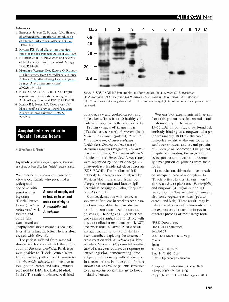
(SDS–PAGE). The binding of IgE
antibody to allergens was analyzed by
Western blot using serum from the
allergic patient and anti-human IgE
peroxidase conjugate (Dako, Carpinter-
ia, CA) (Fig. 1).
Contact dermatitis with lettuce is
somewhat frequent in workers who han-
dle these vegetables, but can also be
found in people sensitized to various
pollens (1). Helbling et al. (2) described
two cases of sensitization to lettuce with
positive radioallergosorbent test (RAST)
and prick tests to carrot. A case of an
allergic reaction to lettuce intake has
been described depicting the absence of
cross-reaction with A. vulgaris (3). Nev-
ertheless, Vila et al. (4) presented another
case of a mucous–cutaneous response to
lettuce ingestion, demonstrating some
antigenic commonality with A. vulgaris.
In a recent study, Enrique et al. (5) have
shown that 52.45% of patients sensitized
to P. acerifolia present allergy to food,
including lettuce.
Western blot experiments with serum
from this patient revealed several bands
predominantly in the range of
15–65 kDa. In our study, we found IgE
antibody binding to a mugwort allergen
(approximately 18 kDa), the same
molecular weight as the one found in
sunflower extracts, and several proteins
of P. acerifolia. Moreover, this patient,
in spite of tolerating the ingestion of
leeks, potatoes and carrots, presented
IgE recognition of proteins from these
foods.
In conclusion, this patient has revealed
an infrequent case of anaphylaxis to
�Tudela� lettuce hearts (L. sativa var.),
skin reactivity to plane tree (P. acerifolia)
and mugwort (A. vulgaris), and IgE
recognition by Western blot to these and
also some vegetable extracts (potato,
carrot, and leek). These results may be
indicative of a case of poly-sensitization,
the expression of general epitopes in
different proteins or most likely both.
*R&D Department,
DIATER Laboratorios,
Soledad 37
28330 San Martin de la Vega
Madrid
Spain
Tel: 34 91 808 77 27
Fax: 34 91 895 80 24
E-mail: [email protected]
Accepted for publication 19 May 2003
Allergy 2003: 58:1205–1206
Copyright � Blackwell Munksgaard 2003
A case of anaphylaxisto lettuce heart andcross-reactivity toP. acerifolia andA. vulgaris.
Figure 1. SDS PAGE IgE immunoblot. (1) Baby lettuce. (2) A. porrum. (3) S. tuberosum.
(4) P. acerifolia. (5) C. scolymus. (6) D. sativus. (7) A. vulgaris. (8) H. annus. (9) T. officinale.
(10) H. brasiliensis. (C-) negative control. The molecular weight (kDa) of markers run in parallel are
indicated.
1205
ALLERGY Net

References1. Franck P, Kanny G, Dousset B, Nabet P,
Moneret Vautrin DA. Lettuce allergy.
Allergy 2000;55:201–202.
2. Helbling A, Schwartz HJ, Lopez M,
Lehrer SB. Lettuce and carrot allergy: are
they related? Allergy Proc 1994;15:33–37.
3. Cadot P, Kochuyt AM, Deman R,
Stevens EA. Inhalative occupational and
ingestive inmediate-type allergy caused by
chicory (Chicorium intybus). Clin Exp
Allergy 1996;26:940–944.
4. Vila L, Sanchez G, Sans ML, Dieguez I,
Martinez J, Palacios R et al. Study of a
case of hypersensitivity to lettuce (Lactuca
sativa). Clin Exp Allergy 1998;28:1031–
1035.
5. Enrique E, Cistero Bahima A, Barto-
lome B, Alonso R, San Miguel Moncin
MM, Bartra J et al. Platanus acerifolia
pollinosis and food allergy. Allergy
2002;51:351–356.
Peanut and tree nut allergy inchildren: role of peanutsnacks in Israel?
Y. Levy*, A. Broides, N. Segal, Y. L. Danon
Key words: anaphylaxis; food allergy; peanuts; treenuts.
Prevalence rates of peanut and tree nut
allergy in Israel are 0.04 and 0.02%,
respectively (1). Children are often
exposed to
peanuts very
early owing to
the popularity of
locally produced
peanut snacks,
which have a
spongy texture
and melt on contact with saliva, making
them safe for consumption even before
6 months of age. The aim of this study
was to determine the age of first allergic
reaction to peanuts and tree nuts in Israel
and to outline the clinical features of
these allergies.
File review of all 992 infants and
children evaluated for food allergy
between January 1999 and July 2002
yielded 21 with peanut allergy (including
three also with tree nut allergy) and
eight with tree nut allergy. Diagnosis
was based on an unequivocal history of
immediate reaction to peanuts or tree
nuts involving one or more organ sys-
tems (skin, gastrointestinal, respiratory)
and a positive skin prick test (ALK
Abello, Port Washington, NY) or blood
test for specific IgE (>0.35 IU/ml)
(AlaSTAT, DPC, Los Angeles, CA).
Sixteen patients had atopic dermatitis
and 18 (15 with peanut allergy) had
additional food allergies (13 to eggs,
four to sesame, seven to milk, one to
apples). The characteristics of the first
allergic reaction to peanuts or tree nuts
are shown in Table 1. In 18 patients
with peanut allergy (86%), the first
allergic reaction occurred to peanut
snacks.
The prevalence of atopic dermatitis
and other food allergies and the clinical
presentation were similar to findings in
the literature (2). Of interest is the low
prevalence of peanut/tree nut allergy,
with our 29 patients accounting for
2.9% of patients evaluated for food
allergy, compared with 28 and 50%
reported in French studies (3, 4). At the
same time, the median age of the first
allergic reaction to peanuts of
8.3 months in our patients was consid-
erably lower than in series from the
USA and Europe (14 months to
4.4 years) (2–6). The lower prevalence of
peanut allergy can be explained by the
lower average consumption of peanut
products in Israel (1.4 kg per person per
year) (7) compared with the USA
(2.7 kg) (8) and the different methods of
production. The peanuts in most of the
locally produced peanut snacks in Israel
are boiled in water for 30 min at 80�C(Local factories, personal communica-
tion), whereas most peanuts in the
USA are dry-roasted at a much higher
temperature of 170�C, which increases
the allergenicity of the three major
peanut proteins (8).
Early exposure to peanut snacks may
lead to an early age of first allergic
reaction. Clinicians need to educate
parents to refrain from offering peanut
snacks to children younger than
2 years.
*Kipper Institute of Immunology
Schneider Children’s Medical Center of Israel
14 Kaplan Street
Petah Tiqva 49202
Israel
Tel: 972-3-925 3652
Fax: 972-3-925 905
E-mail: [email protected]
Accepted for publication 20 May 2003
Allergy 2003: 58:1206–1207
Copyright � Blackwell Munksgaard 2003
Early exposure topeanut snacks maylead to an early age offirst allergic reaction.
Table 1. Characteristics of the first allergic reaction to peanuts or tree nuts
Peanuts (n ¼ 21) Tree nuts (n ¼ 11)
Age (months)
Range 3–108 36–144
Median 8.25 50
Foods causing reactions 18: peanut snacks 6: cashew/pistachio
3: peanuts 1: pistachio ice cream
1: pecan
1: walnut
1: mixed nuts (granola)
1: nut spread
Symptoms and signs (one or more)
Skin 21 (100%) 11 (100%)
Gastrointestinal 5 (23.8%) 5 (45.5%)
Respiratory 6 (28.5%) 5 (45.5%)
Cardiovascular 0 1 (9%)
Skin: urticaria, angioedema, rash, exacerbation of atopic dermatitis.
Gastrointestinal: vomiting, abdominal pain, diarrhea.
Respiratory: rhinorrhea, cough, hoarseness, shortness of breath, wheezing.
Cardiovascular: hypotension.
1206
ALLERGY Net

References1. Dalal I, Binson I, Reifen R, Amitai Z,
ShohatT,RahmamiS et al. Foodallergy is a
matter of geography after all: sesame as a
major cause of severe IgE-mediated food
allergic reactions among infants and young
children in Israel. Allergy 2002;57:362–365.
2. Sicherer SH, Wesley-Burks A, Sampson
HA. Clinical features of acute allergic
reactions to peanut and tree nut in children.
Pediatrics 1998;102:e6.
3. Rance F, Abbal M, Lauwers-Cances V.
Improved screening for peanut allergy by
the combined use of skin prick tests and
specific IgE assays. J Allergy Clin Immunol
2002;109:1027–1033.
4. Moneret-Vautrin DA, Rance F, Kanny
G, Olsewski A, Gueant JL, Dutau G
et al. Food allergy to peanuts in France –
evaluation of 142 observations. Clin Exp
Allergy 1998;28:1113–1119.
5. Sicherer SH, Furlong TJ, Munoz-Fur-
long A, Wesley-Burks A, Sampson HA. A
voluntary registry for peanut and tree nut
allergy: characteristics of the first 5149 reg-
istrants. J Allergy Clin Immunol
2001;108:128–132.
6. Tariq SM, Stevens M, Matthews S, Rid-
out S, Twiselton R, Hide DW. Cohort
study of peanut and tree nut sensitization by
age of 4 years. Br Med J 1996;313:514–517.
7. Sheskin A, Regev A. Israel Agriculture –
Facts and Figures, 2nd edn, Dec. 2001.
Available at http://www.agri.gov.il.
8. Beyer K, Morrow E, Xiu-Min L, Bardina
L, Bannon GA, Wesley-Burks A et al.
Effects of cooking methods on peanut
allergenicity. J Allergy Clin Immunol
2001;107:1077–1081.
Maculopapular rash inducedby diltiazem: allergologicalinvestigations in four patientsand cross reactions betweencalcium channel blockers
C. Cholez, P. Trechot, J.-L. Schmutz, G. Faure,M.-C. Bene, A. Barbaud*
Key words: calcium channel blockers; cross-reaction;drug intradermal test; drug patch test; drug prick test;drug skin testing; lymphocyte activation test.
Drug skin tests were performed in four
patients who have developed a maculo-
papular rash
(MPR) 8–
12 days after the
beginning of a
treatment with
diltiazem, in or-
der to determine
the value of
patch tests (PT) and cross reactions
among calcium channel blockers (CCB).
Six weeks after the MPR, drug PT were
performed with the commercialized
forms of diltiazem following the guide-
lines of the European Society of Con-
tact Dermatitis (ESCD). The PT were
also done with the commercialized
forms of other CCB. When PT were
negative, prick tests (prick T) were
performed in two cases and one intra-
dermal test (IDT) with nimodipine in
one case. Lymphocyte activation tests
(LAT) were performed in three cases.
The PT were positive in all cases
without any cross reactions with other
CCB, except in one patient who had
positive PT with verapamil. Prick T in
two of two cases and IDT with ni-
modipine in one of one case remained
negative. The LAT were positive in
three of four cases. This study empha-
sizes the value of PT with diltiazem in
cutaneous adverse drug reactions
(CADR) because of this CCB, but PT
could have a lesser value with other
CCB. Cross reactions on PT seem to be
rare. More, although CCB are usually
divided in three classes, we suggest to
divide them into dihydropyridines and
�nondihydropyridines�.Maculopapular exanthema is the most
common cutaneous CADR and can be
induced by almost all drugs. In literature,
many cases have been reported on
CADR induced by CCB, more often with
diltiazem which has been associated with
a variety of cutaneous reactions from
exanthema to severe cutaneous reactions
such as Stevens–Johnson syndrome (SJS)
and toxic epidermal necrolysis (TEN).
We report on four patients who had
developed MPR because of diltiazem
with a likely imputability, confirmed in
all cases by positive PT and also the study
about cross reactions between CCB as
cross reactions between CCB have not so
much been studied.
Four patients (one man and three
women; mean age 60 years) developed a
MPR, 8–12 days after the beginning of a
treatment by diltiazem. All routine
laboratory investigations were normal
except the sedimentation rate (mean SR
26.3 mm). Six weeks after the CADR,
drug PT were performed with the com-
mercialized form of diltiazem, following
the guidelines of the ESCD (1). Drug PT
with the commercialized forms of dil-
tiazem were performed with pills crushed,
reduced in powder then diluted at 30% in
water, petrolatum (pet.) and alcohol in
three of four cases; pur, diluted at 30% in
water and pet. in one of four cases. In all
cases, PT were also performed with the
commercialized forms of other CCB
diluted at 30% in pet.: verapamil, nicar-
dipine, nifedipine, nitrendipine, nimodi-
pine, in order to study cross-reactivity
between CCB. Skin tests were read at
20 min, day 2, and day 4. When PT were
negative, prick T with these drugs were
performed in two of four cases and in one
case an IDT with injectable nimodipine
was performed. In three of four cases,
LAT were performed, according to the
method described by Kohler et al. (2). All
the patients had a very likely intrinsic
imputability according to criteria pro-
posed by Moore et al. (3).
Drug skin test and in vitro test are
shown in Table 1.
In all cases, PT were positive with
diltiazem diluted at 30% whatever the
vehicle used. Therewere no cross reactions
on PT with nimodipine and nifedipine.
The three patients tested with nicardipine
and nitrendipine had negative PT. There
were cross reactions on PT between dil-
tiazem and verapamil diluted at 30% in
pet. in only one of four patients. The prick
Twere negativewith nifedipine (one of one
case tested), verapamil (one of one case
tested), nicardipine (one of one case tes-
ted), nimodipine (two of two cases tested)
and diltiazem (two of two cases tested).
The IDT with nimodipine was negative
in one of one case tested. The LAT with
diltiazem were positive in three of four
cases tested.
Maculopapular rash is a frequent
CADR reported with many drugs, inclu-
ding antibiotics, antineoplastic drugs,
antiepileptics which occured usually 24 h
to 10 days after the beginning of the
treatment. Among CCB, diltiazem has
been considered as a causative factor of
a wide spectrum of cutaneous adverse
Suggested guidelinesfollowing the divisionof CCB intodihydropyridines and'nondihydropyridines'.
1207
ALLERGY Net

reactions such as MPR (4–10), psoriasi-
form eruption (9), exfoliative dermatitis
(5, 11), acute generalized exanthematous
pustulosis (12–15), hypersensitivity syn-
drome (16, 17), severe erythema multi-
forme, SJS and TEN (4). Other CCB,
such as nifedipine or verapamil have also
been associated with MPR (7). Skin tests
have been reported to be helpful in
determining the cause of CADR, their
results depend on the drug tested but also
on the clinical features of the CADR. In a
prospective study involving 72 patients
who had developed CADR, 43% had
pertinent positive PT (18). Among these
72 patients, one of them had developed
MPR after have taken diltiazem with
positive PT at 4 days. The results
obtained in the herein reported study
including four patients, emphasizes the
value of PT with diliazem with positivity
of all PT with diltiazem (four of four
cases) in investigations CADR because of
this drug. In literature, skin testing with
diltiazem have already been reported to
be useful to diagnose eruptions caused by
this drug (5, 6, 8–10, 13–15), on contrary,
PT with other CCB, such as verapamil,
nifedipine, nisoldipine or nicardipine
does not seem to be useful because of
their low reported positivity (9). This is
due either because CADR with diltiazem
have been reported in literature more
often than other CCB or because
diltiazem widely prescribed induces a
higher number of CADR.
Skin tests with diltiazem performed
following the guidelines of the ESCD (1),
seem sensitive (four of four patients had
positive PT). These tests have also a good
specificity, as on 11 negative control
subjects, selected following methods pre-
viously published (18), PT with diltiazem
diluted at 30% in water and pet. were
negative. Positivity of these tests and
LAT (three of four positive) is in favor of
a mechanism of delayed cellular hyper-
sensitivity.
The CCB are frequently used in cardi-
ology to manage ischemic heart disease
or high blood pressure. They belong to a
heterogeneous chemical group and the
CCB function is not limited to a partic-
ular chemical structure. Therefore, bufl-
omedil, perhexilline, bepridil, flunarizine
and cinnarizine are all CCB. The CCB
are usually divided in three classes:
dihydropyridines (amlodipine, felodipine,
isradipine, lacidipine, nicardipine, nifedi-
pine, nimodipine, nitrendipine and
nisoldipine), phenylalkylamines with
verapamil and benzothiazepines with
diltiazem. Dihydropyridines molecules
have a common membrane receptor that
binds tritiated nitrendipine or nimodipine
(19). Verapamil and diltiazem have a
strereospecific receptor that binds tritiat-
ed cinnarizine (19). Thus, we propose to
divide CCB into dihydropyridines and
�nondihydropyridines�.In literature, there are some discrep-
ancies concerning cross reactions
between CCB. For example, Kuo et al.
(20), reported on the case of a woman
who experienced nonthrombocytopenic
purpura with nifedipine with a similar
eruption 48 h after a new treatment by
diltiazem. Baker and Cacchione (21),
reported the case of a 52-year-old man
who developed a MPR approximately
24–36 h after starting diltiazem therapy.
Three days after diltiazem was discon-
tinued, the patient received amlodipine
with the same cutaneous reaction within
1–2 h after amlodipine administration.
These two cases lead to think that there
is a cross-reaction between dihydropyri-
dines and �nondihydropyridines�. How-
ever, we can observe that in the case
reported by Kuo et al. (20), a skin
biopsy sample taken from a lesion dis-
played a leucocytoclastic vasculitis which
has often a chronic evolution, with
sometimes recurrences, perhaps ruling
out the responsability of diltiazem in
relapse of the purpuric lesions. Con-
cerning the patient presented by Baker
and Cacchione (21), the second CCB
(amlodipine) was readministered only
3 days after diltiazem was discontinued,
which appears to be a very short delay,
making difficult to specify if it is a
second CADR to amlodipine, with a
cross reaction between diltiazem and
amlodipine, or if it is the manifestation
of a long lasting CADR because of
diltiazem. In the case described by
Hammentgen et al. (10), a 60-year-old
man had a MPR because of diltiazem
without any cutaneous reactions after
having been rechallenged with nifedi-
pine. In our study, one patient who had
a MPR because of diltiazem had a
fortuitous well-tolerated challenge with
lacidipine, belonging to the dihydropy-
ridines. Concerning cross reactions
between the dihydropyridines, Bewley
et al. (22), have reported on the case of a
Table 1. Results of drug patch testing
Patient
no. 1
Patient
no. 2
Patient
no. 3
Patient
no. 4
Patch-tests
Monotildiem� et Tildiem� cp (diltiazem)
Pure np np np +
30% water + + + +
30% vaseline + + + +
30% alcohol + + + np
Isoptine� (verapamil) + ) ) )Loxen� (nicardipine) ) ) ) np
Nidrel� (nitrendipine) ) ) ) np
Nimotop� (nimodipine) ) ) ) )Adalate� (nifedipine) ) ) ) )30% vaseline
Prick-tests
Adalate� (nifedipine) ) np np np
Isoptine� (verapamil) ) np np np
Loxen� (nicardipine) ) np np np
Nimotop� (nimodipine) ) np ) np
Tildiem� (diltiazem) ) np ) np
IDR with Nimotop� (nimodipine) ) np np np
LAT with diltiazem + + np +
np, not performed; LAT, lymphocyte activation test.
1208
ALLERGY Net

62-year-old patient, with a history of
high blood pressure treated by amlodi-
pine for 2 years without any cutaneous
eruption. This patient was admitted in
hospital to treat a chronic plaque pso-
riasis and during admission, antihyper-
tensive medication was changed to
amlodipine. Three days after this
change, he developed an erythema
multiforme, after which the amlodipine
was stopped and nifedipine readminis-
tered without no further complications.
Kitamura et al. (9), described a 56-year-
old patient who experienced a psoriasi-
form eruption because of nifedipine,
with the same eruption after having
taken nisoldipine.
In literature, diltiazem is the CCB the
most frequently reported as responsible
in inducing CADR. In most of the cases,
CADR are MPR which occured usually
10 days after the beginning of the CCB.
According to previous reports and these
results, PT seem to be useful in diagno-
sing CADR due to diltiazem, but PT
could have a lesser value with other CCB.
Finally, although CCB are divided
into three classes, we suggest, from our
results, those previously published and
the chemical analysis of the chemical
structures of CCB to divide them into
two chemical classes: dihydropyridines
and �nondihydropyridines�. According to
this classification, it could be possible, in
case of CADR to CCB, to follow these
guidelines:
1. Skin tests should be performed,
6 weeks to 6 months after the
CADR, with commercialized forms
diluted at 30% in pet. and/or with
the pure drug diluted at 10% in pet.
and water. In case of severe cutane-
ous reactions such as SJS, Lyell’s
syndrome or hypersensitivity
syndrome, these tests could also be
performed but with caution and in
beginning with very low concentra-
tions of the drugs i.e. 0.1% in pet-
rolatum then if negative with
progressively enhanced concentra-
tions. We have no experience con-
cerning patch testing in severe
CADR because of CCB.
2. In cases of CADR with CCB
belonging to the dihydropirydines, it
could be possible, to readminister,
under hospital surveillance, if PT
with these CCB are negative, a
�nondihydropyridine�.
Further larger studies are necessary to
validate these guidelines.
*Fournier Hospital
University Hospital of Nancy
Department of Dermatology
36, quai de la bataille
54000 Nancy
France
Tel: +33 (0)3 83 85 24 65
Fax: +33 (0)3 83 85 24 12
E-mail: [email protected]
Accepted for publication 25 March 2003
Allergy 2003: 58:1207–1209
Copyright � Blackwell Munksgaard 2003
References1. Barbaud A, Goncalo M, Bruynzeel D,
Bircher A. Guidelines for performing skin
tests with drugs in the investigation of
cutaneous adverse drug reactions. Contact
Dermatitis 2001;45:321–328.
2. Kohler C, Kolopp-Sarda MN, De
March-Kennel A, Barbaud A, Bene MC,
Faure GC. Sequential assesment of cell
cycle S in flow cytometry: a non isotopic
method to measure lymphocyte activation
in vitro. Anal Cell Pathol 1997;14:51–59.
3. Moore N, Biour M, Paux G, Loupie E,
Begaud B, Boismare F et al. Adverse
drug reaction monitoring: doing it the
French way. Lancet 1985;2:1056–1058.
4. Knowles S, Gupta AK, Shear NH. The
spectrum of cutaneous reactions associated
with diltiazem: three cases and a review of
the literature. J Am Acad Dermatol
1998;38:201–206.
5. Sousa-Basto A, Azenha A, Duarte M,
Pardal-Oliveira F. Generalized cutane-
ous reaction to diltiazem. Contact Der-
matitis 1993;28:44–45.
6. Barbaud A, Trechot P, Gillet-Terver
M, Zannad F, Schmutz JL. Investigations
immunoallergologiques dans une toxider-
mie au diltiazem (Tildiem 300 LP).
Therapie 1993;48:499–500.
7. Stern R, Khalsa JH. Cutaneous adverse
reactions associated with calcium channel
blockers. Arch Intern Med 1989;149:
829–832.
8. Romano A, Pietrantonio F, Gacovich A
et al. Delayed hypersensitivity to diltiazem
in twopatients.AnnAllergy 1992;69: 31–32.
9. Kitamura K, Kanasashi M, Suga C,
Saito S, Yoshida S, Ikezawa Z. Cuta-
neous reactions induced by calcium
channel blocker: high frequency of
psoriasiform eruptions. J Dermatol
1993;20:279–286.
10. Hammentgen R, Lutz G, Kohler U,
Nitsch J. Makulopapuloses exanthem bei
diltiazem-therapie. Dtsch Med Wschr
1988;113:1283–1285.
11. Odeh M. Exfoliative dermatitis associated
with diltiazem. J Toxicol Clin Toxicol
1997;35:101–104.
12. Lambert D, Dalac S, Beer F, Chavannet
P, Portier H. Acute generalized exanthe-
matous pustular induced by diltiazem. Br J
Dermatol 1988;118:308–309.
13. Vicente-Calleja JM, Aguirre A, Landa
N, Crespo V, Gonzalez-Perez R. Acute
generalized exanthematous pustulosis due
to diltiazem: confirmation by patch testing.
Br J Dermatol 1997;137:837–839.
14. Wakelin S, James M. Diltiazem-induced
acute generalised exanthematous pustulo-
sis. Clin Exp Dematol 1995;20:341–344.
15. Jan V, Machet L, Gironet N, Martin L,
Machet MC, Lorette G et al. Acute
generalized exanthematous pustulosis
induced by diltiazem: value of patch
testing. Dermatology 1998;197:274–275.
16. Lahav M, Arav R. Diltiazem and
thrombopenia. Ann Intern Med 1989;
110:327.
17. Dominguez EA, Hamill RJ. Drug in-
duced fever due to diltiazem. Arch Intern
Med 1991;151:1869–1870.
18. Barbaud A, Reichert-Penetrat S,
Trechot P, Jacquin-Petit MA, Ehlinger
A, Noirez V et al. The use of skin testing in
the investigation of cutaneous adverse drug
reactions. Br J Dermatol 1998;139: 49–58.
19. Zannad F, Baille N. Les antagonistes
calciques. In: Gilgenkrantz JM, Royer
RJ, Zannad F, editors. Therapeutique en
pathologie cardio-vasculaire. Paris: Mede-
cine-Sciences Flammarion, 1987: 124–142.
20. Kuo M, Winiarski N, Garella S. Non-
thrombocytopenic purpura associated
sequentially with nifedipine and diltiazem.
Ann Pharmacother 1992;26:1089–1090.
21. Baker AB, Cacchione JG. Dermatologic
cross-sensitivity between diltiazem and
amlodipine. Ann Pharmacother
1994;28:118–119.
22. Bewley A, Feher M, Staughton R.
Erythema multiforme following substitu-
tion of amlodipine for nifedipine. Br Med J
1993;307:241.
1209
ALLERGY Net

Drug allergy in universitystudents from Porto, Portugal
H. Falc¼o*, N. Lunet, E. Gomes, L. Cunha, H. Barros
Key words: antibiotics; anti-inflammatory agents; aspirin;drug hypersensitivity; lactam; nonsteroidal; penicillins.
Drug allergy is considered responsible for
substantial morbidity and mortality, and
increased health
costs. However,
its true frequency
is not known be-
cause of scanty
epidemiological
data, and avail-
able information requires a cautious
interpretation (1).
Aiming to quantify the prevalence of
self-reported drug allergy, we performed
a cross-sectional survey of 2150 Portu-
guese university students (67.6% females)
during 2001. Participants were ap-
proached in the classroom, at different
days and times, resulting in the study of
37% of all registered students.
The lifetime occurrence of drug allergy,
the drugs that are involved and the
characteristics of the most serious epi-
sode were assessed using a self-adminis-
tered questionnaire. Symptoms were
classified as dermatological (pruritus,
erythematous wheals and oedema), ocu-
lar and respiratory (redness or eye
itching, tears, nose itching or discharge,
blocked nose or sneezing, dyspnoea,
wheezing or cough), gastrointestinal
(vomiting, nausea, diarrhoea and
abdominal pain) or systemic (sweating or
perspiration, fainting and tachycardia).
The life prevalence of one or more drug
allergy episodes was 7.7%, with 3.1%
allergic tob-lactams, 2.1% to nonsteroidal
anti-inflammatory drugs (NSAIDs) and
3.0% to other drugs. Three participants
(0.1%) declared allergy to b-lactams and
NSAIDs, three (0.1%) to b-lactam anti-
biotics and other drugs, four (0.2%) to
NSAIDs and other drugs, and two (0.1%)
reported allergy to b-lactams, NSAIDs
and other drugs. No significant differences
were observed according to sex.
When a drug was specifically recalled
(44% of participants), penicillin (57.1%),
amoxicillin (19.1%) and amoxicillin
associated with clavulanic acid (23.8%)
were the more frequently incriminated
b-lactams, and aspirin (41.4%), lysine
acetylsalicylate (27.6%) and nimesulide
(20.7%) were the more often reported
NSAIDs. Regarding other types of drugs,
non-b lactam anti-infectious drugs were
the most commonly reported.
Dermatological manifestations were
the most frequently described, both in
cases of allergy to b-lactams (79.7%) and
NSAIDs (58.4%), followed by gastroin-
testinal (22.0 and 33.4%, respectively),
systemic (28.8 and 22.2%, respectively),
and ocular and respiratory (17.0 and
27.8%, respectively).
In our survey, the prevalence of drug
allergy was lower than previously repor-
ted (2, 3). Possible explanations are the
young age of our participants, with lower
drug consumption, and the fact that
older adults evaluated in other surveys
may have been exposed to penicillin and
ampicillin preparations used before the
1970s, often contaminated with trace
quantities of macromolecules or drug
polymers, more allergenic than those
available nowadays (4).
Highly educated individuals may
improve the quality and accuracy of
self-reported information, and the recall
of a drug allergy is expected to be more
accurate in young individuals as less time
elapsed since an allergic reaction,
increasing the internal validity of our
investigation. This is an advantage of our
study, but recall bias remains, as per-
ceived by the amount of missing infor-
mation concerning the characteristics of
allergy episodes or brand names of the
drugs involved, probably reflecting that
many reactions occurred at young ages.
Drug allergy was recalled by 7.7% of
university students in Portugal. Although
many of these recalls might not reflect
true allergy, these individuals will prob-
ably be given second-line treatments,
usually more expensive and less effective.
Nuno Lunet gratefully acknowledges a
Grant from Fundacao para a Ciencia e a
Tecnologia (SFRH/BD/3293/2000).
*Servico de Higiene e Epidemiologia da
Faculdade de Medicina do Porto
Al. Prof. Hernani Monteiro
4200-319 Porto
Portugal
Accepted for publication 30 April 2003
Allergy 2003: 58:1210
Copyright � Blackwell Munksgaard 2003
References1. Demoly P, Bousquet J. Epidemiology of
drug allergy. Curr Opin Allergy Clin
Immunol 2001;1:305–310.
2. Kerr JR. Penicillin allergy: a study of
incidence as reported by patients. Br J Clin
Pract 1994;48:5–7.
3. Hung OR, Bands C, Laney G, Drover D,
Stevens S, MacSween M. Drug allergies in
the surgical population. Can J Anaesth
1994;41:1149–1155.
4. Adkinson N. Drug allergy. In: Middleton
E, Jr, Reed C, Ellis E, Adkinson F, Jr,
Yunginger J, Busse W, editors. Allergy
principles & practice. St Louis, Missouri:
Mosby Year Book, Inc., 1998:1212–1224.
Anaphylactic reactions toformaldehyde in root canalsealant after endodontictreatment: four cases ofanaphylactic shock and threeof generalized urticaria
J. J. Braun*, H. Zana, A. Purohit, J. Valfrey,Ph. Scherer, Y. HaBkel, F. de Blay, G. Pauli
Key words: allergy to formaldehyde; anaphylacticshock; angioedema; anti-formaldehyde IgE; endodontictreatment; formaldehyde; root canal sealant; urticaria.
The authors report seven cases of allergic
reactions, four cases of anaphylactic
shock and three of generalized urticaria,
to formaldehyde contained in root canal
sealant after endodontic treatment.
The clinical
presentation,
skin tests, high
levels of anti-
formaldehyde
immunoglobulin
E (IgE), as well
as the study of
the previous cases reported in the litera-
ture, suggest allergic IgE mediated
mechanisms. These very infrequent but
potentially severe reactions in endodontic
therapy focus attention on the different
manifestations related to formaldehyde,
Drug allergy wasrecalled by 7.7% ofuniversity students inPortugal.
Anaphylaxis toformaldehydecontained in root canalsealant.
1210
ALLERGY Net

the involved mechanisms, the diagnostic
procedure and the prevention possibilities
in dentistry.
Formaldehyde (formalin, paraformal-
dehyde, trioxymethylene) is widely used
in industry, cosmetics, disinfectants,
medications and root canal sealants
(1, 2).
The pathological reactions related to
formaldehyde such as nasal, laryngeal
and bronchopulmonary lesions appear-
ing upon inhalation, gastrointestinal
lesions appearing upon ingestion, and
cutaneous necrosis or contact dermatitis,
may be caused by simple irritant
mechanisms (1–3). In addition, hyper-
sensitivity or allergic reactions such as
rhinitis, asthma, generalized urticaria,
angioedema and anaphylactic shock,
have been described (4–27).
Despite the high frequency of root
canal treatments with sealant containing
formaldehyde, very few cases of well
documented allergy to root canal sealant
have been reported in the literature.
These sealants are a complex mixture of
potentially irritating and/or sensitizing
substances, such as metals, eugenol, for-
maldehyde, menthol, phenol, etc (5, 6).
Formaldehyde release from root canal
sealant has been demonstrated in vitro
and in vivo, and as a hapten may induce
anaphylactic reactions after reacting with
other proteins to become a complete
allergen. (12, 18, 28–32).
The authors report seven cases of
allergic reactions, four cases of anaphy-
lactic shock and three of generalized
urticaria, to formaldehyde contained in
root canal sealant after endodontic
treatment.
The clinical presentation, skin tests,
high levels of anti-formaldehyde immu-
noglobulin E (IgE), as well as the study of
the previous cases reported in the litera-
ture, suggest allergic IgE mediated
mechanisms. These very infrequent but
potentially severe reactions in endodontic
therapy focus attention on the different
manifestations related to formaldehyde,
the involved mechanisms, the diagnostic
procedure and the prevention possibilities
in dentistry.
Case 1: A 41-year-old nonatopic man
presented with a periapical granuloma of
tooth 24 requiring endodontic treatment.
Several minutes after complete dental
treatment the patient complained of a
sensation of warmth, generalized pruritus
and respiratory difficulty. Thirty minutes
later he developed anaphylactic shock
with a drop in systolic blood pressure to
50 mmHg and lost consciousness. The
outcome was favorable with an emer-
gency resuscitation.
The root canal sealant used was Spad�
(Quetigny, France), a mixture of powder
and liquids with the following composi-
tion: phenylmercury borate, calcium
hydroxide, hydrocortisone acetate, tri-
oxymethylene, titanium oxide, barium
sulfate, zinc oxide (powder), glycerin,
resorcinol, hydrochloric acid (liquid 1)
and glycerin and formaldehyde 87%
(liquid 2).
Skin prick tests to common aeroaller-
gens and latex performed after the acci-
dent were negative. The skin prick test to
liquid 1 was negative and to liquid 2 was
mildly positive, with a 3 mm diameter
wheal vs 4 mm to codeine as a positive
control. The same tests were negative in
five control subjects. Patch tests, per-
formed using finn chambers, to the
standard battery of International
Contact Dermatitis Research Group
(ICDRG) allergens containing formalde-
hyde and to resorcinol (Trolab
Allergenes, Reinbeck, Germany) were
negative. Patch tests to the powder and
two liquids (1% solution in Vaseline)
induced a strongly positive delayed
reaction with a confluent eczema beyond
the test area. Anti-formaldehyde IgE
were class 4 (25 kU/l: RAST CAP RIA,
Pharmacia, Uppsala, Sweden).
Case 2: A 45-year-old woman under-
went several root canal treatments with
Spad� between 1987 and 1997 without
any complications (tooth 16, 17 and 26).
In 1997, a second endodontic treatment
of tooth 26 with Spad� was followed by
discomfort, anxiety, pruritus of the hands
and pallor, which regressed after antihis-
tamine treatment. The patient underwent
treatment of tooth 16 for the second time
in 1998 with Method Z� (Zizine France).
Fifteen minutes later, she experienced
tachycardia with extreme apprehension,
pruritus and erythema of the hands and
was unresponsive to antihistamines. This
was followed by angioedema, dyspnoea
and severe systolic hypotension
(60 mmHg). The outcome was favorable
with administration of adrenaline and
systemic steroids.
The root canal sealant Method Z�
contains: enoxolone, barium sulfate, ex-
cipient (powder), resorcinol, hydrochloric
acid, excipient (liquid 1), and formalde-
hyde 35% and excipient (liquid 2).
Skin prick tests to common aeroaller-
gens, latex and to liquid 1 and 2 at the
concentration of 1% were negative. Patch
tests with liquid 2 (1% solution) induced
a local eczema after 48 h. RAST to
formaldehyde was class 1 (0.41 KU/l;
AlaSTAT DPC).
Case 3: A 43-year-old woman under-
went an endodontic treatment of tooth 42
with Zial Z� (Zizine, France). Several
similar procedures had been performed in
the past. She experienced thoracic
oppression and erythema developing
within 24 h on two occasions. Two hours
after the last treatment the patient
developed generalized erythema, angi-
oedema, vomiting, diarrhea, and hypo-
tension and lost consciousness twice.
After the first emergency resuscitation
with adrenaline, she developed cardiac
arrest whilst in hospital. The final out-
come was favorable.
The root canal sealant Zial� contains:
hydrocortisone, trioxymethylene, diiodo-
thynol, E110, barium sulfate, zinc oxide,
magnesium stearate (powder) and euge-
nol (liquid).
Skin prick tests and patch tests to
liquid and powder at different concen-
trations were negative. Anti-formalde-
hyde IgE were class 3 (5.09 KU/l RAST
CAP RIA Pharmacia Sweden).
Case 4: A 50-year-old man underwent
several root canal treatments with Spad�.
The treatment of tooth 45 in July 1996
was followed 2 h later by urticaria of the
head and facial edema. In December
1996, 30 min after treatment of tooth 5,
the patient developed pruritus, general-
ized urticaria, abdominal pain and dis-
comfort. An allergy to the local
anesthetic agent (lidocaine) was suspec-
ted but the skin tests and the challenge
test were negative. In September 2002 the
patient needed extraction of tooth 45 and
endodontic treatment of tooth 44. This
procedure was hemorrhagic and tooth 45
treated in 1996 was broken during the
extraction. Fifteen minutes later, the
patient presented with facial erythema,
pruritus, generalized urticaria, abdominal
pain, dyspnoea, discomfort with tachy-
cardia and severe hypotension. The
1211
ALLERGY Net

outcome was favorable with administra-
tion of adrenaline and steroids given
several times in the emergency hospital.
One month later, obturation of the pulp
chamber of tooth 38 without apical
treatment (pulpotomy) was well toler-
ated.
Skin prick tests to the powder and
liquids of Spad� performed with 1%
dilution and pure form as used in
dentistry were negative. Anti-formalde-
hyde IgE were class 6 (>100 KU/l;
Unicap Pharmacia Sweden).
Case 5: A 40-year-old woman, with a
history of allergy to grass pollen and
house dust mite, was treated for the
second time for a granuloma of tooth 21
with Resoplast� (Pierre Roland, France)
and Temp Bond�. Three hours after
dental treatment the patient developed
abdominal pain and pruritus of the scalp
followed by urticaria of the face, neck,
upper extremity and chest without hypo-
tension. Outcome was favorable with
symptomatic treatment in the emergency
hospital.
Resoplast� has the following compo-
sition: deltahydrocortisone, bismuth ni-
trate (powder); benzalkonium chloride
and formaldehyde (liquid 1) and sulfo-
salicylic acid and resorcinol (liquid 2).
Temp Bond� (Kerr, Romulus, MI)
contains zinc oxide and eugenol.
Skin prick tests to undiluted liquid 1
were positive giving a 7 mm diameter
wheal and edema of the forearm. They
were negative in four control subjects.
Similar tests with liquid 2 and eugenol
were negative. Anti-formaldehyde IgE
were class 5 (98.5 KU/l; RAST AlaSTAT
DPC).
Case 6: A 64-year-old woman who
underwent endodontic treatment with
Spad� on two previous occasions, had
experienced moderate to severe local
edema. On the third occasion, 4 h after
the procedure, in addition to local
edema, she developed nausea, vertigo
and generalized urticaria, which persisted
for 3 days despite antihistamine treat-
ment.
Skin tests were not performed. Anti-
formaldehyde IgE were class 5 (65 KU/l;
RAST Alastat DPC).
Case 7: A 56-year-old man underwent
root canal treatment of tooth 28 with
Spad� in February 2000 after several
previous uncomplicated endodontic pro-
cedures. Thirty minutes later he devel-
oped first a significant localized edema
and than an edema of the whole face.
Several hours later he presented with
generalized urticaria that lasted 3 days.
Prick skin tests were weakly positive to
formaldehyde. Anti-formaldehyde IgE
were class 6 (>100 KU/l Unicap Phar-
macia Sweden).
The adverse reactions to formalde-
hyde, such as respiratory (asthma and
rhinitis) and cutaneous (contact derma-
titis) reactions and anaphylactic shock in
hemodialysis, are well documented.
However, despite its widespread use, IgE
dependant allergic reactions are rarely
described (1–3, 14, 18, 27, 33, 34).
In odonto-stomatology the root canal
sealants containing formaldehyde are still
widely used. Different side effects related
to endodontic treatment have been
reported, such as infection, inflammation,
necrosis, arthritis, paresthesia of the
dental branch of the mandibular nerve,
fungal caseous sinusitis etc. (5, 35–44). In
dentistry, formaldehyde is used for its
antibacterial activity, for devitalization of
the tooth pulp and for its role in poly-
condensation of resorcine. (5, 11–14, 18).
Release of formaldehyde from endodon-
tic material has been known for a long
time. Different in vitro and in vivo studies
have shown a systemic diffusion of C14
labeled formaldehyde from endodontic
material. The formaldehyde release may
be enhanced by repetitive endodontic
treatments, apicetomies, extraction of the
treated tooth and dental overfilling with
extrusion of sealant in the periapex or in
the apical granuloma (5, 28–32, 35).
Compared with the pulp chamber, which
is relatively inert from an immunological
point of view, the periapex constitute a
network of vascular and nervous systems
joining the tooth to the rest of the
immune system (5, 6, 36, 38, 40, 43, 44).
Formaldehyde is a low molecular
weight chemical which, acting as a hap-
ten, may react with other molecules such
as cutaneous proteins, serum proteins,
proteins of the pulp chamber or of the
periapex, or even with another compo-
nent of the root canal sealant to become a
complete allergen (3, 5, 12, 14, 15, 17, 18).
Despite the frequency of formaldehyde
use and the number of endodontic treat-
ments (453 000 in 1990 in Denmark for
5 million inhabitants) the allergic reac-
tions to it in dentistry remain infrequent
(5, 6, 18). They are probably underesti-
mated in endodontic practice. Thirty-five
cases of allergic reactions to formalde-
hyde (7–27) including seven personal
observations have been described
(Table 1). These allergic reactions can be
of different severity ranging from local or
focal reactions to life-threatening ana-
phylactic reactions: 15 cases of anaphy-
lactic shock, 18 cases with urticaria and/
or angioedema, nausea, dyspnoea, exan-
thema, pruritus and two cases with non-
clearly defined symptoms. An additional
case of formaldehyde related anaphylac-
tic shock in a patient undergoing renal
dialysis has been reported (33, 34). The
symptoms could be of early onset,
appearing within several minutes to 1 h
after dental treatment (nine cases), or
delayed, appearing from 2 to 24 h after
the treatment (21 cases) (Table 1).
The skin tests to formaldehyde are not
standardized and may provoke even
delayed severe systemic reactions (15, 34).
Prick tests to 0.1 and 1% formaldehyde
solution are often negative and are
inconsistently positive to the pure solu-
tion as used in dentistry. Patch tests to
the standard battery of ICDRG con-
taining formaldehyde are very often
negative. Those skin tests to the native
solution used in dentistry, or to 1%
formaldehyde solution, sometimes give a
delayed positive reaction, but their clin-
ical significance is difficult to establish in
some cases (14, 18). Skin tests were
positive in 19 cases and negative in
11 cases (Table 1).
The measurement of specific IgE to
formaldehyde is an important diagnostic
element and may suggest underlying
allergic mechanisms. Positive RAST,
often with higher class, was detected in
all cases when it was analyzed (20
cases). However, in some cases specific
IgE have been detected without associ-
ated clinical symptoms (2, 3, 44–46).
This may raise the question of its real
significance in view of ubiquitous
exposure to formaldehyde, particularly
by respiration. Formaldehyde in powder
form or in aqueous solution may be
more reactive than in gaseous form and
thus may lead to sensitization in odon-
to-stomatology. This sensitization could
result from domestic or occupational
contact with formaldehyde (cosmetics,
1212
ALLERGY Net

certain medicines, in dentists, anato-
mists, pathologists, etc) and especially
after previous endodontic treatments
(all seven in our case reports). Dental
overfilling with extrusion of root canal
sealant and also instrumental interven-
tion (apicectomie, dental extraction of
treated tooth, repeated treatments of
the same tooth, etc) may promote
diffusion of the soluble formaldehyde in
the apical or periapical region which is
inflammatory and hypervascularized in
conditions such as apical granuloma (5,
28–30, 32, 35, 40–42). The more or less
rapid diffusion of formaldehyde after
the endodontic treatment and the
necessity of binding with a protein to
form an antigenic conjugate may ex-
plain the more or less rapid induction
of anaphylactic shock in certain cases
and the inconsistently positive skin tests
(12, 18).
Despite the widespread domestic and
occupational use of formaldehyde and
the frequency of endodontic treatments
with sealant containing formaldehyde,
IgE dependant allergic reactions in
dentistry appear to be rare but they
could be potentially severe and life
threatening. Their incidence is unknown
and is perhaps underestimated in the
literature.
Our cases, as well as those reported in
the literature, suggest that in dentistry, in
the case of an allergic reaction, it is
important to consider formaldehyde
contained in root canal sealant as an
etiological agent, along with local anes-
thetic and latex.
There is a need to use biocompatible
material which does not contain for-
maldehyde and which does not release
any component in endodontic
treatment.
Use of sodium hypochlorite 3% for
disinfecting and obturation with gutta
percha and/or cements or sealants without
formaldehyde AH Plus (Detrey-Dentsply,
Konstantz, Germany), Sealapex� (Kerr
Romulus, MI, USA), Pulpispad (Spad,
Quetigny, France) and avoiding apical
extrusion of sealant can be proposed (5, 6,
18, 38, 39, 43, 44, 47, 48).
*Service de Pneumologie, Hopital Lyautey
Hopitaux Universitaires de Strasbourg
BP 42, 67091 Strasbourg Cedex
France
Accepted for publication 12 May 2003
Allergy 2003: 58:1210–1215
Copyright � Blackwell Munksgaard 2003
References1. Foussereau J. Guide de dermato-allergol-
ogie professionnelle. Paris: Masson, 1991.
2. Leroyer CH, Dewitte JD. Asthme au
formaldehyde. In: Bessot JC, Pauli G,
editors. L’asthme professionnel. Paris:
Margaux Orange, 1999: 353–363.
3. Smedley J Editorial. Is formaldehyde an
important cause of allergic respiratory
disease? Clin Exp Allergy 1996;26:
247–249.
4. Braun JJ, Zana H, Bessot JC, De Blay
F, Pauli G. Choc anaphylactique par al-
lergie au formol d’une pate canalaire lors
d’un traitement endodontique. Revue
francaise d’Allergologie 1998;38:
705–708.
Table 1. Case reports of immunoglobulin E dependant reactions to formaldehyde after endodontic
treatment
Authors
(references)
Patients
(gender/age) Symptoms
Time of
onset (h) RAST Skin tests
Wedental (20) M/54 AS 2 NMD NMD
Molina (15) M/35 AS 3 NMD +
Ito (25) M/60 AS 0.7 NMD +
Ebner (9) M/57 AS 1 + )Ebner (9) F/33 AS 5–6 + )Fehr (11) M/39 AS 0.5–2 + +
Gensau (12) F/43 AS 3 + +
Wantke (19) F/67 AS 10–12 + )Sayama (26) F/39 AS 2 NMD +
Modre (22) M/31 AS 5 + +
Kunisada (27) F/50 AS 8 + +
Case 1 M/41 AS 0.5 + +
Case 2 F/45 AS 0.25 + +
Case 3 F/43 AS 2 + )Case 4 M/50 AS 0.25 + )Bercher (21) M/NMD U 3.5 NMD NMD
Rousseau-Ducelle (16) M/30 AOE Few hours NMD NMD
Rousseau-Ducelle (16) F/37 AOE+U 3.5 NMD NMD
Al Nashi (23) F/23 AOE 1 NMD +
Burri (6) F/20 AOE + U NMD NMD +
Drouet (8) F/NMD U 4–6 NMD +/)Forman (24) M/57 AOE 4 NMD )Ebner (9) M/57 AOE+U 10–12 + )Fehr (11) F/40 AOE + U NMD NMD +
Fehr (11) F/59 AOE NMD NMD +
Gensau (12) F/47 AOE 7 + )Gensau (12) F/30 U 12 + )El Sayed (10) F/37 U Few hours NMD +
Sporcic (17) F/52 AOE + U 9.5 NMD +
Tas (18) M/53 U 0.25 + +
Case 5 F/40 U 3 + +
Case 6 F/64 U 4 + NMD
Case 7 M/56 AOE+U 0.5 + +
Ebner (9) M/NMD Not defined NMD + )Ebner (9) F/NMD Not defined NMD + )
Total 14 M
21 F
15 AS
18 U/AOE
2 NMD
9 < 1 H
21 > 2 H
5 NMD
20 +
0 )15 NMD
19 +
11 )5 NMD
AS, anaphylactic shock; AOE, angioedema; U, urticaria; NMD, not mentioned or not done.
1213
ALLERGY Net

5. Braun JJ, Valfrey J, Scherer Ph, Zana
H, Ha Y, Pauli G. Allergie IgE depen-
dante au formol de pate canalaire lors du
traitement endodontique. Rev Stomatol
Chir Maxillofac 2000;101:169–174.
6. Hakel Y, Braun JJ, Zana H, Boukari A,
De Blay F, Pauli G. Anaphylactic shock
during endodontic treatment due to aller-
gie to formaldehyde in a root canal sealant.
J Endodontics 2000;26:529–531.
7. Burri C, Wuthrich B. Quincke-Odem mit
Urtikaria nach Zahnwurzelbehandlung mit
einem paraformaldehyd-haltigen Dental-
antiseptikum bei Spattyp-Sensibilisierung
auf Paraformaldehyd. Allergologie
1985;8:264–268.
8. Candura F. Formaldehyde-induced ana-
phylaxis after dental treatment. Letter to the
Editor. Contact Dermatitis 1991;25:335.
9. Drouet M, Le Selin J, Bonneau JC,
Sabbah A. Allergie a la pate canalaire.
Allergie et immunologie 1986;18:41–43.
10. EbnerH,KraftD.Formaldehyde induced
anaphylaxis after dental treatment. Contact
Dermatitis 1991;24:307–309.
11. El Sayed F, Seite-Bellezza D, Sans B,
Bayle-Lebey P, Marguery MC, Bazex J.
Contact urticaria from formaldehyde in a
root canal dental paste. Contact Dermati-
tis 1995;33:353.
12. Fehr B, Huwyler T, Wuthrich B. For-
maldehyde and paraformaldehyde allergy.
Allergic reactions to formaldehyde and
paraformaldehyde. Schweiz Monatssch
Zahnmed 1992;102:64–67.
13. Gensau A, Pirkhammer D, Aberer W.
Anaphylaxie durch parafomaldehydehal-
tige Dentalmaterialen. Allergologie
1994;9:439–441.
14. Kranke B, Aberer W. Formaldehyd
und Paraformaldehyd in der Zahnmedizin
als Ursache Schwerer anaphylacto Reak-
tionen. Allergologie 1997;5:246–251.
15. Molina C, Passemard N, Godefroid JM.
Allergie au formol et odontostomatologie.
Revue Francaise d’Allergologie 1971;11:
11–18.
16. Rousseau-Decelle. Deux cas d’oedeme de
Quincke et d’urticaire generalisee cons-
ecutifs a l’emploi de trioxymethylene. Rev
Stomatol 1936;38:569.
17. Sporcic Z, Paranos S. Allergy to a
tooth devitalizing material. Allergy
2001;56:249.
18. Tas E, Pletscher M, Bircher AJ.
IgE-mediated urticaria from formalde-
hyde in a dental root canal compound.
J Invest Allergol Clin Immunol 2002;12:
130–133.
19. Wantke F, Hemmer W, Halgmuller T,
Gotz M, Jarisch R. Anaphylaxis after
dental treatment with a formaldehyde-
containing tooth-filling material. Allergy
1995;50:274–276.
20. Wedendal PA. Allergic shock following
root canal treatment with tricresol-
formalin. Svensk Tandlak-T 1945;47:
319–321.
21. Bercher J. Un cas d’urticaire recidivante
apres l’emploi de pate rose. Rev. Stomatol
1936;38:577–580.
22. Modre B, Kranke B. Anaphylactic reac-
tion to formaldehyde. Allergy 2001;56:
263–264.
23. Al Nashi YG, Al-Rubayi A. A case of
sensitivity to tricresol formalin. Br Dent J
1977;142:52.
24. Forman Gh, Ord RA. Allergic endodon-
tic angioedema in response to periapicale
endomethasone. Br Dent J 1986;160:348–
350.
25. Ito M, Sai M, Handa Y. Allergic
reaction to formaldehyde contained in
formocresol. J Dent Med (in Jap)
1988;28:897–904.
26. Sayama S, Tanabe H, Kizaki J. A case of
anaphylactic shock caused by dental paste
for root canal. Jpn J Clin Dematol (in Jap)
1996;50:1067–1069.
27. Kunisada M, Adachi A, Asano H, Hor-
ikawa T. Anaphylaxis du to formaldehyde
released from root canal disinfectant.
Contact dermatitis 2002;47:215–218.
28. Araki K, Isaka H, Ishii T, Suda H.
Excretion of 14C-formaldehyde distributed
systemically through root canal following
pulpectomy. Endodontics and Dental
Traumatology 1993;9:196–199.
29. Block RM, Lewis RD, Hirsch J, Coffey
J, Langeland K. Systemic distribution of
14C-labeled paraformaldehyde incorpor-
ated with formocresol following pulpoto-
mies in dogs. Journal of Endontics
1983;9:176–189.
30. Hata G, Nishikawa J, Kawazoc S, Toda
T. Systemic distribution of 14C-labeled
formaldehyde applied in the root canal
following pulpectomy. J Endodontics
1989;15:539–543.
31. Koch MJ, Wunstel E, Stein G. Formal-
dehyde release from ground root canal
sealer in vitro. J Endodontics 2001;27:
396–397.
32. Myers DR, Shoaf HK, Dirksen TR,
Pashley DH, Whiford GM. Distribution
of 14C-formaldehyde after pulpotomy with
formolcresol. J Am Dent Assoc
1978;96:805–813.
33. Bousquet J, Rivory JP, Maurice F,
Skassabrociek W, Larrson P, Johansson
SGO et al. Allergy in chronic haemodi-
alysis. A double blind intravenous chal-
lenge with formaldehyde. Clin Allergy
1987;17:499–506.
34. Maurice F, Rivory JP, Larsson PH,
Johansson SGO, Bousquet J. Anaphy-
lactic shock caused by formaldehyde in
a patient undergoing long-term haemodi-
alysis. J Allergy Clin Immunol 1986;77:
594–597.
35. Bergenholtz G, Lekholm U, Milthon
R, Engstrom B. Influence of apical over-
instrumentation and overfilling on re-
treated root canals. J Endodon
1979;5:301–310.
36. Bogaerts P, Simon JHS. Absence de
guerison apres traitement endodontique
adequat. Rev Belge Med Dent 1992;4:
101–115.
37. Eriksen HM, Bjertnes E, Orstavic D.
Prevalence and quality of endodontic
treatment in an urban adult population in
Norway. Endod Dent Traumatol
1988;4:122–126.
38. Lin L, Skribner JE, Gaengler P.
Factors associated with endodontic
treatment failures. J Endodon
1992;12:625–627.
39. Mallouf EM, Gutmann JL. Biological
perspectives on the non-surgical
endodontic management of periradicular
pathosis. Int Endod J 1994;27:
154–162.
40. Odesjo B, Hellden L, Salonen L,
Langeland K. Prevalence of previous
endodontic treatment, technical standard
and occurence of periapical lesions in a
randomly selected adult, general popula-
tion. Endod Dent Traumatol 1990;6:
265–272.
41. Riccucci D. Apical limit of root canal
instrumentation and obturation. Part 1.
Literature review. Int Endod J
1998;31:384–393.
42. Riccucci D, Langeland K. Apical limit
of root canal instrumentation and obtura-
tion. Part 2. A histological study. Int
Endod J 1998;31:394–409.
43. Sjogren U, Hagglund B, Sundqvist G,
Wing K. Factors affecting the long-term
results of endodontic treatment. J Endo-
don 1990;10:498–504.
44. Smith CS, Setchell DJ, Harty FJ.
Factors influencing the success of con-
ventional root canal therapy-a five year
retrospective study. Int Endod J
1993;26:321–333.
1214
ALLERGY Net

45. Patterson R, Pateras U, Grammer LC,
Harris KE. Human antibodies against
formaldehyde-human conjugates or hu-
man serum albumin in individuals exposed
to formaldehyde. Int Arch Allergy Appl
Immunol 1986;79:53–61.
46. Wantke F, Focke M, Hemmer W, Tsca-
bitscher M, Gann M, Tappler P et al.
Formaldehyde and phenol exposure during
an anatomy dissection course: a possible
source of IgE-mediated sensitization. Al-
lergy 1996;57:837–841.
47. Ha Y, Wittenmeyer W, Bateman G,
Bentaleb A, Allemann C. A new method
for the quantitative analysis of endodontic
microleakage. J Endodontics 1999;25:
172–177.
48. Watts A, Paterson RC. ��Usage�� testingof root-canal sealing materials. A critical
review. J Dent 1992;20:259–265.
Acute hepatitis and rash tofluconazole
F. W. Su, P. Perumalswami, L. C. Grammer*
Key words: drug allergy; fluconazole; hepatitis.
Fluconazole is a triazole antifungal agent
commonly prescribed for oral, vaginal,
and esophageal
candidiasis.
There have been
occasional re-
ports of hyper-
sensitivity reac-
tions including
maculopapular
rashes, fixed
drug eruptions,
angioedema, and
Stevens-Johnson
syndrome (1, 2). We report a case
of fluconazole hypersensitivity in a
healthy male presenting as a rash and
hepatitis with a striking elevation in
transaminases.
A 39-year-old male, previously
healthy, ingested 150 mg fluconazole
upon suggestion by his wife who had
recurrent candidiasis. He took a second
dose of 150 mg fluconazole 1 week after
the initial dose. After 4 days, he devel-
oped generalized weakness and malaise
for which he took 500 mg acetamino-
phen for two consecutive days (1 g
total). He then developed jaundice,
scleral icterus, and a mildly pruritic
erythematous rash on his chest, abdo-
men, extremities, and back. He did not
take any other medications and had no
risk factors for liver disease including
alcohol use. Aside from the jaundice and
rash, the patient was noted to have a
temperature of 100.0 F. Laboratory
studies were significant for a mild eosi-
nophilia of 810 cells/ll, alanine amino-
transferase 4192 U/l (ALT, normal
0–42 U/l), aspartate aminotransferase
2267 U/L (AST, normal 0–48 U/l),
alkaline phosphatase 141 U/l (normal
20–125 U/l) and albumin 4.1 g/dl
(normal 3.2–5.0 g/dl). The total
bilirubin level was 31.5 mg/dl (normal
<1.3 mg/dl) with a conjugated bilirubin
of 9.9 mg/dl. Prothrombin time (PT)
and partial thromboplastin time (PTT)
were normal. An abdominal sonogram
was normal. He had negative serological
studies for viral hepatitis (A, B, and C),
toxoplasmosis, cytomegalic inclusion
virus (CMV), herpes simplex virus
(HSV), parvovirus, Epstein Barr virus
(EBV), and autoimmune hepatitis (anti-
nuclear antigen (ANA) and anti-smooth
muscle antibodies). A dermatologic
biopsy revealed numerous necrotic ker-
atinocytes. Liver biopsy showed both
portal and lobular inflammation with
cholestasis and apoptosis.
A diagnosis of fluconazole hypersen-
sitivity was made based on the find-
ings. The patient was treated with
intravenous methylprednisolone 60 mg
twice a day and two doses of intra-
venous immunoglobulin (IVIG 1 g/kg).
The following day, liver parameters
declined and the patient subjectively
improved. A slow taper of prednisone
ensued with eventual normalization of
liver parameters approximately
3 months after the initial ingestion of
fluconazole.
Due to its extensive metabolism by
the liver, ketoconazole is the azole
agent most commonly reported to
cause hepatotoxicity. Fluconazole-
induced liver injury is less common and
has been reported primarily in patients
with HIV or underlying liver disease
(3). In this case, the absence of pre-
existing liver disease and the presence
of eosinophilia, rash, and fever sup-
ports an immunoallergic reaction to
fluconazole.
The exact mechanism of fluconazole-
mediated hypersensitivity has not been
well elucidated. The proposed mechanism
for drug-induced hepatitis is that a
metabolite of the drug serves as a hapten
and binds to a hepatic enzyme to form an
antigen. Although antibodies to flucon-
azole have not been clearly identified,
autoantibodies have been detected in
hepatitis due to halothane, anticonvul-
sants, and nitrofurantoin (4). Positive
patch testing to fluconazole has also been
described in a case of fixed drug eruption
(5).
Although not necessary, liver biopsy
may be helpful in excluding other etiol-
ogies of liver disease. A mixed cholestatic
and hepatocellular picture is commonly
seen in allergic hepatitis.
In general, liver enzymes should
return to normal by 4 weeks after
withdrawal of the offending medication,
although it make take longer in chole-
static injury. Corticosteroids may be
helpful especially if there is evidence of
concomitant skin manifestations or eo-
sinophilia (4). The use of IVIG in this
patient was based on evidence in
uncontrolled studies supporting its
benefit in the treatment of toxic
epidermal necrolysis (6).
To our knowledge, this is the first
reported case of fluconazole hypersensi-
tivity in a healthy person that presented
as hepatitis with a bilirubin value more
than 30 mg/dl and transaminase levels in
the several thousands.
Supported by the Ernest S. Bazley
grant to the Northwestern Memorial
Hospital and Northwestern University
Feinberg School of Medicine.
*Department of Medicine
Division of Allergy-Immunology
Northwestern University Feinberg School of
Medicine
676 N. St. Clair St
Suite 14018
Chicago, IL 60611, USA
Tel: 312 695 4000
Fax: 312 695 4141
E-mail: [email protected]
Accepted for publication 2 June 2003
Allergy 2003: 58:1215–1216
Copyright � Blackwell Munksgaard 2003
A 39-year-old healthymale ingests nontoxicdoses of fluconazoleand develops hepatitisand rash consistentwith a hypersensitivityreaction.
1215
ALLERGY Net

References1. Neuhaus G, Pavic N, Pletscher M. Ana-
phylactic reaction after oral fluconazole.
BMJ 1991;302:1341.
2. Gussenhoven M, Haak A, Peereboom-
Wynia J. Stevens-Johnson syndrome after
fluconazole. Lancet 1991;338:120.
3. Jacobson MA, Hanks DK, Ferrel LD.
Fatal acute hepatic necrosis due to flucon-
azole. Am J Med 1994;96:188–190.
4. Zimmerman HJ. Drug-induced liver
disease. Clin Liver Dis 2000;4:73–96.
5. Heikkila H, Timonen K, Stubb S.
Fixed drug eruption due to fluconazole.
J Am Acad Dermatol 2000;42:83–84.
6. Viard I, Wehrli P, Bullani R, Schneider
P, Holler N, Salomon D et al. Inhibition
of toxic epidermal necrolysis by blockade of
CD95 with human intravenous immuno-
globulin. Science 1998;282:490–493.
1216
ALLERGY Net

Related Documents