Molecular medicine of microRNAs: structure, function and implications for diabetes Erica Hennessy and Lorraine O’Driscoll* MicroRNAs (miRNAs) are a family of endogenous small noncoding RNA molecules, of 19–28 nucleotides in length. In humans, up to 3% of all genes are estimated to encode these evolutionarily conserved sequences. miRNAs are thought to control expression of thousands of target mRNAs. Mammalian miRNAs generally negatively regulate gene expression by repressing translation, possibly through effects on mRNA stability and compartmentalisation, and/or the translation process itself. An extensive range of in silico and experimental techniques have been applied to our understanding of the occurrence and functional relevance of such sequences, and antisense technologies have been successfully used to control miRNA expression in vitro and in vivo. Interestingly, miRNAs have been identified in both normal and pathological conditions, including differentiation and development, metabolism, proliferation, cell death, viral infection and cancer. Of specific relevance and excitement to the area of diabetes research, miRNA regulation has been implicated in insulin secretion from pancreatic b-cells, diabetic heart conditions and nephropathy. Further analyses of miRNAs in vitro and in vivo will, undoubtedly, enable us determine their potential to be exploited as therapeutic targets in diabetes. Small RNAs are a family of regulatory noncoding RNAs up to 40 nucleotides in length that can induce gene silencing through specific base- pairing with target mRNA molecules. Apart from their major function of gene regulation (Ref. 1), small RNAs in plants defend genomes against random integration of transposable elements and attack from invasive nucleic acids such as viruses (Ref. 2); this mechanism of defence against viral infection may also occur in mammals (Ref. 3). MicroRNAs (miRNAs) represent a major class of these small regulatory RNAs. Following transcription of miRNA genes, one or two miRNAs can be generated from a single hairpin-loop precursor RNA (Ref. 4), although some precursor molecules are known to contain National Institute for Cellular Biotechnology, Dublin City University, Dublin 9, Ireland. *Corresponding author: Lorraine O’Driscoll, National Institute for Cellular Biotechnology, Dublin City University, Dublin 9, Ireland. Tel: +353 1 7005402; Fax: +353 1 7005484; E-mail: lorraine. [email protected] expert reviews http://www.expertreviews.org/ in molecular medicine 1 Accession information: doi:10.1017/S1462399408000781; Vol. 10; e24; August 2008 & 2008 Cambridge University Press Molecular medicine of microRNAs: structure, function and implications for diabetes

Welcome message from author
This document is posted to help you gain knowledge. Please leave a comment to let me know what you think about it! Share it to your friends and learn new things together.
Transcript

Molecular medicine of microRNAs:
structure, function and implications
for diabetes
Erica Hennessy and Lorraine O’Driscoll*
MicroRNAs (miRNAs) are a family of endogenous small noncoding RNAmolecules, of 19–28 nucleotides in length. In humans, up to 3% of all genes areestimated to encode these evolutionarily conserved sequences. miRNAs arethought to control expression of thousands of target mRNAs. MammalianmiRNAs generally negatively regulate gene expression by repressingtranslation, possibly through effects on mRNA stability andcompartmentalisation, and/or the translation process itself. An extensiverange of in silico and experimental techniques have been applied to ourunderstanding of the occurrence and functional relevance of such sequences,and antisense technologies have been successfully used to control miRNAexpression in vitro and in vivo. Interestingly, miRNAs have been identified inboth normal and pathological conditions, including differentiation anddevelopment, metabolism, proliferation, cell death, viral infection and cancer.Of specific relevance and excitement to the area of diabetes research, miRNAregulation has been implicated in insulin secretion from pancreatic b-cells,diabetic heart conditions and nephropathy. Further analyses of miRNAs in vitroand in vivo will, undoubtedly, enable us determine their potential to beexploited as therapeutic targets in diabetes.
Small RNAs are a family of regulatory noncodingRNAs up to 40 nucleotides in length that caninduce gene silencing through specific base-pairing with target mRNA molecules. Apart fromtheir major function of gene regulation (Ref. 1),small RNAs in plants defend genomes againstrandom integration of transposable elements andattack from invasive nucleic acids such as viruses
(Ref. 2); this mechanism of defence against viralinfection may also occur in mammals (Ref. 3).MicroRNAs (miRNAs) represent a major class ofthese small regulatory RNAs.
Following transcription of miRNA genes, one ortwo miRNAs can be generated from a singlehairpin-loop precursor RNA (Ref. 4), althoughsome precursor molecules are known to contain
National Institute for Cellular Biotechnology, Dublin City University, Dublin 9, Ireland.
*Corresponding author: Lorraine O’Driscoll, National Institute for Cellular Biotechnology, DublinCity University, Dublin 9, Ireland. Tel: +353 1 7005402; Fax: +353 1 7005484; E-mail: [email protected]
expert reviewshttp://www.expertreviews.org/ in molecular medicine
1Accession information: doi:10.1017/S1462399408000781; Vol. 10; e24; August 2008
&2008 Cambridge University Press
Mo
lecu
lar
med
icin
eo
fm
icro
RN
As:
stru
ctur
e,fu
ncti
on
and
imp
licat
ions
for
dia
bet
es

more than six hairpin loops, referred to as miRNAclusters (Ref. 5). miRNAs bind to complementarysequences within the 30 untranslated region(30 UTR) of their target mRNA transcript and, byvirtue of proteins associated with the miRNA,usually direct target cleavage (if there is perfectcomplementarity with the target), ortranslational repression without cleavage oftarget (if partial complementarity with target)(Ref. 6). The ‘seed’ region (nucleotides 2–7) atthe 50 end of the miRNA is often sufficient forspecificity and functionality of the miRNA (Ref. 7).
Hundreds of miRNA genes are predicted to bepresent in mammals, with each miRNAapparently regulating multiple mRNAs, andmultiple miRNAs regulating each mRNA(Refs 8, 9, 10). miRNAs are proposed to beinvolved in regulating at least a third of allgenes within the human genome (Ref. 11)although, of the hundreds of miRNAsidentified to date, the biological function(s) ofonly very few has been elucidated (Ref. 12).
miRNA discoveryThefirstmiRNA,lin-4,wasidentifiedin1993duringa genetic screen for mutants that disruptdevelopmental timing in Caenorhabditis elegans(Ref. 13). The lin-4 gene was shown to produce apair of small RNAs of approximately 61 and 22nucleotides in length, with the larger being theprecursor of the smaller. Both RNAs containedsequences complementary to sites in the 30 UTRof lin-14 mRNA, suggesting that lin-4 regulateslin-14 translation by an antisense RNA–RNAinteraction (Refs 14, 15). A second C. elegansmiRNA, let-7, was discovered in 2000 (Ref. 16);let-7 is also involved in developmental timingand represses expression of the lin-41 and hbl-1mRNAs (Refs 17, 18, 19). let-7 and lin-41 arephylogenetically conserved among a wide varietyof multicellular organisms, indicating that thesesmall RNAs could represent a general mechanismfor post-transcriptional regulation (Ref. 4).
Since these initial discoveries, manymiRNAs have been identified in single-celledand multicellular organisms, including plant andmammalian cells (a database of knownand predicted endogenous miRNAs is availableat http://www.sanger.ac.uk/Software/Rfam/mirna). Although the exact number of miRNAgenes in the human genome has yet to bedetermined, current estimates range toapproximately 800 (http://microrna.sanger.ac.
uk/sequences/). It is thought that many newmiRNA genes may have evolved throughduplication and mutation, with the number ofgene duplications possibly correlating with thelevel of complexity of the organism (Refs 11, 20).Furthermore, RNA editing (i.e. site-specificmodification of an RNA sequence to yield aproduct differing from that encoded by theDNA template) has been reported in at least 6%of human miRNAs, which may further increasethe diversity of miRNAs and their targets (Ref. 21).
miRNA biogenesisMammalian miRNA genes are generallytranscribed by RNA polymerase (pol) II (Ref. 22).However, recent reports show that humanmiRNAs mir-515-1, mir-517a, mir-517c and mir-519a-1 of the C19MC loci are transcribed byRNA pol III (Ref. 23), and bioinformatic analysissuggests that miRNA sequences containingupstream Alu, tRNA and mammalian-wideinterspersed repeat (MWIR) sequences may alsobe transcribed by RNA pol III (Ref. 23). Thesetranscripts are subsequently capped,polyadenylated and spliced, generating primarymiRNA transcripts (pri-miRNAs) (Ref. 24). Thepri-miRNAs contain hairpin-loop domains fromwhich mature miRNAs, contained within onearm of the hairpin-loop, are produced. In alimited number of cases a mature miRNA can beproduced from either arm of the hairpin-loop; inthese events the miRNAs can be named indifferent ways – for example, mir-458-3p andmir-458-5p, or mir-202 and mir-202* (with theless predominantly expressed miRNAdesignated by the asterisk) (Ref. 4).
Pri-miRNAs are cleaved by the ‘microprocessorcomplex’, which comprises the double-stranded-RNA-specific RNase-III–type endonucleaseDrosha (RNASEN) and its cofactor DGCR8(Refs 25, 26, 27) (Fig. 1). DGCR8 apparentlyfunctions to recognise the hairpin-loop of pri-miRNAs and to orientate the catalytic RNase IIIdomain of Drosha to ensure correct cleavage,which releases hairpin-shaped precursormiRNAs (pre-miRNAs) of approximately 70nucleotides in length (Ref. 28) (Fig. 1). Cleavageby Drosha introduces staggered cuts on eachside of the RNA helix stem, resulting in a 50
phosphate and a two-nucleotide overhang at the30 end (Ref. 28). In flies and nematodes, severalfunctional miRNAs have been discovered thatbypass the general biogenesis pathway. These
expert reviewshttp://www.expertreviews.org/ in molecular medicine
2Accession information: doi:10.1017/S1462399408000781; Vol. 10; e24; August 2008
&2008 Cambridge University Press
Mo
lecu
lar
med
icin
eo
fm
icro
RN
As:
stru
ctur
e,fu
ncti
on
and
imp
licat
ions
for
dia
bet
es
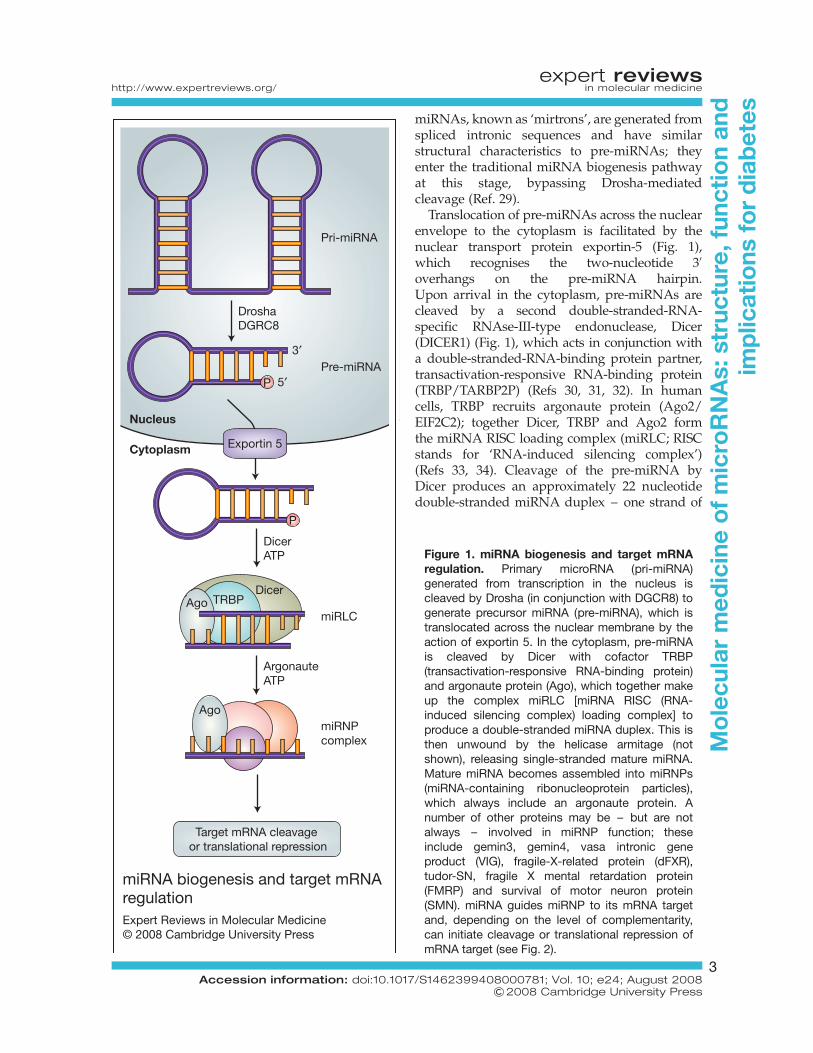
miRNAs, known as ‘mirtrons’, are generated fromspliced intronic sequences and have similarstructural characteristics to pre-miRNAs; theyenter the traditional miRNA biogenesis pathwayat this stage, bypassing Drosha-mediatedcleavage (Ref. 29).
Translocation of pre-miRNAs across the nuclearenvelope to the cytoplasm is facilitated by thenuclear transport protein exportin-5 (Fig. 1),which recognises the two-nucleotide 30
overhangs on the pre-miRNA hairpin.Upon arrival in the cytoplasm, pre-miRNAs arecleaved by a second double-stranded-RNA-specific RNAse-III-type endonuclease, Dicer(DICER1) (Fig. 1), which acts in conjunction witha double-stranded-RNA-binding protein partner,transactivation-responsive RNA-binding protein(TRBP/TARBP2P) (Refs 30, 31, 32). In humancells, TRBP recruits argonaute protein (Ago2/EIF2C2); together Dicer, TRBP and Ago2 formthe miRNA RISC loading complex (miRLC; RISCstands for ‘RNA-induced silencing complex’)(Refs 33, 34). Cleavage of the pre-miRNA byDicer produces an approximately 22 nucleotidedouble-stranded miRNA duplex – one strand of
Figure 1. miRNA biogenesis and target mRNAregulation. Primary microRNA (pri-miRNA)generated from transcription in the nucleus iscleaved by Drosha (in conjunction with DGCR8) togenerate precursor miRNA (pre-miRNA), which istranslocated across the nuclear membrane by theaction of exportin 5. In the cytoplasm, pre-miRNAis cleaved by Dicer with cofactor TRBP(transactivation-responsive RNA-binding protein)and argonaute protein (Ago), which together makeup the complex miRLC [miRNA RISC (RNA-induced silencing complex) loading complex] toproduce a double-stranded miRNA duplex. This isthen unwound by the helicase armitage (notshown), releasing single-stranded mature miRNA.Mature miRNA becomes assembled into miRNPs(miRNA-containing ribonucleoprotein particles),which always include an argonaute protein. Anumber of other proteins may be – but are notalways – involved in miRNP function; theseinclude gemin3, gemin4, vasa intronic geneproduct (VIG), fragile-X-related protein (dFXR),tudor-SN, fragile X mental retardation protein(FMRP) and survival of motor neuron protein(SMN). miRNA guides miRNP to its mRNA targetand, depending on the level of complementarity,can initiate cleavage or translational repression ofmRNA target (see Fig. 2).
miRNA biogenesis and target mRNAregulation Expert Reviews in Molecular Medicine© 2008 Cambridge University Press
TRBP
P
Drosha
5′
3′Pre-miRNA
miRLC
Pri-miRNA
Nucleus
Cytoplasm
DGRC8
Dicer
DicerAgo
ATP
Exportin 5
miRNPcomplex
Target mRNA cleavage or translational repression
Ago
ArgonauteATP
P
expert reviewshttp://www.expertreviews.org/ in molecular medicine
3Accession information: doi:10.1017/S1462399408000781; Vol. 10; e24; August 2008
&2008 Cambridge University Press
Mo
lecu
lar
med
icin
eo
fm
icro
RN
As:
stru
ctur
e,fu
ncti
on
and
imp
licat
ions
for
dia
bet
es

which will become the mature miRNA. The duplexis then unwound by the DEAD-box helicasearmitage, releasing the single-stranded maturemiRNA (Refs 35, 36).
The Ago2-bound mature miRNA then becomesassembled into effector complexes termed miRNA-containing ribonucleoprotein particles (miRNPs)(Ref. 37) (Fig. 1). Several forms of miRNPs existthat differ in size and composition, but eachform of miRNP contains a member of theargonaute protein family. The major function ofmiRNAs is to guide the miRNP complex to itstarget mRNA, where its associated argonauteprotein mediates the effect (Ref. 38). Severalother miRNP components have been identified,including gemin3 (DDX20), gemin4, vasa intronicgene product (VIG), fragile-X-related protein(dFXR), and the tudor staphylococcal-nuclease-domain-containing protein (tudor-SN) (Refs 39,40). Gemin3 is a putative DEAD-box RNAhelicase, which may function in the unwindingof the mRNA target (Ref. 35), but the precise roleof the other proteins in RNA-silencing eventsremains unclear. Although miRNAs functionprimarily in the cytoplasm, one miRNA, mir-29bhas been found to localise in the nucleus; this islikely due to a hexanucleotide terminal motif inthe 30 region that directs the mature miRNA tobe imported back into the nucleus after it isprocessed in the cytoplasm (Ref. 41).
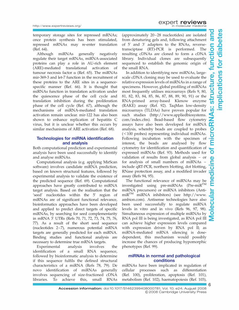
Mechanism(s) of miRNA actionIn mammals, miRNAs usually exhibit partialcomplementarity with their mRNA targets;perfect or near-perfect base pairing is quite rare inthese organisms, but is predominantly found inplant miRNAs. Partial complementarity ofmiRNA to mRNA usually leads to translationalinhibition (Ref. 42), although animal miRNAs canalso induce target degradation despite the lack ofperfect complementarity (Refs 43, 44, 45). Severalproposed models exist for the mechanism oftranslational repression, including miRNAsrepressing translation at both pre-initiation andpost-initiation stages (Fig. 2), and effects onmRNA stability (decapping and deadenylation)and compartmentalisation into translationallyrepressive sites (Fig. 2); it still remains to bedeciphered which of these model mechanisms arecause and consequence of translational repression.
miRNAs affecting initiation steps only affect cap-dependent translation, possibly through m7G caprecognition (Refs 46, 47, 48, 49, 50). Argonaute
proteins contain structural similarities to thecap-binding protein eIF4E, and thus it has beensuggested that translational repression mayoccur due to competition between argonaute andeIF4E for binding to the cap structure (Ref. 51)(Fig. 2a). Argonaute proteins are also thought torecruit eIF6, which binds to the large ribosomalsubunit, preventing binding of the small subunitand thus inhibiting mRNA translation (Ref. 52)(Fig. 2a).
Much evidence also exists for post-initiationmechanisms of repression, which affect bothcap-dependent and cap-independent translation(Ref. 53). Polysome profile experiments indicatethat, under conditions of translationalrepression, target mRNAs are fully loaded withribosomes (Refs 15, 54), a number of which areengaged in active translation (Ref. 53),suggesting that translation initiation andelongation phases are not compromised. Twopossible theories were suggested to explainthese findings. The ribosome ‘drop-off’ theorysuggests that ribosomes engaged in translationof miRNA-associated mRNAs are prone toterminate translation prematurely (Fig. 2b).Alternatively, association of active ribosomeswith repressed mRNAs could also be explainedby the ability of miRNP complex to recruitproteolytic enzymes to degrade the nascentpolypeptide as it emerges from the ribosome(Ref. 15) (Fig. 2b). Conflicting evidence exists onthe role of proteolytic enzymes in miRNAfunction, as targeting of reporter proteins andthe use of proteinase inhibitors have shown noeffect on translational repression (Refs 50, 53).
miRNAs are apparently also involved inregulating mRNA stability and induction ofdecay of repressed mRNA targets. Argonauteproteins, miRNAs and their repressed targetmRNAs have recently been shown to becompartmentalised in cytoplasmic foci calledP-bodies (Refs 50, 55, 56, 57, 58, 59). These aresites of translational repression and mRNAdecay; they are rich in factors associated withthese processes, and are lacking in ribosomes orany other factors associated with translationinitiation (Ref. 60). It is proposed that P-bodyproteins may participate in the formation of arepressive complex on the target mRNA, whichcould eventually lead to mRNA aggregation intoP-bodies (Ref. 61). Within P-bodies, miRNA/mRNA-bound argonaute protein recruits GW182protein (TNRC6A), which subsequently recruits
expert reviewshttp://www.expertreviews.org/ in molecular medicine
4Accession information: doi:10.1017/S1462399408000781; Vol. 10; e24; August 2008
&2008 Cambridge University Press
Mo
lecu
lar
med
icin
eo
fm
icro
RN
As:
stru
ctur
e,fu
ncti
on
and
imp
licat
ions
for
dia
bet
es

deadenylating enzyme CCR4–NOT1 (CNOT1),and this is followed by mRNA decapping byDCP1–DCP2 enzyme – thereby affectingstability of repressed mRNA. Repressed mRNAs
are then degraded by 50 to 30 exonucleaseactivity of XRN1 (50-exoribonuclease 1) (Refs 43,55, 57, 62, 63) (Fig. 2c). In addition to facilitatingmRNA degradation, P-bodies may function as
Proposed mechanisms of miRNA actionExpert Reviews in Molecular Medicine © 2008 Cambridge University Press
DCP1–DCP2 XRN1
miRNP complex
GuidemiRNA
Small ribosomal subunita
c
b
eIF6
m7G
Large ribosomalsubunit
AAAAgo
EIF4E
EIF4G
m7GAAA
Ago
GW182
Nascentpolypeptide
P-body
DegradationStorage
CCR4–NOT1
Ribosome
m7GAAA
Ago
Proteinase
Stopcodon
UAG
Figure 2. Proposed mechanisms of miRNA action. MicroRNAs (miRNAs) can inhibit translation at pre- andpost-initiation stages. (a) At pre-initiation stages, the miRNP complex may affect m7G-cap-dependenttranslation through competition of the argonaute protein with the eIF4G initiation complex for binding to thecap structure; argonaute proteins also recruit eIF6, which prevents large ribosomal subunit binding to thesmall subunit. (b) At postinitiation stages, miRNPs may cause ribosomes to terminate translationprematurely, generating truncated polypeptides, or recruit proteolytic enzymes that degrade the polypeptidechain as it emerges from the ribosome. Repressed mRNAs arising from these models can then betransported to P-bodies for storage or degradation: the miRNP complex recruits GW182 protein; the lattersubsequently recruits deadenylase enzyme CCR4–NOT1; the mRNA is then decapped by DCP1–DCP2,and degraded by exonuclease activity of XRN1.
expert reviewshttp://www.expertreviews.org/ in molecular medicine
5Accession information: doi:10.1017/S1462399408000781; Vol. 10; e24; August 2008
&2008 Cambridge University Press
Mo
lecu
lar
med
icin
eo
fm
icro
RN
As:
stru
ctur
e,fu
ncti
on
and
imp
licat
ions
for
dia
bet
es
temporary storage sites for repressed mRNAs;once protein synthesis has been stimulated,repressed mRNAs may re-enter translation(Ref. 64).
Although miRNAs generally negativelyregulate their target mRNAs, miRNA-associatedproteins can play a role in AU-rich element(ARE)-mediated translational activation oftumour necrosis factor a (Ref. 65). The miRNAsmir-369-3 and let-7 function in the recruitment ofthese proteins to the ARE sites in a sequence-specific manner (Ref. 66). It is thought thatmiRNAs function in translation activation underthe quiescence phase of the cell cycle andtranslation inhibition during the proliferationphase of the cell cycle (Ref. 67), although themechanisms of miRNA-mediated translationactivation remain unclear. mir-122 has also beenshown to enhance replication of hepatitis Cvirus, but it is unclear whether this occurs bysimilar mechanisms of ARE activation (Ref. 68).
Technologies for miRNA identificationand analysis
Both computational prediction and experimentalanalysis have been used successfully to identifyand analyse miRNAs.
Computational analysis (e.g. applying MirScansoftware) involves candidate miRNA prediction,based on known structural features, followed byexperimental analysis to validate the existence ofthe predicted sequence (Ref. 69). Computationalapproaches have greatly contributed to miRNAtarget analysis. Based on the realisation that the‘seed’ nucleotides within the 50 region ofmiRNAs are of significant functional relevance,bioinformatics approaches have been developedand applied to predict direct targets of specificmiRNAs, by searching for seed complementarityin mRNA 30 UTRs (Refs 70, 71, 72, 73, 74, 75, 76,77). As a result of the short seed sequence(nucleotides 2–7), numerous potential mRNAtargets are generally predicted for each miRNA.Binding studies and functional analysis arenecessary to determine true mRNA targets.
Experimental analysis involves theidentification of a small RNA sequence,followed by bioinformatic analysis to determineif this sequence fulfils the defined structuralcharacteristics of a miRNA (Refs 78, 79). Denovo identification of miRNAs generallyinvolves sequencing of size-fractioned cDNAlibraries. To achieve this, small RNAs
(approximately 20–28 nucleotides) are isolatedfrom denaturing gels and, following attachmentof 50 and 30 adapters to the RNAs, reverse-transciptase (RT)-PCR is performed. Theresulting cDNAs are cloned to form a cDNAlibrary. Individual clones are subsequentlysequenced to establish the genomic origin ofthe small RNA.
In addition to identifying new miRNAs, large-scale cDNA cloning may be used to evaluate therelative expression levels of miRNAs in a range ofspecimens. However, global profiling of miRNAsmost frequently utilises microarrays (Refs 9, 80,81, 82, 83, 84, 85, 86, 87, 88, 89, 90, 91) or theRNA-primed array-based Klenow enzyme(RAKE) assay (Ref. 92). TaqMan low-densitymicroarrays (TLDAs) have proven popular forsuch studies (http://www.appliedbiosystems.com/index.cfm). Bead-based flow cytometryassays have also been developed for miRNAanalysis, whereby beads are coupled to probes(,100 probes) representing individual miRNAs.Following incubation with the specimen ofinterest, the beads are analysed by flowcytometry for identification and quantification ofexpressed miRNAs (Ref. 93). Methods used forvalidation of results from global analysis – orfor analysis of small numbers of miRNAs –include qRT-PCR, northern blotting, dot blotting,RNase protection assay, and a modified invaderassay (Refs 94, 95).
The functional relevance of miRNAs may beinvestigated using pre-miRNAs (Pre-miRTM
miRNA precursors) or miRNA inhibitors (Anti-miRTM miRNA inhibitors) (see http://www.ambion.com). Antisense technologies have alsobeen used successfully to regulate miRNAlevels in vitro and in vivo (Refs 96, 97, 98).Simultaneous expression of multiple miRNAs byRNA pol III is being investigated, as RNA pol IIIcan achieve higher expression levels comparedwith expression driven by RNA pol II; asmiRNA-mediated mRNA silencing is dose-dependent, this mechanism would possiblyincrease the chances of producing hypomorphicphenotypes (Ref. 99).
miRNAs in normal and pathologicalconditions
miRNAs have been implicated in regulation ofcellular processes such as differentiation(Ref. 100), proliferation, apoptosis (Ref. 101),metabolism (Ref. 102), haematopoiesis (Ref. 103),
expert reviewshttp://www.expertreviews.org/ in molecular medicine
6Accession information: doi:10.1017/S1462399408000781; Vol. 10; e24; August 2008
&2008 Cambridge University Press
Mo
lecu
lar
med
icin
eo
fm
icro
RN
As:
stru
ctur
e,fu
ncti
on
and
imp
licat
ions
for
dia
bet
es
cardiogenesis (Ref. 104), morphogenesis andinsulin secretion (Ref. 105), in addition to actingin several feedback loops involved in signaltransduction pathways (Ref. 106). miRNAs arevital for cell survival: elimination of miRNAmaturation by Dicer knockout leads toembryonic lethality in mice (Ref. 107). miRNAsare involved in such a wide variety of cellularprocesses that it is likely their dysregulation orabnormal expression could lead to a range ofdisease states. miRNAs have already beenimplicated in the pathogenesis of several humandiseases, such as neurological disorders, cancer,and viral and metabolic diseases (Ref. 98).
Neurological disordersSpinal muscular atrophy (SMA), a progressiveneurodegenerative disease, is caused by deletionor loss of function mutations in the SMN(survival of motor neuron) protein (Ref. 108).SMN is a component of the miRNP complex thatperforms the effector functions of the miRNApathway (Ref. 37). Fragile X syndrome is causedby inactivation of the gene FMR1, and hencesilencing of the fragile X mental retardationprotein (FMRP), which is also associated withmiRNP complex formation (Ref. 109). Thesestudies indicate that disruptions in the miRNPmachinery and hence miRNA activity can lead todisease states. Tourette syndrome is associatedwith a single-nucleotide polymorphism (SNP) inthe 30 UTR of the SLITRK1 gene, which is thebinding site of mir-189; this SNP hence modifiesthe interaction of mir-189 (Ref. 110). In addition,mir-134 regulation of LIMK1 in hippocampalneurons controls spine development and possiblyalso contributes to synaptic development,maturation and plasticity (Ref. 111); thus,dysregulation of mir-134 could potentially lead tocomplications in these processes.
CancerMany miRNA genes are thought to reside atchromosomal breakpoints or fragile sitesassociated with cancer (Ref. 112). The mir-15/16cluster is located at one such site and is deletedin the majority of B cell chronic lymphocyticleukaemias (B-CLLs) (Ref. 113), as well as mantlecell lymphomas and prostate cancers (Ref. 114),suggesting that mir-15/16 may function astumour suppressors. Members of the let-7 familyalso located at fragile sites (Ref. 112) arefrequently deleted in cancer patients, leading to
elevated levels of the oncogene product RAS(Ref. 115). Some miRNAs have also been shownto possess oncogenic potential; the mir-17-92cluster, which contains six miRNAs, is located ata chromosome site that is amplified in a range ofcancers and overexpression leads to acceleratedtumour development in mouse B cell lymphomamodels (Ref. 5). Overexpression of the individualmiRNAs from the cluster did not reveal thesame oncogenic potential, indicating thatinteraction between a range of miRNAs could benecessary for the development of diseasephenotypes. mir-155, which is elevated inBurkitt lymphoma, also acts as an oncogene,with overexpression in B cells leading todevelopment of pre-B-cell lymphomas (Ref. 116).These putative miRNA tumour suppressors andoncogenes represent a potential set of miRNAtherapeutic targets. Microarray profiling ofmiRNAs in tumour tissues and cell lines hasidentified miRNA differentially expressed indifferent tumour types, indicating potential use oftumour miRNA profiling in cancers for predictionof developmental lineage, differentiation state,and prognosis (Ref. 93).
Viral diseaseHost mir-32 expression restricts infection of theprimate foamy virus 1 (PFV-1), with inhibition ofmir-32 leading to doubling of the PFV-1proliferation rates in host cells (Ref. 117). PFV-1encodes the Tas protein, which is known to be asuppressor of RNA silencing (Ref. 117), therebyremoving the growth limitation inflicted bymir-32 by disrupting the silencing machinery.Many viruses encode similar suppressors of RNAsilencing – for example, the Tat protein fromhuman immunodeficiency virus 1 (HIV-1)(Ref. 118) and the B2 protein from Nodamuravirus (Ref. 119).
miRNAs represent an efficient mechanism forviruses to use to manipulate host machinery, asthey require less space on the viral genomethan alternative protein products. ViralmiRNAs can target both viral and host mRNAsfor repression. Twelve miRNAs from theKarposi sarcoma-associated herpesvirus(KSHV) genome expressed in cells led to thedownregulation of a number of genes includingthrombospondin 1 (THBS-1), which is a knowntumour suppressor and antiangiogenic factor. Itis thought that these KSHV miRNAs maycontribute directly to pathogenesis of KSHV by
expert reviewshttp://www.expertreviews.org/ in molecular medicine
7Accession information: doi:10.1017/S1462399408000781; Vol. 10; e24; August 2008
&2008 Cambridge University Press
Mo
lecu
lar
med
icin
eo
fm
icro
RN
As:
stru
ctur
e,fu
ncti
on
and
imp
licat
ions
for
dia
bet
es
downregulation of THBS-1 (Ref. 120). The simianvirus 40 (SV40) encodes a miRNA that is perfectlycomplementary to transcripts coding viral Tantigens, leading to their degradation (Ref. 121).This destruction of viral T antigens aids thevirus in evading immune detection by the host.The hepatitis C virus (HCV) enchancesreplication via a novel interaction of abundantlyexpressed mir-122 with the 50 UTR of the viralgenome (Ref. 68). Interferons (IFNs) are keymolecules involved in eliciting the antiviralresponse once an infection has been detected(Ref. 122). IFN-b has recently been implicatedin the activation of several miRNAs inmammals that have antiviral properties againstHCV (Ref. 123), and treatment also leads toreduced mir-122 expression (Ref. 123), whichlimits HCV replication (Ref. 68). These studiesidentify a number of different miRNAs thatcould be therapeutically targeted to hinder viralinfection, aid host detection of infection, andprevent viral manipulation of host machinery.
miRNAs relevant to diabetesDiabetes mellitus is a metabolic disorder in whichinsulin either is not secreted in sufficient amountsfrom b-cells or does not efficiently stimulate itstarget cells. Despite high glucose levels, cellsstarve, as a result of impaired glucose entry intocells. Current treatments for diabetes cannotefficiently control glycaemic levels, resulting inepisodes of hyper- and hypoglycaemia(Ref. 124), which increases the possibility ofdeveloping secondary complications such asretinopathy, nephropathy and neuropathy(Ref. 125). In the search for more-targetedmolecular therapies, miRNAs implicated ininsulin secretion and diabetic complicationshave recently attracted attention.
miRNAs associated with b-cell insulinsecretionRecent experimental work has revealed a limitednumber of miRNAs – including mir-375, mir-124a and mir-9 – associated with varioussubcellular events involved in glucose-stimulatedinsulin secretion (GSIS) (Refs 126, 127, 128). Inaddition, bioinformatic analysis has indicatedpotential miRNA target sites in a range of othermRNAs encoding proteins involved inexocytosis – including VAMP2 (vesicle-associatedmembrane protein 2), SNAP25 (synaptosomal-associated protein 25kDa), syntaxin-1, Rab27a
(member of the RAS oncogene family),granuphilin (SYTL4) and MyRIP (myosin VIIAand Rab interacting protein). Some miRNAs (mir-153, mir-1, mir-133, mir-200 and mir-34) havepredicted target sites in several of thesefunctionally related genes (e.g. mir-153 and mir-1have putative target sites in VAMP2 andSNAP25) (Ref. 129). Although the miRNA targetsites identified by bioinformatics have yet to beexperimentally validated, this gives an insightinto the potential extent of complexnetworking of molecules involved in exocytosisregulation.
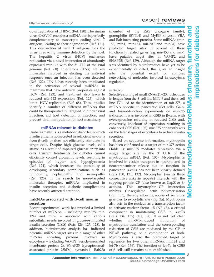
mir-375Selective cloning of small RNAs 21–23 nucleotidesin length from the b-cell line MIN-6 and the a-cellline TC1 led to the identification of mir-375, amiRNA specific to pancreatic islet cells. Gain-and loss-of-function experiments on mir-375indicated it was involved in GSIS in b-cells, withoverexpression resulting in reduced GSIS and,conversely, knockout of expression resulting inenhanced GSIS (Ref. 105). mir-375 apparently actson the later stages of exocytosis to reduce insulinsecretion.
Based on sequence information, myotrophinhas been confirmed as a target of mir-375 action(Table 1); mir-375 mediates repression via asingle target site in the 30 UTR of themyotrophin mRNA (Ref. 105). Myotrophin isinvolved in vesicle transport in neurons and inneurotransmitter release but its function inpancreatic b-cells has not been clearly defined(Refs 130, 131, 132). Myotrophin (via its threeconsecutive ankyrin repeats) interacts with thecapping protein CP (also known as CapZ or b-actinin). This myotrophin–CP interactioninhibits CP-regulated actin polymerisation(Ref. 133), thereby allowing access of secretorygranules to exocytotic site (Fig. 3a). Myotrophinalso acts in the nucleus as a transcription factorto activate nuclear factor kB (NF-kB), a criticalcomponent in maintaining GSIS in b-cells(Refs 134, 135) (Fig. 3a). It is not yet clearwhether mir-375-induced inhibition ofmyotrophin translation and the correspondingreduction of GSIS are mediated by the CP orNF-kB pathway, or a combination of both.Myotrophin is also the predicted target ofrepression for two other miRNAs: mir124 andlet-7b (Ref. 136). The function of let-7b in GSISof b-cells still remains to be established.
expert reviewshttp://www.expertreviews.org/ in molecular medicine
8Accession information: doi:10.1017/S1462399408000781; Vol. 10; e24; August 2008
&2008 Cambridge University Press
Mo
lecu
lar
med
icin
eo
fm
icro
RN
As:
stru
ctur
e,fu
ncti
on
and
imp
licat
ions
for
dia
bet
es

More recently, knockdown of mir-375 inzebrafish embryos has revealed a role for thismiRNA in pancreatic islet development(Ref. 137). When morpholino oligonucleotideswere injected into one-cell-stage embryos,resulting in a knockdown of mir-375 activityduring the first four days of development,insulin staining showed the formation of anislet at 24 h post fertilisation but by day 3 theislet had fallen apart and insulin-positive cellswere scattered (Ref. 137). The original formationof an islet at 24 h suggests that mir-375expression is not essential in early endocrineformation, but more so for maintenance oftissue identity at a later stage. It has not yetbeen deciphered whether this scattered isletphenotype occurs as a result of mir-375 actionon myotrophin expression or whether othermir-375 targets are involved.
mir-124aMir-124a exists in three different isoforms – mir-124a1, 2 and 3 – encoded on chromosome 14, 3and 2, respectively, in the mouse genome. Theisoform mir-124a2 is differentially expressedduring pancreas development, with a sixfoldupregulation at embryonic stage e18.5compared with e14.5 (Ref. 138). e18.5 is thecritical stage for b-cell differentiation, indicatingthat mir-124a2 might be significant in this process.
Using PicTar (http://pictar.bio.nyu.edu/cgi-bin/PicTar) (Ref. 136) and miRanda (http://
www.microrna.org/mammalian/index_new.html)(Ref. 139) bioinformatics tools, the forkhead/winged helix transcription factor boxa2(FOXA2) mRNA was identified as a potentialtarget of mir-124a (Table 1). This relationshipwas subsequently confirmed by over- andunderexpression of mir-124a2 in MIN6 murinepancreatic b-cells, using Pre-Mir and Anti-Mirtechnology (Pre-miRTM miRNA precursors andAnti-miRTM miRNA inhibitors; see http://www.ambion.com). CREB-1 (cAMP-response-element-binding protein), a stimulus-inducibletranscription factor, was also predicted as apotential target of mir-124a regulation, and mir-124a2 over- and underexpression correspond withdecreasing and increasing levels of CREB-1,respectively (Ref. 138) (Table 1). As FOXA2 is atarget of CREB-1 regulation (Ref. 140), thissuggests that FOXA2 expression may beregulated by mir-124a2 directly as well asindirectly (via CREB-1) (Fig. 3b).
FOXA2 is an upstream regulatorof the homeoboxprotein PDX-1 (Refs 141, 142). PDX-1 is essential forb-cell differentiation, glucose homeostasis andpancreas development (Refs 143, 144) (Fig. 3b),and the human orthologue (insulin promoterfactor; IPF1) is mutated in a proportion ofearly-onset type 2 diabetic patients (Ref. 145).Manipulation of FOXA2 expression, byoverexpression or inhibition of mir-124a2,corresponds with a decrease and increase in PDX-1mRNA levels, respectively (Ref. 138). PDX-1
Table 1. miRNAs implicated in b-cell insulin secretion and diabetic complications,and their mRNA targets
Process/condition miRNAa Target mRNA Ref.
b-Cell insulin secretion mir-375 Myotrophin 105
mir-124a FOXA2CREB-1Rab27A
138138149
mir-9 OC2 152
Diabetic kidney glomeruli mir-192 SIP-1 166
Diabetic heart mir-133 HERG 175aThese miRNAs represent potential targets of therapeutic intervention in the treatment of diabetes and relatedcomplications.Abbreviations: CREB-1, cAMP-response-element-binding protein 1; FOXA2, forkhead/winged helixtranscription factor boxa 2; HERG, human ether-a-go-go related gene; miRNA, microRNA; OC2, onecut 2; SIP-1,SMAD-interacting protein 1.
expert reviewshttp://www.expertreviews.org/ in molecular medicine
9Accession information: doi:10.1017/S1462399408000781; Vol. 10; e24; August 2008
&2008 Cambridge University Press
Mo
lecu
lar
med
icin
eo
fm
icro
RN
As:
stru
ctur
e,fu
ncti
on
and
imp
licat
ions
for
dia
bet
es
regulates expression of the insulin gene;consequently, overexpression and inhibition ofmir-124a2 leads to a decrease and increase ininsulin mRNA levels, respectively (Ref. 138).
Further downstream targets of FOXA2regulation are the KATP channel subunits SUR1
(sulphonylurea receptor 1) and KIR6.2 (inwardrectifier Kþ channel member 6.2) (Ref. 146),which are critical for regulated insulin release;mutations in either of these genes can lead topersistent hyperinsulinaemic hypoglycaemiaof infancy (PHHI) in humans (Ref. 147).
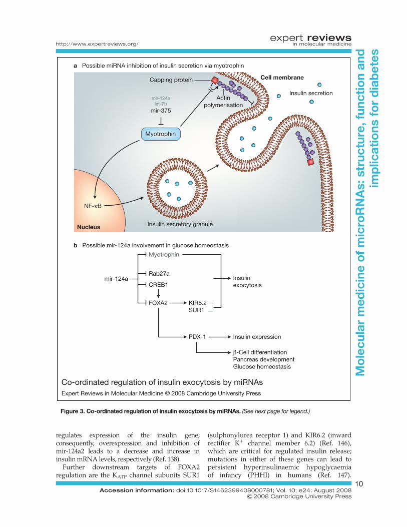
Co-ordinated regulation of insulin exocytosis by miRNAsExpert Reviews in Molecular Medicine © 2008 Cambridge University Press
mir-124alet-7b
mir-375
Actinpolymerisation
Insulin secretion
Myotrophin
Insulin secretory granuleNucleus
NF-κB
Capping protein Cell membrane
MyotrophinMyotrophin
Rab27aInsulinexocytosis
Insulin expression
β-Cell differentiationPancreas developmentGlucose homeostasis
CREB1mir-124a
FOXA2 KIR6.2SUR1
PDX-1
a Possible miRNA inhibition of insulin secretion via myotrophin
b Possible mir-124a involvement in glucose homeostasis
Figure 3. Co-ordinated regulation of insulin exocytosis by miRNAs. (See next page for legend.)
expert reviewshttp://www.expertreviews.org/ in molecular medicine
10Accession information: doi:10.1017/S1462399408000781; Vol. 10; e24; August 2008
&2008 Cambridge University Press
Mo
lecu
lar
med
icin
eo
fm
icro
RN
As:
stru
ctur
e,fu
ncti
on
and
imp
licat
ions
for
dia
bet
es

Overexpression of mir-124a2 leads to increasedCa2þ levels within the cell (Ref. 138).Knockdown of the SUR1 and KIR6.2 subunitsresults in impaired KATP channels, causing abuild-up of Kþ ions within the cell, whichstimulates opening of voltage-gated calciumchannels, thereby allowing Ca2þ ions to enterthe cell (increased Ca2þ ions usually stimulateexocytosis). Thus, reduced expression of KATP
channel subunits could explain the increase incytosolic free Ca2þ concentrations followingtransfection with mir-124a2. FOXA2 deficiencyin mice leads to loss of GSIS and excessiveinsulin release in response to amino acidstimuli (Ref. 146). However, mir-124a2-inducedreduction in FOXA2 levels has not shown asdramatic an effect on GSIS as seen in theFOXA2-null mouse (Ref. 138).
Rab27A, which is also involved in GSIS(Ref. 148), has recently been shown to be thetarget of mir-124a action via a binding site inthe 30 UTR of Rab27A mRNA (Ref. 149). Mir-124a also indirectly regulates expression ofseveral other components of the exocytoticmachinery in MIN6-B1 cells, includingSNAP25, Rab3A, synapsin 1A (SYN1) andNOC2 (nucleolar complex associated 2)(Ref. 149). Overexpression of mir-124a in thesecells leads to reduced GSIS. In the same study,mir-96 was identified as a regulator ofgranuphillin and NOC2, and its expression inMIN6-B1 cells leads to a reduction instimulated insulin secretion (Ref. 149).
mir-9Mir-9 is expressed predominately in neurons inboth human and mouse models (Refs 150, 151),and to a lesser extent in pancreatic b-cells in ratand mouse models (Ref. 152). Onecut2transcription factor (OC2), which negatively
regulates granuphilin (also known as SLP4/SYTL4) expression, has been identified as amir-9 target (Table 1). A basal level of mir-9expression is needed to maintain optimumonecut2 expression levels for normal b-cellfunction (Ref. 152), but mir-9 overexpression inrat INS-1E b-cells leads to a reduced GSIS inthese cells (Ref. 152).
Granuphilin associates with insulin secretorygranules (Ref. 153) and promotes targeting ofthese granules to the plasma membrane(Ref. 154); however, it is a negative modulatorof exocytosis as it imposes a constraint toinhibit fusion until the correct signals arereceived by the cell (Ref. 154). Overexpressionof mir-9 leads to increased levels of granuphilinexpression due to the removal of the repressiveeffects of onecut2 on the granuphilin promoter(Ref. 152), and hence reduced GSIS is observedas a result of its negative effects on exocytosis.Granuphilin-null mice also show impairedGSIS, with reduced quantity of insulin granulesdocked to the b-cell membrane, and converselyexhibit increased insulin exocytosis in responseto stimulus (Ref. 155).
Binding partners of granuphilin include theGTP-binding proteins Rab3/Rab27, the SNARE-binding protein Munc-18 and the tSNAREprotein syntaxin-1, which are involved inexocytosis of secretory granules in pancreaticb-cells (Refs 152, 156, 157). mir-9-inducedreduction of exocytosis does not occur throughmanipulation of Rab3, Rab27 and SNAREproteins such as SNAP25, VAMP-2 andsyntaxin-1, as the expression levels of these keyexocytosis proteins are unchanged in mir-9-transfected cells relative to control cells(Ref. 152). However, it is as yet unknown whetherthe mir-9-mediated reduction of secretagogue-stimulated exocytosis via granuphilin occurs
Figure 3. Co-ordinated regulation of insulin exocytosis by miRNAs. (Legend; see previous page for figure.)(a) Possible microRNA (miRNA) inhibition of insulin secretion via myotrophin. Overexpression of the myotrophin-targeting miRNA mir-375 results in reduced glucose-stimulated insulin secretion, which can be explained throughcytoplasmic and/or nuclear actions of myotrophin. Myotrophin interacts with capping protein to inhibit actinpolymerisation. Inhibition of actin polymerisation allows access of insulin granules to the cell membrane forexocytosis. In addition, myotrophin interacts with transcription factor NF-kB, which controls expression ofseveral genes critical for glucose-stimulated insulin secretion. Myotrophin also contains putative binding sites forthe miRNAs let-7b and mir-124a. (b) Possible mir-124a involvement in glucose homeostasis. Mir-124a targetsRab27a and also FOXA2 (directly and indirectly via CREB1). Myotrophin has also been identified as a potentialmir-124a target. FOXA2 may influence several targets relevant to diabetes via PDX-1, including insulin mRNAlevels and possibly also KATP channel subunits KIR6.2 and SUR1 (involved in regulated insulin release). It alsoplays a role in b-cell differentiation, pancreas development and glucose homeostasis.
expert reviewshttp://www.expertreviews.org/ in molecular medicine
11Accession information: doi:10.1017/S1462399408000781; Vol. 10; e24; August 2008
&2008 Cambridge University Press
Mo
lecu
lar
med
icin
eo
fm
icro
RN
As:
stru
ctur
e,fu
ncti
on
and
imp
licat
ions
for
dia
bet
es

through downstream manipulation of Munc-18activity. The effect of granuphilin on Munc-18 isnot alone sufficient to mediate such a profoundknockdown of stimulus-induced exocytosis(Ref. 156), suggesting that granuphilin andpossibly mir-9 have additional targets thatparticipate in this process.
miRNAs associated with diabetic kidneyglomeruliDiabetic nephropathy – generally defined asurinary albumin excretion of .300 mg per 24 hor abnormal renal function characterised byabnormality in serum creatinine, creatinineclearance, or glomerular filtration rate – is themost common cause of kidney failure in patientswith diabetes. The abnormal renal function isthought to arise largely from accumulation ofextracellular matrix (ECM) proteins in themesangial cells, hypertrophy of glomerular andtubular elements, and thickening of theglomerular and tubular basement membranes(Refs 158, 159).
ECM proteins such as collagen 1a1 and 1a2 arepositively regulated by transforming growthfactor b (TGF-b), which is upregulated inmesangial cells under diabetic conditions(Refs 160, 161). TGF-b is known to upregulateECM proteins via SMAD transcription factorsand mitogen-activated protein kinases (MAPKs)(Refs 162, 163, 164, 165); in addition, recentwork has revealed TGF-b downregulates theE-box repressor proteins dEF1 and SMAD-interacting protein 1 (SIP1), which mediaterepression of collagen expression at its E-boxelement (Ref. 166). dEF1 can also repress SMADproteins (Ref. 167).
Several miRNAs, including mir-192, -194, -204,-215 and -216, are preferentially expressed in thekidney, as compared with other tissues (Ref. 85).Using computational miRNA target predictionsfrom miRNA databases (http://cbio.mskcc.org;http://microrna.sanger.ac.uk/index.shtml) theE-box repressor SIP1 was shown to contain apotential target site for mir-192 and mir-215regulation.
Using a luciferase reporter system, SIP1 wasvalidated as a target of mir-192 regulation(Table 1), but not of mir-215. TGF-b treatmentinduces mir-192 expression. TGF-b-inducedmir-192 expression or mir-192 transfection candecrease SIP1 levels, while mir-192 inhibitorincreases SIP1 levels (Ref. 166). The mechanism
of TGF-b regulation of mir-192 expression is notcompletely understood. The mir-192 promotercontains a binding site for the proto-oncogeneETS-1 (Ref. 85), which is also induced by TGF-bexpression (Ref. 168), representing a possiblemechanism of TGF-b regulation of mir-192expression.
mir-192 overexpression leads to repression oftranslation of its target SIP1, thereby increasinglevels of collagen expression. Repression of dEF1using short hairpin RNA (shRNA; forstable transfection of siRNA) shows similareffects, resulting in increased levels of collagenexpression; however, double transfection of amir-192 mimic and dEF1 shRNA shows a muchlarger increase in collagen expression than eitherachieved separately, suggesting that these twomechanisms act synergistically in the control ofcollagen expression (Ref. 166).
In vivo analysis of type1 and type2 diabetic miceshowed elevated levels of mir-192, TGF-b andcollagen 1a2 in the renal glomeruli (Ref. 166),suggesting the possible involvement of mir-192-mediated collagen expression in the pathogenesisof diabetic nephropathy, or other diabeticcomplications where TGF-b levels are raised.
miRNAs associated with diabetic heartCardiovascular disease is the principal cause ofdeath in more than 60% of diabetic cases, withan annual mortality of approximately 5.4%,thereby decreasing life expectancy by up to 10years (Refs 169, 170). For diabetic patients, themost prominent cardiac electrical disturbance isan abnormal QT interval, which is associatedwith increased risk of sudden cardiac death(Refs 171, 172). QT interval is the total durationfor ventricular depolarisation and repolarisationof cardiac myocytes, which is controlled by theflow of inward and outward ion currents.Increasing inward currents and/or decreasingoutward currents lead to prolonged QT interval.The outward currents occur via a number of Kþ
channels.Human ether-a-go-go related gene (HERG)
encodes one of these channels – the rapiddelayed rectifier Kþ current channel (IKr). HERGis downregulated in diabetic hearts, therebycontributing to slowed repolarisation andprolonged QT interval (Refs 173, 174). HERGexpression is downregulated at the post-transcriptional level: HERG mRNA levels remainconstant, while HERG protein levels are reduced
expert reviewshttp://www.expertreviews.org/ in molecular medicine
12Accession information: doi:10.1017/S1462399408000781; Vol. 10; e24; August 2008
&2008 Cambridge University Press
Mo
lecu
lar
med
icin
eo
fm
icro
RN
As:
stru
ctur
e,fu
ncti
on
and
imp
licat
ions
for
dia
bet
es
by 60% in diabetic heart as compared withnondiabetic/control heart (Refs 173, 174, 175).
mir-1 and mir-133 are specifically expressed inadult cardiac and skeletal muscle tissues, andupregulated in rabbit diabetic heart tissue andalso in ventricular samples from humandiabetic patients (Refs 104, 176). Using aluciferase reporter plasmid and westernblotting, HERG mRNA was shown to be atarget of mir-133 action (Table 1), while mir-1had no effect on HERG expression (Ref. 175).IKr, the channel for rapid delayed rectifier Kþ
current, was shown to be underexpressed indiabetic hearts and healthy hearts transfectedwith mir-133, while transfection of a mir-133inhibitor AMO-133 partially rectified thedepression of IKr in diabetic hearts, andcompletely rectified expression of IKr in mir-133-transfected healthy hearts (Ref. 175).
Serum response factor (SRF) is a cardiactranscription factor highly overexpressed indiabetic hearts (Ref. 175). SRF is essential forexpression of mir-1 and mir-133 (Refs 104, 176).SRF siRNA or the SRF inhibitor distamycinreduced expression of mir-1 and mir-133 indiabetic cardiac myocytes. Transfection of SRFsiRNA into cardiac myocytes of diabetic heartsresulted in increased levels of IKr expression(Ref. 175). It still remains to be seen whether useof AMO-133 or SRF siRNA in vivo increases IKr
expression sufficiently to correct or reduceprolonged QT interval in diabetic subjects.
mir-133 is also known to repress expression ofKCNQ1, which is involved in the formation of theslow delayed rectifier Kþ current channel (IKs)(Ref. 177), although it is currently unknownwhether this channel plays a role in thedevelopment of long QT syndrome in diabeticpatients.
Clinical implications/applicationsUntil recently, miRNAs had not been considered asclassical therapeutic targets, as they do not code forproteins. Initial studies aimed at exploitingmiRNAs as a form of therapy have shownpromising results. Following intravenousinjection of modified antisense oligonucleotides(termed antagomirs) into mice, in vivo inhibitionof four miRNAs – mir-16, mir-122, mir-192 andmir-194 – has been successfully demonstrated(Ref. 96). This approach resulted not only inblockage of target miRNAs, but also in theirdegradation in most organs analysed, including
liver, kidney, heart, lung, intestine, bone marrow,muscle, skin, fat, ovaries and adrenals. Lack ofeffect observed in brain is possibly due torestricted diffusion of charged nucleic acidsacross the blood–brain barrier. Alternativeapproaches to targeting miRNAs therapeuticallyby inhibiting Drosha, Dicer or other miRNApathway components are being investigated.Conversely, where reduced miRNA expressed isassociated with a disease phenotype andincreased expression of relevant miRNA couldbe of potential therapeutic relevance to rescuedisease phenotype, introduction of miRNAmimics is being investigated. However, suitableexpression vectors have yet to be identified forthe safe delivery and maintenance of such effectslong-term (Ref. 178).
Research in progress and outstandingresearch questions
The importance of miRNAs in normal andpathological conditions is still being realised.Recent studies have clearly indicated anassociation between dysregulated expression ofthese short RNAs in regulated and defectiveinsulin secretion from b-cells and in diabetickidney and heart disease. Recently,overexpression of a specific miRNA (mir-29) –which is upregulated in diabetic rats – has beenfound to have a functional role in insulinresistance (Ref. 179) and, furthermore, analysisof murine pancreas development has indicated aunique miRNA profile to be necessary duringpancreas development for generation of normalb-cells (Ref. 180). So, while studies associatingmiRNAs with diabetes are so far limited innumbers, they suggest important roles formiRNAs as potential biomarkers and possiblytherapeutic targets. More extensive studiesinvestigating the expression and functionalrelevance of miRNAs in both type 1 and type 2diabetes will undoubtedly increase ourunderstanding of these complex conditions andwill hopefully aid in the identification of noveltherapeutic targets and interventions.
Acknowledgements and fundingThe authors acknowledge support from Ireland’sHigher Educational Authority Programme forResearch in Third Level Institutes (PRTLI)Cycle 3, the Health Research Board, andDublin City University’s Research FellowshipAward.
expert reviewshttp://www.expertreviews.org/ in molecular medicine
13Accession information: doi:10.1017/S1462399408000781; Vol. 10; e24; August 2008
&2008 Cambridge University Press
Mo
lecu
lar
med
icin
eo
fm
icro
RN
As:
stru
ctur
e,fu
ncti
on
and
imp
licat
ions
for
dia
bet
es
References1 Bartel, D.P. and Chen, C.Z. (2004) Micromanagers
of gene expression: the potentially widespread
influence of metazoan microRNAs. Nat Rev Genet
5, 396-400
2 Zamore, P.D. (2004) Plant RNAi: how a viral
silencing suppressor inactivates siRNA. Curr Biol
14, R198-200
3 Kumar, A. (2008) RNA interference: a multifaceted
innate antiviral defense. Retrovirology 5, 17
4 Lau, N.C. et al. (2001) An abundant class of tiny
RNAs with probable regulatory roles in
Caenorhabditis elegans. Science 294, 858-862
5 He, L. et al. (2005) A microRNA polycistron
as a potential human oncogene. Nature 435,
828-833
6 Bartel, D.P. (2004) MicroRNAs: genomics,
biogenesis, mechanism, and function. Cell 116,
281-297
7 Brennecke, J. et al. (2005) Principles of microRNA-
target recognition. PLoS Biol 3, e85
8 Leung, A.K. and Sharp, P.A. (2007) microRNAs:
a safeguard against turmoil? Cell 130, 581-585
9 Lim, L.P. et al. (2005) Microarray analysis shows
that some microRNAs downregulate large
numbers of target mRNAs. Nature 433, 769-773
10 Lim, L.P. et al. (2003) Vertebrate microRNA genes.
Science 299, 1540
11 Zamore, P.D. and Haley, B. (2005) Ribo-gnome:
the big world of small RNAs. Science 309,
1519-1524
12 Rajewsky, N. (2006) microRNA target predictions
in animals. Nat Genet 38 Suppl, S8-13
13 Lee, R.C., Feinbaum, R.L. and Ambros, V. (1993)
The C. elegans heterochronic gene lin-4 encodes
small RNAs with antisense complementarity to
lin-14. Cell 75, 843-854
14 Ha, I., Wightman, B. and Ruvkun, G. (1996) A
bulged lin-4/lin-14 RNA duplex is sufficient for
Caenorhabditis elegans lin-14 temporal gradient
formation. Genes Dev 10, 3041-3050
15 Olsen, P.H. and Ambros, V. (1999) The lin-4
regulatory RNA controls developmental timing in
Caenorhabditis elegans by blocking LIN-14
protein synthesis after the initiation of translation.
Dev Biol 216, 671-680
16 Reinhart, B.J. et al. (2000) The 21-nucleotide let-7
RNA regulates developmental timing in
Caenorhabditis elegans. Nature 403, 901-906
17 Slack, F.J. et al. (2000) The lin-41 RBCC gene acts in
the C. elegans heterochronic pathway between the
let-7 regulatory RNA and the LIN-29 transcription
factor. Mol Cell 5, 659-669
18 Lin, S.Y. et al. (2003) The C elegans hunchback
homolog, hbl-1, controls temporal patterning and
is a probable microRNA target. Dev Cell 4, 639-650
19 Vella, M.C. et al. (2004) The C. elegans microRNA
let-7 binds to imperfect let-7 complementary sites
from the lin-41 30UTR. Genes Dev 18, 132-137
20 Takuno, S. and Innan, H. (2008) Evolution of
complexity in miRNA-mediated gene regulation
systems. Trends Genet 24, 56-59
21 Blow, M.J. et al. (2006) RNA editing of human
microRNAs. Genome Biol 7, R27
22 Lee, Y. et al. (2004) MicroRNA genes are transcribed
by RNA polymerase II. EMBO J 23, 4051-4060
23 Borchert, G.M., Lanier, W. and Davidson, B.L.
(2006) RNA polymerase III transcribes human
microRNAs. Nat Struct Mol Biol 13, 1097-1101
24 Cai, X., Hagedorn, C.H. and Cullen, B.R. (2004)
Human microRNAs are processed from capped,
polyadenylated transcripts that can also function
as mRNAs. RNA 10, 1957-1966
25 Denli, A.M. et al. (2004) Processing of primary
microRNAs by the Microprocessor complex.
Nature 432, 231-235
26 Gregory, R.I. et al. (2004) The Microprocessor
complex mediates the genesis of microRNAs.
Nature 432, 235-240
27 Han, J. et al. (2004) The Drosha-DGCR8 complex in
primary microRNA processing. Genes Dev 18,
3016-3027
28 Lee, Y. et al. (2003) The nuclear RNase III Drosha
initiates microRNA processing. Nature 425, 415-419
29 Ruby, J.G., Jan, C.H. and Bartel, D.P. (2007) Intronic
microRNA precursors that bypass Drosha
processing. Nature 448, 83-86
30 Jiang, F. et al. (2005) Dicer-1 and R3D1-L catalyze
microRNA maturation in Drosophila. Genes Dev
19, 1674-1679
31 Chendrimada, T.P. et al. (2005) TRBP recruits the
Dicer complex to Ago2 for microRNA processing
and gene silencing. Nature 436, 740-744
32 Saito, K. et al. (2005) Processing of pre-microRNAs
by the Dicer-1-Loquacious complex in Drosophila
cells. PLoS Biol 3, e235
33 Gregory, R.I. et al. (2005) Human RISC couples
microRNA biogenesis and posttranscriptional
gene silencing. Cell 123, 631-640
34 Maniataki, E. and Mourelatos, Z. (2005) A human,
ATP-independent, RISC assembly machine fueled
by pre-miRNA. Genes Dev 19, 2979-2990
35 Nykanen, A., Haley, B. and Zamore, P.D. (2001)
ATP requirements and small interfering RNA
structure in the RNA interference pathway. Cell
107, 309-321
expert reviewshttp://www.expertreviews.org/ in molecular medicine
14Accession information: doi:10.1017/S1462399408000781; Vol. 10; e24; August 2008
&2008 Cambridge University Press
Mo
lecu
lar
med
icin
eo
fm
icro
RN
As:
stru
ctur
e,fu
ncti
on
and
imp
licat
ions
for
dia
bet
es
36 Cook, H.A. et al. (2004) The Drosophila SDE3
homolog armitage is required for oskar mRNA
silencing and embryonic axis specification. Cell
116, 817-829
37 Mourelatos, Z. et al. (2002) miRNPs: a novel class of
ribonucleoproteins containing numerous
microRNAs. Genes Dev 16, 720-728
38 Pillai, R.S., Artus, C.G. and Filipowicz, W. (2004)
Tethering of human Ago proteins to mRNA
mimics the miRNA-mediated repression of protein
synthesis. RNA 10, 1518-1525
39 Caudy, A.A. et al. (2002) Fragile X-related protein
and VIG associate with the RNA interference
machinery. Genes Dev 16, 2491-2496
40 Caudy, A.A. et al. (2003) A micrococcal nuclease
homologue in RNAi effector complexes. Nature
425, 411-414
41 Hwang, H.W., Wentzel, E.A. and Mendell, J.T.
(2007) A hexanucleotide element
directs microRNA nuclear import. Science 315,
97-100
42 Zeng, Y., Yi, R. and Cullen, B.R. (2003) MicroRNAs
and small interfering RNAs can inhibit mRNA
expression by similar mechanisms. Proc Natl Acad
Sci U S A 100, 9779-9784
43 Bagga, S. et al. (2005) Regulation by let-7 and lin-4
miRNAs results in target mRNA degradation. Cell
122, 553-563
44 Jing, Q. et al. (2005) Involvement of microRNA in
AU-rich element-mediated mRNA instability. Cell
120, 623-634
45 Giraldez, A.J. et al. (2006) Zebrafish MiR-430
promotes deadenylation and clearance of maternal
mRNAs. Science 312, 75-79
46 Humphreys, D.T. et al. (2005) MicroRNAs
control translation initiation by inhibiting
eukaryotic initiation factor 4E/cap and poly(A)
tail function. Proc Natl Acad Sci U S A 102,
16961-16966
47 Wang, B. et al. (2006) Recapitulation of short RNA-
directed translational gene silencing in vitro. Mol
Cell 22, 553-560
48 Mathonnet, G. et al. (2007) MicroRNA inhibition
of translation initiation in vitro by targeting
the cap-binding complex eIF4F. Science 317,
1764-1767
49 Wakiyama, M. et al. (2007) Let-7 microRNA-
mediated mRNA deadenylation and translational
repression in a mammalian cell-free system. Genes
Dev 21, 1857-1862
50 Pillai, R.S. et al. (2005) Inhibition of translational
initiation by Let-7 MicroRNA in human cells.
Science 309, 1573-1576
51 Kiriakidou, M. et al. (2007) An mRNA m7G cap
binding-like motif within human Ago2 represses
translation. Cell 129, 1141-1151
52 Chendrimada, T.P. et al. (2007) MicroRNA
silencing through RISC recruitment of eIF6. Nature
447, 823-828
53 Petersen, C.P. et al. (2006) Short RNAs repress
translation after initiation in mammalian cells. Mol
Cell 21, 533-542
54 Seggerson, K., Tang, L. and Moss, E.G. (2002) Two
genetic circuits repress the Caenorhabditis elegans
heterochronic gene lin-28 after translation
initiation. Dev Biol 243, 215-225
55 Behm-Ansmant, I. et al. (2006) mRNA degradation
by miRNAs and GW182 requires both CCR4:NOT
deadenylase and DCP1:DCP2 decapping
complexes. Genes Dev 20, 1885-1898
56 Sen, G.L. and Blau, H.M. (2005) Argonaute 2/RISC
resides in sites of mammalian mRNA decay known
as cytoplasmic bodies. Nat Cell Biol 7, 633-636
57 Liu, J. et al. (2005) MicroRNA-dependent
localization of targeted mRNAs to mammalian
P-bodies. Nat Cell Biol 7, 719-723
58 Jakymiw, A. et al. (2005) Disruption of GW bodies
impairs mammalian RNA interference. Nat Cell
Biol 7, 1267-1274
59 Meister, G. et al. (2005) Identification of novel
argonaute-associated proteins. Curr Biol 15,
2149-2155
60 Parker, R. and Song, H. (2004) The enzymes and
control of eukaryotic mRNA turnover. Nat Struct
Mol Biol 11, 121-127
61 Pillai, R.S., Bhattacharyya, S.N. and Filipowicz, W.
(2007) Repression of protein synthesis by miRNAs:
how many mechanisms? Trends Cell Biol 17,
118-126
62 Schmitter, D. et al. (2006) Effects of Dicer and
Argonaute down-regulation on mRNA levels in
human HEK293 cells. Nucleic Acids Res 34,
4801-4815
63 Rehwinkel, J. et al. (2005) A crucial role for GW182
andtheDCP1:DCP2decappingcomplex inmiRNA-
mediated gene silencing. RNA 11, 1640-1647
64 Brengues, M., Teixeira, D. and Parker, R. (2005)
Movement of eukaryotic mRNAs between
polysomes and cytoplasmic processing bodies.
Science 310, 486-489
65 Vasudevan, S. and Steitz, J.A. (2007) AU-rich-
element-mediated upregulation of
translation by FXR1 and Argonaute 2. Cell 128,
1105-1118
66 Vasudevan, S., Tong, Y. and Steitz, J.A. (2007)
Switching from repression to activation:
expert reviewshttp://www.expertreviews.org/ in molecular medicine
15Accession information: doi:10.1017/S1462399408000781; Vol. 10; e24; August 2008
&2008 Cambridge University Press
Mo
lecu
lar
med
icin
eo
fm
icro
RN
As:
stru
ctur
e,fu
ncti
on
and
imp
licat
ions
for
dia
bet
es

microRNAs can up-regulate translation. Science
318, 1931-1934
67 Vasudevan, S., Tong, Y. and Steitz, J.A. (2008)
Cell-cycle control of microRNA-mediated
translation regulation. Cell Cycle 7, 1545-1549
68 Jopling, C.L. et al. (2005) Modulation of hepatitis C
virus RNA abundance by a liver-specific
MicroRNA. Science 309, 1577-1581
69 Yoon, S. and De Micheli, G. (2006) Computational
identification of microRNAs and their targets.
Birth Defects Res C Embryo Today 78, 118-128
70 Bentwich, I. et al. (2005) Identification of hundreds
of conserved and nonconserved human
microRNAs. Nat Genet 37, 766-770
71 Kiriakidou, M. et al. (2004) A combined
computational-experimental approach predicts
human microRNA targets. Genes Dev 18,
1165-1178
72 Lewis, B.P. et al. (2003) Prediction of mammalian
microRNA targets. Cell 115, 787-798
73 Farh, K.K. et al. (2005) The widespread impact of
mammalian MicroRNAs on mRNA repression and
evolution. Science 310, 1817-1821
74 Bentwich, I. (2005) Prediction and validation of
microRNAs and their targets. FEBS Lett 579,
5904-5910
75 Stark, A. et al. (2005) Animal MicroRNAs confer
robustness to gene expression and have a
significant impact on 30UTR evolution. Cell 123,
1133-1146
76 Sood, P. et al. (2006) Cell-type-specific signatures of
microRNAs on target mRNA expression. Proc Natl
Acad Sci U S A 103, 2746-2751
77 Maziere, P. and Enright, A.J. (2007) Prediction
of microRNA targets. Drug Discov Today 12,
452-458
78 Zilberstein, C.B. et al. (2006) A high-throughput
approach for associating MicroRNAs
with their activity conditions. J Comput Biol 13,
245-266
79 Berezikov, E., Cuppen, E. and Plasterk, R.H. (2006)
Approaches to microRNA discovery. Nat Genet 38
Suppl, S2-7
80 Krichevsky, A.M. et al. (2003) A microRNA array
reveals extensive regulation of microRNAs during
brain development. RNA 9, 1274-1281
81 Babak, T. et al. (2004) Probing microRNAs with
microarrays: tissue specificity and functional
inference. RNA 10, 1813-1819
82 Barad, O. et al. (2004) MicroRNA expression
detected by oligonucleotide microarrays: system
establishment and expression profiling in human
tissues. Genome Res 14, 2486-2494
83 Liu, C.G. et al. (2004) An oligonucleotide microchip
for genome-wide microRNA profiling in human
and mouse tissues. Proc Natl Acad Sci U S A 101,
9740-9744
84 Miska, E.A. et al. (2004) Microarray analysis of
microRNA expression in the developing
mammalian brain. Genome Biol 5, R68
85 Sun, Y. et al. (2004) Development of a micro-array
to detect human and mouse microRNAs and
characterization of expression in human organs.
Nucleic Acids Res 32, e188
86 Thomson, J.M. et al. (2004) A custom microarray
platform for analysis of microRNA gene
expression. Nat Methods 1, 47-53
87 Baskerville, S. and Bartel, D.P. (2005) Microarray
profiling of microRNAs reveals frequent
coexpression with neighboring miRNAs and host
genes. RNA 11, 241-247
88 Monticelli, S. et al. (2005) MicroRNA profiling
of the murine hematopoietic system. Genome Biol
6, R71
89 Shingara, J. et al. (2005) An optimized isolation and
labeling platform for accurate microRNA
expression profiling. RNA 11, 1461-1470
90 Wienholds, E. et al. (2005) MicroRNA expression in
zebrafish embryonic development. Science 309,
310-311
91 Castoldi, M. et al. (2006) A sensitive array for
microRNA expression profiling (miChip) based on
locked nucleic acids (LNA). RNA 12, 913-920
92 Nelson, P.T. et al. (2004) Microarray-based, high-
throughput gene expression profiling of
microRNAs. Nat Methods 1, 155-161
93 Lu, J. et al. (2005) MicroRNA expression profiles
classify human cancers. Nature 435, 834-838
94 Allawi, H.T. et al. (2004) Quantitation of
MicroRNAs using a modified Invader assay.
RNA 10, 1153-1161
95 Kim, V.N. and Nam, J.W. (2006) Genomics of
microRNA. Trends Genet 22, 165-173
96 Krutzfeldt, J. et al. (2005) Silencing of
microRNAs in vivo with ‘antagomirs’. Nature 438,
685-689
97 Davis, S. et al. (2006) Improved targeting of
miRNA with antisense oligonucleotides. Nucleic
Acids Res 34, 2294-2304
98 Esau, C. et al. (2006) miR-122 regulation of lipid
metabolism revealed by in vivo antisense
targeting. Cell Metab 3, 87-98
99 Zhou, H., Huang, C. and Xia, X.G. (2008) A
tightly regulated Pol III promoter for synthesis of
miRNA genes in tandem. Biochim Biophys Acta,
Apr 7 [Epub ahead of print]
expert reviewshttp://www.expertreviews.org/ in molecular medicine
16Accession information: doi:10.1017/S1462399408000781; Vol. 10; e24; August 2008
&2008 Cambridge University Press
Mo
lecu
lar
med
icin
eo
fm
icro
RN
As:
stru
ctur
e,fu
ncti
on
and
imp
licat
ions
for
dia
bet
es
100 Esau, C. et al. (2004) MicroRNA-143 regulates
adipocyte differentiation. J Biol Chem 279,
52361-52365
101 Brennecke, J. et al. (2003) bantam encodes a
developmentally regulated microRNA that
controls cell proliferation and regulates the
proapoptotic gene hid in Drosophila. Cell
113, 25-36
102 Krutzfeldt, J. and Stoffel, M. (2006) MicroRNAs:
a new class of regulatory genes affecting
metabolism. Cell Metab 4, 9-12
103 Chen, C.Z. et al. (2004) MicroRNAs modulate
hematopoietic lineage differentiation. Science
303, 83-86
104 Zhao, Y., Samal, E. and Srivastava, D. (2005) Serum
response factor regulates a muscle-specific
microRNA that targets Hand2 during
cardiogenesis. Nature 436, 214-220
105 Poy, M.N. et al. (2004) A pancreatic islet-specific
microRNA regulates insulin secretion. Nature 432,
226-230
106 Carthew, R.W. (2006) Gene regulation by
microRNAs. Curr Opin Genet Dev 16, 203-208
107 Harfe, B.D. et al. (2005) The RNaseIII enzyme Dicer
is required for morphogenesis but not patterning
of the vertebrate limb. Proc Natl Acad Sci U S A 102,
10898-10903
108 Dostie, J. et al. (2003) Numerous microRNPs in
neuronal cells containing novel microRNAs.
RNA 9, 180-186
109 Jin, P. et al. (2004) Biochemical and genetic
interaction between the fragile X mental
retardation protein and the microRNA pathway.
Nat Neurosci 7, 113-117
110 Abelson, J.F. et al. (2005) Sequence variants in
SLITRK1 are associated with Tourette’s syndrome.
Science 310, 317-320
111 Schratt, G.M. et al. (2006) A brain-specific
microRNA regulates dendritic spine development.
Nature 439, 283-289
112 Calin, G.A. et al. (2004) Human microRNA genes
are frequently located at fragile sites and genomic
regions involved in cancers. Proc Natl Acad Sci
U S A 101, 2999-3004
113 Calin, G.A. et al. (2002) Frequent deletions and
down-regulation of micro-RNA genes miR15 and
miR16 at 13q14 in chronic lymphocytic leukemia.
Proc Natl Acad Sci U S A 99, 15524-15529
114 Stilgenbauer, S. et al. (1998) Expressed sequences as
candidates for a novel tumor suppressor gene at
band 13q14 in B-cell chronic lymphocytic leukemia
and mantle cell lymphoma. Oncogene 16,
1891-1897
115 Johnson, S.M. et al. (2005) RAS is regulated by the
let-7 microRNA family. Cell 120, 635-647
116 Costinean, S. et al. (2006) Pre-B cell proliferation
and lymphoblastic leukemia/high-grade
lymphoma in E(mu)-miR155 transgenic mice. Proc
Natl Acad Sci U S A 103, 7024-7029
117 Lecellier, C.H. et al. (2005) A cellular microRNA
mediates antiviral defense in human cells. Science
308, 557-560
118 Bennasser, Y. et al. (2005) Evidence that HIV-1
encodes an siRNA and a suppressor of RNA
silencing. Immunity 22, 607-619
119 Sullivan, C.S. and Ganem, D. (2005) A virus-
encoded inhibitor that blocks RNA interference in
mammalian cells. J Virol 79, 7371-7379
120 Samols, M.A. et al. (2007) Identification of cellular
genes targeted by KSHV-encoded microRNAs.
PLoS Pathog 3, e65
121 Sullivan,C.S.etal. (2005)SV40-encodedmicroRNAs
regulate viral gene expression and reduce
susceptibility tocytotoxicTcells.Nature435,682-686
122 Cullen, B.R. (2006) Is RNA interference involved in
intrinsic antiviral immunity in mammals? Nat
Immunol 7, 563-567
123 Pedersen, I.M. et al. (2007) Interferon modulation
of cellular microRNAs as an antiviral mechanism.
Nature 449, 919-922
124 Nathan, D.M. (1993) Long-term complications of
diabetes mellitus. N Engl J Med 328, 1676-1685
125 [No authors listed] (1993) The effect of intensive
treatment of diabetes on the development and
progression of long-term complications in insulin-
dependent diabetes mellitus. The Diabetes Control
and Complications Trial Research Group. N Engl J
Med 329, 977-986
126 O’Driscoll, L., Gammell, P. and Clynes, M. (2004)
Mechanisms associated with loss of glucose
responsiveness in beta cells. Transplant Proc 36,
1159-1162
127 O’Driscoll, L. et al. (2006) Phenotypic and global
gene expression profile changes between low
passage and high passage MIN-6 cells.
J Endocrinol 191, 665-676
128 Dowling, P. et al. (2006) Proteomic screening of
glucose-responsive and glucose non-responsive
MIN-6 beta cells reveals differential expression
of proteins involved in protein folding, secretion
and oxidative stress. Proteomics 6, 6578-6587
129 Abderrahmani, A. et al. (2006) Mechanisms
controlling the expression of the components of the
exocytotic apparatus under physiological and
pathological conditions. Biochem Soc Trans 34,
696-700
expert reviewshttp://www.expertreviews.org/ in molecular medicine
17Accession information: doi:10.1017/S1462399408000781; Vol. 10; e24; August 2008
&2008 Cambridge University Press
Mo
lecu
lar
med
icin
eo
fm
icro
RN
As:
stru
ctur
e,fu
ncti
on
and
imp
licat
ions
for
dia
bet
es

130 Dunn, W. et al. (2005) Human cytomegalovirus
expresses novel microRNAs during productive
viral infection. Cell Microbiol 7, 1684-1695
131 Yamakuni, T. et al. (2002) V-1, a catecholamine
biosynthesis regulatory protein, positively controls
catecholamine secretion in PC12D cells. FEBS Lett
530, 94-98
132 Antonin, W., Riedel, D. and von Mollard, G.F.
(2000) The SNARE Vti1a-beta is localized to small
synaptic vesicles and participates in a novel
SNARE complex. J Neurosci 20, 5724-5732
133 Taoka, M. et al. (2003) V-1, a protein expressed
transiently during murine cerebellar development,
regulates actin polymerization via interaction with
capping protein. J Biol Chem 278, 5864-5870
134 Hammar, E.B. et al. (2005) Activation of NF-kappaB
by extracellular matrix is involved in spreading and
glucose-stimulated insulin secretion of pancreatic
beta cells. J Biol Chem 280, 30630-30637
135 Norlin, S., Ahlgren, U. and Edlund, H. (2005)
Nuclear factor-{kappa}B activity in {beta}-cells is
required for glucose-stimulated insulin secretion.
Diabetes 54, 125-132
136 Krek, A. et al. (2005) Combinatorial microRNA
target predictions. Nat Genet 37, 495-500
137 Kloosterman, W.P. et al. (2007) Targeted inhibition
of miRNA maturation with morpholinos reveals a
role for miR-375 in pancreatic islet development.
PLoS Biol 5, e203
138 Baroukh, N. et al. (2007) MicroRNA-124a regulates
Foxa2 expression and intracellular signaling in
pancreatic beta-cell lines. J Biol Chem 282,
19575-19588
139 Enright, A.J. et al. (2003) MicroRNA targets in
Drosophila. Genome Biol 5, R1
140 Zhang, X. et al. (2005) Genome-wide analysis of
cAMP-response element binding protein
occupancy, phosphorylation, and target gene
activation in human tissues. Proc Natl Acad Sci
U S A 102, 4459-4464
141 Wu, K.L. et al. (1997) Hepatocyte nuclear factor
3beta is involved in pancreatic beta-cell-specific
transcription of the pdx-1 gene. Mol Cell Biol 17,
6002-6013
142 Lee, C.S. et al. (2002) Foxa2 controls Pdx1 gene
expression in pancreatic beta-cells in vivo.
Diabetes 51, 2546-2551
143 Jonsson, J. et al. (1994) Insulin-promoter-factor 1 is
required for pancreas development in mice.
Nature 371, 606-609
144 Offield, M.F. et al. (1996) PDX-1 is required for
pancreatic outgrowth and differentiation of the
rostral duodenum. Development 122, 983-995
145 Stoffers, D.A. et al. (1997) Pancreatic agenesis
attributable to a single nucleotide deletion in the
human IPF1 gene coding sequence. Nat Genet 15,
106-110
146 Lantz, K.A. et al. (2004) Foxa2 regulates multiple
pathways of insulin secretion. J Clin Invest 114,
512-520
147 Meissner, T., Beinbrech, B. and Mayatepek, E.
(1999) Congenital hyperinsulinism: molecular
basis of a heterogeneous disease. Hum Mutat 13,
351-361
148 Kasai, K. et al. (2005) Rab27a mediates the tight
docking of insulin granules onto the plasma
membrane during glucose stimulation. J Clin
Invest 115, 388-396
149 Lovis, P., Gattesco, S. and Regazzi, R. (2008)
Regulation of the expression of components of the
exocytotic machinery of insulin-secreting cells by
microRNAs. Biol Chem 389, 305-312
150 Lagos-Quintana, M. et al. (2002) Identification of
tissue-specific microRNAs from mouse. Curr Biol
12, 735-739
151 Landgraf, P. et al. (2007) A mammalian microRNA
expression atlas based on small RNA library
sequencing. Cell 129, 1401-1414
152 Plaisance, V. et al. (2006) MicroRNA-9 controls the
expression of Granuphilin/Slp4 and the secretory
response of insulin-producing cells. J Biol Chem
281, 26932-26942
153 Wang, J. et al. (1999) Novel rabphilin-3-like protein
associates with insulin-containing granules
in pancreatic beta cells. J Biol Chem 274,
28542-28548
154 Torii, S. et al. (2004) Rab27 effector granuphilin
promotes the plasma membrane targeting of
insulin granules via interaction with syntaxin 1a.
J Biol Chem 279, 22532-22538
155 Gomi, H. et al. (2005) Granuphilin molecularly
docks insulin granules to the fusion machinery.
J Cell Biol 171, 99-109
156 Coppola, T. et al. (2002) Pancreatic beta-cell protein
granuphilin binds Rab3 and Munc-18 and controls
exocytosis. Mol Biol Cell 13, 1906-1915
157 Torii, S. et al. (2002) Granuphilin modulates the
exocytosis of secretory granules through
interaction with syntaxin 1a. Mol Cell Biol 22,
5518-5526
158 Schleicher, E.D. and Olgemoller, B. (1992)
Glomerular changes in diabetes mellitus. Eur J Clin
Chem Clin Biochem 30, 635-640
159 Ziyadeh, F.N. (1993) The extracellular matrix
in diabetic nephropathy. Am J Kidney Dis 22,
736-744
expert reviewshttp://www.expertreviews.org/ in molecular medicine
18Accession information: doi:10.1017/S1462399408000781; Vol. 10; e24; August 2008
&2008 Cambridge University Press
Mo
lecu
lar
med
icin
eo
fm
icro
RN
As:
stru
ctur
e,fu
ncti
on
and
imp
licat
ions
for
dia
bet
es
160 Reeves, W.B. and Andreoli, T.E. (2000)
Transforming growth factor beta contributes to
progressive diabetic nephropathy. Proc Natl Acad
Sci U S A 97, 7667-7669
161 Sharma, K. and Ziyadeh, F.N. (1995)
Hyperglycemia and diabetic kidney disease. The
case for transforming growth factor-beta as a key
mediator. Diabetes 44, 1139-1146
162 Poncelet, A.C. and Schnaper, H.W. (2001) Sp1 and
Smad proteins cooperate to mediate transforming
growth factor-beta 1-induced alpha 2(I) collagen
expression in human glomerular mesangial cells.
J Biol Chem 276, 6983-6992
163 Tsuchida, K. et al. (2003) Role of Smad4 on TGF-
beta-induced extracellular matrix stimulation in
mesangial cells. Kidney Int 63, 2000-2009
164 Chin, B.Y. et al. (2001) Stimulation of pro-
alpha(1)(I) collagen by TGF-beta(1) in mesangial
cells: role of the p38 MAPK pathway. Am J Physiol
Renal Physiol 280, F495-504
165 Hayashida, T. et al. (1999) TGF-beta1 activates
MAP kinase in human mesangial cells: a possible
role in collagen expression. Kidney Int 56,
1710-1720
166 Kato, M. et al. (2007) MicroRNA-192 in diabetic
kidney glomeruli and its function in TGF-beta-
induced collagen expression via inhibition of E-box
repressors. Proc Natl Acad Sci U S A 104, 3432-3437
167 Postigo, A.A. (2003) Opposing functions of ZEB
proteins in the regulation of the TGFbeta/BMP
signaling pathway. EMBO J 22, 2443-2452
168 Liu, S. et al. (2005) ERK-dependent signaling
pathway and transcriptional factor Ets-1 regulate
matrix metalloproteinase-9 production in
transforming growth factor-beta1 stimulated
glomerular podocytes. Cell Physiol Biochem 16,
207-216
169 Donnelly, R. et al. (2000) ABC of arterial and
venous disease: vascular complications of
diabetes. BMJ 320, 1062-1066
170 Duncan, C. et al. (1992) An audit of non-insulin-
dependent diabetics attending a district general
hospital diabetic clinic: implications for shared
care between hospital and general practice. Health
Bull (Edinb) 50, 302-308
171 Casis, O. and Echevarria, E. (2004) Diabetic
cardiomyopathy: electromechanical cellular
alterations. Curr Vasc Pharmacol 2, 237-248
172 Veglio, M., Chinaglia, A. and Cavallo-Perin, P.
(2004) QT interval, cardiovascular risk factors and
risk of death in diabetes. J Endocrinol Invest 27,
175-181
173 Zhang, Y. et al. (2007) Ionic mechanisms
underlying abnormal QT prolongation and the
associated arrhythmias in diabetic rabbits: a role of
rapid delayed rectifier Kþ current. Cell Physiol
Biochem 19, 225-238
174 Zhang, Y. et al. (2006) Restoring depressed
HERG Kþ channel function as a mechanism for
insulin treatment of abnormal QT prolongation
and associated arrhythmias in diabetic rabbits. Am
J Physiol Heart Circ Physiol 291, H1446-1455
175 Xiao, J. et al. (2007) MicroRNA miR-133 represses
HERG Kþ channel expression contributing to QT
prolongation in diabetic hearts. J Biol Chem 282,
12363-12367
176 Chen, J.F. et al. (2006) The role of microRNA-1
and microRNA-133 in skeletal muscle
proliferation and differentiation. Nat Genet 38,
228-233
177 Luo, X. et al. (2007) Transcriptional activation by
stimulating protein 1 and post-transcriptional
repression by muscle-specific microRNAs of IKs-
encoding genes and potential implications in
regional heterogeneity of their expressions. J Cell
Physiol 212, 358-367
178 Esau, C.C. and Monia, B.P. (2007) Therapeutic
potential for microRNAs. Adv Drug Deliv Rev 59,
101-114
179 He, A. et al. (2007) Overexpression of micro
ribonucleic acid 29, highly up-regulated in diabetic
rats, leads to insulin resistance in 3T3-L1
adipocytes. Mol Endocrinol 21, 2785-2794
180 Lynn, F.C. et al. (2007) MicroRNA expression is
required for pancreatic islet cell genesis in the
mouse. Diabetes 56, 2983-2945
expert reviewshttp://www.expertreviews.org/ in molecular medicine
19Accession information: doi:10.1017/S1462399408000781; Vol. 10; e24; August 2008
&2008 Cambridge University Press
Mo
lecu
lar
med
icin
eo
fm
icro
RN
As:
stru
ctur
e,fu
ncti
on
and
imp
licat
ions
for
dia
bet
es

Further reading, resources and contacts
Websites of the Computational Biology Center of the Memorial Sloan-Kettering Cancer Center, New York, USA,provide a range of bioinformatic tools, including a searchable database for predicted miRNA targets andexpression:
http://cbio.mskcc.org
http://www.microrna.org/microrna/home.do
MiRBase of the Wellcome Trust Sanger Institute, Cambridge, UK, provides data previously accessible from themiRNA Registry and is a searchable database of published miRNA sequences and annotation. The miRBaseTarget database is a new resource at this site for predicted miRNA targets in animals:
http://microrna.sanger.ac.uk/sequences/
The Ambion/Applied Biosystems website provides an excellent miRNA resource page, detailing miRNAprocessing, function, expression and targets. Ambion/Applied Biosystems also provide all reagentsrequired for miRNA isolation, miRNA RT-PCR, and miRNA functional analysis, by use of Pre-mirTM
miRNA precusors or Anti-mirTM miRNA inhibitors:
http://www.ambion.com
Features associated with this article
FiguresFigure 1. miRNA biogenesis and target mRNA regulation.Figure 2. Proposed mechanisms of miRNA action.Figure 3. Co-ordinated regulation of insulin exocytosis by miRNAs.
TableTable 1. miRNAs implicated in b-cell insulin secretion and diabetic complications, and their mRNA targets.
Citation details for this article
Erica Hennessy and Lorraine O’Driscoll (2008) Molecular medicine of microRNAs: structure, function andimplications for diabetes. Expert Rev. Mol. Med. Vol. 10, e24, August 2008, doi:10.1017/S1462399408000781
expert reviewshttp://www.expertreviews.org/ in molecular medicine
20Accession information: doi:10.1017/S1462399408000781; Vol. 10; e24; August 2008
&2008 Cambridge University Press
Mo
lecu
lar
med
icin
eo
fm
icro
RN
As:
stru
ctur
e,fu
ncti
on
and
imp
licat
ions
for
dia
bet
es
Related Documents


