ORIGINAL PAPER Molecular Mechanisms of Vitamin D Action Mark R. Haussler • G. Kerr Whitfield • Ichiro Kaneko • Carol A. Haussler • David Hsieh • Jui-Cheng Hsieh • Peter W. Jurutka Received: 30 March 2012 / Accepted: 15 May 2012 Ó Springer Science+Business Media, LLC 2012 Abstract The hormonal metabolite of vitamin D, 1a,25- dihydroxyvitamin D 3 (1,25D), initiates biological respon- ses via binding to the vitamin D receptor (VDR). When occupied by 1,25D, VDR interacts with the retinoid X receptor (RXR) to form a heterodimer that binds to vitamin D responsive elements in the region of genes directly controlled by 1,25D. By recruiting complexes of either coactivators or corepressors, ligand-activated VDR-RXR modulates the transcription of genes encoding proteins that promulgate the traditional functions of vitamin D, includ- ing signaling intestinal calcium and phosphate absorption to effect skeletal and calcium homeostasis. Thus, vitamin D action in a particular cell depends upon the metabolic production or delivery of sufficient concentrations of the 1,25D ligand, expression of adequate VDR and RXR coreceptor proteins, and cell-specific programming of transcriptional responses to regulate select genes that encode proteins that function in mediating the effects of vitamin D. For example, 1,25D induces RANKL, SPP1 (osteopontin), and BGP (osteocalcin) to govern bone mineral remodeling; TRPV6, CaBP 9k , and claudin 2 to promote intestinal calcium absorption; and TRPV5, klotho, and Npt2c to regulate renal calcium and phosphate reabsorption. VDR appears to function unliganded by 1,25D in keratinocytes to drive mammalian hair cycling via regulation of genes such as CASP14, S100A8, SOSTDC1, and others affecting Wnt signaling. Finally, alternative, low-affinity, non-vitamin D VDR ligands, e.g., lithocholic acid, docosahexaenoic acid, and curcumin, have been reported. Combined alternative VDR ligand(s) and 1,25D/ VDR control of gene expression may delay chronic disorders of aging such as osteoporosis, type 2 diabetes, cardiovascular disease, and cancer. Keywords Coactivator Corepressor 1a,25-Dihydroxyvitamin D 3 Retinoid X receptor Transcription Vitamin D receptor Vitamin D responsive element Vitamin D Bioactivation and Its Endocrine/Mineral Feedback Control The hormonal precursor vitamin D 3 can be either obtained in the diet or formed from 7-dehydrocholesterol in skin via a nonenzymatic, UV light-dependent reaction (Fig. 1). Vitamin D 3 is then transported to the liver, where it is hydroxylated at the C-25 position of the side chain to produce 25-hy- droxyvitamin D 3 (25D), the major circulating form of vitamin D 3 . The final step in the production of the hormonal form occurs mainly, but not exclusively, in the kidney via a tightly regulated 1a-hydroxylation reaction (Fig. 1). The cytochrome P-450-containing (CYP) enzymes that catalyze 25- and 1a-hydroxylations are microsomal CYP2R1 and mitochondrial CYP27B1, respectively. As depicted in Fig. 1, 1a,25-dihydroxyvitamin D 3 (1,25D) circulates, bound to plasma vitamin D binding protein (DBP), to var- ious target tissues to exert its endocrine actions, which are The authors have stated that they have no conflict of interest. M. R. Haussler (&) G. K. Whitfield I. Kaneko C. A. Haussler D. Hsieh J.-C. Hsieh P. W. Jurutka Department of Basic Medical Sciences, University of Arizona College of Medicine-Phoenix, 425 North 5th Street, Phoenix, AZ 85004-2157, USA e-mail: [email protected] I. Kaneko P. W. Jurutka Division of Mathematical and Natural Sciences, Arizona State University, 4701 West Thunderbird Road, Phoenix, AZ 85306, USA 123 Calcif Tissue Int DOI 10.1007/s00223-012-9619-0

Welcome message from author
This document is posted to help you gain knowledge. Please leave a comment to let me know what you think about it! Share it to your friends and learn new things together.
Transcript

ORIGINAL PAPER
Molecular Mechanisms of Vitamin D Action
Mark R. Haussler • G. Kerr Whitfield •
Ichiro Kaneko • Carol A. Haussler •
David Hsieh • Jui-Cheng Hsieh • Peter W. Jurutka
Received: 30 March 2012 / Accepted: 15 May 2012
� Springer Science+Business Media, LLC 2012
Abstract The hormonal metabolite of vitamin D, 1a,25-
dihydroxyvitamin D3 (1,25D), initiates biological respon-
ses via binding to the vitamin D receptor (VDR). When
occupied by 1,25D, VDR interacts with the retinoid X
receptor (RXR) to form a heterodimer that binds to vitamin
D responsive elements in the region of genes directly
controlled by 1,25D. By recruiting complexes of either
coactivators or corepressors, ligand-activated VDR-RXR
modulates the transcription of genes encoding proteins that
promulgate the traditional functions of vitamin D, includ-
ing signaling intestinal calcium and phosphate absorption
to effect skeletal and calcium homeostasis. Thus, vitamin D
action in a particular cell depends upon the metabolic
production or delivery of sufficient concentrations of the
1,25D ligand, expression of adequate VDR and RXR
coreceptor proteins, and cell-specific programming of
transcriptional responses to regulate select genes that
encode proteins that function in mediating the effects of
vitamin D. For example, 1,25D induces RANKL, SPP1
(osteopontin), and BGP (osteocalcin) to govern bone
mineral remodeling; TRPV6, CaBP9k, and claudin 2 to
promote intestinal calcium absorption; and TRPV5, klotho,
and Npt2c to regulate renal calcium and phosphate
reabsorption. VDR appears to function unliganded by
1,25D in keratinocytes to drive mammalian hair cycling via
regulation of genes such as CASP14, S100A8, SOSTDC1,
and others affecting Wnt signaling. Finally, alternative,
low-affinity, non-vitamin D VDR ligands, e.g., lithocholic
acid, docosahexaenoic acid, and curcumin, have been
reported. Combined alternative VDR ligand(s) and 1,25D/
VDR control of gene expression may delay chronic
disorders of aging such as osteoporosis, type 2 diabetes,
cardiovascular disease, and cancer.
Keywords Coactivator � Corepressor �1a,25-Dihydroxyvitamin D3 � Retinoid X receptor �Transcription � Vitamin D receptor �Vitamin D responsive element
Vitamin D Bioactivation and Its Endocrine/Mineral
Feedback Control
The hormonal precursor vitamin D3 can be either obtained
in the diet or formed from 7-dehydrocholesterol in skin via a
nonenzymatic, UV light-dependent reaction (Fig. 1). Vitamin
D3 is then transported to the liver, where it is hydroxylated
at the C-25 position of the side chain to produce 25-hy-
droxyvitamin D3 (25D), the major circulating form of
vitamin D3. The final step in the production of the hormonal
form occurs mainly, but not exclusively, in the kidney via a
tightly regulated 1a-hydroxylation reaction (Fig. 1). The
cytochrome P-450-containing (CYP) enzymes that catalyze
25- and 1a-hydroxylations are microsomal CYP2R1
and mitochondrial CYP27B1, respectively. As depicted in
Fig. 1, 1a,25-dihydroxyvitamin D3 (1,25D) circulates,
bound to plasma vitamin D binding protein (DBP), to var-
ious target tissues to exert its endocrine actions, which are
The authors have stated that they have no conflict of interest.
M. R. Haussler (&) � G. K. Whitfield � I. Kaneko �C. A. Haussler � D. Hsieh � J.-C. Hsieh � P. W. Jurutka
Department of Basic Medical Sciences, University of Arizona
College of Medicine-Phoenix, 425 North 5th Street, Phoenix,
AZ 85004-2157, USA
e-mail: [email protected]
I. Kaneko � P. W. Jurutka
Division of Mathematical and Natural Sciences, Arizona State
University, 4701 West Thunderbird Road, Phoenix,
AZ 85306, USA
123
Calcif Tissue Int
DOI 10.1007/s00223-012-9619-0

mediated by the vitamin D receptor (VDR). Many of the
long-recognized functions of 1,25D involve the regulation
of calcium and phosphate metabolism, raising the blood
levels of these ions via intestinal absorption and renal
reabsorption to facilitate bone mineralization, as well as
activating bone resorption as part of the skeletal remodeling
cycle [1].
The parathyroid gland also expresses VDR [2], and VDR
liganded with 1,25D suppresses parathyroid hormone
(PTH) synthesis by a direct action on gene transcription [3].
This negative feedback loop, which curtails the stimulation
of CYP27B1 by PTH under low-calcium conditions
(Fig. 1), serves to limit the bone-resorbing effects of PTH in
anticipation of 1,25D-mediated increases in both intestinal
calcium absorption and bone resorption, thus preventing
hypercalcemia. More recent understanding of the homeo-
static control of phosphate has emerged, emanating origi-
nally from characterization of unsolved familial hypo- or
hyperphosphatemic disorders, which we now know are
caused by deranged levels of bone-derived FGF23 [4]. In
short, FGF23 has emerged as a dramatic new phosphate
regulator and a second phosphaturic hormone after PTH. It
has been demonstrated [5] that 1,25D induces the release of
FGF23 from bone, specifically from osteocytes of the
osteoblastic lineage (Fig. 1), a process that is independently
stimulated by high circulating phosphate levels (Fig. 1).
Thus, in a striking and elegant example of biological sym-
metry, PTH is repressed by 1,25D and calcium, whereas
FGF23 is induced by 1,25D and phosphate, protecting
mammals against hypercalcemia and hyperphosphatemia,
respectively, either of which can elicit ectopic calcification.
In addition to effecting bone mineral homeostasis by
functioning at the small intestine, kidneys, bone, and
parathyroid glands, 1,25D acts through its VDR mediator
to influence a number of other cell types. These extraos-
seous actions of 1,25D-VDR include differentiation of
certain cells in skin [6] and in the immune system (Fig. 1)
[7]. Interestingly, the skin and the immune system are
recognized as extrarenal sites of CYP27B1 catalysis to
produce 1,25D locally, creating intracrine and paracrine
systems (Fig. 1) distinct from the endocrine actions of
1,25D-VDR on the small intestine, kidneys, skeleton, and
parathyroid. Apparently, higher circulating 25D levels are
required for optimal intracrine actions of 1,25D (Fig. 1).
This insight stems from a multitude of epidemiologic
associations between low 25D levels and chronic disease,
coupled with statistically significant protection against a
host of pathologies by much higher circulating 25D [8].
Thus, as depicted schematically in Fig. 1, locally produced
1,25D appears to be capable of protecting the vasculature
to reduce the risk of heart attack and stroke, controlling the
adaptive immune system to lower the incidence of auto-
immune disease while boosting the innate immune system
to fight infection, effecting xenobiotic detoxification, and
exerting anti-inflammatory and anticancer pressure on
epithelial cells prone to fatal malignancies.
As illustrated in Fig. 1 for kidney, an important mech-
anism by which the 1,25D/VDR-mediated endocrine or
intracrine signal is terminated in all target cells is the
catalytic action of CYP24A1, an enzyme that initiates the
process of 1,25D catabolism [9]. The CYP24A1 gene is
transcriptionally activated by 1,25D [10], as well as by
FGF23 (Fig. 1). In addition, the 1a-hydroxylase (1a-OH-
ase) CYP27B1 gene is repressed by FGF23 and 1,25D, with
the latter hormone acting via a short negative feedback
loop to limit the production of 1,25D [11]. Therefore, the
Vitamin D3
D BindingProtein (DBP)
Vitamin D Receptor
Kidney
Circulating1α,25(OH)2D3
Circulating25(OH)D3
Diet
CYP27B1
CYP24A1
24-hydroxylatedD3 Metabolites
Vitamin DCatabolism
Parathyroid
PTH
1,25
VDR
25(OH)D3
Skin
LiverCYP2R1
Low Ca2+
PTHSynthesis
1,25
VDR
1,25
1,25
VDR
VDR
Plasma
HOA
OH
C D
1,25(OH)2D3
OH
Extrarenal (local) 1,25(OH)2D3 production: skin, immune system, colon, vasculature, etc.
Extraosseous effects of 1,25(OH)2D3-VDR:Immunoregulation, antimicrobial defense,xenobiotic detoxification, anti-inflammatory, anti-cancer actions, cardiovascular benefits
Intestinal calcium and phosphate absorption,
renal reabsorption, bone remodeling
Endocrine action
Intracrine action
Osteocyte
FGF23Synthesis
FGF23
High PO43-
1α,25(OH)2D3
Fig. 1 Vitamin D acquisition,
regulation of metabolic
activation/catabolism, and
receptor-mediated endocrine
and intracrine actions of the
1,25D hormone in selected
tissues
M. R. Haussler et al.: Molecular Mechanisms of Vitamin D Action
123

vitamin D endocrine system is elegantly governed by
feedback controls of vitamin D bioactivation that interpret
bone mineral ion status and prevent the pathologies of
hypervitaminosis D via feedforward induction of 1,25D
catabolism. The vitamin D intracrine system, in contrast,
appears to be dependent more on the availability of ample
25D substrate to generate local 1,25D to lower the risk of
chronic diseases of the epithelial (skin, colon, etc.),
immune, and cardiovascular systems.
Molecular Structure and Function of VDR, a Member
of the Nuclear Receptor Superfamily
The biological responses to the 1,25D hormone are medi-
ated by VDR, originally identified as a chromatin-associ-
ated protein [12] that binds 1,25D with high affinity and
specificity. The liganding of VDR triggers tight association
between VDR and its heterodimeric partner, a retinoid X
receptor (RXR) isoform (Fig. 2b); and this liganded VDR-
RXR heterodimer is conformed to recognize vitamin D
responsive elements (VDREs) in the DNA sequence of
vitamin D-regulated genes [13]. Table 1 provides a list of
VDR-RXR target genes recognized by the combined DNA
binding domain (DBD) zinc fingers of the two receptors
and their C-terminal extensions (CTEs) (Fig. 2a). In gen-
eral, VDREs consist of either a direct repeat of two
hexanucleotide half-elements with a spacer of three
nucleotides (DR3) or an everted repeat of two half-ele-
ments with a spacer of six nucleotides (ER6), with DR3s
being the most common. In positive DR3 VDREs, VDR
has been shown to occupy the 30 half-element, with RXR
residing on the 50 half-site [14]. The ‘‘optimal’’ VDRE,
which was experimentally determined via binding of ran-
domized oligonucleotides to a VDR-RXR heterodimer
[15], possesses a 30 (VDR) half-site of PGTTCA, where P
is a purine base, and a 50 (RXR) half-site of PGGTCA. The
half-sites that exist in natural DR3 elements usually contain
one to three bases that do not match the optimal VDRE.
The multiple sequence variations in natural VDREs
(Table 1) may provide a spectrum of affinities for the
VDR-RXR heterodimer. Another possibility, for which
evidence is accumulating [16], is that variant VDRE
sequences induce unique conformations in the VDR-RXR
complex, thereby promoting association of the heterodimer
with distinct subsets of comodulators to permit differential
actions in diverse tissues.
Several VDREs occur as a single copy in the proximal
promoter of vitamin D-regulated genes (Table 1). How-
ever, ChIP and ChIP scanning [17–20] of genomic DNA
introduced the properties of multiplicity and remoteness to
VDREs. Genes possessing multiple VDREs require all
VDR-RXR docking sites for maximal induction by 1,25D,
and the individual VDREs appear to function synergisti-
cally in attracting coactivators and basal factors for trans-
activation. The concept, from analyzing a number of VDR-
regulated genes, is that the docking sequences for VDR-
RXR consist of clearly defined DR3 and ER6 motifs, often
in multiple copies dispersed up to 100 kb 50 or 30 of the
transcription start site. The most logical model is that
remote VDREs are juxtaposed with more proximal VDREs
via chromatin looping, creating a single platform that
supports regulation of the transcription machine by VDR-
RXR and complexed comodulators. Table 1 paints a pic-
ture of the biological breadth of VDR-mediated gene
control. The catalog of VDRE-containing genes can be
B Ligand-dependent Activation
GGTTCA GGTTCA5' 3'
3nt
DR3 VDRE
1,25(OH)2D3-stimulated Transactivation via RXR-VDR
C Ligand-dependent Repression
GGGTCA GGGTGT5' 3'
3nt
DR3 VDRE
1,25(OH)2D3-mediated Transrepression via VDR-RXR
1,25(OH)2D3 Ligand
1,25(OH)2D3 Ligand
Non-specific DNA “sliding”
Non-specific DNA “sliding”
2F
A
RXR VDR2
FA AF2
RXR VDR
Helix 3,5
RXR VDR
AF2
LXXLL
+2
FA
VDR
2F
A
RXRVDR RXR
AF2AF2
Corepressor
RXRVDR
LX
XL
L
Helix 9,10
Helix 9,10Helix 3,5
Helix 3,5Helix 3,5
Helix 9,10
Helix 9,10
AllostericInfluence
AllostericInfluence
Hetero-dimerization
DBD CTE Ligand Binding/Heterodimerization
A VDR Structure-Function
βstrands
H9 H10
H12(AF2)
H1 H2 H7 H8H3 H5
loop
Transactivation
Coactivator
Fig. 2 Structure–function relationships and proposed mechanisms of
gene induction and repression by VDR. a A schematic view of the
functional domains in human VDR. b Allosteric model of RXR-VDR-
mediated transactivation after binding 1,25D and recruiting coacti-
vator/docking on a high-affinity positive VDRE (mouse osteopontin).
c Allosteric model for VDR-RXR-mediated transrepression after
binding 1,25D and attracting corepressor/docking in reverse polarity
on a high-affinity negative VDRE (chicken PTH)
M. R. Haussler et al.: Molecular Mechanisms of Vitamin D Action
123

grouped into major biological networks influenced by VDR
as follows: (1) bone, (2) mineral, (3) detoxification, (4) cell
life (proliferation, differentiation, migration, and death),
(5) immune, and (6) metabolism (amino acid, lipid, and
carbohydrate). In toto, it is clear that VDR affects some of
life’s most fundamental processes.
Various domains of the 427-amino acid human VDR are
highlighted in Fig. 2a, with the two major functional units
being the N-terminal zinc finger DBD and the C-terminal
ligand binding (LBD)/heterodimerization domain. DNA-
binding point mutations in the zinc finger DBD of hVDR
[21] confer the phenotype of hereditary hypocalcemic
vitamin D-resistant rickets type II (HVDRR). The VDR
LBD is a sandwich-like structure of at least 12 a-helices [22]
presenting VDR surfaces for heterodimerization with RXR
(predominantly helices [H] 9 and 10 and the loop between
H8 and H9) as well as interaction with coactivators. Coac-
tivator interfaces in VDR, as depicted in Fig. 2a, b, consist
of portions of H3, H5, and H12 [the last constituting the
activation function-2 (AF-2) domain].
Mechanisms of VDR-Mediated Regulation of Gene
Expression
Very recently, the structure of intact hVDR, heterodimer-
ized with full-length RXRa, docked on a VDRE, and
occupied with 1,25D plus a single coactivator, was deter-
mined in solution via small-angle X-ray scattering and
fluorescence resonance energy transfer techniques [23]. In
addition, allosteric communication between interaction
surfaces of the VDR-RXR complex has been elucidated by
hydrogen–deuterium exchange technology [16]. These
advances allow for visualization of the arrangement of the
DBD and LBD relative to one another, revealing that
binding to ligand, DNA, and coactivators generates a
number of VDR/RXR conformations.
Induction of Gene Expression
The process of gene control by vitamin D is best understood
for VDR mediation of 1,25D-stimulated transcription, for
which RXR heterodimerization is an obligatory initial step.
Figure 2b illustrates, in hypothetical fashion, the hormonal
ligand influencing VDR to interact more efficaciously with
its heterodimeric partner, with a VDRE, and with coacti-
vators. Several steps apparently are set in motion by the
ligand binding event. The presence of bound 1,25D ligand
results in a dramatic conformational change in the position
of H12 at the C terminus of VDR, bringing it to the ‘‘closed’’
position to serve the AF-2 role as part of a platform for
coactivator binding [24]. The attraction of a coactivator to
the H3, H5, and H12 platform of liganded VDR likely
allosterically stabilizes the VDR-RXR heterodimer on the
Table 1 Representative VDREs in genes directly modulated in their expression by 1,25D
Gene Network Bioeffect (s) Type Location 50-Half Spacer 30-Half References
rBGP Bone Bone metabolism Positive -456 GGGTGA atg AGGACA [120]
mSPP1 Bone Bone metabolism Positive -757 GGTTCA cga GGTTCA [120]
mLRP5 Bone Bone anabolism Positive ?656 GGGTCA ctg GGGTCA [20]
mLRP5 Bone Bone anabolism Positive ?19 kb GGGTCA tgc AGGTTC [18]
mRANKL Bone Bone resorption Positive -22.7 kb TGACCT cctttg GGGTCA [1]
mRANKL Bone Bone resorption Positive -76 kb GGTTGC ctg AGTTCA [28]
cPTH Mineral Mineral homeostasis Negative -60 GGGTCA gga GGGTGT [121]
hTRPV6 Mineral Intestinal Ca2? transport Positive -2100 GGGTCA gtg GGTTCG [17]
hTRPV6 Mineral Intestinal Ca2? transport Positive -2155 AGGTCT tgg GGTTCA [17]
hFGF23 Mineral Renal phosphate reabsorption Positive -32.9 kb TGAACT caaggg AGGGCA [38]
hklotho Mineral Renal phosphate reabsorption Positive -31 kb AGTTCA aga AGTTCA [63]
hCYP24A1 Detox 1,25D detoxification Positive -151 AGGTGA gcg AGGGCG [120]
hCYP24A1 Detox 1,25D detoxification Positive -274 AGTTCA ccg GGTGTG [120]
hCYP3A4 Detox Xenobiotic detoxification Positive -169 TGAACT caaagg AGGTCA [85]
hCYP3A4 Detox Xenobiotic detoxification Positive -7.7 kb GGGTCA gca AGTTCA [84]
hp21 Cell life Cell cycle control Positive -765 AGGGAG att GGTTCA [120]
hFOXO1 Cell life Cell cycle control Positive -2856 GGGTCA cca AGGTGA [120]
rPTHrP Cell life Mammalian hair cycle Negative -805 AGGTTA ctc AGTGAA [103]
hSOSTDC1 (Wise) Cell life Mammalian hair cycle Negative -6214 AGGACA gca GGGACA [118]
hCAMP Immune Antimicrobial peptide Positive -615 GGTTCA atg GGTTCA [120]
mCBS Metabolism Homocysteine clearance Positive ?5923 GGGTTG atg AGTTCA [74]
M. R. Haussler et al.: Molecular Mechanisms of Vitamin D Action
123

VDRE and may even assist in triggering strong heterodi-
merization by inducing the VDR LBD to migrate to the 50
side of the RXR LBD and in so doing rotate the RXR LBD
180�, employing the driving force of the ionic and hydro-
phobic interactions between H9 and H10 in hVDR and the
corresponding helices in RXR (Fig. 2b). Therefore, ligand-
intensified heterodimerization, VDRE docking, and coacti-
vator recruitment by VDR appear to be functionally
inseparable, yet experimentally dissociable, events that occur
in concert to effect 1,25D-elicited gene transcription. Finally,
as depicted in Fig. 2b and supported experimentally [25], the
liganding of VDR conformationally influences its RXR het-
eropartner to cause the AF-2 region of RXR to pivot into the
‘‘closed’’ or active position. RXR may potentially bind an
additional coactivator (not shown in Fig. 2b). In contrast,
VDR is referred to as a nonpermissive primary receptor
because RXR may not be able to bind its 9-cis retinoic acid
ligand when heterodimerized to liganded VDR [25].
Repression of Gene Expression
Ligand-dependent repression of gene transcription by
VDR-RXR likely shares some molecular features with
induction but appears to occur via multiple routes. One
theme of repression is the recruitment of a nuclear receptor
corepressor(s) to alter the architecture of chromatin in the
vicinity of the target gene to that of heterochromatin. This
restructuring of chromatin is catalyzed by histone deacet-
ylases and demethylases attracted to the receptor-tethered
corepressor. The initial step in VDR-mediated repression,
as illustrated in Fig. 2c, is hypothesized to be docking of
liganded VDR-RXR on a negative VDRE, which then
conforms liganded VDR such that it binds corepressor
rather than coactivator. We postulate that the information
driving this conformational shift in VDR is intrinsic to the
negative VDRE sequence [13]. Further, because noncon-
sensus nucleotides in negative VDREs appear to occur in
either or both half-elements, we contend that such base-pair
changes may drive RXR-VDR into reverse polarity on the
negative VDRE (Fig. 2c), an event that favors the
recruitment of corepressor over coactivator to overlapping
docking sites in H3–H6. A key question is the role of the
RXR heteropartner in gene repression by VDR. One pos-
sibility is that RXR is simply a ‘‘silent’’ partner in VDRE
binding, with negative nucleotides in the 50 half-site al-
losterically conforming VDR to attract corepressor. How-
ever, because the negative cPTH VDRE (Fig. 2c) possesses
nonconsensus base pairs in the terminus of the 30 half-site
only and can be converted to a positive VDRE by altering
these 30 terminal bases from GT to CA [26], we favor a role
for the RXR LBD in allosterically locking VDR into a
corepressor docking conformation (Fig. 2c). This concept
is consistent with the data that Zhang et al. [16] obtained
using hydrogen–deuterium exchange, but more direct
experiments will be required to verify the hypothetical
model presented in Fig. 2c. Negative regulation by VDR
appears also to involve epigenetic mechanisms; for exam-
ple, the CYP27B1 gene is repressed by 1,25D through
epigenetic DNA demethylation [27].
Integrated Model for the Induction of Gene Expression
by 1,25D-VDR
An integrated picture of gene expression control by 1,25D
shows liganded VDR-RXR serving as a ‘‘nucleus’’ to
recruit comodulators for signal transduction and is pre-
sented in Fig. 3a in the form of a sequential recruitment
hypothesis. The key tenet of this sequential model is DNA
looping to facilitate contact between comodulators tethered
to enhancer elements and the transcriptional start site. VDR
initially heterodimerizes with RXR in response to ligand
binding in order to recognize direct repeat responsive ele-
ments in the promoters of regulated genes (steps 1 and 2 in
Fig. 3a). Previous research with VDR-activated genes
indicates that many factors participate in transactivation
[13]. These include sequential recruitment of six additional
groups of factors, as detailed in Fig. 3a. Many of these
factors interact with the same C-terminal AF-2 motif; thus,
it is difficult to conceive of these factors all interacting with
VDR to effect transactivation except in a sequential man-
ner (Fig. 3a) or in a complex in which multiple VDR-RXR
heterodimers are present (Fig. 3c).
The RANKL gene promoter (Fig. 3b) is a model system
for studying the steps in transcriptional activation by the
liganded VDR-RXR heterodimer using in silico analysis as
well as ChIP, gel mobility shift, and transcription assays.
Based on our studies [20] and those of Pike and associates
[28], Fig. 3c depicts a postulated chromatin looping model
for the mouse RANKL gene. Instead of separate events in
which various factors sequentially bind to a single VDR-
RXR heterodimer (Fig. 3a), we propose that in genes such
as RANKL, which possess multiple VDREs, the chromatin
looping model (Fig. 3c) allows for simultaneous binding of
multiple factors in a supercomplex at the promoter. Indeed,
direct evidence for chromosomal looping in VDR-medi-
ated transcriptional modulation has been obtained via
chromosome conformation capture technology [29].
Moreover, active VDREs are located anywhere from 76 kb
upstream in the RANKL gene (Fig. 3b) [28] to 19–29 kb
downstream in the mouse LRP5 [18, 20] and VDR [30]
genes and 2–4 kb upstream of the TRPV6 gene [17, 20]. It
therefore seems likely that nuclear receptors, including
VDR, utilize chromosomal looping in their mechanism of
transactivation, allowing for the formation of a ‘‘clover-
leaf’’ structure (Fig. 3c) that permits the functioning of
M. R. Haussler et al.: Molecular Mechanisms of Vitamin D Action
123

multiple coactivators immediately upstream of the tran-
scriptional start site (TSS).
1,25D Action in Bone
1,25D-VDR regulates the expression of a number of genes
in bone cells, many of which encode bone remodeling
effectors that are either catabolic or anabolic, and secreted
hormones that influence vitamin D and mineral metabolism
as well as other endocrine systems such as glucose control
and fertility. The skeleton serves as a sensor of phosphate
levels in a manner analogous to the function of the para-
thyroid glands as a calcium monitor, and both tissues
employ secreted hormones to effect mineral homeostasis
through regulation of circulating 1,25D concentrations.
Analogous to parathyroid chief cells, osteocytes and oste-
oblasts respond to 1,25D-VDR, with bone cells expressing
and releasing FGF23 to control phosphatemia and repress
CYP27B1 as well as to induce CYP24A1 for a feedback
reduction in 1,25D levels. In terms of the direct action of
1,25D-VDR on the skeleton, a major question is whether
the net effect of vitamin D on bone is anabolic or catabolic.
As will be discussed below, 1,25D appears to facilitate
bone formation at physiologically optimal concentrations,
while higher levels of the hormone promote resorption and
limit mineralization to sculpt bone.
Catabolic Actions of Vitamin D on Bone
It is well established that the primary effect of 1,25D-VDR
is to promote bone resorption, both in experimental animals
and in cultures of calvaria, leading to the conclusion that,
like PTH, vitamin D is catabolic to the skeleton. With
respect to gene regulation in osteoblasts, 1,25D-VDR
enhances the expression of RANKL (Fig. 4; Table 1) [1] to
stimulate bone resorption through osteoclastogenesis. Os-
teoprotegerin, the soluble decoy receptor for RANKL that
tempers its activity, is repressed by 1,25D in osteoblasts [1]
to amplify the bioeffect of RANKL. Moreover, 1,25D
represses Runx2 expression, thereby blunting osteoblast
differentiation through the bone morphogenetic protein
(BMP) pathway that normally cooperates with Wnt sig-
naling in determining cell fate. Runx2 is a known mediator
of the anabolic consequences of intermittent, low-dose
PTH in bone [31], indicating that 1,25D opposes PTH by
TFIIB
TFIIB
Ligand
RXRVDRLigand
DR3 VDRE (osteocalcin)3nt3' 5'
RXRVDRVDR
AF
2
2F
A
RNA Polymerase II
1
3
AF2
ATP-dependentChromatin
Remodeling
SWI/SNF
P
5
AF2
Mediator
pCAF
HATActivity
SRC-1
CBP/p300
TAFII28,55,135
4
TR
IP1/
SU
G
2
86 7
DRIPs
205
NCoA-62
RXR VDRLigandAF2
PAF2 U
-76 K -22.7 K-60 K/-69 K -16 K
RANKL Gene
TSS
RANKL Gene
TSS
RXR VDR RXR VDRRXR VDRRXR VDR RXR VDR
RXR
VDRLigandAF2AF2
VDRRXR
VDR
Ligand
AF2
AF2
RXR
VDRLigand AF2
AF2
RXR
VDR
Ligand
AF2
AF2
TRIP
1/SU
G
87
NCoA-62
TFIIB
5
TAFII28,55,135 4
DRIPs
3
ATP-dependentChromatin
Remodeling
SWI/SNF
pCAF
HATActivity
SRC-1
CBP/p300
6
A
B
C
1 2
TBP
AGTGGGACAGGG Non-specific DNA
TATAA
TBP/TATATargeting
PIC Stabilization by TFIIB Delivery
Transcriptional Initiation
RNA Processing
Ubiquiti-nation
ChromatinRemodeling
Fig. 3 Chromatin looping model of gene regulation by VDR.
a Sequential model of gene activation by the 1,25D-bound VDR-
RXR heterodimer, exemplified here on the rat osteocalcin gene
(containing a single VDRE). Numbers inside circles refer to discrete
stages in the activation of a VDR target gene, with steps 1 and 2
constituting ligand-intensified heterodimerization of VDR with RXR
and recognition of the VDRE by the liganded heterodimer, respec-
tively. Further steps include sequential recruitment of the following
groups of six additional factors: step 3, histone acetyl transferases
(HATs), such as SRC-1, CBP/p300, and pCAF, or factors involved in
ATP-dependent chromatin remodeling, such as the mammalian
homologs of SWI/SNF; step 4, TATA binding protein associated
factors (TAFs, especially TAFs 28, 55, and 135); step 5, basal
transcription factors such as TFIIB; step 6, D-receptor interacting
proteins (DRIPs, especially DRIP205, a subunit of the mediator
complex that couples transactivators to the C-terminal tail of RNA
polymerase II); step 7, NCoA-62, a coactivator for VDR and related
nuclear receptors that might also couple transcription to RNA
splicing; and step 8, TRIP1, the mammalian homolog of the yeast
SUG factor, resulting in progressive ubiquitination of VDR, ulti-
mately leading to its recognition and degradation by the proteasome.
b The presence of several potential VDREs in the 50 flanking region
of the mouse RANKL gene (Table 1) [1]. c Depiction of how these
VDREs might cooperate in a chromatin-looping model
M. R. Haussler et al.: Molecular Mechanisms of Vitamin D Action
123

attenuating osteoblastogenesis and, therefore, functions in
this capacity as an antianabolic signal, thereby favoring
catabolism. Supporting this concept, Tanaka and Seino
[32] transplanted VDR-/- mouse bone into a VDR?/?
background of normal mice and observed significantly
increased mineralization of the VDR-null bone, suggesting
that, directly in the skeleton, 1,25D-VDR action favors
catabolism and/or antimineralization.
Anabolic Actions of Vitamin D on Bone
Osteopontin or SPP1 (Fig. 4; Table 1) is induced by 1,25D
in osteoblasts, increases osteoblast survival, and triggers
ossification of the skeleton, while serving as an inducible
inhibitor of vascular calcification and associated disease
[33]. However, secreted osteopontin enhances cell survival,
growth, and migration, rendering it one of the few cancer-
promoting principles induced by 1,25D. 1,25D significantly
increases the expression of LRP5 (Fig. 4; Table 1) [18, 20],
a gene product that stimulates osteoblast proliferation via
enhanced canonical Wnt signaling and is thereby anabolic
to bone [34]. Osteocalcin (bone Gla protein, or BGP)
(Fig. 4; Table 1) is another classical 1,25D target in oste-
oblasts. Recently, utilizing BGLAP-null animals, it has been
shown that osteocalcin expression is important for robust,
fracture-resistant bones [35]. Finally, osteocalcin has been
identified by Karsenty and coworkers [36] as a bone-
secreted hormone that improves insulin release from
pancreatic b-cells, increases insulin metabolic responsive-
ness, and is required for optimal fertility in male mice [37].
FGF23 is another bone-secreted hormone which, like
PTH, elicits phosphaturia; and FGF23 is elaborated by
bone under conditions of hyperphosphatemia (Fig. 4).
FGF23 gene expression is markedly upregulated by 1,25D
[5] in osteocyte-like cells of the osteoblast lineage, and a
VDRE (Table 1) has been identified in the human FGF23
gene [38]. 1,25D also controls two osteocyte-expressed
genes (PHEX and DMP-1) upstream of FGF23, rendering
the modulation of FGF23 by 1,25D quite complex (Fig. 4).
FGF23-null mice [39] are hyperphosphatemic and display
ectopic calcification and markedly elevated 1,25D in blood,
exhibiting the additional phenotypes of skin atrophy,
osteoporosis, vascular disease, and emphysema. Many of
these pathologies are also the consequence of hypervita-
minosis D [40], and therefore 1,25D must be ‘‘detoxified’’
and sustained in an optimal range to maintain healthful
aging. The biological effects of 1,25D are curtailed by
CYP24A1-catalyzed catabolism of 1,25D in all tissues
including bone, providing an ‘‘off’’ signal when the hor-
mone has executed its actions. CYP24A1 is induced by
FGF23 [41] as well as by the 1,25D hormone, with potent
VDREs identified in the CYP24A1 gene (Table 1). Mice
with ablation of the CYP24A1 gene can die early, with
survivors displaying defective endochondral bone forma-
tion and fracture healing [42]. Indeed, loss-of-function
mutations in CYP24A1 have been identified in patients with
BloodCa.PO4
PHEX
FGF23
Cell surface generated
signals
+
+
Bone
+
Ca2+
PO43-
RANKL
High PO43-
in bloodDMP-1 genePHEX gene
FGF23 gene
Osteoclast
Osteoblast
Osteocyte
1,25D
Kidney
HTTs
PO43- sensor
(postulated)
Mineralization
Resorption
+
DMP-1
BGP gene
SPP1 gene
PO4 reabsorption
CYP27B1CYP24A1
}
} 1,25D
Hypophos-phatemia
LRP5/FzLRP5 gene
+WNT
BMPβ-Catenin
Lef1-TCFSMAD1,5
Runx2Osx
1,25DRXR-VDR
BMPRRunx2 gene
Osteopontin
Osteocalcin
Cell survival,growth andmigration
Adipose/muscle:insulin
sensitivity
Pancreaticβ-cell:Insulin
secretion
1,25DRXR-VDR
++anabolism
catabolism
Fig. 4 1,25D-VDR action in
bone cells. Vitamin D hormone
signals via VDR-RXR liganding
in osteoblasts and osteocytes.
BGP(BGLAP) encodes
osteocalcin, SPP1 encodes
osteopontin, Fz is the Frizzled
coreceptor (together with LRP5)
for Wnt ligands. HTT:
hyperphosphatemia transducing
transfactors. Other factors are
defined and discussed in the text
M. R. Haussler et al.: Molecular Mechanisms of Vitamin D Action
123
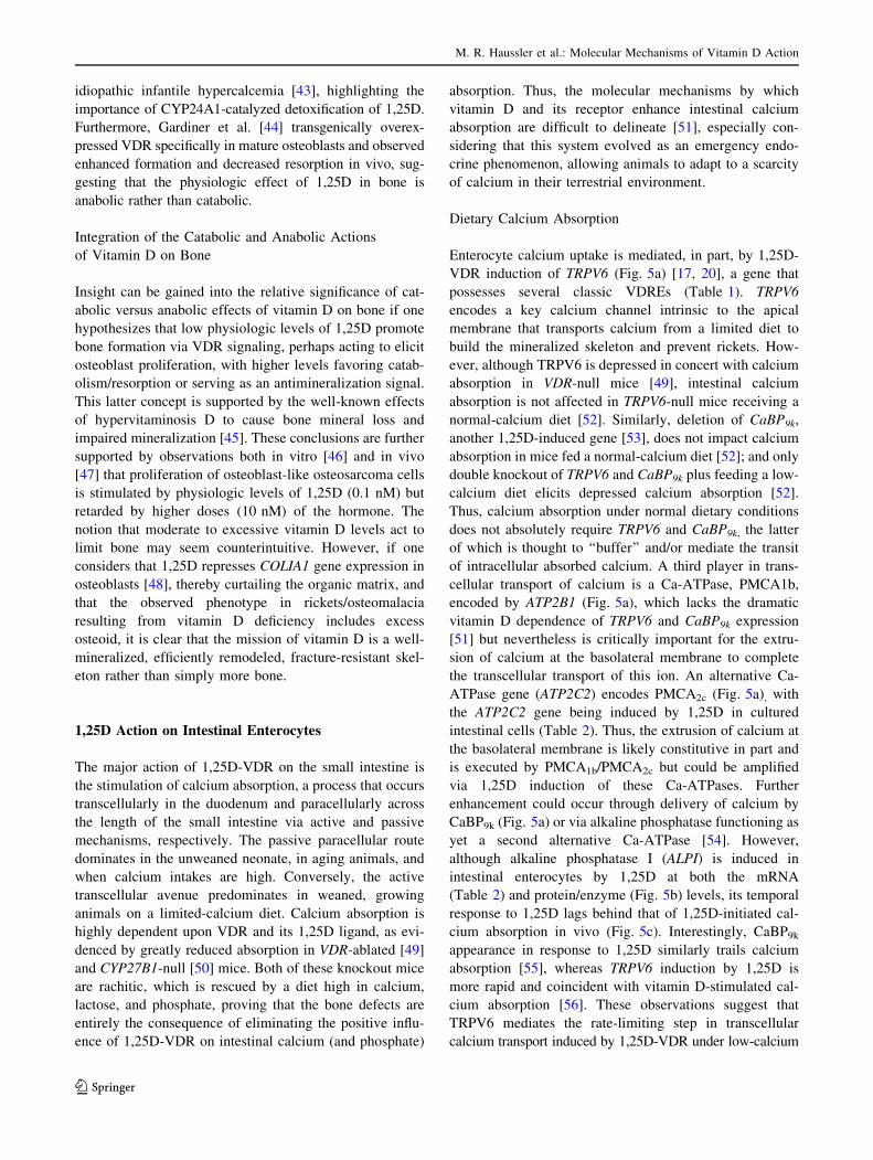
idiopathic infantile hypercalcemia [43], highlighting the
importance of CYP24A1-catalyzed detoxification of 1,25D.
Furthermore, Gardiner et al. [44] transgenically overex-
pressed VDR specifically in mature osteoblasts and observed
enhanced formation and decreased resorption in vivo, sug-
gesting that the physiologic effect of 1,25D in bone is
anabolic rather than catabolic.
Integration of the Catabolic and Anabolic Actions
of Vitamin D on Bone
Insight can be gained into the relative significance of cat-
abolic versus anabolic effects of vitamin D on bone if one
hypothesizes that low physiologic levels of 1,25D promote
bone formation via VDR signaling, perhaps acting to elicit
osteoblast proliferation, with higher levels favoring catab-
olism/resorption or serving as an antimineralization signal.
This latter concept is supported by the well-known effects
of hypervitaminosis D to cause bone mineral loss and
impaired mineralization [45]. These conclusions are further
supported by observations both in vitro [46] and in vivo
[47] that proliferation of osteoblast-like osteosarcoma cells
is stimulated by physiologic levels of 1,25D (0.1 nM) but
retarded by higher doses (10 nM) of the hormone. The
notion that moderate to excessive vitamin D levels act to
limit bone may seem counterintuitive. However, if one
considers that 1,25D represses COLIA1 gene expression in
osteoblasts [48], thereby curtailing the organic matrix, and
that the observed phenotype in rickets/osteomalacia
resulting from vitamin D deficiency includes excess
osteoid, it is clear that the mission of vitamin D is a well-
mineralized, efficiently remodeled, fracture-resistant skel-
eton rather than simply more bone.
1,25D Action on Intestinal Enterocytes
The major action of 1,25D-VDR on the small intestine is
the stimulation of calcium absorption, a process that occurs
transcellularly in the duodenum and paracellularly across
the length of the small intestine via active and passive
mechanisms, respectively. The passive paracellular route
dominates in the unweaned neonate, in aging animals, and
when calcium intakes are high. Conversely, the active
transcellular avenue predominates in weaned, growing
animals on a limited-calcium diet. Calcium absorption is
highly dependent upon VDR and its 1,25D ligand, as evi-
denced by greatly reduced absorption in VDR-ablated [49]
and CYP27B1-null [50] mice. Both of these knockout mice
are rachitic, which is rescued by a diet high in calcium,
lactose, and phosphate, proving that the bone defects are
entirely the consequence of eliminating the positive influ-
ence of 1,25D-VDR on intestinal calcium (and phosphate)
absorption. Thus, the molecular mechanisms by which
vitamin D and its receptor enhance intestinal calcium
absorption are difficult to delineate [51], especially con-
sidering that this system evolved as an emergency endo-
crine phenomenon, allowing animals to adapt to a scarcity
of calcium in their terrestrial environment.
Dietary Calcium Absorption
Enterocyte calcium uptake is mediated, in part, by 1,25D-
VDR induction of TRPV6 (Fig. 5a) [17, 20], a gene that
possesses several classic VDREs (Table 1). TRPV6
encodes a key calcium channel intrinsic to the apical
membrane that transports calcium from a limited diet to
build the mineralized skeleton and prevent rickets. How-
ever, although TRPV6 is depressed in concert with calcium
absorption in VDR-null mice [49], intestinal calcium
absorption is not affected in TRPV6-null mice receiving a
normal-calcium diet [52]. Similarly, deletion of CaBP9k,
another 1,25D-induced gene [53], does not impact calcium
absorption in mice fed a normal-calcium diet [52]; and only
double knockout of TRPV6 and CaBP9k plus feeding a low-
calcium diet elicits depressed calcium absorption [52].
Thus, calcium absorption under normal dietary conditions
does not absolutely require TRPV6 and CaBP9k, the latter
of which is thought to ‘‘buffer’’ and/or mediate the transit
of intracellular absorbed calcium. A third player in trans-
cellular transport of calcium is a Ca-ATPase, PMCA1b,
encoded by ATP2B1 (Fig. 5a), which lacks the dramatic
vitamin D dependence of TRPV6 and CaBP9k expression
[51] but nevertheless is critically important for the extru-
sion of calcium at the basolateral membrane to complete
the transcellular transport of this ion. An alternative Ca-
ATPase gene (ATP2C2) encodes PMCA2c (Fig. 5a), with
the ATP2C2 gene being induced by 1,25D in cultured
intestinal cells (Table 2). Thus, the extrusion of calcium at
the basolateral membrane is likely constitutive in part and
is executed by PMCA1b/PMCA2c but could be amplified
via 1,25D induction of these Ca-ATPases. Further
enhancement could occur through delivery of calcium by
CaBP9k (Fig. 5a) or via alkaline phosphatase functioning as
yet a second alternative Ca-ATPase [54]. However,
although alkaline phosphatase I (ALPI) is induced in
intestinal enterocytes by 1,25D at both the mRNA
(Table 2) and protein/enzyme (Fig. 5b) levels, its temporal
response to 1,25D lags behind that of 1,25D-initiated cal-
cium absorption in vivo (Fig. 5c). Interestingly, CaBP9k
appearance in response to 1,25D similarly trails calcium
absorption [55], whereas TRPV6 induction by 1,25D is
more rapid and coincident with vitamin D-stimulated cal-
cium absorption [56]. These observations suggest that
TRPV6 mediates the rate-limiting step in transcellular
calcium transport induced by 1,25D-VDR under low-calcium
M. R. Haussler et al.: Molecular Mechanisms of Vitamin D Action
123

conditions and that CaBP9k and ALPI play supportive or
secondary roles.
Multiple Roles of Alkaline Phosphatase
Besides a possible secondary role as a calcium ATPase in
calcium translocation, ALPI, which encodes a membrane
protein that is also secreted on both the apical and baso-
lateral sides, may perform other functions in response to
1,25D. Knockout of ALPI in mice [57] accelerates fat
absorption, indicating that ALPI governs lipid absorption,
likely through attachment to a secreted lipid particle (SLP)
transferring fat (Fig. 5a). By inducing ALPI, 1,25D-VDR
could be considered ‘‘antiobesity’’ in animals on a high-fat
diet. Moreover, ALPI associates with CaBP9k [58], raising
the possibility of a link between calcium and fat translo-
cation, with ALPI serving as a switching mechanism con-
necting these two phenomena (Fig. 5a). Finally, ALPI
(tethered to the apical membrane) has been shown to
function as a microbial defense barrier via the dephos-
phorylation of lipopolysaccharide [59]. It is instructive to
compare the intestinal action of 1,25D with that in bone,
the only other tissue in which we observe ALP induction
(Fig. 5b). Bone, liver, and kidney express a tissue-
nonspecific isozyme of ALP (TNAP). Mice with ablated
TNAP and humans with loss-of-function mutations in this
gene display hypophosphatasia, characterized by rickets
and osteomalacia, epileptic seizures, and increased levels
1,25D
TRPV6 Gene
CaBP9K Gene
Cldn2,12 Genes
DBP
1,25D
Ca2+
IntestinalLumen
Ca2+TRPV6
CaBP9K
Blood
Nucleus
Claudin-2, -12
Ca2+ Ca2+
Ca2+
ALPIALPI
Cubulin
PMCA1b
SLPFat Fat
ALPIALPIMicrobial Defense
via LPS de-phosphorylation
A
B C
-D +D
Tissue
Intestine Kidney Liver Bone
Tis
sue
Alk
alin
e P
ho
sph
atas
e
(IU
/mg
Pro
tein
)
0
5
10
15
1,25D (hours)
Ca absorption
Alk. Phos.
2+
Ca-ATPase
5
10
(cp
m
Ca/
200µ
l Pla
sma
x 10
)
45-2
Inte
stin
al A
lkal
ine
Ph
osp
hat
ase
(I
U/m
g P
rote
in)
10
15
20
2510 15 205
RXR VDR
1,25D
RXR VDR
1,25D
RXR VDR
1,25D
Paracelluar
PMCA2c
Cal
ciu
m A
bso
rpti
on
Fig. 5 1,25D-VDR action in small intestine. a Model for 1,25D-VDR
signaling of transepithelial and paracellular calcium transport in the
small intestine. SLP secreted lipid particle, for which exocytosis at the
basolateral membrane is initiated by cubilin; DBP vitamin D binding
protein, ALPI alkaline phosphatase I, PMCA1b plasma membrane
calcium-ATPase 1b. b Vitamin D increases alkaline phosphatase
activity selectively in intestine and bone of vitamin D-deficient
chicks. Alkaline phosphatase enzyme activity was assayed as
described elsewhere [54] in 20 % butanol extracts of tissue
homogenates or slices (bone) 40 h after a single oral dose of 50 IU
of vitamin D3. Each result is the average of separate determinations in
10 animals (±SD). c Comparison of the time courses of 1,25D-
mediated increases in calcium absorption and intestinal alkaline
phosphatase after dosing vitamin D-deficient chickens with 390
pmoles of 1,25D at ‘‘0’’ time, as indicated by the bold arrow.
Intestinal calcium absorption was measured as described previously
[122]. Each value represents the average of four animals (±SD)
M. R. Haussler et al.: Molecular Mechanisms of Vitamin D Action
123

of inorganic pyrophosphate [57]. The bone phenotype in
hypophosphatasia is the result of lack of cleavage of
extracellular pyrophosphate, a mineralization inhibitor, by
TNAP. Thus, by inducing TNAP in bone, 1,25D functions
as a promineralization hormone, yet another anabolic effect
of vitamin D in the skeleton.
Dietary Phosphate Absorption and Importance
of the Paracellular Route
Intestinal phosphate absorption per se is promoted by 1,25D
via the induction of Npt2b in rat intestinal enterocytes [60],
but because phosphate is abundant in the diet, the phosphate
absorption effect of 1,25D may not be as physiologically
relevant as the profound effect on calcium transport. Yet,
phosphate is a fundamental biologic component of not only
mineralized bone but essential biomolecules such as DNA,
RNA, phospholipids, phosphoproteins, ATP, and metabolic
intermediates; and under conditions of limiting dietary
phosphate, the significance of 1,25D-mediated phosphate
absorption likely is exposed. Despite the contribution of
vitamin D-dependent translocation of calcium and phos-
phate across the enterocyte, it is clear that paracellular
mechanisms also participate in the absorption of these bone
minerals. In fact, phosphate is predominantly absorbed in
this fashion, possibly because of its abundance in the diet. In
the case of calcium, 1,25D induces the expression of genes
encoding participants in the intercellular junction that
function as paracellular cation channels, such as claudin-2
and -12 (Fig. 5a; Table 2), and influences cadherins and
connexins to modulate intercellular adhesion. Therefore, a
fraction of vitamin D-mediated calcium absorption is
facilitated by paracellular mechanisms, which clearly
cannot be ascribed to the induction of enterocyte transport
proteins such as TRPV6, CaBP9k, and PMCA1b. Finally, a
component of intestinal calcium absorption is probably
mediated by nongenomic vitamin D mechanisms [61].
1,25D Action in the Kidney
The premier role of the kidney in the vitamin D endocrine
system is generation of the 1,25D hormone by the catalytic
action of CYP27B1 in the proximal tubule (Fig. 6). Renal
CYP27B1 is induced by PTH under low-calcium condi-
tions, whereas the enzyme is feedback-repressed by 1,25D
in a short feedback loop and by FGF23 via a long loop.
Bone-derived FGF23, like PTH, concomitantly signals
inhibition of renal phosphate reabsorption in the proximal
tubule by suppressing Npt2a (Fig. 6). Interestingly, FGF23
binds to different FGF receptors to mediate CYP27B1
repression (and CYP24A1 induction) versus Npt2a inhibi-
tion, namely, FGFR3/4 and FGFR1, respectively [62].
However, all three isoforms of FGFR require a coreceptor,
klotho, a bona fide longevity gene expressed primarily in
renal tubules, to bind FGF23 with high affinity. 1,25D
induces klotho mRNA [63], and a VDRE (Table 1) in the
human Klotho (KL) gene has been identified. Upregulation
of klotho by 1,25D is consistent with potentiation of
FGF23 signaling in the kidney and perhaps protection of
other cell types (e.g., vascular) where a secreted form of
klotho may exert beneficial effects (Fig. 6) [64].
The renal vitamin D hormone 1,25D acts directly on the
proximal tubule via VDR to stimulate phosphate reab-
sorption through induction of Npt2a [65] and Npt2c
[66, 67]. The physiologic impact of 1,25D on renal
Table 2 Key genes upregulated by 1,25D in cultured human colon cancer cells and keratinocytes via Affymetrix DNA Genechip microarray
Human colon cancer cell (Caco-2) mRNA induced by 1,25Da Human keratinocyte (KERTr CCD-1106) mRNA induced by 1,25Da
Class Genes Class Genes
Calcium transport-
related
TRPV6, CLDN2, ATP2C2, ALPI,ALPPL2
Epidermal
differentiation,
keratin-related
LCE (1D, 1F, 2B), S100A (2, 4, 6), SPRR1B, KRT (13,
16, 34, 38, 71), KRTAP (4, 5-1, 5-4, 8-1, 10-2, 10-4,
10-7, 10-9, 12-2)
Detoxification CYP (24A1, 1A1, 2S1, 3A5/43), SULT(1A2, 1C2), ABC transporters (A11,
B1, D1)
Detoxification CYP (24A1, 2D6)
Transcription factors CDX-2, MED9, JUNB, CEBPA, MX2 Transcription factors JUNB, CREG2, ID1, SALL4, ZNF257, HNF1A
Immunomodulation,
inflammation,
oxidation
S100A4, NOX1, G6PD, KNG1, IRF8,
ORM1, ORM2, DEFB32, CDC34Immunomodulation,
inflammation,
oxidation
CAMP, DEFB109, DEFB132, G6PD, EFCAB4A/B,
COLEC11, NFATC2, LGALS9, IGSF9B, IL25
Development and
cancer-related
TGFB2, CEACAM6, EPHB4,
EFNA5, DACT2, GLT8D4, TIMP2,
TIMP3
Development and
cancer-related
CASP14, BMP6, SFRP1, DNER, CST1, ADRB2/A1B,
CA9, PNOC, DND1, MEG8, DUSP10
At least a 1.2-fold upregulationa [1,25D] = 10 nM for 24 h
M. R. Haussler et al.: Molecular Mechanisms of Vitamin D Action
123

phosphate reabsorption is not fully understood, although
patients with loss-of-function mutations in Npt2c display
significant hypercalciuric, hypophosphatemic rickets/
osteomalacia [68]. In contrast, mice with ablated Npt2c
exhibit no phosphate imbalance but display deranged cal-
cium and 1,25D metabolism [68], suggesting that in
rodents Npt2c is more relevant to calcium homeostasis,
with phosphate reabsorption carried out predominantly by
Npt2a.
Calcium is reabsorbed actively in the distal renal tubules
by a process analogous to 1,25D-VDR-induced duodenal
calcium absorption (Fig. 5a). In the distal nephron, the
rate-limiting step is the induction by 1,25D of the TRPV5-
encoded calcium channel, which is fixed in the membrane
facing the glomerular filtrate through the glycosidase
enzymatic activity of klotho on extracellular carbohydrate
moieties in TRPV5 [69]. TRPV5 in the distal renal tubule
(Fig. 6) thus plays a role in 1,25D-induced calcium trans-
location akin to that of TRPV6 in the intestine. Moreover,
renal klotho could be considered analogous to intestinal
ALP in that both membrane enzymes are induced by 1,25D
and perform secondary, supportive extracellular functions
in regulating calcium and phosphate transport, as well as
possibly acting systemically to promote immune function
and elicit longevity. Intracellularly, calcium is buffered and
transferred by CaBP28k in the distal nephron (Fig. 6) after
induction by 1,25D in a manner similar to the induction of
CaBP9k (CALB3) in the intestine. Extrusion of calcium into
the bloodstream by the distal tubules is accomplished by
PMCA1b, identical to the mechanism in enterocytes, and
via NCX1, a sodium/calcium exchanger [51]. In summary,
as depicted in Fig. 6, renal calcium reclamation is crucial
to calcium homeostasis and is accomplished by the acti-
vation of TRPV5 by PTH plus the induction of TRPV5 by
1,25D (Fig. 6), both under conditions of low calcium.
Supportive roles in calcium reabsorption are played by
klotho, CaBP28k, PMCA1b, and NCX1, with klotho and
CaBP28k being the most dependent on 1,25D-VDR.
Therefore, the kidney is the source of two essential
hormones, 1,25D and klotho. As such, the kidney, and
vitamin D actions therein, impacts virtually every cell in
the body, especially those that express VDR in reasonable
concentrations. Illustrated in Fig. 6 are three examples of
this phenomenon. In the case of immune function (Fig. 6,
upper left), 1,25D-VDR induces cathelicidin [70] to acti-
vate the innate immune system to fight infection (Table 1);
and evidence is accumulating that the risk of human
infections such as tuberculosis is reduced by sufficient
circulating levels of 25D [71]. In addition, 1,25-VDR
represses IL-17 [6] to temper the adaptive immune system
and possibly lower the risk of autoimmune disorders such
as type 1 diabetes mellitus, multiple sclerosis, and
rheumatoid arthritis that have been linked to vitamin D
deficiency in association studies. 1,25D-VDR is anti-
inflammatory by blunting NFjB [72] and COX2 [73], and
inflammation is considered a common denominator in
Smooth muscle cells
Endothelium
Vasculature
BloodCa.PO4
Reabsorption
Ca2+
PO43-
1,25DRXR-VDR
Anticancer/Detox EffectsImmune Modulation Cardiovascular Influences
BreastProstateSkinColon
Epithelial Cells
T-cellsB-cellsMacrophages
Immune Cells
1,25D
Blood25D
Proximal TubuleDistal Tubule
Nephron
1,25DRXR-VDR
Synthesis SecretedForm
+
FGFR1
FGFR3,4
1,25DRXR-VDR
Kidney
Npt2a
Npt2c
TRPV5Klotho
Klotho
KlothoCYP27B1
PTH
FGF23
PTH
+
1,25D
KlothoBlood
intracrine conversion
CYP24A1FGF23 1,25D+
Catabolism to1,24,25D, etc.in all tissues
CaBP28K
Klotho
Fig. 6 The kidney responds to
1,25D, FGF23, and PTH to
regulate vitamin D bioactivation
and calcium/phosphate
reabsorption and serves as an
endocrine source of 1,25D and
klotho. The effects of kidney-
derived 1,25D and klotho on
various tissues are discussed in
the text
M. R. Haussler et al.: Molecular Mechanisms of Vitamin D Action
123

maladies such as cardiovascular disease and ischemic
stroke, as well as cancer. In the realm of cardiovascular
disease (Fig. 6, upper center) as well as neurodegenerative
disorders of aging such as Alzheimer disease, excess cir-
culating homocysteine is considered a negative risk factor;
and 1,25D-VDR has recently been shown [74] to induce
cystathionine b-synthase (CBS, Table 1) to catalyze the
elimination of homocysteine. More directly with respect to
1,25D and heart disease, the three relevant cell types,
namely, endothelial, smooth muscle, and cardiac myocyte,
all express VDR; and knockout of the receptor in mice
elicits left ventricular hypertrophy and fibrosis [75]. In
addition, klotho likely cooperates with 1,25D to the benefit
of the vasculature, with amelioration of hypertension,
oxidative stress, and vascular smooth muscle proliferation/
migration [64], although controlled studies are required to
demonstrate that these effects of vitamin D and klotho
occur in humans. Prevention of epithelial cell cancers by
1,25D and klotho (Fig. 6, upper right) represents the third
extrarenal realm wherein 1,25D-VDR-regulated genes
encode factors impacting cell life/cancer. VDR likely
reduces the risk for many cancers by inducing the p53 and
p21 (Table 1) tumor suppressors [76], as well as DNA
mismatch repair enzymes in the colon [77]. VDR knockout
mice exhibit enhanced colonic proliferation [78] plus
amplified mammary gland ductal extension, end buds, and
density [79], indicating that the fundamental actions of
VDR to promote cell differentiation and apoptosis [80]
play an important role in reducing the risk of age-related
epithelial cell cancers such as those of the colon and breast.
Finally, klotho has been implicated recently in the pre-
vention of pancreatic neoplasia [81]. However, as pointed
out by Manson et al. [82], evidence that vitamin D and/or
klotho are preventative for human cancer is lacking. Nev-
ertheless, through control of vital genes and cell functions,
1,25D-VDR and/or klotho are excellent candidates to allow
one to age well, not only by delaying osteoporosis, frac-
tures, and ectopic calcification via control of calcium and
phosphate but also by tempering malignancy, oxidative
damage, infections, autoimmunity, inflammation/pain, and
cardiovascular and neurodegenerative diseases.
1,25D Action in the Colonocyte
Of the epithelial cells postulated to be protected by VDR
signaling, those of the colon and skin express the highest
levels of VDR, comparable to those in the calcemic tissues:
intestine, kidney, and bone. One theme for liganded VDR
action in colon, analogous to the role of pregnane X
receptor (PXR) and constitutive androstane receptor (CAR)
[83] in the liver, is the induction of CYP enzymes for
xenobiotic detoxification. As illustrated in Fig. 7, a major
target for VDR (and PXR) in humans is CYP3A4 [84, 85],
for which the detoxification substrates include lithocholic
acid (LCA), a toxic secondary bile acid generated in the
enteric system by bacteria. Initial studies focused on VDR
liganded to 1,25D as a regulator of CYP3A4, but later
experiments revealed that LCA is also capable of binding
VDR to upregulate expression of human CYP3A4 or its
equivalent in rats (CYP3A23) or mice (CYP3A11) [84].
There is also evidence that CYP enzymes other than
CYP24A1 and CYP3A4 may be VDR targets [86]. Addi-
tionally, 1,25D induces SULT2A (Fig. 7), an enzyme that
detoxifies sterols via 3a-sulfation [87]. In fact, when we
screened mRNAs induced by 1,25D in human (Caco-2)
colon cancer cells (Table 2), besides mRNAs encoding
calcium translocating proteins, a major group of induced
mRNAs coded for detoxification agents, including CYPs,
SULTs, and ABC transporters. In accordance with this
observation, Meyer et al. [88], utilizing ChIP-Seq tech-
nology in the human LS180 colon cell line, demonstrated
that VDR/RXR binds in the vicinity of many CYP and
other detoxification-related genes.
A model for the pathophysiologic significance in humans
of LCA detoxification as a consequence of liganded VDR
signaling is depicted in Fig. 7. The precursor to LCA,
chenodeoxycholic acid, is produced in the liver via a
pathway that is controlled in a positive fashion by LXR and
in a negative feedback loop by FXR [89] (both of these
receptors form heterodimers with RXR, not shown). LCA,
formed through 7-dehydroxylation by gut bacteria, is not a
good substrate for the enterohepatic bile acid reuptake
LCA
CYP3A4
6αOH LCA and 3αSO4 LCA in lumen
COLON
HO H
COO
HO H
COOLCA
+
RXR VDR
CholesterolCYP 7A1
FXR−
Oxysterols
LXR
ChenodeoxycholicAcid
+
7-dehydroxylation
Transporter
Colon Xenobiotic
Detoxification
6αOH LCA
ABC
HO H
COO
HO H
COO
LCA
Gut Bacteria
RXR VDR
LIVER
CholesterolCYP 7A1
FXR−
Oxysterols
LXR
ChenodeoxycholicAcid
+
Colonocyte
1,25OR
3αSO4 LCASULT2A+
--
-
--
-
Fig. 7 Pathophysiologic roles of two VDR ligands in preventing
colon cancer
M. R. Haussler et al.: Molecular Mechanisms of Vitamin D Action
123

system and, thus, remains in the enteric tract and passes to
the colon, where it can exert carcinogenic effects [90]. VDR
in the colonocyte is proposed to bind LCA (or its 3-keto
derivative) and induce CYP3A4 [84]. CYP3A4 then cata-
lyzes the 6a-hydroxylation of LCA, thus converting it into a
substrate for an ABC efflux transporter [89]. We have
shown that 1,25D, which is formed in the kidney or locally
in colon via the action of CYP27B1, can also induce
CYP3A4 [83]. Thus, both of these nutritionally modulated
ligands for VDR possess the important potential to serve as
agents for promoting detoxification of LCA and possibly
other intestinal endo- or xenobiotics, with the end result
likely being a reduction in colon cancer incidence.
Of course, 1,25D-VDR no doubt elicits additional
effects besides detoxification in normal or malignant
colonic cells, and we observed that 1,25D induces the
expression of transcription factors (e.g., CDX-2, involved
in intestinal development), immunomodulators (e.g., DEFB32,
an antimicrobial), anti-inflammatory (e.g., NOX1) and anti-
oxidant (e.g., G6PD) enzymes, as well as many anticancer
principles (e.g., TGFB2, TIMP2, TIMP3) in Caco-2 cells
(Table 2). Finally, 1,25D has been shown to suppress
b-catenin signaling in colon cancer cells [80], and b-cate-
nin is a proto-oncogene that participates in the growth-
promoting Wnt signaling pathway that is thought to play a
significant role in the etiology of colon cancer. These
findings are all consistent with the proposed prodifferen-
tiative, antioxidant, and proimmune influences of 1,25D-
VDR that are thought to contribute to the anticancer
potential of vitamin D.
1,25D Action in Skin
VDR is abundantly expressed in the skin. Moreover, VDR-
null mice display the phenotype of alopecia and dermal
cysts, which is not ameliorated with a high-calcium, -lac-
tose, and -phosphate rescue diet. Surprisingly, however,
there exists no corresponding pathologic phenotype in the
skin of mice unable to synthesize 1,25D, suggesting that
the action of VDR in the skin is independent of the 1,25D
ligand. Along with VDR [91], b-catenin is absolutely
required in keratinocytes [92] to permit mammalian hair
cycling, and the ligand-independent action of VDR to
mediate the hair cycle is thought to involve Wnt signaling
in skin stem cells [91, 93].
As illustrated schematically in Fig. 8, the regulation of
mammalian hair cycling is complex, consisting of the
convergence of two signaling pathways, BMP and Wnt.
Starting at the upper left of Fig. 8, Noggin from the dermal
papilla initially antagonizes BMP4 signaling in bulb (or
bulge) keratinocytes, allowing for the accumulation of
Lef1/TCF, a transcriptional coactivator which targets genes
via DNA-binding partners such as b-catenin. Cessation of
Noggin signaling reinstates BMP signal transduction via
SMADs, provided that the Wnt modulator in surface
ectoderm (Wise or SOSTDC1), which antagonizes both
Wnt and BMP pathways [94], has also been repressed. Wnt
ligand, e.g., Wnt 10b, signaling leads to accumulation of
b-catenin, which facilitates cooperation with Lef1/TCF to
induce genes encoding factors, such as sonic hedgehog
(Shh), that trigger the hair cycle to transition from telogen
(resting) to anagen (growth). VDR apparently promotes
b-catenin-Lef1/TCF function by either serving as a coac-
tivator of the b-catenin transcription complex or inducing a
positive member of this protein complex.
Moreover, we propose that VDR also functions in
keratinocytes to drive the hair cycle by controlling gene
expression through negative as well as positive VDREs,
based upon gene ablation studies which indicate that VDR,
its RXRa [95] DNA binding heteropartner, as well as the
coactivator DRIP205 [6] and the corepressor hairless (Hr)
[96] all are required for normal hair cycling. The most
important role of VDR in controlling the hair cycle appears
to be repression of key target genes. This conclusion is
based upon the observation that knockout of Hr, a VDR
corepressor which colocalizes with VDR in the outer root
sheath of the hair follicle [97], produces a phenocopy of the
VDR-null mouse with respect to alopecia but does not
affect bone and mineral metabolism [98]. At least one of
the molecular functions of Hr is recruitment of histone
deacetylases (HDACs) to promote a repressive hetero-
chromatin architecture in the vicinity of the target gene
[99]; another proposed function of Hr is to catalyze histone
demethylation (HDMe) to further attenuate transcription
[100].
Anagen Telogen Catagen
PTHrP
HAIRFOLLICLE
DermalPapilla
BULB
HAIRFOLLICLE
DermalPapilla
BULB
Keratinocyte
Lef1/TCF
SMADs
BMP4
BMPR
Noggin
1
2
β-catenin
Wnt ligand
FzDsh
LRP
RXRα
Hr
Shh, Hoxc13, msx-1, msx-2
negative VDRE
HDMeHDACs
VDR
VDR
PTHR
Wise
Fig. 8 Model for VDR action in keratinocytes to sustain the
mammalian hair cycle. Wnt ortholog of Drosophila wingless and
mouse int-1, Lef1 lymphoid enhancer factor-1, TCF T cell-specific
factor, msx-1/-2 orthologs of Drosophila muscle-specific homeobox
protein. Factors that are membrane receptors or transporters are
boxed. Solid arrows indicate activation, and dotted lines ending in asolid perpendicular line denote inhibition
M. R. Haussler et al.: Molecular Mechanisms of Vitamin D Action
123

What genes are targeted by the VDR-RXRa-Hr complex
for repression? Thompson and coworkers [93] defined
SOSTDC1 (encoding Wise) as a gene overexpressed in
keratinocytes from Hr-null mice, and Kato and coworkers
[101] characterized S100A8 as a gene overexpressed in
VDR-null keratinocytes. In determining the effect of acti-
vated VDR on the expression of SOSTDC1 and S100A8
mRNA levels in human keratinocytes [1], we observed that
SOSTDC1 is strikingly repressed after 18 h of 1,25D
treatment of keratinocytes. Suppression of SOSTDC1
mRNA by 1,25D was verified utilizing cDNA microarray
analysis of human cells [1]. Because Wise not only
antagonizes the Wnt pathway by binding to LRP but also
inhibits the BMP pathway through neutralization of BMP4
[94], repression of SOSTDC1 by VDR could constitute a
major event in initiating the mammalian hair cycle (Fig. 8).
Similarly, 1,25D rapidly represses expression in human
keratinocytes of S100A8 and its obligatory S100A9 het-
eropartner in calcium binding [1]. This inhibition of
S100A8/-A9 expression by 1,25D-VDR is in stark contrast
to the induction of S100A8/-A9 observed in HL-60 pro-
myelocytic leukemia cells when differentiated by 1,25D
along the macrophage lineage [102]. Thus, 1,25D regulates
S100A8/-A9 expression in a cell-selective fashion. One
additional gene repressed by 1,25D-VDR, namely, PTHrP
[103], is known to encode a suppressor of the telogen to
anagen transition in the hair follicle, as well as to promote
entry into catagen [104], providing yet another VDR-
RXRa-Hr repressed gene target that participates in hair
cycle control (Fig. 8). In conclusion, VDR is crucial for the
regeneration of hair, an obvious shield that protects skin
and facilitates healthful aging, via both protein–protein and
protein–DNA interactions that potentiate Wnt, BMP, and
calcium signaling.
Although unliganded VDR is the dominant force in
driving the mammalian hair cycle, 1,25D, either produced
locally or generated in the kidney, is capable of signaling
keratinocyte differentiation and is used clinically as an
antiproliferative agent in the treatment of psoriasis [105].
Thus, vitamin D acts in the skin to depress growth and to
promote differentiation, just as it does in many cancer cell
lines. In fact, the VDR-null mouse is supersensitive to
dimethylbenz[a]anthracene-induced skin cancer [106] as
well as UV light-induced skin malignancy [107], and
1,25D is a candidate for the prevention of skin cancer.
Interestingly, photoirradiation of the skin produces vitamin
D, and the CYPs catalyzing bioactivation to 1,25D are
expressed in the skin and, therefore, able to produce local
1,25D to protect the epithelium against UV-induced
photodamage and malignancy. In addition, 1,25D induces
the expression of a number of genes in cultured keratino-
cytes, the products of which are potential prodifferentiative
and structural components as well as detoxification,
immunomodulation, and anti-inflammatory/antioxidation
principles (Table 2). For example, 1,25D induces caspase-
14 (CASP14) in keratinocytes (Table 2), and this nona-
poptotic caspase is crucial for keratinocyte differentiation
[98]. Also, genes in the epidermal differentiation complex
(LCE-1D, -1F, -2B) are induced by 1,25D in human kera-
tinocytes (Table 2). 1,25D induces cathelicidin (CAMP)
and several defensins in keratinocytes (Table 2), indicating
that vitamin D modulates the immune complement in skin.
Finally, 1,25D increases the expression of a number of
keratin-related transcripts, as well as the late cornified
envelope (LCE) proteins, suggesting that vitamin D signal-
ing supports the skin structurally and mediates barrier
function development. In summary, unliganded VDR func-
tions to drive the mammalian hair cycle in cooperation with
Hr, primarily via the repression of gene expression, whereas
1,25D acts via VDR binding to signal the development and
barrier function of the skin. This latter activity is apparently
redundant with other signalers, such as calcium, but never-
theless is important therapeutically in the prevention and
treatment of hyperproliferative skin diseases.
Ontogeny and Evolution of VDR and Its 1,25D Ligand
VDR appears to have adapted evolutionarily to become a
‘‘specialty’’ regulator of intestinal calcium absorption and
hair growth in terrestrial animals, providing both a miner-
alized skeleton for locomotion in a calcium-scarce envi-
ronment and physical protection against the harmful UV
radiation of the sun. Yet, as discussed above specifically for
the colon, VDR also has retained its ancient PXR-like
ability to effect xenobiotic detoxification via CYP induc-
tion. In fact, based upon examination of the evolutionary
position of VDR among the 48 human nuclear receptors,
VDR is extremely closely related to PXR, both structurally
and functionally [13]. The major action of PXR is in liver
protection via induction of CYP enzymes that participate in
xenobiotic detoxification. VDR could conceivably com-
plement PXR by serving as a general guardian of epithelial
cell integrity, especially at environmentally or xenobioti-
cally exposed sites such as the intestine, kidney, and skin.
In this section we examine the evolutionary origin of
VDR and its hormonal ligand. In other words, do VDR and
1,25D predate their modern functions of promoting intes-
tinal calcium and phosphate absorption to create calcified
tissue and of signaling keratinocyte differentiation to drive
mammalian hair cycling? A second issue addressed in this
section is VDR ontogenesis, which includes the temporal
expression of VDR in the development of classic vitamin D
target tissues such as the intestine, kidney and bone as well
as in ‘‘non-vitamin D target tissues’’ such as liver and
muscle.
M. R. Haussler et al.: Molecular Mechanisms of Vitamin D Action
123

With respect to VDR ontogeny, we examined VDR
concentrations in the developing chicken, focusing on the
small intestine, which expresses high levels of the receptor
in adults (positive control), as well as skeletal muscle and
liver, which express very low levels of VDR in the avian
adult (negative controls). Immunoblotting (Fig. 9a, right
portion of inset) confirms the high expression of avian
intestinal VDR compared to the paucity of VDR in muscle
and liver in the adult chicken. In striking contrast, immu-
noblotting (Fig. 9a, left portion of inset) reveals a com-
plementary expression pattern of VDR in these same
tissues in the 9 day-old embryo. VDR is barely expressed
in embryonic intestine but moderately expressed in
embryonic skeletal muscle and liver. To track the apparent
Fraction No. (4 drops)
00
4
3010 200
8
7S
3S
Fraction No. (4 drops)
00
4
3010 200
dpm
[ H
]1,2
5D/F
ract
ion
(x 1
0 )
-33
87S
3S
+ polyclonal antibody
no antibody
+ polyclonal antibody
+ monoclonal 4A5 antibody
no antibody
Spe
cific
ally
Bou
nd [
H]1
,25D
(dpm
x 1
0 /
mg
prot
ein)
-2
Embryo (Days)(Hatching)
D-deficientAdult
A
B C
dpm
[ H
]1,2
5D/F
ract
ion
(x 1
0 )
-33
+ monoclonal 4A5 antibody
58/60 kDa
Avian VDR
3
14
12
10
8
6
4
2
0
9 11 13 15 17 19 21
Fig. 9 Ontogenesis and evolution of VDR. a Profile of VDR as
monitored by high-affinity/specific binding of [3H]1,25D in skeletal
muscle (open circles) and liver (solid circles) of the developing
chicken. Nuclear extracts were incubated 16 h at 4 �C with 1 nM
[3H]1,25D (180 Ci/mmol) to radiolabel the receptor and a 50-fold
excess of radioinert 25(OH)D3 to eliminate any ligand binding to
contaminating DBP. Radiolabeled VDR was isolated and counted by
DEAE filter assay-liquid scintillation procedures [123]. Data are the
average of triplicate determinations ± SD. Inset immunoblot detec-
tion of VDR in embryonic and adult chicken tissues. Nuclear extracts
from each tissue were incubated as detailed above to radiolabel the
receptor, which was then purified by DNA-cellulose chromatography,
concentrated, and subjected to SDS-PAGE, followed by transfer to
nitrocellulose membranes for immunological probing with 9A7
monoclonal antibody [124]. For embryo samples, total protein levels
applied to each lane were as follows: liver, 1.5 mg; muscle, 5.0 mg;
intestine, 1.0 mg. For adult tissue samples, total protein levels applied
to each lane were as follows: liver, 1.5 mg; muscle, 1.5 mg; intestine,
1.5 mg. b Identification of VDR in frog (Rana catesbeiana) skin
cytosol by shifting its position in sucrose density gradient centrifu-
gation with specific polyclonal and monoclonal antibodies. Sucrose
density gradient centrifugation of samples of [3H]1,25D-labeled VDR
in tissue cytosols was performed as described elsewhere [125].
c Identification of VDR in fish (Oncorhynchus mykiss) pituitary gland
cytosol by shifting its position in sucrose density gradient centrifu-
gation with specific polyclonal and monoclonal antibodies. Because
the epitope for the 4A5 monoclonal antibody is not well conserved in
fish VDR, it is not shifted by this antibody; but we demonstrated that
it is totally immunoprecipitated when a rabbit anti-rat secondary
antibody is employed in conjunction with the 9A7 monoclonal
antibody for which the epitope is 100 % conserved between human
and fish VDR (data not shown)
M. R. Haussler et al.: Molecular Mechanisms of Vitamin D Action
123

decline of VDR in muscle and liver during embryogenesis,
we monitored the receptor via high-affinity binding of the
radiolabeled hormonal ligand. As shown in Fig. 9a, both
skeletal muscle and liver VDR binding activities descend
gradually between 9 and 21 days (hatching) to levels that
approximate the low concentrations in the adult. The
conclusion from these data is that, as expected, the target
intestine in avian species primarily expresses VDR after
hatching when it is required to signal dietary calcium
absorption, whereas ‘‘nontarget’’ tissues unexpectedly
express VDR significantly at the early embryonic stage but
lose this expression when the cells differentiate and
develop into adult tissues.
There are two interpretations to this latter phenomenon.
One is that only a select cell type(s) within the ‘‘nontarget’’
tissues expresses VDR, and this cell type is dominant in the
embryo but scarce in the adult tissue. The second explana-
tion is that VDR is moderately expressed in stem cells and all
cell types, either declining comparably with development if
unnecessary for signaling in that tissue or escalating in
vitamin D target tissues (e.g., gut) when required to mediate
signaling by the calcemic hormone, such as after weaning in
rodents. In favor of the first explanation, it is noteworthy that
in skeletal muscle VDR is primarily expressed in myoblasts
rather than myotubes and that 1,25D initiates the differen-
tiation of myoblasts to myotubes in skeletal muscle [108].
Thus, it may be only the myoblasts in adult muscle that are
reactive to 1,25D, and this is consistent with a recent study
by Garcia et al. [109] showing that mouse C2C12 myoblastic
cells respond to 1,25D to induce markers of differentiation
such as MyoD and myogenin, while repressing myostatin.
Analogously, it has been recently shown [110, 111] that the
minor population of stellate cells in the liver, which are key
mediators of progressive hepatic fibrosis following an insult,
dramatically express VDR and respond to 1,25D by
repressing inflammatory mediators/chemokines and profib-
rotic collagens/collagenases, not only suppressing hepatic
fibrosis but perhaps altering the hepatic microenvironment
to lower the risk of malignancy.
The overall conclusion is that rather than ‘‘being
expressed in every cell in the body,’’ as is touted in the
popular press, VDR is expressed in select cells within
virtually every tissue in the body. VDR expression is
governed by the state of differentiation of the cell, being
present in stem cells if needed to control the early devel-
opment of that line and perhaps cast aside when not
required in the end-stage differentiated cell type. Con-
versely, VDR may not regulate early cell development but
instead be induced in differentiated adult tissues to perform
signaling for the phenotypic functions of that tissue, as
occurs for bone mineral ion transport by enterocytes, renal
epithelial cells, and osteoblasts/osteocytes. Because the
major cell type in liver is the hepatocyte, the primary cell
in skeletal muscle is the myocyte/myotube, and VDR is not
expressed significantly in either of these cell types, neither
of these bulk tissues is considered a vitamin D target. Yet
by initiating differentiation of myoblasts and suppressing
inflammation following an insult via hepatic stellate cells,
vitamin D is clearly involved in the prevention of sarco-
penia and liver fibrosis, respectively.
How evolutionarily ancient is VDR in the eukaryotic
animal kingdom? VDR is expressed in neither Caeno-
rhabditis elegans nor yeast, but frog and fish tissues
express VDR as monitored by its high-affinity binding of
radiolabeled 1,25D, sedimentation coefficient (molecular
size and shape) in a sucrose density gradient, and immu-
noreactivity (increased sedimentation coefficient) to specific
monoclonal and polyclonal VDR antibodies. Interestingly,
the frog expresses VDR in high concentrations in the
skin (Fig. 9b), whereas the pituitary gland (Fig. 9c) is a
rich source of VDR in fish, where the respective roles of
VDR could be analogous to 1,25D signaling cell differ-
entiation and neuroendocrine control in mammals. In
support of the receptor protein data in Fig. 9b, c, VDR
cDNAs have been cloned from frog [112] and fish [113]
species, and VDR homologs appear to be present in all
fish and amphibian genomes thus far sequenced (data not
shown). Frogs and fish not only express VDR but also
synthesize ample amounts of the 1,25D hormonal ligand.
Frogs display a circulating level of 14 pg/mL and fish
200 pg/mL compared to the human level of 33 pg/mL
[114]. Because of the high bioavailability of calcium in
fresh- and seawater, we assert that 1,25D does not
necessarily function as a calcemic hormone in lower
animals, and VDR likely does not drive skin appendage
formation in amphibians and scaleless fish as it does in
mammals.
In support of this point, Whitfield et al. [115] cloned
VDR and demonstrated its expression in the sea lamprey, a
very basal vertebrate akin to hagfish, possessing neither
calcified tissue nor hair/skin accessory features, and yet this
organism biosynthesizes 1,25D [116]. The distribution of
VDR in lamprey, showing abundance in the skin and
mouth but not intestine [115], suggests that the original
function of liganded VDR was protection against the
environment by signaling xenobiotic detoxification. In fact,
lamprey VDR preferentially activates transcription of
CYP3A4 VDRE-containing reporter constructs compared
to constructs with calcemic VDREs such as those in oste-
ocalcin and TRPV6 [115]. Thus, an evolutionarily ancient
role of liganded VDR appears to be that of detoxification
[115], and this detoxification function for vitamin D-VDR
is retained in higher vertebrates, as discussed above. Based
on the observations in embryos, a second evolutionarily
ancient role of VDR may be the fundamental control of cell
differentiation/proliferation, a process that could have
M. R. Haussler et al.: Molecular Mechanisms of Vitamin D Action
123

morphed into the putative anticancer effects of 1,25D-VDR
in humans.
Nonvitamin D Ligands for VDR
One notable feature of the VDR/PXR/CAR subfamily of
nuclear receptors involved in detoxification is their ability
to recognize multiple ligands. The diversity of ligands for
PXR is especially broad and includes not only endogenous
steroids but also an array of other lipophilic compounds
such as the secondary bile acid LCA, the antibiotic rif-
ampicin, as well as xenobiotics such as hyperforin, the
active ingredient of St. John’s wort [117]. We have iden-
tified several additional nutritional lipids as candidate low-
affinity VDR ligands which may function locally in high
concentrations. Figure 10 reveals that these novel putative
VDR ligands include x3- and x6-essential polyunsaturated
fatty acids, docosahexaenoic acid and arachidonic acid,
respectively [118], the vitamin E derivative c-tocotrienol,
and curcumin [119], the latter of which is a turmeric-
derived polyphenol found in curry. Thus, it is now recog-
nized that VDR binds several ligands beyond the 1,25D
hormone. Considering the structures for prototypical PXR,
CAR, and FXR ligands and comparing these compounds
with the ligand binding profile of VDR, expanded to
include LCA and its 3-keto derivative, it is evident that
VDR exhibits a ligand profile resembling that of the closest
VDR relatives in the nuclear receptor superfamily, espe-
cially when it is noted that both PXR and FXR [84] are also
activated by LCA to some extent. Thus, VDR, PXR, and
CAR are three nuclear receptors that bind a host of ligands,
heterodimerize with RXR to signal detoxification of
xenobiotics, and overlap somewhat in their target gene
repertoires, which are laden with CYPs.
Conclusions
In summary, in this review we have highlighted the latest
developments in the mechanism of vitamin D action and its
biological consequences. A new understanding of the
physiology of vitamin D bioactivation in relation to phos-
phate metabolism and aging has been achieved, with the
induction by 1,25D-VDR of FGF23 in bone and klotho in
kidney taking center stage as the mechanism whereby
vitamin D mediates phosphate homeostasis and possibly
delays the chronic diseases of aging. As a DNA-binding
chromosomal protein, VDR is a charter member of the
nuclear receptor superfamily, specifically a member of the
VDR/PXR/CAR subfamily that is rooted evolutionarily in
signaling detoxification.
24 89 201 427
Zn Zn
1 111
H3+N H5 H9&10A
159
T COO–
Ligand Binding DomainDNA BindingDomain (DBD)
AF-2Hinge H3
1α,25-Dihydroxyvitamin D3 Lithocholic Acid
Curcumin γ-Tocotrienol
COOH
Docosahexaenoic Acid “ω3”22
Arachidonic Acid “ω6”
20
COOH
TFIIB
Heterodimerization with RXRs
Co-Act
Transactivation
Gene Bioeffect
Intestinal Ca2+ TransportTRPV6
p21 Cell Cycle Control
CYP3A4 Xenobiotic DetoxificationCYP24A1 1,25D Detoxification
FGF23Npt2c
PhosphateHomeostasis
Mammalian Hair Cycle
PTHrPSOSTDC1S100A8
Hr
SPP1BGP BoneRANKL Metabolism LRP5
Klotho Longevity
loop
COOH
HO
OH
OH HO
CH3
O
HO
CH3
O O O
OH
HO
O
H
CH3CH3
Fig. 10 Novel ligands for VDR shown in the context of the
functional domains in human VDR, its interacting comodulators,
and a summary of the genes and bioeffects signaled by liganded VDR.
Left the human VDR zinc finger DNA binding domain, which, in
cooperation with the corresponding domain in the RXR heteropartner,
mediates direct association with the target genes listed at the lower
left, leading to the indicated physiological effects. The official gene
symbol for BGP is BGLAP, that for RANKL is TNFSF11, that for
Npt2c is SLC34A3, that for PTHrP is PTHLH, and that for klotho is
KL. Right below the ligand binding domain are illustrated selected
VDR ligands, including several novel ligands discussed in the text
M. R. Haussler et al.: Molecular Mechanisms of Vitamin D Action
123

1,25D-VDR functions mainly through genomic mecha-
nisms, although the hormone-receptor complex also acts
rapidly via nongenomic mechanisms, as detailed elsewhere
[120]. Moreover, there is evidence that for phenomena
such as stimulating intestinal calcium transport, 1,25D
operates via VDR-independent mechanisms [61]. In the
case of genomic mechanisms, new insight into the control
of transcription by 1,25D-VDR-RXR has been gained
through macromolecular structural studies, which indicate
that the vitamin D ligand, the DNA sequence of the VDRE,
and the recruited coactivator/corepressor all are capable of
allosterically influencing the conformation of the VDR-
RXR binary receptor. Functionally important VDREs have
been located at remote positions 50 and 30 of the tran-
scription start site in 1,25D-regulated genes, implicating
DNA-looping and chromatin architecture as major forces
in the nucleation of gene-expression regulation by vitamin
D. Finally, epigenetic modification of both DNA and his-
tones to modulate gene availability for transcription com-
pletes the ‘‘painting’’ of what the authors consider the
‘‘molecular masterpiece’’ of the genomic mechanism of
vitamin D action.
The consequences of physiologic (optimal) levels of
1,25D acting directly on the skeleton via VDR genomic
signaling appear to comprise a delicate balance of anabolic
and catabolic events. Either a deficiency or an excess of
1,25D is deleterious to bone, primarily the result of path-
ologic resorption, respectively, via PTH signaling in D
deficiency and via RANKL action in vitamin D toxicity.
However, the paramount physiologic effect of 1,25D-VDR
genomic signaling remains the promotion of intestinal
absorption of calcium and phosphate, especially when
either of these ions is limited in the diet. The bottom line is
that the primary mechanism whereby 1,25D-VDR prevents
rickets and osteomalacia is the induction of intestinal cal-
cium and phosphate absorption from the gut, thereby pro-
viding these ions for proper bone mineralization.
With respect to the appearance of VDR in classic target
tissues, for example, small intestine, VDR is induced (in
part by 1,25D) in enterocytes when needed to signal cal-
cium absorption, such as at weaning in mammals and at
hatching in birds. In ‘‘nontarget’’ tissues such as the liver,
VDR (along with CYP27B1) is induced in select cells by
an insult, for example, in the minor cell population of
stellate cells, where 1,25D-VDR exerts anti-inflammatory
effects to lessen hepatic fibrosis. A similar phenomenon of
VDR/CYP27B1 induction occurs in macrophages follow-
ing an infection, where 1,25D-VDR induces cathelicidin
and defensins to bolster the innate immune system.
VDR is expressed in stem cells and during the early
embryonic period in many tissues, highlighting a possible
role for vitamin D in development. This function in cell
life may constitute an evolutionarily ancient action of
1,25D-VDR since both the ligand and receptor are present
in basal vertebrates such as the lamprey. Sea lampreys
possess neither calcified tissues nor hair, demonstrating
that the major functions of VDR, namely, calcemia for
bone mineralization and hair cycling to provide a shield of
protection against UV damage, are relatively modern
manifestations of VDR evolution adopted by terrestrial
animals. Fortunately, humans appear to have retained the
evolutionarily ancient VDR actions of cell growth control
and detoxification, rendering 1,25D-VDR a natural cancer
chemopreventative. Summarized in Fig. 10 are the actions
of 1,25D and alternative (low-affinity) ligands that bind
VDR, a macromolecule with the ability to translate small-
molecule effectors into biological mediators capable of cell
cycle control, initiation of detoxification, driving the hair
cycle, and regulation of extracellular calcium and phos-
phate levels to insure a mineralized, fracture-free skeleton
unaccompanied by ectopically calcified tissues.
References
1. Haussler MR, Haussler CA, Whitfield GK, Hsieh JC, Thompson
PD, Barthel TK, Bartik L, Egan JB, Wu Y, Kubicek JL,
Lowmiller CL, Moffet EW, Forster RE, Jurutka PW (2010) The
nuclear vitamin D receptor controls the expression of genes
encoding factors which feed the ‘‘Fountain of Youth’’ to mediate
healthful aging. J Steroid Biochem Mol Biol 121:88–97
2. Brumbaugh PF, Hughes MR, Haussler MR (1975) Cytoplasmic
and nuclear binding components for 1a,25-dihydroxyvitamin D3
in chick parathyroid glands. Proc Natl Acad Sci USA 72:
4871–4875
3. DeMay MB, Kiernan MS, DeLuca HF, Kronenberg HM (1992)
Sequences in the human parathyroid hormone gene that bind the
1,25-dihydroxyvitamin D3 receptor and mediate transcriptional
repression in response to 1,25-dihydroxyvitamin D3. Proc Natl
Acad Sci USA 89:8097–8101
4. Bergwitz C, Juppner H (2010) Regulation of phosphate
homeostasis by PTH, vitamin D, and FGF23. Annu Rev Med 61:
91–104
5. Kolek OI, Hines ER, Jones MD, Lesueur LK, Lipko MA, Kiela
PR, Collins JF, Haussler MR, Ghishan FK (2005) 1a25-Di-
hydroxyvitamin D3 upregulates FGF23 gene expression in bone:
the final link in a renal-gastrointestinal-skeletal axis that controls
phosphate transport. Am J Physiol Gastrointest Liver Physiol
289:G1036–G1042
6. Joshi S, Pantalena LC, Liu XK, Gaffen SL, Liu H, Rohowsky-
Kochan C, Ichiyama K, Yoshimura A, Steinman L, Christakos
S, Youssef S (2011) 1,25-Dihydroxyvitamin D3 ameliorates
Th17 autoimmunity via transcriptional modulation of interleu-
kin-17A. Mol Cell Biol 31:3653–3669
7. Mora JR, Iwata M, von Andrian UH (2008) Vitamin effects on
the immune system: vitamins A and D take centre stage. Nat
Rev Immunol 8:685–698
8. Bikle D (2009) Extrarenal synthesis of 1,25-dihydroxyvitamin D
and its health implications. Clin Rev Bone Miner Metab 7:
114–125
9. St-Arnaud R (2010) CYP24A1-deficient mice as a tool to
uncover a biological activity for vitamin D metabolites
hydroxylated at position 24. J Steroid Biochem Mol Biol 121:
254–256
M. R. Haussler et al.: Molecular Mechanisms of Vitamin D Action
123

10. Ohyama Y, Ozono K, Uchida M, Shinki T, Kato S, Suda T,
Yamamoto O, Noshiro M, Kato Y (1994) Identification of a
vitamin D-responsive element in the 5’ flanking region of the rat
25-hydroxyvitamin D3 24-hydroxylase gene. J Biol Chem 269:
10545–10550
11. Murayama A, Takeyama K, Kitanaka S, Kodera Y, Kawaguchi
Y, Hosoya T, Kato S (1999) Positive and negative regulations of
the renal 25-hydroxyvitamin D3 1alpha-hydroxylase gene by
parathyroid hormone, calcitonin, and 1alpha,25(OH)2D3 in
intact animals. Endocrinology 140:2224–2231
12. Haussler MR, Norman AW (1969) Chromosomal receptor for a
vitamin D metabolite. Proc Natl Acad Sci USA 62:155–162
13. Haussler MR, Whitfield GK, Haussler CA, Hsieh J-C, Jurutka
PW (2011) Nuclear vitamin D receptor: natural ligands,
molecular structure–function, and transcriptional control of vital
genes. In: Feldman D, Pike JW, Adams J (eds) vitamin D.
Academic Press, San Diego, pp 137–170
14. Jin CH, Kerner SA, Hong MH, Pike JW (1996) Transcriptional
activation and dimerization functions in the human vitamin D
receptor. Mol Endocrinol 10:945–957
15. Colnot S, Lambert M, Blin C, Thomasset M, Perret C (1995)
Identification of DNA sequences that bind retinoid X receptor-
1,25(OH)2D3-receptor heterodimers with high affinity. Mol Cell
Endocrinol 113:89–98
16. Zhang J, Chalmers MJ, Stayrook KR, Burris LL, Wang Y,
Busby SA, Pascal BD, Garcia-Ordonez RD, Bruning JB, Istrate
MA, Kojetin DJ, Dodge JA, Burris TP, Griffin PR (2011) DNA
binding alters coactivator interaction surfaces of the intact VDR-
RXR complex. Nat Struct Mol Biol 18:556–563
17. Meyer MB, Watanuki M, Kim S, Shevde NK, Pike JW (2006)
The human transient receptor potential vanilloid type 6 distal
promoter contains multiple vitamin D receptor binding sites that
mediate activation by 1,25-dihydroxyvitamin D3 in intestinal
cells. Mol Endocrinol 20:1447–1461
18. Fretz JA, Zella LA, Kim S, Shevde NK, Pike JW (2006) 1,25-
Dihydroxyvitamin D3 regulates the expression of low-density
lipoprotein receptor-related protein 5 via deoxyribonucleic acid
sequence elements located downstream of the start site of
transcription. Mol Endocrinol 20:2215–2230
19. Kim S, Yamazaki M, Shevde NK, Pike JW (2007) Transcrip-
tional control of receptor activator of nuclear factor-kappaB
ligand by the protein kinase A activator forskolin and the
transmembrane glycoprotein 130-activating cytokine, oncostatin
M, is exerted through multiple distal enhancers. Mol Endocrinol
21:197–214
20. Barthel TK, Mathern DR, Whitfield GK, Haussler CA, Hopper
HA, Hsieh JC, Slater SA, Hsieh G, Kaczmarska M, Jurutka PW,
Kolek OI, Ghishan FK, Haussler MR (2007) 1,25-Dihydroxy-
vitamin D3/VDR-mediated induction of FGF23 as well as tran-
scriptional control of other bone anabolic and catabolic genes that
orchestrate the regulation of phosphate and calcium mineral
metabolism. J Steroid Biochem Mol Biol 103:381–388
21. Malloy PJ, Pike JW, Feldman D (1999) The vitamin D recep-
tor and the syndrome of hereditary 1,25-dihydroxyvitamin
D-resistant rickets. Endocr Rev 20:156–188
22. Rochel N, Wurtz JM, Mitschler A, Klaholz B, Moras D (2000)
The crystal structure of the nuclear receptor for vitamin D bound
to its natural ligand. Mol Cell 5:173–179
23. Rochel N, Ciesielski F, Godet J, Moman E, Roessle M, Peluso-
Iltis C, Moulin M, Haertlein M, Callow P, Mely Y, Svergun DI,
Moras D (2011) Common architecture of nuclear receptor het-
erodimers on DNA direct repeat elements with different spac-
ings. Nat Struct Mol Biol 18:564–570
24. Jurutka PW, Hsieh J-C, Remus LS, Whitfield GK, Thompson
PD, Haussler CA, Blanco JCG, Ozato K, Haussler MR (1997)
Mutations in the 1,25-dihydroxyvitamin D3 receptor identifying
C-terminal amino acids required for transcriptional activation
that are functionally dissociated from hormone binding, hete-
rodimeric DNA binding and interaction with basal transcription
factor IIB, in vitro. J Biol Chem 272:14592–14599
25. Thompson PD, Remus LS, Hsieh J-C, Jurutka PW, Whitfield
GK, Galligan MA, Encinas Dominguez C, Haussler CA,
Haussler MR (2001) Distinct retinoid X receptor activation
function-2 residues mediate transactivation in homodimeric and
vitamin D receptor heterodimeric contexts. J Mol Endocrinol
27:211–227
26. Koszewski NJ, Ashok S, Russell J (1999) Turning a negative
into a positive: vitamin D receptor interactions with the avian
parathyroid hormone response element. Mol Endocrinol
13:455–465
27. Kim MS, Kondo T, Takada I, Youn MY, Yamamoto Y,
Takahashi S, Matsumoto T, Fujiyama S, Shirode Y, Yamaoka I,
Kitagawa H, Takeyama K, Shibuya H, Ohtake F, Kato S (2009)
DNA demethylation in hormone-induced transcriptional dere-
pression. Nature 461:1007–1012
28. Kim S, Yamazaki M, Zella LA, Shevde NK, Pike JW (2006)
Activation of receptor activator of NF-kappaB ligand gene
expression by 1,25-dihydroxyvitamin D3 is mediated through
multiple long-range enhancers. Mol Cell Biol 26:6469–6486
29. Saramaki A, Diermeier S, Kellner R, Laitinen H, Vaisanen S,
Carlberg C (2009) Cyclical chromatin looping and transcription
factor association on the regulatory regions of the p21
(CDKN1A) gene in response to 1alpha,25-dihydroxyvitamin D3.
J Biol Chem 284:8073–8082
30. Zella LA, Kim S, Shevde NK, Pike JW (2007) Enhancers
located in the vitamin D receptor gene mediate transcriptional
autoregulation by 1,25-dihydroxyvitamin D3. J Steroid Biochem
Mol Biol 103:435–439
31. Krishnan V, Moore TL, Ma YL, Helvering LM, Frolik CA,
Valasek KM, Ducy P, Geiser AG (2003) Parathyroid hormone
bone anabolic action requires Cbfa1/Runx2-dependent signal-
ing. Mol Endocrinol 17:423–435
32. Tanaka H, Seino Y (2004) Direct action of 1,25-dihydroxyvi-
tamin D on bone: VDRKO bone shows excessive bone forma-
tion in normal mineral condition. J Steroid Biochem Mol Biol
89–90:343–345
33. Weissen-Plenz G, Nitschke Y, Rutsch F (2008) Mechanisms of
arterial calcification: spotlight on the inhibitors. Adv Clin Chem
46:263–293
34. Milat F, Ng KW (2009) Is Wnt signalling the final common
pathway leading to bone formation? Mol Cell Endocrinol
310:52–62
35. Sroga GE, Karim L, Colon W, Vashishth D (2011) Biochemical
characterization of major bone-matrix proteins using nanoscale-
size bone samples and proteomics methodology. Mol Cel Pro-
teomics. doi: 10:M110 006718
36. Lee NK, Sowa H, Hinoi E, Ferron M, Ahn JD, Confavreux C,
Dacquin R, Mee PJ, McKee MD, Jung DY, Zhang Z, Kim JK,
Mauvais-Jarvis F, Ducy P, Karsenty G (2007) Endocrine regu-
lation of energy metabolism by the skeleton. Cell 130:456–
469
37. Oury F, Sumara G, Sumara O, Ferron M, Chang H, Smith CE,
Hermo L, Suarez S, Roth BL, Ducy P, Karsenty G (2011)
Endocrine regulation of male fertility by the skeleton. Cell
144:796–809
38. Haussler MR, Whitfield GK, Kaneko I, Forster R, Saini R, Hsieh
JC, Haussler CA, Jurutka PW (2012) The role of vitamin D in
the FGF23, klotho, and phosphate bone-kidney endocrine axis.
Rev Endocr Metab Disord 13:57–69
39. Shimada T, Kakitani M, Yamazaki Y, Hasegawa H, Takeuchi Y,
Fujita T, Fukumoto S, Tomizuka K, Yamashita T (2004) Tar-
geted ablation of Fgf23 demonstrates an essential physiological
M. R. Haussler et al.: Molecular Mechanisms of Vitamin D Action
123

role of FGF23 in phosphate and vitamin D metabolism. J Clin
Invest 113:561–568
40. Keisala T, Minasyan A, Lou YR, Zou J, Kalueff AV, Pyykko I,
Tuohimaa P (2009) Premature aging in vitamin D receptor
mutant mice. J Steroid Biochem Mol Biol 115:91–97
41. Shimada T, Hasegawa H, Yamazaki Y, Muto T, Hino R,
Takeuchi Y, Fujita T, Nakahara K, Fukumoto S, Yamashita T
(2004) FGF-23 is a potent regulator of vitamin D metabolism
and phosphate homeostasis. J Bone Miner Res 19:429–435
42. Masuda S, Byford V, Arabian A, Sakai Y, Demay MB,
St-Arnaud R, Jones G (2005) Altered pharmacokinetics of
1alpha,25-dihydroxyvitamin D3 and 25-hydroxyvitamin D3 in
the blood and tissues of the 25-hydroxyvitamin D-24-hydroxy-
lase (Cyp24a1) null mouse. Endocrinology 146:825–834
43. Schlingmann KP, Kaufmann M, Weber S, Irwin A, Goos C,
John U, Misselwitz J, Klaus G, Kuwertz-Broking E, Fehrenbach
H, Wingen AM, Guran T, Hoenderop JG, Bindels RJ, Prosser
DE, Jones G, Konrad M (2011) Mutations in CYP24A1 and
idiopathic infantile hypercalcemia. N Engl J Med 365:410–421
44. Gardiner EM, Baldock PA, Thomas GP, Sims NA, Henderson
NK, Hollis B, White CP, Sunn KL, Morrison NA, Walsh WR,
Eisman JA (2000) Increased formation and decreased resorption
of bone in mice with elevated vitamin D receptor in mature cells
of the osteoblastic lineage. FASEB J 14:1908–1916
45. Wronski TJ, Halloran BP, Bikle DD, Globus RK, Morey-Holton
ER (1986) Chronic administration of 1,25-dihydroxyvitamin D3:
increased bone but impaired mineralization. Endocrinology
119:2580–2585
46. Dokoh S, Donaldson CA, Haussler MR (1984) Influence of
1,25-dihydroxyvitamin D3 on cultured osteogenic sarcoma cells:
correlation with the 1,25-dihydroxyvitamin D3 receptor. Cancer
Res 44:2103–2109
47. Yamaoka K, Marion SL, Gallegos A, Haussler MR (1986) 1,25-
Dihydroxyvitamin D3 enhances the growth of tumors in athymic
mice inoculated with receptor rich osteosarcoma cells. Biochem
Biophys Res Commun 139:1292–1298
48. Kream BE, Harrison JR, Krebsbach PH, Bogdanovic Z, Bedalov
A, Pavlin D, Woody CO, Clark SH, Rowe D, Lichtler AC
(1995) Regulation of type I collagen gene expression in bone.
Connect Tissue Res 31:261–264
49. Van Cromphaut SJ, Dewerchin M, Hoenderop JG, Stockmans I,
Van Herck E, Kato S, Bindels RJ, Collen D, Carmeliet P,
Bouillon R, Carmeliet G (2001) Duodenal calcium absorption in
vitamin D receptor-knockout mice: functional and molecular
aspects. Proc Natl Acad Sci USA 98:13324–13329
50. Dardenne O, Prud’homme J, Arabian A, Glorieux FH, St-Ar-
naud R (2001) Targeted inactivation of the 25-hydroxyvitamin
D3–1a-hydroxylase gene (CYP27B1) creates an animal model
of pseudovitamin D-deficiency rickets. Endocrinology 142:
3135–3141
51. Lieben L, Carmeliet G, Masuyama R (2011) Calcemic actions of
vitamin D: effects on the intestine, kidney and bone. Best Pract
Res Clin Endocrinol Metab 25:561–572
52. Benn BS, Ajibade D, Porta A, Dhawan P, Hediger M, Peng JB,
Jiang Y, Oh GT, Jeung EB, Lieben L, Bouillon R, Carmeliet G,
Christakos S (2008) Active intestinal calcium transport in the
absence of transient receptor potential vanilloid type 6 and
calbindin-D9k. Endocrinology 149:3196–3205
53. Meyer J, Fullmer CS, Wasserman RH, Komm BS, Haussler MR
(1992) Dietary restriction of calcium, phosphorus, and vitamin
D elicits differential regulation of the mRNAs for avian intes-
tinal calbindin-D28k and the 1,25-dihydroxyvitamin D3 recep-
tor. J Bone Miner Res 7:441–448
54. Haussler M, Nagode LA, Rasmussen H (1970) Induction of
intestinal brush border alkaline phosphatase by vitamin D and
identity with ca-ATPase. Nature 228:1199–1201
55. Wasserman RH, Brindak ME, Buddle MM, Cai Q, Davis FC,
Fullmer CS, Gilmour RF Jr, Hu C, Mykkanen HM, Tapper DN
(1990) Recent studies on the biological actions of vitamin D on
intestinal transport and the electrophysiology of peripheral nerve
and cardiac muscle. Prog Clin Biol Res 332:99–126
56. Kutuzova GD, Sundersingh F, Vaughan J, Tadi BP, Ansay SE,
Christakos S, Deluca HF (2008) TRPV6 is not required for
1alpha,25-dihydroxyvitamin D3-induced intestinal calcium
absorption in vivo. Proc Natl Acad Sci USA 105:19655–19659
57. Narisawa S, Huang L, Iwasaki A, Hasegawa H, Alpers DH,
Millan JL (2003) Accelerated fat absorption in intestinal alka-
line phosphatase knockout mice. Mol Cell Biol 23:7525–7530
58. Leathers VL, Norman AW (1993) Evidence for calcium medi-
ated conformational changes in calbindin-D28K (the vitamin
D-induced calcium binding protein) interactions with chick
intestinal brush border membrane alkaline phosphatase as
studied via photoaffinity labeling techniques. J Cell Biochem
52:243–252
59. Chen KT, Malo MS, Moss AK, Zeller S, Johnson P, Ebrahimi F,
Mostafa G, Alam SN, Ramasamy S, Warren HS, Hohmann EL,
Hodin RA (2010) Identification of specific targets for the gut
mucosal defense factor intestinal alkaline phosphatase. Am J
Physiol Gastrointest Liver Physiol 299:G467–G475
60. Katai K, Miyamoto K, Kishida S, Segawa H, Nii T, Tanaka H,
Tani Y, Arai H, Tatsumi S, Morita K, Taketani Y, Takeda E
(1999) Regulation of intestinal Na?-dependent phosphate co-
transporters by a low-phosphate diet and 1,25-dihydroxyvitamin
D3. Biochem J 343(pt 3):705–712
61. Khanal RC, Nemere I (2008) Endocrine regulation of calcium
transport in epithelia. Clin Exp Pharmacol Physiol 35:1277–
1287
62. Razzaque MS (2009) The FGF23-Klotho axis: endocrine regu-
lation of phosphate homeostasis. Nat Rev Endocrinol 5:611–619
63. Forster RE, Jurutka PW, Hsieh JC, Haussler CA, Lowmiller CL,
Kaneko I, Haussler MR, Kerr Whitfield G (2011) Vitamin D
receptor controls expression of the anti-aging klotho gene in
mouse and human renal cells. Biochem Biophys Res Commun
414:557–562
64. Wang Y, Sun Z (2009) Klotho gene delivery prevents the
progression of spontaneous hypertension and renal damage.
Hypertension 54:810–817
65. Taketani Y, Segawa H, Chikamori M, Morita K, Tanaka K,
Kido S, Yamamoto H, Iemori Y, Tatsumi S, Tsugawa N, Okano
T, Kobayashi T, Miyamoto K, Takeda E (1998) Regulation
of type II renal Na?-dependent inorganic phosphate transporters
by 1,25-dihydroxyvitamin D3. Identification of a vitamin
D-responsive element in the human NAPi-3 gene. J Biol Chem
273:14575–14581
66. Jurutka PW, Bartik L, Whitfield GK, Mathern DR, Barthel TK,
Gurevich M, Hsieh JC, Kaczmarska M, Haussler CA, Haussler
MR (2007) Vitamin D receptor: key roles in bone mineral
pathophysiology, molecular mechanism of action, and novel
nutritional ligands. J Bone Miner Res 22(Suppl 2):V2–V10
67. Masuda M, Yamamoto H, Kozai M, Tanaka S, Ishiguro M,
Takei Y, Nakahashi O, Ikeda S, Uebanso T, Taketani Y, Segawa
H, Miyamoto K, Takeda E (2010) Regulation of renal sodium-
dependent phosphate co-transporter genes (Npt2a and Npt2c) by
all-trans-retinoic acid and its receptors. Biochem J 429:583–592
68. Segawa H, Aranami F, Kaneko I, Tomoe Y, Miyamoto K (2009)
The roles of Na/Pi-II transporters in phosphate metabolism.
Bone 45(Suppl 1):S2–S7
69. Chang Q, Hoefs S, van der Kemp AW, Topala CN, Bindels RJ,
Hoenderop JG (2005) The beta-glucuronidase klotho hydrolyzes
and activates the TRPV5 channel. Science 310:490–493
70. Liu PT, Stenger S, Li H, Wenzel L, Tan BH, Krutzik SR, Ochoa
MT, Schauber J, Wu K, Meinken C, Kamen DL, Wagner M,
M. R. Haussler et al.: Molecular Mechanisms of Vitamin D Action
123

Bals R, Steinmeyer A, Zugel U, Gallo RL, Eisenberg D, Hew-
ison M, Hollis BW, Adams JS, Bloom BR, Modlin RL (2006)
Toll-like receptor triggering of a vitamin D-mediated human
antimicrobial response. Science 311:1770–1773
71. Fabri M, Stenger S, Shin DM, Yuk JM, Liu PT, Realegeno S,
Lee HM, Krutzik SR, Schenk M, Sieling PA, Teles R, Montoya
D, Iyer SS, Bruns H, Lewinsohn DM, Hollis BW, Hewison M,
Adams JS, Steinmeyer A, Zugel U, Cheng G, Jo EK, Bloom BR,
Modlin RL (2011) Vitamin D is required for IFN-gamma-
mediated antimicrobial activity of human macrophages. Sci
Transl Med 3(104):104ra102
72. Cohen-Lahav M, Shany S, Tobvin D, Chaimovitz C, Douvde-
vani A (2006) Vitamin D decreases NFkappaB activity by
increasing IkappaBalpha levels. Nephrol Dial Transplant 21:
889–897
73. Moreno J, Krishnan AV, Swami S, Nonn L, Peehl DM, Feldman
D (2005) Regulation of prostaglandin metabolism by calcitriol
attenuates growth stimulation in prostate cancer cells. Cancer
Res 65:7917–7925
74. Kriebitzsch C, Verlinden L, Eelen G, van Schoor NM, Swart K,
Lips P, Meyer MB, Pike JW, Boonen S, Carlberg C, Vitvitsky
V, Bouillon R, Banerjee R, Verstuyf A (2011) 1,25-Di-
hydroxyvitamin D3 influences cellular homocysteine levels in
murine preosteoblastic MC3T3-E1 cells by direct regulation
of cystathionine beta-synthase. J Bone Miner Res 26:2991–
3000
75. Xiang W, Kong J, Chen S, Cao LP, Qiao G, Zheng W, Liu W, Li
X, Gardner DG, Li YC (2005) Cardiac hypertrophy in vitamin D
receptor knockout mice: role of the systemic and cardiac renin-
angiotensin systems. Am J Physiol Endocrinol Metab 288:
E125–E132
76. Audo I, Darjatmoko SR, Schlamp CL, Lokken JM, Lindstrom
MJ, Albert DM, Nickells RW (2003) Vitamin D analogues
increase p53, p21, and apoptosis in a xenograft model of human
retinoblastoma. Invest Ophthalmol Vis Sci 44:4192–4199
77. Sidelnikov E, Bostick RM, Flanders WD, Long Q, Fedirko V,
Shaukat A, Daniel CR, Rutherford RE (2010) Effects of calcium
and vitamin D on MLH1 and MSH2 expression in rectal mucosa
of sporadic colorectal adenoma patients. Cancer Epidemiol
Biomarkers Prev 19:1022–1032
78. Kallay E, Pietschmann P, Toyokuni S, Bajna E, Hahn P, Maz-
zucco K, Bieglmayer C, Kato S, Cross HS (2001) Character-
ization of a vitamin D receptor knockout mouse as a model of
colorectal hyperproliferation and DNA damage. Carcinogenesis
22:1429–1435
79. Zinser G, Packman K, Welsh J (2002) Vitamin D3 receptor
ablation alters mammary gland morphogenesis. Development
129:3067–3076
80. Egan JB, Thompson PA, Vitanov MV, Bartik L, Jacobs ET,
Haussler MR, Gerner EW, Jurutka PW (2010) Vitamin D
receptor ligands, adenomatous polyposis coli, and the vitamin D
receptor FokI polymorphism collectively modulate beta-catenin
activity in colon cancer cells. Mol Carcinog 49:337–352
81. Abramovitz L, Rubinek T, Ligumsky H, Bose S, Barshack I,
Avivi C, Kaufman B, Wolf I (2011) KL1 internal repeat
mediates klotho tumor suppressor activities and inhibits bFGF
and IGF-I signaling in pancreatic cancer. Clin Cancer Res 17:
4254–4266
82. Manson JE, Mayne ST, Clinton SK (2011) Vitamin D and
prevention of cancer—ready for prime time? N Engl J Med
364:1385–1387
83. Honkakoski P, Sueyoshi T, Negishi M (2003) Drug-activated
nuclear receptors CAR and PXR. Ann Med 35:172–182
84. Makishima M, Lu TT, Xie W, Whitfield GK, Domoto H, Evans
RM, Haussler MR, Mangelsdorf DJ (2002) Vitamin D receptor
as an intestinal bile acid sensor. Science 296:1313–1316
85. Thompson PD, Jurutka PW, Whitfield GK, Myskowski SM,
Eichhorst KR, Dominguez CE, Haussler CA, Haussler MR
(2002) Liganded VDR induces CYP3A4 in small intestinal and
colon cancer cells via DR3 and ER6 vitamin D responsive
elements. Biochem Biophys Res Commun 299:730–738
86. Drocourt L, Ourlin JC, Pascussi JM, Maurel P, Vilarem MJ
(2002) Expression of CYP3A4, CYP2B6, and CYP2C9 is reg-
ulated by the vitamin D receptor pathway in primary human
hepatocytes. J Biol Chem 277:25125–25132
87. Echchgadda I, Song CS, Roy AK, Chatterjee B (2004) Dehy-
droepiandrosterone sulfotransferase is a target for transcriptional
induction by the vitamin D receptor. Mol Pharmacol 65:720–
729
88. Meyer MB, Goetsch PD, Pike JW (2012) VDR/RXR and TCF4/
beta-catenin cistromes in colonic cells of colorectal tumor ori-
gin: impact on c-FOS and c-MYC gene expression. Mol
Endocrinol 26:37–51
89. Chawla A, Repa JJ, Evans RM, Mangelsdorf DJ (2001) Nuclear
receptors and lipid physiology: opening the X-files. Science
294:1866–1870
90. Kozoni V, Tsioulias G, Shiff S, Rigas B (2000) The effect of
lithocholic acid on proliferation and apoptosis during the early
stages of colon carcinogenesis: differential effect on apoptosis in
the presence of a colon carcinogen. Carcinogenesis 21:999–
1005
91. Cianferotti L, Cox M, Skorija K, Demay MB (2007) Vitamin D
receptor is essential for normal keratinocyte stem cell function.
Proc Natl Acad Sci USA 104:9428–9433
92. Huelsken J, Vogel R, Erdmann B, Cotsarelis G, Birchmeier W
(2001) b-Catenin controls hair follicle morphogenesis and stem
cell differentiation in the skin. Cell 105:533–545
93. Beaudoin GM 3rd, Sisk JM, Coulombe PA, Thompson CC
(2005) Hairless triggers reactivation of hair growth by promot-
ing Wnt signaling. Proc Natl Acad Sci USA 102:14653–14658
94. Lintern KB, Guidato S, Rowe A, Saldanha JW, Itasaki N (2009)
Characterization of wise protein and its molecular mechanism to
interact with both Wnt and BMP signals. J Biol Chem 284:
23159–23168
95. Li M, Indra AK, Warot X, Brocard J, Messaddeq N, Kato S,
Metzger D, Chambon P (2000) Skin abnormalities generated by
temporally controlled RXRalpha mutations in mouse epidermis.
Nature 407:633–636
96. Thompson CC, Sisk JM, Beaudoin GM 3rd (2006) Hairless and
Wnt signaling: allies in epithelial stem cell differentiation. Cell
Cycle 5:1913–1917
97. Hsieh J-C, Sisk JM, Jurutka PW, Haussler CA, Slater SA,
Haussler MR, Thompson CC (2003) Physical and functional
interaction between the vitamin D receptor and hairless core-
pressor, two proteins required for hair cycling. J Biol Chem
278:38665–38674
98. Zarach JM, Beaudoin GM 3rd, Coulombe PA, Thompson CC
(2004) The co-repressor hairless has a role in epithelial cell
differentiation in the skin. Development 131:4189–4200
99. Potter GB, Zarach JM, Sisk JM, Thompson CC (2002) The
thyroid hormone-regulated corepressor hairless associates with
histone deacetylases in neonatal rat brain. Mol Endocrinol
16:2547–2560
100. Hsieh JC, Slater SA, Whitfield GK, Dawson JL, Hsieh G,
Sheedy C, Haussler CA, Haussler MR (2010) Analysis of
hairless corepressor mutants to characterize molecular cooper-
ation with the vitamin D receptor in promoting the mammalian
hair cycle. J Cell Biochem 110:671–686
101. Yamamoto Y, Memezawa A, Takagi K, Ochiai E, Shindo M,
Kato S (2009) A tissue-specific function by unliganded VDR. In:
Abstracts from the 14th Workshop on Vitamin D, Brugge,
Belgium, October 4–8, 2009, p 66
M. R. Haussler et al.: Molecular Mechanisms of Vitamin D Action
123

102. Suzuki T, Tazoe H, Taguchi K, Koyama Y, Ichikawa H,
Hayakawa S, Munakata H, Isemura M (2006) DNA microarray
analysis of changes in gene expression induced by 1,25-di-
hydroxyvitamin D3 in human promyelocytic leukemia HL-60
cells. Biomed Res 27:99–109
103. Falzon M (1996) DNA sequences in the rat parathyroid hor-
mone-related peptide gene responsible for 1,25-dihydroxyvita-
min D3-mediated transcriptional repression. Mol Endocrinol
10:672–681
104. Cho YM, Woodard GL, Dunbar M, Gocken T, Jimenez JA,
Foley J (2003) Hair-cycle-dependent expression of parathyroid
hormone-related protein and its type I receptor: evidence for
regulation at the anagen to catagen transition. J Invest Dermatol
120:715–727
105. Kragballe K (1997) The future of vitamin D in dermatology.
J Am Acad Dermatol 37:S72–S76
106. Zinser GM, Sundberg JP, Welsh J (2002) Vitamin D3 receptor
ablation sensitizes skin to chemically induced tumorigenesis.
Carcinogenesis 23:2103–2109
107. Ellison TI, Smith MK, Gilliam AC, MacDonald PN (2008)
Inactivation of the vitamin D receptor enhances susceptibility of
murine skin to UV-induced tumorigenesis. J Invest Dermatol
128:2508–2517
108. Costa EM, Blau HM, Feldman D (1986) 1,25-Dihydroxyvitamin
D3 receptors and hormonal responses in cloned human skeletal
muscle cells. Endocrinology 119:2214–2220
109. Garcia LA, King KK, Ferrini MG, Norris KC, Artaza JN (2011)
1,25(OH)2vitamin D3 stimulates myogenic differentiation by
inhibiting cell proliferation and modulating the expression of
promyogenic growth factors and myostatin in C2C12 skeletal
muscle cells. Endocrinology 152:2976–2986
110. Abramovitch S, Dahan-Bachar L, Sharvit E, Weisman Y, Ben
Tov A, Brazowski E, Reif S (2011) Vitamin D inhibits prolif-
eration and profibrotic marker expression in hepatic stellate cells
and decreases thioacetamide-induced liver fibrosis in rats. Gut
60:1728–1737
111. Sherman MH, Downes M, Evans RM (2012) Nuclear receptors
as modulators of the tumor microenvironment. Cancer Prev Res
(Phila) 5:3–10
112. Li YC, Bergwitz C, Juppner H, Demay MB (1997) Cloning and
characterization of the vitamin D receptor from Xenopus laevis.
Endocrinology 138:2347–2353
113. Krasowski MD, Ai N, Hagey LR, Kollitz EM, Kullman SW,
Reschly EJ, Ekins S (2011) The evolution of farnesoid X,
vitamin D, and pregnane X receptors: insights from the green-
spotted pufferfish (Tetraodon nigriviridis) and other non-mam-
malian species. BMC Biochem 12:5
114. Dokoh S, Llach F, Haussler MR (1982) 25-Hydroxyvitamin D
and 1,25-dihydroxyvitamin D: new ultrasensitive and accurate
assays. In: Norman AW, Schaefer K, von Herrath D, Grigoleit
H-G (eds) Vitamin D, chemical, biochemical and clinical
endocrinology of calcium metabolism. Walter de Gruyter,
Berlin, pp 743–749
115. Whitfield GK, Dang HTL, Schluter SF, Bernstein RM, Bunag T,
Manzon LA, Hsieh G, Dominguez CE, Youson JH, Haussler
MR, Marchalonis JJ (2003) Cloning of a functional vitamin D
receptor from the lamprey (Petromyzon marinus), an ancient
vertebrate lacking a calcified skeleton and teeth. Endocrinology
144:2704–2716
116. Kobayashi T, Takeuchi A, Okano T (1991) An evolutionary
aspect in vertebrates from the viewpoint of vitamin D3 metab-
olism. In: Norman AW, Bouillon R, Thomasset M (eds) Vitamin
D: gene regulation, structure–function analysis and clinical
application. Walter de Gruyter, New York, pp 679–680
117. Moore LB, Maglich JM, McKee DD, Wisely B, Willson TM,
Kliewer SA, Lambert MH, Moore JT (2002) Pregnane X
receptor (PXR), constitutive androstane receptor (CAR), and
benzoate X receptor (BXR) define three pharmacologically
distinct classes of nuclear receptors. Mol Endocrinol 16:
977–986
118. Haussler MR, Haussler CA, Bartik L, Whitfield GK, Hsieh JC,
Slater S, Jurutka PW (2008) Vitamin D receptor: molecular
signaling and actions of nutritional ligands in disease preven-
tion. Nutr Rev 66:S98–S112
119. Bartik L, Whitfield GK, Kaczmarska M, Lowmiller CL, Moffet
EW, Furmick JK, Hernandez Z, Haussler CA, Haussler MR,
Jurutka PW (2010) Curcumin: a novel nutritionally derived
ligand of the vitamin D receptor with implications for colon
cancer chemoprevention. J Nutr Biochem 21:1153–1161
120. Haussler MR, Jurutka PW, Mizwicki M, Norman AW (2011)
Vitamin D receptor (VDR)-mediated actions of 1alpha,
25(OH)vitamin D: genomic and non-genomic mechanisms. Best
Pract Res Clin Endocrinol Metab 25:543–559
121. Liu SM, Koszewski N, Lupez M, Malluche HH, Olivera A,
Russell J (1996) Characterization of a response element in the
50-flanking region of the avian (chicken) PTH gene that mediates
negative regulation of gene transcription by 1,25-dihydroxyvi-
tamin D3 and binds the vitamin D3 receptor. Mol Endocrinol
10:206–215
122. Haussler MR, Zerwekh JE, Hesse RH, Rizzardo E, Pechet MM
(1973) Biological activity of 1alpha-hydroxycholecalciferol, a
synthetic analog of the hormonal form of vitamin D3. Proc Natl
Acad Sci USA 70:2248–2252
123. Brumbaugh PF, Haussler DH, Bursac KM, Haussler MR (1974)
Filter assay for 1a,25-dihydroxyvitamin D3: utilization of the
hormone’s target tissue chromatin receptor. Biochemistry
(Mosc) 13:4097–4102
124. Allegretto EA, Pike JW, Haussler MR (1987) Immunochemical
detection of unique proteolytic fragments of the chick 1,25-
dihydroxyvitamin D3 receptor. J Biol Chem 262:1312–1319
125. Mangelsdorf DJ, Koeffler HP, Donaldson CA, Pike JW,
Haussler MR (1984) 1,25-Dihydroxyvitamin D3-induced dif-
ferentiation in a human promyelocytic leukemia cell line
(HL-60): receptor-mediated maturation to macrophage-like
cells. J Cell Biol 98:391–398
M. R. Haussler et al.: Molecular Mechanisms of Vitamin D Action
123
Related Documents