Cell, Vol. 119, 629–640, November 24, 2004, Copyright 2004 by Cell Press Molecular Mechanism of AHSP-Mediated Stabilization of -Hemoglobin ized by high-level production of hemoglobin A (HbA), which is composed of two and two subunits, each bound to a heme moiety and produced in a concerted Liang Feng, 1,5 David A. Gell, 2,5 Suiping Zhou, 3,5 Lichuan Gu, 1 Yi Kong, 3 Jianqing Li, 3 Min Hu, 1 Nieng Yan, 1 Christopher Lee, 2 Anne M. Rich, 4 manner (Bunn, 1987). Each subunit uses a histidine side Robert S. Armstrong, 4 Peter A. Lay, 4 chain to coordinate the ferrous atom in the center of Andrew J. Gow, 3 Mitchell J. Weiss, 3, * the bound heme; oxygen binds cooperatively and re- Joel P. Mackay, 2, * and Yigong Shi 1, * versibly to ferrous iron from the opposite side of the 1 Department of Molecular Biology heme plane. The elaborate oxygen binding mechanism Lewis Thomas Laboratory of HbA allows the concentration of oxygen in the blood Princeton University to nearly reach that in open air. Although the syntheses Princeton, New Jersey 08544 of - and -hemoglobin are coordinated for efficient 2 School of Molecular and Microbial Biosciences assembly of HbA, free subunits can sometimes accumu- University of Sydney late (Baglioni, 1966). Indeed, a small excess of Hb is NSW 2006 present in normal erythrocytes (Baglioni, 1966; Baglioni Australia and Campana, 1967). While free -hemoglobin (Hb) 3 The Children’s Hospital of Philadelphia and forms a relatively stable homo-tetramer, free -hemoglo- The University of Pennsylvania bin (Hb) exists as a structurally unstable monomer and Philadelphia, Pennsylvania 19104 is prone to oxidation and precipitation, likely contribut- 4 Center for Heavy Metals Research and ing to the pathophysiology of thalassemia and other Center for Structural Biology blood disorders (Nathan and Gunn, 1966; Rachmilewitz and Structural Chemistry and Schrier, 2001). School of Chemistry Molecular chaperones have long been proposed to University of Sydney help stabilize free globin and hemoglobin subunits. Ef- NSW 2006 fort to identify such chaperones led to the discovery Australia of the -hemoglobin stabilizing protein (AHSP), which specifically interacts with and stabilizes free Hb in the absence of available subunit (Kihm et al., 2002). When Summary available, Hb binds more avidly to the subunit, dis- placing AHSP and forming tetrameric HbA (Gell et al., Hemoglobin A (HbA), the oxygen delivery system in 2002; Kihm et al., 2002). AHSP is an abundant, erythroid- humans, comprises two and two subunits. Free specific protein that protects free Hb from precipitation -hemoglobin (Hb) is unstable, and its precipitation both in solution and in live cells. AHSP knockout mice contributes to the pathophysiology of thalassemia. exhibit reticulocytosis, abnormal erythrocyte morphol- In erythrocytes, the -hemoglobin stabilizing protein ogy with intracellular inclusion bodies, and increased (AHSP) binds Hb and inhibits its precipitation. The reactive oxygen species (ROS) with subsequent cellular crystal structure of AHSP bound to Fe(II)-Hb reveals oxidative damage (Kihm et al., 2002; Y. Kong et al., sub- that AHSP specifically recognizes the G and H helices mitted). of Hb through a hydrophobic interface that largely Although AHSP plays an essential role in modulating recapitulates the 1 - 1 interface of hemoglobin. The the stability of Hb and the assembly of HbA, the under- AHSP-Hb interactions are extensive but suboptimal, lying molecular mechanisms are undetermined. A recent explaining why -hemoglobin can competitively dis- nuclear magnetic resonance study indicated that AHSP place AHSP to form HbA. Remarkably, the Fe(II)-heme adopts a three-helix bundle and interacts with Hb via group in AHSP bound Hb is coordinated by the distal helices 1 and 2 and the intervening segment (Santiveri but not the proximal histidine. Importantly, binding to et al., 2004). AHSP binds to several forms of Hb, as AHSP facilitates the conversion of oxy-Hb to a deoxy- well as apo- globin, in which heme is absent, but the genated, oxidized [Fe(III)], nonreactive form in which molecular bases of these interactions are undefined. For all six coordinate positions are occupied. These obser- example, it is not understood how AHSP specifically vations reveal the molecular mechanisms by which recognizes , but not , chains, or how interaction with AHSP stabilizes free Hb. AHSP results in the stabilization of Hb. In addition, it is unclear how AHSP actively prevents Hb-mediated Introduction oxidative damage in cells, although rearrangements of the distal surface have been shown to alter heme reactiv- Hemoglobin (Hb) is responsible for carrying and deliv- ity (Olson et al., 1997). ering oxygen to tissues and organs in animals (Dickerson In this manuscript, using an integrated approach com- and Geis, 1983; Fermi and Perutz, 1981). In humans, the bining nuclear magnetic resonance, X-ray crystallogra- maturation of red blood cells (erythrocytes) is character- phy, and biochemical and biophysical analyses, we in- vestigate the molecular mechanism by which AHSP stabilizes Hb. Our results not only provide molecular *Correspondence: [email protected] (Y.S.); j.mackay@ answers to the important questions posed above but mmb.usyd.edu.au (J.P.M.); [email protected] (M.J.W.) 5 These authors contributed equally to this work. also, more significantly, define a mechanistic pathway

Welcome message from author
This document is posted to help you gain knowledge. Please leave a comment to let me know what you think about it! Share it to your friends and learn new things together.
Transcript

Cell, Vol. 119, 629–640, November 24, 2004, Copyright 2004 by Cell Press
Molecular Mechanism of AHSP-MediatedStabilization of �-Hemoglobin
ized by high-level production of hemoglobin A (HbA),which is composed of two � and two � subunits, eachbound to a heme moiety and produced in a concerted
Liang Feng,1,5 David A. Gell,2,5 Suiping Zhou,3,5
Lichuan Gu,1 Yi Kong,3 Jianqing Li,3 Min Hu,1
Nieng Yan,1 Christopher Lee,2 Anne M. Rich,4
manner (Bunn, 1987). Each subunit uses a histidine sideRobert S. Armstrong,4 Peter A. Lay,4
chain to coordinate the ferrous atom in the center ofAndrew J. Gow,3 Mitchell J. Weiss,3,*the bound heme; oxygen binds cooperatively and re-Joel P. Mackay,2,* and Yigong Shi1,*versibly to ferrous iron from the opposite side of the1Department of Molecular Biologyheme plane. The elaborate oxygen binding mechanismLewis Thomas Laboratoryof HbA allows the concentration of oxygen in the bloodPrinceton Universityto nearly reach that in open air. Although the synthesesPrinceton, New Jersey 08544of �- and �-hemoglobin are coordinated for efficient2 School of Molecular and Microbial Biosciencesassembly of HbA, free subunits can sometimes accumu-University of Sydneylate (Baglioni, 1966). Indeed, a small excess of �Hb isNSW 2006present in normal erythrocytes (Baglioni, 1966; BaglioniAustraliaand Campana, 1967). While free �-hemoglobin (�Hb)3 The Children’s Hospital of Philadelphia andforms a relatively stable homo-tetramer, free �-hemoglo-The University of Pennsylvaniabin (�Hb) exists as a structurally unstable monomer andPhiladelphia, Pennsylvania 19104is prone to oxidation and precipitation, likely contribut-4 Center for Heavy Metals Research anding to the pathophysiology of � thalassemia and otherCenter for Structural Biologyblood disorders (Nathan and Gunn, 1966; Rachmilewitzand Structural Chemistryand Schrier, 2001).School of Chemistry
Molecular chaperones have long been proposed toUniversity of Sydneyhelp stabilize free globin and hemoglobin subunits. Ef-NSW 2006fort to identify such chaperones led to the discoveryAustraliaof the �-hemoglobin stabilizing protein (AHSP), whichspecifically interacts with and stabilizes free �Hb in theabsence of available � subunit (Kihm et al., 2002). WhenSummaryavailable, �Hb binds more avidly to the � subunit, dis-placing AHSP and forming tetrameric HbA (Gell et al.,Hemoglobin A (HbA), the oxygen delivery system in2002; Kihm et al., 2002). AHSP is an abundant, erythroid-humans, comprises two � and two � subunits. Freespecific protein that protects free �Hb from precipitation�-hemoglobin (�Hb) is unstable, and its precipitationboth in solution and in live cells. AHSP knockout micecontributes to the pathophysiology of � thalassemia.exhibit reticulocytosis, abnormal erythrocyte morphol-In erythrocytes, the �-hemoglobin stabilizing proteinogy with intracellular inclusion bodies, and increased(AHSP) binds �Hb and inhibits its precipitation. Thereactive oxygen species (ROS) with subsequent cellularcrystal structure of AHSP bound to Fe(II)-�Hb revealsoxidative damage (Kihm et al., 2002; Y. Kong et al., sub-that AHSP specifically recognizes the G and H helicesmitted).
of �Hb through a hydrophobic interface that largelyAlthough AHSP plays an essential role in modulating
recapitulates the �1-�1 interface of hemoglobin. Thethe stability of �Hb and the assembly of HbA, the under-
AHSP-�Hb interactions are extensive but suboptimal, lying molecular mechanisms are undetermined. A recentexplaining why �-hemoglobin can competitively dis- nuclear magnetic resonance study indicated that AHSPplace AHSP to form HbA. Remarkably, the Fe(II)-heme adopts a three-helix bundle and interacts with �Hb viagroup in AHSP bound �Hb is coordinated by the distal helices 1 and 2 and the intervening segment (Santiveribut not the proximal histidine. Importantly, binding to et al., 2004). AHSP binds to several forms of �Hb, asAHSP facilitates the conversion of oxy-�Hb to a deoxy- well as apo-� globin, in which heme is absent, but thegenated, oxidized [Fe(III)], nonreactive form in which molecular bases of these interactions are undefined. Forall six coordinate positions are occupied. These obser- example, it is not understood how AHSP specificallyvations reveal the molecular mechanisms by which recognizes �, but not �, chains, or how interaction withAHSP stabilizes free �Hb. AHSP results in the stabilization of �Hb. In addition, it
is unclear how AHSP actively prevents �Hb-mediatedIntroduction oxidative damage in cells, although rearrangements of
the distal surface have been shown to alter heme reactiv-Hemoglobin (Hb) is responsible for carrying and deliv- ity (Olson et al., 1997).ering oxygen to tissues and organs in animals (Dickerson In this manuscript, using an integrated approach com-and Geis, 1983; Fermi and Perutz, 1981). In humans, the bining nuclear magnetic resonance, X-ray crystallogra-maturation of red blood cells (erythrocytes) is character- phy, and biochemical and biophysical analyses, we in-
vestigate the molecular mechanism by which AHSPstabilizes �Hb. Our results not only provide molecular*Correspondence: [email protected] (Y.S.); j.mackay@answers to the important questions posed above butmmb.usyd.edu.au (J.P.M.); [email protected] (M.J.W.)
5 These authors contributed equally to this work. also, more significantly, define a mechanistic pathway

Cell630
Figure 1. Solution Structure of AHSP andIdentification of the �Hb Binding Interface
(A) Stereo view of the 20 lowest-energy struc-tures of trans-Pro AHSP (residues 1–90;Pro30 in trans). A number of well-ordered sidechains in the hydrophobic core are shown.Helices �1–�3 are colored orange, red, andblue, respectively.(B) A ribbon diagram of the lowest energy struc-ture from (A). The well-ordered side chains aredisplayed in stick representation.(C) A representative SDS-PAGE gel showingGST pulldown results for selected point mu-tants of AHSP. The D43R single point muta-tion completely abrogates interaction with�Hb, whereas the binding affinity of the F47Amutant is significantly diminished. A doublemutation having no effect on binding is shownas a control.(D) Surface representation of AHSP in thesame orientation as in (A) and (B), highlightingthe �Hb-interaction interface as identified byNMR chemical shift perturbation and muta-genesis studies. The interaction surface iden-tified by four-alanine substitutions (Supple-mental Table S1 on the Cell website) is shown(yellow), along with the sites of three single-residue point mutations found to destabilizethe interaction (red). Residues that underwentsignificant chemical shift perturbations uponthe addition of one molar equivalent of �Hbare shown in orange.
describing how AHSP protects against cytotoxicity �Hb, in agreement with previous studies (Santiveri etal., 2004).caused by �Hb.
To corroborate the NMR titration data, we prepared20 mutants of AHSP, including single, double, and qua-Results and Discussiondruple amino acid substitutions, and examined theirbinding to �Hb by gel filtration and GST pulldown assaysBiochemical Analysis of the �Hb-AHSP Interaction
We independently determined the solution structure of (Supplemental Table S1 at http://www.cell.com/cgi/content/full/119/5/629/DC1/). These mutants, collec-AHSP alone using NMR methods (Figures 1A and 1B).
As recently reported (Santiveri et al., 2004), the structure tively covering the entire AHSP sequence, were foldedsimilarly to wild-type AHSP as determined by CD/NMR,is comprised of three extended � helices and exists in
two forms that are distinguished by the configuration of gel filtration, and multiangle laser light scattering analy-ses (data not shown). A single point mutation D43R,the peptide bond prior to Pro30. These two conformers
differ largely in the arrangement of the loop region be- which localizes to the N-terminal half of helix �2, led tocomplete loss of interaction with �Hb (Supplementaltween helices 1 and 2.
To identify structural elements important for binding Table S1 on the Cell website and Figure 1C). Two qua-druple mutants, both affecting residues close to Asp43,to �Hb, we titrated �Hb into 15N-labeled AHSP and re-
corded NMR spectra during the process (data not also failed to interact with �Hb. Taken together with theNMR titration data, these results pinpoint the �1-loop-shown). This titration indicates that only one of the two
AHSP conformers binds to �Hb. This experiment also �2 region as an important element of AHSP essentialfor �Hb binding (Figure 1D).led to the identification of residues whose environment
was most altered during complex formation. These re- Next, we created 30 point mutants of �Hb in whichsurface residues were altered and used these to localizesults suggest that the C-terminal end of helix �1, the
N-terminal end of helix �2, and the intervening loop the AHSP binding surface using a yeast two-hybridassay. As anticipated, most mutations had little or nobetween �1 and �2 are the primary contact points for
AHSP-Mediated Stabilization of �-Hemoglobin631
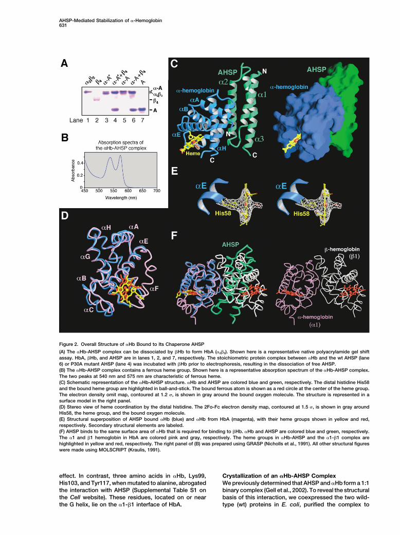
Figure 2. Overall Structure of �Hb Bound to Its Chaperone AHSP
(A) The �Hb-AHSP complex can be dissociated by �Hb to form HbA (�2�2). Shown here is a representative native polyacrylamide gel shiftassay. HbA, �Hb, and AHSP are in lanes 1, 2, and 7, respectively. The stoichiometric protein complex between �Hb and the wt AHSP (lane6) or P30A mutant AHSP (lane 4) was incubated with �Hb prior to electrophoresis, resulting in the dissociation of free AHSP.(B) The �Hb-AHSP complex contains a ferrous heme group. Shown here is a representative absorption spectrum of the �Hb-AHSP complex.The two peaks at 540 nm and 575 nm are characteristic of ferrous heme.(C) Schematic representation of the �Hb-AHSP structure. �Hb and AHSP are colored blue and green, respectively. The distal histidine His58and the bound heme group are highlighted in ball-and-stick. The bound ferrous atom is shown as a red circle at the center of the heme group.The electron density omit map, contoured at 1.2 �, is shown in gray around the bound oxygen molecule. The structure is represented in asurface model in the right panel.(D) Stereo view of heme coordination by the distal histidine. The 2Fo-Fc electron density map, contoured at 1.5 �, is shown in gray aroundHis58, the heme group, and the bound oxygen molecule.(E) Structural superposition of AHSP bound �Hb (blue) and �Hb from HbA (magenta), with their heme groups shown in yellow and red,respectively. Secondary structural elements are labeled.(F) AHSP binds to the same surface area of �Hb that is required for binding to �Hb. �Hb and AHSP are colored blue and green, respectively.The �1 and �1 hemoglobin in HbA are colored pink and gray, respectively. The heme groups in �Hb-AHSP and the �1-�1 complex arehighlighted in yellow and red, respectively. The right panel of (B) was prepared using GRASP (Nicholls et al., 1991). All other structural figureswere made using MOLSCRIPT (Kraulis, 1991).
effect. In contrast, three amino acids in �Hb, Lys99, Crystallization of an �Hb-AHSP ComplexWe previously determined that AHSP and �Hb form a 1:1His103, and Tyr117, when mutated to alanine, abrogated
the interaction with AHSP (Supplemental Table S1 on binary complex (Gell et al., 2002). To reveal the structuralbasis of this interaction, we coexpressed the two wild-the Cell website). These residues, located on or near
the G helix, lie on the �1-�1 interface of HbA. type (wt) proteins in E. coli, purified the complex to

Cell632
Table 1. Summary of Crystallographic Analysis
Data Sets Native Peak Inflection Remote
Wavelength (A) 1.10 1.7372 1.7392 1.7100Resolution (A) 99–2.8 99–2.9 99–2.9 99–2.9Unique reflections 8822 8215 8150 8095Completeness 96.9% (92.0%) 97.8% (86.8%) 97.0% (78.2%) 96.3%
(outer shell) (72.3%)Rsym (outer shell)a 0.055 (0.51) 0.049 (0.36) 0.046 (0.39) 0.046 (0.45)Data redundancy 4.2 5.3 5.5 5.7Average I/� (outer shell) 22.9 (2.4) 43.2 (2.9) 41.6 (2.3) 40.8 (1.9)Anomalous difference (%) 4.1 3.6 3.8Cullis R factor 0.98 0.58 0.73Phasing power 0.53/0.38 2.6/2.1 1.46/1.20
(centric/acentric)Mean figure of merit 0.61
(20–3.2 A)
Refinement statistics
Rmsdc
Number of reflections Total number of Completeness R-factorb
Resolution range (A) (|F| � 0) atoms (water) of data (Rfree) Bond (A) Angle (deg.)
20–2.8 8330 1792 (46) 92.3% 0.263 0.01 1.87(0.292)
Ramachandran plot
Most favored Additionally allowed Generously allowed Disallowed
82.0% 16.9% 1.1% 0%
a Rsym � �h�i | Ih,i � Ih |/�h�i Ih,i, where Ih is the mean intensity of the i observations of symmetry-related reflections of h.b R � � | Fobs � Fcalc |/�Fobs, where Fobs � FP, and Fcalc is the calculated protein structure factor from the atomic model (Rfree was calculated with5% of the reflections).c Rmsd (root-mean-square deviations) in bond lengths and angles are the deviations from ideal values.
homogeneity, and crystallized it in two different space using anomalous signals from the ferrous atom and re-groups. However, these crystals were too small to allow fined to 2.8 A resolution (Table 1 and Figure 2C).structure determination at an atomic resolution. Be- The ferrous oxy-�Hb-AHSP complex is exclusively incause our NMR data, together with a previous report a �-helical conformation (Figure 2C). In the crystals,(Santiveri et al., 2004), indicated that the cis and trans AHSP adopts an elongated three-helix bundle, whileforms of Pro30 in AHSP are in equilibrium but that only �Hb comprises six � helices. AHSP binds �Hb on thea single form binds to �Hb, we introduced a single point opposite site of the molecule to the heme pocket (Figuremutation (P30A) into AHSP, therefore presumably elimi- 2C). AHSP binding appears to trigger a dramatic confor-nating the cis conformation. This protein modification mational change in �Hb, with the entire F helix becomingslightly improved the size of these crystals. Next, we disordered (Figure 2D). Consequently, �Hb in the �Hb-removed the C-terminal 11 residues of AHSP, which are AHSP complex is different from that in HbA with a root-known to exist in a disordered conformation and are mean-square deviation (rmsd) of 1.3 A for 126 aligneddispensable for �Hb binding and stabilization (Gell et C� atoms. This is in contrast to all previously reportedal., 2002; Santiveri et al., 2004; and unpublished data). structures of HbA, which are very similar to each otherThese combined efforts resulted in the generation of as manifested by an rmsd of 0.5 A for all 141 C� atomslarge, well-ordered crystals for the complex between of �Hb in the R and T states. Strikingly, although the�Hb and AHSP (residues 1–91, P30A). heme group is invariably bound by the proximal histidine
Before we proceeded to structure determination, we (His87, or F8 histidine) in previously reported structuresfurther characterized the �Hb-AHSP complex. Incuba- of HbA (www.rcsb.org/), it is coordinated by the distaltion of this �Hb-AHSP complex with �Hb resulted in the histidine (His58, or E7 histidine) in the oxy-�Hb-AHSPdissociation of AHSP and subsequent formation of the complex (Figure 2E). The Fe(II)-heme group is planar,�2�2 complex (HbA) (Figure 2A). Importantly, �Hb in this with oxygen bound as the sixth chelating ligand.complex contains an oxygenated, ferrous [Fe(II)] heme, The interface of the �Hb-AHSP complex primarily in-as evidenced by its characteristic peaks at 540 nm and volves four � helices, two from each protein (Figure 2C575 nm of the absorption spectrum (Figure 2B). This and Figure 3). As anticipated (Supplemental Table S1spectrum is essentially identical to that of the free on the Cell website), the C-terminal half of helix �1, theoxy-�Hb. N-terminal half of helix �2, and the intervening linker
sequences of AHSP closely stack against the G and Hhelices of �Hb. These interactions result in the burial ofOverall Structure of the �Hb-AHSP Complex1780 A2 surface area from the two proteins. Remarkably,The structure of the oxy-�Hb-AHSP complex was deter-
mined by multiwavelength anomalous dispersion (MAD) the G and H helices of �Hb are also the primary structural

AHSP-Mediated Stabilization of �-Hemoglobin633
Figure 3. Sequence Alignment of AHSP or�Hb from Human, Cow, Pig, Rat, and Mouse
Secondary structural elements are indicatedabove the sequences. Conserved residuesare highlighted in yellow. Intermolecular hy-drogen bonds are indicated by red arrows.Amino acids that become buried upon com-plex formation with AHSP (A) or �Hb (B) areidentified by blue columns below the se-quences, with the height of the column corre-sponding to the area of buried surface. Aminoacids that coordinate the heme group are col-ored blue and magenta, respectively, for �Hbin HbA and in �Hb-AHSP complex. The proxi-mal (His87) and distal histidines (His58) arecolored red.
elements that interact with the �-hemoglobin to form carbonyl oxygen of Ala123 and the carboxylate ofthe �1-�1 complex (Figure 2F). Segments of the �1 and Asp126, respectively (Figure 3 and Figure 4B).�2 helices in AHSP superimpose with corresponding The structural observations are in excellent agree-helices in �Hb, with similar interactions. This partially ment with our mutational analysis. All three residuesexplains why binding of �Hb by �Hb dissociates AHSP in �Hb (Lys99, His103, and Phe117), whose mutationfrom the �Hb-AHSP complex. abrogated binding to AHSP (Supplemental Table S1 on
the Cell website), are located at the interface and partici-pate in direct interactions with AHSP. Lys99 and Phe117Interface of the �Hb-AHSP Complexare involved in a number of intermolecular van der WaalsThe interface of the �Hb-AHSP complex is dominatedcontacts, whereas His103 donates a hydrogen bond toby networks of van der Waals interactions, involvingAsp43 of AHSP. Notably, mutation of Asp43 to Arg inapproximately 30 amino acids (Figures 4A and 4B). InAHSP also resulted in the disruption of the �Hb-AHSPaddition, three specific intermolecular hydrogen bondsinteractions (Supplemental Table S1).appear to provide the specificity for this recognition.
Judged by the area of buried surface upon complexAt one end of the interface, Pro119 of �Hb is nestled information, the �Hb-AHSP interface is quite comparablea hydrophobic pocket formed by four aromatic residues,to that of the �1-�1 complex in HbA. In this case, howPhe47, Tyr48, Tyr51, and Tyr52, and the aliphatic sidedoes �Hb gain an edge in its interaction with the �chain of Glu17 in AHSP (Figure 4B). In the center of thesubunit? Compared to the �1-�1 complex, a large num-interface, Leu21 and Trp44 of AHSP pack against eachber of polar and charged amino acids are buried at theother and interact with a relatively flat surface of �Hb,�Hb-AHSP interface, yet the majority of these residueswithin van der Waals contact distance of six amino acidsare not making specific contacts via their polar sidein �Hb. Surrounding residues Leu21 and Trp44 of AHSPchains. For example, Lys99 and His122 of �Hb and Tyr48are three intermolecular hydrogen bonds. The carboxyl-of AHSP, all located at the center of the interface, doate side chain of Asp43 in AHSP accepts a hydrogennot specifically bridge a hydrogen bond (Figure 4B).bond from His103 of �Hb, whereas Gln24 and Gln25 of
AHSP donate two hydrogen bonds to the backbone Perhaps more importantly, the intermolecular van der

Cell634
Figure 4. The Interface between �Hb andAHSP
(A) The 2Fo-Fc electron density map at the�Hb-AHSP interface in stereo. �Hb and AHSPare colored blue and green, respectively. Im-portant interface residues are shown.(B) A closeup view of the �Hb-AHSP interfacein stereo. �Hb and AHSP are colored blueand green, respectively. The side chains ofthe interface amino acids from �Hb and AHSPare colored yellow and orange, respectively.Hydrogen bonds are represented by reddashed lines. Oxygen and nitrogen atoms areshown as red and blue balls, respectively.(C) Comparison of the �Hb-AHSP interfacewith that of the �1-�1 complex. A representa-tive section across the center of the �Hb-AHSP interface is shown in the left panel;the corresponding cross-section of the �1-�1complex is shown in the right panel. �Hband AHSP are represented by blue and greensurface mesh, respectively.
Waals distances in the �Hb-AHSP complex tend to be element. This arrangement, in conjunction with the largeinteraction interface, may also provide flexibility to ac-longer than those at the �1-�1 interface, resulting in
loose packing and poor shape complementarity (Figure commodate suboptimal, but sufficiently strong, interac-tions with the distinct conformations of reduced, oxi-4C). Hence, the overall interactions between �Hb and
AHSP are suboptimal compared to those of the �1-�1 dized �Hb and even apo-�-globin, which lacks heme(Gell et al., 2002). Finally, despite 46% sequence identitycomplex.
These unique structural features serve well the func- between the two globin chains, a number of the impor-tant interface residues in �Hb, such as His103, His 122,tion of AHSP as a specific chaperone for �Hb. On one
hand, the large interface and the three intermolecular and Asp126, are not conserved in �Hb, explaining whyAHSP does not bind to �Hb.hydrogen bonds ensure that AHSP can efficiently and
specifically capture free �Hb. On the other hand, thesenonoptimal �Hb-AHSP interactions can be disrupted by Conservation of Interactions
Amino acid residues that participate in the �Hb-AHSPincoming �Hb for the formation of functional HbA. Inaddition, the AHSP binding element of �Hb is located interactions are highly conserved in both proteins. In
AHSP, 12 out of 19 interface residues, including all threein an area that is opposite to that of the heme binding

AHSP-Mediated Stabilization of �-Hemoglobin635
Figure 5. Structural Rearrangements in the Heme Binding Area of �Hb
(A) A closeup view of the heme binding area in the AHSP bound �Hb. The main chain and side chains of �Hb are colored blue and orange,respectively. The heme group is highlighted in yellow, with the bound ferrous atom shown as a red ball in the center.(B) The heme groups from AHSP bound �Hb and HbA are superimposed to highlight the drastic conformational shift. In HbA, the N� atom ofthe His58 imidazole ring is 4.3 A away from the ferrous atom. In AHSP bound �Hb, the distance is 2.13 A.(C) Comparison of the heme binding amino acids in AHSP bound �Hb with those in �Hb of HbA. The coloring scheme for the AHSP bound�Hb is the same as in (A). The main chain and side chains of �Hb from HbA are colored magenta and green, respectively.(D) AHSP binding greatly accelerates the oxidation of Fe(II)-�Hb. The absorption spectra of �Hb alone, HbA, and �Hb-AHSP complex weretaken at 30 min intervals. The diminishing peaks at 540 nm and 575 nm are characteristic of ongoing oxidation.
that participate in intermolecular hydrogen bonds, are and Leu91 make several van der Waals contacts to sta-bilize the heme group in HbA (Figure 5C), yet they doinvariant among the corresponding orthologs from hu-
man, cow, pig, rat, and mouse (Figure 3 and Figure 4). not directly interact with the heme group in the �Hb-AHSP complex (Figure 5A). In contrast, Phe46, whichThis is in contrast to the invariant 47 residues out of a
total of 102 amino acids in these AHSP proteins. Simi- does not directly coordinate the heme group in HbA, iswithin van der Waals contact distance of the porphyrinlarly, 20 out of 25 interface residues of �Hb, including
all three that hydrogen bond to AHSP, are invariant ring of the heme group in the �Hb-AHSP complex (Figure5A). Nonetheless, the majority of the heme-stabilizingamong the �Hb orthologs (Figure 3), compared to 96
out of 141 total amino acids (Figure 3 and Figure 4). In interactions are preserved between these two com-plexes. Most importantly, Phe43 and Phe98, which inaddition, the variant residues at the �Hb-AHSP interface
tend to be highly conserved (Figure 3 and Figure 4) HbA anchor the heme group from below and above theheme plane, respectively, adapt to the changes andand have no apparent predicted negative impact on the
interactions. This analysis indicates that the amino acid largely maintain these interactions (Figure 5A).As a consequence of the conformational shift, theresidues that participate in the �Hb-AHSP interactions
are selectively preserved during evolution, further indi- sixth chelating position for the heme ferrous atom isnow positioned facing the F helix region of �Hb. As thiscating their functional significance.helix is disordered in the �Hb-AHSP complex, any ligandis now completely solvent exposed (Figure 5A). In con-Conformational Shift at the Heme Binding Site
Upon binding to AHSP, �Hb undergoes dramatic confor- trast, oxygen binds to the heme ferrous atom in a hy-drophobic pocket of HbA, is coordinated by the proximalmational changes, with the entire F helix becoming flexi-
ble and disordered in the crystals (Figure 2D). Remark- histidine, and is well protected from solvent (Fermi andPerutz, 1981). The increased solvent exposure of theably, the distal His58, rather than the proximal His87,
coordinates the heme group, with a distance of 2.13 A heme in the �Hb-AHSP complex is predicted to resultin a faster rate of oxidation. Indeed, compared to thebetween the N� atom of the imidazole ring of His58
and the ferrous atom of the bound heme (Figure 5A). isolated �Hb and HbA, we observed rapid changes in theabsorption spectrum of the oxy-�Hb-AHSP complex,Compared to that in HbA, the iron atom and the heme
group shift over a distance of approximately 3.1 A (Fig- consistent with an increased rate of oxidation (Figure5D). Furthermore, although the structure of free �Hb hasure 5B). Accordingly, compared to that in HbA, the amino
acid residues that coordinate the heme group in the not been determined, this result suggests that the hemeenvironment of the free �Hb resembles the native hy-�Hb-AHSP complex undergo significant rearrange-
ments, which result in the loss as well as creation of a drophobic pocket seen in HbA, supporting the view thatrearrangements of the heme pocket observed in thenumber of interactions (Figure 3). For example, Leu83
Cell636
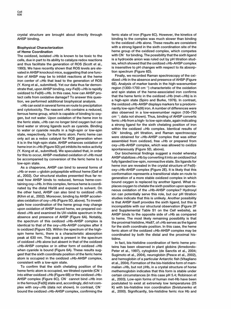
crystal structure are brought about directly through ferric state of iron (Figure 6C). However, the kinetics ofbinding to the complex was much slower than bindingAHSP binding.to the oxidized �Hb alone. These results are consistentwith a strong ligand in the sixth coordination site of theBiophysical Characterizationheme group of the oxidized complex, which competesof Heme Coordinationwith CN� for binding. The possibility that the sixth ligandThe oxidized, isolated �Hb is known to be toxic to theis a hydroxide anion was ruled out by pH titration stud-cells, due in part to its ability to catalyze redox reactionsies, which showed that the oxidized �Hb-AHSP complexand thus facilitate the generation of ROS (Scott et al.,is insensitive to pH changes with respect to its absorp-1993). We have recently shown that ROS levels are ele-tion spectrum (Figure 6D).vated in AHSP knockout mice, suggesting that one func-
Finally, we recorded Raman spectroscopy of the oxi-tion of AHSP may be to inhibit reactions at the hemedized �Hb in the absence and presence of AHSP (Figureiron center of �Hb that lead to the generation of ROS6E). Analysis of marker bands in the high-wavenumber(Y. Kong et al., submitted). Yet our data thus far demon-region (1300–1700 cm�1) characteristic of the oxidationstrate that, upon AHSP binding, oxy-Fe(II)-�Hb is rapidlyand spin states of the heme-associated iron confirmsoxidized to Fe(III)-�Hb. In this case, how can AHSP pro-that the heme ferric in the oxidized �Hb (met-�Hb) is intect cells from oxidative damage? To answer this ques-a high-spin state (Spiro and Burke, 1976). In contrast,tion, we performed additional biophysical analysis.the oxidized �Hb-AHSP displays markers for a predomi-�Hb can exist in several forms en route to precipitationnantly low-spin Fe(III) iron. A number of differences wereand cytotoxicity. The nascent �Hb contains a reduced,also observed in a low-wavenumber region (100–750ferrous heme group, which is capable of binding to oxy-cm�1; data not shown). Thus, binding of AHSP convertsgen, but not water. Upon oxidation of the heme iron toferric �Hb from a high- to low-spin state, again indicatingthe ferric state, �Hb can no longer bind oxygen but cana strong ligand for the sixth chelating position of ironbind water or strong ligands such as cyanide. Bindingwithin the oxidized �Hb complex. Identical results ofto water or cyanide results in a high-spin or low-spinCN� binding, pH titration, and Raman spectroscopystate, respectively, for the ferric atom. Ferric heme canwere obtained for �Hb-AHSP complex that was eitheronly act as a redox catalyst for ROS generation whenassembled from oxidized, free �Hb or prepared fromit is in the high-spin state. AHSP enhances oxidation ofoxy-�Hb-AHSP complex, which was allowed to oxidizeheme iron in �Hb (Figure 5D) yet inhibits its redox activityspontaneously (Figure 5D, above).(Y. Kong et al., submitted). We speculated that, in order
Our biochemical findings suggest a model wherebyfor this to occur, AHSP-mediated oxidation of �Hb mustAHSP stabilizes �Hb by converting it into an oxidized butbe accompanied by conversion of the ferric heme to afully liganded low-spin, nonreactive state. Six ligands forlow-spin state.heme iron are revealed in the crystal structure of Fe(II)-As a chaperone, AHSP can bind to several forms ofoxy-�Hb-AHSP complex (Figure 2E). It is likely that this
�Hb or even �-globin polypeptide without heme (Gell etconformation represents a transitional state en route toal., 2002). Our structural studies presented thus far re-generation of a more stable oxidized complex in which
veal how AHSP binds to reduced, ferrous heme-con-bound oxygen is replaced by another ligand. What re-
taining oxy-�Hb. In this complex, ferrous heme is coordi-places oxygen to chelate the sixth position upon sponta-
nated by the distal His58 and exposed to solvent. Onneous oxidation of the �Hb-AHSP complex? Hydroxyl
the other hand, AHSP can also bind to oxidized �Hb ion can potentially serve this role, but our pH titration(Kihm et al., 2002). Moreover, binding by AHSP acceler- studies indicate that this is unlikely. Another possibilityates oxidation of oxy-�Hb (Figure 5D, above). To investi- is that AHSP itself provides the sixth ligand, but this isgate how coordination of the heme group may change incompatible with our structural observation (Figure 2Fupon oxidation of AHSP bound heme, we prepared oxi- and Supplemental Table S1 on the Cell website), asdized �Hb and examined its UV-visible spectrum in the AHSP binds to the opposite side of �Hb as comparedabsence and presence of AHSP (Figure 6A). Notably, to heme. The most likely remaining possibility is thatthe spectrum of this oxidized �Hb-AHSP complex is the proximal histidine, His87, of �Hb serves as the ligandidentical to that of the oxy-�Hb-AHSP complex after it for the sixth coordinate position. In this case, the hemeis oxidized (Figure 5D). Within the spectrum of the high- ferric atom of the oxidized �Hb-AHSP complex may bespin heme ferric, there is a characteristic absorption coordinated by both the distal and the proximal his-peak at 630 nm. This peak is present in the spectrum tidine.of oxidized �Hb alone but absent in that of the oxidized In fact, bis-histidine coordination of ferric heme pro-�Hb-AHSP complex or in either form of oxidized �Hb teins has been observed in plant globins (Arredondo-when cyanide is bound (Figure 6A). These results sug- Peter et al., 1997), cytoglobin (de Sanctis et al., 2004;gest that the sixth coordinate position of the ferric heme Sugimoto et al., 2004), neuroglobin (Pesce et al., 2002),atom is occupied in the oxidized �Hb-AHSP complex, and hemoglobin of a particular Antarctic fish (Vitaglianoconsistent with a low-spin state. et al., 2004). Formation of the bis-histidine form of mam-
To confirm that the sixth chelating position of the malian �Hb, but not �Hb, in a crystal structure of horseheme ferric atom is occupied, we titrated cyanide (CN�) methemoglobin indicates that this form is stable underinto either oxidized �Hb (Figure 6B) or the oxidized �Hb- certain circumstances (in this case pH 5.4; Robinson etAHSP complex (Figure 6C). CN� cannot bind �Hb iron al., 2003). Low-spin forms of human met-Hb have beenin the ferrous [Fe(II)] state and, accordingly, did not com- postulated to exist at extremely low temperatures (25plex with oxy-�Hb (data not shown). In contrast, CN� K) with bis-histidine iron coordination (Svistunenko et
al., 2000). Significantly, bis-histidine ferric rice Hb andbound the oxidized �Hb-AHSP complex, confirming the

AHSP-Mediated Stabilization of �-Hemoglobin637
Figure 6. The Ferric Heme in the Oxidized �Hb-AHSP Complex Is Coordinated by a Strong Ligand and Exists in a Low-Spin State
(A) Absorption spectra of Fe(III)-�Hb (black), Fe(III)-�Hb-AHSP (red), Fe(III)-�Hb-CN� (blue), and Fe(III)-�Hb-AHSP-CN� (pink).(B) Titration of free Fe(III)-�Hb by cyanide (CN�). The difference spectra recorded at 2 min intervals after addition of CN� are shown. Becausethe position for the sixth chelating position is vacant, cyanide can readily bind there.(C) Titration of the Fe(III)-�Hb-AHSP complex by cyanide (CN�). Difference spectra were recorded as described in (B). The relatively slowkinetics is indicative of a strong ligand already bound at the sixth chelating position.(D) pH-independent absorption spectra of the oxidized �Hb-AHSP complex.(E) Raman spectra of oxy-�Hb, oxy-�Hb AHSP left to auto-oxidize, Fe(III)-met-�Hb, and Fe(III)-met-�Hb-AHSP. These data indicate thatthe heme ferric atom in the Fe(III)-�Hb-AHSP complex exists in a low-spin state. The strong oxidation state marker band (OX) at �1375 cm�1
seen in met-�Hb-AHSP and oxy-�Hb-AHSP samples is consistent with Fe(III) heme. Vibrations characteristic of low-spin (LS) heme ironcomplexes are dominant in the higher-wavenumber region of the complex spectra. In contrast, Fe(III)-met-�Hb sample is predominantly ofhigh-spin (HS) character, as previously described for met-HbA.
Antarctic fish Hb have both been characterized as low- ferric atom is coordinated by six strong ligands andspin complexes and give rise to a visible absorption exists in a low-spin state. It is through this mechanismspectrum that is very similar to the spectrum of the that AHSP protects cells from oxidative damage. Ourferric �Hb-AHSP complex. Hence, it is likely that AHSP data suggest that, in the oxidized complex, both distalfacilitates the formation of a naturally occurring globin and proximal histidines are likely bound to the ferricconformation as a mechanism to stabilize �Hb. heme group, although a definite proof is lacking.
In mediating its chaperone function, AHSP appearsto have two primary and related functions. First, throughPathway of �Hb Precipitationspecific binding in a fashion that partially recapitulatesA mechanistic picture emerges from our studies (Figurethe �1-�1 interface, AHSP stabilizes �Hb and minimizes7). As a chaperone, AHSP can bind to both reducedthe chances for �Hb to form cytotoxic precipitates in(ferrous) and oxidized (ferric) �Hb. In the reduced com-cells (Figure 7). This binding also gives more time forplex, the heme group is coordinated by the distal His58,the reduced �Hb to be complexed by the next availablewith the bound oxygen exposed to solvent. Hence,� subunit, or for the oxidized �Hb to be reduced. InAHSP bound oxy-�Hb is rapidly oxidized. The oxidationthis regard, AHSP is shifting the equilibrium away fromof �Hb by itself may not be harmful to the red bloodprecipitation and cytotoxicity (Figure 7). Second, by fa-cells, which contain extremely high levels of superoxidecilitating the oxidation of the heme group and by seques-dismutase and catalase to convert superoxide radicalstering the oxidized heme in a biochemically inert state,generated by oxidation to H2O and O2. Importantly, the
oxidized, Fe(III)-�Hb is catalytically inert, as the heme AHSP ensures that �Hb does not cause oxidative dam-

Cell638
Figure 7. A Pathway of �Hb Precipitation
AHSP can bind to both reduced and oxidized �Hb. In the reduced complex, the heme group is coordinated by the distal His58, with thebound oxygen exposed to solvent. The AHSP bound �Hb is rapidly oxidized. On one hand, the oxidized, Fe(III)-�Hb is catalytically inert, asthe heme ferric atom is coordinated by six strong ligands and exists in a low-spin state. It is this function of AHSP that protects cells fromoxidative damage. On the other hand, the specific binding of �Hb by AHSP prevents �Hb from precipitation and allows more time for �Hbto be resuscitated.
macia), for coexpression in bacteria. �-globin is GST tagged andage to cells. The order of events from �Hb bound in theAHSP is untagged. Free �Hb and AHSP were individually overex-�Hb-AHSP complex to formation of native HbA (Figurepressed in pGEX-2T as a GST fusion protein. All proteins were7) may depend in part upon the rate of reduction of ferricpurified as described (Chai et al., 2001). Small crystals of the oxy-
�Hb-AHSP complex by methemoglobin reductase, �Hb-AHSP complex were grown at 4C using the hanging dropwhich relies upon a productive interaction between the vapor diffusion method. The well buffer contained 0.1 M HEPES (pH
7.5) and 15% (w/v) PEG5000-monomethyl ether. The size of crystalssoluble Fe(II) cytochrome b5 and the Fe(III) �Hb-AHSPwas improved by protein engineering in AHSP (P30A, deletion ofcomplex. Interactions between Fe(III) met-HbA and Fe(II)C-terminal 11 residues) and by the presence of 1.15 mM C-HEGA,cytochrome b5 are only beginning to be understoodwhich has no impact on the biochemical properties of the �Hb-and appear highly transient, requiring close heme-hemeAHSP complex in solution. These crystals appeared immediately
distances in order for the oxidized met-HbA to be re- and grew to full sizes within a few hours, with a typical dimensionduced (Naito et al., 2001). of 0.3 � 0.3 � 0.3 mm3. The crystals belong to the space group
P41212 and contain one complex per asymmetric unit. The unit cellhas a dimension of a � b � 69.5, c � 104.2, and � � � � � � 90Conclusiondegrees. The crystals contain the ferrous oxy-�Hb, and the oxidationHemoglobin is a classic paradigm for the study of proteinof �Hb in the crystals resulted in the alteration of the crystal packingstructure and function. This is not only due to the promi-lattice and severe decay in X-ray diffraction.
nent functional significance of hemoglobin in biology,but more importantly, because the rich regulatory fea-tures of this protein complex represent an ideal case Data Collection and Structure Determination
Crystals were equilibrated in buffer containing 0.1 M HEPES (pHfor in-depth investigation in a variety of biochemical and7.5), 17% (w/v) PEG5000-monomethyl ether, and 21% (v/v) glycerolbiophysical disciplines (Dickerson and Geis, 1983; Fermiand were flash frozen under cold nitrogen stream. The native andand Perutz, 1981). In this study, we identify the moleculariron MAD data sets were collected at the NSLS X25 and X12C
mechanisms by which AHSP stabilizes �Hb and protects beamlines, respectively, at the Brookhaven National Laboratories.against cytotoxicity caused by labile �Hb. This new facet The data were processed with Denzo and Scalepack (Otwinowskiof hemoglobin regulation contributes significantly to our and Minor, 1997). The position of the iron atom was identified using
SOLVE (Terwilliger and Berendzen, 1996) and refined usingunderstanding of its homeostatic regulation in cells.MLPHARE (CCP4, 1994). The experimental electron density mapwas unambiguous and had all � helices identified. The atomic mod-Experimental Proceduresels of AHSP and �Hb were built using O (Jones et al., 1991) andrefined using CNS (Brunger et al., 1998). The final refined atomicProtein Purification and Crystallizationmodel contains residues 3–91 of AHSP and residues 2–76 and 92–The cDNAs of �-globin and AHSP were cloned in tandem into pSURE
vector, a custom-modified derivative of pGEX-2T (Amersham Phar- 139 of �Hb at 2.8 A resolution.

AHSP-Mediated Stabilization of �-Hemoglobin639
AHSP Mutagenesis and In Vitro Protein-Protein 20 conformers with the lowest energy were used in each case torepresent the solution structure of cis-Pro30-AHSP and trans-Interaction Studies
Point mutants of AHSP were cloned into the bacterial expression Pro30-AHSP.Backbone resonances for ASHP in the �Hb-AHSP complex werevectors pGEX-2T or pGEX-6P1 by standard PCR methods. Mutant
clones were expressed as protein fusions with glutathione S-trans- obtained from HNCA, HN(CO)CA and HNCO triple resonance experi-ments recorded at 288 K and 303 K. 15N-HSQC titration experimentsferase (GST) and were cleaved from GST for use in solution phase
interaction studies. GST pulldown assays were performed with �50 were performed by titration of native �Hb into labeled AHSP up toa 1.2 M excess of �Hb. g of GST-AHSP (mutant or wt) on glutathione agarose beads and
100 g native �Hb in final volume of 500 l buffer (20 mM Na2HPO4/NaH2PO4, 150 mM NaCl, 0.1% IGEPAL CA630 detergent [pH 7.0]) Resonance Raman Spectroscopywith 30 min incubation at 4C followed by seven washes of the Samples for resonance Raman spectroscopy were prepared at ap-beads in the same buffer prior to analysis of interacting protein by proximately 0.4 mM concentration in 20 mM Na2HPO4/NaH2PO4 (pHSDS-PAGE. To investigate AHSP-�Hb interactions in solution phase, 7.0) and 100 mM NaCl. Laser light at 413.1 nm was generated fromwe performed size exclusion chromatography in 20 mM Na2HPO4/ a Spectra Physics 2020 CW Kr ion laser. Powers at the sampleNaH2PO4, 150 mM NaCl (24 ml Superose 12 column on an ACTA- ranged from 10–80 mW, and the laser spot radius at the sampleBASIC FPLC system; Amersham-Pharmacia), with in-line multiangle was 0.5 mm. The scattered light was focused with a Leitz f1.0 lensstatic light scattering (MALS)—a technique to yield shape-indepen- and collected at a 90 scattering geometry from the quartz spinningdent molecular weight information following the Rayleigh-Debey- cell (�4000 rpm). A polarization scrambler was used in all experi-Gans approximation for a dilute polymer solution (Folta-Stogniew ments to correct for the polarization bias of the Jobin-Yvon U1000and Williams, 1999). MALS was performed using a mini-DAWN (Wy- double monochromator (1800 grooves/mm gratings). The slit settingatt Technology Corp., Santa Barbara, CA) at 690 nm laser wave- was 300 m for all experiments. The data were collected at 0.5 cm�1
length. Voltage/light scattering intensity was calibrated against a intervals with a 2 s averaging time. Before each experiment, thestandard sample of HPLC grade toluene, yielding a calibration con- monochromator was calibrated using the mercury emission linesstant of 8.534 � 10�6 V�1. Protein concentrations for calculation of of the fluorescent lights in the laboratory, and the final data weremolecular mass by MALS were obtained from in-line refractive index calibrated to the spectrum obtained from a standard indene samplemeasurements using a value of 0.185 ml g�1 for dn/dc at 690 nm on the same day. All experiments were conducted at room temper-wavelength. ature.
Yeast Two-Hybrid Native Polyacrylamide GelPoint mutants of murine �Hb were generated by PCR, and PCR �Hb-AHSP complex (200 M) was incubated with equal molar ratioproducts were cloned into pGAD10 (BD Biosciences). Full-length of �Hb on ice for 10 min in 25 l assay buffer containing 20 mMmurine AHSP was subcloned into pGBT9 (BD Biosciences). Cotrans- sodium phosphate (pH 7.4) and 100 mM NaCl. Sample taken fromfection of S. cerevisiae strain AH109 (Clonetech) with �Hb-pGAD10 the mixture (15 l) was applied to a 6% native gel under 150 voltsand AHSP-pGBT-9 conferred on the yeast the ability to grow on constant voltage at 4C. The electrophoresis buffer contains 89 mMmedia lacking supplemental histidine in the case of a productive Tris (pH 8.8) and 89 mM boric acid. Other protein samples were�Hb-AHSP protein-protein interaction. Growth on His-negative me- similarly prepared. The proteins were visualized by Coomassiedia (SD broth: 0.67% yeast nitrogen base supplemented with rele- blue staining.vant amino acids) was determined by serial dilution of liquid culturesonto minimal media plates. Wild-type and mutant �Hb in pGAD10
Spectroscopic Analysis of KCN Bindingwere transformed along with empty pGBT9 and plated as nega-
�Hb-AHSP complex was prepared by incubating 40 M of purifiedtive controls.
�Hb with an equimolar ratio of recombinant AHSP on ice for 30 minand then at room temperature overnight in sodium phosphate buffer
NMR Spectroscopy (20 mM; pH 7.4) containing 100 mM NaCl and 10 M DTPA. Met �HbFor NMR experiments, human AHSP (residues 1–90) was homoge- was obtained by incubating oxy-�Hb with potassium ferricyanide innously labeled with 15N and 13C isotopes by bacterial expression a molar ratio of 1:4. The reaction of �Hb-AHSP complex or met �Hbon 15NH4Cl and 13C-glucose as sole nitrogen and carbon sources, (final concentration 40 M) with KCN (final concentration 160 M)respectively (Marley et al., 2001). Native �Hb was obtained from was conducted at room temperature, and then light absorbancehuman blood. Both proteins were purified as described previously was recorded at indicated time.(Gell et al., 2002) and prepared at approximately 1 mM concentration Rate of cyanomethemoglobin formation of �Hb (FeIII) and �Hb-in 20 mM Na2HPO4/NaH2PO4 (pH 6.9) (95% 1H2O, 5% 2H2O), 100 mM AHSP complex was illustrated by absorbance difference after addi-NaCl, and 2 M 5,5-dimethylsilapentanesulfonate (DSS). tion of KCN. The first trace was recorded at time 0, the second trace
AHSP backbone and side chain 1H, 15N, and 13C resonances were was recorded at 1 min, and subsequent traces were recorded everyassigned using a standard set of triple resonance experiments, es- 2 min.sentially as described previously (Deane et al., 2002). Aromatic sidechains were assigned from a 13C-NOESY-HSQC and 2D TOCSY Acknowledgmentsspectra of unlabeled protein in 1H2O/2H2O or �99% 2H2O. NMR spec-tra were recorded at 288 K on a Bruker DXR600 spectrometer, We thank M. Becker and A. Sexana at BNL NSLS beamlines forprocessed using XWINNMR, and analyzed using the program help. This work was supported by grants from the US NationalSparky (Goddard and Kneller, 2004). Two sets of resonances were Institutes of Health (Y.S. and M.J.W.) and the Australian Researchobserved for 42 of the 90 amino acids, and inspection of chemical Council (P.A.L. and J.P.M.). S.Z. is supported by a Fellowship fromshift and NOE data showed that these corresponded to cis and the Cooley’s Anemia Foundation.trans configurations of the Asp29-Pro30 peptide bond (Schubert etal., 2002). Stereo-specific assignment of 13C� and 13C� methyl groups
Received: August 20, 2004of Leu and Val residues were made from a 12% 13C-labeled sampleRevised: September 28, 2004(Neri et al., 1989). φ and � angle restraints were obtained from aAccepted: October 14, 2004combination of TALOS (Cornilescu et al., 1999) and HNHA dataPublished: November 23, 2004(Vuister and Bax, 1993). NOEs were initially assigned manually with
iterative rounds of assignment and structure calculation using theReferencesprogram DYANA (Guntert et al., 1997). A total of 70 φ and 70 �
angle constraints for both the trans- and cis-Pro30 AHSP conform-ers, along with 516 NOE restraints for the trans-Pro30 conformer of Arredondo-Peter, R., Hargrove, M.S., Sarath, G., Moran, J.F., Lohr-
man, J., Olson, J.S., and Klucas, R.V. (1997). Rice hemoglobins.AHSP and 512 NOE restraints for the cis-Pro30 conformer wereused to generate final structures in CNS (Brunger et al., 1998). The Gene cloning, analysis, and O2-binding kinetics of a recombinant
Cell640
protein synthesized in Escherichia coli. Plant Physiol. 115, 1259– methyl groups of valine and leucine in the DNA-binding domain ofthe 434 repressor by biosynthetically directed fractional 13C label-1266.ing. Biochemistry 28, 7510–7516.Baglioni, C. (1966). Chromosomal and cytoplasmic regulation ofNicholls, A., Sharp, K.A., and Honig, B. (1991). Protein folding andhaemoglobin synthesis. Bibl. Haematol. 29, 1056–1063.association: insights from the interfacial and thermodynamic prop-Baglioni, C., and Campana, T. (1967). Alpha-chain and globin: inter-erties of hydrocarbons. Proteins. 11, 281–296.mediates in the synthesis of rabbit hemoglobin. Eur. J. Biochem.Olson, J.S., Eich, R.F., Smith, L.P., Warren, J.J., and Knowles, B.C.2, 480–492.(1997). Protein engineering strategies for designing more stable he-Brunger, A.T., Adams, P.D., Clore, G.M., Delano, W.L., Gros, P.,moglobin-based blood substitutes. Artif. Cells Blood Substit. Immo-Grosse-Kunstleve, R.W., Jiang, J.S., Kuszewski, J., Nilges, M.,bil. Biotechnol. 25, 227–241.Pannu, N.S., et al. (1998). Crystallography and NMR system: A newOtwinowski, Z., and Minor, W. (1997). Processing of X-ray diffractionsoftware suite for macromolecular structure determination. Actadata collected in oscillation mode. Methods Enzymol. 276, 307–326.Crystallogr. D Biol. Crystallogr. 54, 905–921.Pesce, A., Bolognesi, M., Bocedi, A., Ascenzi, P., Dewilde, S.,Bunn, H.F. (1987). Subunit assembly of hemoglobin: an importantMoens, L., Hankeln, T., and Burmester, T. (2002). Neuroglobin anddeterminant of hematologic phenotype. Blood 69, 1–6.cytoglobin. Fresh blood for the vertebrate globin family. EMBO Rep.CCP4 (Collaborative Computational Project, Number 4) (1994). The3, 1146–1151.CCP4 suite: programs for protein crystallography. Acta Crystallogr.Rachmilewitz, E.A., and Schrier, S.L. (2001). Pathophysiology of �D Biol. Crystallogr. 50, 760–763.thalassemia. In Disorders of Hemoglobin, M.H. Steinberg, B.G. For-Chai, J., Wu, Q., Shiozaki, E., Srinivasula, S.M., Alnemri, E.S., andget, D.R. Higgs, and R.L. Nagel, eds. (Cambridge: Cambridge Univer-Shi, Y. (2001). Crystal structure of a procaspase-7 zymogen: mecha-sity Press), pp. 233–251.nisms of activation and substrate binding. Cell 107, 399–407.Robinson, V.L., Smith, B.B., and Arnone, A. (2003). A pH-dependentCornilescu, G., Delaglio, F., and Bax, A. (1999). Protein backboneaquomet-to-hemichrome transition in crystalline horse methemo-angle restraints from searching a database for chemical shift andglobin. Biochemistry 42, 10113–10125.sequence homology. J. Biomol. NMR 13, 289–302.Santiveri, C.M., Manuel Perez-Canadillas, J., Vadivelu, M.K., Allen,
Deane, J.E., Visvader, J.E., Mackay, J.P., and Matthew, J.M. (2002).M.D., Rutherford, T.J., Watkins, N.A., and Bycroft, M. (2004). NMR
1H, 15N, and 13C assignments of FLIN4, an intramolecularstructure of the �-hemoglobin stabilizing protein: Insights into con-
LMO4:ldb1 complex. J. Biomol. NMR 23, 165–166.formational heterogeneity and binding. J. Biol. Chem. 279, 34963–
de Sanctis, D., Dewilde, S., Pesce, A., Moens, L., Ascenzi, P., Han- 34970 Published online June 3, 2004. 10.1074/jbc.M405016200.keln, T., Burmester, T., and Bolognesi, M. (2004). Crystal structure
Schubert, M., Labudde, D., Oschkinat, H., and Schmieder, P. (2002).of cytoglobin: the fourth globin type discovered in man displays
A software tool for the prediction of Xaa-Pro peptide bond confor-heme hexa-coordination. J. Mol. Biol. 336, 917–927.
mations in proteins based on 13C chemical shift statistics. J. Biomol.Dickerson, R.E., and Geis, I. (1983). Hemoglobin: Structure, Func- NMR 24, 149–154.tion, Evolution and Pathology (Menlo Park, CA: Benjamin/Cummings Scott, M.D., van den Berg, J.J., Repka, T., Rouyer-Fessard, P., Heb-Publishing Co.). bel, R.P., Beuzard, Y., and Lubin, B.H. (1993). Effect of excessFermi, G., and Perutz, M.F. (1981). Atlas of Molecular Structure in �-hemoglobin chains on cellular and membrane oxidation in modelBiology: Haemoglobin and Myoglobin (Oxford: Clarendon). �-thalassemic erythrocytes. J. Clin. Invest. 91, 1706–1712.Folta-Stogniew, E., and Williams, K.R. (1999). Determination of mo- Spiro, T.G., and Burke, J.M. (1976). Protein control of porphyrinlecular masses of proteins in solution: implementation of an HPLC conformation. Comparison of resonance Raman spectra of hemesize exclusion chromatography and laser light scattering service in proteins with mesoporphyrin IX analogues. J. Am. Chem. Soc. 98,a core laboratory. J. Biomol. Tech. 10, 51–63. 5482–5489.Gell, D., Kong, Y., Eaton, S.A., Weiss, M.J., and Mackay, J.P. (2002). Sugimoto, H., Makino, M., Sawai, H., Kawada, N., Yoshizato, K., andBiophysical characterization of the �-globin binding protein Shiro, Y. (2004). Structural basis of human cytoglobin for ligand�-hemoglobin stabilizing protein. J. Biol. Chem. 277, 40602–40609. binding. J. Mol. Biol. 339, 873–885.Goddard, T.D., and Kneller, D.G. (2004). SPARKY 3.11 (computer Svistunenko, D.A., Sharpe, M.A., Nicholls, P., Blenkinsop, C., Davies,program). University of California, San Francisco. N.A., Dunne, J., Wilson, M.T., and Cooper, C.E. (2000). The pH de-
pendence of naturally occurring low-spin forms of methaemoglobinGuntert, P., Mumenthaler, C., and Wuthrich, K. (1997). Torsion angleand metmyoglobin: an EPR study. Biochem. J. 351, 595–605.dynamics for NMR structure calculation with the new program DY-
ANA. J. Mol. Biol. 273, 283–298. Terwilliger, T.C., and Berendzen, J. (1996). Correlated phasing ofmultiple isomorphous replacement data. Acta Crystallogr. D Biol.Jones, T.A., Zou, J.-Y., Cowan, S.W., and Kjeldgaard, M. (1991).Crystallogr. 52, 749–757.Improved methods for building protein models in electron density
maps and the location of errors in these models. Acta Crystallogr. Vitagliano, L., Bonomi, G., Riccio, A., di Prisco, G., Smulevich, G.,A 47, 110–119. and Mazzarella, L. (2004). The oxidation process of Antarctic fish
hemoglobins. Eur. J. Biochem. 271, 1651–1659.Kihm, A.J., Kong, Y., Hong, W., Russell, J.E., Rouda, S., Adachi, K.,Vuister, G., and Bax, A. (1993). Quantitative J correlation: a newSimon, M.C., Blobel, G.A., and Weiss, M.J. (2002). An abundantapproach for measuring homonuclear three-bond J(HNH�) couplingerythroid protein that stabilizes free �-haemoglobin. Nature 417,constants in 15N-enriched proteins. J. Am. Chem. Soc. 115, 7772–758–763.7777.Kraulis, P.J. (1991). Molscript: a program to produce both detailed
and schematic plots of protein structures. J. Appl. Crystallogr. 24,Accession Numbers946–950.
Marley, J., Lu, M., and Bracken, C. (2001). A method for efficientThe atomic coordinates of the ferrous oxy-�Hb-AHSP complex andisotopic labeling of recombinant proteins. J. Biomol. NMR 20, 71–75.Pro30-trans AHSP have been deposited in the Protein Data Bank
Naito, N.R., Hui, H.L., Noble, R.W., and Hoffman, B.M. (2001). Deter- with the accession codes of 1Y01 and 1XZY, respectively.mination of the hemoglobin surface domains that react with cyto-chrome b5. Biochemistry 40, 2060–2065.
Nathan, D.G., and Gunn, R.B. (1966). Thalassemia: the conse-quences of unbalanced hemoglobin synthesis. Am. J. Med. 41,815–830.
Neri, D., Szyperski, T., Otting, G., Senn, H., and Wuthrich, K. (1989).Stereospecific nuclear magnetic resonance assignments of the
Related Documents