Molecular Evolution of Mollusc Shell Proteins: Insights from Proteomic Analysis of the Edible Mussel Mytilus Benjamin Marie • Nathalie Le Roy • Isabelle Zanella-Cle ´on • Michel Becchi • Fre ´de ´ric Marin Received: 15 February 2011 / Accepted: 23 May 2011 Ó Springer Science+Business Media, LLC 2011 Abstract Shell matrix proteins (SMPs) that are embed- ded within calcified layers of mollusc shells are believed to play an essential role in controlling the biomineral syn- thesis and in increasing its mechanical properties. Among the wide diversity of mollusc shell textures, nacro-pris- matic shells represent a tremendous opportunity for the investigation of the SMP evolution. Indeed, nacro-pris- matic texture appears early in Cambrian molluscs and is still present in the shell of some bivalves, gastropods, cephalopods and very likely also, of some monoplacoph- orans. One key question is to know whether these shells are constructed from similar matrix protein assemblages, i.e. whether they share a common origin. Most of the molec- ular data published so far are restricted to two genera, the bivalve Pinctada and the gastropod Haliotis. The shell protein content of these two genera are clearly different, suggesting independent origins or considerable genetic drift from a common ancestor. In order to describe puta- tively conserved mollusc shell proteins, here we have investigated the SMP set of a new bivalve model belonging to another genera, the edible mussel Mytilus, using an up-to-date proteomic approach based on the interrogation of more than 70,000 EST sequences, recently available from NCBI public databases. We describe nine novel SMPs, among which three are completely novel, four are homologues of Pinctada SMPs and two are very likely homologues of Haliotis SMPs. This latter result constitutes the first report of conserved SMPs between bivalves and gastropods. More generally, our data suggest that mollusc SMP set may follow a mosaic pattern within the different mollusc models (Mytilus, Pinctada, Haliotis). We discuss the function of such proteins in calcifying matrices, the molecular evolution of SMP genes and the origin of mol- lusc nacro-prismatic SMPs. Keywords Biomineralization Á Proteomics Á Mollusc shell nacre Á Organic matrix Á Evolution Background The explosive radiation of metazoan taxa during the Pro- terozoic–Cambrian transition was shaped on the appear- ance of several innovations, including the ability to form biomineralized skeletons. Among them, the calcium car- bonate shell, that protects the mollusc soft tissues, consti- tutes an excellent model for studying the process of biomineral formation and its evolution. The wide mor- phological diversity of shell-bearing molluscs (bivalves, gastropods, cephalopods, monoplacophorans and scapho- pods, about 100,000? species (Ponder and Lindberg 2008)) also extends to a tremendous diversity of shell micro-textures, including ‘‘prismatic’’, ‘‘nacreous’’, ‘‘foli- ated’’, ‘‘cross-lamellar’’, ‘‘granular’’ ‘‘composite-pris- matic’’ and ‘‘homogeneous’’ structures (Bøggild 1930; Carter 1990; Chateigner et al. 2000). Despite this diversity, Electronic supplementary material The online version of this article (doi:10.1007/s00239-011-9451-6) contains supplementary material, which is available to authorized users. B. Marie (&) Á N. Le Roy Á F. Marin (&) UMR 5561 CNRS Bioge ´osciences, Universite ´ de Bourgogne, 6 bd. Gabriel, Dijon 21000, France e-mail: [email protected]; [email protected] F. Marin e-mail: [email protected] I. Zanella-Cle ´on Á M. Becchi IFR 128 BioSciences Gerland-Lyon Sud, UMR 5086 CNRS, Institut de Biologie et Chimie des Prote ´ines, Universite ´ de Lyon 1, Lyon, France 123 J Mol Evol DOI 10.1007/s00239-011-9451-6

Welcome message from author
This document is posted to help you gain knowledge. Please leave a comment to let me know what you think about it! Share it to your friends and learn new things together.
Transcript

Molecular Evolution of Mollusc Shell Proteins: Insightsfrom Proteomic Analysis of the Edible Mussel Mytilus
Benjamin Marie • Nathalie Le Roy •
Isabelle Zanella-Cleon • Michel Becchi •
Frederic Marin
Received: 15 February 2011 / Accepted: 23 May 2011
� Springer Science+Business Media, LLC 2011
Abstract Shell matrix proteins (SMPs) that are embed-
ded within calcified layers of mollusc shells are believed to
play an essential role in controlling the biomineral syn-
thesis and in increasing its mechanical properties. Among
the wide diversity of mollusc shell textures, nacro-pris-
matic shells represent a tremendous opportunity for the
investigation of the SMP evolution. Indeed, nacro-pris-
matic texture appears early in Cambrian molluscs and is
still present in the shell of some bivalves, gastropods,
cephalopods and very likely also, of some monoplacoph-
orans. One key question is to know whether these shells are
constructed from similar matrix protein assemblages, i.e.
whether they share a common origin. Most of the molec-
ular data published so far are restricted to two genera, the
bivalve Pinctada and the gastropod Haliotis. The shell
protein content of these two genera are clearly different,
suggesting independent origins or considerable genetic
drift from a common ancestor. In order to describe puta-
tively conserved mollusc shell proteins, here we have
investigated the SMP set of a new bivalve model belonging
to another genera, the edible mussel Mytilus, using an
up-to-date proteomic approach based on the interrogation
of more than 70,000 EST sequences, recently available
from NCBI public databases. We describe nine novel
SMPs, among which three are completely novel, four are
homologues of Pinctada SMPs and two are very likely
homologues of Haliotis SMPs. This latter result constitutes
the first report of conserved SMPs between bivalves and
gastropods. More generally, our data suggest that mollusc
SMP set may follow a mosaic pattern within the different
mollusc models (Mytilus, Pinctada, Haliotis). We discuss
the function of such proteins in calcifying matrices, the
molecular evolution of SMP genes and the origin of mol-
lusc nacro-prismatic SMPs.
Keywords Biomineralization � Proteomics � Mollusc
shell nacre � Organic matrix � Evolution
Background
The explosive radiation of metazoan taxa during the Pro-
terozoic–Cambrian transition was shaped on the appear-
ance of several innovations, including the ability to form
biomineralized skeletons. Among them, the calcium car-
bonate shell, that protects the mollusc soft tissues, consti-
tutes an excellent model for studying the process of
biomineral formation and its evolution. The wide mor-
phological diversity of shell-bearing molluscs (bivalves,
gastropods, cephalopods, monoplacophorans and scapho-
pods, about 100,000? species (Ponder and Lindberg
2008)) also extends to a tremendous diversity of shell
micro-textures, including ‘‘prismatic’’, ‘‘nacreous’’, ‘‘foli-
ated’’, ‘‘cross-lamellar’’, ‘‘granular’’ ‘‘composite-pris-
matic’’ and ‘‘homogeneous’’ structures (Bøggild 1930;
Carter 1990; Chateigner et al. 2000). Despite this diversity,
Electronic supplementary material The online version of thisarticle (doi:10.1007/s00239-011-9451-6) contains supplementarymaterial, which is available to authorized users.
B. Marie (&) � N. Le Roy � F. Marin (&)
UMR 5561 CNRS Biogeosciences, Universite de Bourgogne,
6 bd. Gabriel, Dijon 21000, France
e-mail: [email protected]; [email protected]
F. Marin
e-mail: [email protected]
I. Zanella-Cleon � M. Becchi
IFR 128 BioSciences Gerland-Lyon Sud, UMR 5086 CNRS,
Institut de Biologie et Chimie des Proteines, Universite
de Lyon 1, Lyon, France
123
J Mol Evol
DOI 10.1007/s00239-011-9451-6

all molluscan shell layered structures are extracellular and
synthesised according to the same physiological pathway:
they result from the secretory activity of an evolutionarily
homologous organ known as the mantle. In short, the
mantle epithelium extrudes the ionic precursors of the shell
minerals (Bielefeld et al., 1992), together with an extra-
cellular ‘cell-free’ organic matrix that is incorporated into
and surrounds nascent CaCO3 crystals during the shell
growth. Although the organic shell matrix represents only a
small part of the CaCO3 shell weight (between 0.1 and 5%
w/w according to the different species and microstruc-
tures), it is well known to be essential for the control of the
biomineral formation (Mann 1988). It is, in particular,
involved in the arrangement of the organic framework
(Sudo et al. 1997), in the regulation of the CaCO3 pre-
cipitation (Wheeler et al. 1981) and in the control of the
crystal polymorph—aragonite and/or calcite (Falini et al.
1996). The biochemical characteristics of the organic
matrix, usually purified and studied following decalcifica-
tion of the shell, indicate that it comprises a heterogeneous
set of macromolecules including chitin, hydrophobic
‘framework’ proteins, soluble proteins and glycoproteins
(Crenshaw 1972; Weiner and Traub, 1984; Lowenstam and
Weiner 1989; Keith et al. 1993; Levi-Kalisman et al. 2001;
Bedouet et al. 2001; Marie et al. 2007, 2009a).
Because of its exceptional toughness (Jackson et al.
1988, Berthelat 2010), of its commercial value and
remarkable biocompatibility properties when implanted in
vivo (Atlan et al. 1997; Westbroek and Marin 1998), nacre
is among the most studied mineralized biomaterial (Mann
2001). Many authors consider it as the reference model for
understanding at the micro- and nano-scales how molluscs
control the regular deposition of calcium carbonate crystals
(Rousseau et al. 2005; Lin and Meyers 2005; Checa et al.
2009a; Gilbert et al. 2008). Nacre, also called mother-of-
pearl, is the calcified internal layer of several mollusc
shells. The mature nacreous layer consists of the super-
imposition of around 0.5-lm-thick aragonitic tablets,
embedded in a peripheral thin organic matrix (Nakahara
1991; Addadi et al. 2006; Nudelman et al. 2008; Weiss
2010). The prisms that are almost always associated to
nacre, are composed of calcitic or aragonitic needle-like
structures of various lengths and diameters that always
constitute the external calcified layer of shells, and that
grow inward by accretion of crystal units on the inner
surface of the periostracum (Marin et al. 2007; Checa et al.
2005). The individual prisms are stacked together in an
insoluble and hydrophobic organic sheath, which forms a
honeycomb-like structure. They also comprise a slight
intracrystalline organic fraction (Marin et al. 2005).
Nacro-prismatic shell microstructure assemblage appeared
in the Early Cambrian (Carter 1990; Feng and Sun 2003;
Vendrasco et al. 2010). Since then, it remained apparently
almost unchanged. Nowadays, nacro-prismatic microstruc-
tures are represented in at least three mollusc classes, bivalves,
gastropods and cephalopods. In monoplacophorans, true
nacreous layers are potentially observed only in one extant
genus (Checa et al. 2009b). One key question is to know
whether they are constructed from similar matrix protein
assemblages, i.e. whether they share a common origin. If so,
one can wonder whether the shell matrix proteins (SMPs) are
conserved within the different taxa.
Answering these questions is laborious, but will shed a
light on the process of recruitment of SMPs in the Cam-
brian, and on the evolutionary constraints exerted on these
proteins during the Phanerozoic. In fact, since the eluci-
dation of the primary structure of Nacrein (Miyamoto et al.
1996), the first-described nacre protein, the number of SMP
sequences has increased, but remains limited (Marin et al.
2008). Currently, pursuing discovery of mollusc SMPs is
particularly promising because of the genomic and tran-
scriptomic resources that are expending rapidly for many
phylogenetically diverse species that are also experimen-
tally tractable (Weiss and Schonitzer 2006; Jackson et al.
2007a; Suzuki et al. 2009, Auzoux-Bordenave et al. 2010;
Mamangkey and Southgate 2009; Inoue et al. 2010). Given
the high proportion of novel genes being reported from
non-model EST datasets and the flood of sequence data
from next generation technologies, these results emphasise
the importance of proteomic approaches for the validation
and the annotation of coding sequences. This is especially
relevant in the field of molluscan biomineralization where
all characterised biomineral-associated proteins have no
known homologues in any model species (Marin et al.
2008). There is already good precedent for such work in
global investigations of mollusc shell proteins based on
transcriptomics (Jackson et al. 2006, 2007a, 2010; Wang
et al. 2010), proteomics (Marie et al. 2009b, 2010a, b), or
both (Joubert et al. 2010). It is important to notice that
these works concern mostly two mollusc genera—the pearl
oyster Pinctada spp. and the abalone Haliotis spp.—which
comprise more than 90% of the molecular data for mollusc
SMPs (Marin et al. 2008). In a puzzling manner, despite
microstructural resemblance between the nacro-prismatic
shell layers of these two genera, no sequence homology has
been reported so far for mollusc SMPs. Moreover, recent
comparative EST approach performed on Haliotis and
Pinctada calcifying tissues have highlighted clear differ-
ences in the gene sets devoted to the control of shell for-
mation within the two genera (Jackson et al. 2010).
Interestingly, Joubert et al. (2010) have highlighted that
some biomineral-related proteins from gastropods share
sequence similarity with the pearl oyster mantle EST-
deduced proteins, but until now no direct evidence of their
implication in shell formation process has been produced.
On the other hand, although most of the protein domains of
J Mol Evol
123
mollusc SMPs do not exhibit sequence similarity with
other known proteins, few of them present striking domain
homology with known extracellular matrix (ECM) proteins
from vertebrates, suggesting a deep Precambrian origin
(545 ? Ma). For example, N66/Nacrein presents two car-
bonic anhydrase domains (Miyamoto et al. 1996), Perlu-
strin shows similarities with insulin-like growth factor
binding proteins (Weiss et al. 2001), Pif-177 presents a
Von Willerbrand A domain (Suzuki et al. 2009), Perlucin
exhibits a C-type lectin domain (Mann et al. 2000), and
Perlwapin possesses whey acidic protein (WAP) domains
(Treccani et al. 2006).
In the present study, we have investigated the SMPs of
the edible mussel Mytilus, in order to compare them with
those of the above-mentioned taxa. Despite the recent
increasing interest in mytilid shells for ocean acidification
purposes (Miller et al. 2009; Gazeau et al. 2010), very few
works focus on the calcifying shell organic matrix of rep-
resentatives of this group (Weiner et al. 1977; Weiner
1983; Keith et al. 1993). To date, only one extrapallial fluid
protein has been described (Hattan et al. 2001; Yin et al.
2005) and almost no data exist on Mytilus SMPs (Weiner
1983; Keith et al. 1993). By combining a proteomic
approach based on the parallel investigation of the SMPs of
three closely related species of edible mussel (M. edulis,
M. galloprovincialis and M. californianus), and by inter-
rogating the EST dataset recently published for this genera
(Tanguy et al. 2008; Vernier et al. 2009; Craft et al. 2010),
we report here for the first time the primary structure of
nine SMPs, associated with the nacreous and the prismatic
shell microstructures of Mytilus. These include three novel
proteins (one of them probably corresponding to the
N-terminus of P21 which was partially characterised by
Keith et al. (1993)), four homologous proteins of Pinctada
SMPs, and two homologous proteins of Haliotis SMPs.
These results constitute the first report of conserved SMPs
between Bivalvia and Gastropoda. We discuss the function
of such proteins in calcifying matrix, the molecular evo-
lution of SMP genes and the origin of mollusc nacro-
prismatic SMPs.
Materials and Methods
Shell Matrix Extraction
Fresh adult M. edulis, M. galloprovincialis and M. califor-
nianus shells (6–12 cm in length) were collected from the
Brittany coast (France), the Adriatic coast (Croatia) and the
Californian coast (USA), respectively. Superficial organic
contaminants as well as the periostracum were removed by
incubating intact shells in NaOCl (1%, v/v) for 24 h. Shell
calcified layers (nacreous ? prismatic layers) were then
thoroughly rinsed with deionised water, dried and then
roughly crushed into fine powder ([200 lm). All subsequent
extractions were performed at 4�C as previously described
(Marin et al., 2005), with some modifications. Shell powder
samples were decalcified overnight in cold dilute acetic acid
(5%, v/v), which was slowly added by an automated titrator
(Titronic Universal, Schott, Mainz, Germany) at a flow rate
of 100 lL every 5 s. The solutions (final pH around 4.2) were
centrifuged at 3,9009g (30 min). The resulting pellets,
corresponding to the acid-insoluble matrices (AIMs), were
rinsed 6 times with MilliQ water, freeze-dried and weighed.
The supernatants comprising the acido-soluble matrices
(ASMs) were filtered (5 lm) before being concentrated with
an Amicon ultrafiltration system on a Millipore� membrane
(YM10; 10 kDa cut-off). The concentrated solutions (about
5–10 ml) were extensively dialysed against 1 l MilliQ water
(3 days, several water changes) before being freeze-dried
and weighed.
Protein Cleavage
The trypsin digestion of the AIMs from the calcified shell
layers (nacre ? prisms) of M. edulis, M. galloprovincialis
and M. californianus was performed in solution (Marie et al.
2008, 2009a). The samples (0.1 mg) were reduced with
25 lL of 10 mM dithiothreitol in 50 mM NH4HCO3 for
30 min at 50�C. Alkylation was performed with 50 lL of
50 mM iodoacetamide in 50 mM NH4HCO3 for 30 min at
room temperature in the dark. Then the solution was treated
with 1 lg of trypsin (Sequence grade, Promega, USA) in 10
lL 50 mM NH4HCO3 overnight at 37�C. The sample was
dried in a vacuum concentrator and re-suspended in 30 lL of
0.1% trifluoroacetic acid and 4% CH3CN.
Mass Spectrometry Analysis
Mass spectrometry was performed using a Q-Star XL
nanospray quadrupole/time-of-flight tandem mass spec-
trometer, nanospray-qQ-TOF–MS/MS (Applied Biosys-
tems, France), coupled to an online nano liquid
chromatography system (Ultimate Famos Switchos from
Dionex, The Netherlands). One microlitre of samples were
loaded onto a trap column (PepMap100 C18; 5 lm; 100 A;
300 lm 9 5 mm, Dionex), washed for 3 min at 25
lL min-1 with 0.05% trifluoroacetic acid/2% acetonitrile,
then eluted onto a C18 reverse phase column (PepMap100
C18; 3 lm; 100 A; 75 lm 9 150 mm, Dionex). Peptides
were separated at a flow rate of 0.300 lL min-1 with a
linear gradient of 5–80% acetonitrile in 0.1% formic acid
over 120 min. MS data were acquired automatically using
Analyst QS 1.1 software (Applied Biosystems). Following
a MS survey scan over m/z 400–1600, MS/MS spectra were
J Mol Evol
123
sequentially and dynamically acquired for the three most
intense peptide molecular ions over m/z 65–2000. The
collision energy was set by the software according to the
charge and mass of the precursor ion. The MS and MS/MS
data were recalibrated using internal reference ions from a
trypsin autolysis peptide at m/z 842.51 [M ? H]? and
m/z 421.76 [M ? 2H]2?.
MS Data Analysis
Protein identification was performed using the MASCOT
search engine (Matrix Science, London, UK; version 2.1)
against a protein database comprising the around 70,000
nucleotide sequences derived from the EST libraries of
Mytilus spp. (mainly represented by the around 5,000,
19,000 and 42,300 sequences from M. edulis, M. gallo-
provincialis and M. californianus, respectively), down-
loaded (March 2010) from the NCBI server (http://
www.ncbi.nlm.nih.gov). LC–MS/MS data were searched
using carbamidomethylation as fixed modification, and
methionine oxidation as variable modification. The peptide
mass and fragment ion tolerances were set to 0.5 Da. The
peptide hits were manually confirmed by the interpretation
of the raw LC–MS/MS spectra with analyst QS software
(Version 1.1). Quality criteria were the peptide MS value,
the assignment of major peaks to uninterrupted y- and
b-ion series of at least 3–4 consecutive amino acids and the
match with the de novo interpretations proposed by the
software.
Sequence Analysis
Protein sequence identification was attempted using
BLASTp and tBLASTn analysis performed against Swiss-
Prot, GenBank’s nrdb and dbEST using the online tool
provided by UniProt (www.uniprot.org) and NCBI (http://
blast.ncbi.nlm.nih.gov/blast.cgi) servers. Signal peptides
were predicted using SignalP 3.0 (http://www.cbs.dtu.dk/
services/SignalP/), and conserved domains were predicted
using SMART (http://smart.embl-heidelberg.de/) and In-
terProScan (http://www.ebi.ac.uk/Tools/InterProScan/).
The sequence alignments were performed with Clustal-W
or hierarchical-clustering algorithms using UniProt (www.
uniprot.org) or the MULTALIN (http://bioinfo.genotoul.
fr/multalin/multalin.html) online tools, using default para-
meters.
Phylogenetic Analysis
Representative complete sequences of the major non-ver-
tebrate metazoan CAs were selected from the results of
BLAST searched performed with Mcal-CA, using UniProt
and NCBI online tools, against Swiss-Prot, GenBank’s nrdb
and dbEST, or the specific blast tool available from Lottia
gigantea genome web site (http://genome.jgi-psf.org/pages/
blast.jsf?db=Lotgi1). These selected sequences were com-
pared with the molluscan mantle-secreted and non-secreted
CAs and the M. californianus sequence detected in the
current data set. The multiple alignment was created using
T-Coffee (Notredame et al. 2000) set to standard parame-
ters, and then a phylogenic reconstruction was using the
maximum like-hood method PhyML (Guindon and Gascuel
2003) from the www.phylogeny.fr server (Dereeper et al.
2008). Accession numbers of the sequences used are: Elysia
timida HP152215; Plakobranchus ocellatus HP204215;
Strongylocentrotus purpuratus XP001179236; Capitella
teleta EY522037; Lottia gigantea Lotgi1|238082|, Lot-
gi1|239188| from genome assembly (http://genome.
jgi-psf.org/Lotgi1/Lotgi1.download.ftp.html); Crassostrea
gigas CU996533; Pinctada maxima EZ420150, Q9NL38;
Pinctada fucata Q27908; Mytilus californianus P86856;
Haliotis gigantea BAH58349, BAH58350; Turbo marmo-
ratus Q8N0R6; Callinectes sapidus A3FFY1; Panaeus
monodon A9XTM5; Culex quinquefasciatus B0W447;
Drosophila simulans Q3YMV3; Riftia pachyptila Q8MPH8;
Nematostella vectensis A6QR76; Amphimedon queenslan-
dica A6QR75, A6QR76, A6QR77; Ectocarpus siliculosus
D8LB10; Chlamydomonas reinhardtii P20507.
Results
The Mytilus Shell
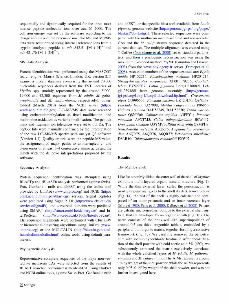
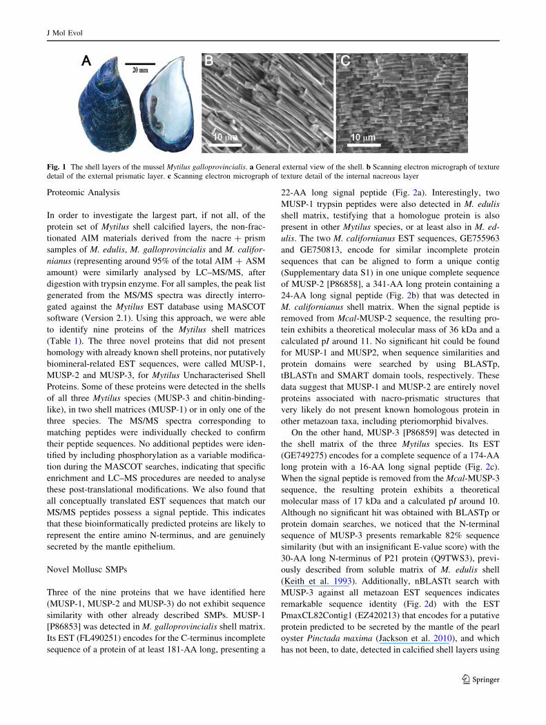
Like for other Mytilidae, the outer wall of the shell of Mytilus
exhibits a multi-layered organo-mineral structure (Fig. 1).
While the thin external layer, called the periostracum, is
mostly organic and gives to the shell its dark brown colour
(Fig. 1a), the rest of the shell is highly calcified and com-
posed of an outer prismatic and an inner nacreous layer
(Mutvei 1980; Feng et al. 2000; Dalbeck et al. 2008). Prisms
are calcitic micro-needles, oblique to the external shell sur-
face, that are enveloped by an organic sheath (Fig. 1b). The
nacre consists of the brick-wall-like superimposition of
around 0.5-lm thick aragonitic tablets, embedded by a
peripheral thin organic matrix, together forming a cohesive
framework (Fig. 1c). We carefully removed the periostra-
cum with sodium hypochlorite treatment. After decalcifica-
tion of the shell powder with cold acetic acid 5% (4�C), we
subsequently extracted the matrix exclusively associated
with the whole calcified layers of M. edulis, M. gallopro-
vincialis and M. californianus. The AIMs represents around
1% by weight of the shell powder, while the ASMs represents
only 0.05–0.1% by weight of the shell powder, and was not
further investigated here.
J Mol Evol
123
Proteomic Analysis
In order to investigate the largest part, if not all, of the
protein set of Mytilus shell calcified layers, the non-frac-
tionated AIM materials derived from the nacre ? prism
samples of M. edulis, M. galloprovincialis and M. califor-
nianus (representing around 95% of the total AIM ? ASM
amount) were similarly analysed by LC–MS/MS, after
digestion with trypsin enzyme. For all samples, the peak list
generated from the MS/MS spectra was directly interro-
gated against the Mytilus EST database using MASCOT
software (Version 2.1). Using this approach, we were able
to identify nine proteins of the Mytilus shell matrices
(Table 1). The three novel proteins that did not present
homology with already known shell proteins, nor putatively
biomineral-related EST sequences, were called MUSP-1,
MUSP-2 and MUSP-3, for Mytilus Uncharacterised Shell
Proteins. Some of these proteins were detected in the shells
of all three Mytilus species (MUSP-3 and chitin-binding-
like), in two shell matrices (MUSP-1) or in only one of the
three species. The MS/MS spectra corresponding to
matching peptides were individually checked to confirm
their peptide sequences. No additional peptides were iden-
tified by including phosphorylation as a variable modifica-
tion during the MASCOT searches, indicating that specific
enrichment and LC–MS procedures are needed to analyse
these post-translational modifications. We also found that
all conceptually translated EST sequences that match our
MS/MS peptides possess a signal peptide. This indicates
that these bioinformatically predicted proteins are likely to
represent the entire amino N-terminus, and are genuinely
secreted by the mantle epithelium.
Novel Mollusc SMPs
Three of the nine proteins that we have identified here
(MUSP-1, MUSP-2 and MUSP-3) do not exhibit sequence
similarity with other already described SMPs. MUSP-1
[P86853] was detected in M. galloprovincialis shell matrix.
Its EST (FL490251) encodes for the C-terminus incomplete
sequence of a protein of at least 181-AA long, presenting a
22-AA long signal peptide (Fig. 2a). Interestingly, two
MUSP-1 trypsin peptides were also detected in M. edulis
shell matrix, testifying that a homologue protein is also
present in other Mytilus species, or at least also in M. ed-
ulis. The two M. californianus EST sequences, GE755963
and GE750813, encode for similar incomplete protein
sequences that can be aligned to form a unique contig
(Supplementary data S1) in one unique complete sequence
of MUSP-2 [P86858], a 341-AA long protein containing a
24-AA long signal peptide (Fig. 2b) that was detected in
M. californianus shell matrix. When the signal peptide is
removed from Mcal-MUSP-2 sequence, the resulting pro-
tein exhibits a theoretical molecular mass of 36 kDa and a
calculated pI around 11. No significant hit could be found
for MUSP-1 and MUSP2, when sequence similarities and
protein domains were searched by using BLASTp,
tBLASTn and SMART domain tools, respectively. These
data suggest that MUSP-1 and MUSP-2 are entirely novel
proteins associated with nacro-prismatic structures that
very likely do not present known homologous protein in
other metazoan taxa, including pteriomorphid bivalves.
On the other hand, MUSP-3 [P86859] was detected in
the shell matrix of the three Mytilus species. Its EST
(GE749275) encodes for a complete sequence of a 174-AA
long protein with a 16-AA long signal peptide (Fig. 2c).
When the signal peptide is removed from the Mcal-MUSP-3
sequence, the resulting protein exhibits a theoretical
molecular mass of 17 kDa and a calculated pI around 10.
Although no significant hit was obtained with BLASTp or
protein domain searches, we noticed that the N-terminal
sequence of MUSP-3 presents remarkable 82% sequence
similarity (but with an insignificant E-value score) with the
30-AA long N-terminus of P21 protein (Q9TWS3), previ-
ously described from soluble matrix of M. edulis shell
(Keith et al. 1993). Additionally, nBLASTt search with
MUSP-3 against all metazoan EST sequences indicates
remarkable sequence identity (Fig. 2d) with the EST
PmaxCL82Contig1 (EZ420213) that encodes for a putative
protein predicted to be secreted by the mantle of the pearl
oyster Pinctada maxima (Jackson et al. 2010), and which
has not been, to date, detected in calcified shell layers using
Fig. 1 The shell layers of the mussel Mytilus galloprovincialis. a General external view of the shell. b Scanning electron micrograph of texture
detail of the external prismatic layer. c Scanning electron micrograph of texture detail of the internal nacreous layer
J Mol Evol
123

Ta
ble
1Id
enti
fica
tio
no
fth
esh
ell
mat
rix
pro
tein
so
fM
ytil
us
by
MS
/MS
anal
ysi
s
Sh
ell
pro
tein
[Sw
issP
rot
nu
mb
er]
ES
TA
cc.
No
.S
pec
ies
Mas
cot
sco
re(n
um
ber
of
pep
tid
es)
Co
mp
lete
seq
uen
ce/
sig
nal
pep
tid
e
Th
eori
tica
l
mas
s/p
IP
rote
in(h
om
olo
gy
/do
mai
n)
Med
uM
ga
lM
cal
Nac
rein
-lik
ea[P
86
85
6]
GE
75
12
62
––
23
3(5
)N
o/?
–/–
No
vel
(Car
bo
nic
anh
yd
rase
/CA
do
mai
ns)
GE
74
90
08
M.
cali
forn
ian
us
MU
SP
-1[P
86
85
3]
FL
49
02
51
97
(2)
23
0(5
)–
No
/Yes
–/–
No
vel
(no
ho
mo
log
/no
reco
gn
ised
do
mai
n)
M.
ga
llo
pro
vin
cia
lis
MS
I60
-lik
e[P
86
85
7]
GE
74
96
43
––
20
6(4
)N
o/?
–/–
No
vel
(MS
I60
/A-
and
G-r
ich
do
mai
ns)
M.
cali
forn
ian
us
MU
SP
-2a
[P8
68
58
]G
E7
55
96
3–
–2
03
(4)
Yes
a/Y
es3
6k
Daa
/pI
=1
1a
No
vel
(no
ho
mo
log
/no
reco
gn
ised
do
mai
n)
GE
75
08
13
M.
cali
forn
ian
us
Per
lwap
in-l
ike
[P8
68
55
]F
L4
94
66
4–
13
7(3
)–
Yes
/Yes
14
kD
a/pI
=1
0N
ov
el(P
erlw
apin
/2W
AP
do
mai
ns)
M.
ga
llo
pro
vin
cia
lis
MU
SP
-3[P
86
85
9]
GE
74
92
75
70
(1)
13
4(2
)1
27
(2)
Yes
/Yes
17
kD
a/pI
=1
0N
ov
el(P
21
and
Pm
axC
L8
2/n
ore
cog
nis
edd
om
ain
)
M.
cali
forn
ian
us
Ch
itin
-bd
-lik
ea[P
86
86
0]
ES
39
33
95
10
9(2
)1
14
(2)
11
4(2
)N
o/?
–/–
No
vel
(Pm
axC
L2
1/2
Ch
itin
-bin
din
gan
dle
ctin
do
mai
ns)
ES
39
35
50
M.
cali
forn
ian
us
Per
luci
n-l
ike
[P8
68
54
]A
J62
44
13
–8
3(2
)–
Yes
/Yes
16
kD
a/pI
=6
No
vel
(Per
luci
n/C
-ty
pe
lect
ine
do
mai
n)
M.
ga
llo
pro
vin
cia
lis
Fib
ron
ecti
n-l
ike
[P8
68
61
]G
E7
59
31
5–
–5
2(1
)N
o/Y
es–
/–N
ov
el(P
max
CL
36
6/F
N3
do
mai
n)
M.
cali
forn
ian
us
Acc
.N
o.
=A
cces
sio
nn
um
ber
,C
A=
Car
bo
nic
anh
yd
rase
,C
hit
in-b
d=
Ch
itin
-bin
din
g,
FN
3=
Fib
ron
ecti
no
fty
pe
3,
MU
SP
=M
ytil
us
Un
char
acte
rise
dS
hel
lP
rote
in,
WA
P=
Wh
eyac
idic
pro
tein
,M
edu
=M
ytil
us
edu
lis,
Mg
al
=M
ytil
us
ga
llo
pro
vin
cia
lis,
Mca
l=
Myt
ilu
sca
lifo
rnia
nu
sa
Ded
uce
dfr
om
clu
ster
edE
ST
seq
uen
ces
J Mol Evol
123

a similar proteomic approach (B. Marie, unpublished data).
These observations suggest that MUSP-3-related proteins
constitute a novel family of pteriomorphid conserved
proteins.
Bivalve-Conserved SMPs
We have identified four Mytilus SMPs that exhibit high
sequence homologies with SMPs extracted from the nacro-
prismatic shells of Pinctada bivalves (carbonic anhydrase,
MSI60, chitin-binding and fibronectin). Peptides matching
Mcal-carbonic anhydrase (Mcal-CA) [P86856] were
detected in the M. californianus shell samples (Fig. 3a).
The putative ORF for this protein was deduced from the
alignment of the ESTs GE751262 and GE749008 in a
unique contig (Supplementary data S2). The conceptually
derived protein sequence of Mcal-CA exhibits a N-termi-
nus incomplete sequence of 321-AA long. The Mcal-CA
sequence exhibits a characteristic CA domain with high
sequence identity for AA position conserved in metazoan
CA and involved in the catalytic activity of the enzymatic
domain, suggesting that Mcal-CA is an active CA
(Fig. 3b). Figure 3c shows the phylogenetic reconstruction
of the relationships between different mollusc and meta-
zoan CAs (Fig. 3c). This analysis clearly indicates that
Mcal-CA belongs to a group of molluscan mantle-secreted
CAs that are likely to be included in shell or involved in
shell deposition that is distinct from other molluscan non-
secreted CAs.
Interestingly, our results confirm the presence of CA in
other pteriomorph bivalve shells, but also indicate that the
Mytilus shell CA does not present the GN-repeat sequen-
ces, characteristic of Pinctada Nacrein proteins (Smith-
Keune and Jerry 2009). However, in spite of the absence of
GN repeats in Mytilus shell CA, the best sequence align-
ment was observed with enzymatic CA domain of Nacrein,
the shell-specific CA from Pinctada fucata (Fig. 4a). The
specific role of CA in a bivalve nacre extracellular calci-
fying matrix is still puzzling and may be related to the fine
regulation of ionic balance at the vicinity of the biomineral
structure formation.
Four different peptides corresponding to the partial
sequence of Mcal-MSI60 [P86857] were detected in the
shell matrix of M. californianus. The conceptually deduced
sequence of the EST GE749643 encodes for the 189-AA
long C-terminus sequence of a poly-Ala protein that pre-
sents high sequence similarities with MSI60 (Fig. 4b),
previously described from the shell of Pinctada fucata
(Sudo et al. 1997). MSI60 is a nacre specific insoluble
framework protein that exhibits 11 poly-Ala blocks and 39
poly-Gly blocks dispersed throughout the sequence. The
poly-Ala blocks confer to MSI60 structural similarity with
silk fibroins.
The ES393395 and ES393550 EST sequences can be
aligned in a unique contig sequence (Supplementary data
S3) and the resulting sequence encodes for the C-terminus
of a 294-AA long incomplete sequence of Mcal-Chitin-
binding [P86860] that can be detected in the calcified shell
layers of M. edulis, M. galloprovincialis and M. califor-
nianus. A SMART search for protein domains indicates
that it contains a Peritrophin-A chitin-binding domain
(Pfam:CBM_14). Interestingly, the result of the tBLASTn
search indicates a high sequence similarity (if not a true
homology) with a putative protein (Fig. 4c) encoded by the
EST PmaxCL21Contig1 EZ420121 (Jackson et al. 2010),
that was in parallel detected by similar proteomic analysis
of the nacreous layer of the pearl oyster Pinctada maxima
(Marie B., unpublished data).
Additionally, one peptide corresponding of the partial
putative sequence of Mcal-Fibronectin [P86861], concep-
tually deduced from the EST GE759315, was detected in
the shell layer of M. californianus. This EST encodes for a
Fig. 2 Sequence analysis of novel shell proteins: MUSP-1, MUSP-2
and MUSP-3. a The AA sequence of Mgal-MUSP-1 [P86853] is
deduced from the translation of the EST entry FL490251. b The AA
sequence of Mcal-MUSP-2 [P86858] is deduced from the alignment
of the translated sequences of the entry GE755963 and GE750813 in a
unique contig (Supplementary data S1). c The AA sequence of Mcal-MUSP-3 [P86859] is deduced from the translation of the EST entry
GE749275. The predicted signal peptides are underlined. The
peptides identified by MS/MS are indicated in red/grey. The asterisksmark the stop codons. Missing sequence information is indicated by
‘‘?’’. d Alignment of AA sequences of MUSP-3 [P86859] with the
deduced sequence from GE749275 of Pinctada maxima. The
predicted signal peptides are underlined. Conserved AA positions
are shaded in blue (Color figure online)
J Mol Evol
123

224-AA long N-terminus sequence presenting a 17-AA
long signal peptide, for which SMART search indicates the
presence of a fibronectin-type 3 domain (FN3). The
tBLASTn search indicates a high sequence similarity with
a putative fibronectin-containing protein (Fig. 4d), encoded
by the EST PmaxCL366Contig1 EZ420486 (Jackson et al.
2010), that was also detected by proteomic analysis of the
prismatic layer of the pearl oyster Pinctada maxima (Marie
B., unpublished data).
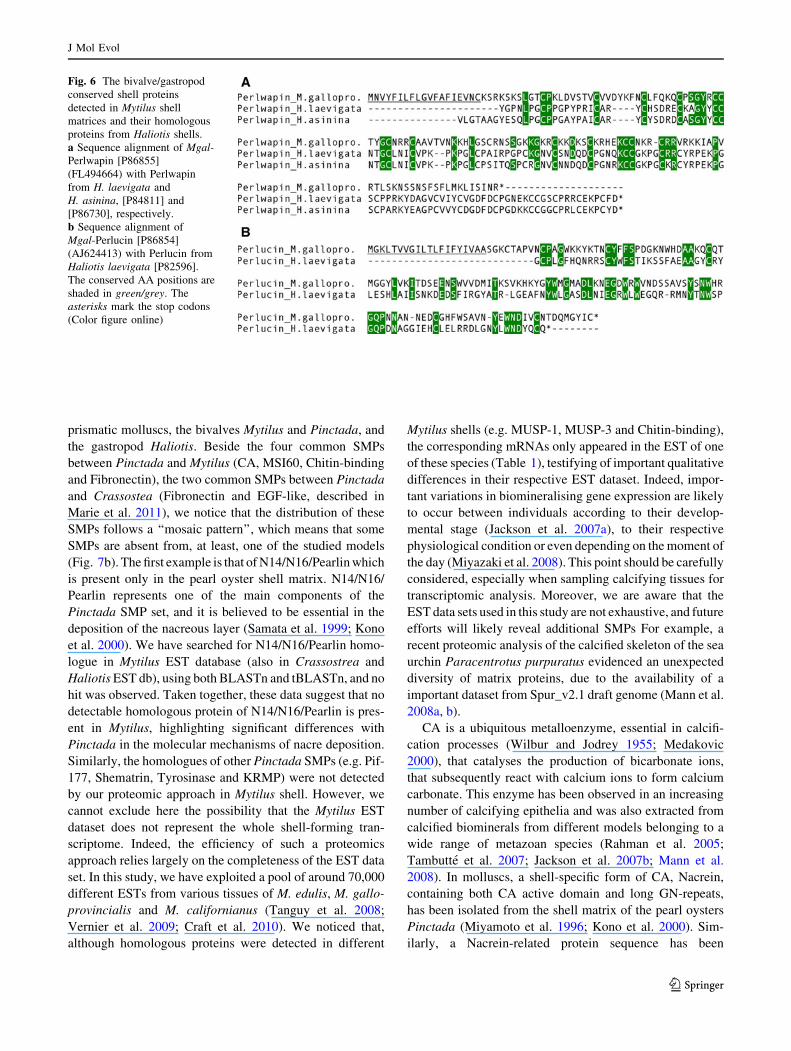
Bivalve/Gastropod-Conserved SMPs
One of the most interesting results of our study was the
detection in Mytilus shell matrix of two proteins that
present high sequence homologies with SMPs extracted
from the nacro-prismatic shells of Haliotis gastropods,
constituting the first report of conserved SMPs between the
two taxa. Three and two different peptides corresponding
to the sequences of Mgal-Perlwapin [P86855] and Mgal-
Perlucin [P86854], respectively, were detected in the shell
matrix of M. galloprovincialis. Figure 5 illustrates the de
novo sequencing of the three peptides observed for Mgal-
Perlwapin. Following signal sequence removal, Mcal-Per-
lwapin and Mcal-Perlucin are characterised by theoretical
pIs of 10 and 6, and theoretical molecular weights of 14
and 16 kDa, respectively. The Mgal-Perlwapin EST
(FL494664) encodes a 141-AA long protein, presenting a
19-AA long signal peptide and two consecutive whey
acidic protein domains (WAP). Mgal-Perlwapin presents
high sequence similarity with Perlwapin proteins. Inter-
estingly, Perlwapin proteins have been previously descri-
bed from the shell of the gastropods H. laevigata (Treccani
et al. 2006) and H. asinina (Marie et al. 2010b). These
shell-extracted Perlwapin exhibit three successive WAP
domains and their sequence alignment with Mgal-Perlwa-
pin (Fig. 6a) indicates a good conservation of the Cys
residues of the WAP domains that are potentially involved
in protease inhibitor function. On the other hand, the Mgal-
Perlucin EST (AJ624413) encodes a 156-AA long protein,
presenting a 20-AA long signal peptide and a characteristic
C-type lectin domain (CTL). Mgal-Perlucin presents high
sequence similarity with the C-type lectin domain con-
taining proteins from various metazoans and especially
with the Perlucin protein (Fig. 6b) that has been previously
described from the shell of the gastropods H. laevigata
(Mann et al. 2000). The alignment of these two shell-
extracted Perlucin sequences (Fig. 6b) presents a good
conservation of the Cys- and Trp-rich regions that are
Fig. 3 Sequence analysis of Mytilus shell carbonic anhydrase (CA).
a The protein sequence of Mcal-CA [P86856] is deduced from the
alignment of the translated sequences of the entry GE751262 and
GE749008 in a unique contig (Supplementary data S3). The peptides
identified by MS/MS are indicated in red/grey. Missing sequence
information is indicated by ‘‘?’’ and asterisk marks the stop codon.
b Sequence alignment of a partial sequence of Mcal-CA with
representative CA from various metazoan taxa. We observed that the
enzymatic domains (shaded in purple/grey) are conserved in all
represented CA forms. c Phylogram of CAs from various metazoan
taxa. Included in the phylogenetic analysis are representative metazoan
CAs, the molluscan mantle-secreted and molluscan shell-extracted CAs
involved in biocalcification, other molluscan CAs, and two outgroup
sequences (brown and green alga). Numerals at each node show local
likelihood ratio values estimated by PhyML. The scale bar indicates an
evolutionary distance of 0.8 AA substitution per position in the
sequences. The Mytilus shell-extracted CA [P86856] is shown by a
black arrow. (SP) and (-) indicate that the protein presents a
characteristic signal peptide sequence or not, respectively. Starsindicate that proteins were extracted from calcified shell layers (Color
figure online)
J Mol Evol
123

involved in the Ca2?-dependent carbohydrate recognition
ability of C-type lectins.
Discussion
Mollusc Nacro-Prismatic Shells
Nacre and prisms seem to be evolutionary-conserved
microstructures that could have been observed in the shell
of numerous molluscs from the early Cambrian to our days.
At first sight, these microstructures are described by simple
terminologies, ‘prism’ on one side and ‘nacre’ on the other
side. However, these terminologies are misleading. For
example, while gastropod and cephalopod nacres are
described as ‘‘columnar’’, the bivalve nacre is presented as
‘‘sheet nacre’’, with characteristic arrangement of nacre
tablets in a ‘brick-wall’ manner (Nakahara 1991). Fur-
thermore, the arrangements of the three axes that charac-
terise their crystal orientations differ between the different
mollusc classes (Chateigner et al. 2000, 2010). Indeed,
both gastropod and bivalve nacres orient the c axis per-
pendicular to the shell surface, but in the other hand, the
b axis is oriented in the direction of shell growth for
bivalves (Chateigner et al., 2000), whereas, in gastropod
nacre, b and c axis are gradually co-oriented from
the prismatic boundary (Gilbert et al. 2008). These
crystallographic differences suggest that the modes of
deposition of aragonite platelets could be different within
the different mollusc clades, at the molecular level.
Different Sets of Nacro-Prismatic SMPs
The organic matrices extracted from nacro-prismatic shell
of several gastropods, cephalopods and bivalves have been
the subject of many investigations since they were believed
to control the deposition of calcified shell layer (Crenshaw
1972; Mann 1988; Lowenstam and Weiner 1989). As the
amino acid analysis of nacro-prismatic shells of different
molluscs exhibited similar compositions, with characteris-
tically high contents of Gly, Ala and Asx residues (Keith
et al. 1993), it was postulated that the molecular mecha-
nism controlling the formation of these different shell
layers is identical from clade to clade. But the discovery of
an increasing number of SMPs has revealed an unexpected
diversity of nacro-prismatic associated proteins among the
different taxa (Marin et al. 2008), rendering this idea
oversimplified. Furthermore, a preliminary comparative
proteomic approach on four nacreous molluscs has sug-
gested that the nacre protein content of these four genera
are different (Marie et al. 2009b). More recently, Jackson
et al. (2010) have demonstrated, by using a specific EST
approach, the drastic differences in the respective shell
building gene sets of the bivalve Pinctada and of the
Fig. 4 The bivalve conserved shell proteins detected in Mytilus shell
matrices and their homologous proteins from Pinctada shells.
a Sequence alignment of Mcal-CA (alignment of GE751262 and
GE749008 in a unique contig, Supplementary data S1) with Nacrein
from Pinctada fucata [Q27908]. b Sequence alignment of Mcal-MSI60
[P86857] (GE749643) with MSI60 from Pinctada fucata [O02402].
c Sequence alignment of Mcal-Chitin-binding [P86860] (alignment of
ES393395 and ES393550 in a unique peptide, Supplementary data S1)
with chitin-binding from Pinctada maxima (PmaxCL21Contig1
EZ420121). C-bind = chitin-binding. d Sequence alignment of
Mcal-Fibronectin [P86861] (GE759315) with Fibronectin from Pinct-ada maxima (PmaxCL366Contig1 EZ420486). FN3 = Fibronectin of
type 3. The conserved AA positions are shaded in blue/grey. The
asterisks mark the stop codons (Color figure online)
J Mol Evol
123

gastropod Haliotis. As underlined by these authors, the data
suggest that ‘‘the Bivalvia and the Gastropoda have either
independently evolved the ability to deposit nacre or that
subsequent to the genesis of the ability in a common
ancestor, bivalves or gastropods have significantly modi-
fied the molecular mechanism that guide this process’’
(Jackson et al. 2010). We generally agree with this
statement—based solely on two genera—but feel that it
should be balanced, with the introduction of the new data
on Mytilus, especially those that show unexpected simi-
larities between mussel and abalone Perlwapins and
Perlucins.
Figure 7a summarises the list of the around sixty SMPs
that are now known for the three main models of nacro-
Fig. 5 Example of de novo
sequencing for the three
peptides matching with Mgal-Perlwapin sequence. a MS/MS
spectrum of the CAAVTVNK
peptide (m/z 431.74). b MS/MS
spectrum of the FNCLFQK
peptide (m/z 478.74). c MS/MS
spectrum of the CAAVTVNKK
peptide (m/z 495.78). The de
novo sequencing was performed
by considering precise mass
differences between adjacent
b and y ion series
J Mol Evol
123
prismatic molluscs, the bivalves Mytilus and Pinctada, and
the gastropod Haliotis. Beside the four common SMPs
between Pinctada and Mytilus (CA, MSI60, Chitin-binding
and Fibronectin), the two common SMPs between Pinctada
and Crassostea (Fibronectin and EGF-like, described in
Marie et al. 2011), we notice that the distribution of these
SMPs follows a ‘‘mosaic pattern’’, which means that some
SMPs are absent from, at least, one of the studied models
(Fig. 7b). The first example is that of N14/N16/Pearlin which
is present only in the pearl oyster shell matrix. N14/N16/
Pearlin represents one of the main components of the
Pinctada SMP set, and it is believed to be essential in the
deposition of the nacreous layer (Samata et al. 1999; Kono
et al. 2000). We have searched for N14/N16/Pearlin homo-
logue in Mytilus EST database (also in Crassostrea and
Haliotis EST db), using both BLASTn and tBLASTn, and no
hit was observed. Taken together, these data suggest that no
detectable homologous protein of N14/N16/Pearlin is pres-
ent in Mytilus, highlighting significant differences with
Pinctada in the molecular mechanisms of nacre deposition.
Similarly, the homologues of other Pinctada SMPs (e.g. Pif-
177, Shematrin, Tyrosinase and KRMP) were not detected
by our proteomic approach in Mytilus shell. However, we
cannot exclude here the possibility that the Mytilus EST
dataset does not represent the whole shell-forming tran-
scriptome. Indeed, the efficiency of such a proteomics
approach relies largely on the completeness of the EST data
set. In this study, we have exploited a pool of around 70,000
different ESTs from various tissues of M. edulis, M. gallo-
provincialis and M. californianus (Tanguy et al. 2008;
Vernier et al. 2009; Craft et al. 2010). We noticed that,
although homologous proteins were detected in different
Mytilus shells (e.g. MUSP-1, MUSP-3 and Chitin-binding),
the corresponding mRNAs only appeared in the EST of one
of these species (Table 1), testifying of important qualitative
differences in their respective EST dataset. Indeed, impor-
tant variations in biomineralising gene expression are likely
to occur between individuals according to their develop-
mental stage (Jackson et al. 2007a), to their respective
physiological condition or even depending on the moment of
the day (Miyazaki et al. 2008). This point should be carefully
considered, especially when sampling calcifying tissues for
transcriptomic analysis. Moreover, we are aware that the
EST data sets used in this study are not exhaustive, and future
efforts will likely reveal additional SMPs For example, a
recent proteomic analysis of the calcified skeleton of the sea
urchin Paracentrotus purpuratus evidenced an unexpected
diversity of matrix proteins, due to the availability of a
important dataset from Spur_v2.1 draft genome (Mann et al.
2008a, b).
CA is a ubiquitous metalloenzyme, essential in calcifi-
cation processes (Wilbur and Jodrey 1955; Medakovic
2000), that catalyses the production of bicarbonate ions,
that subsequently react with calcium ions to form calcium
carbonate. This enzyme has been observed in an increasing
number of calcifying epithelia and was also extracted from
calcified biominerals from different models belonging to a
wide range of metazoan species (Rahman et al. 2005;
Tambutte et al. 2007; Jackson et al. 2007b; Mann et al.
2008). In molluscs, a shell-specific form of CA, Nacrein,
containing both CA active domain and long GN-repeats,
has been isolated from the shell matrix of the pearl oysters
Pinctada (Miyamoto et al. 1996; Kono et al. 2000). Sim-
ilarly, a Nacrein-related protein sequence has been
Fig. 6 The bivalve/gastropod
conserved shell proteins
detected in Mytilus shell
matrices and their homologous
proteins from Haliotis shells.
a Sequence alignment of Mgal-Perlwapin [P86855]
(FL494664) with Perlwapin
from H. laevigata and
H. asinina, [P84811] and
[P86730], respectively.
b Sequence alignment of
Mgal-Perlucin [P86854]
(AJ624413) with Perlucin from
Haliotis laevigata [P82596].
The conserved AA positions are
shaded in green/grey. The
asterisks mark the stop codons
(Color figure online)
J Mol Evol
123

described from the analysis of the mRNA of the mantle of
the gastropod Turbo marmoratus (Miyamoto et al. 2003),
but to date no CA has been directly detected from the shell
nacre of this gastropod nor from a cephalopod nacre (Marie
et al. 2009a). On the other hand, the proteomic investiga-
tion of the limpet Lottia gigantea SMPs reveals the pres-
ence of two different CAs in the calcifying matrix of this
gastropod. As shown in Fig. 3c, the phylogenetic recon-
struction of non-vertebrate metazoan CAs clearly distin-
guishes two groups of mollusc CAs—the mantle-secreted
CAs and the non-secreted CAs—suggesting a common
origin between bivalves and gastropods for the recruitment
of a specific CA for the process of shell deposition. Sur-
prisingly, and in spite of numerous studies, no shell CA has
ever been detected in the Haliotis shell matrices (for review
see Marin et al. 2008; Jackson et al. 2010; Marie et al.
2010b; Le Roy et al., unpublished data). However, we
cannot exclude the possibility that one or more mantle-
secreted CAs could be specifically involved in shell
deposition process, but remain absent of shell-integrated
matrix protein set, as suggested by molecular biology data
(Le Roy et al., unpublished data). This proves without
ambiguity that major calcifying matrix proteins, although
always in contact with the mineralization front, may be
ultimately not integrated within the calcifying shell matrix
during the deposition of the calcified shell layers.
Interestingly, our study emphasises the apparent absence
of EP (extrapallial protein, Q6UQ16) in the Mytilus SMP set,
in spite of the presence of EP mRNA in the Mytilus EST
dataset used for our proteomic investigation. EP is a His-rich
Ca2?-binding glycoprotein that was clearly detected from
extrapallial fluid of Mytilus edulis by both MALDI or ESI
MS/MS techniques, and that was previously supposed to be
part of the SMP (Hattan et al. 2001; Yin et al. 2005). Our data
suggests that this protein is not incorporated within the shell,
while it represents the main extrapallial fluid content and its
apparent strong interactions with calcium ions.
Evolution and Origin of Mollusc SMPs
The fact that Perlwapin and Perlucin homologues have
been observed from the nacro-prismatic shell matrix of
Fig. 7 Evolution of the composition of the calcifying matrix in
molluscs. a Comparison of shell matrix composition between the
nacro-prismatic shell models Pinctada, Haliotis and Mytilus. Blackand grey boxes indicate when the proteins were isolated from the
prismatic or the nacreous layer, respectively. b Presence/absence
mosaic pattern of mollusc SMPs in front of the phylogenetic
relationship of the main models. ‘‘?’’ indicates that although CA
was observed by the mantle epithelial cells, no CA was directly
observed from the shell of Haliotis spp. (Le Roy et al., unpublished).
Fibronect = Fibronectin; Chit-bind = chitin-binding; HUSP = Hal-iotis Uncharacterised Shell Protein
J Mol Evol
123

both Mytilus and Haliotis may suggest that these proteins
were present in the calcifying matrix of the last common
ancestor of conchiferan molluscs. Alternately, we cannot
exclude the possibility that these proteins were recruited
twice independently in both classes. As the sequence
similarities of these proteins are restricted to the active site
residues—a fact that allows assigning them to their
respective family—it is still difficult to determine whether
they are orthologues or paralogues, and more work at the
gene level should be performed to give an unambiguous
answer.
Interestingly, the similarities detected between the
bivalve and gastropod Perlwapins and Perlucins find an
echo with previous findings on putatively conserved shell
proteins domains, such as Kunitz-like domains, detected in
both Pinctada and Haliotis (Liu et al. 2007; Marie et al.
2010b). Here again, such domains may be inherited from
the last common ancestor of bivalves and gastropods, or
may result from independent recruitments. So far, we
cannot yet definitively conclude on the evolutionary sce-
nario for calcifying matrix proteins of the different con-
chiferan molluscs.
SMP Description by Interrogating EST Dataset
with a Proteomic Approach
Our observations make obvious the value of EST libraries
when used in conjunction with a shotgun proteomic
approach for the investigation of calcifying matrix proteins.
By establishing that several of the predicted proteins from
the EST dataset are actually components of the shell, we
are able to make hypotheses about their direct contribution
to shell construction and the implications of their evolution
among calcifying shell matrices. Without this, the EST
dataset is simply a list of sequences that can be associated
to putatively secreted protein sequences, for which func-
tional assumption can be only attempted according to
sequence similarity with already described proteins, and is
not valuable for the description of novel proteins.
The main contribution of this article is that it is taking
the current push of sequencing huge numbers of ESTs to
the next step, which is to try and get some functional
information from these genes for biomineral formation
purposes. The challenge that now faces the field is to
characterise the function of novel biomineral associated
proteins, using in vivo or in vitro techniques.
Conclusion
Aside from the significant differences in the molecular
mechanisms used by the bivalve Pinctada and the gastro-
pod Haliotis for nacro-prismatic shell deposition (review
Marin et al. 2008, 2010b; Jackson et al. 2010), we observed
that the shell protein set of the nacro-prismatic bivalve
Mytilus is partly similar to that of other bivalves, but also
shares few similarities with that of the gastropod Haliotis.
The evolutionary picture that emerges is, for the moment,
patchy. We suggest that the mollusc SMP sets may follow a
mosaic phylogenetic pattern, suggesting that the process of
the integration in the shell of the mantle secreted proteins
may be a complex phenomenon, which does not take place
according to the taxonomic position of the considered
species. We believe, furthermore, that important molecular
functions for shell calcification may not be represented in
the shell, once formed.
The origin and evolution of molluscan SMPs appears to
be a complex phenomenon, which will require large-scale
comparisons across the whole Mollusca phylum, which
means, accurate systematic and wide sampling of mollusc
species, for deciphering the whole protein set (mantle
secreted proteins, extrapallial fluid proteins together with
shell-incorporated matrix proteins) involved in shell
calcification.
Acknowledgments The work of BM, NLR and FM is financially
supported by an ANR (ACCRO-EARTH, ref. BLAN06-2_159971,
coordinator Gilles Ramstein, LSCE) during the period 2007–2011.
The ‘‘Conseil Regional de Bourgogne’’ (Dijon, France) provided
additional supports for the acquisition of new equipment in the Bio-
geosciences research unit (UMR CNRS 5561). A complementary
financial support was provided by INSU (Action INTERRVIE 2010).
BM would like to thanks Davorin Medakovic for providing the fresh
shells of Mytilus galloprovincialis and Jerome Thomas for handling
shell pictures. The present protein sequences appear in the UniProtKB
under the accession numbers P86853–P86861.
References
Adamkewicz SL, Harasewych MG, Blake J, Saudek D, Bult CJ
(1997) A molecular phylogeny of the bivalve molluscs. Mol Biol
Evol 14:619–629
Addadi L, Joester D, Nudelman F, Weiner S (2006) Mollusk shell
formation: a source of new concepts for understanding biomin-
eralization processes. Chem Rev 12:980–987
Atlan G, Balmain N, Berland S, Vidal B, Lopez E (1997)
Reconstruction of human maxillary defects with nacre powder:
histological evidence for bone regeneration. C R Acad Sci Paris
320:253–258
Auzoux-Bordenave S, Badou A, Gaume B, Berland S, Helleouet MN,
Milet C, Huchette S (2010) Ultrastructure, chemistry and
mineralogy of the growing shell of the European abalone
Haliotis tuberculata. J Struct Biol 171:277–290
Bedouet L, Schuller J, Marin F, Milet C, Lopez E, Giraud M (2001)
Soluble proteins of the nacre of the giant oyster Pinctadamaxima and of the abalone Haliotis tuberculata: extraction and
partial analysis of nacre proteins. Comp Biochem Physiol B
128:389–400
Berthelat F (2010) Nacre from mollusk shell: a model for high-
performance structural materials. Bioinspir Biomin 20:035001
J Mol Evol
123
Bielefeld U, Zierold K, Korje KH, Becker W (1992) Calcium
localization in the shell-forming tissue of the freshwater snail,
Biomphalaria glabrata: a comparative study of various methods
for localizing calcium. Histochem J 24:927–938
Bøggild O (1930) The shell structure of the mollusks. K Dan Vidensk
Selsk Skr Naturvidensk Math Afd 9:233–326
Carter JG (1990) Skeletal biomineralization: patterns, processes and
evolutionary trends. Van Nostrand Reinhold, New York
Chateigner D, Hedegaard C, Wenk HR (2000) Mollusc shell
microstructures and crystallographic textures. J Struct Geol
22:1723–1735
Chateigner D, Ouhenia S, Krauss C, Hedegaard C, Gil O, Morales M,
Lutterotti L, Rousseau M, Lopez E (2010) Voyaging around
nacre with the X-ray shuttle: from bio-mineralisation to
prosthetics via mollusc phylogeny. Mater Sci Eng A 528:37–51
Checa AG, Rodriguez-Navarro AB, Esteban-Delgado JF (2005) The
nature and formation of calcitic columnar prismatic shell layers
in pteriomorphian bivalves. Biomaterials 26:6404–6414
Checa AG, Cartwright JH, Willinger MG (2009a) The key role of the
surface membrane in why gastropod nacre grows in towers. Proc
Nat Acad Sci USA 106:38–43
Checa A, Ramirez-Rico J, Gonzalez-Segura A, Sanchez-Navas A
(2009b) Nacre and false-nacre (foliated aragonite) in extant
monoplacophorans (= Tryblidiida: Mollusca). Naturwissens-
chaften 96:111–122
Craft JA, Gilbert JA, Temperton B, Dempsey KE, Ashelford K,
Tiwari B, Hutchinson TH, Chipman JK (2010) Pyrosequencing
of Mytilus galloprovincialis cDNAs: tissue-specific expression
patterns. PLoS One 5:e8875
Crenshaw MA (1972) The soluble matrix from Mercenaria merce-naria shell. Biomineralization 6:6–11
Dalbeck P, England J, Cusack M, Lee MR, Fallick AE (2008)
Crystallographic and chemistry of the calcium carbonate poly-
morph switch in M. edulis shells. Eur J Mineral 18:601–609
Dereeper A, Guignon V, Blanc G, Audic S, Buffet S, Chevenet F,
Dufayard J-F, Guindon S, Lefort V, Lescot M, Claverie J-M,
Gascuel O (2008) Phylogeny.fr: robust phylogenetic analysis for
the non-specialist. Nucleic Acid Res 36:465–469
Falini G, Albeck S, Weiner S, Addadi L (1996) Control of aragonite
and calcite polymorphism by mollusk shell macromolecules.
Science 271:67–69
Feng W, Sun W (2003) Phosphate replicated and replaced micro-
structure of molluscan shells from the earliest Cambrian in
China. Acta Palaeontol Pol 48:21–30
Feng QL, Li HB, Pu G, Zhang M, Cui FZ, Li D (2000) Crystallo-
graphic alignment of calcite prisms in the oblique prismatic layer
of the Mytilus edulis shell. J Mater Sci 35:3337–3340
Gazeau F, Gattuso J-P, Pronker E, Peene J, Heip CH, Middelburg JJ
(2010) Effect of ocean acidification on the early stages of blue
mussel Mytilus edulis. Biogeosciences 7:2051–2060
Gilbert PU, Metzler RA, Zhou D, Scholl A, Doran A, Young A, Kunz
M, Tamura N, Coppersmith SN (2008) Gradual ordering in red
abalone nacre. J Am Chem Soc 130:17519–17527
Guindon S, Gascuel O (2003) A simple, fast, and accurate algorithm
to estimate large phylogenies by maximum likelihood. Syst Biol
52:696–704
Hattan SJ, Laue TM, Chasteen ND (2001) Purification and charac-
terization of a novel calcium-binding protein from the extrapal-
lial fluid of the mollusc, Mytilus edulis. J Biol Chem
276:4461–4468
Inoue N, Ishibashi R, Ishikawa T, Atsumi T, Aoki H, Komura A
(2010) Gene expression patterns and pearl formation in the
Japanese pearl oyster (Pinctada fucata): a comparison of gene
expression patterns between the pearl sac and the mantles
tissues. Aquaculture 308:S68–S74
Jackson AP, Vincent JV, Turner RM (1988) The mechanical design of
nacre. Proc R Soc Lond 234:415–440
Jackson DJ, McDougall C, Green KM, Simpson F, Worheide G,
Degnan BM (2006) A rapidly evolving secretome builds and
patterns a sea shell. BMC Biol 4:40
Jackson DJ, Worheide G, Degnan BM (2007a) Dynamic expression
of ancient and novel molluscan shell genes during ecological
transitions. BMC Evol Biol 7:160
Jackson DJ, Macis L, Reitner J, Degnan BM, Worheide G (2007b)
Sponge paleogenomics reveals an ancient role for carbonic
anhydrase in skeletogenesis. Science 316:1893–1895
Jackson DJ, McDougall C, Woodcroft B, Moase P, Rose RA, Kube
M, Reinhardt R, Rokshar DS, Montagnani C, Joubert C,
Piquemal D, Degnan BM (2010) Parallel evolution of nacre
building gene sets in molluscs. Mol Biol Evol 27:591–608
Joubert C, Piquemal D, Marie B, Manchon L, Pierrat F, Zanella-
Cleon I, Cochennec-Laureau N, Gueguen Y, Montagnani C
(2010) Transcriptome and proteome analysis of Pinctadamargaritifera calcifying mantle and shell: focus on biomineral-
ization. BMC Genomics 11:613
Keith J, Stockwell S, Ball D, Remillard K, Kaplan D, Thanhauser T,
Sherwood R (1993) Comparative analysis of macromolecules in
mollusc shells. Comp Biochem Physiol B 105:487–496
Kono M, Hayashi N, Samata T (2000) Molecular mechanism of the
nacreous layer formation in Pinctada maxima. Biochem Biophys
Res Comm 269:213–218
Levi-Kalisman Y, Falini G, Addadi L, Weiner S (2001) Structure of
the nacreous organic matrix of a bivalve mollusk shell examined
in the hydrated state using cryo-TEM. J Struct Biol 135:8–17
Lin A, Meyers MA (2005) Growth and structure in abalone shell.
Mater Sci Eng 390:27–41
Liu HL, Liu SF, Ge YJ, Liu J, Wang XY, Xie LP, Zhang RQ, Wang Z
(2007) Identification and characterization of a biomineralization
related gene PFMG1 highly expressed in the mantle of Pinctadafucata. Biochemistry 46:844–851
Lowenstam HA, Weiner S (1989) On biomineralization. Oxford
University Press, New York
Mamangkey NG, Southgate PC (2009) Regeneration of excised
mantle tissue by the silver-lip pearl oyster, Pinctada maxima(Jameson). Fish Shellfish Immun 27:164–174
Mann S (1988) Molecular recognition in biomineralization. Nature
332:119–124
Mann S (2001) Biomineralization: principles and concepts in
bioinorganic materials chemistry. Oxford University Press,
New York
Mann K, Weiss IM, Andre S, Gabius HJ, Fritz M (2000) The amino
acid sequence of the abalone (Haliotis laevigata) nacre protein
perlucin. Eur J Biochem 267:5257–5264
Mann K, Poustka AJ, Mann M (2008a) The sea urchin (Strongylo-centrotus purpuratus) test and spine proteomes. Proteome Sci
6:22
Mann K, Poustka AJ, Mann M (2008b) In-depth, high-accuracy
proteomics of sea urchin tooth organic matrix. Proteome Sci 6:33
Marie B, Luquet G, Pais de Barros J-P, Guichard N, Medakovic D,
Marin F (2007) The shell matrix of the freshwater mussel Uniopictorum (paleoheterodonta, unionoida): involvement of acidic
polysaccharides from glycoproteins in nacre mineralization.
FEBS J 274:2933–2945
Marie B, Luquet G, Bedouet L, Milet C, Guichard N, Medakovic D,
Marin F (2008) Nacre calcification in the freshwater mussel Uniopictorum: carbonic anhydrase activity and purification of a
95 kDa calcium-binding glycoprotein. ChemBioChem 9:2515–
2523
Marie B, Marin F, Marie A, Bedouet L, Dubost L, Alcaraz G, Milet C,
Luquet G (2009a) Evolution of nacre: biochemistry and
J Mol Evol
123

proteomics of the shell organic matrix of the cephalopod
Nautilus macromphalus. ChemBioChem 10:1495–1506
Marie B, Le Roy N, Marie A, Dubost L, Milet C, Bedouet L, Becchi
M, Zanella-Cleon I, Jackson DJ, Degnan BM, Luquet G, Marin F
(2009b) Nacre evolution: a proteomic approach. Mater Res Soc
Symp Proc 1187:KK01–KK03
Marie B, Zanella-Cleon I, Le Roy N, Becchi M, Luquet G, Marin F
(2010a) Proteomic analysis of the acid-soluble nacre matrix of
the bivalve Unio pictorum: detection of novel carbonic anhy-
drase and putative protease inhibitor proteins. ChemBioChem
11:2138–2147
Marie B, Marie A, Jackson DJ, Dubost L, Degnan BM, Milet C,
Marin F (2010b) Proteomic analysis of the organic matrix of the
abalone Haliotis asinina calcified shell. Proteome Sci 8:54
Marie B, Zanella-Cleon I, Guichard N, Becchi M, Marin F (2011)
Novel proteins from the calcifying shel matrix of the pacific
oyster Crassostrea gigas. Mar Biotechnol. doi:10.1007/s10126-
011-9379-2
Marin F, Amons R, Guichard N, Stiger M, Hecker A, Luquet G,
Layrolle P, Alcaraz G, Riondet C, Woestbroek P (2005)
Caspartin and Calprismin, two proteins of the shell calcitic
prisms of the mediterranean fan mussel Pinna nobilis. J Biol
Chem 280:33895–33908
Marin F, Pokroy B, Luquet G, Layrolle P, De Groot K (2007) Protein
mapping of calcium carbonate biominerals by immunogold.
Biomaterials 28:2368–2377
Marin F, Luquet G, Marie B, Medakovic D (2008) Molluscan shell
proteins: primary structure, origin, and evolution. Curr Top Dev
Biol 80:209–276
Medakovic D (2000) Carbonic anhydrase activity and biomineraliza-
tion process in embryos, larvae and adult blue mussels Mytilusedulis. Hegol Mar Res 54:1–6
Miller AW, Reynolds AC, Sobrino C, Riedel GF (2009) Shellfish face
uncertain future in high CO2 world: influence of acidification on
oyster larvae calcification and growth in estuaries. PLoS One
4:e5661
Miyamoto H, Miyashita T, Okushima M, Nakano S, Morita T,
Matsushiro A (1996) A carbonic anhydrase from the nacreous
layer in oyster pearls. Proc Natl Acad Sci USA 93:9657–9660
Miyamoto H, Yano M, Miyashita T (2003) Similarities in the
structure of nacrein, the shell-matrix protein, in a bivalve and a
gastropod. J Moll Stud 69:87–89
Miyazaki Y, Usui T, Kajikawa A, Hishiyama H, Matsuzawa N,
Nishida T, Machii A, Samata T (2008) Daily oscillation of gene
expression associated with nacreous layer formation. Front
Mater Sci China 2:162–166
Mutvei H (1980) The nacreous layer in molluscan shells. In: Omori
M, Watabe N (eds) The mechanisms of mineralization in
biological systems. Tokay University press, Tokyo, pp 49–56
Nakahara H (1991) Nacre formation in bivalve and gastropod
molluscs. In: Suga S, Nakahara H (eds) Mechanisms and
phylogeny of mineralization in biological systems. Springer-
Verlag, New York, pp 343–350
Notredame C, Higgins DG, Heringa J (2000) T-Coffee: a novel
method for fast and accurate multiple sequence alignment. J Mol
Biol 302:205–217
Nudelman F, Shimoni E, Klein E, Rousseau M, Bourrat X, Lopez E,
Addadi L, Weiner S (2008) Forming nacreous layer of the shells
of the bivalves Atrina rigida and Pinctada margaritifera: an
environmental- and cryo-scanning electron microscopy study.
J Struct Biol 162:290–300
Ponder W, Lindberg DR (2008) Phylogeny and evolution of the
mollusca. University of California Press, Berkeley
Rahman AM, Isa Y, Uehara T (2005) Proteins of calcified endoskel-
eton: II. Partial amino acid sequences of endoskeletal proteins
and the characterization of proteinaceous organic matrix of
spicules from the alcyonarian, Sinularia polydactyla. Proteomics
5:885–893
Rousseau M, Lopez E, Stempfle P, Brendle M, Franke L, Guette A,
Naslain R, Bourrat X (2005) Multiscale structure of sheet nacre.
Biomaterials 26:6254–6262
Samata T, Hayashi N, Kono M, Hasegawa K, Horita C, Akera S
(1999) A new matrix protein family related to the nacreous layer
formation of Pinctada fucata. FEBS Lett 462:225–229
Smith-Keune C, Jerry DR (2009) High level of intra-specific variation
in the NG repeat region of the Pinctada maxima N66 organic
matrix protein. Aquat Res 40:1054–1063
Sudo S, Fujikawa T, Nagakura T, Ohkubo T, Sakaguchi K, Tanaka M,
Nakashima K, Takahashi T (1997) Structure of mollusc shell
framework proteins. Nature 387:563–564
Suzuki M, Saruwatari K, Kogure T, Yamamoto Y, Nishimura T, Kato
T, Nagasawa H (2009) An acidic matrix protein, Pif, is a key
macromolecules for nacre formation. Science 325:1388–1390
Tambutte S, Tambutte E, Zoccola D, Caminiti N, Lotto S, Moya A,
Allemand D, Adkins J (2007) Characterization and role of
carbonic anhydrase in the calcification process of the azooxan-
thellate coral Tubastrea aurea. Mar Biol 151:71–83
Tanguy A, Bierne N, Saavedra C, Pina B, Bachere E, Kube M, Bazin
E, Bonhomme F, Boudry P, Boulo V, Boutet I, Cancela L,
Dossat C, Favrel P, Huvet A, Jarque S, Jollivet D, Klages S,
Lapegue S, Leite R, Moal J, Moraga D, Reinhardt R, Samain J-F,
Zouros E, Canario A (2008) Increasing genomic information in
bivalves through new EST collections in four species: develop-
ment of new genetic markers for environmental studies and
genome evolution. Gene 408:27–36
Treccani L, Mann K, Heinemann F, Fritz M (2006) Perlwapin, an
abalone nacre protein with three four-disulfide core (whey acidic
protein) domains, inhibits the growth of calcium carbonate
crystals. Biophys J 91:2601–2608
Vendrasco MJ, Porter SM, Kouchinsky A, Li G, Fernandez CZ (2010)
New data on molluscs and their shell microstructures from the
Middle Cambrian Gowers Formation, Australia. Palaeontology
53:97–135
Vernier P, De Pitta C, Bernante F, Varotto L, De Nardi B, Bovo G,
Roch P, Novoa B, Figueras A, Pallavicini A, Lanfranchi G
(2009) Mytibase: a knowledgebase of mussel (M. galloprovin-cialis) transcribed sequences. BMC Genomics 10:72
Wang A, Wang Y, Gu Z, Li S, Shi Y, Guo X (2010) Development of
expressed sequence tags from the pearl oyster, Pinctada martensiDunker. Mar Biotechnol. doi:10.1007/s10126-010-9296-9
Weiner S (1983) Mollusk shell formation: isolation of two organic
matrix proteins associated with calcite deposition in the bivalve
Mytilus californianus. Biochemistry 22:4139–4145
Weiner S, Traub W (1984) Macromolecules in mollusc shells and
their functions in biomineralization. Philos Trans R Soc Lond
304:425–434
Weiner S, Lowenstam HA, Hood L (1977) Discrete molecular weight
components of the organic matrices of mollusc shells. J Exp Mar
Biol Ecol 30:45–51
Weiss I (2010) Jewels in Pearl. ChemBioChem 11:297–300
Weiss I, Schonitzer V (2006) The distribution of chitin in larval shells
of the bivalve mollusk Mytilus galloprovincialis. J Struct Biol
153:264–277
Weiss IM, Gohring W, Fritz M, Mann K (2001) Perlustrin, a Haliotislaevigata (abalone) nacre protein, is homologous to the insulin-
like growth factor binding protein N-terminal module of
vertebrates. Biochem Biophys Res Commun 285:244–249
Westbroek P, Marin F (1998) A marriage of bone and nacre. Nature
392:861–862
Wheeler AP, George JW, Evans CA (1981) Control of CaCO3
nucleation and crystal growth by soluble matrix of oyster shell.
Science 212:1397–1398
J Mol Evol
123
Wilbur KM, Jodrey LH (1955) Studies on shell formation. V. The
inhibition of shell formation by carbonic anhydrase inhibitors.
Biol Bull 108:359–365
Yin Y, Hang J, Paine ML, Reinhold VN, Chasteen D (2005)
Structural characterisation of the major extrapallial fluid protein
of the mollusc Mytilus edulis: implication for function. Bio-
chemistry 44:10720–10731
J Mol Evol
123
Related Documents