NEOPLASIA Molecular events contributing to cell death in malignant human hematopoietic cells elicited by an IgG3-avidin fusion protein targeting the transferrin receptor Patrick P. Ng, Gustavo Helguera, Tracy R. Daniels, Simon Z. Lomas, Jose A. Rodriguez, Gary Schiller, Benjamin Bonavida, Sherie L. Morrison, and Manuel L. Penichet We have previously reported that an anti– human transferrin receptor IgG3-avidin fusion protein (anti–hTfR IgG3-Av) inhib- its the proliferation of an erythroleukemia- cell line. We have now found that anti–hTfR IgG3-Av also inhibits the proliferation of additional human malignant B and plasma cells. Anti–hTfR IgG3-Av induces internal- ization and rapid degradation of the TfR. These events can be reproduced in cells treated with anti–hTfR IgG3 cross-linked with a secondary Ab, suggesting that they result from increased TfR cross- linking. Confocal microscopy of cells treated with anti–hTfR IgG3-Av shows that the TfR is directed to an intracellular compartment expressing the lysosomal marker LAMP-1. The degradation of TfR is partially blocked by cysteine protease inhibitors. Furthermore, cells treated with anti–hTfR IgG3-Av exhibit mitochondrial depolarization and activation of caspases 9, 8, and 3. The mitochondrial damage and cell death can be prevented by iron supplementation, but cannot be fully blocked by a pan-caspase inhibitor. These results suggest that anti–hTfR IgG3-Av induces lethal iron deprivation, but the resulting cell death does not solely de- pend on caspase activation. This report provides insights into the mechanism of cell death induced by anti-TfR Abs such as anti–hTfR IgG3-Av, a molecule that may be useful in the treatment of B-cell malignancies such as multiple myeloma. (Blood. 2006;108:2747-2756) © 2006 by The American Society of Hematology Introduction The primary function of transferrin (Tf) is to transport iron through the blood. After binding to the transferrin receptor (TfR) on the cell surface, Tf is internalized into an acidic compartment where the bound iron is released. The Tf-TfR complex then returns to the cell surface and the ligand dissociates from the receptor. 1 Studies have shown that the TfR is expressed more abundantly in malignant tissues than their normal counterparts. 2-7 This differ- ence in expression level, in addition to its ability to internalize and its central roles in cell growth and division, makes the TfR an attractive target for cancer therapeutics. In fact, both anti-TfR antibodies (Abs) and Tf-toxin conjugates have shown efficacy against cancers in preclinical and clinical settings. 8-12 We have previously demonstrated that anti–rat TfR-IgG3-Av forms strong noncovalent interactions with different biotinylated molecules and delivers them into cancer cells through receptor-mediated endocy- tosis. 13 This novel molecule can be used as a universal delivery system for a wide range of therapeutic agents without the need to make a different chemical conjugate or genetic fusion protein for every targeted therapeutic. We also unexpectedly discovered that anti–rat TfR-IgG3-Av, but not an irrelevant IgG3-Av, inhibited the growth of a rat myeloma and a T-cell lymphoma-cell line. However, it did not inhibit the growth of either a carcinoma or a gliosarcoma-cell line. 13 Treatment with anti–rat TfR-IgG3 containing the same variable regions did not inhibit growth. Furthermore, we found that anti–rat TfR-IgG3-Av exists as a noncovalent dimer with 4 antigen-binding sites, probably due to the interaction among the 4 avidins located on 2 separate fusion proteins, since avidin in solution forms a tetrameric structure. 13 Thus, the inhibitory effect of the fusion protein may be due, at least in part, to its ability to cross-link cell-surface TfRs. Furthermore, we reported that a similar fusion protein specific for the human TfR (anti–hTfR IgG3-Av), but not a murine anti–TfR IgG1 (128.1) sharing the same variable regions, inhibited the growth of the erythroleukemia- cell line K562. 13 However, the mechanism of growth inhibition by these 2 fusion proteins, as well as the therapeutic potential of anti–hTfR IgG3-Av, was not explored. Now we report that anti–hTfR IgG3-Av inhibits the growth of malignant B- and plasma-cell lines and cells isolated from patients with multiple myeloma (MM), a malignancy that is generally regarded as incurable. Using 2 of the most sensitive cell lines, ARH-77 and IM-9, we show that anti–hTfR IgG3-Av induces rapid TfR degradation, iron deprivation, mitochondrial damage, and cell death. Among different cancers, hematopoietic tumors are particu- larly suitable for treatment using TfR-targeting therapeutics since they both express high levels of TfR 14-17 and are known to be more sensitive to the inhibitory effect of anti-TfR Abs than other malignancies. 18 Increased understanding of the mechanism of cell From the Department of Microbiology, Immunology, and Molecular Genetics, the Division of Surgical Oncology, the Department of Surgery, the Division of Hematology and Oncology, the Department of Medicine, and the Jonsson Comprehensive Cancer Center, David Geffen School of Medicine, University of California, Los Angeles. Submitted July 7, 2005; accepted June 2, 2006. Prepublished online as Blood First Edition Paper, June 27, 2006; DOI 10.1182/blood-2006-04-020263. Supported in part by grants CA86915 and CA107023 from the National Institutes of Health (NIH), the 2003 Jonsson Cancer Center Foundation Interdisciplinary Grant, and the 2004 Brian D. Novis International Myeloma Foundation Senior Grant Award. The online version of this article contains a data supplement. Reprints: Manuel L. Penichet, Division of Surgical Oncology, Department of Surgery, UCLA, 10833 Le Conte Ave, 54-140 CHS Mail code 178218, Los Angeles, CA 90095-1782; e-mail: [email protected]. The publication costs of this article were defrayed in part by page charge payment. Therefore, and solely to indicate this fact, this article is hereby marked ‘‘advertisement’’ in accordance with 18 U.S.C. section 1734. © 2006 by The American Society of Hematology 2747 BLOOD, 15 OCTOBER 2006 VOLUME 108, NUMBER 8 tapraid4/zh8-bloo/zh8-bloo/zh802006/zh80247d06g sangreyj S15 9/6/06 15:31 Art: 2006/020263 Input-DCT-??? Not for distribution: this preliminary material is embargoed until publication.

Welcome message from author
This document is posted to help you gain knowledge. Please leave a comment to let me know what you think about it! Share it to your friends and learn new things together.
Transcript
NEOPLASIA
Molecular events contributing to cell death in malignant human hematopoieticcells elicited by an IgG3-avidin fusion protein targeting the transferrin receptorPatrick P. Ng, Gustavo Helguera, Tracy R. Daniels, Simon Z. Lomas, Jose A. Rodriguez, Gary Schiller, Benjamin Bonavida,Sherie L. Morrison, and Manuel L. Penichet
We have previously reported that an anti–human transferrin receptor IgG3-avidinfusion protein (anti–hTfR IgG3-Av) inhib-its the proliferation of an erythroleukemia-cell line. We have now found that anti–hTfRIgG3-Av also inhibits the proliferation ofadditional human malignant B and plasmacells. Anti–hTfR IgG3-Av induces internal-ization and rapid degradation of the TfR.These events can be reproduced in cellstreated with anti–hTfR IgG3 cross-linkedwith a secondary Ab, suggesting thatthey result from increased TfR cross-
linking. Confocal microscopy of cellstreated with anti–hTfR IgG3-Av showsthat the TfR is directed to an intracellularcompartment expressing the lysosomalmarker LAMP-1. The degradation of TfR ispartially blocked by cysteine proteaseinhibitors. Furthermore, cells treated withanti–hTfR IgG3-Av exhibit mitochondrialdepolarization and activation of caspases9, 8, and 3. The mitochondrial damageand cell death can be prevented by ironsupplementation, but cannot be fullyblocked by a pan-caspase inhibitor. These
results suggest that anti–hTfR IgG3-Avinduces lethal iron deprivation, but theresulting cell death does not solely de-pend on caspase activation. This reportprovides insights into the mechanism ofcell death induced by anti-TfR Abs suchas anti–hTfR IgG3-Av, a molecule thatmay be useful in the treatment of B-cellmalignancies such as multiple myeloma.(Blood. 2006;108:2747-2756)
© 2006 by The American Society of Hematology
Introduction
The primary function of transferrin (Tf) is to transport iron throughthe blood. After binding to the transferrin receptor (TfR) on the cellsurface, Tf is internalized into an acidic compartment where thebound iron is released. The Tf-TfR complex then returns to the cellsurface and the ligand dissociates from the receptor.1
Studies have shown that the TfR is expressed more abundantlyin malignant tissues than their normal counterparts.2-7 This differ-ence in expression level, in addition to its ability to internalize andits central roles in cell growth and division, makes the TfR anattractive target for cancer therapeutics. In fact, both anti-TfRantibodies (Abs) and Tf-toxin conjugates have shown efficacyagainst cancers in preclinical and clinical settings.8-12 We havepreviously demonstrated that anti–rat TfR-IgG3-Av forms strongnoncovalent interactions with different biotinylated molecules anddelivers them into cancer cells through receptor-mediated endocy-tosis.13 This novel molecule can be used as a universal deliverysystem for a wide range of therapeutic agents without the need tomake a different chemical conjugate or genetic fusion protein forevery targeted therapeutic.
We also unexpectedly discovered that anti–rat TfR-IgG3-Av,but not an irrelevant IgG3-Av, inhibited the growth of a ratmyeloma and a T-cell lymphoma-cell line. However, it did notinhibit the growth of either a carcinoma or a gliosarcoma-cellline.13 Treatment with anti–rat TfR-IgG3 containing the same
variable regions did not inhibit growth. Furthermore, we found thatanti–rat TfR-IgG3-Av exists as a noncovalent dimer with 4antigen-binding sites, probably due to the interaction among the 4avidins located on 2 separate fusion proteins, since avidin insolution forms a tetrameric structure.13 Thus, the inhibitory effectof the fusion protein may be due, at least in part, to its ability tocross-link cell-surface TfRs. Furthermore, we reported that asimilar fusion protein specific for the human TfR (anti–hTfRIgG3-Av), but not a murine anti–TfR IgG1 (128.1) sharing thesame variable regions, inhibited the growth of the erythroleukemia-cell line K562.13 However, the mechanism of growth inhibition bythese 2 fusion proteins, as well as the therapeutic potential ofanti–hTfR IgG3-Av, was not explored.
Now we report that anti–hTfR IgG3-Av inhibits the growth ofmalignant B- and plasma-cell lines and cells isolated from patientswith multiple myeloma (MM), a malignancy that is generallyregarded as incurable. Using 2 of the most sensitive cell lines,ARH-77 and IM-9, we show that anti–hTfR IgG3-Av induces rapidTfR degradation, iron deprivation, mitochondrial damage, and celldeath. Among different cancers, hematopoietic tumors are particu-larly suitable for treatment using TfR-targeting therapeutics sincethey both express high levels of TfR14-17 and are known to be moresensitive to the inhibitory effect of anti-TfR Abs than othermalignancies.18 Increased understanding of the mechanism of cell
From the Department of Microbiology, Immunology, and Molecular Genetics,the Division of Surgical Oncology, the Department of Surgery, the Division ofHematology and Oncology, the Department of Medicine, and the JonssonComprehensive Cancer Center, David Geffen School of Medicine, University ofCalifornia, Los Angeles.
Submitted July 7, 2005; accepted June 2, 2006. Prepublished online as BloodFirst Edition Paper, June 27, 2006; DOI 10.1182/blood-2006-04-020263.
Supported in part by grants CA86915 and CA107023 from the National Institutes ofHealth (NIH), the 2003 Jonsson Cancer Center Foundation Interdisciplinary Grant,
and the 2004 Brian D. Novis International Myeloma Foundation Senior GrantAward.
The online version of this article contains a data supplement.
Reprints: Manuel L. Penichet, Division of Surgical Oncology, Department ofSurgery, UCLA, 10833 Le Conte Ave, 54-140 CHS Mail code 178218, LosAngeles, CA 90095-1782; e-mail: [email protected].
The publication costs of this article were defrayed in part by page chargepayment. Therefore, and solely to indicate this fact, this article is herebymarked ‘‘advertisement’’ in accordance with 18 U.S.C. section 1734.
© 2006 by The American Society of Hematology
2747BLOOD, 15 OCTOBER 2006 ! VOLUME 108, NUMBER 8
tapraid4/zh8-bloo/zh8-bloo/zh802006/zh80247d06g sangreyj S!15 9/6/06 15:31 Art: 2006/020263 Input-DCT-???
Not for distribution: this preliminary material is embargoed until publication.
death induced by anti–hTfR IgG3-Av may make it possible todesign improved therapeutics for the treatment of hematopoieticmalignancies.
Materials and methods
Antibodies and antibody fusion proteins
Recombinant anti–hTfR IgG3, constructed by substituting the variableregions of anti–dansyl IgG319,20 with those of the murine IgG1 anti–humanTfR monoclonal Ab (mAb) 128.1,21 was expressed in the murine myeloma-cell line NS0/1. Anti–hTfR IgG3-Av and anti–dansyl IgG3-Av have beenpreviously described.13,22 Abs and Ab fusion proteins were purified andcharacterized as described previously.13 The murine anti–human IgG3 mAbHP6050 was a kind gift from Robert Hamilton (Johns Hopkins University,Baltimore, MD). Murine antihuman TfR mAb (H68.4) and goat anti–mouseIgG–horseradish peroxidase (HRP) conjugate were from Zymed (South SanFrancisco, CA). Rabbit polyclonal anti–human TfR1 was purchased fromSanta Cruz Biotechnology (Santa Cruz, CA). Murine anti–"-actin mAb wasfrom Sigma (St Louis, MO); the biotinylated mouse anti–LAMP-1 mAband goat anti–mouse IgG–FITC were from BD Pharmingen (BectonDickinson, Franklin Lakes, NJ). The mouse IgG1/# isotype control mAbswere from eBioscience (San Diego, CA).
Cell lines and primary cells
MM.1S, S6B45, OCI-My5, U266, 8226/S, and 8226/DOX40 (doxorubicin-resistant variant of 8226/S) are human MM-cell lines. ARH-77 and IM-9are Epstein-Barr virus (EBV)–transformed lymphoblastoid-cell lines estab-lished from cells isolated from IgG plasma-cell leukemia and MM patients,respectively. When injected into SCID mice, ARH-77 behaves like humanMM, with mice developing hypercalcemia, lytic bone lesions, and hindlimb paralysis.23,24 U266, 8226, ARH-77, and IM-9 cell lines werepurchased from ATCC (Rockville, MD). The MM.1S, S6B45, and OCI-My5 cell lines were kindly provided by Drs Kenneth Anderson andDarminder Chauhan (Harvard University), and the 8226/DOX40-cell linewas provided by Alan Lichtenstein (UCLA). The human erythroleukemia-cell line K562 was kindly provided by J. Larrick (Palo Alto Institute ofMolecular Medicine, Mountain View, CA). Cells were cultured at 37°C, 5%CO2 in RPMI 1640 medium (GIBCO BRL, Grand Island, NY), with 5%fetal bovine serum (FBS; HyClone, Logan, UT) in all the experimentsunless stated otherwise. Serum complement was inactivated by incubationat 56°C for 30 minutes.
Bone marrow aspirates from a patient with MM and another withplasma-cell leukemia (PCL), a more aggressive variant of MM,25 wereobtained with informed consent following the standards of the institutionalreview board of UCLA. Mononuclear cells were separated by Ficoll-PaquePlus density gradient centrifugation (Amersham Pharmacia Biosciences,Uppsala, Sweden). CD138$ cells were separated from bone marrowmononuclear cells by positive selection using EasySep Human CD138$
Selection magnetic nanoparticles following the manufacturer’s instructions(StemCell Technologies, Vancouver, BC, Canada).
Proliferation assays using cell lines
Cells (5000/well of a 96-well plate) were treated with the indicated reagentsfor 72 hours at 37°C. The cells were then cultured with 4 %Ci (0.148MBq)/mL [3H]-thymidine (ICN Biomedicals, Irvine, CA) for an additional24 hours. Cells were harvested and radioactivity was counted as previouslydescribed.13 In the assay with 9 different cell lines, cells were cultured inDulbecco modified Eagle medium (GIBCO BRL) supplemented with 2.5%FBS. Similar results were obtained using cells cultured in RPMI mediumwith 5% FBS. In the cross-linking assay, anti–hTfR IgG3 was mixed with a5-fold excess of anti-hIgG3 mAb for 2 hours at 4°C before being added tothe cells. In the metal supplement assay, cells were incubated with orwithout ferric ammonium citrate or zinc sulfate (Sigma) during theentire study.
Proliferation assays using primary cells
IM-9, U266, or primary cells (20 000/well of a 96-well plate) were culturedin Iscoves modified Dulbecco medium (GIBCO BRL) supplemented withnonessential amino acids (Invitrogen, Carlsbad, CA) and 10% FBS andwere treated with buffer or 100 nM anti–hTfR IgG3-Av for 48 hours at37°C. The cells were then pulsed with 4 %Ci (0.148 MBq)/mL [3H]-thymidine for an additional 48 hours. Cells were harvested and radioactivitywas counted as previously described.13
Detection of cell-surface TfR
Cells (5 & 105) treated with buffer, Ab, or Ab fusion protein were harvested,washed, and incubated with 5 %g Tf-FITC (Molecular Probes, Eugene, OR)in medium containing 1% BSA for 1 hour on ice. Cells were then washedand analyzed with a BD-LSR Analytic Flow Cytometer (Becton Dickinson).
Immunoblotting
Cells (5 & 105) were incubated with the indicated reagents, washed, andresuspended in lysis buffer (0.125% NP-40, 0.875% Brij 97, 10 mM Tris,150 mM NaCl, 2 mM EDTA, 1 mM PMSF, 2.5 %M aprotinin and leupeptin,pH 7.5). Lysate (20 %g) was reduced and resolved by sodium dodecylsulfate–polyacrylamide gel electrophoresis (SDS-PAGE). Proteins werethen transferred onto PVDF membranes (Millipore, Bedford, MA) using aGeneMate semidry blotter (ISC BioExpress, Kaysville, UT). The mem-branes were blocked with 5% milk and incubated overnight with anti–human TfR (H68.4) in 5% milk, 0.2% Tween 20 in PBS, followed bywashing and incubation with anti–mouse IgG-HRP. The membranes werethen incubated with SuperSignal West Pico chemiluminescent substrate(Pierce, Rockford, IL) before being exposed to film.
Radiolabeling and immunoprecipitation of TfR fragments
Cells (2 & 106) labeled with [35S]-methionine for 18 hours were washedand cultured with anti–hTfR IgG3 or anti–hTfR IgG3-Av for an additional 6hours. The culture supernatants were then immunoprecipitated usinganti–human TfR polyclonal Ab and protein A–conjugated Sepharose beads(Sigma). Samples were reduced and resolved by SDS-PAGE. The gel wasdried and exposed to film.
Apoptosis and mitochondrial membrane potential assays
Cells (75 000) were treated with the indicated reagents, washed, and stainedwith propidium iodide (PI) and annexin V Alexa Fluor 350 or 488conjugates following procedures suggested by the manufacturer of theVybrant Apoptosis kit (Molecular Probes). Up to 10 000 events wererecorded for each flow cytometry measurement. To measure mitochondrialdepolarization, 40 nM DiOC6(3) (Molecular Probes) was added to theculture for 30 minutes. Samples were analyzed by flow cytometry. DFO andZ-VAD-FMK were purchased from Calbiochem (La Jolla, CA).
Caspase activity assay
Cells (25 000 per well of a 96-well plate) were treated with buffer oranti–hTfR IgG3-Av, and caspase activity was measured at 12, 24, 36, 44,48, and 52 hours using fluorogenic substrates specific for caspases 9(Ac-LEHD-AMC), 8 (Ac-IETD-AMC), and 3 (Ac-DMQD-AMC) (AlexisBiochemicals, San Diego, CA). Ac-DMQD-AMC, instead of Ac-DEVD-AMC,was used because it has been shown to be more specific for caspase 3.26,27
At each time point, reaction buffer (1.5% NP-40, 0.3% CHAPS, 30%sucrose, 30 mM MgCl2, 150 mM KCl, 450 mM NaCl, 150 mM HEPES,1.2 mM EDTA, 30 mM DTT, 3 mM PMSF, pH 7.4) containing 150 %Mfluorogenic substrate was added to each well and incubated for 4 hours at37°C. Fluorescence was measured by a Synergy HT Microplate Reader(Bio-Tek Instruments, Winooski, VT) at 360/460 nm. Substrate incubationfor 4 hours yields greater differences between the control and experimentalsample values when compared with other incubation times.
2748 NG et al BLOOD, 15 OCTOBER 2006 ! VOLUME 108, NUMBER 8
tapraid4/zh8-bloo/zh8-bloo/zh802006/zh80247d06g sangreyj S!15 9/6/06 15:31 Art: 2006/020263 Input-DCT-???
Not for distribution: this preliminary material is embargoed until publication.
Confocal microscopy
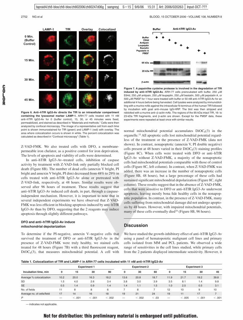
Cells (2 & 106) treated with anti–hTfR IgG3-Av were fixed with 4%paraformaldehyde and permeabilized with 0.2% Tween 20 in PBS. Afterblocking for 2 hours on ice in confocal buffer (2% calf serum, 0.1% sodiumazide in PBS) containing 10% human serum (Omega Scientific, Tarzana,CA), cells were incubated on ice for 2 hours with 1 %g anti-TfR (H68.4) orisotype control mAb, and for 1 hour with anti–mouse IgG-FITC (1:100).Cells were then fixed with 4% paraformaldehyde and incubated on ice for2 hours with 0.1 %g biotinylated anti–LAMP-1 or isotype control, and for1 hour with streptavidin–Alexa Fluor 568 (1:500; Molecular Probes). Cellswere washed twice with confocal buffer after each incubation. Stained cellswere resuspended in Prolong solution (Molecular Probes) and mountedonto slides. Images were taken using an MRC1024ES confocal system(Bio-Rad Laboratories, Hercules, CA) equipped with a Nikon E800microscope (Nikon, Melville, NY), a Nikon 63&/1.4 numeric aperture oilobjective lens, and LaserSharp 2000 acquisition software version 6(Bio-Rad). Colocalization studies were performed using ImageJ softwareversion 1.32 (National Institutes of Health, Bethesda, MD). The meanfluorescence intensity (MFI) of the red and green channels in each imagefield was determined and a threshold for positive staining of MFI $ 2 SDset. A pixel was considered colocalized when the overlapping area of the2 channels was at least 50% of the area of the pixel. The percentcolocalization for each field was the number of colocalized pixels, dividedby the total number of positive pixels. The significance of values comparedto 0 minutes was determined using the Student t test.
Results
Anti–hTfR IgG3-Av inhibits the growth of a panel of malignantB- and plasma-cell lines, and primary cells from patients
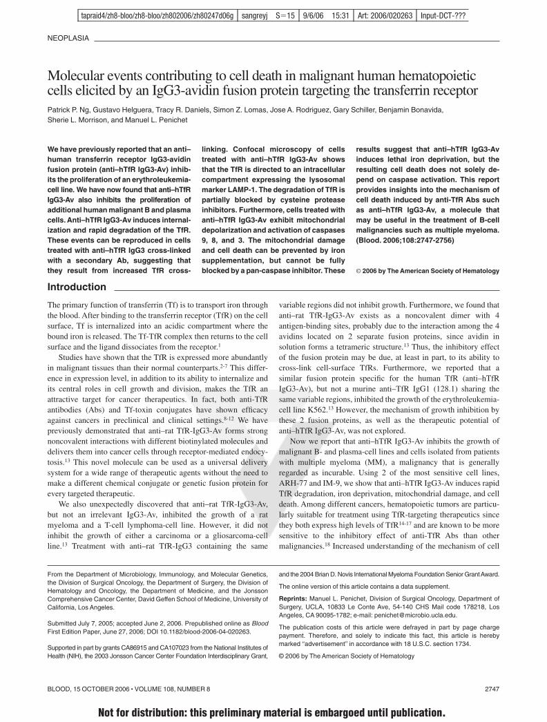
We previously reported that anti–hTfR IgG3-Av is an antiprolifera-tive/proapoptotic drug that strongly inhibits the human erythroleu-kemia-cell line K562.13 To explore the potential of anti–hTfRIgG3-Av as a therapeutic for plasma-cell malignancies, we mea-sured the proliferation of a panel of human malignant B- andplasma-cell lines incubated with the fusion protein. K562 cellswere included in the assay for comparison. Anti–hTfR IgG3-Avinhibits the growth of all the cell lines tested, but to differentextents (Figure 1A). Anti–hTfR IgG3-Av also significantly inhibitsthe in vitro growth of primary cells isolated from 2 patients, onediagnosed with multiple myeloma and one with plasma-cellleukemia (Figure 1B). Further studies with a larger sample size arenecessary to confirm the spectrum of sensitivities to treatment withanti–hTR-IgG3-Av in primary cells. The 2 most sensitivecell lines, ARH-77 and IM-9, were used in subsequent experi-ments to determine the mechanism of growth inhibition byanti–hTfR IgG3-Av.
Anti–hTfR IgG3-Av or anti–hTfR IgG3 cross-linked with asecondary Ab inhibits the growth and induces apoptosisin ARH-77 cells
Previous studies suggest that anti–hTfR IgG3-Av inhibits cellgrowth by cross-linking the TfR.13 To test this hypothesis, wemeasured proliferation and apoptosis in ARH-77 cells treated withdifferent anti-hTfR molecules. Anti–hTfR IgG3-Av completelyinhibited [3H]-thymidine incorporation (Figure 1C) and resulted ina significant increase in the number of dead (annexin V bright, PIbright) and apoptotic (annexin V bright, PI dim) cells from 48 to 96hours (Figure 2A). The total percentage of dead cells includingapoptotic cells (annexin V bright, PI bright and annexin V bright, PIdim) resulting from anti–hTfR IgG3-Av treatment increased from63% at 48 hours, to 79% at 72 hours, and to 86% at 96 hours. In
another experiment, treatment with anti–hTfR IgG3-Av inducedcell death in 85% of the cells compared with 47% in cells treatedwith a 3-fold higher concentration of anti–hTfR IgG3 (Figure 2B).However, the growth inhibitory and proapoptotic effects of anti–hTfR IgG3 could be enhanced to levels comparable with those ofanti–hTfR IgG3-Av by cross-linking with a secondary mAb(Figures 1C and 2B). Similar results were obtained using IM-9cells (data not shown). These results suggest that the tetravalent(4 antigen-binding sites) anti–hTfR IgG3-Av has a stronger cyto-toxic effect than anti–hTfR IgG3 due, at least in part, to itsincreased cross-linking of cell-surface TfR.
Anti–hTfR IgG3-Av down-regulates cell-surfaceexpression of TfR
The level of surface TfR on ARH-77 cells incubated with anti-hTfRmolecules was measured by flow cytometry using a Tf-FITCconjugate. Based on our findings (data not shown) and published
K562
U266
MM.1S
S6B45
8226
/S
8226
Dox 40
OCI-My5
ARH-77
IM-9
0
20
40
60
80
100
120
[3 H]-
Th
ymid
ine
Inco
rpo
rati
on
(%
co
ntr
ol)
A
0
20
40
60
80
100
120
[3 H]-
Th
ymid
ine
Inco
rpo
rati
on
(%
co
ntr
ol)
MMPCL
IM-9
U266
B C
0
20
40
60
80
100
120
[3 H]-
Th
ymid
ine
Inco
rpo
rati
on
(%
co
ntr
ol)
Anti-hTfR
IgG3-
Av
Anti-hTfR
IgG3
Cross
-linke
dAnti-
hTfRIgG3
Anti-dan
syl Ig
G3-Av
Cross
-linkin
gAb
Anti-dan
syl Ig
G30
20
40
60
80
100
120
Figure 1. Anti–hTfR IgG3-Av inhibits the growth of malignant hematopoieticcells. (A) Proliferation assay of a panel of hematopoietic malignant cell linesincubated with 34 nM anti–hTfR IgG3-Av for 72 hours. Cells were incubated with4 %Ci (0.148 MBq)/mL [3H]-thymidine for an additional 24 hours prior to harvesting.Data represent the mean of quadruplicate samples of 2 independent determinationsof [3H]-thymidine incorporation. (B) Proliferation assay of primary cells isolated fromthe bone marrow of a patient with MM and a patient with PCL. Myeloma cells from thepatients were isolated in 2 independent experiments by positive selection. As acontrol, 2 cell lines, IM-9 (highly sensitive) and U266 (less sensitive), were tested inparallel. Control data from the experiment with the PCL patient are shown. All cellswere treated in triplicate with 100 nM anti–hTfR IgG3-Av for 48 hours and thenincubated with 4 %Ci (0.148 MBq)/mL [3H]-thymidine for an additional 48 hours priorto harvesting and determination of radioactivity. (C) ARH-77 cells were incubated with11 nM anti–hTfR IgG3-Av, anti–hTfR IgG3, anti–hTfR IgG3 cross-linked with a 5-foldexcess of secondary Ab, secondary Ab alone, the anti–dansyl IgG3 isotype control, orthe anti–dansyl IgG3-Av for 72 hours. Cells were incubated with 4 %Ci (0.148MBq)/mL [3H]-thymidine for an additional 24 hours prior to harvesting and determina-tion of radioactivity. Data shown are representative of 3 independent experiments. Alldata are presented as the percentage [3H]-thymidine incorporation compared withcontrol cells treated with buffer. Error bars indicate the standard deviation.
ANTI–hTfR IgG3-AVIDIN–MEDIATED CELL DEATH 2749BLOOD, 15 OCTOBER 2006 ! VOLUME 108, NUMBER 8
F1
F2
tapraid4/zh8-bloo/zh8-bloo/zh802006/zh80247d06g sangreyj S!15 9/6/06 15:31 Art: 2006/020263 Input-DCT-???
Not for distribution: this preliminary material is embargoed until publication.

data on 128.1,21 anti–hTfR IgG3 and anti–hTfR IgG3-Av do notinhibit the binding of Tf to the TfR. Therefore, their presence doesnot affect the binding of Tf-FITC to the receptor. Both anti-TfRAbs caused an extensive and rapid decrease in surface TfRexpression, which remained low throughout the experiment (Fig-ure 3; also see Figure S1, available on the Blood website byclicking on the Supplemental Figure link at the top of the onlinearticle). Although the differences were small, anti–hTfR IgG3-Avconsistently induced greater cell-surface TfR down-regulation thananti–hTfR IgG3. Treatment of IM-9 cells yielded similar results(data not shown). Comparable TfR down-regulation was alsoobserved in cells treated with anti–hTfR IgG3 cross-linked with asecondary mAb (data not shown).
Anti–hTfR IgG3-Av or anti–hTfR IgG3 cross-linked with asecondary Ab induces intracellular degradation of the TfR
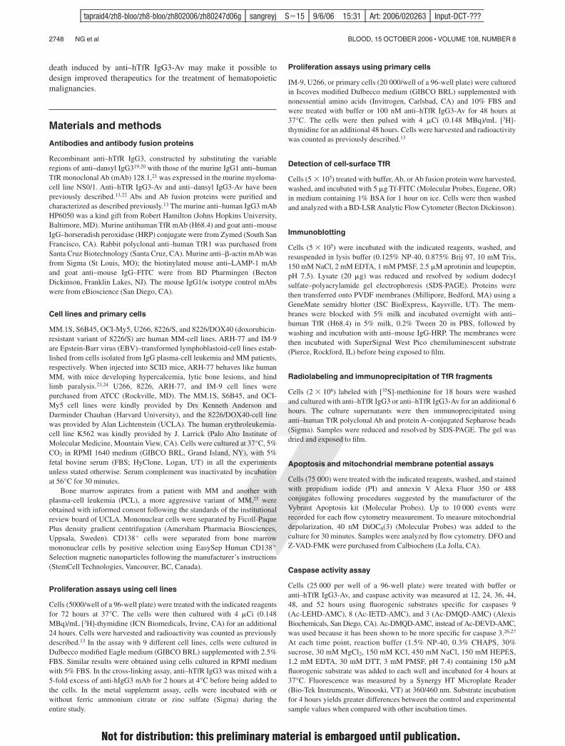
To determine the fate of down-regulated surface TfR, we analyzedthe lysates of treated cells by immunoblotting. Intact TfR vanishedbetween 4 to 12 hours after treatment with anti–hTfR IgG3-Av. Incontrast, anti–hTfR IgG3 induced less extensive receptor loss(Figure 4A). However, TfR loss following binding of anti–hTfRIgG3 could be increased by cross-linking with a secondary mAb(Figure 4B). When cells were treated with anti–hTfR IgG3-Av forshorter times, over half of the intact receptors had disappeared by20 minutes (Figure 4C).
The rapid disappearance of TfR could be a result of shedding ofsurface TfR. However, we were unable to detect any TfR fragments inthe culture media of treated cells (data not shown). Alternatively, TfRcould be degraded intracellularly. In fact, immunoblotting of lysatesfrom cells treated with anti–hTfR IgG3-Av showed small fragments ofTfR (approximately 16 and 20 kDa) concomitant with the disappear-ance of full-length TfR (95 kDa) (Figure 4D). Based on their sizes andthe fact that they were recognized by a mAb against the cytoplasmicN-terminus of TfR (a type II transmembrane protein), these fragmentsappeared to be a result of proteolytic cleavages in the extracellularregion of the TfR. The degradation may take place in the lumen of anintracellular compartment since anti–hTfR IgG3-Av is internalizedthrough TfR-mediated endocytosis.13
Iron supplement blocks the antiproliferative and proapoptoticeffects of anti–hTfR IgG3-Av
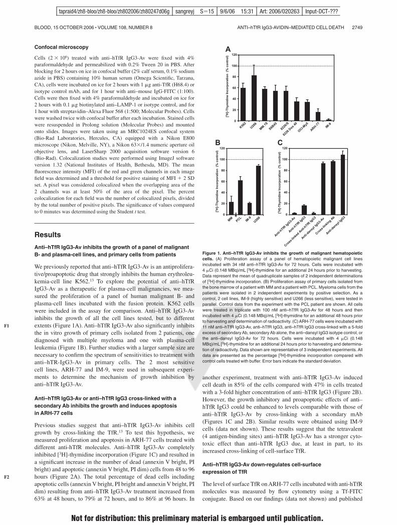
To determine if anti–hTfR IgG3-Av inhibits growth by disruptingTfR-dependent iron uptake, we incubated cells with the fusion protein inthe presence of metal supplements. Iron, but not another divalenttransition metal, zinc, completely blocked the growth inhibitory andproapoptotic effects of anti–hTfR IgG3-Av (Figure 5A-B). Similarresults were obtained in cells treated with anti–hTfR IgG3 alone orcross-linked with a secondary Ab (data not shown). Iron supplement inuntreated cells did not promote cell growth (data not shown). Inconclusion, these data suggest that the anti-hTfR Abs induce apoptosis,at least in part, by causing iron deprivation.
Anti–hTfR IgG3-Av directs TfR to an intracellular compartmentexpressing LAMP-1, but degradation of the receptor is notsensitive to increase of pH in the lysosome
To determine if anti–hTfR IgG3-Av directs TfR to the lysosome fordegradation, we analyzed treated cells by confocal microscopy.Increased colocalization of TfR and LAMP-1 was observedbeginning 15 minutes after treatment with anti–hTfR IgG3-Av(Figure 6; Table 1). Despite the data indicating that TfR travels tothe lysosome, the degradation of the receptor could not be blockedby coincubation with 20 mM ammonium chloride or 200 %Mchloroquine (data not shown), chemicals that increase the pH of thelysosome. This suggests that the proteases responsible for thedegradation of TfR are still functional at higher pHs.
A papainlike cysteine protease is involved in the degradationof TfR induced by anti–hTfR IgG3-Av
To identify the proteases that degrade the TfR, we used immunoblot-ting to detect TfR and its cleavage products in the lysates of
Time (hours)
Su
rfac
e T
fR E
xpre
ssio
n(r
atio
ove
r b
ackg
rou
nd
sig
nal
)
Buffer
Anti-hTfR IgG3
Anti-hTfR IgG3-Av
0
1
2
3
4
5
6
7
0 20 40 60 80
Figure 3. Anti–hTfR IgG3-Av down-regulates TfR expression on the surface ofARH-77 cells. Cells treated with buffer (F), 11 nM anti–hTfR IgG3 (f), or 11 nManti–hTfR IgG3-Av (Œ) were harvested at 4, 12, 24, 48, and 72 hours. Cells were thenincubated with Tf-FITC, washed, and analyzed by flow cytometry. This experimentwas repeated twice with similar results.
Figure 2. Anti–hTfR IgG3-Av induces apoptosis in ARH-77 cells. (A) Cells wereincubated with 11 nM anti–hTfR IgG3 or anti–hTfR IgG3-Av for the indicated times.Cells were then washed, stained with annexin V–Alexa Fluor 488 and PI, andanalyzed by flow cytometry. Data shown are representative of 3 independentexperiments. (B) ARH-77 cells were treated with 1.2 nM anti–hTfR IgG3-Av, 3.7 nManti–hTfR IgG3, or 3.7 nM anti–hTfR IgG3 cross-linked with a 5-fold excess ofsecondary Ab for 96 hours and analyzed by flow cytometry. The percentage of cellslocated in each quadrant is shown in the corner. Results are representative of 2independent experiments.
2750 NG et al BLOOD, 15 OCTOBER 2006 ! VOLUME 108, NUMBER 8
F3
F4
F5
F6
T1
tapraid4/zh8-bloo/zh8-bloo/zh802006/zh80247d06g sangreyj S!15 9/6/06 15:31 Art: 2006/020263 Input-DCT-???
Not for distribution: this preliminary material is embargoed until publication.
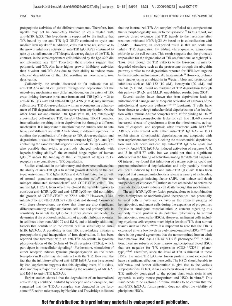
ARH-77 cells treated with anti–hTfR IgG3-Av and a variety ofproteases inhibitors (Figure 7). None of the agents completelyinhibited the destruction of the 95-kDa TfR, indicating that theprocess involves more than one protease family. PMSF, pepsta-tin A, and bestatin, specific inhibitors of serine, aspartic acidproteases, and aminopeptidases, respectively,28,29 did not inhibit theformation of the 16- and 20-kDa fragments containing the N-terminus. In contrast, antipain and leupeptin, inhibitors of bothcysteine and serine proteases, and E64d, which inhibits mostcysteine proteases and trypsin,28-30 blocked the cleavage thatgenerated the 16- and 20-kDa fragments. However, they were notable to block cleavage at a more C-terminal site that resulted in anapproximately 23-kDa fragment (Figure 7). The fact that onlypapainlike cysteine proteases are inhibited by antipain, leupeptin,and E64d, but not by PMSF, suggests that one or more of theseenzymes are involved in the generation of the 16- and 20-kDafragments. Inhibition of metalloproteinases using metal chelatorssuch as o-phenanthroline and GM6001 also did not interfere withthe production of the 16- and 20-kDa fragments (data not shown).In conclusion, these results indicate that upon treatment withanti–hTfR IgG3-Av, the extracellular portion of the internalizedTfR is cleaved at multiple sites by different proteases including apapainlike cysteine protease.
Anti–hTfR IgG3-Av induces the activation of caspases9, 8, and 3
To determine if treatment with anti–hTfR IgG3-Av induces caspaseactivation, we measured the activity of caspases 9, 8, and 3 inARH-77 cells treated with the fusion protein. Caspases 9 and 8 arekey components of the mitochondrial and death receptor–mediatedapoptotic pathway, respectively, while caspase 3 is the executionercaspase of both pathways.31 Caspase activity determined by using aspecific fluorogenic substrate has been shown to correlate withimmunoblots detecting the active form of the protease.32 Activationof all 3 caspases was seen only between 44 hours and 52 hours,with the highest increase observed 48 hours after treatment withanti–hTfR IgG3-Av (Figure 8A). We did not find a significantdifference in the timing of activation among the caspases.
Cell death induced by DFO or anti–hTfR IgG3-Av is partiallyblocked by a broad-spectrum caspase inhibitor
Although we had evidence of caspase activation in cells treatedwith anti–hTfR IgG3-Av, it was unclear whether cell death wascaspase dependent. To answer this question, we coincubated cellswith anti–hTfR IgG3-Av and the broad-spectrum caspase inhibitor
Buffer Control
4 h 24 h12 h
ARH-77
TfR
IM-9
4 h
TfR (95 kDa)
Buffer Contro
l
Cross
-linke
d Anti-hTfR
IgG3
Anti-hTfR
IgG3-Av
1:1 1:5
Anti-hTfR
IgG3
20' 5' 10' 20'
TfR
Cont. Anti-hTfR IgG3-Av
β-actin
Anti-hTfR
IgG3-Av
Control
20 kDa 14 kDa
95 kDa
Anti-hTfR IgG3-Av
Anti-hTfR IgG3
β-actin
β-actin
β-actin
A
B
C
D
Figure 4. Anti–hTfR IgG3-Av induces the highestlevels of intracellular degradation of the TfR. (A)ARH-77 and IM-9 cells were treated with buffer, 11 nManti–hTfR IgG3, or 11 nM anti–hTfR IgG3-Av. ARH-77 wastreated for 4, 12, and 24 hours, while IM-9 was treated for4 hours. (B) ARH-77 cells were treated with buffer, 11 nManti–hTfR IgG3, 11 nM anti–hTfR IgG3 cross-linked withsecondary mAb in 1:1 or 1:5 ratios, or 11 nM anti–hTfRIgG3-Av for 8 hours. (C) ARH-77 cells were treated withbuffer or 11 nM anti–hTfR IgG3-Av for 5, 10, and 20 min-utes. (D) ARH-77 cells were treated with buffer or 11 nManti–hTfR IgG3-Av for 4 hours. In all cases, cells wereharvested, and whole-cell lysates were prepared andanalyzed by immunoblotting with a murine mAb againstthe intracellular N-terminus of the human TfR followed byincubation with goat anti–mouse IgG-HRP conjugate. Theblot was then stripped and reblotted with a murine anti–"-actin mAb. In panels A, B, and C, only the regions of the95-kDa intact TfR and "-actin are shown. In panel D, theblot from 14 to 95 kDa is shown. These experiments wererepeated at least twice with similar results.
Buffer Anti-hTfR IgG3-Av
Anti-hTfR IgG3-Av+ FACBuffer + FAC
79
6
14
79
4
14
23
24
5214
11
74
Pro
pidi
um Io
dide
(cel
l dea
th m
arke
r)
Alexa Fluor 488 Labeled Annexin V (early apoptosis marker)
[3 H]-
Thym
idin
e In
corp
orat
ion
(% c
ontr
ol)
0
10
20
30
40
50
60
70
80
90
100
110
ARH-77 IM-9
Buffer
ZnSO4FAC
Buffer
ZnSO4FAC
A B
Figure 5. Iron supplement blocks the antiproliferative and proapoptoticeffects of anti–hTfR IgG3-Av. (A) ARH-77 and IM-9 cells were treated for72 hours with 11 nM anti–hTfR IgG3-Av alone or in the presence of 30 %M zincsulfate (ZnSO4) or 25 %M ferric ammonium citrate (FAC). The cells were thencultured in the presence of [3H]-thymidine for an additional 24 hours andharvested, and the incorporation of [3H]-thymidine was determined. Each valueis the mean of quadruplicate samples expressed as the percentage of thecontrol mean (controls are cells treated with buffer alone, with or without metalsalt supplement; data not shown). (B) ARH-77 cells were treated for 96 hourswith buffer or 11 nM anti–hTfR IgG3-Av with or without 50 %M FAC. The cellswere then washed, stained with annexin V–Alexa Fluor 488 and PI, andanalyzed by flow cytometry. The percentage of cells located in each quadrant isshown at the corner.
ANTI–hTfR IgG3-AVIDIN–MEDIATED CELL DEATH 2751BLOOD, 15 OCTOBER 2006 ! VOLUME 108, NUMBER 8
F7
F8
tapraid4/zh8-bloo/zh8-bloo/zh802006/zh80247d06g sangreyj S!15 9/6/06 15:31 Art: 2006/020263 Input-DCT-???
Not for distribution: this preliminary material is embargoed until publication.
Z-VAD-FMK. We also treated cells with DFO, a membrane-permeable iron chelator, as a positive control for iron deprivation.The levels of apoptosis and viability of cells were determined.
In anti–hTfR IgG3-Av–treated cells, inhibition of caspaseactivity by treatment with Z-VAD-fmk only partially blocked celldeath (Figure 8B). The number of dead cells (annexin V bright, PIbright and annexin V bright, PI dim) decreased from 48% to 29% incells treated with anti–hTfR IgG3-Av alone or pretreated withZ-VAD-fmk, respectively, at 48 hours. Similar results were ob-served after 96 hours of treatment. These results suggest thatanti–hTfR IgG3-Av induced cell death, in part, through a caspase-independent mechanism. However, it is important to note that inseveral independent experiments we have observed that Z-VAD-FMK was less efficient in blocking apoptosis induced by anti–hTfRIgG3-Av than by DFO, suggesting that the 2 reagents may induceapoptosis through slightly different pathways.
DFO and anti–hTfR IgG3-Av inducemitochondrial depolarization
To determine if the PI-negative, annexin V–negative cells thatsurvived the treatment of DFO or anti–hTfR IgG3-Av in thepresence of Z-VAD-FMK were truly healthy, we stained cellstreated for 48 hours (Figure 7B) with a third fluorescent reagent,DiOC6(3), that measures mitochondrial potential. A cell with
normal mitochondrial potential accumulates DiOC6(3) in theorganelle.33 All apoptotic cells lost mitochondrial potential regard-less of the treatment or the presence of Z-VAD-FMK (data notshown). In contrast, nonapoptotic (annexin V, PI double negative)cells present at 48 hours varied in their DiOC6(3) staining profiles(Figure 8C). When cells were treated with DFO or anti–hTfRIgG3-Av without Z-VAD-FMK, a majority of the nonapoptoticcells had mitochondrial potentials comparable with those of controlcells (Figure 8C, left column). In contrast, when Z-VAD-FMK wasadded, there was an increase in the number of nonapoptotic cells(Figure 8B, 48 hours), but a large percentage of these cells hadsustained significant mitochondrial depolarization (Figure 8C, rightcolumn). These results suggest that in the absence of Z-VAD-FMK,cells that were sensitive to DFO or anti–hTfR IgG3-Av underwentapoptosis, leaving mostly bona fide healthy cells in the nonapop-totic population. In contrast, in the presence of Z-VAD-FMK, manycells suffering from mitochondrial damage did not undergo apopto-sis by 48 hours. However, with impaired mitochondrial potentials,many of these cells eventually died34 (Figure 8B, 96 hours).
Discussion
We have studied the growth-inhibitory effect of anti–hTfR IgG3-Avusing a panel of hematopoietic malignant cell lines and primarycells isolated from MM and PCL patients. We observed a widerange of sensitivities in the cell lines studied, while primary cellsfrom the 2 patients displayed intermediate sensitivity. However, it
Table 1. Colocalization of TfR and LAMP-1 in ARH-77 cells incubated with 11 nM anti–hTfR IgG3-Av
Incubation time, min
Experiment 1 Experiment 2 Experiment 3
0 15 30 90 0 30 60 0 15 30 45
Average % colocalization 10.2 20.0 16.3 18.2 13.6 20.8 18.7 11.9 21.7 19.2 39.8
SD 1.5 4.0 2.6 3.4 3.8 3.0 3.9 3.5 8.1 1.4 9.8
SE 0.5 1.4 0.9 1.4 1.4 1.1 1.5 1.0 2.5 0.5 3.1
No. of fields 11 8 8 6 7 8 7 12 10 9 10
Average no. of cells/field 17 14 16 18 19 21 24 19 11 17 7
P — ' .001 ' .001 ' .002 — ' .002 ' .03 — ' .005 ' .001 ' .001
— indicates not applicable.
LAMP-1
15 Min.
0 Min.(Buffer
Control)
TfR Overlay Colocalized
30 Min.
45 Min.
Figure 6. Anti–hTfR IgG3-Av directs the TfR to an intracellular compartmentcontaining the lysosomal marker LAMP-1. ARH-77 cells treated with 11 nManti–hTfR IgG3-Av for 0 (buffer control), 15, 30, or 45 minutes were fixed,permeabilized, and stained as described in “Materials and methods.” Cells were thenanalyzed by confocal microscopy. The image of a representative cell from each timepoint is shown immunostained for TfR (green) and LAMP-1 (red) with overlay. Thearea where colocalization occurs is shown in white. The percent colocalization wascalculated as described in “Confocal microscopy” (Table 1).
Figure 7. A papainlike cysteine protease is involved in the degradation of TfRinduced by anti–hTfR IgG3-Av. ARH-77 cells preincubated with buffer, 250 %ME64d, 250 %M antipain, 250 %M leupeptin, 250 %M bestatin, 500 %M pepstatin A, or500 %M PMSF for 1 hour were treated with buffer or 50 nM anti–hTfR IgG3-Av for anadditional 4 hours before being harvested. Cell lysates were analyzed by immunoblot-ting with a murine mAb against the intracellular N-terminus of the human TfR followedby incubation with goat anti–mouse IgG-HRP. The blot was then stripped andreblotted with a murine anti–"-actin mAb. The regions of the 95-kDa intact TfR, 16- to23-kDa TfR fragments, and "-actin are shown. Except for the PMSF data, theseexperiments were repeated at least once with similar results.
2752 NG et al BLOOD, 15 OCTOBER 2006 ! VOLUME 108, NUMBER 8
COLOR
tapraid4/zh8-bloo/zh8-bloo/zh802006/zh80247d06g sangreyj S!15 9/6/06 15:31 Art: 2006/020263 Input-DCT-???
Not for distribution: this preliminary material is embargoed until publication.

is important to note that additional studies using a larger cohort ofMM patients are required.
Using the 2 most sensitive cell lines, ARH-77 and IM-9, wefound that the tetravalent anti–hTfR IgG3-Av induces significantlymore apoptosis than does the bivalent anti–hTfR IgG3. Cytotoxic-ity is associated with cell-surface TfR down-regulation, rapid TfRdegradation, and iron deprivation. The fact that these events can bereproduced in cells treated with anti–hTfR IgG3 cross-linked witha secondary Ab suggests that, first, the potent cytotoxicity ofanti–hTfR IgG3-Av stems from its ability to increase the cross-linking of cell-surface TfR; and second, increased cross-linking ofTfR results in more efficient degradation of the receptor and moresevere cytotoxicity. These observations provide new insights intothe mechanism of growth inhibition by anti-TfR Abs.
The growth inhibitory property of anti-TfR Abs has beenappreciated since the 1980s.35-39 A rat anti–murine TfR IgM (RI7208) that extensively cross-linked the TfR was reported to blockthe internalization of the TfR-Tf complex, resulting in irondeprivation and growth inhibition.40,41 In addition, a murineanti–human TfR IgA (42/6) showed potent growth inhibitoryactivity that could not be attributed completely to its ability toblock Tf binding to the receptor.21,42-47 A rat anti–murine TfR IgG(RI7 217) and 2 murine anti–human TfR IgGs (OKT9 and B3/25)did not cause significant growth inhibition unless used in combina-tion or cross-linked with secondary Abs.21,40,41,47,48 Based on thesedata, it was suggested that polymeric molecules such as anti–TfRIgA and IgM, and anti–TfR IgGs cross-linked with secondaryAbs, inhibit growth by blocking internalization of the TfR-Tf
complex.18,21,41,47 Whether TfR on cells treated with anti–TfRIgA or cross-linked anti–TfR IgGs is degraded intracellularly wasnot determined.
In contrast to the model described in the previous paragraph,our data indicate for the first time that some polymericmolecules (anti–hTfR IgG3-Av, cross-linked anti–hTfR IgG3)induce significant levels of apoptosis not by blocking TfRinternalization but instead by down-regulating cell-surface TfRand causing its intracellular degradation. Consistent with thishypothesis, previous studies of the anti–human TfR IgA 42/6, atetravalent Ab, had shown that treated cells readily internalized42/6 and down-regulated surface TfR.44,47 However, neither thefate of the internalized TfR nor the contribution of surface TfRdown-regulation to the overall inhibitory effect of 42/6, whichblocks Tf binding to TfR, was known. In addition, the growth ofthe human leukemic T-cell line CCRF-CEM was stronglyinhibited by treatment using pairs of 2 anti–TfR IgG mAbstargeting different epitopes on the TfR; this likely increases thelevel of TfR cross-linking. It was found that these pairs ofanti–TfR IgGs down-regulated cell-surface TfR.21 Overall,these studies suggest that polymeric molecules such as anti–hTfR IgG3-Av, anti–TfR IgA, and anti–TfR IgG cross-linkedwith a secondary Ab inhibit growth by a mechanism differentfrom what had been reported for the anti–TfR IgM.
Although anti–hTfR IgG3, anti–hTfR IgG3 cross-linked with asecondary Ab, and anti–hTfR IgG3-Av all down-regulate cell-surface TfR, the latter 2 are far more efficient in directing thereceptor for degradation. This may explain the difference in the
A
0
50
100
150
200
250
300
350
Caspase 9 Caspase 3 Caspase 8
Cas
pase
Act
ivity
(arb
itrar
y flu
ores
cenc
e un
it)Buffer
Anti-hTfR IgG3-Av
Alexa Fluor 350 Annexin V (early apoptosis marker)
R2
45
14
39 84
5
10
51 28
20
69
12
17
86
4
8 90 5
3
Buffer Z-VAD-FMK
72
6
21
6
9
15
26
59
29
42
29
22
28
50
43
29
27
84
Buffer Z-VAD-FMK
Buf
fer
Con
trol
DFO
Ant
i-hTf
R Ig
G3-
Av
48 h 96 h
Pro
pidi
umIo
dide
(cel
l dea
th m
arke
r)
B
C
Log Fluorescence Intensity
Cel
l Num
ber
Control
DFO
Anti-hTfR IgG3-Av
Buffer Z-VAD-FMK
Figure 8. Anti–hTfR IgG3-Av induces caspase activation, mitochondrial depolarization, and a partially caspase-independent cell death in ARH-77 cells. (A) Cellstreated with buffer or 50 nM anti–hTfR IgG3-Av for 48 hours were lysed and caspase activities measured by specific fluorogenic substrates. Each value is the mean ofquadruplicate samples. (B) Cells preincubated with or without 100 %M Z-VAD-FMK were treated with buffer, 50 %M DFO, or 50 nM anti–hTfR IgG3-Av for 48 or 96 hours. Cellswere then washed and stained with DiOC6(3), annexin V–Alexa Fluor 350, and PI, and analyzed by flow cytometry. The annexin V/PI profiles of treated cells at 48 and 96 hoursare shown with the percentages of cells located in each quadrant. (C) Nonapoptotic cells (lower left quadrants of panel B) at 48 hours were gated and their mitochondrialmembrane potentials shown in log fluorescence intensity. These experiments were repeated in both ARH-77 and IM-9 cells with similar results.
ANTI–hTfR IgG3-AVIDIN–MEDIATED CELL DEATH 2753BLOOD, 15 OCTOBER 2006 ! VOLUME 108, NUMBER 8
tapraid4/zh8-bloo/zh8-bloo/zh802006/zh80247d06g sangreyj S!15 9/6/06 15:31 Art: 2006/020263 Input-DCT-???
Not for distribution: this preliminary material is embargoed until publication.
proapoptotic activities of the different treatments. Therefore, ironuptake may not be completely blocked in cells treated withanti–hTfR IgG3. This hypothesis is supported by the finding thatTfR bound by the anti–TfR IgG OKT9 continued to cycle andmediate iron uptake.48 In addition, cells that were not sensitive tothe growth inhibitory activity of anti–TfR IgG B3/25 continued totake up a small amount of Tf despite down-regulation of the TfR. Incontrast, in the same experiment cells inhibited by the IgA 42/6 didnot internalize any Tf.47 Therefore, these studies suggest thatpolymeric anti-TfR Abs have higher growth inhibitory activitiesthan bivalent anti-TfR Abs due to their ability to induce moreefficient degradation of the TfR, resulting in more severe irondeprivation.
Collectively, the results discussed so far indicate that allanti-TfR Abs inhibit cell growth through iron deprivation but theunderlying mechanism may differ and depend on the extent of TfRcross-linking. Increase in valence from an anti–TfR IgG (v ! 2) toanti–hTfR IgG3-Av and anti–hTfR IgA 42/6 (v ! 4) may increasecell-surface TfR down-regulation with an accompanying enhance-ment of TfR degradation, and more severe iron deprivation. On theother hand, rat anti–murine TfR IgMs (v ! 10, 12) extensivelycross-linked cell-surface TfR, thereby blocking TfR-Tf complexinternalization resulting in iron deprivation but through a differentmechanism. It is important to mention that the studies done to datehave used different anti-TfR Abs binding to different epitopes. Toconfirm the contribution of valence to TfR down-regulation anddegradation, it would be important to compare IgG, IgA, and IgMcontaining the same variable regions. For anti–hTfR IgG3-Av, it isalso possible that avidin, a positively charged molecule withheparin-binding ability,49 the extended hinge region of humanIgG3,50 and/or the binding of the Fc fragment of IgG3 to Fcreceptors may contribute to TfR degradation.
Studies conducted in our laboratory and elsewhere indicate thatthe ability of anti–TfR IgGs to inhibit growth depends on the celltype. Anti–human TfR IgGs B3/25 and 43/31 inhibited the growthof normal granulocyte/macrophage progenitor cells45 but notmitogen-stimulated mononuclear47 and CCRF-CEM cells.21 Themurine IgG1 128.1, from which we cloned the variable regions toconstruct anti–hTfR IgG3 and anti–hTfR IgG3-Av, did not inhibitthe growth of CCRF-CEM21 or K562 cells.13 However, 128.1inhibited the growth of ARH-77 cells (data not shown). Consistentwith these observations, we show that there are also significantdifferences among malignant plasma-cell lines and primary cells insensitivity to anti–hTfR IgG3-Av. Further studies are needed todetermine if the proposed mechanism of growth inhibition operatesin cell lines other than ARH-77 and IM-9, and to identify additionalfactors that contribute to the overall cellular sensitivity to anti–hTfR IgG3-Av. A possibility is that TfR cross-linking initiates aproapoptotic signal independent of iron deprivation. It has beenreported that treatment with an anti-TfR Ab results in tyrosinephosphorylation of the (-chain of T-cell receptors (TCRs), whichparticipate in intracellular signaling.51 Furthermore, stimulation ofeither receptor induces tyrosine phosphorylation on the other.Receptors in B cells may also interact with the TfR. However, thefact that the inhibitory effect of anti–hTfR IgG3-Av can be reversedby iron supplement suggests that this signaling event, if it exists,does not play a major role in determining the sensitivity of ARH-77and IM-9 to anti–hTfR IgG3-Av.
Earlier studies showed that the degradation of an internalizedanti–TfR IgG could be inhibited by leupeptin and chloroquine, andsuggested that the TfR-Ab complex was degraded in the lyso-some.48 Electron microscopy by Hopkins and Trowbridge indicated
that the internalized TfR-Ab complex trafficked to a compartmentthat is morphologically similar to the lysosome.52 In this report, weprovide direct evidence that TfR travels to the lysosome aftertreatment with anti–hTfR IgG3-Av based on its colocalization withLAMP-1. However, an unexpected result is that we could notinhibit TfR degradation by adding chloroquine or ammoniumchloride to the cell culture. This result suggests that the proteasesresponsible for the degradation of TfR are functional at higher pHs.Thus, even though the TfR trafficks to the lysosome, it may bedegraded elsewhere such as the proteasome through the ubiquitinpathway similar to the degradation reported for HER/neu targetedby the recombinant humanized Ab trastuzumab.53 However, prelimi-nary studies using antiubiquitin in Western blots and proteosomalinhibitors such as MG-132 (10 %M), lactacystin (20 %M), andPS-341 (500 nM) found no evidence of TfR degradation throughthis pathway (P.P.N. and M.L.P., unpublished results, June 2004).
Several studies have shown that iron deprivation leads tomitochondrial damage and subsequent activation of caspases of themitochondrial apoptosis pathway.32,54-59 Leukemic T cells havebeen shown to undergo mitochondrial depolarization after incuba-tion with a murine Ab that competes with Tf for binding to TfR,60
and the human promyelocytic leukemic cell line HL-60 showedincreased release of cytochrome c from the mitochondria, activa-tion of caspases, and apoptosis after treatment with DFO.58,61
ARH-77 cells treated with either anti–hTfR IgG3-Av or DFOexhibit similar mitochondrial depolarization and apoptosis, withiron supplement completely blocking the mitochondrial depolariza-tion and cell death induced by anti–hTfR IgG3-Av (data notshown). Anti–hTfR IgG3-Av induced activation of caspases 9, 8,and 3 in ARH-77 cells, but we could not find a significantdifference in the timing of activation among the different caspases.Of interest, we found that inhibition of caspase activity could notprevent mitochondrial depolarization and only partially blockedcell death induced by DFO and anti–hTfR IgG3-Av. It has beenreported that damaged mitochondria release a variety of molecules,such as apoptosis-inducing factor (AIF), that induce cell deathindependent of caspases.62 Further studies are needed to determineif anti–hTfR IgG3-Av induces cell death through this mechanism.
The anti–hTfR IgG3-Av fusion protein, alone or in combinationwith biotinylated or nonbiotinylated therapeutics, can potentiallybe used both in vivo and ex vivo in the efficient purging ofhematopoietic malignant cells during the expansion of progenitorsfor use in autologous transplantation. A concern regarding thisantibody fusion protein is its potential cytotoxicity to normalhematopoietic stem cells (HSCs). However, malignant cells includ-ing myeloma cells express much higher levels of TfR than normaltissues such as HSCs.16,63,64 It is important to note that the TfR isexpressed at very low levels in early, noncommitted HSCs,65-69 andthere is the general agreement that the noncommitted human adultbone marrow HSC has a CD34$/CD71lo phenotype.66-68 In addi-tion, there are subsets of bone marrow and peripheral blood HSCsthat are negative for TfR expression (CD34$/CD71) pheno-type).65-68 Therefore, since the level of TfR is minimal in theseHSCs, the anti–hTfR IgG3-Av fusion protein is not expected tohave a significant effect on these cells. The HSCs should be able toself-renew and further differentiate to give rise to the varioussubpopulations. In fact, it has even been shown that an anti–murineTfR antibody conjugated to the potent plant toxin ricin is notcytotoxic to early mouse progenitors and HSCs in vitro.17 Thisissue needs to be explored in future studies to be certain that theanti–hTfR IgG3-Av fusion protein does not affect the viability ofpluripotent HSCs.
2754 NG et al BLOOD, 15 OCTOBER 2006 ! VOLUME 108, NUMBER 8
tapraid4/zh8-bloo/zh8-bloo/zh802006/zh80247d06g sangreyj S!15 9/6/06 15:31 Art: 2006/020263 Input-DCT-???
Not for distribution: this preliminary material is embargoed until publication.
In this report, we have described several key aspects of themechanism of cell death induced by anti–hTfR IgG3-Av inARH-77 cells, a widely used model for studying MM.23,24,70,71 Thisinformation sheds light on the mechanism of inhibition by anti-TfRAbs in general, and will be useful for designing therapeutics forMM and other hematopoietic malignancies based on anti–hTfRIgG3-Av. These mechanistic studies were restricted to the 2 mostsensitive cell lines and further studies are needed to confirm themechanism of cell death in the other cell lines and primary cellsfrom patients. Further studies are also needed to explain thedifferences in sensitivity among the panel of hematopoietic malig-nant cell lines, since we have seen that the sensitivity is notcorrelated to the levels of TfR expression (P.P.N., G.H., and M.L.P.,unpublished data, June 2005). In addition to its proapoptoticactivity, anti–hTfR IgG3-Av is the first anti-TfR to contain thehuman IgG3 constant regions, which are known to bind to Fcreceptors and fix complement. These properties have been shownto contribute to the antitumor activity of Abs in patients,72 and mayenhance the in vivo effectiveness of anti–hTfR IgG3-Av. Thetumoricidal activity of anti–hTfR IgG3-Av can be further increased
by combining it with a biotinylated toxin as suggested by prelimi-nary data using malignant hematopoietic cell lines (P.P.N., G.H.,and M.L.P., unpublished results, July 2005). Further developmentof this multifunctional fusion protein may lead to effectivetherapeutics for hematopoietic malignancies such as multiplemyeloma.
Acknowledgments
We thank Dr Jay Dela Cruz (UCLA) for his assistance withconfocal microscopy. The MM.1S, S6B45, and OCI-My5 cell lineswere kindly provided by Drs Kenneth Anderson and DarminderChauhan (Harvard University), and Alan Lichtenstein (UCLA)generously provided the 8226/DOX40 cell line. The humanerythroleukemia-cell line K562 was kindly provided by J. Larrick(Palo Alto Institute of Molecular Medicine, Mountain View, CA).We also thank Drs Thomas Ganz, Elizabeth Neufeld, KoteswaraChintalacharuvu, Juran Kato-Stankiewicz, Milena Pervan, and LeaGuo (UCLA) for their helpful suggestions.
References1. Ponka P, Lok CN. The transferrin receptor: role in
health and disease. Int J Biochem Cell Biol. 1999;31:1111-1137.
2. Omary MB, Trowbridge IS, Minowada J. Humancell-surface glycoprotein with unusual properties.Nature. 1980;286:888-891.
3. Faulk WP, Hsi BL, Stevens PJ. Transferrin andtransferrin receptors in carcinoma of the breast.Lancet. 1980;2:390-392.
4. Shindelman JE, Ortmeyer AE, Sussman HH.Demonstration of the transferrin receptor in hu-man breast cancer tissue: potential marker foridentifying dividing cells. Int J Cancer. 1981;27:329-334.
5. Trowbridge IS, Lesley J, Schulte R. Murine cellsurface transferrin receptor: studies with an anti-receptor monoclonal antibody. J Cell Physiol.1982;112:403-410.
6. Prost AC, Menegaux F, Langlois P, et al. Differen-tial transferrin receptor density in human colorec-tal cancer: a potential probe for diagnosis andtherapy. Int J Oncol. 1998;13:871-875.
7. Shinohara H, Fan D, Ozawa S, et al. Site-specificexpression of transferrin receptor by human coloncancer cells directly correlates with eradication byantitransferrin recombinant immunotoxin. Int JOncol. 2000;17:643-651.
8. Brooks D, Taylor C, Dos Santos B, et al. Phase Iatrial of murine immunoglobulin A antitransferrinreceptor antibody 42/6. Clin Cancer Res. 1995;1:1259-1265.
9. Mayers GL, Raghavan D, Hitt S, Glaves D. Trans-ferrin-gemcitabine conjugates: application to che-motherapy. Proc 89th Annual Meeting Am AssocCancer Res. 1998;39:63-64.
10. Mayers GL, Razeq J, Abu-Hadid MM. Cytotoxicdrug conjugates for treatment of neoplastic dis-ease. US patent no. 5,393,737. February 28,1995.
11. Laske DW, Muraszko KM, Oldfield EH, et al. In-traventricular immunotoxin therapy for leptomen-ingeal neoplasia. Neurosurgery. 1997;41:1039-1049; discussion: 1049-1051.
12. Weaver M, Laske DW. Transferrin receptor li-gand-targeted toxin conjugate (Tf-CRM107) fortherapy of malignant gliomas. J Neurooncol.2003;65:3-13.
13. Ng PP, Dela Cruz JS, Sorour DN, et al. An anti-transferrin receptor-avidin fusion protein exhibitsboth strong proapoptotic activity and the ability todeliver various molecules into cancer cells. ProcNatl Acad Sci U S A. 2002;99:10706-10711.
14. Habelshaw HA, Lister TA, Stansfeld AG. Correla-tion of transferrin receptor expression with histo-logical class and outcome in non-Hodgkin lym-phoma. Lancet. 1983;1:498-500.
15. Beguin Y, Lampertz S, Degroote D, Igot D, Mal-aise M, Fillet G. Soluble Cd23 and other recep-tors (Cd4, Cd8, Cd25, Cd71) in serum of patientswith chronic lymphocytic leukemia. Leukemia.1993;7:2019-2025.
16. Jefferies WA, Brandon MR, Williams AF, Hunt SV.Analysis of lymphopoietic stem cells with a mono-clonal antibody to the rattransferrin receptor.Immunology. 1985;54:333-341.
17. Lesley J, Domingo DL, Schulte R, Trowbridge IS.Effect of an anti-murine transferrin receptor-ricinA conjugate on bone marrow stem and progenitorcells treated in vitro. Exp Cell Res. 1984;150:400-407.
18. Trowbridge IS. Transferrin receptor as a potentialtherapeutic target. Prog Allergy. 1988;45:121-146.
19. Coloma MJ. Production and Characterization ofNovel Recombinant Antibodies [PhD thesis]. LosAngeles: Department of Microbiology and Mo-lecular Genetics, University of California at LosAngeles; 1997.
20. Morrison SL, Wims L, Wallick S, Tan L, Oi VT. Ge-netically engineered antibody molecules and theirapplication. Ann N YAcad Sci. 1987;507:187-198.
21. White S, Taetle R, Seligman PA, Rutherford M,Trowbridge IS. Combinations of anti-transferrinreceptor monoclonal antibodies inhibit humantumor cell growth in vitro and in vivo: evidence forsynergistic antiproliferative effects. Cancer Res.1990;50:6295-6301.
22. Shin SU, Wu D, Ramanathan R, Pardridge WM,Morrison SL. Functional and pharmacokineticproperties of antibody-avidin fusion proteins.J Immunol. 1997;158:4797-4804.
23. Gado K, Silva S, Paloczi K, Domjan G, Falus A.Mouse plasmacytoma: an experimental model ofhuman multiple myeloma. Haematologica. 2001;86:227-236.
24. Cruz JC, Alsina M, Craig F, et al. Ibandronate de-creases bone disease development and oste-oclast stimulatory activity in an in vivo model ofhuman myeloma. Exp Hematol. 2001;29:441-447.
25. Hayman SR, Fonseca R. Plasma cell leukemia.Curr Treat Options Oncol. 2001;2:205-216.
26. Garcia-Calvo M, Peterson EP, Rasper DM, et al.Purification and catalytic properties of humancaspase family members. Cell Death Differ. 1999;6:362-369.
27. Tomita Y, Bilim V, Hara N, Kasahara T, TakahashiK. Role of IRF-1 and caspase-7 in IFN-gammaenhancement of Fas-mediated apoptosis inACHN renal cell carcinoma cells. Int J Cancer.2003;104:400-408.
28. Beynon RJ, Bond JS. Proteolytic Enzymes: APractical Approach. New York: Oxford UniversityPress; 2001.
29. Zollner H. Handbook of Enzyme Inhibitors. Wein-heim, Germany: VCH Verlagsgesellschaft; 1989.
30. Sreedharan SK, Verma C, Caves LS, et al. Dem-onstration that 1-trans-epoxysuccinyl-L-leucyl-amido-(4-guanidino) butane (E-64) is one of themost effective low Mr inhibitors of trypsin-catalysedhydrolysis: characterization by kinetic analysis andby energy minimization and molecular dynamicssimulation of the E-64-beta-trypsin complex. Bio-chem J. 1996;316(pt 3):777-786.
31. Creagh EM, Conroy H, Martin SJ. Caspase-activation pathways in apoptosis and immunity.Immunol Rev. 2003;193:10-21.
32. Greene BT, Thorburn J, Willingham MC, et al.Activation of caspase pathways during iron chela-tor-mediated apoptosis. J Biol Chem. 2002;277:25568-25575.
33. Korchak HM, Rich AM, Wilkenfeld C, Ruther-ford LE, Weissmann G. A carbocyanine dye,DiOC6(3), acts as a mitochondrial probe in hu-man neutrophils. Biochem Biophys Res Com-mun. 1982;108:1495-1501.
34. Green DR, Kroemer G. The pathophysiology ofmitochondrial cell death. Science. 2004;305:626-629.
35. Trowbridge IS, Domingo DL. Anti-transferrin re-ceptor monoclonal antibody and toxin-antibodyconjugates affect growth of human tumour cells.Nature. 1981;294:171-173.
36. Lesley JF, Schulte RJ. Selection of cell lines re-sistant to anti-transferrin receptor antibody: evi-dence for a mutation in transferrin receptor. MolCell Biol. 1984;4:1675-1681.
37. Vaickus L, Levy R. Antiproliferative monoclonalantibodies: detection and initial characterization.J Immunol. 1985;135:1987-1997.
38. Lesley J, Schulte R. Selection and characteriza-tion of transferrin receptor mutants using recep-tor-specific antibodies. Immunogenetics. 1986;24:163-170.
39. Kemp JD, Thorson JA, McAlmont TH, HorowitzM, Cowdery JS, Ballas ZK. Role of the transferrinreceptor in lymphocyte growth: a rat IgG mono-clonal antibody against the murine transferrin
ANTI–hTfR IgG3-AVIDIN–MEDIATED CELL DEATH 2755BLOOD, 15 OCTOBER 2006 ! VOLUME 108, NUMBER 8
tapraid4/zh8-bloo/zh8-bloo/zh802006/zh80247d06g sangreyj S!15 9/6/06 15:31 Art: 2006/020263 Input-DCT-???
Not for distribution: this preliminary material is embargoed until publication.
receptor produces highly selective inhibition ofT and B cell activation protocols. J Immunol.1987;138:2422-2426.
40. Lesley JF, Schulte RJ. Inhibition of cell growth bymonoclonal anti-transferrin receptor antibodies.Mol Cell Biol. 1985;5:1814-1821.
41. Lesley J, Schulte R, Woods J. Modulation oftransferrin receptor expression and function byanti-transferrin receptor antibodies and antibodyfragments. Exp Cell Res. 1989;182:215-233.
42. Trowbridge IS, Lopez F. Monoclonal antibody totransferrin receptor blocks transferrin binding andinhibits human tumor cell growth in vitro. ProcNatl Acad Sci U S A. 1982;79:1175-1179.
43. Mendelsohn J, Trowbridge I, Castagnola J. Inhibi-tion of human lymphocyte proliferation by mono-clonal antibody to transferrin receptor. Blood.1983;62:821-826.
44. Neckers LM, Cossman J. Transferrin receptorinduction in mitogen-stimulated human T lympho-cytes is required for DNA synthesis and cell divi-sion and is regulated by interleukin 2. Proc NatlAcad Sci U S A. 1983;80:3494-3498.
45. Taetle R, Honeysett JM, Trowbridge I. Effects ofanti-transferrin receptor antibodies on growth ofnormal and malignant myeloid cells. Int J Cancer.1983;32:343-349.
46. Taetle R, Rhyner K, Castagnola J, To D, Mendel-sohn J. Role of transferrin, Fe, and transferrinreceptors in myeloid leukemia cell growth: studieswith an antitransferrin receptor monoclonal anti-body. J Clin Invest. 1985;75:1061-1067.
47. Taetle R, Castagnola J, Mendelsohn J. Mecha-nisms of growth inhibition by anti-transferrin re-ceptor monoclonal antibodies. Cancer Res. 1986;46:1759-1763.
48. Weissman AM, Klausner RD, Rao K, Harford JB.Exposure of K562 cells to anti-receptor monoclo-nal antibody OKT9 results in rapid redistributionand enhanced degradation of the transferrin re-ceptor. J Cell Biol. 1986;102:951-958.
49. Kett WC, Osmond RI, Moe L, Skett SE, KinnearBF, Coombe DR. Avidin is a heparin-binding pro-tein: affinity, specificity and structural analysis.Biochim Biophys Acta. 2003;1620:225-234.
50. Phillips ML, Tao MH, Morrison SL, SchumakerVN. Human/mouse chimeric monoclonal antibod-ies with human IgG1, IgG2, IgG3 and IgG4 con-stant domains: electron microscopic and hydro-
dynamic characterization. Mol Immunol. 1994;31:1201-1210.
51. Salmeron A, Borroto A, Fresno M, Crumpton MJ,Ley SC, Alarcon B. Transferrin receptor inducestyrosine phosphorylation in T cells and is physi-cally associated with the TCR zeta-chain. J Im-munol. 1995;154:1675-1683.
52. Hopkins CR, Trowbridge IS. Internalization andprocessing of transferrin and the transferrin re-ceptor in human carcinoma A431 cells. J CellBiol. 1983;97:508-521.
53. Klapper LN, Waterman H, Sela M, Yarden Y. Tu-mor-inhibitory antibodies to HER-2/ErbB-2 mayact by recruiting c-Cbl and enhancing ubiquitina-tion of HER-2. Cancer Res. 2000;60:3384-3388.
54. Ackrell BA, Maguire JJ, Dallman PR, KearneyEB. Effect of iron deficiency on succinate- andNADH-ubiquinone oxidoreductases in skeletalmuscle mitochondria. J Biol Chem. 1984;259:10053-10059.
55. Ranasinghe AW, Johnson NW, Scragg MA, Wil-liams RA. Iron deficiency reduces cytochromeconcentrations of mitochondria isolated fromhamster cheek pouch epithelium. J Oral PatholMed. 1989;18:582-585.
56. Maclean K, Yang H, Cleveland JL. Serum sup-presses myeloid progenitor apoptosis by regulat-ing iron homeostasis. J Cell Biochem. 2001;82:171-186.
57. Walter PB, Knutson MD, Paler-Martinez A, et al.Iron deficiency and iron excess damage mito-chondria and mitochondrial DNA in rats. Proc NatlAcad Sci U S A. 2002;99:2264-2269.
58. Kim BS, Yoon KH, Oh HM, et al. Involvement ofp38 MAP kinase during iron chelator-mediatedapoptotic cell death. Cell Immunol. 2002;220:96-106.
59. Buss JL, Neuzil J, Gellert N, Weber C, Ponka P.Pyridoxal isonicotinoyl hydrazone analogs induceapoptosis in hematopoietic cells due to their iron-chelating properties. Biochem Pharmacol. 2003;65:161-172.
60. Moura IC, Lepelletier Y, Arnulf B, et al. A neutraliz-ing monoclonal antibody (mAb A24) directedagainst the transferrin receptor induces apoptosisof tumor T lymphocytes from ATL patients. Blood.2004;103:1838-1845.
61. Choi SC, Kim BS, Song MY, et al. Downregula-tion of p38 kinase pathway by cAMP responseelement-binding protein protects HL-60 cells from
iron chelator-induced apoptosis. Free Radic BiolMed. 2003;35:1171-1184.
62. Lorenzo HK, Susin SA, Penninger J, Kroemer G.Apoptosis inducing factor (AIF): a phylogeneti-cally old, caspase-independent effector of celldeath. Cell Death Differ. 1999;6:516-524.
63. Yeh CJ, Taylor CG, Faulk WP. Transferrin bindingby peripheral blood mononuclear cells in humanlymphomas, myelomas and leukemias. VoxSang. 1984;46:217-223.
64. Lesley J, Hyman R, Schulte R, Trotter J. Expres-sion of transferrin receptor on murine hematopoi-etic progenitors. Cell Immunol. 1984;83:14-25.
65. Peschle C, Botta R, Muller R, Valtieri M, ZieglerBL. Purification and functional assay of pluripo-tent hematopoietic stem cells. Rev Clin Exp He-matol. 2001;5:3-14.
66. Gross S, Helm K, Gruntmeir JJ, Stillman WS, Py-att DW, Irons RD. Characterization and pheno-typic analysis of differentiating CD34$ humanbone marrow cells in liquid culture. Eur J Haema-tol. 1997;59:318-326.
67. Bender JG, Unverzagt K, Walker DE, et al. Phe-notypic analysis and characterization of CD34$cells from normal human bone marrow, cordblood, peripheral blood, and mobilized peripheralblood from patients undergoing autologous stemcell transplantation. Clin Immunol Immunopathol.1994;70:10-18.
68. Lansdorp PM, Dragowska W. Long-term erythro-poiesis from constant numbers of CD34$ cells inserum-free cultures initiated with highly purifiedprogenitor cells from human bone marrow. J ExpMed. 1992;175:1501-1509.
69. Sieff C, Bicknell D, Caine G, Robinson J, Lam G,Greaves MF. Changes in cell surface antigen ex-pression during hemopoietic differentiation.Blood. 1982;60:703-713.
70. Urashima M, Chen BP, Chen S, et al. The devel-opment of a model for the homing of multiple my-eloma cells to human bone marrow. Blood. 1997;90:754-765.
71. Mitsiades CS, Treon SP, Mitsiades N, et al.TRAIL/Apo2L ligand selectively induces apopto-sis and overcomes drug resistance in multiplemyeloma: therapeutic applications. Blood. 2001;98:795-804.
72. Carter P. Improving the efficacy of antibody-based cancer therapies. Nat Rev Cancer. 2001;1:118-129.
2756 NG et al BLOOD, 15 OCTOBER 2006 ! VOLUME 108, NUMBER 8
tapraid4/zh8-bloo/zh8-bloo/zh802006/zh80247d06g sangreyj S!15 9/6/06 15:31 Art: 2006/020263 Input-DCT-???
Not for distribution: this preliminary material is embargoed until publication.
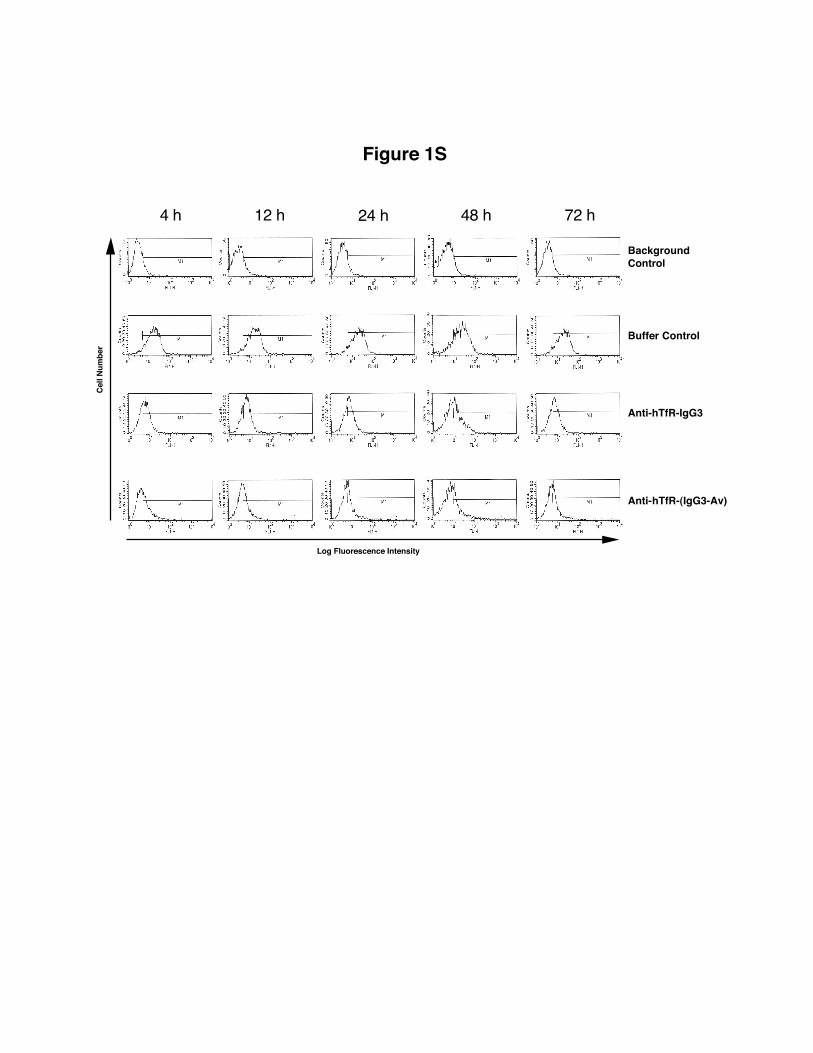
Figure 1S
72 h24 h12 h4 h 48 h
Cell
Num
ber
Log Fluorescence Intensity
BackgroundControl
Buffer Control
Anti-hTfR-IgG3
Anti-hTfR-(IgG3-Av)
Related Documents


![IgG3:F(6)A2 IgG3:F(6)A2[6]G1 - Waters Corporation...For derivatized IgG3:F(6)A2, the predominant ion was the 3+ and significant levels of 4+ were observed and only observed for the](https://static.cupdf.com/doc/110x72/6038bdcf51e1d909ba727104/igg3f6a2-igg3f6a26g1-waters-corporation-for-derivatized-igg3f6a2.jpg)
