Molecular Etiology of Atherogenesis – In Vitro Induction of Lipidosis in Macrophages with a New LDL Model Luis M. B. B. Estronca 1 , Joao C. P. Silva 1 , Julio L. Sampaio 2 , Andrej Shevchenko 2 , Paul Verkade 3 , Alfin D. N. Vaz 4 , Winchil L. C. Vaz 5 , Otilia V. Vieira 1 * 1 Center for Neuroscience and Cell Biology, University of Coimbra, Largo Marque ˆs de Pombal, Coimbra, Portugal, 2 Max-Planck Institute for Molecular Cell Biology and Genetics. Pfotenhauerstrasse, Dresden, Germany, 3 Schools of Biochemistry, and Physiology and Pharmacology, Medical Sciences, University of Bristol, Bristol, United Kingdom, 4 Pharmacokinetics, Dynamics & Metabolism, Pfizer Global Research and Development, Groton, Connecticut, United States of America, 5 Department of Chemistry, University of Coimbra, Coimbra, Portugal Abstract Background: Atherosclerosis starts by lipid accumulation in the arterial intima and progresses into a chronic vascular inflammatory disease. A major atherogenic process is the formation of lipid-loaded macrophages in which a breakdown of the endolysomal pathway results in irreversible accumulation of cargo in the late endocytic compartments with a phenotype similar to several forms of lipidosis. Macrophages exposed to oxidized LDL exihibit this phenomenon in vitro and manifest an impaired degradation of internalized lipids and enhanced inflammatory stimulation. Identification of the specific chemical component(s) causing this phenotype has been elusive because of the chemical complexity of oxidized LDL. Methodology/Principal Findings: Lipid ‘‘core aldehydes’’ are formed in oxidized LDL and exist in atherosclerotic plaques. These aldehydes are slowly oxidized in situ and (much faster) by intracellular aldehyde oxidizing systems to cholesteryl hemiesters. We show that a single cholesteryl hemiester incorporated into native, non-oxidized LDL induces a lipidosis phenotype with subsequent cell death in macrophages. Internalization of the cholesteryl hemiester via the native LDL vehicle induced lipid accumulation in a time- and concentration-dependent manner in ‘‘frozen’’ endolysosomes. Quantitative shotgun lipidomics analysis showed that internalized lipid in cholesteryl hemiester-intoxicated cells remained largely unprocessed in those lipid-rich organelles. Conclusions/Significance: The principle elucidated with the present cholesteryl hemiester-containing native-LDL model, extended to other molecular components of oxidized LDL, will help in defining the molecular etiology and etiological hierarchy of atherogenic agents. Citation: Estronca LMBB, Silva JCP, Sampaio JL, Shevchenko A, Verkade P, et al. (2012) Molecular Etiology of Atherogenesis – In Vitro Induction of Lipidosis in Macrophages with a New LDL Model. PLoS ONE 7(4): e34822. doi:10.1371/journal.pone.0034822 Editor: Sally Martin, The University of Queensland, Australia Received October 5, 2011; Accepted March 6, 2012; Published April 13, 2012 Copyright: ß 2012 Estronca et al. This is an open-access article distributed under the terms of the Creative Commons Attribution License, which permits unrestricted use, distribution, and reproduction in any medium, provided the original author and source are credited. Funding: This work was supported by the Research grant PTDC/SAU/MII/66285/2006 and PTDC/BIA-BCM/112138/2009 from the Foundation for Science and Technology of the Portuguese Ministry of Science and Higher Education (FCT). LE is a holder of postdoctoral fellowships from the FCT (Ref.: SFRH/BPD/26843/ 2006). Lipidomics analysis performed in the AS laboratory was supported by a TRR 83 grant from the Deutsche Forschungsgemeinschaft and Virtual Liver grant (Code/0315757) from the Bundesministerium fu ¨ r Bildung und Forschung. The funders had no role in study design, data collection and analysis, decision to publish, or preparation of the manuscript. Competing Interests: AV is an employee of Pfizer Global Research and Development. There are no patents, products in development or marketed products to declare. This does not alter the authors’ adherence to all the PLoS ONE policies on sharing data and materials, as detailed online in the guide for authors. * E-mail: [email protected] Introduction Atherogenesis is a slow progressive process characterized by a complex sequence of events. It is initiated by subendothelial retention and subsequent processing of low density lipoproteins (LDL) in the arterial intima leading to endothelial activation, trans- endothelial migration of monocytes, their conversion to macro- phages, lipid accumulation in the macrophages and arterial smooth muscle cells, their subsequent apoptosis, and defective clearance of the apoptotic debris. A ‘‘fatty streak’’ that reflects deposits of lipid in the intima is the first visible sign of this pathology (reviewed in [1–5]). Normally, LDL endocytosed by cells via the LDL-receptor are delivered to lysosomes where their cholesteryl esters are hydro- lyzed by acid hydrolases to free cholesterol that is then either exported from the cell or re-esterified in the endoplasmic reticulum and stored in the cytosol in lipid storage droplets [1]. Modified LDL are internalized by macrophages via a large number of receptors that are not particularly specific to LDL [6], or even via macropinocytosis [7]. It is the post-internalization processing of LDL by sub-endothelial cells (including macrophages and smooth muscle cells) that determines whether the ingested material is used and/or re-cycled, or becomes toxic to these cells and elicits a sequence of signaling events and, ultimately, cell death. Cells that take up large amounts of modified LDL particles develop the microscopic appearance that has come to be known as ‘‘foam cells’’ [8]. However, foam cells, as they appear in light microscopy, may be of different types 2 one in which the rapidly internalized lipid is duly processed and stored in lipid droplets for later use, another in which the lipid accumulates irreversibly in PLoS ONE | www.plosone.org 1 April 2012 | Volume 7 | Issue 4 | e34822

Welcome message from author
This document is posted to help you gain knowledge. Please leave a comment to let me know what you think about it! Share it to your friends and learn new things together.
Transcript
Molecular Etiology of Atherogenesis – In Vitro Inductionof Lipidosis in Macrophages with a New LDL ModelLuis M. B. B. Estronca1, Joao C. P. Silva1, Julio L. Sampaio2, Andrej Shevchenko2, Paul Verkade3,
Alfin D. N. Vaz4, Winchil L. C. Vaz5, Otilia V. Vieira1*
1 Center for Neuroscience and Cell Biology, University of Coimbra, Largo Marques de Pombal, Coimbra, Portugal, 2 Max-Planck Institute for Molecular Cell Biology and
Genetics. Pfotenhauerstrasse, Dresden, Germany, 3 Schools of Biochemistry, and Physiology and Pharmacology, Medical Sciences, University of Bristol, Bristol, United
Kingdom, 4 Pharmacokinetics, Dynamics & Metabolism, Pfizer Global Research and Development, Groton, Connecticut, United States of America, 5 Department of
Chemistry, University of Coimbra, Coimbra, Portugal
Abstract
Background: Atherosclerosis starts by lipid accumulation in the arterial intima and progresses into a chronic vascularinflammatory disease. A major atherogenic process is the formation of lipid-loaded macrophages in which a breakdown ofthe endolysomal pathway results in irreversible accumulation of cargo in the late endocytic compartments with a phenotypesimilar to several forms of lipidosis. Macrophages exposed to oxidized LDL exihibit this phenomenon in vitro and manifestan impaired degradation of internalized lipids and enhanced inflammatory stimulation. Identification of the specificchemical component(s) causing this phenotype has been elusive because of the chemical complexity of oxidized LDL.
Methodology/Principal Findings: Lipid ‘‘core aldehydes’’ are formed in oxidized LDL and exist in atherosclerotic plaques.These aldehydes are slowly oxidized in situ and (much faster) by intracellular aldehyde oxidizing systems to cholesterylhemiesters. We show that a single cholesteryl hemiester incorporated into native, non-oxidized LDL induces a lipidosisphenotype with subsequent cell death in macrophages. Internalization of the cholesteryl hemiester via the native LDLvehicle induced lipid accumulation in a time- and concentration-dependent manner in ‘‘frozen’’ endolysosomes.Quantitative shotgun lipidomics analysis showed that internalized lipid in cholesteryl hemiester-intoxicated cells remainedlargely unprocessed in those lipid-rich organelles.
Conclusions/Significance: The principle elucidated with the present cholesteryl hemiester-containing native-LDL model,extended to other molecular components of oxidized LDL, will help in defining the molecular etiology and etiologicalhierarchy of atherogenic agents.
Citation: Estronca LMBB, Silva JCP, Sampaio JL, Shevchenko A, Verkade P, et al. (2012) Molecular Etiology of Atherogenesis – In Vitro Induction of Lipidosis inMacrophages with a New LDL Model. PLoS ONE 7(4): e34822. doi:10.1371/journal.pone.0034822
Editor: Sally Martin, The University of Queensland, Australia
Received October 5, 2011; Accepted March 6, 2012; Published April 13, 2012
Copyright: � 2012 Estronca et al. This is an open-access article distributed under the terms of the Creative Commons Attribution License, which permitsunrestricted use, distribution, and reproduction in any medium, provided the original author and source are credited.
Funding: This work was supported by the Research grant PTDC/SAU/MII/66285/2006 and PTDC/BIA-BCM/112138/2009 from the Foundation for Science andTechnology of the Portuguese Ministry of Science and Higher Education (FCT). LE is a holder of postdoctoral fellowships from the FCT (Ref.: SFRH/BPD/26843/2006). Lipidomics analysis performed in the AS laboratory was supported by a TRR 83 grant from the Deutsche Forschungsgemeinschaft and Virtual Liver grant(Code/0315757) from the Bundesministerium fur Bildung und Forschung. The funders had no role in study design, data collection and analysis, decision topublish, or preparation of the manuscript.
Competing Interests: AV is an employee of Pfizer Global Research and Development. There are no patents, products in development or marketed products todeclare. This does not alter the authors’ adherence to all the PLoS ONE policies on sharing data and materials, as detailed online in the guide for authors.
* E-mail: [email protected]
Introduction
Atherogenesis is a slow progressive process characterized by
a complex sequence of events. It is initiated by subendothelial
retention and subsequent processing of low density lipoproteins
(LDL) in the arterial intima leading to endothelial activation, trans-
endothelial migration of monocytes, their conversion to macro-
phages, lipid accumulation in the macrophages and arterial
smooth muscle cells, their subsequent apoptosis, and defective
clearance of the apoptotic debris. A ‘‘fatty streak’’ that reflects
deposits of lipid in the intima is the first visible sign of this pathology
(reviewed in [1–5]).
Normally, LDL endocytosed by cells via the LDL-receptor are
delivered to lysosomes where their cholesteryl esters are hydro-
lyzed by acid hydrolases to free cholesterol that is then either
exported from the cell or re-esterified in the endoplasmic
reticulum and stored in the cytosol in lipid storage droplets [1].
Modified LDL are internalized by macrophages via a large
number of receptors that are not particularly specific to LDL [6],
or even via macropinocytosis [7]. It is the post-internalization
processing of LDL by sub-endothelial cells (including macrophages
and smooth muscle cells) that determines whether the ingested
material is used and/or re-cycled, or becomes toxic to these cells
and elicits a sequence of signaling events and, ultimately, cell
death. Cells that take up large amounts of modified LDL particles
develop the microscopic appearance that has come to be known as
‘‘foam cells’’ [8]. However, foam cells, as they appear in light
microscopy, may be of different types 2 one in which the rapidly
internalized lipid is duly processed and stored in lipid droplets for
later use, another in which the lipid accumulates irreversibly in
PLoS ONE | www.plosone.org 1 April 2012 | Volume 7 | Issue 4 | e34822

a ‘‘frozen’’ endolysosomal compartment due to impairment of
some activity that is vital for further processing. The latter type of
lipid engorged endolysosomes, also manifested in several lipid-
storage disorders, are pathological, lead to cell death, and are
a hallmark of atherogenesis (reviewed in [9]).
The ‘‘oxidative-modification of LDL’’ hypothesis [10], one of
the oldest and, arguably, most tested hypothesis for the etiology of
atherogenesis, has been reviewed from several perspectives over
the past 25 years [2,9–17]. Oxidized LDL (Ox-LDL) exist in vivo in
the artery wall and stimulate endothelial cells to produce pro-
inflammatory molecules that recruit monocytes and promote their
differentiation to macrophages. The macrophages then internalize
the Ox-LDL, are unable to metabolically handle it, and begin to
accumulate ingested cargo irreversibly in the endolysosomal
compartment in a manner similar to that seen in lipid storage
diseases [9]. Other hypotheses for the chemical etiology of
atherogenesis have been proposed [3,5,18], but do not necessarily
invalidate the idea that oxidative modifications of subendothelial-
trapped LDL may be simultaneously at work.
Ox-LDL contain a myriad of lipid oxidation products with
biological activity. Cholestryl linoleate and cholesteryl arachido-
nate, the predominant cholesteryl esters with more than one
double bond in LDL, produce several aldehydes upon oxidation:
some (such as 4-hydroxynon-2-enal and malondialdehyde) are
quite water-soluble while others are less polar and have variable
degrees of partitioning between lipid and aqueous phases [19].
The latter group of aldehyde products most of which have
cholesterol or derivatives of cholesterol as part of their structure,
have come to be known as ‘‘core-aldehydes’’, are present in
atherosclerotic lesions and resist hydrolysis upon internalization by
macrophages [20,21]. Being amphiphilic molecules, they may be
expected to partition between the lipid environment in which they
are formed, cell membranes, the (intra- and extracellular) aqueous
phase, and translocate readily across cellular membranes. With
time, in an oxidizing environment, or via the action of intracellular
aldehyde oxidizing systems, they will be oxidized to stable
cholesteryl hemiesters of the corresponding bi-acids as shown in
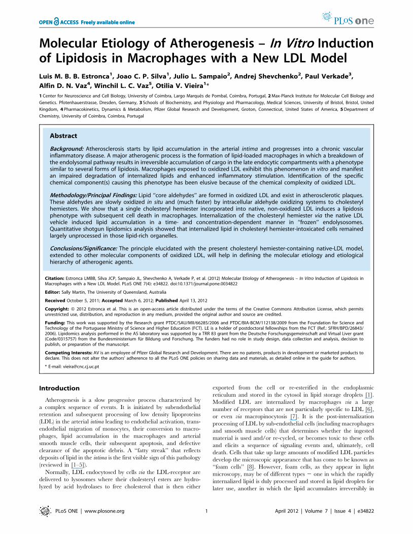
Figure 1A. Cholesteryl-hemiesters have been identified in vitro as
components of Ox-LDL [22], and are ligands for b2-glycoprotein
1, a major antigen for antiphospholipid antibodies present in
patients with antiphospholipid syndrome in which high serum
levels of the complexes of Ox-LDL with b2-glycoprotein 1 are
associated with arterial thrombosis [23].
Several experimental models of Ox-LDL have been used to
examine the molecular mechanisms of the phenomenology
described above. The physiological relevance of methods used in
vitro to oxidize LDL is debatable, and the complexity of the
product with regard to the oxidized components makes these
models of Ox-LDL difficult to work with. This is particularly
applicable to comparisons of studies from different laboratories
since the detailed chemical composition of Ox-LDL is almost
never reported and preparations isolated from tissues vary greatly
among laboratories (reviewed in [17]). The large number of
potentially bioactive products of lipid peroxidation in Ox-LDL
makes it practically impossible to associate a specific biological
response with a specific chemical component of the Ox-LDL
models. To circumvent this problem we have developed an LDL
model in which native LDL (Nat-LDL) are enriched in a single
chemical species that is one of the stable end products of oxidation
of native cholesteryl esters. We have focused on the stable
hemiesters of cholesterol that are expected to be formed from the
cholesteryl ester aldehydes that result from the peroxidation of
cholesteryl esters (see Figure 1A). This group of compounds has an
unmodified cholesterol moiety esterified to one of the carboxylic
acid groups of short chain aliphatic dicarboxylic acids (mostly
azelaic acid) and, to our knowledge, their potential atherogenicity
has been largely ignored in the literature. Under physiological
conditions the negative charge of the cholesteryl hemiesters
increases their potential to partition into the aqueous phase while
the cholesteryl moiety favors their partitioning into lipid phases
(including all types of cell membranes). When formed in Ox-LDL
cholesteryl hemiesters may be expected to accumulate at the polar
surface of the LDL particles, to which they would impart a negative
surface charge, and rapidly partition via passive diffusion and
trans-membrane translocation into all membranes and the cytosol
of neighbouring cells or cells that have internalized Ox-LDL.
We began by assessing the effect of LDL charge, resulting from
the incorporation of a cholesteryl hemiester, cholesteryl hemi-
succinate (Chs), in Nat-LDL. Specifically, we examined the
involvement of Chs in the formation of a lipidosis phenotype
and in cell death when macrophages were exposed to Chs-loaded
Nat-LDL (Chs-LDL). We show that Chs is sufficient to induce
a lipidosis phenotype and apoptotic cell death, two characteristic
features of Ox-LDL. We also show that Chs-LDL lead mainly to
lipid over-accumulation in endolysosomal structures, not lipid-
storage organelles. Our results suggest that in vitro cholesteryl
hemiesters alone are able to induce lipidosis, typically seen in lipid
storage pathologies and atherosclerosis, and that this process may
also have relevance in vivo. We propose that this model can be used
to systematically screen the effects of various other components of
Ox-LDL and establish clear hierarchies in the molecular etiology
of atherogenic lipidosis.
Results
Incorporation of Chs into LDL generates negativelycharged particles
Several in vitro experimental Ox-LDL models have been used to
examine the molecular etiology of atherogenic lipidosis in
macrophages and, consequently, elucidate the origin of athero-
genesis. However, the multitude of oxidation products in Ox-LDL
makes it difficult to establish etiological hierarchies. To avoid this
complication and study the biological effects of a specific family of
compounds that may be formed upon oxidation of LDL, we
generated a new model of LDL enriched specifically with Chs as
a model cholesteryl hemiester. Cholesteryl-4-oxobutyrate, a pre-
cursor of Chs, is one of the ‘‘core aldehyde’’ products of the
peroxidation of LDL [20]. Cholesteryl hemiesters are known to be
formed in vitro under conditions of oxidative stress, and have been
identified as ligands for the plasma protein b2GPI [23]. Elevated
levels of Ox-LDL/b2GPI complexes in vivo seem to be correlated
with acute coronary syndromes [24,25]. Chs has all the
fundamental physico-chemical properties (amphiphilicity, charge,
apolar structure and potential to partition into and translocate
across membranes) of the cholesteryl hemiesters expected to be
formed from cholesteryl esters in LDL (Figure 1A and [20,26]).
Thus, we addressed the involvement of Chs-LDL in the induction
of lipidosis and posterior death in macrophages.
Chs-LDL were generated by incubating Nat-LDL with Chs-
containing POPC liposomes (with a Chs/POPC molar ratio of
70:30) at different LDL:liposome ratios. After incubation, the LDL
particles were re-isolated by ultracentrifugation (see Methods) to
eliminate unincorporated Chs and liposomes. Chs incorporation
into the LDL was assessed by measuring radioactivity of the LDL
particles after incubation with liposomes containing 3H-Chs and
by assessing the LDL charge by electrophoretic mobility in an
agarose gel. Figure 1B shows the electrophoretic mobility of Nat-
LDL (lane 1), acetylated LDL (Ac-LDL) (lane 2), and LDL
Cholesteryl Hemiesters in Atherogenesis
PLoS ONE | www.plosone.org 2 April 2012 | Volume 7 | Issue 4 | e34822
Cholesteryl Hemiesters in Atherogenesis
PLoS ONE | www.plosone.org 3 April 2012 | Volume 7 | Issue 4 | e34822

enriched in Chs at different Chs/LDL molar ratios (250:1-lane 3;
500:1-lane 4 and 1000:1-lane 5). The incorporation of Chs into
LDL gives these particles a more negative surface charge when
compared with Nat-LDL. However, the electrophoretic mobility
of Ac-LDL is higher than that of Chs-LDL even at a Chs/LDL
ratio of 1000:1. The possibility that spontaneous oxidation of LDL
and consequent alterations in LDL charge might occur during Chs
loading was confirmed by treating Nat-LDL similarly, but
excluding Chs. These control LDL were shown to have an
unchanged electrophoretic mobility. Thus, the LDL were neither
significantly oxidized nor aggregated during Chs-loading.
Since this work is an attempt to present a new approach, and to
establish a proof of concept, to study the molecular etiology of
lipidosis in vitro, it must be emphasized here that Chs-LDL particles
are only a model for cholesteryl hemiester-containing LDL
without any other alteration of the chemical constitution of the
particles. We emphasize here that cholesteryl hemiesters have
been largely ignored in the literature as potential causes of
atherogenesis.
Nat-LDL enriched in Chs induce lipid accumulation andcell death
Macrophages and other sub-endothelial cells suffering from
lipidosis are a defining characteristic of atherosclerotic plaques and
are believed to be one of the first stages in atherogenesis. Ox-LDL
are known to induce lipidosis in macrophages and subsequently
cause apoptotic cell death due to uncontrolled uptake of lipid-rich
particles combined with putative blockage of any one (or more) of
several lipid processing reactions. Qualitatively, the process is
morphologically similar to phenotypes seen in several lipid-storage
pathologies [9]. In order to assess the ability of the Chs-LDL
model to mimic this feature of Ox-LDL, RAW264.7 macrophages
(referred to as RAW cells hereafter) were incubated with
increasing amounts of Chs-LDL for 24 or 48 h and then fixed,
stained with Oil-Red O or Bodipy, dyes that stain neutral lipid
deposits in cells, and analyzed by confocal microscopy. Lipid
loaded RAW cells resulted when these were exposed to Chs-LDL
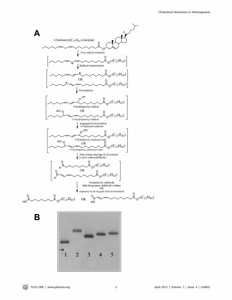
with a Chs/LDL ratio of 1000:1 for 48 h (Figure 2C), the lower
Chs:LDL ratios did not produce any significant intracellular lipid
accumulation. Therefore, only Chs-LDL (1000:1) was used in
further experiments. Since Ac-LDL are also known to rapidly
produce lipid engorged cells [27], although the lipid is accumu-
lated in storage organelles (lipid droplets) in this case and is,
therefore, reversible [28], RAW cells were exposed to Ac-LDL as
a positive control (Figure 2A), Nat-LDL as a negative control
(Figure 2B), and Chs-POPC liposomes at a molar ratio 45:55
(these liposomes have the same number of Chs molecules per
particle as Chs-LDL (1000:1)). After 48 h incubation with Chs-
POPC liposomes RAW cells also exhibited lipid accumulation
(Figure 2D) but less exuberantly than Chs-LDL. Since Nat-LDL in
the absence of serum always induce a massive lipid accumulation
in RAW cells, all experiments were carried out in the presence of
serum.
The effect of Ac-LDL, Chs-LDL and Nat-LDL concentration
on total volume of lipid deposits per cell as a function of time is
shown in Figure 2E–F. Lipid accumulation was faster in
macrophages incubated with Ac-LDL (lipid droplets were visible
after 24 h incubation with any concentration, expressed as mg
LDL apoprotein/ml, of Ac-LDL tested) than with any of the other
LDL models and showed saturation behavior as expected for
saturable receptor-mediated uptake [9] (Figure 2E, red curve). In
contrast, exposure to Chs-LDL resulted in a continuous nonlinear
non-saturating increase in neutral lipids inside the cell at
concentrations between 100 and 400 mg/ml (see Figure 2E–F,
green curves), the latter being the highest concentration tested.
The results obtained with Chs-LDL at 400 mg/ml assume
particular significance when it is considered that LDL concentra-
tions in the arterial intima range from about 0.7 to 2.7 mg/ml
[29,30]. RAW cells incubated with Chs-POPC liposomes showed
similar but much slower non-saturable lipid accumulation with
increasing Chs concentration (expressed as mmol/liter of Chs in
the incubation medium) (Figure 2F, purple curve) that became
noticeable only upon incubation for 48 h, but reached a level that
was only 42% of that observed upon incubation with Chs-LDL.
Lipid accumulation in cells incubated with Nat-LDL was
negligibly small (Figure 2E–F, blue curve). Clearly, the kinetics
and, therefore, the mechanism of intracellular lipid accumulation
into macrophages is LDL-model dependent.
Another striking difference between the lipid-rich structures in
macrophages incubated with Ac-LDL and Chs-LDL was their
average cross sectional areas – 0.8 mm2 and 3 mm2 in cells
incubated with 400 mg/ml of Ac-LDL and Chs-LDL, respectively.
From confocal microscopy images, Figure 2, it was clear that
Chs-LDL also induced massive apoptotic cell death (visualized by
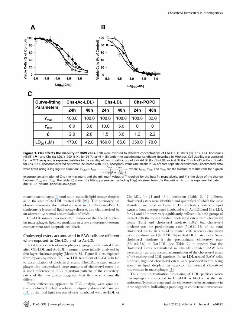
DAPI staining). Toxicity was measured by the MTT test, 24
(Figure 3A) and 48 h (Figure 3B) post-exposure to Chs-(Ac-LDL)
(1000:1 molar ratio), Chs-LDL (1000:1 molar ratio), and Chs-
POPC (45:55 molar ratio) liposomes. The MTT test is based on
the reduction of MTT to a formazan by intracellular metabolically
derived reducing equivalents. Under our experimental conditions
Ac-LDL, Nat-LDL, and POPC liposomes were not toxic towards
RAW cells and were used as controls for Chs-(Ac-LDL), Chs-LDL
and Chs-POPC liposomes, respectively. Cell viability is expressed
as a percentage of the viability of control cells. The LD50, Chs
concentration at which cell viability was 50% of the control, was
determined for each exposure time (Figure 3C). Chs was toxic to
RAW cells, the toxicity being dose- and time-dependent but only
weakly vehicle-dependent (Figure 3A–C). These toxicity results are
in agreement with the anti-proliferative and apoptotic activities of
Chs in cancer cells [31].
It is of interest to note that while the theoretical curves fitted to
the data in Figure 3C show that Chs-POPC treatment of cells (for
24 or 48 h) leaves no residual viable cells at ‘‘infinite’’
concentrations of Chs, both Chs-(Ac-LDL) and Chs-LDL
treatment leaves a residual population of between 5 and 10% of
cells alive (as judged by the MTT assay). This, for the present
mysterious, observation may be related to macrophage sub-
populations in which some sub-populations are more susceptible to
acute Chs toxicity and others are not. We are presently
investigating this possibility.
Chs-LDL exposure leads to lysosomal lipid accumulationin RAW cells
Most mammalian cells package neutral lipids into storage
droplets that are surrounded by a monolayer of phospholipids and
a specific set of proteins including the adipose differentiation-
related protein (ADRP or adipophilin) [32]. The lipid structures
formed in RAW cells were characterized by incubating the
Figure 1. Chs increases the negative charge of Nat-LDL. (A) A brief description of the oxidation of cholesteryl linoleate to cholesterylhemiesters; (B) Agarose gel electrophoresis of Nat-LDL, and derivatives. Lane 1, Nat-LDL; Lane 2, Ac-LDL; Lane 3, Chs-LDL (250:1); Lane 4, Chs-LDL(500:1); and Lane 5, Chs-LDL (1000:1).doi:10.1371/journal.pone.0034822.g001
Cholesteryl Hemiesters in Atherogenesis
PLoS ONE | www.plosone.org 4 April 2012 | Volume 7 | Issue 4 | e34822
macrophages with the different LDL models for 48 h, followed by
fixing and staining with Bodipy for neutral lipids and with an
antibody for ADRP. At first sight, the lipid-rich structures in
macrophages incubated with Ac-LDL and Chs-LDL were both
decorated with ADRP (Figure 4A-AIII and B-BIII, respectively).
However, in cells incubated with the Ac-LDL, ADRP was partially
localized in the cytosol (Figure 4A-AIII), whereas in cells incubated
with Chs-LDL (Figure 4B-BIII) ADRP was almost totally localized
around the neutral lipid structures. More detailed differences
between the lipid structures were identified by transmission
electron microscopy. At the ultrastructural level RAW cells
incubated with Chs-LDL were full of large vesicles with electron
dense material inside and surrounded by a lipid bilayer (Figure 5B,
arrows) while the cells incubated with Ac-LDL showed cytosolic
lipid-storage droplets (Figure 5A). Co-localization studies in Chs-
LDL treated RAW cells for 48 h showed that the large Bodipy-
positive vacuoles were decorated on the surface by the Lysosomal
Associated Membrane Protein-2 (LAMP-2), a late endosome/
lysosome marker (see Figure 6B-BIII and the graph with the
fluorescence distribution, Figure 6BIV). This feature was never
observed in macrophages incubated with Ac-LDL (Figure 6A-AIII
and AIV).
Thus, RAW cells exposed to Chs-LDL accumulate neutral
lipids in late endosome/lysosome structures (similar to Ox-LDL
Figure 2. Addition of Chs-LDL to RAW cells induces the formation of large Oil Red-positive organelles. RAW cells were incubated for48 h with Ac-LDL (A), Nat-LDL (B), Chs-LDL (1000:1) (C), all at 400 mg LDL protein/ml; or Chs-POPC liposomes (45:55) (D) at a particle densityequivalent to that of Chs-LDL. After incubation the cells were fixed with PFA and stained with Oil-Red O and DAPI as described under Methods. Redstain, lipid organelles. Blue stain, nuclei. The images are projections of Z-stacks. Bars, 10 mm. Quantitative estimation of the total volume of lipid-richstructures per cell as a function of the Apo-B concentration (lower X-axis scale) or Chs (upper X-axis scale) concentrations after 24 h (E) and 48 h (F)incubations are also shown.doi:10.1371/journal.pone.0034822.g002
Cholesteryl Hemiesters in Atherogenesis
PLoS ONE | www.plosone.org 5 April 2012 | Volume 7 | Issue 4 | e34822
treated macrophages [9]) and not in cytosolic lipid storage droplets
as in the case of Ac-LDL treated cells [28]. The phenotype we
observe resembles the pathology seen in the Niemann-Pick C
syndrome (a lysosomal lipid-storage disease), also characterized by
an aberrant lysosomal accumulation of lipids.
Chs-LDL mimics two important features of the Ox-LDL effect
on macrophages: lipid accumulation in a late endosome/lysosome
compartment and apoptotic cell death.
Cholesteryl esters accumulated in RAW cells are differentwhen exposed to Chs-LDL and to Ac-LDL
Total lipid extracts of macrophages engorged with neutral lipids
after Chs-LDL and Ac-LDL treatment were initially analyzed by
thin layer chromatography (Methods S1, Figure S1). As expected
from reports by others [28], Ac-LDL treatment of RAW cells led
to accumulation of cholesteryl esters. Chs-LDL treated macro-
phages also accumulated large amounts of cholesteryl esters but
a small difference in TLC migration patterns of the cholesteryl
esters of the two groups suggested that they were chemically
different.
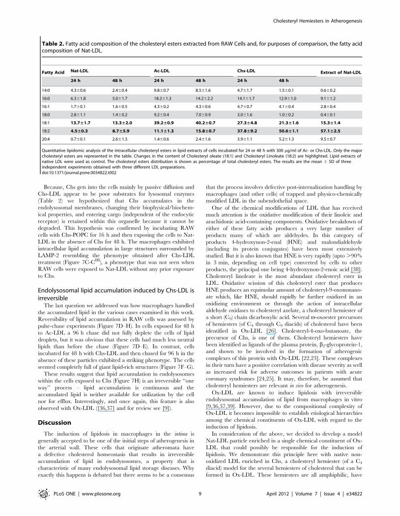
These differences, apparent in TLC analysis, were quantita-
tively confirmed by high resolution shotgun lipidomics MS analysis
[33] of the total lipid extracts of cells incubated with Ac-LDL or
Chs-LDL for 24 and 48 h incubation (Table 1). 17 different
cholesteryl esters were identified and quantified of which the most
abundant are listed in Table 2. The cholesteryl esters of lipid
extracts from macrophages incubated with Ac-LDL and Chs-LDL
for 24 and 48 h were very significantly different. In both groups of
treated cells the most abundant cholesteryl esters were cholesteryl
oleate (18:1) and cholesteryl linoleate (18:2) but cholesteryl
linoleate was the predominant ester (50.661.1% of the total
cholesteryl esters) in Chs-LDL treated cells whereas cholesteryl
oleate predominated (40.260.7%) in Ac-LDL treated cells. Since
cholesteryl linoleate is the predominant cholesteryl ester
(57.162.5%) in Nat-LDL (see Table 2), it appears that the
cholesteryl esters accumulated in Chs-LDL treated RAW cells
were simply an unprocessed accumulation of the cholesteryl esters
of the endocytosed LDL particles. In Ac-LDL treated RAW cells,
however, ingested cholesteryl esters were processed before being
stored in lipid droplets, as expected for normal cholesterol
homeostasis in macrophages [1].
Thus, post-internalization processing of LDL particles when
macrophages are exposed to Chs-LDL is blocked at the late
endosome/lysosome stage and the cholesteryl esters accumulate in
these organelles, indicating a pathology in cholesterol homeostasis.
Figure 3. Chs affects the viability of RAW cells. Cells were exposed to different concentrations of Chs-LDL (1000:1) (O), Chs-POPC liposomes(45:55) (¤), and Chs-(Ac-LDL) (1000:1) (X), for 24 (A) or 48 h (B) under the experimental conditions described in Methods. Cell viability was assessedby the MTT assay and is expressed relative to the viability of control cells exposed to Nat-LDL (for Chs-LDL) or Ac-LDL (for Chs-(Ac-LDL)). Control cellsfor Chs-POPC liposomes treated cells were incubated with POPC liposomes. Values are means 6 SD of three separate experiments. Experimental data
were fitted using a log-logistic equation: Y Chs½ �~YminzYmax{Ymin
1zexp b lnChs½ �
LD50½ �
� �h i, where Y[Chs], Ymax and Ymin are the fraction of viable cells for a given
exposure concentration of Chs, the maximum, and the minimum values of Y required for the best fit, respectively, and b is the slope of the changebetween Ymax and Ymin. The table (C) shows the fitting parameters (including LD50) obtained from the theoretical fits to the experimental data.doi:10.1371/journal.pone.0034822.g003
Cholesteryl Hemiesters in Atherogenesis
PLoS ONE | www.plosone.org 6 April 2012 | Volume 7 | Issue 4 | e34822

Chs is internalized mainly in a vehicle-independentmanner, possibly via passive diffusion
To elucidate the mechanisms of Chs entry, macrophages were
treated for 3 to 12 h with radioactively labeled Chs in Chs-(Ac-
LDL), Chs-LDL, and Chs-POPC liposomes with similar total Chs
concentrations. The intracellular radioactivity was examined as
a function of incubation time (Figure 7A). Surprisingly, Chs
incorporation into the cells was only weakly vector dependent
(only slightly faster for Chs-(Ac-LDL) and Chs-LDL treated cells as
compared with Chs-POPC liposome-treated cells) suggesting that
Chs entered the cells mainly via passive diffusion and trans-
membrane translocation between the Chs-containing particles and
the cells. One scenario for this process could be a rapid
equilibrium of the type:
Chs LDL particle=liposomeð Þ'Chs aqueous phaseð Þ'
Chs cell membraneð Þ'Chs cell interiorð Þ
This equilibrium only becomes possible because Chs is amphi-
philic with an appreciable, albeit low, solubility in the aqueous
phase.
To further clarify this apparent vector-independent Chs uptake,
we examined the effect of blocking scavenger receptors in RAW
cells that are known to play a role in the uptake of modified LDL,
namely, CD36, SRA (CD204) and SR-BI [34]. We compared Chs
uptake by RAW cells in the presence and absence of receptor-
blocking antibodies as described in [35]. Chs uptake was not
affected by any of the scavenger receptor-inhibiting antibodies
when macrophages were exposed to Chs-POPC liposomes or to
Chs-LDL for 3 h (Figure 7B). However, a 22.4% (p,0.05) and
27.9% (p,0.1) reduction of Chs uptake was observed when
macrophages were exposed to Chs-(Ac-LDL) in the presence of
anti-CD204 or SR-BI inhibitory antibodies. This result suggests
that in addition to passive diffusion, endocytosis of Ac-LDL via
CD204 and SR-BI receptors, but not CD36, contributes in a small
measure to Chs internalization via the Ac-LDL vehicle [1]. A
similar mechanism involving some (as yet) unidentified receptor
may also be involved in Chs-LDL treated cells.
Figure 4. ADRP, a marker of lipid droplets, decorates theintracellular lipid structures induced by Chs-LDL. RAW cells wereincubated for 48 h with Ac-LDL (A-AIII) or Chs-LDL (B-BIII). Panels (A)and (B) are confocal single-slice images and show the neutral lipids andthe ADRP distribution. Neutral lipid-rich structures, in green, werestained with Bodipy 493/503. ADRP, in red, was visualized by immuno-staining with polyclonal antibodies as described under Methods. Panels(A) and (B) are merged images. In panels (AI) and (BI) the regionsoutlined with the rectangles in panels (A) and (B), respectively, areenlarged. Panels (AII) and (BII) show Bodipy staining 493/503, panels(AIII) and (BIII) show the distribution of ADRP. Bars, 10 mm.doi:10.1371/journal.pone.0034822.g004
Figure 5. Chs-LDL induces the formation of bilayer vesicles fullof electron dense material. Transmission electron microscopy ofcells treated for 24 h with Ac-LDL (A) or Chs-LDL (B). Cytoplasmic lipiddroplets (organelles with a single monolayer) and vesicular structureswith a bilayer and with electron dense material are visible in cellstreated with Ac-LDL and Chs-LDL, respectively. The asterisk (*) indicatescytoplasmic lipid droplets in (A). Arrows point to bilayer vesicles withelectron dense material inside in (B). Bars, 500 nm.doi:10.1371/journal.pone.0034822.g005
Cholesteryl Hemiesters in Atherogenesis
PLoS ONE | www.plosone.org 7 April 2012 | Volume 7 | Issue 4 | e34822
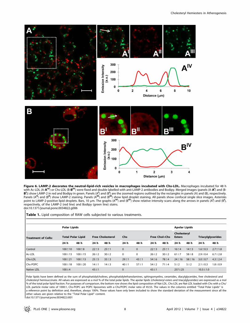
Figure 6. LAMP-2 decorates the neutral-lipid-rich vesicles in macrophages incubated with Chs-LDL. Macrophages incubated for 48 hwith Ac-LDL (A-AIV) or Chs-LDL (B-BIV) were fixed and double labelled with anti-LAMP-2 antibodies and Bodipy. Merged images (panels (A-AI) and (B-BI)) show LAMP-2 in red and Bodipy in green. Panels (AI) and (BI) are the zoomed regions outlined by the rectangles in panels (A) and (B), respectively.Panels (AII) and (BII) show LAMP-2 staining. Panels (AIII) and (BIII) show lipid droplet staining. All panels show confocal single slice images. Asteriskspoint to LAMP-2-positive lipid droplets. Bars, 10 mm. The graphs (AIV) and (BIV) show relative intensity scans along the arrows in panels (AI) and (BI),respectively, of the LAMP-2 (red line) and Bodipy (green line) stains.doi:10.1371/journal.pone.0034822.g006
Table 1. Lipid composition of RAW cells subjected to various treatments.
Polar Lipids Apolar Lipids
Treatment of Cells: Total Polar Lipid Free Cholesterol Chs Free Chol+ChsCholesterylEsters Triacylglycerides
24 h 48 h 24 h 48 h 24 h 48 h 24 h 48 h 24 h 48 h 24 h 48 h
Control 100610 10068 2263 2561 0 0 2263 2561 1664 1463 1.660.3 2.761.8
Ac-LDL 100613 100615 2662 3062 0 0 2662 3062 6167 5868 2.960.4 6.762.8
Chs-LDL 100621 100613 2565 3563 2961 4361 5466 7864 24616 58616 3.060.7 4.362.4
Chs-POPC 100618 100620 1461 1463 4061 5761 5462 7164 562 562 2.160.3 1.860.9
Native LDL 10064 4361 0 4361 257625 10.361.0
Polar lipids have been defined as the sum of phosphatidylcholines, phosphatidylethanolamines, sphingomyelins, ceramides, diacylglycerides, free cholesterol andcholesteryl hemisuccinate. All values are expressed as a mol % of the total polar lipids. The apolar lipids (cholesteryl esters and triacylglycerides) are expressed as a mol% of the total polar lipid fraction. For purposes of comparison, the bottom row shows the lipid composition of Nat-LDL. Chs-LDL are Nat-LDL loaded with Chs with a Chs/LDL particle molar ratio of 1000:1, Chs-POPC are POPC liposomes with a Chs/POPC molar ratio of 45:55. The values in the columns entitled ‘‘Total Polar Lipids’’ isa reference point by definition and, therefore, always 100%. These values have only been included to show the standard deviation of the measurement since all theother values are given relative to the ‘‘Total Polar Lipid’’ content.doi:10.1371/journal.pone.0034822.t001
Cholesteryl Hemiesters in Atherogenesis
PLoS ONE | www.plosone.org 8 April 2012 | Volume 7 | Issue 4 | e34822
Because, Chs gets into the cells mainly by passive diffusion and
Chs-LDL appear to be poor substrates for lysosomal enzymes
(Table 2) we hypothesized that Chs accumulates in the
endolysosomal membranes, changing their biophysical/biochem-
ical properties, and entering cargo (independent of the endocytic
receptor) is retained within this organelle because it cannot be
degraded. This hypothesis was confirmed by incubating RAW
cells with Chs-POPC for 16 h and then exposing the cells to Nat-
LDL in the absence of Chs for 48 h. The macrophages exhibited
intracellular lipid accumulation in large structures surrounded by
LAMP-2 resembling the phenotype obtained after Chs-LDL
treatment (Figure 7C-CIII), a phenotype that was not seen when
RAW cells were exposed to Nat-LDL without any prior exposure
to Chs.
Endolysosomal lipid accumulation induced by Chs-LDL isirreversible
The last question we addressed was how macrophages handled
the accumulated lipid in the various cases examined in this work.
Reversibility of lipid accumulation in RAW cells was assessed by
pulse-chase experiments (Figure 7D–H). In cells exposed for 48 h
to Ac-LDL a 96 h chase did not fully deplete the cells of lipid
droplets, but it was obvious that these cells had much less neutral
lipids than before the chase (Figure 7D–E). In contrast, cells
incubated for 48 h with Chs-LDL and then chased for 96 h in the
absence of these particles exhibited a striking phenotype. The cells
seemed completely full of giant lipid-rich structures (Figure 7F–G).
These results suggest that lipid accumulation in endolysosomes
within the cells exposed to Chs (Figure 7H) is an irreversible ‘‘one
way’’ process – lipid accumulation is continuous and the
accumulated lipid is neither available for utilization by the cell
nor for efflux. Interestingly, and once again, this feature is also
observed with Ox-LDL ([36,37] and for review see [9]).
Discussion
The induction of lipidosis in macrophages in the intima is
generally accepted to be one of the initial steps of atherogenesis in
the arterial wall. These cells that originate atheromata have
a defective cholesterol homeostasis that results in irreversible
accumulation of lipid in endolysosomes, a property that is
characteristic of many endolysosomal lipid storage diseases. Why
exactly this happens is debated but there seems to be a consensus
that the process involves defective post-internalization handling by
macrophages (and other cells) of trapped and physico-chemically
modified LDL in the subendothelial space.
One of the chemical modifications of LDL that has received
much attention is the oxidative modification of their linoleic and
arachidonic acid-containing components. Oxidative breakdown of
either of these fatty acids produces a very large number of
products many of which are aldehydes. In this category of
products 4-hydroxynon-2-enal (HNE) and malondialdehyde
(including its protein conjugates) have been most extensively
studied. But it is also known that HNE is very rapidly (upto .90%
in 3 min, depending on cell type) converted by cells to other
products, the principal one being 4-hydroxynon-2-enoic acid [38].
Cholesteryl linoleate is the most abundant cholesteryl ester in
LDL. Oxidative scission of this cholesteryl ester that produces
HNE produces an equimolar amount of cholesteryl-9-oxononano-
ate which, like HNE, should rapidly be further oxidized in an
oxidizing environment or through the action of intracellular
aldehyde oxidases to cholesteryl azelate, a cholesteryl hemiester of
a short (C9) chain dicarboxylic acid. Several v-oxoester precursors
of hemiesters (of C4 through C9 diacids) of cholesterol have been
identified in Ox-LDL [26]. Cholesteryl-4-oxo-butanoate, the
precursor of Chs, is one of them. Cholesteryl hemiesters have
been identified as ligands of the plasma protein, b2-glycoprotein-1,
and shown to be involved in the formation of atherogenic
complexes of this protein with Ox-LDL [22,23]. These complexes
in their turn have a positive correlation with disease severity as well
as increased risk for adverse outcomes in patients with acute
coronary syndromes [24,25]. It may, therefore, be assumed that
cholesteryl hemiesters are relevant in vivo for atherogenesis.
Ox-LDL are known to induce lipidosis with irreversible
endolysosomal accumulation of lipid from macrophages in vitro
[9,36,37,39]. However, due to the compositional complexity of
Ox-LDL it becomes impossible to establish etiological hierarchies
among the chemical constituents of Ox-LDL with regard to the
induction of lipidosis.
In consideration of the above, we decided to develop a model
Nat-LDL particle enriched in a single chemical constituent of Ox-
LDL that could possibly be responsible for the induction of
lipidosis. We demonstrate this principle here with native non-
oxidized LDL enriched in Chs, a cholesteryl hemiester (of a C4
diacid) model for the several hemiesters of cholesterol that can be
formed in Ox-LDL. These hemiesters are all amphiphilic, have
Table 2. Fatty acid composition of the cholesteryl esters extracted from RAW Cells and, for purposes of comparison, the fatty acidcomposition of Nat-LDL.
Fatty Acid Nat-LDL Ac-LDL Chs-LDL Extract of Nat-LDL
24 h 48 h 24 h 48 h 24 h 48 h
14:0 4.360.6 2.460.4 9.860.7 8.561.6 4.761.7 1.560.1 0.660.2
16:0 6.361.8 5.061.7 18.261.3 14.262.2 14.161.7 12.961.0 9.161.2
16:1 1.760.1 1.660.5 4.360.2 4.360.6 4.760.7 4.160.4 2.860.4
18:0 2.861.1 1.460.2 9.260.4 7.060.9 3.061.6 1.060.2 0.460.1
18:1 13.7±1.7 13.3±2.0 39.2±0.9 40.2±0.7 27.3±4.8 21.3±1.6 15.3±1.4
18:2 4.5±0.3 8.7±5.9 11.1±1.3 15.8±0.7 37.8±9.2 50.6±1.1 57.1±2.5
20:4 0.760.1 2.661.3 1.460.6 2.461.6 3.961.1 5.261.3 9.560.7
Quantitative lipidomic analysis of the intracellular cholesteryl esters in lipid extracts of cells incubated for 24 or 48 h with 300 mg/ml of Ac- or Chs-LDL. Only the majorcholesteryl esters are represented in the table. Changes in the content of Cholesteryl oleate (18:1) and Cholesteryl Linoleate (18:2) are highlighted. Lipid extracts ofnative LDL were used as control. The cholesteryl esters distribution is shown as percentage of total cholesteryl esters. The results are the mean 6 SD of threeindependent experiments obtained with three different LDL preparations.doi:10.1371/journal.pone.0034822.t002
Cholesteryl Hemiesters in Atherogenesis
PLoS ONE | www.plosone.org 9 April 2012 | Volume 7 | Issue 4 | e34822
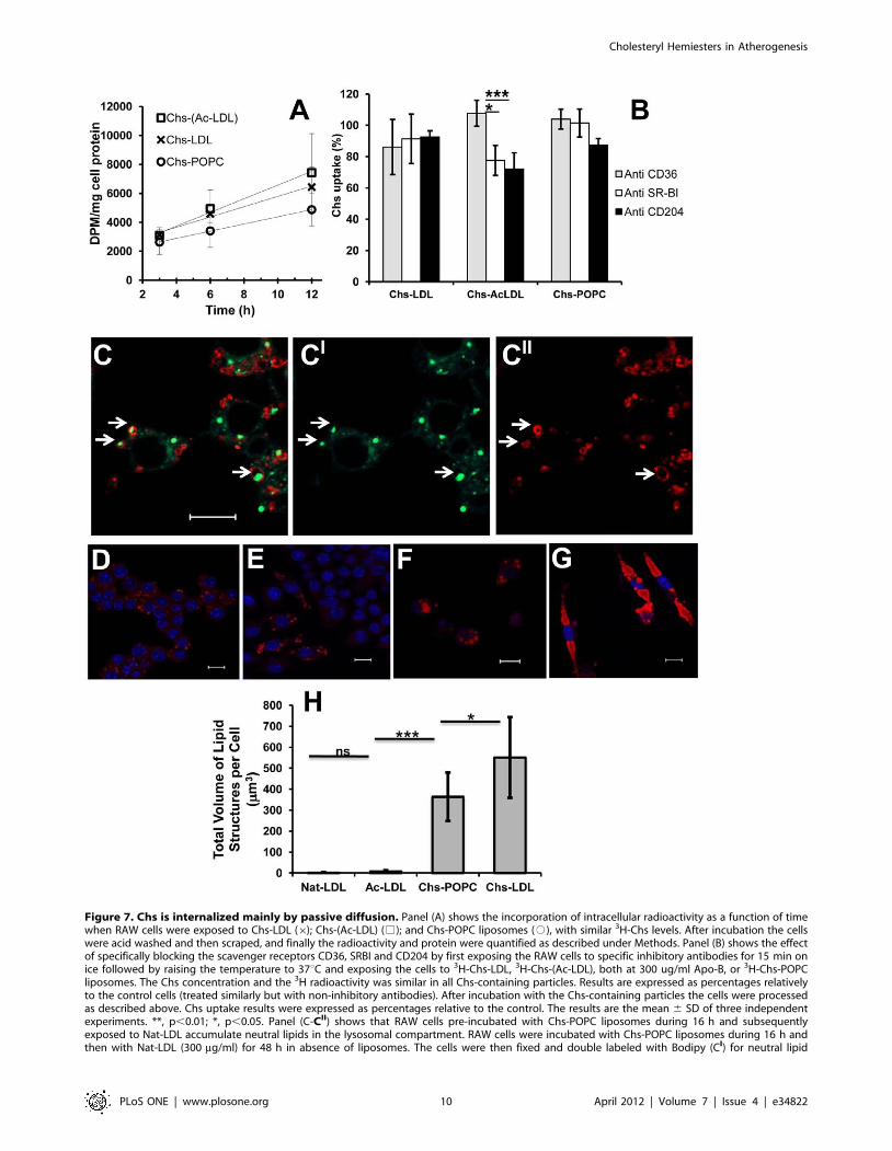
Figure 7. Chs is internalized mainly by passive diffusion. Panel (A) shows the incorporation of intracellular radioactivity as a function of timewhen RAW cells were exposed to Chs-LDL (6); Chs-(Ac-LDL) (%); and Chs-POPC liposomes (#), with similar 3H-Chs levels. After incubation the cellswere acid washed and then scraped, and finally the radioactivity and protein were quantified as described under Methods. Panel (B) shows the effectof specifically blocking the scavenger receptors CD36, SRBI and CD204 by first exposing the RAW cells to specific inhibitory antibodies for 15 min onice followed by raising the temperature to 37uC and exposing the cells to 3H-Chs-LDL, 3H-Chs-(Ac-LDL), both at 300 ug/ml Apo-B, or 3H-Chs-POPCliposomes. The Chs concentration and the 3H radioactivity was similar in all Chs-containing particles. Results are expressed as percentages relativelyto the control cells (treated similarly but with non-inhibitory antibodies). After incubation with the Chs-containing particles the cells were processedas described above. Chs uptake results were expressed as percentages relative to the control. The results are the mean 6 SD of three independentexperiments. **, p,0.01; *, p,0.05. Panel (C-CII) shows that RAW cells pre-incubated with Chs-POPC liposomes during 16 h and subsequentlyexposed to Nat-LDL accumulate neutral lipids in the lysosomal compartment. RAW cells were incubated with Chs-POPC liposomes during 16 h andthen with Nat-LDL (300 mg/ml) for 48 h in absence of liposomes. The cells were then fixed and double labeled with Bodipy (CI) for neutral lipid
Cholesteryl Hemiesters in Atherogenesis
PLoS ONE | www.plosone.org 10 April 2012 | Volume 7 | Issue 4 | e34822
a negative charge at physiological pH and can be expected to
partition favorably into cell membranes; they equilibrate through
passive diffusion between the Ox-LDL (where they are formed)
and the aqueous (extra- and intracellular) phases, as well as all
membranes of neighboring cells. Cholesteryl hemiesters, or for
that matter, their precursor v-oxoacid esters of cholesterol, have
been mostly ignored as atherogenic agents in the literature
although the other scission product of cholesteryl linoleate, HNE,
has received abundant attention. The methodology we describe
here may be profitably used to study the ability of several other
components of Ox-LDL (isolated or in controlled combinations) to
induce lipidosis with irreversible endolysosomal lipid accumulation
in macrophages and other sub-endothelial cells. The model may,
in principle, be useful in studying detailed kinetics of the
phenomenon as well as to study synergistic and/or antagonistic
processes among the chemical constituents of oxidized, or
otherwise modified, LDL. We have previously described strategies
for enriching lipid aggregates (including LDL) with different kinds
of amphiphiles [40,41].
The effects of Chs on RAW cells may be summarized as follows:
1) Formation of lipid loaded cells with lipid accumulation in the
endolysosomal compartment; 2) Incapacity of affected cells to
metabolize (hydrolyze and re-esterify) internalized cholesteryl
esters; 3) Irreversible and uncontrolled lipid accumulation in the
endolysosomal compartment; and 4) Apoptotic cell death.
We have no data that informs us as to exactly why Chs has these
effects. The reasons may be complex but we shall speculate on this
theme. We have shown that the internalization of Chs is largely
a vehicle-independent process, probably occurring mostly via
passive diffusion and equilibration between the LDL particle and
its immediate environment (including the aqueous phase and all
neighboring cells). Chs is an amphiphile which, due to its
cholesterol moiety, partitions very favorably into membranes.
The simplest effect that can be imagined is an increase in
membrane order, similar to the effect of cholesterol [18,42].
Changes in membrane order are known to affect enzyme activity
including the activity of the lysosomal H+-ATPase which is
responsible for acidification of lysosomes [43]. In principle, at least
two membrane loci can be imagined where incorporation of Chs
would cause perturbations of membrane physiology – the
endosome/lysosome membrane and the membrane of the
endoplasmic reticulum. In the former, changes in the pH of the
endolysosomal compartment and consequent modulation of
hydrolytic enzyme activities may be expected. The endoplasmic
reticulum is the locus of Acyl-CoA-cholesterol acyltransferase
(ACAT) activity and inhibition of this enzyme (assuming that
cholesteryl esters continue to be hydrolyzed in the endolysosomes)
would result in a continuously increasing intracellular free
cholesterol concentration with a consequent stiffening of all
cellular membranes including that of lysosomes so that, eventually,
this compartment would also malfunction. Our results (Table 2)
indicate that the internalized cholesteryl linoleate in cells exposed
to Chs-LDL is not hydrolyzed in the endolysosomal compartment
where it accumulates, whereas those exposed to Ac-LDL
hydrolyze the internalized cholesteryl esters, re-esterify the
cholesterol to cholesteryl oleate and store it in lipid droplets that
can be depleted in the normal processes of cholesterol homeostasis.
Also, the total (free cholesterol+Chs) content of Chs-treated cells is
more than twice the free cholesterol content of control cells or cells
treated with Ac-LDL (Table 1).
Clearly the effects observed by us are not simply due to
ingestion of a large amount of cholesteryl esters by the cell – the
cholesteryl esters of Ac-LDL, which are internalized more rapidly
than Chs-LDL, are processed normally and do not create
a pathological state. The pathological state is caused by the Chs
in Chs-LDL. Our results (Table S1) indicate that Chs is very slowly
(if at all) hydrolyzed in the RAW cells. It, therefore, accumulates
with time and the cells’ incapacity to handle the accumulated Chs
is probably responsible for the cell damage observed here. We are
further investigating the details of this process. Artificially
increased levels of cellular free cholesterol are known to cause
lipidosis in macrophages [44,45]. We propose that cholesteryl
hemiester accumulation within cells in the vicinity of Ox-LDL (or
by cells that internalize Ox-LDL) in the arterial intima may be at
least one of the processes that is responsible for atherogenesis.
We recognize that the Chs concentrations to which the RAW
cells were subjected in this work may be much higher than can be
accounted for under physiological conditions. However, the
spontaneous oxidation of Nat-LDL in cell culture media imposes
limits on exposure times and, therefore, requires higher doses. A
more detailed study of the dose and exposure-time dependence of
the effects we report here may be warranted in the future.
Finally, we note with particular interest, that whereas the large
majority of the RAW cells subjected to Chs exposure died
apoptotically, a small subset, representing between 5 and 10% of
the population, survived and seemed to accumulate lipid
progressively and irreversibly in the endolysosomal compartment.
This observation may be related to macrophage heterogeneity
[43]. If so, it will be of interest to know exactly which subset of
a macrophage population forms atherogenic cells. This could be
one of the keys to eventual prophylactic therapies.
Materials and Methods
Chemicals and antibodiesOil-red, Cholesteryl hemisuccinate, Dextran (mol wt 9,000–
11,000) were obtained from Sigma. PO-PC and cholesterol were
purchased from Avanti Polar Lipids (Alabaster, AL). [3H]-
Colesterol was from GE Healthcare. The other chemicals used
were of analytical grade from local sources.
Rhodamin-phalloidin, Bodipy 493/503 and FITC-Dextran
were purchased from Molecular Probes. DAPI was from Fluka.
The anti-mouse (ABL-93) Lamp-2 antibody was from the
Developmental Studies Hybridoma Bank, (University of Iowa,
Iowa City, IA). Polyclonal guinea-pig anti-ADRP antibody was
from Progen Biotechnik (Heidelberg, DE). Secondary antibodies
were from Molecular Probes or from Jackson Immunoresearch.
Mouse Anti-mouse CD36 (552544) and the negative control
mouse anti-mouse IgA (553476) were purchased from BD
Pharmigen; rat anti-mouse CD204 (MCA1322) and the negative
control rat anti-mouse g2b (MCA1125) were purchased from AbD
Serotec. Rabbit anti-mouse SR-BI (NB400-113) and the negative
staining and anti-LAMP-2 antibody (CII). The merged image (C) shows Bodipy staining in green and LAMP-2 staining in red. Arrows point to LAMP-2-positive cytosolic structures that contain neutral lipids. Bar, 10 mm. Panels (D–G) show that the lysosomal accumulation of neutral lipids induced bypre-treatment of RAW cells with Chs-LDL is irreversible. Raw cells were pulsed with 300 mg/ml of Ac-LDL (panel D) or Chs-LDL (panel F) for 48 h andthen chased for 96 h. Cells pulsed with Ac-LDL (D) and then chased (E). Cells pulsed with Chs-LDL (F) and then chased (G). Lipid-rich structuresvisualized by Oil-red staining (red), DAPI staining (blue). All are merged images. Bars, 10 mm. The total volume of lipid structures per cell, quantifiedafter the 96 h chase, is shown in panel (H). The results are the mean 6 SD of three independent experiments. In every experiment 20 individual cellswere analyzed. ***, p,0.0001; *, p,0.05; ns, not significant.doi:10.1371/journal.pone.0034822.g007
Cholesteryl Hemiesters in Atherogenesis
PLoS ONE | www.plosone.org 11 April 2012 | Volume 7 | Issue 4 | e34822
control rabbit anti-mouse IgG (NB810-56910) were purchased
from Novus Biologicals.
Preparation of liposomesAqueous suspensions of lipids were prepared by mixing POPC
and Chs at the desired ratios in an azeotropic mixture of
chloroform and methanol. The solvent was evaporated by blowing
dry nitrogen over the heated (blowing hot air over the external
surface of the tube) solution and then leaving the residue in
a vacuum desiccator for at least 12 h at 23uC. The dry residue and
the hydration solution (20 mM Hepes, 0.11 M NaCl, 1 mM
EDTA, pH 7.4) with or without Dextran in an amount necessary
to obtain a density of 1.044 g/ml, were preheated in a water bath
at 65uC. The samples were submitted to several cycles of vortex/
incubation/mild sonication at 65uC for at least 1 h. The resulting
multilamellar vesicle (MLV) suspensions were extruded through
two stacked polycarbonate filters (Nucleopore) with a pore
diameter of 0.1 mm using a minimum of 6 passes. During the
extrusion the water-jacketed extruder (Lipex Biomembranes,
Vancouver, British Columbia, Canada) was maintained at 65uC.
Final lipid molar ratios of Chs/POPC were: 0/100, 70/30, and
45/55.
LDL isolation, acetylation and Chs incorporationHuman LDL were isolated using a Beckman L80 ultracentri-
fuge equipped with a 70.1 Ti fixed angle rotor as described
previously [46]. LDL were acetylated by repeated additions of
acetic anhydride as described [47]. Chs-LDL were prepared by
incubating Nat-LDL with Chs-POPC (70:30) liposomes in Hepes
buffer containing dextran (density of 1.044 g/ml) overnight at
different LDL/Chs ratios at RT without stirring. After incubation
the Chs-LDL were re-centrifuged to eliminate the remaining Chs-
POPC liposomes and after dialysis they were passed through
a 0.2 mm filter. Chs content of Chs-LDL was assessed by
measuring the radioactivity of the samples. All stock and working
solutions of different LDL models were stored under N2 at 4uC for
a maximum of 2 weeks.
Agarose gel electrophoresisElectrophoresis of LDL preparations was carried out in 0.5%
agarose gels in barbital buffer, pH 8.6, at a constant voltage of
75 V for 45 min, and LDL were stained with 0.5% Paragon Blue
in 5% acetic acid for 3 min at room temperature. The gel was
destained at room temperature with 5% acetic acid, followed by
20% acetic acid, 30% methanol. In each agarose strip, Nat-LDL
were used as a control.
Cell culture and incubation with the different LDL modelsand Chs-POPC liposomes
RAW 264.7 cells (ATCC) were maintained in DMEM
supplemented with 10% fetal calf serum, 100 U/ml of penicillin
and streptomycin. Cells were grown in a humidified incubator at
37uC under 5% CO2 and used for assays during the exponential
growth phase. For lipid droplet staining RAW cells were grown on
glass-coverslips or in 24 well-plates for 24 h. They were then
incubated with Nat-LDL; Ac-LDL, Chs-LDL, Chs-(Ac-LDL) or
Chs-POPC liposomes for different periods of time and at different
concentrations, as indicated in the figure legends. After incubation
the cells were fixed with PFA.
Immunofluorescence and Confocal microscopyCells were grown on 24-well plates and fixed with 4% PFA for
60 min followed by quenching of the aldehyde groups with glycine
or ammonium chloride and permeabilization with isopropanol
(60%) or saponin (0.1%) for LAMP-2 and ADRP staining,
respectively. Cells were then incubated with the primary
antibodies for 1 h at room temperature, washed, and finally
incubated with the secondary antibodies conjugated with a fluor-
ophore for another 1 h. Antibody dilutions were 1:100 for primary
antibodies and 1:500 for secondary antibodies conjugated with
Cy3 or 1:200 for secondary antibodies conjugated with Cy5 or
Alexa FluorH594. Fixed samples were analyzed by using the LSM
510 META point-scan confocal laser microscope (Zeiss) with a 636oil-immersion objective.
Neutral lipid staining and quantificationNeutral lipids were stained either with Oil-Red O (in 60%
isopropanol, prepared from a stock solution at 0.4% w/v in 100%
isopropanol) for 10 min or with Bodipy 493/503 (diluted 1:1000
in PBS from a saturated ethanolic solution of Bodipy) for 15 min.
DAPI at a final concentration of 30 nM was added for 20 min at
room temperature to visualize nuclei. Quantification of the
number and size of the lipid-rich structures was performed using
ImageJ software.
Electron MicroscopyCells were seeded on glass coverslips and treated the next day.
Cells were fixed in 2.5% glutaraldehyde in cacodylate buffer and
processed for standard Epon embedding. 70 nm sections were
analyzed in a TECNAI12 Biotwin electron microscope equipped
with a bottom-mount 464 k CCD camera (FEI company).
Toxicity evaluation by the MTT testCell viability was measured by the MTT assay as previously
described [48].
Mass SpectrometryCells were incubated in 6-well plates with Nat-LDL, Ac-LDL,
Chs-LDL or Chs-POPC liposomes for 24 and 48 h. After
incubations cells were washed three times with 150 mM
ammonium bicarbonate and harvested from wells by scraping
into 1 ml of ammonium bicarbonate. Protein content of the
suspensions was evaluated and aliquots of the cell lysates or Nat-
LDL suspensions containing 10 and 1 mg of protein, respectively,
were extracted as described previously [49]. For absolute
quantification, internal standards were added to the samples prior
to lipid extraction. Cholesterol was quantified as described by
Sandhoff et al. [50]. Top-down shotgun analysis was performed on
a LTQ Orbitrap XL mass spectrometer (Thermo Fisher Scientific,
Bremen, Germany) equipped with a TriVersa NanoMate robotic
nanoflow ion source (Advion BioSciences, Ithaca, NJ) as described
in [33]. Lipids were identified and quantified by LipidXplorer
software developed in house [51].
Chs uptakeAfter incubating the macrophages with 3H-Chs-LDL, 3H-Chs-
(Ac-LDL), and 3H-Chs-POPC liposomes with similar radioactivity
for different periods of time at 37uC, cell-associated [3H]-Chs was
determined after rinsing macrophages with DMEM with 0.5%
BSA at pH 3.4 for 10 min on ice, three further rinses with PBS,
and a final rinse in distilled water. Macrophages were scraped off
into 1 ml distilled water. Then cell samples were assayed for 3H
radioactivity and for cellular protein.
For the experiments with the inhibitory antibodies the cells were
incubated at 4uC for 15 min with 10 mg/ml of anti-CD36, anti-
SR-BI, anti-CD204 and their respective antibody controls; after
Cholesteryl Hemiesters in Atherogenesis
PLoS ONE | www.plosone.org 12 April 2012 | Volume 7 | Issue 4 | e34822
which 300 mg/ml of 3H-Chs-(Ac-LDL), 3H-Chs-LDL, or 3H-Chs-
POPC liposomes (45:55) with equivalent amounts of Chs were
added to cells. The cells were then shifted to 37uC for 3 h (in
presence of the inhibitory antibodies). After incubation the cells
were treated as described above and the radioactivity and the cell
protein quantified.
Protein assayThe protein content of cell extracts was measured by the
bicinchoninic acid (BCA, Pierce) assay using bovine serum
albumin as a standard.
Statistical analysisResults are expressed as the means 6 standard deviations (S.D.).
Statistical significance was assessed by the Student t-test (two-
tailed). A p value of ,0.05 was considered to be statistically
significant.
Supporting Information
Figure S1 Chs-LDL are poorly degraded within thelysosomal structures. (a) Macrophages were incubated for
24 h with 300 mg/ml of Nat-LDL (lane 1), Ac-LDL (lane 2) or
Chs-LDL (lane 3). In lane 4 lipid extracts of cells incubated with
Chs:POPC liposomes (45:55) were loaded. At the end of the
incubation time the lipids were extracted and resolved by TLC as
in Methods S1. In each lane 60 mg of cell protein was loaded.
(PDF)
Methods S1 Synthesis of 3H-Chs. Lipid extraction andpreparative TLC. Intracellular Chs hydrolysis.
(PDF)
Table S1 Hydrolysis and re-esterification of cholesterolderived from 3H-Chs in RAW cells. RAW cells were treated
for 24 and 48 h with 3H-Chs-POPC liposomes (45:55). The
compounds were separated by TLC prior to measurement of
radioactivity.
(PDF)
Acknowledgments
We thank Graca Raposo, Institut Curie (CNRS), Paris, for some electron
microscopy done in the progress of this work.
Author Contributions
Conceived and designed the experiments: LE AV WV OV. Performed the
experiments: LE JCS JLS PV OV. Analyzed the data: LE JCS JLS PV WV
OV. Contributed reagents/materials/analysis tools: AS. Wrote the paper:
AV WV OV.
References
1. Brown MS, Goldstein JL (1983) Lipoprotein metabolism in the macrophage:
implications for cholesterol deposition in atherosclerosis. Annu Rev Biochem 52:
223–261.
2. Steinberg D, Parthasarathy S, Carew TE, Khoo JC, Witztum JL (1989) Beyond
cholesterol. Modifications of low-density lipoprotein that increase its atheroge-
nicity. New Engl J Med 320: 915–924.
3. Tabas I (1999) Nonoxidative modifications of lipoproteins in atherogenesis.
Annu Rev Nutr 19: 123–139.
4. Lusis AJ (2000) Atherosclerosis. Nature 407: 233–241.
5. Moore KJ, Tabas I (2011) Macrophages in the pathogenesis of atherosclerosis.
Cell 145: 341–355.
6. Hartvigsen K, Chou MY, Hansen LF, Shaw PX, Tsimikas S, et al. (2009) The
role of innate immunity in atherogenesis. J Lipid Res 50 Suppl: S388–S393.
7. Choi SH, Harkewicz R, Lee JH, Boullier A, Almazan F, et al. (2009)
Lipoprotein accumulation in macrophages via toll-like receptor-4-dependent
fluid phase uptake. Circ Res 104: 1355–1363.
8. de Duve C (1974) The participation of lysosomes in the transformation of
smooth muscle cells to foamy cells in the aorta of cholesterol-fed rabbits. Acta
Cardiol Suppl 20: 9–25.
9. Schmitz G, Grandl M (2009) Endolysosomal phospholipidosis and cytosolic lipid
droplet storage and release in macrophages. Biochim Biophys Acta 1791:
524–539.
10. Steinberg D, Witztum JL (2010) Oxidized low-density lipoprotein and
atherosclerosis. Arterioscler Thromb Vasc Biol 30: 2311–2316.
11. Heinecke JW (1997) Mechanisms of oxidative damage of low density lipoprotein
in human atherosclerosis. Curr Opin Lipidol 8: 268–274.
12. Spiteller G (1998) Linoleic acid peroxidation–the dominant lipid peroxidation
process in low density lipoprotein–and its relationship to chronic diseases. Chem
Phys Lipids 95: 105–162.
13. Stocker R, Keaney JF, Jr. (2004) Role of oxidative modifications in
atherosclerosis. Physiol Rev 84: 1381–1478.
14. Jessup W, Kritharides L, Stocker R (2004) Lipid oxidation in atherogenesis: an
overview. Biochem Soc Trans 32: 134–138.
15. Berliner JA, Leitinger N, Tsimikas S (2009) The role of oxidized phospholipids
in atherosclerosis. J Lipid Res 50 Suppl: S207–S212.
16. Steinberg D (2009) The LDL modification hypothesis of atherogenesis: an
update. J Lipid Res 50 Suppl: S376–S381.
17. Levitan I, Volkov S, Subbaiah PV (2010) Oxidized LDL: diversity, patterns of
recognition, and pathophysiology. Antioxid Redox Signal 13: 39–75.
18. Maxfield FR, Tabas I (2005) Role of cholesterol and lipid organization in
disease. Nature 438: 612–621.
19. Esterbauer H, Jurgens G, Quehenberger O, Koller E (1987) Autoxidation of
human low density lipoprotein: loss of polyunsaturated fatty acids and vitamin E
and generation of aldehydes. J Lipid Res 28: 495–509.
20. Kamido H, Kuksis A, Marai L, Myher JJ (1995) Lipid ester-bound aldehydes
among copper-catalyzed peroxidation products of human plasma lipoproteins.
J Lipid Res 36: 1876–1886.
21. Hoppe G, Ravandi A, Herrera D, Kuksis A, Hoff HF (1997) Oxidation products
of cholesteryl linoleate are resistant to hydrolysis in macrophages, formcomplexes with proteins, and are present in human atherosclerotic lesions.
J Lipid Res 38: 1347–1360.
22. Kobayashi K, Matsuura E, Liu Q, Furukawa J, Kaihara K, et al. (2001) A
specific ligand for beta(2)-glycoprotein I mediates autoantibody-dependentuptake of oxidized low density lipoprotein by macrophages. J Lipid Res 42:
697–709.
23. Kobayashi K, Kishi M, Atsumi T, Bertolaccini ML, Makino H, et al. (2003)
Circulating oxidized LDL forms complexes with beta2-glycoprotein I:implication as an atherogenic autoantigen. J Lipid Res 44: 716–726.
24. Greco TP, Conti-Kelly A, Anthony J, Greco T, Doyle R, et al. (2010) Oxidized-LDL/b2-GPI complexes are associated with disease severity and increased risk
for adverse outcomes in patients with acute coronary syndromes. Am J Clin
Pathol 133: 737–743.
25. Ames PRJ, Ortiz-Cadenas A, Garcia-De La Torre I, Nava A, Oregon-
Miranda A, et al. (2010) Rosuvastatin treatment is associated with a decrease ofserum oxidized LDL/b2-GPI complex concentration in type 2 diabetes.
Br J Diabetes Vasc Dis 10: 292–299.
26. Kamido H, Kuksis A, Marai L, Myher JJ (1993) Identification of core aldehydes
among in vitro peroxidation products of cholesteryl esters. Lipids 28: 331–336.
27. Goldstein JL, Ho YK, Basu SK, Brown MS (1979) Binding site on macrophages
that mediates uptake and degradation of acetylated low density lipoprotein,producing massive cholesterol deposition. Proc Natl Acad Sci USA 76: 333–337.
28. Brown MS, Goldstein JL, Krieger M, Ho YK, Anderson RG (1979) Reversibleaccumulation of cholesteryl esters in macrophages incubated with acetylated
lipoproteins. J Cell Biol 82: 597–613.
29. Smith EB, Ashall C (1983) Low-density lipoprotein concentration in interstitial
fluid from human atherosclerotic lesions. Relation to theories of endothelialdamage and lipoprotein binding. Biochim Biophys Acta 754: 249–257.
30. Hoff HF, Gaubatz JW, Gotto AM, Jr. (1978) Apo B concentration in the normalhuman aorta. Biochem Biophys Res Commun 85: 1424–1430.
31. Ishimaru C, Kuriyama I, Shimazaki N, Koiwai O, Sakaguchi K, et al. (2007)Cholesterol hemisuccinate: a selective inhibitor of family X DNA polymerases.
Biochem Biophys Res Commun 354: 619–625.
32. Miura S, Gan JW, Brzostowski J, Parisi MJ, Schultz CJ, et al. (2002) Functional
conservation for lipid storage droplet association among Perilipin, ADRP, andTIP47 (PAT)-related proteins in mammals, Drosophila, and Dictyostelium. J Biol
Chem 277: 32253–32257.
33. Schwudke D, Hannich JT, Surendranath V, Grimard V, Moehring T, et al.
(2007) Top-down lipidomic screens by multivariate analysis of high-resolutionsurvey mass spectra. Anal Chem 79: 4083–4093.
34. Greaves DR, Gordon S (2009) The macrophage scavenger receptor at 30 yearsof age: current knowledge and future challenges. J Lipid Res 50 Suppl:
S282–S286.
35. Guest CB, Hartman ME, O’Connor JC, Chakour KS, Sovari AA, et al. (2007)
Phagocytosis of cholesteryl ester is amplified in diabetic mouse macrophages andis largely mediated by CD36 and SR-A. PLoS One 2: e511.
Cholesteryl Hemiesters in Atherogenesis
PLoS ONE | www.plosone.org 13 April 2012 | Volume 7 | Issue 4 | e34822
36. Yancey PG, Jerome WG (2001) Lysosomal cholesterol derived from mildly
oxidized low density lipoprotein is resistant to efflux. J Lipid Res 42: 317–327.
37. Yancey PG, Miles S, Schwegel J, Jerome WG (2002) Uptake and trafficking of
mildly oxidized LDL and acetylated LDL in THP-1 cells does not explain the
differences in lysosomal metabolism of these two lipoproteins. Microsc
Microanal 8: 81–93.
38. Siems W, Crifo C, Capuozzo E, Uchida K, Grune T, et al. (2010) Metabolism of
4-hydroxy-2-nonenal in human polymorphonuclear leukocytes. Arch Biochem
Biophys 503: 248–252.
39. Dhaliwal BS, Steinbrecher UP (2000) Cholesterol delivered to macrophages by
oxidized low density lipoprotein is sequestered in lysosomes and fails to efflux
normally. J Lipid Res 41: 1658–1665.
40. Estronca LM, Moreno MJ, Vaz WLC (2007) Kinetics and thermodynamics of
the association of dehydroergosterol with lipid bilayer membranes. Biophys J 93:
4244–4253.
41. Estronca LM, Moreno MJ, Laranjinha JA, Almeida LM, Vaz WL (2005)
Kinetics and thermodynamics of lipid amphiphile exchange between lipopro-
teins and albumin in serum. Biophys J 88: 557–565.
42. Simons K, Vaz WL (2004) Model systems, lipid rafts, and cell membranes. Annu
Rev Biophys Biomol Struct 33: 269–295.
43. Wang J, Zhang GJ (2005) Influence of membrane physical state on lysosomal
potassium ion permeability. Cell Biol Int 29: 393–401.
44. Kellner-Weibel G, Yancey PG, Jerome WG, Walser T, Mason RP, et al. (1999)
Crystallization of free cholesterol in model macrophage foam cells. ArteriosclerThromb Vasc Biol 19: 1891–1898.
45. Tabas I (2002) Consequences of cellular cholesterol accumulation: basic
concepts and physiological implications. J Clin Invest 110: 905–911.46. Vieira OV, Laranjinha JA, Madeira VM, Almeida LM (1996) Rapid isolation of
low density lipoproteins in a concentrated fraction free from water-solubleplasma antioxidants. J Lipid Res 37: 2715–2721.
47. Basu SK, Goldstein JL, Anderson GW, Brown MS (1976) Degradation of
cationized low density lipoprotein and regulation of cholesterol metabolism inhomozygous familial hypercholesterolemia fibroblasts. Proc Natl Acad Sci USA
73: 3178–3182.48. Vieira OV, Hartmann DO, Cardoso CM, Oberdoerfer D, Baptista M, et al.
(2008) Surfactants as microbicides and contraceptive agents: a systematic in vitrostudy. PLoS One 3: e2913.
49. Zech T, Ejsing CS, Gaus K, de Wet B, Shevchenko A, et al. (2009)
Accumulation of raft lipids in T-cell plasma membrane domains engaged inTCR signalling. EMBO J 28: 466–476.
50. Sandhoff R, Brugger B, Jeckel D, Lehmann WD, Wieland FT (1999)Determination of cholesterol at the low picomole level by nano-electrospray
ionization tandem mass spectrometry. J Lipid Res 40: 126–132.
51. Herzog R, Schwudke D, Schuhmann K, Sampaio JL, Bornstein SR, et al. (2011)A novel informatics concept for high-throughput shotgun lipidomics based on
the molecular fragmentation query language. Genome Biol 12: R8.
Cholesteryl Hemiesters in Atherogenesis
PLoS ONE | www.plosone.org 14 April 2012 | Volume 7 | Issue 4 | e34822
Related Documents