Review Molecular enzymology of lipoxygenases Igor Ivanov a , Dagmar Heydeck a , Katharina Hofheinz a , Jana Roffeis a , Valerie B. O’Donnell b , Hartmut Kuhn a,⇑ , Matthias Walther a a Institute of Biochemistry, University Medicine Berlin – Charité, Oudenarder Str. 16, D-13347 Berlin, Germany b School of Medicine, Heath Park, Cardiff University, CF14 4XN, UK article info Article history: Received 16 July 2010 and in revised form 19 August 2010 Available online 27 August 2010 Keywords: Eicosanoids Lipid peroxidation Inflammation Cell development Catalysis Knockout mice Radicals abstract Lipoxygenases (LOXs) are lipid peroxidizing enzymes, implicated in the pathogenesis of inflammatory and hyperproliferative diseases, which represent potential targets for pharmacological intervention. Although soybean LOX1 was discovered more than 60 years ago, the structural biology of these enzymes was not studied until the mid 1990s. In 1993 the first crystal structure for a plant LOX was solved and following this protein biochemistry and molecular enzymology became major fields in LOX research. This review focuses on recent developments in molecular enzymology of LOXs and summarizes our current understanding of the structural basis of LOX catalysis. Various hypotheses explaining the reaction spec- ificity of different isoforms are critically reviewed and their pros and cons briefly discussed. Moreover, we summarize the current knowledge of LOX evolution by profiling the existence of LOX-related genomic sequences in the three kingdoms of life. Such sequences are found in eukaryotes and bacteria but not in archaea. Although the biological role of LOXs in lower organisms is far from clear, sequence data sug- gests that this enzyme family might have evolved shortly after the appearance of atmospheric oxygen on earth. Ó 2010 Elsevier Inc. All rights reserved. Introduction Lipoxygenases (LOXs) 1 are non-heme iron-containing dioxygen- ases [1–3] that catalyze the stereo-specific peroxidation of polyun- saturated fatty acids containing at least one 1-cis,4-cis-pentadiene system (Fig. 1). Fatty acid oxygenation by LOXs generally consists of four elementary reactions (hydrogen abstraction, radical rear- rangement, oxygen insertion, peroxy radical reduction), usually pro- ceeding in a sterically controlled manner (Fig. 1). Hydrogen abstraction, which constitutes the rate limiting step [4], follows a quantum-mechanical mechanism [5]. In soybean LOX1, H-atom transfer corresponds to a proton-coupled electron transfer [6,7]. The transferred electron does not localize on the proton, but tunnels directly from the substrate to the ferric iron in a concerted proton tunneling–electron tunneling process [6]. In the transition state the covalently linked Fe–O–H–C bridge lowers the energy barrier and provides an efficient pathway for this tunneling [6]. In most cells, the concentration of free fatty acids is limited and thus, LOX substrates have to be liberated from cellular stores by es- ter lipid hydrolyzing enzymes [8]. However, certain LOX-isoforms are capable of oxygenating polyunsaturated fatty acids that are incorporated in ester lipids located in biomembranes or lipopro- teins [9–11]. The conventional nomenclature classifies animal LOXs with respect to their positional specificity of arachidonic acid oxygenation as 5-LOXs, 8-LOXs, 11-LOXs, 12-LOXs or 15-LOXs. This classification is not optimum since: (i) Arachidonic acid is not a good substrate for many non-mammalian LOXs and with other substrate fatty acids (e.g. linoleic acid) their reaction specificity can be quite different. (ii) Evolutionarily-related LOX-isoforms can exhibit distinct reaction specificities in contrast to those shar- ing a low degree of phylogenetic relatedness. Moreover, the rapidly growing availability of genomic sequences and our inability to pre- dict the reaction specificity of arachidonic acid oxygenation from the primary structure of the enzymes leads to the confusing situa- tion that most LOX-isoforms (for which we only have sequence information) cannot be classified according to a function-based en- zyme nomenclature. Thus, a sequence-based classification proce- dure that considers the phylogenetic relatedness of the enzymes is desirable. Unfortunately, despite numerous attempts, no simple and unifying LOX nomenclature has been introduced that over- comes the above mentioned problems. The lack of comprehensive and straightforward classification criteria makes it difficult for non-expert scientists to follow the current developments in LOX research. 0003-9861/$ - see front matter Ó 2010 Elsevier Inc. All rights reserved. doi:10.1016/j.abb.2010.08.016 ⇑ Corresponding author. Fax: +49 30 450 528905. E-mail address: [email protected] (H. Kuhn). 1 Abbreviation used: LOX(s), lipoxygenase(s); 15-H(p)ETE, (5Z,8Z,11Z,13E)-15- hydro(pero)xyeicosa-5,8,11,13-tetraenoic acid; 12-H(p)ETE, (5Z,8Z,10E,14Z)-12- hydro(pero)xyeicosa-5,8,10,14-tetraenoic acid; 11-H(p)ETE, (5Z,8Z,12E,14Z)-11- hydro(pero)xyeicosa-5,8,12,14-tetraenoic acid; 8-H(p)ETE, (5Z,9E,11Z,14Z)-8- hydro(pero)xyeicosa-5,9,11,14-tetraenoic acid; 5-H(p)ETE, (6E,8Z,11Z,14Z)-5- hydro(pero)xyeicosa-6,8,11,14-tetraenoic acid; 13-H(p)ODE, (9Z,11E)-13- hydro(pero)xyoctadeca-9,11-dienoic acid; SAXS, small angle X-ray scattering. Archives of Biochemistry and Biophysics 503 (2010) 161–174 Contents lists available at ScienceDirect Archives of Biochemistry and Biophysics journal homepage: www.elsevier.com/locate/yabbi

Welcome message from author
This document is posted to help you gain knowledge. Please leave a comment to let me know what you think about it! Share it to your friends and learn new things together.
Transcript
Archives of Biochemistry and Biophysics 503 (2010) 161–174
Contents lists available at ScienceDirect
Archives of Biochemistry and Biophysics
journal homepage: www.elsevier .com/ locate/yabbi
Review
Molecular enzymology of lipoxygenases
Igor Ivanov a, Dagmar Heydeck a, Katharina Hofheinz a, Jana Roffeis a, Valerie B. O’Donnell b,Hartmut Kuhn a,⇑, Matthias Walther a
a Institute of Biochemistry, University Medicine Berlin – Charité, Oudenarder Str. 16, D-13347 Berlin, Germanyb School of Medicine, Heath Park, Cardiff University, CF14 4XN, UK
a r t i c l e i n f o
Article history:Received 16 July 2010and in revised form 19 August 2010Available online 27 August 2010
Keywords:EicosanoidsLipid peroxidationInflammationCell developmentCatalysisKnockout miceRadicals
0003-9861/$ - see front matter � 2010 Elsevier Inc. Adoi:10.1016/j.abb.2010.08.016
⇑ Corresponding author. Fax: +49 30 450 528905.E-mail address: [email protected] (H. Kuh
1 Abbreviation used: LOX(s), lipoxygenase(s); 15-Hhydro(pero)xyeicosa-5,8,11,13-tetraenoic acid; 12-Hhydro(pero)xyeicosa-5,8,10,14-tetraenoic acid; 11-Hhydro(pero)xyeicosa-5,8,12,14-tetraenoic acid; 8-Hhydro(pero)xyeicosa-5,9,11,14-tetraenoic acid; 5-Hhydro(pero)xyeicosa-6,8,11,14-tetraenoic acid;hydro(pero)xyoctadeca-9,11-dienoic acid; SAXS, small
a b s t r a c t
Lipoxygenases (LOXs) are lipid peroxidizing enzymes, implicated in the pathogenesis of inflammatoryand hyperproliferative diseases, which represent potential targets for pharmacological intervention.Although soybean LOX1 was discovered more than 60 years ago, the structural biology of these enzymeswas not studied until the mid 1990s. In 1993 the first crystal structure for a plant LOX was solved andfollowing this protein biochemistry and molecular enzymology became major fields in LOX research. Thisreview focuses on recent developments in molecular enzymology of LOXs and summarizes our currentunderstanding of the structural basis of LOX catalysis. Various hypotheses explaining the reaction spec-ificity of different isoforms are critically reviewed and their pros and cons briefly discussed. Moreover, wesummarize the current knowledge of LOX evolution by profiling the existence of LOX-related genomicsequences in the three kingdoms of life. Such sequences are found in eukaryotes and bacteria but notin archaea. Although the biological role of LOXs in lower organisms is far from clear, sequence data sug-gests that this enzyme family might have evolved shortly after the appearance of atmospheric oxygen onearth.
� 2010 Elsevier Inc. All rights reserved.
Introduction In most cells, the concentration of free fatty acids is limited and
Lipoxygenases (LOXs)1 are non-heme iron-containing dioxygen-ases [1–3] that catalyze the stereo-specific peroxidation of polyun-saturated fatty acids containing at least one 1-cis,4-cis-pentadienesystem (Fig. 1). Fatty acid oxygenation by LOXs generally consistsof four elementary reactions (hydrogen abstraction, radical rear-rangement, oxygen insertion, peroxy radical reduction), usually pro-ceeding in a sterically controlled manner (Fig. 1). Hydrogenabstraction, which constitutes the rate limiting step [4], follows aquantum-mechanical mechanism [5]. In soybean LOX1, H-atomtransfer corresponds to a proton-coupled electron transfer [6,7].The transferred electron does not localize on the proton, but tunnelsdirectly from the substrate to the ferric iron in a concerted protontunneling–electron tunneling process [6]. In the transition statethe covalently linked Fe–O–H–C bridge lowers the energy barrierand provides an efficient pathway for this tunneling [6].
ll rights reserved.
n).(p)ETE, (5Z,8Z,11Z,13E)-15-(p)ETE, (5Z,8Z,10E,14Z)-12-(p)ETE, (5Z,8Z,12E,14Z)-11-(p)ETE, (5Z,9E,11Z,14Z)-8-(p)ETE, (6E,8Z,11Z,14Z)-5-
13-H(p)ODE, (9Z,11E)-13-angle X-ray scattering.
thus, LOX substrates have to be liberated from cellular stores by es-ter lipid hydrolyzing enzymes [8]. However, certain LOX-isoformsare capable of oxygenating polyunsaturated fatty acids that areincorporated in ester lipids located in biomembranes or lipopro-teins [9–11]. The conventional nomenclature classifies animalLOXs with respect to their positional specificity of arachidonic acidoxygenation as 5-LOXs, 8-LOXs, 11-LOXs, 12-LOXs or 15-LOXs. Thisclassification is not optimum since: (i) Arachidonic acid is not agood substrate for many non-mammalian LOXs and with othersubstrate fatty acids (e.g. linoleic acid) their reaction specificitycan be quite different. (ii) Evolutionarily-related LOX-isoformscan exhibit distinct reaction specificities in contrast to those shar-ing a low degree of phylogenetic relatedness. Moreover, the rapidlygrowing availability of genomic sequences and our inability to pre-dict the reaction specificity of arachidonic acid oxygenation fromthe primary structure of the enzymes leads to the confusing situa-tion that most LOX-isoforms (for which we only have sequenceinformation) cannot be classified according to a function-based en-zyme nomenclature. Thus, a sequence-based classification proce-dure that considers the phylogenetic relatedness of the enzymesis desirable. Unfortunately, despite numerous attempts, no simpleand unifying LOX nomenclature has been introduced that over-comes the above mentioned problems. The lack of comprehensiveand straightforward classification criteria makes it difficult fornon-expert scientists to follow the current developments in LOXresearch.
Fig. 1. Detailed mechanism of the LOX-reaction. LOX catalyzed oxygenation of fattyacids consists of four consecutive elementary reactions, the stereochemistry ofwhich are tightly controlled. (i) Stereoselective hydrogen abstraction from abisallylic methylene: The hydrogen atom is removed as proton and the resultingelectron is picked up by the ferric non-heme iron that is reduced to the ferrousform. (ii) Radical rearrangement: During this elementary reaction the radicalelectron is dislocated either in the direction of the methyl end of the fatty acid ([+2]rearrangement) or in the direction of the carboxylate ([�2] rearrangement). (iii)Oxygen insertion: Molecular dioxygen is introduced antarafacially (from oppositedirection of the plane determined by the double bond system) related to hydrogenabstraction. If the hydrogen located above the double bond plane is removed,dioxygen is introduced from below this plane. (iv) peroxy radical reduction: Theperoxy radical formed via oxygen insertion is reduced by an electron from theferrous non-heme iron converting the radical to the corresponding anion. Therebythe iron is reoxidized to its ferric form. Finally, the peroxy anion is protonated.
162 I. Ivanov et al. / Archives of Biochemistry and Biophysics 503 (2010) 161–174
Another confusing problem, which LOX research shares withother areas in molecular enzymology, is isoform-multiplicity with-in a particular species and functional heterogeneity of the differentisoenzymes. In soybeans, up to 13 different LOX-isoforms havebeen identified, while the rice genome contains more than 20 dif-ferent LOX genes. The human genome contains six functional LOXgenes, five of which are found clustered on chromosome 17 withonly the 5-LOX gene on chromosome 10. In contrast, the murinegenome contains seven functional LOX genes (the gene for themurine epidermis 12S-LOX is a functionless pseudogene in the hu-man LOX gene cluster), located in a syntenic region on the mousechromosome 11. Here again, only 5-LOX is located on a differentchromosome (chromosome 6).
A Pubmed search with the keyword ‘‘lipoxygenase” gives about14,000 hits with more than 500 new articles published annually.Thus, it is impossible to reference all of these papers in our review.Furthermore, this article is intended to cover thematic priorities,which have either emerged recently or have not been reviewedin the past. Since we will focus on structural and evolutionary as-pects, a detailed discussion of the biological function of LOXs goesbeyond the scope of this review. However, we wish to point outtwo aspects of LOX biology: (i) 30 years ago leukotrienes generatedby 5-LOX were identified as potent pro-inflammatory mediators[12] [13]. Since then, additional pro-inflammatory products havebeen discovered [14]. On the other hand, anti-inflammatory and/or pro-resolving lipids generated by mammalian isoforms havealso been identified, including lipoxins [15,16], resolvins [17], pro-tectins [18,19] and maresins [20]. Thus, LOX products play impor-tant roles in the development of acute inflammation but they havealso been implicated in inflammatory resolution. (ii) Mice deficient
in 12R-LOX develop normally during pregnancy but die immedi-ately after birth due to excessive dehydration [21,22]. Althoughthe molecular mechanisms of postpartum mortality are unknown,the enzyme was implicated in the formation of the epidermalwater barrier. Genetic polymorphisms of the corresponding humangene have been related to ichthyosis [23], a disease characterizedby dry, thickened, scaly or flaky skin.
To obtain more information on different aspects of LOX biologythe reader is referred to other reviews, which address their role incancer [24–26], vascular biology [27,28], and inflammation [29,30].
The structural basis of LOX catalysis
Mammalian lipoxygenases consist of a single polypetide chain thatfolds into a two-domain structure but in lower organisms fusionproteins may occur
The complete crystal structures of several plant and animalLOXs are available (Table 1), along with a partial set of X-ray coor-dinates for the human platelet-type 12-LOX. X-ray data is alsoavailable for enzyme–ligand complexes of plant LOXs (Table 1).
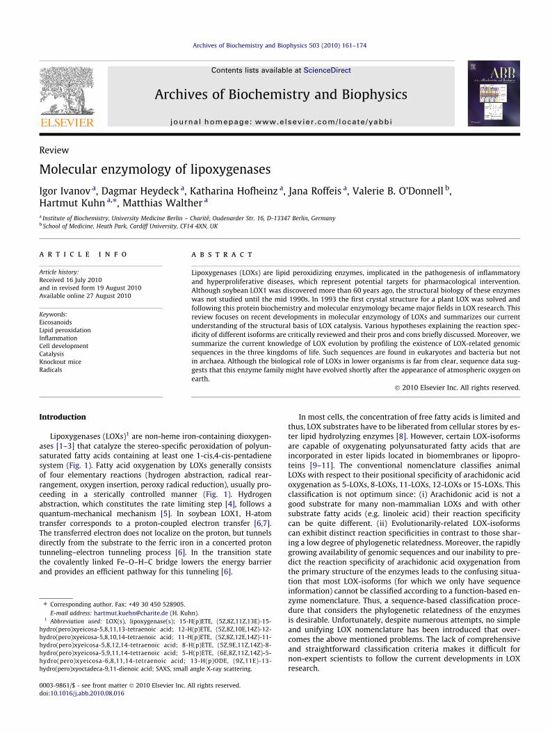
Most LOXs consist of a single polypeptide chain that folds into atwo-domain structure; a small N-terminal b-barrel domain and alarger mostly helical catalytic domain. The rabbit 12/15-LOX(Fig. 2A) is of cylindric shape (height of 10 nm) with an elipticground square (longer diameter 6.1 nm, shorter one of 4.5 nm).The structure of coral 8R-LOX (Fig. 2B) is closely related to the rab-bit enzyme also resembling a cylinder (diameter 6 nm, height10 nm) [48]. In contrast, the soybean LOX1 (Fig. 2C) is ellipsoid(9 nm � 6.5 nm � 6 nm) [36].
In lower organisms, LOXs may occur as fusion proteins, in whichthe LOX-domain is linked to another catalytic domain that plays arole in the secondary metabolism of hydroperoxy fatty acids. Thefirst LOX-fusion protein was discovered in the coral Plexaura homo-malla [50]. Here, the LOX-domain that produces 8R-HpETE is linkedto a heme-containing peroxidase domain, which converts the fattyacid peroxide to an allene oxide. This unstable intermediate mayfurther be converted to cyclopentenone eicosanoids. The fusionprotein was cloned and the two subenzymes were separately ex-pressed and characterized [51,52]. Their crystal structures werealso solved [34,48,53]. Although the degree of amino acid conser-vation between the LOX-domain of this fusion protein and the rab-bit 12/15-LOX was not particularly impressive (30%), the 3D-structures of the two LOX-isoforms are rather similar. The low-res-olution structure of the whole fusion protein indicated that the al-lene oxide synthase domain interacts non-covalently with bothLOX sub-domains while the putative calcium-binding sites andthe membrane interacting Trp residues (see ‘‘The small N-terminalLOX-domain is important for membrane binding and regulates thecatalytic activity”) are not shielded but remain surface exposed[49]. Membrane binding of the fusion protein induces alterationsin the spatial orientation of the different sub-domains (interdo-main movement), as indicated by the appearance of a new proteo-lytic cleavage site [49].
Another LOX-fusion protein, sharing 84% sequence identitywith the P. homomalla enzyme, was detected in the coral Gersemiafruticosa [54] suggesting a broader distribution of these enzymes inoctocorals. In addition, allene oxide synthase/LOX-fusion proteinshave been discovered in the cyanobacteria Anabaena PCC 7120and Acaryochloris marina [55,56]. In contrast to the coral enzymes,in which the fusion proteins contain complete LOXs, the cyanobac-teria isoforms lack the N-terminal LOX-domain and the catalyticLOX-domain is truncated (but active). Although the biological roleof these fusion proteins is unclear, they have been implicated inthe biosynthesis of lipid signaling molecules [50,57]. In all LOX-fu-
Table 1Structural data available for LOXs from the protein data bank. In addition to the given LOX structures some mutants of soybean LOX1 [31–33] and of coral 8R-LOX [34] have beenpublished. Furthermore, homology based models of the structures of human 15-LOX1 (2ABT), 12-LOX (2ABU), and 5-LOX (2ABV) were released by A. Prakasam and P. Mathur (tobe published). The best resolved structure of each isoform is given in bold.
LOX-isoform Remarks/ligands Resolution PDB entry Reference
Soybean LOX1 2.60 2SBL [35]1.40 1YGE [36]
New refinement of 1YGE 1.40 1F8N [32]Soybean LOX3 2.60 1LNH [37]
At ambient temperature 2.0 1RRH [38]at 93 K 2.0 1RRL [38]
Soybean VLX-B 2.4 2IUJ [39]Soybean VLX-D 2.4 2IUK [39]Soybean LOX3 With 4-hydroperoxy-2-methoxy-phenol 2.2 1HU9 [40]Soybean LOX3 With 13S-HPODE 2.0 1IK3 [41]Soybean LOX3 With protocatechuic acid 2.1 1N8Q [42]Soybean LOX3 With epigallocatechin 2.1 1JNQ [43]Soybean LOX3 With 4-nitrocatechol 2.15 1NO3 [44]
(1BYT) [45]Rabbit 12/15-LOX With inhibitor RS7 2.40 1LOX [46]
New refinement of 1LOX,dimer with and without inhibitor RS7
2.40 2P0M [47]
Coral 8R-LOX 3.2 2FNQ [48]1.85 3FG1 [34]
Plexaura homomalla allene oxide synthase 8R-LOX-fusion protein 3.51 3DY5 [49]Human platelet-type 12-LOX Incomplete structure 2.6 3D3L Tresaugues et al. (to be published)
I. Ivanov et al. / Archives of Biochemistry and Biophysics 503 (2010) 161–174 163
sion proteins, the LOX-domains are linked to the C-terminus of theother enzyme subunit. Thus, the C-terminal amino acid, whichconstitutes one of the five immediate iron ligands, remains free.This is of functional relevance since previous mutagenesis studieshave indicated that C-terminal truncation of LOXs abolishes theenzymatic activity.
The small N-terminal LOX-domain is important for membrane bindingand regulates the catalytic activity
The N-terminal domain (b-barrel domain) of all LOX-isoformswith available X-ray data has been seen to consist primarily ofanti-parallel b-strands. Their overall structure resembles that ofthe C2-domain of pancreatic lipase, implicated in membrane bind-ing [58]. For soybean LOX1, the b-barrel domain comprises the first146 amino acid residues. In the case of rabbit 12/15-LOX and coral8R-LOX, the b-barrel domains are formed from the N-terminal 110amino acids or the first 114 residues, respectively. N- and C-termi-nal domains are covalently interconnected by a randomly-coiledoligopeptide. Although the b-barrel domains of the soybean LOX-isoforms are significantly larger than those of the animal enzymes,their overall structures are similar (Fig. 2). The N-terminal domaincontacts the C-domain via an inter-domain contact plane, whichamounts about 1600 Å2 for the rabbit 12/15-LOX [46]. This is sig-nificantly larger (2600 Å2) for the soybean LOX1 suggesting that in-ter-domain binding forces may be stronger for this enzyme.
The high degree of conservation of the two-domain structure inLOXs suggests a functional role for the N-terminal b-barrel domain.Limited proteolysis of soybean LOX1 forms a truncated LOX lackingthe N-terminal domain [59]. This ‘‘mini-LOX” is catalytically activeand exhibits a decreased affinity for linoleic acid (KM of 24.2 lMfor mini-LOX vs. 11.2 lM for native LOX). In contrast, Vmax is aug-mented (363 s-1 for mini-LOX vs 55 s-1 for the native enzyme) andthus, the catalytic efficiency (kcat/KM) is improved by truncation.Recent studies indicated that the non-heme iron can be reversiblyremoved from the active site of mini-LOX, in contrast to the com-plete enzyme [60]. This indicates that N-terminal truncation altersthe structure of the active site so that the iron falls off more easily[60]. Unfortunately, the mini-LOX has not been crystallized and adetailed description of the structural alterations induced by N-ter-minal truncation is not possible. Gene technical truncation of theN-terminal b-barrel domain of the rabbit 12/15-LOX [61] results
in reduction of the catalytic efficiency (1.43 vs. 0.14 1/s lM). Inter-estingly, the truncation mutant undergoes more rapid suicidalinactivation during arachidonic acid oxygenation but the mecha-nisms involved are unknown. Taken together this data suggeststhat the N-terminal b-barrel domain is not essential for the cata-lytic activity, but may play a role in the regulation of turnover[61,62].
Due to its structural similarity to the b-barrel domain of humanlipases [46,63], the N-terminal domains of plant and animal LOXshave been implicated in membrane binding [64,65]. Indeed, site-directed mutagenesis of surface-exposed tryptophans in the N-ter-minal domain inpairs membrane binding of the human 5-LOX andcoral 8R-LOX [48,66]. For the rabbit 12/15-LOX, truncation of the b-barrel domain also reduces membrane binding but the isolated cat-alytic domain still binds to biomembranes, although to a lesser ex-tend. Site-directed mutagenesis suggested that surface-exposedhydrophobic amino acids in both domains are involved in this pro-cess [61,67]. For soybean LOX1, proteolytic cleavage of the N-ter-minal b-barrel domain augments membrane binding [59].
Human 5-LOX requires Ca2+-dependent membrane associationfor turnover [68,69]. Ca2+ also enhances membrane binding of rab-bit 12/15-LOX but specific Ca2+-binding amino acids do not appearto be involved [67,70]. Moreover, inspection of the b-barrel domainsurface of the rabbit enzyme does not reveal the presence of spe-cific calcium-binding motifs. In contrast, coral 8R-LOX containsspecific calcium-binding sites in the b-barrel domain, formed bythe side chains of Asp39 and Asp45 as well as Asp19, Asn44, andGlu47. These surface-exposed calcium-binding residues corre-spond to those proposed for the human 5-LOX and might initiateinsertion of Trp41 and Trp77 into the phospholipid bilayer ofmembranes [48]. Since the calcium-binding residues and the twoTrp are located on the same side as the putative entrance intothe substrate-binding pocket, they may orient the catalytic domainto facilitate fatty acid acquisition from the membrane phase.
The C-terminal domain contains the substrate-binding pocket and thecatalytic non-heme iron
The catalytic domain of all LOX-isoforms consists primarily ofa-helices and harbors the catalytically active non-heme iron. Forthe soybean LOX1, helix 9 (comprising residues 473 through 518with a length of 65 Å) is the central structural element [36]. Of
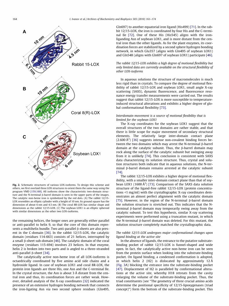
Fig. 2. Schematic structures of various LOX-isoforms. To design this scheme andothers, we first overlaid three LOX structures to orient them the same way using theprogram VMD [159,160]. All isoforms show the characteristic two-domain struc-ture and the N-terminal b-barrel domain is seen in the upper parts of the images.The catalytic non-heme iron is symbolized by the red dots. (A) The rabbit 12/15-LOX resembles an elliptic cylinder with a height of 10 nm. Its ground square has thedimension of about 6 nm and 4.5 nm. (B) The coral 8R-LOX has similar shape anddimensions as the rabbit 12/15-LOX. (C) The soybean LOX1 is an elliptic spheroidwith similar dimensions as the other two LOX-isoforms.
164 I. Ivanov et al. / Archives of Biochemistry and Biophysics 503 (2010) 161–174
the remaining helices, the longer ones are generally either parallelor anti-parallel to helix 9, so that the core of this domain repre-sents a multihelix bundle. Two anti-parallel b-sheets are also pres-ent in the C-domain [36]. In the rabbit 12/15-LOX, the catalyticdomain (residues 114-663) consists of 21 helices, interrupted bya small b-sheet sub-domain [46]. The catalytic domain of the coralenzyme (residues 115-694) involves 23 helices. In that enzyme,helix 2 is broken into two parts and is angled to a three-strandedanti-parallel b-sheet [34].
The catalytically active non-heme iron of all LOX-isoforms isoctaedrically coordinated by five amino acid side chains and ahydroxide ligand. In case of soybean LOX1 and coral 8R-LOX, theprotein iron ligands are three His, one Asn and the C-terminal Ile.In the crystal structure, the Asn is about 3 Å distant from the cen-tral iron and thus, its coordination forces are rather weak. How-ever, detailed analysis of the X-ray coordinates [32] indicated thepresence of an extensive hydrogen bonding network that connectsthe iron-ligating Asn via two second sphere residues (Gln495,
Gln697) to another equatorial iron ligand (His499) [71]. In the rab-bit 12/15-LOX, the iron is coordinated by four His and the C-termi-nal Ile [72]. One of these His (His545) aligns with the iron-liganding Asn of soybean LOX1, and is more distant from the cen-tral iron than the other ligands. As for the plant enzymes, its coor-dination forces are stabilized by a second sphere hydrogen bondingnetwork, in which Glu357 (aligns with Gln495 of soybean LOX1)and Gln548 (aligns with Gln697 of soybean LOX1) participate [46].
The rabbit 12/15-LOX exhibits a high degree of motional flexibility butonly limited data are currently available on the structural flexibility ofother LOX-isoforms
In aqueous solutions the structure of macromolecules is muchless rigid than in crystals. To compare the degree of motional flex-ibility of rabbit 12/15-LOX and soybean LOX1, small angle X-rayscattering (SAXS), dynamic fluorescence, and fluorescence reso-nance energy transfer measurements were carried out. The resultssuggest that rabbit 12/15-LOX is more susceptible to temperature-induced structural alterations and exhibits a higher degree of glo-bal conformational flexibility [73].
Interdomain-movement is a source of motional flexibility that islimited for the soybean LOX1
The X-ray coordinates for the soybean LOX1 suggest that theoverall structures of the two domains are rather stable, and thatthere is little scope for major movement of secondary structuralelements. The relatively large inter-domain contact plane(2.600 Å2) [36] suggests intense non-covalent binding forces be-tween the two domains which may arrest the N-terminal b-barreldomain at the catalytic subunit. Thus, the b-barrel domain mayrock along the surface of the catalytic subunit but swinging awayfrom it is unlikely [74]. This conclusion is consistent with SAXSdata characterizing its solution structure. Thus, crystal and solu-tion structures both indicate that in aqueous solutions, the N-ter-minal b-barrel domain remains arrested at the catalytic subunit[74].
The rabbit 12/15-LOX exhibits a higher degree of motional flex-ibility, with a smaller inter-domain contact plane than that of soy-bean LOX1 (1600 Å2) [73]. Comparison of the SAXS data solutionstructure of the ligand-free rabbit 12/15-LOX (protein concentra-tions <1 mg/ml) with the crystallographic X-ray coordinates dem-onstrates an almost perfect alignment with the catalytic domain[75]. However, in the region of the N-terminal b-barrel domainthe solution structure is stretched out. This indicates that the N-terminal b-barrel domain may temporarily swing away from thecatalytic subunit. To test this hypothesis, similar X-ray scatteringexperiments were performed using a truncation mutant, in whichthe N-terminal b-barrel domain was deleted. For this mutant, thesolution structure completely matched the crystallographic data.
The rabbit 12/15-LOX undergoes major conformational changes uponligand binding at the active site
In the absence of ligands, the entrance to the putative substrate-binding pocket of rabbit 12/15-LOX is funnel-shaped and wideopen. In fact, the catalytically active non-heme iron can be seenfrom the protein surface when looking into the substrate-bindingpocket. On ligand binding, a condensed conformation is adopted,in which helix 2 (H2) is dislocated by approximately 12 Å(Fig. 3A) blocking the entrance into the substrate-binding pocket[47]. Displacement of H2 is paralleled by conformational altera-tions at the active site, whereby H18 retreats from the cavityenlarging the volume of the substrate-binding pocket. Thus, thetriad constituents (see ‘‘The geometry of three crucial amino acidsdetermine the positional specificity of 12/15-lipoxygenases (triadconcept)”) form the bottom of the substrate-binding pocket. This
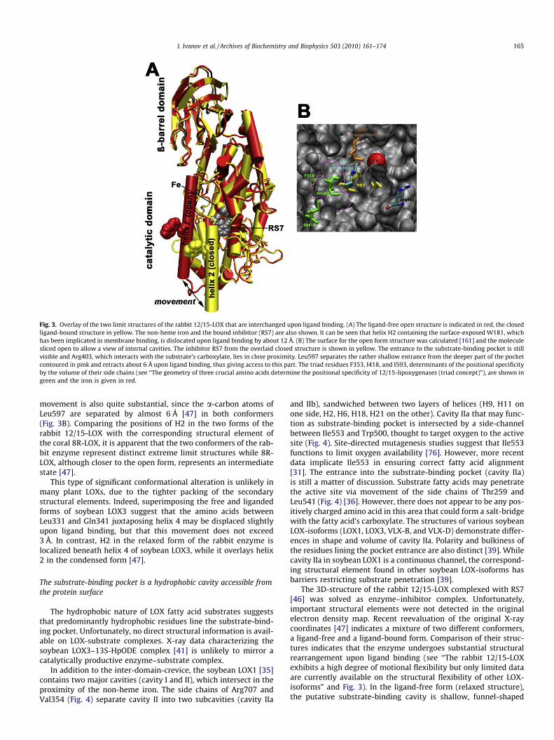
Fig. 3. Overlay of the two limit structures of the rabbit 12/15-LOX that are interchanged upon ligand binding. (A) The ligand-free open structure is indicated in red, the closedligand-bound structure in yellow. The non-heme iron and the bound inhibitor (RS7) are also shown. It can be seen that helix H2 containing the surface-exposed W181, whichhas been implicated in membrane binding, is dislocated upon ligand binding by about 12 Å. (B) The surface for the open form structure was calculated [161] and the moleculesliced open to allow a view of internal cavities. The inhibitor RS7 from the overlaid closed structure is shown in yellow. The entrance to the substrate-binding pocket is stillvisible and Arg403, which interacts with the substrate’s carboxylate, lies in close proximity. Leu597 separates the rather shallow entrance from the deeper part of the pocketcontoured in pink and retracts about 6 Å upon ligand binding, thus giving access to this part. The triad residues F353, I418, and I593, determinants of the positional specificityby the volume of their side chains (see ‘‘The geometry of three crucial amino acids determine the positional specificity of 12/15-lipoxygenases (triad concept)”), are shown ingreen and the iron is given in red.
I. Ivanov et al. / Archives of Biochemistry and Biophysics 503 (2010) 161–174 165
movement is also quite substantial, since the a-carbon atoms ofLeu597 are separated by almost 6 Å [47] in both conformers(Fig. 3B). Comparing the positions of H2 in the two forms of therabbit 12/15-LOX with the corresponding structural element ofthe coral 8R-LOX, it is apparent that the two conformers of the rab-bit enzyme represent distinct extreme limit structures while 8R-LOX, although closer to the open form, represents an intermediatestate [47].
This type of significant conformational alteration is unlikely inmany plant LOXs, due to the tighter packing of the secondarystructural elements. Indeed, superimposing the free and ligandedforms of soybean LOX3 suggest that the amino acids betweenLeu331 and Gln341 juxtaposing helix 4 may be displaced slightlyupon ligand binding, but that this movement does not exceed3 Å. In contrast, H2 in the relaxed form of the rabbit enzyme islocalized beneath helix 4 of soybean LOX3, while it overlays helix2 in the condensed form [47].
The substrate-binding pocket is a hydrophobic cavity accessible fromthe protein surface
The hydrophobic nature of LOX fatty acid substrates suggeststhat predominantly hydrophobic residues line the substrate-bind-ing pocket. Unfortunately, no direct structural information is avail-able on LOX-substrate complexes. X-ray data characterizing thesoybean LOX3–13S-HpODE complex [41] is unlikely to mirror acatalytically productive enzyme–substrate complex.
In addition to the inter-domain-crevice, the soybean LOX1 [35]contains two major cavities (cavity I and II), which intersect in theproximity of the non-heme iron. The side chains of Arg707 andVal354 (Fig. 4) separate cavity II into two subcavities (cavity IIa
and IIb), sandwiched between two layers of helices (H9, H11 onone side, H2, H6, H18, H21 on the other). Cavity IIa that may func-tion as substrate-binding pocket is intersected by a side-channelbetween Ile553 and Trp500, thought to target oxygen to the activesite (Fig. 4). Site-directed mutagenesis studies suggest that Ile553functions to limit oxygen availability [76]. However, more recentdata implicate Ile553 in ensuring correct fatty acid alignment[31]. The entrance into the substrate-binding pocket (cavity IIa)is still a matter of discussion. Substrate fatty acids may penetratethe active site via movement of the side chains of Thr259 andLeu541 (Fig. 4) [36]. However, there does not appear to be any pos-itively charged amino acid in this area that could form a salt-bridgewith the fatty acid’s carboxylate. The structures of various soybeanLOX-isoforms (LOX1, LOX3, VLX-B, and VLX-D) demonstrate differ-ences in shape and volume of cavity IIa. Polarity and bulkiness ofthe residues lining the pocket entrance are also distinct [39]. Whilecavity IIa in soybean LOX1 is a continuous channel, the correspond-ing structural element found in other soybean LOX-isoforms hasbarriers restricting substrate penetration [39].
The 3D-structure of the rabbit 12/15-LOX complexed with RS7[46] was solved as enzyme–inhibitor complex. Unfortunately,important structural elements were not detected in the originalelectron density map. Recent reevaluation of the original X-raycoordinates [47] indicates a mixture of two different conformers,a ligand-free and a ligand-bound form. Comparison of their struc-tures indicates that the enzyme undergoes substantial structuralrearrangement upon ligand binding (see ‘‘The rabbit 12/15-LOXexhibits a high degree of motional flexibility but only limited dataare currently available on the structural flexibility of other LOX-isoforms” and Fig. 3). In the ligand-free form (relaxed structure),the putative substrate-binding cavity is shallow, funnel-shaped
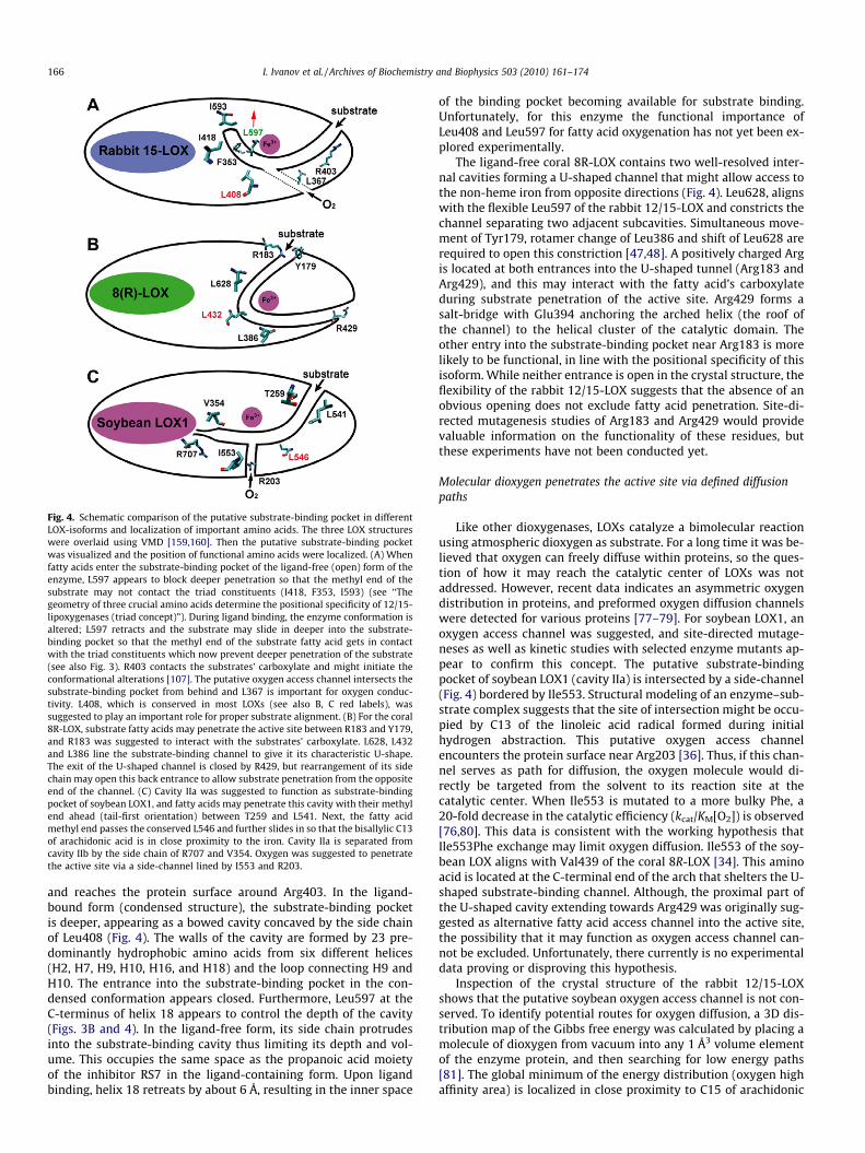
Fig. 4. Schematic comparison of the putative substrate-binding pocket in differentLOX-isoforms and localization of important amino acids. The three LOX structureswere overlaid using VMD [159,160]. Then the putative substrate-binding pocketwas visualized and the position of functional amino acids were localized. (A) Whenfatty acids enter the substrate-binding pocket of the ligand-free (open) form of theenzyme, L597 appears to block deeper penetration so that the methyl end of thesubstrate may not contact the triad constituents (I418, F353, I593) (see ‘‘Thegeometry of three crucial amino acids determine the positional specificity of 12/15-lipoxygenases (triad concept)”). During ligand binding, the enzyme conformation isaltered; L597 retracts and the substrate may slide in deeper into the substrate-binding pocket so that the methyl end of the substrate fatty acid gets in contactwith the triad constituents which now prevent deeper penetration of the substrate(see also Fig. 3). R403 contacts the substrates’ carboxylate and might initiate theconformational alterations [107]. The putative oxygen access channel intersects thesubstrate-binding pocket from behind and L367 is important for oxygen conduc-tivity. L408, which is conserved in most LOXs (see also B, C red labels), wassuggested to play an important role for proper substrate alignment. (B) For the coral8R-LOX, substrate fatty acids may penetrate the active site between R183 and Y179,and R183 was suggested to interact with the substrates’ carboxylate. L628, L432and L386 line the substrate-binding channel to give it its characteristic U-shape.The exit of the U-shaped channel is closed by R429, but rearrangement of its sidechain may open this back entrance to allow substrate penetration from the oppositeend of the channel. (C) Cavity IIa was suggested to function as substrate-bindingpocket of soybean LOX1, and fatty acids may penetrate this cavity with their methylend ahead (tail-first orientation) between T259 and L541. Next, the fatty acidmethyl end passes the conserved L546 and further slides in so that the bisallylic C13of arachidonic acid is in close proximity to the iron. Cavity IIa is separated fromcavity IIb by the side chain of R707 and V354. Oxygen was suggested to penetratethe active site via a side-channel lined by I553 and R203.
166 I. Ivanov et al. / Archives of Biochemistry and Biophysics 503 (2010) 161–174
and reaches the protein surface around Arg403. In the ligand-bound form (condensed structure), the substrate-binding pocketis deeper, appearing as a bowed cavity concaved by the side chainof Leu408 (Fig. 4). The walls of the cavity are formed by 23 pre-dominantly hydrophobic amino acids from six different helices(H2, H7, H9, H10, H16, and H18) and the loop connecting H9 andH10. The entrance into the substrate-binding pocket in the con-densed conformation appears closed. Furthermore, Leu597 at theC-terminus of helix 18 appears to control the depth of the cavity(Figs. 3B and 4). In the ligand-free form, its side chain protrudesinto the substrate-binding cavity thus limiting its depth and vol-ume. This occupies the same space as the propanoic acid moietyof the inhibitor RS7 in the ligand-containing form. Upon ligandbinding, helix 18 retreats by about 6 Å, resulting in the inner space
of the binding pocket becoming available for substrate binding.Unfortunately, for this enzyme the functional importance ofLeu408 and Leu597 for fatty acid oxygenation has not yet been ex-plored experimentally.
The ligand-free coral 8R-LOX contains two well-resolved inter-nal cavities forming a U-shaped channel that might allow access tothe non-heme iron from opposite directions (Fig. 4). Leu628, alignswith the flexible Leu597 of the rabbit 12/15-LOX and constricts thechannel separating two adjacent subcavities. Simultaneous move-ment of Tyr179, rotamer change of Leu386 and shift of Leu628 arerequired to open this constriction [47,48]. A positively charged Argis located at both entrances into the U-shaped tunnel (Arg183 andArg429), and this may interact with the fatty acid’s carboxylateduring substrate penetration of the active site. Arg429 forms asalt-bridge with Glu394 anchoring the arched helix (the roof ofthe channel) to the helical cluster of the catalytic domain. Theother entry into the substrate-binding pocket near Arg183 is morelikely to be functional, in line with the positional specificity of thisisoform. While neither entrance is open in the crystal structure, theflexibility of the rabbit 12/15-LOX suggests that the absence of anobvious opening does not exclude fatty acid penetration. Site-di-rected mutagenesis studies of Arg183 and Arg429 would providevaluable information on the functionality of these residues, butthese experiments have not been conducted yet.
Molecular dioxygen penetrates the active site via defined diffusionpaths
Like other dioxygenases, LOXs catalyze a bimolecular reactionusing atmospheric dioxygen as substrate. For a long time it was be-lieved that oxygen can freely diffuse within proteins, so the ques-tion of how it may reach the catalytic center of LOXs was notaddressed. However, recent data indicates an asymmetric oxygendistribution in proteins, and preformed oxygen diffusion channelswere detected for various proteins [77–79]. For soybean LOX1, anoxygen access channel was suggested, and site-directed mutage-neses as well as kinetic studies with selected enzyme mutants ap-pear to confirm this concept. The putative substrate-bindingpocket of soybean LOX1 (cavity IIa) is intersected by a side-channel(Fig. 4) bordered by Ile553. Structural modeling of an enzyme–sub-strate complex suggests that the site of intersection might be occu-pied by C13 of the linoleic acid radical formed during initialhydrogen abstraction. This putative oxygen access channelencounters the protein surface near Arg203 [36]. Thus, if this chan-nel serves as path for diffusion, the oxygen molecule would di-rectly be targeted from the solvent to its reaction site at thecatalytic center. When Ile553 is mutated to a more bulky Phe, a20-fold decrease in the catalytic efficiency (kcat/KM[O2]) is observed[76,80]. This data is consistent with the working hypothesis thatIle553Phe exchange may limit oxygen diffusion. Ile553 of the soy-bean LOX aligns with Val439 of the coral 8R-LOX [34]. This aminoacid is located at the C-terminal end of the arch that shelters the U-shaped substrate-binding channel. Although, the proximal part ofthe U-shaped cavity extending towards Arg429 was originally sug-gested as alternative fatty acid access channel into the active site,the possibility that it may function as oxygen access channel can-not be excluded. Unfortunately, there currently is no experimentaldata proving or disproving this hypothesis.
Inspection of the crystal structure of the rabbit 12/15-LOXshows that the putative soybean oxygen access channel is not con-served. To identify potential routes for oxygen diffusion, a 3D dis-tribution map of the Gibbs free energy was calculated by placing amolecule of dioxygen from vacuum into any 1 Å3 volume elementof the enzyme protein, and then searching for low energy paths[81]. The global minimum of the energy distribution (oxygen highaffinity area) is localized in close proximity to C15 of arachidonic
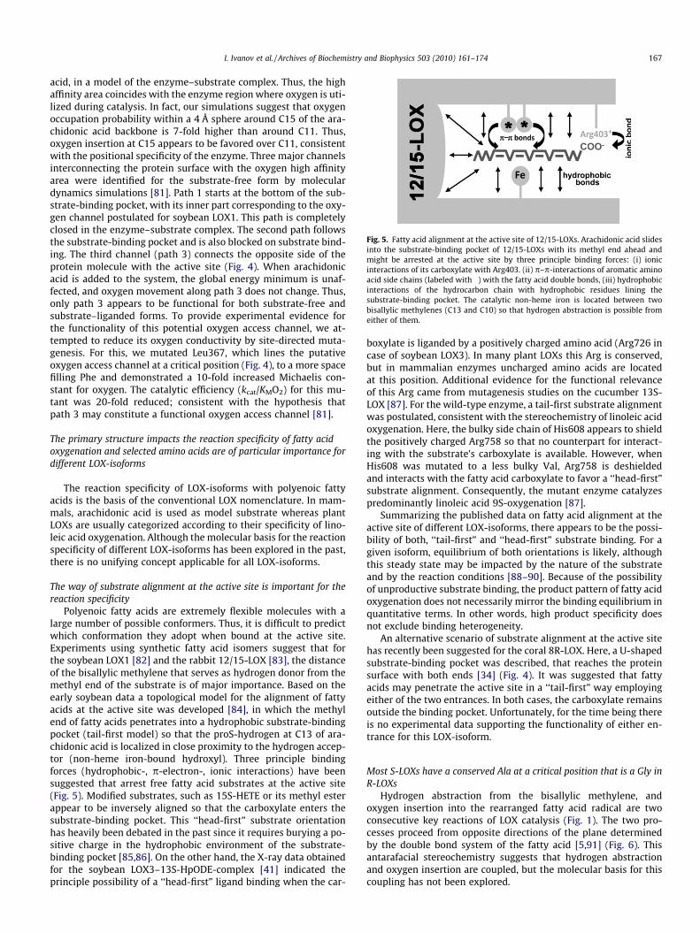
Fig. 5. Fatty acid alignment at the active site of 12/15-LOXs. Arachidonic acid slidesinto the substrate-binding pocket of 12/15-LOXs with its methyl end ahead andmight be arrested at the active site by three principle binding forces: (i) ionicinteractions of its carboxylate with Arg403. (ii) p–p-interactions of aromatic aminoacid side chains (labeled with �) with the fatty acid double bonds, (iii) hydrophobicinteractions of the hydrocarbon chain with hydrophobic residues lining thesubstrate-binding pocket. The catalytic non-heme iron is located between twobisallylic methylenes (C13 and C10) so that hydrogen abstraction is possible fromeither of them.
I. Ivanov et al. / Archives of Biochemistry and Biophysics 503 (2010) 161–174 167
acid, in a model of the enzyme–substrate complex. Thus, the highaffinity area coincides with the enzyme region where oxygen is uti-lized during catalysis. In fact, our simulations suggest that oxygenoccupation probability within a 4 Å sphere around C15 of the ara-chidonic acid backbone is 7-fold higher than around C11. Thus,oxygen insertion at C15 appears to be favored over C11, consistentwith the positional specificity of the enzyme. Three major channelsinterconnecting the protein surface with the oxygen high affinityarea were identified for the substrate-free form by moleculardynamics simulations [81]. Path 1 starts at the bottom of the sub-strate-binding pocket, with its inner part corresponding to the oxy-gen channel postulated for soybean LOX1. This path is completelyclosed in the enzyme–substrate complex. The second path followsthe substrate-binding pocket and is also blocked on substrate bind-ing. The third channel (path 3) connects the opposite side of theprotein molecule with the active site (Fig. 4). When arachidonicacid is added to the system, the global energy minimum is unaf-fected, and oxygen movement along path 3 does not change. Thus,only path 3 appears to be functional for both substrate-free andsubstrate–liganded forms. To provide experimental evidence forthe functionality of this potential oxygen access channel, we at-tempted to reduce its oxygen conductivity by site-directed muta-genesis. For this, we mutated Leu367, which lines the putativeoxygen access channel at a critical position (Fig. 4), to a more spacefilling Phe and demonstrated a 10-fold increased Michaelis con-stant for oxygen. The catalytic efficiency (kcat/KMO2) for this mu-tant was 20-fold reduced; consistent with the hypothesis thatpath 3 may constitute a functional oxygen access channel [81].
The primary structure impacts the reaction specificity of fatty acidoxygenation and selected amino acids are of particular importance fordifferent LOX-isoforms
The reaction specificity of LOX-isoforms with polyenoic fattyacids is the basis of the conventional LOX nomenclature. In mam-mals, arachidonic acid is used as model substrate whereas plantLOXs are usually categorized according to their specificity of lino-leic acid oxygenation. Although the molecular basis for the reactionspecificity of different LOX-isoforms has been explored in the past,there is no unifying concept applicable for all LOX-isoforms.
The way of substrate alignment at the active site is important for thereaction specificity
Polyenoic fatty acids are extremely flexible molecules with alarge number of possible conformers. Thus, it is difficult to predictwhich conformation they adopt when bound at the active site.Experiments using synthetic fatty acid isomers suggest that forthe soybean LOX1 [82] and the rabbit 12/15-LOX [83], the distanceof the bisallylic methylene that serves as hydrogen donor from themethyl end of the substrate is of major importance. Based on theearly soybean data a topological model for the alignment of fattyacids at the active site was developed [84], in which the methylend of fatty acids penetrates into a hydrophobic substrate-bindingpocket (tail-first model) so that the proS-hydrogen at C13 of ara-chidonic acid is localized in close proximity to the hydrogen accep-tor (non-heme iron-bound hydroxyl). Three principle bindingforces (hydrophobic-, p-electron-, ionic interactions) have beensuggested that arrest free fatty acid substrates at the active site(Fig. 5). Modified substrates, such as 15S-HETE or its methyl esterappear to be inversely aligned so that the carboxylate enters thesubstrate-binding pocket. This ‘‘head-first” substrate orientationhas heavily been debated in the past since it requires burying a po-sitive charge in the hydrophobic environment of the substrate-binding pocket [85,86]. On the other hand, the X-ray data obtainedfor the soybean LOX3–13S-HpODE-complex [41] indicated theprinciple possibility of a ‘‘head-first” ligand binding when the car-
boxylate is liganded by a positively charged amino acid (Arg726 incase of soybean LOX3). In many plant LOXs this Arg is conserved,but in mammalian enzymes uncharged amino acids are locatedat this position. Additional evidence for the functional relevanceof this Arg came from mutagenesis studies on the cucumber 13S-LOX [87]. For the wild-type enzyme, a tail-first substrate alignmentwas postulated, consistent with the stereochemistry of linoleic acidoxygenation. Here, the bulky side chain of His608 appears to shieldthe positively charged Arg758 so that no counterpart for interact-ing with the substrate’s carboxylate is available. However, whenHis608 was mutated to a less bulky Val, Arg758 is deshieldedand interacts with the fatty acid carboxylate to favor a ‘‘head-first”substrate alignment. Consequently, the mutant enzyme catalyzespredominantly linoleic acid 9S-oxygenation [87].
Summarizing the published data on fatty acid alignment at theactive site of different LOX-isoforms, there appears to be the possi-bility of both, ‘‘tail-first” and ‘‘head-first” substrate binding. For agiven isoform, equilibrium of both orientations is likely, althoughthis steady state may be impacted by the nature of the substrateand by the reaction conditions [88–90]. Because of the possibilityof unproductive substrate binding, the product pattern of fatty acidoxygenation does not necessarily mirror the binding equilibrium inquantitative terms. In other words, high product specificity doesnot exclude binding heterogeneity.
An alternative scenario of substrate alignment at the active sitehas recently been suggested for the coral 8R-LOX. Here, a U-shapedsubstrate-binding pocket was described, that reaches the proteinsurface with both ends [34] (Fig. 4). It was suggested that fattyacids may penetrate the active site in a ‘‘tail-first” way employingeither of the two entrances. In both cases, the carboxylate remainsoutside the binding pocket. Unfortunately, for the time being thereis no experimental data supporting the functionality of either en-trance for this LOX-isoform.
Most S-LOXs have a conserved Ala at a critical position that is a Gly inR-LOXs
Hydrogen abstraction from the bisallylic methylene, andoxygen insertion into the rearranged fatty acid radical are twoconsecutive key reactions of LOX catalysis (Fig. 1). The two pro-cesses proceed from opposite directions of the plane determinedby the double bond system of the fatty acid [5,91] (Fig. 6). Thisantarafacial stereochemistry suggests that hydrogen abstractionand oxygen insertion are coupled, but the molecular basis for thiscoupling has not been explored.
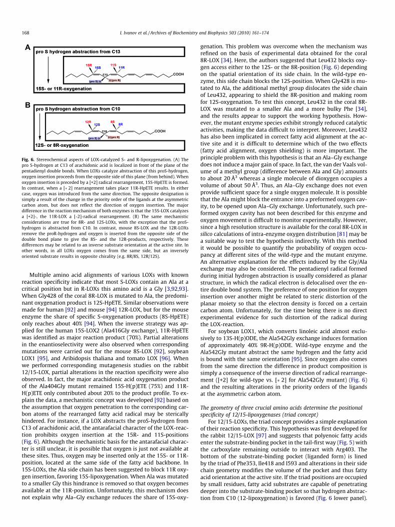
Fig. 6. Stereochemical aspects of LOX-catalyzed S- and R-lipoxygenation. (A) Thepro S-hydrogen at C13 of arachidonic acid is localized in front of the plane of thepentadienyl double bonds. When LOXs catalyze abstraction of this proS-hydrogen,oxygen insertion proceeds from the opposite side of this plane (from behind). Whenoxygen insertion is preceded by a [+2] radical rearrangement, 15S-HpETE is formed.In contrast, when a [�2] rearrangement takes place 11R-HpETE results. In eithercase, oxygen was introduced from the same direction. The opposite designation issimply a result of the change in the priority order of the ligands at the asymmetriccarbon atom, but does not reflect the direction of oxygen insertion. The majordifference in the reaction mechanism of both enzymes is that the 15S-LOX catalyzesa [+2]-, the 11R-LOX a [-2]-radical rearrangement. (B) The same mechanisticconsiderations are true for 8R- and 12S-LOXs, with the exception that the proS-hydrogen is abstracted from C10. In contrast, mouse 8S-LOX and the 12R-LOXsremove the proR-hydrogen and oxygen is inserted from the opposite side of thedouble bond plane to give the 8S- and the 12R-products, respectively. Thesedifferences may be related to an inverse substrate orientation at the active site. Inother words, in all LOXs oxygen comes from the same side, but an inverselyoriented substrate results in opposite chirality (e.g. 8R/8S, 12R/12S).
168 I. Ivanov et al. / Archives of Biochemistry and Biophysics 503 (2010) 161–174
Multiple amino acid alignments of various LOXs with knownreaction specificity indicate that most S-LOXs contain an Ala at acritical position but in R-LOXs this amino acid is a Gly [3,92,93].When Gly428 of the coral 8R-LOX is mutated to Ala, the predomi-nant oxygenation product is 12S-HpETE. Similar observations weremade for human [92] and mouse [94] 12R-LOX, but for the mouseenzyme the share of specific S-oxygenation products (8S-HpETE)only reaches about 40% [94]. When the inverse strategy was ap-plied for the human 15S-LOX2 (Ala416Gly exchange), 11R-HpETEwas identified as major reaction product (70%). Partial alterationsin the enantioselectivity were also observed when correspondingmutations were carried out for the mouse 8S-LOX [92], soybeanLOX1 [95], and Aribidopsis thaliana and tomato LOX [96]. Whenwe performed corresponding mutagenesis studies on the rabbit12/15-LOX, partial alterations in the reaction specificity were alsoobserved. In fact, the major arachidonic acid oxygenation productof the Ala404Gly mutant remained 15S-H(p)ETE (75%) and 11R-H(p)ETE only contributed about 20% to the product profile. To ex-plain the data, a mechanistic concept was developed [92] based onthe assumption that oxygen penetration to the corresponding car-bon atoms of the rearranged fatty acid radical may be stericallyhindered. For instance, if a LOX abstracts the proS-hydrogen fromC13 of arachidonic acid, the antarafacial character of the LOX-reac-tion prohibits oxygen insertion at the 15R- and 11S-positions(Fig. 6). Although the mechanistic basis for the antarafacial charac-ter is still unclear, it is possible that oxygen is just not available atthese sites. Thus, oxygen may be inserted only at the 15S- or 11R-position, located at the same side of the fatty acid backbone. In15S-LOXs, the Ala side chain has been suggested to block 11R oxy-gen insertion, favoring 15S-lipoxygenation. When Ala was mutatedto a smaller Gly this hindrance is removed so that oxygen becomesavailable at the 11R-position. Unfortunately, this mechanism doesnot explain why Ala–Gly exchange reduces the share of 15S-oxy-
genation. This problem was overcome when the mechanism wasrefined on the basis of experimental data obtained for the coral8R-LOX [34]. Here, the authors suggested that Leu432 blocks oxy-gen access either to the 12S- or the 8R-position (Fig. 6) dependingon the spatial orientation of its side chain. In the wild-type en-zyme, this side chain blocks the 12S-position. When Gly428 is mu-tated to Ala, the additional methyl group dislocates the side chainof Leu432, appearing to shield the 8R-position and making roomfor 12S-oxygenation. To test this concept, Leu432 in the coral 8R-LOX was mutated to a smaller Ala and a more bulky Phe [34],and the results appear to support the working hypothesis. How-ever, the mutant enzyme species exhibit strongly reduced catalyticactivities, making the data difficult to interpret. Moreover, Leu432has also been implicated in correct fatty acid alignment at the ac-tive site and it is difficult to determine which of the two effects(fatty acid alignment, oxygen shielding) is more important. Theprinciple problem with this hypothesis is that an Ala–Gly exchangedoes not induce a major gain of space. In fact, the van der Vaals vol-ume of a methyl group (difference between Ala and Gly) amountsto about 20 Å3 whereas a single molecule of dioxygen occupies avolume of about 50 Å3. Thus, an Ala–Gly exchange does not evenprovide sufficient space for a single oxygen molecule. It is possiblethat the Ala might block the entrance into a preformed oxygen cav-ity, to be opened upon Ala–Gly exchange. Unfortunately, such pre-formed oxygen cavity has not been described for this enzyme andoxygen movement is difficult to monitor experimentally. However,since a high resolution structure is available for the coral 8R-LOX insilico calculations of intra-enzyme oxygen distribution [81] may bea suitable way to test the hypothesis indirectly. With this methodit would be possible to quantify the probability of oxygen occu-pancy at different sites of the wild-type and the mutant enzyme.An alternative explanation for the effects induced by the Gly/Alaexchange may also be considered. The pentadienyl radical formedduring initial hydrogen abstraction is usually considered as planarstructure, in which the radical electron is delocalised over the en-tire double bond system. The preference of one position for oxygeninsertion over another might be related to steric distortion of theplanar moiety so that the electron density is forced on a certaincarbon atom. Unfortunately, for the time being there is no directexperimental evidence for such distortion of the radical duringthe LOX-reaction.
For soybean LOX1, which converts linoleic acid almost exclu-sively to 13S-H(p)ODE, the Ala542Gly exchange induces formationof approximately 40% 9R-H(p)ODE. Wild-type enzyme and theAla542Gly mutant abstract the same hydrogen and the fatty acidis bound with the same orientation [95]. Since oxygen also comesfrom the same direction the difference in product composition issimply a consequence of the inverse direction of radical rearrange-ment ([+2] for wild-type vs. [�2] for Ala542Gly mutant) (Fig. 6)and the resulting alterations in the priority orders of the ligandsat the asymmetric carbon atom.
The geometry of three crucial amino acids determine the positionalspecificity of 12/15-lipoxygenases (triad concept)
For 12/15-LOXs, the triad concept provides a simple explanationof their reaction specificity. This hypothesis was first developed forthe rabbit 12/15-LOX [97] and suggests that polyenoic fatty acidsenter the substrate-binding pocket in the tail-first way (Fig. 5) withthe carboxylate remaining outside to interact with Arg403. Thebottom of the substrate-binding pocket (liganded form) is linedby the triad of Phe353, Ile418 and I593 and alterations in their sidechain geometry modifies the volume of the pocket and thus fattyacid orientation at the active site. If the triad positions are occupiedby small residues, fatty acid substrates are capable of penetratingdeeper into the substrate-binding pocket so that hydrogen abstrac-tion from C10 (12-lipoxygenation) is favored (Fig. 6 lower panel).
I. Ivanov et al. / Archives of Biochemistry and Biophysics 503 (2010) 161–174 169
In contrast, if more space filling residues are located at these posi-tions, hydrogen is mainly abstracted from C13 (15-lipoxygenation)(Fig. 6 upper panel). Site-directed mutagenesis studies on the fol-lowing 12/15-LOXs support this concept: human 12/15-LOX[98,99], rabbit 12/15-LOX [97,100], rhesus monkey 12/15-LOX[101], orangutan 12/15-LOX [101], mouse 12/15-LOX [102], rat12/15-LOX [103], and pig 12/15-LOX [104]. The relative impor-tance of the triad constituents varies for different isoenzymes.For human and orangutan 12/15-LOXs, Phe353 and Ile418 (num-bering according to the rabbit enzyme) play a major role since sin-gle mutations of these amino acids to less-space filling residuesconvert the enzyme to an almost completely 12-lipoxygenatingenzyme. Consequently, these residues are considered first-orderdeterminants. For the mouse 12/15-LOX, single mutations of thetriad constituents induce only partial alterations of the positionalspecificity, but combined mutations are more effective. Similarobservations were made for the human platelet 12-LOX, althougheven multiple mutations induce only minor alterations inspecificity (14%) [101]. Studies on human 5-LOX indicated thatsimultaneous mutations of the triad constituents increase theshare of 15S-oxygenation. In fact, for the quadruple mutantF359W + A424I + N425M + A603I 15S-H(p)ETE was identified asthe dominant oxygenation product [105]. Although the mechanis-tic basis for these alterations is not known, the data indicate a5-LOX can be converted to a 15-lipoxygenating enzyme bydecreasing the volume of the substrate-binding pocket [105].Mutagenesis data on the triad constituents of mouse 12R-LOX[94] and human 15-LOX2 [101] do not support the triad conceptindicating that it may not be applicable for epidermis-type LOX-isoforms. Thus, this does not constitute a comprehensive modelexplaining the reaction specificity of all animal LOX-isoforms.
In silico models of the soybean LOX1–linoleate complex [95]suggested that the methyl end of the fatty acid deeply penetratesinto the substrate-binding pocket to contact Phe557. This residuealigns with a second order triad constituent (Met419) of the rabbitenzyme and seems to be conserved as a bulky residue among plant13-LOXs, whereas it is a smaller Val in plant 9-LOXs. Mutagenesisstudies at this position with the cucumber 13S-LOX confirmed itsimportance for positional specificity [87] as described above. Fur-thermore, when Val542 of the cucumber lipid body LOX, whichaligns with Phe353 of the rabbit enzyme, is mutated, the positionalspecificity is altered [106]. Taken together, this data suggests thatthe triad constituents may also play a role in substrate positioningin plant LOXs.
An improved model for the 12/15-LOX–arachidonic acid com-plex has recently been published [107] based on quantum-mechanical electronic structure calculations and moleculardynamics simulations. This supports the ‘‘boot-shaped” conforma-tion of the substrate-binding cavity and confirms relocation ofLeu597 upon substrate binding. It also suggests that sufficientspace is available at the active site to allow conformational vari-ability of arachidonic acid, in particular for alternative positioningof the double bond system. According to this, arachidonic acid isaligned at the active site with the C10 proS-hydrogen localized clo-ser to the enzyme’s proton acceptor (iron-bound hydroxyl) thanthe C13 proS-hydrogen. Nevertheless, during arachidonic acid oxy-genation, abstraction of the C13 proS-hydrogen is strongly favored.The authors suggest that despite the spatial proximity, abstractionof the C10 proS-hydrogen is energetically hindered, and thereforeC13-hydrogen removal is preferred.
The reaction specificity of human 15-LOX2 and its murine ortholog(8S-LOX) cannot be explained with the triad concept but twoadditional amino acid residues are of importance
Despite the fact that human 15-LOX2 and murine 8S-LOX havedifferent positional specificities, they are ortholog enzymes with a
high degree of sequence identity (78%). Two amino acid residues,Asp602 and Val603 in human 15-LOX2 (Tyr603 and His604 in mur-ine 8S-LOX) have been identified as major sequence determinantsfor the positional specificity of these enzymes [108]. In contrast,mutation of the triad residues has little impact on the reactionspecificity of epidermis-type LOX-isoforms [101]. The mechanisticbasis for the different positional specificities of human 15-LOX2and mouse 8S-LOX can be explained by opposite substrate orienta-tion at the active site [89,108]. Human 15-LOX2 binds arachidonicacid in a ‘‘tail-first” way with hydrogen abstraction taking place atC13, followed by [+2] radical rearrangement, and oxygen insertionat C15 (Fig. 6). In contrast, murine 8S-LOX binds arachidonic acid inthe ‘‘head-first” orientation involving C10 hydrogen abstraction,[�2] radical rearrangement, and oxygen insertion at C8. His604 iscritical for inverse substrate orientation and is suggested to inter-act with the fatty acid’s carboxylate [89,108].
There is no unifying mechanistic concept explaining the reactionspecificity of all LOX-isoforms
The reaction specificity of a LOX is the consequence of the stereo-chemistry of the four elementary reactions (hydrogen abstraction,radical rearrangement, oxygen insertion, radical reduction). Becauseof the antarafacial character, hydrogen abstraction and oxygeninsertion are mechanistically coupled but the structural basis for thiscoupling is unknown. However, it is unclear why certain LOXs cata-lyze a [+2] and others a [�2] radical rearrangement (Fig. 6). The car-bon centered radical intermediate formed via hydrogen abstractionis usually depicted as a pentadienyl radical, in which the electrondensity is equally distributed over the entire pentadienyl moiety.However, if the electron density is focused at either of the carbonatoms, the oxygen acceptor site would be predetermined. Thereare several ways to achieve such predetermination:
(a) Electron drawing amino acids might localize the electrondensity of the fatty acid radical to focus it to a certain carbonatom of the pentadienyl system (delocalizing hypothesis).
(b) The planar pentadienyl radical is distorted, which may favorthe formation of an en-allyl radical, in which the electrondensity is focused at a certain carbon atom (distortionhypothesis).
(c) Although the electron density may be equally distributedover the entire pentadienyl system, oxygen is selectively tar-geted to a certain carbon atom so that only a particular per-oxy radical is formed (oxygen targeting hypothesis).
(d) If radical oxygenation is a reversible process and proceedsrandomly, peroxy radical reduction might be stereoselective(reduction hypothesis).
Which of these mechanisms dominates for LOX-isoforms re-mains unclear, and it is possible that a combination of severalcauses is responsible for the high degree of reaction specificity ofLOXs. Our molecular dynamics simulations of oxygen diffusion in-side the rabbit 12/15-LOX suggest that the probability of oxygenoccupancy at C15 of arachidonic acid is approximately 7-timeshigher than at C11. This is consistent with the positional specificityof the enzyme, but if guided oxygen diffusion is to be the onlymechanism of stereocontrol, about 15% of 11-H(p)ETE should bedetected as an arachidonic acid oxygenation product of the wild-type enzyme. This is clearly not the case for native rabbit 12/15-LOX. Moreover, our oxygen distribution maps usually show grad-ual alterations in the probability of oxygen occupancy within small(4–6 Å) radius spheres. In contrast, to explain the usually highpositional specificity of most LOX-isoforms, a steep decline in theprobability of oxygen occupancy within a small radius sphereshould occur. In other words, the targeting hypothesis may con-
170 I. Ivanov et al. / Archives of Biochemistry and Biophysics 503 (2010) 161–174
tribute to the positional specificity, but may not be the exclusivemechanism responsible for this enzyme property.
The reaction specificity of certain LOX-isoforms depends on theexperimental conditions
It has been suggested that changes in reaction conditions mayalter LOX specificity. In fact, pH alterations of the reaction buffermodify the positional specificity of plant LOXs [88,109]. Similarobservations have recently been made for a LOX isolated from Mor-mordica charantia [110], but the structural basis for these changesremains unclear. Although pH alterations frequently occur in vivo,little is known of the impact on LOX specificity in animals. We re-cently investigated the pH-dependence of selected vertebrateLOXs, and observed a remarkable stability of the product patternin the near physiological range [89]. However, subtle structuralalterations induced by targeted mutagenesis and alterations inthe substrate concentrations both induce a pronounced pH-depen-dence of the reaction specificity of some isoforms. For instance, forthe V603H mutant of human 15-LOX2 8S-lipoxygenation is domi-nant (65%) at acidic pH, whereas 15S-H(p)ETE is the major oxygen-ation product at pH 8. Similarly, the product pattern of the wild-type mouse 8S-LOX is hardly altered in the near physiological pHrange, but H604F exchange induces strong pH-dependent altera-tions in the positional specificity. Taken together, this data sug-gests that the specificity of LOXs depends on the reactionconditions, but the biological relevance of this remains unclear.
Allosteric regulation of LOX activity
LOXs are monomeric enzymes, and thus, allosteric regulation oftheir catalytic activity has never been explored in detail. For thehuman 5-LOX, allosteric regulation has been suggested with spe-cific binding sites for ATP and Ca2+ [111]. However, it still remainsunclear why ATP binding increases the catalytic activity of this en-zyme. Kinetic studies on oxygenation of synthetic fatty acid sul-fates by the human 12/15-LOX suggested allosteric regulatorymechanisms for this enzyme [112], and stopped-flow kineticsdemonstrated that the regulators might not bind at the active site.The products of linoleic and arachidonic acid lipoxygenation al-tered the catalytic efficiency (kcat/KM) of human 15-LOX1 and 15-LOX2 [113,114]: The binding of 12-H(p)ETE or 13-H(p)ODE in-creased both the catalytic efficiency of arachidonic acid oxygena-tion related to linoleic acid oxygenation [increase in (kcat/KM)AA/(kcat/KM)LA ratio] and the oxygen affinity of human 12/15-LOX1for arachidonic acid oxygenation, while decreasing oxygen affinityfor linoleic acid oxygenation [113]. Thus, in a cellular systemwhere both fatty acids are simultaneously available, the bindingof allosteric effectors might alter the substrate specificity (prefer-ence for arachidonic acid oxygenation). Interestingly, the catalyticactivity of the soybean LOX1 was not affected by 15-HETE nor 13-HODE [115]. However, inhibitors of fatty acid oxygenation likeoleyl- and palmitoleyl sulfate not only lowered the catalytic activ-ity of the enzyme, but also altered the substrate specificity byincreasing the arachidonic acid/linoleic acid ratio. This data sug-gests that both enzymes may undergo allosteric regulation butthe molecular mechanism of this has not been clarified. It also re-mains unclear where the allosteric regulator might actually bind atthe enzyme and what structural alterations ligand binding mightinduce.
The human platelet 12-LOX might occur as active dimer and exhibits astrong tendency for non-covalent oligomerization
SAXS data on both rabbit 12/15-LOX [75] and soybean LOX1[74] suggest that the two enzymes occur as hydrated monomers
in aqueous solutions at low protein concentrations (<1 mg/ml).This data is supported by native gel electrophoresis, gel filtrationchromatography and mass spectrometry of the rabbit enzyme (Iva-nov et al. unpublished data). With all these methods, we did notfind convincing evidence for dimerization. It should, however, beenstressed that at higher protein concentrations, which are far be-yond physiologically relevant levels, the rabbit enzyme shows atendency for irreversible protein aggregation. The molecular basisfor this process remains unclear.
On the other hand, SAXS data on the human platelet 12-LOXsuggested that this enzyme might occur as a dimer in aqueoussolution. It also exhibits a tendency to aggregate into larger oligo-mers [116]. More detailed mechanistic studies suggest that oligo-merization may be driven by the formation of intermoleculardisulfide bridges, which are broken under reducing conditions. Incontrast, enzyme dimers appear to be stable under reducing condi-tions excluding the importance of disulfide bridges [116]. Massspectral analysis of the enzyme in aqueous solution indicated aprotein with an apparent molecular mass of 77 kDa, characteristicof enzyme monomers. The apparent contradiction between theSAXS data and the mass spectral analysis might be explained bythe fact that the non-covalently linked dimers are thermodynami-cally unstable during mass spectral analysis.
Evolutionary aspects of LOXs
Lipoxygenase sequences are present in eucaryotes and bacteria but notin archea
LOXs are widely distributed in plants [8,117], mammals [1,101]and selected marine organisms [118,119]. More recently, LOX-iso-forms have been detected in various prokaryotes [120–122]. To ob-tain more detailed information on the LOX distribution, wesearched various databases (GenBank, Refseq, Uniprot, Ensembl)for LOX-specific sequences. Unfortunately, the LOX family is struc-turally quite heterogeneous, since cDNA or protein alignments ofknown LOX-isoforms indicate that there is no joint characteristicsequence that is shared by all. For our search we employed the se-quences representing the region surrounding the direct iron li-gands and show the highest degree of conservation. The positiveresults were sorted according to kingdom and phylum, and exam-ples for different biological orders are given in Fig. 7. LOX se-quences can be found in two of the three kingdoms of life(eukaryotes, prokaryotes). In the third, the archaea, no LOX was de-tected which is not surprising considering their extreme habitats.In fact, for many archaea, the LOX substrate molecular dioxygenis toxic. It should be stressed that detection of a LOX-related se-quence does not necessarily mean that an active enzyme is actuallyexpressed. Moreover, classification of the sequences into the differ-ent subfamilies (12/15-LOX, platelet-type 12-LOX, 5-LOX, epider-mal type LOXs) is not possible on the sole basis of sequenceinformation.
During evolution, LOXs have arisen in several prokaryotes (cya-nobacteria and proteobacteria), in unicellular protista (red andgreen algae, the amoeba Dictyostelium discoideum), fungi, plants(mosses as well as flowering plants) and animals (Fig. 7). In ani-mals, LOX sequences occur in many species ranging from the verybasic placozoa Trichoplax adhaerens up to primates and include,among others, coral, tick, acorn worm, fish, frog, chicken, bat,hedgehog, mouse, dolphin, elephant, monkey, and human.
Many of the LOX-containing species serve as model organismsrepresenting certain developmental stages of eukaryotes. Volvoxcarteri is one of the most basic multicellular organisms that still ex-ist, and T. adhaerens is one of the most basic eumetazoa, being clo-sely related to Cnidaria [123]. Other model organisms containing
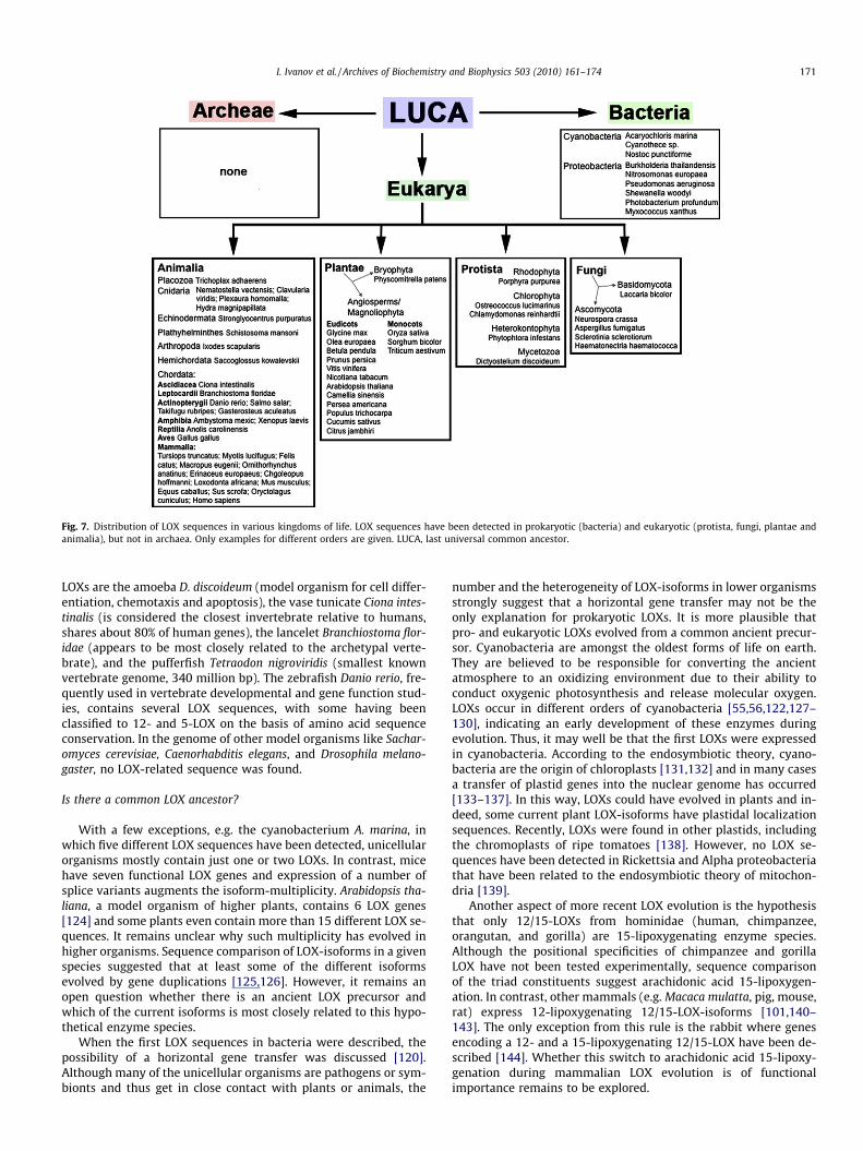
Fig. 7. Distribution of LOX sequences in various kingdoms of life. LOX sequences have been detected in prokaryotic (bacteria) and eukaryotic (protista, fungi, plantae andanimalia), but not in archaea. Only examples for different orders are given. LUCA, last universal common ancestor.
I. Ivanov et al. / Archives of Biochemistry and Biophysics 503 (2010) 161–174 171
LOXs are the amoeba D. discoideum (model organism for cell differ-entiation, chemotaxis and apoptosis), the vase tunicate Ciona intes-tinalis (is considered the closest invertebrate relative to humans,shares about 80% of human genes), the lancelet Branchiostoma flor-idae (appears to be most closely related to the archetypal verte-brate), and the pufferfish Tetraodon nigroviridis (smallest knownvertebrate genome, 340 million bp). The zebrafish Danio rerio, fre-quently used in vertebrate developmental and gene function stud-ies, contains several LOX sequences, with some having beenclassified to 12- and 5-LOX on the basis of amino acid sequenceconservation. In the genome of other model organisms like Sachar-omyces cerevisiae, Caenorhabditis elegans, and Drosophila melano-gaster, no LOX-related sequence was found.
Is there a common LOX ancestor?
With a few exceptions, e.g. the cyanobacterium A. marina, inwhich five different LOX sequences have been detected, unicellularorganisms mostly contain just one or two LOXs. In contrast, micehave seven functional LOX genes and expression of a number ofsplice variants augments the isoform-multiplicity. Arabidopsis tha-liana, a model organism of higher plants, contains 6 LOX genes[124] and some plants even contain more than 15 different LOX se-quences. It remains unclear why such multiplicity has evolved inhigher organisms. Sequence comparison of LOX-isoforms in a givenspecies suggested that at least some of the different isoformsevolved by gene duplications [125,126]. However, it remains anopen question whether there is an ancient LOX precursor andwhich of the current isoforms is most closely related to this hypo-thetical enzyme species.
When the first LOX sequences in bacteria were described, thepossibility of a horizontal gene transfer was discussed [120].Although many of the unicellular organisms are pathogens or sym-bionts and thus get in close contact with plants or animals, the
number and the heterogeneity of LOX-isoforms in lower organismsstrongly suggest that a horizontal gene transfer may not be theonly explanation for prokaryotic LOXs. It is more plausible thatpro- and eukaryotic LOXs evolved from a common ancient precur-sor. Cyanobacteria are amongst the oldest forms of life on earth.They are believed to be responsible for converting the ancientatmosphere to an oxidizing environment due to their ability toconduct oxygenic photosynthesis and release molecular oxygen.LOXs occur in different orders of cyanobacteria [55,56,122,127–130], indicating an early development of these enzymes duringevolution. Thus, it may well be that the first LOXs were expressedin cyanobacteria. According to the endosymbiotic theory, cyano-bacteria are the origin of chloroplasts [131,132] and in many casesa transfer of plastid genes into the nuclear genome has occurred[133–137]. In this way, LOXs could have evolved in plants and in-deed, some current plant LOX-isoforms have plastidal localizationsequences. Recently, LOXs were found in other plastids, includingthe chromoplasts of ripe tomatoes [138]. However, no LOX se-quences have been detected in Rickettsia and Alpha proteobacteriathat have been related to the endosymbiotic theory of mitochon-dria [139].
Another aspect of more recent LOX evolution is the hypothesisthat only 12/15-LOXs from hominidae (human, chimpanzee,orangutan, and gorilla) are 15-lipoxygenating enzyme species.Although the positional specificities of chimpanzee and gorillaLOX have not been tested experimentally, sequence comparisonof the triad constituents suggest arachidonic acid 15-lipoxygen-ation. In contrast, other mammals (e.g. Macaca mulatta, pig, mouse,rat) express 12-lipoxygenating 12/15-LOX-isoforms [101,140–143]. The only exception from this rule is the rabbit where genesencoding a 12- and a 15-lipoxygenating 12/15-LOX have been de-scribed [144]. Whether this switch to arachidonic acid 15-lipoxy-genation during mammalian LOX evolution is of functionalimportance remains to be explored.
172 I. Ivanov et al. / Archives of Biochemistry and Biophysics 503 (2010) 161–174
The biological role of LOX-isoforms in lower organisms remainsunclear
The biological function of mammalian LOXs has been exploredby targeted knockout of the corresponding genes. Mice in whichthe 12/15-LOX gene [145], the 5-LOX gene [146] or the platelet-type 12-LOX gene [147] is disrupted, are viable and develop nor-mally. This also applies for a 12/15-LOX + 5-LOX double knockout[148]. In plants, LOXs have been implicated in numerous functions(see Ref. [149] and [150] for a review) which include the produc-tion of phytohormons [151], seed germination [152], leaf senes-cence [153,154], and flavor development [155,156]. However, itremains unclear why certain plants express such a large varietyof LOX-isoforms.
In lower organisms the biological role of LOXs has not been ex-plored in detail. In the coral P. homomalla, the enzyme has beenimplicated in the generation of prostaglandins [157] which amountfrom 2% to 3% of the organisms dry weight. Unfortunately, the bio-logical role of this prostaglandin derivative for coral physiology hasnot yet been clarified. When cyanobacteria are wounded by sonica-tion, formation of endogenous LOX products is strongly increased[128]. In other lower organisms, LOX-isoforms have been impli-cated in defense mechanisms, stress responses or cell differentia-tion and maturation but in general the molecular mechanismshave not been well elucidated.
Future development and perspective
In 1993 the crystal structure of the soybean LOX1 was solved,and in the following years X-ray coordinates for additional iso-forms were published (Table 1). On the basis of this data, it isnow possible to model the principle 3D-structure of other isoformsand a model for the potato 5-LOX has been suggested [158]. How-ever, it is dangerous and can be misleading to conclude structuraldetails and functional consequences of structural peculiarities sim-ply on the basis of sequence similarity and in the absence of func-tional data. Although such functional data are sometimes difficultto interpret, they are needed to confirm or disprove workinghypotheses.
A hallmark in recent LOX structural biology was the reinterpre-tation of the original X-ray coordinates of the rabbit 12/15-LOX–inhibitor complex [47]. This report demonstrates that LOXs are dy-namic structures that undergo conformational alterations upon li-gand binding. These changes are quite impressive and impact boththe protein surface (12 Å movement of H2) and the structure of theputative substrate-binding pocket (6 Å movement of Leu597). Thisdata strongly suggests functional consequences of these structuralalterations. Moreover, when we inspected the internal cavity sys-tem of the relaxed (ligand-free) and the condensed (ligand-bound)enzyme structure in more detail, we found marked differences. Inthe relaxed structure, the triad constituents are not part of the sub-strate-binding pocket, which is strongly narrowed by the sidechain of Leu597. However, in the condensed structure, Leu597 isdislocated to further open the pocket so that the substrate fattyacid can contact the triad constituents, in agreement with themutagenesis data [97,100]. Although the dynamic character ofthe rabbit enzyme is consistent with X-ray scattering and dynamicfluorescence data, it remains unclear whether other LOX-isoformsexhibit a similar degree of motional flexibility. Dynamic measure-ments of structural alterations in other LOX-isoforms are requiredto address this open question.
Another open point in the structural biology of LOXs is the bio-logical importance of the N-terminal b-barrel domain. Mutagenesisand limited proteolysis have implicated this domain in membranebinding and activity regulation. However, the molecular basis for
this function remains unclear. Although the catalytic domain ofLOXs appears to be a structurally stable unit, it cannot be excludedthat N-terminal truncation may lead to structural alterations in thecatalytic domain responsible for the alterations in the catalyticactivity. Unfortunately, nothing is known on the structure of trun-cated LOX-isoforms. To clarify this point it would be helpful tohave direct structural data on the mini-LOX, which is producedfrom soybean LOX1 by limited proteolysis.
The biological relevance of LOX-isoforms in higher plants andanimals has been a matter of discussion for many years and knock-out studies have advanced the field in several ways. The recent dis-covery of LOX-isoforms in lower organisms and even inprokaryotes might speed up this development. There are, however,two major problems: (i) At the moment little is known on the bio-logical function of such LOX-isoforms. In fact, except from rarecases it remains unclear of whether or not the detected LOX se-quences are actually expressed as functional proteins. Moreover,many of them have not even been characterized with respect totheir enzymatic properties. (ii) If in the future, experimental dataon the biological relevance of such LOX-isoforms becomes avail-able, it remains unclear what we can learn from such findings forthe biology of LOXs in higher plants and animals. However, lowerorganisms are easier to handle and loss and gain-of-function strat-egies are more straightforward then corresponding experiments inhigher species. Thus, irrespective of the uncertainties discussedabove, more knowledge on the biology of LOXs in lower organismsis expected to boost LOX research in general.
References
[1] A.R. Brash, J. Biol. Chem. 274 (1999) 23679–23682.[2] H. Kuhn, J. Saam, S. Eibach, H.G. Holzhutter, I. Ivanov, M. Walther, Biochem.
Biophys. Res. Commun. 338 (2005) 93–101.[3] C. Schneider, D.A. Pratt, N.A. Porter, A.R. Brash, Chem. Biol. 14 (2007) 473–
488.[4] M.H. Glickman, J.P. Klinman, Biochemistry 34 (1995) 14077–14092.[5] K.W. Rickert, J.P. Klinman, Biochemistry 38 (1999) 12218–12228.[6] N. Lehnert, E.I. Solomon, J. Biol. Inorg. Chem. 8 (2003) 294–305.[7] E. Hatcher, A.V. Soudackov, S. Hammes-Schiffer, J. Am. Chem. Soc. 126 (2004)
5763–5775.[8] A. Andreou, I. Feussner, Phytochemistry 70 (2009) 1504–1510.[9] J.M. Upston, J. Neuzil, P.K. Witting, R. Alleva, R. Stocker, J. Biol. Chem. 272
(1997) 30067–30074.[10] Y. Takahashi, W.C. Glasgow, H. Suzuki, Y. Taketani, S. Yamamoto, M. Anton, H.
Kuhn, A.R. Brash, Eur. J. Biochem. 218 (1993) 165–171.[11] T. Schewe, W. Halangk, C. Hiebsch, S.M. Rapoport, FEBS Lett. 60 (1975) 149–
152.[12] B. Samuelsson, P. Borgeat, S. Hammarstrom, R.C. Murphy, Adv. Prostaglandin
Thromboxane Leukotriene Res. 6 (1980) 1–18.[13] B.A. Jakschik, L.H. Lee, Nature 287 (1980) 51–52.[14] S. Feltenmark, N. Gautam, A. Brunnstrom, W. Griffiths, L. Backman, C. Edenius,
L. Lindbom, M. Bjorkholm, H.E. Claesson, Proc. Natl. Acad. Sci. USA 105 (2008)680–685.
[15] C.N. Serhan, M. Hamberg, B. Samuelsson, Proc. Natl. Acad. Sci. USA 81 (1984)5335–5339.
[16] P. Maderna, C. Godson, Br. J. Pharmacol. 158 (2009) 947–959.[17] C.N. Serhan, K. Gotlinger, S. Hong, M. Arita, Prostaglandins Other Lipid
Mediators 73 (2004) 155–172.[18] A. Ariel, P.L. Li, W. Wang, W.X. Tang, G. Fredman, S. Hong, K.H. Gotlinger, C.N.
Serhan, J. Biol. Chem. 280 (2005) 43079–43086.[19] J.M. Schwab, N. Chiang, M. Arita, C.N. Serhan, Nature 447 (2007) 869–874.[20] C.N. Serhan, R. Yang, K. Martinod, K. Kasuga, P.S. Pillai, T.F. Porter, S.F. Oh, M.
Spite, J. Exp. Med. 206 (2009) 15–23.[21] G. Furstenberger, N. Epp, K.M. Eckl, H.C. Hennies, C. Jorgensen, P. Hallenborg,
K. Kristiansen, P. Krieg, Prostaglandins Other Lipid Mediators 82 (2007) 128–134.
[22] N. Epp, G. Furstenberger, K. Muller, J. Cell Biol. 177 (2007) 173–182.[23] K.M. Eckl, J. Invest. Dermatol. 129 (2009) 1421–1428.[24] S. Bhattacharya, G. Mathew, D.G. Jayne, S. Pelengaris, M. Khan, Tumour Biol.
30 (2009) 185–199.[25] G.P. Pidgeon, J. Lysaght, S. Krishnamoorthy, J.V. Reynolds, K. O’Byrne, D. Nie,
K.V. Honn, Cancer Metastasis Rev. 26 (2007) 503–524.[26] J.J. Moreno, Biochem. Pharmacol. 77 (2009) 1–10.[27] Y. Chawengsub, K.M. Gauthier, W.B. Campbell, Am. J. Physiol. Heart Circ.
Physiol. 297 (2009) H495–H507.[28] N. Mochizuki, Y.G. Kwon, Circ. Res. 102 (2008) 143–145.[29] N.P. Duroudier, A.S. Tulah, I. Sayers, Allergy 64 (2009) 823–839.
I. Ivanov et al. / Archives of Biochemistry and Biophysics 503 (2010) 161–174 173
[30] M. Hersberger, Clin. Chem. Lab. Med. (2008).[31] M.P. Meyer, D.R. Tomchick, J.P. Klinman, Proc. Natl. Acad. Sci. USA 105 (2008)
1146–1151.[32] D.R. Tomchick, P. Phan, M. Cymborowski, W. Minor, T.R. Holman,
Biochemistry 40 (2001) 7509–7517.[33] E.N. Segraves, M. Chruszcz, M.L. Neidig, V. Ruddat, J. Zhou, A.T. Wecksler, W.
Minor, E.I. Solomon, T.R. Holman, Biochemistry 45 (2006) 10233–10242.[34] D.B. Neau, N.C. Gilbert, S.G. Bartlett, W. Boeglin, A.R. Brash, M.E. Newcomer,
Biochemistry 48 (2009) 7906–7915.[35] J.C. Boyington, B.J. Gaffney, L.M. Amzel, Biochem. Soc. Trans. 21 (Pt 3) (1993)
744–748.[36] W. Minor, J. Steczko, B. Stec, Z. Otwinowski, J.T. Bolin, R. Walter, B. Axelrod,
Biochemistry 35 (1996) 10687–10701.[37] E. Skrzypczak-Jankun, L.M. Amzel, B.A. Kroa, M.O. Funk Jr., Proteins 29 (1997)
15–31.[38] E. Skrzypczak-Jankun, O.Y. Borbulevych, M.I. Zavodszky, M.R. Baranski, K.
Padmanabhan, V. Petricek, J. Jankun, Acta Crystallogr. Sect. D Biol. Crystallogr.62 (2006) 766–775.
[39] B. Youn, G.E. Sellhorn, R.J. Mirchel, B.J. Gaffney, H.D. Grimes, C. Kang, Proteins65 (2006) 1008–1020.
[40] E. Skrzypczak-Jankun, K. Zhou, N.P. McCabe, S.H. Selman, J. Jankun, Int. J. Mol.Med. 12 (2003) 17–24.
[41] E. Skrzypczak-Jankun, R.A. Bross, R.T. Carroll, W.R. Dunham, M.O. Funk Jr., J.Am. Chem. Soc. 123 (2001) 10814–10820.
[42] O.Y. Borbulevych, J. Jankun, S.H. Selman, E. Skrzypczak-Jankun, Proteins 54(2004) 13–19.
[43] E. Skrzypczak-Jankun, K. Zhou, J. Jankun, Int. J. Mol. Med. 12 (2003) 415–420.[44] E. Skrzypczak-Jankun, O.Y. Borbulevych, J. Jankun, Acta Crystallogr. Sect. D
Biol. Crystallogr. 60 (2004) 613–615.[45] C. Pham, J. Jankun, E. Skrzypczak-Jankun, R.A. Flowers 2nd, M.O. Funk Jr.,
Biochemistry 37 (1998) 17952–17957.[46] S.A. Gillmor, A. Villasenor, R. Fletterick, E. Sigal, M.F. Browner, Nat. Struct.
Biol. 4 (1997) 1003–1009.[47] J. Choi, J.K. Chon, S. Kim, W. Shin, Proteins 70 (2008) 1023–1032.[48] M.L. Oldham, A.R. Brash, M.E. Newcomer, J. Biol. Chem. 280 (2005) 39545–
39552.[49] N.C. Gilbert, M. Niebuhr, H. Tsuruta, T. Bordelon, O. Ridderbusch, A. Dassey, A.R.
Brash, S.G. Bartlett, M.E. Newcomer, Biochemistry 47 (2008) 10665–10676.[50] R. Koljak, O. Boutaud, B.H. Shieh, N. Samel, A.R. Brash, Science 277 (1997)
1994–1996.[51] O. Boutaud, A.R. Brash, J. Biol. Chem. 274 (1999) 33764–33770.[52] B.D. Abraham, M. Sono, O. Boutaud, A. Shriner, J.H. Dawson, A.R. Brash, B.J.
Gaffney, Biochemistry 40 (2001) 2251–2259.[53] M.L. Oldham, A.R. Brash, M.E. Newcomer, Proc. Natl. Acad. Sci. USA 102 (2005)
297–302.[54] H. Lohelaid, R. Jarving, K. Valmsen, K. Varvas, M. Kreen, I. Jarving, N. Samel,
Biochim. Biophys. Acta 1780 (2008) 315–321.[55] Y. Zheng, W.E. Boeglin, C. Schneider, A.R. Brash, J. Biol. Chem. 283 (2008)
5138–5147.[56] C. Schneider, K. Niisuke, W.E. Boeglin, M. Voehler, D.F. Stec, N.A. Porter, A.R.
Brash, Proc. Natl. Acad. Sci. USA 104 (2007) 18941–18945.[57] B. Gao, W.E. Boeglin, Y. Zheng, C. Schneider, A.R. Brash, J. Biol. Chem. 284
(2009) 22087–22098.[58] H. Chahinian, B. Sias, F. Carriere, Curr. Protein Pept. Sci. 1 (2000) 91–103.[59] M. Maccarrone, M.L. Salucci, Biochemistry 40 (2001) 6819–6827.[60] E. Dainese, C.B. Angelucci, A. Sabatucci, V. De Filippis, G. Mei, M. Maccarrone,
FASEB J. 24 (2010) 1725–1736.[61] M. Walther, M. Anton, M. Wiedmann, R. Fletterick, H. Kuhn, J. Biol. Chem. 277
(2002) 27360–27366.[62] S. Romanov, R. Wiesner, G. Myagkova, H. Kuhn, I. Ivanov, Biochemistry 45
(2006) 3554–3562.[63] F.K. Winkler, A. D’Arcy, W. Hunziker, Nature 343 (1990) 771–774.[64] C. May, M. Hohne, P. Gnau, K. Schwennesen, H. Kindl, Eur. J. Biochem. 267
(2000) 1100–1109.[65] S.A. Tatulian, J. Steczko, W. Minor, Biochemistry 37 (1998) 15481–15490.[66] S. Kulkarni, S. Das, C.D. Funk, D. Murray, W. Cho, J. Biol. Chem. 277 (2002)
13167–13174.[67] M. Walther, R. Wiesner, H. Kuhn, J. Biol. Chem. 279 (2004) 3717–3725.[68] T. Hammarberg, P. Provost, B. Persson, O. Radmark, J. Biol. Chem. 275 (2000)
38787–38793.[69] X.S. Chen, C.D. Funk, The. N-terminal, J. Biol. Chem. 276 (2001) 811–818.[70] R. Brinckmann, K. Schnurr, D. Heydeck, T. Rosenbach, G. Kolde, H. Kuhn, Blood
91 (1998) 64–74.[71] G. Schenk, M.L. Neidig, J. Zhou, T.R. Holman, E.I. Solomon, Biochemistry 42
(2003) 7294–7302.[72] R.J. Kuban, R. Wiesner, J. Rathman, G. Veldink, H. Nolting, V.A. Sole, H. Kuhn,
Biochem. J. 332 (Pt 1) (1998) 237–242.[73] G. Mei, A. Di Venere, E. Nicolai, C.B. Angelucci, I. Ivanov, A. Sabatucci, E.
Dainese, H. Kuhn, M. Maccarrone, Biochemistry 47 (2008) 9234–9242.[74] E. Dainese, A. Sabatucci, J. Mol. Biol. 349 (2005) 143–152.[75] M. Hammel, M. Walther, R. Prassl, H. Kuhn, J. Mol. Biol. 343 (2004) 917–929.[76] M.J. Knapp, J.P. Klinman, Biochemistry 42 (2003) 11466–11475.[77] K. Chu, J. Vojtchovsky, B.H. McMahon, R.M. Sweet, J. Berendzen, I. Schlichting,
Nature 403 (2000) 921–923.[78] A. Ostermann, R. Waschipky, F.G. Parak, G.U. Nienhaus, Nature 404 (2000)
205–208.
[79] E.E. Scott, Q.H. Gibson, Biochemistry 36 (1997) 11909–11917.[80] M.J. Knapp, F.P. Seebeck, J.P. Klinman, J. Am. Chem. Soc. 123 (2001) 2931–
2932.[81] J. Saam, I. Ivanov, M. Walther, H.G. Holzhutter, H. Kuhn, Proc. Natl. Acad. Sci.
USA 104 (2007) 13319–13324.[82] M. Hamberg, B. Samuelsson, J. Biol. Chem. 242 (1967) 5329–5335.[83] H. Kuhn, H. Sprecher, A.R. Brash, J. Biol. Chem. 265 (1990) 16300–16305.[84] H. Kuhn, T. Schewe, S.M. Rapoport, Adv. Enzymol. Relat. Areas Mol. Biol. 58
(1986) 273–311.[85] M. Browner, S.A. Gillmor, R. Fletterick, Nat. Struct. Biol. 5 (1998) 179.[86] S.T. Prigge, B.J. Gaffney, L.M. Amzel, Nat. Struct. Biol. 5 (1998) 178–179.[87] E. Hornung, M. Walther, H. Kuhn, I. Feussner, Proc. Natl. Acad. Sci. USA 96
(1999) 4192–4197.[88] H.W. Gardner, Biochim. Biophys. Acta 1001 (1989) 274–281.[89] M. Walther, J. Roffeis, C. Jansen, M. Anton, I. Ivanov, H. Kuhn, Biochim.
Biophys. Acta (2009).[90] M. Walther, I. Ivanov, G. Myagkova, H. Kuhn, Chem. Biol. 8 (2001) 779–790.[91] R.L. Maas, A.R. Brash, Proc. Natl. Acad. Sci. USA 80 (1983) 2884–2888.[92] G. Coffa, A.R. Brash, Proc. Natl. Acad. Sci. USA 101 (2004) 15579–15584.[93] G. Coffa, C. Schneider, A.R. Brash, Biochem. Biophys. Res. Commun. 338 (2005)
87–92.[94] S. Meruvu, M. Walther, I. Ivanov, S. Hammarstrom, G. Furstenberger, P. Krieg,
P. Reddanna, H. Kuhn, J. Biol. Chem. 280 (2005) 36633–36641.[95] G. Coffa, A.N. Imber, B.C. Maguire, G. Laxmikanthan, C. Schneider, B.J. Gaffney,
A.R. Brash, J. Biol. Chem. 280 (2005) 38756–38766.[96] W.E. Boeglin, A. Itoh, Y. Zheng, G. Coffa, G.A. Howe, A.R. Brash, Lipids 43
(2008) 979–987.[97] S. Borngraber, M. Browner, S. Gillmor, C. Gerth, M. Anton, R. Fletterick, H.
Kuhn, J. Biol. Chem. 274 (1999) 37345–37350.[98] D.L. Sloane, R. Leung, J. Barnett, C.S. Craik, E. Sigal, Protein Eng. 8 (1995) 275–
282.[99] D.L. Sloane, R. Leung, C.S. Craik, E. Sigal, Nature 354 (1991) 149–152.
[100] S. Borngraber, R.J. Kuban, M. Anton, H. Kuhn, J. Mol. Biol. 264 (1996) 1145–1153.
[101] R. Vogel, C. Jansen, J. Roffeis, P. Reddanna, P. Forsell, H.E. Claesson, H. Kuhn, M.Walther, J. Biol. Chem. 285 (2010) 5369–5376.
[102] F. Burger, P. Krieg, F. Marks, G. Furstenberger, Biochem. J. 348 (Pt 2) (2000)329–335.
[103] T. Watanabe, J.Z. Haeggstrom, Biochem. Biophys. Res. Commun. 192 (1993)1023–1029.
[104] H. Suzuki, K. Kishimoto, T. Yoshimoto, S. Yamamoto, F. Kanai, Y. Ebina, A.Miyatake, T. Tanabe, Biochim. Biophys. Acta 1210 (1994) 308–316.
[105] K. Schwarz, M. Walther, M. Anton, C. Gerth, I. Feussner, H. Kuhn, J. Biol. Chem.276 (2001) 773–779.
[106] E. Hornung, S. Rosahl, H. Kuhn, I. Feussner, Biochem. Soc. Trans. 28 (2000)825–826.
[107] L. Toledo, L. Masgrau, J.D. Marechal, J.M. Lluch, A. Gonzalez-Lafont, J. Phys.Chem. B 114 (2010) 7037–7046.
[108] M. Jisaka, R.B. Kim, W.E. Boeglin, A.R. Brash, J. Biol. Chem. 275 (2000) 1287–1293.
[109] G.A. Veldink, G.J. Garssen, J.F. Vliegenthart, J. Boldingh, Biochem. Biophys. Res.Commun. 47 (1972) 22–26.
[110] E. Hornung, S. Kunze, A. Liavonchanka, G. Zimmermann, D. Kuhn, K. Fritsche,A. Renz, H. Kuhn, I. Feussner, Phytochemistry 69 (2008) 2774–2780.
[111] J.P. Falgueyret, D. Denis, D. Macdonald, J.H. Hutchinson, D. Riendeau,Biochemistry 34 (1995) 13603–13611.
[112] R. Mogul, E. Johansen, T.R. Holman, Biochemistry 39 (2000) 4801–4807.[113] A.T. Wecksler, C. Jacquot, Biochemistry 48 (2009) 6259–6267.[114] A.T. Wecksler, V. Kenyon, J.D. Deschamps, T.R. Holman, Biochemistry 47
(2008) 7364–7375.[115] A.T. Wecksler, N.K. Garcia, T.R. Holman, Bioorg. Med. Chem. 17 (2009) 6534–
6539.[116] A.M. Aleem, J. Jankun, J.D. Dignam, M. Walther, H. Kuhn, D.I. Svergun, E.
Skrzypczak-Jankun, J. Mol. Biol. 376 (2008) 193–209.[117] E.H. Oliw, Prostaglandins Other Lipid Mediators 68–69 (2002) 313–323.[118] L. De Petrocellis, V. Di Marzo, Prostaglandins Leukotrienes Essent. Fatty Acids
51 (1994) 215–229.[119] D.J. Hawkins, A.R. Brash, J. Biol. Chem. 262 (1987) 7629–7634.[120] H. Porta, M. Rocha-Sosa, Microbiology 147 (2001) 3199–3200.[121] R.E. Vance, S. Hong, K. Gronert, C.N. Serhan, J.J. Mekalanos, Proc. Natl. Acad.
Sci. USA 101 (2004) 2135–2139.[122] T. Koeduka, T. Kajiwara, K. Matsui, Curr. Microbiol. 54 (2007) 315–319.[123] M. Srivastava, E. Begovic, J. Chapman, N.H. Putnam, U. Hellsten, T.
Kawashima, A. Kuo, T. Mitros, A. Salamov, M.L. Carpenter, A.Y. Signorovitch,M.A. Moreno, K. Kamm, J. Grimwood, J. Schmutz, H. Shapiro, I.V. Grigoriev,L.W. Buss, B. Schierwater, S.L. Dellaporta, D.S. Rokhsar, Nature 454 (2008)955–960.
[124] G. Bannenberg, M. Martinez, M. Hamberg, C. Castresana, Lipids 44 (2009) 85–95.
[125] H. Toh, C. Yokoyama, T. Tanabe, T. Yoshimoto, S. Yamamoto, Prostaglandins44 (1992) 291–315.
[126] J.H. Shin, K. Van, D.H. Kim, K.D. Kim, Y.E. Jang, B.S. Choi, M.Y. Kim, S.H. Lee,BMC Plant Biol. 8 (2008) 133.
[127] A.Z. Andreou, M. Vanko, L. Bezakova, I. Feussner, Phytochemistry 69 (2008)1832–1837.
[128] I. Lang, I. Feussner, Phytochemistry 68 (2007) 1120–1127.
174 I. Ivanov et al. / Archives of Biochemistry and Biophysics 503 (2010) 161–174
[129] I. Lang, C. Gobel, A. Porzel, I. Heilmann, I. Feussner, Biochem. J. 410 (2008)347–357.
[130] A. Andreou, C. Gobel, M. Hamberg, I. Feussner, J. Biol. Chem. 285 (2010)14178–14186.
[131] C. Mereschkowski, Biol Centralbl 25 (1905) 593–604.[132] L. Sagan, J. Theor. Biol. 14 (1967) 255–274.[133] C.Y. Huang, M.A. Ayliffe, J.N. Timmis, Nature 422 (2003) 72–76.[134] P. Maliga, Nature 422 (2003) 31–32.[135] C.Y. Huang, M.A. Ayliffe, J.N. Timmis, Proc. Natl. Acad. Sci. USA 101 (2004)
9710–9715.[136] J.N. Timmis, M.A. Ayliffe, C.Y. Huang, W. Martin, Nat. Rev. Genet. 5 (2004)
123–135.[137] W. Martin, B. Stoebe, V. Goremykin, S. Hapsmann, M. Hasegawa, K.V.
Kowallik, Nature 393 (1998) 162–165.[138] C. Barsan, P. Sanchez-Bel, C. Rombaldi, I. Egea, M. Rossignol, M. Kuntz, M.
Zouine, A. Latche, M. Bouzayen, J.C. Pech, J. Exp. Bot. 61 (2010) 2413–2431.[139] V.V. Emelyanov, Eur. J. Biochem. 270 (2003) 1599–1618.[140] M. Johannesson, L. Backman, H.E. Claesson, P.K. Forsell, Prostaglandins
Leukotrienes Essent. Fatty Acids 82 (2010) 121–129.[141] T. Yoshimoto, H. Suzuki, S. Yamamoto, T. Takai, C. Yokoyama, T. Tanabe, Proc.
Natl. Acad. Sci. USA 87 (1990) 2142–2146.[142] X.S. Chen, U. Kurre, N.A. Jenkins, N.G. Copeland, C.D. Funk, J. Biol. Chem. 269
(1994) 13979–13987.[143] T. Watanabe, J.F. Medina, J.Z. Haeggstrom, O. Radmark, B. Samuelsson, Eur. J.
Biochem. 212 (1993) 605–612.[144] M. Berger, K. Schwarz, H. Thiele, I. Reimann, A. Huth, S. Borngraber, H. Kuhn,
B.J. Thiele, J. Mol. Biol. 278 (1998) 935–948.
[145] D. Sun, C.D. Funk, J. Biol. Chem. 271 (1996) 24055–24062.[146] X.S. Chen, J.R. Sheller, E.N. Johnson, C.D. Funk, Nature 372 (1994) 179–182.[147] E.N. Johnson, L.F. Brass, C.D. Funk, Proc. Natl. Acad. Sci. USA 95 (1998) 3100–
3105.[148] D. Poeckel, K.A. Zemski Berry, R.C. Murphy, C.D. Funk, J. Biol. Chem. 284
(2009) 21077–21089.[149] H. Porta, M. Rocha-Sosa, Plant Physiol. 130 (2002) 15–21.[150] I. Feussner, C. Wasternack, Annu. Rev. Plant Physiol. Plant Mol. Biol. 53 (2002)
275–297.[151] B.A. Vick, D.C. Zimmerman, Biochem. Biophys. Res. Commun. 111 (1983)
470–477.[152] I. Feussner, C. Wasternack, H. Kindl, H. Kuhn, Proc. Natl. Acad. Sci. USA 92
(1995) 11849–11853.[153] J.F. Bousquet, K.V. Thimann, Proc. Natl. Acad. Sci. USA 81 (1984) 1724–1727.[154] Y. He, H. Fukushige, D.F. Hildebrand, S. Gan, Plant Physiol. 128 (2002) 876–
884.[155] G. Chen, R. Hackett, D. Walker, A. Taylor, Z. Lin, D. Grierson, Plant Physiol. 136
(2004) 2641–2651.[156] M. Ridolfi, S. Terenziani, M. Patumi, G. Fontanazza, J. Agric. Food Chem. 50
(2002) 835–839.[157] W.C. Song, A.R. Brash, Arch. Biochem. Biophys. 290 (1991) 427–435.[158] P. Aparoy, R.N. Reddy, L. Guruprasad, M.R. Reddy, P. Reddanna, J. Comput.-
Aided Mol. Des. 22 (2008) 611–619.[159] W. Humphrey, A. Dalke, K. Schulten, J. Mol. Graphics 14 (1996) 33–38.[160] R.B. Russell, G.J. Barton, Proteins 14 (1992) 309–323.[161] A. Varshney, F.P. Brooks, W.V. Wright, IEEE Comput. Graphics Appl. 14 (1994)
19–25.
Related Documents