Molecular Dynamics of Mesophilic-Like Mutants of a Cold-Adapted Enzyme: Insights into Distal Effects Induced by the Mutations Elena Papaleo*, Marco Pasi ¤ , Matteo Tiberti, Luca De Gioia Department of Biotechnology and Biosciences, University of Milano-Bicocca, Milan, Italy Abstract Networks and clusters of intramolecular interactions, as well as their ‘‘communication’’ across the three-dimensional architecture have a prominent role in determining protein stability and function. Special attention has been dedicated to their role in thermal adaptation. In the present contribution, seven previously experimentally characterized mutants of a cold-adapted a-amylase, featuring mesophilic-like behavior, have been investigated by multiple molecular dynamics simulations, essential dynamics and analyses of correlated motions and electrostatic interactions. Our data elucidate the molecular mechanisms underlying the ability of single and multiple mutations to globally modulate dynamic properties of the cold-adapted a-amylase, including both local and complex unpredictable distal effects. Our investigation also shows, in agreement with the experimental data, that the conversion of the cold-adapted enzyme in a warm-adapted variant cannot be completely achieved by the introduction of few mutations, also providing the rationale behind these effects. Moreover, pivotal residues, which are likely to mediate the effects induced by the mutations, have been identified from our analyses, as well as a group of suitable candidates for protein engineering. In fact, a subset of residues here identified (as an isoleucine, or networks of mesophilic-like salt bridges in the proximity of the catalytic site) should be considered, in experimental studies, to get a more efficient modification of the features of the cold-adapted enzyme. Citation: Papaleo E, Pasi M, Tiberti M, De Gioia L (2011) Molecular Dynamics of Mesophilic-Like Mutants of a Cold-Adapted Enzyme: Insights into Distal Effects Induced by the Mutations. PLoS ONE 6(9): e24214. doi:10.1371/journal.pone.0024214 Editor: Franca Fraternali, King9s College London, United Kingdom Received April 25, 2011; Accepted August 2, 2011; Published September 7, 2011 Copyright: ß 2011 Papaleo et al. This is an open-access article distributed under the terms of the Creative Commons Attribution License, which permits unrestricted use, distribution, and reproduction in any medium, provided the original author and source are credited. Funding: This work was supported by CASPUR (Consorzio Interuniversitario per le Applicazio-ni di Supercalcolo per Universita ` e Ricerca) Standard HPC Grant 2010 to EP. The funders had no role in study design, data analysis, decision to publish or preparation of the manuscript. Competing Interests: The authors have declared that no competing interests exist. * E-mail: [email protected] ¤ Current address: Universite ´ Lyon 1, CNRS, UMR 5086, Bases Mole ´culaires et Structurales des Syste ` mes Infectieux, IBCP FR3302, Lyon, France Introduction A detailed comprehension of molecular mechanisms that rule the relationship between stability, flexibility, and activity in extremophilic enzymes is of crucial importance both for funda- mental and applicative research [1–3]. Enzymes isolated from psychrophilic organisms have received particular attention from the scientific community in the last 20 years, thanks to their unique properties in terms of high activity at detrimental temperatures, low thermal stability and unusual specificity, offering a wide spectrum of industrial applications [4,5]. The increasing number of primary sequences and three- dimensional (3D) structures of enzymes from extremophiles [6– 8] has provided a suitable background to disclose molecular determinants of their structural stability. It is well established that psychrophilic enzymes use different adaptation strategies [9,10], with each protein family adopting its own structural strategy [9,11]. The molecular determinants and the exact relationships between activity, stability and flexibility in cold-adapted enzymes are still a matter of debate. In fact, the intrinsic thermolability and increased low temperature activity of psychrophilic enzymes prompt for a direct link between activity and stability [12]. Otherwise, it has been suggested that thermolability may be associated to a lack of evolutionary pressure for stable enzymes in low temperature habitats [13,14]. The existence of non-canonical cold-adapted enzymes, featuring both unusual thermal stability and high catalytic efficiency at low temperatures [15,16], along with the capability to uncouple activity and stability in in vitro evolution studies [17], make the definition of activity-stability- flexibility trade-off even more difficult. Structural flexibility and rigidity are likely to cooperate, each acting on specific areas of the enzyme structure, nevertheless, they are difficult to quantify for a small and anisotropic material such as a protein molecule [9,12]. In this context, the current view on the relationships between protein dynamics and function [18–22] suggests that protein function is rooted in the free energy landscape [19] and that fluctuations at equilibrium can influence biological functions. In fact, backbone flexibility profiles diverge slowly, being conserved both in protein family and superfamily [20,23,24]. However, a recent study has shown that warm- and cold-adapted enzymes belonging to the same family present common dynamics signature related to the same fold, but also specific differences which may reflect temperature adaptation [25]. In fact, the striking correspondence between picosecond dynamics and longer scale conformational changes suggests that the physical origin of functionally important collective motions is the fast time- scale local motions [19] and that differences in the fast fluctuations PLoS ONE | www.plosone.org 1 September 2011 | Volume 6 | Issue 9 | e24214

Welcome message from author
This document is posted to help you gain knowledge. Please leave a comment to let me know what you think about it! Share it to your friends and learn new things together.
Transcript
Molecular Dynamics of Mesophilic-Like Mutants of aCold-Adapted Enzyme: Insights into Distal EffectsInduced by the MutationsElena Papaleo*, Marco Pasi¤, Matteo Tiberti, Luca De Gioia
Department of Biotechnology and Biosciences, University of Milano-Bicocca, Milan, Italy
Abstract
Networks and clusters of intramolecular interactions, as well as their ‘‘communication’’ across the three-dimensionalarchitecture have a prominent role in determining protein stability and function. Special attention has been dedicated totheir role in thermal adaptation. In the present contribution, seven previously experimentally characterized mutants of acold-adapted a-amylase, featuring mesophilic-like behavior, have been investigated by multiple molecular dynamicssimulations, essential dynamics and analyses of correlated motions and electrostatic interactions. Our data elucidate themolecular mechanisms underlying the ability of single and multiple mutations to globally modulate dynamic properties ofthe cold-adapted a-amylase, including both local and complex unpredictable distal effects. Our investigation also shows, inagreement with the experimental data, that the conversion of the cold-adapted enzyme in a warm-adapted variant cannotbe completely achieved by the introduction of few mutations, also providing the rationale behind these effects. Moreover,pivotal residues, which are likely to mediate the effects induced by the mutations, have been identified from our analyses, aswell as a group of suitable candidates for protein engineering. In fact, a subset of residues here identified (as an isoleucine,or networks of mesophilic-like salt bridges in the proximity of the catalytic site) should be considered, in experimentalstudies, to get a more efficient modification of the features of the cold-adapted enzyme.
Citation: Papaleo E, Pasi M, Tiberti M, De Gioia L (2011) Molecular Dynamics of Mesophilic-Like Mutants of a Cold-Adapted Enzyme: Insights into Distal EffectsInduced by the Mutations. PLoS ONE 6(9): e24214. doi:10.1371/journal.pone.0024214
Editor: Franca Fraternali, King9s College London, United Kingdom
Received April 25, 2011; Accepted August 2, 2011; Published September 7, 2011
Copyright: � 2011 Papaleo et al. This is an open-access article distributed under the terms of the Creative Commons Attribution License, which permitsunrestricted use, distribution, and reproduction in any medium, provided the original author and source are credited.
Funding: This work was supported by CASPUR (Consorzio Interuniversitario per le Applicazio-ni di Supercalcolo per Universita e Ricerca) Standard HPC Grant2010 to EP. The funders had no role in study design, data analysis, decision to publish or preparation of the manuscript.
Competing Interests: The authors have declared that no competing interests exist.
* E-mail: [email protected]
¤ Current address: Universite Lyon 1, CNRS, UMR 5086, Bases Moleculaires et Structurales des Systemes Infectieux, IBCP FR3302, Lyon, France
Introduction
A detailed comprehension of molecular mechanisms that rule
the relationship between stability, flexibility, and activity in
extremophilic enzymes is of crucial importance both for funda-
mental and applicative research [1–3]. Enzymes isolated from
psychrophilic organisms have received particular attention from
the scientific community in the last 20 years, thanks to their unique
properties in terms of high activity at detrimental temperatures,
low thermal stability and unusual specificity, offering a wide
spectrum of industrial applications [4,5].
The increasing number of primary sequences and three-
dimensional (3D) structures of enzymes from extremophiles [6–
8] has provided a suitable background to disclose molecular
determinants of their structural stability. It is well established that
psychrophilic enzymes use different adaptation strategies [9,10],
with each protein family adopting its own structural strategy
[9,11].
The molecular determinants and the exact relationships
between activity, stability and flexibility in cold-adapted enzymes
are still a matter of debate. In fact, the intrinsic thermolability and
increased low temperature activity of psychrophilic enzymes
prompt for a direct link between activity and stability [12].
Otherwise, it has been suggested that thermolability may be
associated to a lack of evolutionary pressure for stable enzymes in
low temperature habitats [13,14]. The existence of non-canonical
cold-adapted enzymes, featuring both unusual thermal stability
and high catalytic efficiency at low temperatures [15,16], along
with the capability to uncouple activity and stability in in vitro
evolution studies [17], make the definition of activity-stability-
flexibility trade-off even more difficult.
Structural flexibility and rigidity are likely to cooperate, each
acting on specific areas of the enzyme structure, nevertheless, they
are difficult to quantify for a small and anisotropic material such as
a protein molecule [9,12]. In this context, the current view on the
relationships between protein dynamics and function [18–22]
suggests that protein function is rooted in the free energy
landscape [19] and that fluctuations at equilibrium can influence
biological functions. In fact, backbone flexibility profiles diverge
slowly, being conserved both in protein family and superfamily
[20,23,24]. However, a recent study has shown that warm- and
cold-adapted enzymes belonging to the same family present
common dynamics signature related to the same fold, but also
specific differences which may reflect temperature adaptation [25].
In fact, the striking correspondence between picosecond dynamics
and longer scale conformational changes suggests that the physical
origin of functionally important collective motions is the fast time-
scale local motions [19] and that differences in the fast fluctuations
PLoS ONE | www.plosone.org 1 September 2011 | Volume 6 | Issue 9 | e24214
are encoded by differences in the primary sequence. Moreover, as
results of recent advances both in biophysical spectroscopies and
molecular dynamics (MD) simulations, it is possible to extract
detailed information on coupled motions and networks of
communicating residues in the 3D structure and during dynamics,
thanks for example to analysis of cross-correlation of atomic
fluctuations [26–28], or long-range pathway of communicating
residues [28–31]. The psychrophilic chloride-dependent [32] a-
amylase from the Antarctic bacterium Pseudoalteromonas haloplanktis
(namely AHA) is a paradigm for the study of molecular
determinants of enzyme cold-adaptation. In particular, AHA
was one of the first psychrophilic enzymes for which the 3D
structure was solved [33] (Figure 1), revealing the presence of three
distinct structural domains (namely A, B and C). It has been
suggested that AHA acquires a low conformational stability and
high flexibility through a reduction or weakening of inter- and
intra-domain interactions, as well as it features decreased
activation enthalpy and a concomitant improvement in kcat if
compared to mammalian a-amylases, as pig pancreatic a-amylase
(PPA) [34]. AHA site-directed mutagenesis [35–37] has been
insightful in clarifying the relationships between activity and
structural stability. In particular, single and multiple mutants were
investigated to restore weak intra-molecular interactions, which
are present in the closest mesophilic homolog, PPA. It turns out
that, the reestablishment of weak interactions in AHA produces
variants with both thermal stability and catalytic efficiency shifted
toward ‘‘mesophilic-like’’ values (Figure S1 in Supporting
Information S1). The majority of the investigated mutations
feature increase in Tm (melting temperature) and a decrease in
both kcat and Km [35,37,38]. The authors speculated that
structural stabilization may be related to improved rigidity of the
active site: reducing its flexibility, in fact, would increase the
activation energy [39], leading to a reduction of kcat values, and
would constrain the ground-state of the enzyme-substrate complex
to a narrower distribution of conformational states, thus lowering
Km.
In the present study, seven of the previously characterized AHA
mutants [35,37,38] featuring the most clear-cut effects on activity
and thermal stability (Figure 1, Figure S1 in Supporting
Information S1), were investigated by multiple MD simulations
in explicit solvent (collecting more than 0.25 ms, overall). The aim
of our study is the elucidation of effects induced by the mutations
in atomic details with particular attention to long range effects, as
well as to identify the determinants of the uncompleted conversion
of AHA mutants in mesophilic-like variants. The restored weak
interactions are capable of modifying the AHA dynamics in the
direction of the warm-adapted enzyme, inducing a complex array
of long range effects. Our results also point out a subset of critical
residues for PPA and AHA dynamics and structure, not previously
identified, which can be a suitable test case for AHA protein
engineering in a mesophilic-like direction.
Results
Local effects induced by the mutations in the AHAmutant variants
A detailed structural characterization of the AHA mutant
variants investigated by D’Amico et al. [35,37] was not previously
carried out and, advantaged by the available dynamic framework,
we have initially evaluated whether the mutations have effectively
restored the targeted weak interactions. A description of local
effects induced by the substitutions is also provided, by monitoring
the persistence of residues in the surroundings of the mutations.
The interactions hypothesized by D’Amico and coworkers were
successfully restored (Figure S5–S9 in Supporting Information S1),
although our dynamic analysis points out more complex effects
and interaction networks present in PPA, which have to be
considered and which can also explain the weak overall effects
induced by some mutations on the kinetic and thermodynamic
properties of AHA. The networks which are not restored in the
AHA mutants, due to several amino acidic substitutions with
respect to PPA, interest in particular electrostatic interactions as
salt bridges and aromatic clusters.
N12R mutation restores the salt bridge between R12A (R20P in
PPA), on b1-a1 loop and D15A (D23P) on a1 (Figure 1, Figure S2
in Supporting Information S1). In PPA R20P forms a network with
both D23P and E369P (which cannot be established in AHA due to
P319A) and via its aliphatic carbons interact with hydrophobic
residues in b8-a8 loop (366–370P, Figure S3 in Supporting
Information S1), successfully restored in AHA.
N150D mutation reestablishes, in AHA, a surface salt bridge
between D150A, on a3, and K190A on a4 (Figure 1, Figure S3B in
Supporting Information S1). However, in PPA the corresponding
D173-K213P bridge belongs to a more complex cluster of
electrostatic interactions, which extends through domain B,
connecting it with the proximity of the catalytic domain, and cannot
be established in AHA due to several substitutions (see below).
Q164I mutation reinforces a hydrophobic cluster including a3
(150–166A), b4 (170–174A), as well as a3-b4 and a4-b5 (190–200A)
loops (Figure 1, Figure S3C–D in Supporting Information S1). A
clear increase in Hp is observed for AHA mutants which bear
Q164I mutation (AHAQI, AHA5SS and AHA5) thanks to both
local (a3) and distal (a4-b5) effects, resulting in a global increase of
residues with high surrounding hydrophobicity (Hp.20 kcal/mol)
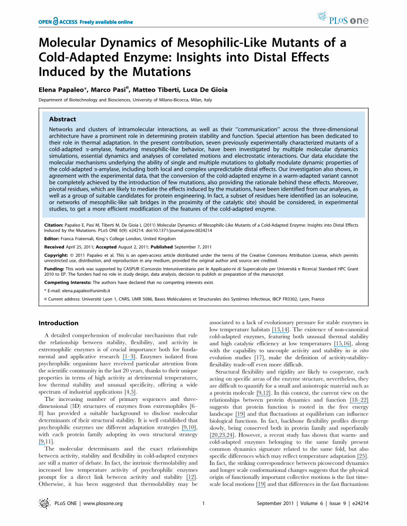
(Figure 2 and 3).
V196F mutation, in the proximity of Q164I and K300R
mutations (Figure 1, Figure S3D and S4 in Supporting
Information S1), has been engineered to re-establish two aromatic
interactions between F196A (b5), and residues Y82A and F198A
(corresponding to Y94P and F291P in PPA), on b3 and b5
respectively (Figure S4 in Supporting Information S1). In PPA,
F229P is part of a larger cluster of aromatic and hydrophobic
Figure 1. AHA mutants engineered in order to restoreinteractions typical of the warm-adapted homolog, PPA.Localization of all the mutations included in the simulated AHAmutants, on the 3D structure. Secondary structure elements areindicated as cartoon, white, grey and black cartoons indicate domainA, B and C, respectively. The yellow dots and sticks indicate thelocalization of the catalytic triad, the spheres Ca2+ and Cl2 cofactorsand the mutated residues are indicated as orange sticks.doi:10.1371/journal.pone.0024214.g001
Molecular Dynamics of Psychrophilic Enzyme Mutants
PLoS ONE | www.plosone.org 2 September 2011 | Volume 6 | Issue 9 | e24214
Figure 2. Hydrogen bonds and surrounding hydrophobicity (Hp) are described as histograms representing the percentage ofdonors+acceptors involved in H-bonds (first set, y axis on the left) or of residues with surrounding hydrophobicity in the indicatedrange (Hp, all other sets, y axis on the right); each set shows values computed for AHA, PPA and the mutants.doi:10.1371/journal.pone.0024214.g002
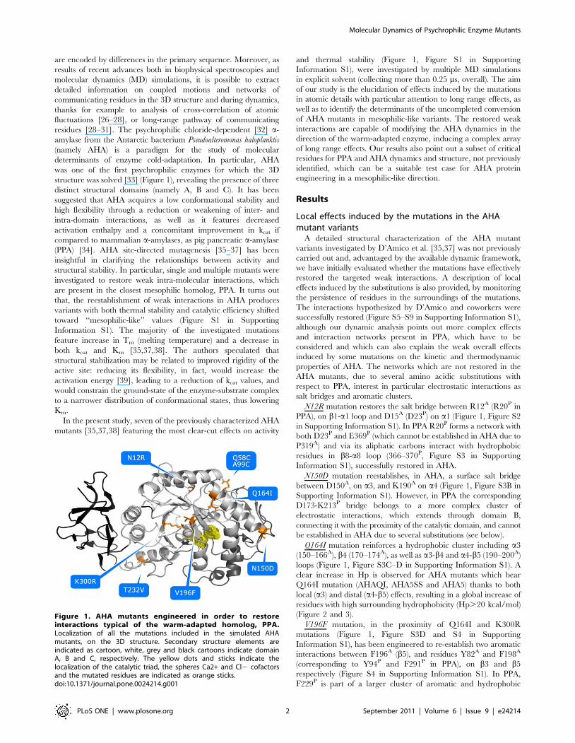
Figure 3. Rmsf in the essential subspace and surrounding hydrophobicity profiles, plotted as a function of the sequences of AHA(labels in the top row) and PPA (labels in the bottom row), and smoothed using window-averaging (window-size = 5 residues). Aschematic representation of the most frequently attained secondary structure is shown in the lower plot, where a-helices of the a/b-barrel structureof domain A are labelled according to their order in the primary sequence.doi:10.1371/journal.pone.0024214.g003
Molecular Dynamics of Psychrophilic Enzyme Mutants
PLoS ONE | www.plosone.org 3 September 2011 | Volume 6 | Issue 9 | e24214
residues, which also interact with the protein N-terminal extremity
(residues 1–13P); whereas in AHA the N-terminal is 8 residues
shorter, thus not allowing the extension of the network (Figure
S5A in Supporting Information S1). Moreover, the presence of
two arginines (R92P and R252P) in the macro-aromatic cluster of
PPA, and not in AHA, accounts for a further stabilizing
contribution due to cation-PI interactions. AHA mutants bearing
V196F (AHAVF, AHA5SS and AHA5) show a local increase in
the surrounding hydrophobicity (Figure 3), thanks to the favorable
packing induced by mutation, even in absence of the reconstruc-
tion of other more complex interactions.
T232V mutation strengthens a hydrophobic cluster between a6
and a6-b7, in the proximity of the interface between domains A
and C (Figure 1, Figure S5B–C in Supporting Information S1).
AHA variants bearing T232V mutation (AHATV, AHA5SS and
AHASS) show a higher persistence of hydrophobic residues and a
consequent increase in surrounding hydrophobicity (Figure 3),
including in particular L238A, F244A and W248A (Figure S5C in
Supporting Information S1).
K300R mutation, in the chloride-binding site and in the
proximity of the active site (Figure 1), should provide bi-dentate
coordination for the Cl2 ion, even if a more complex network of
interactions is mediated by R337P in PPA (Figure S5D and S6 in
Supporting Information S1). In fact, the mutation is located
among three macro-clusters of aromatic residues, conserved in
both AHA and PPA (Figure S6 in Supporting Information S1).
The K300R mutation has slight effects on the persistence of the
aromatic residues in the surrounding of the mutated residues
(Figure S5D in Supporting Information S1), while it strengthens a
salt bridge with D261 (R337P-D297P in PPA). This is in line with
the observed role of arginine if compared to lysine in an aromatic
context [40], role which is not ascribable to a intrinsic higher
cation-PI binding ability of Arg. In fact, the Arg side chain is larger
and less water-solvated than the cognate Lys thus, likely to benefit
from a better van der Waals interactions with aromatic rings.
Moreover, as suggested also by Thornton and colleagues [41], the
Arg side chain can still be involved in H-bonds or salt bridges
while simultaneously interacting with aromatic rings, whereas Lys
typically has to relinquish H-bonds to bind to aromatic residues.
Effects of the mutation on the structural and dynamicproperties of AHA
To achieve an overall description of the structural and dynamic
properties of the AHA mutants, several properties have been
analyzed, from H-bonds and surrounding hydrophobicity (Hp)
(Figure 2 and 3), rmsf profiles (Figure 3), the anisotropic
temperature factors of the essential subspace (Figure 4), most
relevant positive correlated motions (Figure 5), and salt bridge
interactions and networks (Figure 6, 7, Figure S7 and Table S1 in
Supporting Information S1), along with the clusters of salt-bridges
defined by a spatial proximity criterion (Figure 6, 7, Figure S7 and
Table S1 in Supporting Information S1).
In fact, it is well-known that the protein stability results from a
delicate balance between different weak intramolecular interac-
tions, which in turn also modulate protein dynamics. Electrostatic
interactions, and in particular salt-bridges, have been shown to
play a crucial role in protein stability [42–45], featuring both local
and distal variable effects [46,47].
Salt-bridges are generally highly flexible and cooperatively
organized in salt-bridge networks in the protein structure, strongly
influencing protein dynamics. Therefore, they are a suitable group
of intramolecular interactions which may be used as a reference to
identify dynamic intramolecular networks and how they are
modified by mutations. In light of these observation, we define not
only salt-bridge pairs and networks, according to their persistence
during the simulations, but also how they are organized in spatial
proximity clusters, as previously applied to other comparative
studies on extremophilic enzymes [48]. Our description does not
account for calculations of salt-bridge strength and its influence on
protein stability [46,47,49], which still lacks an accurate definition
in extremophilic a-amylases, both since experiments and simula-
tions at different temperatures are still not available and also since
most of the residues involved in salt-bridges which differ between
AHA and AHA mutants are generally solvent exposed (with a
solvent accessibility of their side chains generally higher than 30%
during the simulation time) and therefore likely not to be
negatively influenced by desolvation penalties.
Globally, few differences, mainly in domain A, are observed in
terms of H-bond content and surrounding hydrophobicity (Figure 2
and 3). All mutants show a reduced content of residues with low
surrounding hydrophobicity, an increase in residues characterized
by higher Hp values, especially the variants which include hydro-
phobic or aromatic mutations (AHA5SS, AHA5, AHATV, AH
AQI, AHAVF) (Figure 2 and 3), as also discussed above.
The patterns of correlated motions and flexibility of AHA and
PPA present distinctive features, which have been described in
details in a previous publication [50]. The main relevant aspect is
related to ba loops of different length and composition in the
surroundings of the active site. In particular, AHA has most of its
flexibility localized on the L7 loop (b7-a7) and the C-terminal
extremity of L3 (L3C), as well as at the L3–L5 interface, whereas
PPA has the highest flexibility scattered far from the catalytic site
toward the most solvent-exposed L8 and the N-terminal of L3
(L3N) (Figure 3 and 4).
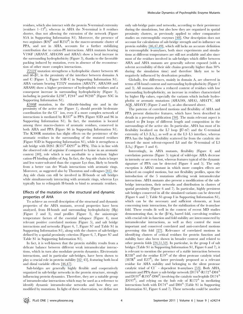
Interestingly, in AHA mutants, flexibility (Figure 4) and
correlation patterns (Figure 5) typical of wild-type AHA decrease
in intensity or are even lost, whereas features typical of the dynamic
signature of PPA can be detected (Figure 4 and 5). The only
exception is AHA5 mutant in which more complex effects are
induced on coupled motions, but not flexibility profiles, upon the
introduction of the 5 mutations affecting weak intramolecular
interactions. AHA mutants also present a modification of the salt-
bridge interactions, their networks and distribution in clusters of
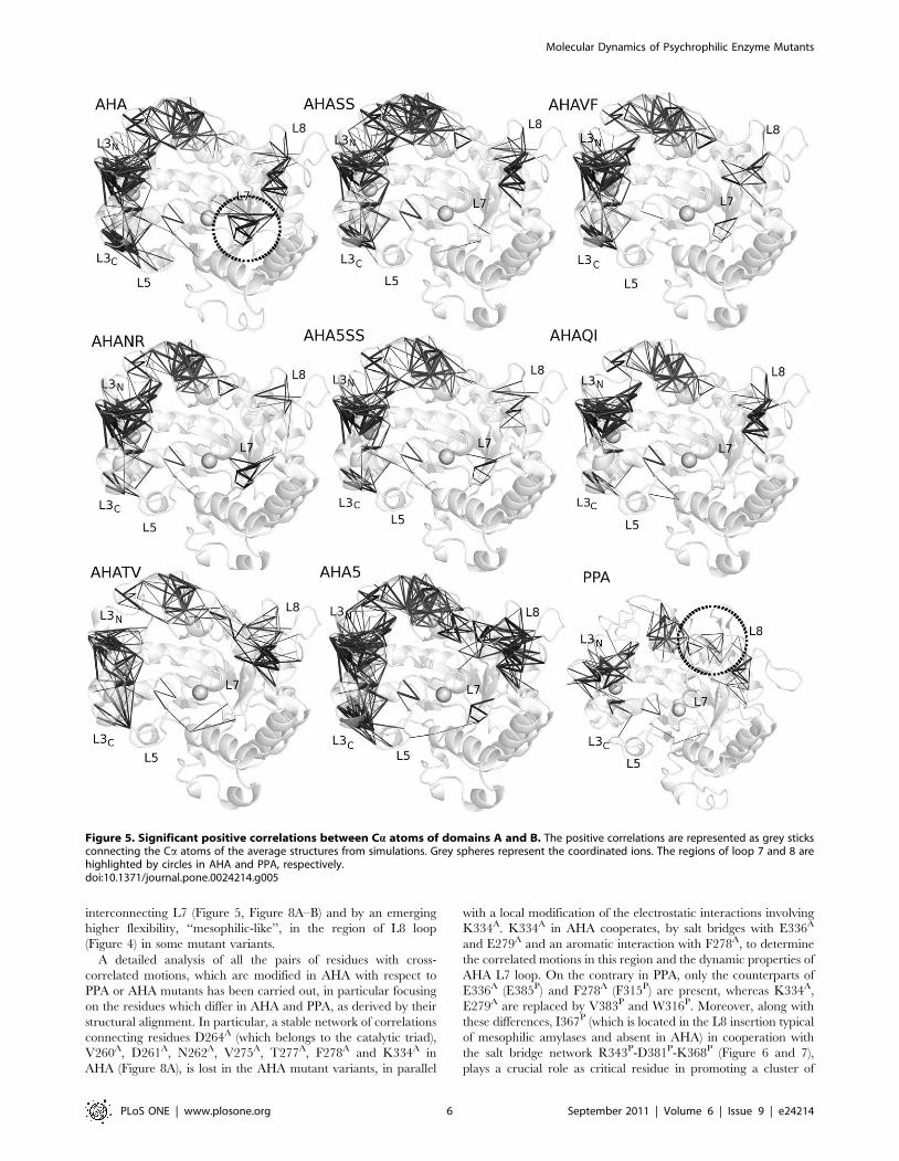
spatial proximity (Figure 6 and 7). In particular, highly persistent
salt bridges conserved in all the simulated variants can be detected
(Figure 6 and 7; Table S1-group I in Supporting Information S1),
which can be the necessary and sufficient elements, at least
concerning ionic interactions, for the stabilization of the a-amylase
fold. These results fit well in the context of recent MD studies
demonstrating that, in the (b/a)8 barrel fold, coevolving residues
with crucial role in function and fold stability are interconnected by
intramolecular interactions, as well as they control the most
important and conserved correlated and anti-correlated motions
governing this fold [27]. Relevance of correlated motions in
identifying clusters of critical residues for protein function and
stability have also been shown in broader context and related to
other protein folds [24,51,52]. In particular, in the group I of salt
bridges (Table S1 in Supporting Information S1, Figure 6 and 7), it
is relevant to mention the presence of a stable interaction between
R338A and the residue E19A of the silent protease catalytic triad
(387RP and E27P), the latter previously proposed as a relevant
residue for AHA stability and belonging to the silent protease
catalytic triad of Cl2 - dependent a-amylases [53]. Both AHA,
mutants and PPA share a salt-bridge network (D174A-R172A-D84A
and D197P-R195P-D96P) involving the catalytic nucleophile D174A
(D197P), and relying on the hub role of R172A in mediating
interactions both with D174A and D84A (Table S1 in Supporting
Information S1, Figure 6 and 7). These networks could be another
Molecular Dynamics of Psychrophilic Enzyme Mutants
PLoS ONE | www.plosone.org 4 September 2011 | Volume 6 | Issue 9 | e24214
common elements for a-amylase fold, allowing the correct
orientation of the nucleophile side chain in the catalytic site.
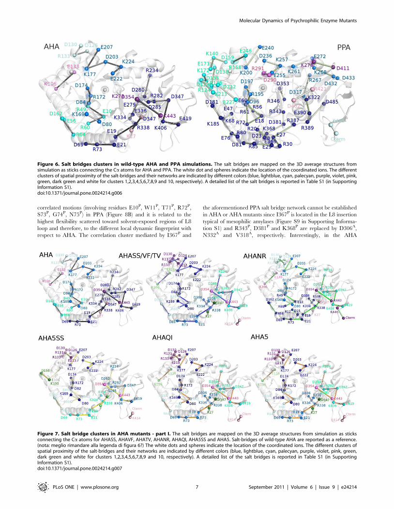
A group of salt bridges (group IV, Table S1 in Supporting
Information S1) and their networks, similar to that of PPA, and
differing from those of AHA, can be identified in the mutants
(Figure 7, 8, Table S1 in Supporting Information S1) and their
relevance will be discussed in details in the next sections. This
confirms a trend toward mesophilic-like properties in AHA
mutants, as well as long range effects induced by the mutations.
The selected mutations cannot restore the overall electrostatic
networks of the mesophilic amylase, since several salt bridges of
PPA lack suitable corresponding residues in AHA and the opposite
(groups II and III, Table S1 in Supporting Information S1). In
particular, several arginine residues of PPA are absent in AHA
(Table S1 in Supporting Information S1), accounting for
differential salt-bridges interactions and networks, in line with
the stabilizing effects induced by replacement of AHA lysines with
homoarginines [54].
Modification in dynamics properties around L7/L8 loopDomain A is the central and largest domain of a-amylases; it
accommodates the active site at the heart of its (b/a)8 barrel
structure. ba loops forming the substrate-binding cleft (Figure 1),
have a role in substrate recognition and processing [55–57] in
several a-amylases. In particular, loop L8 has been identified as an
initial contact point for the incoming substrate, mediating enzy-
matic specificity [58], whereas loop L7 may rearrange upon
substrate binding and release, contributing to the substrate
placement inside the active site [59].
The entity of L7 flexibility is decreased in most of the mutant
variants (Figure 3 and 4), accompanied by a concomitant
disappearance of one of the highest correlated networks
Figure 4. Anisotropic temperature factors computed for fluctuations in the essential subspace. The anisotropic temperature factors areshown as grey ellipsoids centred on the Ca atoms of the average structures from simulations. Grey spheres represent the coordinated ions. Theregions of loop 7 and 8 are highlighted by circles in AHA and PPA, respectively.doi:10.1371/journal.pone.0024214.g004
Molecular Dynamics of Psychrophilic Enzyme Mutants
PLoS ONE | www.plosone.org 5 September 2011 | Volume 6 | Issue 9 | e24214
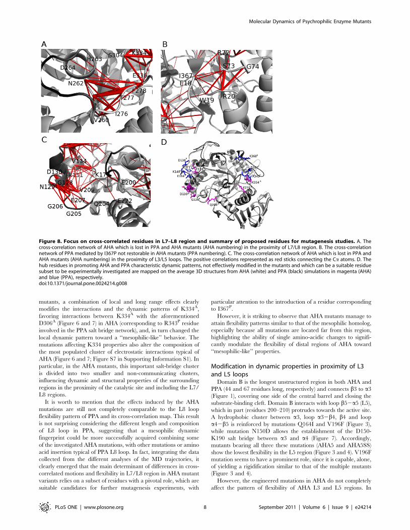
interconnecting L7 (Figure 5, Figure 8A–B) and by an emerging
higher flexibility, ‘‘mesophilic-like’’, in the region of L8 loop
(Figure 4) in some mutant variants.
A detailed analysis of all the pairs of residues with cross-
correlated motions, which are modified in AHA with respect to
PPA or AHA mutants has been carried out, in particular focusing
on the residues which differ in AHA and PPA, as derived by their
structural alignment. In particular, a stable network of correlations
connecting residues D264A (which belongs to the catalytic triad),
V260A, D261A, N262A, V275A, T277A, F278A and K334A in
AHA (Figure 8A), is lost in the AHA mutant variants, in parallel
with a local modification of the electrostatic interactions involving
K334A. K334A in AHA cooperates, by salt bridges with E336A
and E279A and an aromatic interaction with F278A, to determine
the correlated motions in this region and the dynamic properties of
AHA L7 loop. On the contrary in PPA, only the counterparts of
E336A (E385P) and F278A (F315P) are present, whereas K334A,
E279A are replaced by V383P and W316P. Moreover, along with
these differences, I367P (which is located in the L8 insertion typical
of mesophilic amylases and absent in AHA) in cooperation with
the salt bridge network R343P-D381P-K368P (Figure 6 and 7),
plays a crucial role as critical residue in promoting a cluster of
Figure 5. Significant positive correlations between Ca atoms of domains A and B. The positive correlations are represented as grey sticksconnecting the Ca atoms of the average structures from simulations. Grey spheres represent the coordinated ions. The regions of loop 7 and 8 arehighlighted by circles in AHA and PPA, respectively.doi:10.1371/journal.pone.0024214.g005
Molecular Dynamics of Psychrophilic Enzyme Mutants
PLoS ONE | www.plosone.org 6 September 2011 | Volume 6 | Issue 9 | e24214
correlated motions (involving residues E10P, W11P, T71P, R72P,
S73P, G74P, N75P) in PPA (Figure 8B) and it is related to the
highest flexibility scattered toward solvent-exposed regions of L8
loop and therefore, to the different local dynamic fingerprint with
respect to AHA. The correlation cluster mediated by I367P and
the aforementioned PPA salt bridge network cannot be established
in AHA or AHA mutants since I367P is located in the L8 insertion
typical of mesophilic amylases (Figure S9 in Supporting Informa-
tion S1) and R343P, D381P and K368P are replaced by D306A,
N332A and V318A, respectively. Interestingly, in the AHA
Figure 6. Salt bridges clusters in wild-type AHA and PPA simulations. The salt bridges are mapped on the 3D average structures fromsimulation as sticks connecting the Ca atoms for AHA and PPA. The white dot and spheres indicate the location of the coordinated ions. The differentclusters of spatial proximity of the salt-bridges and their networks are indicated by different colors (blue, lightblue, cyan, palecyan, purple, violet, pink,green, dark green and white for clusters 1,2,3,4,5,6,7,8,9 and 10, respectively). A detailed list of the salt bridges is reported in Table S1 (in SupportingInformation S1).doi:10.1371/journal.pone.0024214.g006
Figure 7. Salt bridge clusters in AHA mutants - part I. The salt bridges are mapped on the 3D average structures from simulation as sticksconnecting the Ca atoms for AHASS, AHAVF, AHATV, AHANR, AHAQI, AHA5SS and AHA5. Salt-bridges of wild-type AHA are reported as a reference.(nota: meglio rimandare alla legenda di figura 6?) The white dots and spheres indicate the location of the coordinated ions. The different clusters ofspatial proximity of the salt-bridges and their networks are indicated by different colors (blue, lightblue, cyan, palecyan, purple, violet, pink, green,dark green and white for clusters 1,2,3,4,5,6,7,8,9 and 10, respectively). A detailed list of the salt bridges is reported in Table S1 (in SupportingInformation S1).doi:10.1371/journal.pone.0024214.g007
Molecular Dynamics of Psychrophilic Enzyme Mutants
PLoS ONE | www.plosone.org 7 September 2011 | Volume 6 | Issue 9 | e24214
mutants, a combination of local and long range effects clearly
modifies the interactions and the dynamic patterns of K334A,
favoring interactions between K334A with the aforementioned
D306A (Figure 6 and 7) in AHA (corresponding to R343P residue
involved in the PPA salt bridge network), and, in turn changed the
local dynamic pattern toward a ‘‘mesophilic-like’’ behavior. The
mutations affecting K334 properties also alter the composition of
the most populated cluster of electrostatic interactions typical of
AHA (Figure 6 and 7; Figure S7 in Supporting Information S1). In
particular, in the AHA mutants, this important salt-bridge cluster
is divided into two smaller and non-communicating clusters,
influencing dynamic and structural properties of the surrounding
regions in the proximity of the catalytic site and including the L7/
L8 regions.
It is worth to mention that the effects induced by the AHA
mutations are still not completely comparable to the L8 loop
flexibility pattern of PPA and its cross-correlation map. This result
is not surprising considering the different length and composition
of L8 loop in PPA, suggesting that a mesophilic dynamic
fingerprint could be more successfully acquired combining some
of the investigated AHA mutations, with other mutations or amino
acid insertion typical of PPA L8 loop. In fact, integrating the data
collected from the different analyses of the MD trajectories, it
clearly emerged that the main determinant of differences in cross-
correlated motions and flexibility in L7/L8 region in AHA mutant
variants relies on a subset of residues with a pivotal role, which are
suitable candidates for further mutagenesis experiments, with
particular attention to the introduction of a residue corresponding
to I367P.
However, it is striking to observe that AHA mutants manage to
attain flexibility patterns similar to that of the mesophilic homolog,
especially because all mutations are located far from this region,
highlighting the ability of single amino-acidic changes to signifi-
cantly modulate the flexibility of distal regions of AHA toward
‘‘mesophilic-like’’ properties.
Modification in dynamic properties in proximity of L3and L5 loops
Domain B is the longest unstructured region in both AHA and
PPA (44 and 67 residues long, respectively) and connects b3 to a3
(Figure 1), covering one side of the central barrel and closing the
substrate-binding cleft. Domain B interacts with loop b52a5 (L5),
which in part (residues 200–210) protrudes towards the active site.
A hydrophobic cluster between a3, loop a32b4, b4 and loop
a42b5 is reinforced by mutations Q164I and V196F (Figure 3),
while mutation N150D allows the establishment of the D150-
K190 salt bridge between a3 and a4 (Figure 7). Accordingly,
mutants bearing all three these mutations (AHA5 and AHA5SS)
show the lowest flexibility in the L5 region (Figure 3 and 4). V196F
mutation seems to have a prominent role, since it is capable, alone,
of yielding a rigidification similar to that of the multiple mutants
(Figure 3 and 4).
However, the engineered mutations in AHA do not completely
affect the pattern of flexibility of AHA L3 and L5 regions. In
Figure 8. Focus on cross-correlated residues in L7–L8 region and summary of proposed residues for mutagenesis studies. A. Thecross-correlation network of AHA which is lost in PPA and AHA mutants (AHA numbering) in the proximity of L7/L8 region. B. The cross-correlationnetwork of PPA mediated by I367P not restorable in AHA mutants (PPA numbering). C. The cross-correlation network of AHA which is lost in PPA andAHA mutants (AHA numbering) in the proximity of L3/L5 loops. The positive correlations represented as red sticks connecting the Ca atoms. D. Thehub residues in promoting AHA and PPA characteristic dynamic patterns, not effectively modified in the mutants and which can be a suitable residuesubset to be experimentally investigated are mapped on the average 3D structures from AHA (white) and PPA (black) simulations in magenta (AHA)and blue (PPA), respectively.doi:10.1371/journal.pone.0024214.g008
Molecular Dynamics of Psychrophilic Enzyme Mutants
PLoS ONE | www.plosone.org 8 September 2011 | Volume 6 | Issue 9 | e24214
particular, the flexibility of AHA mutants is not displaced toward
the L3N part of the loop as in PPA (Figure 4), whereas more
remarkable effects can be identified in the decreased flexibility and
modification in cross-correlation patterns at the interface between
L3 and L5 (observable for example in AHASS, AHAVF, AHANR
and AHA5) (Figure 4 and 5). However, it is interesting to observe
that the highest flexibility of AHA at the interface between L5 and
L3 is strongly reduced in the mutant variants with some of them
displacing the flexibility toward more exposed regions of the L5
loop. In particular, the major modifications in the dynamic
properties of AHA mutants, at this site, rely on differences in the
salt bridge network mediated by D203A (E207A-K177A-D203A-
K224A-E222A) (Figure 7), which is interrupted in some mutants
and in PPA, (lacking D203A-K177A) as well as on the appearance
in the AHA mutants of a salt bridge between residues E213A and
R133A (whereas in PPA the corresponding glutamate residue,
E249P, provide a salt bridge in the same area but with different
orientation mediated by R158P), along with a general weakening
of the cross-correlation networks in this region (Figure 8C). In fact,
most of the AHA mutants lack or attenuate the correlated motions
(Figure 8C) connecting residues in the 128–130A region with
residues 204–206A. Moreover, some AHA mutants lose correla-
tion toward E222A (belonging to the salt bridge cluster mediated
by D203A). In the same area, PPA conserves only correlated
motions involving K200P (the homologous residue to K177A).
Moreover, AHA and its mutants maintained in this regions a salt
bridge cluster between D126A-R131A-D130A which is absent in
PPA, due to deletion and amino acidic substitutions.
In summary, the most relevant differences between AHA and
PPA in 128–130A and 204–206A regions are Q204A (L237P),
G128A (N152P) D126A (S150P) and D130A, R131A (missing in
PPA). Moreover, in PPA, the displacement of flexibility toward the
N-terminal portion of L3 loop is also related to the presence of two
salt bridge clusters which are absent, and not allowed to be
restored due to deletion or substitutions, in AHA and its mutants
(D159P-K140P-E171P and D138P-R214P). K106A is the corre-
sponding residue of R214P but mediates in AHA and its mutants
interactions with E138A (Q161P).
In order to re-establish a PPA-like behavior in AHA mutants, a
more complex network of interactions and communicating
residues should be restored in this area, including pivotal residues
in L3 which allow the flexibility to be displaced at the N-terminal
extremity of the loop. Otherwise, mutations of AHA residues at
the interface between L3 and L5 could be included to more
successfully abolish the network of cross-correlated motions
between the two loops, which is lacking in PPA.
Domain C: a structural domain shielding hydrophobicresidues of the catalytic domain
Domain C is composed of 8 b strands arranged in a greek-key
motif and tightly packed against a6, a7 and a 8 of the barrel
(Figure 1). The interface between domains C and A is rich in
aromatic and hydrophobic residues which contribute to the
stabilization of the interdomain interface, as well as they mediate
the extension of the hydrophobic core of domain A towards
domain C [33]. The sandwich-like structure of domain C features,
in both AHA and PPA, asymmetry between the two composing b-
sheets: the buried b-sheet constitutes the interface with domain A
and is stable and ordered, while the solvent-exposed b-sheet is
highly disordered, and split into 2 separate 2-stranded b-sheets
(Figure S9 in Supporting Information S1). The latter ones are
highly persistent in PPA, whereas in AHA main chain H-bonds
stabilizing the central part of these sheets are weakened.
Interestingly, most of the mutants display PPA-like H-bond
pattern (with the exception of AHA5SS) and the presence of a
PPA-like salt bridge involving the C-terminal residues of the
proteins, along with disappearance of some salt bridges at the
interface between domain A and C typical of AHA (Figure 7;
Table S1 in Supporting Information S1). This behaviour was
unexpected. In fact, domain C does not carry any of the studied
mutations, and nevertheless, shows structural and dynamical
differences in mutated forms, demonstrating that single mutations
have global and long range effects, affecting regions very distant
from the site of mutation.
Discussion
Intramolecular weak interactions have a fundamental role in
stabilizing protein structures. Special attention has been given to
their role in the context of thermal adaptation of proteins
[7,44,60], with particular regard to electrostatic interactions
[46,61–64]. The present study provides molecular details related
to the observed variations in thermal stability and kinetic
parameters of AHA mutants with respect to the wild-type cold-
and warm-adapted counterparts, as well as it points out long range
effects induced by the mutations.
Our MD investigation shows that the AHA mutations are
capable of eliciting effects, in agreement with the restoring or
strengthening of the target interactions, on the dynamic environ-
ment of the mutated residues in a mesophilic-like direction. If the
minor entity of the introduced mutations is considered, since few
residues in a multi-domain protein of about 500 amino acids are
mutated, it is striking to observe the wide range of different
dynamical behaviours these mutants exhibit.
Interestingly, the local effects, in the mutation sites, are also able
to modify the dynamic character of the mutants, producing
complex distal effects that underlie the intimate interplay between
hydrophobicity, electrostatic interactions and protein dynamics.
The mutations are extremely effective in modifying AHA
structural and flexibility properties, as hypothesized by the
experimental characterization [37], showing also that, in the case
of multiple mutants, the modification of activity and thermal
stability to mesophilic values is strongly advantaged. The ability of
AHA mutations to elicit a mesophilic-like behaviour is confirmed
in the results of MD simulations, where the comparison is
restricted exclusively to structural and dynamic properties. In fact,
the mutants exhibit secondary structure persistence, flexibility,
correlated motions, electrostatic interactions, and, hydrophobicity
in-between of those seen for the two wild-type enzymes.
However, fundamental differences both in the structure and the
sequence of the two wild-type enzymes still exist and make it
impossible for these mutations alone to reproduce the full set of
stabilizing local interactions seen in PPA. In fact, our investigation
also shown, in excellent agreement with the experimental values,
that the conversion of AHA in a mesophilic enzyme cannot be
completely achieved by the introduction of these mutations alone.
In fact, other substitutions, highlighted in our study, should be
considered to get more efficient modification of AHA toward the
mesophilic counterpart, as summarized in Figure 8D, as K334A or
I367P.
Our MD simulations of AHA mutant variants and their
comparison to dynamic features of the wild-type counterparts
also point out other long range effects induced by the mutations.
In particular, the effect of flexibility is largely apparent in regions
featuring high flexibility in AHA (loop 7) and PPA (loop 8), which
mainly cluster in regions near the active site and substrate-binding
groove, and long range effects are even transmitted to the C-
terminal domain. In this context, the present investigation
Molecular Dynamics of Psychrophilic Enzyme Mutants
PLoS ONE | www.plosone.org 9 September 2011 | Volume 6 | Issue 9 | e24214
provides atomic-detailed evidence of the ability of the selected
mutations to modulate the dynamic properties of AHA, and offers
unprecedented insight in the way this modulation takes place in a
large protein. Interestingly, a correlation (with a Pearson corre-
lation coefficient higher than 0.78) exists between the combination
of mesophilic-like salt bridge interactions acquired by the AHA
mutants (highlighted in Table S1 in Supporting Information S1)
and the experimentally determined kcat/Km and Tm values of
each mutant [37]. These interactions include, in particular, the
restored PPA-like D150A-K190A salt-bridge, the loss of K177A-
D203A salt-bridge and the modification of interactions involving
K334A.
In a broader context, the approach applied here, integrating
different properties related to protein dynamics (in particular the
parallel and integrated evaluation of correlated motions and
electrostatic interaction network), could be useful to predict and
rationalize the effects induced by a mutation on protein activity
and stability of an extremophilic enzymes, if comparative studies
with other temperature-adapted counterparts can be carried out.
Materials and Methods
System setup and molecular dynamics simulationsThe X-ray structure of AHA (PDB entry 1AQH [33]) was in silico
modified to model 7 mutant forms: 5 single mutants AHANR
(N12R), AHAQI (Q164I), AHAVF (V196F) and AHATV (T232V)
[35]; one double mutant AHASS (Q58C A99C) and 2 multiple
mutants AHA5 (N150D, Q164I, V196F, T232V, K300R) and
AHA5SS (Q58C, A99C, N150D, Q164I, V196F, T232V, K300R)
[37]. MD simulations were performed using GROMOS96 force-
field with the GROMACS software (www.gromacs.org). The
mutated forms of AHA were soaked in a dodecahedral box
including counterions (Na+) and SPC [65] water molecules using
periodic boundary conditions, with a minimum distance between
the solute and the box of 0.5 nm (average box size of 527.66 nm3
including 49893 atoms) and the systems were equilibrated in several
steps [50]. Productive MD simulations were performed in the NPT
ensemble (300K, 1 bar and 2 fs time-step with LINCS algorithm
[66]). Electrostatic interactions were calculated using PME [67]
summation scheme. Van der Waals and Coulomb interactions were
truncated at 0.8 nm and conformations stored every 2 ps. To
improve conformational sampling, six independent 6 ns simulations
were carried out for each system. MD simulations showing the
slowest convergence of root mean square deviation (rmsd) values
have been elongated up to 10 ns (run 2 of AHAQI and run 3 of
AHA5SS) and the first 0.2–2 ns depending on the system have been
discarded to assure stability of the trajectories. In fact, MD multiple-
replica approach allows a wider conformational sampling than only
few longer molecular dynamics simulations, especially in the case of
large multi-domain proteins as AHA is, if the stability of the
trajectory and convergence of the analyzed properties have been
carefully verified [68–71]. The equilibrated portions of the
simulations of the same system were joined into a macro-trajectory.
The conformational ensemble collected for each mutant is compa-
rable, in terms of simulations protocol, degree of essential motions
captured by the first principal components and number of frames in
the collected ensemble to that achieved for the wild-type cold-
(AHA) and warm-adapted (PPA) adapted amylases previously
published [50], in order to carry out a prompt comparison of
dynamical properties.
Molecular and structural propertiesSecondary structure was determined for all stored conforma-
tions using DSSP [72] and the most frequently attained secondary
structure for each residue was evaluated to obtain a residue-
dependent persistence degree of secondary structure profile
(PDSSP). Intramolecular hydrogen bonds (H-bonds) were evalu-
ated using both a donor-acceptor distance cut-off of 0.35 nm and
an acceptor-donor-hydrogen angular cut-off (#30u); ensemble
averages were normalized using the total number of donors and
acceptors.
Surrounding hydrophobicity (Hp) was computed for each
residue according to [73]. In particular Hp was computed for
each residue as the sum of hydrophobicity indexes, obtained from
thermodynamic transfer experiments [74], of the surrounding
residues within a 0.8 nm distance cutoff:
Hp(i)~XN
j=i
H(rd{rij)hj
where the summation runs over the whole protein, rij is the
distance between the Ca atoms of residues i and j, H(x) is the
Heaviside step function [H(x) = 1 if x$0, and zero otherwise], rd is
the distance cutoff and hj is the hydrophobicity index of residues j
in kcal/mol. The adopted cutoff has been demonstrated to be
suitable to characterize the hydrophobic behaviour of amino acids
and to accommodate both the local and non local interactions
[73,75].
Hp is therefore a measure of the tendency of the environment of
each residue to exclude water, computed on the basis of the 3D
structure using experimental information. Computed on equilib-
rium ensembles obtained from MD simulations, it gives an
indication of the relative strength and stability of the hydrophobic
clusters in a protein. It should be in principle able of identifying
the effects of a mutation taking into account both the short-range
effects, due to changes in the physico-chemical properties of the
mutated residues, and the long-range effects of mutation on
structure and dynamics. However, it is worth mentioning that the
accuracy of the computed values is hindered, independently of the
hydrophobicity scale adopted, by two strong assumptions. In fact,
by summing up the contributions of different residues, the Hp
calculation assumes that these contributions are independent and
therefore additive, which may lead to misleading conclusions. On
the other hand, the use of hydrophobicity indexes assumes that
these values are transferable from the experimental study to the
analysis of folded protein structures [76].
Residue-based comparison of the proteins was carried out
according to a manually-checked structural alignment with DALI
(Figure S8 in Supporting Information S1). Domains A, B and C of
AHA are composed of residues 1–86/147–356, 87–146 and 357–
448, respectively, whereas domains A, B and C of PPA are
composed of residues 1–98/170–404, 99–169 and 405–496.
Amino acid numbers in the text are labelled with a superscript
A or P, referring to the AHA or PPA sequences, respectively. The
8 b-strands and the 8 a-helices, composing the (b-a)-barrel of
domain A, are referred to as b1 to b8 and a1 to a8 according to
their order in the sequence.
Protein dynamicsThe essential dynamics (ED) analysis reveals high-amplitude
concerted motions in the equilibrated portion of the trajectories,
based on the diagonalization of the Ca covariance matrix of the
atomic positional fluctuations [77]. About 10 eigenvectors are
required to obtain the description of 70% of the variance in the
simulated proteins. The collection of the selected eigenvectors
describing the collective motions is termed essential subspace and
can describe protein motions at a reasonable level of accuracy.
Molecular Dynamics of Psychrophilic Enzyme Mutants
PLoS ONE | www.plosone.org 10 September 2011 | Volume 6 | Issue 9 | e24214
Motions described by the essential subspace have been mapped on
average 3D-structures by representing with ellipsoids (at proba-
bility 0.1) the computed anisotropic temperature factors obtained
from the per-residue Ca anisotropic U-tensors of cross-correlations
of motion. The average 3D-structure is defined as the sampled
conformation with the least distance (in terms of main-chain rmsd)
from the ensemble average of atomic positions. The cosine content
and overlap indexes as root mean square inner product have been
monitored to evaluate the sampling quality, according to standard
procedures applied in our laboratory [78].
The per-residue Ca root mean square fluctuation (rmsf) was
calculated with respect to the average structure, after projection of
the trajectories on the essential subspace. To properly assess the
rmsf convergence and the consistency of flexibility profiles, per-
residue rmsf profiles have been computed on partially overlapping
time-windows, starting from the first frame of the macro-
trajectories and moving onwards by steps of 500 ps. This
calculation has been repeated for different window lengths (1, 2,
5, 10, 15 and 20 ns) and an average rmsf profile for each time-
window has been calculated (Figure S10 in Supporting Informa-
tion S1).
Correlation plots were obtained by first computing Cacorrelation matrices [26] C(i,j), using non-overlapping averaging
windows of 1 ns (Figure S11 in Supporting Information S1), and
also compared, for validation, to correlations on averaging
windows of 2 and 3 ns. C(i,j) has been calculated according to,
Ci,j~c(i,j)
c(i,i)1=2c(j,j)1=2
Where c(i,j) is the covariance matrix of protein fluctuations
between residues i and j.
Only the most significant (|C(i,j)|.0.4) long range (|i2j|.12)
correlations have been considered. The cutoff of sequence distance
has been selected to exclude from the analysis correlations relative
to a-helices structure and contiguous residues in the primary
sequence. Moreover, since an average C(i,j) matrix has been
considered for the analysis (even if the single C(i,j) matrix relative
to each 1 ns window have been compared to the average one), a
cutoff of 0.4 in absolute value have been selected to identify
significant correlations and to exclude pairs of residues which are
poorly communicating each other and likely to be characterized
by uncoupled motions. In particular, on 1 ns time-scale, only
positively correlated motions are identified above this cutoff.
Moreover, in order to clearly identify differences in the patterns of
coupled motions of AHA and PPA functional regions, in the
correlation plots the correlations relative to secondary structural
elements (correlations within the b-barrel and correlations
between a-helices) have been discarded. Correlations were then
plotted on the 3D structures by connecting atoms i and j with lines,
with thickness proportional to C(i,j).
Salt bridge interactions and networksSalt bridges were identified as oppositely charged groups at less
than 0.4 nm of distance in at least 24% of the macro-trajectory
frames. In order to check the validity of the results also other
distance (0.45 and 0.5 nm) and persistence (20%) cutoffs were
employed (data not shown). The cut-off of 24%, in particular, was
selected as the persistence value which best divided the intera-
ctions dataset in well-separated groups, defined as signal and noise
(Figure S12 in Supporting Information S1), according to a
protocol previously applied [48] and summarized in Figure S12
in the Supporting Information S1. In particular, the distribution of
charge-charge pairs at a defined cutoff of distance has been
analyzed in terms of probability density function. It turns out that
there are several ion pairs at low persistence (,10%) which are
likely to be not relevant for protein structure and dynamics and
identified as ‘‘noise’’ signal. Whereas, at persistence greater of 30%
the number of ionic pairs is generally constant. Therefore, the
selected cutoff of 24% should be able to divide the dataset in the
two regions of low and high significance. This cutoff has been
calculated by two supervised classification methods, trained on a
set composed by two classes: noise (interactions below 10%) and
signal (interactions above 30%). A Support Vector Machine
(SVM) and a k-Nearest Neighbours (kNN, k = 4) classifiers, as
implemented in Matlab suite, have been trained on this set and
used to classify all the interactions between 10 and 30%.
To identify the clusters of spatial proximity of salt-bridges, the
residues involved in salt bridges have been represented as nodes of
an unrooted unoriented graph, in which two nodes were
connected by arcs if an interaction was found between them or
if two of them were at less than 5 residues of distance in the
sequence. An exhaustive search procedure was carried out on the
graph to isolate the networks of interactions, which have been
represented as interaction clusters (indicated by different colors in
Figure 6 and 7).
Supporting Information
Supporting Information S1 This file contains all thesupporting tables and figures for this article.
(PDF)
Acknowledgments
We thank Dr. Gaetano Invernizzi for fruitful discussion and comments,
along with a careful reading of the manuscript.
Author Contributions
Conceived and designed the experiments: EP. Performed the experiments:
EP MP MT. Analyzed the data: EP MP MT. Wrote the paper: EP MP
LDG.
References
1. Trivedi S, Gehlot HS, Rao SR (2006) Protein thermostability in Archaea and
Eubacteria. Genet Mol Res 5: 816–827.
2. D’Amico S, Collins T, Marx J-C, Feller G, Gerday C (2006) Psychrophilic
microorganisms: challenges for life. EMBO Rep 7: 385–389.
3. Rodrigues DF, Tiedje JM (2008) Coping with our cold planet. Appl Environ
Microb 74: 1677–1686.
4. Gerday C, Aittaleb M, Bentahir M, Chessa JP, Claverie P, et al. (2000) Cold-
adapted enzymes: from fundamentals to biotechnology. Trends Biotechnol 18:
103–107.
5. Joseph B, Ramteke PW, Thomas G (2008) Cold active microbial lipases: Some
hot issues and recent developments. Biotechnol Adv 26: 457–470.
6. Siddiqui KS, Cavicchioli R (2006) Cold-adapted enzymes. Annu Rev Biochem
75: 403–433.
7. Gianese G, Bossa F, Pascarella S (2002) Comparative structural analysis of
psychrophilic and meso- and thermophilic enzymes. Proteins 47: 236–249.
8. Casanueva A, Tuffin M, Cary C, Cowan DA (2010) Molecular adaptations to
psychrophily: the impact of ‘omic’ technologies. Trends Microbiol 18: 374–381.
9. Georlette D, Blaise V, Collins T, D’Amico S, Gratia E, et al. (2004) Some like it
cold: biocatalysis at low temperatures. FEMS Microbiol Rev 28: 25–42.
10. Smalas AO, Leiros HK, Os V, Willassen NP (2000) Cold adapted enzymes.
Biotechnol Annu Rev 6: 1–57.
11. Fields PA (2001) Review: Protein function at thermal extremes: balancing
stability and flexibility. Comp Biochem Physiol A Mol Integr Physiol 129:
417–431.
12. Feller G (2010) Protein stability and enzyme activity at extreme biological
temperatures. J Phys-Condens Mat 22: 323101–323116.
Molecular Dynamics of Psychrophilic Enzyme Mutants
PLoS ONE | www.plosone.org 11 September 2011 | Volume 6 | Issue 9 | e24214
13. Arnold FH, Wintrode PL, Miyazaki K, Gershenson A (2001) How enzymesadapt: lessons from directed evolution. Trends Biochem Sci 26: 100–106.
14. Miyazaki K, Wintrode PL, Grayling RA, Rubingh DN, Arnold FH (2000)Directed evolution study of temperature adaptation in a psychrophilic enzyme.
J Mol Biol 297: 1015–1026.
15. Fedøy A-E, Yang N, Martinez A, Leiros H-KS, Steen IH (2007) Structural and
functional properties of isocitrate dehydrogenase from the psychrophilic
bacterium Desulfotalea psychrophila reveal a cold-active enzyme with anunusual high thermal stability. J Mol Biol 372: 130–149.
16. Leiros H-KS, Pey AL, Innselset M, Moe E, Leiros I, et al. (2007) Structure ofphenylalanine hydroxylase from Colwellia psychrerythraea 34H, a monomeric
cold active enzyme with local flexibility around the active site and high overallstability. J Biol Chem 282: 21973–21986.
17. Wintrode PL, Arnold FH (2000) Temperature adaptation of enzymes: lessonsfrom laboratory evolution. Adv Protein Chem 55: 161–225.
18. Karplus M, Kuriyan J (2005) Molecular dynamics and protein function. P NatlAcad Sci USA 102: 6679–6685.
19. Henzler-Wildman KA, Lei M, Thai V, Kerns SJ, Karplus M, et al. (2007) Ahierarchy of timescales in protein dynamics is linked to enzyme catalysis. Nature
450: 913–U927.
20. Marcos E, Crehuet R, Bahar I (2010) On the conservation of the slow
conformational dynamics within the amino acid kinase family: the NAGK
paradigm. Plos Comput Biol 6: 14.
21. Villali J, Kern D (2010) Choreographing an enzyme’s dance. Curr Opin Chem
Biol 14: 636–643.
22. Nashine VC, Hammes-Schiffer S, Benkovic SJ (2010) Coupled motions in
enzyme catalysis. Curr Opin Chem Biol 14: 644–651.
23. Maguid S, Fernandez-Alberti S, Echave J (2008) Evolutionary conservation of
protein vibrational dynamics. Gene 422: 7–13.
24. Law AB, Fuentes EJ, Lee AL (2009) Conservation of side-chain dynamics within
a protein family. J Am Chem Soc 131: 6322–6323.
25. Papaleo E, Pasi M, Riccardi L, Sambi I, Fantucci P, et al. (2008) Protein
flexibility in psychrophilic and mesophilic trypsins. Evidence of evolutionaryconservation of protein dynamics in trypsin-like serine-proteases. FEBS Lett 582:
1008–1018.
26. Hunenberger PH, Mark AE, Vangunsteren WF (1995) Fluctuation and cross-
correlation analysis of protein motions observed in nanosecond molecular-
dynamics simulations. J Mol Biol 252: 492–503.
27. Shen HB, Xu F, Hu HR, Wang FF, Wu Q, et al. (2008) Coevolving residues of
(beta/alpha)(8)-barrel proteins play roles in stabilizing active site architectureand coordinating protein dynamics. J Struct Biol 164: 281–292.
28. Johnson E (2011) NMR order parameters calculated in an expanding referenceframe: identifying sites of short- and long-range motion. J Biomol NMR 50:
59–70.
29. Morra G, Verkhivker G, Colombo G (2009) Modeling signal propagation
mechanisms and ligand-based conformational dynamics of the Hsp90 molecular
chaperone full-length dimer. Plos Comput Biol 5.
30. Amaro RE, Sethi A, Myers RS, Davisson VJ, Luthey-Schulten ZA (2007) A
network of conserved interactions regulates the allosteric signal in a glutamineamidotransferase. Biochemistry-US 46: 2156–2173.
31. Vijayabaskar MS, Vishveshwara S (2010) Interaction energy based proteinstructure networks. Biophys J 99: 3704–3715.
32. Aghajari N, Feller G, Gerday C, Haser R (2002) Structural basis of alpha-amylase activation by chloride. Protein Sci 11: 1435–1441.
33. Aghajari N, Feller G, Gerday C, Haser R (1998) Structures of the psychrophilicAlteromonas haloplanctis alpha-amylase give insights into cold adaptation at a
molecular level. Struct Fold Des 6: 1503–1516.
34. Feller G, Payan F, Theys F, Qian M, Haser R, et al. (1994) Stability and
structural analysis of alpha-amylase from the antarctic psychrophile Alteromonashaloplanctis A23. Eur J Biochem 222: 441–447.
35. D’Amico S, Gerday C, Feller G (2001) Structural determinants of cold
adaptation and stability in a large protein. J Biol Chem 276: 25791–25796.
36. D’Amico S, Gerday C, Feller G (2002) Structural determinants of cold
adaptation and stability in a psychrophilic alpha-amylase. Biologia 57: 213–219.
37. D’Amico S, Gerday C, Feller G (2003) Temperature adaptation of proteins:
engineering mesophilic-like activity and stability in a cold-adapted alpha-amylase. J Mol Biol 332: 981–988.
38. D’Amico S, Gerday C, Feller G (2002) Dual effects of an extra disulfide bond onthe activity and stability of a cold-adapted alpha-amylase. J Biol Chem 277:
46110–46115.
39. Lonhienne T, Gerday C, Feller G (2000) Psychrophilic enzymes: revisiting the
thermodynamic parameters of activation may explain local flexibility. BBA-Protein Struct M 1543: 1–10.
40. Gallivan JP, Dougherty DA (1999) Cation-pi interactions in structural biology.P Natl Acad Sci USA 96: 9459–9464.
41. Mitchell JBO, Nandi CL, McDonald IK, Thornton JM, Price SL (1994) Amino/
aromatic interactions in proteins - is the evidence stacked agains hydrogen-bonding? J Mol Biol 239: 315–331.
42. de Bakker PIW, Hunenberger PH, McCammon JA (1999) Molecular dynamicssimulations of the hyperthermophilic protein Sac7d from Sulfolobus acidocal-
darius: contribution of salt bridges to thermostability. J Mol Biol 285:1811–1830.
43. Kumar S, Nussinov R (2002) Close-range electrostatic interactions in proteins.Chembiochem 3: 604–617.
44. Bae E, Phillips GN (2004) Structures and analysis of highly homologouspsychrophilic, mesophilic, and thermophilic adenylate kinases. J Biol Chem 279:
28202–28208.
45. Missimer JH, Steinmetz MO, Baron R, Winkler FK, Kammerer RA, et al.(2007) Configurational entropy elucidates the role of salt-bridge networks in
protein thermostability. Protein Sci 16: 1349–1359.
46. Kumar S, Nussinov R (2001) How do thermophilic proteins deal with heat? Cell
Mol Life Sci 58: 1216–1233.
47. Bosshard HR, Marti DN, Jelesarov I (2004) Protein stabilization by salt bridges:concepts, experimental approaches and clarification of some misunderstandings.
J Mol Recognit 17: 1–16.
48. Tiberti M, Papaleo E (2011) Dynamic properties of extremophilic subtilisin-like
serine-proteases. J Struct Biol 174: 69–83.
49. Kumar S, Nussinov R (2002) Relationship between ion pair geometries andelectrostatic strengths in proteins. Biophys J 83: 1595–1612.
50. Pasi M, Riccardi L, Fantucci P, Gioia LD, Papaleo E (2009) Dynamic properties
of a psychrophilic alpha-amylase in comparison with a mesophilic homologue.J Phys Chem B 113: 13585–13595.
51. Raimondi F, Orozco M, Fanelli F (2010) Deciphering the deformation modesassociated with function retention and specialization in members of the Ras
superfamily. Structure 18: 402–414.
52. Angelova K, Felline A, Lee M, Patel M, Puett D, et al. (2010) Conserved aminoacids participate in the structure networks deputed to intramolecular
communication in the lutropin receptor. Cell Mol Life Sci 68: 1227–1239.
53. Marx JC, Poncin J, Simorre JP, Ramteke PW, Feller G (2008) The noncatalytic
triad of alpha-amylases: A novel structural motif involved in conformational
stability. Proteins 70: 320–328.
54. Siddiqui KS, Poljak A, Guilhaus M, De Francisci D, Curmi PMG, et al. (2006)
Role of lysine versus arginine in enzyme cold-adaptation: Modifying lysine tohomo-arginine stabilizes the cold-adapted alpha-amylase from Pseudoalteramo-
nas haloplanktis. Proteins 64: 486–501.
55. Brayer GD, Luo YG, Withers SG (1995) The structure of human pancreaticalphas amylase at 1.8 Angstrom resolution and comparisons with related
enzymes. Protein Sci 4: 1730–1742.
56. Andre G, Tran V (2004) Putative implication of alpha-amylase loop 7 in themechanism of substrate binding and reaction products release. Biopolymers 75:
95–108.
57. Gottschalk TE, Tull D, Aghajari N, Haser R, Svensson B (2001) Specificity
modulation of barley alpha-amylase through biased random mutagenesis
involving a conserved tripeptide in betaRalpha loop 7 of the catalytic (beta/alpha)(8)-barrel domain. Biochemistry-US 40: 12844–12854.
58. Aghajari N, Feller G, Gerday C, Haser R (1998) Crystal structures of thepsychrophilic alpha-amylase from Alteromonas haloplanctis in its native form
and complexed with an inhibitor. Protein Sci 7: 564–572.
59. Ramasubbu N, Ragunath C, Mishra PJ (2003) Probing the role of a mobile loopin substrate binding and enzyme activity of human salivary amylase. J of Mol
Biol 325: 1061–1076.
60. Zhu S, Elcock AH (2010) A complete thermodynamic characterization of
electrostatic and hydrophobic associations in the temperature range 0 to 100
degrees C from explicit-solvent molecular dynamics simulations. J Chem TheoryComput 6: 1293–1306.
61. Kumar S, Nussinov R (2004) Different roles of electrostatics in heat and in cold:adaptation by citrate synthase. Chembiochem 5: 280–290.
62. Elcock AH (1998) The stability of salt bridges at high temperatures: implications
for hyperthermophilic proteins. J Mol Biol 284: 489–502.
63. Goldstein RA (2007) Amino-acid interactions in psychrophiles, mesophiles,
thermophiles, and hyperthermophiles: Insights from the quasi-chemicalapproximation. Protein Sci 16: 1887–1895.
64. Sigurdardottir AG, Arnorsdottir J, Thorbjarnardottir SH, Eggertsson G,
Suhre K, et al. (2009) Characteristics of mutants designed to incorporate anew ion pair into the structure of a cold adapted subtilisin-like serine proteinase.
Biochim Biophys Acta 1794: 512–518.
65. Fuhrmans M, Sanders BP, Marrink SJ, de Vries AH (2010) Effects of bundlingon the properties of the SPC water model. Theor Chem Acc 125: 335–344.
66. Hess B, Bekker H, Berendsen HJC, Fraaije J (1997) LINCS: A linear constraintsolver for molecular simulations. J Comput Chem 18: 1463–1472.
67. Darden T, York D, Pedersen L (1993) Particle-Mesh Ewald - an N*log(N)
method for Ewald sums in large systems. J Chem Phys 98: 10089–10092.
68. Caves LS, Evanseck JD, Karplus M (1998) Locally accessible conformations of
proteins: multiple molecular dynamics simulations of crambin. Protein Sci 7:649–666.
69. Friedman R, Caflisch A (2010) On the orientation of the catalytic dyad in
aspartic proteases. Proteins 78: 1575–1582.
70. Loccisano AE, Acevedo O, DeChancie J, Schulze BG, Evanseck JD (2004)
Enhanced sampling by multiple molecular dynamics trajectories: carbonmonoxymyoglobin 10 micros A0RA(1–3) transition from ten 400 picosecond simula-
tions. J Mol Graph Model 22: 369–376.
71. Monticelli L, Sorin EJ, Tieleman DP, Pande VS, Colombo G (2008) Molecularsimulation of multistate peptide dynamics: a comparison between microsecond
timescale sampling and multiple shorter trajectories. J Comput Chem 29:1740–1752.
72. Kabsch W, Sander C (1983) Dictionary of protein secondary structure: pattern
recognition of hydrogen-bonded and geometrical features. Biopolymers 22:2577–2637.
Molecular Dynamics of Psychrophilic Enzyme Mutants
PLoS ONE | www.plosone.org 12 September 2011 | Volume 6 | Issue 9 | e24214
73. Motono C, Gromiha MM, Kumar S (2008) Thermodynamic and kinetic
determinants of Thermotoga maritima cold shock protein stability: A structural
and dynamic analysis. Proteins 71: 655–669.
74. Jones DD (1975) Amino-acid properties and side-chain orientation in proteins -
Cross-correlation approach. J Theor Biol 50: 167–183.
75. Debe DA, Goddard WA (1999) First principles prediction of protein folding
rates. J Mol Biol 294: 619–625.
76. Southall NT, Dill KA, Haymet ADJ (2002) A view of the hydrophobic effect
(vol 106, pg 523, 2002). J Phys Chem B 106: 2812–2812.77. Amadei A, Linssen AB, Berendsen HJ (1993) Essential dynamics of proteins.
Proteins 17: 412–425.
78. Papaleo E, Mereghetti P, Fantucci P, Grandori R, De Gioia L (2009) Free-energy landscape, principal component analysis, and structural clustering to
identify representative conformations from molecular dynamics simulations: Themyoglobin case. J Mol Graph Model 27: 889–899.
Molecular Dynamics of Psychrophilic Enzyme Mutants
PLoS ONE | www.plosone.org 13 September 2011 | Volume 6 | Issue 9 | e24214
Related Documents


![Kobe University Repository : Kernelovule development along the proximal-distal axis [24]. ant mutants fail to both initiate and elongate the integu-ment [25,26]. The aberrant ovule](https://static.cupdf.com/doc/110x72/5eb3709f1488434a551ee9ff/kobe-university-repository-ovule-development-along-the-proximal-distal-axis-24.jpg)