233 Molecular Diversity of K + Channels WILLIAM A. COETZEE, a,b,c YIMY AMARILLO, b JOANNA CHIU, c ALAN CHOW, b DAVID LAU, b TOM MCCORMACK, b HERMAN MORENO, b MARCELA S. NADAL, b ANDER OZAITA, b DAVID POUNTNEY, c MICHAEL SAGANICH, b ELEAZAR VEGA-SAENZ DE MIERA, b AND BERNARDO RUDY b,d Departments of b Physiology and Neuroscience, c Pediatric Cardiology, and d Biochemistry, New York University School of Medicine, 550 First Avenue, New York, New York 10016, USA ABSTRACT: K + channel principal subunits are by far the largest and most diverse of the ion channels. This diversity originates partly from the large number of genes cod- ing for K + channel principal subunits, but also from other processes such as alterna- tive splicing, generating multiple mRNA transcripts from a single gene, heteromeric assembly of different principal subunits, as well as possible RNA editing and post- translational modifications. In this chapter, we attempt to give an overview (mostly in tabular format) of the different genes coding for K + channel principal and accessory subunits and their genealogical relationships. We discuss the possible correlation of different principal subunits with native K + channels, the biophysical and pharmaco- logical properties of channels formed when principal subunits are expressed in heter- ologous expression systems, and their patterns of tissue expression. In addition, we devote a section to describing how diversity of K + channels can be conferred by het- eromultimer formation, accessory subunits, alternative splicing, RNA editing and posttranslational modifications. We trust that this collection of facts will be of use to those attempting to compare the properties of new subunits to the properties of oth- ers already known or to those interested in a comparison between native channels and cloned candidates. he first molecular components of K + channels were identified only about a decade ago by molecular cloning methods. 1−5 However, the number of cloned and character- ized components has grown so much that reviewing the molecular biology of K + channels has become a daunting, if not impossible, task in a chapter of these dimensions. Several excellent reviews, discussing specific aspects of this subject have appeared in recent years. 6−16 The present chapter presents a review as comprehensive as possible of all the K + channel subunits known to date, aimed mainly at scientists who might be interested in finding possible molecular correlates for their functional findings. We limited the scope by focusing primarily on mammalian K + channel principal and auxiliary subunits. Most of the data are presented in tabular format. It is possible that our tables have missing and even erroneous data. We wish to apologize to our colleagues for these errors and omissions and will appreciate receiving comments. Given space limitations we have not included specific references from which the data in the tables were extracted. To alleviate this constraint, we intend to publicize a web page e on which this information will be accessible along with additional data that similarly had to be omitted because of space considerations. The availability of K + channel cDNAs has allowed enormous progress in the under- standing of the structure and molecular mechanisms of function of K + channels. Important T a Corresponding authors: William A. Coetzee, D.Sc., Pediatric Cardiology, TH517, New York Uni- versity School of Medicine, 550 First Avenue, New York, New York 10016. Phone: 212-263-8518; fax: 212-263-1393; e-mail: [email protected] and Bernardo Rudy, Department of Physiology and Neuroscience, New York University School of Medicine, 550 First Avenue, New York, New York 10016. Phone: 212-263-0431; fax: 212-689-9060; e-mail; rudyb01@ mcrcr6.med.nyu.edu e http://k-channels.med.nyu.edu/

Welcome message from author
This document is posted to help you gain knowledge. Please leave a comment to let me know what you think about it! Share it to your friends and learn new things together.
Transcript

233
Molecular Diversity of K+ ChannelsWILLIAM A. COETZEE,a,b,c YIMY AMARILLO,b JOANNA CHIU,c ALAN CHOW,b DAVID LAU,b TOM MCCORMACK,b HERMAN MORENO,b MARCELA S. NADAL,b ANDER OZAITA,b DAVID POUNTNEY,c MICHAEL SAGANICH,b
ELEAZAR VEGA-SAENZ DE MIERA,b AND BERNARDO RUDYb,d
Departments of bPhysiology and Neuroscience, cPediatric Cardiology, and dBiochemistry, New York University School of Medicine, 550 First Avenue, New York, New York 10016, USA
ABSTRACT: K+ channel principal subunits are by far the largest and most diverse ofthe ion channels. This diversity originates partly from the large number of genes cod-ing for K+ channel principal subunits, but also from other processes such as alterna-tive splicing, generating multiple mRNA transcripts from a single gene, heteromericassembly of different principal subunits, as well as possible RNA editing and post-translational modifications. In this chapter, we attempt to give an overview (mostly intabular format) of the different genes coding for K+ channel principal and accessorysubunits and their genealogical relationships. We discuss the possible correlation ofdifferent principal subunits with native K+ channels, the biophysical and pharmaco-logical properties of channels formed when principal subunits are expressed in heter-ologous expression systems, and their patterns of tissue expression. In addition, wedevote a section to describing how diversity of K+ channels can be conferred by het-eromultimer formation, accessory subunits, alternative splicing, RNA editing andposttranslational modifications. We trust that this collection of facts will be of use tothose attempting to compare the properties of new subunits to the properties of oth-ers already known or to those interested in a comparison between native channelsand cloned candidates.
he first molecular components of K+ channels were identified only about a decadeago by molecular cloning methods.1−5 However, the number of cloned and character-
ized components has grown so much that reviewing the molecular biology of K+ channelshas become a daunting, if not impossible, task in a chapter of these dimensions. Severalexcellent reviews, discussing specific aspects of this subject have appeared in recentyears.6−16 The present chapter presents a review as comprehensive as possible of all the K+
channel subunits known to date, aimed mainly at scientists who might be interested infinding possible molecular correlates for their functional findings. We limited the scope byfocusing primarily on mammalian K+ channel principal and auxiliary subunits. Most of thedata are presented in tabular format. It is possible that our tables have missing and evenerroneous data. We wish to apologize to our colleagues for these errors and omissions andwill appreciate receiving comments. Given space limitations we have not included specificreferences from which the data in the tables were extracted. To alleviate this constraint, weintend to publicize a web pagee on which this information will be accessible along withadditional data that similarly had to be omitted because of space considerations.
The availability of K+ channel cDNAs has allowed enormous progress in the under-standing of the structure and molecular mechanisms of function of K+ channels. Important
T
aCorresponding authors: William A. Coetzee, D.Sc., Pediatric Cardiology, TH517, New York Uni-versity School of Medicine, 550 First Avenue, New York, New York 10016. Phone: 212-263-8518; fax:212-263-1393; e-mail: [email protected] and Bernardo Rudy, Department of Physiologyand Neuroscience, New York University School of Medicine, 550 First Avenue, New York, New York10016. Phone: 212-263-0431; fax: 212-689-9060; e-mail; rudyb01@ mcrcr6.med.nyu.edu
ehttp://k-channels.med.nyu.edu/

234 ANNALS NEW YORK ACADEMY OF SCIENCES
new insights into the mechanisms of ionic selectivity, voltage- and calcium-dependent gat-ing, inactivation and blockade of these channels have been obtained. These efforts recentlyculminated with the crystallization and high-resolution structural analysis of a K+
channel17—the first natural membrane channel for which high-resolution real structuralinformation is now available. This work provided strong evidence in favor of a tetramericK+ channel structure. This structure was first suggested based on the similarities betweensix transmembrane K+ channel subunits and each of the four internally homologousrepeats of Na+ and Ca2+ channels18 and was supported experimentally.17,19,20 In this struc-ture, four independent subunits or homologous repeats of one or two subunits form theinfrastructure of a channel with a fourfold symmetry around a central pore. We can expectthat this breakthrough will bring a deeper and accelerated understanding of the structureand function of K+ channels.
Less progress has been made in understanding the physiological significance of theenormous molecular diversity of K+ channel protein subunits (TABLE 1f). Over 100 differ-ent proteins, subunits of distinct types of K+ channels, have been identified to date, and thelist is rapidly growing (FIG. 1). In addition to the pore-forming or principal subunits (often
FIGURE 1. Schematic representation of the three groups of K+ channel principal subunits. They areclassified into three groups in terms of their predicted membrane topology—those that have sixtransmembrane domains (TMDs), those with four transmembrane domains and those with only twotransmembrane domains. Each group of principal subunits is divided into discrete families on thebasis of sequence similarity (see FIGS. 2 and 3). Each family can be further subdivided into severalsubfamilies, which often contain several closely related subfamily members. A functional classifica-tion places the voltage- and Ca2+-regulated K+ channels in the 6TMD group, the “leak” K+ channelsin the 4TMD group, and the inward rectifier K+ (Kir) channels in the 2TMD group. Also shown inthe figure are some of the auxiliary subunits that have been shown to alter expression levels and/orkinetics of K+ channel principal subunits when expressed in heterologous expression systems. Forclarity, they are grouped together with the principal subunits with which they have been shown tointeract (see text for more details).
f All tables appear at the end of the paper.
COETZEE et al.: MOLECULAR DIVERSITY OF K+ CHANNELS 235
called α subunits), which determine the infrastructure of the channel, many K+ channels(like Na+ and Ca2+ channels) contain auxiliary proteins that can modify the properties ofthe channels, often significantly. Most of the known principal K+ channel subunits expressin heterologous expression systems as functional homomultimeric channel complexes.However, some principal subunits do not form functional homomultimeric channels, butmust coassemble with other (similar) subunits for expression of functional channels (e.g.,G-protein−activated K+ channels; GIRKs). These subunits may be called coassembly prin-cipal subunits. All known principal subunits of K+ channels show a certain amount ofsequence and structural similarity, and they might all be related through evolution. Thereis sufficient primary sequence similarity between the different principal subunits thatmembers of new families of K+ channel proteins have been discovered by degenerate PCRor by screening databases of “expressed sequence tags” (ESTs) and sequences depositedby various genome projects. It remains possible, however, that some unknown K+ channelsmay have principal subunits that are unrelated to those known today.
Auxiliary subunits (sometimes referred to as β subunits) have primary sequences notresembling principal subunits. They interact with channel complexes containing principalsubunits and may alter their electrophysiological or biophysical properties, expression lev-els, or expression patterns. In addition, many K+ channel molecular complexes interactwith additional proteins such as regulatory enzymes and elements of the cytoskeleton (seechapters by Morgan Sheng and John Adelman, this volume). We term these associatedproteins, with the understanding that the distinction between auxiliary subunits and associ-ated proteins may not be simple on occasions. Since K+ channels are thought to be multi-mers of principal subunits, which may form heteromeric channels with closely relatedprincipal subunits in various combinations, the number of possible distinct K+ channelsbased on these different combinations may be in the order of hundreds, if not thousands(see DIVERSITY CONFERRED BY HETEROMULTIMER FORMATION..., below). However, it is notknown how much of this potential diversity is actually used in native cells. A major task offuture research is to identify physiological roles of the cloned proteins, starting with theidentification of native channels containing specific types of cloned subunits. This is par-ticularly important because most of the cloning work has been done in the absence of priorisolation of native proteins. Therefore, the exact relationship between the molecular com-ponents identified by cloning and native channels is, in most cases, not known and must bea priority for future research.
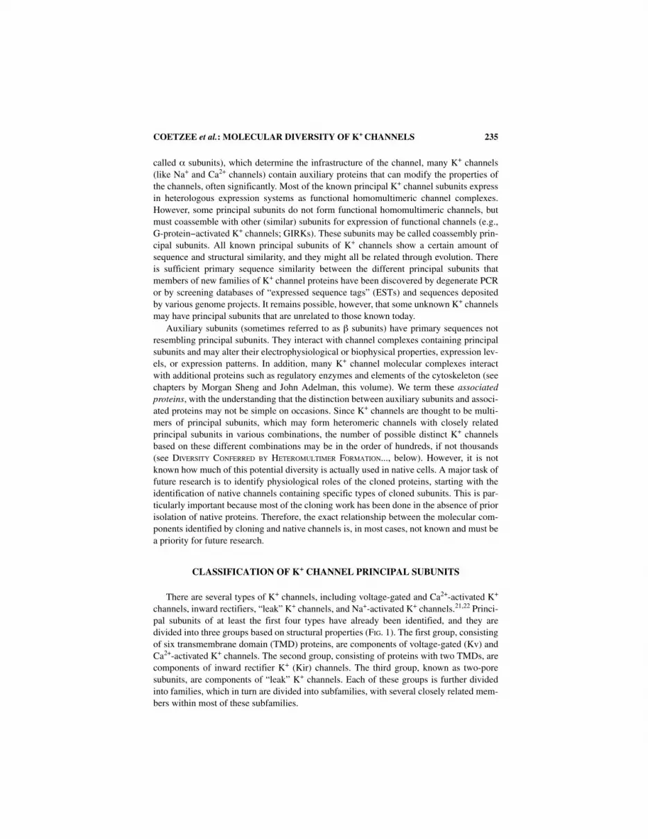
CLASSIFICATION OF K+ CHANNEL PRINCIPAL SUBUNITS
There are several types of K+ channels, including voltage-gated and Ca2+-activated K+
channels, inward rectifiers, “leak” K+ channels, and Na+-activated K+ channels.21,22 Princi-pal subunits of at least the first four types have already been identified, and they aredivided into three groups based on structural properties (FIG. 1). The first group, consistingof six transmembrane domain (TMD) proteins, are components of voltage-gated (Kv) andCa2+-activated K+ channels. The second group, consisting of proteins with two TMDs, arecomponents of inward rectifier K+ (Kir) channels. The third group, known as two-poresubunits, are components of “leak” K+ channels. Each of these groups is further dividedinto families, which in turn are divided into subfamilies, with several closely related mem-bers within most of these subfamilies.

236 ANNALS NEW YORK ACADEMY OF SCIENCES
The first major group of K+ channel principal subunits to be identified contain sixtransmembrane domains (TMDs) (S1-S6), with a conserved P (pore or H5) domain. Func-tionally, they form voltage- and/or Ca2+-activated K+ channels when expressed in heterolo-gous expression systems. This group contains the Kv family (with eight subfamilies: Kv1−Kv6 and Kv8−Kv9) as well as members of the KQT, eag, SK, and slo families of principalsubunits.
The second major group of pore-forming subunits are components of inward-rectifyingK+ (Kir) channels and the first members were first identified by expression cloning.23−25
Kir principal subunits have a predicted membrane topology of two TMDs (M1−M2) and apore domain, analogous to S5-P-S6 of the 6TMD K+ channel subunits. There are currentlyseven subfamilies (Kir1−Kir7), most of which form K+ channels with various degrees ofinward rectification when expressed in heterologous expression systems.
A third group of mammalian K+ channel principal subunits was recently described andcontains four putative TMDs (M1−M4) and two P domains (P1 and P2).12,26 Structurally,these principal subunits have a predicted membrane topology as if they consisted of twospliced Kir subunits. Whereas the 6TMD and 2TMD principal subunits are thought toassemble as tetrameric proteins to form functional channels (see above), the 4TMD sub-units are thought to dimerize, thereby retaining the fourfold symmetry around the centralpore.12,27 There are currently four members in this novel family of K+ channel principalsubunits (FIG. 1 and TABLE 1), but it is possible that more members might be cloned in thenear future (see later). Functionally, these principal subunits express K+ selective channelsthat do not appear to gate in a manner as observed with channels formed by Kir principalsubunits. Since the current responds to changes in extracellular K+ concentration in a man-ner described by the Goldman-Hodgkin-Katz equation, these channels are also referred toas “leak” K+ channels.12 Recent reports 28 indicate that at least some of these channels canbe extensively modulated (e.g., by arachidonic acid or pH; TABLE 1).
The P domain of K+ channel principal subunits is critically important for channel func-tion. Approaches using both mutagenesis 29,30 and X-ray crystallography17 suggest a rolefor this domain in the formation of the K+ selective pore of the channel. The consensuspore sequenceg calculated from a simultaneous alignment of the P domains of K+ channelprincipal subunits that are shown in TABLE 1 is: [TS]-[MLQ]-T-T-[IV]-G-Y-G31 andappears to be hallmark of K+ channel principal subunits.
Mammalian K+ Channel Principal and Auxiliary Subunits
TABLE 1 lists published sequences of mammalian (mostly from human, rat, and mouse)K+ channel principal and auxiliary subunits cloned to date. As far as possible, we usedstandard nomenclature32,33 to describe the various genes. In addition to Genbank or Swis-sprot accession numbers and trivial (author-assigned) names of known alternativelyspliced variants, we also give the gene name as defined by the Human Genome Organiza-tion (HUGO) Nomenclature Committee,h chromosomal localization, and possible (or con-firmed) associated diseases related to allelic variants. These data for principal subunits
g The consensus pore sequence was calculated from a simultaneous alignment of the pore regionsof K+ channel principal subunits using MEME (http://www.sdsc.edu/MEME). A residue was includedif it had a probability of occurrence larger than 0.2.
h http://www.gene.ucl.ac.uk/nomenclature/

COETZEE et al.: MOLECULAR DIVERSITY OF K+ CHANNELS 237
have been subdivided by functional classification, as described above. The auxiliary sub-units are listed in terms of the main principal subunits with which they are thought to inter-act.
Genealogical Analysis of Genes Coding for Principal K+ Channel Subunits
We performed a genealogical analysis of the K+ channel principal subunits shown inTABLE 1 with the aim of examining relatedness between the various genes. For this analy-sis, we divided the K+ channel principal subunits on the basis of their predicted transmem-brane topology. FIGURE 2 is a phylogenetic tree of six TMD voltage-gated and Ca2+-activated K+ channels generated by parsimony analysis, and FIGURE 3 shows a similar anal-ysis for the two TMD principal subunits representing Kir principal subunits.
For the six-TMD group, a strong primary node (bootstrap value of 99% in FIG. 2) existsthat includes the members of all of the Kv subfamilies (Kv1−Kv6 and Kv8−Kv9). Thequestion may arise whether the KQT family should be considered as part of the Kv family.However, the KQT family has a much weaker sequence identity to Kv genes (19−25%)than is found between members of the Kv subfamilies (33−50%) (TABLE 2A). It is there-fore likely that the divergence of the KQT and the Kv gene families preceded the diver-gence of the different Kv subfamily members. Thus, for the purposes of this review, theKQT family will be regarded as a discrete family. The analysis shows that members ofeag, erg, and elk subfamilies showed clustering with a strong node (bootstrap value of100%), suggesting that they all originated from a single ancestral gene.
Interestingly, although there are large structural differences (e.g., the length of the C-terminus) between SK and slo, these families (which are all principal subunits of Ca2+-activated K+ channels) grouped together with a strong bootstrap value (73%; FIG. 2). Thus,within the regions used to perform the parsimony analysis (which included most of the sixtransmembrane domains), characters exist that diagnosed SK and Slo as close relatives.
It is often difficult to determine whether a gene belongs to a certain subfamily. For thepurposes of this review, we used identity scores as the only criterion to subdivide genesinto different subfamilies. Other criteria may exist, such as the presence of regulatorysequences (e.g., ATP-binding sequences, etc.), similar electrophysiological or pharmaco-logical phenotypes in expressed channels, or similar regulation by metabolic pathways.Concentrating only on similarity of the primary sequences, it is interesting to note that anidentity score of >55%i exists among members of individual Kv, eag, KQT, and slo sub-families (TABLE 2A). In fact, this threshold value of ~55% holds true within each of the6TMD K+ channel subfamilies except for the SK genes. Within the SK gene family, SK4appears to diverge from other SK family members (TABLE 2B), suggesting that SK4 mightrepresent a member of a new emerging subfamily of SK genes.
Within the 2TMD family, members of the Kir2, Kir3, Kir5 (the latter having only a sin-gle member), and Kir6 subfamilies each fall into their own expected groups (FIG. 3). Theclassification of the remainder of the genes (members of Kir1, Kir4, and Kir7 subfamilies)
i Note that this number was derived from multiple sequence alignments where portions of unstablesequences were removed for the generation of trees. When whole sequences are aligned, this numbermay be smaller. The threshold value of 55% identity can be extracted from the percent identity tablefor the 2TMD potassium channels as well as the 6TMD channels; i.e., in both identity tables, genesthat have identity values of less than 55% belong to different subfamilies.

238 ANNALS NEW YORK ACADEMY OF SCIENCES
FIGURE 2. Strict consensus phylogenetic tree of 6TMD voltage-gated and calcium-activated K+
channels generated by parsimony. The parsimony tree is generated by heuristic search using PAUP3.1.1.138 Ten random addition searches with TBR branch swapping were performed. The E. coli K+
channel homologue ECOKCH (accession #L12044) was used as an outgroup). ClustalX was used togenerate all alignments of amino acid sequences. By varying the alignment-parameters, we were ableto identify regions that lack alignment stability. We proceeded to remove alignment ambiguousregions by implementing a “culling” procedure.139 In this analysis, only regions that span the sixtransmembrane domains were used. All characters were equally weighted in the analysis. Bootstrapvalues generated using PAUP 3.1.1138 are shown on the tree when available, and they represent ameasure of node robustness. Sequences of eag2, elk1, and elk2 used in this analysis are from B.Ganetzky (see this volume).

COETZEE et al.: MOLECULAR DIVERSITY OF K+ CHANNELS 239
FIGURE 3. One of two most parsimonious trees of 2TMD inward rectifier K+ channels that aregenerated by heuristic search. For methods used, please refer to the legend of FIG. 2. The S. lividansskc1 K+ channel principal subunit (accession Z37969) was used as an outgroup. Except for smallportions of the NH2 and COOH terminals, most of the amino acid sequences were used in this anal-ysis. To measure node robustness, we generated bootstrap values (shown on tree when available)using PAUP 3.1.1.
240 ANNALS NEW YORK ACADEMY OF SCIENCES
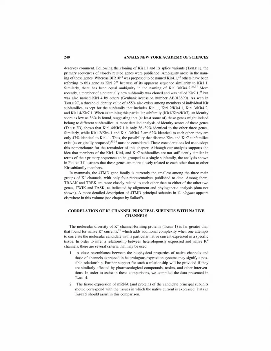
deserves comment. Following the cloning of Kir1.1 and its splice variants (TABLE 1), theprimary sequences of closely related genes were published. Ambiguity arose in the nam-ing of these genes. Whereas BIR1034 was proposed to be named Kir4.1,33 others have beenreferring to this gene as Kir1.235 because of its apparent sequence similarity to Kir1.1.Similarly, there has been equal ambiguity in the naming of Kir1.3/Kir4.2.36,37 Morerecently, a member of a potentially new subfamily was cloned and was called Kir7.1,38 butwas also named Kir1.4 by others (Genbank accession number AB013890). As seen inTABLE 2C, a threshold identity value of >55% also exists among members of individual Kirsubfamilies, except for the subfamily that includes Kir1.1, Kir1.2/Kir4.1, Kir1.3/Kir4.2,and Kir1.4/Kir7.1. When examining this particular subfamily (Kir1/Kir4/Kir7), an identityscore as low as 36% is found, suggesting that (at least some of) these genes might indeedbelong to different subfamilies. A more detailed analysis of identity scores of these genes(TABLE 2D) shows that Kir1.4/Kir7.1 is only 36−39% identical to the other three genes.Similarly, while Kir1.2/Kir4.1 and Kir1.3/Kir4.2 are 62% identical to each other, they areonly 47% identical to Kir1.1. Thus, the possibility that discrete Kir4 and Kir7 subfamiliesexist (as originally proposed)33,38 must be considered. These considerations led us to adoptthis nomenclature for the remainder of this chapter. Although our analysis supports theidea that members of the Kir1, Kir4, and Kir7 subfamilies are not sufficiently similar interms of their primary sequences to be grouped as a single subfamily, the analysis shownin FIGURE 3 illustrates that these genes are more closely related to each other than to otherKir subfamily members.
In mammals, the 4TMD gene family is currently the smallest among the three maingroups of K+ channels, with only four representatives published to date. Among them,TRAAK and TREK are more closely related to each other than to either of the other twogenes, TWIK and TASK, as indicated by alignment and phylogenetic analysis (data notshown). A more detailed description of 4TMD principal subunits in C. elegans appearselsewhere in this volume (see chapter by Salkoff).
CORRELATION OF K+ CHANNEL PRINCIPAL SUBUNITS WITH NATIVE CHANNELS
The molecular diversity of K+ channel-forming proteins (TABLE 1) is far greater thanthat found for native K+ currents,21 which adds additional complexity when one attemptsto correlate the molecular candidate with a particular native current expressed in a specifictissue. In order to infer a relationship between heterologously expressed and native K+
channels, there are several criteria that may be used.
1. A close resemblance between the biophysical properties of native channels andthose of channels expressed in heterologous expression systems may signify a pos-sible relationship. Further support for such a relationship will be provided if theyare similarly affected by pharmacological compounds, toxins, and other interven-tions. In order to assist in these comparisons, we compiled the data presented inTABLE 4.
2. The tissue expression of mRNA (and protein) of the candidate principal subunitsshould correspond with the tissues in which the native current is expressed. Data inTABLE 5 should assist in this comparison.

COETZEE et al.: MOLECULAR DIVERSITY OF K+ CHANNELS 241
3. The functional consequences of processes such as heteromultimeric assembly bydifferent principal subunits, their regulation by auxiliary subunits, and posttransla-tional modification should be considered. We present examples where these pro-cesses have been shown to influence channel properties. Because of the amount ofdetail involved, this information is supplied mainly in textual format (DIVERSITY
CONFERRED BY HETEROMULTIMER FORMATION..., below).
4. Strong support for a causal relationship between native and candidate cloned K+
channel principal subunits can be obtained from experiments involving deletion oroverexpression of the target channel principal subunits (transgenic animals or over-expression of wild-type, antisense, or dominant negative constructs in isolatedcells). Since this is a relatively new area of investigation, there are only a few exam-ples where these methodologies have been applied (see TABLE 3).
It is important to note, however, that these criteria do not exclusively define a relation-ship between cloned and native proteins. For example, negative results might well becaused by unknown events occurring at the molecular level, such as interactions of K+
channel principal subunits with other cytosolic or membrane-bound proteins or their regu-lation by unknown endogenous compounds and peptides or poorly understood posttransla-tional modifications.
FUNCTIONAL PROPERTIES OF K+ CHANNELS IN HETEROLOGOUS EXPRESSION SYSTEMS
We have listed the functional and pharmacological properties of the currents expressedby K+ channel principal subunits in heterologous expression systems in TABLE 4. Wherepossible, we report data obtained from expression studies using mammalian cells underpatch-clamp conditions. Otherwise, we report data obtained from the Xenopus expressionsystem. In cases in which there is close agreement between several reports on the sameprotein, we report an averaged value. When large discrepancies exist in published valuesto make it impossible to find a consensus value, we give a range of values. In come cases,we favored reports in which a more extensive functional analysis was performed.
In using these tables it is important to remember that experimental variables can haveimportant effects on published parameters. In addition, a native channel composed of aparticular subunit may have properties different than those in heterologous expression sys-tems due to factors such as those listed in DIVERSITY CONFERRED BY HETEROMULTIMER FOR-MATION..., below, or other factors that are sometimes difficult to predict. We would like tomention three such examples of problems encountered using heterologous expression sys-tems: First, it has been observed that Kir3.1 expresses G-protein−activated K+ channels inXenopus oocytes, but not in mammalian cultured cells. It is now clear that this is due to thepresence of an intrinsic Kir3.4 subunit (called XIR) in Xenopus oocytes39 (see chapter byWickman in this volume) that is not found in mammalian cells and is required for expres-sion of G-protein−activated K+ channels. Second, artifacts can be caused by the unusuallylarge amounts of channels expressed in heterologous expression systems. The flow oflarge currents may lead to effects such as K+ accumulation in the extracellular spaces,which in turn can modify the behavior of expressed channels. Last, some channels can bevery sensitive to particular elements in the extracellular or intracellular solution, such as

242 ANNALS NEW YORK ACADEMY OF SCIENCES
blockade of several voltage-gated K+ channels by Mg2+. Care should therefore be exer-cised when comparing the electrophysiological phenotype of native K+ currents with thosepresented in TABLE 4.
TISSUE EXPRESSION OF K+ CHANNEL SUBUNITS
Studies on the cell- and tissue-specific expression of K+ channel genes vary widely.Some genes such as Kv3 (see chapter by Rudy et al., this volume), Kir3 (see chapter byKevin Wickman et al., this volume), and several Kv1’s16 have been studied in great detailin several laboratories. Others have been studied to a much lesser extent. We attempted togive a representative overview of expression patterns of known K+ channel genes in TABLE
5. However, the nature of available information will be reflected in our compilation.Different laboratories use different methods to analyze expression levels of various K+
channel transcripts. Some of these methods are more quantitative in nature (Northern blotanalysis and RNAse protection assays) but have a limited spatial resolution. In contrast, insitu hybridization techniques can have excellent spatial resolution, but quantification ismore difficult. RT-PCR is very sensitive, and results obtained with this technique are diffi-cult to quantify. Given this sensitivity, it is often difficult to interpret a result when noattempt at quantification is made. Antibodies have now been raised to a number of K+
channel proteins, and they can be great tools for the analysis of protein products, but prob-lems of specificity can arise. We present a table on the tissue and brain distribution of K+
channel gene products (TABLE 5). However, caution should be exercised in using this table.For example, results for some genes may come from many studies using different meth-ods, whereas for others data may have been derived from a single study. Great care shouldbe taken when making a quantitative comparison of expression levels between differentgenes because of the variations in methodologies used in different laboratories as well asthe subjectivity of different investigators when grading the intensities of signals.
DIVERSITY CONFERRED BY HETEROMULTIMER FORMATION,ACCESSORY SUBUNITS, ALTERNATIVE SPLICING, RNA EDITING AND
POSTTRANSLATIONAL MODIFICATION
Over 50 mammalian genes encoding principal subunits of K+ channels are listed inTABLE 1. The total number of different subunits, however, is even larger since many ofthese genes undergo RNA processing, such as alternative splicing resulting in multipleprotein products from each gene. Considering these factors, this results in a large number(>100) of different mammalian principal subunits (TABLE 1).
However, the total number of different functional types of K+ channels is probably sig-nificantly larger. In addition to the already large number of subunits, the diversity can bemagnified by virtue of the oligomeric structure of the functional channel complex (seeintroductory section of this paper). Many of the principal subunits can form functionalhomomultimeric as well as heteromultimeric channels. Depending on the number of pos-sible combinations, this can dramatically increase the number of possible functionally dis-tinct channels obtained from these subunits.

COETZEE et al.: MOLECULAR DIVERSITY OF K+ CHANNELS 243
Finally, channel function can also be influenced by auxiliary subunits and posttransla-tional modifications. If the channel complex exists with the auxiliary proteins in somecells and without them in other cells, or if it is differentially modified by posttranslationalmodification, these factors can also increase the diversity of functional units. This sectionhighlights the contributions of these important mechanisms to K+ channel function anddiversity.
Heteromultimeric Assembly of Principal Subunits
Heteromultimeric assembly of K+ channel proteins can theoretically provide the cellwith a “modular” system for increasing K+ channel diversity while reducing the require-ment for more genes. For example, 210 functionally distinct tetrameric channels of differ-ent subunit combinations might be formed from seven different subunits.j Channeldiversity can be increased even more if the possibility of heteromultimerization of alterna-tive splice variants is considered.
When considering the large overlap in the expression of different K+ channel subunits(see TABLE 5), it is quite possible that many native channels are the result of heteromericassociation of primary subunits. However, coexpression of two subunits within a given celldoes not necessarily warrant the occurrence of coassembly (see below). It is thus of greatimportance to elucidate the mechanisms that govern subunit recognition and assembly inorder to understand the potential for heteromultimer formation and the composition ofnative channels. Here we discuss what is known about the rules governing subunit interac-tions and evidence for their formation in native tissue in the different families of K+ chan-nels.
Heteromultimeric Assembly of Kv and KQT Principal Subunits
For Kv1−Kv4 subfamilies, coexpression of different Kv cRNAs of the same, but not ofdifferent, subfamilies in Xenopus oocytes leads to the expression of channel properties thatcan not be the result of the sum of two independent channels. These currents have beeninterpreted as resulting from novel K+ channel proteins formed as heteromultimers fromdifferent principal subunits of the same Kv subfamily.40−44 These heteromultimeric chan-nels tend to have properties that are intermediary between those of the two homomulti-meric channels, but in some cases certain properties dominate. For example, a channelcontaining three noninactivating and one N-type inactivating subunit produces an inacti-vating channel.20,41−43 In another example, Kv1.6 subunits have been shown to have a spe-cific N-type inactivation-prevention (NIP) domain, which produces noninactivatingcurrents when heteromultimerized.45 Subfamily selectivity among Kv subunits is the resultof recognition domains located in the N-terminal region of the protein and referred to asthe NAB or T1 domains.46,47 Interestingly, these domains are also involved in the interac-tion of Kv1 proteins with auxiliary (Kvβ) subunits.48,49
Evidence for the heteromultimerization of Kv proteins from the same subfamily in vivohas come from coimmunoprecipitation experiments in rat and mouse brain.50−53 (also see
j [p+(n-1)]!/[p!(n-1)!], where p is the aggregation number and n is the number of subunits.

244 ANNALS NEW YORK ACADEMY OF SCIENCES
Rudy et al., volume). For example, antibodies specific to Kv1.1 immunoprecipitate Kv1.2and Kv1.4 proteins in nondenatured brain membrane extracts.50,51 The coexpression ofKv1.4 and Kv1.2 in axons and terminals of many cells suggests the native A-type K+ cur-rent may result from Kv1.4/Kv1.2 heteromultimers within these compartments.50 Con-versely, subunits of the same subfamily have also been shown to localize to separatecompartments within the same cell.54 The cellular mechanisms of subunit targeting andassembly could be important as well. Whether or not heteromultimers are formed willdepend on whether channel assembly occurs before or after the subunits have been sortedand shipped to their appropriate destinations.
The two Kv2 subfamily members (Kv2.1 and Kv2.2) are able to function as homomul-timeric or heteromultimeric channels. There are reports that their kinetics and voltagedependence are altered when they are coexpressed with members of other, closely related,Kv subfamilies (Kv5, Kv6, Kv8, Kv9; see FIG. 1).55−57 An enormous potential for func-tional diversity is apparent when considering all of the possible combinations of theseprincipal subunits. Interestingly, members of the Kv5, Kv6, Kv8, and Kv9 subfamilies donot express currents by themselves in heterologous expression systems. Since the primarysequence similarity of Kv5, Kv6, Kv8, and Kv9 principal subunits with the Kv2 subfamilymembers are particularly high in the T1 domain, one can speculate that these coassemblyprincipal subunits may have evolved to regulate the function of Kv2-related proteins.
The KQT family currently has three members. The first to be described, KQT1(KvLQT1), is found predominantly in the heart and liver, whereas KQT2 and KQT3expression is restricted to the brain. Coexpression of KCNQ2 and KCNQ3 in Xenopusoocytes results in a 15-fold increase in current amplitude compared to expression of eachsubunit alone.58 The overlapping distribution of KQT2 and KQT3 suggest they may func-tion only as heteromultimers in vivo. In support of this argument, it is interesting to notethat mutations of these two genes both give rise to the same genetic disorder (TABLE 1).
Heteromultimeric Assembly of eag, erg, and elk Subunits
There is currently a single published mammalian homologue of the Drosophila eag thatproduces currents in heterologous expression systems (TABLE 1). Although more eag fam-ily members have been described, the full-length cDNA coding sequences remain to becloned and expressed (see chapter by Ganetzky, this volume). There are three members ofthe Erg subfamily with diverse physiological properties. However, their interactions havenot been characterized. A mammalian elk homologue has been cloned and was found toproduce functional currents. At least two new elk family members have been discovered.59
Heteromultimers between the two splice variants of erg (erg1a and erg1b; TABLE 1)have been studied in vitro, resulting in currents with properties more similar to those of thenative current (IKr) in heart.60 It is not yet known whether eag, erg, and elk principal sub-units can coassemble as heteromultimers. With the cloning and expression of more mem-bers of each subfamily, it will be interesting to see if they follow the same rules forheteromultimerization as Kv channels. There is already evidence that subunit interactionswithin this family will be complicated. For example, it has been suggested that membersof the eag family can interact with members of the Kv family in Xenopus oocytes.61 How-ever, whether such interactions will take place in vivo remains to be determined.

COETZEE et al.: MOLECULAR DIVERSITY OF K+ CHANNELS 245
Heteromultimeric Assembly of Kir Subunits
There does not appear to be a consistent manner in which Kir subunits coassemblewithin or between subfamilies. The four members of the Kir3 subfamily serve as an exam-ple of where heteromultimerization of subunits from the same subfamily appears to be anecessary requirement for the formation of functional channels. Thus, the channelsresponsible for the acetylcholine-activated K+ current in atrial muscle is a complex ofKir3.1 and Kir3.4 proteins (see chapter by Wickman et al., this volume). Neurons proba-bly contain functional heteromultimers of Kir3.1 with Kir3.2 or Kir3.3 (see chapter byKevin Wickman, this volume; see also Ref. 62). In contrast to the Kir3 subunits, the evi-dence for heteromultimeric assembly within other Kir subfamilies members is less strongor there are even indications to the contrary. For example, despite the colocalization ofKir2.1 and Kir2.3 in neural tissue,63 biochemical and electrophysiological experimentsexamining protein-protein interactions suggest that heteromultimeric coassembly of sub-units within the Kir2 subfamily may not occur.64 Recent experiments using antisense oli-gonucleotides directed against Kir2.1 transcripts demonstrated a specific inhibition of a21-pS conductance channel (which is equivalent to the unitary conductance of Kir2.1 inheterologous expression systems) in cardiac myocytes.65 Since the occurrence of nativeinward rectifier K+ channels with different unitary conductances was unchanged, onecould argue that heteromultimeric assemblies of Kir2.1 with other K+ channel principalsubunits did not occur (or more unlikely that, if it did, the single-channel conductanceremained unchanged). The regions responsible for determining compatibility within mem-bers of the Kir2 subfamilies have been elucidated using a combination of deletion mutantsand chimeric channels.64 The results of such experiments reveal interactions conferred bydomains found within the proximal C-terminus and second transmembrane segments.However, it is possible that other regions may also play a role.66
There are also published examples of heteromultimerization between subunits fromdifferent Kir subfamilies. For example, Kir5.1 (which by itself expresses no current inoocytes34) significantly alters expression levels and the single-channel conductance ofKir4.1, a member of a different subfamily.67 The finding that Kir5.1 altered neither currentamplitude nor the macroscopic phenotype of Kir1.1, Kir2.1, Kir2.3, Kir3.1, Kir3.2, orKir3.4 suggests that a specific interaction between Kir4.1 and Kir5.1 may occur in vivo.
Heteromultimeric Assembly of slo and SK Subunits
The slo family currently has two members (TABLE 1). Heteromultimerization betweenthe two slo principal subunits has not been examined. Despite the small number of familymembers, slo1 (the principal subunit of BK Ca2+-activitated K+ channels) has a very largenumber of possible alternative spliced versions. Tested splice variants show functionalvariability.68−71 Heteromultimerization between splice variants could thus allow an enor-mous number of possible functionally diverse channels. Single-cell RT-PCR studies ofindividual hair cells of the chick cochlea demonstrate that multiple splice variants of sloare found within a single cell72,73 (see also chapter by Robert Fettiplace, this volume), sug-gesting that at least a proportion of this potential diversity might indeed occur in vivo.
The SK family now has four members (TABLE 1). SK1−3 each express small-conduc-tance Ca2+-activated K+ channels with similar properties but differing in their pharmacol-ogy (see chapter by Adelman, this volume). The fourth (SK4) expresses an intermediary-

246 ANNALS NEW YORK ACADEMY OF SCIENCES
conductance Ca2+-activated K+ channel. Coexpression of these genes has not yet beentested, but their overlapping tissue distribution suggests that heteromultimer formationmight be possible in vivo.74 Although the SK1-SK3 subunits have very similar properties,the NH2 and COOH terminal amino acid sequences vary significantly which could poten-tially provide additional diversity through differential modulation through associating pro-teins. Conversely, divergence in these regions may also prevent heteromultimer formation.
Heteromultimeric Assembly of “Leak” K+ Channel Subunits
Heteromultimer formation within the 4TMD family of K+ channels has not yet beenexplored. It can be predicted from the number of similar genes expressed in C. elegans thata multitude of “two-pore” subunits may exist in mammals (see chapter by Larry Salkoff,this volume). Whether or not the diversity of this family of K+ channels is increased byheteromeric subunit interactions remains to be determined.
Auxiliary Subunits
There is increasing evidence for the existence of auxiliary subunits, some peripheraland some integral membrane proteins, that regulate the expression levels and functionalproperties of K+ channel proteins (TABLE 1). Several types of K+ channels are affected inthis manner, including those consisting of Kv, KQT, eag, slo, and Kir principal subunits.
To date, the auxiliary subunits that have been characterized in most detail are the mem-bers of the Kvβ auxiliary subunit family. They are products of three genes (TABLE 1). Weadopt the nomenclature shown in TABLE 1 to conform to convention and to signify theirsequence relatedness. Affinity purification of brain K+ channels using dendrotoxin led tothe isolation of an auxiliary “β” subunit (β2 or Kvβ2) that binds noncovalently with a 1:1stoichiometry to Kv1 principal subunits.75,76 Identification of this sequence soon led to theisolation of related cDNAs. Three genes have been identified, and each has been shown toproduce several isoforms by alternative splicing (TABLE 1). These subunits lack putativetransmembrane domains, potential glycolsylation sites, or leader sequences, suggestingthat they are cytoplasmic proteins.76 Although they have some sequence similarities withaldo-keto reductase enzymes, Kvβ auxiliary subunits probably lack such enzymatic activ-ity.77 A detailed review of Kvβ subunits is present in this volume (chapter by Pongs et al.).Briefly, one function of the three Kvβ1 isoforms (which contain a variable inactivatingball domain) is to induce inactivation in otherwise noninactivating Kv1 channels by pro-viding an extrinsic N-type inactivating domain.78 In contrast, Kvβ2 accelerates inactiva-tion only when Kv1.4 subunits form part of expressed (inactivating) Kv1 channels,probably by interacting with the intrinsic Kv1.4 inactivating ball.79−81 For some Kv1 chan-nels, Kvβ1 and Kvβ2 may also shift the voltage dependence of activation in heterologousexpression systems.82 A second role assigned to these auxiliary subunits is to act as chap-erones during channel biosynthesis83,84 and thus to increase expression levels, an effectfirst described for the interaction of Kvβ2 with Kv1.4.79 While Kvβ1 and Kvβ2 proteinsappear to interact exclusively with Kv1 principal subunits, Kvβ3 and a recently discoveredK+ channel auxiliary subunit, KChAP,85 appear to interact also with Kv2 principal sub-units and to enhance current levels without an effect on channel kinetics or gating.85,86

COETZEE et al.: MOLECULAR DIVERSITY OF K+ CHANNELS 247
Although Kvβ3 and KChAP appear to have similar roles, their mechanisms of actionmight be different since chimeras between Kv2.1 and Kv2.2 indicate that the COOH-ter-minal end of the Kv2.2 protein is essential for its Kvβ3 sensitivity.86 In contrast, KChAPappears to bind to the NH2 termini of Kv1 and Kv2 principal subunits.85
Since assembly of Kv1 principal subunits occurs mainly in a subfamily-specific man-ner, it has been suggested that this subfamily specificity may also apply to their associationwith auxiliary subunits.49,87 However, this may not strictly be the case, since there is evi-dence that the Drosophila auxiliary subunit homologue Hyperkinetic (Hk) associates withmembers of the eag and Shaker families and alters their expression levels and/or kinet-ics.88 Recently, yet another auxiliary subunit has been identified, KCR1, which acceleratesthe activation of rat eag expressed in Xenopus oocytes or in COS-7 cells.89
MinK is a 15-kDa single-transmembrane protein that is present in cardiac and auditorycells.5,90−93 that coassembles with KvLQT1 to form the slow cardiac repolarization current(IKs).
94–96 (see chapter by Sanguinetti, this volume) and with HERG to regulate the rapidlyactivating cardiac delayed rectifier (IKr).
97 Hence, minK contributes to two important out-ward currents that determine repolarization from the plateau phase of the action potentialin ventricular myocytes.98,99 Although it does not contain a pore domain characteristic ofK+ channels’ principal subunits,100,101 mutagenesis experiments suggest that minK maycontribute to the formation of the channel pore,102 implying that K+-selective pores mayinclude structures other than P domains or structures having a strict P-loop geometry.
The auxiliary subunit of high-conductance Ca2+-activated K+ channels (composed ofprincipal subunits of the slo family) is a two-transmembrane protein of 191 amino acidsand bears little sequence homology to any other known K+ channel auxiliary subunit.103 Ithas been shown that this subunit contributes to the high-affinity receptor forcharybdotoxin104 but apparently does not affect sensitivity to this drug. Mutagenesis hasrevealed that the large extracellular loop of the maxi K+ channel auxiliary subunit has arestricted conformation with two important disulfide bridges. Specifically, four aminoacids are critical in conferring high-affinity 251-Chtx binding to the complex. Function-ally, it has been demonstrated that this subunit also confers higher Ca2+ sensitivity to Ca2+-activated K+ channels containing slo subunits (see TABLE 4).
Slob is a novel protein isolated by a yeast two-hybrid screen based on its interactionwith the COOH-terminal domain of the Drosophila slowpoke (dSlo) Ca2+-dependent K+
channel.105 Native Slob and dSlo coimmunoprecipitate together from fly head lysates, andthey redistribute and colocalize in discrete intracellular structures when coexpressed inheterologous host cells. Although direct application of Slob to excised inside-out mem-brane patches can strongly activate dSlo (but not human slowpoke channels),105 the in vivofunction of Slob remains to be elucidated.
The Kir channel accessory subunit Kv2.2v (TABLE 1) acts as a negative regulator of theinward rectifier Kv2.2 through heteromeric assembly.
Finally, although Kir6 principal subunits (or C-terminal truncated Kir6 subunits) canexpress independently in heterologous expression systems,106,107 the functional phenotypeof native KATP channels is conferred only when Kir6 principal subunits are coexpressedwith auxiliary subunits (sulfonylurea receptors, SUR1 or SUR2; see TABLE 1). SUR1 andSUR2 are members of the 12 transmembrane-spanning domain proteins, called ATP-bind-ing cassette (ABC) proteins. SUR1 or SUR2 associate with Kir6 subunits as an octamericassembly.108 Prevailing evidence suggests that the inhibitory effects of nucleotides onKATP channels are mediated via the principal Kir6 subunits, whereas the potentiation by

248 ANNALS NEW YORK ACADEMY OF SCIENCES
ADP is conferred by the sulfonylurea receptor subunit, SUR.106 SUR auxiliary subunitsare also responsible for conferring properties such as sensitivity to pharmacological com-pounds that block (e.g., glibenclamide) or increase (e.g., pinacidil) opening of the multi-meric channel complex. The specific assembly between the principal and auxiliary sub-units that takes place seems to be at least partly responsible for the functional diversity ofnative KATP channels (Table 3; reviewed by Babenko109).
Alternative Splicing
TABLE 1 lists alternative splice versions of principle and accessory K+ channel subunitsknown to date. In many cases the use of alternative exons results in channels with diverseproperties. TABLE 6 lists only the K+ channel principal subunits studied so far where alter-native splicing leads to diversity in channel function. The functional consequences of K+
channel gene splicing have been grouped into four categories: (1) effects on electrophysio-logical properties, where splicing changes channel properties such as kinetics; (2) effectson expression, where splicing results in changes in gene expression and/or tissue distribu-tion; (3) effects on subcellular localization, where alternative splicing results in changes inchannel targeting to different cellular compartments; (4) effects on modulation, wheresplicing alters the effects of modulators (e.g., by protein kinases). Blank spaces indicatethat no changes are observed or they remain to be tested. The purpose of TABLE 6 is toemphasize how K+ channel diversity could be enhanced by mechanisms other than geneduplication and divergence. Furthermore, it should be emphasized that native channelsmay result from the heteromultimerization between different splice variants, thus increas-ing diversity even further.
RNA Editing
RNA editing by adenosine deamination has evolved as a mechanism to produce func-tionally diverse proteins from the same gene. The best-characterized example in the brainis the RNA editing of glutamate-activated receptor channel (GluR) mRNA110,111 (seechapter by Sprengel in the Glutamate Receptor section, this volume). RNA editing ofmammalian K+ channels has not been described. However, in squid, five purine transitionsfound in cDNA clones encoding sqKv2 K+ channel are also generated by RNA editing.112
The conductance-voltage relationships determined for the two most frequently edited sitesof sqKv2, Y576C (pore region) and I597V (S6 segment), did not differ. However, the rateof channel closure upon repolarization was significantly affected by both substitutions.112
Posttranslational Modifications
Posttranslational modifications, particularly protein phosphorylation and dephosphory-lation, are known to underlie modulation of the activity of ion channels, and hence, modu-lation of neuronal excitability.113−115 Posttranslational processing or the distinctintracellular microenvironment of the channel could contribute to the diversity of K+ chan-nels in native cells. The modulation of Kv3.4 channels by protein kinase C (PKC) is a
COETZEE et al.: MOLECULAR DIVERSITY OF K+ CHANNELS 249
good example of how channel function and phenotype can be altered in this manner.116
Phosphorylation of the amino-terminal activation domain of Kv3.4 by PKC suppresses N-type inactivation, converting these channels from rapidly inactivating A type to noninacti-vating delayed rectifier type.116,117 Similarly, Roeper and coworkers found that the balancebetween phosphorylated and dephosphorylated Kv1.4 channels is regulated by changes inintracellular Ca2+ concentration, rendering Kv1.4 inactivation gating Ca2+ sensitive.118
They showed that Ca2+-calmodulin−dependent protein kinase (CaMKII) phosphorylationof a single amino-terminal residue of Kv1.4 slows inactivation gating and acceleratesrecovery from N-type inactivated states; while on the contrary, dephosphorylation of thisresidue induces a 5 to 10 times faster inactivation of Kv1.4.118
Finally, the dynamic regulation of ion channel interactions with the cytoskeleton maycontribute to the diversity of K+ channel properties and mediate aspects of synaptic plas-ticity (see chapter by Sheng, this volume). For example, Kir2.3 bind to PSD-95, a cytosk-eletal protein of postsynaptic densities that clusters NMDA receptors and voltage-dependent K+ channels: Kir2.3 colocalizes with PSD-95 in neuronal populations in fore-brain, and a PSD-95/Kir2.3 complex occurs in hippocampus.119 Within the C-terminal tailof Kir2.3, a serine residue critical for interaction with PSD-95, is also a substrate for phos-phorylation by protein kinase A (PKA).119 Thus, ion channel interactions with thepostsynaptic density are regulated by a physiological mechanism, since stimulation ofPKA in intact cells causes rapid dissociation of the channel from PSD-95.119,120 It also fol-lows from this work that any posttranslational modification that accounts, directly or indi-rectly, for changes in the localization or clustering of K+ channels, may contribute to thefunctional diversity of this family of ion channels.
SUMMARY AND PERSPECTIVE
We attempted to provide a comprehensive, yet succinct, overview of the moleculardiversity of K+ channel subunits. However, due to the rapidly evolving nature of this fieldof research, it is very likely that this chapter will be outdated by the time of publication.With the recent sequencing of bacterial and yeast genomes, it became clear that manygenes exist for which no definite function could be assigned. Given the relatively simplegenome of these organisms compared to that of humans (~100,000 genes) and the fact thatonly a small proportion of the human genome has currently been sequenced, it will be nosurprise if many more genes coding for K+ channel principal and auxiliary subunits areuncovered in the near future. Importantly, sequences might become available to describe avariety of other protein subunits interacting with channels and to modify their function andexpression patterns. The incredible diversity of subunits and subunit interaction at themolecular level is at present hard to reconcile with the more limited (although diverse inits own right) complement of native K+ channels in cells. The correlation between molecu-lar subunits and native K+ channels in normal and pathophysiological states must remainto be a priority for future research.
These advances also have the promise of providing a wealth of information that will beof benefit to those studying regulation of K+ channel expression. Promoters, enhancers,and silencers provide yet another level of diversity by virtue of controlling expression lev-els in response to environmental influences, as well as tissue-specific expression patterns.The recent progress in the determination of the 3D structure of a bacterial K+ channel

250 ANNALS NEW YORK ACADEMY OF SCIENCES
protein17 will pave the way for similar studies on K+ channel principal and auxiliary sub-units. These structural analyses should provide better tools with which to study protein-protein interactions as well as drug-protein interaction. Ultimately, this new knowledgecan be used to develop more effective protein-specific therapeutic approaches for patho-physiological states involving K+ channel dysfunction. This, and the possibility of gene-specific treatment of diseases related to defects in channel subunit proteins, are increas-ingly areas of active research and will remain fertile areas for future studies.
TABLES
TABLE 1. Mammalian K+ Channel Principal and Auxiliary Subunits
A. Principal Subunits of Voltage-Activated K+ Channels
SubunitGene Locus Designation Species
Trivial Name
Chromosome Localization
Alternative Splicing
Accession Number
Associated Diseases
Kv1.1 KCNA1 Human HUKI 12p13 L02750 Episodic ataxia, myokymia syndrome
Rat RK1 X12589
RCK1
RBK1
Mouse MK1 6 M30439
MBK1
Kv1.2 KCNA2 Human HUKIV 12 L02752
Rat RBK2 J04731
RK2
RCK5
NGK1
Mouse MK2 3 M30440
Kv1.3 KCNA3 Human HUKIII 1p21 M55515
HPCN3
Rat RCK3 M31744
RGK5
KV3
Mouse MK3 3 M30441
Kv1.4 KCNA4 Human HUKII 11p14 M55514
HPCN2
Rat RCK4 M32867
RHK1
RIC3
Mouse 2 U03723

COETZEE et al.: MOLECULAR DIVERSITY OF K+ CHANNELS 251
A. Principal Subunits of Voltage-Activated K+ Channels (continued)
SubunitGene Locus Designation Species
Trivial Name
Chromosome Localization
Alternative Splicing
Accession Number
Associated Diseases
Kv1.5 KCNA5 Human HK2 12p13 M55513
HPCN1
Rat KV1 M27158
RK4
RCK7
Mouse 6 L22218
Mouse Kv1.5_5′ Kv1.5a C49507
Kv1.5_3′ Kv1.5b C49507
Kv1.6 KCNA6 Human HBK2 X17622
Rat KV2 M27159
RCK2
Mouse MK1.6 6 M96688
Kv1.7 KCNA7 Human 19q13.3
Mouse 7 AF032099
Kv2.1 KCNB1 Human 20q13.2 L02840
Rat DRK1 X16476
Mouse 2 M64228
Kv2.2 KCNB2 Human U69962
Rat CDRK M77482
Kv3.1 KCNC1 Human NGK2-KV4
11p15 Kv3.1a S56770
KShIIIB
Mouse NGK2 7 Y07521
Mshaw22
Rat KV4 Kv3.1b M37845
Raw2
Kv3.2 KCNC2 Human 12
Rat RKShIIIA Kv3.2a M34052
Rshaw12
Mouse Mshaw12 10
Rat Kv3.2b M59211
Rat Kv3.2c M59213
Rat Raw1 Kv3.2d M84202
Kv3.3 KCNC3 Human 19q13.3−13.4
Kv3.3a AF055989
Rat RKShIIID Kv3.3a M84210
Rat Kv3.3b M84211
Mouse MShaw19 7 Kv3.3c S69381

252 ANNALS NEW YORK ACADEMY OF SCIENCES
A. Principal Subunits of Voltage-Activated K+ Channels (continued)
SubunitGene Locus Designation Species
Trivial Name
Chromosome Localization
Alternative Splicing
Accession Number
Associated Diseases
Kv3.4 KCNC4 Human HKShIIIC 1p21 Kv3.4b M64676
Rat Raw3 Kv3.4a X62841
Rat 10, 7 Kv3.4c
Kv4.1 KCND1 Human Kv4.1 Xp11.23 Kv4.1 AJ005898
Mouse MShal M64226
Kv4.2 KCND2 Human 7q?
Rat RK5 S64320
Mouse 6
Kv4.3 KCND3 Human Kv4.3M Kv4.3a AF048712
Rat KShIVB U42975
Rat Kv4.3S Kv4.3b L48619
Human Kv4.3L Kv4.3c AF048713
Rat AB003587
Kv5.1 KCNH1 Human KH1 2p25 AF033382
Rat IK8 M81783
Kv6.1 KCNH2 Human KH2 20q13 AF033383
Rat K13 M81784
Kv8.1 Human 8q22.3−8q24.1
Rat Kv2.3r X98564
Mouse U62810
Kv9.1 KCNS1 Human AF043473
Mouse AF008573
Kv9.2 KCNS2 Human 8q22.4 Cohen syndrome?
Mouse AF008574
Kv9.3 KCNS3 Human AF043472
Rat AF029056
Eag1 Rat Reag-1 Eag1a Z34264
Mouse Meag-1 U04294
Bull BTeag1 Y13430
Bull BTeag2 Eag1b Y13431
Eag2b Rat AF073891
Elk1c Rat AF61957
Elk2 Human Helk-2d
Rat AF073892
Mouse Melk-2e

COETZEE et al.: MOLECULAR DIVERSITY OF K+ CHANNELS 253
A. Principal Subunits of Voltage-Activated K+ Channels (continued)
SubunitGene Locus Designation Species
Trivial Name
Chromosome Localization
Alternative Splicing
Accession Number
Associated Diseases
Erg1 LQT Human h-erg 7q35−7q36 Erg1a U04270 LQT2 syndrome
Rat r-erg Z96109
Mouse Merg1a 5 AF012868
Mouse Merg1a’ Erg1b AF012871
Humanf HERGb Erg1c
Mouse Merg1b AF012869
Erg2 Rat AF016192
Erg3 Rat AF016191
KQT1 KCNQ1 Human KvLQT1 11p15.5 KCNQ1a U40990 Long QT syndrome, type I
Mouse U70068
Human tKvLQT1 KCNQ1b AF051426
KQT2g KCNQ2 Human 20q13.3 KCNQ2a Y15065
AF033348 Benign familial neonatal convulsions(BFNC)
Human KQT2.1 KCNQ2b AF074247
KQT3 KCNQ3 Human 8q24 AF033347 (partial clone)
BFNC
a This sequence differs from Kv3.3a, but this could be a sequencing artifact.b B. Ganetzky also described a partial rat Eag2 sequence in this volume (Reag-2). There are some
differences between his sequence and the sequence submitted to Genbank (AF073891).c B. Ganetzy also described a partial Elk1 sequence in this volume (Relk-1). There are clear differ-
ences between his sequence and the rat Elk1 sequence submitted to Genbank (AF61957).d B. Ganetzky called this sequence Helk-2. According to the alignment on partial sequences, it
seems to be the human homolog of rat Elk2 submitted to Genbank.e Same case as in Helk-2.f Refs. 60 and 140.g The existence of 11 splice variants have been reported (no data available in databases).141

254 ANNALS NEW YORK ACADEMY OF SCIENCES
B. Principal Subunits of Ca2+-Activated K+ Channels
SubunitGene Locus Designation Species
Trivial Name
Chromosome Localization
Alternative Splicing
Accession Number
Associated Diseases
SK1 KCNN1 Human hSK1 U69883
Rat rSK1 U69885
SK2 KCNN2 Rat rSK2 U69882
SK3 KCNN3 Human hKCa3 22q11−22q13.1
AF031815 Schizo-phrenia? Bipolar disease?
hSK3
Rat rSK3 U69884
SK4 KCNN4 Human hKCa4 19q13.2 AF000972
hSK4
hIKCa1
hIK1
Mouse mIK1 AF042487
Slo1 KCNMA1 Human hslo1 10q22.2−10q23.1
U23767
Rat rslo1 U55995
Mouse mslo1a L16912
Slo3 Human hslo3 n/a
Mouse mslo3 AF039213aThere is alternative splicing.

COETZEE et al.: MOLECULAR DIVERSITY OF K+ CHANNELS 255
C. Principal Subunits of Inward Rectifier K+ Channels
SubunitGene Locus Designation Species
Trivial Name
Chromosome Localization
Alternative Splicing
Accession Number
Associated Diseases
Kir1.1 KCNJ1 Human ROMK1 11q24 Kir1.1a U12541 Barter’s syndrome
Rat KAB-1 X72341
Human ROMK2 Kir1.1b U12542
ROMK1B
Rat S69385
Mouse AF012834
Human ROMK3 Kir1.1ca U12543
ROMK1A
Rat S78155
Human ROMK4 Kir1.1d U12544
Human ROMK5 Kir1.1e U12545
Human ROMK6 Kir1.1fb U65406
Ratc n/a
Humand ROMK1C Kir1.1g n/a
Rat ROMK6.1 Kir1.1h AF081368
Kir2.1 KCNJ2 Human IRK1 17 U12507
HH-IRK1
Rat RBL-IRK1 Q64273
Mouse MMIRK1 X73052
MB-IRK1
Kir2.2 KCNJ12 Human IRK2 17p11.1 L36069
HIRK
Rat RB-IRK2 X78461
Mouse MB-IRK2 X80417
Kir2.2v KCNJN1 Human HKIR2.2v 17p11.2−17p11.1
U53143
Kir2.3 KCNJ4 Human HIR 22q13.1 U07364
HRK1
IRK3
Rat BIR11 X87635
Mouse MB-IRK3 U11075
Kir2.4 KCNJ14 Rat IRK4 AJ003065

256 ANNALS NEW YORK ACADEMY OF SCIENCES
C. Principal Subunits of Inward Rectifier K+ Channels (continued)
SubunitGene Locus Designation Species
Trivial Name
Chromosome Localization
Alternative Splicing
Accession Number
Associated Diseases
Kir3.1 KCNJ3 Human HGIRK1 2q24.1 Kir3.1a U50964
Rat GIRK1 L25264
KGA
KGB1
Kir3.110
Mouse MBGIRK1 1582163
Rat Kir3.101 Kir3.1b U60025
Rat Kir3.100 Kir3.1c U42423
Kir3.1delta
Rat Kir3.111 Kir3.1d U72410
Kir3.1-deltaB
Kir3.2 KCNJ7 Human GIRK2 21q22.1 Kir3.2ae L78480 Mapped to Down syndrome chromo-some region 1
KCNJ6 KATP2
BIR1
Rat KATP-2 U21087
Mouse GIRK2−1 16 U51122 Weaver mouse mutation
MBGIRK2
Mouse GIRK2A-1 Kir3.2b U51123
Mouse GIRK2A-2 Kir3.2c U51124
Mouse GIRK2B Kir3.2d U51125
Mouse GIRK2C Kir3.2e U51126
Mouse GIRK2D Kir3.2f f n/a
Kir3.3 KCNJ9 Human GIRK3 1q21−1q23 U52152
Rat RBGIRK3 L77929
Mouse MBGIRK3 U11860
Kir3.4 KCNJ5 Human GIRK4 11q24 U52154
CIR
hc-KATP1
Rat rc-KATP1 L35771
Mouse U72061
Kir4.1 (Kir1.2)
KCNJ10 Human 1q U52155
Rat BIR10 X83585
KAB-2
COETZEE et al.: MOLECULAR DIVERSITY OF K+ CHANNELS 257
C. Principal Subunits of Inward Rectifier K+ Channels (continued)
SubunitGene Locus Designation Species
Trivial Name
Chromosome Localization
Alternative Splicing
Accession Number
Associated Diseases
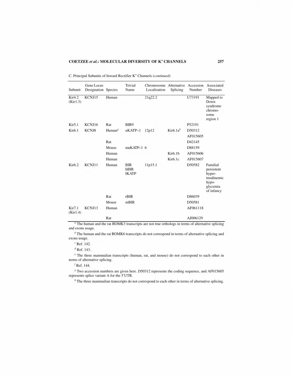
Kir4.2 (Kir1.3)
KCNJ15 Human 21q22.2 U73191 Mapped to Down syndrome chromo-some region 1
Kir5.1 KCNJ16 Rat BIR9 P52191
Kir6.1 KCNJ8 Humang uKATP−1 12p12 Kir6.1ah D50312
AF015605
Rat D42145
Mouse muKATP−1 6 D88159
Human Kir6.1b AF015606
Human Kir6.1c AF015607
Kir6.2 KCNJ11 Human BIRhBIRIKATP
11p15.1 D50582 Familial persistent hyper-insulinemichypo-glycemia of infancy
Rat rBIR D86039
Mouse mBIR D50581
Kir7.1 (Kir1.4)
KCNJ13 Human AF061118
Rat AJ006129a The human and the rat ROMK3 transcripts are not true orthologs in terms of alternative splicing
and exons usage.b The human and the rat ROMK6 transcripts do not correspond in terms of alternative splicing and
exons usage.c Ref. 142.d Ref. 143.e The three mammalian transcripts (human, rat, and mouse) do not correspond to each other in
terms of alternative splicing.f Ref. 144.g Two accession numbers are given here. D50312 represents the coding sequence, and AF015605
represents splice variant A for the 5′UTR.h The three mammalian transcripts do not correspond to each other in terms of alternative splicing.

258 ANNALS NEW YORK ACADEMY OF SCIENCES
D. Principal Subunits of Two-Pore K+ Channels
SubunitGene Locus Designation Species
Trivial Name
Chromosome Localization
Alternative Splicing
Accession Number
Associated Diseases
TWIK KCNK1 Human TWIK-1 1q42−1q43 U33632
Mouse 8 U86009
TREK KCNK2 Mouse U73488
TASK Human AF006823
Rat AF031384
Mouse AF006824
TRAAK Mouse AF056492
E. Auxiliary Subunits of Kv and eag Channels
SubunitGene Locus Designation Species
Trivial Name
Chromosome Localization
Alternative Splicing
Accession Number
Associated Diseases
Kvβ1 KCNA1B Human HKvβ1a 3q26.1 Kvβ1.1 U33428
Rat Kvβ1 X70662
Mouse X97281
Human hKvβ3 Kvβ1.2 U16953
Human hKvβ1.3 Kvβ1.3 L47665
Kvβ2 KCNA2B Human 1p36.3 Kvβ2.1 U33429 Charcot-Marie-Tooth disease, type 2A?
Rat RCKβ2 X76724
Kvβ2.1
Mouse 4 U31908
Human Kvβ2.2 AF044253
Kvβ3 KCNA3B Human KCNA3.1B 17q13 Kvβ3.1 AF016411
Rat Kvβ3 X76723
RCKβ3
Mouse mKvβ4 Kvβ3.2 U65593
KChAP Rat AF032872

COETZEE et al.: MOLECULAR DIVERSITY OF K+ CHANNELS 259
F. Auxiliary Subunits of Ca2+-Activated K+ Channels
SubunitGene Locus Designation Species
Trivial Name
Chromosome Localization
Alternative Splicing
Accession Number
Associated Diseases
Slo Beta Human U38907
Rat U40602
Mouse AJ001291
G. Auxiliary Subunits of Inward Rectifying K+ Channels
SubunitGene Locus Designation Species
Trivial Name
Chromosome Localization
Alternative Splicing
Accession Number
Associated Diseases
SUR1 SUR Human 11p15.1 Q09428 Familial persistent hyper-insulinemichypo-glycemia of infancy
Rat X97279
Rat SUR1B AF039595
SUR2 Human 12p12.1 SUR2A AF061324
Mouse mSUR 6 D80637
Human SUR2B AF061324
Rat AF019628
Mouse D086038

260 ANNALS NEW YORK ACADEMY OF SCIENCES
T
AB
LE
2. S
eque
nce
Iden
tity
(%
)a bet
wee
n G
enes
Cod
ing
for
K+ C
hann
el P
rinc
ipal
Sub
units

COETZEE et al.: MOLECULAR DIVERSITY OF K+ CHANNELS 261
TABLE 3. Native Channels and Physiological Significance of the Molecular Diversity of K+ Channels
Channel Proteins Native Channels
Kv1.x (probably in heteromultimeric channels also containing Kvβ subunits)
4-AP−sensitive voltage-gated K+ channels of both delayed rectifier and “A” types, including the “D” current (a dendrotoxin–sensitive voltage-dependent current with variable kinetics and voltage dependence; also sensitive to very low 4-AP concentrations).121,122 “D” channels are probably various combinations of Kv1.2 (or Kv1.1 or Kv1.6) with other Kv1 proteins and Kvβ subunits. Kv1.4 (possibly in homomultimeric and heteromultimeric channels) might be responsible for fast “A” type K+ currents in terminals and axons.50,123,124 Use of antisense oligonucleotides suggests that Kv1.5 is responsible for the ultrarapid delayed rectifier current (IKur) in human atrium.125
Kv2.x, possibly in combination with Kv5.1, Kv6.1, Kv8.1, and Kv9.1−9.3
4-AP–sensitive delayed rectifiers with variable kinetics, slow inactivation and voltage dependence. Oxygen-sensitive currents in pulmonary artery myocytes.126
Kv3.1−Kv3.4 (may exist as homo- and heteromultimeric channels)
High voltage−activating, fast deactivating voltage-dependent K+ currents, which may contain a fast inactivating component if they include Kv3.4 proteins. Channels containing mainly Kv3.1 and Kv3.2 proteins (and perhaps Kv3.3) are most likely delayed rectifiers with slow inactivation. Currents blocked by 1 mM TEA which are key in the repolarization of short action potentials in fast spiking neurons (see chapter by Rudy et al., this volume).
Kv4.x (possibly in association with unidentified auxiliary subunits)
Classical low voltage− or subthreshold-activating A-type currents and the Ito in heart. Evidence for a relationship with Ito includes use of antisense oligonucleotides.127,128 Blocked by mM concentrations of 4-AP.
KQT KQT1 is responsible for the slowly activating delayed rectifier (IKs) in heart.94,95 Mutations in this gene are responsible for a form of LQT syndrome (see chapter by Sanguinetti, this volume). KQT2 in association with KQT3 may form delayed rectifier type currents in neurons (see text). Mutations in these two genes cause a form of epilepsy (see TABLE 1).
eag “M” current?129,130
elk ?
erg Erg1 is responsible for the rapidly activating delayed rectifier current (IKr) in heart (in combination with minK).97 Evidence includes use of antisense oligonucleotides.131 LQT syndrome mapped to mutations of herg and minK (see TABLE 1 and chapter by Sanguinetti, this volume).
slo (different alternatively spliced versions, with and without a β subunit)
Maxi-K or BK Ca2+-activated channels. Evidence includes biochemical purification from native tissue.132,133
SK 1−3 Small-conductance apamin-sensitive and -insensitive Ca2+-activated K+ channels (see chapter by Adelman, this volume).
SK4 Medium-conductance Ca2+-activated K+ channels.134−136
Two-pore K+ channels “Leak” K+ channels, which help regulate the resting potential.12
Kir1.1−Kir1.3 Weak inward rectifiers.

262 ANNALS NEW YORK ACADEMY OF SCIENCES
TABLE 3. Native Channels and Physiological Significance of the Molecular Diversity of K+ Channels (continued)
Channel Proteins Native Channels
Kir2.1−2.4 Kir2.1: IK1 in cardiac ventricular myocytes, 21 pS channel.65 Common strongly rectifying inward rectifier channel.
Kir3.1 + Kir3.4 G-protein−activated K+ channels in neurons and heart (particularly in atrium).137 Kir3.1 + Kir3.4 acetylcholine-activated K+ current (IK,Ach) in heart (see chapter by Kevin Wickman, this volume).
Kir6.1 plus SUR2B KNDP (33pS nucleotide−sensitive K+ channel in vascular smooth muscle). Activated by ADP, GDP; blocked by glibenclamide (IC50 = 25 nM)
Kir6.2 plus SUR1 ATP-sensitive K+ current (IK,ATP) in pancreatic β-cells. γ = 70 pS; Ki (ATP) = 10 µM; KCOs (diazoxide > pinacidil), glibenclamide (IC50 < 10 nM)
Kir6.2 plus SUR2A ATP-sensitive K+ current (IK,ATP) in heart and skeletal muscle. γ = 80 pS; Ki (ATP) = 175 µM; KCOs (pinacidil > diazoxide), glibenclamide (IC50 = 1.2 µM)
Kir6.2 plus SUR2B ATP-sensitive K+ current (IK,ATP) in vascular smooth muscle. γ = 80 pS; Ki (ATP) = 53 µM; KCOs (pinacidil > diazoxide), glibenclamide (IC50 = 1 µM)
NOTE: γ = unitary conductance.

COETZEE et al.: MOLECULAR DIVERSITY OF K+ CHANNELS 263
TA
BL
E 4
. Fun
ctio
nal P
rope
rtie
s of
K+ C
hann
el P
rote
ins
Exp
ress
ed in
Het
erol
ogou
s E
xpre
ssio
n S
yste
ms
A. F
unct
iona
l Pro
pert
ies
of V
olta
ge-A
ctiv
ated
K+ C
hann
els
Ele
ctro
phys
iolo
gica
l Pro
pert
ies
Act
ivat
ion
Dea
cti-
vati
on
τ (m
s)b
Inac
tivat
ion
Sin
gle
Cha
n-ne
l g
(pS)
Phar
mac
olog
ical
Pro
pert
ies
(IC
50)
Von
(m
V)
V1/
2 (m
V)
k(m
V)
τaV
1/2
(mV
)k
(mV
)τ
(ms)
cτ r
ec
TE
Ao
(mM
)4-
AP
(m
M)
DT
X
(nM
)C
TX
(n
M)
Oth
er P
rope
rtie
s
Kv1
.1− 6
0 to
− 5
0− 3
06
to 9
++
+14
Ver
y sl
owd
≈ 10
0.5
0.16
to
1.1
12 to
21
NB
TE
A (h
uman
) IC
50
20m
M?
HgT
X IC
5031
pM
Kv1
.2−4
0− 5
to 5
13+
23V
ery
slow
9.2
to
17>
10
0.2
to
0.8
2.8
to
241.
7 to
17
HgT
X I
C50
170
pM
Kv1
.3− 5
0− 3
05
to 7
++
38− 4
4.7
250
to
600
9.6
to
1410
to
500.
2 to
1.
525
0?0.
5 to
2.
6M
gTX
IC
50 2
30 p
M
HgT
X IC
50 8
6 pM
Sho
ws
cum
ulat
ive
inac
tivat
ion
Kv1
.4− 5
0− 2
212
++
+15
to
55− 6
5 to
− 4
5+
2.3
τ 1 ≈
20
τ 2 ≈
40
2.2
to
3.8s
(−80
m
V)
4.7
NB
0.7
to
13>
200
NB
Kv1
.5− 5
0− 1
07
++
+23
Ver
y sl
ow8
NB
< 0
.1 to
0.
4N
BN
B
Kv1
.6− 5
0− 2
06
to 8
++
+V
ery
slow
91.
7 to
70.
3 to
1.
520
to
251
MgT
X I
C50
3 n
MH
gTX
IC
50 6
nM
Kv1
.7− 4
0− 2
08
++
+5.
1 to
5.
3N
AN
A14
Slo
w21
NB
0.25
NA
NB
MgT
X I
C50
116
nM
N
xTX
IC
50 1
8 nM
ShK
TX
IC
50 1
3 nM
Kv2
.1− 2
0 to
− 3
0≈
105
to 1
9+
15 to
20
− 20
5 to
13
Ver
y sl
owe
1.6
s (−
90 m
V)
8 to
94
to 1
00.
5 to
4.
5N
BN
BH
aTX
KD
100
nM
(see
not
e f)

264 ANNALS NEW YORK ACADEMY OF SCIENCES
A. F
unct
iona
l Pro
pert
ies
of V
olta
ge-A
ctiv
ated
K+ C
hann
els
( con
tinu
ed)
Ele
ctro
phys
iolo
gica
l Pro
pert
ies
Act
ivat
ion
Dea
cti-
vati
on
τ (m
s)b
Inac
tivat
ion
Sin
gle
Cha
n-ne
l g
(pS)
Phar
mac
olog
ical
Pro
pert
ies
(IC
50)
Von
(m
V)
V1/
2 (m
V)
k(m
V)
τaV
1/2
(mV
)k
(mV
)τ
(ms)
cτ r
ec
TE
Ao
(mM
)4-
AP
(m
M)
DT
X
(nM
)C
TX
(n
M)
Oth
er P
rope
rtie
s
Kv2
.2− 2
0 to
− 3
0≈
10≈
18+
NA
− 30
13V
ery
slow
eN
A≈
14≈
81.
5N
AN
AN
ote
f
Kv3
.1− 1
0+
10 to
18
8 to
11
++
1.4
Ver
y sl
ow16
to
270.
15 to
0.
20.
02 to
0.
6N
BN
BC
hrom
akal
in
KD
0.2
37 m
M
Kv3
.2− 1
0+
10 to
13
7 to
9+
+2
Ver
y sl
ow16
to
200.
150.
6 to
0.
9N
BN
B
Kv3
.3− 1
07
6+
+2
5.2
6.1
240
NA
140.
141.
2
Kv3
.4− 1
0+
13 to
19
7 to
11
++
+N
A− 2
0 to
− 3
24.
8 to
8.
310
to
20N
A12
to
140.
09 to
0.
30.
5 to
0.
6N
BN
BB
DS-
I IC
50 4
7 nM
BD
S-II
IC
50 5
6 nM
Kv4
.1− 6
0 to
− 5
0≈ − 1
0=
13
++
NA
− 69
to
− 50
4.7
to 8
τ 122
.6τ 2
86.4
τ 336
8h
200
ms
(−10
0 m
V)g
6 to
8N
B9
NA
NA
Kv4
.2≈ − 5
0− 4
to− 1
513
to
20+
+≈
40− 4
1 to
− 6
66
to 8
τ 1 1
5 to
20 [�
80%
]τ 2
61
to
120
150
to
300
ms
( −80
m
V)g
4 to
5N
B2
to 5
iN
BN
AH
pTX
1 an
d H
pTX
2 IC
50
100
nM,
HpT
X3
IC50
67
nMj
Kv4
.3−6
0 to
−5
0=
−20
≈ 13
++
20 to
40
≈ −6
04.
5 to
7τ 1
30[ ≈
70%
]τ 2
160
100
to
200
msg
( −80
to
−100
m
V)
NA
NB
5 to
10i
NA
NA
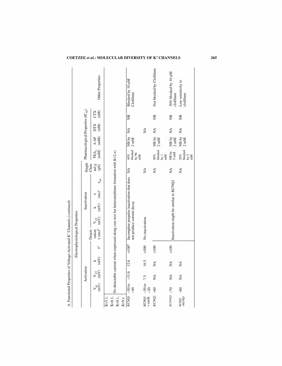
COETZEE et al.: MOLECULAR DIVERSITY OF K+ CHANNELS 265
A. F
unct
iona
l Pro
pert
ies
of V
olta
ge-A
ctiv
ated
K+ C
hann
els
( con
tinu
ed)
Ele
ctro
phys
iolo
gica
l Pro
pert
ies
Act
ivat
ion
Dea
cti-
vati
on
τ (m
s)b
Inac
tivat
ion
Sin
gle
Cha
n-ne
l g
(pS)
Phar
mac
olog
ical
Pro
pert
ies
(IC
50)
Von
(m
V)
V1/
2 (m
V)
k(m
V)
τaV
1/2
(mV
)k
(mV
)τ
(ms)
cτ r
ec
TE
Ao
(mM
)4-
AP
(m
M)
DT
X
(nM
)C
TX
(n
M)
Oth
er P
rope
rtie
s
Kv5
.1,
No
dete
ctab
le c
urre
nt w
hen
expr
esse
d al
ong
(see
text
for
het
erom
ultim
er f
orm
atio
n w
ith K
v2.x
)K
v6.1
,
Kv8
.1,
Kv9
.x
KC
NQ
1− 5
0 to
− 4
0− 1
1.6
12.6
>10
0kIn
com
plet
e pr
epul
se in
activ
atio
n th
at d
oes
not p
rodu
ce c
urre
nt d
ecay
NA
40%
bl
ocke
d by
96
mM
NB
by
2 m
MN
AN
BB
lock
ed b
y 10
µM
C
lofi
lium
KC
NQ
1+
min
K− 5
0 to
− 2
07.
516
.5>
500
No
inac
tivat
ion
NA
NA
KC
NQ
2− 6
0N
AN
A>
100
Inac
tivat
ion
mig
ht b
e si
mil
ar to
KC
NQ
1
NA
90%
bl
ocke
d by
1
mM
NB
by
2 m
MN
AN
BN
ot b
lock
ed b
y C
lofi
lium
KC
XN
Q3− 7
0N
AN
A>
100
NA
NB
by
5 m
MN
B b
y 2
mM
NA
NB
30%
blo
cked
by
10 µ
M
clof
iliu
mK
CN
Q2
+K
CN
Q3
− 60
NA
NA
NA
20%
bl
ocke
d by
5
mM
NB
by
2 m
MN
AN
BL
ow s
ensi
tivit
y to
cl
ofil
ium

266 ANNALS NEW YORK ACADEMY OF SCIENCES
A. F
unct
iona
l Pro
pert
ies
of V
olta
ge-A
ctiv
ated
K+ C
hann
els
( con
tinu
ed)
Ele
ctro
phys
iolo
gica
l Pro
pert
ies
Act
ivat
ion
Dea
cti-
vati
on
τ (m
s)b
Inac
tivat
ion
Sin
gle
Cha
n-ne
l g
(pS)
Phar
mac
olog
ical
Pro
pert
ies
(IC
50)
Von
(m
V)
V1/
2 (m
V)
k(m
V)
τaV
1/2
(mV
)k
(mV
)τ
(ms)
cτ r
ec
TE
Ao
(mM
)4-
AP
(m
M)
DT
X
(nM
)C
TX
(n
M)
Oth
er P
rope
rtie
s
Eag
1− 4
0− 7
.519
.6+
+l
τ 1 4
.6τ 2
76
(−90
mV
)
Slow
NA
28>
100
NA
NA
M c
urre
nt?
IKx
in r
od
oute
r se
gmen
t?
Erg
1− 6
0 to
− 5
0− 2
16
to 8
Hun
-dr
eds
of m
s
Erg
1 an
d 2
beha
ve a
s sl
owly
act
ivat
ing
dela
yed
rect
ifie
rs (τ ~
100
ms)
, but
at p
ositi
ve v
olta
ges
(> 0
m
V)
ther
e is
a r
educ
tion
in s
tead
y st
ate
(ss)
cu
rren
t pro
duci
ng a
neg
ativ
e sl
ope
I−V
and
in
war
d re
ctif
icat
ion.
Erg
3 cu
rren
ts h
ave
a pr
omin
ent t
rans
ient
com
pone
nt a
nd a
ctiv
ate
5−10
ti
mes
fas
ter
and
also
hav
e a
nega
tive
slop
e I−
V.
Upo
n re
pola
riza
tion
fro
m p
ositi
ve v
olta
ges
the
3 ch
anne
ls p
rodu
ce la
rge
curr
ents
that
dec
ay s
low
ly.
Thi
s be
havi
or is
due
to th
e pr
esen
ce o
f a
volt
age-
depe
nden
t “C
” in
activ
atio
n pr
oces
s, w
hich
is 2
to
10 ti
mes
fas
ter
than
act
ivat
ion
at m
ost v
olta
ges.
R
ecov
ery
from
inac
tivat
ion
is v
ery
fast
for
all
3
chan
nels
(<
10
ms)
.
5 to
12
in s
ym-
met
ric
[K+]
50 NA
NA
NA
NA
NA
NA
NA
Blo
cked
by
dofe
tilid
e E
4031
KD
99 n
MB
lock
ed b
y 2
µM
MK
-49
9
Erg
2− 4
0− 3
.58.
3N
AN
AN
AN
AN
AE
4031
KD
116
nM
Erg
3− 8
0 to
− 7
0− 4
47.
2N
AN
AN
AN
AE
4031
KD
193
nM
Elk
1− 4
0m9.
313
.169
NA
NB
by
10 m
MN
B b
y 10
mM
NB
by
10 µ
M E
4031
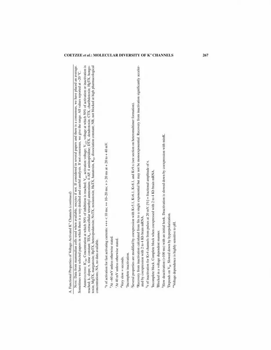
COETZEE et al.: MOLECULAR DIVERSITY OF K+ CHANNELS 267
A. F
unct
iona
l Pro
pert
ies
of V
olta
ge-A
ctiv
ated
K+ C
hann
els
( con
tinu
ed)
NO
TE: D
ata
from
mam
mal
ian
cell
s us
ed w
hen
avai
labl
e, o
ocyt
es if
not
. If
cons
ider
ed in
sev
eral
pap
ers
and
ther
e is
a c
onse
nsus
, we
have
pla
ced
an a
vera
ge.
Som
etim
es w
e ha
ve s
elec
ted
pape
rs in
whi
ch th
ere
is a
ver
y de
taile
d an
d ca
refu
l ana
lysi
s. I
f no
t con
sens
us, w
e gi
ve th
e ra
nge.
All
val
ues
repo
rted
at ∼
20° C
.
AB
BR
EV
IAT
ION
S: I
C50
, Con
cent
ratio
n at
whi
ch 5
0% o
f in
hibi
tion
is r
each
ed; V
on, a
ctiv
atio
n vo
ltag
e; V
1/2,
vol
tage
at w
hich
50%
of
activ
atio
n or
inac
tivat
ion
isre
ache
d; k
, slo
pe; τ ,
tim
e co
nsta
nt;
TE
Ao,
ext
race
llula
r te
trae
thyl
am
mon
ium
; 4-
AP,
4 a
min
opir
idin
e; D
TX
, de
ndro
toxi
n; C
TX
, ch
arib
doto
xin;
HgT
X,
hong
o-to
xin;
MgT
X, m
arga
toxi
n; H
pTX
, het
erop
odat
oxin
; NxT
X, n
oxiu
stox
in; H
aTX
, han
atox
in, K
D, d
isso
ciat
ion
cons
tant
; NB
, not
blo
cked
at h
igh
phar
mac
olog
ical
conc
entr
atio
ns; N
A, n
o da
ta a
vail
able
.
a τ o
f ac
tivat
ion
for
fast
act
ivat
ing
curr
ents
: ++
+ <
10
ms;
++
10−
20 m
s; +
> 2
0 m
s at
+ 2
0 to
+ 4
0 m
V.
b At −
60 m
V u
nles
s ot
herw
ise
stat
ed.
c At 4
0 m
V u
nles
s ot
herw
ise
stat
ed.
d Ver
y sl
ow =
sec
onds
.e In
com
plet
e in
activ
atio
n.f S
ever
al p
rope
rtie
s ar
e m
odif
ied
by c
oexp
ress
ion
with
Kv5
.1, K
v6.1
, Kv8
.1, a
nd K
v9.x
(se
e se
ctio
n on
het
erom
ulti
mer
for
mat
ion)
.g R
ecov
ery
from
ina
ctiv
atio
n ca
lcul
ated
fro
m f
its to
a s
ingl
e ex
pone
ntia
l but
may
not
be
mon
oexp
onen
tial.
Rec
over
y fr
om i
nact
ivat
ion
sign
ific
antly
acc
eler
-at
ed b
y co
expr
essi
on w
ith 2
to 4
Kb
brai
n m
RN
A.
h τ o
f in
activ
atio
n fo
r K
v4 c
hann
els
from
pul
ses
at 2
0 m
V. [
%]
= f
ract
iona
l am
plitu
de o
f τ .
i Inco
mpl
ete
bloc
k. C
ompl
ete
bloc
k w
hen
coex
pres
sed
wit
h 2
to 4
Kb
brai
n m
RN
A.
j Blo
cked
in a
vol
tage
dep
ende
nt m
anne
r.k Sl
ow d
eact
ivat
ion
(>10
0 m
s) w
ith a
n in
itia
l hoo
k. D
eact
ivat
ion
is s
low
ed d
own
by c
oexp
ress
ion
wit
h m
inK
.l D
epen
ds o
n V
H. S
low
ed d
own
by h
yper
pola
riza
tion.
mV
olta
ge d
epen
denc
e is
hig
hly
sens
itive
to p
H.

268 ANNALS NEW YORK ACADEMY OF SCIENCES
B.
Fun
ctio
nal P
rope
rtie
s of
Ca2+
-Act
ivat
ed K
+ C
hann
els
Vol
tage
Dep
ende
nce
Ca2+
Sen
sitiv
ity
Sin
gle
Cha
nnel
γ (
pS)
Phar
mac
olog
y(I
C50
or
KD
)O
ther
Pro
pert
ies
Slo1
Con
duct
ance
is v
olta
ge
depe
nden
t. V
1/2
shif
ts w
ith
Ca2+
con
cent
ratio
n fr
om
≈ 50
mV
at 4
µM
[C
a2+]
to− 3
0 m
V a
t 100
µM
[C
a2+].
V
olta
ge d
epen
denc
e is
als
o sh
ifte
d by
coe
xpre
ssio
n w
ith
β su
buni
t.
Ca2+
sen
sitiv
ity
is lo
wer
than
ch
anne
ls f
rom
SK
fam
ily
and
chan
ges
sign
ific
antl
y de
pend
ing
on th
e al
tern
ativ
e sp
liced
ver
sion
and
on
the
pres
ence
of
the β
subu
nit.
> 2
00 in
sym
met
ric
[K+]
TE
A 8
0 to
330
µM
CT
X I
C50
2 to
40
nM.
Iber
ioto
xin
(IbT
X)
IC50
6 to
11
nM
. β s
ubun
it d
ecre
ases
se
nsiti
vity
to Ib
TX
(IC
50 1
02
to 1
60 n
M)
and
conf
ers
sens
itivi
ty to
the
agon
ist
DH
S-1,
and
doe
s no
t aff
ect
CT
X s
ensi
tivity
.
Slo3
Con
duct
ance
is s
ensi
tive
to v
olta
ge a
nd p
H b
ut a
ppar
entl
y no
t to
Ca2+
106
in s
ymm
etri
c [K
+]
TE
A 4
9 m
ME
xpre
sses
onl
y in
test
is
SK1
Vol
tage
inde
pend
ent
K0.
5 0.
71 µ
M H
ill c
oeff
icie
nt 3
.99.
2 in
sym
met
ric
[K+]
Apa
min
inse
nsit
ive
d-T
uboc
urar
ine
76.2
µM
SK2
Vol
tage
inde
pend
ent
K0.
5 0.
63 µ
M H
ill c
oeff
icie
nt 4
.89.
9 in
sym
met
ric
[K+]
Apa
min
63
pMd-
Tub
ocur
arin
e 2.
4 µ
M
SK3
Vol
tage
inde
pend
ent
Sim
ilar
to S
K1
and
SK2
≈ 10
Apa
min
1− 2
nM
SK4
Vol
tage
inde
pend
ent
KD
270
nM
Hil
l coe
ffic
ient
2.7
33 in
sym
met
ric
[K+]
Apa
min
inse
nsit
ive
Not
blo
cked
by
IbT
X a
nd
MgT
X; i
nwar
dly
rect
ifyi
ng
9 in
asy
mm
etri
c [K
+]
TE
A 3
0 m
M; C
TX
10
nM
Cor
trim
azol
e 38
7 nM

COETZEE et al.: MOLECULAR DIVERSITY OF K+ CHANNELS 269
C.
Func
tiona
l Pro
pert
ies
of I
nwar
d-R
ecti
fier
K+ C
hann
els
γ (p
S)b
Mea
n O
pen
Tim
e (m
s)M
ean
Clo
sed
Tim
e (m
s)R
ectif
icat
ionc
Blo
cker
s
Spec
ial P
rope
rtie
sSp
erm
idin
edS
perm
ined
Ba2+
o (µ
M)
Cs+
o (µ
M)
Kir
1.1
31− 3
9 24
ms
(−80
mV
)1.
1 &
35
ms
(–80
mV
) W
eak
2.16
mM
ND
70 (− 1
00 m
V)
1.2
mM
(−12
0 m
V)
Act
ivat
ed b
y A
TP
i (bl
ocke
d by
M
g2+; I
C50
= 1
.7− 4
.2 m
M)e
Kir
2.1
21− 2
311
7−18
5 m
s(−
100
mV
) 0.
8 &
26m
s(−
100
mV
) St
rong
8 nM
& 2
.9
µM
0.9
nM &
0.6
µ
M
0.15
(− 1
40 m
V)
14 (− 1
40 m
V);
54 m
M (
0 m
V)
Blo
cked
by
Mg2+
; IC
50 =
17
&
2170
µM
e
Kir
2.2
34− 4
1 71
ms
(−10
0 m
V)
0.7
ms
& 1
1.9
ms
(−10
0 m
Vf )
Stro
ngN
DN
D6
(−80
mV
) N
D
Kir
2.3
13− 1
6 N
DN
DSt
rong
ND
ND
93− 1
83(−
130
mV
)30− 4
7(−
130
mV
) S
ensi
tive
to p
Ho
and
AT
Pi
Kir
2.4
15
ND
ND
Stro
ngN
DN
D39
0(−
80 m
V)
8.06
mM
(−80
mV
)
Kir
3.1
27− 4
2 (3
9g )0.
26, 1
.2 &
7.2
ms
(−60
mV
)N
DSt
rong
ND
ND
10− 9
494
Exp
ress
es a
s a
hete
rom
ulti
mer
w
ith
Kir
3.2
or K
ir3.
4
Kir
3.2
30 (
35h )
0.1
& 0
.5 m
s (0
.5 &
3.5
msh )
(−80
mV
i )
ND
Stro
ngN
D10
µM
94 (−
120
mV
) 94 (−
120
mV
) E
xpre
sses
as
a he
tero
mul
tim
er
wit
h K
ir3.
1 or
Kir
3.4
Kir
3.3
——
——
——
——
No
expr
essi
on a
lone
Coa
ssem
bly
subu
nit o
f K
ir3.
1 &
Kir
3.2

270 ANNALS NEW YORK ACADEMY OF SCIENCES
C.
Func
tiona
l Pro
pert
ies
of I
nwar
d-R
ecti
fier
K+ C
hann
els
( con
tinu
ed)
γ (p
S)b
Mea
n O
pen
Tim
e (m
s)M
ean
Clo
sed
Tim
e (m
s)R
ectif
icat
ionc
Blo
cker
s
Spec
ial P
rope
rtie
sSp
erm
idin
edS
perm
ined
Ba2+
o (µ
M)
Cs+
o (µ
M)
Kir
3.4
31− 3
3 (3
5h )1.
3 m
s(−
80 m
Vh )
Stro
ngN
DN
D92 (−
60 m
V)
61 (−60
mV
) C
oass
embl
y su
buni
t of
Kir
3.1
Inhi
bits
exp
ress
ion
of K
ir3.
2 M
ay
form
str
etch
-act
ivat
ed K
+
chan
nels
Kir
4.1
22 &
36
j 10
0−20
0 m
s(−
100
mV
) 1
& 3
0 m
s(−
100
mV
) W
eak
40 n
MN
D99 (−
120
mV
) 11
2(0
mV
) A
ctiv
ated
by
AT
Pi;
inhi
bite
d by
pH
i (pK
a =
6.2
)
Kir
4.2
——
——
——
——
No
expr
essi
on a
s ho
mom
ulti
mer
ic s
ubun
its;
in
hibi
ts e
xpre
ssio
n of
Kir
1.1
or K
ir1.
2 cu
rren
ts
Kir
5.1
——
——
——
—-—
No
expr
essi
on a
s ho
mom
ulti
mer
ic s
ubun
itsk
Kir
7.1
‘0.
05N
DN
DSt
rong
ND
ND
1000
10,0
00
a Exc
ludi
ng K
AT
P c
hann
els,
for
whi
ch s
ee T
AB
LE 4
D.
b Sin
gle-
chan
nel c
ondu
ctan
ce (γ )
was
mea
sure
d at
hig
h (1
40− 1
50 m
M)
sym
met
rica
l K+ c
once
ntra
tions
.c R
ecti
fica
tion
is c
onfe
rred
by
bloc
k of
the
open
cha
nnel
by
intr
acel
lula
r M
g2+ a
nd p
olya
min
es.
d Mea
sure
d at
+40
mV
.e A
ll K
ir c
hann
els
are
bloc
ked
to s
ome
exte
nt b
y M
g2+ (
one
of th
e pr
ime
reas
ons
for
rect
ific
atio
n). I
C50
val
ues
are
not a
vail
able
in a
ll c
ases
.f C
hann
el a
ctiv
ity
of K
ir2.
2 oc
curr
ed in
bur
sts,
sep
arat
ed b
y cl
osin
gs o
f >
200
ms.
The
gap
dur
atio
n in
crea
ses
wit
h hy
perp
olar
izat
ion.
g Whe
n co
expr
esse
d w
ith K
ir3.
4.h W
hen
coex
pres
sed
with
Kir
3.1.
i Ope
ning
s oc
cur
in b
urst
s; th
e m
ean
open
tim
e or
bur
st k
inet
ics
wer
e no
t ana
lyze
d.j T
he 3
6 pS
cha
nnel
was
onl
y ob
serv
ed in
Xen
opus
ooc
ytes
and
not
in m
amm
alia
n ce
lls,
sug
gest
ing
coas
sem
bly
with
end
ogen
ous
Xen
opus
sub
units
.k H
as b
een
repo
rted
to f
orm
fun
ctio
nal h
eter
omul
timer
ic a
ssem
blie
s w
ith
Kir
4.1

COETZEE et al.: MOLECULAR DIVERSITY OF K+ CHANNELS 271
D.
Fun
ctio
nal P
rope
rtie
s of
AT
P-S
ensi
tive
K+ C
hann
els
Prin
cipa
l Su
buni
t with
/w
ithou
t A
uxili
ary
Sub
unit
Sing
le-C
hann
el
Con
duct
ance
γ (p
S)M
ean
Ope
n T
ime
(ms)
Mea
n C
lose
d T
ime
(ms)
Mea
n B
urst
D
urat
ion
(ms)
Inte
rbur
st
Dur
atio
n (m
s)N
ucle
otid
e B
lock
(µ
M)
Blo
cker
s (n
M)
Ope
ners
(µ
M)
Kir
6.1
alon
e70
3.31
(− 6
0 m
V)
0.9
(−60
mV
)N
DN
DN
DG
lib
(no
bloc
k)
No
effe
ct
Kir
6.1+
SUR
1N
DN
DN
DN
DN
DN
DTo
l (17
.7 µ
M)
ND
Kir
6.1+
SUR
2B32
.9N
D‘
ND
ND
ND
AT
P =
GT
P (
3)a
Glib
(<
3 µ
M)
Pin
acid
il (
< 1
00
µM
)
Kir
6.2
alon
e57
.6b
700.
8−1.
9(−
60 m
Vb )
0.31
&12
.6(−
60 m
Vb )
2.4−
8.3
(–60
mV
b )N
DM
g-A
TP
(106
) M
g-A
TP
(115−
145)
b
No
bloc
kN
o ef
fect
Kir
6.2+
SUR
158
− 76
(73)
b2.
1(−
80 m
V)
0.56
(−80
mV
)17
.9
(−60
mV
) 24
& 2
43(−
60 m
V)
AT
P (6− 3
4);
Mg-
AT
P (2
8);
AT
P (1
3−18
)b
Glib
(1.
8−8.
6)
> T
ol (
4 − 3
2 µ
M)
Dia
zoxi
de (
20−
100
µM
) >
pi
naci
dil
Kir
6.2+
SUR
2A79− 8
01.
3(–
80 m
V)
0.16
(–80
mV
)N
DN
DA
TP
(100− 1
48)
> A
DP>
AM
P
Mg-
AT
P (1
72)
Glib
(16
0)
Glib
(35
0)c
Glib
(63
0)d
Tol (
120
µM
)
Pina
cidi
l (10
) >
ni
cora
ndil
>>
di
azox
ide
Kir
6.2+
SUR
2B80
.3N
DN
DN
DN
DA
TP
(67.
9) >
M
g-A
TP
(300
) G
lib (
< 1
µM
)P
inac
idil
>
diaz
oxid
ea A
ctiv
ated
by
nucl
eoti
des.
b C-t
erm
inal
trun
cate
d fo
rm o
f K
ir6.
2.c R
b+ e
fflu
x m
easu
rem
ents
.d B
indi
ng a
ssay
s.

272 ANNALS NEW YORK ACADEMY OF SCIENCES
E.
Fun
ctio
nal P
rope
rtie
s of
Tw
o-Po
re K
+ C
hann
els
γ (p
S)a
Rec
tific
atio
nS
peci
al P
rope
rtie
s
Pha
rmac
olog
y
Ba2+ 0
Cs2+ 0
TW
IK35
Inw
ard
(wea
k)35
− 100
µM
Not
blo
cked
TR
EK
14O
utw
ardb
100
µM
Not
blo
cked
TASK
16O
utw
ardb
Hig
hly
sens
itiv
e to
ex
trac
ellu
lar
pH p
Ka
=
6.3
Hill
= 1
.6
29%
blo
cked
by
100
µM
30%
blo
cked
by
100
µM
TR
AA
K45
Out
war
dbR
egul
ated
by
arac
hido
nic
acid
and
po
lyun
satu
rate
d fa
tty
acid
s
1 m
M a
t 30
mV
Not
blo
cked
a Sin
gle-
chan
nel c
ondu
ctan
ce (γ )
was
mea
sure
d at
hig
h (1
40− 1
50 m
M)
sym
met
rica
l K+ c
once
ntra
tion
s.b O
beys
Gol
dman
-Hod
gkin
-Kat
z cu
rren
t equ
atio
n fo
r ch
ange
s in
[K
] o.

COETZEE et al.: MOLECULAR DIVERSITY OF K+ CHANNELS 273
TA
BL
E 5
.T
issu
e E
xpre
ssio
n of
K+ C
hann
el P
rinc
ipal
Sub
units
Tis
sue
Dis
trib
utio
naB
rain
Reg
ion
sp
He
Ki
SKL
uB
rO
ther
sp
CX
Th
CB
OB
HC
Oth
erPr
otei
n D
istr
ibut
ion
Kv1
.1r
––
––
+r
++
++
+A
xons
and
term
inal
s.K
v1.2
r +
i–
ND
ND
+r
++
++
+H
bA
xons
and
ner
ve
term
inal
s Pa
rano
dal i
n no
des
of R
anvi
erK
v1.3
r –
±–
++
Re,
spl
een,
ly
mph
rN
DN
D+
++
Som
atod
endr
itic
Kv1
.4
r+
––
ND
+r
++
++
+C
PA
xons
and
term
inal
sK
v1.5
r+
++
++
Pitu
itar
y, a
orta
rN
DN
DN
DN
D+
Som
as a
nd p
roxi
mal
de
ndri
tes
Kv1
.6
r/h
––
––
+r
ND
ND
±±
+So
mat
oden
driti
cK
v1.7
r+
±+
+±
ND
Kv2
.1r
+–
±–
±r
+±
++
+H
bSo
mat
ic a
nd p
roxi
mal
de
ndri
tes
Kv2
.2r
ND
ND
ND
ND
+r
++
++
+So
mat
ic a
nd n
euro
pil
Kv3
.1r
±–
±–
+T-
lym
phr
++
++
+R
TSo
mat
ic a
nd a
xona
l, te
rmin
als
Kv3
.2r
±–
––
+r
++
±±
+So
mas
and
axo
n co
llate
rals
Kv3
.3r
±–
–N
D+
Thy
r +
±+
++
Kv3
.4r
±–
+–
+r
±–
±–
±K
v4.1
r±
ND
ND
ND
±r
––
–+
+K
v4.2
r+
ND
ND
ND
+V
as d
efer
ense
r+
++
++
Hb,
CP
Som
atod
endr
itic
Kv4
.3r
++
e+
e+
e+
Vas
def
eren
ser
++
++
+H
b, S
NPC
, CP
Kv5
.1r
±±
±–
+r
±±
–+
±K
v6.1
r–
±±
+±
Li
r+
++
++
A, C
P

274 ANNALS NEW YORK ACADEMY OF SCIENCES
TA
BL
E 5
.T
issu
e E
xpre
ssio
n of
K+ C
hann
el P
rinc
ipal
Sub
units
(co
ntin
ued)
Tis
sue
Dis
trib
utio
naB
rain
Reg
ion
sp
He
Ki
SKL
uB
rO
ther
sp
CX
Th
CB
OB
HC
Oth
erPr
otei
n D
istr
ibut
ion
KQ
T1
h+
+–
+–
Pla,
Pa
ND
KQ
T2
h–
––
–+
ha+
++
ND
+A
, C. n
ucl.
KQ
T3
h–
––
–+
h+
+±
ND
+A
, C. n
ucl.
Kv8
.1b
r–
––
–+
r+
− +
++
CP
Kv9
.1m
/r–
––
–+
m+
–+
++
Kv9
.2m
––
––
+m
+–
++
+H
b, B
AN
Kv9
.3r
±±
–+
+N
DK
ir1.
1r
–+
–±
+Sp
leen
r−a
± ?a
−aN
D−a
Hyp
otha
lam
usK
ir2.
1m
+–
+N
D+
r+
++
++
Pur
kinj
e ce
lls,
CP
Kir
2.2
h/r/
m+
/+/+
ND
/+/+
ND
/+/+
ND
/+/+
ND
/+/+
r+
++
±±
CP,
Str
ong
in
Th
and
CB
Kir
2.3
h/r/
m+
/−/−
− ?/−
/−± /− /− ?
− /− /
ND
+r
+±
± ?+
+C
P, T
h re
t. nu
cleo
us,
amyg
dala
a
Kir
2.4
h/r
–/+
ND
/+N
D/–
–/±
+N
DM
idbr
ain,
br
ains
tem
Kir
3.1
r/m
+N
D/−
± ?+
/ND
+A
triu
m >
ve
ntri
cle
r+
++
++
Not
in
Purk
inje
cel
ls
Kir
3.2
r/m
––
––
+H
igh
in r
at
panc
reas
r+
+–
++
C3
pyra
mid
al
cells
K
ir3.
3m
––
––
+r
++
++
+B
rain
stem
, m
idbr
ain
Kir
3.4
r/m
++
e /ND
−e /ND
−e /ND
+/−
r+
+±
+±
Kir
4.1
(Kir
1.2)
h/r
− /−
±/±
− /−
− /N
D+
/+r
ND
±+
ND
±A
, C. c
allo
sum
, S
. nig
ra

COETZEE et al.: MOLECULAR DIVERSITY OF K+ CHANNELS 275
TA
BL
E 5
.T
issu
e E
xpre
ssio
n of
K+ C
hann
el P
rinc
ipal
Sub
unit
s ( c
onti
nued
)
Tis
sue
Dis
trib
utio
naB
rain
Reg
ion
sp
He
Ki
SKL
uB
rO
ther
sp
CX
Th
CB
OB
HC
Oth
erP
rot.
Dis
t.
Kir
4.2
(Kir
1.3)
h–
+–
±–
PaN
D
Kir
5.1
re+
±+
±+
re+
f±
+N
D+
g
Kir
6r
++
h+
ND
?L
iN
DK
ir6.
2r
+N
D+
–+
Pitu
itary
, pa
r+
++
++
Mid
brai
n, C
P,
brai
nste
mK
ir7.
1h
−e+
e−e
−e+
ePr
osta
te,
test
is, s
mal
l in
test
ine
ND
Pur
kinj
e an
d py
ram
idal
cel
l la
yer
Eag
1r
––
––
+r
+−
++
+E
rg1
hi+
ND
ND
ND
+SC
G, C
G,
SM
G,
ND
Re,
adr
, thy
Erg
2rh
–N
DN
DN
D–
CG
, SM
G, R
eN
DE
rg3
rh–
ND
ND
ND
+SC
G, C
G,
SMG
ND
SK1
r +
––
–+
Adr
r +
–+
++
SK2
r +
––
–+
Adr
r +
++
++
Pon
tine
nucl
eus
SK3
r +
ND
ND
ND
+A
drr
–+
–+
–S
upra
opti
c nu
cleu
sSK
4h
––
–+
–A
dr, p
laN
ASK
Ca3
h N
DN
DN
DN
DN
DN
DSK
Ca4
h +
-+
+-
Pla,
thy,
co,
T-
lym
ph.,
Li
NA
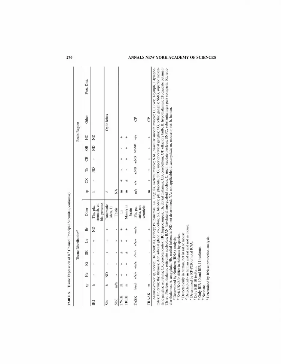
276 ANNALS NEW YORK ACADEMY OF SCIENCES
TA
BL
E 5
.T
issu
e E
xpre
ssio
n of
K+ C
hann
el P
rinc
ipal
Sub
unit
s ( c
onti
nued
)
Tis
sue
Dis
trib
utio
naB
rain
Reg
ion
sp
He
Ki
SKL
uB
rO
ther
sp
CX
Th
CB
OB
HC
Oth
erP
rot.
Dis
t.
IK1
h -
--
+N
DT
hy, p
la,
stom
ach,
co,
bl
a, p
rost
ate
h -
ND
-N
DN
D
Slo
hN
D-
++
+Pa
ncre
atic
is
lets
, Li
dO
ptic
lobe
s
Slo3
m/h
--
--
-Te
stis
NA
TW
IKm
++
±+
+L
im
+-
+-
+T
RE
Km
++
±+
+M
ainl
y in
br
ain
m±
-+
++
TAS
Kh/
m/r
+/+
/++
/±/±
± ?/−
/±+
/+/+
+/±
/±P
la, p
a,at
ria,
not
ve
ntri
cle
m/r
+/+
+/N
D+
/ND
ND
/ND
+/+
CP
TR
AA
Km
--
--
+m
+±
++
+C
PA
BB
RE
VIA
TIO
NS:
sp, s
peci
e; H
e, h
eart
; K
i, ki
dney
; Pa
, pan
crea
s; L
u, l
ung;
Sk,
ske
leta
l m
uscl
e; S
.M.,
vasc
ular
sm
ooth
mus
cle;
Li,
Liv
er;
T-ly
mph
, T-l
ymph
o-cy
tes;
Br,
brai
n; th
y, th
ymus
; Adr
, adr
enal
gla
nd; c
o, c
olon
; bla
, bla
dder
; pla
, pla
cent
a; S
CG
, sup
erio
r ce
rvic
al g
angl
ia; C
G, c
elia
c ga
ngli
a; S
MG
, sup
erio
r m
esen
-te
ric
gang
lia; r
e, r
etin
a; C
X, c
ereb
ral c
orte
x; H
C, h
ippo
cam
pus;
Th,
dor
sal t
hala
mus
; CB
, cer
ebel
lum
; OT,
olf
acto
ry b
ulb;
H, h
ypot
hala
mus
; CP,
cau
date
put
amen
;T
h re
t. N
ucle
us, t
hala
mic
ret
icul
ate
nucl
eolu
s; B
AN
, bas
olat
eral
am
ygda
loid
nuc
leus
, C. n
ucl.,
cau
date
nuc
leus
; SN
PC, s
ubst
antia
nig
ra p
ars
com
pact
a; R
t, re
tic-
ular
thal
amus
; A, a
myg
dala
; Hb,
med
ial h
eben
ula;
ND
: not
det
erm
ined
; NA
: not
app
lica
ble;
d, d
roso
phil
a; m
, mou
se; r
, rat
; h, h
uman
.a D
eter
min
ed b
y N
orth
ern
(RN
A)
anal
ysis
.b K
v8.1
/Kv2
.3r
diff
er in
thal
amus
by
spec
ies.
c Det
ecte
d on
ly in
hum
an, n
ot in
rat
or
mou
se.
d Det
ecte
d on
ly in
hum
an a
nd r
at a
nd n
ot in
mou
se.
e Det
erm
ined
by
RT-
PCR
of
tota
l RN
A.
f Onl
y B
IR 1
0 is
ofor
m.
g Onl
y B
IR 1
0 an
d B
IR 1
1 is
ofor
ms.
h Neo
nate
.i D
eter
min
ed b
y R
Nas
e pr
otec
tion
ana
lysi
s.
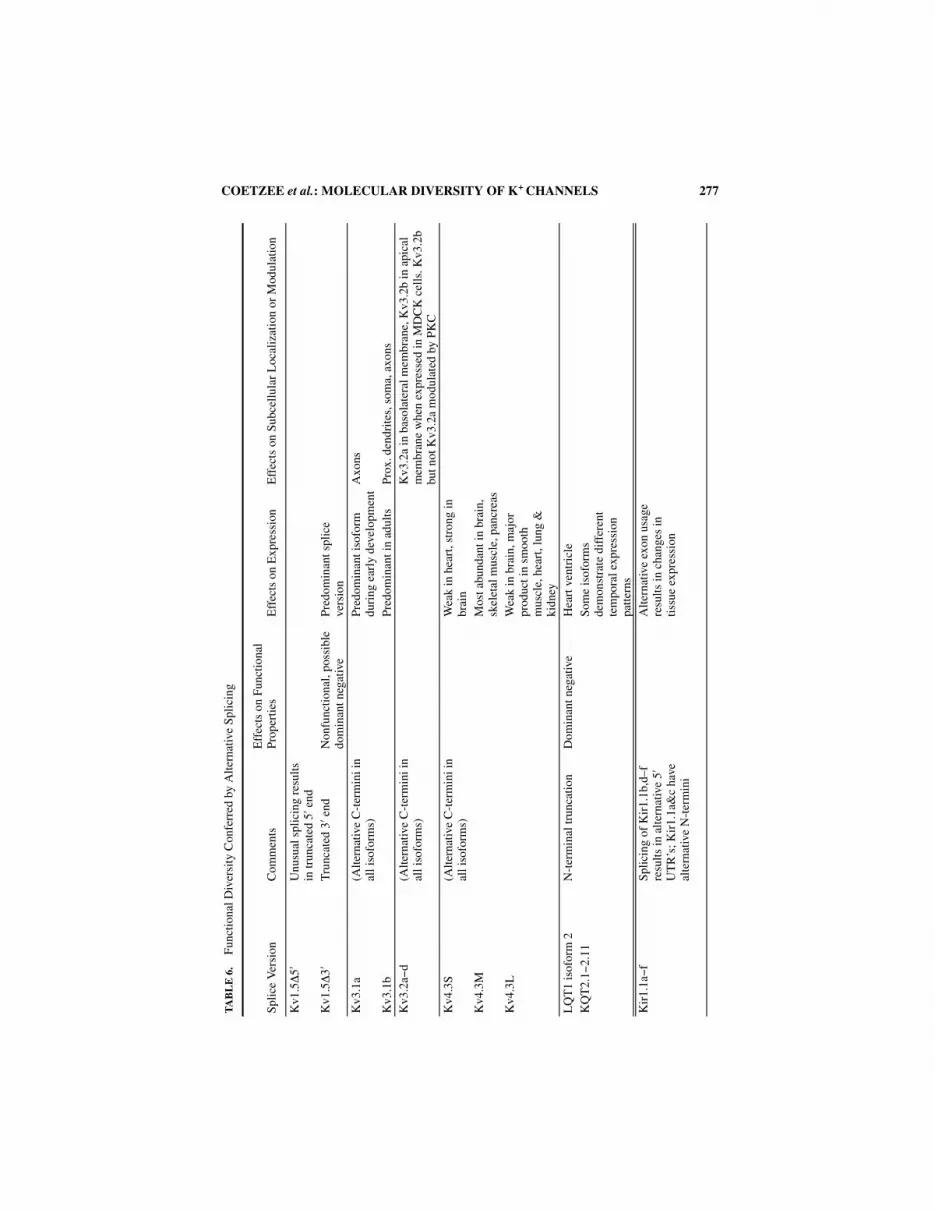
COETZEE et al.: MOLECULAR DIVERSITY OF K+ CHANNELS 277
TA
BL
E 6
.F
unct
iona
l Div
ersi
ty C
onfe
rred
by
Alte
rnat
ive
Splic
ing
Splic
e V
ersi
onC
omm
ents
Eff
ects
on
Func
tiona
l P
rope
rtie
sE
ffec
ts o
n E
xpre
ssio
nE
ffec
ts o
n Su
bcel
lula
r L
ocal
izat
ion
or M
odul
atio
n
Kv1
.5∆
5′U
nusu
al s
plic
ing
resu
lts
in tr
unca
ted
5′ e
ndK
v1.5∆
3′T
runc
ated
3′ e
ndN
onfu
nctio
nal,
poss
ible
do
min
ant n
egat
ive
Pre
dom
inan
t spl
ice
vers
ion
Kv3
.1a
(Alte
rnat
ive
C-t
erm
ini i
n al
l iso
form
s)P
redo
min
ant i
sofo
rm
duri
ng e
arly
dev
elop
men
tA
xons
Kv3
.1b
Pre
dom
inan
t in
adul
tsP
rox.
den
drite
s, s
oma,
axo
nsK
v3.2
a−d
(Alte
rnat
ive
C-t
erm
ini i
n al
l iso
form
s)K
v3.2
a in
bas
olat
eral
mem
bran
e, K
v3.2
b in
api
cal
mem
bran
e w
hen
expr
esse
d in
MD
CK
cel
ls. K
v3.2
b bu
t not
Kv3
.2a
mod
ulat
ed b
y P
KC
Kv4
.3S
(Alte
rnat
ive
C-t
erm
ini i
n al
l iso
form
s)W
eak
in h
eart
, str
ong
in
brai
nK
v4.3
MM
ost a
bund
ant i
n br
ain,
sk
elet
al m
uscl
e, p
ancr
eas
Kv4
.3L
Wea
k in
bra
in, m
ajor
pr
oduc
t in
smoo
th
mus
cle,
hea
rt, l
ung
&
kidn
eyL
QT
1 is
ofor
m 2
N-t
erm
inal
trun
catio
nD
omin
ant n
egat
ive
Hea
rt v
entr
icle
KQ
T2.
1−2.
11S
ome
isof
orm
s de
mon
stra
te d
iffe
rent
te
mpo
ral e
xpre
ssio
n pa
tter
nsK
ir1.
1a− f
Spl
icin
g of
Kir
1.1b
,d–f
re
sult
s in
alt
erna
tive
5′
UT
R’s
; Kir
1.1a
&c
have
al
tern
ativ
e N
-ter
min
i
Alt
erna
tive
exon
usa
ge
resu
lts in
cha
nges
in
tiss
ue e
xpre
ssio
n

278 ANNALS NEW YORK ACADEMY OF SCIENCES
TA
BL
E 6
.Fu
ncti
onal
Div
ersi
ty C
onfe
rred
by
Alte
rnat
ive
Splic
ing
( con
tinu
ed)
Splic
e V
ersi
onC
omm
ents
Eff
ects
on
Func
tiona
l P
rope
rtie
sE
ffec
ts o
n E
xpre
ssio
nE
ffec
ts o
n S
ubce
llul
ar L
ocal
izat
ion
or M
odul
atio
n
Kir
3.1a− d
(Alte
rnat
ive
C-t
erm
ini i
n al
l iso
form
s)C
hang
es in
G p
rote
in
activ
atio
n in
Kir
3.1c
/3.
1a h
eter
omul
tim
ers
Alt
erna
tive
exon
usa
ge
resu
lts in
cha
nges
in
tiss
ue e
xpre
ssio
nK
ir3.
2a− e
In K
ir3.
2a− e
spl
icin
g re
sult
s in
alt
erna
tive
C-
term
ini;
Kir
3.2e
als
o ha
s tr
unca
ted
N-t
erm
inus
.
Kir
3.2b
&c
expr
essi
on in
te
stes
is r
estr
icte
d to
a
subs
et o
f tu
bule
s
Kir
6.1a
(Alte
rnat
ive
5′ U
TR
’s in
al
l iso
form
s)H
eart
and
pan
crea
sK
ir6.
1bH
eart
and
ske
leta
l mus
cle
Kir
6.1c
Hea
rtsl
o(s
lo c
hann
els
have
the
abil
ity to
gen
erat
e nu
mer
ous
spli
ce
isof
orm
s. H
uman
slo
co
ntai
ns 5
spl
icin
g ca
sset
tes
with
in th
e C
-te
rmin
us)
(Tes
ted
isof
orm
s de
mon
stra
te v
aria
tion
s in
kin
etic
s an
d ca
lciu
m
sens
itiv
ity)
(Tes
ted
isof
orm
s de
mon
stra
te u
niqu
e ex
pres
sion
pat
tern
s.
Exp
ress
ion
of is
ofor
ms
in
adre
nal c
hrom
affi
n ti
ssue
con
trol
led
by
AC
TH
)E
ag1a
Fast
er a
ctiv
atio
n ki
neti
csP
redo
min
ant i
sofo
rmE
ag1b
27aa
inse
rtio
n be
twee
n S
3 an
d S
4E
rg1a
Slo
wer
dea
ctiv
atio
n ki
neti
csH
eart
, bra
in, t
este
s
Erg
1a′
N-t
erm
inal
trun
catio
nD
eact
ivat
ion
kine
tics
m
ore
rapi
dN
ot e
xpre
ssed
ab
unda
ntly
Erg
1bS
hort
er/d
iver
gent
N
term
inus
Dea
ctiv
atio
n ki
neti
cs
mor
e ra
pid
than
Erg
1a,
hete
rom
ulti
mer
izat
ion
with
Erg
1a in
crea
ses
deac
tivat
ion
kine
tics
Hea
rt

COETZEE et al.: MOLECULAR DIVERSITY OF K+ CHANNELS 279
REFERENCES
1. PAPAZIAN, D.M., T.L. SCHWARZ, B.L. TEMPEL, Y.N. JAN & L.Y. JAN. 1987. Cloning of genomicand complementary DNA from Shaker, a putative potassium channel gene from Drosophila.Science 237: 749−753.
2. TEMPEL, B.L., D.M. PAPAZIAN, T.L. SCHWARZ, Y.N. JAN & L.Y. JAN. 1987. Sequence of a probablepotassium channel component encoded at Shaker locus of Drosophila. Science 237: 770−775.
3. KAMB, A., L.E. IVERSON & M.A. TANOUYE. 1987. Molecular characterization of Shaker, a Droso-phila gene that encodes a potassium channel. Cell 50: 405−413.
4. STUHMER, W., M. STOCKER, B. SAKMANN, P. SEEBURG, A. BAUMANN, A. GRUPE & O. PONGS. 1988.Potassium channels expressed from rat brain cDNA have delayed rectifier properties. FEBSLett. 242: 199−206.
5. TAKUMI, T., H. OHKUBO & S. NAKANISHI. 1988. Cloning of a membrane protein that induces aslow voltage-gated potassium current. Science 242: 1042−1045.
6. JAN, L.Y. & Y.N. JAN. 1997. Voltage-gated and inwardly rectifying potassium channels. J. Phys-iol. (Lond.) 505: 267−282.
7. AGUILAR-BRYAN, L., J.P. CLEMENT, G. GONZALEZ, K. KUNJILWAR, A. BABENKO & J. BRYAN. 1998.Toward understanding the assembly and structure of KATP channels. Physiol. Rev. 78: 227−245.
8. ISOMOTO, S., KONDO, C. & KURACHI, Y. 1997. Inwardly rectifying potassium channels: Theirmolecular heterogeneity and function. Jpn. J. Physiol. 47: 11−39.
9. NICHOLS, C.G. & A.N. LOPATIN. 1997. Inward rectifier potassium channels. Ann. Rev. Physiol.59: 171−191.
10. BARRY, D.M. & J.M. NERBONNE. 1996. Myocardial potassium channels: Electrophysiologicaland molecular diversity. Ann. Rev. Physiol. 58: 363−394.
11. QUAYLE, J.M., M.T. NELSON & N.B. STANDEN. 1997. ATP-sensitive and inwardly rectifyingpotassium channels in smooth muscle. Physiol. Rev. 77: 1165−1232.
12. GOLDSTEIN, S.A.N., K.W. WANG, N. ILAN & M.H. PAUSCH. 1998. Sequence and function of thetwo P domain potassium channels: Implication of an emerging superfamily. J. Mol. Med. 76:13−20.
13. VEGA-SAENZ DE MIERA, E., M. WEISER, C. KENTROS, D. LAU, H. MORENO, P. SERODIO & B. RUDY.1994. Shaw-related K+ channels in mammals. In Handbook of Membrane Channels. 41−78.Academic Press. New York.
14. LEHMANN-HORN, F. & R. RUDEL. 1996. Molecular pathophysiology of voltage-gated ion chan-nels. Rev. Physiol. Biochem. Pharmacol. 128: 195−268.
15. DOLLY, J.O. & D.N. PARCEJ. 1996. Molecular properties of voltage-gated K+ channels. J. Bioen-erg. Biomembr. 28: 231−253.
16. CHANDY, K.G. & G.A. GUTMAN. 1995. Voltage-gated potassium channel genes. In Handbook ofReceptors and Channels: Ligand and Voltage-Gated Ion Channels. ed. R.A. North, Ed.: 1−71.CRC Press. Boca Raton, FL.
17. DOYLE, D.A., J.M. CABRAL, R.A. PFUETZNER, A. KUO, J.M. GULBIS, S.L. COHEN, B.T. CHAIT & R.MACKINNON. 1998. The structure of the potassium channel: Molecular basis of K+ conductionand selectivity. Science 280: 69−77.
18. CATTERALL, W.A. 1993. Structure and modulation of Na+ and Ca2+ channels. Ann. N.Y. Acad.Sci. 707: 1−19.
19. MACKINNON, R. 1991. Determination of the subunit stoichiometry of a voltage- activated potas-sium channel. Nature 350: 232−235.
20. MACKINNON, R., R.W. ALDRICH & A.W. LEE. 1993. Functional stoichiometry of Shaker potas-sium channel inactivation. Science 262: 757−759.
21. RUDY, B. 1988. Diversity and ubiquity of K channels. Neuroscience 25: 729−749.22. HILLE, B. 1992. Ionic Channels of Excitable Membranes. Sinauer Associates, Inc. Sunderland,
MA.23. HO, K., C.G. NICHOLS, W.J. LEDERER, J. LYTTON, P.M. VASSILEV, M.V. KANAZIRSKA & S.C. HEBERT.
1993. Cloning and expression of an inwardly rectifying ATP-regulated potassium channel.Nature 362: 31−38.
24. KUBO, Y., T.J. BALDWIN, Y.N. JAN & L.Y. JAN. 1993. Primary structure and functional expressionof a mouse inward rectifier potassium channel. Nature 362: 127−133.

280 ANNALS NEW YORK ACADEMY OF SCIENCES
25. DASCAL, N., W. SCHREIBMAYER, N.F. LIM et al. 1993. Atrial G protein−activated K+ channel:Expression cloning and molecular properties. Proc. Natl. Acad. Sci. USA. 90: 10235−10239.
26. LESAGE, F., E. GUILLEMARE, M. FINK, F. DUPRAT, M. LAZDUNSKI, G. ROMEY & J. BARHANIN. 1996.TWIK-1, a ubiquitous human weakly inward rectifying K+ channel with a novel structure.EMBO J. 15: 1004−1011.
27. LESAGE, F., R. REYES, M. FINK, F. DUPRAT, E. GUILLEMARE & M. LAZDUNSKI. 1996. Dimerizationof TWIK-1 K+ channel subunits via a disulfide bridge. EMBO J. 15: 6400−6407.
28. FINK, M., F. LESAGE, F. DUPRAT, C. HEURTEAUX, R. REYES, M. FOSSET & M. LAZDUNSKI. 1998. Aneuronal two P domain K+ channel stimulated by arachidonic acid and polyunsaturated fattyacids. Embo J. 17: 3297−3308.
29. TAGLIALATELA, M., M.S. CHAMPAGNE, J.A. DREWE & A.M. BROWN. 1994. Comparison of H5, S6,and H5-S6 exchanges on pore properties of voltage-dependent K+ channels. J. Biol. Chem.269: 13867−13873.
30. HEGINBOTHAM, L., Z. LU, T. ABRAMSON & R. MACKINNON. 1994. Mutations in the K+ channel sig-nature sequence. Biophys. J. 66: 1061−1067.
31. BAILIE, T.L. & C. ELKAN 1994. Fitting a mixture model by expectation maximization to discovermotifs in biopolymers. AAAI Press. Menlo Park, CA.
32. CHANDY, K.G. 1991. Simplified gene nomenclature. Nature 352: 26.33. DOUPNIK, C.A., N. DAVIDSON & H.A. LESTER. 1995. The inward rectifier potassium channel fam-
ily. Curr. Opin. Neurobiol. 5: 268−277.34. BOND, C.T., M. PESSIA, X.M. XIA, A. LAGRUTTA, M.P. KAVANAUGH & J.P. ADELMAN. 1994. Clon-
ing and expression of a family of inward rectifier potassium channels. Recept. Channels 2:183−191.
35. SHUCK, M.E., T.M. PISER, J.H. BOCK, J.L. SLIGHTOM, K.S. LEE & M.J. BIENKOWSKI. 1997. Cloningand characterization of two K+ inward rectifier (Kir) 1.1 potassium channel homologs fromhuman kidney (Kir1.2 and Kir1.3). J. Biol. Chem. 272: 586−593.
36. GOSSET, P., G.A. GHEZALA, B. KORN, M.L. YASPO, A. POUTSKA, H. LEHRACH, P.M. SINET & N.CRÉAU. 1997. A new inward rectifier potassium channel gene (KCNJ15) localized on chromo-some 21 in the Down syndrome chromosome region 1 (DCR1). Genomics 44: 237−241.
37. SHUCK, M.E., T.M. PISER, J.H. BOCK, J.L. SLIGHTOM, K.S. LEE & M.J. BIENKOWSKI. 1997. Cloningand characterization of two K+ inward rectifier (Kir) 1.1 potassium channel homologs fromhuman kidney (Kir1.2 and Kir1.3). J. Biol. Chem. 272: 586−593.
38. KRAPIVINSKY, G., I. MEDINA, L. ENG, L. KRAPIVINSKY, Y.H. YANG & D.E. CLAPHAM. 1998. A novelinward rectifier K+ channel with unique pore properties. Neuron 20: 995−1005.
39. HEDIN, K.E., N.F. LIM & D.E. CLAPHAM. 1996. Cloning of a Xenopus laevis inwardly rectifyingK+ channel subunit that permits GIRK1 expression of IKACh currents in oocytes. Neuron 16:423−429.
40. CHRISTIE, M.J., R.A. NORTH, P.B. OSBORNE, J. DOUGLASS & J. ADELMAN. 1990. Heteropolymericpotassium channels expressed in Xenopus oocytes from cloned subunits. Neuron 4: 405−411.
41. ISACOFF, E.Y., Y.N. JAN & L.Y. JAN. 1990. Evidence for the formation of heteromultimeric potas-sium channels in Xenopus oocytes. Nature 345: 530−534.
42. MCCORMACK, K., J.W. LIN, L.E. IVERSON & B. RUDY. 1990. Shaker K+ channel subunits from het-eromultimeric channels with novel functional properties. Biochem. Biophys. Res. Commun.171: 1361−1371.
43. RUPPERSBERG, J.P., K.H. SCHROTER, B. SAKMANN, M. STOCKER, S. SEWING & O. PONGS. 1990. Het-eromultimeric channels formed by rat brain potassium-channel proteins. Nature 345: 535−537.
44. WEISER, M., E. VEGA-SAENZ DE MIERA, C. KENTROS, H. MORENO, L. FRANZEN, D. HILLMAN, H.BAKER & B. RUDY. 1994. Differential expression of Shaw-related K+ channels in the rat centralnervous system. J. Neurosci. 14: 949−972.
45. ROEPER, J., S. SEWING, Y. ZHANG, T. SOMMER, S.G. WANNER & O. PONGS. 1998. NIP domain pre-vents N-type inactivation in voltage-gated potassium channels. Nature 391: 390−393.
46. LI, M., Y.N. JAN & L.Y. JAN. 1992. Specification of subunit assembly by the hydrophilic amino-terminal domain of the Shaker potassium channel. Science 257: 1225−1230.
47. SHEN, N.V. & P.J. PFAFFINGER. 1995. Molecular recognition and assembly sequences involved inthe subfamily-specific assembly of voltage-gated K+ channel subunit proteins. Neuron 14:625−633.

COETZEE et al.: MOLECULAR DIVERSITY OF K+ CHANNELS 281
48. YU, W., J. XU & M. LI. 1996. NAB domain is essential for the subunit assembly of both alpha-alpha and alpha-beta complexes of shaker-like potassium channels. Neuron 16: 441−453.
49. SEWING, S., J. ROEPER & O. PONGS. 1996. Kv β1 subunit binding specific for shaker-relatedpotassium channel α subunits. Neuron 16: 455−463.
50. SHENG, M., Y.J. LIAO, Y.N. JAN & L.Y. JAN. 1993. Presynaptic A-current based on heteromultim-eric K+ channels detected in vivo. Nature 365: 72−75.
51. WANG, H., D.D. KUNKEL, T.M. MARTIN, P.A. SCHWARTZKROIN & B.L. TEMPEL. 1993. Heteromulti-meric K+ channels in terminal and juxtaparanodal regions of neurons. Nature 365: 75−79.
52. CHOW, A., C. FARB, A. ERISIR & B. RUDY. 1998. K+ channel expression distinguishes betweentwo subpopulations of parvalbumin-containing cortical interneurons. Submitted for publica-tion.
53. HERNÁNDEZ-PINEDA, R., A. CHOW, Y. AMARILLO, H. MORENO, M. SAGANICH & B. RUDY. 1999.Kv3.1-Kv3.2 heteromultimeric channels underlie a high voltage-activating component of thedelayed rectifier K+ current in projecting neurons from the Globus Pallidus. J. Neurophysiol.In press.
54. MI, H., T.J. DEERINCK, M.H. ELLISMAN & T.L. SCHWARZ. 1995. Differential distribution of closelyrelated potassium channels in rat Schwann cells. J. Neurosci. 15: 3761−3774.
55. KRAMER, J.W., M.A. POST, A.M. BROWN & G.E. KIRSCH. 1998. Modulation of potassium channelgating by coexpression of Kv2.1 with regulatory Kv5.1 or Kv6.1 alpha-subunits. Am. J. Phys-iol. 274: C1501−C1510
56. SALINAS, M., F. DUPRAT, C. HEURTEAUX, J.P. HUGNOT & M. LAZDUNSKI. 1997. New modulatoryalpha subunits for mammalian Shab K+ channels. J. Biol. Chem. 272: 24371−24379.
57. PATEL, A.J., M. LAZDUNSKI & E. HONORÉ. 1997. Kv2.1/Kv9.3, a novel ATP-dependent delayed-rectifier K+ channel in oxygen-sensitive pulmonary artery myocytes. EMBO J. 16: 6615−6625.
58. YANG, W.P., P.C. LEVESQUE, W.A. LITTLE, M.L. CONDER, P. RAMAKRISHNAN, M.G. NEUBAUER &M.A. BLANAR. 1998. Functional expression of two KvLQT1-related potassium channelsresponsible for an inherited idiopathic epilepsy. J. Biol. Chem. 273: 19419−19423.
59. SHI, W., H.S. WANG, Z. PAN, R.S. WYMORE, I.S. COHEN, D. MCKINNON & J.E. DIXON. 1998. Clon-ing of a mammalian elk potassium channel gene and EAG mRNA distribution in rat sympa-thetic ganglia. J. Physiol. (Lond.) 511: 675−682.
60. LONDON, B., M.C. TRUDEAU, K.P. NEWTON, A.K. BEYER, N.G. COPELAND, D.J. GILBERT, N.A. JEN-KINS, C.A. SATLER & G.A. ROBERTSON. 1997. Two isoforms of the mouse ether-a-go-go−related gene coassemble to form channels with properties similar to the rapidly activatingcomponent of the cardiac delayed rectifier K+ current. Circ. Res. 81: 870−878.
61. CHEN, M.L., T. HOSHI & C.F. WU. 1996. Heteromultimeric interactions among K+ channel sub-units from Shaker and eag families in Xenopus oocytes. Neuron 17: 535−542.
62. LIAO, Y.J., Y.N. JAN & L.Y. JAN. 1996. Heteromultimerization of G-protein−gated inwardly recti-fying K+ channel proteins GIRK1 and GIRK2 and their altered expression in weaver brain. J.Neurosci. 16: 7137−7150.
63. FINK, M., F. DUPRAT, C. HEURTEAUX, F. LESAGE, G. ROMEY, J. BARHANIN & M. LAZDUNSKI. 1996.Dominant negative chimeras provide evidence for homo and heteromultimeric assembly ofinward rectifier K+ channel proteins via their N-terminal end. FEBS Lett. 378: 64−68.
64. TINKER, A., Y.N. JAN & L.Y. JAN. 1996. Regions responsible for the assembly of inwardly retify-ing potassium channels. Cell 87: 857−868.
65. NAKAMURA, T.Y., M. ARTMAN, B. RUDY & W.A. COETZEE. 1998. Inihibition of rat ventricular IK1with antisense oligonucleotides targeted to Kir2.1 mRNA. Am. J. Physiol. 274: H892−H900
66. FINK, M., F. DUPRAT, C. HEURTEAUX, F. LESAGE, G. ROMEY, J. BARHANIN & M. LAZDUNSKI. 1996.Dominant negative chimeras provide evidence for homo and heteromultimeric assembly ofinward rectifier K+ channel proteins via their N-terminal end. FEBS Lett. 378: 64−68.
67. PESSIA, M., S.J. TUCKER, K. LEE, C.T. BOND & J.P. ADELMAN. 1996. Subunit positional effectsrevealed by novel heteromeric inwardly rectifying K+ channels. EMBO J. 15: 2980−2987.
68. ADELMAN, J.P., K.Z. SHEN, M.P. KAVANAUGH, R.A. WARREN, Y.N. WU, A. LAGRUTTA, C.T. BOND &R.A. NORTH. 1992. Calcium-activated potassium channels expressed from cloned complemen-tary DNAs. Neuron 9: 209−216.

282 ANNALS NEW YORK ACADEMY OF SCIENCES
69. TSENG-CRANK, J., C.D. FOSTER, J.D. KRAUSE, R. MERTZ, N. GODINOT, T.J. DICHIARA & P.H. REIN-HART. 1994. Cloning, expression, and distribution of functionally distinct Ca2+-activated K+
channel isoforms from human brain. Neuron 13: 1315−1330.70. XIE, J. & D.P. MCCOBB. 1998. Control of alternative splicing of potassium channels by stress
hormones. Science 280: 443−446.71. BUTLER, A., S. TSUNODA, D.P. MCCOBB, A. WEI & L. SALKOFF. 1993. mSlo, a complex mouse
gene encoding “maxi” calcium-activated potassium channels. Science 261: 221−224.72. NAVARATNAM, D.S., T.J. BELL, T.D. TU, E.L. COHEN & J.C. OBERHOLTZER. 1997. Differential dis-
tribution of Ca2+-activated K+ channel splice variants among hair cells along the tonotopicaxis of the chick cochlea. Neuron 19: 1077−1085.
73. ROSENBLATT, K.P., Z.P. SUN, S. HELLER & A.J. HUDSPETH. 1997. Distribution of Ca2+-activated K+
channel isoforms along the tonotopic gradient of the chicken’s cochlea. Neuron 19: 1061−1075.
74. KOHLER, M., B. HIRSCHBERG, C.T. BOND, J.M. KINZIE, N.V. MARRION, J. MAYLIE & J.P. ADELMAN.1996. Small-conductance, calcium-activated potassium channels from mammalian brain. Sci-ence 273: 1709−1714.
75. REHM, H. & M. LAZDUNSKI. 1988. Purification and subunit structure of a putative K+ channelprotein identified by its binding properties to dendrotoxin I. Proc. Natl. Acad. Sci. USA 85:4919−4923.
76. SCOTT, V.E.S., J. RETTIG, D.N. PARCEJ, J.N. KEEN, J.B.C. FINDLAY, O. PONGS & J.O. DOLLY. 1994.Primary structure of a beta subunit of alpha-dendrotoxin-sensitive K+ channels from bovinebrain. Proc. Natl. Acad. Sci. USA 91: 1637−1641.
77. MCCORMACK, T. & K. MCCORMACK. 1994. Shaker K+ channel beta subunits belong to anNAD(P)H-dependent oxidoreductase superfamily [letter]. Cell 79: 1133−1135.
78. RETTIG, J., S.H. HEINEMANN, F. WUNDER, C. LORRA, R. WITTKA, O. DOLLY & O. PONGS. 1994.Inactivation properties of voltage gated K+ channels altered by the presence of β-subunit.Nature 369: 289−294.
79. MCCORMACK, K., T. MCCORMACK, M. TANOUYE, B. RUDY & W. STÜHMER. 1995. Alternative splic-ing of the human Shaker K+ channel β1 gene and functional expression of the β2 gene prod-uct. FEBS Lett. 370: 32−36.
80. HEINEMANN, S.H., J. RETTIG, F. WUNDER & O. PONGS. 1995. Molecular and functional character-ization of a rat brain Kv beta 3 potassium channel subunit. FEBS Lett. 377: 383−389.
81. WANG, Z., J. KIEHN, Q. YANG, A.M. BROWN & B.A. WIBLE. 1996. Comparison of binding andblock produced by alternatively spliced Kvbeta1 subunits. J. Biol. Chem. 271: 28311−28317.
82. ENGLAND, S.K., V.N. UEBELE, J. KODALI, P.B. BENNETT & M.M. TAMKUN. 1995. A novel K+ chan-nel β-subunit (hKvβ1.3) is produced via alternative mRNA splicings. J. Biol. Chem. 270:28531−28534.
83. SHI, G.Y., K. NAKAHIRA, S. HAMMOND, K.J. RHODES, L.E. SCHECHTER & J.S. TRIMMER. 1996. βSubunits promote K+ channel surface expression through effects early in biosynthesis. Neuron16: 843−852.
84. NAGAYA, N. & D.M. PAPAZIAN. 1997. Potassium channel α and β subunits assemble in the endo-plasmic reticulum. J. Biol. Chem. 272: 3022−3027.
85. WIBLE, B.A., Q. YANG, Y.A. KURYSHEV, E.A. ACCILI & A.M. BROWN. 1998. Cloning and expres-sion of a novel K+ channel regulatory protein, KChAP. J. Biol. Chem. 273: 11745−11751.
86. FINK, M., F. DUPRAT, F. LESAGE, C. HEURTEAUX, G. ROMEY, J. BARHANIN & M. LAZDUNSKI. 1996. Anew K+ channel β subunit to specifically enhance Kv2.2 (CDRK) expression. J. Biol. Chem.271: 26341−26348.
87. RHODES, K.J., B.W. STRASSLE, M.M. MONAGHAN, Z. BEKELE-ARCURI, M.F. MATOS & J.S. TRIMMER.1997. Association and colocalization of the Kvβ1 and Kvβ2 β-subunits with Kv1 α-subunitsin mammalian brain K+ channel complexes. J. Neurosci. 17: 8246−8258.
88. WILSON, G.F., Z. WANG, S.W. CHOUINARD, L.C. GRIFFITH & B. GANETZKY. 1998. Interaction of theK channel beta subunit, Hyperkinetic, with eag family members. J. Biol. Chem. 273: 6389−6394.
89. HOSHI, N., H. TAKAHASHI, M. SHAHIDULLAH, S. YOKOYAMA & H. HIGASHIDA. 1998. KCR1, a mem-brane protein that facilitates functional expression of non-inactivating K+ currents associateswith rat EAG voltage-dependent K+ channels [In Process Citation]. J. Biol. Chem. 273:23080−23085.

COETZEE et al.: MOLECULAR DIVERSITY OF K+ CHANNELS 283
90. FOLANDER, K., J.S. SMITH, J. ANTANAVAGE, C. BENNETT, R.B. STEIN & R. SWANSON. 1990. Cloningand expression of the delayed-rectifier Isk channel from neonatal rat heart and diethylstil-bestrol-rrimed rat uterus. Proc. Natl. Acad. Sci. USA 87: 2975−2979.
91. SAKAGAMI, M., K. FUKAZAWA, T. MATSUNAGA, H. FUJITA, N. MORI, T. TAKUMI, H. OHKUBO & S.NAKANISHI. 1991. Cellular localization of rat Isk protein in the stria vascularis by immunohis-tochemical observation. Hear. Res. 56: 168−172.
92. FREEMAN, L.C. & R.S. KASS. 1993. Expression of a minimal K+ channel protein in mammaliancells and immunolocalization in guinea pig heart. Circ. Res. 73: 968−973.
93. VARNUM, M.D., A.E. BUSCH, C.T. BOND, J. MAYLIE & J.P. ADELMAN. 1993. The min K channelunderlies the cardiac potassium current IKs and mediates species-specific responses to proteinkinase C. Proc. Natl. Acad. Sci. USA 90: 11528−11532.
94. BARHANIN, J., F. LESAGE, E. GUILLEMARE, M. FINK, M. LAZDUNSKI & G. ROMEY. 1996. KvLQT1and IsK (minK) proteins associate to form the IKs cardiac potassium current. Nature 384: 78−80.
95. SANGUINETTI, M.C., M.E. CURRAN, A. ZOU, J. SHEN, P.S. SPECTOR, D.L. ATKINSON & M.T. KEAT-ING. 1996. Coassembly of KvLQT1 and minK (IsK) proteins to form cardiac IKs potassiumchannel. Nature 384: 80−83.
96. SALATA, J.J., N.K. JURKIEWICZ, J. WANG, B.E. EVANS, H.T. ORME & M.C. SANGUINETTI. 1998. Anovel benzodiazepine that activates cardiac slow delayed rectifier K+ currents. Mol. Pharma-col. 54: 220−230.
97. MCDONALD, T.V., Z.H. YU, Z. MING, E. PALMA, M.B. MEYERS, K.W. WANG, S.A.N. GOLDSTEIN &G.I. FISHMAN. 1997. A minK-HERG complex regulates the cardiac potassium current IKr.Nature 388: 289−292.
98. SANGUINETTI, M.C. & N.K. JURKIEWICZ. 1990. Two components of cardiac delayed rectifier K+
current: Differential sensitivity to block by class III antiarrhythmic agents. J. Gen. Physiol. 96:194−214.
99. SANGUINETTI, M.C. & N.K. JURKIEWICZ. 1991. Delayed rectifier outward K+ current is composedof two currents in guinea pig atrial cells. Am. J. Physiol. 260: H393−399.
100. BUSCH, A.E., M.P. KAVANAUGH, M.D. VARNUM, J.P. ADELMAN & R.A. NORTH. 1992. Regulationby second messengers of the slowly activating, voltage-dependent potassium currentexpressed in Xenopus oocytes. J. Physiol. 450: 491−502.
101. BLUMENTHAL, E.M. & L.K. KACZMAREK. 1994. The minK potassium channel exists in functionaland nonfunctional forms when expressed in the plasma membrane of Xenopus oocytes. J.Neurosci. 14: 3097−3105.
102. TAI, K.K. & S.A.N. GOLDSTEIN. 1998. The conduction pore of a cardiac potassium channel.Nature 391: 605−608.
103. KNAUS, H.G., K. FOLANDER, M. GARCIA-CALVO, M.L. GARCIA, G.J. KACZOROWSKI, M. SMITH & R.SWANSON. 1994. Primary sequence and immunological characterization of beta-subunit of highconductance Ca(2+)-activated K+ channel from smooth muscle. J. Biol. Chem. 269: 17274−17278.
104. HANNER, M., R. VIANNA-JORGE, A. KAMASSAH, W.A. SCHMALHOFER, H.G. KNAUS, G.J. KACZO-ROWSKI & M.L. GARCIA. 1998. The beta subunit of the high conductance calcium-activatedpotassium channel. Identification of residues involved in charybdotoxin binding. J. Biol.Chem. 273: 16289−16296.
105. SCHOPPERLE, W.M., M.H. HOLMQVIST, Y. ZHOU, J. WANG, Z. WANG, L.C. GRIFFITH, I. KESELMAN, F.KUSINITZ, D. DAGAN & I.B. LEVITAN. 1998. Slob, a novel protein that interacts with the slow-poke calcium-dependent potassium channel. Neuron 20: 565−573.
106. TUCKER, S.J., F.M. GRIBBLE, C. ZHAO, S. TRAPP & F.M. ASHCROFT. 1997. Truncation of Kir6.2 pro-duces ATP-sensitive K+ channels in the absence of the sulphonylurea receptor. Nature 387:179−183.
107. JOHN, S.A., J.R. MONCK, J.N. WEISS & B. RIBALET. 1998. The sulphonylurea receptor SUR1 reg-ulates ATP-sensitive mouse Kir6.2 K+ channels linked to the green fluorescent protein inhuman embryonic kidney cells. J. Physiol. (Lond.) 510: 333−346.
108. SHYNG, S. & C.G. NICHOLS. 1997. Octameric stoichiometry of the KATP channel complex. J. Gen.Physiol. 110: 655−664.
109. BABENKO, A.P., L. AGUILAR-BRYAN & J. BRYAN. 1998. A view of sur/KIR6.X, KATP channels.Ann. Rev. Physiol. 60: 667−687.

284 ANNALS NEW YORK ACADEMY OF SCIENCES
110. SEEBURG, P.H., M. HIGUCHI & R. SPRENGEL. 1998. RNA editing of brain glutamate receptor chan-nels: Mechanism and physiology. Brain Res. Brain Res. Rev. 26: 217−229.
111. MAAS, S., T. MELCHER & P.H. SEEBURG. 1997. Mammalian RNA-dependent deaminases andedited mRNAs. Curr. Opin. Cell Biol. 9: 343−349.
112. PATTON, D.E., T. SILVA & F. BEZANILLA. 1997. RNA editing generates a diverse array of tran-scripts encoding squid Kv2 K+ channels with altered functional properties. Neuron 19: 711−722.
113. JONAS, E.A. & L.K. KACZMAREK. 1996. Regulation of potassium channels by protein kinases.Curr. Opin. Neurobiol. 6: 318−323.
114. LEVITAN, I.B. 1994. Modulation of ion channels by protein phosphorylation and dephosphoryla-tion. Ann. Rev. Physiol. 56: 193−212.
115. KACZMERECK, K.L. & I.B. LEVITAN. 1987. Neuromodulation: The Biochemical Control of Neu-ronal Excitability. Oxford University Press. Oxford.
116. COVARRUBIAS, M., A. WEI, L. SALKOFF & T.B. VYAS. 1994. Elimination of rapid potassium chan-nel inactivation by phosphorylation of the inactivation gate. Neuron 13: 1403−1412.
117. BECK, E.J., R.G. SORENSEN, S.J. SLATER & M. COVARRUBIAS. 1998. Interactions between multiplephosphorylation sites in the inactivation particle of a K+ channel. Insights into the molecularmechanism of protein kinase C action. J. Gen. Physiol. 112: 71−84.
118. ROEPER, J., C. LORRA & O. PONGS. 1997. Frequency-dependent inactivation of mammalian A-type K+ channel KV1.4 regulated by Ca2+/calmodulin-dependent protein kinase. J. Neurosci.17: 3379−3391.
119. COHEN, N.A., J.E. BRENMAN, S.H. SNYDER & D.S. BREDT. 1996. Binding of the inward rectifierK+ channel Kir 2.3 to PSD-95 is regulated by protein kinase A phosphorylation. Neuron 17:759−767.
120. COHEN, N.A., Q. SHA, E.N. MAKHINA, A.N. LOPATIN, M.E. LINDER, S.H. SNYDER & C.G. NICHOLS.1996. Inhibition of an inward rectifier potassium channel (Kim2.3) by G-protein βgammasubunits. J. Biol. Chem. 271: 32301−32305.
121. STORM, J.F. 1988. Temporal integration by a slowly inactivating K+ current in hippocampal neu-rons. Nature 336: 379−381.
122. WU, R.L. & M.E. BARISH. 1992. Two pharmacologically and kinetically distinct transient potas-sium currents in cultured embryonic mouse hippocampal neurons. J. Neurosci. 12: 2235−2246.
123. SHENG, M., M.L. TSAUR, Y.N. JAN & L.Y. JAN. 1992. Subcellular segregation of two A-type K+
channel proteins in rat central neurons. Neuron 9: 271−284.124. DEBANNE, D., N.C., GUERINEAU, B.H. GAHWILER & S.M. THOMPSON. 1997. Action-potential prop-
agation gated by an axonal I(A)-like K+ conductance in hippocampus. Nature 389: 286−289.125. FENG, J.L., B. WIBLE, G.R. LI, Z.G. WANG & S. NATTEL. 1997. Antisense oligodeoxynucleotides
directed against Kv1.5 mRNA specifically inhibit ultrarapid delayed rectifier K+ current incultured adult human atrial myocytes. Circ. Res. 80: 572−579.
126. PATEL, A.J., M. LAZDUNSKI & E. HONORE. 1997. Kv2.1/Kv9.3, a novel ATP-dependent delayed-rectifier K+ channel in oxygen-sensitive pulmonary artery myocytes. EMBO J. 16: 6615−6625.
127. FISET, C., R.B. CLARK, Y. SHIMONI & W. GILES. 1997. Shal-type channels contribute to the Ca2+-independent transient outward K+ current in rat ventricle. J. Physiol. (Lond.) 500: 51−63.
128. NAKAMURA, T.Y., W.A. COETZEE, E. VEGA-SAENZ DE MIERA, M. ARTMAN & B. RUDY. 1997. Mod-ulation of Kv4 channels, key components of rat ventricular transient outward K+ current byPKC. Am. J. Physiol. 273: H1775−H1786.
129. MARRION, N.V. 1997. Does r-EAG contribute to the M-current? Trends Neurosci. 20: 243−244.130. MATHIE, A. & C.S. WATKINS. 1997. Is EAG the answer to the M-current? Trends Neurosci. 20:
14.131. FENG, J.L., B. WIBLE, G.R. LI, Z. WANG & S. NATTEL. 1996. Antisense oligonucleotides directed
against Kv1.5 mRNA specifically inhibit the ultrarapid delayed rectifier in cultured adulthuman atrial myocytes. Circulation 94: I-528(Abstr.).
132. GARCIA-CALVO, M., H.G. KNAUS, O.B. MCMANUS, K.M. GIANGIACOMO, G.J. KACZOROWSKI &M.L. GARCIA. 1994. Purification and reconstitution of the high-conductance, calcium-acti-vated potassium channel from tracheal smooth muscle. J. Biol. Chem. 269: 676−682.

COETZEE et al.: MOLECULAR DIVERSITY OF K+ CHANNELS 285
133. KNAUS, H.G., A. EBERHART, R.O. KOCH, P. MUNUJOS, W.A. SCHMALHOFER, J.W. WARMKE, G.J.KACZOROWSKI & M.L. GARCIA. 1995. Characterization of tissue-expressed alpha subunits ofthe high conductance Ca2+-activated K+ channel. J. Biol. Chem. 270: 22434−22439.
134. ISHII, T.M., C. SILVIA, B. HIRSCHBERG, C.T. BOND, J.P. ADELMAN & J. MAYLIE. 1997. A humanintermediate conductance calcium-activated potassium channel. Proc. Natl. Acad. Sci. USA94: 11651−11656.
135. LOGSDON, N.J., J. KANG, J.A. TOGO, E.P. CHRISTIAN & J. AIYAR. 1997. A novel gene, hKCa4,encodes the calcium-activated potassium channel in human T lymphocytes. J. Biol. Chem.272: 32723−32726.
136. JOINER, W.J., L.Y. WANG, M.D. TANG & L.K. KACZMAREK. 1997. hSk4, a member of a novel sub-family of calcium-activated potassium channels. Proc. Natl. Acad. Sci. USA 94: 11013−11018.
137. COREY, S.G. KRAPIVINSKY, L. KRAPIVINSKY & D.E. CLAPHAM. 1998. Number and stoichiometry ofsubunits in the native atrial G-protein−gated K+ channel, IKACh. J. Biol. Chem. 273: 5271−5278.
138. SWOFFORD, D.L. 1993. PAUP. (3.1.1). (Computer Program). Smithsonian Institution. WashingtonDC, Sinauer Associates. Sunderland, MA.
139. GATESY, J., R. DESALLE & W. WHEELER. 1993. Alignment-ambiguous nucleotide sites and theexclusion of systematic data. Mol. Phylogenet. Evol. 2: 152−157.
140. LEES-MILLER, J.P., C. KONDO, L. WANG & H.J. DUFF. 1997. Electrophysiological characterizationof an alternatively processed ERG K+ channel in mouse and human hearts. Circ. Res. 81:719−726.
141. NAKAMURA, M., H. WATANABE, Y. KUBO, M. YOKOYAMA, T. MATSUMOTO, H. SASAI & Y. NISHI.1998. KQT2, a new putative potassium channel family produced by alternative splicing. Isola-tion, genomic structure, and alternative splicing of the putative potassium channels. Recep.Channels 5: 255−271.
142. KONDO, C., S. ISOMOTO, S. MATSUMOTO, M. YAMADA, Y. HORIO, S. YAMASHITA, K. TAKEMURA-KAMEDA, Y. MATSUZAWA & Y. KURACHI. 1996. Cloning and functional expression of a novelisoform of ROMK inwardly rectifying ATP-dependent K+ channel, ROMK6 (Kir1.1f). FEBSLett. 399: 122−126.
143. YANO, H., L.H. PHILIPSON, J.L. KUGLER, Y. TOKUYAMA, E.M. DAVIS, M.M. LE BEAU, D.J. NELSON,G.I. BELL & J. TAKEDA. 1994. Alternative splicing of human inwardly rectifying K+ channelROMK1 mRNA. Molecular Pharmacology 45: 854−860.
144. WEI, J., M.E. HODES, R. PIVA, Y. FENG, Y. WANG, B. GHETTI & S.R. DLOUHY. 1998. Characteriza-tion of murine girk2 transcript isoforms: Structure and differential expression. Genomics 51:379−390.
[NOTE ADDED IN PROOF: After this paper was submitted, Wang et al. (1998, Science282: 1890–1893) have presented strong evidence that KCNQ2 and KCNQ3 subunits formchannels mediating the “M-current.”]
Related Documents