Molecular diagnosis of the myeloproliferative neoplasms: UK guidelines for the detection of JAK2 V617F and other relevant mutations Anthony J. Bench, 1 Helen E. White, 2,3 Letizia Foroni, 4 Anna L. Godfrey, 5 Gareth Gerrard, 4 Susanna Akiki, 6 Abida Awan, 7 Ian Carter, 8 Andrea Goday-Fernandez, 1 Stephen E. Langabeer, 9 Tim Clench, 10 Jordan Clark, 11 Paul A. Evans, 12 David Grimwade, 13 Anna Schuh, 14 Mary F. McMullin, 15 Anthony R. Green, 5 Claire N. Harrison 16 and Nicholas C. P. Cross 2,3 1 Molecular Malignancy Laboratory and Haemato-Oncology Diagnostic Service, Cambridge University Hospitals NHS Foundation Trust, Cambridge, 2 National Genetics Reference Laboratory, Salisbury NHS Foundation Trust, Salisbury, 3 Faculty of Medicine, University of Southampton, Southampton, 4 Imperial Molecular Pathology, Imperial College Academic Health Science Centre, London, 5 Cambridge Institute for Medical Research, Department of Haematology, University of Cambridge, Cambridge, 6 West Midlands Regional Genetics Laboratory, Birmingham Women’s NHS Foundation Trust, Birmingham, 7 Molecular Diagnostics Centre, Manches- ter Royal Infirmary, Manchester, 8 Nottingham University Hospitals NHS Trust, Nottingham, UK, 9 Cancer Molecular Diagnostics, St. James’s Hospital, DublinIreland, 10 Bristol Royal Infirmary, Bristol, 11 UK NEQAS for Leucocyte Immunophenotyping, Sheffield, 12 HMDS, Leeds Institute of Oncology, St. James’s University Hospital, Leeds, 13 Department of Medical and Molecular Genetics, King’s College London School of Medicine, London, 14 Oxford Cancer and Haematology Centre, Churchill Hospital, Oxford, 15 Belfast City Hospital, Queen’s University Belfast, Belfast, and 16 Guy’s and St Thomas’ NHS Foundation Trust, Guy’s Hospital, London UK Summary Molecular genetic assays for the detection of the JAK2 V617F (c.1849G>T) and other pathogenetic mutations within JAK2 exon 12 and MPL exon 10 are part of the rou- tine diagnostic workup for patients presenting with erythro- cytosis, thrombocytosis or otherwise suspected to have a myeloproliferative neoplasm. A wide choice of techniques are available for the detection of these mutations, leading to potential difficulties for clinical laboratories in deciding upon the most appropriate assay, which can lead to prob- lems with inter-laboratory standardization. Here, we discuss the most important issues for a clinical diagnostic labora- tory in choosing a technique, particularly for detection of the JAK2 V617F mutation at diagnosis. The JAK2 V617F detection assay should be both specific and sensitive enough to detect a mutant allele burden as low as 1–3%. Indeed, the use of sensitive assays increases the detection rate of the JAK2 V617F mutation within myeloproliferative neoplasms. Given their diagnostic relevance, it is also beneficial and rel- atively straightforward to screen JAK2 V617F negative patients for JAK2 exon 12 mutations (in the case of erythr- ocytosis) or MPL exon 10 mutations (thrombocytosis or myelofibrosis) using appropriate assays. Molecular results should be considered in the context of clinical findings and other haematological or laboratory results. Keywords: myeloproliferative neoplasm, molecular diagnosis, JAK2, MPL. Introduction The classical BCR-ABL1 negative myeloproliferative neo- plasms (MPN) comprise polycythaemia vera (PV), essential thrombocythaemia (ET) and primary myelofibrosis (PMF). In 2005, an acquired mutation within JAK2 exon 14 was identi- fied (c.1849G>T), which results in a valine to phenylalanine substitution at codon 617 – p.Val617Phe, usually abbreviated to V617F (Baxter et al, 2005; James et al, 2005; Kralovics et al, 2005; Levine et al, 2005a). This codon lies in the JH2 pseudokinase domain of JAK2 and the mutation is thought to interfere with JH2-mediated autoinhibition leading to constit- utive activation of the tyrosine kinase function. This results in activation of a number of downstream pathways including JAK/STAT, PI3K/AKT and MAPK/ERK. The JAK2 V617F mutation has been observed in up to 98% of patients with PV and 50–60% of patients with ET and PMF. With the excep- tion of the syndrome ‘refractory anaemia with ringed sidero- blasts associated with marked thrombocytosis’ (RARS-T) where it is observed in approximately one half of patients (Szpurka et al, 2006; Schmitt-Graeff et al, 2008), the JAK2 V617F mutation is uncommon in other myeloid disorders, such as myelodysplastic syndrome, chronic myelomonocytic Correspondence: Dr Anthony J. Bench, Molecular Malignancy Laboratory and Haemato-Oncology Diagnostic Service, Box 234, Cambridge University Hospitals NHS Foundation Trust, Hills Road, Cambridge, CB2 0QQ, UK. E-mail: [email protected] ª 2012 Blackwell Publishing Ltd, British Journal of Haematology doi:10.1111/bjh.12075 guideline

Welcome message from author
This document is posted to help you gain knowledge. Please leave a comment to let me know what you think about it! Share it to your friends and learn new things together.
Transcript
Molecular diagnosis of the myeloproliferative neoplasms: UKguidelines for the detection of JAK2 V617F and other relevantmutations
Anthony J. Bench,1 Helen E. White,2,3 Letizia Foroni,4 Anna L. Godfrey,5 Gareth Gerrard,4 Susanna Akiki,6 Abida Awan,7
Ian Carter,8 Andrea Goday-Fernandez,1 Stephen E. Langabeer,9 Tim Clench,10 Jordan Clark,11 Paul A. Evans,12 David
Grimwade,13 Anna Schuh,14 Mary F. McMullin,15 Anthony R. Green,5 Claire N. Harrison16 and Nicholas C. P. Cross2,3
1Molecular Malignancy Laboratory and Haemato-Oncology Diagnostic Service, Cambridge University Hospitals NHS Foundation
Trust, Cambridge, 2National Genetics Reference Laboratory, Salisbury NHS Foundation Trust, Salisbury, 3Faculty of Medicine,
University of Southampton, Southampton, 4Imperial Molecular Pathology, Imperial College Academic Health Science Centre, London,5Cambridge Institute for Medical Research, Department of Haematology, University of Cambridge, Cambridge, 6West Midlands
Regional Genetics Laboratory, Birmingham Women’s NHS Foundation Trust, Birmingham, 7Molecular Diagnostics Centre, Manches-
ter Royal Infirmary, Manchester, 8Nottingham University Hospitals NHS Trust, Nottingham, UK, 9Cancer Molecular Diagnostics,
St. James’s Hospital, DublinIreland, 10Bristol Royal Infirmary, Bristol, 11UK NEQAS for Leucocyte Immunophenotyping, Sheffield,12HMDS, Leeds Institute of Oncology, St. James’s University Hospital, Leeds, 13Department of Medical and Molecular Genetics, King’s
College London School of Medicine, London, 14Oxford Cancer and Haematology Centre, Churchill Hospital, Oxford, 15Belfast City
Hospital, Queen’s University Belfast, Belfast, and 16Guy’s and St Thomas’ NHS Foundation Trust, Guy’s Hospital, London UK
Summary
Molecular genetic assays for the detection of the JAK2
V617F (c.1849G>T) and other pathogenetic mutations
within JAK2 exon 12 and MPL exon 10 are part of the rou-
tine diagnostic workup for patients presenting with erythro-
cytosis, thrombocytosis or otherwise suspected to have a
myeloproliferative neoplasm. A wide choice of techniques
are available for the detection of these mutations, leading to
potential difficulties for clinical laboratories in deciding
upon the most appropriate assay, which can lead to prob-
lems with inter-laboratory standardization. Here, we discuss
the most important issues for a clinical diagnostic labora-
tory in choosing a technique, particularly for detection of
the JAK2 V617F mutation at diagnosis. The JAK2 V617F
detection assay should be both specific and sensitive enough
to detect a mutant allele burden as low as 1–3%. Indeed,
the use of sensitive assays increases the detection rate of the
JAK2 V617F mutation within myeloproliferative neoplasms.
Given their diagnostic relevance, it is also beneficial and rel-
atively straightforward to screen JAK2 V617F negative
patients for JAK2 exon 12 mutations (in the case of erythr-
ocytosis) or MPL exon 10 mutations (thrombocytosis or
myelofibrosis) using appropriate assays. Molecular results
should be considered in the context of clinical findings and
other haematological or laboratory results.
Keywords: myeloproliferative neoplasm, molecular diagnosis,
JAK2, MPL.
Introduction
The classical BCR-ABL1 negative myeloproliferative neo-
plasms (MPN) comprise polycythaemia vera (PV), essential
thrombocythaemia (ET) and primary myelofibrosis (PMF). In
2005, an acquired mutation within JAK2 exon 14 was identi-
fied (c.1849G>T), which results in a valine to phenylalanine
substitution at codon 617 – p.Val617Phe, usually abbreviated
to V617F (Baxter et al, 2005; James et al, 2005; Kralovics
et al, 2005; Levine et al, 2005a). This codon lies in the JH2
pseudokinase domain of JAK2 and the mutation is thought to
interfere with JH2-mediated autoinhibition leading to constit-
utive activation of the tyrosine kinase function. This results in
activation of a number of downstream pathways including
JAK/STAT, PI3K/AKT and MAPK/ERK. The JAK2 V617F
mutation has been observed in up to 98% of patients with PV
and 50–60% of patients with ET and PMF. With the excep-
tion of the syndrome ‘refractory anaemia with ringed sidero-
blasts associated with marked thrombocytosis’ (RARS-T)
where it is observed in approximately one half of patients
(Szpurka et al, 2006; Schmitt-Graeff et al, 2008), the JAK2
V617F mutation is uncommon in other myeloid disorders,
such as myelodysplastic syndrome, chronic myelomonocytic
Correspondence: Dr Anthony J. Bench, Molecular Malignancy
Laboratory and Haemato-Oncology Diagnostic Service, Box 234,
Cambridge University Hospitals NHS Foundation Trust, Hills Road,
Cambridge, CB2 0QQ, UK.
E-mail: [email protected]
ª 2012 Blackwell Publishing Ltd, British Journal of Haematology doi:10.1111/bjh.12075
guideline
leukaemia and acute myeloid leukaemia (Jones et al, 2005;
Levine et al, 2005b).
In 2007, mutations within exon 12 of JAK2 were described
in some JAK2 V617F negative PV patients (Scott et al, 2007a)
as well as in patients previously categorized as idiopathic ery-
throcytosis, raising the suggestion that all patients with PV
carry a mutation within JAK2 (Scott et al, 2007b; McMullin,
2008; Wang et al, 2008). At least 17 different mutations have
now been described within exon 12 (Passamonti et al, 2011).
Although these mutations are not located within the JH2
domain, they are also thought to interfere with JH2-mediated
autoinhibition. In 2006, mutations within exon 10 of the
thrombopoietin receptor, MPL, were identified in ET and
PMF patients (Pardanani et al, 2006) and at least five different
pathogenetic mutations have been described (Chaligne et al,
2008; Schnittger et al, 2009a; Boyd et al, 2010) that affect
codons S505 or W515. Other variants have also been described
(Ma et al, 2011) but their pathogenicity is not known.
The demonstration of an acquired mutation within JAK2
and/or MPL now forms part of the World Health Organiza-
tion criteria for the diagnosis of MPN (Swerdlow et al,
2008). Patients presenting with erythrocytosis should be
assessed for the presence of a JAK2 mutation. The British
Committee for Standards in Haematology (BCSH) guidelines
state that the presence of a JAK2 mutation (V617F or exon
12) and a raised haematocrit (>0�52 male; >0�48 female) or
raised red cell mass (>25% above predicted) is sufficient to
make a diagnosis of PV (McMullin et al, 2007; McMullin,
2008). Likewise, patients presenting with a persistent
thrombocytosis should be assessed for the presence of JAK2
V617F and, if negative, MPL exon 10 mutations (Harrison
et al, 2010). The presence of an acquired pathogenetic muta-
tion (i.e. JAK2 V617F and/or MPL exon 10 mutation) and a
sustained thrombocytosis (platelet count > 450 9 109/l) in
the absence of evidence for another myeloid malignancy is
sufficient to make a diagnosis of ET (Harrison et al, 2010).
For PMF, the demonstration of JAK2 V617F and/or MPL
exon 10 mutations is a major diagnostic criterion because it
confirms the primary nature of the disorder (Swerdlow et al,
2008). The demonstration of a JAK2 V617F mutation in
samples from patients not otherwise meeting specific diag-
nostic criteria for a MPN, for example presenting with unex-
plained splanchnic vein thrombosis (Dentali et al, 2009),
suggests an underlying MPN or, more rarely, another mye-
loid malignancy.
The aim of these guidelines is to provide information and
suggestions for those diagnostic laboratories that perform or
wish to perform assays for the detection of JAK2 V617F, for
which many different techniques are available. Diagnostic
assays are also available for the detection of JAK2 exon 12
and MPL exon 10 mutations and these are also discussed. A
strategy for the efficient combination of JAK2 V617F, JAK2
exon 12 and MPL exon 10 mutations detection assays in
suspected myeloproliferative neoplasms is discussed. These
guidelines are broadly in line with the screening strategy
proposed by Tefferi et al (2011). A step-wise algorithm for
supplementary JAK2 exon 12 or MPL exon 10 mutation
analysis is both cost-effective and an efficient use of available
material and may reduce the need for a bone marrow biopsy
in some patients. Furthermore, these guidelines highlight the
technical issues of relevance for diagnostic laboratories.
Detection of the JAK2 V617F mutation atdiagnosis
A number of different factors that contribute to the choice
of assay for the detection of the JAK2 V617F mutation at
diagnosis will be discussed. Quantification of the JAK2
V617F mutation, either at diagnosis for prognostic informa-
tion or during treatment as a means of minimal residual dis-
ease assessment, is not discussed here although the use of
such assays is entirely applicable in the diagnostic setting.
Many assays for quantification of the JAK2 V617F mutant
burden have been developed which differ markedly in their
performance with respect to specificity and sensitivity and
these have been subject to a comprehensive comparison by
the European LeukemiaNet and MPN&MPNr-EuroNet study
groups (Jovanovic et al, 2011).
Type of sample
DNA extracted from peripheral blood or bone marrow is
acceptable for JAK2 V617F mutation analysis provided the
nucleic acid obtained is of acceptable quality for the assay to
be performed successfully. In most cases, peripheral blood is
the preferred option and EDTA is the usual anticoagulant.
Use of other anti-coagulants is acceptable although care
should be taken in the case of Lithium heparin tubes to
completely remove any anticoagulant during the nucleic acid
extraction procedure because its presence may inhibit poly-
merase chain reaction (PCR) amplification (Yokota et al,
1999). A sample of sufficient volume to obtain a reasonable
amount of nucleic acid should be taken (2–10 ml peripheral
blood is usually fine although some centres have optimized
routine DNA extraction from smaller volumes), although
samples from neutropenic patients may yield less nucleic
acid. There does not appear to be a major difference in the
JAK2 V617F allele burden between whole blood and bone
marrow (Larsen et al, 2008). Therefore, a bone marrow aspi-
rate may be assessed (taken into an EDTA tube or cytoge-
netic culture medium) but is generally not necessary if
peripheral blood is available. The sample should be received
within 24–48 h after being taken but, in our experience, for
DNA analysis samples of up to 1 week old are usually
acceptable for non-quantitative tests. Peripheral blood may
be frozen until nucleic acid is extracted and it is often possi-
ble to extract DNA from stained or (preferably) unstained
and unfixed slides if necessary (Jones et al, 2006). The use of
DNA derived from plasma for the detection of the JAK2
V617F mutation has been described (Ma et al, 2008). How-
Guideline
2 ª 2012 Blackwell Publishing Ltd, British Journal of Haematology
ever, this methodology has not been independently validated
and does not offer any obvious advantage over DNA derived
directly from peripheral blood.
Isolation of peripheral blood granulocytes
An important question often raised is whether isolation of
peripheral blood granulocytes is necessary to perform JAK2
V617F mutation detection assays. The JAK2 V617F mutation
arises in a haematopoietic progenitor cell but, in most
patients, is restricted to the myeloid lineage. In addition, the
proportion of myeloid cells carrying the JAK2 V617F muta-
tion can vary widely amongst patients. In general, patients
with ET tend to carry a lower overall level of the JAK2
V617F mutation compared to those with PV or PMF (Passa-
monti & Rumi, 2009) (due to lower proportion of JAK2
V617F positive cells and the presence of a monoallelic muta-
tion in most V617F positive cells). A further confounding
factor is that patients who have been treated with hydroxyc-
arbamide may exhibit lower levels of JAK2 V617F (Girodon
et al, 2008). Although other studies have found minimal
changes in V617F levels on hydroxycarbamide (Antonioli
et al, 2010), the possibility of a lower disease burden is worth
bearing in mind if JAK2 V617F testing is carried out after
cytoreductive treatment. Overall, the quantitative level of the
JAK2 V617F mutation is about 15% lower in peripheral
blood compared to purified granulocytes (Hermouet et al,
2007) due to the presence of JAK2 V617F negative lympho-
cytes. If highly sensitive assays are used, there is no difference
in detection rate between peripheral blood and granulocytes
as a source of material (Hermouet et al, 2007; Goday-Fer-
nandez et al, 2008; Cankovic et al, 2009). However, when a
moderately sensitive assay, such as agarose gel-based allele-
specific PCR (Baxter et al, 2005; sensitivity approximately
3%) was used, purification of granulocytes increased the
detection rate from 92% to 97% for PV and from 57% to
61% for ET (Goday-Fernandez et al, 2008). Hence, isolation
of granulocytes should not be required provided the assay is
sufficiently sensitive (sensitivity of 1–3% or better). If the
assay utilized has a lower sensitivity (see Table I), then
enrichment of granulocytes (Asimakopoulos et al, 1996) may
be necessary. An alternative approach is to prepare nucleic
acid from erythropoietin (EPO) independent erythroid burst-
forming unit (BFU-E) colonies, as such colonies should con-
sist entirely of clonal malignant cells. However, this is time
consuming, technically challenging, no quality assurance
scheme is available and EPO-independent BFU-E are not
observed in all patients.
Nucleic acid
Genomic DNA is the preferred choice of nucleic acid due to its
stability although assays involving RNA/cDNA are also avail-
able. Analysis of RNA/cDNA also allows platelets to be interro-
gated, but this is not necessary on a routine basis. Commercial
DNA purification kits, either manual or automatic, generally
give reliable DNA of acceptable quantity and quality, as do
many in-house purification methods. It is advisable to process
both control samples and the sample under investigation using
the same method to reduce variability. DNA concentration
should also be calculated using the same method for all sam-
ples because different instruments may produce widely varying
apparent concentrations (e.g. UV spectrophotometer; Nano-
drop spectrophotometer (Thermo Scientific, Wilmington, DE,
USA); Qubit fluorometer (Life Technologies, Paisley, UK)).
These two points are probably more important for ‘compara-
tive’ techniques, such as high resolution melt (HRM) analysis
or denaturing high performance liquid chromatography
(dHPLC). The amount of template nucleic acid required will
depend on the particular assay chosen.
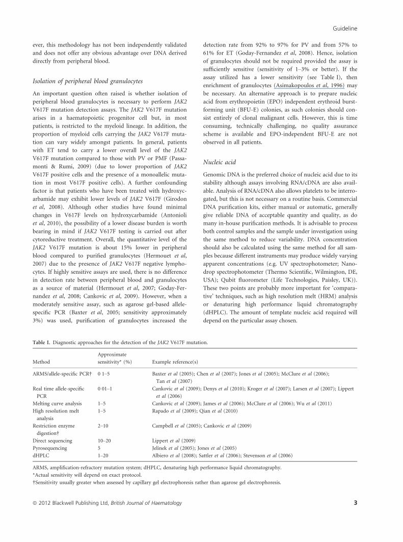
Table I. Diagnostic approaches for the detection of the JAK2 V617F mutation.
Method
Approximate
sensitivity* (%) Example reference(s)
ARMS/allele-specific PCR† 0�1–5 Baxter et al (2005); Chen et al (2007); Jones et al (2005); McClure et al (2006);
Tan et al (2007)
Real time allele-specific
PCR
0�01–1 Cankovic et al (2009); Denys et al (2010); Kroger et al (2007); Larsen et al (2007); Lippert
et al (2006)
Melting curve analysis 1–5 Cankovic et al (2009); James et al (2006); McClure et al (2006); Wu et al (2011)
High resolution melt
analysis
1–5 Rapado et al (2009); Qian et al (2010)
Restriction enzyme
digestion†
2–10 Campbell et al (2005); Cankovic et al (2009)
Direct sequencing 10–20 Lippert et al (2009)
Pyrosequencing 5 Jelinek et al (2005); Jones et al (2005)
dHPLC 1–20 Albiero et al (2008); Sattler et al (2006); Stevenson et al (2006)
ARMS, amplification-refractory mutation system; dHPLC, denaturing high performance liquid chromatography.
*Actual sensitivity will depend on exact protocol.
†Sensitivity usually greater when assessed by capillary gel electrophoresis rather than agarose gel electrophoresis.
Guideline
ª 2012 Blackwell Publishing Ltd, British Journal of Haematology 3

Choice of assay and sensitivity
A large number of different approaches for the detection of
the JAK2 V617F mutation have been described (Table I). For
each type of technique, slightly different assays have been
designed that vary with instrument, primer and/or probe
sequence and detection method. The techniques described
broadly fall into two main categories. Firstly, those assays
that are designed to specifically target the c.1849G>T muta-
tion (for example, allele-specific PCR) and, secondly, muta-
tion scanning assays that target the region of exon 14
encompassing the c.1849G>T mutation (for example, direct
sequencing, HRM analysis). For assays that specifically target
the mutant allele, specificity is usually achieved through the
use of a mutation-specific primer or probe. Commercial kits
are available for detection of JAK2 V617F and these are based
on similar approaches.
Two main criteria are important in the choice of an assay.
Firstly, it should be specific (i.e. no false negatives or a
clearly defined background level such that JAK2 V617F nega-
tive and positive cases can be readily distinguished). Sec-
ondly, the assay must be sensitive enough to be able to
identify a JAK2 V617F mutant allele with a burden as low as
1–3%. This threshold has been shown to be pathogenetically
relevant and carry clinical significance (Wang et al, 2008;
Mason et al, 2011). Consequently, direct sequencing is not
recommended as the method of choice because it only has a
sensitivity of 10–20%. Other assays that possess a sensitivity
of 3–5%, such as restriction enzyme digestion, agarose gel-
based allele-specific PCR and pyrosequencing, may also fail
to identify a small number of patients who carry a pathoge-
netically important low level JAK2 V617F mutation. Use of
more sensitive assays does indeed increase the detection rate
of JAK2 V617F in both PV and ET patients particularly when
unfractionated peripheral blood is assessed (Campbell et al,
2005; Goday-Fernandez et al, 2008; Wang et al, 2008; Canko-
vic et al, 2009; Lippert et al, 2009). Finally, to achieve a sen-
sitivity of 1–3%, it is necessary to analyse at least 20 ng of
genomic DNA, equivalent to 3030 diploid genomes.
False positives
False positive results may also occur due to cross-reactivity of
primers or probes (Mason et al, 2011). Hence, particularly
with highly sensitive assays, it is critically important to assess
the false positive rate using a series of healthy control samples
(see below). The assay should also be able to give an indica-
tion of the quality/quantity of the DNA to judge whether it
carries sufficient sensitivity for each patient. Sample quality
may be judged using absolute copy number of a control gene,
the CT value for a control gene, the strength of a band on a
gel, the peak height of fragment, the height of the (pyro)
sequence or other appropriate output. Specific criteria should
be laid down to identify samples that are of poor quality. It is
important to stress that all results of molecular investigations
should be considered in the context of clinical, morphologi-
cal, haematological and other laboratory findings.
Interpretation of low level JAK2 V617F at diagnosis
A result of <1–3% V617F should be interpreted in the
context of clinical, morphological, haematological and other
laboratory findings. Such considerations not only mitigate
against occasional technical aberrations but it has been
claimed that JAK2 V617F may occasionally be found in
haematologically normal individuals when highly sensitive
assays are used (Sidon et al, 2006; Xu et al, 2007; Nielsen
et al, 2011). Assuming the result does not represent a false
positive, it is reproducible and the amplification is distinct
from appropriate normal controls, such a result may well
represent a true low level clone. In patients with other labo-
ratory or clinical criteria suggestive of a MPN, this result
provides objective evidence in support of the diagnosis. Low
level JAK2 V617F may occur for a number of reasons: (i)
prior treatment with cytoreductive therapy may reduce the
level of the JAK2 V617F positive clone within the sample
(Girodon et al, 2008); (ii) the presence of two MPN clones
in the patient, only one of which is JAK2 V617F positive
(Beer et al, 2009). Mutation assessment of JAK2 exon 12 or
MPL exon 10 may reveal the existence of such second clones.
In a patient with a low level JAK2 V617F mutation but
with a normal full blood count, the clinical significance is
less clear. Obviously, iron-deficient PV has to be excluded. It
is still possible that this may reflect the presence of a chronic
MPN and that the relevant blood parameters have risen
above the individual’s own baseline, but are not yet above
the upper limit for the normal range of the appropriate
population. Alternatively, a low level JAK2 V617F positive
clone may remain stable, or even occasionally disappear with
time (for example, if it arises in a short-lived haematopoietic
precursor), without significant clinical effects. Such patients
may warrant further clinical surveillance. Whatever the cause
and clinical situation pertaining to a low level JAK2 V617F
mutation, it is prudent to obtain a fresh sample (e.g. within
3–6 months if possible) to enable the assay to be repeated.
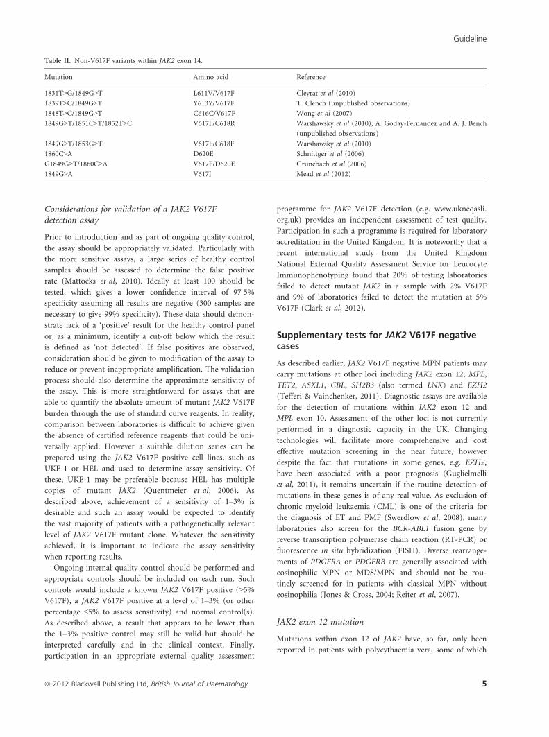
False low levels and false negatives
False low levels or even false negatives can occur due to the
presence of an additional exon 14 mutation or inherited
polymorphism. If these additional changes lie within one of
the primer or probe binding sites, they may reduce the effi-
ciency of V617F-specific PCR amplification. Rare instances of
additional acquired mutations or constitutional variants have
been reported (Table II). Depending on the assay utilized,
these can lead to a false negative result for V617F or appar-
ent low level amplification. Assessment of the c.1849G>T(V617F) mutation by an additional method that utilizes
different primer/probe sets may be helpful in situations of
apparent low level amplification.
Guideline
4 ª 2012 Blackwell Publishing Ltd, British Journal of Haematology
Considerations for validation of a JAK2 V617Fdetection assay
Prior to introduction and as part of ongoing quality control,
the assay should be appropriately validated. Particularly with
the more sensitive assays, a large series of healthy control
samples should be assessed to determine the false positive
rate (Mattocks et al, 2010). Ideally at least 100 should be
tested, which gives a lower confidence interval of 97�5%specificity assuming all results are negative (300 samples are
necessary to give 99% specificity). These data should demon-
strate lack of a ‘positive’ result for the healthy control panel
or, as a minimum, identify a cut-off below which the result
is defined as ‘not detected’. If false positives are observed,
consideration should be given to modification of the assay to
reduce or prevent inappropriate amplification. The validation
process should also determine the approximate sensitivity of
the assay. This is more straightforward for assays that are
able to quantify the absolute amount of mutant JAK2 V617F
burden through the use of standard curve reagents. In reality,
comparison between laboratories is difficult to achieve given
the absence of certified reference reagents that could be uni-
versally applied. However a suitable dilution series can be
prepared using the JAK2 V617F positive cell lines, such as
UKE-1 or HEL and used to determine assay sensitivity. Of
these, UKE-1 may be preferable because HEL has multiple
copies of mutant JAK2 (Quentmeier et al, 2006). As
described above, achievement of a sensitivity of 1–3% is
desirable and such an assay would be expected to identify
the vast majority of patients with a pathogenetically relevant
level of JAK2 V617F mutant clone. Whatever the sensitivity
achieved, it is important to indicate the assay sensitivity
when reporting results.
Ongoing internal quality control should be performed and
appropriate controls should be included on each run. Such
controls would include a known JAK2 V617F positive (>5%V617F), a JAK2 V617F positive at a level of 1–3% (or other
percentage <5% to assess sensitivity) and normal control(s).
As described above, a result that appears to be lower than
the 1–3% positive control may still be valid but should be
interpreted carefully and in the clinical context. Finally,
participation in an appropriate external quality assessment
programme for JAK2 V617F detection (e.g. www.ukneqasli.
org.uk) provides an independent assessment of test quality.
Participation in such a programme is required for laboratory
accreditation in the United Kingdom. It is noteworthy that a
recent international study from the United Kingdom
National External Quality Assessment Service for Leucocyte
Immunophenotyping found that 20% of testing laboratories
failed to detect mutant JAK2 in a sample with 2% V617F
and 9% of laboratories failed to detect the mutation at 5%
V617F (Clark et al, 2012).
Supplementary tests for JAK2 V617F negativecases
As described earlier, JAK2 V617F negative MPN patients may
carry mutations at other loci including JAK2 exon 12, MPL,
TET2, ASXL1, CBL, SH2B3 (also termed LNK) and EZH2
(Tefferi & Vainchenker, 2011). Diagnostic assays are available
for the detection of mutations within JAK2 exon 12 and
MPL exon 10. Assessment of the other loci is not currently
performed in a diagnostic capacity in the UK. Changing
technologies will facilitate more comprehensive and cost
effective mutation screening in the near future, however
despite the fact that mutations in some genes, e.g. EZH2,
have been associated with a poor prognosis (Guglielmelli
et al, 2011), it remains uncertain if the routine detection of
mutations in these genes is of any real value. As exclusion of
chronic myeloid leukaemia (CML) is one of the criteria for
the diagnosis of ET and PMF (Swerdlow et al, 2008), many
laboratories also screen for the BCR-ABL1 fusion gene by
reverse transcription polymerase chain reaction (RT-PCR) or
fluorescence in situ hybridization (FISH). Diverse rearrange-
ments of PDGFRA or PDGFRB are generally associated with
eosinophilic MPN or MDS/MPN and should not be rou-
tinely screened for in patients with classical MPN without
eosinophilia (Jones & Cross, 2004; Reiter et al, 2007).
JAK2 exon 12 mutation
Mutations within exon 12 of JAK2 have, so far, only been
reported in patients with polycythaemia vera, some of which
Table II. Non-V617F variants within JAK2 exon 14.
Mutation Amino acid Reference
1831T>G/1849G>T L611V/V617F Cleyrat et al (2010)
1839T>C/1849G>T Y613Y/V617F T. Clench (unpublished observations)
1848T>C/1849G>T C616C/V617F Wong et al (2007)
1849G>T/1851C>T/1852T>C V617F/C618R Warshawsky et al (2010); A. Goday-Fernandez and A. J. Bench
(unpublished observations)
1849G>T/1853G>T V617F/C618F Warshawsky et al (2010)
1860C>A D620E Schnittger et al (2006)
G1849G>T/1860C>A V617F/D620E Grunebach et al (2006)
1849G>A V617I Mead et al (2012)
Guideline
ª 2012 Blackwell Publishing Ltd, British Journal of Haematology 5
were classified as idiopathic erythrocytosis (Percy et al, 2007;
Passamonti et al, 2011). JAK2 exon 12 mutation positive
patients tend to be characterized by isolated erythrocytosis,
erythroid hyperplasia and low serum EPO (Percy et al, 2007;
Scott et al, 2007a). At least 17 different mutations have been
described, often as a result of a six base pair deletion (Cazzo-
la, 2007; Passamonti et al, 2011). These mutations fall into
three main groups – those that result in a deletion of
glutamic acid at codon 543 (E543del); those that lead to a
lysine to leucine substitution at codon 539 (K539L) and
duplications that lead to substitution of the phenylalanine at
codon 547.
Given that the presence of a JAK2 exon 12 mutation in a
patient with erythrocytosis is diagnostic for PV (McMullin
et al, 2007), one strategy is to screen all patients presenting
with unexplained erythrocytosis who are JAK2 V617F nega-
tive for mutations within JAK2 exon 12 (Fig 1). Alternatively,
as most cases of JAK2 V617F negative erythrocytosis turn out
not to carry an exon 12 mutation (Fig 1), other tests, such
as measurement of serum erythropoietin (EPO), isolation of
EPO-independent BFU-E colonies or examination of the
bone marrow trephine biopsy, could be performed to exclude
cases unlikely to be true PV.
Due to the large number of possible mutations, techniques
that target specific mutations, such as allele-specific PCR, are
of limited value for the detection of JAK2 exon 12 mutations.
Direct sequencing remains an option but the level of disease
in peripheral blood is often even lower than for JAK2 V617F
mutation due to the erythroid lineage specificity. Direct
sequencing would probably require analysis of the bone mar-
row aspirate or EPO-independent BFU-E colonies to be of
sufficient sensitivity (Cazzola, 2007). Highly sensitive muta-
tion scanning methods have been developed for the identifi-
cation of JAK2 exon 12 mutations. The most commonly
used for are HRM analysis (Jones et al, 2008; Rapado et al,
2009; Ugo et al, 2010), melting curve assay (Schnittger et al,
2009b) and dHPLC. The sensitivity of these assays range
from 1% to 10% depending on the mutation present. More
sensitive assays, such as PCR clamping assays (Laughlin et al,
2010), would enable low level JAK2 exon 12 positive clones
to be identified in the peripheral blood.
MPL exon 10 mutations
Mutations within MPL exon 10 have been reported in 5–10%
of patients with ET and PMF patients but not in any PV
patients (Pardanani et al, 2006, 2011; Beer et al, 2008). At
least 5 pathogenetic mutations within MPL exon 10 have been
described (Pardanani et al, 2006; Beer et al, 2008; Chaligne
et al, 2008; Schnittger et al, 2009a; Boyd et al, 2010) (W515L;
W515K; W515R; W515A; S505N). Other mutations within
MPL have been observed although the pathogenetic signifi-
cance of some of these mutations is not clear (Williams et al,
2007; Chaligne et al, 2008; Pardanani et al, 2011).
Because of the positive diagnostic value of demonstration
of a MPL exon 10 mutation, especially for patients presenting
with unexplained thrombocytosis, screening for MPL exon 10
mutations has been recommended in cases of suspected ET or
PMF that are JAK2 V617F negative (Swerdlow et al, 2008;
Harrison et al, 2010) (Fig 1). Bone marrow examination to
assess megakaryocyte morphology may not be necessary in
patients with ET for whom a JAK2 V617F or MPL exon 10
mutation has been demonstrated (Harrison et al, 2010).
In contrast to JAK2 exon 12 mutations, the repertoire of
MPL exon 10 mutations is relatively restricted. Therefore,
two main approaches have been applied for the detection of
MPL exon 10 mutations:
• An allele-specific PCR approach for each known mutation
in a similar fashion to JAK2 V617F mutation detection. As
for detection of JAK2 V617F, pyrosequencing (Beer et al,
2008), allele-specific PCR (Beer et al, 2008) or allele-spe-
cific real time PCR assays are available (Laurent et al,
2007; Ghaderi et al, 2008; Pancrazzi et al, 2008), with real
time PCR assays generally possessing higher sensitivity (up
to 0�1%). Real time PCR thus enables detection of low
level MPL W515L/K mutations in the peripheral blood as
for JAK2 V617F real time PCR assays. The disadvantage of
such an approach is that multiple PCR assays are required
to detect all possible mutations. Furthermore, allele-spe-
cific real time PCR and pyrosequencing assays are only
available for detection of W515L and W515K mutations.
• Whole exon mutation scanning approach. The most fre-
quently applied approaches are HRM (Boyd et al, 2010)
and melting curve analysis (Pardanani et al, 2006, 2011;
Schnittger et al, 2009a). These approaches offer the advan-
tage of quickly assessing patients for all W515 mutations
and S505 mutations. The sensitivity for these assays is
approximately 2–5% – i.e. less sensitive than real time
allele-specific PCR but substantially better than direct
sequencing. Whether low frequency MPL exon 10 muta-
tion positive clones are missed by these assays is not
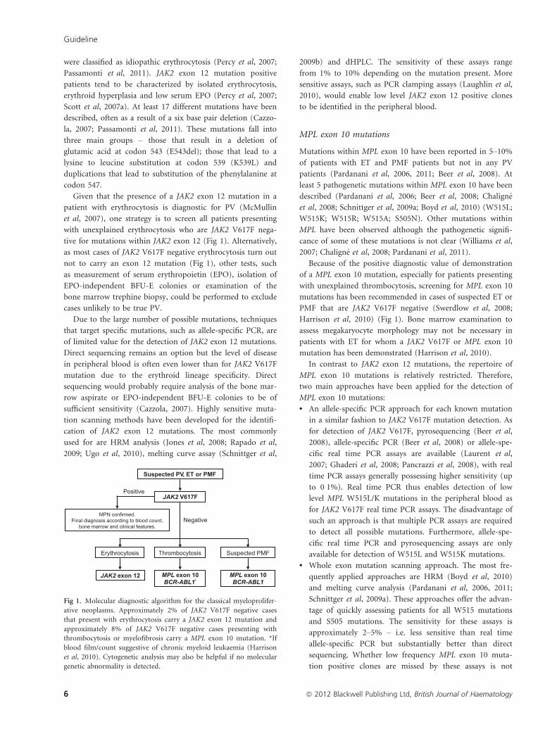
Suspected PV, ET or PMF
JAK2 V617F
MPN confirmed. Final diagnosis according to blood count,
bone marrow and clinical features.
Erythrocytosis Thrombocytosis Suspected PMF
JAK2 exon 12 MPL exon 10 BCR-ABL1*
MPL exon 10 BCR-ABL1
Positive
Negative
Fig 1. Molecular diagnostic algorithm for the classical myeloprolifer-
ative neoplasms. Approximately 2% of JAK2 V617F negative cases
that present with erythrocytosis carry a JAK2 exon 12 mutation and
approximately 8% of JAK2 V617F negative cases presenting with
thrombocytosis or myelofibrosis carry a MPL exon 10 mutation. *If
blood film/count suggestive of chronic myeloid leukaemia (Harrison
et al, 2010). Cytogenetic analysis may also be helpful if no molecular
genetic abnormality is detected.
Guideline
6 ª 2012 Blackwell Publishing Ltd, British Journal of Haematology
known. However, given that low level JAK2 V617F muta-
tion is common in ET, it would be expected that some ET
patients possess low level MPL exon 10 mutations. The
combination of mutation scanning methods with PCR
methods that preferentially amplify the mutant allele could
improve sensitivity.
BCR-ABL1 assessment
Exclusion of CML is a requirement in the diagnostic criteria
of both ET and PMF but not PV (Swerdlow et al, 2008).
Despite the rare occurrences of JAK2 V617F positive/BCR-
ABL1 positive cases (Hussein et al, 2008; Pieri et al, 2011)
the demonstration of a BCR-ABL1 fusion in a patient with
thrombocytosis or myelofibrosis indicates a diagnosis of
CML and excludes a diagnosis of ET or PMF. Guidelines for
investigation of thrombocytosis (Harrison et al, 2010) indi-
cate that screening for the BCR-ABL1 fusion gene should
only be necessary if atypical features, such as basophilia or
left shift of neutrophils, are present within the blood irre-
spective of the JAK2 V617F status. Whether assessment for
the BCR-ABL1 fusion gene needs to be carried out for JAK2
V617F or MPL exon 10 positive PMF is unclear but may be
useful if these mutations are not detected to exclude a diag-
nosis of CML (Swerdlow et al, 2008).
Other causes of erythrocytosis and thrombocytosis
A number of non-malignant causes of erythrocytosis and
thrombocytosis may be investigated and an increasing panel
of genes have been identified that are implicated in familial
erythrocytosis and thrombocytosis. Congenital causes of ery-
thocytosis include mutations in globin genes giving rise to
high oxygen affinity haemoglobin, BPGM mutation resulting
in bisphosphoglycerate mutase deficiency, mutations in com-
ponents of EPO signalling pathway (EPOR) and mutations
within components of oxygen sensing pathways such as
within VHL, EGLN1 (also termed PHD2) and EPAS1
(HIF2A). Especially in younger patients, mutations within
such genes may identify the cause of the erythrocytosis
(McMullin, 2008). Inherited forms of thrombocythaemia
may be caused by mutations within the 5′ untranslated
region of THPO (also termed TPO) or within the MPL locus
itself, including K39N (MPL-Baltimore), P106L and S505N
mutations (Skoda, 2010). Recently, two families with inher-
ited thrombocytosis and activating mutations within JAK2
(V617I and R564Q) have been reported (Etheridge et al,
2011; Mead et al, 2012). By contrast, the V617F mutation
itself has not been reported to be inherited in familial cases
although family members may acquire the mutation inde-
pendently (Cario et al, 2005).
Acknowledgements
DG and NCPC gratefully acknowledge support from the
Minimal Residual Disease Workpackage (WP12) of the Euro-
pean LeukemiaNet. AJB, AGF and TC carried out experi-
ments. AJB, HEW, LF, ALG, GG, SA, AA, IC, SEL, TC, JC,
PAE, DG, AS, MFM, ARG, CNH and NCPC wrote the
manuscript.
References
Albiero, E., Bernardi, M., Madeo, D., Ruggeri, M.
& Rodeghiero, F. (2008) A new TMHA-DHPLC
assay for the rapid mutation screening of JAK2
exon 14 in myeloproliferative disorders. Ameri-
can Journal of Hematology, 83, 603–604.
Antonioli, E., Carobbio, A., Pieri, L., Pancrazzi, A.,
Guglielmelli, P., Delaini, F., Ponziani, V., Bart-
alucci, N., Tozzi, L., Bosi, A., Rambaldi, A., Bar-
bui, T. & Vannucchi, A.M. (2010) Hydroxyurea
does not appreciably reduce JAK2 V617F allele
burden in patients with polycythemia vera or
essential thrombocythemia. Haematologica, 95,
1435–1438.
Asimakopoulos, F.A., Gilbert, J.G.R., Aldred, M.A.,
Pearson, T.C. & Green, A.R. (1996) Interstitial
deletion constitutes the major mechanism for
loss of heterozygosity on chromosome 20q in
polycythemia vera. Blood, 88, 2690–2698.
Baxter, E.J., Scott, L.M., Campbell, P.J., East, C.,
Fourouclas, N., Swanton, S., Vassiliou, G.S.,
Bench, A.J., Boyd, E.M., Curtin, N., Scott, M.A.
& Erber, W.N.; The Cancer Genome Project &
Green AR (2005) Acquired mutation of the
tyrosine kinase JAK2 in human myeloprolifera-
tive disorders. Lancet, 365, 1054–1061.
Beer, P.A., Campbell, P.J., Scott, L.M., Bench, A.J.,
Erber, W.N., Bareford, D., Wilkins, B.S., Reilly, J.
T., Hasselbalch, H.C., Bowman, R., Wheatley, K.,
Buck, G., Harrison, C.N. & Green, A.R. (2008)
MPL mutations in myeloproliferative disorders:
analysis of the PT-1 Cohort. Blood, 112, 141–149.
Beer, P.A., Jones, A.V., Bench, A.J., Goday-Fernan-
dez, A., Boyd, E.M., Vaghela, K.J., Erber, W.N.,
Odeh, B., Wright, C., McMullin, M.F., Cullis, J.,
Huntly, B.J., Harrison, C.N., Cross, N.C. &
Green, A.R. (2009) Clonal diversity in the mye-
loproliferative neoplasms: independent origins
of genetically distinct clones. British Journal of
Haematology, 144, 904–908.
Boyd, E.M., Bench, A.J., Goday-Fernandez, A.,
Anand, S., Vaghela, K.J., Beer, P., Scott, M.A.,
Bareford, D., Green, A.R., Huntly, B. & Erber,
W.N. (2010) Clinical utility of routine MPL
exon 10 analysis in the diagnosisof essential
thrombocythaemia and primary myelofibrosis.
British Journal of Haematology, 149, 250–257.
Campbell, P.J., Scott, L.M., Buck, G., Wheatley, K.,
East, C.L., Marsden, J.T., Duffy, A., Boyd, E.M.,
Bench, A.J., Scott, M.A., Vassiliou, G.S., Milligan,
D.W., Smith, S.R., Erber, W.N., Bareford, D.,
Wilkins, B.S., Reilly, J.T., Harrison, C.N. & Green,
A.R. (2005) Definition of subtypes of essential
thrombocythaemia and relation to polycythaemia
vera based on JAK2 V617F mutation status: a
prospective study. Lancet, 366, 1945–1953.
Cankovic, M., Whiteley, L., Hawley, R.C., Zarbo,
R.J. & Chitale, D. (2009) Clinical performance
of JAK2 V617F mutation detection assays in a
molecular diagnostics laboratory. American
Journal of Clinical Pathology, 132, 713–721.
Cario, H., Goerttler, P.S., Steimle, C., Levine, R.L.
& Pahl, H.L. (2005) The JAK2 V617F mutation
is acquired secondary to the predisposing alter-
ation in familial polycythaemia vera. British
Journal of Haematology, 130, 795–805.
Cazzola, M. (2007) Somatic mutations of JAK2
exon 12 as a molecular basis of erythrocytosis.
Haematologica, 92, 1585–1589.
Chaligne, R., Tonetti, C., Besancenot, R., Roy, L.,
Marty, C., Mossuz, P., Kiladjian, J.J., Socie, G.,
Bordessoule, D., Le Bousse-Kerdiles, M.C., Vain-
chenker, W. & Giraudier, S. (2008) New muta-
tions of MPL in primitive myelofibrosis: only
the MPL W515 mutations promote a G1/S-
phase transition. Leukemia, 22, 1557–1566.
Chen, Q., Lu, P., Jones, A.V., Silver, R.T. & Wang,
Y.L. (2007) Amplification refractory mutation
system, a highly sensitive and simple polymerase
chain reaction assay, for the detection of JAK2
Guideline
ª 2012 Blackwell Publishing Ltd, British Journal of Haematology 7
V617F mutation in chronic myeloproliferative
disorders. The Journal of Molecular Diagnostics,
9, 272–276.
Clark, J.R., Jack, A.L., Barnett, D. & Reilly, J.T.
(2012) JAK2 V617F false negative rate in the
UK NEQAS LI programme. British Journal of
Haematology, 157(Suppl 1), 56.
Cleyrat, C., Jelinek, J., Girodon, F., Boissinot, M.,
Ponge, T., Harousseau, J.L., Issa, J.P. & Hermo-
uet, S. (2010) JAK2 mutation and disease phe-
notype: a double L611V/V617F in cis mutation
of JAK2 is associated with isolated erythrocytosis
and increased activation of AKT and ERK1/2
rather than STAT5. Leukemia, 24, 1069–1073.
Dentali, F., Squizzato, A., Brivio, L.,Appio, L.,
Campiotti, L., Crowther, M., Grandi, A.M. &
Ageno, W. (2009) JAK2 V617F mutation for the
early diagnosis of Ph- myeloproliferative neo-
plasms in patients with venous thromboembo-
lism: a meta-analysis. Blood, 113, 5617–5623.
Denys, B., El Housni, H., Nollet, F., Verhasselt, B.
& Philippe, J. (2010) A real-time polymerase
chain reaction assay for rapid, sensitive, and
specific quantification of the JAK2 V617F
mutation using a locked nucleic acid-modified
oligonucleotide. The Journal of Molecular Diag-
nostics, 12, 512–519.
Etheridge, L., Corbo, L.M., Kaushansky, K., Chan,
E. & Hitchcock, I.S. (2011) A novel activating
JAK2 mutation, JAK2R564Q, causes familial
essential thrombocytosis (fET) via mechanisms
distinct from JAK2 V617F. Blood, 118, 123.
(ASH Annual Meeting Abstracts).
Ghaderi, M., Stromberg, O. & Porwit, A. (2008)
Rapid real-time PCR assay for detection of MPL
W515L mutation in patients with chronic mye-
loproliferative disorders. International Journal of
Laboratory Hematology, 32, 122–126.
Girodon, F., Schaeffer, C., Cleyrat, C., Mounier, M.,
Lafont, I., Santos, F.D., Duval, A., Maynadie, M.
& Hermouet, S. (2008) Frequent reduction or
absence of detection of the JAK2-mutated clone
in JAK2 V617F-positive patients within the first
years of hydroxyurea therapy. Haematologica, 93,
1723–1727.
Goday-Fernandez, A., Boyd, E., Bench, A. & Erber,
W. (2008) Real time detection of JAK2 V617F
in the myeloproliferative disorders. Haematolog-
ica, 93(s1), 302. Abs.0754.
Grunebach, F., Bross-Bach, U., Kanz, L. & Bross-
art, P. (2006) Detection of a new JAK2 D620E
mutation in addition to V617F in a patient with
polycythemia vera. Leukemia, 20, 2210–2211.
Guglielmelli, P., Biamonte, F., Score, J., Hidalgo-
Curtis, C., Cervantes, F., Maffioli, M., Fanelli,
T., Ernst, T., Winkelman, N., Jones, A.V., Zoi,
K., Reiter, A., Duncombe, A., Villani, L., Bosi,
A., Barosi, G., Cross, N.C. & Vannucchi, A.M.
(2011) EZH2 mutational status predicts poor
survival in myelofibrosis. Blood, 118, 5227–5234.
Harrison, C.N., Bareford, D., Butt, N., Campbell,
P., Conneally, E., Drummond, M., Erber, W.,
Everington, T., Green, A.R., Hall, G.W., Hunt,
B.J., Ludlam, C.A., Murrin, R., Nelson-Piercy,
C., Radia, D.H., Reilly, J.T., Van der Walt, J.,
Wilkins, B. & McMullin, M.F. (2010) Guideline
for investigation and management of adults and
children presenting with a thrombocytosis.
British Journal of Haematology, 149, 352–375.
Hermouet, S., Dobo, I., Lippert, E., Boursier, M.
C., Ergand, L., Perrault-Hu, F. & Pineau, D.
(2007) Comparison of whole blood vs purified
blood granulocytes for the detection and quanti-
tation of JAK2 V617F. Leukemia, 21, 1128–1130.
Hussein, K., Bock, O., Theophile, K., Seegers, A.,
Arps, H., Basten, O., Grips, K.H., Franz-Werner,
J., Busche, G. & Kreipe, H. (2008) Chronic mye-
loproliferative diseases with concurrent BCR-
ABL junction and JAK2 V617F mutation. Leuke-
mia, 22, 1059–1062.
James, C., Ugo, V., Le Couedic, J.P., Staerk, J., Del-
hommeau, F., Lacout, C., Garcon, L., Raslova,
H., Berger, R., Bennaceur-Griscelli, A., Villeval, J.
L., Constantinescu, S.N., Casadevall, N. & Vain-
chenker, W. (2005) A unique clonal JAK2 muta-
tion leading to constitutive signalling causes
polycythaemia vera. Nature, 434, 1144–1148.
James, C., Delhommeau, F., Marzac, C., Teyssan-
dier, I., Couedic, J.P., Giraudier, S., Roy, L.,
Saulnier, P., Lacroix, L., Maury, S., Tulliez, M.,
Vainchenker, W., Ugo, V. & Casadevall, N.
(2006) Detection of JAK2 V617F as a first inten-
tion diagnostic test for erythrocytosis. Leukemia,
20, 350–353.
Jelinek, J., Oki, Y., Gharibyan, V., Bueso-Ramos,
C., Prchal, J.T., Verstovsek, S., Beran, M., Estey,
E., Kantarjian, H.M. & Issa, J.P. (2005) JAK2
mutation 1849G>T is rare in acute leukemias
but can be found in CMML, Philadelphia chro-
mosome–negative CML, and megakaryocytic
leukemia. Blood, 106, 3370–3373.
Jones, A.V. & Cross, N.C. (2004) Oncogenic deriv-
atives of platelet-derived growth factor receptors.
Cellular and Molecular Life Sciences, 61, 2912–
2923.
Jones, A.V., Kreil, S., Zoi, K., Waghorn, K., Curtis,
C., Zhang, L., Score, J., Seear, R., Chase, A.J.,
Grand, F.H., White, H., Zoi, C., Loukopoulos,
D., Terpos, E., Vervessou, E.C., Schultheis, B.,
Emig, M., Ernst, T., Lengfelder, E., Hehlmann,
R., Hochhaus, A., Oscier, D., Silver, R.T., Reiter,
A. & Cross, N.C. (2005) Widespread occurrence
of the JAK2 V617F mutation in chronic myelo-
proliferative disorders. Blood, 106, 2162–2168.
Jones, A.V., Silver, R.T., Waghorn, K., Curtis, C.,
Kreil, S., Zoi, K., Hochhaus, A., Oscier, D., Met-
zgeroth, G., Lengfelder, E., Reiter, A., Chase, A.
J. & Cross, N.C. (2006) Minimal molecular
response in polycythemia vera patients treated
with imatinib or interferon alpha. Blood, 107,
3339–3341.
Jones, A.V., Cross, N.C.P., White, H.E., Green, A.
R. & Scott, L.M. (2008) Rapid identification of
JAK2 exon 12 mutations using high resolution
melting analysis. Haematologica, 93, 1560–1564.
Jovanovic, J.V., Vannucchi, A.M., Lippert, E.,
Oppliger Leibundgut, E., Maroc, N., Hermouet,
S., Nickless, G., Cassinat, B., Guglielmelli, P.,
van der Reijden, B., Bench, A., Tobal, K., Wil-
kins, B., Cuthill, K., Ivey, A., Yeoman, K., Percy,
M.J., Schwemmers, S., Gruender, A., Kelley, T.
W., Reading, S., Pancrazzi, A., McMullin, M.F.,
Pahl, H.L., Harrison, C.N., Prchal, J.T., Chomi-
enne, C., Kiladjian, J.J., Barbui, T. & Grimwade,
D. (2011) Systematic evaluation of DNA-based
quantitative-polymerase chain reaction (Q-PCR)
assays to track treatment response in patients
with JAK2-V617F associated myeloproliferative
neoplasms: a Joint European LeukemiaNet/
MPN&MPNr-EuroNet study. Blood, 118, 2812.
(ASH Annual Meeting Abstracts).
Kralovics, R., Passamonti, F., Buser, A.S., Teo, S.S.,
Tiedt, R., Passweg, J.R., Tichelli, A., Cazzola, M.
& Skoda, R.C. (2005) A gain-of-function muta-
tion of JAK2 in myeloproliferative disorders.
New England Journal of Medicine, 352, 1779–
1790.
Kroger, N., Badbaran, A., Holler, E., Hahn, J.,
Kobbe, G., Bornhauser, M., Reiter, A., Zabelina,
T., Zander, A.R. & Fehse, B. (2007) Monitoring
of the JAK2-V617F mutation by highly sensitive
quantitative real-time PCR after allogeneic stem
cell transplantation in patients with myelofibro-
sis. Blood, 109, 1316–1321.
Larsen, T.S., Christensen, J.H., Hasselbalch, H.C. &
Pallisgaard, N. (2007) The JAK2 V617F mutation
involves B- and T-lymphocyte lineages in a sub-
group of patients with Philadelphia-chromosome
negative chronic myeloproliferative disorders.
British Journal of Haematology, 136, 745–751.
Larsen, T.S., Pallisgaard, N., Møller, M.B. & Has-
selbalch, H.C. (2008) Quantitative assessment of
the JAK2 V617F allele burden: equivalent levels
in peripheral blood and bone marrow. Leuke-
mia, 22, 194–195.
Laughlin, T.S., Moliterno, A.R., Stein, B.L. & Roth-
berg, P.G. (2010) Detection of exon 12 muta-
tions in the JAK2 gene: enhanced analytical
sensitivity using clamped PCR and nucleotide
sequencing. The Journal of Molecular Diagnostics,
12, 278–282.
Laurent, C., Demas, V., Delabesse, E. & Brousset,
P. (2007) Detection of the MPL W515L muta-
tion in bone marrow core biopsy specimens
with essential thrombocythemia using the Taq-
Man assay. Human Pathology, 38, 1581–1582.
Levine, R.L., Wadleigh, M., Cools, J., Ebert, B.L.,
Wernig, G., Huntly, B.J., Boggon, T.J., Wlo-
darska, I., Clark, J.J., Moore, S., Adelsperger, J.,
Koo, S., Lee, J.C., Gabriel, S., Mercher, T.,
D’Andrea, A., Frohling, S., Dohner, K., Mary-
nen, P., Vandenberghe, P., Mesa, R.A., Tefferi,
A., Griffin, J.D., Eck, M.J., Sellers, W.R., Meyer-
son, M., Golub, T.R., Lee, S.J. & Gilliland, D.G.
(2005a) Activating mutation in the tyrosine
kinase JAK2 in polycythemia vera, essential
thrombocythemia, and myeloid metaplasia with
myelofibrosis. Cancer Cell, 7, 387–397.
Levine, R.L., Loriaux, M., Huntly, B.J.P., Loh, M.
L., Beran, M., Stoffregen, E., Berger, R., Clark, J.
J., Willis, S.G., Nguyen, K.T., Flores, N.J., Estey,
E., Gattermann, N., Armstrong, S., Look, A.T.,
Griffin, J.D., Bernard, O.A., Heinrich, M.C., Gil-
liland, D.G., Druker, B. & Deininger, M.W.
(2005b) The JAK2 V617F activating mutation
Guideline
8 ª 2012 Blackwell Publishing Ltd, British Journal of Haematology
occurs in chronic myelomonocytic leukemia and
acute myeloid leukemia, but not in acute
lymphoblastic leukemia or chronic lymphocytic
leukemia. Blood, 106, 3377–3379.
Lippert, E., Boissinot, M., Kralovics, R., Girodon,
F., Dobo, I., Praloran, V., Boiret-Dupre, N.,
Skoda, R.C. & Hermouet, S. (2006) The JAK2-
V617F mutation is frequently present at diagno-
sis in patients with essential thrombocythemia
and polycythemia vera. Blood, 108, 1865–1867.
Lippert, E., Girodon, F., Hammond, E., Jelinek, J.,
Reading, N.S., Fehse, B., Hanlon, K., Hermans,
M., Richard, C., Swierczek, S., Ugo, V., Carillo,
S., Harrivel, V., Marzac, C., Pietra, D., Sobas,
M., Mounier, M., Migeon, M., Ellard, S., Kro-
ger, N., Herrmann, R., Prchal, J.T., Skoda, R.C.
& Hermouet, S. (2009) Concordance of assays
designed for the quantification of JAK2 V617F:
a multicenter study. Haematologica, 94, 38–45.
Ma, W., Kantarjian, H., Zhang, X., Sun, W., Bul-
ler, A.M., Jilani, I., Schwartz, J.G., Giles, F. &
Albitar, M. (2008) Higher detection rate of
JAK2 mutation using plasma. Blood, 111, 3906–
3907.
Ma, W., Zhang, X., Wang, X., Zhang, Z., Yeh, C.
H., Uyeji, J. & Albitar, M. (2011) MPL mutation
profile in JAK2 mutation-negative patients with
myeloproliferative disorders. Diagnostic Molecu-
lar Pathology, 20, 34–39.
Mason, J., Akiki, S. & Griffiths, M.J. (2011) Pitfalls
in molecular diagnosis in haemato-oncology.
Journal of Clinical Pathology, 64, 275–278.
Mattocks, C.J., Morris, M.A., Matthijs, G., Swin-
nen, E., Corveleyn, A., Dequeker, E., Muller, C.
R., Pratt, V. & Wallace, A. (2010) A standard-
ized framework for the validation and verifica-
tion of clinical molecular genetic tests. European
Journal of Human Genetics, 18, 1276–1288.
McClure, R., Mai, M. & Lasho, T. (2006) Valida-
tion of two clinically useful assays for evaluation
of JAK2 V617F mutation in chronic myelopro-
liferative disorders. Leukemia, 20, 168–171.
McMullin, M.F. (2008) The classification and diag-
nosis of erythrocytosis. International Journal of
Laboratory Hematology, 30, 447–459.
McMullin, M.F., Reilly, J.T., Campbell, P., Bare-
ford, D., Green, A.R., Harrison, C.N., Conneally,
E. & Ryan, K. (2007) Amendment to the guide-
line for diagnosis and investigation of polycytha-
emia/erythrocytosis. British Journal of
Haematology, 138, 821–822.
Mead, A.J., Rugless, M.J., Jacobsen, S.E. & Schuh,
A. (2012) Germline JAK2 mutation in a family
with hereditary thrombocytosis. The New Eng-
land Journal of Medicine, 366, 967–969.
Nielsen, C., Birgens, H.S., Nordestgaard, B.G.,
Kjaer, L. & Bojesen, S.E. (2011) The JAK2
V617F somatic mutation, mortality and cancer
risk in the general population. Haematologica,
96, 450–453.
Pancrazzi, A., Guglielmelli, P., Ponziani, V., Ber-
gamaschi, G., Bosi, A., Barosi, G. & Vannucchi,
A.M. (2008) A sensitive detection method for
MPLW515L or MPLW515K mutation in chronic
myeloproliferative disorders with locked nucleic
acid-modified probes and real-time polymerase
chain reaction. The Journal of Molecular Diag-
nostics, 10, 435–441.
Pardanani, A.D., Levine, R.L., Lasho, T., Pikman,
Y., Mesa, R.A., Wadleigh, M., Steensma, D.P.,
Elliott, M.A., Wolanskyj, A.P., Hogan, W.J.,
McClure, R.F., Litzow, M.R., Gilliland, D.G. &
Tefferi, A. (2006) MPL515 mutations in myelo-
proliferative and other myeloid disorders: a
study of 1182 patients. Blood, 108, 3472–3476.
Pardanani, A., Guglielmelli, P., Lasho, T.L., Panc-
razzi, A., Finke, C.M., Vannucchi, A.M. & Tefferi,
A. (2011) Primary myelofibrosis with or without
mutant MPL: comparison of survival and clinical
features involving 603 patients. Leukemia, 25,
1834–1839.
Passamonti, F. & Rumi, E. (2009) Clinical rele-
vance of JAK2 (V617F) mutant allele burden.
Haematologica, 94, 7–10.
Passamonti, F., Elena, C., Schnittger, S., Skoda, R.
C., Green, A.R., Girodon, F., Kiladjian, J.J.,
McMullin, M.F., Ruggeri, M., Besses, C., Van-
nucchi, A.M., Lippert, E., Gisslinger, H., Rumi,
E., Lehmann, T., Ortmann, C.A., Pietra, D., Pas-
cutto, C., Haferlach, T. & Cazzola, M. (2011)
Molecular and clinical features of the myelopro-
liferative neoplasm associated with JAK2 exon
12 mutations. Blood, 117, 2813–2816.
Percy, M.J., Scott, L.M., Erber, W.N., Harrison, C.
N., Reilly, J.T., Jones, F.G., Green, A.R. &
McMullin, M.F. (2007) The frequency of JAK2
exon 12 mutations in idiopathic erythrocytosis
patients with low serum erythropoietin levels.
Haematologica, 92, 1607–1614.
Pieri, L., Spolverini, A., Scappini, B., Occhini, U.,
Birtolo, S., Bosi, A., Albano, F., Fava, C. & Van-
nucchi, A.M. (2011) Concomitant occurrence of
BCR-ABL and JAK2 V617F mutation. Blood,
118, 3445–3446.
Qian, J., Lin, J., Yao, D.M., Chen, Q., Xiao, G.F.,
Ji, R.B., Li, Y., Yang, J. & Qian, Z. (2010) Rapid
detection of JAK2 V617F mutation using high-
resolution melting analysis with LightScanner
platform. Clinica Chimica Acta, 411, 2097–2100.
Quentmeier, H., MacLeod, R.A., Zaborski, M. &
Drexler, H.G. (2006) JAK2 V617F tyrosine
kinase mutation in cell lines derived from mye-
loproliferative disorders. Leukemia, 20, 471–476.
Rapado, I., Grande, S., Albizua, E., Ayala, R., Her-
nandez, J.A., Gallardo, M., Gilsanz, F. & Marti-
nez-Lopez, J. (2009) High resolution melting
analysis for JAK2 exon 14 and exon 12 muta-
tions – A diagnostic tool for myeloproliferative
neoplasms. The Journal of Molecular Diagnostics,
11, 155–161.
Reiter, A., Grimwade, D. & Cross, N.C. (2007)
Diagnostic and therapeutic management of
eosinophilia-associated chronic myeloprolifera-
tive disorders. Haematologica, 92, 1153–1158.
Sattler, M., Walz, C., Crowley, B.J., Lengfelder, E.,
Janne, P.A., Rogers, A.M., Kuang, Y., Distel, R.J.,
Reiter, A. & Griffin, J.D. (2006) A sensitive
high-throughput method to detect activating
mutations of Jak2 in peripheral-blood samples.
Blood, 107, 1237–1238.
Schmitt-Graeff, A.H., Teo, S.S., Olschewski, M.,
Schaub, F., Haxelmans, S., Kirn, A., Reinecke,
P., Germing, U. & Skoda, R.C. (2008) JAK2
V617F mutation status identifies subtypes of
refractory anemia with ringed sideroblasts asso-
ciated with marked thrombocytosis. Haemato-
logica, 93, 34–40.
Schnittger, S., Bacher, U., Kern, W., Schroder, M.,
Haferlach, T. & Schoch, C. (2006) Report on
two novel nucleotide exchanges in the JAK2
pseudokinase domain: D620E and E627E. Leuke-
mia, 20, 2195–2197.
Schnittger, S., Bacher, U., Haferlach, C., Beelen,
D., Bojko, P., Burkle, D., Dengler, R., Distelrath,
A., Eckart, M., Eckert, R., Fries, S., Knoblich, J.,
Kochling, G., Laubenstein, H.P., Petrides, P.,
Planker, M., Pihusch, R., Weide, R., Kern, W. &
Haferlach, T. (2009a) Characterization of 35
new cases with four different MPLW515 muta-
tions and essential thrombocytosis or primary
myelofibrosis. Haematologica, 94, 141–144.
Schnittger, S., Bacher, U., Haferlach, C., Geer, T.,
Muller, P., Mittermuller, J., Petrides, P., Schlag,
R., Sandner, R., Selbach, J., Slawik, H.R., Tessen,
H.W., Wehmeyer, J., Kern, W. & Haferlach, T.
(2009b) Detection of JAK2 exon 12 mutations
in 15 patients with JAK2 V617F negative polycy-
themia vera. Haematologica, 94, 414–418.
Scott, L.M., Tong, W., Levine, R.L., Scott, M.A.,
Beer, P.A., Stratton, M.R., Futreal, P.A., Erber,
W.N., McMullin, M.F., Harrison, C.N., Warren,
A.J., Gilliland, D.G., Lodish, H.F. & Green, A.R.
(2007a) JAK2 exon 12 mutations in polycythe-
mia vera and idiopathic erythrocytosis. New
England Journal of Medicine, 356, 459–468.
Scott, L.M., Beer, P.A., Bench, A.J., Erber, W.N. &
Green, A.R. (2007b) Prevalance of JAK2 V617F
and exon 12 mutations in polycythaemia vera.
British Journal of Haematology, 139, 511–512.
Sidon, P., El Housni, H., Dessars, B. & Heimann,
P. (2006) The JAK2 V617F mutation is detect-
able at very low level in peripheral blood of
healthy donors. Leukemia, 20, 1622.
Skoda, R.C. (2010) Hereditary myeloproliferative
disorders. Haematologica, 95, 6–8.
Stevenson, W.S., Hoyt, R., Bell, A., Guipponi, M.,
Juneja, S., Grigg, A.P., Curtis, D.J., Scott, H.S.,
Szer, J., Alexander, W.S., Tuckfield, A. & Roberts,
A.W. (2006) Genetic heterogeneity of granulo-
cytes for the JAK2 V617F mutation in essential
thrombocythaemia: implications for mutation
detection in peripheral blood. Pathology, 38,
336–342.
Swerdlow, S.H., Campo, E., Harris, N.L., Jaffe, E.
S., Pileri, S.A., Stein, H., Thiele, J. & Vardiman,
J.W. (2008) WHO Classification of Tumours of
Haematopoietic and Lymphoid Tissues. IARC
Press, Lyon, France.
Szpurka, H., Tiu, R., Murugesan, G., Aboudola, S.,
Hsi, E.D., Theil, K.S., Sekeres, M.A. & Maciejew-
ski, J.P. (2006) Refractory anemia with ringed
sideroblasts associated with marked thrombocy-
tosis (RARS-T), another myeloproliferative con-
dition characterized by JAK2 V617F mutation.
Blood, 108, 2173–2181.
Guideline
ª 2012 Blackwell Publishing Ltd, British Journal of Haematology 9
Tan, A.Y., Westerman, D.A. & Dobrovic, A. (2007)
A simple, rapid, and sensitive method for the
detection of the JAK2 V617F mutation. Ameri-
can Journal of Clinical Pathology, 127, 977–981.
Tefferi, A. & Vainchenker, W. (2011) Myeloprolif-
erative neoplasms: molecular pathophysiology,
essential clinical understanding, and treatment
strategies. Journal of Clinical Oncology, 29,
573–582.
Tefferi, A., Noel, P. & Hanson, C.A. (2011) Uses
and abuses of JAK2 and MPL mutation tests in
myeloproliferative neoplasms. The Journal of
Molecular Diagnostics, 13, 461–466.
Ugo, V., Tondeur, S., Menot, M.L., Bonnin, N., Le
Gac, G., Tonetti, C., Mansat-De Mas, V., Lecu-
cq, L., Kiladjian, J.J., Chomienne, C., Dosquet,
C., Parquet, N., Darnige, L., Porneuf, M., Escof-
fre-Barbe, M., Giraudier, S., Delabesse, E. &
Cassinat, B. (2010) Interlaboratory development
and validation of a HRM method applied to the
detection of JAK2 exon 12 mutations in polycy-
themia vera patients. PLoS ONE, 5, e8893.
Wang, Y.L., Vandris, K., Jones, A., Cross, N.C.,
Christos, P., Adriano, F. & Silver, R.T. (2008)
JAK2 mutations are present in all cases of poly-
cythemia vera. Leukemia, 22, 1289.
Warshawsky, I., Mularo, F., Hren, C. & Jakubow-
ski, M. (2010) Failure of the Ipsogen Muta-
Screen kit to detect the JAK2 617V>F mutation
in samples with additional rare exon 14 muta-
tions: implications for clinical testing and report
of a novel 618C>F mutation in addition to
617V>F. Blood, 115, 3175–3176.
Williams, D.M., Kim, A.H., Rogers, O., Spivak, J.L. &
Moliterno, A.R. (2007) Phenotypic variations and
new mutations in JAK2 V617F–negative polycy-
themia vera, erythrocytosis, and idiopathic myelo-
fibrosis. Experimental hematology, 35, 1641–1646.
Wong, C.L., Ma, E.S., Wang, C.L., Lam, H.Y. &
Ma, S.Y. (2007) JAK2 V617F due to a novel TG-
CT mutation at nucleotides 1848–1849: diagnos-
tic implication. Leukemia, 21, 1344–1346.
Wu, Z., Yuan, H., Zhang, X., Liu, W., Xu, J.,
Zhang, W. & Guan, M. (2011) Development
and inter-laboratory validation of unlabeled
probe melting curve analysis for detection of
JAK2 V617F mutation in polycythemia vera.
PLoS ONE, 6, e26534.
Xu, X., Zhang, Q., Luo, J., Xing, S., Li, Q.,
Krantz, S.B., Fu, X. & Zhao, Z.J. (2007) JAK2
(V617F): prevalence in a large Chinese hospital
population. Blood, 109, 339–342.
Yokota, M., Tatsumi, N., Nathalang, O., Yamada,
T. & Tsuda, I. (1999) Effects of heparin on
polymerase chain reaction for blood white
cells. Journal of Clinical Laboratory Analysis, 13,
133–140.
Guideline
10 ª 2012 Blackwell Publishing Ltd, British Journal of Haematology
Related Documents