BioMed Central Page 1 of 8 (page number not for citation purposes) BMC Cancer Open Access Research article Molecular characterization of EGFR, PDGFRA and VEGFR2 in cervical adenosquamous carcinoma Adhemar Longatto-Filho* 1,2 , Céline Pinheiro 1 , Olga Martinho 1 , Marise AR Moreira 3 , Luiz FJ Ribeiro 4 , Geraldo S Queiroz 4 , Fernando C Schmitt 5,6 , Fátima Baltazar 1 and Rui M Reis* 1 Address: 1 Life and Health Sciences Research Institute (ICVS), School of Health Sciences, University of Minho, Braga, Portugal, 2 Instituto Adolfo Lutz, São Paulo, SP, Brazil, 3 Department of Pathology of the School of Medicine of the Federal University of Goiás, Goiânia, Go, Brazil, 4 Hospital Araújo Jorge, Goiânia, Go, Brazil, 5 IPATIMUP, Porto, Portugal and 6 School of Medicine of the University of Porto, Porto, Portugal Email: Adhemar Longatto-Filho* - [email protected]; Céline Pinheiro - [email protected]; Olga Martinho - [email protected]; Marise AR Moreira - [email protected]; Luiz FJ Ribeiro - [email protected]; Geraldo S Queiroz - [email protected]; Fernando C Schmitt - [email protected]; Fátima Baltazar - [email protected]; Rui M Reis* - [email protected] * Corresponding authors Abstract Background: Adenosquamous carcinoma of the uterine cervix is an infrequent but aggressive subtype of cervical cancer. A better understanding of its biological behaviour is warranted to define more accurate prognosis and therapeutic targets. Currently, the blockage of receptor tyrosine kinase (RTKs) activity is an efficient therapeutic strategy for many different cancers. The objective of this study was to investigate EGFR, PDGFRA and VEGFR2 RTKs overexpression and activating gene mutations in a cohort of 30 adenosquamous carcinomas of the uterine cervix. Methods: EGFR, PDGFRA and VEGFR2 immunohistochemistry was performed in all samples, followed by DNA isolation from the gross macroscopically dissection of the neoplastic area. Screening for EGFR (exons 18–21) and PDGFRA (exons 12, 14 and 18) mutations was done by PCR – single-strand conformational polymorphism (PCR-SSCP). Results: Despite the presence of EGFR immunohistochemical positive reactions in 43% (13/30) of the samples, no EGFR activating mutations in the hotspot region (exons 18–21) were identified. A silent base substitution (CAG>CAA) in EGFR exon 20 at codon 787 (Q787Q) was found in 17 cases (56%). All PDGFRA immunohistochemical reactions were positive and consistently observed in the stromal component, staining fibroblasts and endothelial cells, as well as in the cytoplasm of malignant cells. No activating PDGFRA mutations were found, yet, several silent mutations were observed, such as a base substitution in exon 12 (CCA>CCG) at codon 567 (P567P) in 9 cases and in exon 18 (GTC>GTT) at codon 824 (V824V) in 4 cases. We also observed the presence of base substitutions in intron 14 (IVS14+3G>A and IVS14+49G>A) in two different cases, and in intron 18 (IVS18-50insA) in 4 cases. VEGFR2 positivity was observed in 22 of 30 cases (73.3%), and was significantly associated with lack of metastasis (p = 0.038). Conclusion: This is the most extensive analysis of EGFR, PDGFRA and VEGFR2 in cervical adenosquamous carcinomas. Despite the absence of EGFR and PDGFRA activating mutations, the presence of overexpression of these three important therapeutic targets in a subset of cases may be important in predicting the sensitivity of adenosquamous carcinoma to specific anti-RTKs drugs. Published: 29 June 2009 BMC Cancer 2009, 9:212 doi:10.1186/1471-2407-9-212 Received: 23 December 2008 Accepted: 29 June 2009 This article is available from: http://www.biomedcentral.com/1471-2407/9/212 © 2009 Longatto-Filho et al; licensee BioMed Central Ltd. This is an Open Access article distributed under the terms of the Creative Commons Attribution License (http://creativecommons.org/licenses/by/2.0 ), which permits unrestricted use, distribution, and reproduction in any medium, provided the original work is properly cited.

Welcome message from author
This document is posted to help you gain knowledge. Please leave a comment to let me know what you think about it! Share it to your friends and learn new things together.
Transcript
BioMed CentralBMC Cancer
ss
Open AcceResearch articleMolecular characterization of EGFR, PDGFRA and VEGFR2 in cervical adenosquamous carcinomaAdhemar Longatto-Filho*1,2, Céline Pinheiro1, Olga Martinho1, Marise AR Moreira3, Luiz FJ Ribeiro4, Geraldo S Queiroz4, Fernando C Schmitt5,6, Fátima Baltazar1 and Rui M Reis*1Address: 1Life and Health Sciences Research Institute (ICVS), School of Health Sciences, University of Minho, Braga, Portugal, 2Instituto Adolfo Lutz, São Paulo, SP, Brazil, 3Department of Pathology of the School of Medicine of the Federal University of Goiás, Goiânia, Go, Brazil, 4Hospital Araújo Jorge, Goiânia, Go, Brazil, 5IPATIMUP, Porto, Portugal and 6School of Medicine of the University of Porto, Porto, Portugal
Email: Adhemar Longatto-Filho* - [email protected]; Céline Pinheiro - [email protected]; Olga Martinho - [email protected]; Marise AR Moreira - [email protected]; Luiz FJ Ribeiro - [email protected]; Geraldo S Queiroz - [email protected]; Fernando C Schmitt - [email protected]; Fátima Baltazar - [email protected]; Rui M Reis* - [email protected]
* Corresponding authors
AbstractBackground: Adenosquamous carcinoma of the uterine cervix is an infrequent but aggressive subtype of cervicalcancer. A better understanding of its biological behaviour is warranted to define more accurate prognosis and therapeutictargets. Currently, the blockage of receptor tyrosine kinase (RTKs) activity is an efficient therapeutic strategy for manydifferent cancers. The objective of this study was to investigate EGFR, PDGFRA and VEGFR2 RTKs overexpression andactivating gene mutations in a cohort of 30 adenosquamous carcinomas of the uterine cervix.
Methods: EGFR, PDGFRA and VEGFR2 immunohistochemistry was performed in all samples, followed by DNAisolation from the gross macroscopically dissection of the neoplastic area. Screening for EGFR (exons 18–21) and PDGFRA(exons 12, 14 and 18) mutations was done by PCR – single-strand conformational polymorphism (PCR-SSCP).
Results: Despite the presence of EGFR immunohistochemical positive reactions in 43% (13/30) of the samples, no EGFRactivating mutations in the hotspot region (exons 18–21) were identified. A silent base substitution (CAG>CAA) in EGFRexon 20 at codon 787 (Q787Q) was found in 17 cases (56%). All PDGFRA immunohistochemical reactions were positiveand consistently observed in the stromal component, staining fibroblasts and endothelial cells, as well as in the cytoplasmof malignant cells. No activating PDGFRA mutations were found, yet, several silent mutations were observed, such as abase substitution in exon 12 (CCA>CCG) at codon 567 (P567P) in 9 cases and in exon 18 (GTC>GTT) at codon 824(V824V) in 4 cases. We also observed the presence of base substitutions in intron 14 (IVS14+3G>A and IVS14+49G>A)in two different cases, and in intron 18 (IVS18-50insA) in 4 cases. VEGFR2 positivity was observed in 22 of 30 cases(73.3%), and was significantly associated with lack of metastasis (p = 0.038).
Conclusion: This is the most extensive analysis of EGFR, PDGFRA and VEGFR2 in cervical adenosquamous carcinomas.Despite the absence of EGFR and PDGFRA activating mutations, the presence of overexpression of these three importanttherapeutic targets in a subset of cases may be important in predicting the sensitivity of adenosquamous carcinoma tospecific anti-RTKs drugs.
Published: 29 June 2009
BMC Cancer 2009, 9:212 doi:10.1186/1471-2407-9-212
Received: 23 December 2008Accepted: 29 June 2009
This article is available from: http://www.biomedcentral.com/1471-2407/9/212
© 2009 Longatto-Filho et al; licensee BioMed Central Ltd. This is an Open Access article distributed under the terms of the Creative Commons Attribution License (http://creativecommons.org/licenses/by/2.0), which permits unrestricted use, distribution, and reproduction in any medium, provided the original work is properly cited.
Page 1 of 8(page number not for citation purposes)
BMC Cancer 2009, 9:212 http://www.biomedcentral.com/1471-2407/9/212
BackgroundAdenosquamous carcinoma (ASC) of the uterine cervix isa relatively infrequent histological subtype of cervical can-cer, associated with very aggressive behaviour and reducedsurvival rates [1]. ASC histopathological interpretationremains controversial; theoretically, ASC is a mixture ofmalignant glandular and squamous epithelial elements.However, the practical application of this morphologicalcriterion is far from being straightforward and the prog-nostic significance of the histological alterations is con-tentious and does not exactly predict the clinicalbehaviour of ASC [2-4]. The cofactors which contribute tothe progression of HPV-infected cervical carcinoma areapparently diverse in each type of histogenetic differenti-ation. The high risk HPV infection persistence is assumedas a necessary but not sufficient factor to cervical cancerdevelopment, and the genetic and molecular disparitiesinvolved in the carcinoma progression are still poorlyunderstood [5]. Therefore, a better understanding of ASCbiology is needed to identify the key players and potentialnovel therapeutic strategies.
Disruption of the mitogenic signalling mechanisms, par-ticularly the ones mediated by receptor tyrosine kinases(RTKs), is a hallmark of the carcinogenic process and cur-rently constitutes an important therapeutic target group[6]. RTKs are transmembrane proteins constituted by anextracellular, a transmembrane, a juxtamembrane, and anintracellular domain where two kinase regions are located[7]. Upon growth factor binding, receptor dimerizes andautophosphorylates its intracellular tyrosine residues thatactivate several downstream signalling cascades, like MAPkinase, PI3-kinase, and JAK/STAT pathways, affecting cel-lular gene expression [8]. In the neoplastic developmentand progression, RTKS are commonly deregulated, andexcessive phosphorylation sustains signal transductionpathways in an activated state, leading to tumour growthand progression, proliferation, dedifferentiation, inhibi-tion of apoptosis, metastasis and angiogenesis [9,10].Among the distinct RTK classes, class I [e.g. epidermalgrowth factor receptor (EGFR)] and class III [e.g. platelet-derived growth factor receptor-α (PDGFR-α), KIT, vascu-lar endothelial growth factor receptors 1 (VEGFR1), alsoknown as Flt-1, and VEGFR2 or Flk-1] [11] have been con-sistently implicated in solid neoplasm tumourigenesis.
EGFR was the first RTK to be directly linked to human can-cers [12]. The use of EGFR antagonists, namely mono-clonal antibodies directed to the extracellular domain,such as Cetuximab (Erbitux®) and small molecule tyrosinekinase inhibitors, such as Gefitinib (Iressa®) and Erlotinib(Tarceva®), have raised great expectations [13]. Recently,several molecular alterations have been associated withpatient's response to these new anti-EGFR drugs, in partic-ular, EGFR mutations in hotspot regions of the intracellu-
lar kinase domain (exons 18–21) were predictive of apositive response to Gefitinib and Erlotinib in a subset oflung cancer [11]. Positive therapeutic results have alsobeen reported using KIT and PDGFRA inhibitors such asImatinib mesylate (Gleevec®) for gastrointestinal stromaltumours (GISTs) [14]. Likewise anti-EGFR drugs, specificgene activating mutations of KIT and PDGFRA gene seemto predict patients' response to Imatinib [15]. Besides theuse of selective inhibitors, promising therapeutic resultsare being attained with multi-target inhibitors such asSunitinib (Sutent®), which targets KIT, PDGFR, VEGFR2,Sorafenib (Nexavar®) that targets KIT, VEGFR2, PDGFRand intracellular tyrosine kinases, such as BRAF [14] andPazopanib, which targets also KIT, PDGFR and VEGFR[16]. VEGFR2 is not only a mitogenic factor, but essen-tially an important angiogenic factor; consequently,blocking its activity potentially enhances therapeuticresponse [17]. Recently, we reported absence of KITmolecular alterations in ASC and provided evidence forKIT activation through KIT/SCF co-expression in a smallproportion of cases [18], however there are no similarstudies with other RTK therapeutic targets.
The aim of this study was to investigate the presence ofEGFR, PDGFRA and VEGFR2 RTKs aberrations, namelyoverexpression and activating gene mutations in a cohortof 30 adenosquamous carcinomas of the cervix. Accord-ingly, we intended to assess the potentiality of these RTKsas therapeutic targets in this aggressive tumour type.
MethodsMaterialsThis retrospective study comprised a series of 30 patientswith ASC of the uterine cervix, examined and treated attwo Hospitals, retrieved from the files of Araújo JorgeHospital and from the Pathology Department of theSchool of Medicine of the Federal University of Goiás,Goiania, in Goias State, Brazil, from 1986 to 2000. All his-topathological diagnoses were revised by two of theauthors (FCS, MARM) and categorized according to theWHO classification [19]. The age of the patients rangedfrom 24 to 77 years old (mean 49 and median 44.7 years).Clinico-pathological data was available for 29/30 patientsand included age at diagnosis, lymph-node and/or distantmetastasis, recurrence and overall survival (Table 1). Ofthese, 12 patients (41.4%) presented lymph-node and/ordistant metastasis and 2 (6.9%) presented disease recur-rence. The chosen cut-off for follow-up was 24 months,leaving 20 cases for analysis (mean 38.5 and median 33months). The present study was approved by the localEthic Committees.
EGFR, PDGFRA and VEGFR2 ImmunohistochemistryEGFR immunohistochemistry analysis of the presentseries was previously assessed [20]. For PDGFRA and
Page 2 of 8(page number not for citation purposes)
BMC Cancer 2009, 9:212 http://www.biomedcentral.com/1471-2407/9/212
VEGFR2 expression, immunohistochemistry procedurewas performed according to streptavidin-biotin-peroxi-dase complex principle, using specific antibodies raisedagainst PDGFRA (dilution 1:175, Clone C-20, Santa CruzBiotechnology, CA), and VEGFR2 (dilution 1:50, Neo-markers, LabVision Corporation, Fremont, CA), as previ-ously described [21-23]. In brief, deparaffinised andrehydrated sections were pre-treated by microwaving in10 mM citrate buffer (pH 6.0) three times for 5 minutes at600W. After incubation of VEGFR2 (overnight at 4°C)and PDGFRA (30 minutes at room temperature) primaryantibody, the secondary biotinylated goat anti-polyvalentantibody was applied for 10 minutes, followed by incuba-tion with streptavidin-peroxidase complex. The immunereaction was visualised by DAB as a chromogen (Ultravi-sion Detection System Anti-polyvalent, HRP/DAB; LabVi-sion Corporation, Fremont, CA). Appropriated positiveand negative controls were included in each run: for PDG-FRA, cutaneous-mucosa transition of the anal region,namely medium calibre vessels with a muscular layer wasused as positive controls; an angiosarcoma tissue withimmunostaining of the blood vessels was used forVEGFR2. For negative controls, primary antibodies were
omitted and also replaced by a universal negative controlantibody (CEA, rabbit anti-human, DAKO Corporation,Carpinteria, CA). All sections were counterstained withGill-2 haematoxylin. The immunohistochemical reac-tions were evaluated as described previously [21]. Briefly,sections were semi-quantitatively scored as follows: (-),0% of immunoreactive cells; (+), <5% of immunoreactivecells; (++), 5–50% of immunoreactive cells; and (+++),>50% of immunoreactive cells. Samples with scores (-)and (+) were considered negative, and those with scores(++) and (+++) were considered positive.
DNA isolationSerial 10 μm unstained sections of paraffin blocks werecut, and one adjacent hematoxylin and eosin-stained sec-tion was taken for identification and selection of thetumour tissue. Selected areas containing at least 85% oftumour were marked and, using a sterile needle (Neolus,25 G-0.5 mm), gross macroscopically dissection was per-formed. Tissue was placed into a microfuge tube and DNAisolation was performed using Qiagen's QIAamp® DNAMicro Kit (Qiagen, Hilden, Germany), as previouslydescribed [18,24].
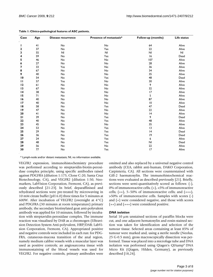
Table 1: Clinico-pathological features of ASC patients.
Case Age Disease recurrence Presence of metastasis* Follow-up (months) Life status
1 41 No No 64 Alive2 37 No Yes 22 Alive3 55 NI NI NI NI4 59 No Yes 16 Dead5 46 No No 107 Alive6 27 No No 28 Alive7 33 Yes No 36 Alive8 67 No No 34 Alive9 40 No No 35 Alive10 54 No Yes 48 Dead11 57 Yes No 30 Alive12 61 No No 9 Alive13 67 No No 32 Alive14 38 No No 17 Alive15 71 No No 5 Alive16 40 No No 87 Alive17 40 No No 10 Alive18 58 No Yes 47 Dead19 47 No No 22 Alive20 41 No No 14 Alive21 39 No Yes 9 Dead22 40 No Yes 48 Alive23 45 No No 35 Alive24 53 No Yes 13 Alive25 29 No Yes 14 Dead26 36 No Yes 19 Dead27 24 No Yes 6 Alive28 48 No Yes NI Dead29 56 No No 22 Alive30 77 No Yes 17 Alive
* Lymph-node and/or distant metastasis; NI, no information available.
Page 3 of 8(page number not for citation purposes)

BMC Cancer 2009, 9:212 http://www.biomedcentral.com/1471-2407/9/212
Screening for EGFR and PDGFRA MutationsScreening for EGFR (exons 18–21) and PDGFRA (exons12, 14 and 18) mutations was done by PCR – single-strand conformational polymorphism (PCR-SSCP), aspreviously described [21,22,25]. Samples showing amobility shift different from the normal pattern weredirectly sequenced (Stabvida, Investigation and Servicesin Biological Sciences Lda, Oeiras, Portugal), as described[25]. All positive cases were confirmed twice with a newand independent PCR amplification, followed by directsequencing.
Statistical analysisData were stored and analyzed using the SPSS statisticalsoftware (version 16.0, SPSS Inc., Chicago, IL). All com-parisons were examined for statistical significance usingPearson's chi-square (χ2) test and Fisher's exact test (whenn < 5), being threshold for significance p values < 0.05.Survival curve was plotted using the method of Kaplanand Meier and data compared using the log-rank test.
ResultsAssessment of EGFR, PDGFRA and VEGFR2 overexpres-sion and activating gene mutations was performed in acohort of 30 adenosquamous carcinomas of the uterinecervix. The results are summarized in Table 2 and aredetailed bellow.
EGFR profileWe have previously found that approximately 43% (13/30) of cases were positive (2+/3+) for EGFR immunohis-tochemistry (Figure 1A) [20]. In order to determine themolecular basis of such overexpression, we have per-formed a mutation analysis of cases. No activating muta-tions in the hotspot region (exons 18–21) of EGFR genewere identified. Nevertheless, a silent base substitution(CAG>CAA) in EGFR exon 20 at codon 787 (Q787Q) wasfound in 17 cases (56%) (Table 3).
No statistically significant correlations were observedbetween EGFR gene alteration and the clinico-pathologi-cal parameters (data not shown).
Table 2: Molecular alterations of EGFR, PDGFRA and VEGFR2 in ASC patients.
Case EGFR PDGFRA VEGFR2
Mutations IHC* Mutations IHC IHC
1 Q787Q - P567P; V824V; IVS18-50insA +++ ++2 Normal +++ Normal +++ -3 Q787Q +++ V824V; IVS18-50insA +++ +++4 Q787Q +++ V824V; IVS18-50insA +++ +++5 Q787Q +++ V824V; IVS18-50insA +++ +++6 Normal + Normal +++ +++7 Q787Q +++ P567P; IVS14+3G>A +++ ++8 Q787Q - Normal +++ +++9 Normal - Normal +++ +++10 Normal +++ P567P +++ +++11 Normal - P567P np +++12 Q787Q - Normal +++ +++13 Q787Q - Normal ++ +++14 Normal ++ Normal +++ ++15 Q787Q + Normal +++ +++16 Normal +++ P567P +++ +17 Q787Q - P567P ++ +++18 Normal - Normal +++ +19 Q787Q +++ Normal +++ ++20 Q787Q - Normal +++ +++21 Normal + P567P np +++22 Normal ++ P567P +++ +23 Normal +++ Normal ++ ++24 Q787Q - Normal ++ +25 Q787Q + IVS14+49G>A +++ +26 Q787Q - P567P +++ +++27 Normal - Normal np ++28 Q787Q ++ Normal ++ +29 Q787Q - Normal +++ +30 Normal +++ Normal ++ +++
IHC: Immunohistochemistry; *: previously reported in [20]; np: not possible due to tissue limitation.
Page 4 of 8(page number not for citation purposes)
BMC Cancer 2009, 9:212 http://www.biomedcentral.com/1471-2407/9/212
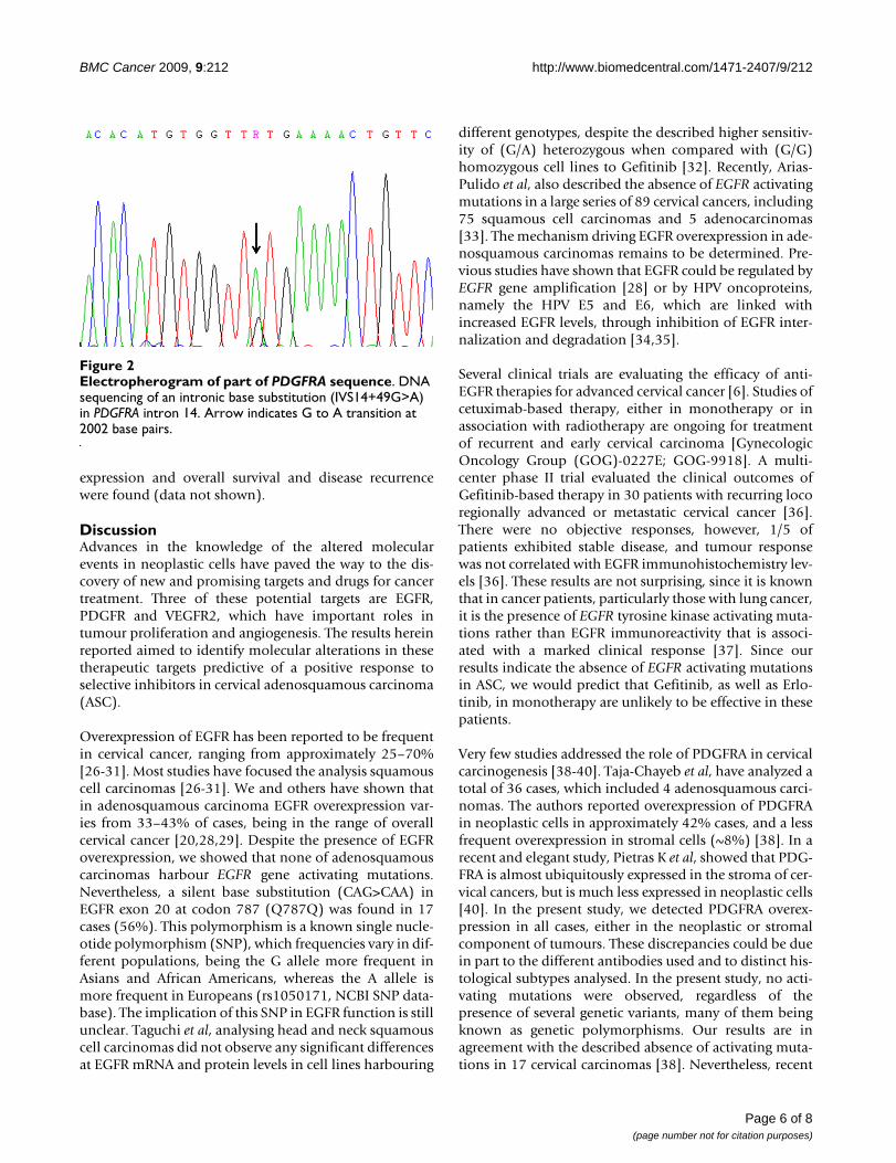
PDGFRA profilePDGFRA immunohistochemical analysis was only possi-ble in 27 out of 30 cases, all of them being positive forPDGFRA expression. Six cases showed moderate positivity(2+) and 21 showed strong positivity (3+) (Figure 1B).Positive immunoreactions were observed in the cyto-plasm of malignant cells, and were consistently observedin the stromal component, staining fibroblasts andendothelial cells.
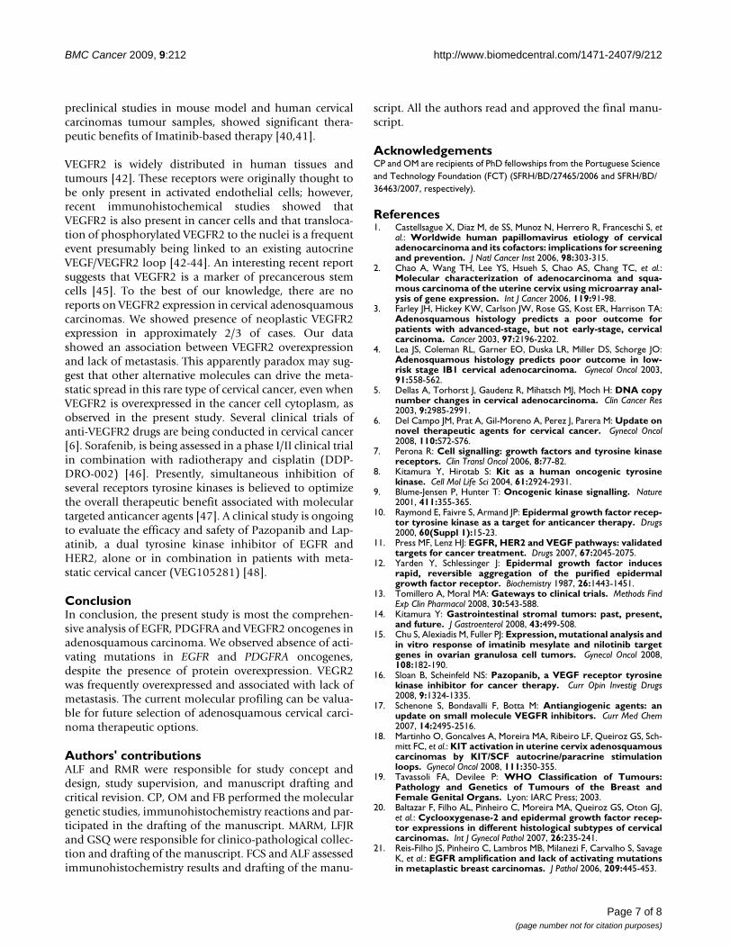
Mutation analysis of PDGFRA hotspot exons 12, 14 and18 of all 30 cases, revealed absence of activating muta-tions. Yet, we observed several silent mutations, such as abase substitution in exon 12 (CCA>CCG) at codon 567(P567P) in 9 cases and in exon 18 (GTC>GTT) at codon824 (V824V) in 4 cases (Table 2). We also observed basesubstitutions in intron 14 (IVS14+3G>A andIVS14+49G>A) (Figure 2), in two different cases, and inintron 18 (IVS18-50insA) in 4 cases (Table 3).
Since all cases were positive for PDGFRA expression, anal-ysis of correlations with the clinico-pathological data wasnot performed.
VEGFR2 profileVEGFR2 immunohistochemical positivity was observedin 22 of 30 cases (73.3%), with 6 cases showing moderateexpression (2+) and 16 cases presenting strong positiveimmunoreaction (3+). Positive immunoreactions wereexclusively observed in the cytoplasm of malignant cells.No stromal positive reaction was identified, but faint pos-itive staining was observed in a few endothelial cells sur-rounding the neoplastic cells (Figure 1C).
Correlation analysis between VEGFR2 expression andclinico-pathological data revealed a significant associa-tion between VEGFR2 overexpression and lack of metasta-sis (p = 0.038). No associations between VEGFR2
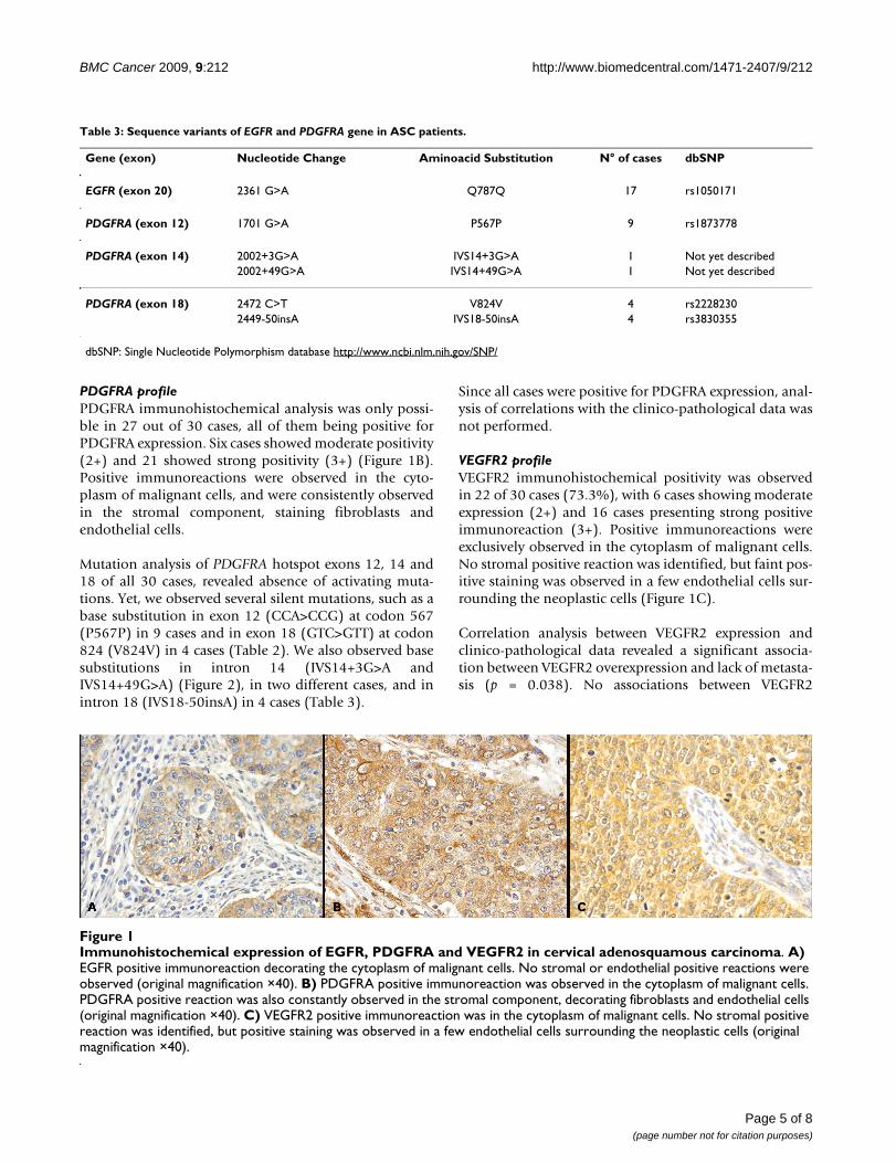
Table 3: Sequence variants of EGFR and PDGFRA gene in ASC patients.
Gene (exon) Nucleotide Change Aminoacid Substitution N° of cases dbSNP
EGFR (exon 20) 2361 G>A Q787Q 17 rs1050171
PDGFRA (exon 12) 1701 G>A P567P 9 rs1873778
PDGFRA (exon 14) 2002+3G>A IVS14+3G>A 1 Not yet described2002+49G>A IVS14+49G>A 1 Not yet described
PDGFRA (exon 18) 2472 C>T V824V 4 rs22282302449-50insA IVS18-50insA 4 rs3830355
dbSNP: Single Nucleotide Polymorphism database http://www.ncbi.nlm.nih.gov/SNP/
Immunohistochemical expression of EGFR, PDGFRA and VEGFR2 in cervical adenosquamous carcinomaFigure 1Immunohistochemical expression of EGFR, PDGFRA and VEGFR2 in cervical adenosquamous carcinoma. A) EGFR positive immunoreaction decorating the cytoplasm of malignant cells. No stromal or endothelial positive reactions were observed (original magnification ×40). B) PDGFRA positive immunoreaction was observed in the cytoplasm of malignant cells. PDGFRA positive reaction was also constantly observed in the stromal component, decorating fibroblasts and endothelial cells (original magnification ×40). C) VEGFR2 positive immunoreaction was in the cytoplasm of malignant cells. No stromal positive reaction was identified, but positive staining was observed in a few endothelial cells surrounding the neoplastic cells (original magnification ×40).
Page 5 of 8(page number not for citation purposes)
BMC Cancer 2009, 9:212 http://www.biomedcentral.com/1471-2407/9/212
expression and overall survival and disease recurrencewere found (data not shown).
DiscussionAdvances in the knowledge of the altered molecularevents in neoplastic cells have paved the way to the dis-covery of new and promising targets and drugs for cancertreatment. Three of these potential targets are EGFR,PDGFR and VEGFR2, which have important roles intumour proliferation and angiogenesis. The results hereinreported aimed to identify molecular alterations in thesetherapeutic targets predictive of a positive response toselective inhibitors in cervical adenosquamous carcinoma(ASC).
Overexpression of EGFR has been reported to be frequentin cervical cancer, ranging from approximately 25–70%[26-31]. Most studies have focused the analysis squamouscell carcinomas [26-31]. We and others have shown thatin adenosquamous carcinoma EGFR overexpression var-ies from 33–43% of cases, being in the range of overallcervical cancer [20,28,29]. Despite the presence of EGFRoverexpression, we showed that none of adenosquamouscarcinomas harbour EGFR gene activating mutations.Nevertheless, a silent base substitution (CAG>CAA) inEGFR exon 20 at codon 787 (Q787Q) was found in 17cases (56%). This polymorphism is a known single nucle-otide polymorphism (SNP), which frequencies vary in dif-ferent populations, being the G allele more frequent inAsians and African Americans, whereas the A allele ismore frequent in Europeans (rs1050171, NCBI SNP data-base). The implication of this SNP in EGFR function is stillunclear. Taguchi et al, analysing head and neck squamouscell carcinomas did not observe any significant differencesat EGFR mRNA and protein levels in cell lines harbouring
different genotypes, despite the described higher sensitiv-ity of (G/A) heterozygous when compared with (G/G)homozygous cell lines to Gefitinib [32]. Recently, Arias-Pulido et al, also described the absence of EGFR activatingmutations in a large series of 89 cervical cancers, including75 squamous cell carcinomas and 5 adenocarcinomas[33]. The mechanism driving EGFR overexpression in ade-nosquamous carcinomas remains to be determined. Pre-vious studies have shown that EGFR could be regulated byEGFR gene amplification [28] or by HPV oncoproteins,namely the HPV E5 and E6, which are linked withincreased EGFR levels, through inhibition of EGFR inter-nalization and degradation [34,35].
Several clinical trials are evaluating the efficacy of anti-EGFR therapies for advanced cervical cancer [6]. Studies ofcetuximab-based therapy, either in monotherapy or inassociation with radiotherapy are ongoing for treatmentof recurrent and early cervical carcinoma [GynecologicOncology Group (GOG)-0227E; GOG-9918]. A multi-center phase II trial evaluated the clinical outcomes ofGefitinib-based therapy in 30 patients with recurring locoregionally advanced or metastatic cervical cancer [36].There were no objective responses, however, 1/5 ofpatients exhibited stable disease, and tumour responsewas not correlated with EGFR immunohistochemistry lev-els [36]. These results are not surprising, since it is knownthat in cancer patients, particularly those with lung cancer,it is the presence of EGFR tyrosine kinase activating muta-tions rather than EGFR immunoreactivity that is associ-ated with a marked clinical response [37]. Since ourresults indicate the absence of EGFR activating mutationsin ASC, we would predict that Gefitinib, as well as Erlo-tinib, in monotherapy are unlikely to be effective in thesepatients.
Very few studies addressed the role of PDGFRA in cervicalcarcinogenesis [38-40]. Taja-Chayeb et al, have analyzed atotal of 36 cases, which included 4 adenosquamous carci-nomas. The authors reported overexpression of PDGFRAin neoplastic cells in approximately 42% cases, and a lessfrequent overexpression in stromal cells (~8%) [38]. In arecent and elegant study, Pietras K et al, showed that PDG-FRA is almost ubiquitously expressed in the stroma of cer-vical cancers, but is much less expressed in neoplastic cells[40]. In the present study, we detected PDGFRA overex-pression in all cases, either in the neoplastic or stromalcomponent of tumours. These discrepancies could be duein part to the different antibodies used and to distinct his-tological subtypes analysed. In the present study, no acti-vating mutations were observed, regardless of thepresence of several genetic variants, many of them beingknown as genetic polymorphisms. Our results are inagreement with the described absence of activating muta-tions in 17 cervical carcinomas [38]. Nevertheless, recent
Electropherogram of part of PDGFRA sequenceFigure 2Electropherogram of part of PDGFRA sequence. DNA sequencing of an intronic base substitution (IVS14+49G>A) in PDGFRA intron 14. Arrow indicates G to A transition at 2002 base pairs.
Page 6 of 8(page number not for citation purposes)
BMC Cancer 2009, 9:212 http://www.biomedcentral.com/1471-2407/9/212
preclinical studies in mouse model and human cervicalcarcinomas tumour samples, showed significant thera-peutic benefits of Imatinib-based therapy [40,41].
VEGFR2 is widely distributed in human tissues andtumours [42]. These receptors were originally thought tobe only present in activated endothelial cells; however,recent immunohistochemical studies showed thatVEGFR2 is also present in cancer cells and that transloca-tion of phosphorylated VEGFR2 to the nuclei is a frequentevent presumably being linked to an existing autocrineVEGF/VEGFR2 loop [42-44]. An interesting recent reportsuggests that VEGFR2 is a marker of precancerous stemcells [45]. To the best of our knowledge, there are noreports on VEGFR2 expression in cervical adenosquamouscarcinomas. We showed presence of neoplastic VEGFR2expression in approximately 2/3 of cases. Our datashowed an association between VEGFR2 overexpressionand lack of metastasis. This apparently paradox may sug-gest that other alternative molecules can drive the meta-static spread in this rare type of cervical cancer, even whenVEGFR2 is overexpressed in the cancer cell cytoplasm, asobserved in the present study. Several clinical trials ofanti-VEGFR2 drugs are being conducted in cervical cancer[6]. Sorafenib, is being assessed in a phase I/II clinical trialin combination with radiotherapy and cisplatin (DDP-DRO-002) [46]. Presently, simultaneous inhibition ofseveral receptors tyrosine kinases is believed to optimizethe overall therapeutic benefit associated with moleculartargeted anticancer agents [47]. A clinical study is ongoingto evaluate the efficacy and safety of Pazopanib and Lap-atinib, a dual tyrosine kinase inhibitor of EGFR andHER2, alone or in combination in patients with meta-static cervical cancer (VEG105281) [48].
ConclusionIn conclusion, the present study is most the comprehen-sive analysis of EGFR, PDGFRA and VEGFR2 oncogenes inadenosquamous carcinoma. We observed absence of acti-vating mutations in EGFR and PDGFRA oncogenes,despite the presence of protein overexpression. VEGR2was frequently overexpressed and associated with lack ofmetastasis. The current molecular profiling can be valua-ble for future selection of adenosquamous cervical carci-noma therapeutic options.
Authors' contributionsALF and RMR were responsible for study concept anddesign, study supervision, and manuscript drafting andcritical revision. CP, OM and FB performed the moleculargenetic studies, immunohistochemistry reactions and par-ticipated in the drafting of the manuscript. MARM, LFJRand GSQ were responsible for clinico-pathological collec-tion and drafting of the manuscript. FCS and ALF assessedimmunohistochemistry results and drafting of the manu-
script. All the authors read and approved the final manu-script.
AcknowledgementsCP and OM are recipients of PhD fellowships from the Portuguese Science and Technology Foundation (FCT) (SFRH/BD/27465/2006 and SFRH/BD/36463/2007, respectively).
References1. Castellsague X, Diaz M, de SS, Munoz N, Herrero R, Franceschi S, et
al.: Worldwide human papillomavirus etiology of cervicaladenocarcinoma and its cofactors: implications for screeningand prevention. J Natl Cancer Inst 2006, 98:303-315.
2. Chao A, Wang TH, Lee YS, Hsueh S, Chao AS, Chang TC, et al.:Molecular characterization of adenocarcinoma and squa-mous carcinoma of the uterine cervix using microarray anal-ysis of gene expression. Int J Cancer 2006, 119:91-98.
3. Farley JH, Hickey KW, Carlson JW, Rose GS, Kost ER, Harrison TA:Adenosquamous histology predicts a poor outcome forpatients with advanced-stage, but not early-stage, cervicalcarcinoma. Cancer 2003, 97:2196-2202.
4. Lea JS, Coleman RL, Garner EO, Duska LR, Miller DS, Schorge JO:Adenosquamous histology predicts poor outcome in low-risk stage IB1 cervical adenocarcinoma. Gynecol Oncol 2003,91:558-562.
5. Dellas A, Torhorst J, Gaudenz R, Mihatsch MJ, Moch H: DNA copynumber changes in cervical adenocarcinoma. Clin Cancer Res2003, 9:2985-2991.
6. Del Campo JM, Prat A, Gil-Moreno A, Perez J, Parera M: Update onnovel therapeutic agents for cervical cancer. Gynecol Oncol2008, 110:S72-S76.
7. Perona R: Cell signalling: growth factors and tyrosine kinasereceptors. Clin Transl Oncol 2006, 8:77-82.
8. Kitamura Y, Hirotab S: Kit as a human oncogenic tyrosinekinase. Cell Mol Life Sci 2004, 61:2924-2931.
9. Blume-Jensen P, Hunter T: Oncogenic kinase signalling. Nature2001, 411:355-365.
10. Raymond E, Faivre S, Armand JP: Epidermal growth factor recep-tor tyrosine kinase as a target for anticancer therapy. Drugs2000, 60(Suppl 1):15-23.
11. Press MF, Lenz HJ: EGFR, HER2 and VEGF pathways: validatedtargets for cancer treatment. Drugs 2007, 67:2045-2075.
12. Yarden Y, Schlessinger J: Epidermal growth factor inducesrapid, reversible aggregation of the purified epidermalgrowth factor receptor. Biochemistry 1987, 26:1443-1451.
13. Tomillero A, Moral MA: Gateways to clinical trials. Methods FindExp Clin Pharmacol 2008, 30:543-588.
14. Kitamura Y: Gastrointestinal stromal tumors: past, present,and future. J Gastroenterol 2008, 43:499-508.
15. Chu S, Alexiadis M, Fuller PJ: Expression, mutational analysis andin vitro response of imatinib mesylate and nilotinib targetgenes in ovarian granulosa cell tumors. Gynecol Oncol 2008,108:182-190.
16. Sloan B, Scheinfeld NS: Pazopanib, a VEGF receptor tyrosinekinase inhibitor for cancer therapy. Curr Opin Investig Drugs2008, 9:1324-1335.
17. Schenone S, Bondavalli F, Botta M: Antiangiogenic agents: anupdate on small molecule VEGFR inhibitors. Curr Med Chem2007, 14:2495-2516.
18. Martinho O, Goncalves A, Moreira MA, Ribeiro LF, Queiroz GS, Sch-mitt FC, et al.: KIT activation in uterine cervix adenosquamouscarcinomas by KIT/SCF autocrine/paracrine stimulationloops. Gynecol Oncol 2008, 111:350-355.
19. Tavassoli FA, Devilee P: WHO Classification of Tumours:Pathology and Genetics of Tumours of the Breast andFemale Genital Organs. Lyon: IARC Press; 2003.
20. Baltazar F, Filho AL, Pinheiro C, Moreira MA, Queiroz GS, Oton GJ,et al.: Cyclooxygenase-2 and epidermal growth factor recep-tor expressions in different histological subtypes of cervicalcarcinomas. Int J Gynecol Pathol 2007, 26:235-241.
21. Reis-Filho JS, Pinheiro C, Lambros MB, Milanezi F, Carvalho S, SavageK, et al.: EGFR amplification and lack of activating mutationsin metaplastic breast carcinomas. J Pathol 2006, 209:445-453.
Page 7 of 8(page number not for citation purposes)
http://www.ncbi.nlm.nih.gov/entrez/query.fcgi?cmd=Retrieve&db=PubMed&dopt=Abstract&list_uids=3494473
http://www.ncbi.nlm.nih.gov/entrez/query.fcgi?cmd=Retrieve&db=PubMed&dopt=Abstract&list_uids=3494473
BMC Cancer 2009, 9:212 http://www.biomedcentral.com/1471-2407/9/212
Publish with BioMed Central and every scientist can read your work free of charge
"BioMed Central will be the most significant development for disseminating the results of biomedical research in our lifetime."
Sir Paul Nurse, Cancer Research UK
Your research papers will be:
available free of charge to the entire biomedical community
peer reviewed and published immediately upon acceptance
cited in PubMed and archived on PubMed Central
yours — you keep the copyright
Submit your manuscript here:http://www.biomedcentral.com/info/publishing_adv.asp
BioMedcentral
22. Carvalho I, Milanezi F, Martins A, Reis RM, Schmitt F: Overexpres-sion of platelet-derived growth factor receptor alpha inbreast cancer is associated with tumour progression. BreastCancer Res 2005, 7:R788-R795.
23. Longatto FA, Costa SM, Milanezi F, Montruccoli D, Montruccoli GC,Baltazar F, et al.: Immunohistochemical expression of VEGF-Aand its ligands in non-neoplastic lesions of the breast sam-pling-assisted by dynamic angiothermography. Oncol Rep2007, 18:1201-1206.
24. Basto D, Trovisco V, Lopes JM, Martins A, Pardal F, Soares P, et al.:Mutation analysis of B-RAF gene in human gliomas. Acta Neu-ropathol (Berl) 2005, 109:207-210.
25. Gomes AL, Gouveia A, Capelinha AF, de la CD, Silva P, Reis RM, etal.: Molecular alterations of KIT and PDGFRA in GISTs: eval-uation of a Portuguese series. J Clin Pathol 2008, 61:203-208.
26. Cho NH, Kim YB, Park TK, Kim GE, Park K, Song KJ: P63 and EGFRas prognostic predictors in stage IIB radiation-treated cervi-cal squamous cell carcinoma. Gynecol Oncol 2003, 91:346-353.
27. Kristensen GB, Holm R, Abeler VM, Trope CG: Evaluation of theprognostic significance of cathepsin D, epidermal growthfactor receptor, and c-erbB-2 in early cervical squamous cellcarcinoma. An immunohistochemical study. Cancer 1996,78:433-440.
28. Kersemaekers AM, Fleuren GJ, Kenter GG, Broek LJ Van den, UljeeSM, Hermans J, et al.: Oncogene alterations in carcinomas ofthe uterine cervix: overexpression of the epidermal growthfactor receptor is associated with poor prognosis. Clin CancerRes 1999, 5:577-586.
29. Hale RJ, Buckley CH, Gullick WJ, Fox H, Williams J, Wilcox FL: Prog-nostic value of epidermal growth factor receptor expressionin cervical carcinoma. J Clin Pathol 1993, 46:149-153.
30. Kim GE, Kim YB, Cho NH, Chung HC, Pyo HR, Lee JD, et al.: Syn-chronous coexpression of epidermal growth factor receptorand cyclooxygenase-2 in carcinomas of the uterine cervix: apotential predictor of poor survival. Clin Cancer Res 2004,10:1366-1374.
31. Shen L, Shui Y, Wang X, Sheng L, Yang Z, Xue D, et al.: EGFR andHER2 expression in primary cervical cancers and corre-sponding lymph node metastases: implications for targetedradiotherapy. BMC Cancer 2008, 8:232.
32. Taguchi T, Tsukuda M, Imagawa-Ishiguro Y, Kato Y, Sano D: Involve-ment of EGFR in the response of squamous cell carcinoma ofthe head and neck cell lines to gefitinib. Oncol Rep 2008,19:65-71.
33. Arias-Pulido H, Joste N, Chavez A, Muller CY, Dai D, Smith HO, etal.: Absence of epidermal growth factor receptor mutationsin cervical cancer. Int J Gynecol Cancer 2008, 18:749-754.
34. Straight SW, Hinkle PM, Jewers RJ, McCance DJ: The E5 oncopro-tein of human papillomavirus type 16 transforms fibroblastsand effects the downregulation of the epidermal growth fac-tor receptor in keratinocytes. J Virol 1993, 67:4521-4532.
35. Zhang B, Srirangam A, Potter DA, Roman A: HPV16 E5 proteindisrupts the c-Cbl-EGFR interaction and EGFR ubiquitina-tion in human foreskin keratinocytes. Oncogene 2005,24:2585-2588.
36. Goncalves A, Fabbro M, Lhomme C, Gladieff L, Extra JM, Floquet A,et al.: A phase II trial to evaluate gefitinib as second- or third-line treatment in patients with recurring locoregionallyadvanced or metastatic cervical cancer. Gynecol Oncol 2008,108:42-46.
37. Lynch TJ, Bell DW, Sordella R, Gurubhagavatula S, Okimoto RA,Brannigan BW, et al.: Activating mutations in the epidermalgrowth factor receptor underlying responsiveness of non-small-cell lung cancer to gefitinib. N Engl J Med 2004,20;350:2129-2139.
38. Taja-Chayeb L, Chavez-Blanco A, Martinez-Tlahuel J, Gonzalez-FierroA, Candelaria M, Chanona-Vilchis J, et al.: Expression of plateletderived growth factor family members and the potentialrole of imatinib mesylate for cervical cancer. Cancer Cell Int2006, 6:22.
39. Ryu SY, Oka K, Tsujii H, Suzuki Y, Nakano T: Platelet-derivedendothelial cell growth factor as a prognostic factor for radi-otherapy outcome in patients with adenocarcinoma of theuterine cervix. Gynecol Oncol 2003, 89:414-419.
40. Pietras K, Pahler J, Bergers G, Hanahan D: Functions of paracrinePDGF signaling in the proangiogenic tumor stroma revealedby pharmacological targeting. PLoS Med 2008, 5:e19.
41. Kummel S, Heidecke H, Brock B, Denkert C, Hecktor J, Koninger A,et al.: [Imatinib – a possible therapeutic option for cervicalcarcinoma: results of a preclinical phase I study]. GynakolGeburtshilfliche Rundsch 2008, 48:94-100.
42. Stewart M, Turley H, Cook N, Pezzella F, Pillai G, Ogilvie D, et al.:The angiogenic receptor KDR is widely distributed in humantissues and tumours and relocates intracellularly on phos-phorylation. An immunohistochemical study. Histopathology2003, 43:33-39.
43. Giatromanolaki A, Koukourakis MI, Turley H, Sivridis E, Harris AL,Gatter KC: Phosphorylated KDR expression in endometrialcancer cells relates to HIF1alpha/VEGF pathway and unfa-vourable prognosis. Mod Pathol 2006, 19:701-707.
44. Fox SB, Turley H, Cheale M, Blazquez C, Roberts H, James N, et al.:Phosphorylated KDR is expressed in the neoplastic and stro-mal elements of human renal tumours and shuttles from cellmembrane to nucleus. J Pathol 2004, 202:313-320.
45. Shen R, Ye Y, Chen L, Yan Q, Barsky SH, Gao JX: Precancerousstem cells can serve as tumor vasculogenic progenitors. PLoSONE 2008, 3:e1652.
46. A Phase I/II Study of Cisplatin and Radiation in CombinationWith Sorafenib in Cervical Cancer 2008 [http://clinicaltrials.gov/ct2/show/record/NCT00510250].
47. Faivre S, Djelloul S, Raymond E: New paradigms in anticancertherapy: targeting multiple signaling pathways with kinaseinhibitors. Semin Oncol 2006, 33:407-420.
48. National Ovarian Cancer Coalition Dallas – Ft Worth Chap-ter 2008 [http://www.dfwovarian.org/clinical_trials.html].
Pre-publication historyThe pre-publication history for this paper can be accessedhere:
http://www.biomedcentral.com/1471-2407/9/212/prepub
Page 8 of 8(page number not for citation purposes)
http://www.ncbi.nlm.nih.gov/entrez/query.fcgi?cmd=Retrieve&db=PubMed&dopt=Abstract&list_uids=8697388
http://www.ncbi.nlm.nih.gov/entrez/query.fcgi?cmd=Retrieve&db=PubMed&dopt=Abstract&list_uids=8697388
http://www.ncbi.nlm.nih.gov/entrez/query.fcgi?cmd=Retrieve&db=PubMed&dopt=Abstract&list_uids=8697388
http://www.ncbi.nlm.nih.gov/entrez/query.fcgi?cmd=Retrieve&db=PubMed&dopt=Abstract&list_uids=8459036
http://www.ncbi.nlm.nih.gov/entrez/query.fcgi?cmd=Retrieve&db=PubMed&dopt=Abstract&list_uids=8459036
http://www.ncbi.nlm.nih.gov/entrez/query.fcgi?cmd=Retrieve&db=PubMed&dopt=Abstract&list_uids=8459036
http://www.ncbi.nlm.nih.gov/entrez/query.fcgi?cmd=Retrieve&db=PubMed&dopt=Abstract&list_uids=8392596
http://www.ncbi.nlm.nih.gov/entrez/query.fcgi?cmd=Retrieve&db=PubMed&dopt=Abstract&list_uids=8392596
Related Documents