39 Riassunto Questo studio ha avuto l’obiettivo di raccogliere i dati relativi ai tipi di parvovirus canino (PVC) presenti in cani in Turchia. Sessantacinque campioni sono stati prelevati nel periodo aprile 2009-febbraio 2010. Lo studio ha coinvolto cani con segni clinici di infezione da parvovirus ed esemplari sani. I campioni raccolti sono stati testati per PCV usando la prova della reazione a catena della polimerasi. I 25 campioni (38,4%) risultati positivi sono stati successivamente caratterizzati con analisi di sequenza. I risultati hanno mostrato come entrambi i ceppi PVC-2a (17/25, 68%) e PVC-2b (8/25, 32%) circolassero in Turchia nel periodo del campionamento. Il presente è il primo studio sviluppato in Turchia concernente la caratterizzazione molecolare di ceppi di PVC, condotto su campioni prelevati da cani e basato sul sequenziamento parziale del gene VP2. Parole chiave Cane, Parvovirus canino 2, Reazione a catena della polimerasi, Sequenziamento, Turchia. Caratterizzazione molecolare dell’infezione da parvovirus canino in cani in Turchia Summary This study provides data about canine parvovirus (CPV) types circulating among dogs in Turkey. Sixty-five samples from dogs with and without clinical signs of parvovirus infection were collected between April 2009 and February 2010. The samples were subsequently tested for CPV using polymerase chain reaction (PCR). Twenty-five samples (38.4%) were positive; when positive samples were characterized by sequence analysis, results showed that both CPV-2a (17/25, 68%) and CPV-2b (8/25, 32%) strains are circulating among domestic dogs in Turkey. This is the first molecular characterization study of CPVs from dogs based on partial VP2 gene sequences in Turkey. Keywords Canine parvovirus 2, Polymerase chain reaction (PCR), Sequence Analysis, Turkey. Veterinaria Italiana 2015, 51 (1), 39-44. doi: 10.12834/VetIt.263.908.3 Accepted: 23.09.2014 | Available on line: 31.03.2015 1 Department of Virology, Faculty of Veterinary Medicine, University of Atatürk, 25240, Yakutiye, Erzurum, Turkey. 2 Department of Virology, Faculty of Veterinary Medicine, University of Ankara, 06110, Dışkapı, Ankara, Turkey. * Corresponding author at: Department of Virology, Faculty of Veterinary Medicine, University of Atatürk, 25240, Yakutiye, Erzurum, Turkey. Tel.: + 90 442 2315547, e-mail: [email protected]. Mehmet Özkan Timurkan 1* & Tuba Çiğdem Oğuzoğlu 2 Molecular characterization of canine parvovirus (CPV) infection in dogs in Turkey Within a few years from its emergence, 2 antigenic variants were detected and designated as CPV-2a (426Asn) and CPV-2b (426Asp) on the basis of the reactivity of monoclonal antibodies and amino acid conformation on the capsid protein (Decaro et al. 2008, Parrish et al. 1991). Both variants have completely replaced the original type 2, and they currently occur worldwide among the canine population (Calderon et al. 2009, Decaro et al. 2006, Decaro et al. 2007, Steinel et al. 1998). A third variant, first detected in Italy, was named CPV-2c (426Glu) (Buonavoglia et al. 2001). The first goal of this study was to determine the distribution of CPV infection among dog populations Introduction 1 Canine parvovirus (CPV) (carnivore protoparvovirus), a significant worldwide canine pathogen belonging to the family Parvoviridae, was first described in 1978 (Carmichael 2005). The highly contagious and principal etiological agent of hemorrhagic enteritis in dogs has been identified as canine parvovirus type 2 (CPV-2). Because CPV-2 is very similar to feline panleukopenia virus (FPLV), it has been argued that FPLV had mutated into CPV-2 (Decaro and Buonavoglia 2012). 1 The GenBank accession numbers for the sequences reported in this paper are KF500484–KF500508.

Welcome message from author
This document is posted to help you gain knowledge. Please leave a comment to let me know what you think about it! Share it to your friends and learn new things together.
Transcript

39
RiassuntoQuesto studio ha avuto l’obiettivo di raccogliere i dati relativi ai tipi di parvovirus canino (PVC) presenti in cani in Turchia. Sessantacinque campioni sono stati prelevati nel periodo aprile 2009-febbraio 2010. Lo studio ha coinvolto cani con segni clinici di infezione da parvovirus ed esemplari sani. I campioni raccolti sono stati testati per PCV usando la prova della reazione a catena della polimerasi. I 25 campioni (38,4%) risultati positivi sono stati successivamente caratterizzati con analisi di sequenza. I risultati hanno mostrato come entrambi i ceppi PVC-2a (17/25, 68%) e PVC-2b (8/25, 32%) circolassero in Turchia nel periodo del campionamento. Il presente è il primo studio sviluppato in Turchia concernente la caratterizzazione molecolare di ceppi di PVC, condotto su campioni prelevati da cani e basato sul sequenziamento parziale del gene VP2.
Parole chiaveCane,Parvovirus canino 2,Reazione a catena della polimerasi,Sequenziamento,Turchia.
Caratterizzazione molecolare dell’infezione da parvovirus caninoin cani in Turchia
SummaryThis study provides data about canine parvovirus (CPV) types circulating among dogs in Turkey. Sixty-five samples from dogs with and without clinical signs of parvovirus infection were collected between April 2009 and February 2010. The samples were subsequently tested for CPV using polymerase chain reaction (PCR). Twenty-five samples (38.4%) were positive; when positive samples were characterized by sequence analysis, results showed that both CPV-2a (17/25, 68%) and CPV-2b (8/25, 32%) strains are circulating among domestic dogs in Turkey. This is the first molecular characterization study of CPVs from dogs based on partial VP2 gene sequences in Turkey.
KeywordsCanine parvovirus 2,Polymerase chain reaction (PCR),Sequence Analysis,Turkey.
Veterinaria Italiana 2015, 51 (1), 39-44. doi: 10.12834/VetIt.263.908.3Accepted: 23.09.2014 | Available on line: 31.03.2015
1 Department of Virology, Faculty of Veterinary Medicine, University of Atatürk, 25240, Yakutiye, Erzurum, Turkey.2 Department of Virology, Faculty of Veterinary Medicine, University of Ankara, 06110, Dışkapı, Ankara, Turkey.
* Corresponding author at: Department of Virology, Faculty of Veterinary Medicine, University of Atatürk,25240, Yakutiye, Erzurum, Turkey.
Tel.: + 90 442 2315547, e-mail: [email protected].
Mehmet Özkan Timurkan1* & Tuba Çiğdem Oğuzoğlu2
Molecular characterization of canine parvovirus (CPV)infection in dogs in Turkey
Within a few years from its emergence, 2 antigenic variants were detected and designated as CPV-2a (426Asn) and CPV-2b (426Asp) on the basis of the reactivity of monoclonal antibodies and amino acid conformation on the capsid protein (Decaro et al. 2008, Parrish et al. 1991). Both variants have completely replaced the original type 2, and they currently occur worldwide among the canine population (Calderon et al. 2009, Decaro et al. 2006, Decaro et al. 2007, Steinel et al. 1998). A third variant, first detected in Italy, was named CPV-2c (426Glu) (Buonavoglia et al. 2001).
The first goal of this study was to determine the distribution of CPV infection among dog populations
Introduction1 Canine parvovirus (CPV) (carnivore protoparvovirus), a significant worldwide canine pathogen belonging to the family Parvoviridae, was first described in 1978 (Carmichael 2005). The highly contagious and principal etiological agent of hemorrhagic enteritis in dogs has been identified as canine parvovirus type 2 (CPV-2). Because CPV-2 is very similar to feline panleukopenia virus (FPLV), it has been argued that FPLV had mutated into CPV-2 (Decaro and Buonavoglia 2012).
1 The GenBank accession numbers for the sequences reported in this paper are KF500484–KF500508.
40 Veterinaria Italiana 2015, 51 (1), 39-44. doi: 10.12834/VetIt.263.908.3
CPV infection in Turkey Timurkan & Oğuzoğlu
in Turkey; while the second objective was to describe CPV molecular differentiation by sequence analysis of the VP2 gene region of the Turkish CPV-2 strains. These strains have then been compared to vaccine strains currently in use in Turkey.
Materials and methods
SamplesWe investigated faecal (n = 35) and buffy coat (n = 30) samples from 65 dogs. The samples were submitted to our laboratory (Department of Virology, Faculty of Veterinary Medicine, Ankara University, Turkey) by clinicians for routine processing between April 2009 and February 2010. The presence (n = 25) or absence (n = 10) of clinical symptoms characteristic of CPV infection were evaluated to determine the health status of each animal. However, follow-up information was not available. Clinical symptoms in the dogs included fever, anorexia, depression, gastrointestinal problems, and vomiting. In 4 cases, there were no data concerning the dog age. We also lacked data about the vaccination status of 13 dogs, and the breed of 11 of the tested animals; finally data regarding sex were also missing.
MethodsThe faecal samples were homogenized in phosphate buffered saline (PBS) and centrifuged at 3,000 rpm for 15 minutes. After centrifugation, the supernatant was collected for DNA extraction. Blood samples were centrifuged at 380 g for 10 minutes. For the buffy coat samples, 200 µl of the supernatant were collected and mixed with an equal volume of PBS. The DNA of each sample was extracted using the phenol-chloroform-isoamyl alcohol method described by Sambrok and colleagues (Sambrok et al. 1989).
Extracted faecal DNA was used as a template for PCR, which was in turn conducted with primers that recognize all known CPV and FPV strains. The PCR was also performed with primer pairs for amplification of the partial VP2 gene, as reported in literature (Buonavoglia et al. 2001, Pereira et al. 2000). The primers used in this study were designated Pabs-Pabas (680 bp) and Hfor-Hrev (629 bp). For both primer pairs, the temperature profile included an initial denaturing at 94 °C for 6 minutes, 35 cycles of denaturing at 94 °C for 1 minute, annealing at 52 °C for 1 minute, extension at 72 °C for 2 minutes, and a final extension at 72 °C for 10 minutes. Annealing temperatures were the same in both primers.
The amplicons from 680 bp (n = 10) PCR products and 629 bp (n = 15) PCR products were purified
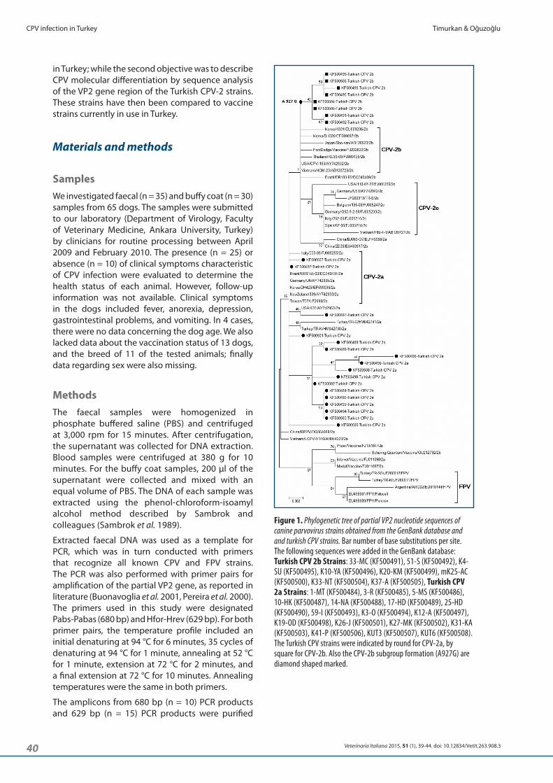
Figure 1. Phylogenetic tree of partial VP2 nucleotide sequences of canine parvovirus strains obtained from the GenBank database and and turkish CPV strains. Bar number of base substitutions per site. The following sequences were added in the GenBank database: Turkish CPV 2b Strains: 33-MC (KF500491), 51-S (KF500492), K4-SU (KF500495), K10-YA (KF500496), K20-KM (KF500499), mK25-AC (KF500500), K33-NT (KF500504), K37-A (KF500505), Turkish CPV 2a Strains: 1-MT (KF500484), 3-R (KF500485), 5-MS (KF500486), 10-HK (KF500487), 14-NA (KF500488), 17-HD (KF500489), 25-HD (KF500490), 59-I (KF500493), K3-O (KF500494), K12-A (KF500497), K19-OD (KF500498), K26-J (KF500501), K27-MK (KF500502), K31-KA (KF500503), K41-P (KF500506), KUT3 (KF500507), KUT6 (KF500508). The Turkish CPV strains were indicated by round for CPV-2a, by square for CPV-2b. Also the CPV-2b subgroup formation (A927G) are diamond shaped marked.

41Veterinaria Italiana 2015, 51 (1), 39-44. doi: 10.12834/VetIt.263.908.3
Timurkan & Oğuzoğlu CPV infection in Turkey
and sequenced. The sequencing was performed with a Beckman Coulter CEQ 8000 genetic analyzer (Beckman Coulter, Istanbul, Turkey) using the GenomeLab™ Methods Development Kit. Sequences were compared with other sequences available from the GenBank database (http://www.ncbi.nlm.nih.gov) and aligned with BioEdit software (version 7.0.5.3) using the ClustalW method (Hall 1999). The MEGA software program, version 5.1 (Tamura et al. 2011), was used to construct a phylogenetic tree using the maximum likelihood method, with bootstrap values calculated with 1,000 replicates (Figure 1).
ResultsAmplicons were detected in 38.4% (25/64) of the samples with Pabs-Pabas primer sets. Table I shows the age, vaccine status, sex, and breed information of the positive sampled animals. Among the positive samples, 57.1% (12/21) of the dogs were 1-3 months old and 23.8% (5/21) were 4-6 months old; 52.1%
Table I. Age, breed, sex, immunization status of Turkish dogs found positive to Canine parvovirus.
Sample No Sample ID Age Vaccinated Breed Sample Type Sex Genotype1 1-MT 3 m NDA NDA L Male 2a
2 3-R 4 m NDA NDA L Female 2a
3 5-MS 1 m NDA Kangal RS Male 2a
4 10-HK 2 m No Terrier L Male 2a
5 14-NA 1 m No NDA RS Female 2a
6 17-HD 2.5 m NDA Rottweiler L Male 2a
7 25-HD 8 m Yes Mix L Female 2a
8 33-MC 5 m NDA G. Retriever L Female 2b
9 51-S 2 m No NDA RS Male 2b
10 59-I 1 m NDA NDA L Female 2a
11 K3-O 1 m NDA NDA RS Female 2a
12 K4-SU 3 m NDA cocker RS Female 2b
13 K10-YA 7.5 m No Mix RS Male 2b
14 K12-A 8 m No Mix RS Male 2a
15 K19-OD 3 m No NDA L NDA 2a
16 K20-KM NDA NDA Mix L Male 2b
17 K25-AC 2 m NDA NDA L NDA 2b
18 K26-J NDA NDA Mix L Male 2a
19 K27-MK NDA NDA NDA RS Female 2a
20 K31-KA NDA NDA NDA RS Female 2a
21 K33-NT 5 m Yes NDA RS Female 2b
22 K37-A 5 m Yes Mix RS Female 2b
23 K41-P 8 m Yes G. Retriever RS Male 2a
24 KUT3 3 m No Mix RS Male 2a
25 KUT6 6 m No Mix RS Male 2aL = Leucocyte; RS = Rectal Swab; NDA = No data available; m = month.
(12/23) were male and 47.8% (11/23) were female. According to the data collected during the study by pet owners, 66.6% (8/12) of the dogs were not vaccinated and 33.3% (4/12) were vaccinated. Known breeds were kangal, terrier, rottweiler, golden retriever, and cocker spaniel (n = 6); 57.1% (8/14) of the dogs were of mixed breed, and there was no breed information regarding 11 other dogs.
The PCR fragments of the partial VP2 gene were successfully amplified with both primers from the faeces (n = 14) and buffy coat (n = 11) samples of different dogs. After analysis, the obtained sequences were compared with other reference CPV and FPLV strains stored in the GenBank database and with sequences of 4 canine (Vanguard, Pfizer; Nobivac, Intervet; Parvodog, Merial; Quantum, Schering) and 2 feline (Felocell, Pfizer and Purevax, Merial) vaccine strains. These vaccines, which are also included in the phylogenetic tree, are commonly used in Turkey. From 25 CPV-2 positive samples, 17 were identified as CPV-2a and 8 as CPV-2b. No CPV-2c variants were found. Table II shows the targeted segment of the
42 Veterinaria Italiana 2015, 51 (1), 39-44. doi: 10.12834/VetIt.263.908.3
CPV infection in Turkey Timurkan & Oğuzoğlu
CPV-2a and CPV-2b strains were placed in different branches. Most of the Turkish CPV-2a sequences were placed as a separate group within reference CPV-2a sequences; on the other hand, all the Turkish CPV-2b strains were located in a completely different branch of the phylogenetic tree than the reference 2b strains.
DiscussionThere have been few studies on CPV infections in dogs in Turkey (Yılmaz et al. 2005, Yeşilbağ et al. 2007), and no study describing CPV molecular characteristics has been provided so far. In a previous study (Muz et al. 2012), which investigated the 426th amino acid for detection of CPV-2 types in cats, CPV-2a and CPV-2c were detected as variants.
VP2 protein and the 8 variable positions that were detected when compared to reference sequences.
As shown in Table II, some substitutions were observed in all of our CPV-2a strains, specifically, 297 (SerAla), 300 (AlaGly), 305 (AspTyr), and 375 (AsnAsp). We also found a substitution at 426 (AsnAsp), which is known as an indicator of the CPV-2 variant CPV-2b. Similar nucleotide variation (A927G) was detected in all Turkish CPV-2b strains. A mutation in residue 440 (ThrAla) was detected in 5 samples (Table II). The nucleotide identities of our strains were 98.3-99.8%, with each other, and 97.8-99.7% with vaccine strains (data not shown).
The phylogenetic tree (Figure 1) was constructed using partial VP2 nucleotide sequences of canine parvovirus strains obtained from the GenBank database and Turkish CPV strains. The Turkish
Table II. Variable nucleotides in the VP2 gene partial sequences of the Canine parvovirus strains found in Turkish dogs.
Nt. positiona
aa residue3675
aa2973685
aa3003699
aa3053869
aa3613912
aa3754062
aa4264105
aa4404494
aa571M38245 T C G G A A A A
1-MT G G T A G - G -
3-R G G T A G - G -
5-MS G G T - G - - -
10-HK G G T A G - - -
14-NA G G T A G - - G
17-HD G G T A G - - -
25-HD G G T A G - - -
33-MC G G T - G G - C
51-S G G T - G G - C
59-I G G T A G - G -
K3-O G G T A G - G -
K4-SU G G T - G G - -
K10-YA G G T - G G - -
K12-A G G T - G - - -
K19-OD G G T A G - - -
K20-KM G G T - G G - -
K25-AC G G T - G G - -
K26-J G G T - G - - -
K27-MK G G T A G - - -
K31-KA G G T A G - G -
K33-NT G G T - G G - -
K37-A G G T - G G - -
K41-P G G T - G - - G
KUT3 G G T - G - - -
KUT6 G G T A G - - -
aa. change
TCTGCT
SA
GCTGGT
AG
GATTAT
DY
CGGCGA
RR
AATGAT
ND
AATGAT
ND
ACAGCA
TA
GTAGTG/CVV
a = Nucleotide positions are referred to the sequences of CPV-2 strain CPV-b (accession no. M38245).
43Veterinaria Italiana 2015, 51 (1), 39-44. doi: 10.12834/VetIt.263.908.3
Timurkan & Oğuzoğlu CPV infection in Turkey
CPV-2a and CPV-2b were also detected in this study as variants in dogs for the same amino acid.
We studied partial-length VP2 gene sequences of CPV to determine the distribution of CPV infection and investigate the genetic variability of CPV strains circulating in Turkey and their correspondence with currently used vaccine strains. Our sequence analysis indicated that CPV-2a is the most common variant, although CPV-2b variant is also detected sporadically. It is noteworthy that the variant 2c was not detected in our samples.
The amino acid residues determined in our study were variable. We detected residue 440 (Thr440Ala) in some (5/17, 29.4%) CPV-2a strains (1-MT, 3-R, 59-I, K3-O, and K31-KA); residue 440 is known to be an important factor for antigenicity (Decaro et al. 2009). Severe diarrhoea was noted as clinical findings in those 5 samples. Therefore, we suggest further studies in order to better understand the relationship between these residues and the severity of clinical symptoms. The other altered amino acid residues in circulating strains in Turkey were found at the VP2 gene (positions 297, 300, 305, and 375) according to the M38245 strain. In addition, the A927G nucleotide variation may
cause subgroup formation in CPV-2b; therefore, this variation should also be investigated.
According to Turkish veterinary policy, dogs are vaccinated at 2 months of age; the vaccines routinely used include variants of CPV 2 and 2b.
The strains of canine parvovirus obtained from cats in Turkey occupy a completely different branch in the phylogenetic tree from the ones that we obtained from dog. In this study, Turkish CPV-2a and 2b strains from dogs grouped together in a completely different branch of the phylogenetic tree than the vaccine strains. Although CPV-2a variant was determined in field strains, it is not present in commercial vaccine strains. This finding can be used for molecular comparisons of vaccine and field strains, and it may be helpful in further vaccine development studies (Decaro et al. 2007, Nandi et al. 2010). In addition, the usual variants of strains in a region or a country should be clarified before choosing a vaccine.
The control strategies for CPV infections in Turkey can be improved by means of further molecular studies using additional samples from different subgroups. It is also necessary to continue the periodic investigation of CPV strains from dogs.

44 Veterinaria Italiana 2015, 51 (1), 39-44. doi: 10.12834/VetIt.263.908.3
CPV infection in Turkey Timurkan & Oğuzoğlu
Buonavoglia C., Martella V., Pratelli A., Tempesta M., Cavalli A., Buonavoglia D., Bozzo G., Elia G., Decaro N. & Carmichael L. 2001. Evidence for evolution of canine parvovirus type-2 in Italy. J Gen Virol, 82, 1555-1560.
Calderon M.G., Mattion N., Bucafusco D., Fogel F., Remorini P. & La Torre J. 2009. Molecular characterization of canine parvovirus strains in Argentina: detection of the pathogenic variant CPV2c in vaccinated dogs. J Virol Methods, 159, 141-145.
Carmichael LE. 2005. An annotated historical account of canine parvovirus. J Vet Med B Infect Dis Vet Public Health, 52, 303-311.
Decaro N., Elia G., Desario C., Roperto S., Martella V., Campolo M., Lorusso A., Cavalli A. & Buonavoglia C. 2006. A minor Groove binder probe real-time PCR assay for discrimination between type 2-based vaccines and field strains of canine parvovirus. J Virol Methods, 136, 65-70.
Decaro N., Desario C., Addie D.D., Martella V., Vieira M.J., Elia G., Zicola A., Davis C., Thompson G., Thiry E., Truyen U. & Buonavoglia C. 2007. Molecular epidemiology of canine parvovirus in Europe. Emerg Infect Dis, 13, 1222-1224.
Decaro N., Desario C., Elia G., Martella V., Mari V., Lavazza A., Nardi M. & Buonavoglia C. 2008. Evidence for immunisation failure in vaccinated adult dogs infected with canine parvovirus type 2c. New Microbiol, 31, 125-130.
Decaro N., Desario C., Parisi A., Martella V., Lorusso A., Miccolupo A., Mari V., Colaianni M.L., Cavalli A., Di Trani L. & Buonavoglia C. 2009. Genetic analysis of canine parvovirus type 2c. Virology, 385 (1), 5-10.
Decaro N. & Buonavoglia C. 2012. Canine parvovirus - a review of epidemiological and diagnostic aspects, with emphasis on type 2c. Vet Microbiol, 155 (1), 1-12.
Hall T.A. 1999. BioEdit: a user-friendly biological sequence alignment editor and analysis program for Windows 95/98/NT. Nucleic Acids Symp Ser, 41, 95-98.
References
Muz D., Oğuzoğlu T.C., Timurkan M.O. & Akın H. 2012. Characterization of the partial VP2 gene region of canine parvoviruses in domestic cats from Turkey. Virus Genes, 44, 301-308.
Nandi S., Anbazhagan R. & Kumar M. 2010. Molecular characterisation and nucleotide sequence analysis of canine parvovirus strains in vaccines in India. Vet Ital, 46, 69-81.
Parrish C.R., Aquadro C.F., Strassheim M.L., Evermann J.F., Sgro J.Y. & Mohammed H.O. 1991. Rapid antigenic-type replacement and DNA sequence evolution of canine parvovirus. J Virol, 65, 6544-6552.
Pereira C.A., Monezi T.A., Mehnert D.U., D’Angelo M. & Durigon E.L. 2000. Molecular characterization of canine parvovirus in Brazil by polymerase chain reaction assay. Vet Microbiol, 75, 127-133.
Steinel A., Venter E.H., van Vuuren M. & Truyen U. 1998. Antigenic and genetic analysis of canine parvoviruses in southern Africa. J Vet Res, 65, 239-242.
Sambrook J., Fritsch E.F. & Maniatis T. 1989. Molecular Cloning: a laboratory manual. 2nd ed. N.Y., Cold Spring Harbor Laboratory, Cold Spring Harbor Laboratory Press.
Tamura K., Peterson D., Peterson N., Stecher G., Nei M. & Kumar S. 2011. MEGA5: molecular evolutionary genetics analysis using maximum likelihood, evolutionary distance, and maximum parsimony methods. Mol Biol Evol, 28, 2731-2739.
Yılmaz Z., Pratelli A. & Torun S. 2005. Distribution of antigen types of canine parvovirus type 2 in dogs with hemorrhagic enteritis in Turkey. Turk J Vet Anim Sci, 29, 1073-1076.
Yeşilbağ K., Yilmaz Z., Ozkul A. & Pratelli A. 2007. Aetiological role of viruses in puppies with diarrhoea. Vet Rec, 161, 169-170.
Related Documents
![Prevalence of Canine Parvovirus in Domestic Dogs …...A blood profile typically shows marked thrombocytopenia and leucopenia [15, 23, 25, 26]. Prevalence of CPV differs from one geographical](https://static.cupdf.com/doc/110x72/5f7b76b785c7f11b071fcf8d/prevalence-of-canine-parvovirus-in-domestic-dogs-a-blood-profile-typically-shows.jpg)