Molecular and Cellular Biochemistry 276: 71–80, 2005. c ❣ Springer 2005 Free radical scavenging activity of Cleome gynandra L. leaves on adjuvant induced arthritis in rats R.T. Narendhirakannan, 1 S. Subramanian 2 and M. Kandaswamy 1 1 Department of Inorganic Chemistry, University of Madras, Chennai, India; 2 Department of Advanced Biochemistry, University of Madras, Chennai, India Received 6 January 2005; accepted 3 March 2005 Abstract The generation of free radicals has been implicated in the causation of several diseases of known and unknown etiologies such as, rheumatoid arthritis, diabetes, cancer, etc., and compounds that can scavenge free radicals have great potential in ameliorating these disease processes. The present study was aimed to investigate the possible anti-oxidant potential of Cleome gynandra leaf extract at a dose of 150 mg/kg body weight for 30 days on adjuvant induced arthritis in experimental rats. Oral administration of C. gynandra leaf extract significantly increased the levels of lipid peroxides and activities of catalase, glutathione peroxidase and decreased the levels of reduced glutathione and superoxide dismutase activity in arthritis induced rats. The free radical scavenging activity of the plant was further evidenced by histological observations made on the limb tissue. The presence of biologically active ingredients and vital trace elements in the leaves readily account for free radical scavenging property of C. gynandra. (Mol Cell Biochem 276: 71–80, 2005) Key words: arthritis, anti-oxidants, Cleome gynandra, cat’s whiskers, free radicals, reactive oxygen species Abbreviation: ROS, reactive oxygen species; RA, rheumatoid arthritis; NSAIDs, non steroidal anti-inflammatory drugs; WHO, World Health Organization; FCA, Freund’s complete adjuvant; LPO, lipid peroxidation; TBARS, thiobarbituric acid reactive substances; SOD, superoxide dismutase; CAT, catalase; GPx, glutathione peroxidase; GSH, reduced glutathione Introduction Free radical formation occurs continuously in the cells as a consequence of both enzymatic and non-enzymatic reactions. Enzymatic reactions, which serve as source of free radicals, include those involved in the respiratory chain, phagocytosis, prostaglandin synthesis, and in cytochrome p450 system. Free radicals arise in non-enzymatic reactions of oxygen with organic compounds as well as those initiated by ionizing radiations. Reactive oxygen species (ROS) are an important part of the defense mechanisms against infection, but excessive generation of free oxygen radicals Address for offprints: M. Kandaswamy, Professor and Head, Department of Inorganic Chemistry, University of Madras, Guindy Campus, Chennai 600025, Tamilnadu, India (E-mail: [email protected]) may damage tissues, forming lipid peroxides (LPO) and has been implicated in the pathogenesis of many diseases such as cancer, diabetes, arthritis, etc [1]. Among the ROS, the hydroxyl radicals are the most reactive that it can act on any type of molecule. However, their primary target appears to be polyunsaturated fatty acids, which are the major constituents of plasma membrane [2]. Rheumatoid arthritis (RA) is a common human autoim- mune disease characterized by chronic inflammation of the synovial joints and by subsequent progressive, erosive destruction of articular tissue. It affects about 1% of the human population [3]. RA has been associated not only with

Welcome message from author
This document is posted to help you gain knowledge. Please leave a comment to let me know what you think about it! Share it to your friends and learn new things together.
Transcript
Molecular and Cellular Biochemistry 276: 71–80, 2005. c�Springer 2005
Free radical scavenging activity of Cleomegynandra L. leaves on adjuvant inducedarthritis in rats
R.T. Narendhirakannan,1 S. Subramanian2 and M. Kandaswamy1
1Department of Inorganic Chemistry, University of Madras, Chennai, India; 2Department of Advanced Biochemistry,University of Madras, Chennai, India
Received 6 January 2005; accepted 3 March 2005
Abstract
The generation of free radicals has been implicated in the causation of several diseases of known and unknown etiologies such as,rheumatoid arthritis, diabetes, cancer, etc., and compounds that can scavenge free radicals have great potential in amelioratingthese disease processes. The present study was aimed to investigate the possible anti-oxidant potential of Cleome gynandra leafextract at a dose of 150 mg/kg body weight for 30 days on adjuvant induced arthritis in experimental rats. Oral administrationof C. gynandra leaf extract significantly increased the levels of lipid peroxides and activities of catalase, glutathione peroxidaseand decreased the levels of reduced glutathione and superoxide dismutase activity in arthritis induced rats. The free radicalscavenging activity of the plant was further evidenced by histological observations made on the limb tissue. The presence ofbiologically active ingredients and vital trace elements in the leaves readily account for free radical scavenging property of C.gynandra. (Mol Cell Biochem 276: 71–80, 2005)
Key words: arthritis, anti-oxidants, Cleome gynandra, cat’s whiskers, free radicals, reactive oxygen species
Abbreviation: ROS, reactive oxygen species; RA, rheumatoid arthritis; NSAIDs, non steroidal anti-inflammatory drugs; WHO,World Health Organization; FCA, Freund’s complete adjuvant; LPO, lipid peroxidation; TBARS, thiobarbituric acid reactivesubstances; SOD, superoxide dismutase; CAT, catalase; GPx, glutathione peroxidase; GSH, reduced glutathione
Introduction
Free radical formation occurs continuously in the cellsas a consequence of both enzymatic and non-enzymaticreactions. Enzymatic reactions, which serve as source offree radicals, include those involved in the respiratory chain,phagocytosis, prostaglandin synthesis, and in cytochromep450 system. Free radicals arise in non-enzymatic reactionsof oxygen with organic compounds as well as those initiatedby ionizing radiations. Reactive oxygen species (ROS)are an important part of the defense mechanisms againstinfection, but excessive generation of free oxygen radicals
Address for offprints: M. Kandaswamy, Professor and Head, Department of Inorganic Chemistry, University of Madras, Guindy Campus, Chennai 600025,Tamilnadu, India (E-mail: [email protected])
may damage tissues, forming lipid peroxides (LPO) andhas been implicated in the pathogenesis of many diseasessuch as cancer, diabetes, arthritis, etc [1]. Among the ROS,the hydroxyl radicals are the most reactive that it can acton any type of molecule. However, their primary targetappears to be polyunsaturated fatty acids, which are themajor constituents of plasma membrane [2].
Rheumatoid arthritis (RA) is a common human autoim-mune disease characterized by chronic inflammation ofthe synovial joints and by subsequent progressive, erosivedestruction of articular tissue. It affects about 1% of thehuman population [3]. RA has been associated not only with
72
free radical formation but also with low anti-oxidant status.[4]. The role of reactive oxygen species in the pathogenesisof degenerative joint disease has already been documented[5, 6]. Both steroidal and non-steroidal anti-inflammatorydrugs currently used for the amelioration of the symptomsof the disease offer only temporary relief and often causesevere side effects like peptic ulcer and renal failure[7]. Therefore, new drugs without side effects are beingstudied all over the world as an alternate to NSAIDs andopiates.
According to WHO, still 80% of the world population relyon plant drugs and WHO has also recommended the evalu-ation of the effectiveness of plants in conditions where welack safe modern drugs [8].
Cleome gynandra (Cat’s whiskers), a common weed thatgrows throughout India, West Africa, Tanzania, Uganda,and Nigeria is an erect herbaceous annual herb which haslong been used as a house hold remedy for a variety of ail-ments including inflammation [9]. Its leaves contain exces-sive amounts of proteins, vitamins (A and C) and minerals.The leaves and seeds of Cat’s whiskers are used in many coun-tries for earache, epileptic fits, stomachache, constipation andinflammation. [10–13], fresh leaves of Cleome gynandra areused in Ayurveda and Siddha medicine for a variety of diseaseconditions [14]. The claimed medicinal effects of C. gynan-dra have not been investigated using controlled experimentsin detail.
Adjuvant induced arthritis (AIA) in experimental rats; achronic inflammatory disease characterized by infiltration ofthe synovial membrane and associated with destruction ofthe joints resembles closely to the human rheumatoid arthritis[15]. The present investigation was aimed to evaluate the freeradical scavenging activity of C. gynandra in AIA rats.
Materials and methods
Plant materials
Fresh C. gynandra leaves were collected from a rural re-gion of Dindigul district, Tamil Nadu, India and the plantwas identified by Prof. V. Kaviyarasan, Centre for AdvancedStudies in Botany, University of Madras, Chennai, India andthe voucher specimen of the plant is being retained in thedepartment herbarium (No. 1228).
Phytochemical screening
Phytochemical screening was carried out in various solventextracts (ethanol, petroleum ether, chloroform and aque-ous) to ascertain the qualitative composition of secondarymetabolites like sugars, phenolic compounds, tannins, an-
throquinones, flavonoids, saponins, steroids, resins, lectins,glycosides and alkaloids by employing coloration and pre-cipitation test [16]. From these, ethanol extract was found tocontain most of the active components and hence the samewas used for further experimentation.
Preparation of Cleome gynandra leaf extract
The leaves were chopped and subjected to extraction with95% ethanol by Soxhlet apparatus for 15 h. Nearly 85% ofthe solvent was recovered by distillation over a boiling wa-ter bath at atmospheric pressure and the remaining underreduced pressure (temperature at 40–50 ◦C). The yield was3.2 g/100 g. The extract was stored in a refrigerator until fur-ther use.
Test animals
Male albino rats of Wistar strain weighing around 160–180 gwere procured from Tamil Nadu Veterinary and Animal Sci-ence University, Chennai for the present study. The animalswere housed in solid-bottomed polypropylene cages and ac-climatized to animal house conditions. The rats were fed withcommercial rat diet (Hindustan Lever Limited, Mumbai, In-dia) and water ad libitum. The experiments were designed andconducted in accordance with the ethical norms approved byMinistry of Social Justices and Empowerment, Governmentof India and Institutional Animal Ethical Committee Guide-lines (IAEC). Once arthritis developed, food was served onthe bottom of the cages as severely arthritic rats have diffi-culty in feeding from the cage top.
Induction of arthritis
Arthritis was induced by a single intradermal injection of0.1 ml of Freund’s complete adjuvant (FCA) containing10 mg/ml dry heat-killed Mycobacterium tuberculosis permilliliter sterile paraffin oil (Difco Laboratories) into afoot pad of the left hind paw of male rats [17]. A glasssyringe (1 ml) with the locking hubs and a 26 G needlewas used for injection. The rats were anesthetized withether inhalation prior to and during adjuvant injection, asthe very viscous nature of the adjuvant exert difficultywhile injecting. The swelling in hind paws were periodicallyexamined in each paw from the ankle using plethysmography[18].
Experimental design
Animals were divided into four groups of six animals in eachgroup as follows:
73
Group I: Control ratsGroup II: Control rats administered with extract of C. gynan-
dra leaf. (150 mg/kg body weight/rat/day for 30 days)Group III: Adjuvant induced arthritic ratsGroup IV: Arthritis induced rats administered with extract
of C. gynandra leaf. (150 mg/kg body weight/rat/day for30 days by intubations starting 10 days after adjuvant in-jection.)
Measurement of paw volume
The paw volume was measured using plethysmography. Dur-ing the experimental period, scoring of paw was restricted to2–3 times a week to avoid excessive handling of the animalsas this can reduce the severity or incidence of arthritis afteradjuvant injection and the paw oedema was calculated.
Preparation of tissue homogenate
Joint cartilage tissue samples were homogenized in a solutioncontaining 5% trichloroacetic acid and 5 mM EDTA at 4 ◦Cand centrifuged for 10 min at 15,000 g in 4 ◦C [19].
Histological studies
Hind limbs were removed and fixed in 10% buffered forma-lin. The limbs were decalcified in 15% formic acid, processedfor paraffin embedding, sectioned 5 µm thickness and sub-sequently stained with haematoxylin-eosin for histologicalexamination [20].
Biochemical assays
Thiobarbituric acid reactive substances (TBARS)
Lipid peroxides (TBARS) in plasma and tissue homogenatewere estimated using thiobarbituric acid reactive substancesby the method of Ohkawa et al. [21]. To 0.2 ml of tissuehomogenate or plasma, 0.2 ml of 8.1% SDS, 1.5 ml of 20%acetic acid and 1.5 ml of 0.8% TBA were added. The mix-ture was diluted to 4 ml with water and then heated in a wa-ter bath at 95 ◦C for 60 min using glass ball as a condenser.After cooling, 1 ml of water and 5 ml of n-butanol/pyridinemixture were added and shaken vigorously. After centrifuga-tion at 4000 rpm for 10 min, the organic layer was taken andits absorbance at 532 nm was measured in a Shimadzu UVspectrophotometer. 1,1,3,3-tetramethoxypropane was used asstandard. The level of lipid peroxides was expressed as millimoles of TBA reactants/100 g of wet tissue and n mol/ml inplasma.
Measurement of glutathione (GSH)
Reduced glutathione was estimated by the method of Ellman[22]. A 0.1 ml of tissue homogenate or plasma was precipi-tated with 5% TCA. The contents were mixed well for com-plete precipitation of proteins and centrifuged. To an aliquotof clear supernatant, 2.0 ml of 0.6 mM DTNB reagent and0.2 M phosphate buffer (pH 8.6) were added to obtain a finalvolume of 4.0 ml. The absorbance was read at 412 nm againsta blank containing 5% TCA instead of sample. A series ofstandards treated in a similar way were also run to determinethe glutathione content. The amount of glutathione was ex-pressed as mg/100 g wet tissue in milligram and mg/dl inplasma.
Measurement of superoxide dismutase (SOD) (EC 1.15.1.1)
SOD was assayed by the method of Misra and Fridovich[23]. A 0.1 ml of tissue homogenate was added to the tubecontaining 0.75 ml ethanol and 0.15 ml chloroform (chilled inice) and centrifuged. To 0.5 ml of supernatant, added 0.5 mlof 0.6 mM EDTA solution and 1 ml of 0.1 M carbonate–bicarbonate buffer (pH 10.2). The reaction was initiated bythe addition of 0.5 ml of 1.8 mM epinephrine and the increasein absorbance at 480 nm. The enzyme activity is expressedas 50% inhibition of epinephrine autooxidation/min.
Measurement of glutathione peroxidase (GPx) (EC 1.11.1.9)
GPx was assayed by the method of Rotruck et al. [24]. Thereaction mixture consisting 0.2 ml of 0.8 mM EDTA, 0.1 mlof 10 mM sodium azide, 0.1 ml of 2.5 mM H2O2, 0.2 ml of4 mM reduced glutathione, 0.4 ml of 0.4 M phosphate buffer(pH 7.0), and 0.2 ml homogenate was incubated at 37 ◦C for10 min. The reaction was arrested by the addition of 0.5 mlof 10% TCA and the tubes were centrifuged at 2000 rpm.To the supernatant, 3 ml of 0.3 M disodium hydrogenphosphate and 1.0 ml of 0.04% DTNB were added and thecolour developed was read at 420 nm immediately. Theactivity of GPx was expressed as micromoles of glutathioneoxidized/min/mg protein.
Measurement of catalase (EC 1.11.1.6)
CAT was assayed by the method of Takahara et al. [25]. To1.2 ml of 50 mM phosphate buffer (pH 7.0), 0.2 ml of thetissue homogenate was added and the enzyme reaction wasstarted by the addition of 1.0 ml of 30 mM H2O2 solution.The decrease in absorbance was measured at 240 nm at 30 secintervals for 3 min. The enzyme blank was run simultaneouslywith 1.0 ml of distilled water instead of hydrogen peroxide.
74
The enzyme activity is expressed as micromoles of H2O2
decomposed/min/mg protein.
Vitamin E
Vitamin E was estimated by the method of Desai [26]. To1.0 ml of Plasma 1.0 ml of ethanol was added and thor-oughly mixed. Then 3 ml of petroleum ether was added,shaken rapidly and centrifuged. Two millilitres of supernatantwas taken and evaporated to dryness. To this 0.2 ml of 0.2%bathophenanthoraline was added. The assay mixture was pro-tected from light and 0.2 ml of 0.001 M ferric chloride wasadded followed by 0.2 ml of 0.001 M o-phosphoric acid. Thetotal volume was made up to 3 ml with ethanol. The colourdeveloped was read at 530 nm. The level of vitamin E wasexpressed as mg/dl of plasma.
Vitamin C
Vitamin C was determined by the method of Omaye et al.[27]. To 0.5 ml of plasma, 0.5 ml of water and 1 ml of 5% TCAwere added, mixed thoroughly and centrifuged. To 1 ml of thesupernatant, 0.2 ml of DTC reagent was added and incubatedat 37 ◦C for 3 h. Then 1.5 ml of 65% sulphuric acid was added,mixed well and the solution was allowed to stand at roomtemperature for another 30 min. The colour developed wasread at 520 nm. The level of vitamin C expressed as mg/dl ofplasma.
Ceruloplasmin
Ceruloplasmin was estimated by the method of Ravin [28].A total of 0.1 ml of plasma was taken in to three 15 ml testtubes, one for control and two for tests respectively. Onemillilitre of 0.02% sodium azide was added to the controltube only. Then 8.0 ml of 0.1 m acetate buffer (pH 5.2) wasadded to all the tubes, followed by the addition of 1.0 ml of0.1% p-phenyl diamine. The solution was mixed thoroughlyand placed in a water bath at 37 ◦C for 1 h. After incubationthe tubes containing test solution. The contents were shakenwell and cooled at 4 ◦C for 30 min. the intensity of the colourdeveloped was measured at 530 nm. The ceruloplasmin con-tent was expressed as mg/dl of plasma.
Estimation of protein
Protein was estimated by the method of Lowry et al. [29]. To0.1 ml of tissue homogenate, 0.9 ml of water and 4.5 ml ofalkaline copper reagent were added and kept at room temper-ature for 10 min. Then 0.5 ml of Folin’s reagent was added
and the colour developed was read after 20 min at 640 nm.The level of protein was expressed as mg/g of tissue.
Statistical analysis
All the grouped data were statistically evaluated withSPSS/10 software. Hypothesis testing methods included oneway analysis of variance (ANOVA) followed by least signif-icant difference (LSD) test. P values of less than 0.05 wereconsidered to indicate statistical significance. All the resultswere expressed as mean±S.D. for six animals in each group.
Results
Paw volume
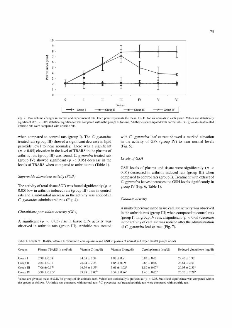
The paw volume changes in normal and experimental groupsof rats are shown in Fig. 1. In arthritis induced rats (group III),there was an appreciable increase in paw volume in the in-jected hind legs within 3–5 days. The rats also exhibited a de-crease in body weight gain during this initial period. By thistime the tail had become noticeably thickened and in somerats spondylitis developed. A significant (p < 0.05) increaseof paw volume was continued up to 20th day, and the increasein volume was stabilized after 20th day. The thickness wasretained till the end of experimental period in AIA rats (groupIII). Decrease in paw volume was noticed from the third weekof treatment and a significant reduction in paw volume wasobserved after the fourth week of C. gynandra treatment ingroup IV rats.
Histological studies
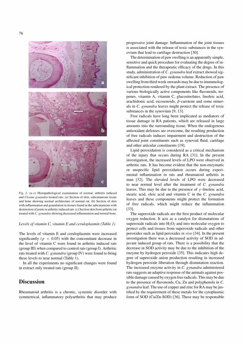
Histopathological studies revealed marked infiltration ofleukocytes and eosinophilic inflammatory exudates in thesynovial membrane (Fig. 2). Fig. 2a show the normal archi-tecture of skin, subcutaneous tissue and bone of control rat(group I). Fig. 2b illustrate the altered architecture in arthri-tis induced rat. The observations include a massive influxof inflammatory cells, synovial hyperplasia, accumulation ofabundant monomorphonuclear and polymorphonuclear cellsin the joint space and congestion of vessels (group III). Fig. 2cshow significant regeneration of synocytes and disappearanceof inflammatory exudates, mild focal infiltration of cells insynovial region, few cuboidal cells lining the synovial mem-brane in C. gynandra extract treated rat (group IV).
Concentration of TBARS
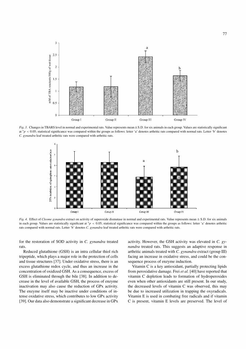
Figure 3 elucidates the significant (p < 0.05) increase in thelevel of tissue TBARS in arthritis induced rats (group III)
75
Fig. 1. Paw volume changes in normal and experimental rats. Each point represents the mean ± S.D. for six animals in each group. Values are statisticallysignificant at ∗p < 0.05; statistical significance was compared within the groups as follows: aArthritic rats compared with normal rats. bC. gynandra leaf treatedarthritic rats were compared with arthritic rats.
when compared to control rats (group I). The C. gynandratreated rats (group III) showed a significant decrease in lipidperoxide level to near normalcy. There was a significant(p < 0.05) elevation in the level of TBARS in the plasma ofarthritic rats (group III) was found. C. gynandra treated rats(group IV) showed significant (p < 0.05) decrease in thelevels of TBARS when compared to arthritic rats (Table 1).
Superoxide dismutase activity (SOD)
The activity of total tissue SOD was found significantly (p <
0.05) low in arthritis induced rats (group III) than in controlrats and a substantial increase in the activity was noticed inC. gynandra administered rats (Fig. 4).
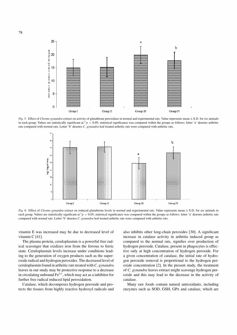
Glutathione peroxidase activity (GPx)
A significant (p < 0.05) rise in tissue GPx activity wasobserved in arthritic rats (group III). Arthritic rats treated
Table 1. Levels of TBARS, vitamin E, vitamin C, ceruloplasmin and GSH in plasma of normal and experimental groups of rats
Groups Plasma TBARS (n mol/ml) Vitamin C (mg/dl) Vitamin E (mg/dl) Ceruloplasmin (mg/dl) Reduced glutathione (mg/dl)
Group I 2.99 ± 0.38 24.38 ± 2.34 1.82 ± 0.81 0.83 ± 0.02 29.40 ± 1.92
Group II 2.84 ± 0.31 25.04 ± 2.26 1.85 ± 0.09 0.86 ± 0.06 28.64 ± 2.51
Group III 7.06 ± 0.97a 16.59 ± 1.53a 3.61 ± 1.02a 1.89 ± 0.07a 20.05 ± 2.33a
Group IV 3.96 ± 0.8.5b 19.28 ± 2.05b 2.54 ± 0.96b 1.46 ± 0.05b 25.70 ± 2.28b
Values are given as mean ± S.D. for groups of six animals each. Values are statistically significant at ∗p < 0.05. Statistical significance was compared withinthe groups as follows. aArthritic rats compared with normal rats. bC. gynandra leaf treated arthritic rats were compared with arthritic rats.
with C. gynandra leaf extract showed a marked elevationin the activity of GPx (group IV) to near normal levels(Fig. 5).
Levels of GSH
GSH levels of plasma and tissue were significantly (p <
0.05) decreased in arthritis induced rats (group III) whencompared to control rats (group I). Treatment with extract ofC. gynandra leaves increases the GSH levels significantly ingroup IV (Fig. 6, Table 1).
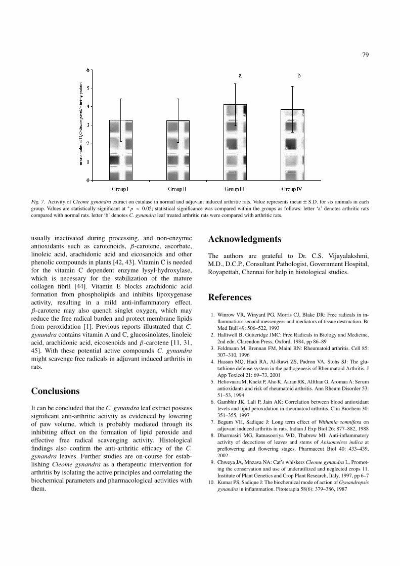
Catalase activity
A marked increase in the tissue catalase activity was observedin the arthritic rats (group III) when compared to control rats(group I). In group IV rats, a significant (p < 0.05) decreasein the activity of catalase was noticed after the administrationof C. gynandra leaf extract (Fig. 7).
76
Fig. 2. (a–c) Histopathological examination of normal, arthritis inducedand Cleome gynandra treated rats. (a) Section of skin, subcutaneous tissueand bone showing normal architecture of normal rat. (b) Section of skinwith inflammation and granulation in tissues found in the subcutaneous withdestruction of joints in arthritic induced rats. (c) Section skin from arthritic rattreated with C. gynandra showing decreased inflammation and normal bone.
Levels of vitamin C, vitamin E and ceruloplasmin (Table 1)
The levels of vitamin E and ceruloplasmin were increasedsignificantly (p < 0.05) with the concomitant decrease inthe level of vitamin C were found in arthritis induced rats(group III) when compared to control rats (group I). Arthriticrats treated with C. gynandra (group IV) were found to bringthese levels to near normal (Table 1).
In all the experiments no significant changes were foundin extract only treated rats (group II).
Discussion
Rheumatoid arthritis is a chronic, systemic disorder withsymmetrical, inflammatory polyarthritis that may produce
progressive joint damage. Inflammation of the joint tissuesis associated with the release of toxic substances in the syn-ovium that lead to cartilage destruction [30].
The determination of paw swelling is an apparently simple,sensitive and quick procedure for evaluating the degree of in-flammation and the therapeutic efficacy of the drugs. In thisstudy, administration of C. gynandra leaf extract showed sig-nificant inhibition of paw oedema volume. Reduction of pawswelling from third week onwards may be due to immunolog-ical protection rendered by the plant extract. The presence ofvarious biologically active components like flavonoids, ter-penes, vitamin A, vitamin C, glucosinolates, linoleic acid,arachidonic acid, eicosenoids, β-carotene and some miner-als in C. gynandra leaves might protect the release of toxicsubstances in the synovium [9, 13].
Free radicals have long been implicated as mediators oftissue damage in RA patients, which are released in largeamounts into the surrounding tissue. When the endogenousantioxidant defenses are overcome, the resulting productionof free radicals induces impairment and destruction of theaffected joint constituents such as synovial fluid, cartilageand other articular constituents [19].
Lipid peroxidation is considered as a critical mechanismof the injury that occurs during RA [31]. In the presentinvestigation, the increased levels of LPO were observed inarthritic rats. It has become evident that the non-enzymaticor unspecific lipid peroxidation occurs during experi-mental inflammation in rats and rheumatoid arthritis inman [32]. The elevated levels of LPO were decreasedto near normal level after the treatment of C. gynandraleaves. This may be due to the presence of γ -linoleic acid,stearic acid, oleic acid and vitamin C in the C. gynandraleaves and these components might protect the formationof free radicals, which might reduce the inflammation[33].
The superoxide radicals are the first product of molecularoxygen reduction. It acts as a catalyst for dismutations ofsuperoxide radicals into H2O2 and into molecular oxygen toprotect cells and tissues from superoxide radicals and otherperoxides such as lipid peroxides in vivo [34]. In the presentinvestigation there was a decreased activity of SOD in ad-juvant induced group of rats. There is a possibility that thedecrease in SOD activity may be due to the inhibition of theenzyme by hydrogen peroxide [35]. This indicates high de-gree of superoxide anion production resulting in increasedhydrogen peroxide liberation through dismutation reaction.The increased enzyme activity in C. gynandra administeredrats suggests an adaptive response of the animals against pos-sible damage caused by oxygen free radicals. This may be dueto the presence of flavonoids, Cu, Zn and polyphenols in C.gynandra leaf. The use of copper and zinc for RA may be jus-tified by the requirement of these metals for the cytoplasmicform of SOD (Cu/Zn-SOD) [36]. These may be responsible
77
Fig. 3. Changes in TBARS level in normal and experimental rats. Value represents mean±S.D. for six animals in each group. Values are statistically significantat ∗p < 0.05; statistical significance was compared within the groups as follows: letter ‘a’ denotes arthritic rats compared with normal rats. Letter ‘b’ denotesC. gynandra leaf treated arthritic rats were compared with arthritic rats.
Fig. 4. Effect of Cleome gynandra extract on activity of superoxide dismutase in normal and experimental rats. Value represents mean ± S.D. for six animalsin each group. Values are statistically significant at ∗p < 0.05; statistical significance was compared within the groups as follows: letter ‘a’ denotes arthriticrats compared with normal rats. Letter ‘b’ denotes C. gynandra leaf treated arthritic rats were compared with arthritic rats.
for the restoration of SOD activity in C. gynandra treatedrats.
Reduced glutathione (GSH) is an intra cellular thiol richtripeptide, which plays a major role in the protection of cellsand tissue structures [37]. Under oxidative stress, there is anexcess glutathione redox cycle, and thus an increase in theconcentration of oxidized GSH. As a consequence, excess ofGSH is eliminated through the bile [38]. In addition to de-crease in the level of available GSH, the process of enzymeinactivation may also cause the reduction of GPx activity.The enzyme itself may be inactive under conditions of in-tense oxidative stress, which contributes to low GPx activity[39]. Our data also demonstrate a significant decrease in GPx
activity. However, the GSH activity was elevated in C. gy-nandra treated rats. This suggests an adaptive response inarthritic animals treated with C. gynandra extract (group III)facing an increase in oxidative stress, and could be the con-sequence process of enzyme induction.
Vitamin C is a key antioxidant, partially protecting lipidsfrom peroxidative damage. Frei et al. [40] have reported thatvitamin C depletion leads to formation of hydroperoxideseven when other antioxidants are still present. In our study,the decreased levels of vitamin C was observed, this maybe due to increased utilization in trapping the oxyradicals.Vitamin E is used in combating free radicals and if vitaminC is present, vitamin E levels are preserved. The level of
78
Fig. 5. Effect of Cleome gynandra extract on activity of glutathione peroxidase in normal and experimental rats. Value represents mean ± S.D. for six animalsin each group. Values are statistically significant at ∗ p < 0.05; statistical significance was compared within the groups as follows: letter ’a’ denotes arthriticrats compared with normal rats. Letter ’b’ denotes C. gynandra leaf treated arthritic rats were compared with arthritic rats.
Fig. 6. Effect of Cleome gynandra extract on reduced glutathione levels in normal and experimental rats. Value represents mean ± S.D. for six animals ineach group. Values are statistically significant at ∗p < 0.05; statistical significance was compared within the groups as follows: letter ‘a’ denotes arthritic ratscompared with normal rats. Letter ‘b’ denotes C. gynandra leaf treated arthritic rats were compared with arthritic rats.
vitamin E was increased may be due to decreased level ofvitamin C [41].
The plasma protein, ceruloplasmin is a powerful free rad-ical scavenger that oxidizes iron from the ferrous to ferricstate. Ceruloplasmin levels increase under conditions lead-ing to the generation of oxygen products such as the super-oxide radical and hydrogen peroxides. The decreased level ofceruloplasmin found in arthritic rats treated with C. gynandraleaves in our study may be protective response to a decreasein circulating unbound Fe2+, which may act as a inhibitor forfurther free radical induced lipid peroxidation.
Catalase, which decomposes hydrogen peroxide and pro-tects the tissues from highly reactive hydroxyl radicals and
also inhibits other long-chain peroxides [30]. A significantincrease in catalase activity in arthritis induced group ascompared to the normal rats, signifies over production ofhydrogen peroxide. Catalase, present in phagocytes is effec-tive only at high concentration of hydrogen peroxide. Fora given concentration of catalase, the initial rate of hydro-gen peroxide removal is proportional to the hydrogen per-oxide concentration [2]. In the present study, the treatmentof C. gynandra leaves extract might scavenge hydrogen per-oxide and this may lead to the decrease in the activity ofcatalase.
Many raw foods contain natural antioxidants, includingenzymes such as SOD, GSH, GPx and catalase, which are
79
Fig. 7. Activity of Cleome gynandra extract on catalase in normal and adjuvant induced arthritic rats. Value represents mean ± S.D. for six animals in eachgroup. Values are statistically significant at ∗ p < 0.05; statistical significance was compared within the groups as follows: letter ‘a’ denotes arthritic ratscompared with normal rats. letter ‘b’ denotes C. gynandra leaf treated arthritic rats were compared with arthritic rats.
usually inactivated during processing, and non-enzymicantioxidants such as carotenoids, β-carotene, ascorbate,linoleic acid, arachidonic acid and eicosanoids and otherphenolic compounds in plants [42, 43]. Vitamin C is neededfor the vitamin C dependent enzyme lysyl-hydroxylase,which is necessary for the stabilization of the maturecollagen fibril [44]. Vitamin E blocks arachidonic acidformation from phospholipids and inhibits lipoxygenaseactivity, resulting in a mild anti-inflammatory effect.β-carotene may also quench singlet oxygen, which mayreduce the free radical burden and protect membrane lipidsfrom peroxidation [1]. Previous reports illustrated that C.gynandra contains vitamin A and C, glucosinolates, linoleicacid, arachidonic acid, eicosenoids and β-carotene [11, 31,45]. With these potential active compounds C. gynandramight scavenge free radicals in adjuvant induced arthritis inrats.
Conclusions
It can be concluded that the C. gynandra leaf extract possesssignificant anti-arthritic activity as evidenced by loweringof paw volume, which is probably mediated through itsinhibiting effect on the formation of lipid peroxide andeffective free radical scavenging activity. Histologicalfindings also confirm the anti-arthritic efficacy of the C.gynandra leaves. Further studies are on-course for estab-lishing Cleome gynandra as a therapeutic intervention forarthritis by isolating the active principles and correlating thebiochemical parameters and pharmacological activities withthem.
Acknowledgments
The authors are grateful to Dr. C.S. Vijayalakshmi,M.D., D.C.P., Consultant Pathologist, Government Hospital,Royapettah, Chennai for help in histological studies.
References
1. Winrow VR, Winyard PG, Morris CJ, Blake DR: Free radicals in in-flammation: second messengers and mediators of tissue destruction. BrMed Bull 49: 506–522, 1993
2. Halliwell B, Gutteridge JMC: Free Radicals in Biology and Medicine,2nd edn. Clarendon Press, Oxford, 1984, pp 86–89
3. Feldmann M, Brennan FM, Maini RN: Rheumatoid arthritis. Cell 85:307–310, 1996
4. Hassan MQ, Hadi RA, Al-Rawi ZS, Padron VA, Stohs SJ: The glu-tathione defense system in the pathogenesis of Rheumatoid Arthritis. JApp Toxicol 21: 69–73, 2001
5. Heliovaara M, Knekt P, Aho K, Aaran RK, Alfthan G, Aromaa A: Serumantioxidants and risk of rheumatoid arthritis. Ann Rheum Disorder 53:51–53, 1994
6. Gambhir JK, Lali P, Jain AK: Correlation between blood antioxidantlevels and lipid peroxidation in rheumatoid arthritis. Clin Biochem 30:351–355, 1997
7. Begum VH, Sadique J: Long term effect of Withania somnifera onadjuvant induced arthritis in rats. Indian J Exp Biol 26: 877–882, 1988
8. Dharmasiri MG, Ratnasooriya WD, Thabrew MI: Anti-inflammatoryactivity of decoctions of leaves and stems of Anisomeless indica atpreflowering and flowering stages. Pharmaceut Biol 40: 433–439,2002
9. Chweya JA, Mnzava NA: Cat’s whiskers Cleome gynandra L. Promot-ing the conservation and use of underutilized and neglected crops 11.Institute of Plant Genetics and Crop Plant Research, Italy, 1997, pp 6–7
10. Kumar PS, Sadique J: The biochemical mode of action of Gynandropsisgynandra in inflammation. Fitoterapia 58(6): 379–386, 1987
80
11. Sreeramulu N: Chemical composition of some green leafy vegetablesgrown in Tanzania. J Plant Foods 4(3), 139–141, 1982
12. Narendhirakannan RT, Krishnakumari S, Subramanian S, KandaswamyM: The protective effect of Cleome gynandra leaf extract on adjuvantinduced arthritis in rats. Indian J Pharmacol 35: 410, 2003
13. Narendhirakannan RT, Subramanian S, Kandaswamy M: Anti-inflammatory activity of Cleome gynandra. L on hematological andcellular constituents in adjuvant induced arthritic rats. J Med Food 8(1):93–99, 2005
14. Krithikar KR, Basu BD: An ICS: Capparacea. In: B. Singh and M. P.Singh (eds.), Indian Medicinal Plants, Oriental Enterprises, Dehradun,1984, pp 251–253
15. Watlz DT, Drmartino JJ, Misher A: Adjuvant induced arthritis in rats. IIDrug effects on physiologic, biochemical and immunologic parameters.J Pharmacol Exp Ther 178: 223, 1971
16. Harbone JB: Phytochemical Methods. Chapman and Hall, London,1973, pp 52–105
17. Mizushima Y, Tsukada W, Akimoto T: A modification of rat adjuvantarthritis for testing anti-rheumatic drugs. J Pharm Pharmacol 24: 781–785, 1972
18. Winter CA, Risley EA, Nuss CW: Carageenan induced edema in hindpaw of the rats as an assay for anti-inflammatory drugs. Proc Soc ExpBio Med 111: 544–547, 1962
19. Campo GM, Angela A, Campo S, Ferlazzo AM, Altavilla D, CalatroniA: Efficacy of treatment with glycosaminoglycans on experimentalcollagen-induced arthritis in rats. Arthritis Res Ther 5: 122–131, 2003
20. Durie FH, Fava RA, Foy TM, Aruffo A, Ledbetter JA, Noelle RJ: Preven-tion of collagen-induced arthritis with an antibody to gp39, the ligandfor CD40. Science 261: 1328–1330, 1993
21. Ohkawa H, Oshishi N, Yag K: Assay of lipid peroxidation in animaltissue by thiobarbituric acid reaction. Anal Biochem 95: 351–358, 1979
22. Ellman GL: Tissue sulphydryl groups. Arch Biochem Biophys 82: 70–77, 1959
23. Misra HP, Fridovich I: The role of superoxide anion in the autooxidationof epinephrine and a simple assay for superoxide dismutase. J Biol Chem247: 3170–3175, 1972
24. Rotruck JT, Pope AL, Gasther HE, Hafeman DG, Hoekstra WG:Selenium-biochemical role as a component of glutathione peroxidase.Science 179: 588–590, 1973
25. Takahara S, Hamilton BH, Nell JV, Ogura Y, Nishimura ET: Hypocata-lasemia, a new genetic carrier states. J Clin Invest 29: 610–619, 1960
26. Desai ED: Vitamin E analysis methods for animal tissue. Methods En-zymol 105: 138–142, 1984
27. Omaye ST, Turnbull JD, Sauberlich HE: Selected methods for the deter-minations of ascorbic acid in animal cells, tissues and fluids. MethodsEnzymol 62: 3–11, 1971
28. Ravin HA: An improved colorimetric enzymatic assay of ceruloplasmin.J Lab Clin Med 58: 161–168, 1961
29. Lowry OH, Rosebrough NJ, Farr AC, Randall RJ: Protein measurementwith folin phenol reagent. J Biol Chem 193: 265–267, 1951
30. Darlington LG, Stone TW: Antioxidants and fatty acids in the ameliora-tion of rheumatoid arthritis and related disorders. Br J Nutr 85: 251–269,2001
31. Aeseth J, Haugen M, Forre O: Rheumatoid arthritis and metal com-pounds – Perspectives on the role of oxygen radical detoxification.Analyst 123: 3–6, 1998
32. Bonta IL, Parnham MJ, Vincent JE, Bragt PC: Anti-rheumatic drugs:Present deadlock and new vistas. In: G.P. Ellis and G.P. West (eds).Progress in Medicinal Chemistry, Elsevier/North Holland, New York,1980, pp 85–273
33. Tappia PS, Man WJ, Grimble RF: Influence of unsaturated fatty acidson the production of tumor necrosis factor and interleukin-6 by rat peri-toneal macrophages. Mol Cell Biochem 143: 89–98, 1955
34. Okabe T, Hamaguchi K, Inafuku T, Hara M: Aging and superoxidedismutase activity on cerebrospinal fluid. J Neurol Sci 141: 100–104,1996
35. Jira W, Spiteller G, Richter A: Increased levels of lipid oxidation prod-ucts in low density lipoproteins of patients suffering from rheumatoidarthritis. Chem Phys Lipids 87: 81–89, 1997
36. Gutteridge JMC: Bleomycin-detectable iron in knee-joint synovial fluidfrom arthritic patients and its relationship to the extra-cellular antioxi-dant activities of ceruloplasmin, transferrin and lactoferrin. Biochem J245: 415–421
37. Meister A: Selective modification of glutathione metabolism. Science220: 472–477, 1983
38. Ekclow L, Moldeus P, Orrenius S: Oxidation of glutathione during hy-droperoxide metabolism. A study using isolated hepatocytes and glu-tathione reductase inhibitor 1,3-bis (2-chloroethyl)-1-nitrosourea. Eur JBiochem 138: 459–463, 1984
39. Condell RA, Trappel AL: Evidence for suitability of glutathione perox-idase as a protective enzyme. Studies of oxidative damage, resaturationand proteolysis. Arch Biochem Biophy 223: 407–416, 1983
40. Frei B, England L, Ames BN: Ascorbate is an outstanding antioxidantin human blood plasma. Proc Nat Aca Sci (USA) 86: 6377–6381, 1986
41. Jain SK, Levine SN: Elevated lipid peroxidation and vitamin E quininelevels in heart ventricles of streptozotocin treated diabetic rats. Free RadBiol Med 18: 337–341, 1995
42. Chaudiere J, Ferrari-illou R: Intracellular antioxidants: from chemicalto biochemical mechanism. Food Chem Toxicol 37: 949–962, 1999
43. Pryor WA: Vitamin E and heart disease: basic science to clinical inter-vention trails. Free Rad Bio Med 28: 141–164, 2000
44. Merry P, Grootveld M, Lunec J, Blake DR: Oxidative damage to lipidswithin the inflamed human joint provides evidence of radical-mediatedhypoxic-reperfusion injury. Am J Clin Nutr Supple 1: 362–369, 1991
45. Chweya JA: Identification and nutritional importance of indigenousgreen leafy vegetables in Kenya. Acta Hortic 153: 99–108, 1985
Related Documents


