review article The new england journal of medicine n engl j med 359;26 www.nejm.org december 25, 2008 2814 Molecular Origins of Cancer Molecular Basis of Metastasis Anne C. Chiang, M.D., Ph.D., and Joan Massagué, Ph.D. From the Department of Medicine (A.C.C.), the Cancer Biology and Genetics Program (J.M.), and the Howard Hughes Medical Institute (J.M.), Memorial Sloan-Kettering Cancer Center, New York. Address reprint requests to Dr. Massagué at Box 116, Me- morial Sloan-Kettering Cancer Center, 1275 York Ave., New York, NY 10065, or at [email protected]. N Engl J Med 2008;359:2814-23. Copyright © 2008 Massachusetts Medical Society. M etastasis is the end product of an evolutionary process in which diverse interactions between cancer cells and their microenviron- ment yield alterations that allow these cells to transcend their programmed behavior. Tumor cells thus populate and flourish in new tissue habitats and, ulti- mately, cause organ dysfunction and death. Understanding the many molecular play- ers and processes involved in metastasis could lead to effective, targeted approaches to prevent and treat cancer metastasis. The tumor–node–metastasis (TNM) staging system used for most solid tumors considers the tumor size and degree of local invasion (T), the number, size, and location of lymph nodes (N), and the presence or absence of distant metastases (M). 1 Metastases of tumors originating in different sites, such as the breast or lung, are treated differently because they are thought to behave like the tissue of origin, with characteristic patterns and kinetics of spread, and distinct profiles of chemo- sensitivity. Lymph nodes are of paramount importance in current staging practices, but it is hard to interpret the clinical significance of the distance of metastases from the primary site (e.g., a supraclavicular N3 vs. a mediastinal N2 lymph node in lung cancer). Indeed, the distance from the primary tumor to the organ of metas- tasis does not affect staging. For this reason, the real value of staging is to serve as an indicator of the primary cancer’s composite capability to metastasize, rather than to ensure that the tumor lies within the prescribed limits of a local inter- vention. Recent advances bring hope for characterizing the metastatic behavior of cancer cells beyond the simplistic TNM stage. In the future, staging could include identification of subpopulations of tumor cells that have different metastatic behav- ior. A deeper understanding of the molecular and genetic concepts and processes involved in metastasis may pave the way toward new prognostic models and ways of planning treatment. Basic Concepts of Metastasis Origins of Cellular Heterogeneity Primary tumors consist of heterogeneous populations of cells with genetic altera- tions that allow them to surmount physical boundaries, disseminate, and colonize a distant organ. Metastasis is a succession of these individual processes 2-4 (Fig. 1), and fully metastatic cells are rare clones in the primary tumor. In animal models, 0.01% or fewer of the cancer cells entering the circulation develop into metastases. 5,6 The intrinsic genomic instability of cancer cells increases the frequency of altera- tions necessary to acquire metastatic capacity. The genomic instability and hetero- geneity of tumor cells are apparent in the chromosomal gains, losses, and rearrange- ments associated with cancer. DNA integrity can be compromised by aberrant cell-cycle progression, telomeric crisis (i.e., telomere dysfunction characterized by cytogenetic abnormalities and chromosomal instability), inactivation of DNA repair Copyright © 2008 Massachusetts Medical Society. All rights reserved. Downloaded from www.nejm.org by ROSANE O. SANTANA MD on April 18, 2009 .

Welcome message from author
This document is posted to help you gain knowledge. Please leave a comment to let me know what you think about it! Share it to your friends and learn new things together.
Transcript
review article
T h e n e w e ngl a nd j o u r na l o f m e dic i n e
n engl j med 359;26 www.nejm.org december 25, 20082814
Molecular Origins of Cancer
Molecular Basis of MetastasisAnne C. Chiang, M.D., Ph.D., and Joan Massagué, Ph.D.
From the Department of Medicine (A.C.C.), the Cancer Biology and Genetics Program (J.M.), and the Howard Hughes Medical Institute (J.M.), Memorial Sloan-Kettering Cancer Center, New York. Address reprint requests to Dr. Massagué at Box 116, Me-morial Sloan-Kettering Cancer Center, 1275 York Ave., New York, NY 10065, or at [email protected].
N Engl J Med 2008;359:2814-23.Copyright © 2008 Massachusetts Medical Society.
Metastasis is the end product of an evolutionary process in which diverse interactions between cancer cells and their microenviron-ment yield alterations that allow these cells to transcend their programmed
behavior. Tumor cells thus populate and flourish in new tissue habitats and, ulti-mately, cause organ dysfunction and death. Understanding the many molecular play-ers and processes involved in metastasis could lead to effective, targeted approaches to prevent and treat cancer metastasis.
The tumor–node–metastasis (TNM) staging system used for most solid tumors considers the tumor size and degree of local invasion (T), the number, size, and location of lymph nodes (N), and the presence or absence of distant metastases (M).1 Metastases of tumors originating in different sites, such as the breast or lung, are treated differently because they are thought to behave like the tissue of origin, with characteristic patterns and kinetics of spread, and distinct profiles of chemo-sensitivity. Lymph nodes are of paramount importance in current staging practices, but it is hard to interpret the clinical significance of the distance of metastases from the primary site (e.g., a supraclavicular N3 vs. a mediastinal N2 lymph node in lung cancer). Indeed, the distance from the primary tumor to the organ of metas-tasis does not affect staging. For this reason, the real value of staging is to serve as an indicator of the primary cancer’s composite capability to metastasize, rather than to ensure that the tumor lies within the prescribed limits of a local inter-vention. Recent advances bring hope for characterizing the metastatic behavior of cancer cells beyond the simplistic TNM stage. In the future, staging could include identification of subpopulations of tumor cells that have different metastatic behav-ior. A deeper understanding of the molecular and genetic concepts and processes involved in metastasis may pave the way toward new prognostic models and ways of planning treatment.
B a sic Concep t s of Me ta s ta sis
Origins of Cellular Heterogeneity
Primary tumors consist of heterogeneous populations of cells with genetic altera-tions that allow them to surmount physical boundaries, disseminate, and colonize a distant organ. Metastasis is a succession of these individual processes2-4 (Fig. 1), and fully metastatic cells are rare clones in the primary tumor. In animal models, 0.01% or fewer of the cancer cells entering the circulation develop into metastases.5,6 The intrinsic genomic instability of cancer cells increases the frequency of altera-tions necessary to acquire metastatic capacity. The genomic instability and hetero-geneity of tumor cells are apparent in the chromosomal gains, losses, and rearrange-ments associated with cancer. DNA integrity can be compromised by aberrant cell-cycle progression, telomeric crisis (i.e., telomere dysfunction characterized by cytogenetic abnormalities and chromosomal instability), inactivation of DNA repair
Copyright © 2008 Massachusetts Medical Society. All rights reserved. Downloaded from www.nejm.org by ROSANE O. SANTANA MD on April 18, 2009 .
Molecular Origins of Cancer
n engl j med 359;26 www.nejm.org december 25, 2008 2815
genes (see the Glossary), and altered epigenetic control mechanisms. For example, 50% of cancers have lost the tumor-suppressor protein p53, which responds to DNA damage by inducing apoptosis or arresting cell growth.7 Loss of p53 allows the accumulation of cells with DNA damage.8
Selective Pressures of the Tumor Microenvironment
Each tissue has a physical structure and an estab-lished functional anatomy complete with compart-mental boundaries, a vascular supply, and a char-acteristic extracellular milieu of nutrients and stroma. Cancer cells that circumvent this organi-zation become exposed to environmental stresses, including a lack of oxygen or nutrients, a low pH, reactive oxygen species, and mediators of the in-flammatory response. Such pressures can select
tumor cells with the capability of growth despite these challenges and in the process can cause them to acquire an aggressive phenotype. For ex-ample, hypoxia stabilizes hypoxia-inducible factor (HIF), which cues a program of gene expression that leads to changes in anaerobic metabolism, angiogenesis, invasion, and survival.9,10 HIF boosts the expression of lysyl oxidase; lysyl oxidase regu-lates the activity of focal adhesion kinase in a way that enhances cell-matrix adhesion and invasion.11 High levels of lysyl oxidase correlate with shorter metastasis-free survival and a poor prognosis in head and neck cancer, as well as in estrogen-recep-tor–negative breast cancer.12 Another product of HIF-induced gene activation, the chemokine (C-X-C motif) receptor CXCR4, together with its ligand, the chemokine stromal-cell–derived factor 1 (SDF-1, also called CXC chemokine ligand 12 [CXCL12]),
Tumor initiation: unlimited growth potential, survival, genomic instability
Genes: KRAS, BRAF, EGFR, HER2, P13K (suppressors: APC, p53, PTEN, BRCA1, VHL1)
Genes: RHoC, LOX, VEGF, CSF-1, ID1, TWIST1, MET, FGFR, MMP-9, NEDD9
Metastasis initiation: invasion, marrow mobilization, angiogenesis, epithelial-to-mesenchymal transition
Genes: EREG, COX-2, MMP-1, CCL5, ANGPTL4
Metastasis progression: vascular remodeling, immune evasion, extravasation
Genes: CXCR4, RANKL, CTGF, interleukin-11, endothelin-1
Metastasis virulence: organ-specific functions
Primary tumor
Metastasis
1
Schwartz
12/05/08
AUTHOR PLEASE NOTE:Figure has been redrawn and type has been reset
Please check carefully
Author
Fig #
Title
ME
DEArtist
Issue date
COLOR FIGURE
Draft 5Massague
Knoper
Tumor initiationand metastasis
12/25/08
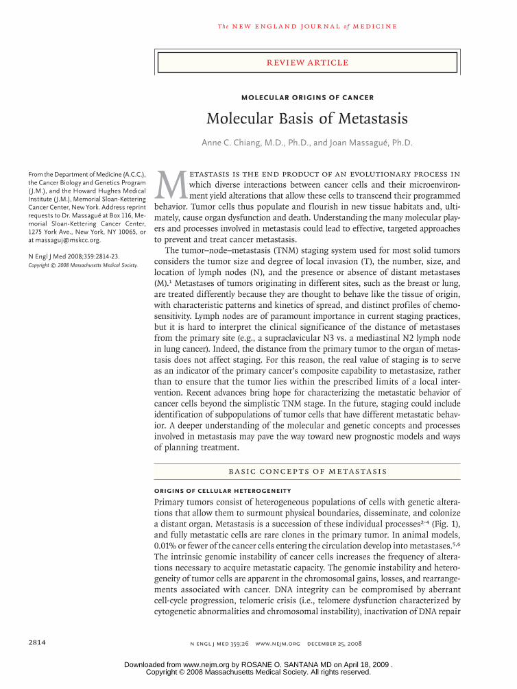
Figure 1. Tumor Initiation and Metastasis.
The initiation and progression of tumors depend on the acquisition of specific functions by cancer cells at both the primary and meta-static sites. Functions associated with tumor initiation are provided by oncogenic mutations and inactivation of tumor-suppressor genes. Functions associated with the initiation of metastasis include functions to which tumor cells resort for local invasion and for circumvent-ing hypoxia and other limitations facing a growing tumor. Most functions for the initiation of both the tumor and metastasis remain es-sential for cancer cells to continue their metastatic development. Functions for metastasis progression provide a local advantage in a primary tumor and a distinct and sometimes organ-specific function during metastasis. Cancer cells that are endowed with these three sets of functions still depend on functions associated with metastasis virulence; these functions confer a selective advantage solely dur-ing the adaptation and takeover of a specific organ microenvironment. Genes associated with each of these functions have been identi-fied in recent years.
Copyright © 2008 Massachusetts Medical Society. All rights reserved. Downloaded from www.nejm.org by ROSANE O. SANTANA MD on April 18, 2009 .

T h e n e w e ngl a nd j o u r na l o f m e dic i n e
n engl j med 359;26 www.nejm.org december 25, 20082816
facilitates the survival of cancer cells at sites of metastasis in breast cancer and renal-cell cancer.13
Cancer Stem Cells and Metastasis
The question of the extent to which self-renewing cancer stem cells initiate and sustain cancers of different types is a subject of intense investiga-tion, and there are probably different answers ac-cording to different tumor types. Such cells are envisioned as a subpopulation of cancer cells that — by one mechanism or another — have the capacity to act as tumor-propagating cells.14 These cells might resist apoptosis and DNA dam-age caused by drugs; they might also require a niche or specific micro environment in order to grow.15 Such attributes would support the estab-lishment of both primary and metastatic tumors. The SDF-1–CXCR4 axis is thought to function in support of cancer cells and stem cells or precur-sor cells.16 A “premetastatic” niche has been de-scribed in animal models in which bone marrow–
derived progenitor cells home to specific distant sites before the formation of a metastasis.17,18 The ability of stem cells to evade destruction and sur-vive in distant sites, including the bone marrow, may explain why micrometastases can remain dor-mant after removal of the primary tumor, only to recur years later.
The En v ironmen t of the Pr im a r y T umor
Invasion and Epithelial-to-Mesenchymal Transition
In many primary tumors with invasive properties, intercellular adhesion is reduced, often because of a loss of E-cadherin, a direct mediator of cell–cell adhesive interactions. The cytoplasmic tail of E-cadherin is tethered, via α-catenin and β-catenin, to the actin cytoskeleton; one of actin’s proper-ties is to maintain cell junctions. The importance of maintaining intercellular adhesion was shown in a mouse model of pancreatic cancer in which disruption of the expression of E-cadherin led to early invasion and metastasis.19 Various mecha-nisms can cause a loss of E-cadherin: mutations resulting in an inactive protein, gene silencing by promoter methylation, or down-regulation stim-ulated by growth factor receptors (e.g., epidermal growth factor receptor [EGFR], fibroblast growth factor receptor [FGFR], insulin-like growth fac-tor I [IGF-I] receptor, and MET) or SRC family kinases.19,20 Expression of the E-cadherin gene (CDH1) is also inhibited by several transcription-al repressors.21,22 Loss of E-cadherin function is necessary, though not sufficient for epithelial-to-mesenchymal transition, a process whereby epithelial cells switch to a mesenchymal progen-itor-cell phenotype, enabling detachment and re-organization of epithelial-cell sheets during em-bryonic development, as well as tumor invasion and metastasis.23
Motility and Extracellular-Matrix Remodeling
The extracellular matrix serves as a scaffold along which cells attach and move by means of con-tacts between cell-surface receptors called integ-rins and extracellular-matrix components such as fibronectin, collagen, and laminin. Integrins also interact in a cytoplasmic complex consisting of focal adhesion kinases and SRC family kinases to mediate attachment to the actin cytoskeleton.
Glossary of Selected Genes
ANGPTL4: Angiopoietin-like 4
APC: Adenomatous polyposis coli
BRAF: (Also known as V-raf murine sarcoma viral onco-gene homologue B1)
BRCA1: Breast-cancer gene 1
COX-2: Cyclooxygenase-2
CSF-1: Colony-stimulating factor 1
CTGF: Connective-tissue growth factor
CXCR4: CXC chemokine receptor 4
EGFR: Epidermal growth factor receptor
EREG: Epiregulin
FGFR: Fibroblast growth factor receptor
HER2: Human epidermal growth factor receptor type 2
ID1: Inhibitor of differentiation-1
MMP-1: Matrix metalloproteinase 1
MMP-9: Matrix metalloproteinase 9
NEDD9: Neural precursor cell expressed, developmen-tally down-regulated 9
P13K: Phosphoinositide 3-kinase
PTEN: Phosphatase and tensin homologue
RANKL: Ligand for the receptor activator of nuclear factor-κB
RHoC: Ras homologue gene family, member C
STK11: Serine–threonine kinase 11 (also known as LKB1)
TWIST1: Twist homologue 1
VEGF: Vascular endothelial growth factor
VHL1: von Hippel–Lindau 1
Copyright © 2008 Massachusetts Medical Society. All rights reserved. Downloaded from www.nejm.org by ROSANE O. SANTANA MD on April 18, 2009 .

Molecular Origins of Cancer
n engl j med 359;26 www.nejm.org december 25, 2008 2817
Through calcium-dependent guanosine triphos-phatases (GTPases), extracellular-matrix signals cause cytoskeletal changes that form individual cytoplasmic extensions called filopodia, which coalesce into larger lamellipodia, structures that are important in migratory movement. Expres-sion profiling of melanoma cell lines obtained by means of in vivo selection has shown that the calcium-dependent GTPase RhoC is important in lung metastasis.24 Homozygous RhoC-deficient mice have normal formation of primary tumors but impaired cancer-cell mobility and almost no lung metastases.25 NEDD9, a scaffolding protein involved in cell adhesion, colocalizes in focal con-tacts with focal adhesion kinase and can promote cell motility and invasion.26 Various members of the matrix metalloproteinase (MMP) family (e.g., MMP-2 and MMP-9) are also implicated in cancer-cell invasion.27-29 Independent screens for genes that mediate bone or lung metastasis in breast cancer have identified MMP-1 as being necessary for spread to the bone and lungs.30,31 The metas-tasis-suppressor microRNA miR335, which inhib-its metastasis to the lungs and bones in human breast-cancer xenografts, suppresses the expres-sion of two mediators of cancer-cell invasion, the transcription factor SOX-4 and tenascin-C, a ma-trix glycoprotein.32 A low level of miR335 in breast-cancer cells is associated with relapse.
Stromal Interactions
Not only are cancer cells able to traverse the structural boundaries of the primary tumor, but they can also co-opt local and bone marrow–derived stromal-cell responses to their advantage. At points of basement-membrane invasion in mouse tumors, tumor-associated macrophages proliferate in response to tumor-derived colony-stimulating factor 1 and produce growth factors (e.g., fibroblast growth factor, EGFR ligands, and platelet-derived growth factor [PDGF]) and pro-teases (e.g., MMPs and cathepsins).33,34 In addi-tion, tumor-associated macrophages activate a particular type of carcinoma-associated mesen-chymal cell, the myofibroblast, which secretes the cytokine SDF-1; this cytokine enables the myofi-broblast to recruit endothelial progenitor cells.35 Impaired metastases of breast-cancer cells to the lungs occur in mice with genetic defects in macro-phages.36 The stroma-derived cytokine, transform-ing growth factor β (TGF-β), induces the expres-sion of genes such as ANGPTL4 in breast-cancer
cells; TGF-β enhances metastatic activity and is associated with increased metastases to the lungs in estrogen-receptor–negative breast cancer.37 In short, several types of stromal cells and their se-creted factors provide selective prometastatic ad-vantages.
Organ-Specific Metastasis
Some types of cancers have a characteristic pro-clivity to metastasize to certain organs, but not to others (Fig. 2).38-42 Breast cancer spreads to
Brain
Breast
Colorectal
Renal-cell
Stomach
Bone
Liver
Lung
Lung
Melanoma
Colorectal
Renal-cell
Melanoma
Breast
Sarcoma
Colorectal
Renal-cell
Lung
Breast
Prostate
Colorectal
Pancreatic
Lung
Breast
2
Schwartz
12/05/08
AUTHOR PLEASE NOTE:Figure has been redrawn and type has been reset
Please check carefully
Author
Fig #
Title
ME
DEArtist
Issue date
COLOR FIGURE
Draft 4Massague
Knoper
Metatastic spread ofsolid tumors
12/25/08
Figure 2. Patterns of Metastatic Spread of Solid Tumors.
Brain metastases may occur as a result of hematogenous spread late in the course of a widely metastatic tumor, or as a result of secondary metastasis from a primary or a metastatic tumor that can access the arterial circulation through the pulmonary venous circulation to seed the brain.38 Tumors with the highest incidence of brain metastases include lung carcinoma, breast carcinoma, melanoma, and to a lesser extent, renal-cell and colorectal car-cinomas. Leptomeningeal disease may develop through the spread of can-cer cells through perineural lymphatic vessels, and it is a sign of advanced disease.38 Some tumors have a strong proclivity for dissemination to the lungs; for example, in one study, the rate of dissemination associated with sarcoma was 23%.39 Other tumors that frequently spread to the lungs in-clude renal-cell, colorectal, melanoma, and breast carcinomas.40,41 Gastro-intestinal tumors easily access the liver circulation through the portal-vein system. The incidence of liver metastases is highest among patients with colorectal or pancreatic cancer, followed by breast and lung cancers.40 Estro-gen-receptor–negative breast-cancer tumors more often metastasize to vis-ceral organs, including the liver, whereas estrogen-receptor–positive breast cancer more often metastasizes to the bone.42 Bone metastasis occurs in patients with primary tumors associated with breast, lung, prostate, renal-cell, and colon cancer, in this order of frequency.40 Bone metastases may be primarily osteo lytic or osteoblastic, depending on the tumor of origin.
Copyright © 2008 Massachusetts Medical Society. All rights reserved. Downloaded from www.nejm.org by ROSANE O. SANTANA MD on April 18, 2009 .

T h e n e w e ngl a nd j o u r na l o f m e dic i n e
n engl j med 359;26 www.nejm.org december 25, 20082818
the bones, lungs, brain, and liver; distant metas-tases of prostate cancer occur most prominently in bone. Breast-cancer and prostate-cancer cells can both spread to and colonize the bone, but they form osteolytic or osteoblastic metastases, respec-tively. According to Paget’s “seed” vs. “soil” hy-pothesis, perceived compatibilities between dis-seminated cancer cells (the seed) and certain distant sites (the soil) have long influenced our view of the metastatic process.43
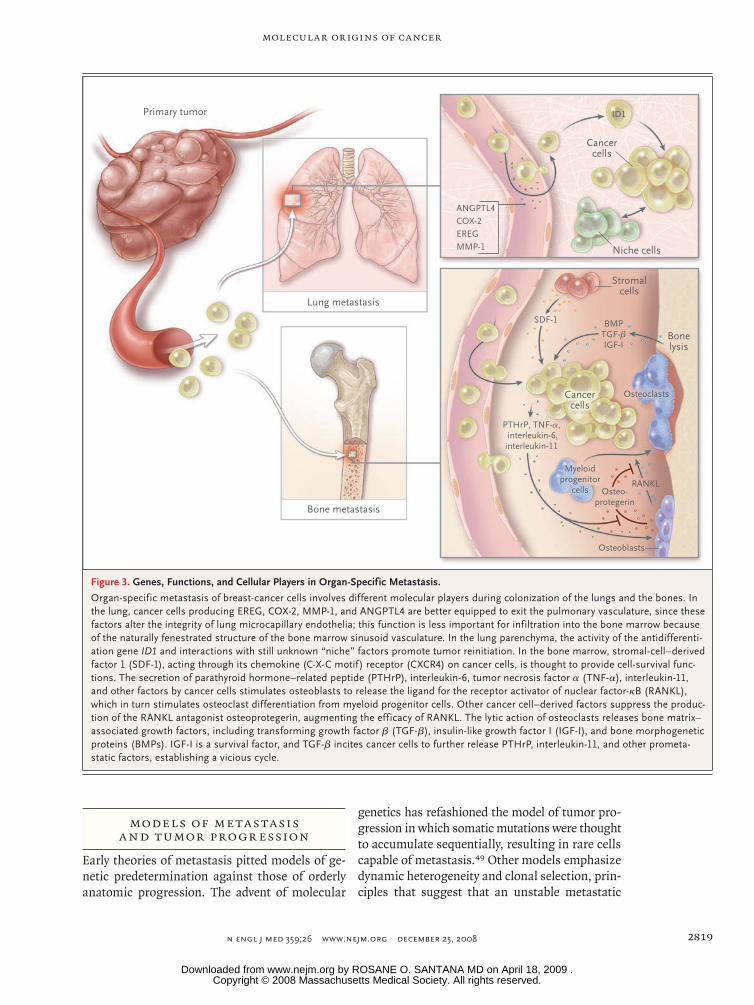
The formation of bone metastases alters bone homeostasis — the balance of action of osteo-clasts in degrading bone against the constant re-building of bone by osteoblasts. Breast-cancer cells preferentially cause osteolytic lesions by in-ducing osteoclasts to secrete PTHrP (parathyroid hormone–related peptide), tumor necrosis factor α (TNF-α), and cytokines such as interleukin-1, interleukin-6, interleukin-8, and interleukin-11. These factors cue osteoblasts to release RANKL (the ligand for the receptor activator of nuclear factor-κB [RANK]), which stimulates osteoclast differentiation (Fig. 3). Osteoclasts demineralize bone, thereby causing the release of growth fac-tors such as bone morphogenetic proteins, IGF-I, and TGF-β from the exposed bone matrix; all these growth factors support cancer-cell prolif-eration and induce further release of PTHrP. In a breast-cancer xenograft model, breast-cancer cells that preferentially colonized bone had up-regu-lated expression of genes encoding CXCR4, osteo-pontin, CTGF, MMP-1, and interleukin-11.30 By contrast, prostate-cancer cells secrete osteoblast-stimulating factors such as Wnt family ligands, bone morphogenetic proteins, PDGF, and endo-thelin-1; these factors stimulate formation of the hallmark osteoblastic metastases of prostate can-cer. Tumor-derived signals suppress the ability of osteoblasts to secrete osteoprotegerin, a RANKL antagonist that blocks RANKL–RANK interaction and resulting osteoclast activation. Thus, factors secreted by cancer cells, or “seeds,” can influence the type of metastases formed.
Cancer cells may regulate the expression of other molecules to target colonization in other organs.44 Such molecules include the gene encod-ing ezrin (an intracellular protein needed for early survival of metastatic osteosarcoma cells in the lung), serine–threonine kinase 11 (STK11, also known as LKB1) (a metastasis-suppressor gene regulating NEDD9 in lung cancer45), and genes in an 18-gene breast-to-lung metastatic
gene-expression signature including the EGFR ligand EREG, COX-2, MMP-1, ANGPTL4, and other mediators of infiltration and colonization by can-cer cells in the lung.46
The soil, or distant metastatic site, is a large-ly nonpermissive environment, as evidenced by the rarity of metastatic clones arising after inject-ing millions of cells into circulation in experi-ments in animals. In humans, also, thousands of circulating tumor cells have been found in the absence of metastases. Certain seed–soil inter-actions can support the cancer cell’s ability to survive in the metastatic microenvironment, in-cluding the RANKL–RANK interaction. Another example involves the SDF-1 chemokine in the bone marrow, which recruits breast-cancer and prostate-cancer cells and enhances their surviv-al.47 Whereas the mechanisms of metastasis to bone and lung have been extensively studied and are partially understood, there is a dearth of in-formation about the molecular basis for metasta-sis to other organs, such as the liver and brain.
A n In tegr ated Model of Me ta s ta sis
In the past decade, our view of metastasis has changed from snapshots detailing specific bio-logic processes to a moving picture of how vari-ous cancer cells acquire functions and co-opt stromal signals for spread and encampment in a distant site. Random genetic and epigenetic alter-ations in cancer cells in combination with a plas-tic and responsive microenvironment support the metastatic evolution of tumors. Moreover, genes needed at individual steps along the metastatic process have been identified.
These genes have been classified into three categories: initiation, progression, and virulence48 (Fig. 1). Genes that are associated with metastatic progression give the cancer cell particular advan-tages at multiple points during its sojourn to a dis-tant site. These advantages can influence the cell’s metastatic destination. Genes associated with the initiation of metastasis and virulence operate in the earliest and latest stages of invasion and growth in the primary tumor and different metastatic habitats, respectively. The use of such a framework to organize specific genes and their functions al-lows a multidimensional picture (including locale and time) of metastasis and may aid in the devel-opment of rational antimetastatic strategies.
Copyright © 2008 Massachusetts Medical Society. All rights reserved. Downloaded from www.nejm.org by ROSANE O. SANTANA MD on April 18, 2009 .
Molecular Origins of Cancer
n engl j med 359;26 www.nejm.org december 25, 2008 2819
Model s of Me ta s ta sis a nd T umor Pro gr ession
Early theories of metastasis pitted models of ge-netic predetermination against those of orderly anatomic progression. The advent of molecular
genetics has refashioned the model of tumor pro-gression in which somatic mutations were thought to accumulate sequentially, resulting in rare cells capable of metastasis.49 Other models emphasize dynamic heterogeneity and clonal selection, prin-ciples that suggest that an unstable metastatic
Primary tumor
Lung metastasis
Bone metastasis
ANGPTL4
EREGCOX-2
MMP-1
ID1
Niche cells
Stromalcells
Bonelysis
BMPTGF-βIGF-I
SDF-1
Osteoblasts
Osteoclasts
RANKLOsteo-
protegerin
PTHrP, TNF-α, interleukin-6,interleukin-11
Cancercells
Cancercells
Myeloidprogenitor
cells
3
Schwartz
12/05/08
AUTHOR PLEASE NOTE:Figure has been redrawn and type has been reset
Please check carefully
Author
Fig #
Title
ME
DEArtist
Issue date
COLOR FIGURE
Draft 5Massague
Knoper
Genes functions andcellular players
12/25/08
Figure 3. Genes, Functions, and Cellular Players in Organ-Specific Metastasis.
Organ-specific metastasis of breast-cancer cells involves different molecular players during colonization of the lungs and the bones. In the lung, cancer cells producing EREG, COX-2, MMP-1, and ANGPTL4 are better equipped to exit the pulmonary vasculature, since these factors alter the integrity of lung microcapillary endothelia; this function is less important for infiltration into the bone marrow because of the naturally fenestrated structure of the bone marrow sinusoid vasculature. In the lung parenchyma, the activity of the antidifferenti-ation gene ID1 and interactions with still unknown “niche” factors promote tumor reinitiation. In the bone marrow, stromal-cell–derived factor 1 (SDF-1), acting through its chemokine (C-X-C motif) receptor (CXCR4) on cancer cells, is thought to provide cell-survival func-tions. The secretion of parathyroid hormone–related peptide (PTHrP), interleukin-6, tumor necrosis factor α (TNF-α), interleukin-11, and other factors by cancer cells stimulates osteoblasts to release the ligand for the receptor activator of nuclear factor-κB (RANKL), which in turn stimulates osteoclast differentiation from myeloid progenitor cells. Other cancer cell–derived factors suppress the produc-tion of the RANKL antagonist osteoprotegerin, augmenting the efficacy of RANKL. The lytic action of osteoclasts releases bone matrix–associated growth factors, including transforming growth factor β (TGF-β), insulin-like growth factor I (IGF-I), and bone morphogenetic proteins (BMPs). IGF-I is a survival factor, and TGF-β incites cancer cells to further release PTHrP, interleukin-11, and other prometa-static factors, establishing a vicious cycle.
Copyright © 2008 Massachusetts Medical Society. All rights reserved. Downloaded from www.nejm.org by ROSANE O. SANTANA MD on April 18, 2009 .

T h e n e w e ngl a nd j o u r na l o f m e dic i n e
n engl j med 359;26 www.nejm.org december 25, 20082820
variant can expand and prevail in the population of cells.50,51 The presence of metastasis genes in gene-expression signatures of primary tumors would seem to challenge the traditional tumor-progression model of somatic evolution in which metastatic cells would be too rare to influence a population-averaged gene-expression profile of the primary tumor. This finding, however, probably reflects an abundance of partially competent can-cer cells that have accumulated a sufficient num-ber of malignant functions to promote expansion of the primary tumor, and which may be neces-sary but not sufficient for forming metastases. By contrast, genes associated with metastatic vir-ulence provide an aggressive edge in survival and proliferation solely during colonization of the metastatic site (Fig. 1). Many of these genes do not give the primary tumor a selective advantage, and thus they would not be represented in gene signatures of the primary tumor.
Me ta s ta sis-Pro gr ession Genes
Genes that are necessary for certain functions such as vascular remodeling can participate in both the primary tumor and the metastatic envi-ronment; these genes are metastasis-progression genes, and they could be enriched in primary tu-mors (Fig. 1). An 18-gene lung-metastatic signa-ture derived from selected in vivo breast-cancer cells that efficiently spread to the lungs includes EREG, COX-2, and MMP-1. These genes cooperate in remodeling the vasculature in sites of mam-mary tumors and lung metastasis. In the breast, they allow neoangiogenesis and intravasation of cancer cells; in the lung, they mediate extravasa-tion of circulating cancer cells from capillaries into the parenchyma.52 Breast cancers with the lung-metastatic signature have a high risk of lung metastases, but not of metastases to the bones or liver. A likely explanation is that extravasation is not essential for passage through the fenestrated vasculature of the bone marrow and liver sinu-soids. Thus, metastasis-progression genes may couple the tissue-specific features of the micro-environment in a particular organ to a matching role in the progression of a primary tumor. Ex-pression of the lung-metastatic signature gene ANGPTL4 is a bystander event in mammary tu-mors, but when cancer cells expressing ANGPTL4 reach lung capillaries, its role in mediating extrav-
asation by disrupting endothelial cell–cell con-tacts becomes manifest.37
Both the cells of primary tumors and metastat-ic cells require the ability to initiate self-renewal and bypass senescence. ID1 (inhibitor of differ-entiation-1) is the sole transcriptional regulator in the lung-metastatic signature, and it can be found in clusters of cancer cells within breast tumors of the basal or triple-negative (i.e., estrogen-recep-tor–negative, progesterone-receptor–negative, and human epidermal growth factor receptor type 2 [HER2]–negative) subtype. Suppression of ID1 expression inhibits the initiation of mammary tumors and metastases in the lungs.53 Thus, ID1 may promote micrometastatic outgrowth from dormancy at the metastatic site. Related to this function, in mouse models of metastatic breast cancer, ID1 cooperates with activated RAS onco-genes to avert cell senescence.54
Me ta s tatic Dissemination
Cancer cells can disseminate from a tumor very early in the life of a tumor. They have been detect-ed in the bone marrow of patients with breast can-cer with early-stage disease. Such cancer cells were genetically distinct from the matched primary tu-mors, but bone marrow–derived cancer cells in patients with overt metastatic disease were less genetically disparate.55,56 This finding may reflect differences between the departure time from the primary neoplasm and the duration of exposure to selective pressures. Dormant cancer cells iso-lated from the bone marrow of transgenic mice with preinvasive breast cancer and patients with ductal carcinoma in situ became activated when transplanted into the bone marrow and caused the growth of lethal tumors.57 Many mechanisms for metastatic dormancy have been postulated.58 In patients with advanced metastatic disease, breast cancer cells that are competent in vascular entry can efficiently exit at a distant organ and perhaps reenter to repeat the process. Tumor in-filtration by means of its own circulating progeny of metastatic cancer cells has been raised as a possible mechanism for the later rapid expansion of tumor growth.59 According to this hypothesis, large primary tumors may also be the end product of aggressive reseeding. This would be a new per-spective on the long-standing observation that metastatic relapse correlates with tumor size.60
Copyright © 2008 Massachusetts Medical Society. All rights reserved. Downloaded from www.nejm.org by ROSANE O. SANTANA MD on April 18, 2009 .

Molecular Origins of Cancer
n engl j med 359;26 www.nejm.org december 25, 2008 2821
Clinic a l Implic ations
Molecular Signatures of Metastasis
Gene-expression signatures of primary breast cancers that predict clinical outcome61-65 gener-ally do not overlap and range from a 70-gene “poor-prognosis” signature (detected with the use of the MammaPrint test) to a hand-picked set of 21 “recurrence” genes (detected with the use of the Oncotype Dx test) that includes estrogen-receptor, HER2, and proliferation markers. Other signatures consist of genes with expressions that are associated with a process or pathway, such as the response to serum mitogens,66 hypoxia,67 ac-tivation of specific oncogenes (e.g., RAS, MYC, and SRC),68,69 stimulation with a growth factor (e.g., TGF-β),37 or treatment with specific chemo-therapeutic drugs to establish a drug-sensitivity profile.70 To specifically identify genes that me-diate metastasis, animal models have been used to select in vivo for highly metastatic and organ-specific derivatives of human cancer cell lines.3 Such signatures can correlate with bone-specific and lung-specific spread.30,46 The lung-metastasis signature further correlates with clinical outcome, including the recurrence of disease in the lungs, in primary breast-cancer tumors.60 Functional validation approaches (e.g., overexpression or knockdown experiments in culture or xenograft experiments) have confirmed that these genes, particularly in combination, are critical for meta-static functions.52
Targets of Therapy
In principle, each metastasis-specific gene is a potential target for a treatment. Ongoing clinical trials target the metastatic initiation gene c-MET (e.g., the small-molecule inhibitor ARQ 197, in phase 1–2 trials) and two metastatic virulence genes, RANK ligand (e.g., denosumab, in phase 3 trials) and TGF-β (e.g., monoclonal antibody GC1008, in phase 1 trials). Combination therapy may be needed to overcome the intrinsic biologic redundancy in metastasis and to target sequen-tial steps in metastasis. In one series of preclini-cal experiments, only combination (not single-agent) therapy with the drugs celecoxib and cetuximab, meant to target two metastatic pro-gression genes, was effective in blocking lung metastases by highly lung-metastatic breast-can-cer cells.52 If cancer cells are constantly on the move between sites of metastasis in the lung,
treatment with these drugs could prevent further reseeding and growth of metastatic sites. Cancer treatment may need to combine multiple anti-metastatic drugs with cytotoxic chemotherapy. For example, bevacizumab, an antibody targeting vascular endothelial growth factor, is being stud-ied in combination with chemotherapy in the ad-juvant setting for colorectal, ovarian, and non–small-cell lung cancers. Therapies that target the mechanisms that keep dormant micrometastases alive are also needed.
Clinical Translation
Clinical trials involving antimetastatic agents face a number of obstacles. Any adjuvant trial to as-sess the recurrence of metastatic disease requires many patients because of the infrequency and lag time to progression of metastatic disease in many types of cancer. Measuring response rates be-yond stable disease will further increase the num-ber of patients in a trial. Moreover, correlative stud-ies of tissue obtained from metastatic sites are essential to understand the results of such trials.
These barriers are sobering, and they perhaps underscore the conceptual shifts that will be needed for the development of new cancer thera-pies. What changes can we envision? The profile of a tumor could include not only histopatho-logical or genetic determinants, or both, but also a molecular snapshot that would indicate a “me-tastasis quotient.” The metastasis quotient could be a measure of how adept the cells are with re-spect to metastatic functions, and it could serve as a prognostic framework. By focusing on meta-static progression and virulence functions, can-cer therapy might be dictated by the metastatic site and not only by the specific tissue of origin. A current example of a treatment targeting a metastatic organ is the use of bisphosphonates or denosumab (an anti-RANK antibody), or both, to treat bone metastases originating from the breast, lung, and even multiple myeloma. Drug regimens for patients with cancer might include multiple drugs targeting different metastatic sites and seeding among sites. There is now hope for achieving the ultimate goal — curing metastatic disease.
Dr. Massagué reports receiving consulting fees from Accele-ron Pharmaceuticals, lecture fees from Eli Lilly, Genzyme, and Eisai. No other potential conflict of interest relevant to this arti-cle was reported.
We thank D. Nguyen, G. Riely, K. Politi, and D. Spriggs for insightful discussions.
Copyright © 2008 Massachusetts Medical Society. All rights reserved. Downloaded from www.nejm.org by ROSANE O. SANTANA MD on April 18, 2009 .

T h e n e w e ngl a nd j o u r na l o f m e dic i n e
n engl j med 359;26 www.nejm.org december 25, 20082822
References
DeVita VT Jr, Lawrence TS, Rosenberg 1. SA, eds. DeVita, Hellman, and Rosenberg’s Cancer: principles & practice of oncology. 8th ed. Philadelphia: Wolters Kluwer/Lip-pincott Williams & Wilkins, 2008.
Weinberg RA. The biology of cancer. 2. New York: Garland Science, 2007.
Fidler IJ. The pathogenesis of cancer 3. metastasis: the ‘seed and soil’ hypothesis revisited. Nat Rev Cancer 2003;3:453-8.
Gupta GP, Massague J. Cancer metas-4. tasis: building a framework. Cell 2006;127: 679-95.
Chambers AF, Groom AC, MacDonald 5. IC. Dissemination and growth of cancer cells in metastatic sites. Nat Rev Cancer 2002;2:563-72.
Luzzi KJ, MacDonald IC, Schmidt EE, 6. et al. Multistep nature of metastatic inef-ficiency: dormancy of solitary cells after successful extravasation and limited sur-vival of early micrometastases. Am J Pathol 1998;153:865-73.
Sykes SM, Mellert HS, Holbert MA, et 7. al. Acetylation of the p53 DNA-binding domain regulates apoptosis induction. Mol Cell 2006;24:841-51.
Halazonetis TD, Gorgoulis VG, Bar-8. tek J. An oncogene-induced DNA damage model for cancer development. Science 2008;319:1352-5.
Pouysségur J, Dayan F, Mazure NM. 9. Hypoxia signalling in cancer and approach-es to enforce tumour regression. Nature 2006;441:437-43.
Bristow RG, Hill RP. Hypoxia and me-10. tabolism: hypoxia, DNA repair and genetic instability. Nat Rev Cancer 2008;8:180-92.
Higgins DF, Kimura K, Bernhardt WM, 11. et al. Hypoxia promotes fibrogenesis in vivo via HIF-1 stimulation of epithelial-to-mesenchymal transition. J Clin Invest 2007;117:3810-20.
Erler JT, Bennewith KL, Nicolau M, 12. et al. Lysyl oxidase is essential for hypoxia-induced metastasis. Nature 2006;440: 1222-6.
Staller P, Sulitkova J, Lisztwan J, Moch 13. H, Oakeley EJ, Krek W. Chemokine recep-tor CXCR4 downregulated by von Hippel-Lindau tumour suppressor pVHL. Nature 2003;425:307-11.
Clarke MF, Fuller M. Stem cells and 14. cancer: two faces of Eve. Cell 2006;124: 1111-5.
Croker AK, Allan AL. Cancer stem 15. cells: implications for the progression and treatment of metastatic disease. J Cell Mol Med 2008;12:374-90.
Kucia M, Reca R, Miekus K, et al. 16. Trafficking of normal stem cells and me-tastasis of cancer stem cells involve simi-lar mechanisms: pivotal role of the SDF-1-CXCR4 axis. Stem Cells 2005;23:879-94.
Kaplan RN, Riba RD, Zacharoulis S, 17. et al. VEGFR1-positive haematopoietic bone marrow progenitors initiate the pre-metastatic niche. Nature 2005;438:820-7.
Wels J, Kaplan RN, Rafii S, Lyden D. 18. Migratory neighbors and distant invaders: tumor-associated niche cells. Genes Dev 2008;22:559-74.
Perl AK, Wilgenbus P, Dahl U, Semb H, 19. Christofori G. A causal role for E-cadherin in the transition from adenoma to carci-noma. Nature 1998;392:190-3.
Thiery JP. Epithelial-mesenchymal tran-20. sitions in tumour progression. Nat Rev Cancer 2002;2:442-54.
Cano A, Pérez-Moreno MA, Rodrigo I, 21. et al. The transcription factor snail con-trols epithelial-mesenchymal transitions by repressing E-cadherin expression. Nat Cell Biol 2000;2:76-83.
Yang J, Mani SA, Donaher JL, et al. 22. Twist, a master regulator of morphogen-esis, plays an essential role in tumor me-tastasis. Cell 2004;117:927-39.
Yang MH, Wu MZ, Chiou SH, et al. 23. Direct regulation of TWIST by HIF-1alpha promotes metastasis. Nat Cell Biol 2008; 10:295-305.
Clark EA, Golub TR, Lander ES, Hynes 24. RO. Genomic analysis of metastasis reveals an essential role for RhoC. Nature 2000; 406:532-5. [Erratum, Nature 2001;411: 974.]
Hakem A, Sanchez-Sweatman O, You-25. Ten A, et al. RhoC is dispensable for em-bryogenesis and tumor initiation but es-sential for metastasis. Genes Dev 2005;19: 1974-9.
Kim M, Gans JD, Nogueira C, et al. 26. Comparative oncogenomics identifies NEDD9 as a melanoma metastasis gene. Cell 2006;125:1269-81.
Egeblad M, Werb Z. New functions for 27. the matrix metalloproteinases in cancer progression. Nat Rev Cancer 2002;2:161-74.
López-Otin C, Matrisian LM. Emerg-28. ing roles of proteases in tumour suppres-sion. Nat Rev Cancer 2007;7:800-8.
Martin MD, Matrisian LM. The other 29. side of MMPs: protective roles in tumor progression. Cancer Metastasis Rev 2007; 26:717-24.
Kang Y, Siegel PM, Shu W, et al. A mul-30. tigenic program mediating breast cancer metastasis to bone. Cancer Cell 2003;3: 537-49.
Minn AJ, Kang Y, Serganova I, et al. 31. Distinct organ-specific metastatic poten-tial of individual breast cancer cells and primary tumors. J Clin Invest 2005;115:44-55.
Tavazoie SF, Alarcón C, Oskarsson T, 32. et al. Endogenous human microRNAs that suppress breast cancer metastasis. Nature 2008;451:147-52.
Lewis CE, Pollard JW. Distinct role of 33. macrophages in different tumor microen-vironments. Cancer Res 2006;66:605-12.
Joyce JA. Therapeutic targeting of the 34. tumor microenvironment. Cancer Cell 2005;7:513-20.
Orimo A, Gupta PB, Sgroi DC, et al. 35. Stromal fibroblasts present in invasive hu-man breast carcinomas promote tumor growth and angiogenesis through elevated SDF-1/CXCL12 secretion. Cell 2005;121: 335-48.
Condeelis J, Pollard JW. Macrophages: 36. obligate partners for tumor cell migra-tion, invasion, and metastasis. Cell 2006; 124:263-6.
Padua D, Zhang XH, Wang Q, et al. 37. TGFbeta primes breast tumors for lung metastasis seeding through angiopoietin-like 4. Cell 2008;133:66-77.
Gavrilovic IT, Posner JB. Brain metas-38. tases: epidemiology and pathophysiology. J Neurooncol 2005;75:5-14.
Billingsley KG, Burt ME, Jara E, et al. 39. Pulmonary metastases from soft tissue sarcoma: analysis of patterns of diseases and postmetastasis survival. Ann Surg 1999;229:602-10.
Hess KR, Varadhachary GR, Taylor SH, 40. et al. Metastatic patterns in adenocarci-noma. Cancer 2006;106:1624-33.
Leiter U, Meier F, Schittek B, Garbe C. 41. The natural course of cutaneous melano-ma. J Surg Oncol 2004;86:172-8.
Lacroix M. Significance, detection and 42. markers of disseminated breast cancer cells. Endocr Relat Cancer 2006;13:1033-67.
Paget S. The distribution of secondary 43. growths in cancer of the breast. Lancet 1889;1:571-3.
Khanna C, Wan X, Bose S, et al. The 44. membrane-cytoskeleton linker ezrin is necessary for osteosarcoma metastasis. Nat Med 2004;10:182-6.
Ji H, Ramsey MR, Hayes DN, et al. 45. LKB1 modulates lung cancer differentia-tion and metastasis. Nature 2007;448: 807-10.
Minn AJ, Gupta GP, Siegel PM, et al. 46. Genes that mediate breast cancer metas-tasis to lung. Nature 2005;436:518-24.
Wang J, Loberg R, Taichman RS. The 47. pivotal role of CXCL12 (SDF-1)/CXCR4 axis in bone metastasis. Cancer Metasta-sis Rev 2006;25:573-87.
Nguyen DX, Massague J. Genetic de-48. terminants of cancer metastasis. Nat Rev Genet 2007;8:341-52.
Vogelstein B, Fearon ER, Hamilton SR, 49. et al. Genetic alterations during colorec-tal-tumor development. N Engl J Med 1988; 319:525-32.
Ling V, Chambers AF, Harris JF, Hill 50. RP. Quantitative genetic analysis of tumor progression. Cancer Metastasis Rev 1985; 4:173-92.
Waghorne C, Thomas M, Lagarde A, 51. Kerbel RS, Breitman ML. Genetic evidence for progressive selection and overgrowth of primary tumors by metastatic cell sub-populations. Cancer Res 1988;48:6109-14.
Gupta GP, Nguyen DX, Chiang AC, et 52. al. Mediators of vascular remodelling co-
Copyright © 2008 Massachusetts Medical Society. All rights reserved. Downloaded from www.nejm.org by ROSANE O. SANTANA MD on April 18, 2009 .

Molecular Origins of Cancer
n engl j med 359;26 www.nejm.org december 25, 2008 2823
opted for sequential steps in lung metas-tasis. Nature 2007;446:765-70.
Gupta GP, Perk J, Acharyya S, et al. ID 53. genes mediate tumor reinitiation during breast cancer lung metastasis. Proc Natl Acad Sci U S A 2007;104:19506-11.
Swarbrick A, Roy E, Allen T, Bishop 54. JM. Id1 cooperates with oncogenic Ras to induce metastatic mammary carcinoma by subversion of the cellular senescence re-sponse. Proc Natl Acad Sci U S A 2008; 105:5402-7.
Klein CA, Blankenstein TJ, Schmidt-55. Kittler O, et al. Genetic heterogeneity of single disseminated tumour cells in mini-mal residual cancer. Lancet 2002;360: 683-9.
Schmidt-Kittler O, Ragg T, Daskalakis 56. A, et al. From latent disseminated cells to overt metastasis: genetic analysis of sys-temic breast cancer progression. Proc Natl Acad Sci U S A 2003;100:7737-42.
Hüsemann Y, Geigl JB, Schubert F, et 57. al. Systemic spread is an early step in breast cancer. Cancer Cell 2008;13:58-68.
Aguirre-Ghiso JA. The problem of can-58.
cer dormancy: understanding the basic mechanisms and identifying therapeutic opportunities. Cell Cycle 2006;5:1740-3.
Norton L, Massagué J. Is cancer a dis-59. ease of self-seeding? Nat Med 2006;12: 875-8.
Minn AJ, Gupta GP, Padua D, et al. 60. Lung metastasis genes couple breast tu-mor size and metastatic spread. Proc Natl Acad Sci U S A 2007;104:6740-5.
Bertucci F, Cervera N, Birnbaum D. 61. A gene signature in breast cancer. N Engl J Med 2007;356:1887-8.
Buyse M, Loi S, van’t Veer L, et al. 62. Validation and clinical utility of a 70-gene prognostic signature for women with node-negative breast cancer. J Natl Cancer Inst 2006;98:1183-92.
Fan C, Oh DS, Wessels L, et al. Con-63. cordance among gene-expression–based predictors for breast cancer. N Engl J Med 2006;355:560-9.
van de Vijver MJ, He YD, van’t Veer LJ, 64. et al. A gene-expression signature as a pre-dictor of survival in breast cancer. N Engl J Med 2002;347:1999-2009.
van’t Veer LJ, Dai H, van de Vijver MJ, 65.
et al. Gene expression profiling predicts clinical outcome of breast cancer. Nature 2002;415:530-6.
Chang HY, Sneddon JB, Alizadeh AA, 66. et al. Gene expression signature of fibro-blast serum response predicts human cancer progression: similarities between tumors and wounds. PLoS Biol 2004;2(2): e7.
Chi JT, Wang Z, Nuyten DS, et al. Gene 67. expression programs in response to hy-poxia: cell type specificity and prognostic significance in human cancers. PLoS Med 2006;3(3):e47.
Bild AH, Potti A, Nevins JR. Linking 68. oncogenic pathways with therapeutic op-portunities. Nat Rev Cancer 2006;6:735-41.
Bild AH, Yao G, Chang JT, et al. Onco-69. genic pathway signatures in human can-cers as a guide to targeted therapies. Na-ture 2006;439:353-7.
Nevins JR, Potti A. Mining gene ex-70. pression profiles: expression signatures as cancer phenotypes. Nat Rev Genet 2007;8: 601-9.Copyright © 2008 Massachusetts Medical Society.
personal archives in the journal online
Individual subscribers can store articles and searches using a feature on the Journal’s Web site (www.nejm.org) called “Personal Archive.” Each article and search result links to this feature. Users can create
personal folders and move articles into them for convenient retrieval later.
Copyright © 2008 Massachusetts Medical Society. All rights reserved. Downloaded from www.nejm.org by ROSANE O. SANTANA MD on April 18, 2009 .
Related Documents