Molecular basis of chromatic adaptation in pennate diatom Phaeodactylum tricornutum Miroslava Herbstová a,b , David Bína a,b , Peter Koník b , Zdenko Gardian a,b , František Vácha a,b , Radek Litvín a,b, ⁎ a Institute of Plant Molecular Biology, Biology Centre CAS, Branisovska 31, 37005 Ceske Budejovice, Czech Republic b Faculty of Science, University of South Bohemia, Branisovska 1760, 37005 Ceske Budejovice, Czech Republic abstract article info Article history: Received 22 October 2014 Received in revised form 25 February 2015 Accepted 27 February 2015 Available online 6 March 2015 Keywords: Chromatic adaptation Diatom Phaeodactylum tricornutum Light harvesting antenna Red-shifted antenna complex Heterokonta The remarkable adaptability of diatoms living in a highly variable environment assures their prominence among marine primary producers. The present study integrates biochemical, biophysical and genomic data to bring new insights into the molecular mechanism of chromatic adaptation of pennate diatoms in model species Phaeodactylum tricornutum, a marine eukaryote alga possessing the capability to shift its absorption up to ~700 nm as a consequence of incident light enhanced in the red component. Presence of these low energy spec- tral forms of Chl a is manifested by room temperature fluorescence emission maximum at 710 nm (F710). Here we report a successful isolation of the supramolecular protein complex emitting F710 and identify a member of the Fucoxanthin Chlorophyll a/c binding Protein family, Lhcf15, as its key building block. This red-shifted antenna complex of P. tricornutum appears to be functionally connected to photosystem II. Phylogenetic analyses do not support relation of Lhcf15 of P. tricornutum to other known red-shifted antenna proteins thus indicating a case of convergent evolutionary adaptation towards survival in shaded environments. © 2015 Elsevier B.V. All rights reserved. 1. Introduction Marine photosynthesis powers a substantial part of the biosphere and forms a major portion of global carbon cycle. Diatoms (Bacillariophyceae), a group of eukaryote algae, play a considerable role in oceanic carbon fix- ation [1]. Diatoms belong to the group of Heterokonta (Stramenopila), a group of protists originating around the beginning of the Palaeozoic [2]. However, the line leading to them is much older and is based on a common eukaryote ancestor which engulfed a red alga, creating a sec- ondary plastid [3,4]. The unifying characteristic of this diverse group of photosynthetic protists is the cell wall made of silicon dioxide. The symmetry of diatom silica cell walls has been used to establish a simple classification into pennate (bilaterally symmetric) and centric (radially symmetric) diatoms. This split is partially reflected in their life- styles — centric diatoms are often planktonic whereas the pennates are mostly benthic [5,6]. More recent phylogenetic studies have complicat- ed this traditional distinction by identifying radial centric diatoms as the basal group with respect to a branch containing both multipolar centric and pennate diatom clades [7,8]. Due to their importance, diatoms were the first heterokont algae to have their genomes sequenced, first the centric Thalassiosira pseudonana [9] followed by pennate Phaeodactylum tricornutum [10]. Although diatom ancestry includes red algae their light harvest- ing machinery does not rely on phycobilisomes but is based on in- trinsic membrane light harvesting complexes known as fucoxanthin- chlorophyll a/c binding proteins (FCPs). FCPs of diatoms contain three transmembrane α-helices and belong to the LHC superfamily [11]. Dia- tom nuclear genomes contain several tens of FCP genes [9,12]. Based on function and sequence homologies, FCPs are classified into three major clades encoded by: (i) lhcf genes representing major anten- na proteins of diatoms, (ii) lhcr genes homologous to PSI antennas of red algae [13–15] and (iii) lhcx genes related to stress-related (LhcSR) pro- teins (formerly called LI818) of green alga Chlamydomonas [16–18]. The major Lhcf type of FCPs is known to form trimers and higher oligomeric states [19–21]. As to the specific proteins forming these trimers and oligomers, the available information is still limited. Although it has been found that Lhcf1 and 5 proteins of P. tricornutum form a stable tri- mer [22], analyses of wild type samples always show a mixture of many isoforms [14,15,23,24]. It has been known for a long time that some pennate diatom species exhibit a shift of their emission spectra to the far-red region, a phenom- enon accompanied by a change in absorption spectrum, in response to changes of the quality of incident light [25–31].A first successful step to- wards characterization of the protein species involved in this process Biochimica et Biophysica Acta 1847 (2015) 534–543 Abbreviations: CLH, Chromera light harvesting; DL, day light; DM, n-dodecyl β-D- maltoside; F, fluorescence; DCMU, 3-(3,4-dichlorophenyl)-1,1-dimethylurea; FCP, fucoxanthin-chlorophyll protein; Chl(s), chlorophyll(s); LL, low light; MS, mass spectrome- try; PCP, peridinin-chlorophyll protein PS, photosystem; Q, plastoquinon; RC, reaction centre; RL, red light; RT, room temperature SDS-PAGE, sodium dodecyl-sulphate- polyacrylamide gel electrophoresis; SG, sucrose gradient; VCP, violaxanthin-chlorophyll a binding protein. ⁎ Corresponding author at: Institute of Plant Molecular Biology, Biology Centre CAS, Branisovska 31, 37005 Ceske Budejovice, Czech Republic. Tel.: +420 38 777 5523. E-mail address: [email protected] (R. Litvín). http://dx.doi.org/10.1016/j.bbabio.2015.02.016 0005-2728/© 2015 Elsevier B.V. All rights reserved. Contents lists available at ScienceDirect Biochimica et Biophysica Acta journal homepage: www.elsevier.com/locate/bbabio

Welcome message from author
This document is posted to help you gain knowledge. Please leave a comment to let me know what you think about it! Share it to your friends and learn new things together.
Transcript

Biochimica et Biophysica Acta 1847 (2015) 534–543
Contents lists available at ScienceDirect
Biochimica et Biophysica Acta
j ourna l homepage: www.e lsev ie r .com/ locate /bbab io
Molecular basis of chromatic adaptation in pennate diatomPhaeodactylum tricornutum
Miroslava Herbstová a,b, David Bína a,b, Peter Koník b, Zdenko Gardian a,b, František Vácha a,b, Radek Litvín a,b,⁎a Institute of Plant Molecular Biology, Biology Centre CAS, Branisovska 31, 37005 Ceske Budejovice, Czech Republicb Faculty of Science, University of South Bohemia, Branisovska 1760, 37005 Ceske Budejovice, Czech Republic
Abbreviations: CLH, Chromera light harvesting; DL,maltoside; F, fluorescence; DCMU, 3-(3,4-dichlorophfucoxanthin-chlorophyll protein;Chl(s), chlorophyll(s);LLtry; PCP, peridinin-chlorophyll protein PS, photosystemcentre; RL, red light; RT, room temperature SDS-PAGpolyacrylamide gel electrophoresis; SG, sucrose gradient;binding protein.⁎ Corresponding author at: Institute of Plant Molecul
Branisovska 31, 37005 Ceske Budejovice, Czech Republic.E-mail address: [email protected] (R. Litvín).
http://dx.doi.org/10.1016/j.bbabio.2015.02.0160005-2728/© 2015 Elsevier B.V. All rights reserved.
a b s t r a c t
a r t i c l e i n f oArticle history:Received 22 October 2014Received in revised form 25 February 2015Accepted 27 February 2015Available online 6 March 2015
Keywords:Chromatic adaptationDiatomPhaeodactylum tricornutumLight harvesting antennaRed-shifted antenna complexHeterokonta
The remarkable adaptability of diatoms living in a highly variable environment assures their prominence amongmarine primary producers. The present study integrates biochemical, biophysical and genomic data to bring newinsights into the molecular mechanism of chromatic adaptation of pennate diatoms in model speciesPhaeodactylum tricornutum, a marine eukaryote alga possessing the capability to shift its absorption up to~700 nm as a consequence of incident light enhanced in the red component. Presence of these low energy spec-tral forms of Chl a is manifested by room temperature fluorescence emission maximum at 710 nm (F710). Herewe report a successful isolation of the supramolecular protein complex emitting F710 and identify a member ofthe Fucoxanthin Chlorophyll a/c binding Protein family, Lhcf15, as its key building block. This red-shifted antennacomplex of P. tricornutum appears to be functionally connected to photosystem II. Phylogenetic analyses do notsupport relation of Lhcf15 of P. tricornutum to other known red-shifted antenna proteins thus indicating a caseof convergent evolutionary adaptation towards survival in shaded environments.
© 2015 Elsevier B.V. All rights reserved.
1. Introduction
Marine photosynthesis powers a substantial part of the biosphere andforms amajor portion of global carbon cycle. Diatoms (Bacillariophyceae),a group of eukaryote algae, play a considerable role in oceanic carbon fix-ation [1]. Diatoms belong to the group of Heterokonta (Stramenopila), agroup of protists originating around the beginning of the Palaeozoic[2]. However, the line leading to them is much older and is based on acommon eukaryote ancestor which engulfed a red alga, creating a sec-ondary plastid [3,4]. The unifying characteristic of this diverse groupof photosynthetic protists is the cell wall made of silicon dioxide.
The symmetry of diatom silica cell walls has been used to establish asimple classification into pennate (bilaterally symmetric) and centric(radially symmetric) diatoms. This split is partially reflected in their life-styles— centric diatoms are often planktonic whereas the pennates aremostly benthic [5,6]. More recent phylogenetic studies have complicat-ed this traditional distinction by identifying radial centric diatoms as the
day light; DM, n-dodecyl β-D-enyl)-1,1-dimethylurea; FCP,, low light;MS,mass spectrome-; Q, plastoquinon; RC, reactionE, sodium dodecyl-sulphate-VCP, violaxanthin-chlorophyll a
ar Biology, Biology Centre CAS,Tel.: +420 38 777 5523.
basal group with respect to a branch containing both multipolar centricand pennate diatom clades [7,8]. Due to their importance, diatomswerethe first heterokont algae to have their genomes sequenced, first thecentric Thalassiosira pseudonana [9] followed by pennate Phaeodactylumtricornutum [10].
Although diatom ancestry includes red algae their light harvest-ing machinery does not rely on phycobilisomes but is based on in-trinsic membrane light harvesting complexes known as fucoxanthin-chlorophyll a/c binding proteins (FCPs). FCPs of diatoms contain threetransmembrane α-helices and belong to the LHC superfamily [11]. Dia-tom nuclear genomes contain several tens of FCP genes [9,12].
Based on function and sequence homologies, FCPs are classified intothreemajor clades encoded by: (i) lhcf genes representingmajor anten-naproteins of diatoms, (ii) lhcr genes homologous to PSI antennas of redalgae [13–15] and (iii) lhcx genes related to stress-related (LhcSR) pro-teins (formerly called LI818) of green alga Chlamydomonas [16–18]. Themajor Lhcf type of FCPs is known to form trimers and higher oligomericstates [19–21]. As to the specific proteins forming these trimers andoligomers, the available information is still limited. Although it hasbeen found that Lhcf1 and 5 proteins of P. tricornutum form a stable tri-mer [22], analyses of wild type samples always show amixture of manyisoforms [14,15,23,24].
It has been known for a long time that some pennate diatom speciesexhibit a shift of their emission spectra to the far-red region, a phenom-enon accompanied by a change in absorption spectrum, in response tochanges of the quality of incident light [25–31]. Afirst successful step to-wards characterization of the protein species involved in this process

535M. Herbstová et al. / Biochimica et Biophysica Acta 1847 (2015) 534–543
was made by Fujita and Ohki in P. tricornutum [32]. These results sug-gested that the presence of a specific antenna protein of the FCP family,rather than the modulation of PSII:PSI ratio implicated earlier [33], wasresponsible for the spectral shift.
Such enhancement of the absorption in the near-infrared regionis reminiscent of several cyanobacterial species, Acaryochloris andHalomicronema hongdechloris, where it is achieved by using unique pig-mentation [34,35] and was interpreted as an adaptation to existence inshaded niches that are enriched in the far-red light [36].
It is well known that even organismswith Chl a can achieve a similarfeat resulting in a significant change in their emission and absorptionspectra, the best understood example being the red-shifted antenna ofPSI, LHCI, of green plants [37]. However, red shifted Chl a-based antennasystems associated with photosystem II have also been observed. Acoral-associated prasinophyte green algaOstreobium sp. contains an an-tenna complex with absorption and fluorescence shifted towards andbeyond 700 nm [38–40]. The red-shifted absorption and fluorescencehave also been recently described in Chromera velia [41,42], a relativeof apicomplexan parasites which nevertheless has an antenna systemwhich is undoubtedly heterokont in origin [43,44]. The chromatic adap-tation of C. velia is based on synthesis of specific antenna proteins of theLHC group and the process apparently shares many similarities withspectral changes described in diatoms.
The present study brings additional insight into the functioning ofchromatic adaptation of a marine pennate diatom P. tricornutum onthe molecular level. We have successfully isolated a thylakoid mem-brane fraction preserving the 710 nm fluorescence emission at roomtemperature, which allowed the identification of the protein requiredfor the red-shifted antenna complex formation and development ofF710 emission. Present results also suggest that the red-light inducedF710 antenna complex is functionally connected, that is capable oftransferring excitation, to photosystem II (PSII), similarly to red-shifted antenna complexes of C. velia and Ostreobium. In accordancewith the original study of Fujita and Ohki, we have observed a fast dis-appearance of F710 emission in vitro indicating that the shift of Chl atransition to lower energies occurs due to intermolecular interactionswithin a fragile supramolecular protein assembly.
2. Material and methods
2.1. Growth conditions
Diatom, P. tricornutum (SAG culture collection, strain 1090-1a) wasgrown in a modified artificial sea water f/2+Si medium [45]. Cellswere cultivated in 5 l Erlenmeyer flasks under two different spectralqualities hereby denoted as red light (RL) and day light (DL). A halogenlampOSRAMHALOPAR38 and ametal halide lampOSRAMPOWERSTARHQI 250W/D PRO were used as a source of RL and DL, respectively. Foreach spectral quality, cultures were maintained at 22 °C under constantaeration achieved by bubbling with sterile air, and at light intensityof ~20 μmol photons m−2 s−1 in a 16/8 h light/dark regime.
2.2. Fractionation of pigment–protein complexes
Cells in the late logarithmic phase were collected by centrifugation(7000 ×g, 5 min). Isolation of pigment–protein complexes was per-formed as described in [32]. In our experiments, freshly harvested cellswere washed in Tricine buffer (pH 7.5) and resuspended in the samebuffer containing EDTA-free Protease Inhibitor Cocktail (Roche). Subse-quently, cells were broken by two passages through an EmulsiFlex-C5high pressure cell disrupter (Avestin Inc., Canada) at 10,000 psi. Chloro-phyll concentration of the disrupted cells was adjusted to 1 mg ml−1
and the detergent n-dodecyl β-D-maltoside (DM) was added to finalconcentration of 0.5% (w/v). The solubilization step was performed for15 min with continuous mixing in the dark on ice. After removal ofunsolubilized material (30,000 ×g, 20 min, 4 °C), supernatant was
loaded onto a fresh 0.1–1.0 M linear sucrose density gradient preparedin Tricine buffer (pH 7.5) with 0.01% DM (w/v). Ultracentrifugationwas carried out with using SW 40 Ti swing-out rotor (Beckman Coulter)for 17 h at 100,000 ×g, 4 °C. The fractions containing the different isolat-ed pigment–protein complexes were collected with a syringe and im-mediately used for the experimental measurements.
2.3. Gel filtration
Sucrose density gradient zones of interest were concentrated onAmicon Ultra centrifugal filter devices (Merck Millipore) with assignedmolecular weight cut-off 10 kDa and applied to a pre-packed gel filtra-tion column Superdex 200 10/300GL (GEHealthcare). Gel filtrationwascarried out in Tricine buffer (pH 7.5) containing 0.01% DM (w/v) at aflow rate of 0.5 ml min−1 using Econ 4020 FPLC system (Econ, CzechRepublic). Chromatogramswere recorded at 435 nm. The collected frac-tions were directly used for the spectroscopic measurements.
2.4. Spectroscopy
Chlorophyll concentration was determined spectroscopically usingUV300 spectrophotometer (Spectronic Unicam, UK) in 80% (v/v) ace-tone according to [46].
Room temperature absorption spectrawere recorded using ShimadzuUV-2600 spectrometer. Formeasurements of whole cells, the instrumentwas equipped with ISR-2600 Plus integrating sphere.
Room temperature and low temperature (77 K) fluorescence emis-sion spectra were recorded using Spex Fluorolog-2 spectrofluorometer(Jobin Yvon, Edison, NJ, USA) in the spectral range of 640–760 nm (slitwidth 2 nm) with an excitation wavelength of 435 nm (slit width3.2 nm). The instrument was fitted with a locally built light sourceequipped with an incandescent lamp. The irradiance provided by themeasuring beam was≪1 μmol(photons)m−2 s−1 (as measured usingHansatech Quantitherm light meter, Hansatech, UK). DCMU wasadded to the final concentration of 250 μM. The samples with DCMUwere incubated for 5 min in the dark prior to measurement.
The room-temperature emission was recorded in direction perpen-dicular to excitation beam, using 1 cm × 1 cm fluorescence cuvettes.The 77 K spectra were recorded using front-face illumination on sam-ples immersed in liquid nitrogen inside a Dewar vessel using locallymade holders. Chlorophyll concentration of the samples used for spec-troscopic measurements corresponded to ~10 μg ml−1of Chl a.
2.5. Protein composition
Protein composition was determined by SDS-PAGE using precast14% polyacrylamide SDS gels (Serva, Germany). The protein pattern inthe gels was visualized with Coomassie Brilliant Blue G250 staining.Apparent molecular weights of the proteins were estimated by co-electrophoresis of a lowmolecular weight protein standard (Fermentasor Applichem). Gels were loaded on an equal chlorophyll basis.
2.6. Pigment composition
All steps of pigment extractionwere performed on ice in the dark. Analiquot of 50 μl of cell suspension was resuspended in 1500 μl of 100%methanol by vortexing for 10–15 s. Sampleswere sonicated using an ul-trasonic homogenizer 3000MP (Biologics, Inc., USA) at 20% power for6 × 15 s waiting 5 s between pulses, then centrifuged at 13,000 ×g for1min at 4 °C. Supernatant was collected and pellet was subjected to an-other run of extraction repeating the same procedure until the pelletwas colourless. All supernatants were pooled, and placed in anexsiccator connected to vacuum rotary pump to evaporate solvent forstorage.
Pigment analysis was performed by high-performance liquid chro-matography (HPLC), using a system consisting of a Pump Controller

536 M. Herbstová et al. / Biochimica et Biophysica Acta 1847 (2015) 534–543
Delta 600, Autosampler 2707 injection system and a PDA 2996 detector(Waters, USA). Pigments were separated on a reverse phase Zorbax SB-C18 column (4.6 × 150mm, 5 μm, silica-based, non-endcapped; Agilent,USA) using a linear gradient elution. A tertiary solvent system used wasas follows: solvent A (80:20 methanol:0.5 M ammonium acetate (aq.,pH 7.2 v/v)), solvent B (90:10 acetonitrile:water), solvent C (100%ethyl acetate) [47]. The flow rate was 1 ml min−1.
The pigment molar ratios were estimated from areas under thechromatograph peaks displayed at wavelengths corresponding to therespective extinction coefficients. Following molar extinction coeffi-cients (in dm3mmol−1 cm−1) were used: Chl a: 78.3 at 662 nm; Chlc: 211 at 443 nm; fucoxanthin 109.4 at 450 nm, diadinoxanthin 130 at448 nm; β-carotene: 134 at 453 nm [47].
2.7. Mass spectrometry
The bands of interestwere cut out from a Coomassie-stained gel. Gelslices were prepared for tryptic digestion and subjected to in-gel diges-tion using sequencing grade trypsin as described previously [44]. Tryp-tic digests/peptides were analyzed on a nano-scale UPLC on-linecoupled to ESI-Q TOF premier mass spectrometer (Waters). MS/MSspectra were searched against the public protein sequence databases.
2.8. Phylogenetic methods
Publicly available LHC sequences of P. tricornutum and T. pseudonanawere used as the basis for phylogenetic analysis which was further en-hanced by adding select sequences of C. velia, Nannochloropsis gaditana,Emiliania huxleyi and diverse sequences found during GenBank searchesfor homologies. Emphasis was placed on obtaining close homologs ofboth Lhcf15 and C. velia red-CLH. Three green lineage Lhcs were used asoutgroup. In total, 132 LHC sequences were used for the analysis. Aminoacid sequences were aligned by MAFFT (mafft.cbrc.jp/alignment/soft-ware/), using E-INS-i settings [48,49] with default parameters and thephylogeny was constructed by MrBayes 3.2.2 [50] using GTR+Γ+Imodel with default settings. The analysis was run well beyond conver-gence (5,000,000 generations,final standarddeviation of split frequencieswas 0.015).
3. Results
3.1. Chromatic adaptation in pennate diatoms revisited
As a first step, we sought to replicate the results of Fujita & Ohki [32]on red-shifted absorption and fluorescence in P. tricornutum. Cells weregrown under red-enhanced light (RL, provided by an incandescent lightbulb) and day light (DL, approximated by a metal halide lamp) at lowirradiance. These broadband light sources were chosen instead ofmonochromatic LED blue and red illumination to ensure the availabilityof weak full spectrum light to the cells in both treatments.
Cell culture parameters and pigment contents under both growthconditions are summarized in Table 1. The Fv/FM parameter was essen-tially identical in both cultures but much higher than the values report-ed previously [32] and similar to values reported for healthyP. tricornutum cells [51]. Thus, the RL growth condition did not signifi-cantly affect the efficiency of P. tricornutum photosystem II. The growthrate was slower under RL condition in agreement with the fact that a
Table 1Culture parameters and pigment content of P. tricornutum cells grown under RL and DL. Valuesdependent cell harvests. Growth rates were determined in three replicates.
Culture Fv/FM μ⁎ [d−1] Chl c⁎ [mol/mol Chl a] Fucoxanthin⁎ [
RL 0.68 ± 0.01 0.19 ± 0.03 11.1 ± 1.4 60.2 ± 2.6DL 0.69 ± 0.02 0.52 ± 0.07 14.7 ± 0.6 67.5 ± 4.2
⁎ Statistically significant difference (p b 0.01, t-test) between RL and DL cultures.
significant part of the absorption spectrum of P. tricornutum is notfully utilized on RL (for growth curves of the cultures refer to Suppl.Fig. 1A, in 1B see the spectra of cultivation lamps compared to thesolar spectrum). The pigment composition of the cells differed consider-ably. Diadinoxanthin remained in constant proportion to Chl a but theRL-grown cells contained about 10% less fucoxanthin and 25% less Chlc when compared to DL-grown cells.
3.1.1. F710 as a spectroscopic signature of chromatic adaptationIn agreement with earlier report [32], prolonged exposure (8 days)
to RL brought about considerable changes of the room temperaturefluorescence spectrum of P. tricornutum cells (Fig. 1A) whereby theemission maximum was red-shifted by more than 30 nm, to 712 nm(further denoted as F710). We have also observed F710 emission aftertransferring the cells from DL to RL conditions. Upon full acclimationto RL, the ~681 nm emission band (emission maximum of DL-growncells) only remained as a shoulder on the blue edge of the cell emissionspectrum. Approximately a week under RL conditions was required forfull development of the F710 signal, when the cultivation was initiatedfrom the DL-adapted culture. This was in agreement with observationsof Fujita and Ohki [32] who characterized the development of theF710 signal under monochromatic light in cultures adjusted to equalgrowth rates. In linewith these previous results, these changes likely in-dicated a slow acclimation process rather than mere rearrangement ofexisting thylakoid membrane components (cf. [32,41]). A signal similarto F710was also observed in dense DL cultures (OD N 0.3) in the form ofa small shoulder to the main (~681 nm) emission band (Suppl. Fig. 2).
As expected, the F710 fluorescence of whole cells was more pro-nounced in low temperature fluorescence spectra where it becameprominent even in DL cultures, indicating that the responsible pigmentacted as anexcellent exciton trap (Suppl. Fig. 2). Thedevelopment of theF710 species was accompanied by changes of absorption spectra(Fig. 1B). The region of the QY band of Chl a showed broadening of themain peak and an enhancement of absorption towards the far-redpart. RL cell absorbance was ~45% higher above 690 nm when com-pared to DL cells (when the spectra were normalized to equal areas inthe 600–750 nm region).
3.1.2. Association of the F710 emitter with photosystemsWehave compared room temperaturefluorescence spectra ofwhole
RL cells in presence and in absence of a PSII inhibitor DCMU (Fig. 2) andalso emission spectra of fluorescence induction (Kautsky curve) of RL-grown cells (Suppl. Fig. 3). Upon blocking of PSII the fluorescenceyield grew approximately 2× across the emission spectrum but thespectrum shape remained essentially identical and was still dominatedby F710. Thus, if excitation prevented from being trapped in PSII wasemitted from the red-shifted Chl a, part of the pool of the complexesemitting F710 fluorescence had to be functionally connected to PSII.The extent of the relative DCMU-induced change of fluorescence (1 −Fo/FDCMU ≈ 0.55, Fig. 2B) indicated that the PSII-associated red antennaforms a major component of the red-shifted Chl a pool. However, it isevident (Fig. 2B), that the spectral range b700 nm displayed a slightlylarger yield enhancement by DCMU than N700 nm. This can beinterpreted so that the antenna system of PSII also contained complexesemitting (e.g. regular FCP) ~680 nm and that a small part of the red an-tenna did not respond to DCMU. Note that the cell samples for fluores-cence were diluted so as to avoid reabsorption. The assignment of the
are mean ± standard deviation of at least six pigment and Fv/FM determinations from in-
mol/mol Chl a] Diadinoxanthin [mol/mol Chl a] β-carotene [mol/mol Chl a]
13.1 ± 2.2 6.1 ± 0.413.4 ± 3.2 6.2 ± 0.3

Fig. 1. Representative room temperature fluorescence emission spectra of DL- (solid) and RL-grown cells (dashed) of pennate diatom Phaeodactylum tricornutum. (A); excitation wave-length 435 nm. Room temperature absorption spectra of whole cells of P. tricornutum grown in DL (solid) and RL (dashed) conditions (B). The absorption spectra were normalized toequal area in 600–750 nm regions.
537M. Herbstová et al. / Biochimica et Biophysica Acta 1847 (2015) 534–543
red antenna to the light-harvesting machinery of PSII was also corrobo-rated by the spectra of Kautsky induction curve (Suppl. Fig. 3)where theshape of the spectra did not change during the course of actinic illumi-nation (Suppl. Fig. 3A) and, consequently, the time course of the F710peak was identical to that of the “standard” emissionmaximum around680 nm (Suppl. Fig. 3C). To make this point clearer, we have used tran-sitional cells for this experimentwhich had the standard ~681 nmemis-sion still visible as a well-defined peak. For comparison, results of thesame experiment performed on plant (Hibiscus rosa-sinensis) leaf areshown (Suppl. Fig. 3B, D) to demonstrate how the fluorescence signalwould have been affected by the presence of a significant PSI contribu-tion [52].
Due to the striking difference of emission spectra of DL and RL cells,we have used the room temperature F710 feature in the followingworkas a spectral signature of the presence of red-shifted pigments in thesearch for the molecular basis of this phenomenon.
Fig. 2. Effect of PSII inhibitor DCMU on the fluorescence of whole cells of the red-light cul-ture of P. tricornuntum. (A) Raw (in cps) emission spectrameasured before (solid line) andafter addition of 250 μM DCMU (dashed line) on the same sample, representative exam-ple. (B) Relative fluorescence change due to DCMU calculated as Fv/FM = 1 − Fo/FDCMU;mean (solid line) ± SD (grey area) obtained from measurements on four different cul-tures. The cells were diluted to OD at 674 nm= 0.1 to eliminate reabsorption.
3.2. Identification of the F710 emitter
3.2.1. Membrane fractionation and preservation of F710Solubilized thylakoidmembranes from RL- and DL-grown cells were
first subfractionated by sucrose density gradient ultracentrifugation.The gradient separation patternwas identical for both cultivation condi-tions. Five zones (Fig. 3A, Suppl. Fig. 4A) differing in colour could be re-producibly distinguished: zones 2 and 5 were brown, suggestingpresence of FCP antennas with fucoxanthin, while the remainingzones 1, 3 and 4 were green. To assess the presence of F710 signal, fluo-rescence spectra of each zoneweremeasured at both room temperatureand 77 K (Suppl. Fig. 4B and C, respectively). The red-shifted fluores-cence was only observed in zone 5 of RL-culture gradient (Fig. 3B, C).
It should be noted here that the F710 signal was highly unstable andall the experimentalwork had to be done on ice and as fast as possible topreserve themaximal 712/681 ratio. The signalwas lostwhen previous-ly frozen cells were used, thus only freshly harvested cells were used forpreparations. After cell breakage, the F710 signal disappeared complete-ly within 1 h when exposed to room temperature (see Suppl. Fig. 6A).Gentle sample handling without undue shaking was required to pre-servemaximumof the F710 signal. Evenwith these precautions, the de-tergent extract of thylakoids retained only about 30% of the initial F710(Suppl. Fig. 6B).
Protein composition of the sucrose gradient zones was determinedby SDS-PAGE (Suppl. Fig. 5). The upper green zone 1 consisted of a mix-ture of presumably monomeric proteins and free pigment (data notshown), zone 2 (brown) contained most of the light harvesting anten-nas (the bands of molecular weights below 20 kDa) while the mainsubunits of both photosystems dominated in lower SG zones 3 and 4(green). The position of zone 5 (brown) on the bottom of the gradientsuggested that it contained supramolecular protein complexes aswell as membrane rafts (see below). It thus appears that the verymild solubilization was the condition necessary for preserving theF710 signal.
Themajor difference between RL- and DL-grown cells was in the an-tenna protein composition (Suppl. Fig. 5). FCP band of ~16 kDa waspresent in both growth conditions whereas an extra protein bandwith apparent molecular mass of ~18 kDa could be distinguished inRL-grown cells. The RL-specific band was present mainly in the upperzones 1 and 2, and in significant amount in zone 5 of RL-grown cells(Suppl. Fig. 5A, red arrow). Zones 1 and 2 of DL-grown cells containedbands of similar mass, but in much lower amounts than in RL cells,and, more importantly, these proteins were not present in zone 5.Hence, our hypothesis was that this protein band represents a light har-vesting antenna protein responsible for the F710 fluorescence in analo-gy to the red-induced antenna in C. velia [41,42]. The abundance of this

Fig. 3. (A) A typical separation pattern of RL- and/or DL-grown P. tricornutum on a linear sucrose density gradient; the original images of both cultures are available in Suppl. Fig. 4. Fluo-rescence emission spectra of zone 5 of DL (dashed) and RL (solid line) culture recorded at RT (B) and at 77 K (C). Excitation wavelength was 435 nm; spectra were normalized to theirmaxima. Data is representative of seven and three replicates for RL and DL sample, respectively.
538 M. Herbstová et al. / Biochimica et Biophysica Acta 1847 (2015) 534–543
protein in the upper zones of the sucrose gradient from RL cells thatlacked the F710 fluorescence could then be explained straightforwardlyby fragility of the complex in which the red-shifted spectral feature ap-pears only as the result of inter-complex interaction and is not the prop-erty of individual proteins, in clear parallel to the case of C. velia.
3.2.2. Purification of the complex harbouring the F710 emitterIn order to further characterize the RL-specific pigment–protein
complex and to identify the F710 emitter, zones 5 from both RL- and
Fig. 4. Biochemical–biophysical identification and characterization of RL-induced antenna of P.grown (D) P. tricornutum; Coomassie stained SDS-PAGE gels (B, E) and fluorescence emission stions. Main photosystem subunits are marked on SDS gels. Data is representative of seven and
DL-grown P. tricornutum were analyzed by gel filtration chromatogra-phy. Based on the elution profiles (Fig. 4A, D) and absorption spectra(data not shown), four and three fractions were resolved in RL- andDL-grown P. tricornutum, respectively. SDS-PAGE protein profiles offractions 1 and 2 of RL cells and 1 of DL cells (Fig. 4B, E) showed thepresence of components of both photosystems and antenna proteins.A strong band corresponding to the major proteins of PsaA/B hetero-dimer of PSI reaction centre was obvious in both fractions 1 and 2(RL) and fraction 1 (DL). In fraction 1 of DL culture, bands
tricornutum. Gel filtration chromatograms of sucrose gradient zone 5 from RL- (A) and DL-pectra measured at RT (solid line) and at 77 K (dotted line) (C, F) of the gel filtration frac-three replicates for RL and DL sample, respectively.
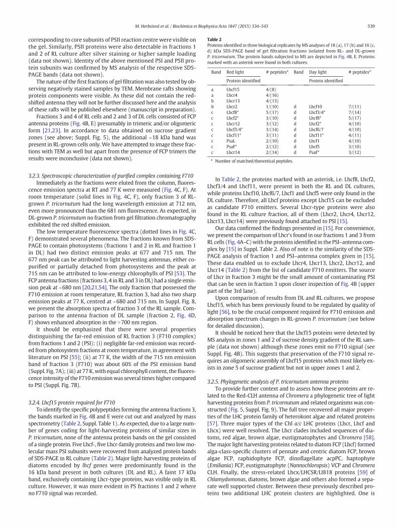
Table 2Proteins identified in three biological replicates byMS analyses of 18 (a), 17 (b) and 16 (c,d) kDa SDS-PAGE band of gel filtration fractions isolated from RL- and DL-grownP. tricornutum. The protein bands subjected to MS are depicted in Fig. 4B, E. Proteinsmarked with an asterisk were found in both cultures.
Band Red light # peptidesa Band Day light # peptidesa
Protein identified Protein identified
a Lhcf15 4/(8)a Lhcr4 4/(16)b Lhcr13 4/(13)b Lhcr2 1/(10) d Lhcf10 7/(11)c Lhcf8* 5/(17) d Lhcf3/4* 7/(14)c Lhcf2* 3/(10) d Lhcf8* 5/(17)c Lhcr12 3/(12) d Lhcf2* 4/(10)c Lhcf3/4* 3/(14) d Lhcf6/7 4/(10)c Lhcf11* 3/(11) d Lhcf11* 4/(11)c PsaL 2/(10) d Lhcf1 4/(10)c PsaF* 2/(12) d Lhcf5 3/(10)c Lhcr14 2/(14) d PsaF* 3/(12)
a Number of matched/theoretical peptides.
539M. Herbstová et al. / Biochimica et Biophysica Acta 1847 (2015) 534–543
corresponding to core subunits of PSII reaction centre were visible onthe gel. Similarly, PSII proteins were also detectable in fractions 1and 2 of RL culture after silver staining or higher sample loading(data not shown). Identity of the above mentioned PSI and PSII pro-tein subunits was confirmed by MS analysis of the respective SDS-PAGE bands (data not shown).
The nature of the first fractions of gel filtrationwas also tested by ob-serving negatively stained samples by TEM. Membrane rafts showingprotein components were visible. As these did not contain the red-shifted antenna they will not be further discussed here and the analysisof these rafts will be published elsewhere (manuscript in preparation).
Fractions 3 and 4 of RL cells and 2 and 3 of DL cells consisted of FCPantenna proteins (Fig. 4B, E) presumably in trimeric and/or oligomericform [21,23]. In accordance to data obtained on sucrose gradientzones (see above; Suppl. Fig. 5), the additional ~18 kDa band waspresent in RL-grown cells only. We have attempted to image these frac-tions with TEM as well but apart from the presence of FCP trimers theresults were inconclusive (data not shown).
3.2.3. Spectroscopic characterization of purified complex containing F710Immediately as the fractions were eluted from the column, fluores-
cence emission spectra at RT and 77 K were measured (Fig. 4C, F). Atroom temperature (solid lines in Fig. 4C, F), only fraction 3 of RL-grown P. tricornutum had the long wavelength emission at 712 nm,even more pronounced than the 681 nm fluorescence. As expected, inDL-grown P. tricornutum no fraction from gel filtration chromatographyexhibited the red shifted emission.
The low temperature fluorescence spectra (dotted lines in Fig. 4C,F) demonstrated several phenomena. The fractions known from SDS-PAGE to contain photosystems (fractions 1 and 2 in RL and fraction 1in DL) had two distinct emission peaks at 677 and 715 nm. The677 nm peak can be attributed to light harvesting antennas, either co-purified or partially detached from photosystems and the peak at715 nm can be attributed to low-energy chlorophylls of PSI [53]. TheFCP antenna fractions (fractions 3, 4 in RL and 3 inDL) had a single emis-sion peak at ~680 nm [20,21,54]. The only fraction that possessed theF710 emission at room temperature, RL fraction 3, had also two sharpemission peaks at 77 K, centred at ~680 and 715 nm. In Suppl. Fig. 8,we present the absorption spectra of fraction 3 of the RL sample. Com-parison to the antenna fraction of DL sample (fraction 2, Fig. 4D,F) shows enhanced absorption in the N700 nm region.
It should be emphasized that there were several propertiesdistinguishing the far-red emission of RL fraction 3 (F710 complex)from fractions 1 and 2 (PSI): (i) negligible far-red emission was record-ed fromphotosystem fractions at room temperature, in agreementwithliterature on PSI [55]; (ii) at 77 K, the width of the 715 nm emissionband of fraction 3 (F710) was about 60% of the PSI emission band(Suppl. Fig. 7A); (iii) at 77K,with equal chlorophyll content, thefluores-cence intensity of the F710 emissionwas several times higher comparedto PSI (Suppl. Fig. 7B).
3.2.4. Lhcf15 protein required for F710To identify the specific polypeptides forming the antenna fractions 3,
the bands marked in Fig. 4B and E were cut out and analyzed by massspectrometry (Table 2, Suppl. Table 1). As expected, due to a large num-ber of genes coding for light-harvesting proteins of similar sizes inP. tricornutum, none of the antenna protein bands on the gel consistedof a single protein. Five Lhcf-, five Lhcr-family proteins and two lowmo-lecular mass PSI subunits were recovered from analyzed protein bandsof SDS-PAGE in RL culture (Table 2). Major light-harvesting proteins ofdiatoms encoded by lhcf genes were predominantly found in the16 kDa band present in both cultures (DL and RL). A faint 17 kDaband, exclusively containing Lhcr-type proteins, was visible only in RLculture. However, it was more evident in PS fractions 1 and 2 whereno F710 signal was recorded.
In Table 2, the proteins marked with an asterisk, i.e. Lhcf8, Lhcf2,Lhcf3/4 and Lhcf11, were present in both the RL and DL cultures,while proteins Lhcf10, Lhcf6/7, Lhcf1 and Lhcf5 were only found in theDL culture. Therefore, all Lhcf proteins except Lhcf15 can be excludedas candidate F710 emitters. Several Lhcr-type proteins were alsofound in the RL culture fraction, all of them (Lhcr2, Lhcr4, Lhcr12,Lhcr13, Lhcr14) were previously found attached to PSI [15].
Our data confirmed the findings presented in [15]. For convenience,we present the comparison of Lhcr's found in our fractions 1 and 3 fromRL cells (Fig. 4A–C)with the proteins identified in the PSI–antenna com-plex by [15] in Suppl. Table 2. Also of note is the similarity of the SDS-PAGE analysis of fraction 1 and PSI–antenna complex given in [15].These data enabled us to exclude Lhcr4, Lhcr13, Lhcr2, Lhcr12, andLhcr14 (Table 2) from the list of candidate F710 emitters. The sourceof Lhcr in fraction 3 might be the small amount of contaminating PSIthat can be seen in fraction 3 upon closer inspection of Fig. 4B (upperpart of the 3rd lane).
Upon comparison of results from DL and RL cultures, we proposeLhcf15, which has been previously found to be regulated by quality oflight [56], to be the crucial component required for F710 emission andabsorption spectrum changes in RL-grown P. tricornutum (see belowfor detailed discussion).
It should be noticed here that the Lhcf15 proteins were detected byMS analysis in zones 1 and 2 of sucrose density gradient of the RL sam-ple (data not shown) although these zones emit no F710 signal (seeSuppl. Fig. 4B). This suggests that preservation of the F710 signal re-quires an oligomeric assembly of Lhcf15 proteins which most likely ex-ists in zone 5 of sucrose gradient but not in upper zones 1 and 2.
3.2.5. Phylogenetic analysis of P. tricornutum antenna proteinsTo provide further context and to assess how these proteins are re-
lated to the Red-CLH antenna of Chromera a phylogenetic tree of lightharvesting proteins from P. tricornutum and related organismswas con-structed (Fig. 5, Suppl. Fig. 9). The full tree recovered all major proper-ties of the LHC protein family of heterokont algae and related proteins[57]. Three major types of the Chl a/c LHC proteins (Lhcr, Lhcf andLhcx) were well resolved. The Lhcr clades included sequences of dia-toms, red algae, brown algae, eustigmatophytes and Chromera [58].Themajor light harvesting proteins related to diatom FCP (Lhcf) formedalga-class-specific clusters of pennate and centric diatom FCP, brownalgae FCP, raphidophyte FCP, dinoflagellate acpPC, haptophyte(Emiliania) FCP, eustigmatophyte (Nannochloropsis) VCP and ChromeraCLH. Finally, the stress-related Lhcx/LHCSR/LI818 proteins [59] ofChlamydomonas, diatoms, brown algae and others also formed a sepa-rate well supported cluster. Between these previously described pro-teins two additional LHC protein clusters are highlighted. One is

Fig. 5. Details from a phylogenetic analysis of Lhc sequences (Suppl. Fig. 9) showing thesequences closely related to Lhcf15, the protein required for the far-red emission inP. tricornutum. Amino acid sequences were aligned by MAFFT using E-INS-i settings withdefault parameters and the phylogeny was constructed by MrBayes 3.2.2 usingGTR+Γ+I model. Node supports (in percents) are indicated.
540 M. Herbstová et al. / Biochimica et Biophysica Acta 1847 (2015) 534–543
related to the Chromera Red-CLHproteins as described recently [42] andthe other one is centred around the aforementioned Lhcf15 protein ofP. tricornutum.
The LHC phylogenetic tree showed that the Lhcf15 protein is diver-gent frommajor diatom Lhcf sequences and embedded in an interestingassortment of LHCs with good support. There were twomore sequencesof P. tricornutum (Lhcf13 and Lhcf14), two from a centric diatomThalassiosira (Lhcf7 and Lhcf11) and one additional heterokont sequenceof chrysophycean alga Ochromonas. Curiously, several haptophyte LHCsequences also showed high similarity to P. tricornutum Lhcf15. Thesame clade of antenna proteins containing haptophyte sequences andLhcf15 of P. tricornutum has been previously recovered in a broad LHCanalysis (labelled as cluster VIIb) [57] or in a survey of P. tricornutumLHCs [23,60].
4. Discussion
The absorption spectra presented in Fig. 1B showed that the thyla-koidmembrane of P. tricornutum responds to changed spectrum of inci-dent radiation by chromatic adaptation involving synthesis of a novelcomponent of the light harvestingmachinery containing a distinct spec-tral form of Chl a absorbing above 690 nm. The presence of such low-energy pigment in RL-grown cells was strikingly demonstrated by thesignificant change in room temperature fluorescence emission spec-trum (Fig. 1A) which helped us track the F710 presence during bio-chemical analyses. As suggested before [41], this change of roomtemperature fluorescence spectrum might be used in searches for fur-ther candidate species possessing red-shifted light harvesting antennas.Comparison of absorption capabilities of the RL and DL adapted culturesabove 690 nm indicated that the RL-grown cells could absorb as muchas 45%more far-red light. Thismight prove to be a significant advantagein an environment where most of the photosynthetically active radia-tion is absorbed by other photosynthetic organisms (see [61] for discus-sion of light-harvesting function of red-shifted pigments).
We find it important to emphasize that the pronounced changes inlight harvesting apparatus described here were provoked by broadbandlight sources. These were used intentionally to ensure that weak excita-tionwas available across the spectrum so that all potential light sensors,such as phytochromes [62], present in the cells were covered. This wasdone in order to maintain ecologically relevant cultivation conditionsusing light quality closer to natural environmentwheremonochromaticillumination cannot be assumed.
One of the issues pertaining to the spectroscopic properties of thecomplexes containing red-shifted Chl a that warrants further discussionis the contribution of PSI to the far-red emission. This issue breaks downto two questions: (i) the contribution of PSI itself; and (ii) the associa-tion of the red-shifted antennawith photosystems. As for the first ques-tion, it has been shown before that diatom PSI does not emit stronglyabove 700 nm at room temperature [55] in agreement with our results(Fig. 4C, F). This is the direct consequence of the fact that only a small
number (~3) of red-shifted Chl a is present in PSI in diatoms [63]. Con-sidering that the relative intensity of emission is proportional to theprobability of an exciton being found in respective pigment pool, onecan assume that the statistics of the energy equilibration in such com-plex containing hundred(s) of Chl a molecules in total [55] is in favourof themore abundant, if higher, energy levels. Thus the far red emissionbecomes visible only at cryogenic temperatureswhere the system is de-void of thermal energy needed to populate the higher (~680 nm) ener-gy levels of the system. But even at 77 K, in case a larger number of FCPantenna is present in addition to the PSI core, the relative contributionof PSI emission in the 715–720 nm region can be diminished [13,63,64]. In this regard, it is illustrative to refer to Suppl. Fig. 4 showing thesucrose gradient zone 5 of the DL culture apparently lacking the far-red emission. Only after the zone was subjected to further purificationto separate the PSI–antenna fraction from other complexes, the red-shifted PSI emission became manifested.
In addition to that, even though peripheral antenna system associat-ed specificallywith PSI exists, these complexes lack the far-red emission[55,64,65]. Thus, if there is a complex present that exhibits the red-shifted emission at room temperature (by virtue of its larger numberof red-shifted Chl a states, in contrast to PSI), it would dominate theemission spectrum at low temperature as well, likely obscuring anyPSI core contribution. We thus assume that the most likely source ofred emission in dense DL cultures as seen in Suppl. Fig. 2 is the F710emitting antenna induced by self-shading of the dense culture.
At closer inspection, our low temperature spectra from gel filtrationfractions provided another helpful tool for disentangling antenna andPSI emission in the case of isolated supercomplexes (Suppl. Fig. 7). Al-though the red PSI emission signal is positioned at the samewavelength(~715 nmat 77 K) as the F710 antenna signal, it is clearlymuch broader(30 nm full width at half maximum vs. 19 nm for F710) [65]. The F710signal is of equal width as the corresponding emission of major FCP an-tenna at 680 nmupon cooling to 77K. On the other hand, even if the redemitter is unambiguously assigned to the antenna, the issue of its con-nection to photosystems remains. While the far red forms of Chl a aretypically associated with (plant) PSI, it has been shown before thatred-shifted antenna complexes are capable of driving PSII photochemis-try [40,41], despite the difference in the respective energies (corre-sponding to ~680 nm for PSII and ~700 nm for the low antenna states).
While a more detailed investigation of the larger-scale organizationof themembrane complexes and the physiological aspects of the far-redantennawere beyond the scope of thepresent paper, our results suggestthat the far-red antenna in P. tricornutum is also associatedwith PSII (assuggested already by Fujita and Ohki [32]), as its emission respondedstrongly to changes of the PSII RC redox state, as seen from the fluores-cence induction curves (Supplementary Fig. 3, cf. [41]), and the reactionto DCMU, an inhibitor of PSII electron transfer (Fig. 2). On the otherhand, the association to PSI cannot be presently excluded based on thefluorescence data alone. Since the diatom thylakoid membrane is notdifferentiated into specific domains corresponding to grana and stromallamellae of plant thylakoids, with either PSII or PSI dominating the pro-tein complement [66], it is likely that the F710 complex serves as an ex-ternal antenna of both photosystems, mirroring the situation in C. velia[41].
As for the identity of the complex carrying the far-red forms of Chl a,we identified the protein marked Lhcf15 as the most likely candidate.This is in agreement with the previous results [56,60] that the Lhcf15protein expression is light quality-dependent and its elevated levelhas been observed in response to the exposure to the red light or toprolonged dark treatment, respectively. In the dataset of [56], Lhcf15was by far the most upregulated antenna protein (about 10×), andalso Lhcf13, Lhcf2 and Lhcf5 showed upregulation on RL by about 20%.It can be concluded that Lhcf15 production is induced by red light expo-sure and it is a key component of a newly formed red antenna complexemitting F710 signal. Most likely, this antenna complex is formed by ag-gregation of Lhcf15 alone and/or possibly other Lhcf-family proteins of

Fig. 6. Illustration of utility of red-shifted antenna systems in conditions where water ab-sorption is combined with screening of photosynthetically active radiation by Chl a-containing phototrophs in the water column. Simulated light conditions under onemetre thick water layer (blue) and in additional presence of dinoflagellates (red) orcyanobacteria (green), both adjusted OD670 = 1. Incident solar spectrum is shown inblack. For details see text.
541M. Herbstová et al. / Biochimica et Biophysica Acta 1847 (2015) 534–543
existing or only slightly modified FCP trimers or higher oligomers par-ticipate in the oligomerization process. Determining other key proteinsin this process seems challenging. Although some differences in thepresence of the major FCP components between RL and DL culturewere observed in our MS/MS data (Table 2), e.g. Lhcf10, Lhcf6/7, Lhcf1and Lhcf5 besides some Lhcr proteins, it appears premature to make aconclusion based on these data. As seen in Table 2, Lhcf15was accompa-nied in the ~18 kDa band (a) (Fig. 4B) in SDS-PAGE from RL culture byanother protein, Lhcr4. However, previous proteomic studies [56,60]which also identified this protein in P. tricornutum cells did not indicateits light-dependent regulation, as opposed to Lhcf15. Moreover, as ourlater investigation indicated, the Lhcr4 is indeed, as its label suggests,a part of the antenna associated with PSI (manuscript in preparation)known to lack the far-red emission, we excluded this protein from fur-ther consideration in regard to the origin of the F710 emission.
The pigment composition of RL versus DL cells shows an interestinganalogy with the situation in Chromera [42] where the Red–CLH com-plex is enriched in violaxanthin, a xanthophyll cycle pigment, at the ex-pense of isofucoxanthin, the major light harvesting carotenoid in thatspecies [44,67]. Diatoms use fucoxanthin in major light harvesting role[68] and diadinoxanthin in the xanthophyll cycle. The RL cells containedsignificantly lower amounts of Chl c and fucoxanthin (relatively to Chla)when compared toDL cells. This is surprising because if the RL culturewas light-limited, as the growth rates suggested, there should havebeen more light harvesting antennas present to compensate for thelow irradiation. FCP antennas contain most of cellular fucoxanthin andChl c. Hence, if the content of these pigments decreased in RL cells it in-dicated that the F710 related proteins are depleted in these pigmentsand probably contain mostly Chl a and diadinoxanthin. Since Red-CLHand F710 proteins are not closely phylogenetically related (see below)this similarity of carotenoid content appears to be a peculiar convergentfeature. Unfortunately, the extreme fragility of the far-red complex ofP. tricornutum seems to preclude any detailed investigation of excitationenergy flows within the complex and their comparison to “canonical”fucoxanthin containing complexes.
The phylogenetic analysis of P. tricornutum antenna proteins (Fig. 5,Suppl. Fig. 9) showed that Lhcf15 protein is amember of awell support-ed protein clade formed by complexes of varied phylogenetic origin,comprising both diatom and haptophyte sequences. It would be proba-bly premature to suggest that all these sequences are related to thechromatic adaptation—we have not found Lhcf13 and Lhcf14 in any re-lation to the F710 antenna and we have not found the F710 signal incentric Cyclotella (not shown, cf. [32]), suggesting that Thalassiosira, an-other centric diatom, will also lack it. However, it can be assumed thatthere has to be some functional aspect shared by these proteins, perhapsassociated with the assembly of specific oligomeric complexes.
The placement of haptophyte sequences within the Lhcf15 clade isparticularly surprising. Haptophytes are an ancient evolutionary lineagesharing common ancestor with heterokonts (stramenopiles), dinoflagel-lates, apicomplexans and ciliates, together forming the chromalveolatesupergroup [69]. Chromalveolates have common plastid origin in sec-ondary endosymbiosis of a red alga [3]. Although haptophytes have along evolutionary history independent of heterokont lineage [69], theirLHCs align firmly along the other predominantly Chl a/c containing an-tenna proteins [57].
It has been proposed recently [51] that the F710 fluorescence (de-tected in low temperature spectra) is related to NPQ capability of dia-tom species, particularly P. tricornutum. As NPQ capacity was not afocus of our work we cannot comment upon this phenomenon as thegrowth conditions used differ considerably from the cited work. TheF710 protein complex is an excellent exciton trap and its involvementin NPQ currently cannot be ruled out. Also the hypothesized highercontent of diadinoxanthin in F710 complex points to its possible NPQinvolvement. However, the conditions which enhanced NPQ inP. tricornutum in the work of Lavaud and Lepetit were either stationaryphase of growth (‘medium’NPQ condition) or intermittent growth light
(‘high’ NPQ condition) resulting in very low growth rates [51]. Bothof these conditions are bound to enhance the F710 signal. The 77 Kfluorescence spectra presented are reminiscent of our spectra ofslightly self-shaded cells (Suppl. Fig. 2), also noted previously else-where [70].
Of the organisms known to utilize red-shifted light harvesting an-tennas, Acaryochloris has been found epiphytic on red algae [71] andin an ascidian, shaded by another cyanobacterium Prochloron [36].H. hongdechloris comes from dense biofilms of stromatolites [72]. Red-shifted Ostreobium was found inside a coral, shaded by dinoflagellates[38]. Chromera is known to reside in or around corals aswell [73]. There-fore the capability to shift at least part of the chlorophyll QY absorptionband to or even beyond 700 nm has appeared several times in marineorganisms dwelling in the shaded environment of both warm andcold (P. tricornutum) seas [74].
Due to the fact that water preferentially absorbs red light, theenvironmental niche for red-shifted light harvesting antennas is limitedto at most the first few metres below surface. In the case ofP. tricornutum, the F710 antenna is probably an adaptation to a benthiclifestyle in shallow coastal waters [75]. P. tricornutum is capable of in-ducing some F710 signal by self-shading (Suppl. Fig. 2) but the full de-velopment of F710 and enhanced absorption above 700 nm is onlypossible under significantly red-shifted growth light. Therefore such en-vironmental shading cannot be provided just by high density popula-tion of diatoms or other algae with similar absorption properties. Solarspectrum screening needed for creating ‘red’ light in situ can be provid-ed by e.g. the light harvesting proteins of cyanobacteria (phycobilins) ordinoflagellates (PCP) living ‘above’ P. tricornutum [42] whether in watercolumn or in a biofilm. For illustration of the above stated points wepresent in Fig. 6 the simulation of the light conditions at one metredepth in presence of shading by dinoflagellate or cyanobacterial cells.The spectra were modelled using solar spectrum at the ground level(AM1.5 global-ASTMG173, [76])modified bywater absorption [78], ab-sorption spectrum of PCP from Symbiodinium sp. [77] or absorptionspectrum of phycobilin-containing cyanobacterium (SynechococcusPCC 7002), both spectra were adjusted to OD= 1 at 670 nm.
Based on these considerations we can predict that members of oneor both of these groups should be present in the P. tricornutum habitatat least for part of the annual cycle.

542 M. Herbstová et al. / Biochimica et Biophysica Acta 1847 (2015) 534–543
4.1. Conclusions
We have described the molecular basis of chromatic adaptation ofpennate diatoms inmodel species P. tricornutum. Based on phylogeneticanalyses, Lhcf15, the key protein likely required for the red-shifted olig-omeric FCP complex formation, is not related to previously describedred-shifted light harvesting proteins of C. velia and Ostreobium sp.Therefore, these adaptations most probably evolved independentlyand represent a case of convergent evolution. P. tricornutum is a thirdwell described case of marine eukaryote alga possessing the capabilityto shift its absorption towards 700 nm. It can be predicted that the num-ber of algae which independently adapted to their environment by de-veloping red-shifted light harvesting antennas based on Chl a is evenhigher. We are currently broadening our search for red-shifted lightharvesting antennas of algae, with a particular focus on heterokonts.The haptophyte algae also seem likely candidates possessing Chl abased red-shifted antennas.
6. Transparency Document
The Transparency Document associated with this article can befound, in the online version.
Acknowledgements
This research was supported by the Czech Science Foundationunder the grant number P501/12/G055 and by institutional supportRVO:60077344. M.H. gratefully acknowledges financial support bythe ALGAMAN project Reg. No. CZ.1.07/2.3.00/20.0203 co-financedby the European Social Fund. We gratefully acknowledge the techni-cal assistance of I. Hunalova and especially F. Matousek.
Appendix A. Supplementary data
Supplementary data to this article can be found online at http://dx.doi.org/10.1016/j.bbabio.2015.02.016.
References
[1] R.C. Dugdale, F.P. Wilkerson, Silicate regulation of new production in the equatorialPacific upwelling, Nature 391 (1998) 270–273.
[2] L.W. Parfrey, D.J.G. Lahr, A.H. Knoll, L.A. Katz, Estimating the timing of early eukary-otic diversification withmultigene molecular clocks, Proc. Natl. Acad. Sci. U. S. A. 108(2011) 13624–13629.
[3] T. Cavalier-Smith, Principles of protein and lipid targeting in secondarysymbiogenesis: euglenoid, dinoflagellate, and sporozoan plastid origins and the eu-karyote family tree, J. Eukaryot. Microbiol. 46 (1999) 347–366.
[4] P.G. Falkowski, M.E. Katz, A.H. Knoll, A. Quigg, J.A. Raven, O. Schofield, F.J.R. Taylor,The evolution of modern eukaryotic phytoplankton, Science 305 (2004) 354–360.
[5] A. Falciatore, C. Bowler, Revealing the molecular secrets of marine diatoms, Annu.Rev. Plant Biol. 53 (2002) 109–130.
[6] W.H.C.F. Kooistra, G. Forlani, M. De Stefano, Adaptations of araphid pennate diatomsto a planktonic existence, Mar. Ecol. 30 (2009) 1–15.
[7] P.A. Sims, D.G. Mann, L.K. Medlin, Evolution of the diatoms: insights from fossil, bi-ological and molecular data, Phycologia 45 (2006) 361–402.
[8] W.H.C.F. Kooistra, R. Gersonde, L. Medlin, D.G. Mann, The origin and evolution of thediatoms: their adaptation to a planktonic existence, in: P.G. Falkowski, A.H. Knoll(Eds.), Evolution of Primary Producers in the Sea, Academic Press, Amsterdam,ISBN: 978-0-12-370518-1, 2007, pp. 207–249.
[9] E.V. Armbrust, J.A. Berges, C. Bowler, B.R. Green, D. Martinez, N.H. Putnam, S. Zhou,A.E. Allen, K.E. Apt, M. Bechner, M.A. Brzezinski, B.K. Chaal, A. Chiovitti, A.K. Davis,M.S. Demarest, J.C. Detter, T. Glavina, D. Goodstein, M.Z. Hadi, U. Hellsten, M.Hildebrand, B.D. Jenkins, J. Jurka, V.V. Kapitonov, N. Kröger, W.W.Y. Lau, T.W. Lane,F.W. Larimer, J.C. Lippmeier, S. Lucas, M. Medina, A. Montsant, M. Obornik, M.S.Parker, B. Palenik, G.J. Pazour, P.M. Richardson, T.A. Rynearson, M.A. Saito, D.C.Schwartz, K. Thamatrakoln, K. Valentin, A. Vardi, F.P. Wilkerson, D.S. Rokhsar, Thegenome of the diatom Thalassiosira pseudonana: ecology, evolution, and metabo-lism, Science 306 (2004) 79–86.
[10] C. Bowler, A.E. Allen, J.H. Badger, J. Grimwood, K. Jabbari, A. Kuo, U. Maheswari, C.Martens, F. Maumus, R.P. Otillar, et al., The Phaeodactylum genome reveals the evo-lutionary history of diatom genomes, Nature 456 (2008) 239–244.
[11] D.G. Durnford, J.A. Deane, S. Tan, G.I. McFadden, E. Gantt, B.R. Green, A phylogeneticassessment of the eukaryotic light-harvesting antenna proteins, with implicationsfor plastid evolution, J. Mol. Evol. 48 (1999) 59–68.
[12] M. Nymark, K.C. Valle, T. Brembu, K. Hancke, P. Winge, K. Andresen, G. Johnsen, A.M.Bones, An integrated analysis of molecular acclimation to high light in the marinediatom Phaeodactylum tricornutum, PLoS ONE 4 (2009) e7743.
[13] T. Veith, J. Brauns, W. Weisheit, M. Mittag, C. Büchel, Identification of a specificfucoxanthin-chlorophyll protein in the light harvesting complex of photosystem Iin the diatom Cyclotella meneghiniana, Biochim. Biophys. Acta 1787 (2009)905–912.
[14] B. Lepetit, D. Volke, M. Gilbert, C. Wilhelm, R. Goss, Evidence for the existence of oneantenna-associated, lipid-dissolved and two protein-bound pools of diadinoxanthincycle pigments in diatoms, Plant Physiol. 154 (2010) 1905–1920.
[15] I. Grouneva, A. Rokka, E.-M. Aro, The thylakoid membrane proteome of two marinediatoms outlines both diatom-specific and species-specific features of the photosyn-thetic machinery, J. Prot. Res. 10 (2011) 5338–5353.
[16] G. Peers, T.B. Truong, E. Ostendorf, A. Busch, D. Elrad, A.R. Grossman, M. Hippler, K.K.Niyogi, An ancient light-harvesting protein is critical for the regulation of algal pho-tosynthesis, Nature 462 (2009) 518–521.
[17] Y. Zhu, J.E. Graham, M. Ludwig, W. Xiong, R.M. Alvey, G. Shen, D.A. Bryant, Roles ofxanthophyll carotenoids in protection against photoinhibition and oxidative stressin the cyanobacterium Synechococcus sp. strain PCC 7002, Arch. Biochem. Biophys.504 (2010) 86–89.
[18] A.G. Koziol, T. Borza, K.-I. Ishida, P. Keeling, R.W. Lee, D.G. Durnford, Tracing the evo-lution of the light-harvesting antennae in chlorophyll a/b-containing organisms,Plant Physiol. 143 (2007) 1802–1816.
[19] C. Büchel, Fucoxanthin-chlorophyll proteins in diatoms: 18 and 19 kDa subunits as-semble into different oligomeric states, Biochemistry 42 (2003) 13027–13034.
[20] B. Lepetit, D. Volke, M. Szabó, R. Hoffmann, G. Garab, C. Wilhelm, R. Goss, Spectro-scopic and molecular characterization of the oligomeric antenna of the diatomPhaeodactylum tricornutum, Biochemistry 46 (2007) 9813–9822.
[21] Z. Gardian, R. Litvín, D. Bína, F. Vácha, Supramolecular organization of fucoxanthin–chlorophyll proteins in centric and pennate diatoms, Photosynth. Res. 121 (2014)79–86.
[22] J. Joshi-Deo, M. Schmidt, A. Gruber, W. Weisheit, M. Mittag, P.G. Kroth, C. Büchel,Characterization of a trimeric light-harvesting complex in the diatomPhaeodactylum tricornutum built of FcpA and FcpE proteins, J. Exp. Bot. 61 (2010)3079–3087.
[23] K. Gundermann, M. Schmidt, W.Weisheit, M. Mittag, C. Büchel, Identification of sev-eral sub-populations in the pool of light harvesting proteins in the pennate diatomPhaeodactylum tricornutum, Biochim. Biophys. Acta 1827 (2013) 303–310.
[24] R. Nagao, S. Takahashi, T. Suzuki, N. Dohmae, K. Nakazato, T. Tomo, Comparison ofoligomeric states and polypeptide compositions of fucoxanthin chlorophyll a/c-binding protein complexes among various diatom species, Photosynth. Res. 117(2013) 281–288.
[25] J.S. Brown, Fluorometric evidence for participation of chlorophyll a-695 in system 2of photosynthesis, Biochim. Biophys. Acta 143 (1967) 391–398.
[26] C.S. French, Changes with age in absorption spectrum of chlorophyll a in a diatom,Arch. Mikrobiol. 59 (1967) 93–103.
[27] K. Sugahara, N. Murata, A. Takamiya, Fluorescence of chlorophyll in brown algae anddiatoms, Plant Cell Physiol. 12 (1971) 377–385.
[28] H. Jupin, G. Giraud, Modification du spectre d'absorption dans le rouge lointaind'unediatomee cultivee en lumiere rouge, Biochim. Biophys. Acta 226 (1971) 98–102.
[29] J. Goedheer, Chlorophyll a forms in Phaeodactylum tricornutum: comparison withother diatoms and brown algae, Biochim. Biophys. Acta 314 (1973) 191–201.
[30] S. Shimura, Y. Fujita, Some properties of the chlorophyll fluorescence of the diatomPhaeodactylum tricornutum, Plant Cell Physiol. 14 (1973) 341–352.
[31] L. Caron, H. Jupin, C. Berkaloff, Effect of light quality on chlorophyll-forms Ca 684, Ca690 and Ca 699 of the diatom Phaeodactylum tricornutum, Photosynth. Res. 4 (1983)21–33.
[32] Y. Fujita, K. Ohki, On the 710 nm fluorescence emitted by the diatom Phaeodactylumtricornutum at room temperature, Plant Cell Physiol. 45 (2004) 392–397.
[33] H. Jupin, Modifications pigmentaires et ultrastructurales chez la diatomée Detonulasp. cultivée en lumière rouge, Arch. Microbiol. 91 (1973) 19–27.
[34] P. Loughlin, Y. Lin, M. Chen, Chlorophyll d and Acaryochloris marina: current status,Photosynth. Res. 116 (2013) 277–293.
[35] M. Chen, Y. Li, D. Birch, R.D. Willows, A cyanobacterium that contains chlorophyllf—a red-absorbing photopigment, FEBS Lett. 586 (2012) 3249–3254.
[36] M. Kühl, M. Chen, P.J. Ralph, U. Schreiber, A.W.D. Larkum, A niche for cyanobacteriacontaining chlorophyll d, Nature 433 (2005) 820.
[37] E. Wientjes, G. Roest, R. Croce, From red to blue to far-red in Lhca4: how does theprotein modulate the spectral properties of the pigments? Biochim. Biophys. Acta1817 (2012) 711–717.
[38] P. Halldal, Photosynthetic capacities and photosynthetic action spectra of endozoicalgae of the massive coral Favia, Biol. Bull. 134 (1968) 411–424.
[39] B. Koehne, G. Elli, R.C. Jennings, C. Wilhelm, H. Trissl, Spectroscopic and molecularcharacterization of a long wavelength absorbing antenna of Ostreobium sp, Biochim.Biophys. Acta 1412 (1999) 94–107.
[40] C.Wilhelm, T. Jakob, Uphill energy transfer from long-wavelength absorbing chloro-phylls to PS II in Ostreobium sp. is functional in carbon assimilation, Photosynth. Res.87 (2006) 323–329.
[41] E. Kotabová, J. Jarešová, R. Kaňa, R. Sobotka, D. Bína, O. Prášil, Novel type of red-shifted chlorophyll a antenna complex from Chromera velia. I. Physiological rele-vance and functional connection to Photosystems, Biochim. Biophys. Acta 1837(2014) 734–743.
[42] D. Bína, Z. Gardian, M. Herbstová, E. Kotabová, P. Koník, R. Litvín, O. Prášil, J. Tichý, F.Vácha, Novel type of red-shifted chlorophyll a antenna complex from Chromeravelia. II. Biochemistry and spectroscopy, Biochim. Biophys. Acta 1837 (2014)802–810.

543M. Herbstová et al. / Biochimica et Biophysica Acta 1847 (2015) 534–543
[43] H. Pan, J. Šlapeta, D. Carter, M. Chen, Phylogenetic analysis of the light-harvestingsystem in Chromera velia, Photosynth. Res. 111 (2012) 19–28.
[44] J. Tichy, Z. Gardian, D. Bina, P. Konik, R. Litvin, M. Herbstova, A. Pain, F. Vacha, Lightharvesting complexes of Chromera velia, photosynthetic relative of apicomplexanparasites, Biochim. Biophys. Acta 1827 (2013) 723–729.
[45] R.R.L. Guillard, J.H. Ryther, Studies of marine planktonic diatoms. I. Cyclotella nanaHustedt and Detonula confervacea Cleve, Can. J. Microbiol. 8 (1962) 229–239.
[46] H.K. Lichtenthaler, Chlorophylls and carotenoids: pigments of photosyntheticbiomembranes, Methods Enzymol. 148 (1987) 350–382.
[47] S.W. Jeffrey, R.F.C. Mantoura, S.W. Wright, Phytoplankton pigments in oceanogra-phy: guidelines to modern methods, 2nd edition, Monographs on OceanographicMethodology, 10, UNESCO Publishing, Paris, 2005.
[48] K. Katoh, K.-I. Kuma, H. Toh, T. Miyata, MAFFT version 5: improvement in accuracyof multiple sequence alignment, Nucleic Acids Res. 33 (2005) 511–518.
[49] K. Katoh, D.M. Standley, MAFFT multiple sequence alignment software version 7:improvements in performance and usability, Mol. Biol. Evol. 30 (2013) 772–780.
[50] F. Ronquist, J.P. Huelsenbeck, MrBayes 3: Bayesian phylogenetic inference undermixed models, Bioinformatics 19 (2003) 1572–1574.
[51] J. Lavaud, B. Lepetit, An explanation for the inter-species variability of thephotoprotective non-photochemical chlorophyll fluorescence quenching in dia-toms, Biochim. Biophys. Acta 1827 (2013) 294–302.
[52] E. Pfündel, Estimating the contribution of Photosystem I to total leaf chlorophyllfluorescence, Photosynth. Res. 56 (1998) 185–195.
[53] C. Berkaloff, L. Caron, B. Rousseau, Subunit organization of PSI particles from brownalgae and diatoms: polypeptide and pigment analysis, Photosynth. Res. 23 (1990)181–193.
[54] K. Gundermann, C. Büchel, Factors determining the fluorescence yield of fucoxanthin-chlorophyll complexes (FCP) involved in non-photochemical quenching in diatoms,Biochim. Biophys. Acta 1817 (2012) 1044–1052.
[55] T. Veith, C. Büchel, The monomeric photosystem I-complex of the diatomPhaeodactylum tricornutum binds specific fucoxanthin chlorophyll proteins(FCPs) as light-harvesting complexes, Biochim. Biophys. Acta 1767 (2007)1428–1435.
[56] B.S. Costa, A. Jungandreas, T. Jakob, W. Weisheit, M. Mittag, C. Wilhelm, Blue light isessential for high light acclimation and photoprotection in the diatom, J. Exp. Bot. 64(2013) 483–493.
[57] G.E. Hoffman,M.V. Sanchez Puerta, C.F. Delwiche, Evolution of light-harvesting com-plex proteins from Chl c-containing algae, BMC Evol. Biol. 11 (2011) 101.
[58] S.M. Dittami, G. Michel, J. Collén, C. Boyen, T. Tonon, Chlorophyll-binding proteinsrevisited—a multigenic family of light-harvesting and stress proteins from abrown algal perspective, BMC Evol. Biol. 10 (2010) 365.
[59] B. Bailleul, A. Rogato, A. de Martino, S. Coesel, P. Cardol, C. Bowler, A. Falciatore, G.Finazzi, An atypical member of the light-harvesting complex stress-related proteinfamily modulates diatom responses to light, Proc. Natl. Acad. Sci. U. S. A. 107(2010) 18214–18219.
[60] M. Nymark, K.C. Valle, K. Hancke, P. Winge, K. Andresen, G. Johnsen, A.M. Bones, T.Brembu, Molecular and photosynthetic responses to prolonged darkness and subse-quent acclimation to re-illumination in the diatom Phaeodactylum tricornutum, PLoSONE 8 (2013) e58722.
[61] H.-W. Trissl, Long-wavelength absorbing antenna pigments and heterogeneous ab-sorption bands concentrate excitons and increase absorption cross section,Photosynth. Res. 35 (1993) 247–263.
[62] N.C. Rockwell, D. Duanmu, S.S. Martin, C. Bachy, D.C. Price, D. Bhattacharya, A.Z.Worden, J.C. Lagarias, Eukaryotic algal phytochromes span the visible spectrum,Proc. Natl. Acad. Sci. U. S. A. 111 (2014) 3871–3876.
[63] A. Yamagishi, Y. Ikeda, M. Komura, H. Koike, K. Satoh, S. Itoh, Y. Shibata, Shallow sinkin an antenna pigment system of photosystem I of a marine centric diatom,Chaetoceros gracilis, revealed by ultrafast fluorescence spectroscopy at 17 K, J.Phys. Chem. B 114 (2010) 9031–9038.
[64] Y. Ikeda, M. Komura, M. Watanabe, C. Minami, H. Koike, S. Itoh, Y. Kashino, K. Satoh,Photosystem I complexes associated with fucoxanthin-chlorophyll-binding proteinsfrom a marine centric diatom, Biochim. Biophys. Acta 1777 (2008) 351–361.
[65] M. Juhas, C. Büchel, Properties of photosystem I antenna protein complexes of thediatom Cyclotella meneghiniana, J. Exp. Bot. 63 (2012) 3673–3681.
[66] A.M. Pyszniak, S.P. Gibbs, Immunocytochemical localization of photosystem I and thefucoxanthin-chlorophyll a/c light-harvesting complex in the diatom Phaeodactylumtricornutum, Protoplasma 166 (1992) 208–217.
[67] M. Durchan, G. Keşan, V. Slouf, M. Fuciman, H. Staleva, J. Tichý, R. Litvín, D. Bína, F.Vácha, T. Polívka, Highly efficient energy transfer from a carbonyl carotenoid tochlorophyll a in the main light harvesting complex of, Biochim. Biophys. Acta1837 (2014) 1748–1755.
[68] S. Akimoto, A. Teshigahara, M. Yokono, M. Mimuro, R. Nagao, T. Tomo, Excitation re-laxation dynamics and energy transfer in fucoxanthin–chlorophyll a/c-protein com-plexes, probed by time-resolved fluorescence, Biochim. Biophys. Acta 1837 (2014)1514–1521.
[69] F. Burki, N. Okamoto, J.-F. Pombert, P.J. Keeling, The evolutionary history ofhaptophytes and cryptophytes: phylogenomic evidence for separate origins, Proc.Biol. Sci. 279 (2012) 2246–2254.
[70] L. Premvardhan, M. Réfrégiers, C. Büchel, Pigment organization effects on energytransfer and Chl a emission imaged in the diatoms C. meneghiniana andP. tricornutum in vivo: a confocal laser scanning fluorescence (CLSF) microscopyand spectroscopy study, J. Phys. Chem. B 117 (2013) 11272–11281.
[71] A. Murakami, H. Miyashita, M. Iseki, K. Adachi, M. Mimuro, Chlorophyll d in an epi-phytic cyanobacterium of red algae, Science 303 (2004) 1633.
[72] M. Chen, M. Schliep, R.D. Willows, Z.-L. Cai, B.A. Neilan, H. Scheer, A red-shifted chlo-rophyll, Science 329 (2010) 1318–1319.
[73] R.B. Moore, M. Oborník, J. Janouskovec, T. Chrudimský, M. Vancová, D.H. Green, S.W.Wright, N.W. Davies, C.J.S. Bolch, K. Heimann, J. Slapeta, O. Hoegh-Guldberg, J.M.Logsdon, D.A. Carter, A photosynthetic alveolate closely related to apicomplexanparasites, Nature 451 (2008) 959–963.
[74] J. Serôdio, S. Cruz, S. Vieira, V. Brotas, Non-photochemical quenching of chlorophyllfluorescence and operation of the xanthophyll cycle in estuarinemicrophytobenthos,J. Exp. Mar. Biol. Ecol. 326 (2005) 157–169.
[75] A. De Martino, A. Meichenin, J. Shi, K. Pan, C. Bowler, Genetic and phenotypic char-acterization of Phaeodactylum tricornutum (Bacillariophyceae) accessions, J. Phycol.43 (2007) 992–1009.
[76] http://rredc.nrel.gov/solar/spectra/am1.5/.[77] J. Jiang, H. Zhang, Y. Kang, D. Bina, C.S. Lo, R.E. Blankenship, Characterization of the
peridinin–chlorophyll a-protein complex in the dinoflagellate Symbiodinium,Biochim. Biophys. Acta 1817 (2012) 983–989.
[78] H. Buiteveld, J.H.M. Hakvoort, M. Donze, The optical properties of pure water, in: J.S.Jaffe (Ed.)Ocean Optics XII, Proceedings of the Society of Photo-optical Instrumenta-tion Engineers (SPIE), 2258, SPIE — Int. Soc. Optical Engineering, Bellingham, USA,1994, pp. 174–183.
Related Documents