Molecular Basis for Modulation of the p53 Target Selectivity by KLF4 Tobias Brandt 1,2 , Fiona M. Townsley 1 , Daniel P. Teufel 1 , Stefan M. V. Freund 1 , Dmitry B. Veprintsev 1,3,4 * 1 Medical Research Council Laboratory of Molecular Biology, Cambridge, United Kingdom, 2 Max Planck Institute of Biophysics, Frankfurt am Main, Germany, 3 Laboratory of Biomolecular Research, Paul Scherrer Institut, Villigen PSI, Switzerland, 4 Department of Biology, ETH Zu ¨ rich, Zu ¨ rich, Switzerland Abstract The tumour suppressor p53 controls transcription of various genes involved in apoptosis, cell-cycle arrest, DNA repair and metabolism. However, its DNA-recognition specificity is not nearly sufficient to explain binding to specific locations in vivo. Here, we present evidence that KLF4 increases the DNA-binding affinity of p53 through the formation of a loosely arranged ternary complex on DNA. This effect depends on the distance between the response elements of KLF4 and p53. Using nuclear magnetic resonance and fluorescence techniques, we found that the amino-terminal domain of p53 interacts with the KLF4 zinc fingers and mapped the interaction site. The strength of this interaction was increased by phosphorylation of the p53 N-terminus, particularly on residues associated with regulation of cell-cycle arrest genes. Taken together, the cooperative binding of KLF4 and p53 to DNA exemplifies a regulatory mechanism that contributes to p53 target selectivity. Citation: Brandt T, Townsley FM, Teufel DP, Freund SMV, Veprintsev DB (2012) Molecular Basis for Modulation of the p53 Target Selectivity by KLF4. PLoS ONE 7(10): e48252. doi:10.1371/journal.pone.0048252 Editor: Claudine Mayer, Institut Pasteur, France Received July 11, 2012; Accepted September 20, 2012; Published October 30, 2012 Copyright: ß 2012 Brandt et al. This is an open-access article distributed under the terms of the Creative Commons Attribution License, which permits unrestricted use, distribution, and reproduction in any medium, provided the original author and source are credited. Funding: TB was supported by the Medical Research Council (MRC) and the Cambridge European Trust. This research was supported by the MRC. The funders had no role in study design, data collection and analysis, decision to publish, or preparation of the manuscript. Competing Interests: The authors have declared that no competing interests exist. * E-mail: [email protected] Introduction The tumour suppressor p53 is at the centre of a large network responsible for the transcription of genes involved in apoptosis, senescence and cell-cycle arrest. Its function is to maintain genomic integrity upon stress. It is not surprising that in approximately half of all characterised tumours, p53 is mutated and dysfunctional [1]. In addition, p53 also controls the transcription of genes involved in a variety of cell survival processes such as DNA repair, metabolism regulation or embryo implantation [2,3]. Sequence specific recognition of DNA response elements is a key to correct functioning of p53 (reviewed in [4]). However, increasing biophysical evidence suggest that DNA-binding specificity of the transcription factors themselves is not nearly sufficient to explain their binding to so few specific locations identified in the whole genome [5,6,7]. The p53 homologs p63 and p73 also recognise the same response elements [8], but carry out functions distinct from p53 [9]. The situation is further complicated by the fact that the genome contains a very large number of putative p53/p63/p73 response elements [6], most of which are not necessarily involved in transcription regulation. A number of regulatory mechanisms have been suggested that provide transcriptional selectivity and the desired cellular response, such as cell death or survival [2,10]. Such regulatory mechanisms involve the DNA-binding properties of p53, the state of chromatin, the concentration of p53 in the nucleus, p53 protein- interactions and post-translational modifications. All domains of p53 (Figure 1A) are involved in some of these regulatory mechanisms. p53 binds DNA as a dimer of dimers [11,12]. Specific contacts are made by the four p53 DNA-binding domains (p53DBD) to the 20 base pair (bp) canonical consensus response element (RE) defined as two repeats of the RRRCWWGYYY (R = A/G, W = A/T, Y = C/T) decamer [13]. Moreover, it has become evident that p53 can also bind promoters which deviate from this sequence or are only composed of half or three-quarter canonical REs [4,6], allowing p53 to bind a more diverse range of targets. In addition to sequence-specific DNA binding, non- specific contact is made to DNA by the p53 C-terminal domain (p53CTD). Thus, p53 is able to slide along DNA in a search for its REs, providing a more efficient recognition mechanism than random diffusion and dissociation [14,15]. Another proposed mechanism involves conformational changes in the L1 loop of the DNA binding domain [16]. Recently, it has been shown that p53 adopts different conformations when bound to DNA via its CTD or via its DBD [17], or that the acetylation of the DNA- recognition domain leads to an increase in its DNA-binding specificity at lower ionic strength [18]. However, it is not clear how the proposed mechanisms increase the selectivity of p53 for a particular sub-set of available binding sites, as well as its specificity over p63 and p73. Several transcription factors are known to modulate p53 transcriptional activity [4]. The transcription factor gut-enriched Kru ¨ppel-like factor 4 (KLF4, Figure 1A) is involved in cell-cycle progression and proliferation. It has been reported to be either a tumour suppressor or an oncogene, depending on the context [19]. Like p53, KLF4 is necessary to mediate G 1 /S phase arrest [20]. A link between KLF4 and the p53 apoptosis/cell-cycle arrest decision has been established. KLF4 is activated following mild DNA damage and promotes cell-cycle arrest, but is repressed upon severe DNA damage leading to cell death [21]. Cancer cells switch their p53-response under c-irradiation from apoptosis towards PLOS ONE | www.plosone.org 1 October 2012 | Volume 7 | Issue 10 | e48252

Welcome message from author
This document is posted to help you gain knowledge. Please leave a comment to let me know what you think about it! Share it to your friends and learn new things together.
Transcript

Molecular Basis for Modulation of the p53 TargetSelectivity by KLF4
Tobias Brandt1,2, Fiona M. Townsley1, Daniel P. Teufel1, Stefan M. V. Freund1, Dmitry B. Veprintsev1,3,4*
1Medical Research Council Laboratory of Molecular Biology, Cambridge, United Kingdom, 2Max Planck Institute of Biophysics, Frankfurt am Main, Germany, 3 Laboratory
of Biomolecular Research, Paul Scherrer Institut, Villigen PSI, Switzerland, 4Department of Biology, ETH Zurich, Zurich, Switzerland
Abstract
The tumour suppressor p53 controls transcription of various genes involved in apoptosis, cell-cycle arrest, DNA repair andmetabolism. However, its DNA-recognition specificity is not nearly sufficient to explain binding to specific locations in vivo.Here, we present evidence that KLF4 increases the DNA-binding affinity of p53 through the formation of a loosely arrangedternary complex on DNA. This effect depends on the distance between the response elements of KLF4 and p53. Usingnuclear magnetic resonance and fluorescence techniques, we found that the amino-terminal domain of p53 interacts withthe KLF4 zinc fingers and mapped the interaction site. The strength of this interaction was increased by phosphorylation ofthe p53 N-terminus, particularly on residues associated with regulation of cell-cycle arrest genes. Taken together, thecooperative binding of KLF4 and p53 to DNA exemplifies a regulatory mechanism that contributes to p53 target selectivity.
Citation: Brandt T, Townsley FM, Teufel DP, Freund SMV, Veprintsev DB (2012) Molecular Basis for Modulation of the p53 Target Selectivity by KLF4. PLoSONE 7(10): e48252. doi:10.1371/journal.pone.0048252
Editor: Claudine Mayer, Institut Pasteur, France
Received July 11, 2012; Accepted September 20, 2012; Published October 30, 2012
Copyright: � 2012 Brandt et al. This is an open-access article distributed under the terms of the Creative Commons Attribution License, which permitsunrestricted use, distribution, and reproduction in any medium, provided the original author and source are credited.
Funding: TB was supported by the Medical Research Council (MRC) and the Cambridge European Trust. This research was supported by the MRC. The fundershad no role in study design, data collection and analysis, decision to publish, or preparation of the manuscript.
Competing Interests: The authors have declared that no competing interests exist.
* E-mail: [email protected]
Introduction
The tumour suppressor p53 is at the centre of a large network
responsible for the transcription of genes involved in apoptosis,
senescence and cell-cycle arrest. Its function is to maintain
genomic integrity upon stress. It is not surprising that in
approximately half of all characterised tumours, p53 is mutated
and dysfunctional [1]. In addition, p53 also controls the
transcription of genes involved in a variety of cell survival
processes such as DNA repair, metabolism regulation or embryo
implantation [2,3]. Sequence specific recognition of DNA
response elements is a key to correct functioning of p53 (reviewed
in [4]). However, increasing biophysical evidence suggest that
DNA-binding specificity of the transcription factors themselves is
not nearly sufficient to explain their binding to so few specific
locations identified in the whole genome [5,6,7]. The p53
homologs p63 and p73 also recognise the same response elements
[8], but carry out functions distinct from p53 [9]. The situation is
further complicated by the fact that the genome contains a very
large number of putative p53/p63/p73 response elements [6],
most of which are not necessarily involved in transcription
regulation.
A number of regulatory mechanisms have been suggested that
provide transcriptional selectivity and the desired cellular
response, such as cell death or survival [2,10]. Such regulatory
mechanisms involve the DNA-binding properties of p53, the state
of chromatin, the concentration of p53 in the nucleus, p53 protein-
interactions and post-translational modifications. All domains of
p53 (Figure 1A) are involved in some of these regulatory
mechanisms. p53 binds DNA as a dimer of dimers [11,12].
Specific contacts are made by the four p53 DNA-binding domains
(p53DBD) to the 20 base pair (bp) canonical consensus response
element (RE) defined as two repeats of the RRRCWWGYYY
(R=A/G, W=A/T, Y=C/T) decamer [13]. Moreover, it has
become evident that p53 can also bind promoters which deviate
from this sequence or are only composed of half or three-quarter
canonical REs [4,6], allowing p53 to bind a more diverse range of
targets. In addition to sequence-specific DNA binding, non-
specific contact is made to DNA by the p53 C-terminal domain
(p53CTD). Thus, p53 is able to slide along DNA in a search for its
REs, providing a more efficient recognition mechanism than
random diffusion and dissociation [14,15]. Another proposed
mechanism involves conformational changes in the L1 loop of the
DNA binding domain [16]. Recently, it has been shown that p53
adopts different conformations when bound to DNA via its CTD
or via its DBD [17], or that the acetylation of the DNA-
recognition domain leads to an increase in its DNA-binding
specificity at lower ionic strength [18]. However, it is not clear how
the proposed mechanisms increase the selectivity of p53 for a
particular sub-set of available binding sites, as well as its specificity
over p63 and p73.
Several transcription factors are known to modulate p53
transcriptional activity [4]. The transcription factor gut-enriched
Kruppel-like factor 4 (KLF4, Figure 1A) is involved in cell-cycle
progression and proliferation. It has been reported to be either a
tumour suppressor or an oncogene, depending on the context
[19]. Like p53, KLF4 is necessary to mediate G1/S phase arrest
[20]. A link between KLF4 and the p53 apoptosis/cell-cycle arrest
decision has been established. KLF4 is activated following mild
DNA damage and promotes cell-cycle arrest, but is repressed upon
severe DNA damage leading to cell death [21]. Cancer cells switch
their p53-response under c-irradiation from apoptosis towards
PLOS ONE | www.plosone.org 1 October 2012 | Volume 7 | Issue 10 | e48252

cell-cycle arrest upon KLF4 expression [22]. Further, KLF4
induces transcription on the p21WAF1/Cip1 promoter synergistically
with p53, and both proteins co-immunoprecipitate [23]. A ChIP
analysis has shown that KLF4 and p53 are bound to the p21WAF1/
Cip1 and the BAX promoters [22].
We hypothesised that the interaction with another transcrip-
tion factor (TF) may enhance p53 target selectivity via ternary
complex formation on DNA. Most promoters contain REs for
more than one TF, possibly permitting the cooperative DNA-
binding of two interacting TFs. Thus, transcriptional selectivity
may be achieved by the combination of appropriate REs of
more than one TF within a promoter region. Here, we use
biophysical methods to characterise the interaction between
KLF4 and p53 and study its effect on p53 DNA-binding. We
show that p53 and KLF4 directly interact, mapped their
interaction sites using nuclear magnetic resonance spectroscopy,
and demonstrated that phosphorylation of p53 enhances this
interaction. We directly measured the effect of cooperative
binding of two transcription factors to DNA using a fluores-
cence anisotropy assay. Strikingly, KLF4 increases the DNA-
binding affinity of p53, and the increase is dependent on the
distance between the p53 and KLF4 response elements. The
concerted cooperative action of two transcription factors on
binding their respective response elements exemplifies a
regulatory mechanism that enhances transcriptional target
selectivity.
Materials and Methods
Gene, Protein, and DNA PreparationStandard protocols were used for the preparation of plasmids,
proteins and fluorescently labelled DNA. We have used an
engineered neutrally stabilised quadruple mutant (M133L/
V203A/N239Y/N268D) of p53 as it is more suitable for extended
biophysical studies than the wild type protein [24,25,26]. A
detailed description can be found in the supplementary informa-
tion (file Text S1).
Fluorescence Anisotropy MeasurementsFluorescence anisotropy was recorded on a Cary Eclipse
spectrometer (Varian) equipped with a titrator (Hamilton).
Excitation and emission wavelengths were 480 nm and
530 nm, respectively. Solutions containing labelled DNA (2–
4 nM) only, labelled DNA (2–4 nM) and unlabelled protein
(40–4000 nM), or labelled KLF4-FlAsH (20 nM) were stirred
inside the cuvettes. Into this fluorescent solution a second
protein was titrated. The concentration of this titrant varied
between 100 nM and 10 mM depending on the affinity to the
studied DNA/DNA-protein complex in order to obtain high
quality titration curves. All experiments were performed at 20uC
in 5 mM DTT, 25 mM NaPi (pH 7.2), 10% glycerol, 0.2 mg/
mL BSA (Sigma) and 50, 100, 150 or 225 mM NaCl,
corresponding to a total ionic strength of 110, 160, 210 or
285 mM (FA110/160/210/285 buffer). Fluorescence intensities
were measured after an equilibration time of 60 s after each
injection. Data were analysed using laboratory software as has
been described before [27].
Figure 1. KLF4 and p53 directly interact. A: Domain structure of p53 and KLF4. Folded domains are shown in grey. p53 comprises an N-terminaldomain (NTD) consisting of the transactivation domains 1 and 2 (TAD1, TAD2) and the proline-rich domain (PRD), a DNA-binding domain (DBD), atetramerisation domain (TD), and a C-terminal domain (CTD). KLF4 domain boundaries for the transcriptional activation domain (AD) and inhibitorydomain (ID) are approximate. Three zinc fingers (ZF) are encoded at the C-terminus. B: Normalised sedimentation coefficient distributions measuredby FDSV-AUC. 225 nM FlAsH-labelled KLF4 in the absence (black) and in presence of 0.5 mM (grey), 5 mM (dotted line) and 50 mM (dashed line)unlabelled p53. Engineered, neutrally stabilised quadruple mutant M133L/V203A/N239Y/N268D p53 was used throughout this study. C: Directinteraction between p53 and KLF4. Fluorescence anisotropy titration at 110 mM total ionic strength with FlAsH-labelled KLF4 as probe and p53 astitrant. D: Fluorescence anisotropy titrations with a labelled p53 RE (*P). No binding is observed if KLF4 is titrated into a DNA-only solution (circles). If a*P-p53 complex is used as a probe (note higher anisotropy value, squares, FA285 buffer), a binding event can be observed.doi:10.1371/journal.pone.0048252.g001
p53 Target Selectivity Is Mediated by KLF4
PLOS ONE | www.plosone.org 2 October 2012 | Volume 7 | Issue 10 | e48252

Titrations with coumarin labelled p53 peptides were done as
previously described [28]. 200 nM solutions of peptide in
50 mM NaCl, 25 mM NaPi pH 7.2, 10% (v/v) glycerol and
5 mM DTT were used. Titrant solution concentration was 100–
150 mM. Excitation and emission wavelengths were 328 nm and
392 nm, respectively.
Analytical UltracentrifugationWe used XL-I analytical ultracentrifuges (Beckman) equipped
with absorbance or fluorescence detection systems (AVIV
Biomedical). Sedimentation velocity experiments with unlabelled
KLF4 or FlAsH-tagged protein were done in 150 mM NaCl,
25 mM phosphate (pH 7.2), 10% glycerol, BSA (0.2 mg/mL, not
used with absorbance detection) and 1 mM b-mercaptoethanol at
10uC as described previously at 45–50 k rpm [8]. Concentrations
of 5 mM unlabelled KLF4 and 75–225 nM KLF4-FlAsH were
used. 25–50 nM labelled DNA and 1 mM unlabelled KLF4 were
used in experiments detecting protein-DNA complexes. Buffer
density and viscosity were calculated using the SEDNTERP
software. Data analysis to obtain sedimentation coefficient traces
was done with the SEDFIT software [29].
NMRSamples were dialysed twice overnight at 4uC into 25 mM NaPi
buffer at pH 7.2, with the addition of 150 mM NaCl and
5 mM DTT. 35 mL 2H2O (Sigma) were added to 500 mL sample.
All experiments were acquired at 20uC on Bruker DRX-600 or
Avance 700 spectrometers. For binding experiments, 1H, 15N-
HSQC spectra were obtained with 15N-labelled protein in
presence and absence of ligand. To obtain high quality 2D
correlation maps, the p53 TC construct was 2D, 15N-labelled and
relaxation-optimised 1H, 15N TROSY sequences replaced stan-
dard HSQC experiments. Protein concentrations were in the
region of 50–100 mM, whereas the peptide ligands were added in
2–4-fold excess. The backbone 1H,15N and 13C assignments for
KLF4 (367–479) (300 mM) were obtained using a standard set of
triple resonance experiments. 90% of all resonances were assigned
unambiguously. A subset of low intensity resonances suggests the
presence of a population of residues neighbouring prolines in cis-
conformation. All data were processed in Topspin (Bruker,
Karlsruhe) and analysed in Sparky [30]. An initial automated
backbone assignment was obtained with MARS [31], and then
manually completed with in-house perl scripts.
BioinformaticsThe human genome sequence (hg19,GRCh37), masked for
repeats, was analysed for the presence of putative p53 and KLF4
response elements using p53BindingPredictor software [6]. We
used previously reported positional weight matrixes for KLF4 [32]
and p53 [6].
Results
Characterisation of KLF4 and p53We expressed recombinant full-length p53 and KLF4
(Figure 1A). We used a thermo-stable quadruple mutant
(M133L/V203A/N239Y/N268D) of p53 (from here on referred
to as p53) which has previously been extensively studied and
biophysically characterised and is stable for the duration of our
measurements [6,8,25,33,34]. Here, we determined the oligomeric
state of KLF4 with and without DNA and characterised its long-
term stability. Using analytical ultra-centrifugation (AUC) we
found that full-length KLF4 was monomeric in solution and that
the sample was homogenous and did not aggregate as judged by
the presence of only one peak in the sedimentation coefficient
distribution profile (Figure S1A and B). Furthermore, NMR
experiments (see below) showed that the three carboxy-terminal
zinc finger domains remain folded over the length of a 3D-NMR
experiment at 20uC (several days). The remaining parts of the
protein were, as predicted by sequence analysis, natively unfolded.
Using AUC and fluorescence anisotropy titrations, we showed that
the monomer of KLF4 binds DNA in a sequence-specific manner
(Figure S1B and C, Tables S1 and S2). Additional details of AUC
and titration experiments are presented within the supplementary
information results section (file Text S2).
Direct Interaction between KLF4 and p53Several studies have been focussed on the in vivo effect of KLF4
on p53-mediated transcription [20,21,22,23]. In order to test the
hypothesis of concerted DNA-binding by two transcription factors,
we carried out experiments to establish whether KLF4 and p53
interact directly.
Firstly, we established that the full-length proteins interact, using
fluorescence detection analytical ultracentrifugation (FD-AUC)
and FlAsH-labelled KLF4 as a probe. The sedimentation profile of
KLF4-FlAsH showed a peak at S= 1.3, corresponding to
monomeric KLF4, and a second peak at S= 2.0. Upon addition
of unlabelled p53 to labelled KLF4 a third peak at S= 3.0
appeared, indicating formation of a KLF4-p53 complex
(Figure 1B). The peak at S= 2.0 can be attributed to a cross-link
via the cysteines of the FlAsH-tag multimer of KLF4, because it
was not observed for non-labelled KLF4 (Figure S1A).
Secondly, KLF4-FlAsH was used as a fluorescent probe in
fluorescence anisotropy titrations at 110 mM total ionic strength
(Figure 1C). The incremental addition of p53 yielded a Kd of
2.6 mM, confirming the direct interaction between the two
proteins. In order to determine whether the binding affinity of
KLF4 towards p53 is altered if p53 is DNA-bound, we used p53 in
complex with its labelled RE (*P) as a probe (Figure 1C). Tight
binding (Kd=100 nM) between a pre-formed p53-DNA complex
(5:1) and KLF4 was observed at 285 mM ionic strength, whereas
KLF4 did not bind free *P. The affinity increase in comparison to
unbound p53 is striking, especially considering the high ionic
strength used which was necessary to prevent KLF4 binding to
free *P. Hence, the KLF4-p53 interaction is stimulated when p53
is in complex with DNA.
Mapping the Interaction between p53 and KLF4Nuclear magnetic resonance (NMR) spectroscopy was used to
characterise the interaction between KLF4 and p53 at single
residue resolution. Based on reported backbone assignments [35],1H, 15N-heteronuclear single quantum coherence (HSQC) exper-
iments were recorded with 15N-p53NTD (1–93). Spectra in the
absence and presence of KLF4 (271–479) revealed significant
chemical shift perturbations (Figure 2A) and enabled us to identify
residues of p53NTD which were involved in the interface with
KLF4 (Figure 2B). The residues exhibiting the highest chemical
shift perturbations clustered in the TAD2 region of p53
(transactivation domain, residues 40–65). Furthermore, several
residues within the TAD1 region of p53 (residues 1–39) were also
affected by the addition of KLF4. No binding to the proline-rich
region (residues 66–93) was detected. Further experiments with15N-p53DBD (94–292) and 2H, 15N-p53TC (313–393) showed no
binding to KLF4 (271–479) (Figure S2), indicating that the p53 N-
terminus is the sole interaction site with KLF4.
To identify the p53NTD binding site on KLF4, 15N-HSQC
experiments were performed using a 15N-labelled KLF4 (367–479)
construct, in the absence and presence of p53 (1–57) as a ligand.
p53 Target Selectivity Is Mediated by KLF4
PLOS ONE | www.plosone.org 3 October 2012 | Volume 7 | Issue 10 | e48252

The 2D 15N-HSQC spectrum of KLF4 (367–479) was indicative
of a structured domain as a result of the presence of three zinc
finger domains (Figure 3A), but also contained a subset of
resonances at a random coil chemical shift position, characteristic
for unfolded residues. Addition of p53 (1–57) or p53 (1–57)pT55
resulted in significant chemical shift perturbations (Figure 3A and
Figure 2. Identification of interacting residues in p53 by NMR. A: Overlay of 2D 1H, 15N-HSQC NMR spectra for p53 NTD in the absence (blue)and presence of 135 mM KLF4 271–479 (red). B: Weighted chemical shift perturbation map of 15N-p53NTD upon addition of KLF4 271–479. No datawere obtained for prolines and residues with no assigned resonances.doi:10.1371/journal.pone.0048252.g002
p53 Target Selectivity Is Mediated by KLF4
PLOS ONE | www.plosone.org 4 October 2012 | Volume 7 | Issue 10 | e48252
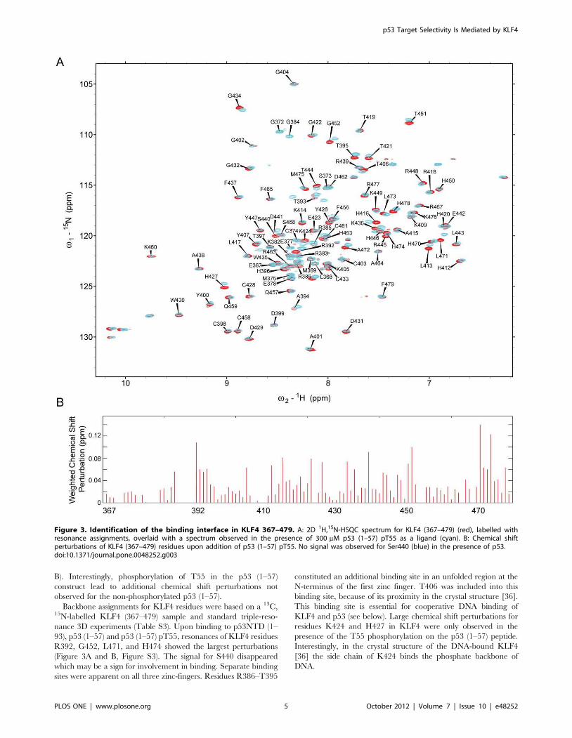
B). Interestingly, phosphorylation of T55 in the p53 (1–57)
construct lead to additional chemical shift perturbations not
observed for the non-phosphorylated p53 (1–57).
Backbone assignments for KLF4 residues were based on a 13C,15N-labelled KLF4 (367–479) sample and standard triple-reso-
nance 3D experiments (Table S3). Upon binding to p53NTD (1–
93), p53 (1–57) and p53 (1–57) pT55, resonances of KLF4 residues
R392, G452, L471, and H474 showed the largest perturbations
(Figure 3A and B, Figure S3). The signal for S440 disappeared
which may be a sign for involvement in binding. Separate binding
sites were apparent on all three zinc-fingers. Residues R386–T395
constituted an additional binding site in an unfolded region at the
N-terminus of the first zinc finger. T406 was included into this
binding site, because of its proximity in the crystal structure [36].
This binding site is essential for cooperative DNA binding of
KLF4 and p53 (see below). Large chemical shift perturbations for
residues K424 and H427 in KLF4 were only observed in the
presence of the T55 phosphorylation on the p53 (1–57) peptide.
Interestingly, in the crystal structure of the DNA-bound KLF4
[36] the side chain of K424 binds the phosphate backbone of
DNA.
Figure 3. Identification of the binding interface in KLF4 367–479. A: 2D 1H,15N-HSQC spectrum for KLF4 (367–479) (red), labelled withresonance assignments, overlaid with a spectrum observed in the presence of 300 mM p53 (1–57) pT55 as a ligand (cyan). B: Chemical shiftperturbations of KLF4 (367–479) residues upon addition of p53 (1–57) pT55. No signal was observed for Ser440 (blue) in the presence of p53.doi:10.1371/journal.pone.0048252.g003
p53 Target Selectivity Is Mediated by KLF4
PLOS ONE | www.plosone.org 5 October 2012 | Volume 7 | Issue 10 | e48252

KLF4 binds exclusively to the N-terminus of p53 via its C-
terminal region, and no binding to 15N-KLF4 (367–479) was
observed for p53TC (293–393) covering the linker region not
tested before (Figure S4). Furthermore, no interaction with p53
was observed for 15N-KLF4 (271–390), the C-terminal part of the
inhibitory domain, which is completely unfolded (Figure S5).
Phosphorylated p53 Binds More Tightly to KLF4Fluorescence anisotropy titrations were used to further char-
acterise the interaction between KLF4 and the p53NTD based on
N-terminal truncated constructs of KLF4 bound to coumarin-
labelled p53NTD peptides p53 (1–57) and (38–57) (Figure S6).
KLF4 (271–479) and KLF4 (367–479) bind p53 (1–57) with a Kdof 30 mM. No binding was detected between p53 (1–57) and KLF4
(391–479), as well as p53 (38–57) and KLF4 (271–479) or KLF4
(391–479). We can conclude, that residues within p53 (1–57) and
KLF4 (367–479) are necessary for the p53-KLF4 interaction.
Further, we studied the effect of phosphorylation at residues
S15, T18, S20, S33, S37, S46, and T55 on p53 binding to KLF4
(367–479) (Table 1). Phosphorylation at S15, T18, S20, S33, and
S37 in the TAD1 region increased the affinity of KLF4 towards
p53 three to four times, whereas phosphorylation at S46 and T55
within TAD2 increased the affinity about 8.5 times. A p53 (1–57)
peptide phosphorylated at all seven serine and threonine positions
showed a 25-fold increase in affinity to KLF4.
KLF4 Enhances Specific DNA-binding Affinity of p53in vitroHaving established that and how KLF4 and p53 interact, we
analysed which effect the protein-protein interaction might have
on p53 DNA-binding properties. To assess the ability of KLF4 and
p53 to bind DNA cooperatively, we developed a fluorescence
anisotropy titration assay for which we generated various
fluorescent DNA constructs (Figure 4A, Table S1). These
constructs contained a 59 fluorescent label (*), followed by a p53
RE (P), a spacer of variable length n, and a KLF4 RE (K). Several
control constructs lacked either the p53 or the KLF4 RE.
Important control experiments showed that KLF4 did not bind
the p53 RE (*P) and vice versa (Tables 2 and S2). With
fluorescence anisotropy titrations the binding of p53 to short
oligonucleotides can be reliably detected [6,8,12,27,37]. We
observed that, in general, p53 showed a higher affinity to
elongated DNA sequences in comparison to *P. This may have
several reasons. Firstly, non-specific binding of the p53 carboxy-
terminal domain is not possible to the short DNA-sequences, but is
likely to contribute to the overall binding if the DNA molecule is
sufficiently long. However, at 285 mM ionic strength this effect
has been shown to be relatively small [18]. Secondly, p53 binds
many DNA sequences weakly such as those which comprise only a
half or three-quarter binding site [4], and which are present in
most DNA sequences. Lastly, the oligonucleotides span a wide
range of lengths and were produced either by PCR or by chemical
synthesis, possibly contributing to changes in the absolute affinity
values. While these experimental limitations may not be avoided,
the important parameter that reflects the impact of KLF4 presence
on the binding of p53 to DNA is the relative increase in the affinity
for DNA of a given length. This parameter is not affected by the
change of the absolute affinity of p53 for DNA in the absence of
KLF4.
p53 was titrated into a solution of labelled DNA, in the presence
or absence of KLF4 (Figure 4A and B). High concentrations
(400 nM) of KLF4 were used in order to fully saturate the labelled
DNA so that only one species (KLF4-bound DNA) was present in
solution. If KLF4 was present, a higher starting anisotropy value
was observed, indicating that KLF4 was bound to DNA. We
found a significant increase in p53 binding affinity to fluorescein-
labelled DNA encoding a p53 RE and a KLF4 RE in the presence
Table 1. Binding of labelled N-terminal p53 peptides to KLF4367–479.
Labelled peptide Kd (KLF4 367–479)/mM* Enhancement ratio
p53 1–57 30611 1
p53 1–57 pS15 9.562.1 3
p53 1–57 pT18 6.961.8 4
p53 1–57 pS20 8.161.6 4
p53 1–57 pS33 6.662.1 4
p53 1–57 pS37 8.561.9 4
p53 1–57 pS46 3.560.9 8
p53 1–57 pT55 3.661.1 8
p53 10–57 hepta P 1.260.3 25
*Experiments were carried out at low ionic strength (110 mM) in order toreliably measure the binding constant. NMR experiments confirmed binding atphysiological ionic strength.doi:10.1371/journal.pone.0048252.t001
Table 2. Binding of p53 to DNA in dependence of addedKLF4 and spacing between cognate binding sites.
DNA
KLF4
construct c (KLF4)/nM Kd ± SD/nM&Enhancementratio
*P FL 0 4567 –
200 3666 1.2
1000 40 1.1
*K – – n.b. –
*PK FL 0 8.161.7 –
400 5.260.6 1.6
*P5K FL 0 8.161.3 –
400 4.660.7 1.8
*P10K FL 0 9.561.8 –
400 5.960.7 1.6
*P30K FL 0 1261.9 –
400 3.560.4 3.4
*P88K FL 0 3.960.7 –
400 1.060.3 3.9
*P185K FL 0 4.060.3 –
400 2.060.7 2.0
*P1000K FL 0 9.563.1 –
400 5.962.0 1.6
*P30K 179–479 0 1261.9 –
400 4.460.6 2.7
*P30K 271–479 0 1261.9 –
400 4.460.6 2.7
*P30K 391–479 0 12 –
400 10 1.2
&Measured by fluorescence anisotropy titrations in FA 285 buffer.doi:10.1371/journal.pone.0048252.t002
p53 Target Selectivity Is Mediated by KLF4
PLOS ONE | www.plosone.org 6 October 2012 | Volume 7 | Issue 10 | e48252

of KLF4 (Table 2). By using spacers of different length, we assessed
the effect of distance between the p53 and KLF4 REs. We found
that the KLF4-dependent affinity increase of p53 to DNA is
highest for DNA sequences with spacers of 30 (*P30K) and 88
(*P88K) base pairs (Table 2, Figure 4B and C). For shorter spacers
of 0, 5, and 10 base pairs the effect is less pronounced, probably
due to steric constraints imposed on the interaction sites. The
increase in p53 affinity to DNA was also diminished if the spacer
length was increased to hundreds of base pairs, suggesting an
optimal distance being required for most efficient p53 RE binding.
Very similar results were obtained for *PK, *P5K, *P10K and
*P30K at an ionic strength of 285 mM (Table 2) and 210 mM
(Table S5).
If a shortened KLF4 construct (391–479), which bound DNA
equally well as full-length KLF4 (Table S2), was used, no effect on
the DNA-binding of p53 was observed (Table 2). Additionally,
corresponding control experiments with several other transcription
factors, such as CP2, HSF1 and YY1, did not yield comparable
increases in affinity (Table S4), confirming that the affinity
enhancement of p53 to its RE can be specifically attributed to
the presence of KLF4. Taken together, both control experiments
showed that enhanced p53 DNA-binding affinity is not observed if
a second transcription factor is bound to the same DNA but does
not interact with p53.
Next, fluorescence anisotropy titrations, designed to ascertain
ternary complex formation, were carried out, using *P DNA to
which KLF4 did not bind (Table S2). No significant increase in
affinity of p53 to *P was observed upon addition of KLF4
(Table 2). Hence, the mere presence of KLF4 in solution was not
enough to enhance p53 binding to DNA. By consequence, an
allosteric mechanism can be ruled out.
KLF4 does not Affect Non-specific Binding of p53 to DNAFurther experiments were done with DNA containing weak
non-canonical p53 REs. Several groups including us have
previously shown that the non-specific interactions of p53 with
DNA are mediated by several lysines of the C-terminal domain
[12,14,38,39]. This interaction is highly sensitive to variations of
the ionic strength. In contrast, specific interactions are significantly
less sensitive to the ionic strength. Consequently, at 210 mM ionic
strength p53 predominantly binds DNA non-specifically via its C-
terminus while at 285 mM ionic strength [17], non-specific
interactions are suppressed and specific interactions are observed.
We assessed the effect of KLF4 on weak p53 binding sites at
285 mM ionic strength using the DNA sequence *108K which
includes a weak three-quarter binding site (Figure S7A, Table S1).
At this ionic strength, non-specific binding is significantly
suppressed [18]. p53 bound weakly to this sequence
(Kd=12006420 nM). However, the affinity was remarkably
increased by addition of 40 nM KLF4 (2-fold, Kd=680693 nM),
100 nM KLF4 (5-fold, Kd=250 nM), and 400 nM KLF4 (8.5-
fold, Kd=140657 nM). We conclude, that KLF4 has the ability to
transform weak p53 REs into more potent ones.
Secondly, we tested if KLF4 also affects non-specific p53-DNA
interactions. These experiments were done at 210 mM ionic
strength, using *47K DNA which also included a weak, three-
quarter binding site. However, at this ionic strength, p53 strongly
and dominantly interacts with DNA non-specifically via its
carboxy-terminal domain [18]. The DNA-binding affinity of p53
towards *47K (2565.2 nM) was not affected by the addition of
400 nM KLF4 (2761.2 nM) (Figure S7B). Overall, these results
indicate that the interaction between KLF4 and p53 enhances
only the specific binding of p53 to DNA.
Figure 4. KLF4 enhances the DNA-binding affinity of p53. A:DNA constructs generated and principle of cooperative fluorescenceanisotropy titrations. Fluorescein (*, star), a p53 RE (P, dark grey), aspacer (n, light grey), and a KLF4 RE (K, black) compose the labelledDNA. The affinity of p53 towards DNA is measured in the presence (+KLF4) and absence (- KLF4) of KLF4. B: Example p53 titration data using*P30K as DNA in the presence (circles) and absence (squares) of KLF4 inFA285 buffer. C: Normalised p53 DNA-binding affinity in the presence of400 nM KLF4 as a function of the distance between the p53 RE and theKLF4 RE.doi:10.1371/journal.pone.0048252.g004
p53 Target Selectivity Is Mediated by KLF4
PLOS ONE | www.plosone.org 7 October 2012 | Volume 7 | Issue 10 | e48252

The Extended Zinc-finger Region of KLF4 is Necessary forIncreased DNA-binding of p53We tested the ability of several deletion constructs of KLF4
(179–479, 271–479, 391–479) to enhance the DNA-binding
affinity of p53. All KLF4 constructs bound DNA as well as the
full-length protein (Table S2). Only the shortest KLF4 construct
(391–479) was not sufficient to increase p53 DNA-binding affinity
to the same extent as full-length KLF4 (Table 2). Elongation of this
KLF4 construct towards the N-terminus (KLF4 367–479) restored
the effect observed for full-length KLF4. A major p53-interaction
site within KLF4 must therefore reside within residues 367–479,
reflecting our NMR results (see above).
In silico Identification of Co-localised KLF4 and p53 REsBoth the p21WAF1/Cip1 and the BAX promoters contain well
documented REs for p53 [40,41], and were shown to respond to
and bind KLF4 [21,22]. Here, we identified putative KLF4 REs in
the vicinity of the p53 binding sites. We found a KLF4 RE
immediately adjacent downstream to the p53 site in the BAX
promoter, and one only 15 bp upstream in the p21 promoter
(Figure 5A). In addition, there were several other KLF4 as well as
p53 REs in those regions. The whole genome analysis suggested
that there is a significant scope for the KLF4-mediated regulation
of the p53 response, as approximately 13% of putative p53
binding sites had a KLF4 site within 200 bp. In the case of
approximately 2% of the REs of both TFs the REs overlapped and
the binding would be mutually exclusive (Figure 5B).
Discussion
KLF4 Increases Target Specificity of p53It has been shown in vivo that p53 and KLF4 synergistically
activate the p21WAF1/Cip1 promoter [23] and are bound simulta-
neously to the promoter regions of p21WAF1/Cip1 and BAX [22].
Here, we present data which demonstrate the means by which the
target selectivity of p53 may be provided: KLF4 increases through
ternary complex formation the DNA-binding affinity of p53.
However, the increase in affinity was observed only, if p53 bound
DNA specifically via its DNA-binding domain. As p53 adopts
different conformations, when bound specifically or non-specifi-
cally to DNA [17], we conclude that the KLF4-p53 interaction is
far stronger for the ‘specific’ conformation that is achieved through
binding of the DBD rather than the CTD. Taken together, our
data suggest that increased levels of KLF4 modulate transcrip-
tional activity of p53 by enriching it at promoters where both
proteins can bind to DNA cooperatively. Importantly, the
enhancement of the DNA binding was observed for both strong
canonical p53 REs as well as for weak non-canonical REs. Given
the abundance of non-canonical REs [4,42], this will further
expand the potential transcription regulation network of p53. We
identified that a significant proportion of putative p53 REs are
close to KLF4 REs. The scope of the p53-KLF4 interaction may,
therefore, well extend beyond the p21WAF1/Cip1 and BAX
promoters. As the interaction between p53 and KLF4 only needs
REs for both proteins within a certain distance range from each
other, it is plausible that such loosely arranged, ‘‘fuzzy’’, sites may
readily appear and disappear in evolution, causing flexible re-
wiring of transcription factor networks.
Identification of Binding Sites in KLF4 and p53Co-IP assays had previously suggested an interaction between
KLF4 and p53, believed to be localised to the p53NTD and the
KLF4 zinc fingers regions [23]. Using NMR and fluorescence
spectroscopy, we mapped interactions between the N-terminal
domain of p53, p53NTD, and the zinc-finger domain of KLF4,
KLF4 367–479. The overall binding affinity for this interaction
was in the micromolar range in the absence of DNA. However,
binding of KLF4 to DNA-bound full-length p53 was much
stronger. This interaction was further enhanced by phosphoryla-
tion of p53NTD. KLF4 interacts with both the TAD1 and TAD2
regions of p53. These regions are also main interaction sites for
proteins such as MDM2, p300, or BRCA2 [28,43,44]. p53 NTD –
MDM2 interactions are characterised by a bleaching of resonanc-
es in the NMR spectra suggesting conformational exchange
processes and binding in the lower micromolar range [35]. For the
KLF4 binding to p53 NTD, we observed chemical shift
perturbations but no bleaching of signals, indicating that the
binding surface is well defined. The interaction site within KLF4
extended over the three zinc-fingers and a short unfolded region at
their N-terminus (Figure 6A). The zinc fingers bind DNA [36] and
are important for the activation of KLF4. Involved in this
interaction are the p53 residues L22 and W23, as well as W53 and
F54. Both L22/W23 and W53/F54 double mutants of p53 do not
induce transcription of apoptosis and cell-cycle arrest genes
[45,46]. It is likely that, similar to p53-MDM2 and p53-p300
interactions, the p53-KLF4 interaction is also affected by these
Figure 5. In silico analysis of the p53 and KLF4 response element co-localisation in the human genome. A: promoter regions of thep21WAF1/Cip1 and BAX genes contain KLF4 response elements in the immediate vicinity of the documented p53 binding sites [40,41]. They also containadditional putative response elements for both TFs, some of which are clustered. B: Whole genome identification of p53 and KLF4 response elementsco-localised within 200 bp.doi:10.1371/journal.pone.0048252.g005
p53 Target Selectivity Is Mediated by KLF4
PLOS ONE | www.plosone.org 8 October 2012 | Volume 7 | Issue 10 | e48252

mutations, contributing to the loss of transactivation of cell-cycle
arrest genes.
Overall, our data reveal the interaction sites of p53 and KLF4.
Their disruption may be of medical importance in tumours which
over-express KLF4, because KLF4 has been shown to switch the
p53 response from apoptosis towards cell-cycle arrest in cancer
cells after c–irradiation [22].
Phosphorylation of p53 Increases its Affinity for KLF4Both TAD1 and TAD2 of p53 are extensively phosphorylated
on multiple residues in response to carcinogenic stress [47].
Phosphorylation of p53 at S15, S20, and S46 drives the p53
response towards apoptosis, and phosphorylation of S46 and T55
promotes p53-dependent transcription of cell-cycle arrest genes
[48,49,50,51,52,53]. Our data (Table 1, Figure S6) show that
phosphorylation of S46 or T55 increased the affinity of the
p53NTD to KLF4 significantly (8 fold), while modifications of
other serine or threonine residues (15, 18, 20, 33 and 37) had
smaller effects (3–4 fold). Furthermore, multi-site phosphorylation
on all available serine and threonine residues in p53 (10–57)
further enhanced the interaction with KLF4 three-fold with
respect to the most potent single site phosphorylation on S46/T55,
indicating cumulative effects. A similar, though more pronounced
tendency, is observed with p300 domains, [28,54] suggesting that
target protein binding affinity enhancements in response to multi-
site p53 phosphorylation may be a general mechanism for
enhancing the p53 transcriptional response. Taken together,
phosphorylation(s) of p53, in particular those associated with the
cell-cycle arrest response, influence the p53-KLF4 interaction.
In the absence of phosphorylation the observed effect of KLF4
on p53 DNA-binding affinity (3–4 fold increase) is not large
enough to explain transcriptional selectivity based purely on this
interaction. However, upon phosphorylation of p53 at S46 and
T55 the effect of KLF4 on p53 DNA-binding affinity should
increase proportionally as these equilibria are linked (Figure S8).
Figure 6. Interaction model for p53 and KLF4. A: View of the KLF4 zinc-finger domain bound to DNA (residues 395–479, human KLF4sequence), PDB 2WBU [36]. Side-chains of residues involved in the interaction are shown (green- moderate (0.035–0.055), orange – intermediate(0.55–0.1) and red – large (.0.1) weighted chemical shift perturbations). Residues with signals shifted only in the presence of the phosphorylatedpeptide, cluster on the second zinc finger and are encircled. Zn-atoms are shown as blue spheres. The binding site observed between residues 386and 395 is not shown as this part was absent in the crystal structure. Please note that DNA was not present in our NMR experiments. B: Cooperativebinding of transcription factors to DNA increases their specificity. TFs are represented by shapes, corresponding REs by horizontal bars of the samecolour, and genomic DNA by a black line. Gene A: DNA-binding specificity of p53 alone is not sufficient to guide it to a specific site. Gene B:Cooperative binding with another TF ‘‘X’’ (e.g., KLF4) mediated by protein-protein interactions would recruit both TFs to a specific locus containingboth REs (p53/‘‘X’’). Gene C: Direct or indirect interactions with another TF ‘‘Y’’ would recruit both TFs to a p53/‘‘Y’’ specific locus.doi:10.1371/journal.pone.0048252.g006
p53 Target Selectivity Is Mediated by KLF4
PLOS ONE | www.plosone.org 9 October 2012 | Volume 7 | Issue 10 | e48252
ConclusionsGiven the involvement of p53 in a variety of signalling events, a
simple on/off-switch mechanism is unlikely to provide the p53
network with enough flexibility to transcribe many different genes
selectively. Several different signals such as p53 activation, post-
translational modifications or histone modifications have to be
integrated to guarantee transcriptional selectivity (reviewed in
[2,10]). In principle, the cooperative binding of two transcription
factors, such as p53 and KLF4, may contribute to the selection of
only a subset of all available cognate sites. Here, we suggest a
model where KLF4 guides p53 towards certain promoters,
provided that both KLF4 and p53 response elements are present
within a certain distance (Figure 6B). There, since p53 is a
tetramer and has four N-termini available, it may simultaneously
interact with KLF4 and possibly other, yet to be identified,
transcription factors, p300/CBP [35], and components of the
transcriptional machinery such as TFIIH [55]. Thereby p53
would combine or ‘‘bridge’’ several regulatory interactions
occurring on DNA, and ensure transcriptional activation that is
specific to the KLF4/p53 combination.
Supporting Information
Figure S1 In vitro characterisation of KLF4.(TIF)
Figure S2 2D NMR experiments with labelled p53DBD/p53TC and KLF4 (271–479).(TIF)
Figure S3 Chemical shift perturbation map for theinteraction between labelled KLF4 (367–479) and (phos-phorylated) N-terminal p53.(TIF)
Figure S4 2D NMR experiments with labelled KLF4(367–479) and p53TC.(TIF)
Figure S5 HSQC of labelled KLF4 (271–390).(TIF)
Figure S6 Fluorescence anisotropy titrations with N-terminal peptides of p53 and KLF4.
(TIF)
Figure S7 Cooperative fluorescence anisotropy titra-
tions using p53, DNA encoding weak p53REs and KLF4.
(TIF)
Figure S8 Scheme of the thermodynamic cycle for
phosphorylation-mediated binding of KLF4 to p53.
(TIF)
Table S1 Fluorescently labelled DNA.
(PDF)
Table S2 DNA-binding affinities of KLF4 determined by
fluorescence anisotropy titrations.
(PDF)
Table S3 Chemical shifts for KLF4 367–479.
(PDF)
Table S4 Cooperative binding assay of p53 and CP2/
HSF1/YY1.
(PDF)
Table S5 Cooperative binding of KLF4 and p53 to DNA
using FA210 buffer.
(PDF)
Text S1 Supplementary materials and methods section.
(DOC)
Text S2 Supplementary results section on the in vitro
characterisation of KLF4.
(DOC)
Acknowledgments
We thank Dr. Stacey Rutledge for a sample of 2D, 15N p53TC.
Author Contributions
Conceived and designed the experiments: TB DBV. Performed the
experiments: TB SMVF. Analyzed the data: TB SMVF DBV. Contributed
reagents/materials/analysis tools: TB DPT FMT. Wrote the paper: TB
DBV.
References
1. Hamroun D, Kato S, Ishioka C, Claustres M, Beroud C, et al. (2006) The UMDTP53 database and website: update and revisions. Hum Mutat 27: 14–20.
2. Murray-Zmijewski F, Slee EA, Lu X (2008) A complex barcode underlies theheterogeneous response of p53 to stress. Nat Rev Mol Cell Biol 9: 702–712.
3. Vousden KH, Prives C (2009) Blinded by the Light: The Growing Complexity ofp53. Cell 137: 413–431.
4. Menendez D, Inga A, Resnick MA (2009) The expanding universe of p53targets. Nat Rev Cancer 9: 724–737.
5. Wei CL, Wu Q, Vega VB, Chiu KP, Ng P, et al. (2006) A Global Map of p53Transcription-Factor Binding Sites in the Human Genome. Cell 124: 207–219.
6. Veprintsev DB, Fersht AR (2008) Algorithm for prediction of tumour suppressorp53 affinity for binding sites in DNA. Nucleic Acids Res 36: 1589–1598.
7. Smeenk L, van Heeringen SJ, Koeppel M, van Driel MA, Bartels SJ, et al. (2008)Characterization of genome-wide p53-binding sites upon stress response.Nucleic Acids Res 36: 3639–3654.
8. Brandt T, Petrovich M, Joerger AC, Veprintsev DB (2009) Conservation ofDNA-binding specificity and oligomerisation properties within the p53 family.BMC Genomics 10: 628.
9. Dotsch V, Bernassola F, Coutandin D, Candi E, Melino G (2010) p63 and p73,the ancestors of p53. Cold Spring Harb Perspect Biol 2: a004887.
10. Beckerman R, Prives C (2010) Transcriptional regulation by p53. Cold SpringHarb Perspect Biol 2: a000935.
11. Kitayner M, Rozenberg H, Kessler N, Rabinovich D, Shaulov L, et al. (2006)Structural basis of DNA recognition by p53 tetramers. Mol Cell 22: 741–753.
12. Weinberg RL, Freund SM, Veprintsev DB, Bycroft M, Fersht AR (2004)Regulation of DNA Binding of p53 by its C-terminal Domain. J Mol Biol 342:801–811.
13. el-Deiry WS, Kern SE, Pietenpol JA, Kinzler KW, Vogelstein B (1992)
Definition of a consensus binding site for p53. Nat Genet 1: 45–49.
14. McKinney K, Mattia M, Gottifredi V, Prives C (2004) p53 linear diffusion along
DNA requires its C terminus. Mol Cell 16: 413–424.
15. Tafvizi A, Huang F, Fersht AR, Mirny LA, van Oijen AM (2011) A single-
molecule characterization of p53 search on DNA. Proc Natl Acad Sci U S A
108: 563–568.
16. Petty TJ, Emamzadah S, Costantino L, Petkova I, Stavridi ES, et al. (2011) An
induced fit mechanism regulates p53 DNA binding kinetics to confer sequence
specificity. Embo J 30: 2167–2176.
17. Melero R, Rajagopalan S, Lazaro M, Joerger AC, Brandt T, et al. (2011)
Electron microscopy studies on the quaternary structure of p53 reveal different
binding modes for p53 tetramers in complex with DNA. Proc Natl Acad Sci U S A
108: 557–562.
18. Arbely E, Natan E, Brandt T, Allen MD, Veprintsev DB, et al. (2011)
Acetylation of lysine 120 of p53 endows DNA-binding specificity at effective
physiological salt concentration. Proc Natl Acad Sci U S A 108: 8251–8256.
19. Rowland BD, Peeper DS (2006) KLF4, p21 and context-dependent opposing
forces in cancer. Nat Rev Cancer 6: 11–23.
20. Yoon HS, Chen X, Yang VW (2003) Kruppel-like factor 4 mediates p53-
dependent G1/S cell cycle arrest in response to DNA damage. J Biol Chem 278:
2101–2105.
21. Zhou Q, Hong Y, Zhan Q, Shen Y, Liu Z (2009) Role for Kruppel-like factor 4
in determining the outcome of p53 response to DNA damage. Cancer Res 69:
8284–8292.
p53 Target Selectivity Is Mediated by KLF4
PLOS ONE | www.plosone.org 10 October 2012 | Volume 7 | Issue 10 | e48252

22. Ghaleb AM, Katz JP, Kaestner KH, Du JX, Yang VW (2007) Kruppel-likefactor 4 exhibits antiapoptotic activity following gamma-radiation-induced DNAdamage. Oncogene 26: 2365–2373.
23. Zhang W, Geiman DE, Shields JM, Dang DT, Mahatan CS, et al. (2000) Thegut-enriched Kruppel-like factor (Kruppel-like factor 4) mediates the transacti-vating effect of p53 on the p21WAF1/Cip1 promoter. J Biol Chem 275: 18391–18398.
24. Nikolova PV, Henckel J, Lane DP, Fersht AR (1998) Semirational design ofactive tumor suppressor p53 DNA binding domain with enhanced stability.ProcNatlAcadSciUSA 95: 14675–14680.
25. Veprintsev DB, Freund SM, Andreeva A, Rutledge SE, Tidow H, et al. (2006)Core domain interactions in full-length p53 in solution. Proc Natl Acad Sci U S A103: 2115–2119.
26. Joerger AC, Allen MD, Fersht AR (2004) Crystal Structure of a SuperstableMutant of Human p53 Core Domain: insights into the mechanism of rescuingoncogenic mutations. J Biol Chem 279: 1291–1296.
27. Weinberg RL, Veprintsev DB, Fersht AR (2004) Cooperative binding oftetrameric p53 to DNA. J Mol Biol 341: 1145–1159.
28. Teufel DP, Bycroft M, Fersht AR (2009) Regulation by phosphorylation of therelative affinities of the N-terminal transactivation domains of p53 for p300domains and Mdm2. Oncogene 28: 2112–2118.
29. Schuck P, Perugini MA, Gonzales NR, Howlett GJ, Schubert D (2002) Size-distribution analysis of proteins by analytical ultracentrifugation: strategies andapplication to model systems. Biophys J 82: 1096–1111.
30. Goddard TD, Kneller DG SPARKY 3. SPARKY 3: University of California,San Francisco.
31. Jung YS, Zweckstetter M (2004) Mars – robust automatic backbone assignmentof proteins. J Biomol NMR 30: 11–23.
32. Chen X, Xu H, Yuan P, Fang F, Huss M, et al. (2008) Integration of ExternalSignaling Pathways with the Core Transcriptional Network in Embryonic StemCells. Cell 133: 1106–1117.
33. Tidow H, Melero R, Mylonas E, Freund SM, Grossmann JG, et al. (2007)Quaternary structures of tumor suppressor p53 and a specific p53 DNAcomplex. Proc Natl Acad Sci U S A 104: 12324–12329.
34. Wells M, Tidow H, Rutherford TJ, Markwick P, Jensen MR, et al. (2008)Structure of tumor suppressor p53 and its intrinsically disordered N-terminaltransactivation domain. Proc Natl Acad Sci U S A 105: 5762–5767.
35. Teufel DP, Freund SM, Bycroft M, Fersht AR (2007) Four domains of p300each bind tightly to a sequence spanning both transactivation subdomains ofp53. Proc Natl Acad Sci U S A 104: 7009–7014.
36. Schuetz A, Nana D, Rose C, Zocher G, Milanovic M, et al. (2011) The structureof the Klf4 DNA-binding domain links to self-renewal and macrophagedifferentiation. Cellular and molecular life sciences : CMLS 68: 3121–3131.
37. Weinberg RL, Veprintsev DB, Bycroft M, Fersht AR (2005) Comparativebinding of p53 to its promoter and DNA recognition elements. J Mol Biol 348:589–596.
38. Ahn J, Prives C (2001) The C-terminus of p53: the more you learn the less youknow. Nat Struct Biol 8: 730–732.
39. Friedler A, Veprintsev DB, Freund SM, von Glos KI, Fersht AR (2005)Modulation of binding of DNA to the C-terminal domain of p53 by acetylation.Structure 13: 629–636.
40. El-Deiry WS, Tokino T, Velculescu VE, Levy DB, Parsons R, et al. (1993)WAF1, a potential mediator of p53 tumor suppression. Cell 75: 817–825.
41. Miyashita T, Reed JC (1995) Tumor suppressor p53 is a direct transcriptionalactivator of the human bax gene. Cell 80: 293–299.
42. Jordan JJ, Menendez D, Inga A, Noureddine M, Bell DA, et al. (2008)Noncanonical DNA motifs as transactivation targets by wild type and mutantp53. PLoS Genet 4: e1000104.
43. Rajagopalan S, Andreeva A, Rutherford TJ, Fersht AR (2010) Mapping thephysical and functional interactions between the tumor suppressors p53 andBRCA2. Proc Natl Acad Sci U S A 107: 8587–8592.
44. Boehme KA, Blattner C (2009) Regulation of p53–insights into a complexprocess. Crit Rev Biochem Mol Biol 44: 367–392.
45. Candau R, Scolnick DM, Darpino P, Ying CY, Halazonetis TD, et al. (1997)Two tandem and independent sub-activation domains in the amino terminus ofp53 require the adaptor complex for activity. Oncogene 15: 807–816.
46. Venot C, Maratrat M, Sierra V, Conseiller E, Debussche L (1999) Definition ofa p53 transactivation function-deficient mutant and characterization of twoindependent p53 transactivation subdomains. Oncogene 18: 2405–2410.
47. Lavin MF, Gueven N (2006) The complexity of p53 stabilization and activation.Cell Death Differ 13: 941–950.
48. Bulavin DV, Saito S, Hollander MC, Sakaguchi K, Anderson CW, et al. (1999)Phosphorylation of human p53 by p38 kinase coordinates N-terminalphosphorylation and apoptosis in response to UV radiation. EMBO J 18:6845–6854.
49. Taira N, Nihira K, Yamaguchi T, Miki Y, Yoshida K (2007) DYRK2 is targetedto the nucleus and controls p53 via Ser46 phosphorylation in the apoptoticresponse to DNA damage. Mol Cell 25: 725–738.
50. Dauth I, Kruger J, Hofmann TG (2007) Homeodomain-interacting proteinkinase 2 is the ionizing radiation-activated p53 serine 46 kinase and is regulatedby ATM. Cancer Res 67: 2274–2279.
51. D’Orazi G, Cecchinelli B, Bruno T, Manni I, Higashimoto Y, et al. (2002)Homeodomain-interacting protein kinase-2 phosphorylates p53 at Ser 46 andmediates apoptosis. Nat Cell Biol 4: 11–19.
52. Hofmann TG, Moller A, Sirma H, Zentgraf H, Taya Y, et al. (2002) Regulationof p53 activity by its interaction with homeodomain-interacting protein kinase-2.Nat Cell Biol 4: 1–10.
53. Yeh PY, Chuang SE, Yeh KH, Song YC, Chang LL, et al. (2004)Phosphorylation of p53 on Thr55 by ERK2 is necessary for doxorubicin-induced p53 activation and cell death. Oncogene 23: 3580–3588.
54. Lee CW, Ferreon JC, Ferreon AC, Arai M, Wright PE (2010) Gradedenhancement of p53 binding to CREB-binding protein (CBP) by multisitephosphorylation. Proc Natl Acad Sci U S A 107: 19290–19295.
55. Di Lello P, Jenkins LM, Jones TN, Nguyen BD, Hara T, et al. (2006) Structureof the Tfb1/p53 complex: Insights into the interaction between the p62/Tfb1subunit of TFIIH and the activation domain of p53. Mol Cell 22: 731–740.
p53 Target Selectivity Is Mediated by KLF4
PLOS ONE | www.plosone.org 11 October 2012 | Volume 7 | Issue 10 | e48252
Related Documents


