EurJ Cancer, Vol.27, No. 7, pp. 922-928,1991. rwinGratBtitain 0277~S379/91$3.00f 0.W 0 1991Pngmna Prcrrplc Feature Articles Molecular Assays of Radiation-induced DNA Damage Stephen J. Whitaker, Simon N. Powell and Trevor J. McMillan There is a need for assays of DNA damage in many areas of laboratory research applied to radiation therapy, in order to understand the molecular processes involved in cell killing by ionising radiation and to predict in viva response. Assays exist which measure many types of DNA damage following ionising radiation. From studies of the dose-response relationships for different types of damage, the double-strand break (dsb) has been shown to be the most significant lesion. Assays for DNA dsb have been of low sensitivity, such that supralethal doses of radiation had to be used in order to study dsb induction or repair. New assays, such as pulsed-field gel electrophoresis, are sensitive to dsb in a dose range relevant to cell survival. In addition, these assays can assess the distribution of dsb in diierent parts of the genome and determine heterogeneity of damage induction and repair. Assays which measure the effects of strand breaks on DNA complexed with nuclear matrix can reveal features of chromatin organisation and their influence on cellular radiosensitivity. EurJ Canw, Vol. 27, No. 7, pp. 922-928,199l INTRODUCTION THE LETHAL effects of ionising radiation are widely accepted to be caused by DNA damage. Measures of this damage are important for a number of reasons. Understanding the molecular processes underlying the response of cells to radiation may help in the search for agents to modify the response. This could benefit both radiotherapy by increasing tumour cell kill, and radioprotection by decreasing normal cell kill. In addition, rapid measures of the radiation response are required in both these areas. In radiotherapy the tumour response is, in part, deter- mined by the inherent radiosensitivity of the tumour cells [I, 21. There is a need to measure inherent radiosensitivity not just for prognostic information but also to define tumour and patient subgroups which may benefit from potential improvements in radiotherapy. Assays to assess the cellular radiosensitivity of an individual’s tumour and normal tissues within a time scale, which makes them suitable as a predictive test have been largely unsuccessful. The development of a DNA damage assay which would predict the magnitude of cell kill is therefore attractive. Assays of DNA damage able to monitor environmental exposure (as well as determining an individual’s sensitivity to that exposure) would be valuable in radioprotection. A variety of lesions is produced by ionising radiation including single-strand or double-strand breaks (ssb or dsb), base and sugar damage, and crosslinks between DNA and proteins or other DNA strands. Unlike ultraviolet radiation or many cyto- toxic drugs, no single DNA lesion has been identified following ionising radiation which leads irrevocably to cell death. Indeed it is likely that there is no single “lethal lesion” induced by ionising radiation. In the absence of a specific lesion, studies of Correspondence to S . J Whitaker. The authors are at the Radiotherapy Research Unit, Institute of Cancer Research, Cotswold Road, Sutton, Surrey, SM2 SNG, U.K. Received 8 Apr. 1991; accepted 9 Apr. 1991. the molecular processes underlying radiation-induced cell killing have been directed at correlations between damage and cell killing. It is the DNA dsb which has received the most attention in recent years as the most significant lesion induced by ionising radiation. The evidence for this is based on a process of elimination, i.e. other lesions, such as DNA ssb or base damage, have been shown not to correlate with cell killing [3-51. Also, agents which produce dsb specifically, e.g. restriction endonu- cleases, cause chromosome aberrations and cell killing in a similar way to ionising radiation [6, 71. A considerable amount of work has been directed at providing sensitive and reliable assays of DNA dsb in irradiated cells. While much has been learned from measuring true DNA dsb, i.e. breaks in naked DNA, it is sometimes revealing to assess damage to DNA in a form closer to its cellular state, i.e. with components of nuclear protein. Some frequently used assays, which measure damage to DNA in both these forms, are outlined in Table 1. In this article we will focus on recent technical advances and discuss current ideas about the mechanisms of radiation response which are suggested by the use of these assays. STRAND-BREAKAGE ASSAYS The purest form of strand-breakage assay measures breaks in naked DNA (i.e. after removal of all cytoplasmic contents, organelles, membranes and nuclear proteins by detergent and proteolytic treatment). Such assays measure either changes in DNA mass using centrifugation, or changes in length by 6l- tration or by electrophoretic mobility. Under neutral conditions, these assays are specific for dsb while under alkaline conditions (pH> 1 l), the DNA double helix separates due to denaturation of interstrand bonds, which allows measurement of ssb. Several recent reviews have described technical aspects of the most 922

Welcome message from author
This document is posted to help you gain knowledge. Please leave a comment to let me know what you think about it! Share it to your friends and learn new things together.
Transcript
EurJ Cancer, Vol. 27, No. 7, pp. 922-928,1991. rwinGratBtitain
0277~S379/91$3.00 f 0.W 0 1991 Pngmna Prcrrplc
Feature Articles
Molecular Assays of Radiation-induced DNA Damage
Stephen J. Whitaker, Simon N. Powell and Trevor J. McMillan
There is a need for assays of DNA damage in many areas of laboratory research applied to radiation therapy, in order to understand the molecular processes involved in cell killing by ionising radiation and to predict in viva response. Assays exist which measure many types of DNA damage following ionising radiation. From studies of the dose-response relationships for different types of damage, the double-strand break (dsb) has been shown to be the most significant lesion. Assays for DNA dsb have been of low sensitivity, such that supralethal doses of radiation had to be used in order to study dsb induction or repair. New assays, such as pulsed-field gel electrophoresis, are sensitive to dsb in a dose range relevant to cell survival. In addition, these assays can assess the distribution of dsb in diierent parts of the genome and determine heterogeneity of damage induction and repair. Assays which measure the effects of strand breaks on DNA complexed with nuclear matrix can reveal features of chromatin organisation and their influence on cellular radiosensitivity. EurJ Canw, Vol. 27, No. 7, pp. 922-928,199l
INTRODUCTION THE LETHAL effects of ionising radiation are widely accepted to be caused by DNA damage. Measures of this damage are important for a number of reasons. Understanding the molecular processes underlying the response of cells to radiation may help in the search for agents to modify the response. This could benefit both radiotherapy by increasing tumour cell kill, and radioprotection by decreasing normal cell kill. In addition, rapid measures of the radiation response are required in both these areas. In radiotherapy the tumour response is, in part, deter- mined by the inherent radiosensitivity of the tumour cells [I, 21. There is a need to measure inherent radiosensitivity not just for prognostic information but also to define tumour and patient subgroups which may benefit from potential improvements in radiotherapy. Assays to assess the cellular radiosensitivity of an individual’s tumour and normal tissues within a time scale, which makes them suitable as a predictive test have been largely unsuccessful. The development of a DNA damage assay which would predict the magnitude of cell kill is therefore attractive. Assays of DNA damage able to monitor environmental exposure (as well as determining an individual’s sensitivity to that exposure) would be valuable in radioprotection.
A variety of lesions is produced by ionising radiation including single-strand or double-strand breaks (ssb or dsb), base and sugar damage, and crosslinks between DNA and proteins or other DNA strands. Unlike ultraviolet radiation or many cyto- toxic drugs, no single DNA lesion has been identified following ionising radiation which leads irrevocably to cell death. Indeed it is likely that there is no single “lethal lesion” induced by ionising radiation. In the absence of a specific lesion, studies of
Correspondence to S . J Whitaker. The authors are at the Radiotherapy Research Unit, Institute of Cancer Research, Cotswold Road, Sutton, Surrey, SM2 SNG, U.K. Received 8 Apr. 1991; accepted 9 Apr. 1991.
the molecular processes underlying radiation-induced cell killing have been directed at correlations between damage and cell killing.
It is the DNA dsb which has received the most attention in recent years as the most significant lesion induced by ionising radiation. The evidence for this is based on a process of elimination, i.e. other lesions, such as DNA ssb or base damage, have been shown not to correlate with cell killing [3-51. Also, agents which produce dsb specifically, e.g. restriction endonu- cleases, cause chromosome aberrations and cell killing in a similar way to ionising radiation [6, 71.
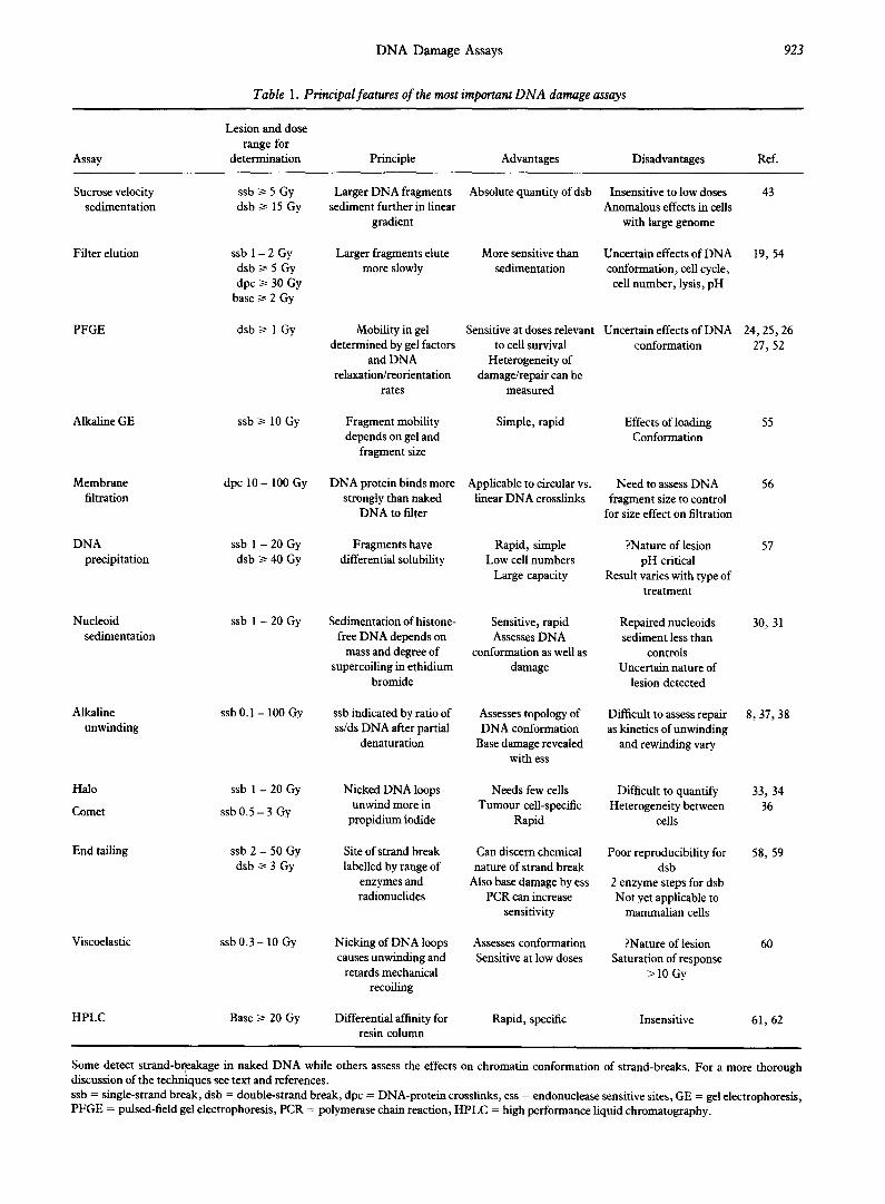
A considerable amount of work has been directed at providing sensitive and reliable assays of DNA dsb in irradiated cells. While much has been learned from measuring true DNA dsb, i.e. breaks in naked DNA, it is sometimes revealing to assess damage to DNA in a form closer to its cellular state, i.e. with components of nuclear protein. Some frequently used assays, which measure damage to DNA in both these forms, are outlined in Table 1. In this article we will focus on recent technical advances and discuss current ideas about the mechanisms of radiation response which are suggested by the use of these assays.
STRAND-BREAKAGE ASSAYS The purest form of strand-breakage assay measures breaks in
naked DNA (i.e. after removal of all cytoplasmic contents, organelles, membranes and nuclear proteins by detergent and proteolytic treatment). Such assays measure either changes in DNA mass using centrifugation, or changes in length by 6l- tration or by electrophoretic mobility. Under neutral conditions, these assays are specific for dsb while under alkaline conditions (pH> 1 l), the DNA double helix separates due to denaturation of interstrand bonds, which allows measurement of ssb. Several recent reviews have described technical aspects of the most
922
DNA Damage Assays 923
Table 1. Principal features of the most important DNA damage assays
Assay
Lesion and dose range for
determination Principle Advantages Disadvantages Ref.
Sucrose velocity ssb 2 5 Gy sedimentation dsbz 15 Gy
Filter elution ssb 1 - 2 Gy dsb > 5 Gy dpc 3 30 Gy
base > 2 Gy
PFGE dsb 3 1 Gy
Larger DNA fragments Absolute quantity of dsb Insensitive to low doses sediment further in linear Anomalous effects in cells
gradient with large genome
43
Larger fragments elute More sensitive than more slowly sedimentation
Uncertain effects of DNA conformation, cell cycle, cell number, lysis, pH
19, 54
Mobility in gel Sensitive at doses relevant determined by gel factors to cell survival
and DNA Heterogeneity of relaxation/reorientation damage/repair can be
rates measured
Uncertain effects of DNA 24,25,26 conformation 27, 52
Simple, rapid Effects of loading Conformation
55
Applicable to circular vs. linear DNA crosslinks
Need to assess DNA fragment size to control
for size effect on filtration
56
Rapid, simple Low cell numbers
Large capacity
?Nature of lesion pH critical
Result varies with type of treatment
57
Sensitive, rapid Assesses DNA
conformation as well as damage
Repaired nucleoids sediment less than
controls Uncertain nature of
lesion detected
30, 31
Assesses topology of DNA conformation
Base damage revealed with ess
Difficult to assess repair as kinetics of unwinding
and rewinding vary
8,37,38
Nicked DNA loops Needs few cells Difficult to quantify unwind more in Tumour cell-specific Heterogeneity between
propidium iodide Rapid cells
33, 34 36
Can discern chemical nature of strand break
Also base damage by ess PCR can increase
sensitivity
Poor reproducibility for dsb
58, 59
2 enzyme steps for dsb Not yet applicable to
mammalian cells
Assesses conformation Sensitive at low doses
?Nature of lesion Saturation of response
>lO Gy
60
Rapid, specific Insensitive 61, 62
Fragment mobility depends on gel and
fragment size
DNA protein binds more strongly than naked
DNA to filter
Alkaline GE ssb 2 10 Gy
Membrane filtration
dpc 10 - 100 Gy
DNA ssb 1 - 20 Gy Fragments have precipitation dsb z 40 Gy differential solubility
Nucleoid sedimentation
ssb 1 - 20 Gy Sedimentation of histone- free DNA depends on
mass and degree of supercoiling in ethidium
bromide
ssb indicated by ratio of ss/ds DNA after partial
denaturation
Alkaline unwinding
ssb 0.1 - 100 Gy
Halo
Comet
End tailing
ssb 1 - 20 Gy
ssb 0.5 - 3 Gy
ssb 2 - 50 Gy dsb 2 3 Gy
Site of strand break labelled by range of
enzymes and radionuclides
Viscoelastic ssb 0.3 - 10 Gy Nicking of DNA loops causes unwinding and
retards mechanical recoiling
Differential affinity for resin column
HPLC Base 2 20 Gy
Some detect strand-breakage in naked DNA while others assess the effects on chromatin conformation of strand-breaks. For a more thorough discussion of the techniques see text and references. ssb = single-strand break, dsb = double-strand break, dpc = DNA-protein crosslinks, ess = endonuclease sensitive sites, GE = gel electrophoresis, PFGE = pulsed-field gel electrophoresis, PCR = polymerase chain reaction, HPLC = high performance liquid chromatography.
924 S.J. Whitaker et al.
commonly used damage assays [e.g. 8-101 so here we will only briefly discuss three principal assays.
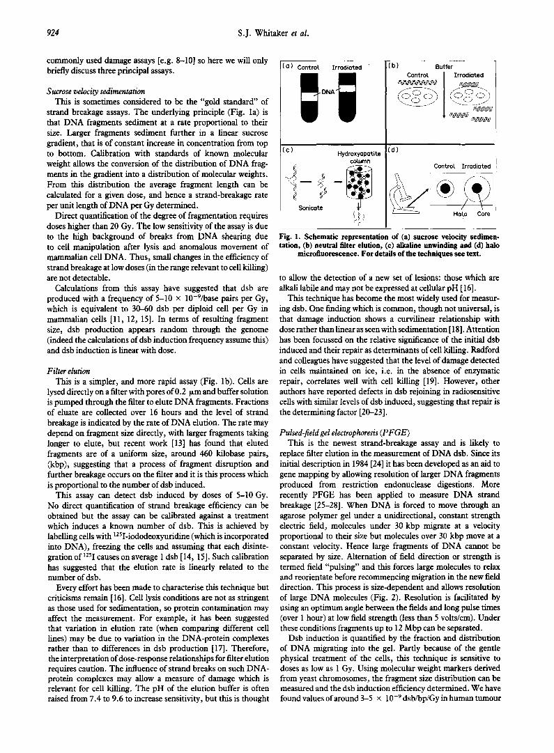
Sucrose velocity sedimentation This is sometimes considered to be the “gold standard” of
strand breakage assays. The underlying principle (Fig. la) is that DNA fragments sediment at a rate proportional to their size. Larger fragments sediment further in a linear sucrose gradient, that is of constant increase in concentration from top to bottom. Calibration with standards of known molecular weight allows the conversion of the distribution of DNA frag- ments in the gradient into a distribution of molecular weights. From this distribution the average fragment length can be calculated for a given dose, and hence a strand-breakage rate per unit length of DNA per Gy determined.
Direct quantification of the degree of fragmentation requires doses higher than 20 Gy. The low sensitivity of the assay is due to the high background of breaks from DNA shearing due to cell manipulation after lysis and anomalous movement of mammalian cell DNA. Thus, small changes in the efficiency of strand breakage at low doses (in the range relevant to cell killing) are not detectable.
Calculations from this assay have suggested that dsb are produced with a frequency of 5-10 x lo-‘/base pairs per Gy, which is equivalent to 30-60 dsb per diploid cell per Gy in mammalian cells [ll, 12, 151. In terms of resulting fragment size, dsb production appears random through the genome (indeed the calculations of dsb induction frequency assume this) and dsb induction is linear with dose.
Filter elution This is a simpler, and more rapid assay (Fig. lb). Cells are
lysed directly on a filter with pores of 0.2 urn and buffer solution is pumped through the filter to elute DNA fragments. Fractions of eluate are collected over 16 hours and the level of strand breakage is indicated by the rate of DNA elution. The rate may depend on fragment size directly, with larger fragments taking longer to elute, but recent work [13] has found that eluted fragments are of a uniform size, around 460 kilobase pairs, (kbp), suggesting that a process of fragment disruption and further breakage occurs on the filter and it is this process which is proportional to the number of dsb induced.
This assay can detect dsb induced by doses of 5-10 Gy. No direct quantification of strand breakage efficiency can be obtained but the assay can be calibrated against a treatment which induces a known number of dsb. This is achieved by labelling cells with lZSI-iododeoxyuridine (which is incorporated into DNA), freezing the cells and assuming that each disinte- gration of lz51 causes on average 1 dsb [14, 151. Such calibration has suggested that the elution rate is linearly related to the number of dsb.
Every effort has been made to characterise this technique but criticisms remain [ 161. Cell lysis conditions are not as stringent as those used for sedimentation, so protein contamination may affect the measurement. For example, it has been suggested that variation in elution rate (when comparing different cell lines) may be due to variation in the DNA-protein complexes rather than to differences in dsb production [17]. Therefore, the interpretation of dose-response relationships for filter elution requires caution. The influence of strand breaks on such DNA- protein complexes may allow a measure of damage which is relevant for cell killing. The pH of the elution buffer is often raised from 7.4 to 9.6 to increase sensitivity, but this is thought
(a ) Control Irradioted
Fig. 1. Schematic representation of (a) sucrose velocity sedimen- tation, (b) neutral titer elution, (c) alkaline unwinding and (d) halo
micro!luorescence. For details of the techniques see text.
to allow the detection of a new set of lesions: those which are alkali labile and may not be expressed at cellular pH [ 161.
This technique has become the most widely used for measur- ing dsb. One finding which is common, though not universal, is that damage induction shows a curvilinear relationship with dose rather than linear as seen with sedimentation [ 181. Attention has been focussed on the relative significance of the initial dsb induced and their repair as determinants of cell killing. Radford and colleagues have suggested that the level of damage detected in cells maintained on ice, i.e. in the absence of enzymatic repair, correlates well with cell killing [19]. However, other authors have reported defects in dsb rejoining in radiosensitive cells with similar levels of dsb induced, suggesting that repair is the determining factor [2&23].
Pulsed-field gel electrophoresis (PFGE) This is the newest strand-breakage assay and is likely to
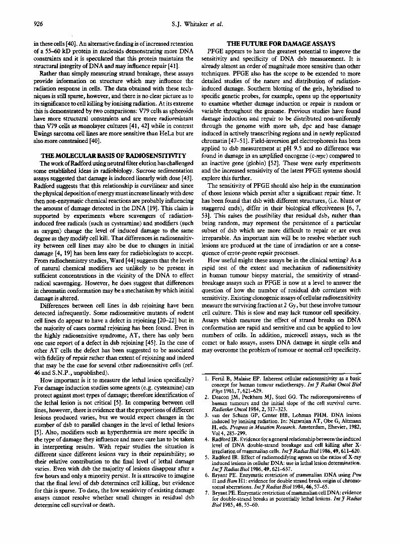
replace filter elution in the measurement of DNA dsb. Since its initial description in 1984 [24] it has been developed as an aid to gene mapping by allowing resolution of larger DNA fragments produced from restriction endonuclease digestions. More recently PFGE has been applied to measure DNA strand breakage [25-281. When DNA is forced to move through an agarose polymer gel under a unidirectional, constant strength electric field, molecules under 30 kbp migrate at a velocity proportional to their size but molecules over 30 kbp move at a constant velocity. Hence large fragments of DNA cannot be separated by size. Alternation of field direction or strength is termed field “pulsing” and this forces large molecules to relax and reorientate before recommencing migration in the new field direction. This process is size-dependent and allows resolution of large DNA molecules (Fig. 2). Resolution is facilitated by using an optimum angle between the fields and long pulse times (over 1 hour) at low field strength (less than 5 volts/cm). Under these conditions fragments up to 12 Mbp can be separated.
Dsb induction is quantified by the fraction and distribution of DNA migrating into the gel. Partly because of the gentle physical treatment of the cells, this technique is sensitive to doses as low as 1 Gy. Using molecular weight markers derived from yeast chromosomes, the fragment size distribution can be measured and the dsb induction efficiency determined. We have found values of around 35 x lop9 dsb/bp/Gy in human tumour
DNA Damage Assays 925
cells (S. J.W. and T. J-M.), which is comparable with the values found with sedimentation [ 111.
Using PFGE, Ahn et al. [29], have recently measured dsb rejoining in mammalian cells irradiated with doses as low as 5 Gy. These experiments are of particular importance as other strand breakage assays require supralethal doses of X-irradiation to follow dsb rejoining. The dsb rejoining measured by these assays derives predominantly from cells destined to die. At doses below 5 Gy, however, there is a higher probability that a cell will survive and thus dsb are more likely to reflect significant lesions for cell killing.
ASSAYS THAT ASSESS DNA HIGHER ORDER STRUCTURE
In order to accommodate a molecule of DNA, which is 50-100 cm in length within a nucleus of 5-10 km diameter, the DNA has to be condensed by a factor of 10’ and yet it has to remain accessible for replication, transcription and repair. This packing is achieved by the formation of a higher order structure of DNA with proteins.
DNA can be isolated whilst retaining some of this structure. Following detergent cell lysis and high salt denaturation (l-2 moY1 NaCl) but without proteinase, DNA is in a super- coiled form called a “nucleoid” which is free of histones but retains some of the higher organisation of chromatin due to other associated proteins of the nuclear matrix [30]. The degree of winding of these nucleoids can be modified by the fluorescent dye ethidium bromide. This dye intercalates between DNA bases and, at increasing concentration, progressively unwinds the normally negative supercoiling. Further increase in dye concentration then induces supercoiling in the opposite or positive sense.
Nucleoid sedimentation In this assay the degree of supercoiling modifies the sedimen-
tation of a nucleoid in a sucrose column by altering its density. Further changes in this density are imposed by radiation since the presence of strand breaks relaxes the DNA. Thus a relation- ship can be seen between the radiation dose and the sedimen- tation distance of the nucleoids in a given concentration of ethidium bromide [ 301.
Analysis of nucleoids has led to some interesting clinical correlations. Deeley and Moore [31] demonstrated decreased repair in a nuclear lysate sedimentation assay in lymphocytes from patients with bowel complications due to radiotherapy. Vaughan et al. [32] have reported a correlation between nucleoid sedimentation in tumour cells present in urine and tumour response in patients with carcinoma of the bladder.
Halo microfluorescence Microscopy allows detection of fluorescence from single cells
and a measurement of the degree of chromatin loop unwinding which is independent of the mass of the nucleoid [33, 341. This is performed on nucleoids stained with propidium iodide (PI; see Fig. Id). In unirradiated cells exposed to a low concentration of PI the unwound’ DNA loops are seen as a “halo”. With increasing concentration of PI the loop size, and thus the halo, increases to a maximum diameter, then reduces due to positive supercoiling or “rewinding”. Following irradiation two changes are observed. Firstly, the loops unwind to a greater degree for a given concentration of dye (the maximum halo size increases). Second, the loops do not completely rewind (unless repair of strand breakage is allowed). Using this technique Taylor et al.
Wells -
5.7
4.6
Mb 3.5
2.2
1.6
0.2
C P OGY 20Gy
Fig. 2. Typical gel following puked-field gel electmphoresis of irradiited human huuour cell DNA. Lane 1 (C) carries Saccbaro- myces cerevisiae chromosomes (only the krgest 2 of 15 chromosomes are seen, of 2.2 and 1.6 Mbp), and lane 2 (P) cazries ScBizosoccliaro- myces pombe chromosomes of 3.5, 4.6 and 5.7 Mbp as mokcukr weight markers. Lanes 3 and 4 show the typical DNA smears from a bladder carcinoma cell line. Lane 3 car&s u&radiated controls while cells in lane 4 have been irradkted on ice with “oco gamma- rays with 20 Gy. PFGE parameters: Ckmped homogeneous electric field (Bio Rad CHEF-DRU) system, 60 minute switching (2 fields +60” to net DNA migration), 4 volts/cm, for 96 hours. Gel = 0.8%
agarose, buffer = 0.5 x TBE at 16°C.
[35] have shown abnormalities in the halo formation of diploid fibroblasts from patients with ataxia-telangiectasia (AT) com- pared with those from normal individuals. They found an increase in the maximum halo size and reduced rewinding after irradiation and have speculated how these differences may relate to the radiosensitivity of the AT cells.
Comet assay This is a similar technique to the halo assay which involves
embedding the cells in agarose gel. Following lysis the nucleoids are subjected to a brief electrophoretic separation where the relaxed coils of chromatin spread out from the nucleoid towards the anode to form a “comet” [36]. The size of the tail of the comet is the endpoint used.
Alkaline unwinding This assay allows partial unwinding of double-stranded DNA
under brief alkaline/high salt denaturation [S, 37, 381 (see Fig. lc). Reannealing of single strands is prevented by a brief ultrasonic shock which randomly fractures all the DNA into small fragments. The resulting proportion of double-stranded to single-stranded DNA fragments produced is measured by differential binding to and then elution from a hydroxyapatite column. The ratio of single-stranded to double-stranded DNA reflects the initial induction of ssb. However, this ratio may be influenced by the higher order structure of DNA when irradiated due to physical “constraints” to unwinding rather than altered strand breakage. One model proposed from these observations involves the nuclear matrix attachment sites. These attachments are believed to be the sites of DNA replication, transcription and possibly repair [39]. It is speculated that increased constraints correlate with increased levels of poly-(ADP-ribose) polymerase
926 S.J. Whitaker et al.
in these cells [40]. An alternative tiding is of increased retention of a 55-60 kD protein in nucleoids demonstrating more DNA constraints and it is speculated that this protein maintains the structural integrity of DNA and may influence repair [41].
Rather than simply measuring strand breakage, these assays provide information on structure which may influence the radiation response in cells. The data obtained with these tech- niques is still sparse, however, and there is no clear picture as to its significance to cell killing by ionising radiation. At its extreme this is demonstrated by two comparisons: V79 cells as spheroids have more structural constraints and are more radioresistant than V79 cells as monolayer cultures [41, 421 while in contrast Ewings sarcoma cell lines are more sensitive than HeLa but are also more constrained [40].
THE MOLECULAR BASIS OF RADIOSENSITMTY The work of Radford using neutral filter elution has challenged
some established ideas in radiobiology. Sucrose sedimentation assays suggested that damage is induced linearly with dose [43]. Radford suggests that this relationship is curvilinear and since the physical deposition of energy must increase linearly with dose then non-enzymatic chemical reactions are probably influencing the amount of damage detected in the DNA [ 191. This claim is supported by experiments where scavengers of radiation- induced free radicals (such as cysteamine) and modifiers (such as oxygen) change the level of induced damage to the same degree as they modify cell kill. That differences in radiosensitiv- ity between cell lines may also be due to changes in initial damage [4, 191 has been less easy for radiobiologists to accept. From radiochemistry studies, Ward [44] suggests that the levels of natural chemical modifiers are unlikely to be present in sufficient concentrations in the vicinity of the DNA to effect radical scavenging. However, he does suggest that differences in chromatin conformation may be a mechanism by which initial damage is altered.
Differences between cell lines in dsb rejoining have been detected infrequently. Some radiosensitive mutants of rodent cell lines do appear to have a defect in rejoining [20-221 but in the majority of cases normal rejoining has been found. Even in the highly radiosensitive syndrome, AT, there has only been one case report of a defect in dsb rejoining [45]. In the case of other AT cells the defect has been suggested to be associated with fidelity of repair rather than extent of rejoining and indeed that may be the case for several other radiosensitive cells (ref. 46 and S.N.P., unpublished).
How important is it to measure the lethal lesion specifically? For damage induction studies some agents (e.g. cysteamine) can protect against most types of damage; therefore identification of the lethal lesion is not critical [5]. In comparing between cell lines, however, there is evidence that the proportions of different lesions produced varies, but we would expect changes in the number of dsb to parallel changes in the level of lethal lesions [S]. Also, modifiers such as hyperthermia are more specific in the type of damage they influence and more care has to be taken in interpreting results. With repair studies the situation is different since different lesions vary in their repairability; so their relative contribution to the final level of lethal damage varies. Even with dsb the majority of lesions disappear after a few hours and only a minority persist. It is attractive to imagine that the final level of dsb determines cell killing, but evidence for this is sparse. To date, the low sensitivity of existing damage assays cannot resolve whether small changes in residual dsb determine cell survival or death.
THE FUTURE FOR DAMAGE ASSAYS PFGE appears to have the greatest potential to improve the
sensitivity and specificity of DNA dsb measurement. It is already almost an order of magnitude more sensitive than other techniques. PFGE also has the scope to be extended to more detailed studies of the nature and distribution of radiation- induced damage. Southern blotting of the gels, hybridised to specific genetic probes, for example, opens up the opportunity to examine whether damage induction or repair is random or variable throughout the genome. Previous studies have found damage induction and repair to be distributed non-uniformly through the genome with more ssb, dpc and base damage induced in actively transcribing regions and in newly replicated chromatin [47-5 11. Field-inversion gel electrophoresis has been applied to dsb measurement at pH 9.5 and no difference was found in damage in an amplified oncogene (c-myc) compared to an inactive gene (globin) [52]. These were early experiments and the increased sensitivity of the latest PFGE systems should explore this further.
The sensitivity of PFGE should also help in the examination of those lesions which persist after a significant repair time. It has been found that dsb with different structures, (i.e. blunt or staggered ends), differ in their biological effectiveness [6, 7, 531. This raises the possibility that residual dsb, rather than being random, may represent the persistence of a particular subset of dsb which are more difficult to repair or are even irreparable. An important aim will be to resolve whether such lesions are produced at the time of irradiation or are a conse- quence of error-prone repair processes.
How useful might these assays be in the clinical setting? As a rapid test of the extent and mechanism of radiosensitivity in human tumour biopsy material, the sensitivity of strand- breakage assays such as PFGE is now at a level to answer the question of how the number of residual dsb correlates with sensitivity. Existing clonogenic assays of cellular radiosensitivity measure the surviving fraction at 2 Gy, but these involve tumour cell culture. This is slow and may lack tumour cell specificity. Assays which measure the effect of strand breaks on DNA conformation are rapid and sensitive and can be applied to low numbers of cells. In addition, microcell assays, such as the comet or halo assays, assess DNA damage in single cells and may overcome the problem of tumour or normal cell specificity.
1.
2.
3.
4.
5.
6.
7.
Fertil B, Malaise EP. Inherent cellular radiosensitivity as a basic concept for human tumour radiotherapy. IntJ Radiat Oncol Biol Phys 1981,7,621-629. Deacon JM, Peckham MJ, Steel GG. The radioresponsiveness of human tumours and the initial slope of the cell survival curve. Radiother Oncoll984,2,317-323. _ van der Schans GP. Center HB. Lohman PHM. DNA lesions induced by ionising kiation. In:‘Natarajan AT, Obe G, Altmann H, eds. Progress in Mutation Research. Amsterdam, Elsevier, 1982, Vol4,285-299. Radford IR. Evidence for a general relationship between the induced level of DNA double-s&d breakage and-cell killing after X- irradiation of mammalian cells. Znt 7 RadiatBiol1986.49.61 l-620. Radford IR. Effect of radiomodify”kg agents on the r&ids of X-ray induced lesions in cellular DNA: use in lethal lesion determination. Inty Radiat Bioll986,49,621-637. Bryant PE. Enzymatic restriction of mammalian DNA using Pwu II &d Barn Hl: kvidence for double strand break origin of chromo- somal aberrations. Znt 7 Radiat Bioll984,46,57-65. Bryant PE. Enzymatic-restriction ofm&ali& cell DNA: evidence for double-strand breaks as potentially lethal lesions. Int J Radiat Bioll985,48,55-60.
DNA Damage Assays 927
8.
9.
10.
11.
12.
13.
14.
15.
16.
17.
18.
19.
20.
21.
22.
23.
24.
25.
26.
27.
28.
29.
30.
31.
32.
Ahnstrom G. Techniques to measure D NA single-strand breaks in cells: a review. ZntJ Radiat Bioll988,54,695-707. Radford IR. The dose-response for low-LET radiation-induced DNA double-strand breakage: methods of measurement and impli- cations for radiation action models. Int 7 Radiat Biol 1988, 54, l-11. McMillan TJ, O’Neill I’, Peacock JH, Prise K. Workshop report 1st L.H. Gray workshop. Measurement of radiation-induced dam- age, ZntJ RadiatBioll990,58,391-396. Lehman AR, Stevens S. Production and repair of double-strand breaks in cells from normal humans and patients with ataxia- telangiectasia. Biochim Biophys Acta 1977,474,49-60. Blocher D. DNA double-strand breaks in Ehrlich ascites tumour cells at low doses of X-rays. 1. Determination of induced breaks by centrifugation at reduced-speed. IntJ RadiatBioll982,42,317-328. Wlodek D. Olive PL. Phvsical basis for detection of DNA double- strand breaks using neutral filter elution. Radiat Res 1990, 124, 326-333. Krisch RE, Sauri CJ. Further studies of DNA damage and lethality from the decay of iadine-125 in bacteriophages. ZntJ Radiat Biol 1975,27,553-560. Radford IR, Hodgson GS. 1251-induced DNA double-strand breaks: use in calibration of the neutral filter elution technique and compari- son with X-ray induced breaks. ZntJ RadiatBioE 1985,48,555-566. Hutchinson F. On the measurement of DNA double-strand breaks by neutral elution. Radiat Res 1989,120,182-186. Okayasu R, Iliakis G. Linear DNA elution dose response curves obtained in CHO cells with non-unwinding filter elution after aonromiate selection of the lvsis conditions. Znt 7 Radiat Biol1989, 6’ 589-581. P&e KM, Davies S, Michael BD. Non-linear dose-effect curve for DNA double-strand breaks by low LET radiation: the effect of eluting buffer composition on the measurement of breaks by the filter elution technique. IntJ Radial Bioll989,56,943-950. Radford IR. The level of induced DNA double-strand breakage correlates with cell killing after X-irradiation. Znt 3 Radiat Biol 1985,48,45-54. Jeggo PA, Kemp LM. X-ray sensitive mutants of Chinese hamster ovary cell line. Isolation and cross-sensitivity to other DNA damag- ingagents. MutatRes 1983,112,313-327. Wlodek D, Hittelman WN. The repair of double-strand DNA breaks correlates with radiosensitivitv of L5178Y-S and L5178Y-R cells. Radiat Res 19817,112,146-155.. Stamato TD. Weinstein R. Giaccia Al. MacKenzie L. Isolation of a cell cycle-dependent gamma-ray-sensitive Chinese hamster ovary cell. Somat Cell Genat 1983,9,165-173. Schwartz JL, Vaughan ATM. Association among DNAchromo- some break rejoining rates, chromatin structure alterations and radiation sensitivitv in human tumour cell lines. Cancer Res 1989. 49,5054-5057. . Schwartz DC, Cantor CR. Separation of yeast chromosome-sized DNAs by pulsed-field gradient gel electrophoresis. Cell 1984, 37, 67-75. Blocher D, Einspenner M, Zajackowski J. CHEF electrophoresis, a sensitive technique for the determination of DNA double-strand breaks. ZntJ Radiat&oll989,56,437-448. Stamato TD, Denko N. Asymmetric field inversion gel electrophor- esis for detecting DNA double-strand breaks in mammalian cells. Radial Res 1990,121,196-205. Ager DD, Dewey WC. Calibration of pulsed-field gel electrophor- esis for measurement of DNA double-strand breaks. Znt 3 Radial Bioll990,58,249-259. Iliakis GE, Metzger L, Denko N, Stamato TD. Detection of DNA double-strand breaks in synchronous cultures of CHO cells by means of asymmetric field inversion gel electrophoresis. IntJ Radial Bioll991,59,321-341. Ahn SY, Nevaldine B, Hahn PJ. Direct measurement by pulsed- field gel electrophoresis of induction and rejoining of X-ray-induced double-strand breaks in cultured mouse cells. Znt J Radiat Biol 1991,59,661-675. Cook PR, Braze11 IA. Supercoils in human DNA. 3 Cell Sci 1975, 19,261-279. Deeley JOT, Moore JL. Nuclear lysate sedimentation measure- ments of peripheral blood lymphocytes from radiotherapy patients. IntJ Radiat Bioll9819,56,963-973. Vaughan ATM, Lynch TH, Anderson P, Kondratowicz GM, Wallace DM. The extraction of biologically relevant data from
33.
34.
35.
36.
37.
38.
39.
40.
41.
42.
43.
44.
45.
46.
47.
48.
49.
50.
51.
52.
53.
54.
55.
56.
57.
bladder carcinoma cells eliminated in the urine. In: Seymour CB, Mothershill C, eds. New Developments in Fundamental and Applied Radiobiology. London, Taylor & Francis, 1991,292-298. Vogelstein B, Pardoll DM, Coffey DS. Supercoiled loops and eukaryotic DNA replication. Cell 1980,22,79-85. Roti Roti JL, Wright WD. Visualization of DNA loops in nucleoids from HeLa cells: assays for DNA damage and repair. Cytometry 1987,8,461-467. Taylor YC, Duncan PG, Zhang X, Wright WD. Differences in the DNA supercoiling response of irradiated cell lines from ataxia- telangiectasia versus unaffected individuals. IntJ Radiat Bioll991, 59,35%371. Ostling 0, Johanson KJ. Microelectrophoretic study of radiation- induced DNA damages in individual mammalian cells. Biochem and Biophys Res &mm& 1984,123,291-298. Rvdbera B. The rate of strand senaration in alkali of DNA of irradiated mammalian cells. Radiat ies 1975,61,274-287. Ahnstrom G, Edvardsson KA. Radiation-induced single-strand breaks in DNA determined by rate of alkaline strand separation and hydroxyapatite chromatography: an alternative to velocity sedimentation. ZntJ RadiatBiol1974,26,493-497. Jackson DA, Cook PR. Replication occurs at a nucleoskeleton. EMBOJ 1986,5,1403-1410. Jorgensen TJ, Prasad SC, Brennan TP, Dritschilo A. Constraints to DNA unwinding near radiation-induced strand breaks in Ewing’s sarcoma cells. Radial Res 1990,123,320-324. Gordon DJ, Milner AE, Beaney RP, Grdina DJ, Vaughan ATM. The increase in radioresistance of Chinese Hamster Cells cultured as spheroids is correlated to changes in nuclear morphology. Radiat Res 1990,121,175-179. Olive PL, Hilton J, Durand RE. DNA conformation of Chinese Hamster V79 cells and sensitivity to ionizing radiation. Radiat Res 1986,107,115-124. Lehman AR, Ormerod MG. Double-strand breaks in the DNA of a mammalian cell after X-irradiation. Biockim Bioplrys Acra 1970, 217,268-277. Ward JF. The yield of DNA double-strand breaks produced intracellularly by ionizing radiation: a review. Znt 3 Radio? Biol 1990,57,1141-1150. Coquerelle TM, Weibezahn KF, Lilcke-Huhle C. Rejoining of double-strand breaks in normal and ataxia-telangiectasia fibroblasts after exposure to @%o gamma-rays, z4’Am o-particles or bleomycin. IntJ RadiatBioll987,51,209-218. Debenham PG, Webb MBT, Stretch A, Thacker J. Examination of vectors with two dominant selectable genes for DNA repair and mutation studies in mammaliancells. MuzatRes 1988,199,145-148. Warters RL, Childers TJ. Radiation-induced base damage in rep- licating chromatin. Radi& Res 1982,90,564-574. - _ Chiu S-M. Oleinick NL. The sensitivitv of active and inactive i chromatin’to ionizing radiation-induced DNA strand breakage. Int J RadiatBioll982,41,71-77. Chiu S-M, Oleinick NL, Friedman LR, StambrookPJ. Hypersensi- tivity of DNA in transcriptionally active chromatin to-ionizing radiation. Biochim Bioohvs Acta 1982.699. 15-21. Oleinick NL, Chiu S-M, Friedman LR. Gamma radiation as a probe of chromatin structure: damage to and repair of active chromatin in the metaphase chromosome. Radiat Res 1984, 98, 629-641. Heussen C, Nackerdien Z, Smith BJ, Bohm L. Irradiation damage in chromatin isolated from V79 Chinese Hamster lung fibroblasts. Radiat Res 1987,110,84-94. Kawamura M, Preisler HD. A new approach to the detection of DNA damage. Leuk Res 1989,13,391-398. Giaccia AJ, MacLaren RA, Denko N, Nicolau D, Stamato TD. Increased sensitivity to killing by restriction enzymes in the XR-1 DNA double-strand break reoair-deficient mutant. Mutat Res 1990. 236,67-76. Bradley MO, Kohn KW. X-ray induced DNA double-strand break production and repair in mammalian cells as measured by neutral filter elution. Nucleic Acids Res 1979,7,793-804. Kovacs E, Langemann H. Investigation of the repair of single- strand breaks in human DNA using alkaline gel electrophoresis. Mutat Res 1990,124,137-140. Center MS, Richardson CC. An endonuclease induced after infec- tion of Escherichia Coli with bacteriophage T7. 1. Purification and properties of the enzyme.3 Biol Chem 1970,245,6285-6291. Olive PL, Chan APS, Cu CS. Comparison between the DNA
928 S.J. Whitaker et al.
precipitation and alkali unwinding assays for detecting DNA strand breaks and cross-links. Cancer Res 1988,48,6444-6449.
58. Hagen UFW. Measurement of strand breaks by end labelling. In: Friedberg EC, Hanawalt PC, eds. DNA Repair: A Laboratory Manual of Research Procedures. New York, Marcel Dekker, 1981, Vol. 1, part B, 431-445.
59. Smeets M, Slebos R, Stoker K and Begg AC. Attempts to measure DNA double-strand breaks using molecular biological methods. Znr 3 Radiat Bioll990,57,1254.
61. Karam LR, Calsou P, Franklin WA, Painter RB, Olsson M, Lindahl T. Moditication of deoxyribose-phosphate residues by extracts of ataxia telangiectasia cells. Mutat Res 1990,236,19-26.
62. Srinivasan S, Glauert HP. Formation of 5-hydroxymethyl-2’-deoxy- uridine in hepatic DNA of rats treated with gamma-irradiation, diethylnitrosamine, 2-acetylaminofluorene or the peroxisome prolif- erator ciprofibrate. Carcinogenesis 1990,11,2021-2024.
60. Wun KLW, Schafer RH. StIUCNrd changes in mammalian cell DNA induced by low dose X-ray damage and subsequent postir-
Acknowledgements-The authors are grateful to Gordon Steel and
radiation incubation in the presence and absence of caffeine. Radiut John Peacock for comments on this manuscript. The authors are funded
Res 1982,90,310-320. by the Cancer Research Campaign and the Bob Champion Trust, Royal Marsden Hospital.
EurJ Cancer, Vol. 27, No. 7, pp. 926931,1991. Primed in Grem Btimin
0277-s379/91$3.00 + 0.w ~1991PogamonPmsplc
Cancer Incidence in New South Wales, Australia Margaret McCredie, Marylon Coates, Tim Churches and Richard Taylor
In 1972, cancer registration began in New South Wales (NSW), the most populous state in Australia. The operations of the Registry are described. By 1990, approximately 316 000 new cases of cancer had been notified from a population that had increased from 4.6 to 5.8 million. In 1981-1984, the most common sites in men were lung, prostate, colon, melanoma and bladder, and in women, breast, melanoma, colon, lung and unknown primary site. Cancers which, between 1973-1976 and 1981-1984, had increased in reported incidence by more than 25% were pharynx and kidney in both sexes, rectum, testis and melanoma in men, and lung and bladder in women; those decreasing by more than 10% were stomach in both sexes, oesophagus in men and cervix in women. Age-standardised incidence rates for melanoma (27.4 [m] and 23.8 [fJ per 100 000 in 1987) and cancer of the renal pelvis in women (1.7 per 100 000 in 1989) are among the highest in the world. EurJCancer, Vol. 27, No. 7, pp. 928-931,199l
INTRODUCTION NEW SOUTH WALES (NSW) is the most populous of the six federated states of Australia. In 1971 the NSW Central Cancer Registry was established to collect statistics for the state from January 1972. NSW, a temperate zone between 28” and 38” south and 141” and 154” east, has an area of 801400 square kilometres excluding the Australian Capital Territory which is bordered on all sides by NSW. The population, in millions, was 4.6 in 1972, 5.4 in 1984 and 5.8 in 1990. In 1984, 31% of the population was aged less than 20 years and 11% 65 years or over. A coastal population predominates, 76% living in the Sydney-Newcastle-Wollongong conurbation along the central coast. At the 1986 census, 89% of the population lived in urban areas and 22% were born overseas.
This report presents a description of the operation of the Registry, and for the period 1972-1984, a summary of cancer incidence in NSW, indicating the most common cancers and those sites which have ch3nged most over time. More recent data are presented for melanoma and cancer of the renal pelvis, sites which are particularly common in New South Wales.
OPERATION OF THE REGISTRY Type of data
All new cases of cancer in residents of NSW are notified to the Registry, exceptions being basal and squamous cell
Correspondence to M. McCredie. The authors are at the NSW Central Cancer Registry, New South Wales Cancer Council, PO Box 380, North Ryde, NSW 2113, Australia. Revised and accepted 4 Feb. 1991.
carcinomas of the skin and in situ cancers at any site. Infor- mation is collected relating to identifying and demographic characteristics, and to the diagnosis of cancer but not to risk factors for cancer, such as occupation or smoking habits.
In the period 1972-1984, most cases were verified histologi- cally (average for all cancers combined was between 87% and 92%) and less than 1% of cases were reported by death certificate only.
Sources of data The main source of data is the compulsory notification form
completed by medical or records staff for each admission of a patient with cancer to every hospital, both public and private, and nursing home. In addition, each radiotherapy department must notify the first attendance each year of each cancer case. Other sources of data are pathology reports relating to cancer received from all major hospitals and private pathology labora- tories, voluntarily up to 1985 and then as a legal requirement. Since 1972, the Registrar-General’s Department has supplied listings of all deaths certified as having cancer as the primary cause, with supplementary listings of cancer as the secondary cause from 1978. Tapes of death from all causes have been supplied from 1985, and direct computer matching will be carried out against Registry files. In 1990, there are approxi- mately 308 000 persons registered representing 3 16 000 new cases of cancer.
Related Documents