Colloids and Surfaces B: Biointerfaces 117 (2014) 360–367 Contents lists available at ScienceDirect Colloids and Surfaces B: Biointerfaces jo ur nal ho me p ag e: www.elsevier.com/locate/colsurfb Molecular arrangements and interconnected bilayer formation induced by alcohol or polyalcohol in phospholipid vesicles Maria Letizia Manca a , Ines Castangia a , Pietro Matricardi b,∗ , Sandrina Lampis c , Xavier Fernàndez-Busquets d , Anna Maria Fadda a , Maria Manconi a a Dept. Scienze della Vita e dell’Ambiente, CNBS, University of Cagliari, Cagliari, Italy b Dept. Chimica e Tecnologie del Farmaco, Sapienza, Università di Roma, Roma, Italy c Dept. Scienze Chimiche e Geologiche, CNBS and CSGI, University of Cagliari, Monserrato (CA), Italy d Barcelona Centre for International Health Research (CRESIB, Hospital Clínic-Universitat de Barcelona) and Institute for Bioengineering of Catalonia (IBEC), Barcelona, Spain a r t i c l e i n f o Article history: Received 4 November 2013 Received in revised form 20 February 2014 Accepted 3 March 2014 Available online 12 March 2014 Keywords: Liposomes Penetration Enhancer containing Vesicle (PEVs) (Poly)alcohols Cryo-TEM DSC SAXS Rheology a b s t r a c t A self-assembled hybrid phospholipid vesicular system containing various penetration enhancers – ethanol, Transcutol and propylenglycol – was prepared and characterized. The effects of the different alcohol or polyalcohols structure and their concentration on the features of the assembled vesicles were evaluated using a combination of different techniques, including cryo-transmission electron microscopy, laser light scattering, differential scanning calorimetry, small- and wide-angle X-ray scattering and rheo- logical analysis. These techniques allow explaining the structural rearrangements of the bilayer assembly due to the alcohol or polyalcohol addition. X-ray scattering studies showed that such addition at the highest concentration (20%) allowed structure modification to oligolamellar vesicles and a bilayer tran- sition to interdigitated phase. Rheological studies confirmed the importance of alcohol or polyalcohol in the structuring dispersions probably due to a partial tilting of phosphatidylcholine acyl chains forming interdigitated and interconnected bilayer vesicles. © 2014 Elsevier B.V. All rights reserved. 1. Introduction In the last decades liposomes have found important appli- cations in the pharmaceutical and cosmetic fields as molecular delivery systems. Indeed, they are nontoxic, biodegradable, non immunogenic and can deliver chemotherapeutic agents by differ- ent administration routes. Innovative liposomes and liposome-like vesicles have been developed to improve their performances as carrier device. These new formulations include niosomes, transfer- osomes, glycerosomes and ethosomes as well as vesicular systems constituted by monoglycerides or polymers (polymersomes) [1–7]. In recent years, numerous studies were carried out about the interactions between phospholipids and other chemicals such as surfactants, terpenes, aliphatic acids, permeation enhancers and alcohols or polyalcohols and their effect on bilayer orga- nization [6,8–18]. Phospholipid phase behavior and the related self-assembling properties of bilayer vesicles in water were exten- sively studied [19]; however, the effect of these external molecules ∗ Corresponding author. Tel.: +39 06 49913300; fax: +39 06 49913133. E-mail address: [email protected] (P. Matricardi). on the physico-chemical properties of lamellar vesicles have not been deeply investigated [20]. Interactions of small amphiphilic molecules with phospholipid dispersions can induce significant changes in the structure of the vesicle bilayer, modifying the bilayer packing and accordingly increasing their potential performance for innovative applications in nanomedicine and nanobiotechnol- ogy. The precise mechanism governing the interactions between lamellar bilayer and perturbing molecules is not completely under- stood, and a large number of physico-chemical studies were carried out in order to explain it using different techniques, such as X-ray scattering, differential scanning calorimetry, fluorescence spectroscopy and NMR [21–24]. In particular it was found that these surface-active molecules promoted a rearrangement of the phospholipid acyl chain structure causing bilayer interdigitation, and they were also responsible for the variation of phase tran- sition temperature strictly dependent on the length of aliphatic chain of alcohols [17,24–26]. Drugs could act as surface-active molecules as well, modifying the bilayer organization and packing [18,27]. The aim of the current study was to improve the knowledge of the influence of simple alcohol or complex polyalcohol con- centrations on the self-assembly of lamellar vesicles because a http://dx.doi.org/10.1016/j.colsurfb.2014.03.010 0927-7765/© 2014 Elsevier B.V. All rights reserved.

Welcome message from author
This document is posted to help you gain knowledge. Please leave a comment to let me know what you think about it! Share it to your friends and learn new things together.
Transcript
Mi
MXa
b
c
d
B
a
ARRAA
KLP((CDSR
1
cdievcocIiaanss
h0
Colloids and Surfaces B: Biointerfaces 117 (2014) 360–367
Contents lists available at ScienceDirect
Colloids and Surfaces B: Biointerfaces
jo ur nal ho me p ag e: www.elsev ier .com/ locate /co lsur fb
olecular arrangements and interconnected bilayer formationnduced by alcohol or polyalcohol in phospholipid vesicles
aria Letizia Mancaa, Ines Castangiaa, Pietro Matricardib,∗, Sandrina Lampisc,avier Fernàndez-Busquetsd, Anna Maria Faddaa, Maria Manconia
Dept. Scienze della Vita e dell’Ambiente, CNBS, University of Cagliari, Cagliari, ItalyDept. Chimica e Tecnologie del Farmaco, Sapienza, Università di Roma, Roma, ItalyDept. Scienze Chimiche e Geologiche, CNBS and CSGI, University of Cagliari, Monserrato (CA), ItalyBarcelona Centre for International Health Research (CRESIB, Hospital Clínic-Universitat de Barcelona) and Institute for Bioengineering of Catalonia (IBEC),arcelona, Spain
r t i c l e i n f o
rticle history:eceived 4 November 2013eceived in revised form 20 February 2014ccepted 3 March 2014vailable online 12 March 2014
eywords:iposomes
a b s t r a c t
A self-assembled hybrid phospholipid vesicular system containing various penetration enhancers –ethanol, Transcutol and propylenglycol – was prepared and characterized. The effects of the differentalcohol or polyalcohols structure and their concentration on the features of the assembled vesicles wereevaluated using a combination of different techniques, including cryo-transmission electron microscopy,laser light scattering, differential scanning calorimetry, small- and wide-angle X-ray scattering and rheo-logical analysis. These techniques allow explaining the structural rearrangements of the bilayer assemblydue to the alcohol or polyalcohol addition. X-ray scattering studies showed that such addition at the
enetration Enhancer containing VesiclePEVs)Poly)alcoholsryo-TEMSC
highest concentration (20%) allowed structure modification to oligolamellar vesicles and a bilayer tran-sition to interdigitated phase. Rheological studies confirmed the importance of alcohol or polyalcohol inthe structuring dispersions probably due to a partial tilting of phosphatidylcholine acyl chains forminginterdigitated and interconnected bilayer vesicles.
© 2014 Elsevier B.V. All rights reserved.
AXSheology. Introduction
In the last decades liposomes have found important appli-ations in the pharmaceutical and cosmetic fields as molecularelivery systems. Indeed, they are nontoxic, biodegradable, non
mmunogenic and can deliver chemotherapeutic agents by differ-nt administration routes. Innovative liposomes and liposome-likeesicles have been developed to improve their performances asarrier device. These new formulations include niosomes, transfer-somes, glycerosomes and ethosomes as well as vesicular systemsonstituted by monoglycerides or polymers (polymersomes) [1–7].n recent years, numerous studies were carried out about thenteractions between phospholipids and other chemicals suchs surfactants, terpenes, aliphatic acids, permeation enhancersnd alcohols or polyalcohols and their effect on bilayer orga-
ization [6,8–18]. Phospholipid phase behavior and the relatedelf-assembling properties of bilayer vesicles in water were exten-ively studied [19]; however, the effect of these external molecules∗ Corresponding author. Tel.: +39 06 49913300; fax: +39 06 49913133.E-mail address: [email protected] (P. Matricardi).
ttp://dx.doi.org/10.1016/j.colsurfb.2014.03.010927-7765/© 2014 Elsevier B.V. All rights reserved.
on the physico-chemical properties of lamellar vesicles have notbeen deeply investigated [20]. Interactions of small amphiphilicmolecules with phospholipid dispersions can induce significantchanges in the structure of the vesicle bilayer, modifying the bilayerpacking and accordingly increasing their potential performancefor innovative applications in nanomedicine and nanobiotechnol-ogy. The precise mechanism governing the interactions betweenlamellar bilayer and perturbing molecules is not completely under-stood, and a large number of physico-chemical studies were carriedout in order to explain it using different techniques, such asX-ray scattering, differential scanning calorimetry, fluorescencespectroscopy and NMR [21–24]. In particular it was found thatthese surface-active molecules promoted a rearrangement of thephospholipid acyl chain structure causing bilayer interdigitation,and they were also responsible for the variation of phase tran-sition temperature strictly dependent on the length of aliphaticchain of alcohols [17,24–26]. Drugs could act as surface-activemolecules as well, modifying the bilayer organization and packing
[18,27].The aim of the current study was to improve the knowledgeof the influence of simple alcohol or complex polyalcohol con-centrations on the self-assembly of lamellar vesicles because a
M.L. Manca et al. / Colloids and Surfaces B: Biointerfaces 117 (2014) 360–367 361
Table 1Acronyms, composition and physical state of vesicular formulations.
Samples P90H (mg/ml) Chol (mg/ml) Trc (% v/v) PG (% v/v) Et (% v/v) PBS (% v/v) Physical state
Empty Liposomes 60 2 0 0 0 100 LiquidEmpty 5Trc-PEVs 60 2 5 0 0 95 LiquidEmpty 10Trc-PEVs 60 2 10 0 0 90 LiquidEmpty 20Trc-PEVs 60 2 20 0 0 80 Soft-solidEmpty 5PG-PEVs 60 2 0 5 0 95 LiquidEmpty 10PG-PEVs 60 2 0 10 0 90 LiquidEmpty 20PG-PEVs 60 2 0 20 0 80 Soft-solid
btv
bhtbe(ahammnepswii
2
2
PGePf((
2
pclPpF1t
2
m
Empty 5Et-PEVs 60 2 0
Empty 10Et-PEVs 60 2 0
Empty 20Et-PEVs 60 2 0
etter understanding of (poly)alcohol–drug–phospholipid interac-ions may be helpful to modulate biophysical properties of bilayeresicles and elucidate their mechanism of action as drug carrier.
We used hydrogenated soy phosphatidylcholine as the mainilayer component and we added different amounts of an alco-ol or polyalcohol (5, 10, 20% v/v) in the water phase. We calledhis vesicles “penetration enhancer containing vesicles” (PEVs)ecause alcohols and polyalcohols can actually act as penetrationnhancers (PEs) [17,18,27]. The effects of the different structuresethanol, diethylene glycol monoethyl ether and propylenglycol),nd concentrations (5, 10, 20%) of PEs on the vesicle featuresave been evaluated. By visual observation some samples appeareds opalescent liquids, whereas others behaved as soft solid-likeaterials. The vesicular nanostructures were deeply investigatedeans of small- and wide-angle X-ray scattering, differential scan-
ing calorimetry, dynamic laser light scattering, cryo-transmissionlectron microscopy, and rheological analysis. We used these com-lementary analyses to create a comprehensive picture of thetructure of lamellar vesicles containing alcohol or polyalcohol,here alcoholic PEs seem to promote the formation of interdig-
tated bilayers and vesicle interconnections responsible for thencreased viscosity and viscoelasticity of the systems.
. Materials and methods
.1. Materials
Hydrogenated soy phosphatidylcholine (Phospholipon® 90H,90H) was kindly supplied by AVG (Milan, Italy) and LipoidmbH (Ludwigshafen, Germany). Diethylene glycol monoethylther (Transcutol® P, Trc) was kindly provided by Gattefossè (Saintriest, France). Phosphate buffer solution (PBS, pH 7) was purchasedrom Carlo Erba Reagents (Milan, Italy). Cholesterol (Chol), ethanolEt) and propylene glycol (PG) were purchased from Sigma–AldrichMilan, Italy).
.2. Vesicle preparation
Table 1 shows the composition of empty vesicles that were pre-ared by means of direct sonication [28,29]. P90H (60 mg/ml) andholesterol (2 mg/ml) were weighted in a glass flask, subsequently,iposomes were hydrated with phosphate buffer (PBS, pH 7), whileEVs were hydrated with a mixture of ethanol, Transcutol or pro-ylene glycol and PBS (at different concentrations 5, 10, 20% v/v).inally, dispersions were sonicated (5 s on and 2 s off, 30 cycles;5 �m of probe amplitude) using a high intensity ultrasonic disin-egrator (Soniprep 150, MSE Crowley, London, United Kingdom).
.3. Vesicle characterization
Vesicle formation and morphology were checked by trans-ission electron microscopy (TEM) and cryo-TEM analysis. TEM
0 5 95 Liquid0 10 90 Liquid0 20 80 Soft-solid
observation was carried out using a JEM-1010 microscope (JeolEurope, Croissy-sur-Seine, France), equipped with a digital cameraMegaView III and Software “AnalySIS”, at an accelerating voltageof 80 kV. Vesicles were examined using a negative staining tech-nique: samples were adsorbed on a carbon grid and stained with1% phosphotungstic acid solution.
For cryo-TEM analysis of the samples, a thin aqueous filmwas formed by placing a 5-�l drop of the suspension on a glow-discharged holey carbon grid, and then blotting it with filter paper.The resulting thin sample films were vitrified by plunging the grid(kept at 100% humidity and room temperature) into ethane, whichwas maintained at its melting point with liquid nitrogen, using aVitrobot (FEI Company, Eindhoven, The Netherlands). The vitreousfilms were transferred to a Tecnai F20 TEM (FEI Company) using aGatan cryotransfer (Gatan, Pleasanton, CA), and the samples wereobserved in a low dose mode. Images were acquired at 200 kV ata temperature between −170 and 175 ◦C, using low-dose imagingconditions not exceeding 20 e–/Å2, with a 4096 × 4096 pixel CCDEagle camera (FEI Company).
The average diameter and polydispersity index (PI) as a mea-sure of the width of the size distribution of the samples, weredetermined by Photon Correlation Spectroscopy using a Zetasizernano-ZS (Malvern Instruments, Worcestershire UK). Samples werebackscattered by a helium–neon laser (633 nm) at an angle of 173◦
and a constant temperature of 25 ◦C. Zeta potential was estimatedusing the Zetasizer nano-ZS by means of the M3-PALS (Mixed ModeMeasurement-Phase Analysis Light Scattering) technique, whichmeasures the particle electrophoretic mobility in a thermostatedcell. Prior to analysis, liposomes were diluted with water or PBSand PEVs with water or the same PE/PBS mixture (5, 10, 20% v/v)used for their preparation. Vesicle stability was also assessed bymonitoring the vesicle average size and zeta potential over 30 daysat 4 ± 1 ◦C.
2.4. Differential Scanning Calorimetry analysis
Differential Scanning Calorimetry (DSC) studies were performedusing a DSC Mettler Toledo model 821e (Mettler Toledo Interna-tional Inc., Barcelona, Spain). The samples were scanned in sealedaluminum pans under nitrogen atmosphere. Thermograms werescanned in the first heating run at a constant rate of 10 ◦C/min anda temperature range of 0–80 ◦C.
2.5. X-ray diffraction
Vesicle structure was probed by Small- and Wide-Angle X-ray Scattering (SWAXS). SAXS and WAXS patterns were recordedsimultaneously, at 25 ◦C, using two linear, one-dimensional,
position-sensitive detectors (PSD 50 M; Hecus X-Ray Systems)containing 1024 channels (width 54.0 �m). Cu K� radiation (wave-length 1.542 A) was provided by a GeniX X-ray generator, operatingat 50 kV and 1 mA.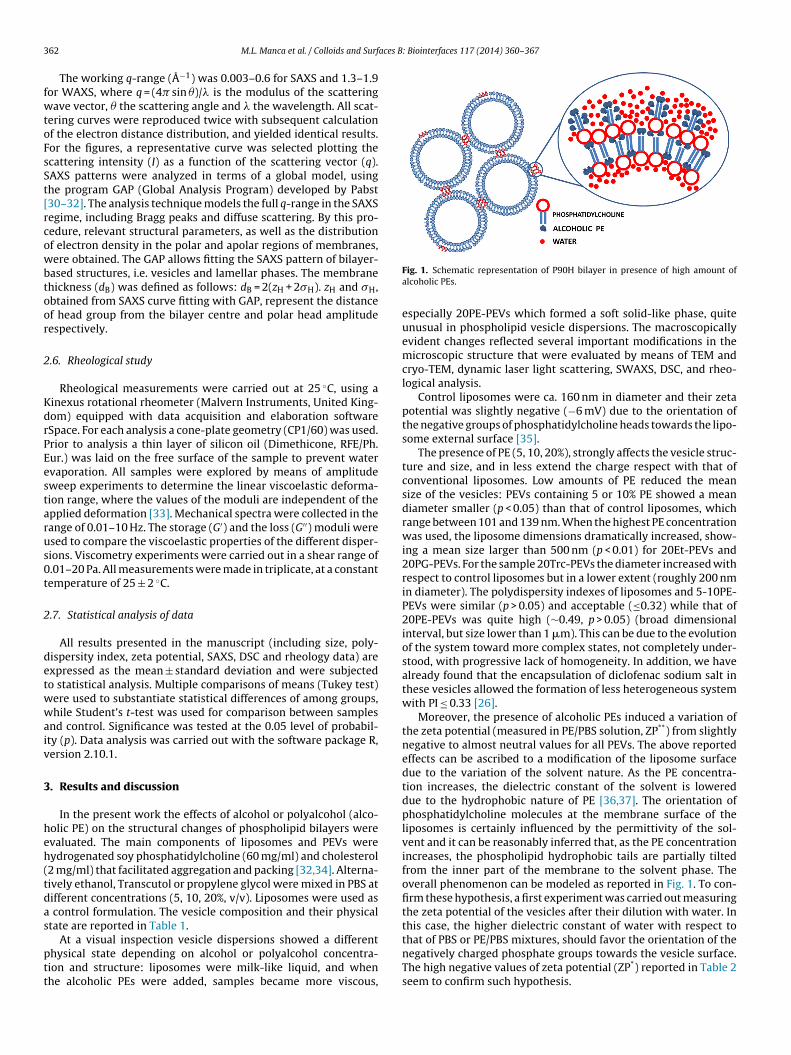
3 aces B: Biointerfaces 117 (2014) 360–367
fwtoFsSt[rcowbtoor
2
KdrPEestarus0t
2
detwwaiv
3
heh(tdas
ptt
62 M.L. Manca et al. / Colloids and Surf
The working q-range (Å−1) was 0.003–0.6 for SAXS and 1.3–1.9or WAXS, where q = (4� sin �)/� is the modulus of the scatteringave vector, � the scattering angle and � the wavelength. All scat-
ering curves were reproduced twice with subsequent calculationf the electron distance distribution, and yielded identical results.or the figures, a representative curve was selected plotting thecattering intensity (I) as a function of the scattering vector (q).AXS patterns were analyzed in terms of a global model, usinghe program GAP (Global Analysis Program) developed by Pabst30–32]. The analysis technique models the full q-range in the SAXSegime, including Bragg peaks and diffuse scattering. By this pro-edure, relevant structural parameters, as well as the distributionf electron density in the polar and apolar regions of membranes,ere obtained. The GAP allows fitting the SAXS pattern of bilayer-
ased structures, i.e. vesicles and lamellar phases. The membranehickness (dB) was defined as follows: dB = 2(zH + 2�H). zH and �H,btained from SAXS curve fitting with GAP, represent the distancef head group from the bilayer centre and polar head amplitudeespectively.
.6. Rheological study
Rheological measurements were carried out at 25 ◦C, using ainexus rotational rheometer (Malvern Instruments, United King-om) equipped with data acquisition and elaboration softwareSpace. For each analysis a cone-plate geometry (CP1/60) was used.rior to analysis a thin layer of silicon oil (Dimethicone, RFE/Ph.ur.) was laid on the free surface of the sample to prevent watervaporation. All samples were explored by means of amplitudeweep experiments to determine the linear viscoelastic deforma-ion range, where the values of the moduli are independent of thepplied deformation [33]. Mechanical spectra were collected in theange of 0.01–10 Hz. The storage (G′) and the loss (G′′) moduli weresed to compare the viscoelastic properties of the different disper-ions. Viscometry experiments were carried out in a shear range of.01–20 Pa. All measurements were made in triplicate, at a constantemperature of 25 ± 2 ◦C.
.7. Statistical analysis of data
All results presented in the manuscript (including size, poly-ispersity index, zeta potential, SAXS, DSC and rheology data) arexpressed as the mean ± standard deviation and were subjectedo statistical analysis. Multiple comparisons of means (Tukey test)ere used to substantiate statistical differences of among groups,hile Student’s t-test was used for comparison between samples
nd control. Significance was tested at the 0.05 level of probabil-ty (p). Data analysis was carried out with the software package R,ersion 2.10.1.
. Results and discussion
In the present work the effects of alcohol or polyalcohol (alco-olic PE) on the structural changes of phospholipid bilayers werevaluated. The main components of liposomes and PEVs wereydrogenated soy phosphatidylcholine (60 mg/ml) and cholesterol2 mg/ml) that facilitated aggregation and packing [32,34]. Alterna-ively ethanol, Transcutol or propylene glycol were mixed in PBS atifferent concentrations (5, 10, 20%, v/v). Liposomes were used as
control formulation. The vesicle composition and their physicaltate are reported in Table 1.
At a visual inspection vesicle dispersions showed a differenthysical state depending on alcohol or polyalcohol concentra-ion and structure: liposomes were milk-like liquid, and whenhe alcoholic PEs were added, samples became more viscous,
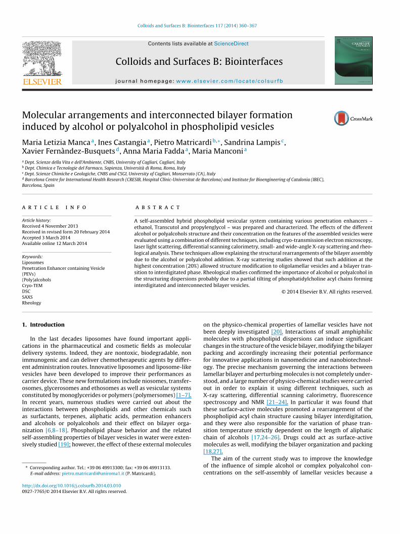
Fig. 1. Schematic representation of P90H bilayer in presence of high amount ofalcoholic PEs.
especially 20PE-PEVs which formed a soft solid-like phase, quiteunusual in phospholipid vesicle dispersions. The macroscopicallyevident changes reflected several important modifications in themicroscopic structure that were evaluated by means of TEM andcryo-TEM, dynamic laser light scattering, SWAXS, DSC, and rheo-logical analysis.
Control liposomes were ca. 160 nm in diameter and their zetapotential was slightly negative (−6 mV) due to the orientation ofthe negative groups of phosphatidylcholine heads towards the lipo-some external surface [35].
The presence of PE (5, 10, 20%), strongly affects the vesicle struc-ture and size, and in less extend the charge respect with that ofconventional liposomes. Low amounts of PE reduced the meansize of the vesicles: PEVs containing 5 or 10% PE showed a meandiameter smaller (p < 0.05) than that of control liposomes, whichrange between 101 and 139 nm. When the highest PE concentrationwas used, the liposome dimensions dramatically increased, show-ing a mean size larger than 500 nm (p < 0.01) for 20Et-PEVs and20PG-PEVs. For the sample 20Trc-PEVs the diameter increased withrespect to control liposomes but in a lower extent (roughly 200 nmin diameter). The polydispersity indexes of liposomes and 5-10PE-PEVs were similar (p > 0.05) and acceptable (≤0.32) while that of20PE-PEVs was quite high (∼0.49, p > 0.05) (broad dimensionalinterval, but size lower than 1 �m). This can be due to the evolutionof the system toward more complex states, not completely under-stood, with progressive lack of homogeneity. In addition, we havealready found that the encapsulation of diclofenac sodium salt inthese vesicles allowed the formation of less heterogeneous systemwith PI ≤ 0.33 [26].
Moreover, the presence of alcoholic PEs induced a variation ofthe zeta potential (measured in PE/PBS solution, ZP**) from slightlynegative to almost neutral values for all PEVs. The above reportedeffects can be ascribed to a modification of the liposome surfacedue to the variation of the solvent nature. As the PE concentra-tion increases, the dielectric constant of the solvent is lowereddue to the hydrophobic nature of PE [36,37]. The orientation ofphosphatidylcholine molecules at the membrane surface of theliposomes is certainly influenced by the permittivity of the sol-vent and it can be reasonably inferred that, as the PE concentrationincreases, the phospholipid hydrophobic tails are partially tiltedfrom the inner part of the membrane to the solvent phase. Theoverall phenomenon can be modeled as reported in Fig. 1. To con-firm these hypothesis, a first experiment was carried out measuringthe zeta potential of the vesicles after their dilution with water. Inthis case, the higher dielectric constant of water with respect to
that of PBS or PE/PBS mixtures, should favor the orientation of thenegatively charged phosphate groups towards the vesicle surface.The high negative values of zeta potential (ZP*) reported in Table 2seem to confirm such hypothesis.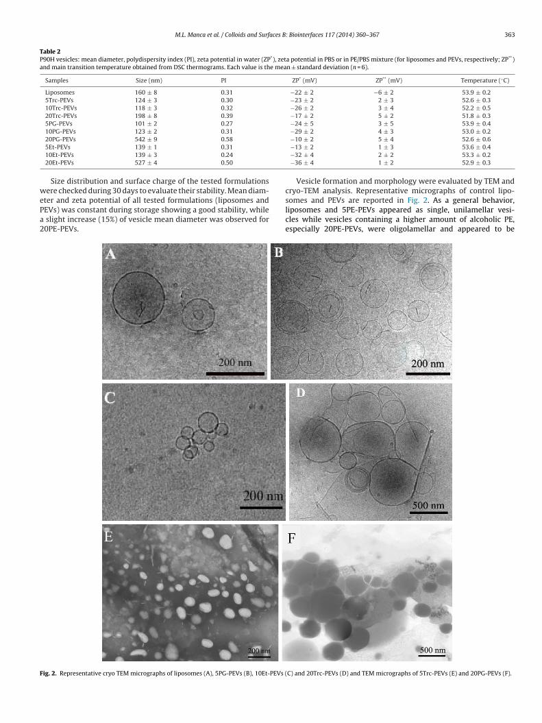
M.L. Manca et al. / Colloids and Surfaces B: Biointerfaces 117 (2014) 360–367 363
Table 2P90H vesicles: mean diameter, polydispersity index (PI), zeta potential in water (ZP*), zeta potential in PBS or in PE/PBS mixture (for liposomes and PEVs, respectively; ZP**)and main transition temperature obtained from DSC thermograms. Each value is the mean ± standard deviation (n = 6).
Samples Size (nm) PI ZP* (mV) ZP** (mV) Temperature (◦C)
Liposomes 160 ± 8 0.31 −22 ± 2 −6 ± 2 53.9 ± 0.25Trc-PEVs 124 ± 3 0.30 −23 ± 2 2 ± 3 52.6 ± 0.310Trc-PEVs 118 ± 3 0.32 −26 ± 2 3 ± 4 52.2 ± 0.520Trc-PEVs 198 ± 8 0.39 −17 ± 2 5 ± 2 51.8 ± 0.35PG-PEVs 101 ± 2 0.27 −24 ± 5 3 ± 5 53.9 ± 0.410PG-PEVs 123 ± 2 0.31 −29 ± 2 4 ± 3 53.0 ± 0.220PG-PEVs 542 ± 9 0.58 −10 ± 2 5 ± 4 52.6 ± 0.6
wePa2
F
5Et-PEVs 139 ± 1 0.3110Et-PEVs 139 ± 3 0.24
20Et-PEVs 527 ± 4 0.50
Size distribution and surface charge of the tested formulationsere checked during 30 days to evaluate their stability. Mean diam-
ter and zeta potential of all tested formulations (liposomes andEVs) was constant during storage showing a good stability, while
slight increase (15%) of vesicle mean diameter was observed for0PE-PEVs.
ig. 2. Representative cryo TEM micrographs of liposomes (A), 5PG-PEVs (B), 10Et-PEVs (
−13 ± 2 1 ± 3 53.6 ± 0.4−32 ± 4 2 ± 2 53.3 ± 0.2−36 ± 4 1 ± 2 52.9 ± 0.3
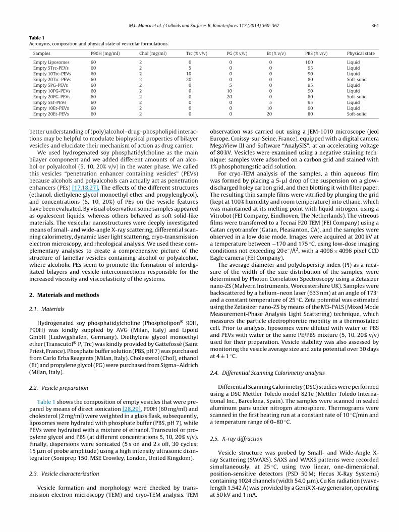
Vesicle formation and morphology were evaluated by TEM andcryo-TEM analysis. Representative micrographs of control lipo-
somes and PEVs are reported in Fig. 2. As a general behavior,liposomes and 5PE-PEVs appeared as single, unilamellar vesi-cles while vesicles containing a higher amount of alcoholic PE,especially 20PE-PEVs, were oligolamellar and appeared to beC) and 20Trc-PEVs (D) and TEM micrographs of 5Trc-PEVs (E) and 20PG-PEVs (F).
3 aces B: Biointerfaces 117 (2014) 360–367
ihti
nmvtsctcpsopccMtptb
t(fope
omrtp
bthup2ccop[lPlt(awacavi4tfmp
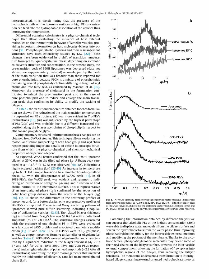
Fig. 3. (A) WAXS intensity profile versus the scattering vector modulus (q) recordedfrom empty liposomes at 25 ◦C, 60 ◦C and 20 PG-PEVs at 25 ◦C; (B) the first order-peak
64 M.L. Manca et al. / Colloids and Surf
nterconnected. It is worth noting that the presence of theydrophobic tails on the liposome surfaces at high PE concentra-ion can facilitate the hydrophobic association of the vesicles thusmproving their interactions.
Differential scanning calorimetry is a physico-chemical tech-ique that allows evaluating the influence of host externalolecules on the thermotropic behavior of lamellar vesicles, pro-
iding important information on host molecules–bilayer interac-ions [38]. Phospholipid/alcohol systems and their rearrangementtructures have been extensively studied by DSC [22]. Thesehanges have been evidenced by a shift of transition tempera-ure from gel to liquid-crystalline phase, depending on alcoholico-solvents structure and concentration. In the present study, there-transition peak of P90H liposomes was depressed (data nothown, see supplementary material) or overlapped by the peakf the main transition that was broader than those reported forure phospholipids, because P90H is a mixture of phospholipidsontaining several phosphatidylcholines differing in length of acylhains and free fatty acid, as confirmed by Manconi et al. [39].oreover, the presence of cholesterol in the formulation con-
ributed to inhibit the pre-transition peak also in the case ofure phospholipids and to reduce and enlarge the main transi-ion peak, thus confirming its ability to modify the packing ofilayers.
In Table 2 the transition temperature obtained for each formula-ion are shown. The reduction of the main transition temperature:i) depended on PE structure, (ii) was more evident in Trc-PEVsormulations [18], (iii) was influenced by the highest percentagef PEs (20%) and was probably due to a different Transcutol dis-osition along the bilayer acyl chains of phospholipids respect tothanol and propylene glycol.
Complementary structural information on these changes can bebtained from SWAXS studies. This technique allows exploring theolecular distance and packing of both head group and acyl chain
egions providing important details on vesicle microscopic struc-ure from which the physico-chemical and chemico-mechanicalroperties of dispersions depend.
As expected, WAXS results confirmed that the P90H liposomeilayer at 25 ◦C was in the tilted gel phase L�′ . A Bragg peak cen-ered at q ∼ 1.5 A−1 (d 4.2 A) was observed (Fig. 3A), indicating aighly ordered packing (L�′ ) [25,40]. An increase in temperaturep to 60 ◦C led sample transition to a lamellar liquid-crystallinehase, L�, with the disappearance of WAXS peak [41]. In all0PE-PEVs, the WAXS peak was evident and symmetric indi-ating no distortion of hexagonal packing and direction of lipidhains normal to the membrane surface. This is representativef an interdigitated phase (L�I) confirmed by the reduction ofolar head group distance from the center of the bilayer (zH)40]. Fig. 3B shows the differences in the scattering profiles ofiposomes and, for a better clarity, only representative profiles ofG-PEVs are reported. The recorded X-ray scattering patterns ofiposomes showed pure diffuse scattering, indicating a popula-ion of unilamellar vesicles [42,43]. The related bilayer thicknessdB) estimated from Bragg’s law was 58.0 ± 1 A with a polar headmplitude (�H) of 2.8 ± 0.2 A. The structural changes associatedith the presence of one alcoholic PE in PEVs were evaluated
s a function of SAXS profiles and associated parameters modifi-ation (Fig. 3B and Table 3). 5-10PE-PEVs were in L�′ gel-phase,s well as empty liposomes forming unilamellar or oligolamellaresicles (Table 3). 20PE-PEVs were all oligolamellar and character-zed by a significant reduction of the bilayer thickness (dB ∼ 51;7 and 42 A for 20Trc-PEVs; 20PG-PEVs and 20Et-PEVs respec-ively) and a slight reduction of polar head amplitude (∼2.2 A for all
ormulations) confirming the layer rearrangements that involvedainly the lipid portion of bilayer (zH) and led to an interdigitatedhase (L�I).
of the SAXS curves as a function of the scattering vector modulus (q) of liposomes andPG-PEVs. For the sake of clarity only the most representative curves were reported.
Combining the information obtained by different analysis wecan suggest that alcoholic PEs at the highest concentration (20%)were able to displace water molecules from the bilayer surface andscreen the hydrophobic tails from the water phase, thus improvingphosphatidylcholine affinity for the intervesicle external mediumand modifying the packing of the membrane. Thanks to the alco-holic screen, phosphatidylcholine molecules may orient some oftheir acyl chains on the bilayer surface, towards the inter-vesicleexternal compartment, allowing the formation of free spaces into
hydrophobic regions of the bilayer, thus reducing the bilayerthickness. The membrane underwent a transformation to interdig-itated bilayer containing external oriented hydrophobic tails too, as
M.L. Manca et al. / Colloids and Surfaces B: Biointerfaces 117 (2014) 360–367 365
Table 3Unilamellar (ULV) or oligolamellar (OLV) vesicles, bilayer thickness (dB), polar head amplitude (�H), and of polar head group from the center of bilayer (zH). Each value is themean ± standard deviation (n = 3).
Samples Structure zH (Å) �H (Å) dB (Å) Phase
Liposomes ULV 23.4 ± 0.2 2.8 ± 0.2 58.0 ± 1 L�′
5Trc-PEVs ULV 23.6 ± 0.1 2.4 ± 0.1 56.8 ± 2 L�′
10Trc-PEVs OLV 23.5 ± 0.2 2.9 ± 0.2 58.6 ± 2 L�′
20Trc-PEVs OLV 20.7 ± 0.3 2.3 ± 0.2 50.6 ± 1 L�I5PG-PEVs ULV 23.3 ± 0.1 2.7 ± 0.3 57.4 ± 1 L�′
10PG-PEVs OLV 23.2 ± 0.1 2.4 ± 0.4 56.0 ± 1 L�′
20PG-PEVs OLV 19.1 ± 0.1 2.1 ± 0.3 46.6 ± 2 L�I5Et-PEVs ULV 23.4 ± 0.1 3.0 ± 0.1 58.8 ± 1 L�′
10Et-PEVs ULV 17.4 ± 0.1 3.8 ± 0.4 58.8 ± 2 L�′
20Et-PEVs OLV 16.6 ± 0.1 2.0 ± 0.3 42.0 ± 1 L�I
F n of fre
ccateslaucs5tcoit
em
ig. 4. Mechanical spectra (storage modulus—G′ and loss modulus—G′′ as a functioxperimental error is included within the symbols.
onfirmed by a lower dB value and a sharp WAXS peak. Thesehanges on vesicle surface allowed a decrease of zeta potentialnd facilitated their aggregation and fusion to form larger struc-ures with a larger bilayer curvature. This phenomenon was lessvident in 20Trc-PEVs probably because of a lower ability of Tran-cutol to form free space into the hydrophobic region of the bilayereading to a lower reduction of the bilayer thickness (dB ∼ 51) and
lower increase of the bilayer curvature. In this work, P90H wassed at high concentration (60 mg/ml) allowing the formation ofoncentrated vesicle dispersion. Moreover, P90H vesicles are rigid,pherical, oligolamellar with a large size (diameter from 198 to42 nm) and are able to encapsulate a high water volume fraction,hus reducing the free solvent among vesicles. Therefore the con-omitant effect of vesicle closeness and the presence of acyl chainsn the bilayer surface allowed the formation of new phospholipidnterconnections between vesicles responsible of solid-like state of
he system (Fig. 1).An extensive rheological analysis was carried out, in order tolucidate supramolecular interactions and to describe the uncom-on soft solid like behaviour which was evidenced for the
equency) of liposomes (A), 20Et-PEVs (B), 20PG-PEVs (C) and 20Trc-PEVs (D). The
suspensions at higher alcoholic PE concentrations (20PE-PEVs), asreported in Table 1.
Mechanical spectra of control liposomes showed a typicalbehaviour of dilute water suspension that comprised G′ and G′′ fre-quency dependent profiles and a cross-over point of the two moduliin the range 0.1–10 Hz. It is worth noting that water solutions ofalcoholic PEs showed a similar response (here not reported), asthey behaved as Newtonian fluids with moduli of the same orderof magnitude as the liposome dispersions.
The mechanical spectra of PEVs showed profiles depending fromboth the structure and the concentration of alcoholic PE. For thecharacterization of the resulting systems, the different ability tostore (G′) and dissipate (G′′) the energy supplied during the rheo-logical experiments was considered.
In Fig. 4, the mechanical spectra of liposome and 20PE-PEVsuspensions are reported. As clearly shown, the presence of alco-
holic PEs modified the structure of the system. In the spectrumof 20Et-PEVs, G′ values become higher than G′′ in the studied fre-quency range and their moduli were higher more than 3 orders ofmagnitude with respect to the liposome dispersion. At the same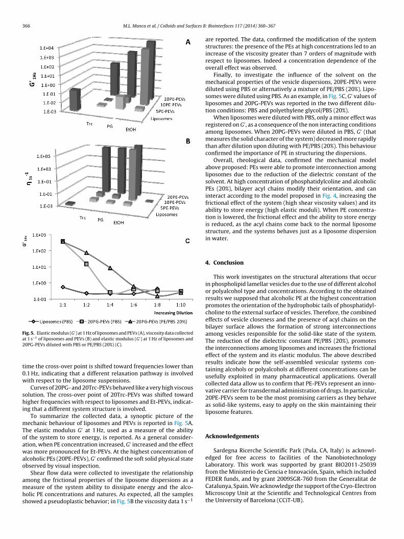
366 M.L. Manca et al. / Colloids and Surfaces B
Fig. 5. Elastic modulus (G′) at 1 Hz of liposomes and PEVs (A), viscosity data collecteda2
t0w
shi
mToawao
amhs
FEDER funds, and by grant 2009SGR-760 from the Generalitat de
t 1 s−1 of liposomes and PEVs (B) and elastic modulus (G′) at 1 Hz of liposomes and0PG-PEVs diluted with PBS or PE/PBS (20%) (C).
ime the cross-over point is shifted toward frequencies lower than.1 Hz, indicating that a different relaxation pathway is involvedith respect to the liposome suspensions.
Curves of 20PG- and 20Trc-PEVs behaved like a very high viscousolution. The cross-over point of 20Trc-PEVs was shifted towardigher frequencies with respect to liposomes and Et-PEVs, indicat-
ng that a different system structure is involved.To summarize the collected data, a synoptic picture of the
echanic behaviour of liposomes and PEVs is reported in Fig. 5A.he elastic modulus G′ at 1 Hz, used as a measure of the abilityf the system to store energy, is reported. As a general consider-tion, when PE concentration increased, G′ increased and the effectas more pronounced for Et-PEVs. At the highest concentration of
lcoholic PEs (20PE-PEVs), G′ confirmed the soft solid physical statebserved by visual inspection.
Shear flow data were collected to investigate the relationshipmong the frictional properties of the liposome dispersions as a
easure of the system ability to dissipate energy and the alco-olic PE concentrations and natures. As expected, all the sampleshowed a pseudoplastic behavior; in Fig. 5B the viscosity data 1 s−1
: Biointerfaces 117 (2014) 360–367
are reported. The data, confirmed the modification of the systemstructures: the presence of the PEs at high concentrations led to anincrease of the viscosity greater than 7 orders of magnitude withrespect to liposomes. Indeed a concentration dependence of theoverall effect was observed.
Finally, to investigate the influence of the solvent on themechanical properties of the vesicle dispersions, 20PE-PEVs werediluted using PBS or alternatively a mixture of PE/PBS (20%). Lipo-somes were diluted using PBS. As an example, in Fig. 5C, G′ values ofliposomes and 20PG-PEVs was reported in the two different dilu-tion conditions: PBS and polyethylene glycol/PBS (20%).
When liposomes were diluted with PBS, only a minor effect wasregistered on G′, as a consequence of the non interacting conditionsamong liposomes. When 20PG-PEVs were diluted in PBS, G′ (thatmeasures the solid character of the system) decreased more rapidlythan after dilution upon diluting with PE/PBS (20%). This behaviourconfirmed the importance of PE in structuring the dispersions.
Overall, rheological data, confirmed the mechanical modelabove proposed: PEs were able to promote interconnection amongliposomes due to the reduction of the dielectric constant of thesolvent. At high concentration of phosphatidylcoline and alcoholicPEs (20%), bilayer acyl chains modify their orientation, and caninteract according to the model proposed in Fig. 4, increasing thefrictional effect of the system (high shear viscosity values) and itsability to store energy (high elastic moduli). When PE concentra-tion is lowered, the frictional effect and the ability to store energyis reduced, as the acyl chains come back to the normal liposomestructure, and the systems behaves just as a liposome dispersionin water.
4. Conclusion
This work investigates on the structural alterations that occurin phospholipid lamellar vesicles due to the use of different alcoholor polyalcohol type and concentrations. According to the obtainedresults we supposed that alcoholic PE at the highest concentrationpromotes the orientation of the hydrophobic tails of phosphatidyl-choline to the external surface of vesicles. Therefore, the combinedeffects of vesicle closeness and the presence of acyl chains on thebilayer surface allows the formation of strong interconnectionsamong vesicles responsible for the solid-like state of the system.The reduction of the dielectric constant PE/PBS (20%), promotesthe interconnections among liposomes and increases the frictionaleffect of the system and its elastic modulus. The above describedresults indicate how the self-assembled vesicular systems con-taining alcohols or polyalcohols at different concentrations can beusefully exploited in many pharmaceutical applications. Overallcollected data allow us to confirm that PE-PEVs represent an inno-vative carrier for transdermal administration of drugs. In particular,20PE-PEVs seem to be the most promising carriers as they behaveas solid-like systems, easy to apply on the skin maintaining theirliposome features.
Acknowledgements
Sardegna Ricerche Scientific Park (Pula, CA, Italy) is acknowl-edged for free access to facilities of the NanobiotechnologyLaboratory. This work was supported by grant BIO2011-25039from the Ministerio de Ciencia e Innovación, Spain, which included
Catalunya, Spain. We acknowledge the support of the Cryo-ElectronMicroscopy Unit at the Scientific and Technological Centres fromthe University of Barcelona (CCiT-UB).

aces B
A
f2
R
[
[
[
[
[
[
[
[
[
[
[
[
[
[
[
[
[
[
[
[
[
[
[
[
[
[
[
[
[
[
[
[
[
Biopharm. 70 (2008) 116–126.
M.L. Manca et al. / Colloids and Surf
ppendix A. Supplementary data
Supplementary data associated with this article can beound, in the online version, at http://dx.doi.org/10.1016/j.colsurfb.014.03.010.
eferences
[1] S. Murgia, A.M. Falchi, M. Mano, S. Lampis, R. Angius, A.M. Carnerup, J. Schmidt,G. Diaz, M. Giacca, Y. Talmon, M. Monduzzi, Nanoparticles from lipid-basedliquid crystals: emulsifier influence on morphology and cytotoxicity, J. Phys.Chem. B 114 (2010) 3518–3525.
[2] M. Carboni, A.M. Falchi, S. Lampis, C. Sinico, M.L. Manca, J. Schmidt, Y. Talmon,S. Murgia, M. Monduzzi, Physicochemical, cytotoxic, and dermal release fea-tures of a novel cationic liposome nanocarrier, Adv. Healthcare Mater. 2 (2013)692–701.
[3] R. Angelico, M. Carboni, S. Lampis, J. Schmidt, Y. Talmon, M. Monduzzi, S. Murgia,Physicochemical and rheological properties of a novel monoolein-based vesiclegel, Soft Matter 9 (2013) 921–928.
[4] H. De Oliveira, J. Thevenot, S. Lecommandoux, Smart polymersomes fortherapy and diagnosis: fast progress toward multifunctional biomimeticnanomedicines, Wiley Interdisciplinary Rev.: Nanomed. Nanobiotechnol. 4(2012) 525–546.
[5] R. Muzzalupo, L. Tavano, R. Cassano, S. Trombino, T. Ferrarelli, N. Picci, A newapproach for the evaluation of niosomes as effective transdermal drug deliverysystems, Eur. J. Pharm. Biopharm. 79 (2011) 28–35.
[6] E. Touitou, N. Dayan, L. Bergelson, B. Godin, M. Eliaz, Ethosomes — novelvesicular carriers for enhanced delivery: characterization and skin penetrationproperties, J. Control. Rel. 65 (2000) 403–418.
[7] L. Tavano, R. Muzzalupo, R. Cassano, S. Trombino, T. Ferrarelli, N. Picci, Newsucrose cocoate based vesicles: Preparation characterization and skin perme-ation studies, Colloids Surf. B: Biointerfaces 75 (2010) 319–322.
[8] G. Cevc, G. Blume, Lipid vesicles penetrate into intact skin owing to the transder-mal osmotic gradients and hydration force, Biochim. Biophys. Acta Biomembr.1104 (1992) 226–232.
[9] G.M.M. El Maghraby, A.C. Williams, B.W. Barry, Interactions of surfactants (edgeactivators) and skin penetration enhancers with liposomes, Int. J. Pharm. 276(2004) 143–161.
10] M.M.A. Elsayed, O.Y. Abdallah, V.F. Naggar, N.M. Khalafallah, Deformable lipo-somes and ethosomes: Mechanism of enhanced skin delivery, Int. J. Pharm. 322(2006) 60–66.
11] M.M.A. Elsayed, O.Y. Abdallah, V.F. Naggar, N.M. Khalafallah, Lipid vesicles forskin delivery of drugs: Reviewing three decades of research, Int. J. Pharm. 332(2007) 1–16.
12] N. Dragicevic-Curic, D. Scheglmann, V. Albrecht, A. Fahr, Temoporfin-loadedinvasomes: Development, characterization and in vitro skin penetration stud-ies, J. Control. Rel. 127 (2008) 59–69.
13] S. Mura, M. Manconi, C. Sinico, D. Valenti, A.M. Fadda, Penetration enhancer-containing vesicles (PEVs) as carriers for cutaneous delivery of minoxidil, Int.J. Pharm. 380 (2009) 72–79.
14] M. Manconi, S. Mura, C. Sinico, A.M. Fadda, A.O. Vila, F. Molina, Developmentand characterization of liposomes containing glycols as carriers for diclofenac,Colloids Surf. A: Physicochem. Eng. Aspects 342 (2009) 53–58.
15] M. Chen, X. Liu, A. Fahr, Skin penetration and deposition of carboxyfluoresceinand temoporfin from different lipid vesicular systems: In vitro study with finiteand infinite dosage application, Int. J. Pharm. 408 (2011) 223–234.
16] S. Mura, M. Manconi, D. Valenti, C. Sinico, A.O. Vila, A.M. Fadda, Transcutolcontaining vesicles for topical delivery of minoxidil, J. Drug Target. 19 (2011)189–196.
17] M. Manconi, C. Caddeo, C. Sinico, D. Valenti, M.C. Mostallino, G. Biggio, A.M.Fadda, Ex vivo skin delivery of diclofenac by transcutol containing liposomesand suggested mechanism of vesicle-skin interaction, Eur. J. Pharm. Biopharm.78 (2011) 27–35.
18] M. Manconi, C. Caddeo, C. Sinico, D. Valenti, M.C. Mostallino, S. Lampis, M.Monduzzi, A.M. Fadda, Penetration enhancer-containing vesicles: Compositiondependence of structural features and skin penetration ability, Eur. J. Pharm.Biopharm. 82 (2012) 352–359.
19] G. Cevc, D. Marsh, Hydration of noncharged lipid bilayer membranes. The-
ory and experiments with phosphatidylethanolamines, Biophys. J. 47 (1985)21–31.20] R.V. McDaniel, T.J. McIntosh, S.A. Simon, Nonelectrolyte substitution for waterin phosphatidylcholine bilayers, Biochim. Biophys. Acta (BBA) Biomembr. 731(1983) 97–108.
[
: Biointerfaces 117 (2014) 360–367 367
21] S.A. Simon, T.J. McIntosh, Interdigitated hydrocarbon chain packing causes thebiphasic transition behavior in lipid/alcohol suspensions, Biochim. Biophys.Acta (BBA) Biomembr. 773 (1984) 169–172.
22] U. Vierl, L. Löbbecke, N. Nagel, G. Cevc, Solute effects on the colloidal and phasebehavior of lipid bilayer membranes: ethanol-dipalmitoylphosphatidylcholinemixtures, Biophys. J. 67 (1994) 1067–1079.
23] L.L. Holte, K. Gawrisch, Determining ethanol distribution in phospholipid mul-tilayers with MAS-NOESY spectra, Biochemistry 36 (1997) 4669–4674.
24] M.F.N. Rosser, H.M. Lu, P. Dea, Effects of alcohols on lipid bilayers with and with-out cholesterol: the dipalmitoylphosphatidylcholine system, Biophys. Chem.81 (1999) 33–44.
25] M. Kranenburg, M. Vlaar, B. Smit, Simulating induced interdigitation in mem-branes, Biophys. J. 87 (2004) 1596–1605.
26] I. Castangia, M.L. Manca, P. Matricardi, C. Sinico, S. Lampis, X. Fernàndez-Busquets, A.M. Fadda, M. Manconi, Effect of diclofenac and glycol intercalationon structural assembly of phospholipid lamellar vesicles, Int. J. Pharm. 456(2013) 1–9.
27] M. Manconi, C. Sinico, C. Caddeo, A.O. Vila, D. Valenti, A.M. Fadda, Penetrationenhancer containing vesicles as carriers for dermal delivery of tretinoin, Int. J.Pharm. 412 (2011) 37–46.
28] S. Madrigal-Carballo, D. Seyler, M. Manconi, S. Mura, A.O. Vila, F. Molina, Anapproach to rheological and electrokinetic behaviour of lipidic vesicles cov-ered with chitosan biopolymer, Colloids Surf. A: Physicochem. Eng. Aspects323 (2008) 149–154.
29] M. Chessa, C. Caddeo, D. Valenti, M. Manconi, C. Sinico, A.M. Fadda, Effectof penetration enhancer containing vesicles on the percutaneous deliv-ery of quercetin through new born pig skin, Pharmaceutics 20113 (2011)497–509.
30] G. Pabst, R. Koschuch, B. Pozo-Navas, M. Rappolt, K. Lohner, P. Laggner, Struc-tural analysis of weakly ordered membrane stacks, J. Appl. Crystallogr. 36(2003) 1378–1388.
31] G. Pabst, M. Rappolt, H. Amenitsch, P. Laggner, Structural information frommultilamellar liposomes at full hydration: Full q-range fitting with high qualityX-ray data, Phys. Rev. E 62 (2000) 4000–4009.
32] P.L. Yeagle, J. Bensen, L. Boni, S.W. Hui, Molecular packing of cholesterol inphospholipid vesicles as probed by dehydroergosterol, Biochim. Biophys. Acta(BBA) Biomembr. 692 (1982) 139–146.
33] C. Sandolo, P. Matricardi, F. Alhaique, T. Coviello, Dynamo-mechanical andrheological characterization of guar gum hydrogels, Eur. Polym. J. 43 (2007)3355–3367.
34] P. Srisuk, P. Thongnopnua, U. Raktanonchai, S. Kanokpanont, Physico-chemicalcharacteristics of methotrexate-entrapped oleic acid-containing deformableliposomes for in vitro transepidermal delivery targeting psoriasis treatment,Int. J. Pharm. 427 (2012) 426–434.
35] Z. Abramovic, U. Sustarsic, K. Teskac, M. Sentjurc, J. Kristl, Influence of nanosizeddelivery systems with benzyl nicotinate and penetration enhancers on skinoxygenation, Int. J. Pharm. 359 (2008) 220–227.
36] G. Akerlof, Dielectric constants of some organic solvent-water mixtures at var-ious temperatures, J. Am. Chem. Soc. 54 (1932) 4125–4139.
37] C. Wohlfarth, Dielectric constant of the mixture (1) water; (2) ethanol, in: M.D.Lechner (Ed.), Landolt-Börnstein Group IV Physical Chemistry: Static Dielec-tric Constants of Pure Liquids and Binary Liquid Mixtures, 17, Springer-Verlag,Berlin/Heidelberg, 2008.
38] R. Pignatello, V.D. Intravaia, G. Puglisi, A calorimetric evaluation of the inter-action of amphiphilic prodrugs of idebenone with a biomembrane model, J.Colloid Interface Sci. 299 (2006) 626–635.
39] M. Manconi, F. Marongiu, G. Ennas, A. Scano, C. Sinico, D. Valenti, A.M. Fadda,Liposomes for (trans)dermal delivery of tretinoin: Influence of drug con-centration and vesicle composition, J. Drug Deliv. Sci. Technol. 18 (2008)309–313.
40] T. Adachi, H. Takahashi, K. Ohki, I. Hatta, Interdigitated structure ofphospholipid-alcohol systems studied by X-ray diffraction, Biophys. J. 68(1995) 1850–1855.
41] Á. Csiszár, E. Klumpp, A. Bóta, K. Szegedi, Effect of 2,4-dichlorophenolon DPPC/water liposomes studied by X-ray and freeze-fracture electronmicroscopy, Chem. Phys. Lipids 126 (2003) 155–166.
42] C. Gómez-Gaete, N. Tsapis, L. Silva, C. Bourgaux, M. Besnard, A. Bochot, E.Fattal, Supramolecular organization and release properties of phospholipid-hyaluronan microparticles encapsulating dexamethasone, Eur. J. Pharm.
43] O. López, M. Cócera, R. Pons, H. Amenitsch, J. Caelles, J.L. Parra, L. Coderch,A. de la Maza, Use of synchrotron radiation SAXS to study the first steps of theinteraction between sodium dodecyl sulfate and charged liposomes, Spectrosc.:An Int. J. 16 (2002) 343–350.
Related Documents