Molecular and morphological characterization of Haemoproteus (Parahaemoproteus) ptilotis, a parasite infecting Australian honeyeaters (Meliphagidae), with remarks on prevalence and potential cryptic speciation Nicholas J. Clark 1,2 *, Robert D. Adlard 2,3 and Sonya M. Clegg 1,4 1 Environmental Futures Research Institute and Griffith School of Environment, Griffith University, Gold Coast Campus, QLD 4222, Australia 2 Natural Environments Program, Queensland Museum, PO Box 3300, South Brisbane, Queensland, 4101, Australia 3 School of Biological Sciences, The University of Queensland, Brisbane, Queensland, 4072, Australia 4 Edward Grey Institute, Department of Zoology, University of Oxford, Oxford, OX1 3PS, United Kingdom *Corresponding author: Tel.: +61432420979 Email: [email protected]

Welcome message from author
This document is posted to help you gain knowledge. Please leave a comment to let me know what you think about it! Share it to your friends and learn new things together.
Transcript
Molecular and morphological characterization of Haemoproteus (Parahaemoproteus) ptilotis, a parasite infecting Australian honeyeaters (Meliphagidae), with remarks on prevalence and potential cryptic speciation
Nicholas J. Clark1,2*, Robert D. Adlard2,3 and Sonya M. Clegg1,4
1 Environmental Futures Research Institute and Griffith School of Environment, Griffith University, Gold Coast Campus, QLD 4222, Australia 2 Natural Environments Program, Queensland Museum, PO Box 3300, South Brisbane, Queensland, 4101, Australia 3 School of Biological Sciences, The University of Queensland, Brisbane, Queensland, 4072, Australia 4 Edward Grey Institute, Department of Zoology, University of Oxford, Oxford, OX1 3PS, United Kingdom
*Corresponding author: Tel.: +61432420979 Email: [email protected]

Abstract
Avian Haemoproteus (Haemosporida) parasites occur in birds on all continents apart
from Antarctica. Molecular screening techniques have uncovered previously
unforeseen levels of Haemoproteus lineage diversity; however, fewer than 20% of
genetic parasite lineages have been linked to morphological descriptions. The
process of linking morphological descriptions to DNA barcodes for Haemoproteus
spp. is important for the study of host-parasite interactions and the potential for
cryptic speciation. Here, we describe cytochrome-b barcodes and morphological
diagnostics for the identification of Haemoproteus (Parahaemoproteus) ptilotis, a
systematically confusing parasite found in Australian honeyeaters (family
Meliphagidae). We characterised infections from the original type host
(Lichenostomus chrysops) as well as from four co-occurring meliphagid species in
southeast Queensland, Australia to investigate intraspecific variation in morphology
and lineage identity. We recorded eight lineages that grouped into a well-supported
monophyletic group, supporting the linkage of the described lineages to H. ptilotis.
However, comparisons of diagnostics between the type host and co-occurring
meliphagid hosts revealed high genetic diversity and variable morphology that could
be indicative of cryptic speciation. This study highlights that morphological
descriptions alongside molecular characterisation remain crucial if we are to gain an
understanding of the true diversity and host-specificity of protozoan parasites in
Australia and elsewhere.
Key words: Australia, cryptic species, DNA barcode, Haemoproteus, host specificity,
PCR

Introduction
Avian haemosporidians (Haemosporida) belonging to the genus Haemoproteus are
vector-borne blood parasites found in birds on all continents apart from Antarctica
(Valkiūnas 2005). Taxonomic descriptions, that are based largely on diagnostic
morphological characteristics of blood stages, have identified over 130
morphospecies (Valkiūnas 2005). However, molecular studies have uncovered
previously unforeseen levels of haemosporidian diversity (Bensch et al. 2009, Ishtiaq
et al. 2010) and the generation of DNA barcodes using partial cytochrome-b (cyt-b)
parasite sequences is well suited to haemosporidian research due to established
polymerase chain reaction (PCR) protocols (Waldenström et al. 2004, Križanauskienė
et al. 2010). Moreover, there is some evidence to suggest that Haemoproteus
lineages whose cyt-b sequences differ by as little as a few base pairs may be
reproductively isolated entities (Hellgren et al. 2007), yet the vast majority of newly
described Haemoproteus DNA lineages are only identified to genus level without
morphological description (Palinauskas et al. 2013, Valkiūnas et al. 2014).
Morphological analysis of blood smears can yield important information on
Haemoproteus spp. diversity as well as the competency of hosts, particularly since
abortive infections can still yield amplified PCR products (Levin et al. 2013, Valkiūnas
et al. 2013). In spite of the powerful research opportunities that PCR affords (Ishtiaq
et al. 2008, Palinauskas et al. 2010, Medeiros et al. 2013, Clark and Clegg 2014),
morphological descriptions remain crucial to haemosporidian taxonomy (Valkiūnas
et al. 2008b, Zehtindjiev et al. 2012, Dimitrov et al. 2014).The process of linking
morphological descriptions to DNA barcodes for Haemoproteus spp. therefore has
merit for the ongoing study of host-parasite interactions (Križanauskienė et al. 2006,
Palinauskas et al. 2007, Valkiūnas et al. 2007).
Honeyeaters (Meliphagidae) belong to one of the most widespread and
ecologically diverse avian families in the south Pacific (Schodde and Mason 1999,
Joseph et al. 2014). Cleland and Johnston (1909) identified a high prevalence of
unknown Haemoproteus spp. infecting meliphagids in Australia, and originally
classified these parasites into three distinct species based on the examinations of
material from different avian host species (Haemoproteus ptilotis, type host
Lichenostomus chrysops; H. philemon, type host Philemon corniculatus; and H.
meliornis, type host Meliornis novaehollandiae). Re-descriptions from original type
material as well as new material (host Manorina melanocephala) led Bennett et al.
(1994) to consider the titles H. meliornis and H. philemon as synonymous with H.
ptilotis. The complex was again revisited by Valkiūnas (2005), with modifications
from Bennett et al. (1994) and from Laird and Laird (1960) used to determine that
morphological variation among host species and / or geography did not warrant the
classification of multiple species. Nevertheless, the degradation of original type
material for H. ptilotis has been a major hindrance to the identification of diagnostic
characteristics (Bennett et al. 1994, Valkiūnas 2005). Morphological and molecular
characterisation of H. ptilotis from the original type host (L. chrysops) as well as from
additional avian host species using fresh material are necessary to appropriately
describe this widespread and systematically confusing parasite. Here, we combine
morphological and molecular techniques to characterise H. ptilotis from the original
type host L. chrysops. In addition, we characterise H. ptilotis infections from co-
occurring meliphagid species to assess morphological and genetic diversity across
avian hosts.

Methods
We collected blood samples and blood smears from meliphagid hosts in southeast
Queensland, Australia between July 2012 and December 2013 (Table 1). Birds were
caught with mistnets, banded with an Australian Bird and Bat Banding Authority
band and released at the site of capture after sampling. Blood samples were taken
from the wing vein and stored in lysis buffer (1%SDS, 20mM NaCl, 10mM TRIS pH 8.0,
10mM EDTA pH 8.0) for DNA extraction. Thin smears were air dried, fixed in 100%
methanol and stained with Giemsa following the protocols in Valkiūnas et al. (2008b).
We prepared two blood smears were for each bird. Smears were examined for H.
ptilotis infection using light microscopy following the screening protocols in
Valkiūnas et al. (2008b). We sampled 29 individuals of the original H. ptilotis type
host, L. chrysops, and identified 15 infected individuals. Additionally, we identified H.
ptilotis infections in 29 of 41 individuals from four co-occurring Meliphagid host
species in the study area (Table 1). Prevalence of H. ptilotis in Meliphagid host
species ranged from 20% (Meliphaga lewinii) to 100% (Manorina melanocephala;
Table 1).
DNA Extraction, PCR and Sequencing
We extracted DNA from blood samples using ammonium acetate/ethanol
precipitation (Richardson et al. 2001). We amplified a fragment of the parasite
cytochrome-b (cyt-b) gene using a nested-polymerase chain reaction (PCR) protocol
(Waldenström et al. 2004) following reaction conditions described in (Clark et al.
2014a). Amplifications were sequenced on an Applied Biosystems 3130xl Genetic

Analyser (Foster City, California, USA) at the Griffith University DNA Sequencing
Facility (Brisbane, Australia).
Phylogenetic analysis
To test whether newly described H. ptilotis lineages were monophyletic, we
constructed a molecular phylogeny of microscopically confirmed Haemoproteus
species. We downloaded Haemoproteus lineages that have been microscopically
assigned to morphospecies from GenBank and the avian malaria database, MalAvi
(Bensch et al. 2009). We aligned and edited sequences in Geneious v5.4 (Drummond
et al. 2011). We created the phylogeny using BEAST version 1.6.1 (Drummond and
Rambaut 2007) with an uncorrelated lognormal relaxed clock and a Yule speciation
prior. We used Akaike’s information criterion in jModelTest version 0.1.1 (Posada
2008) to determine the appropriate evolutionary model (GTR+I+G). One sequence of
Leucocytozoon spp. was used as an outgroup. We carried out two runs of 10 000 000
generations, sampling at every 10 000 generations, and used TRACER version 1.6
(available at http://tree.bio.ed.ac.uk/software/tracer/) to assess convergence. We
removed 10% burnin from each run and combined the two runs using LogCombiner
version 1.4.7 (Drummond and Rambaut 2007). MalAvi lineage names (Bensch et al.
2009) and GenBank accession numbers for all parasite lineages used in the
phylogeny are presented in Fig. 1.
Morphological analysis
An Olympus BH2 light microscope (Tokyo, Japan) equipped with Nikon Digital Sight
camera and NIS-Elements imaging software (Nikon, Tokyo, Japan) was used to

examine slides and to prepare figures. We examined each blood smear for 15–20
min at low magnification (x400), and then at least 100 fields were studied at high
magnification (x1,000). For voucher specimens, we screened the entire blood film for
the presence of co-infections with other haemosporidian parasites. Blood samples
with single infections of H. ptilotis were used to identify diagnostic cyt-b parasite
sequences for morphospecies characterization (DNA barcoding). In addition to the
type host and the four co-occurring Meliphagid host species from which we
extracted DNA, we also screened smears and performed measurements for
infections identified as H. ptilotis in Philemon corniculatus, using smears that were
registered with the International Reference Centre for Avian Haematozoa (IRCAH;
accession numbers G463506, G463508).
Results We confirmed the presence of single infections both by microscopic examination of
blood films and by analysis of chromatograms (absence of double peaks in both
forward and reverse directions). All of the recorded parasite lineages from
meliphagid hosts clustered in a well-supported monophyletic group and could be
readily distinguished from other barcoded Haemoproteus species on the phylogeny
(Fig. 1), supporting the linkage of the described cyt-b lineages to the morphospecies
Haemoproteus (Parahaemoproteus) ptilotis. We identified multiple genetic lineages
of H. ptilotis that vary in their relative genetic distance to the type lineage (0.6%,
LICFRE03 to 5.0%, MYZSAN02), indicating possible intraspecific variation or cryptic
speciation across meliphagid hosts.

Morphological description
Haemoproteus (Parahaemoproteus) ptilotis (Cleland and Johnston, 1909). The full
morphological measurements for the type host and additional hosts are presented in
Table 2.
DNA sequence: Mitochondrial cyt-b sequence LICHRYS01 (505bp, GenBank accession number KP721986).
Avian host and distribution: The lineage LICHRYS01 has only been recorded in the
type host, the yellow-faced honeyeater Lichenostomus chrysops, in southeast
Queensland (QLD), Australia. The following closely related lineages have been
recorded in co-occurring Meliphagid hosts in southeast QLD, Australia: lineage
LICFRE03 (genetic distance to type lineage 0.6%) was recorded in a white-throated
honeyeater Melithreptus albogularis in this study and previously in a bridled
honeyeater Lichenostomus frenatus in northeast QLD, Australia (Beadell et al. 2004);
lineage MELLEW03 (genetic distance 0.6%) was recorded in Lewin’s honeyeater
Meliphaga lewinii; lineage NMIN01 (genetic distance 1.2%) was recorded in the noisy
miner Manorina melanocephala; lineages MELALB01, MELALB02 and MELALB03
(mean genetic distance 1.4%) were recorded in the white-throated honeyeater, M.
albogularis; lineage MYZSAN02 (genetic distance 5.0%) was recorded in the scarlet
honeyeater Myzomela sanguinolenta.
Site of infection: Mature erythrocytes.
Representative blood films: Voucher specimens (accession numbers G465701 –

G456705), L. chrysops, December 2012, collected in Karawatha Forest, Brisbane, QLD,
Australia (by N. Clark and S. Clegg) were deposited in the IRCAH (Brisbane, Australia).
Macrogametocytes (Fig. 2a, b, c, d, e) grow around the nuclei of erythrocytes.
Growing gametocytes initially adhere to the envelope of erythrocytes (Fig. 2a) and
later adhere to the nucleus (Fig. 2b). Mature gametocytes enclose and displace the
nucleus laterally but do not encircle it completely (Fig. 2c, d). The central area of the
pellicle in growing gametocytes frequently does not adhere to the erythrocyte
envelope, which causes a ‘dip’ and gives a dumbbell-like appearance (Fig. 2d). Fully
grown gametocytes fill the erythrocytes up to their poles and are closely appressed
to both the nucleus and envelope of the erythrocyte (Fig. 2e). Outline is usually even
in fully grown gametocytes, but ameboid outgrowths are sometimes seen in growing
gametocytes. The nucleus is compact and variable in form, and is either subterminal
or submedian in position; it usually does not adhere to the erythrocyte nucleus.
Pigment granules are roundish or oval, of medium (0.5 to 1.0 μm) size, usually
scattered throughout the cytoplasm but sometimes concentrated to the poles (on
average 13.0 ± 2.0).
Microgametocytes (Fig. 2f). Fully grown gametocytes can markedly displace the
nucleus of erythrocytes laterally (Table 2; Fig. 2f). Fully grown gametocytes that
completely encircle the nucleus are not observed in the type host but have been
observed in additional hosts (M. sanguinolenta and P. corniculatus). Valutin granules
may be present. Pigment granules are roundish or oval, of small (<0.5 μm) or
medium (0.5 to 1.0 μm) size, usually concentrated to the poles (on average 11.6 ±
2.7). Other features are as for macrogametocytes with the usual sexual dimorphic
characters (see Valkiūnas 2005).
Variation across hosts: Diagnostic characteristics were generally similar across the
different host species sampled (Table 2). However, fully grown microgametocytes
were slightly larger in additional host species than in the type host, and the number
of pigment granules was generally lower in additional host species (Table 2). IRCAH
accession numbers for smears from additional hosts are G465706 - G465710.
Discussion
We have described diagnostic morphological characteristics and identified molecular
markers for Haemoproteus (Parahaemoproteus) ptilotis, a widespread and abundant
vector-borne parasite that infects numerous host species from the family
Meliphagidae (Valkiūnas 2005). Despite the existence of over 1300 unique avian
haemosporidian cyt-b lineages, molecular barcodes have only been identified for
approximately 20% of described morphospecies (Bensch et al. 2009, Valkiūnas et al.
2014). Importantly, this study provides the first linkage of morphospecies to cyt-b
molecular markers for an avian haemosporidian parasite in Australia (Bensch et al.
2009), despite the relatively high prevalence and diversity of Haemoproteus spp.
infections reported in the Australasian region (Adlard et al. 2004, Beadell et al. 2004,
Zamora-Vilchis et al. 2012, Clark et al. 2014b). Phylogenetic trees constructed using
the parasite cyt-b gene often lend support to previous morphological descriptions
(Hellgren et al. 2007, Palinauskas et al. 2007), as was the case in this study. However,
we observed slight differences in gametocyte morphology across meliphagid hosts

and recorded eight unique cyt-b lineages from only four host species, suggesting that
cryptic speciation is a possibility for this parasite.
The honeyeater family is widespread and ecologically diverse in the south
Pacific (Driskell and Christidis 2004, Joseph et al. 2014), which could facilitate
speciation for host-specialist parasites that reach high levels of prevalence (Poulin
and Mouillot 2003, Poulin 2014). Recent evidence suggests that species of
Haemoproteus often display high levels of specificity with respect to their avian
hosts (Beadell et al. 2009, Ventim et al. 2012). Our data shows a high level of host-
conservatism for H. ptilotis lineages infecting the sampled meliphagid host species,
with only one host species presenting multiple unique parasite lineages (M.
albogularis). Given that the sampled hosts all co-occur in southeast Queensland, this
could be interpreted as evidence of cryptic speciation within the H. ptilotis complex
in meliphagid hosts (Bensch et al. 2004, Sehgal et al. 2006). However, if reproductive
isolation has occurred recently, we may not expect to find consistent differences in
parasite morphology. For instance, in this study we identified subtle differences in
the tendency for full enclosure of H. ptilotis microgametocytes across host species, a
feature that has previously led to some confusion in nominating species for the H.
ptilotis complex (Cleland and Johnston 1909, Lederer 2000, Valkiūnas 2005). In
addition, the number of pigment granules in both macro- and microgametocytes is
variable across the small number of host species sampled here, a pattern that has
also been recognized previously for H. ptilotis (Mackerras and Mackerras 1960,
Bennett et al. 1994, Lederer 2000). Even small variations across cyt-b sequences
have been linked to different parasite morphospecies (Hellgren et al. 2007), and
rigorous microscopic examination combined with PCR has already revealed instances

of cryptic speciation (Palinauskas et al. 2014), an important issue to consider when
estimating the true distribution and diversity of haemosporidians (Sehgal et al. 2006).
Data from additional meliphagid hosts and vector host specificity are necessary to
perform experimental infections and determine whether different host species do
indeed harbor reproductively isolated parasite lineages.
We found a high prevalence of H. ptilotis infections in L. chrysops as well as in
the additional meliphagid host species, with the exception of M. lewinii. While few
additional haemosporidian studies have been carried out in Australia (Clark et al.
2014b), two recent studies also found relatively high prevalence in meliphagid hosts,
again with the exception of M. lewinii (Zamora-Vilchis et al. 2012, Laurance et al.
2013). The sampled meliphagid species in this study are all omnivorous and are
capable of utilising a range of forest strata (Schodde and Mason 1999), which may
leave them susceptible to acquiring infectious disease by making vector avoidance
difficult (Hart 1990, Fecchio et al. 2013). In addition, L. chrysops is locally nomadic in
southeast QLD (Clarke et al. 2003), a behaviour that may lead to dispersal-driven
accumulation of vector-borne pathogens (Pérez-Tris and Bensch 2005, Knowles et al.
2013). Nevertheless, the low prevalence of Haemoproteus infections in M. lewinii
suggests that certain aspects of the host’s ecology, immunocompetence or
behaviour towards vectors may result in reduced parasite prevalence (Lima et al.
2010, Fecchio et al. 2013). For instance, the restriction of M. lewinii to primarily wet
forests in eastern Australia (Moran et al. 2004) may prevent high levels of exposure
to the specific vectors responsible for H. ptilotis transmission. Given the paucity of
avian haemosporidian research in Australia (Valkiūnas 2005, Clark et al. 2014b),
future studies are necessary to shed light on the complex interactions between H.

ptilotis and meliphagid hosts.
In conclusion, we have identified morphological and molecular diagnostic
characteristics for Haemoproteus (Parahaemoproteus) ptilotis infecting Australian
meliphagid honeyeaters. In addition, we have provided preliminary evidence for the
possibility of cryptic speciation in this diverse host-parasite system. As molecular
studies continue to identify new levels of haemosporidian diversity, traditional
parasitology techniques will become even more relevant to identifying cryptic
species and describing parasite distributions. The use of morphological descriptions
alongside molecular characterisation remains crucial if we are to gain an
understanding of the true diversity and host-specificity of protozoan parasites in
Australia and elsewhere (O'Donoghue and Adlard 2000, Adlard et al. 2002, Valkiūnas
et al. 2008a, Valkiūnas et al. 2014). The final publication is available at Springer via:
http://dx.doi.org/ 10.1007/s00436-015-4380-8.
Acknowledgements
We are thankful to S. Olsson-Pons and numerous volunteers for help with field
sampling. The project was supported by BirdLife and Birds Queensland grants to N.
Clark and a Griffith University New Researcher Grant to S. M. Clegg. Fieldwork was
completed under Queensland Department of Environment and Resource
Management (DERM) permit WISP10823212, Griffith University Ethics Approval
ENV/01/12/AEC and a project licence from the Australian Bird and Bat Banding
Authority to S. M. Clegg.

References Adlard RD, Peirce MA, Lederer R (2002) New species of Leucocytozoon from the
avian families Otidae, Podargidae and Threskiornithidae. J Nat Hist 36:1261-1267.
Adlard RD, Peirce MA, Lederer R (2004) Blood parasites of birds from south-east Queensland. Emu 104:191-196.
Beadell JS, Covas R, Gebhard C, Ishtiaq F, Melo M, Schmidt BK, Perkins SL, Graves GR, Fleischer RC (2009) Host associations and evolutionary relationships of avian blood parasites from West Africa. Int J Parasitol 39:257-266.
Beadell JS, Gering E, Austin J, Dumbacher JP, Peirce MA, Pratt TK, Atkinson CT, Fleischer RC (2004) Prevalence and differential host-specificity of two avian blood parasite genera in the Australo-Papuan region. Mol Ecol 13:3829-3844.
Bennett GF, Squires-Parsons D, Poldmaa T (1994) The species of Haemoproteus, Leucocytozoon and Trypanosoma of the Australian honeyeater family Meliphagidae (Aves: Passeriformes). Mem Queensl Mus 37:13-18.
Bensch S, Hellgren O, Pérez-Tris J (2009) MalAvi: a public database of malaria parasites and related haemosporidians in avian hosts based on mitochondrial cytochrome b lineages. Mol Ecol Res 9:1353-1358.
Bensch S, Pérez-Tris J, Waldenström J, Hellgren O (2004) Linkage between nuclear and mitochondrial DNA sequences in avian malaria parasites: Multiple cases of cryptic speciation? Evol 58:1617-1621.
Clark NJ, Adlard RD, Clegg SM (2014a) First evidence of avian malaria in Capricorn Silvereyes (Zosterops lateralis chlorocephalus) on Heron Island. The Sunbird 44:1-11.
Clark NJ, Clegg SM (2014) The influence of vagrant hosts and weather patterns on the colonization and persistence of blood parasites in an island bird. J Biog doi: 10.1111/jbi.12454.
Clark NJ, Clegg SM, Lima MR (2014b) A review of global diversity in avian haemosporidians (Plasmodium and Haemoproteus: Haemosporida): new insights from molecular data. Int J Parasitol 44:329-338.
Clarke MF, Schipper C, Boulton R, Ewen J (2003) The social organization and breeding behaviour of the Yellow-faced Honeyeater Lichenostomus chrysops-a migratory passerine from the Southern Hemisphere. Ibis 145:611-623.
Cleland JB, Johnston TH (1909) Descriptions of new haemoprotozoa from birds in New South Wales, with a note on the resemblance between the spermatozoa of certain honeyeaters (Fam. Meliphagidae) and spirochaete-trypanosomes. J R Soc New South Wales 43:75-96.
Dimitrov D, Zehtindjiev P, Bensch S, Ilieva M, Iezhova T, Valkiūnas G (2014) Two new species of Haemoproteus Kruse, 1890 (Haemosporida, Haemoproteidae) from European birds, with emphasis on DNA barcoding for detection of haemosporidians in wildlife. Syst Parasitol 87:135-151.
Driskell AC, Christidis L (2004) Phylogeny and evolution of the Australo-Papuan honeyeaters (Passeriformes, Meliphagidae). Mol Phy Evol 31:943-960.
Drummond AJ, Ashton B, Buxton S, Cheung M, Cooper A, Duran C, Field M, Heled J, Kearse M, Markowitz S, Moir R, Stones-Havas S, Sturrock S, Thierer T, Wilson A (2011) Geneious v5.4.

Drummond AJ, Rambaut A (2007) BEAST: Bayesian evolutionary analysis by sampling trees. BMC Evol Biol 7. doi: 10.1186/1471-2148-1187-1214.
Fecchio A, Lima MR, Svensson LME, Marini MA, Ricklefs RE (2013) Structure and organization of an avian haemosporidian assemblage in a Neotropical savanna in Brazil. Parasitol doi: 10.1017/S0031182012001412.
Hart BL (1990) Behavioral adaptations to pathogens and parasites: five strategies. Neurosci Biobehav Rev 14:273-294.
Hellgren O, Križanauskienė A, Valkiūnas G, Bensch S (2007) Diversity and phylogeny of mitochondrial cytochrome B lineages from six morphospecies of avian Haemoproteus (Haemosporida : Haemoproteidae). J Parasitol 93:889-896.
Ishtiaq F, Clegg SM, Phillimore AB, Black RA, Owens IPF, Sheldon BC. (2010) Biogeographical patterns of blood parasite lineage diversity in avian hosts from southern Melanesian islands. J Biog 37:120-132.
Ishtiaq F, Guillaumot L, Clegg SM, Phillimore AB, Black RA, Owens IPF, Mundy NA, Sheldon BC (2008) Avian haematozoan parasites and their associations with mosquitoes across Southwest Pacific Islands. Mol Ecol 17:4545-4555.
Joseph L, Toon A, Nyári ÁS, Longmore NW, Rowe K, Haryoko T, Trueman J, Gardner JL (2014) A new synthesis of the molecular systematics and biogeography of honeyeaters (Passeriformes: Meliphagidae) highlights biogeographical and ecological complexity of a spectacular avian radiation. Zool Scr 43:235-248.
Knowles SC, Wood MJ, Alves R, Sheldon BC (2013) Dispersal in a patchy landscape reveals contrasting determinants of infection in a wild avian malaria system. J Anim Ecol 83:429-439.
Križanauskienė A, Hellgren O, Kosarev V, Sokolov L, Bensch S, Valkiūnas G (2006) Variation in host specificity between species of avian hemosporidian parasites: Evidence from parasite morphology and cytochrome B gene sequences. J Parasitol 92:1319-1324.
Križanauskienė A, Pérez J, Palinauskas V, Hellgren O, Bensch S, Valkiūnas G (2010) Molecular phylogenetic and morphological analysis of haemosporidian parasites (Haemosporida) in a naturally infected European songbird, the blackcap Sylvia atricapilla, with description of Haemoproteus pallidulus sp nov. Parasitol 137:217-227.
Laird M, Laird E (1960) Culicidae and haematozoa from 'Bellona and Rennell. The Natural History of Rennell Island, British Solomon Islands: Scientific Results of the Danish Rennell Expedition, 1951, and the British Museum (Natural History) Expedition, 1953 2:213.
Laurance SG, Jones D, Westcott D, Mckeown A, Harrington G, Hilbert DW (2013) Habitat fragmentation and ecological traits influence the prevalence of avian blood parasites in a tropical rainforest landscape. Plos One 8:e76227.
Lederer R (2000) Studies on avian haematozoa in Australian Birds. The University of Queensland, Brisbane, Australia.
Levin II, Zwiers P, Deem S, Geest E, Higashiguchi J, Iezhova T, Jiménez-Uzcátegui, Kim D, Morton J, Perlut N, Renfrew R, Sari E, Valkiūnas G, Parker PG (2013) Multiple lineages of avian malaria parasites (Plasmodium) in the Galapagos Islands and evidence for arrival via migratory birds. Cons Biol 27:1366-1377.

Lima MR, Simpson S, Fecchio A, Kyaw C (2010) Low prevalence of haemosporidian parasites in the introduced house sparrow (Passer domesticus) in Brazil. Acta Parasitol 55:297-303.
Mackerras MJ, Mackerras IM (1960) The Haematozoa of Australian Birds. Aust J Zool 8:226-260.
Medeiros MC, Hamer GL, Ricklefs RE (2013) Host compatibility rather than vector-host-encounter rate determines the host range of avian Plasmodium parasites. Proc R Soc Lond Ser B Biol Sci 280:doi:10.1098/rspb.2012.2947.
Moran C, Catterall C, Green RJ, Olsen MF (2004) Functional variation among frugivorous birds: implications for rainforest seed dispersal in a fragmented subtropical landscape. Oecol 141:584-595.
O'Donoghue P, Adlard RD (2000) Catalogue of protozoan parasites recorded in Australia. Mem Queensl Mus 45:1-163.
Palinauskas V, Dolnik OV, Valkiūnas G, Bensch S (2010) Laser microdissection microscopy and single cell PCR of avian haemosporidians. J Parasitol 96:420-424.
Palinauskas V, Iezhova TA, Križanauskienė A, Markovets MY, Bensch S, Valkiūnas G (2013) Molecular characterization and distribution of Haemoproteus minutus (Haemosporida, Haemoproteidae): A pathogenic avian parasite. Parasitol Int 62:358-363.
Palinauskas V, Kosarev V, Shapoval A, Bensch S, Valkiūnas G (2007) Comparison of mitochondrial cytochrome b lineages and morphospecies of two avian malaria parasites of the subgenera Haemamoeba and Giovannolaia (Haemosporida : Plasmodiidae). Zootaxa:39-50.
Palinauskas V, Žiegytė R, Ilgūnas M, Iezhova TA, Bernotienė R, Bolshakov C, Valkiūnas G (2014) Description of the first cryptic avian malaria parasite, Plasmodium homocircumflexum n. sp., with experimental data on its virulence and development in avian hosts and mosquitoes. Int J Parasitol 45:51-62.
Pérez-Tris J, Bensch S (2005) Dispersal increases local transmission of avian malarial parasites. Ecol Let 8:838-845.
Posada D (2008) jModelTest: Phylogenetic Model Averaging. Mol Biol Evol 25:1253-1256.
Poulin R (2014) Parasite biodiversity revisited: frontiers and constraints. Int J Parasitol 44:581-589.
Poulin R, Mouillot D (2003) Parasite specialization from a phylogenetic perspective: a new index of host specificity. Parasitol 126:473-480.
Richardson D, Jury F, Blaakmeer K, Komdeur J, Burke T (2001) Parentage assignment and extra-group paternity in a cooperative breeder: The Seychelles warbler (Acrocephalus sechellensis). Mol Ecol 10:2263-2273.
Schodde R, Mason IJ (1999) Directory of Australian Birds: Passerines: Passerines. CSIRO PUBLISHING.
Sehgal RNM, Hull AC, Anderson NL, Valkiūnas G, Markovets MJ, Kawamura S, Tell LA (2006) Evidence for cryptic speciation of Leucocytozoon spp. (Haemosporida, Leucocytozoidae) in diurnal raptors. J Parasitol 92:375-379.
Valkiūnas G (2005) Avian malaria parasites and other Haemosporida. CRC Press, Boca Raton, Fl.

Valkiūnas G, Iezhova TA, Krizanauskiene A, Palinauskas V, Sehgal RNM, Bensch S (2008a) A comparative analysis of microscopy and PCR-based detection methods for blood parasites. J Parasitol 94:1395-1401.
Valkiūnas G, Iezhova TA, Loiseau C, Chasar A, Smith TB, Sehgal RNM (2008b) New species of haemosporidian parasites (Haemosporida) from African rainforest birds, with remarks on their classification. Parasitol Res 103:1213-1228.
Valkiūnas G, Kazlauskiene R, Bernotiene R, Palinauskas V, Iezhova TA (2013) Abortive long-lasting sporogony of two Haemoproteus species (Haemosporida, Haemoproteidae) in the mosquito Ochlerotatus cantans, with perspectives on haemosporidian vector research. Parasitol Res:1-11.
Valkiūnas G, Krizanauskiene A, Iezhova TA, Hellgren O, Bensch S (2007) Molecular phylogenetic analysis of circumnuclear hemoproteids (Haemosporida : haemoproteidae) of sylviid birds, with a description of Haemoproteus parabelopolskyi sp nov. J Parasitol 93:680-687.
Valkiūnas G, Palinauskas V, Ilgunas M, Bukauskaite D, Dimitrov D, Bernotiene R, Zehtindjiev P, Ilieva M, Iezhova TA (2014) Molecular characterization of five widespread avian haemosporidian parasites (Haemosporida), with perspectives on the PCR-based detection of haemosporidians in wildlife. Parasitol Res:1-13.
Ventim R, Morais J, Pardal S, Mendes L, Ramos JA, Pérez-Tris J (2012) Host-parasite associations and host-specificity in haemoparasites of reed bed passerines. Parasitol 139:310-316.
Waldenström J, Bensch S, Hasselquist D, Östman Ö (2004) A new nested polymerase chain reaction method very efficient in detecting Plasmodium and Haemoproteus infections from avian blood. J Parasitol 90:191-194.
Zamora-Vilchis I, Williams SE, Johnson CN (2012) Environmental temperature affects prevalence of blood parasites of birds on an elevation gradient: implications for disease in a warming climate. Plos One 7:e39208.
Zehtindjiev P, Krizanauskiene A, Bensch S, Palinauskas V, Asghar M, Dimitrov D, Scebba S, Valkiūnas G (2012) A new morphologically distinct avian malaria parasite that fails detection by established polymerase chain reaction–based protocols for amplification of the cytochrome b gene. J Parasitol 98:657-665.

Table 1: Sample size and prevalence of Haemoproteus ptilotis in Meliphagid hosts in
southeast Queensland. The type host is listed in boldface. *No parasite DNA was
available from the host P. corniculatus, smears from this species were included in
parasite morphological analysis (IRCAH accession G463506, G463508).
species site (Lat, Long) # screened # infected with H. ptilotus
# unique cyt-b lineages
Lichenostomus chrysops
Brisbane, QLD (-27.64, 153.07)
29 15 1
Manorina melanocephala
Currumbin, QLD (-28.12, 143.41)
16 16 1
Meliphaga lewinii Currumbin, QLD 10 2 1
Melithreptus albogularis
Brisbane, QLD 14 10 4
Myzomela sanguinolenta
Currumbin, QLD 1 1 1
Philemon corniculatus
Currumbin, QLD 18 10 NA*

Table 2: Morphological measurements (µM ± SE) for blood stages of Haemoproteus
ptilotis infecting the type host (Lichenostomus chrysops) and additional meliphagid
host species (Manorina melanocephala, Meliphaga lewinii, Melithreptus albogularis,
Myzomela sanguinolenta, Philemon corniculatus). NDR, nucleus displacement ratio
(Valkiūnas 2005). N = 25 for all measurements.
Feature L. chrysops Additional hosts
Uninfected erythrocyte
Length 9.1-10.7 (9.9 ± 0.6) 9.8-11.2 (10.4 ± 0.4) Width 4.6-5.8 (5.4 ± 0.4) 4.5-6.6 (5.8 ± 0.7) Area 44.9-62.2 (53.6 ± 4.9) 46.3-71.5 (60.1 ± 7.0)
Uninfected erythrocyte nucleus
Length 4.3-5.6 (5.0 ± 0.4) 4.6-5.9 (5.2 ± 0.4) Width 1.8-2.8 (2.2 ± 0.3) 2.0-2.6 (2.3 ± 0.2) Area 9.3-12.8 (11.0 ± 1.1) 10.0-13.8 (11.8 ± 1.2)
Macrogametocyte
Infected erythrocyte
Length 10.7-12.2 (11.5 ± 0.6) 11.2-12.4 (11.7 ± 0.5) Width 4.6-6.0 (5.3 ± 0.4) 4.1-7.2 (5.6 ± 0.9) Area 52.2-69.7 (61.7 ± 5.1) 48.9-74.5 (64.7 ± 6.8)
Infected erythrocyte nucleus
Length 4.5-5.9 (5.1 ± 0.6) 4.3-5.7 (4.9 ± 0.4) Width 1.9-2.4 (2.2 ± 0.2) 1.9-2.5 (2.1 ± 0.2) Area 8.9-12.7 (11.2 ± 1.0) 9.0-11.6 (10.4 ± 0.9)
Gametocyte
Length 9.8-11.9 (10.9 ± 0.9) 8.9-11.9 (11.0 ± 1.2) Width 1.3-2.3 (1.9 ± 0.3) 1.4-3.0 (2.2 ± 0.6) Area 16.0-26.5 (20.6 ± 3.6) 16.6-34.2 (23.9 ± 5.6) Pigment granules 10.0-18.0 (13.0 ± 2.0) 6.0-12.0 (9.6 ± 1.6) NDR 0.6-1.0 (0.8 ± 0.1) 0.6-1.0 (0.8 ± 0.1)

Microgametocyte
Infected erythrocyte
Length 10.9-13.0 (11.8 ± 0.7) 10.6-12.2 (11.5 ± 0.7) Width 4.8-6.7 (5.6 ± 0.5) 4.6-7.2 (5.7 ± 1.0) Area 57.3-86.7 (65.8 ± 7.4) 54.2-87.4 (66.3 ± 9.5)
Infected erythrocyte nucleus
Length 4.6-5.9 (5.0 ± 0.5) 4.6-5.4 (5.0 ± 0.3) Width 1.9-2.6 (2.2 ± 0.2) 2.0-2.6 (2.2 ± 0.2) Area 9.0-13.8 (11.1 ± 1.3) 9.5-14.1 (10.9 ± 1.3)
Gametocyte
Length 10.3-12.5 (11.7 ± 0.8) 9.7-19.1 (13.9 ± 2.5) Width 1.3-2.9 (2.1 ± 0.6) 1.7-3.2 (2.3 ± 0.7) Area 13.3-32.7 (24.3 ± 5.6) 19.4-37.2 (28.8 ± 5.4) Pigment granules 6.0-16.0 (11.6 ± 2.7) 6.0-10.0 (8.4 ± 1.4) NDR 0.5-1.0 (0.7 ± 0.1) 0.5-0.8 (0.7 ± 0.1)

Figure legends: Figure 1: Bayesian molecular phylogeny of cyt-b Haemoproteus lineages linked to morphologically described species. Presented are MalAvi lineage names, GenBank accession numbers and the linked morphospecies. Shaded area represents lineages linked to Haemoproteus ptilotis in Australian Meliphagids. Numbers at nodes represent Bayesian posterior probabilities of branch placement.
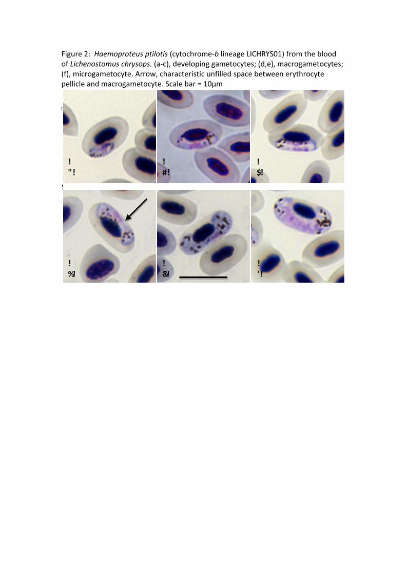
Figure 2: Haemoproteus ptilotis (cytochrome-b lineage LICHRYS01) from the blood of Lichenostomus chrysops. (a-c), developing gametocytes; (d,e), macrogametocytes; (f), microgametocyte. Arrow, characteristic unfilled space between erythrocyte pellicle and macrogametocyte. Scale bar = 10µm
Related Documents