Provided for non-commercial research and educational use only. Not for reproduction, distribution or commercial use. This chapter was originally published in the book International Review of Cell and Molecular Biology, Vol. 278, published by Elsevier, and the attached copy is provided by Elsevier for the author's benefit and for the benefit of the author's institution, for non-commercial research and educational use including without limitation use in instruction at your institution, sending it to specific colleagues who know you, and providing a copy to your institution’s administrator. All other uses, reproduction and distribution, including without limitation commercial reprints, selling or licensing copies or access, or posting on open internet sites, your personal or institution’s website or repository, are prohibited. For exceptions, permission may be sought for such use through Elsevier's permissions site at: http://www.elsevier.com/locate/permissionusematerial From: Paolo Chieffi, Renato Franco, and Giuseppe Portella, Molecular and Cell Biology of Testicular Germ Cell Tumors. In Kwang W. Jeon, editor: International Review of Cell and Molecular Biology, Vol. 278, Burlington: Academic Press, 2009, pp. 277-308. ISBN: 978-0-12-374809-6 © Copyright 2009 Elsevier Inc. Academic Press.

Welcome message from author
This document is posted to help you gain knowledge. Please leave a comment to let me know what you think about it! Share it to your friends and learn new things together.
Transcript
Provided for non-commercial research and educational use only. Not for reproduction, distribution or commercial use.
This chapter was originally published in the book International Review of Cell and Molecular Biology, Vol. 278, published by Elsevier, and the attached copy is provided by Elsevier for the author's benefit and for the benefit of the author's institution, for non-commercial research and educational use including without limitation use in instruction at your institution, sending it to specific colleagues who know you, and providing a copy to your institution’s administrator.
All other uses, reproduction and distribution, including without limitation commercial reprints, selling or licensing copies or access, or posting on open internet sites, your personal
or institution’s website or repository, are prohibited. For exceptions, permission may be sought for such use through Elsevier's permissions site at:
http://www.elsevier.com/locate/permissionusematerial
From: Paolo Chieffi, Renato Franco, and Giuseppe Portella, Molecular and Cell Biology of Testicular Germ Cell Tumors. In Kwang W. Jeon, editor: International Review of Cell and
Molecular Biology, Vol. 278, Burlington: Academic Press, 2009, pp. 277-308. ISBN: 978-0-12-374809-6
© Copyright 2009 Elsevier Inc. Academic Press.
Author's personal copy
C H A P T E R S I X
In
IS
*{
{
ternati
SN 1
DiparArea80131Dipar80131
Molecular and Cell Biology
of Testicular Germ Cell Tumors
Paolo Chieffi,* Renato Franco,† and Giuseppe Portella‡
Contents
1. In
onal
937
timFunNtimN
troduction
Review of Cell and Molecular Biology, Volume 278 # 2009
-6448, DOI: 10.1016/S1937-6448(09)78006-2 All rig
ento di Medicina Sperimentale, II Universita di Napoli, 80138 Naples, Italyzionale di Anatomia Patologica, Istituto Nazionale dei Tumori ‘‘Fondazione G. Pascaaples, Italyento di Biologia e Patologia Cellulare e Molecolare, Universita di Napoli ‘‘Federico Iaples, Italy
Else
hts
le,’
I,’’
278
2. E
pidemiology and Risk Factors 2793. H
istopathology 2803
.1. O rigin and development 2803
.2. C lassification 2823
.3. S taging of testicular tumors 2863
.4. IT GCN, Unclassified 2873
.5. S eminomatous–TGCTs 2873
.6. N SE tumors 2893
.7. S omatic malignancies arising in testicular GCT 2933
.8. R egression of primitive GCTs 2934. P
rognostic and Diagnostic Markers 2944
.1. S erum tumor markers 2944
.2. N ew discovered markers for TGCTs 2955. T
herapy 2975
.1. T raditional treatment 2975
.2. A urora-kinase inhibitors 2985
.3. R eceptor and nonreceptor tyrosine kinases inhibitors 2995
.4. A ngiogenesis inhibitors 3006. C
onclusions and Perspectives 301Ackn
owledgments 301Refe
rences 302Abstract
Although testicular germ cell tumors (TGCTs) are relatively uncommon, they are
particularly important as they tend to affect children and young men, represent-
ing the most common tumor in male aged from 20 to 40 years. TGCTs are a
vier Inc.
reserved.
’
277
278 Paolo Chieffi et al.
Author's personal copy
heterogeneous group of tumors, with specific peculiarities reflecting on epide-
miologic distribution and clinic-pathological features. TGCTs show a high-cure
rates in both seminomas and nonseminomas and represent the model of a
curable neoplasia: sensitive serum tumor markers, accurate prognostic classifi-
cation, contribute to a high effectiveness of cancer therapy. However, up to 30%
of patients diagnosed with metastatic nonseminomas do not achieve a durable
remission, and in metastatic teratomas cisplatin-based treatment resistance
has been observed. These different prognostic and therapeutic features of
TGCTs highlight the need for a better understanding of the molecular biology
of TGCT, that could help to improve disease management and to tailor aggres-
siveness of treatment to the severity of the prognosis.
Key Words: Testis, Testicular cancer, Gonocytes, Testicular germ cells, Kinase
inhibitors. � 2009 Elsevier Inc.
1. Introduction
Testicular germ cell tumors (TGCTs) of the testis are a heterogeneousgroup of neoplasms seen mainly in children and young men (Chieffi, 2007;Oosterhuis and Looijenga, 2005). They are classified as seminomatous (SE-TGCT) and nonseminomatous (NSE-TGCT) tumors. Distinction of pre-buberal TGCTs, exclusively represented by yolk sac tumor (YST) andteratoma, and postpuberal TGCTs, invariably arising from intratubulargerm cell neoplasias (ITGCNs) seems to have a great prognostic relevance(Chaganti and Houldsworth, 2000; Ulbright, 1993).
Postpuberal TCGTs are the most frequent solid malignant tumor in menbetween 20 and 40 years of age, accounting for up to 60% of all malignanciesdiagnosed at this age. Despite a high-cure rate, they represent the mostfrequent cause of death from solid tumors in this age group (Oosterhuis andLooijenga, 2005; Pottern, 1998). Seminomas are radio- and chemo-sensitivetumors, virtually completely curable ( Jones and Vasey, 2003). NSE tumorsare usually treated with surgery and chemotherapy, with different cure ratesdepending on the disease stage (Shelley et al., 2002). The cure rate reaches upto 99% in the early stages of NSE tumors, although in advanced diseasedecreases from 90% in patients with good prognostic category to 50% inpatients with poor prognostic features (Shelley et al., 2002).
The rapid growth and progression of postpuberal TGCTs cause earlylymph node metastases and/or distant metastases. At the time of diagnosisabout 25% of seminoma patients and up to 60% of the nonseminomapatients suffers from metastatic disease (Al Ghamdi and Jewett, 2005;Classen et al., 2001; Peckham, 1988; Perrotti et al., 2004; Porcaro et al.,2002), posing a therapeutic problem since in metastastic disease the
Testicular Germ Cell Tumors 279
Author's personal copy
treatment achieves modest results. Thus, despite the general success ofpostpuberal TGCTs treatment, 10–20% of patients diagnosed with meta-static disease will not achieve a durable complete remission after initialtreatment, either due to incomplete response or a tumors relapse.
The review will focus on the molecular alterations identified in post-puberal TGCTs and on novel targeted antineoplastic strategies that couldcontribute to the cure of chemotherapy resistant TGCTs.
2. Epidemiology and Risk Factors
TGCTs have significantly increased in the past 50 years; this increase isprobably due to changes in environmental factors contributing to thedevelopment of these lesions. A number of environmental factors havebeen investigated to explain the possible links. Some evidence suggestsassociation of increased TGCTs risk and maternal smoking during preg-nancy, adult height, body mass index, diet rich in cheese, and others(Bonner et al., 2002; Dieckmann and Pichlmeier, 2002; Dieckmann et al.,2008, 2009; Garcıa-Rodrıguez et al., 1996; Garner et al., 2003), however,the biological mechanisms remain to be elucidated.
Hypothesized environmental agents involved in the development ofTGCTs, include pesticides (McGlynn et al., 2008) and nonsteroidal estro-gens, such as diethylstilbestrol (DES) (Martin et al., 2008). It has beenproposed that increased levels of estrogen exposure in utero to increase therisk of TGCTs (Garner et al., 2008) and the exposure of women to thenonsteroidal estrogen DES during pregnancy increases the risk of TGCTs(Strohsnitter et al., 2001). However, other studies have not confirmed a rolefor estrogen in TGCTs development (Dieckmann et al., 2001). Despite thecontrasting results reported in the literature a clear role for environmentalfactors in the etiology of TGCTs is suggested by population migrationstudies. Sweden has an incidence of TGCTs about twice that of Finlandand although first generation migrants from Finland to Sweden show noincreased risk (Ekbom et al., 2003), second generation males born to themigrant parents in Sweden have a tendency to an increased frequency(Montgomery et al., 2005).
Familial predisposition to TGCTs, ethnic variations in incidence, and anassociation with certain chromosome abnormality syndromes strongly sug-gest that inherited factors, also, play a role in disease development. Thefamilial predisposition is one of the strongest for any tumor type, since theincreased relative risk of TGCTs development associated with fathers andsons of TGCTs patients is fourfold (Forman et al., 1992). However, gene(s)involved in familial TGCTs have not been identified so far (Krausz andLooijenga, 2008). Genome-wide linkage analysis of affected families has
280 Paolo Chieffi et al.
Author's personal copy
provided evidence for two susceptibility loci, one at Xq27 locus for unde-scended testis probably playing an indirect role and another at 12q whichresults in hyperexpression of the product of the CCND2 gene (LutkeHolzik et al., 2004). It is probable that both genetic and environmentalfactors produce the high familial risk seen in TGCTs and that the interplaybetween these two factors, along with genetic heterogeneity, may makefamilial associated susceptibility loci difficult to determine.
3. Histopathology
3.1. Origin and development
The origin and biology of TGCTs are currently distinct on whether theyoccur in pre- and postpuberal age, being pure teratomas and YSTs with asubstantially benign prognosis the most common hystotypes of prepuberaltestis and seminoma, pure NSE tumors and mixed germ cell tumors (GCTs)with a relative more aggressive behavior typical of adult testis (Looijengaand Oosterhuis, 1999).
It has been suggested that the initiating event in the pathogenesis of TGCToccurs during embryonal development (Chieffi, 2007). The most widelyaccepted model of postpuberal TGCTs development proposes an initialtumorigenic event in utero and the development of a precursor lesion knownas intratubular germ cell neoplasia undifferentiated (ITGCNU), also known ascarcinoma in situ (CIS) (Skakkebaek, 1972). This is followed by a period ofdormancy until after puberty when postpuberal TGCTs emerge. This prepu-bertal dormancy suggests that the TGCTs development is hormonedependent.
Recently, it has been proposed that tumors originate from neoplasticcells that retain stem cell properties such as self-renewal (Wicha, et al.,2006), and this novel hypothesis has fundamental implications for thepathogenesis of TGCTs. According with stem cells hypothesis, tumorsoriginate from tissue stem cells or from their immediate progeny. Thiscellular subcomponent drives tumorigenesis and aberrant differentiation,contributing to cellular heterogeneity of the tumor and also to the resistanceto antineoplastic treatments.
ITGCNU cells are generally accepted as the common preinvasive precur-sor cells that gives rise to postpuberal TGCTs (Oosterhuis and Looijenga,2005; Ulbright, 1999). ITGCNU almost found invariably in the periphery ofovert postpuberal TGCTs and is estimated that it is present in approximately5% of the contralateral testis of patients with postpuberal TGCTs (Berthelsenet al., 1982). Preinvasive ITGCNU cells are supposed to be able to develop indifferent germinal and somatic tissues and are regarded as pluripotent ortotipotent cells and therefore can be considered as TGCTs stem cells.
Testicular Germ Cell Tumors 281
Author's personal copy
ITGCNU cells share morphological similarities with gonocytes and ithas been proposed that ITGCNU cells could be remnants of undifferenti-ated embryonic/fetal cells (Nielsen et al., 1974; Skakkebaek et al., 1987).Their fetal origin is also supported by immunohistochemical studies ofproteins present in ITGCNU, also shown to be present in primordialgerm cells (PGCs) and gonocytes. The identification of ITGCNU cells inprepubertal patients, who later developed TGCTs, indicated that the cellshad originated prior to puberty (Muller et al., 1984).
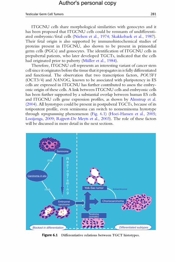
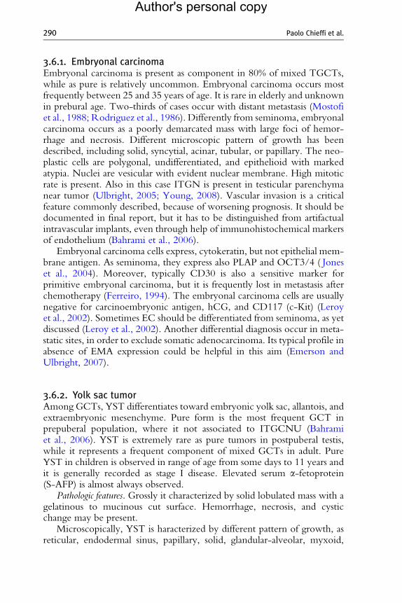
Therefore, ITGCNU cell represents an interesting variant of cancer stemcell since it originates before the tissue that it propagates in is fully differentiatedand functional. The observation that two transcription factors, POU5F1(OCT3/4) and NANOG, known to be associated with pluripotency in EScells are expressed in ITGCNU has further contributed to assess the embry-onic origin of these cells. A link between ITGCNU cells and embryonic cellshas been further supported by a substantial overlap between human ES cellsand ITGCNU cells gene expression profiles, as shown by Almstrup et al.(2004). All hystotypes could be present in postpuberal TGCTs, because of itstotipontent profile, even seminoma can switch to nonseminoma hystotypethrough reprogramming phenomenon (Fig. 6.1) (Hoei-Hansen et al., 2005;Looijenga, 2009; Rajpert-De Meyts et al., 2003). The role of these factorswill be discussed in more detail in the next sections.
Blocked in differentiation
Embryonalcarcinoma
ITSE
Seminoma
Reprograming
Yolk-Sac tumor
Choriocarcinoma
Teratoma(somatic)
Carcinoma in situ
Differentiated subtypes
ExtraembryonalITNS
Figure 6.1 Differentiative relations between TGCT histotypes.
282 Paolo Chieffi et al.
Author's personal copy
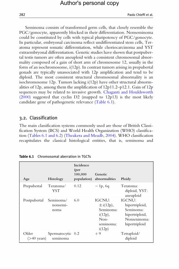
Seminoma consists of transformed germ cells, that closely resemble thePGC/gonocyte, apparently blocked in their differentiation. Nonseminomacould be constituted by cells with typical pluripotency of PGC/gonocyte.In particular, embryonal carcinoma reflect undifferentiated stem cells, Ter-atoma represent somatic differentiation, while choriocarcinoma and YSTextraembryonal differentiation. Genetic studies have shown that postpuber-tal testis tumors are often aneuploid with a consistent chromosomal abnor-mality composed of a gain of short arm of chromosome 12, usually in theform of an isochromosome, i(12p). In contrast tumors arising in prepubertalgonads are typically unassociated with 12p amplification and tend to bediploid. The most consistent structural chromosomal abnormality is anisochromosome 12p. Tumors lacking i(12p) have other structural abnorm-alities of 12p, among them the amplification of 12p11.2–p12.1. Gain of 12psequences may be related to invasive growth. Chaganti and Houldsworth(2000) suggested that cyclin D2 (mapped to 12p13) is the most likelycandidate gene of pathogenetic relevance (Table 6.1).
3.2. Classification
The main classification systems commonly used are those of British Classi-fication System (BCS) and World Health Organization (WHO) classifica-tion (Tables 6.1 and 6.2) (Theakera and Meadb, 2004). WHO classificationrecapitulates the classical histological entities, that is, seminoma and
Table 6.1 Chromosomal aberration in TGCTs
Age Histology
Incidence
(per
100,000
population)
Genetic
abnormalities Ploidy
Prepubertal Teratoma/
YST
0.12 � 1p, 6q Teratoma:
diploid, YST:
aneuploid
Postpubertal Seminoma/
nonsemi-
noma
6.0 IGCNU:
� i(12p),
Seminoma:
i(12p),
Non-
seminoma:
i(12p)
IGCNU:
hipertriploid,
Seminoma:
hipertriploid,
Nonseninoma:
hipertriploid
Older
(>40 years)
Spermatocytic
seminoma
0.2 þ 9 Tetraploid/
diploid
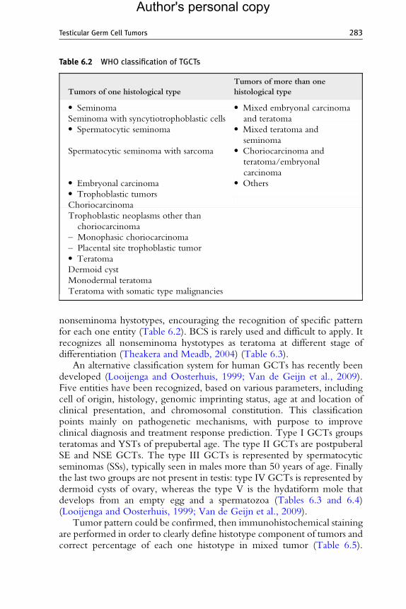
Table 6.2 WHO classification of TGCTs
Tumors of one histological type
Tumors of more than one
histological type
� Seminoma � Mixed embryonal carcinoma
and teratomaSeminoma with syncytiotrophoblastic cells� Spermatocytic seminoma � Mixed teratoma and
seminoma
Spermatocytic seminoma with sarcoma � Choriocarcinoma and
teratoma/embryonal
carcinoma� Embryonal carcinoma � Others� Trophoblastic tumors
Choriocarcinoma
Trophoblastic neoplasms other than
choriocarcinoma
– Monophasic choriocarcinoma
– Placental site trophoblastic tumor� Teratoma
Dermoid cyst
Monodermal teratoma
Teratoma with somatic type malignancies
Testicular Germ Cell Tumors 283
Author's personal copy
nonseminoma hystotypes, encouraging the recognition of specific patternfor each one entity (Table 6.2). BCS is rarely used and difficult to apply. Itrecognizes all nonseminoma hystotypes as teratoma at different stage ofdifferentiation (Theakera and Meadb, 2004) (Table 6.3).
An alternative classification system for human GCTs has recently beendeveloped (Looijenga and Oosterhuis, 1999; Van de Geijn et al., 2009).Five entities have been recognized, based on various parameters, includingcell of origin, histology, genomic imprinting status, age at and location ofclinical presentation, and chromosomal constitution. This classificationpoints mainly on pathogenetic mechanisms, with purpose to improveclinical diagnosis and treatment response prediction. Type I GCTs groupsteratomas and YSTs of prepubertal age. The type II GCTs are postpuberalSE and NSE GCTs. The type III GCTs is represented by spermatocyticseminomas (SSs), typically seen in males more than 50 years of age. Finallythe last two groups are not present in testis: type IV GCTs is represented bydermoid cysts of ovary, whereas the type V is the hydatiform mole thatdevelops from an empty egg and a spermatozoa (Tables 6.3 and 6.4)(Looijenga and Oosterhuis, 1999; Van de Geijn et al., 2009).
Tumor pattern could be confirmed, then immunohistochemical stainingare performed in order to clearly define histotype component of tumors andcorrect percentage of each one histotype in mixed tumor (Table 6.5).
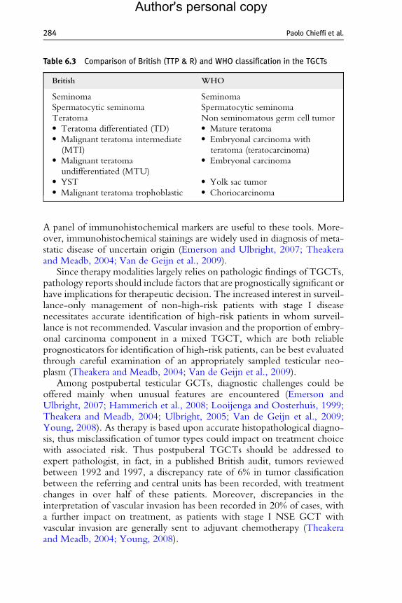
Table 6.3 Comparison of British (TTP & R) and WHO classification in the TGCTs
British WHO
Seminoma Seminoma
Spermatocytic seminoma Spermatocytic seminoma
Teratoma Non seminomatous germ cell tumor� Teratoma differentiated (TD) � Mature teratoma� Malignant teratoma intermediate
(MTI)
� Embryonal carcinoma with
teratoma (teratocarcinoma)� Malignant teratoma
undifferentiated (MTU)
� Embryonal carcinoma
� YST � Yolk sac tumor� Malignant teratoma trophoblastic � Choriocarcinoma
284 Paolo Chieffi et al.
Author's personal copy
A panel of immunohistochemical markers are useful to these tools. More-over, immunohistochemical stainings are widely used in diagnosis of meta-static disease of uncertain origin (Emerson and Ulbright, 2007; Theakeraand Meadb, 2004; Van de Geijn et al., 2009).
Since therapy modalities largely relies on pathologic findings of TGCTs,pathology reports should include factors that are prognostically significant orhave implications for therapeutic decision. The increased interest in surveil-lance-only management of non-high-risk patients with stage I diseasenecessitates accurate identification of high-risk patients in whom surveil-lance is not recommended. Vascular invasion and the proportion of embry-onal carcinoma component in a mixed TGCT, which are both reliableprognosticators for identification of high-risk patients, can be best evaluatedthrough careful examination of an appropriately sampled testicular neo-plasm (Theakera and Meadb, 2004; Van de Geijn et al., 2009).
Among postpubertal testicular GCTs, diagnostic challenges could beoffered mainly when unusual features are encountered (Emerson andUlbright, 2007; Hammerich et al., 2008; Looijenga and Oosterhuis, 1999;Theakera and Meadb, 2004; Ulbright, 2005; Van de Geijn et al., 2009;Young, 2008). As therapy is based upon accurate histopathological diagno-sis, thus misclassification of tumor types could impact on treatment choicewith associated risk. Thus postpuberal TGCTs should be addressed toexpert pathologist, in fact, in a published British audit, tumors reviewedbetween 1992 and 1997, a discrepancy rate of 6% in tumor classificationbetween the referring and central units has been recorded, with treatmentchanges in over half of these patients. Moreover, discrepancies in theinterpretation of vascular invasion has been recorded in 20% of cases, witha further impact on treatment, as patients with stage I NSE GCT withvascular invasion are generally sent to adjuvant chemotherapy (Theakeraand Meadb, 2004; Young, 2008).
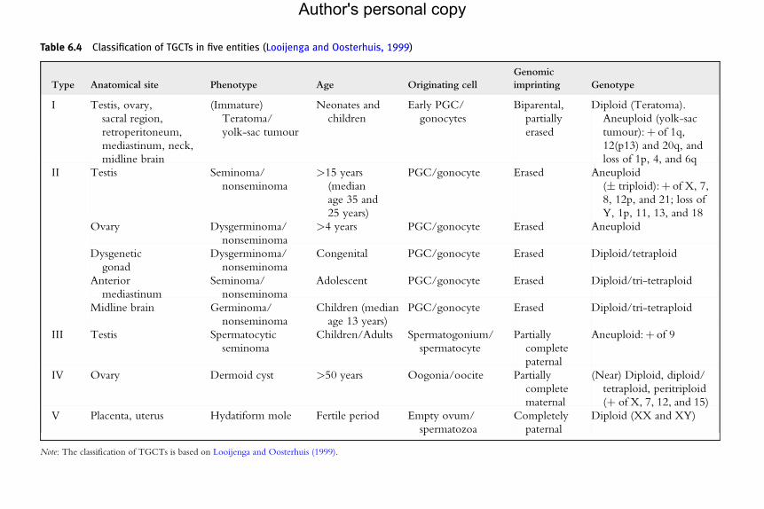
Table 6.4 Classification of TGCTs in five entities (Looijenga and Oosterhuis, 1999)
Type Anatomical site Phenotype Age Originating cell
Genomic
imprinting Genotype
I Testis, ovary,
sacral region,
retroperitoneum,
mediastinum, neck,
midline brain
(Immature)
Teratoma/
yolk-sac tumour
Neonates and
children
Early PGC/
gonocytes
Biparental,
partially
erased
Diploid (Teratoma).
Aneuploid (yolk-sac
tumour):þ of 1q,
12(p13) and 20q, and
loss of 1p, 4, and 6q
II Testis Seminoma/
nonseminoma
>15 years
(median
age 35 and
25 years)
PGC/gonocyte Erased Aneuploid
(� triploid):þ of X, 7,
8, 12p, and 21; loss of
Y, 1p, 11, 13, and 18
Ovary Dysgerminoma/
nonseminoma
>4 years PGC/gonocyte Erased Aneuploid
Dysgenetic
gonad
Dysgerminoma/
nonseminoma
Congenital PGC/gonocyte Erased Diploid/tetraploid
Anterior
mediastinum
Seminoma/
nonseminoma
Adolescent PGC/gonocyte Erased Diploid/tri-tetraploid
Midline brain Germinoma/
nonseminoma
Children (median
age 13 years)
PGC/gonocyte Erased Diploid/tri-tetraploid
III Testis Spermatocytic
seminoma
Children/Adults Spermatogonium/
spermatocyte
Partially
complete
paternal
Aneuploid:þ of 9
IV Ovary Dermoid cyst >50 years Oogonia/oocite Partially
complete
maternal
(Near) Diploid, diploid/
tetraploid, peritriploid
(þ of X, 7, 12, and 15)
V Placenta, uterus Hydatiform mole Fertile period Empty ovum/
spermatozoa
Completely
paternal
Diploid (XX and XY)
Note: The classification of TGCTs is based on Looijenga and Oosterhuis (1999).
Author's personal copy
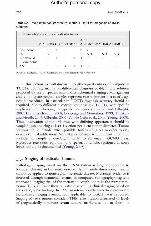
Table 6.5 Main immunohistochemical markers useful for diagnosis of TGCTssubtypes
Immunohistochemistry in testicular tumors
PLAP c-Kit OCT4 CD30 AFP
AE1/
AE3 CK7 EMA HMGA1HMGA2
Seminoma þ þ þ � � v v � þ �SS � v � � � � ND � ND ND
Embryonal
carcinoma
þ � þ þ v þ þ � þ þ
YST þ � � v v þ � � þNotes: þ, expressed; �, not expressed; ND, not determined; v, variable.
286 Paolo Chieffi et al.
Author's personal copy
In this section we will discuss histopathological entities of postpuberalTGCTs, pointing mainly on differential diagnosis problems and solutionproposed by use of specific immunohistochemical stainings. Managementand sampling are surgical samples represent two important phases of diag-nostic procedures. In particular in TGCTs diagnosis accuracy should berequired, due to different histotypes composing a TGCTs, with specificimplications in choicing therapeutic strategies (Emerson and Ulbright,2007; Hammerich et al., 2008; Looijenga and Oosterhuis, 1999; Theakeraand Meadb, 2004; Ulbright, 2005; Van de Geijn et al., 2009; Young, 2008).Thus observation of tumoral areas with differing appearances should besampled, garantueeing at least 1 section per 1 cm tumor diameter. Tumorsections should include, when possible, tonaca albuginea in order to evi-dence eventual infiltration. Normal parenchyma, when present, should beincluded in sample proceeding in order to evidence ITGCNU areas.Moreover rete testis, epididim, and spermatic funicle, sectioned at morelevels, should be documented (Young, 2008).
3.3. Staging of testicular tumors
Pathologic staging based on the TNM system is largely applicable tolocalized disease, and to retroperitoneal lymph node dissections, it reallycannot be applied to nonsurgical metastatic disease. Metastasis evidence isdetected through strumental exams, as computed tomography/magneticresonance imaging size of the metastatic lymph nodes in the retroperito-neum. Thus, adjuvant therapy is stated according clinical staging based onthe radiographic findings. In 1997, an internationally agreed-on prognosticfactor-based staging classification, applicable to TGCTs was proposed.Staging of testis tumors considers TNM classification associated to levelsof prognostically important serum tumoral markers, as human chorionic
Testicular Germ Cell Tumors 287
Author's personal copy
gonadotropin (hCG), a-fetoprotein (AFP), and lactate dehydrogenase (LD),reflecting tumoral turnover (Tables 6.4 and 6.5).
3.4. ITGCN, Unclassified
ITGCNU is the precursor of all invasive TGCTs, excluding SS and prepu-bertal TGCTs, and it is identified adjacent in about 90% of cases. TheITGCNU cells are similar to seminoma cells, suggesting that seminoma isthe invasive component of ITGCNU cells. Without intervention, about50% of ITGCNU cases will progress to an invasive GCT within 5 years ofthe diagnosis of ITGCNU, but on long-term follow-up almost all patientswill develop an invasive GCT. Cytogenetic studies underline similar chro-mosomal status between ITGCNU cells and the adjacent invasive compo-nent, with the gain of 12p found in the invasive part respect to IGCCNU.Thus acquisition of excess genetic material on the short arm of chromosome12 could be critical genetic event leading to invasion (Bahrami et al., 2006).
Pathological findings. Grossly areas with ITGCNU are similar to normaltestis parenchyma. Histologically, ITGCNU cell are large primitive atypicalelements, twice the size of normal germ cells. These cells lie along thethickened basement membrane of atrophic seminiferous tubules or replacethe entire tubules. The neoplastic cells have large nuclei with prominentnucleoli and large clear cytoplasm enriched of glycogen. Immunohisto-chemical neoplastic cells stain for placental-like alkaline phosphatase(PLAP), for c-Kit (CD117), OCT3/4, also known as POU5F1, but arenegative for cytokeratins, hCG, or AFP. D2-40, an antibody raised against atransmembrane mucoprotein called podoplanin, stains ITGCNU cells(Bahrami et al., 2006; Eble et al., 2004; Ulbright, 2005; Young, 2008).
3.5. Seminomatous–TGCTs
Two types of SE-TGCTs are described: (a) classical seminoma and itsvariants; (b) SS.
3.5.1. SeminomaSeminoma is the most common TGCT , representing 50% of the cases(Table 6.6). Moreover seminoma component is recognizable in a largeproportion of mixed GCTs. The peak incidence is between 34 and45 years, which is about one decade later than that of most other GCTs.Painless testicular enlargement is its clinical. More rarely symptoms arelinked to metastasis onset (Ulbright, 2004). In localized disease, extensionto the spermatic cord or epididymis is recorded seen in less than 10% ofpatients, and in 2% of cases both testes are involved (Ro et al., 2000).
Pathologic features. Grossly, seminomas are firm with homogenousappearance, organized in single or multiple nodules. Necrosis and
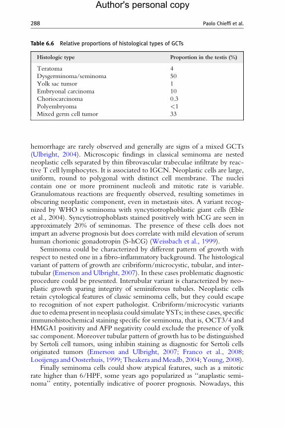
Table 6.6 Relative proportions of histological types of GCTs
Histologic type Proportion in the testis (%)
Teratoma 4
Dysgerminoma/seminoma 50
Yolk sac tumor 1
Embryonal carcinoma 10
Choriocarcinoma 0.3
Polyembryoma <1
Mixed germ cell tumor 33
288 Paolo Chieffi et al.
Author's personal copy
hemorrhage are rarely observed and generally are signs of a mixed GCTs(Ulbright, 2004). Microscopic findings in classical seminoma are nestedneoplastic cells separated by thin fibrovascular trabeculae infiltrate by reac-tive T cell lymphocytes. It is associated to IGCN. Neoplastic cells are large,uniform, round to polygonal with distinct cell membrane. The nucleicontain one or more prominent nucleoli and mitotic rate is variable.Granulomatous reactions are frequently observed, resulting sometimes inobscuring neoplastic component, even in metastasis sites. A variant recog-nized by WHO is seminoma with syncytiotrophoblastic giant cells (Ebleet al., 2004). Syncytiotrophoblasts stained positively with hCG are seen inapproximately 20% of seminomas. The presence of these cells does notimpart an adverse prognosis but does correlate with mild elevation of serumhuman chorionic gonadotropin (S-hCG) (Weissbach et al., 1999).
Seminoma could be characterized by different pattern of growth withrespect to nested one in a fibro-inflammatory background. The histologicalvariant of pattern of growth are cribriform/microcystic, tubular, and inter-tubular (Emerson and Ulbright, 2007). In these cases problematic diagnosticprocedure could be presented. Interubular variant is characterized by neo-plastic growth sparing integrity of seminiferous tubules. Neoplastic cellsretain cytological features of classic seminoma cells, but they could escapeto recognition of not expert pathologist. Cribriform/microcystic variantsdue to edemapresent in neoplasia could simulateYSTs; in these cases, specificimmunohistochemical staining specific for seminoma, that is, OCT3/4 andHMGA1 positivity and AFP negativity could exclude the presence of yolksac component. Moreover tubular pattern of growth has to be distinguishedby Sertoli cell tumors, using inhibin staining as diagnostic for Sertoli cellsoriginated tumors (Emerson and Ulbright, 2007; Franco et al., 2008;Looijenga andOosterhuis, 1999; Theakera andMeadb, 2004; Young, 2008).
Finally seminoma cells could show atypical features, such as a mitoticrate higher than 6/HPF, some years ago popularized as ‘‘anaplastic semi-noma’’ entity, potentially indicative of poorer prognosis. Nowadays, this
Testicular Germ Cell Tumors 289
Author's personal copy
characteristics and others retained not typical in seminoma, that is, pleo-morphism, nuclear overlapping, lack or paucity of lymphocytes infiltrate, lackor paucity of clear cytoplasms seems not related to worse prognosis, above allfor unreproducibility of such morphological observation. But not conven-tional features observed in some seminoma could represent a problem indifferential diagnosis with respect to solid growth of embryonal carcinoma.Combined seminoma embryonal carcinoma is difficult to recognize whensmall foci of embryonal carcinoma are situated within the main bulk of theseminoma. CD30 positivity could help in identifying such foci and to classifyas mixed GCTs (Theakera and Meadb, 2004; Young, 2008).
3.5.2. Spermatocytic seminomaSS accounts for 1–2% of all testicular neoplasm (Talerman, 1980). Althoughit frequently occur in patients older than 50 years, about one-third of SS isseen in the usual age of classical seminoma age range (Young, 2008). Thecell of origin of SS are more differentiated than that of classical seminoma,with cytogenetic loss of chromosome 9, rather than isochromosome 12p(Bahrami et al., 2006; Verdorfer et al., 2004). Moreover, it is not associatedto cryptorchidism, ITGCNU, or other GCTs subtypes (Talerman, 1980). Ithas an indolent natural history, with a very low risk of metastasis. However,sarcomatous dedifferentiation with a very poor prognosis because of hema-togenous spreading has been recorded in 6% of cases (Burke and Mostofi,1993).
Pathological findings. Grossly SS is an homogeneous yellow-gray mass;when sarcomatous dedifferentiation is encountered a more heterogeneouscut surface is described. Histologically, SS consists of diffuse proliferation ofpolymorphic cells, small, intermediate, and large sized. Intermediate cellsare more represented and they are characteristically with ‘‘spiralized’’ chro-matin, as it is seen in spermatocytes. Large cells are generally uninucleatedand sometimes multinucleated. Sarcomatous differentiation can occur withundifferentiated or differentiated patterns, such as chondrosarcoma andrhabdomysarcoma. All immunohistochemical markers commonly describedin other GCT subtypes are negative in SS, with the exception of c-Kit.Moreover, cytoplasmic positivity of CAM 5.2 in SS cells has been described(Bahrami et al., 2006; Eble et al., 2004; Ulbright, 2005; Young, 2008).
3.6. NSE tumors
According to classical classification nonseminoumatous tumors consists ofpure teratoma, embryonal carcinoma, YST, choriocarcinoma, and mixedTGCTs. The pure form of each one histotype is relatively rare with respectto combination of different components in mixed TGCTs.
290 Paolo Chieffi et al.
Author's personal copy
3.6.1. Embryonal carcinomaEmbryonal carcinoma is present as component in 80% of mixed TGCTs,while as pure is relatively uncommon. Embryonal carcinoma occurs mostfrequently between 25 and 35 years of age. It is rare in elderly and unknownin prebural age. Two-thirds of cases occur with distant metastasis (Mostofiet al., 1988; Rodriguez et al., 1986). Differently from seminoma, embryonalcarcinoma occurs as a poorly demarcated mass with large foci of hemor-rhage and necrosis. Different microscopic pattern of growth has beendescribed, including solid, syncytial, acinar, tubular, or papillary. The neo-plastic cells are polygonal, undifferentiated, and epithelioid with markedatypia. Nuclei are vesicular with evident nuclear membrane. High mitoticrate is present. Also in this case ITGN is present in testicular parenchymanear tumor (Ulbright, 2005; Young, 2008). Vascular invasion is a criticalfeature commonly described, because of worsening prognosis. It should bedocumented in final report, but it has to be distinguished from artifactualintravascular implants, even through help of immunohistochemical markersof endothelium (Bahrami et al., 2006).
Embryonal carcinoma cells express, cytokeratin, but not epithelial mem-brane antigen. As seminoma, they express also PLAP and OCT3/4 ( Joneset al., 2004). Moreover, typically CD30 is also a sensitive marker forprimitive embryonal carcinoma, but it is frequently lost in metastasis afterchemotherapy (Ferreiro, 1994). The embryonal carcinoma cells are usuallynegative for carcinoembryonic antigen, hCG, and CD117 (c-Kit) (Leroyet al., 2002). Sometimes EC should be differentiated from seminoma, as yetdiscussed (Leroy et al., 2002). Another differential diagnosis occur in meta-static sites, in order to exclude somatic adenocarcinoma. Its typical profile inabsence of EMA expression could be helpful in this aim (Emerson andUlbright, 2007).
3.6.2. Yolk sac tumorAmong GCTs, YST differentiates toward embryonic yolk sac, allantois, andextraembryonic mesenchyme. Pure form is the most frequent GCT inprepuberal population, where it not associated to ITGCNU (Bahramiet al., 2006). YST is extremely rare as pure tumors in postpuberal testis,while it represents a frequent component of mixed GCTs in adult. PureYST in children is observed in range of age from some days to 11 years andit is generally recorded as stage I disease. Elevated serum a-fetoprotein(S-AFP) is almost always observed.
Pathologic features. Grossly it characterized by solid lobulated mass with agelatinous to mucinous cut surface. Hemorrhage, necrosis, and cysticchange may be present.
Microscopically, YST is haracterized by different pattern of growth, asreticular, endodermal sinus, papillary, solid, glandular-alveolar, myxoid,
Testicular Germ Cell Tumors 291
Author's personal copy
sarcomatoid, hepatoid, and parietal patterns. Generally more pattern areobserved in a single YST, being reticular-mycocistic patter the most com-mon feature. Schiller–Duval bodies is the most characteristic features and isconstituted by central fibrovascular core surrounded by malignant cuboidalto columnar cells. Moreover, PAS positive hyaline globules are frequentlyobserved. AFP immunostaining is commonly observed in YST. Staining forcytokeratin, PLAP, and a1-antitrypsin are always observed, while neoplasmis generally negative for CD30, hCG, and OCT3/4. Recently, Glypican 3,a membrane-bound heparin sulfate proteoglycan, seems to be specificallyexpressed in YST and choriocarcinoma with respect to seminoma, embry-onal carcinoma, and teratoma (Emerson and Ulbright, 2007).
3.6.3. Trophoblastic tumorsAmong trophoblastic tumors, choriocarcinoma is absolutely the most fre-quent. Pure choriocarcinoma is extremely rare. Choriocarcinoma foci areobserved in 8% of mixed GCTs. Both as pure and as component in a mixedGCTs, choriocarcinoma is responsible of an aggressive outcome, because ofrapid hematogenous dissemination (Ulbright, 2005). Patients are diagnosedin an advanced stage and sometimes testis neoplasia is regressed. Very highvalue of hCG (>100,000 mIU/ml) in serum are recorded and it propor-tionally related to worse prognosis (Ulbright, 2005).
Pathologic features. Choriocarcinoma appears as an hemorrhagic nodule,when it is pure and as multiple hemorrhagic areas in an heterogeneous mass,when a component of a mixed GCTs is present.
Histologically, choriocarcinoma consisted of variable amounts of 2 cellcomponents, syncytiotrophoblast and cytotrophoblasts. Syncytiotropho-blasts are multinucleated cells and cytotrophoblasts are uniform, polygonalcells (Bahrami et al., 2006; Ulbright, 2005; Young, 2008). Syncytiotro-phoblasts are positive to hCG and cytothrophoblasts are positive toinhibin. Glypican 3 is expressed by all choriocarcinoma, particularly bysyncytrophoblasts, PLAP staining is observed in 50% of choriocarcinoma(Emerson and Ulbright, 2007).
3.6.4. TeratomaTeratomas are composed of different tissue deriving from one or moregerminal endodermal, mesodermal, and ectodermal layers. Classically matureteratomas is defined when composed of differentiated tissues, while immatureteratoma consisted of embryonic or fetal-like tissues, associated to differen-tiated elements. Monodermal teratoma is composed of tissues derived fromone of the three germ layers (Ulbright, 2005).
Teratoma characteristics has not great prognostic relevance in testicularteratoma. In fact, the behavior of testis teratomas, but less so for ovaryteratomas, depends on patient age, being prepuberal testis pure teratomainvariably benign and postpuberal pure teratoma invariably malignant. Pure
292 Paolo Chieffi et al.
Author's personal copy
teratoma in prepuberal testis is the second most frequent GCTs (Bahramiet al., 2006). It is generally composed of mature tissues, however, variableamount of immature elements could be present; nevertheless, presence ofimmature histology is not an indicator of poor prognosis (Bahrami et al.,2006; Ulbright, 2005). Pure teratoma in postpuberal testis is rare, whileteratomatous elements occur as component of 50% of mixed tumors. In thisage, even when with mature histology, pure teratoma are at risk of metasta-sis. Moreover prepuberal teratomas are diploid; postpuberal teratomas areaneuploid and have isochromosome 12p (Bahrami et al., 2006). Prepuberalteratomas are generally diagnosed in children younger than 4 years withmean age of 20 months. Thirty-seven percent of postpuberal pure teratomaare seen in young patients with metastasis symptoms (Leibovitch et al.,1995).
Pathologic findings. Grossly pure teratomas has heterogeneous cut surface,generally with cystic areas, filled by serous, fluid, mucinous, or keratinicmaterials.
Histologically, tumors are composed of variable amounts of endodermal,ectodermal, and mesodermal tissues. Different types of specialized epithe-lium, neural tissue and cartlage, and other mature tissue are represented.Cytologic atypia is frequently seen in postpuberal teratomas (Ro et al.,2000). Immature histology is defined when some elements are not attribut-able to any adult tissue or when embryonic or fetal tissue are recognized.Generally, immature elements are mesenchymal spindle cells, rarely neuraland epithelial elements are observed.
Vascular invasion could be seen in postpuberal teratomas and is repre-sented by mature or immature elements, having capacity to differentiateinto various cell lineage. Therefore, metastasis could have morphologydifferent from primary site (Bahrami et al., 2006).
ITGCNU is observed in 90% of the cases of postpuberal teratomas,while it is nonpresent in prepuberal teratoma. Orchiectomy is absolutelycurative in prepuberal cases. Dermoid cyst is considered as a specializedform of mature teratoma, occurring in postpuberal testis. It consists ofkeratinizing epithelium with cutaneous appendages; sometimes noncuta-neous mature elements are observed. In contrast to postpuberal teratomas, itis not associated to ITGCNU and does not have malignant potential. Thusorchiectomy could be curative for dermoid cyst, if diagnosis is renderedwhen extensive sampling excludes the presence of any other teratomaelements and/or adjacent ITGCNU (Ro et al., 2000).
3.6.5. Mixed GCTsMixed GCTs of testis is the second most frequent GCT, following semi-noma. It comprises 30–50% of cases. It is associated to serum elevation ofmarkers, such as hCG and AFP, generally related to YST and syncytiotro-phoblastic components, respectively, in this tumor (Ulbright, 2004).

Testicular Germ Cell Tumors 293
Author's personal copy
Pathological findings. Grossly mixed GCTs have a variegate cute surface,reflecting different histological subtypes. In order of frequency embryonalcarcinoma, teratoma, YST, seminoma, choriocarcinoma, and polyem-brioma are described (Mosharafa et al., 2004). The most common associa-tions are embryonal carcinoma/teratoma and embryonal carcinoma/seminona, the latter generally composed of distinct entities, suggestingmulticentric origin rather than initial differentiation of seminoma in embry-onal carcinoma. All subtypes have to be reported with approximate propor-tions (Ro et al., 2000). The significance of embryonal carcinoma proportionis particularly relevant for therapeutic management of patients.
Polyembrioma is considered a distinct form of mixed GCT. It consists ofembryoid bodies that recapitulate amnion cavity and yolk sac and result of amixture of embryonal carcinoma and YST. Embryoid bodies are dispersedin a myxoid stroma. It is generally associated to other tumoral componentsin mixed GCTs and according to some authors, because of its association toimmature teratoma, it could represent the most immature form of teratoma,resembling early embryo (Ulbright, 2004).
3.7. Somatic malignancies arising in testicular GCT
Somatic malignancies generally arise from teratoma components and rarelyfrom YST components and SS. It is described in primary site or in metasta-sis. Primary site somatic malignancies in primary site generally do not affectprognosis, while extratesticular somatic malignancies worsen significantlyprognosis, above all when it is represented by rhabdomyosarcoma or primi-tive neuroectodermal tumor. In 3–6% of cases, somatic malignancies occurin patients with metastasis treated with chemotherapy, probably due todestruction of chemosensitive GCTs, allowing over-growth of preexistingsomatic malignancies (Comiter et al., 1998).
3.8. Regression of primitive GCTs
Primitive GCT of testis could undergo spontaneous regression, resultingin hyaline scarring. This phenomenon has been referred as ‘‘burnout’’GCTs. On microscopic examination hyaline scarring can be associated toscanty residual invasive neoplasm and to ITGCNU of adjacent tubules.Sometimes, hemosiderin deposits chronic inflammatory infiltrate andprominent vessels. Intratubular calcification are considered expression ofburnout embryonal carcinoma (Balzer and Ulbright, 2006; Young, 2008).Clinical diagnosis of regressed GCTs are very difficult to diagnose, butultrasound could individuate testis abnormality, above all when GCTmetastasis has been previously diagnosed.
294 Paolo Chieffi et al.
Author's personal copy
4. Prognostic and Diagnostic Markers
Research is ongoing to identify specific biomarkers that can improvediagnosis, surveillance of tumor progression, and prediction of patientprognosis. The identification of biomarkers that may predict response tospecific therapies also will be useful in stratifying patients for treatmentselection. The clinical use of serum markers has been established duringthe years and are a valid tool for prognostic purpose and the follow-up ofpatients. In recent years, an intense activity toward identifying novel bio-markers has been performed and many proteins are described as ‘‘potentialbiomarkers’’ for any cancer. Most of these novel markers are not beenvalidated and require expensive and time-consuming molecular techniques;markers with a well assessed clinical role will be discussed in Section 4.1.
4.1. Serum tumor markers
Three serum tumor markers, serum lactate dehydrogenase (S-LD) catalytic,S-AFP concentration, and S-hCG concentration, are currently used forprognostic and follow-up purposes. The majority of patients present withelevated serum markers and their prechemotherapy levels have beenintegrated into the International Germ Cell Cancer Consensus Groupconsensus prognostic index for NSE GCT classification. Patients are strati-fied into good-, intermediate-, and poor-prognosis subgroups based on theprimary tumor site, levels of serum tumor markers, and whether extrapul-monary visceral metastases are present (von Eyben, 2003).
AFP is an embryonic protein of molecular weight 70,000 dalton. AFP isnormally produced by the fetal yolk sac, the fetal gastrointestinal tract, andeventually by the fetal liver. Levels of AFP in fetal serum rise until the end ofthe first trimester of gestation and then fall. The normal range of AFP foradults and children is variously reported as under 50, under 10, and under5 ng/mL. At birth, normal infants have high AFP levels, decreasing tonormal levels within the second years of life. AFP is the main tumor markerused to monitor testicular cancer, although it is not produced by pureseminomas (Horwich et al., 2006).
S-hCG levels are essential in both the diagnosis and follow-up of hCG-producing TGCTs. After orchiectomy, an increased level of hCG indicatespersistent disease, whereas after chemotherapy-induced complete remissionof metastatic disease, reappearance of hCG indicates the presence of arelapse.
Although it is indicated that S-LD is a useful marker, studies have shownthat total S-LD levels are not specific. Isoenzyme fraction 1 is the moresensitive and specific for TGCTs patients with seminoma have a raised
Testicular Germ Cell Tumors 295
Author's personal copy
LD-1 more often than a raised AFP and hCG, whereas patients withnonseminoma have a raised AFP more often than a raised LD-1 andhCG. LD-1 is related to the characteristic chromosomal abnormality in alltypes of TGCT, a high copy number of chromosome 12p. In contrast, AFPand hCG are found mainly in NSE GCTs and they related to the histologicdifferentiation of the tumors (von Eyben, 2003).
4.2. New discovered markers for TGCTs
A number of markers has been reported over time that can be used todiscriminate CIS, seminoma, embryonal carcinoma, teratoma, and yolk sac.The most common are HMGA1, HMGA2, PATZ1, Aurora-B, Nek2,OCT3/4, c-Kit, PLAP, NANOG, SOX2, and others. For example,HMGA1 and HMGA2 are differently expressed with respect to the stateof differentiation of TGCTs, with overexpression of both proteins inpluripotential embryonal carcinoma cells and loss of expression ofHMGA1 in YSTs and of both proteins in mature adult tissue of teratomaareas. Therefore, the different profile of HMGA1 and HMGA2 proteinexpression could represent a valuable diagnostic tool in some cases ofproblematic histological differential diagnosis (Franco et al., 2008).
PATZ1 is a recently discovered zinc finger protein that acts as a tran-scriptional repressor. Although expression of PATZ1 protein was increasedin TGCTs, it was delocalized in the cytoplasm, suggesting an impairedfunction (Fedele et al., 2008). Another marker that could be used todiscriminate the different tumor histotype is Aurora-B expression; in fact,it was detected in all CIS, seminomas and embryonal carcinomas analyzedbut not in teratomas and yolk sac carcinomas (Chieffi et al., 2004; Espositoet al., 2009). Aurora-B will be discussed in more detail later.
It is shown that Nek2 protein, a centrosomal kinase required for centro-some disjunction and formation of the mitotic spindle, is upregulated andlocalized in the nucleus of neoplastic cells of seminomas. Such nuclearlocalization and the upregulation of Nek2 protein were also observed inthe Tcam-2 seminoma cell line. In addition, the nuclear localization ofNek2 is a feature of the more undifferentiated germ cells of mouse testis andcorrelates with expression of the stemness markers OCT4 and PLZF(Barbagallo et al., 2009). OCT3/4 is a well-characterized marker forPGCs. It is positive in all cases of CIS, seminoma, and embryonal carcinoma(de Jong et al., 2005; Looijenga et al., 2003a). There has been a variousamount of reports over the years that OCT3/4 is also expressed in normaladult stem cells and nongerm cell-derived cancers. However, recent dataindicate that these observations are likely related to the use of nonspecificantibodies, the latter also recognizing pseudogenes (Atlasi et al., 2008;Ledford, 2007; Lengner et al., 2007; Liedtke et al., 2007). OCT3/4 is atranscription factor of the family of octamer-binding proteins (also known
296 Paolo Chieffi et al.
Author's personal copy
as the POU homeodomain proteins) and is regarded as one of the keyregulators of pluripotency (de Jong and Looijenga, 2006). In addition toOCT3/4, several other embryonic stem-cell-specific proteins are importantfor maintaining the so-called ‘‘stemness’’ of pluripotent cells, such asNANOG and SOX2 (Avilion et al., 2003; Boiani and Scholer, 2005;Yamaguchi et al., 2005; Yates and Chambers, 2005).
NANOG protein was detected in germline stem cells (gonocytes)within the developing testis. In addition, NANOG is highly and specificallyexpressed in CIS, embryonal carcinoma, and seminomas, but not in tera-toma and YSTs revealing a molecular and developmental link betweenGCTs and the embryonic cells from which they arise (Hart et al., 2005).
SOX2 is a member of the SOX protein family, transcription factors thatregulate development from the early embryonal stage to differentiatedlineages of specialized cells. SOX proteins are known to cooperate withPOU proteins. The best characterized SOX–POU cooperation is thatbetween SOX2 and OCT3/4. SOX2 is not detected in human germ cellsregardless of their developmental age, in contrast to data in mouse embryos(de Jong et al., 2008; Perrett et al., 2008). SOX2 is expressed in embryonalcarcinoma, the undifferentiated part of nonseminomas, but it is absent inseminomas, YSTs, and normal spermatogenesis (de Jong et al., 2008; Perrettet al., 2008). CIS cells are indeed negative for SOX2, although SOX2positive Sertoli cells can be present in seminiferous tubules lacking germcells or in the presence of CIS (de Jong et al., 2008).
Expression analysis of SOX family members in TGCTs revealed that isspecifically expressed in CIS and seminoma but not in embryonal carcinoma(de Jong et al., 2008). In addition, SOX17 maps to the chromosomal region8p23, which is gained in seminoma (Korkola et al., 2008). This indicatesthat SOX17 is a candidate SOX protein for cooperation with OCT3/4 inCIS and seminoma. These data also illustrate that SOX17 is a new marker todiscriminate CIS and seminoma from embryonal carcinoma. Of interest isthat SOX17 distinguishes embryonic from adult hematopoietic stem cells(Kim et al., 2007). Current research focuses on the processes that mayregulate the differential expression of SOX2 versus SOX17 and on therole of these SOX proteins in the different histologies of the TGCTsubtypes involved. Analysis of expression patterns in microarray studiesrevealed additional markers, MCFD2, BOB1, and PROM1, for seminomacompared to normal testis (Gashaw et al., 2005). Studies demonstratedindeed increased expression levels of these three proteins in seminomacells compared to normal adult testes (Gashaw et al., 2007). Because allthree of these markers are also expressed at low levels in normal adulttesticular tissue, their suitability as practical additional diagnostic markersremains to be proven.
In recent years, the role of miRNAs in carcinogenesis of human testicu-lar cancer and germ cell development has emerged (Bernstein et al., 2003).
Testicular Germ Cell Tumors 297
Author's personal copy
It was demonstrated that knockout mice for Dicer suffered from an earlydecrease in germ cell number and an impaired ability to differentiate,indicating that Dicer1 and miRNAs are important for both survival andproper differentiation of male germ cells (Maatouk et al., 2008). Subse-quently, it was demonstrated that miRNAs 372 and 373 can overcome cellcycle arrest mediated by p53 (Voorhoeve et al., 2006). In contrast, inTGCT cell lines with mutated p53 or expressing low levels of p53 wereshown to be negative for these miRNAs and it can be assumed thatmiRNAs 372 and 373 can bypass the p53 checkpoint allowing the growthof TGCT. Further research into the functional mechanisms of miRNAs andthe role of DND in TGCT are likely to give more interesting clues.
5. Therapy
5.1. Traditional treatment
A substantial increase in cure rates of the medical treatment of advancedtesticular cancer has raised from approximately 25% in the mid-1970s tonearly 80% today. This is the highest cure rate in solid tumor. Improvedsurvival is primarily due to effective chemotherapy (Einhorn and Donohue,1977). A great advance in chemotherapy for TGCT was the introduction ofcisplatin in association with vinblastine, and bleomycin. The response rateincreased to more than 80% with the use of this regimen in combinationwith surgery (Einhorn and Donohue, 1977). In the late 1980s, investigatorsobserved that clinical and tumor features could predict the likelihood ofpatient response to standard chemotherapy regimens. Algorithms weredeveloped to stratify patients into ‘‘good’’ or ‘‘poor’’ prognostic groups.The International Germ Cell Cancer Collaborative Group (IGCCG) wasformed and a universal classification scheme was developed. In this stratifi-cation system, patients are separated into good-, intermediate-, and poor-prognostic groups according to predicted outcome to cisplatin-combinationchemotherapy, based mainly on histology, but also on primary site, sites ofmetastasis, and serum tumor marker elevation.
Traditional treatment of classic seminoma with clinical stage I (localizeddisease), after orchiectomy, is low-dose radiation to the regional lymphnodes, that guarantee a cure rate of more than 90%. Alternatively surveil-lance and adjuvant chemotherapy could be apply. However, with surveil-lance alone, 15–20% of such cases may relapse. In pooled analysis hightumor sizes and rete testis invasion in stage I worsen significantly survivalwhen surveillance is adopted. (Chung et al., 2002; Porcaro et al., 2002). Onthe other hand for SS, because of its indolent natural history, orchiectomy isgenerally curative. Cases with sarcomatous dedifferentiation requiresaggressive chemotherapeutic treatment, although without a significant
298 Paolo Chieffi et al.
Author's personal copy
improvement of prognosis (Burke and Mostofi, 1993). For pediatricTGCTs, orchiectomy is generally curative. Chemotherapy is reserved torare advanced stage cases of prepuberal YSTs.
Because of frequent retroperitoneal lymph node metastasis in NSE-TGCTs, the standard management for a localized disease (stage I) is retro-peritoneal lymph node dissection and/or surveillance. But surveillancerequires at least 2 years of follow-up, because of frequent late relapse afterorchiectomy. Overall, the cure rate for patients with NSE GCTs in clinicalstage I exceeds 95% (Bahrami et al., 2006). Approximately 70–80% ofpatients with metastatic germ cell cancer can be cured with cisplatin(CDDP)-based combination chemotherapy (Feldman et al., 2008); how-ever, resistance to CDDP treatment may arise. Two types of resistance areobserved: CDDP-refractory disease, characterized by disease stabilizationduring treatment followed by disease progression and absolute CDDP-refractory disease with progression even during treatment (Beyer et al.,1996). Both groups have poor prognosis and the identification of newtreatment alternatives for patients with refractory disease is a priority andnovel molecular targets are being explored, the results of some of the studiescurrently performed are summarized in the next sections of this review.
5.2. Aurora-kinase inhibitors
Errors in mitosis can provide a source of the genomic instability that istypically associated with tumorigenesis. Many mitotic regulators are aber-rantly expressed in tumor cells. The kinases Aurora-A, -B, and -C representa family of protein well conserved throughout eukaryotic evolution andmembers of this family have been extensively studied in a range of differentmodel organisms (Carmena, and Earnshaw, 2003; Katayama, et al., 2003;Vader et al., 2006). All three mammalian members of this family are over-expressed in human cancer cells (Chieffi et al., 2004; Li et al., 2003;Sakakura et al., 2001; Sen et al., 2002). Although the catalytic domains ofthe Auroras are highly conserved, these proteins show different subcellularlocalizations. Aurora-A (STK-15) localizes to the duplicated centrosomesand to the spindle poles in mitosis. It has been implicated in several processesrequired for building a bipolar spindle apparatus, including centrosomematuration and separation. Aurora-A has been found to be overexpressedin the meiotic testicular cells (Mayer et al., 2003). It is interesting to note theaneuploidy of human TGCTs is associated with amplification of centro-somes (Mayer et al., 2003). Aurora-B (AIM-1) is a chromosomal passengerprotein. Aurora-B binds three other chromosome passenger proteins-inner-centromere protein (INCENP), survivin, and borealin (Carmena andEarnshaw, 2006; Gassmann et al., 2004). During mitosis, Aurora-B isrequired for phosphorylation of histone H3 on serine 10, and this mightbe important for chromosome condensation (Ota et al., 2002). Aurora-B

Testicular Germ Cell Tumors 299
Author's personal copy
clearly regulates kinetochore function, as it is required for correct chromo-some alignment and segregation. Aurora-B is also required for spindle-checkpoint function and cytokinesis (Terada et al., 1998).
Aurora-A and -B are overexpressed in primary breast and colon tumorsamples (Katayama et al., 1999; Miyoshi et al., 2001; Tanaka et al., 1999).Aurora-A is localized (20q13) to an amplicon associated with poor progno-sis in patients with breast and colon tumors (Katayama et al., 1999). Manystudies have identified other tumor types, in which Aurora-A was amplifiedor overexpressed (Tatsuka et al., 1998;Zhou et al., 1998). Aurora-C (STK-13)is also overexpressed in colorectal cancers (Takahashi et al., 2000). Thedistribution and the expression of Aurora-B were investigated in neoplasmsderived from germ cells showing that the expression of Aurora-B is aconsistent feature of human seminomas and embryonal carcinomas suggest-ing that Aurora-B is a potential target in the therapy of TGCTs (Chieffiet al., 2004; Esposito et al., 2009). Three Aurora-kinase inhibitors haverecently been described targeting the enzymatic activity of the Aurorakinase and in particular blocking Aurora-B activity: ZM447439, Hesper-adin 8 and VX-680 (Harrington et al., 2004; Hauf et al., 2003; Keen andTaylor, 2004). AZD1152, is a reversible ATP-competitive Aurora inhibi-tor, AZD1152 is 1000-fold more selective for Aurora kinase B than forAurora kinase A, being the Ki values of 0.36 versus 1300 nM, respectively(Wilkinson et al., 2007). AZD1152 has shown highly significant tumorgrowth inhibition in a diverse panel of solid human cancer tumor xenograftmodels, including lung and colorectal cancers and his good solubility makesit suitable for clinical use. AZD1152 and other Aurora inhibitor are cur-rently in early clinical evaluation, showing reversible neutropenia as majorside effect. All these molecules act by inhibiting phosphorylation of histoneH3 on serine 10 and consequently blocking cell division (Ota et al., 2002;Terada et al., 1998). Although germinal cell tumors are highly responsive tocommonly used chemotherapeutic treatment, cases of acute toxicity andchronic collateral effects, such as sterility, are recorded. Therefore, theavailability of novel drugs such as Aurora-B inhibitor(s) could represent anescape from chemotherapy early and late effects.
5.3. Receptor and nonreceptor tyrosine kinases inhibitors
Protein phosphorylation plays key roles in many physiological processes andis often deregulated in neoplastic lesions. Current understanding of howprotein kinases and phosphatases orchestrate the phosphorylation changesthat control cellular functions, has made these enzymes potential drugtargets for the treatment of different types of cancer. Recently, receptorand nonreceptor tyrosine kinases (TKs) have emerged as clinically usefuldrug target molecules for treating cancer (Krause and Van Etten, 2005).
300 Paolo Chieffi et al.
Author's personal copy
Imatinib mesilate (STI-571) was primarily designed to inhibit bcr-ablTK activity and to treat chronic myeloid leukemia. STI-571 is also aninhibitor of c-Kit receptor TK, and is currently the drug of choice for thetherapy of metastatic gastrointestinal stromal tumors (GISTs), which fre-quently express constitutively activated forms of the c-Kit-receptor (Krauseand Van Etten, 2005). Platelet-derived growth factor receptor-a (PDGFR-a), and c-Kit are expressed at high levels in TGCTs (Kemmer et al., 2004;Palumbo et al., 2002; Rapley et al., 2004; Shamblott et al., 1998).
The c-Kit/stem cell factor system is a signaling pathway for migrationand survival of PGCs (Manova et al., 1990). c-Kit is a tyrosine kinasereceptor for the stem cell factor, ligand binding leads to the c-Kit receptorheterodimerization and tyrosine kinase activity and the downstream signalinvolves both apoptosis and cell cycle progression (Rossi et al., 2000).Activating mutations of c-Kit have recently been found in 93% of bilateralTGCTs, albeit in less of 2% of unilateral TGCTs (Looijenga et al., 2003b).These mutations affect codon 816 of c-Kit gene resulting in a constitutionalkinase active, in a manner similar to other receptorial tyrosine kinaseactivating mutations (Looijenga et al., 2003b). However, the mutation inexon 17 is not inhibited by the tyrosine kinase inhibitor imatinib mesylate(Heinrich et al., 2002; Madani et al., 2003).
The success of the tyrosine kinase inhibitors in the treatment of somecancers has invigorated the development of kinase inhibitors as anticancerdrugs and a large number of these compounds are currently undergoingclinical trials and it is likely that molecules capable to inhibit exon 17 c-Kitactivating mutations will be identified contributing to the development ofmolecular targeted therapies.
5.4. Angiogenesis inhibitors
Tumors require access to blood vessels for the supply of oxygen and tomaintain growth. The development and the growth of new vessels (angio-genesis) are essential for tumor growth and progression. Judah Folkman inthe early 1970s proposed the inhibition of tumor blood vessel as a thera-peutic approach for treating cancer patients (Folkman, 1996). The bloodvessel growth in normal tissues is regulated through a balance between theaction of proangiogenic factors, such as vascular endothelial growth factor(i.e., VEGF) (Ferrara, 2004) and the action of angiogenic inhibitors (i.e.,thrombospondin-1) (Gasparini et al., 2005; Nieder et al., 2006). In neo-plastic lesions the angiogenic balance is shifted toward the proangiogenicfactors, and irregular and uncoordinated tumor vessel growth is the result.VEGFR tyrosine kinase, p53, cyclooxygenase-2 (COX-2), and matrix metal-loproteinases (MMPs) all directly and/or indirectly influence the proangio-genic switch (Gasparini et al., 2005; Nieder et al., 2006). More than fiveinhibitors of the VEGF pathway have entered clinical phases I–III trials.
Testicular Germ Cell Tumors 301
Author's personal copy
Bevacizumab (Avastin(TM)), an antibody against VEGF, was shown toprolong survival in a phase III clinical trial in renal cell cancer and was efficientin two randomized clinical trials investigating the treatment of metastaticcolorectal cancer (Midgley and Kerr, 2005; Ranieri et al., 2006).
ZD6474 is an orally bioavailable inhibitor of VEGF receptor-2 tyrosinekinase activity that in preclinical studies has been shown to inhibit bothVEGF-induced signaling in endothelial cells and tumor-induced angiogen-esis. ZD6474 produced significant broad-spectrum antitumor activity in apanel of human tumor xenografts (Lee, 2005; Zakarija and Soff, 2005). Theresults obtained so far with inhibitors of angiogenesis suggest that these arenovel molecules, currently in development could be useful for the treatmentof chemoteraputic resistant TGCTs and to increase patients survival.
6. Conclusions and Perspectives
Both environmental and genetic factors play an important role in thedevelopment of TGCTs. These factor cause the deregulation of the normaldifferentiation processes of PGC. The incidence of TGCTs has beenincreasing over the last decades. Remarkably, differences in incidencebetween adjacent countries such as Sweden and Finland are still largelyunexplained, calling for further studies. Diagnosis is usually based on iden-tification of histological subgroups. In recent years, immunohistochemistrywith a panel of suitable markers, including OCT3/4, SOX2, SOX17,HMGA1, and HMGA 2 and others has given further advantages to discrim-inate between subgroups.
A unique characteristic of TGCTs is their sensitivity to treatment.Although the better responses of seminomas versus nonseminomas is wellreported, as the frequent recurrence of mature teratomas in residual treat-ment-resistant tumors highlighting the need for more effective therapies inthese resistant forms A deeper understanding of the molecular mechanismsunderlying the development of TGCTs may provide new tools to specifi-cally target neoplastic cells and could contribute to overcome acquired andintrinsic chemotherapy resistance. Promising molecules capable to selec-tively target neoplastic cells, that is, the Aurora-B serine–threonine kinases,TKs, and proangiogenic factors inhibitors, already under clinical evaluationwill open a new scenario for TGCTs treatment.
ACKNOWLEDGMENTS
This work was supported by grants to Paolo Chieffi from Italian Ministry of Education(MIUR-PRIN 2007), and to Giuseppe Portella from Associazione Italiana per la Ricerca sulCancro (AIRC).
302 Paolo Chieffi et al.
Author's personal copy
REFERENCES
Al Ghamdi, A.M., Jewett, M.A., 2005. Stage I nonseminomatous germ cell tumours:the case for management by risk stratification. Can. J. Urol. 12, 62–65.
Almstrup, K., Hoei-Hansen, C.E., Wirkner, U., Blake, J., Schwager, C., Ansorge, W., et al.,2004. Embryonic stem cell-like features of testicular carcinoma in situ revealed bygenome-wide gene expression profiling. Cancer Res. 64, 4736–4743.
Atlasi, Y., Mowla, S.J., Ziaee, S.A., Gokhale, P.J., Andrews, P.W., 2008. OCT4 splicedvariants are differentially expressed in human pluripotent and non-pluripotent cells. StemCells 26, 3068–3074.
Avilion, A.A., Nicolis, S.K., Pevny, L.H., Perez, L., Vivian, N., Lovell Badge, R., 2003.Multipotent cell lineages in early mouse development depend on SOX2 function. GenesDev. 17, 126–140.
Bahrami, A., Ro, J.Y., Ayala, A.G., 2006. An overview of testicular germ cell tumors. Arch.Pathol. Lab. Med. 131, 1267–1280.
Balzer, B.L., Ulbright, T.M., 2006. Spontaneous regression of testicular germ cell tumors:an analysis of 42 cases. Am. J. Surg. Pathol. 30, 858–865.
Barbagallo, F., Baronetto, M.P., Franco, R., Chieffi, P., Dolci, S., Fry, A.M., et al., 2009.Increased expression and nuclear localization of the centrosomal kinase Nek2 in humantesticular seminomas. J. Pathol. 217, 431–441.
Bernstein, E., Kim, S.Y., Carmell, M.A., Murchison, E.P., Alcorn, H., Li, M.Z., et al.,2003. Dicer is essential for mouse development. Nat. Genet. 35, 215–217.
Berthelsen, J.G., Skakkebaek, N.E., von der Maase, H., Sorensen, B.L., Mogensen, P.,1982. Screening for carcinoma in situ of the contralateral testis in patients with germinaltesticular cancer. Br. Med. J. 285, 1683–1686.
Beyer, J., Kramar, A., Mandanas, R., Linkesch, W., Greinix, A., Droz, J.P., et al., 1996.High-dose chemotherapy as salvage treatment in germ cell tumors: a multivariate analysisof prognostic variables. J. Clin. Oncol. 14, 2638–2645.
Boiani, M., Scholer, H.R., 2005. Regulatory networks in embryo-derived pluripotent stemcells. Nat. Rev. Mol. Cell Biol. 6, 872–884.
Bonner, M.R., McCann, S.E., Moysich, K.B., 2002. Dietary factors and the risk of testicularcancer. Nutr. Cancer 44, 35–43.
Burke, A.P., Mostofi, F.K., 1993. Spermatocytic seminoma: a clinicopathologic study of 79cases. J. Urol. Pathol. 1, 21–32.
Carmena, M., Earnshaw, W.C., 2003. The cellular geography of aurora kinases. Nat. Rev.Mol. Cell Biol. 4, 842–854.
Carmena, M., Earnshaw, W.C., 2006. INCENP at the kinase crossroads. Nat. Rev. CellBiol. 8, 110–111.
Chaganti, R.S., Houldsworth, J., 2000. Genetics and biology of adult human male germ celltumors. Cancer Res. 60, 1474–1482.
Chieffi, P., 2007. Molecular targets for the treatment of testicular germ cell tumors. MiniRev. Med. Chem. 7, 755–759.
Chieffi, P., Troncone, G., Caleo, A., Libertini, S., Linardopoulos, S., Tramontano, D., et al.,2004. Aurora B expression in normal testis and seminomas. J. Endocrinol. 181, 263–270.
Chung, P., Parker, C., Panzarella, T., Gospodarowicz, M.K., Jewett, S., Milosevic, M.F.,et al., 2002. Surveillance in stage I testicular seminoma. Can. J. Urol. 9, 1637–1640.
Classen, J., Souchon, R., Hehr, T., Bamberg, M., 2001. Treatment of early stage testicularseminoma. J. Cancer Res. Clin. Oncol. 127, 475–481.
Comiter, C.V., Kibel, A.S., Richie, J.P., Nucci, M.R., Renshaw, A.A., 1998. Prognosticfeatures of teratomas with malignant transformation: a clinicopathological study of 21cases. J. Urol. 159, 59–863.
Testicular Germ Cell Tumors 303
Author's personal copy
de Jong, J., Looijenga, L.H., 2006. Stem cell marker OCT3/4 in tumor biology and germcell tumor diagnostics: history and future. Crit. Rev. Oncog. 12, 171–203.
de Jong, J., Stoop,H.,Dohle,G.R.,Bangma,C.H.,Kliffen,M., vanEsser, J.W., et al., 2005. 4/4for pre-invasive and invasive testicular germ cell tumours. J. Pathol. 206, 242–249.
de Jong, J., Stoop, H., Gillis, A.J., van Gurp, R.J., van de Geijn, G.J., Boer, M., et al., 2008.Differential expression of SOX17 and SOX2 in germ cells and stem cells has biologicaland clinical implications. J. Pathol. 215, 21–30.
Dieckmann, K.P., Pichlmeier, U., 2002. Is risk of testicular cancer related to body size? Eur.Urol. 42, 564–569.
Dieckmann, K.P., Endsin, G., Pichlmeier, U., 2001. How valid is the prenatal estrogenexcess hypothesis of testicular germ cell cancer? A case control study on hormone-relatedfactors. Eur. Urol. 40, 677–683.
Dieckmann, K.P., Hartmann, J.T., Classen, J., Ludde, R., Diederichs, M., Pichlmeier, U.,2008. Tallness is associated with risk of testicular cancer: evidence for the nutritionhypothesis. Br. J. Cancer 99, 1517–1521.
Dieckmann, K.P., Hartmann, J.T., Classen, J., Diederichs, M., Pichlmeier, U., 2009.Is increased body mass index associated with the incidence of testicular germ cell cancer?J. Cancer Res. Clin. Oncol. 135, 731–738.
Eble, J.N., Sauter, G., Epstein, J.I., Sesterhenn, I.A., 2004. System and male genital organs.IARC Press, Lyon, France pp. 218–249.
Einhorn, L.H., Donohue, J., 1977. Cis-diamminedichloroplatinum, vinblastine, and bleo-mycin combination chemotherapy in disseminated testicular cancer. Ann. Intern. Med.87, 293–298.
Ekbom, A., Richiardi, L., Akre, O., Montgomery, S.M., Sparen, P., 2003. Age at immigra-tion and duration of stay in relation to risk for testicular cancer among Finnish immigrantsin Sweden. J. Natl. Cancer Inst. 95, 1238–1240.
Emerson, R.E., Ulbright, T.M., 2007. Morphological approach to tumours of the testis andparatestis. J. Clin. Pathol. 60, 866–880.
Esposito, F., Libertini, S., Franco, R., Abagnale, A., Marra, L., Portella, G., et al., 2009.Aurora B expression in post-puberal testicular germ cell tumours. J. Cell. Physiol. 221,435–439.
Fedele, M., Franco, R., Salvatore, G., Baronetto, M.P., Barbagallo, F., Pero, R., et al., 2008.PATZ1 gene has a critical role in the spermatogenesis and testicular tumours. J. Pathol.215, 39–47.
Feldman, D.R., Bosl, G.J., Sheinfeld, J., Motzer, R.J., 2008. Medical treatment of advancedtesticular cancer. JAMA 299, 672–684.
Ferrara, N., 2004. Vascular endothelial growth factor: basic science and clinical progress.Endocr. Rev. 25, 581–611.
Ferreiro, J.A., 1994. Ber-H2 expression in testicular germ cell tumors. Hum. Pathol. 25,522–524.
Folkman, J., 1996. New perspectives in clinical oncology from angiogenesis research. Eur. J.Cancer 32, 2534–2539.
Forman, D., Oliver, R.T., Brett, A.R., Marsh, S.G., Moses, J.H., Bodmer, J.G., et al., 1992.Familial testicular cancer: a report of the UK family register, estimation of risk and anHLA class 1 sib-pair analysis. Br. J. Cancer 65, 255–262.
Franco, R., Esposito, F., Fedele, M., Liguori, G., Pierantoni, G., Botti, G., et al., 2008.Detection of high mobility group proteins A1 and A2 represents a valid diagnostic markerin post-puberal testicular germ cell tumours. J. Pathol. 214, 58–64.
Garcıa-Rodrıguez, J., Garcıa-Martın, M., Nogueras-Ocana, M., de Dios Luna-del-Castillo, M., Espigares Garcıa, M., Olea, N., et al., 1996. Exposure to pesticides andcryptorchidism: geographical evidence of a possible association. Environ. Health Per-spect. 104, 1090–1095.
304 Paolo Chieffi et al.
Author's personal copy
Garner, M.J., Birkett, N.J., Johnson, K.C., Shatenstein, B., Ghadirian, P., Krewski, D.,Canadian Cancer Registries Epidemiology Research Group, 2003. Dietary risk factorsfor testicular carcinoma. Int. J. Cancer 106, 934–941.
Garner, M., Turner, M.C., Ghadirian, P., Krewski, D., Wade, M., 2008. Testicular cancerand hormonally active agents. J. Toxicol. Environ. Health B Crit. Rev. 11, 260–275.
Gashaw, I., Grummer, R., Klein-Hitpass, L., Dushaj, O., Bergmann, M., Brehm, R., et al.,2005. Gene signatures of testicular seminoma with emphasis on expression of ets variantgene 4. Cell. Mol. Life Sci. 62, 2359–2368.
Gashaw, I., Dushaj, O., Behr, R., Biermann, K., Brehm, R., Rubben, H., et al., 2007.Novel germ cell markers characterize testicular seminoma and fetal testis. Mol. Hum.Reprod. 13, 721–727.
Gasparini, G., Longo, R., Toi, M., Ferrara, N., 2005. Angiogenic inhibitors: a newtherapeutic strategy in oncology. Nat. Clin. Pract. Oncol. 2, 562–577.
Gassmann, R., Carvalho, A., Henzing, A.J., Ruchaud, S., Hudson, D.F., Nigg, E.A., et al.,2004. Borealin: a novel chromosomal passenger required for stability of the bipolarmitotic spindle. J. Cell Biol. 166, 179–191.
Hammerich, K.A., Ayala, G.E., Wheele, T., 2008. Application of immunohistochemistry tothe genitourinary system (Prostate, Urinary Bladder, Testis, and Kidney). Arch. Pathol.Lab. Med. 132, 432–440.
Harrington, E.A., Bebbington, D., Moore, J., Rasmussen, R.K., Ajose-Adeogun, A.O.,Nakayama, T., et al., 2004. VX-680, a potent and selective small-molecule inhibitor ofthe Aurora kinases, suppresses tumor growth in vivo. Nat. Med. 10, 262–267.
Hart, A.H., Hartley, L., Parker, K., Ibrahim, M., Looijenga, L.H., Pauchnik, M., et al.,2005. The pluripotency homeobox gene NANOG is expressed in human germ celltumors. Cancer 104, 2092–2098.
Hauf, S., Cole, R.W., LaTerra, S., Zimmer, C., Schnapp, G., Walter, L., et al., 2003. Thesmall molecule Hesperadin reveals a role for Aurora B in correcting kinetochore-microtubule attachment and in maintaining the spindle assembly checkpoint. J. CellBiol. 161, 281–294.
Heinrich, M.C., Blanke, C.D., Druker, B.J., Corless, C.L., 2002. Inhibition of KIT tyrosinekinase activity: a novel molecular approach to the treatment of KIT-positive malignan-cies. J. Clin. Oncol. 20, 1692–1703.
Hoei-Hansen, C.E., Almstrup, K., Nielsen, J.E., Brask Sonne, S., Graem, N.,Skakkebaek, N.E., et al., 2005. Stem cell pluripotency factor NANOG is expressed inhuman fetal gonocytes, testicular carcinoma in situ and germ cell tumours. Histopathol-ogy 47, 48–56.
Horwich, A., Shipley, J., Huddart, R., 2006. Testicular germ-cell cancer. Lancet 367, 754–765.Jones, R.H., Vasey, P.A., 2003. Part II: testicular cancer—management of advanced disease.
Lancet Oncol. 4, 738–747.Jones, T.D., Ulbright, T.M., Eble, J.N., Baldridge, L.A., Cheng, L., 2004. OCT4 staining in
testicular tumors: a sensitive and specific marker for seminoma and embryonal carcinoma.Am. J. Surg. Pathol. 28, 935–940.
Katayama, H., Ota, T., Jisaki, F., Ueda, Y., Tanaka, T., Odashima, S., Suzuki, F., et al.,1999. Mitotic kinase expression and colorectal cancer progression. J. Natl. Cancer Inst.91, 1160–1162.
Katayama, H., Brinkley, W.R., Sen, S., 2003. The Aurora kinases: role in cell transformationand tumorigenesis. Cancer Metastasis Rev. 22, 451–464.
Keen, N., Taylor, S., 2004. Aurora-kinase inhibitors as anticancer agents. Nat. Rev. Cancer4, 927–936.
Kemmer, K., Corless, C.L., Fletcher, J.A., McGreevey, L., Haley, A., Griffith, D., et al.,2004. KIT mutations are common in testicular seminomas. Am. J. Pathol. 164,305–313.
Testicular Germ Cell Tumors 305
Author's personal copy
Kim, I., Saunders, T.L., Morrison, S.J., 2007. Sox17 dependence distinguishes the transcrip-tional regulation of fetal from adult hematopoietic stem cells. Cell 130, 470–483.
Korkola, J.E., Heck, S., Olshen, A.B., Reuter, V.E., Bosl, G.J., Houldsworth, J., et al., 2008.In vivo differentiation and genomic evolution in adult male germ cell tumors. GenesChromosomes Cancer 47, 43–55.
Krause, D.S., Van Etten, R.A., 2005. Tyrosine kinases as targets for cancer therapy. N. Engl.J. Med. 353, 172–187.
Krausz, C., Looijenga, L.H., 2008. Genetic aspects of testicular germ cell tumors. Cell Cycle7, 3519–3524.
Ledford, H., 2007. Doubts raised over stem-cell marker. Nature (London) 449, 647.Lee, D., 2005. Phase II data with ZD6474, a small-molecule kinase inhibitor of epidermal
growth factor receptor and vascular endothelial growth factor receptor, in previouslytreated advanced non-small-cell lung cancer. Clin. Lung Cancer 7, 89–91.
Leibovitch, I., Foster, R.S., Ulbright, T.M., Donohue, J.P., 1995. Adult primary pureteratoma of the testis: the Indiana experience. Cancer 75, 244–2250.
Lengner, C.J., Camargo, F.D., Hochedlinger, K., Welstead, G.G., Zaidi, S., Gokhale, S.,2007. Oct4 expression is not required for mouse somatic stem cell self-renewal. CellStem Cell 1, 403–415.
Leroy, X., Augusto, D., Leteurtre, E., Gosselin, B., 2002. CD30 and CD117 (c-kit) used incombination are useful for distinguishing embryonal carcinoma from seminoma.J. Histochem. Cytochem. 50, 283–285.
Li, D., Zhu, J., Firozi, P.F., Abruzzese, J.L., Evans, D.B., Cleary, K., et al., 2003. Over-expression of oncogenic STK15/BTAK/Aurora A kinase in human pancreatic cancer.Clin. Cancer Res. 9, 991–997.
Liedtke, S., Enczmann, J., Waclawczyk, S., Wernet, P., Kogler, G., 2007. Oct4 and itspseudogenes confuse stem cell research. Cell Stem Cell 1, 364–366.
Looijenga, L.H., 2009. Human testicular (non)seminomatous germ cell tumours: the clinicalimplications of recent pathobiological insights. J. Pathol. 218, 146–162.
Looijenga, L.H., Oosterhuis, J.W., 1999. Pathogenesis of testicular germ cell tumours. Rev.Reprod. 4, 90–100.
Looijenga, L.H., Stoop, H., de Leeuw, H.P., de Gouveia Brazao, C.A., Gillis, A.J., vanRoozendaal, K.E., et al., 2003a. POU5F1 (OCT3/4) identifies cells with pluripotentpotential in human germ cell tumors. Cancer Res. 63, 2244–2250.
Looijenga, L.H., de Leeuw, H., van Oorschot, M., van Gurp, R.J., Stoop, H., Gillis, A.J.,et al., 2003b. Stem cell factor receptor (c-KIT) codon 816 mutations predict develop-ment of bilateral testicular germ-cell tumors. Cancer Res. 63, 7674–7678.
Lutke Holzik, M.F., Rapley, E.A., Hoekstra, H.J., Sleijfer, D.T., Nolte, I.M., Sijmons, R.H.,2004. Genetic predisposition to testicular germ-cell tumours. Lancet Oncol. 5, 363–371.
Maatouk, D.M., Loveland, K.L., McManus, M.T., Moore, K., Harfe, B.D., 2008. Dicer1 isrequired for differentiation of the mouse male germline. Biol. Reprod. 79, 696–703.
Madani, A., Kemmer, K., Sweeney, C., Corless, C., Ulbright, T., Heinrich, M., et al., 2003.Expression of KIT and epidermal growth factor receptor in chemotherapy refractorynon-seminomatous germ-cell tumors. Ann. Oncol. 14, 873–880.
Manova, K., Nocka, K., Besmer, P., Bachvarova, R.F., 1990. Gonadal expression of c-kitencoded at the W locus of the mouse. Development 110, 1057–1069.
Martin, O.V., Shialis, T., Lester, J.N., Scrimshaw, M.D., Boobis, A.R., Voulvoulis, N.,2008. Testicular dysgenesis syndrome and the estrogen hypothesis: a quantitative meta-analysis. Environ. Health Perspect 116, 149–157.
Mayer, F., Stoop, H., Sen, S., Bokemeyer, C., Oosterhuis, J.W., Looijenga, L.H.J., 2003.Aneuploidy of human testicular germ cell tumors is associated with amplification ofcentrosomes. Oncogene 22, 3859–3866.
306 Paolo Chieffi et al.
Author's personal copy
McGlynn, K.A., Quraishi, S.M., Graubard, B.I., Weber, J.P., Rubertone, M.V.,Erickson, R.L., 2008. Persistent organochlorine pesticides and risk of testicular germcell tumors. J. Natl. Cancer Inst. 100, 663–671.
Midgley, R., Kerr, D., 2005. Bevacizumab—current status and future directions. Ann.Oncol. 16, 999–1004.
Miyoshi, Y., Iwao, K., Egawa, C., Noguchi, S., 2001. Association of centrosomal kinaseSTK15/BTAK mRNA expression with chromosomal instability in human breast can-cers. Int. J. Cancer 92, 370–373.
Montgomery, S.M., Granath, F., Ehlin, A., Sparen, P., Ekbom, A., 2005. Germ-celltesticular cancer in offspring of Finnish immigrants to Sweden. Cancer Epidemiol.Biomarkers Prev. 14, 280–282.
Mosharafa, A.A., Foster, R.S., Leibovich, B.C., Ulbright, T.M., Bihrle, R., Einhorn, L.H.,2004. Histology in mixed germ cell tumors: is there a favorite pairing? J. Urol. 171,471–1473.
Mostofi, F.K., Sesterhenn, I.A., Davis Jr., C.J., 1988. Developments in histopathology oftesticular germ cell tumors. Semin. Urol. 6, 171–188.
Muller, J., Skakkebaek, N.E., Nielsen, O.H., Graem, N., 1984. Cryptorchidism and testiscancer. Atypical infantile germ cells followed by carcinoma in situ and invasive carcinomain adulthood. Cancer 54, 629–634.
Nieder, C., Wiedenmann, N., Andratschke, N., Molls, M., 2006. Current status of angio-genesis inhibitors combined with radiation therapy. Cancer Treat. Rev. 32, 348–364.
Nielsen, H., Nielsen, M., Skakkebaek, N.E., 1974. The fine structure of possible carcinoma-in-situ in the seminiferous tubules in the testis of four infertile men. Acta Pathol.Microbiol. Scand. A. 82, 235–248.
Oosterhuis, J.W., Looijenga, L.H., 2005. Testicular germ-cell tumors in a broader perspec-tive. Nat. Rev. Cancer 5, 210–222.
Ota, T., Suto, S., Katayama, H., Han, Z.B., Suzuki, F., Maeda, M., et al., 2002. Increasedmitotic phosphorylation of histone H3 attributable to AIM-1/Aurora-B overexpressioncontributes to chromosome number instability. Cancer Res. 62, 5168–5177.
Palumbo, C., van Roozendaal, K., Gillis, A.J., van Gurp, R.H., de Munnik, H.,Oosterhuis, J.W., et al., 2002. Expression of the PDGF alpha-receptor 1.5 kb transcript,OCT-4, and c-KIT in human normal and malignant tissues Implications for the earlydiagnosis of testicular germ cell tumours and for our understanding of regulatorymechanisms. J. Pathol. 196, 467–477.
Peckham, M., 1988. Testicular cancer. Acta Oncol. 27, 439–453.Perrett, R.M., Turnpenny, L., Eckert, J.J., O’Shea, M., Sonne, S.B., Cameron, I.T., et al.,
2008. The early human germ cell lineage does not express SOX2 during in vivo develop-ment or upon in vitro culture. Biol. Reprod. 78, 852–858.
Perrotti, M., Ankem, M., Bancilla, A., deCarvalho, V., Amenta, P., Weiss, R., 2004.Prospective metastatic risk assignment in clinical stage I nonseminomatous germ celltestis cancer: a single institution pilot study. Urol. Oncol. 22, 174–177.
Porcaro, A.B., Antoniolli, S.Z., Maffei, N., Beltrami, P., Bassetto, M.A., Curti, P., 2002.Management of testicular seminoma advanced disease: report on 14 cases and review ofthe literature. Arch. Ital. Urol. Androl. 74, 81–85.
Pottern, A., 1998. Testicular and penile cancer. In: Ernstoff Heaney Peschel. BlackwellScience, Oxford, UK.
Rajpert-De Meyts, E., Bartkova, J., Samson, M., Hoei-Hansen, C.E., Frydelund-Larsen, L.,Bartek, N.E., Skakkebaek, N.E., 2003. The emerging phenotype of the testicularcarcinoma in situ germ cell. APMIS 111, 267–278.
Ranieri, G., Patruno, R., Ruggirei, E., Montemurro, S., Valerio, P., Ribatti, D., 2006.Vascular endothelial growth factor (VEGF) as a target of bevacizumab in cancer: from thebiology to the clinic. Curr. Med. Chem. 1, 1845–1847.
Testicular Germ Cell Tumors 307
Author's personal copy
Rapley, E.A., Hockley, S., Warren, W., Johnson, L., Huddart, R., Crockford, G., et al.,2004. Somatic mutations of KIT in familial testicular germ cell tumours. Br. J. Cancer 90,2397–2401.
Ro, J.Y., Amin, M.B., Sahin, A.A., Ayala, A.G., 2000. Tumors and tumorous conditions ofthe male genital tract. In: Fletcher, C.D.M. (Ed.), Diagnostic Histopathology of Tumors.2nd ed. Churchill Livingstone, London, England, pp. 733–838.
Rodriguez, P.N., Hafez, G.R., Messing, E.M., 1986. Nonseminomatous germ cell tumor ofthe testicle: does extensive staging of the primary tumor predict the likelihood ofmetastatic disease? J. Urol. 136, 604–608.
Rossi, P., Sette, C., Dolci, S., Geremia, R., 2000. Role of c-kit in mammalian spermato-genesis. J. Endocrinol. Invest. 64, 1153–1159.
Sakakura, C., Hagiwara, A., Yasuoka, R., Fujita, Y., Nakanishi, M., Masuda, K., et al.,2001. Tumour-amplified kinase BTAK is amplified and overexpressed in gastric cancerswith possible involvement in aneuploid formation. Br. J. Cancer 84, 824–831.
Sen, S., Zhou, H., Zhang, R.D., Yoon, D.S., Vakar-Lopez, F., Ito, S., et al., 2002.Amplification/overexpression of a mitotic kinase gene in human bladder cancer.J. Natl. Cancer Inst. 94, 1320–1329.
Shamblott, M.J., Axelman, J., Wang, S., Bugg, E.M., Littlefield, J.W., Donovan, P.J., et al.,1998. Derivation of pluripotent stem cells from cultured human primordial germ cells.Proc. Natl. Acad. Sci. USA 95, 13726–13731.
Shelley, M.D., Burgon, K., Mason, M.D., 2002. Treatment of testicular germ-cell cancer:a cochrane evidence-based systematic review. Cancer Treat. Rev. 28, 237–253.
Skakkebaek, N.E., 1972. Possible carcinoma-in-situ of the testis. Lancet 2, 516–517.Skakkebaek, N.E., Berthelsen, J.G., Giwercman, A., Muller, J., 1987. Carcinoma-in-situ of
the testis: possible origin from gonocytes and precursor of all types of germ cell tumoursexcept spermatocytoma. Int. J. Androl. 10, 19–28.
Strohsnitter, W.C., Noller, K.L., Hoover, R.N., Robboy, S.J., Palmer, J.R., Titus-Ernstoff, L., et al., 2001. Cancer risk in men exposed in utero to diethylstilbestrol.J. Natl. Cancer Inst. 93, 545–551.
Takahashi, T., Futamura, M., Yoshimi, N., Sano, J., Katada, M., Takagi, Y., et al., 2000.Centrosomal kinases, HsAIRK1 and HsAIRK3, are overexpressed in primary colorectalcancers. Jap. J. Cancer Res. 91, 1007–1014.
Talerman, A., 1980. Spermatocytic seminoma: clinicopathological study of 22 cases. Cancer45, 2169–2176.
Tanaka, T., Kimura, M., Matsunaga, K., Fukada, D., Mori, H., Okano, Y., 1999. Cen-trosomal kinase AIK1 is overexpressed in invasive ductal carcinoma of the breast. CancerRes. 59, 2041–2044.
Tatsuka, M., Katayama, H., Ota, T., Tanaka, T., Odashima, S., Suzuki, F., et al., 1998.Multinuclearity and increased ploidy caused by overexpression of the aurora- and Ipl1-like midbody-associated protein mitotic kinase in human cancer cells. Cancer Res. 58,4811–4816.
Terada, Y., Tatsuka, M., Suzuki, F., Yasuda, Y., Fujita, S., Otsu, M.A., 1998. AIM-1: amammalian midbody-associated protein required for cytokinesis. EMBO J. 17, 667–676.
Theakera, J.M., Meadb, G.M., 2004. Diagnostic pitfalls in the histopathological diagnosis oftesticular germ cell tumours. Curr. Diagnostic Pathol. 10, 220–228.
Ulbright, T.M., 1993. Germ cell neoplasm of the testis. Am. J. Surg. Pathol. 17, 1075–1091.Ulbright, T.M., 1999. Protocol for the examination of specimens from patients with
malignant germ cell and sex cord-stromal tumors of the testis, exclusive of paratesticularmalignancies: a basis for checklists. Arch. Pathol. Lab. Med. 123, 14–19.
Ulbright, T.M., 2004. Testicular and paratesticular tumors. In: Mills, S.E. (Ed.), Sternberg’sDiagnostic Surgical Pathology. 14th ed. Lippincott Williams & Wilkins, Philadelphia,PA, pp. 2173–2176.
308 Paolo Chieffi et al.
Author's personal copy
Ulbright, T.M., 2005. Germ cell tumors of the gonads: a selective review emphasizingproblems in differential diagnosis, newly appreciated, and controversial issues. ModernPathol. 18, S61–S79.
Vader, G., Medema, R.H., Lens, S.M., 2006. The chromosomal passenger complex:guiding Aurora-B through mitosis. J. Cell Biol. 173, 833–837.
Van de Geijn, G.J.M., Hersmus, R., Looijejnga, L.H., 2009. Recent developments intesticular germ cell tumor research. Birth Defects Res. (Part C) 87, 96–113.
Verdorfer, I., Rogatsch, H., Tzankov, A., Steiner, H., Mikuz, G., 2004. Molecular cyto-genetic analysis of human spermatocytic seminomas. J. Pathol. 204, 277–281.
von Eyben, F.E., 2003. Laboratory markers and germ cell tumors. Crit. Rev. Clin. Lab. Sci.40, 377–427.
Voorhoeve, P.M., le Sage, C., Schrier, M., Gillis, A.J., Stoop, H., Nagel, R., et al., 2006.A genetic screen implicates miRNA-372 and miRNA-373 as oncogenes in testiculargerm cell tumors. Cell 12, 1169–1181.
Weissbach, L., Bussar-Maatz, R., Lohrs, U., Schubert, G.E., Mann, K., Hartmann, M.,et al., 1999. Prognostic factors in seminomas with special respect to HCG: results of aprospective multicenter study: Seminoma Study Group. Eur. Urol. 36, 601–608.
Wicha, M.S., Liu, S., Dontu, G., 2006. Cancer stem cells: an old idea—a paradigm shift.Cancer Res. 66, 1883–1890.
Wilkinson, R.W., Odedra, R., Heaton, S.P., Wedge, S.R., Keen, N.J., Crafter, C., et al.,2007. AZD1152, a selective inhibitor of Aurora B kinase, inhibits human tumor xeno-graft growth by inducing apoptosis. Clin. Cancer Res. 1, 3682–3688.
Yamaguchi, S., Kimura, H., Tada, M., Nakatsuji, N., Tada, T., 2005. Nanog expression inmouse germ cell development. Gene Expr. Patterns 5, 639–646.
Yates, A., Chambers, I., 2005. The homeodomain protein Nanog and pluripotency inmouse embryonic stem cells. Biochem. Soc. Trans. 33 (Part 6), 1518–1521.
Young, R.H., 2008. Testicular tumors—some new and a few perennial problems. Arch.Pathol. Lab. Med. 132, 548–564.
Zakarija, A., Soff, G., 2005. Update on angiogenesis inhibitors. Curr. Opin. Oncol. 6,578–583.
Zhou, H., Kuang, J., Zhong, L., Kuo, W.L., Gray, J.W., Sahin, A., et al., 1998. Tumouramplified kinase STK15/BTAK induces centrosome amplification, aneploidy and trans-formation. Nat. Genet. 20, 189–193.
Related Documents