MOLECULAR AND CELLULAR BIOLOGY, 0270-7306/00/$04.0010 Nov. 2000, p. 8489–8498 Vol. 20, No. 22 Copyright © 2000, American Society for Microbiology. All Rights Reserved. Acetylation by PCAF Enhances CIITA Nuclear Accumulation and Transactivation of Major Histocompatibility Complex Class II Genes CHARALAMBOS SPILIANAKIS, 1,2 JOSEPH PAPAMATHEAKIS, 1,2 * AND ANDRONIKI KRETSOVALI 1 Foundation for Research and Technology, Institute of Molecular Biology and Biotechnology, 1 and Department of Biology, University of Crete, 2 Heraklion, Crete, Greece Received 15 May 2000/Returned for modification 29 June 2000/Accepted 21 August 2000 The class II transactivator (CIITA), the master regulator of the tissue-specific and interferon gamma- inducible expression of major histocompatibility complex class II genes, synergizes with the histone acetylase coactivator CBP to activate gene transcription. Here we demonstrate that in addition to CBP, PCAF binds to CIITA both in vivo and in vitro and enhances CIITA-dependent transcriptional activation of class II promot- ers. Accordingly, E1A mutants defective for PCAF or CBP interaction show reduced ability in suppressing CIITA activity. Interestingly, CBP and PCAF acetylate CIITA at lysine residues within a nuclear localization signal. We show that CIITA is shuttling between the nucleus and cytoplasm. The shuttling behavior and activity of the protein are regulated by acetylation: overexpression of PCAF or inhibition of cellular deacetylases by trichostatin A increases the nuclear accumulation of CIITA in a manner determined by the presence of the acetylation target lysines. Furthermore, mutagenesis of the acetylated residues reduces the transactivation ability of CIITA. These results support a novel function for acetylation, i.e., to regulate gene expression by stimulating the nuclear accumulation of an activator. Major histocompatibility complex (MHC) class II genes en- code heterodimeric cell surface molecules that are essential for the presentation of foreign antigenic peptides to helper T cells. Human and mouse genes are expressed in antigen-presenting cells as well as in various cell types upon gamma interferon (IFN-g) stimulation (16, 34, 52). The expression of these genes occurs mainly at the transcriptional level and is regulated by an array of functional cis elements (H/W, X, and Y) that are conserved among all class II genes (16). Transcription of class II genes is orchestrated by the assembly of a higher-order multiprotein complex on the promoter and requires recruit- ment of the class II transactivator, CIITA (6, 34). Both con- stitutive and IFN-g-inducible expression of class II genes are determined by the presence of CIITA in a variety of cell types (8, 34, 49). Functional analysis of the structure of CIITA re- vealed the presence of a C-terminal region required for pro- moter recruitment (44, 59) and an N-terminal acidic trans- activation domain that can contact the basic transcriptional machinery (13, 35). Recently, we and others have demonstrated that the histone acetylase CREB binding protein (CBP) interacts with CIITA and functions as a coactivator for both B-cell-specific and IFN- g-induced transcription of MHC class II genes (13, 30). Con- sequently, expression of MHC class II genes was suppressed by the adenovirus E1A protein (30), which is known to strongly bind to and inhibit CBP action (1, 33). The discovery that transcriptional coactivators have histone acetylase activity (4, 41) provided important insights into the process that links chromatin acetylation to transcriptional ac- tivation (20, 31, 50). CBP/p300 and the associated factor PCAF collaborate with many transcription factors as well as with other coactivators, such as SRC-1 and ACTR, to regulate cell proliferation and differentiation (29). In addition to histones, CBP/p300 and PCAF can acetylate nonhistone proteins such as TFIIE, TFIIF (25), p53 (17, 32, 45), EKLF (58), GATA-1 (7, 23), HMG I (Y) (38), HMG17 (21), ACTR (9), Tat (27), MyoD (46), and E2F1 (36). In this paper we demonstrate that, similarly to CBP, PCAF binds to and enhances the action of CIITA as an MHC class II gene coactivator. The weak inhibitory activity of E1A mutants defective for binding to either coactivator shows that the action of CIITA depends on the independent and redundant recruit- ment of either CBP or PCAF. Furthermore, we demonstrate that PCAF and CBP acetylate specific lysine residues within a novel nuclear localization sequence (NLS) of CIITA. We show that CIITA exits from the nucleus in a CRM-1-dependent manner. Acetylation leads to an increase of CIITA accumula- tion in the nucleus. Mutations of acetylation-target lysines re- duce the nuclear levels and transcriptional ability of CIITA. Based on these data, we propose a novel role of acetylation to regulate class II gene expression by affecting the nuclear accu- mulation of CIITA. MATERIALS AND METHODS Cell culture and transfections. HeLa and COS-1 cell lines were maintained in Dulbecco’s modified Eagle’s medium and transfected as previously described (51). Luciferase and chloramphenicol acetyltransferase assays were performed at 24 and 36 h posttransfection, respectively. When indicated, cells were treated with 50 U of IFN-g (R&D) per ml for 12 to 20 h before being harvested. Plasmids. The class II 2353 Ea chloramphenicol acetyltransferase construct has been described previously (51) and used to generate an equivalent luciferase reporter. Full-length CIITA or its derivatives were expressed from the pCDNA3 expression vector (30). pRSV5E1A expressing the 13S product and pRSVmCR1 and pRSVmCR2, which express molecules with deletions in these domains (from amino acids 38 to 65, 125 to 133, and 140 to 185, respectively), were provided by A. van der Eb (40). Rous sarcoma virus E1A expression plasmids with mutations within the CR1 domain that affect binding to pRb (TK496, amino acids 38 to 44 mutated to alanine [3]), CBP (TK460, amino acids 64 to 68 deleted [3]), and PCAF (E55, amino acids 55 to 60 mutated to alanine [43]) were provided by T. Kouzarides. CBP expression plasmids have been described previously (30). PCAF-expressing construct was kindly provided by Y. Nakatani. The CIITA * Corresponding author. Mailing address: Institute of Molecular Biology and Biotechnology, P.O. Box 1527, Heraklion 711 10, Crete, Greece. Phone: 30-81-391175. Fax: 30-81-391101. E-mail: papamath @nefeli.imbb.forth.gr. 8489

Welcome message from author
This document is posted to help you gain knowledge. Please leave a comment to let me know what you think about it! Share it to your friends and learn new things together.
Transcript
MOLECULAR AND CELLULAR BIOLOGY,0270-7306/00/$04.0010
Nov. 2000, p. 8489–8498 Vol. 20, No. 22
Copyright © 2000, American Society for Microbiology. All Rights Reserved.
Acetylation by PCAF Enhances CIITA Nuclear Accumulationand Transactivation of Major Histocompatibility
Complex Class II GenesCHARALAMBOS SPILIANAKIS,1,2 JOSEPH PAPAMATHEAKIS,1,2* AND ANDRONIKI KRETSOVALI1
Foundation for Research and Technology, Institute of Molecular Biology and Biotechnology,1
and Department of Biology, University of Crete,2 Heraklion, Crete, Greece
Received 15 May 2000/Returned for modification 29 June 2000/Accepted 21 August 2000
The class II transactivator (CIITA), the master regulator of the tissue-specific and interferon gamma-inducible expression of major histocompatibility complex class II genes, synergizes with the histone acetylasecoactivator CBP to activate gene transcription. Here we demonstrate that in addition to CBP, PCAF binds toCIITA both in vivo and in vitro and enhances CIITA-dependent transcriptional activation of class II promot-ers. Accordingly, E1A mutants defective for PCAF or CBP interaction show reduced ability in suppressingCIITA activity. Interestingly, CBP and PCAF acetylate CIITA at lysine residues within a nuclear localizationsignal. We show that CIITA is shuttling between the nucleus and cytoplasm. The shuttling behavior and activityof the protein are regulated by acetylation: overexpression of PCAF or inhibition of cellular deacetylases bytrichostatin A increases the nuclear accumulation of CIITA in a manner determined by the presence of theacetylation target lysines. Furthermore, mutagenesis of the acetylated residues reduces the transactivationability of CIITA. These results support a novel function for acetylation, i.e., to regulate gene expression bystimulating the nuclear accumulation of an activator.
Major histocompatibility complex (MHC) class II genes en-code heterodimeric cell surface molecules that are essential forthe presentation of foreign antigenic peptides to helper T cells.Human and mouse genes are expressed in antigen-presentingcells as well as in various cell types upon gamma interferon(IFN-g) stimulation (16, 34, 52). The expression of these genesoccurs mainly at the transcriptional level and is regulated by anarray of functional cis elements (H/W, X, and Y) that areconserved among all class II genes (16). Transcription of classII genes is orchestrated by the assembly of a higher-ordermultiprotein complex on the promoter and requires recruit-ment of the class II transactivator, CIITA (6, 34). Both con-stitutive and IFN-g-inducible expression of class II genes aredetermined by the presence of CIITA in a variety of cell types(8, 34, 49). Functional analysis of the structure of CIITA re-vealed the presence of a C-terminal region required for pro-moter recruitment (44, 59) and an N-terminal acidic trans-activation domain that can contact the basic transcriptionalmachinery (13, 35).
Recently, we and others have demonstrated that the histoneacetylase CREB binding protein (CBP) interacts with CIITAand functions as a coactivator for both B-cell-specific and IFN-g-induced transcription of MHC class II genes (13, 30). Con-sequently, expression of MHC class II genes was suppressed bythe adenovirus E1A protein (30), which is known to stronglybind to and inhibit CBP action (1, 33).
The discovery that transcriptional coactivators have histoneacetylase activity (4, 41) provided important insights into theprocess that links chromatin acetylation to transcriptional ac-tivation (20, 31, 50). CBP/p300 and the associated factor PCAFcollaborate with many transcription factors as well as with
other coactivators, such as SRC-1 and ACTR, to regulate cellproliferation and differentiation (29).
In addition to histones, CBP/p300 and PCAF can acetylatenonhistone proteins such as TFIIE, TFIIF (25), p53 (17, 32,45), EKLF (58), GATA-1 (7, 23), HMG I (Y) (38), HMG17(21), ACTR (9), Tat (27), MyoD (46), and E2F1 (36).
In this paper we demonstrate that, similarly to CBP, PCAFbinds to and enhances the action of CIITA as an MHC class IIgene coactivator. The weak inhibitory activity of E1A mutantsdefective for binding to either coactivator shows that the actionof CIITA depends on the independent and redundant recruit-ment of either CBP or PCAF. Furthermore, we demonstratethat PCAF and CBP acetylate specific lysine residues within anovel nuclear localization sequence (NLS) of CIITA. We showthat CIITA exits from the nucleus in a CRM-1-dependentmanner. Acetylation leads to an increase of CIITA accumula-tion in the nucleus. Mutations of acetylation-target lysines re-duce the nuclear levels and transcriptional ability of CIITA.Based on these data, we propose a novel role of acetylation toregulate class II gene expression by affecting the nuclear accu-mulation of CIITA.
MATERIALS AND METHODS
Cell culture and transfections. HeLa and COS-1 cell lines were maintained inDulbecco’s modified Eagle’s medium and transfected as previously described(51). Luciferase and chloramphenicol acetyltransferase assays were performed at24 and 36 h posttransfection, respectively. When indicated, cells were treatedwith 50 U of IFN-g (R&D) per ml for 12 to 20 h before being harvested.
Plasmids. The class II 2353 Ea chloramphenicol acetyltransferase constructhas been described previously (51) and used to generate an equivalent luciferasereporter. Full-length CIITA or its derivatives were expressed from the pCDNA3expression vector (30). pRSV5E1A expressing the 13S product and pRSVmCR1and pRSVmCR2, which express molecules with deletions in these domains (fromamino acids 38 to 65, 125 to 133, and 140 to 185, respectively), were provided byA. van der Eb (40). Rous sarcoma virus E1A expression plasmids with mutationswithin the CR1 domain that affect binding to pRb (TK496, amino acids 38 to 44mutated to alanine [3]), CBP (TK460, amino acids 64 to 68 deleted [3]), andPCAF (E55, amino acids 55 to 60 mutated to alanine [43]) were provided by T.Kouzarides. CBP expression plasmids have been described previously (30).PCAF-expressing construct was kindly provided by Y. Nakatani. The CIITA
* Corresponding author. Mailing address: Institute of MolecularBiology and Biotechnology, P.O. Box 1527, Heraklion 711 10, Crete,Greece. Phone: 30-81-391175. Fax: 30-81-391101. E-mail: [email protected].
8489
lysine mutants were constructed with the Gene Editor in vitro site-directedmutagenesis system from Promega. The mutagenic primers were K141,144R(59-GTTGGGCAGAGAAGTCAGAGAAGACCCTTC) and K156,159R (59-GCAGACCTGAGGCACTGGAGGCCAGCTGAG). All constructions were ver-ified by sequencing.
In vitro protein-protein interaction experiments. Fragments of PCAF andCIITA were subcloned into pGEX vectors (Pharmacia) in frame with glutathi-one S-transferase. Approximately 2 mg of fusion proteins was immobilized toglutathione-Sepharose beads and incubated with in vitro-translated and 35S-labeled (TNT; Promega) CIITA or PCAF protein in a buffer containing 150 mMKCl, 20 mM HEPES (pH 7.9), 0.1% NP-40, 5 mM MgCl2, and 0.2% bovineserum albumin and supplemented with protease inhibitors. Reactions were car-ried out at 4°C for 5 h, and the mixtures were washed three times in the samebuffer without bovine serum albumin. Bound proteins were subjected to sodiumdodecyl sulfate-polyacrylamide gel electrophoresis (SDS-PAGE) and detectedby autoradiography.
Immunoprecipitation (IP) and Western blot analysis. For in vivo protein-protein interactions, COS-1 cells in 100-mm-diameter dishes were transfectedwith 5 mg of each plasmid using the calcium phosphate method. Whole-cellextracts were prepared in lysis buffer containing 10 mM Tris-HCl (pH 8), 170mM NaCl, 5 mM EDTA, 0.5% NP-40, 1 mM dithiothreitol, and protease inhib-itors. Extracts equivalent to about 5 3 106 cells were incubated for 16 h at 4°Cwith anti-Flag M2 agarose (Sigma). The immunoprecipitated samples werewashed four times with lysis buffer containing 250 mM NaCl and subjected toSDS-PAGE. Western blot analysis was performed using monoclonal anti-HA(Santa Cruz), anti-Flag (Kodak), or anti-green fluorescent protein (GFP) (Clon-tech) antibodies.
In vitro acetylation assays. Substrate proteins (1 to 2 mg) (glutathione S-transferase [GST] fusions of CIITA fragments) were incubated with 0.2 mg ofGST-PCAF protein in 30 ml of acetylation buffer containing 50 mM HEPES (pH7.9), 10% glycerol, 1 mM dithiothreitol, 1 mM phenylmethylsulfonyl fluoride,and 1 ml of [3H]acetyl coenzyme A (41). For IP-histone acetyltransferase (HAT)assays (4) whole-cell extracts from COS-1 cells transfected with CBP or PCAFwere immunoprecipitated with anti-CBP (A22 and C20 [Santa Cruz]) or anti-Flag antibodies.
In vivo labeling. COS-1 cells were transfected with constructs expressingtagged wild-type or mutant GFP-CIITA from amino acids 1 to 114 and 1 to 408.At 36 h after transfection, the cells were incubated for 1 h in Dulbecco’s modifiedEagle’s medium containing sodium [3H]acetate (1 mCi/ml). Whole-cell extractswere immunoprecipitated with anti-GFP antibody. Immunopurified proteinswere subjected to SDS-PAGE and detected by autoradiography. An identicalSDS-PAGE gel was transferred to nitrocellulose, and proteins were detected byimmunoblotting with anti-GFP monoclonal antibody.
GFP analysis. Fusions with GFP were constructed in the vector pEGFP-C1(Clontech). The GFP-p65 construct was provided by D. Thanos. Localization oftransfected proteins was detected by using an Olympus IMT2 fluorescence mi-croscope on living or fixed (PBS-acetone, 2:3) cells. When required, cells werecounterstained with Hoecht 33342 stain. Quantitative protein expression wasdetermined by Western blot analysis with an anti-GFP monoclonal antibody(Clontech).
RESULTS
The acidic activation domain of CIITA binds to PCAF invitro and in vivo. Previous studies have shown that CBP, aprotein acetylase, synergizes with CIITA for maximal expres-sion of MHC class II genes (30). In the present study, we de-cided to investigate the role of PCAF, an acetylase that asso-ciates with CBP (56) and forms a higher-order multiproteincomplex (PCAF complex [42]), in MHC class II gene transcrip-tion.
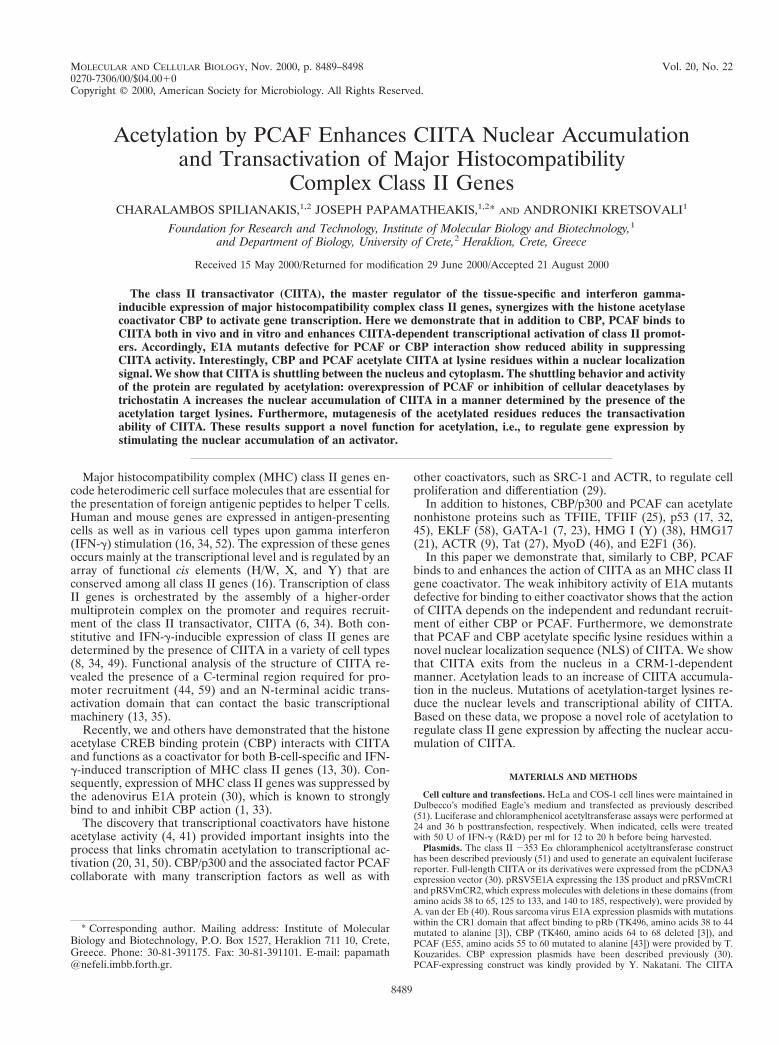
To identify the CIITA region involved in PCAF binding,we used different CIITA fragments fused to GST (Fig. 1A).CIITA fragments containing the amino-terminal transcrip-tion activation domain of CIITA (fragments 1–408 and 1–114[lanes 5 and 6, Fig. 1B]) interacted with PCAF. Interestingly,the same amino-terminal region (amino acids 1 to 114) wasalso responsible for binding to CBP (30). In an attempt to mapmore precisely the CBP and PCAF interaction sites within theN-terminal domain of CIITA, the region extending betweenamino acids 1 to 151 was divided into three smaller parts. Asshown in Fig. 1B, both PCAF and CBP bind predominantly tothe first 80 amino acids of CIITA (Fig. 1B lanes 7, 12, and 18),which contain an a-helix required for full activity of CIITA(13).
To map the region of PCAF that binds CIITA, we used thePCAF deletion constructs depicted in Fig. 2A. As shown in Fig.2B, the C-terminal region (amino acids 370 to 783) of PCAF is
FIG. 1. The activation domain of CIITA binds to both CBP and PCAF. (A)GST fusions of different parts of the CIITA protein used for interaction withPCAF and CBP are shown schematically. (B) In vitro-translated and 35S-labeledPCAF or two regions of CBP (CBP 1–1098 and CBP 1620–1877) were used in aGST pull-down assay with equal amounts (1 mg) of GST alone (lanes 2, 10, and16) or fused with the indicated fragments of CIITA. Input was 5% in lane 1, 30%in lane 15, and 20% in lane 21.
FIG. 2. Regions of PCAF that interact with CIITA. (A) Scheme of theGST-PCAF proteins used for interaction with CIITA. Br., bromodomain. (B) Invitro 35S-labeled CIITA was used in a GST pull-down assay with equal amountsof GST alone (lane 2) or GST fused to the indicated regions of PCAF (lanes 3to 12).
8490 SPILIANAKIS ET AL. MOL. CELL. BIOL.
sufficient for CIITA binding (lane 5) whereas the N-terminalregion (amino acids 1 to 370) is not (lane 4). Further analysisshowed that the HAT domain (contained in fragments 1–654and 511–654 in lanes 6 and 9) interacted weakly with CIITAwhereas the region that harbors the ADA binding domain(fragment 653–736) showed a strong interaction (lane 10). TheBromo domain was devoid of the ability to interact (lanes 11and 12). Taken together, these results show that the region ofPCAF that binds to CIITA is distinct from the region requiredfor binding to CBP and to nuclear receptors and coactivators.
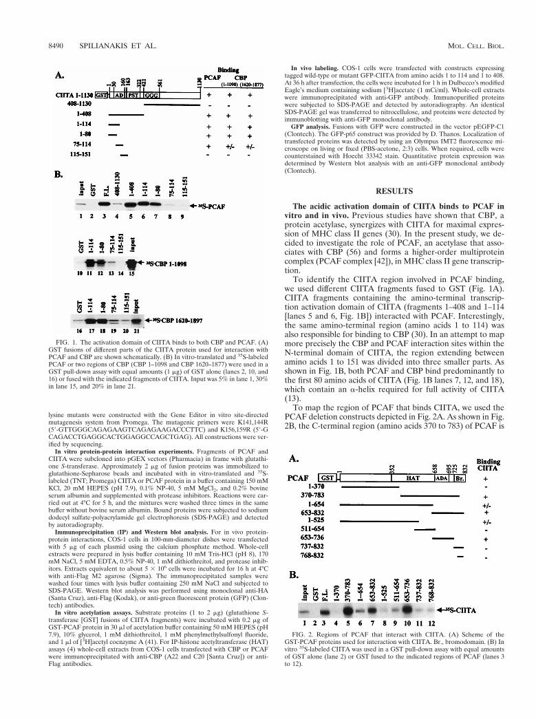
To examine whether PCAF and CIITA associate in vivo, wecotransfected COS-1 cells with expression vectors encodingFlag-tagged PCAF and HA-tagged CIITA and performed co-immunoprecipitation experiments. Figure 3 shows that the in-tact CIITA protein interacts with PCAF (lane 1’) whereas atruncation of the first 102 amino acids (which are required forbinding in vitro to PCAF) does not (lane 2’). We also deter-mined which regions of PCAF are important for in vivo asso-ciation with CIITA. A PCAF protein that contains only thefirst 511 amino acids (PCAF 1–511) and a PCAF that lacks theADA binding region (amino acids 653 to 736) (PCAF DADA)cannot interact with CIITA (lanes 4’ and 5’, respectively), incontrast to the wild-type protein (lane 3’). Therefore, the invivo interaction analysis correlates well with the in vitro datapresented previously.
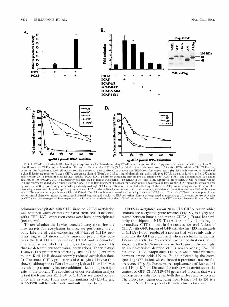
PCAF coactivates MHC class II genes. We next asked ifPCAF was able to coactivate MHC class II genes. HeLa cellswere transiently transfected with the Ea MHC class II pro-moter construct along with a PCAF-expressing plasmid. Theactivity of the promoter was determined from untreated cellsor cells after a 20-h treatment with IFN-g, which is known toinduce CIITA expression, which in turn induces class II genetranscription. Although the presence of PCAF did not signif-icantly affect the basal promoter activity, it strongly enhanced(up to sevenfold) the IFN-g-induced expression (Fig. 4A).
Since the effect of IFN-g on class II gene expression is medi-ated by CIITA, we examined the ability of PCAF to directlycoactivate exogenously provided CIITA. Transfection of aCIITA expression vector resulted in activation of the Ea classII promoter in HeLa cells, whereas PCAF alone did not pro-duce any effect (data not shown). Coexpression of PCAF aug-mented the CIITA-mediated activation (Fig. 4B). Therefore,PCAF and CIITA synergistically activate class II promotertranscription. Deletion of the CBP interaction domain ofPCAF (amino acids 1 to 372) did not abolish the ability ofPCAF to stimulate CIITA-dependent class II transcription(PCAF-DN; Fig. 4B). Thus, the ability of PCAF to stimulateCIITA activity is mainly CBP independent. A PCAF proteinbearing mutations in the HAT domain (PCAF HAT2 [28]) hasa lowered capacity to coactivate CIITA compared to that ofthe wild-type protein. This suggests that histone or proteinacetylation contributes to the synergy observed between PCAFand CIITA. Two PCAF deletion mutants that do not interactwith CIITA (PCAF 1–511 and PCAF DADA [Fig. 4B]) wereunable to coactivate CIITA. This result underlines the impor-tance of strong PCAF binding to CIITA for the coactivation ofclass II genes.
To evaluate the relative importance of CBP and PCAF inMHC class II regulation, we took advantage of E1A mutants(Fig. 4C and D) that specifically impair the binding to eitherPCAF or CBP. Mutant E55 (PCAFm) shows impaired bindingto PCAF (43), and mutant TK460 (CBPm), in which aminoacids 64 to 68 are deleted, cannot bind to CBP (3). Both typesof mutations strongly reduced the ability of E1A to suppressIFN-g induction as well as the direct activation by CIITA of aclass II promoter (Fig. 4C and D). Cotransfection of the 13sE1A or a mutant defective for pRb binding, Rbm (mutantTK496 in which amino acids 38 to 44 are mutated to alanine[3]), showed strong suppression, in contrast to a CR1 deletion,which was a very weak inhibitor (Fig. 4C and D). Thus,sequestration of either coactivator by E1A is not sufficient toreduce promoter activation. Taken together, these resultsdemonstrate the functional redundancy of PCAF and CBP,which can independently enhance the action of CIITA.
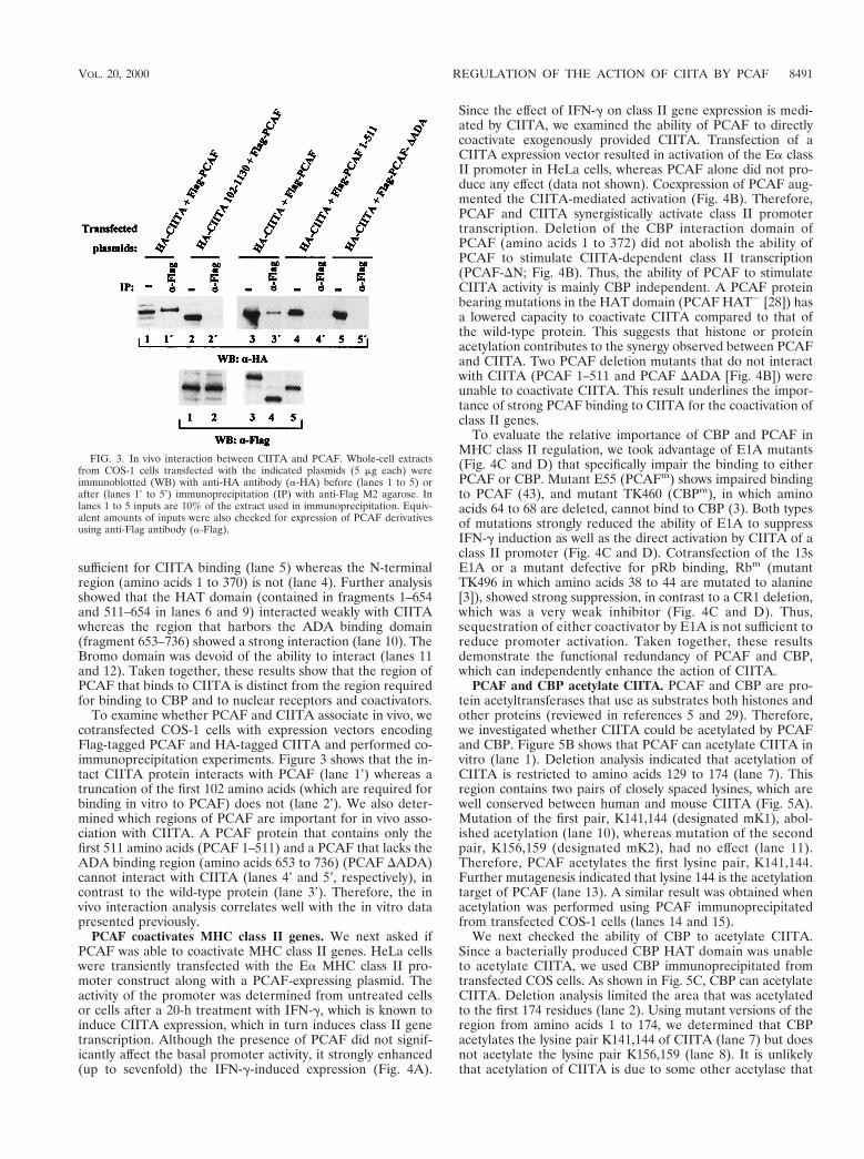
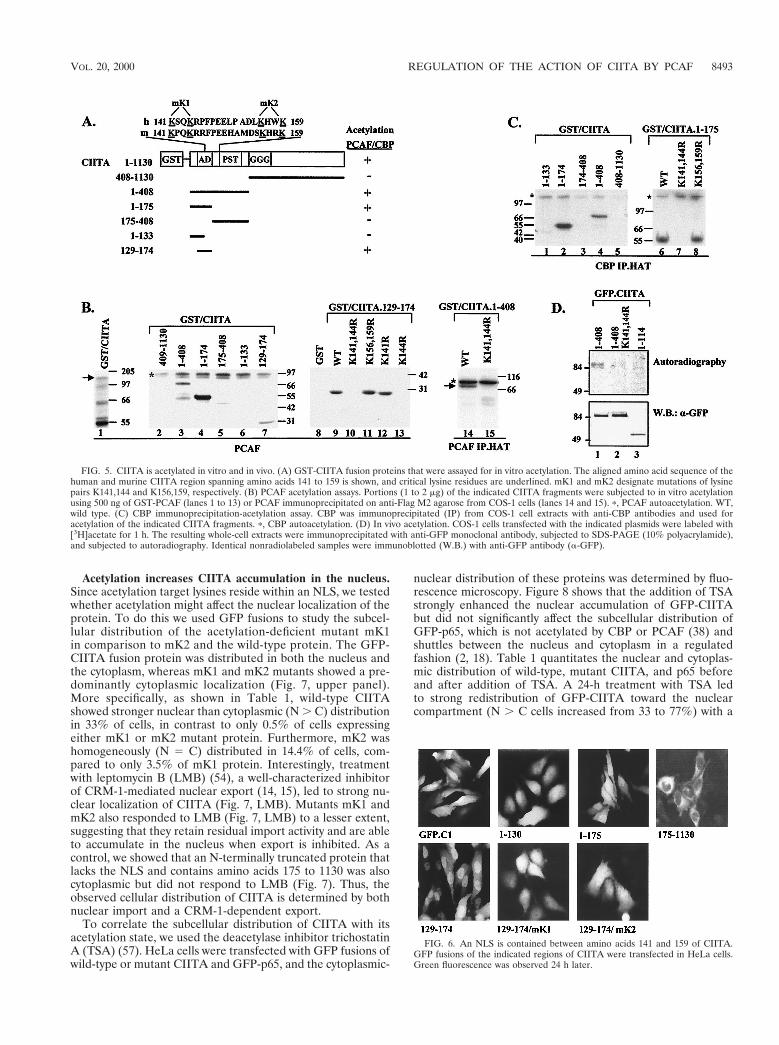
PCAF and CBP acetylate CIITA. PCAF and CBP are pro-tein acetyltransferases that use as substrates both histones andother proteins (reviewed in references 5 and 29). Therefore,we investigated whether CIITA could be acetylated by PCAFand CBP. Figure 5B shows that PCAF can acetylate CIITA invitro (lane 1). Deletion analysis indicated that acetylation ofCIITA is restricted to amino acids 129 to 174 (lane 7). Thisregion contains two pairs of closely spaced lysines, which arewell conserved between human and mouse CIITA (Fig. 5A).Mutation of the first pair, K141,144 (designated mK1), abol-ished acetylation (lane 10), whereas mutation of the secondpair, K156,159 (designated mK2), had no effect (lane 11).Therefore, PCAF acetylates the first lysine pair, K141,144.Further mutagenesis indicated that lysine 144 is the acetylationtarget of PCAF (lane 13). A similar result was obtained whenacetylation was performed using PCAF immunoprecipitatedfrom transfected COS-1 cells (lanes 14 and 15).
We next checked the ability of CBP to acetylate CIITA.Since a bacterially produced CBP HAT domain was unableto acetylate CIITA, we used CBP immunoprecipitated fromtransfected COS cells. As shown in Fig. 5C, CBP can acetylateCIITA. Deletion analysis limited the area that was acetylatedto the first 174 residues (lane 2). Using mutant versions of theregion from amino acids 1 to 174, we determined that CBPacetylates the lysine pair K141,144 of CIITA (lane 7) but doesnot acetylate the lysine pair K156,159 (lane 8). It is unlikelythat acetylation of CIITA is due to some other acetylase that
FIG. 3. In vivo interaction between CIITA and PCAF. Whole-cell extractsfrom COS-1 cells transfected with the indicated plasmids (5 mg each) wereimmunoblotted (WB) with anti-HA antibody (a-HA) before (lanes 1 to 5) orafter (lanes 1’ to 5’) immunoprecipitation (IP) with anti-Flag M2 agarose. Inlanes 1 to 5 inputs are 10% of the extract used in immunoprecipitation. Equiv-alent amounts of inputs were also checked for expression of PCAF derivativesusing anti-Flag antibody (a-Flag).
VOL. 20, 2000 REGULATION OF THE ACTION OF CIITA BY PCAF 8491
coimmunoprecipitates with CBP, since no CIITA acetylationwas obtained when extracts prepared from cells transfectedwith a CBP HAT2 expression vector were immunoprecipitated(not shown).
To test whether the in vitro-detected acetylation sites arealso targets for acetylation in vivo, we performed meta-bolic labeling of cells expressing GFP-tagged CIITA pro-teins. Figure 5D shows that a truncated protein that con-tains the first 114 amino acids of CIITA and is devoid ofany lysine is not labeled (lane 3), excluding the possibilitythat we detected amino-terminal acetylations. The wild-type1–408 CIITA protein was efficiently labeled (lane 1), whereasmutant K141,144R showed severely reduced acetylation (lane2). The intact CIITA protein was also acetylated in vivo (notshown), although the effect of mutating lysines 141 and 144 wasless clear, presumably because additional lysine targets mightexist in the protein. The conclusion of our acetylation analysisis that the lysine pair K141,144 of CIITA is acetylated both invitro and in vivo. From now on, mutants K141,144R andK156,159R will be called mK1 and mK2, respectively.
CIITA is acetylated on an NLS. The CIITA region whichcontains the acetylated lysine residues (Fig. 5A) is highly con-served between human and murine CIITA (47) and has simi-larity to a bipartite NLS. To test the ability of this regionto mediate CIITA import in the nucleus, we used fusions ofCIITA with GFP. Fusion of GFP with the first 130 amino acidsof CIITA (1–130) produced a protein that was evenly distrib-uted, like the GFP protein itself, whereas a fusion of the first175 amino acids (1–175) showed nuclear localization (Fig. 6),suggesting that NLSs may reside in this fragment. Accordingly,an amino-terminal deletion of 174 amino acids (175–1130)rendered CIITA cytoplasmic. The NLS was further restrictedbetween amino acids 129 to 174, as indicated by the corre-sponding GFP fusion, which showed a prominent nuclear flu-orescence (Fig. 6). Furthermore, replacement of lysines 141and 144 (mK1) or 156 and 159 (mK2) with arginines in thecontext of GFP-CIITA/129–174 generated proteins that werehomogeneously distributed in both the nucleus and cytoplasm.Therefore, the region extending from lysines 141 to 159 is abipartite NLS that requires both motifs for its function.
FIG. 4. PCAF coactivates MHC class II gene expression. (A) Plasmids encoding PCAF or vector control (0.5 to 1 mg) were cotransfected with 1 mg of an MHCclass II promoter-CAT reporter plasmid into HeLa cells. Uninduced and IFN-g (50 U/ml)-induced activities were assayed 24 h after IFN-g addition. The CAT activityof vector transfected-uninduced cells was set to 1. Bars represent the standard error of the mean (SEM) from four experiments. (B) HeLa cells were cotransfected witha class II-luciferase reporter (1 mg) a CIITA-expressing plasmid (20 ng), and 0.5 to 1 mg of plasmids expressing wild-type PCAF, a deletion lacking its first 352 aminoacids (PCAF-DN), a mutant that has no HAT activity (PCAF-HAT2) a mutant containing only the first 511 amino acids (PCAF 1–511), and a mutant that lacks aminoacids 653 to 736 (PCAF-D ADA). Luc activity was measured 24 h after transfection. The activity of the class II-Luc reporter in the presence of CIITA protein was setto 1 and represents an induction range between 7- and 15-fold. Bars represent SEM from four experiments. The expression levels of the PCAF molecules were analyzedby Western blotting (WB) using an anti-Flag antibody (a-Flag). (C) HeLa cells were transfected with 1 mg of class II-CAT plasmid along with vector control orincreasing amounts of plasmids expressing the indicated E1A products. Results are means of three experiments, with standard deviation less than 25% of the meanvalue. IFN-g induction ranged between 15- and 45-fold. (D) HeLa cells were cotransfected with 1 mg of class II-CAT and 100 ng of a CIITA-expressing plasmid andvector control plasmid or increasing amounts of plasmids expressing the indicated E1A derivatives. Results are expressed as a percentage of the vector control activationby CIITA and are averages of three experiments, with standard deviation less than 30% of the mean value. Activation by CIITA ranged between 75- and 120-fold.
8492 SPILIANAKIS ET AL. MOL. CELL. BIOL.
Acetylation increases CIITA accumulation in the nucleus.Since acetylation target lysines reside within an NLS, we testedwhether acetylation might affect the nuclear localization of theprotein. To do this we used GFP fusions to study the subcel-lular distribution of the acetylation-deficient mutant mK1in comparison to mK2 and the wild-type protein. The GFP-CIITA fusion protein was distributed in both the nucleus andthe cytoplasm, whereas mK1 and mK2 mutants showed a pre-dominantly cytoplasmic localization (Fig. 7, upper panel).More specifically, as shown in Table 1, wild-type CIITAshowed stronger nuclear than cytoplasmic (N . C) distributionin 33% of cells, in contrast to only 0.5% of cells expressingeither mK1 or mK2 mutant protein. Furthermore, mK2 washomogeneously (N 5 C) distributed in 14.4% of cells, com-pared to only 3.5% of mK1 protein. Interestingly, treatmentwith leptomycin B (LMB) (54), a well-characterized inhibitorof CRM-1-mediated nuclear export (14, 15), led to strong nu-clear localization of CIITA (Fig. 7, LMB). Mutants mK1 andmK2 also responded to LMB (Fig. 7, LMB) to a lesser extent,suggesting that they retain residual import activity and are ableto accumulate in the nucleus when export is inhibited. As acontrol, we showed that an N-terminally truncated protein thatlacks the NLS and contains amino acids 175 to 1130 was alsocytoplasmic but did not respond to LMB (Fig. 7). Thus, theobserved cellular distribution of CIITA is determined by bothnuclear import and a CRM-1-dependent export.
To correlate the subcellular distribution of CIITA with itsacetylation state, we used the deacetylase inhibitor trichostatinA (TSA) (57). HeLa cells were transfected with GFP fusions ofwild-type or mutant CIITA and GFP-p65, and the cytoplasmic-
nuclear distribution of these proteins was determined by fluo-rescence microscopy. Figure 8 shows that the addition of TSAstrongly enhanced the nuclear accumulation of GFP-CIITAbut did not significantly affect the subcellular distribution ofGFP-p65, which is not acetylated by CBP or PCAF (38) andshuttles between the nucleus and cytoplasm in a regulatedfashion (2, 18). Table 1 quantitates the nuclear and cytoplas-mic distribution of wild-type, mutant CIITA, and p65 beforeand after addition of TSA. A 24-h treatment with TSA ledto strong redistribution of GFP-CIITA toward the nuclearcompartment (N . C cells increased from 33 to 77%) with a
FIG. 5. CIITA is acetylated in vitro and in vivo. (A) GST-CIITA fusion proteins that were assayed for in vitro acetylation. The aligned amino acid sequence of thehuman and murine CIITA region spanning amino acids 141 to 159 is shown, and critical lysine residues are underlined. mK1 and mK2 designate mutations of lysinepairs K141,144 and K156,159, respectively. (B) PCAF acetylation assays. Portions (1 to 2 mg) of the indicated CIITA fragments were subjected to in vitro acetylationusing 500 ng of GST-PCAF (lanes 1 to 13) or PCAF immunoprecipitated on anti-Flag M2 agarose from COS-1 cells (lanes 14 and 15). p, PCAF autoacetylation. WT,wild type. (C) CBP immunoprecipitation-acetylation assay. CBP was immunoprecipitated (IP) from COS-1 cell extracts with anti-CBP antibodies and used foracetylation of the indicated CIITA fragments. p, CBP autoacetylation. (D) In vivo acetylation. COS-1 cells transfected with the indicated plasmids were labeled with[3H]acetate for 1 h. The resulting whole-cell extracts were immunoprecipitated with anti-GFP monoclonal antibody, subjected to SDS-PAGE (10% polyacrylamide),and subjected to autoradiography. Identical nonradiolabeled samples were immunoblotted (W.B.) with anti-GFP antibody (a-GFP).
FIG. 6. An NLS is contained between amino acids 141 and 159 of CIITA.GFP fusions of the indicated regions of CIITA were transfected in HeLa cells.Green fluorescence was observed 24 h later.
VOL. 20, 2000 REGULATION OF THE ACTION OF CIITA BY PCAF 8493
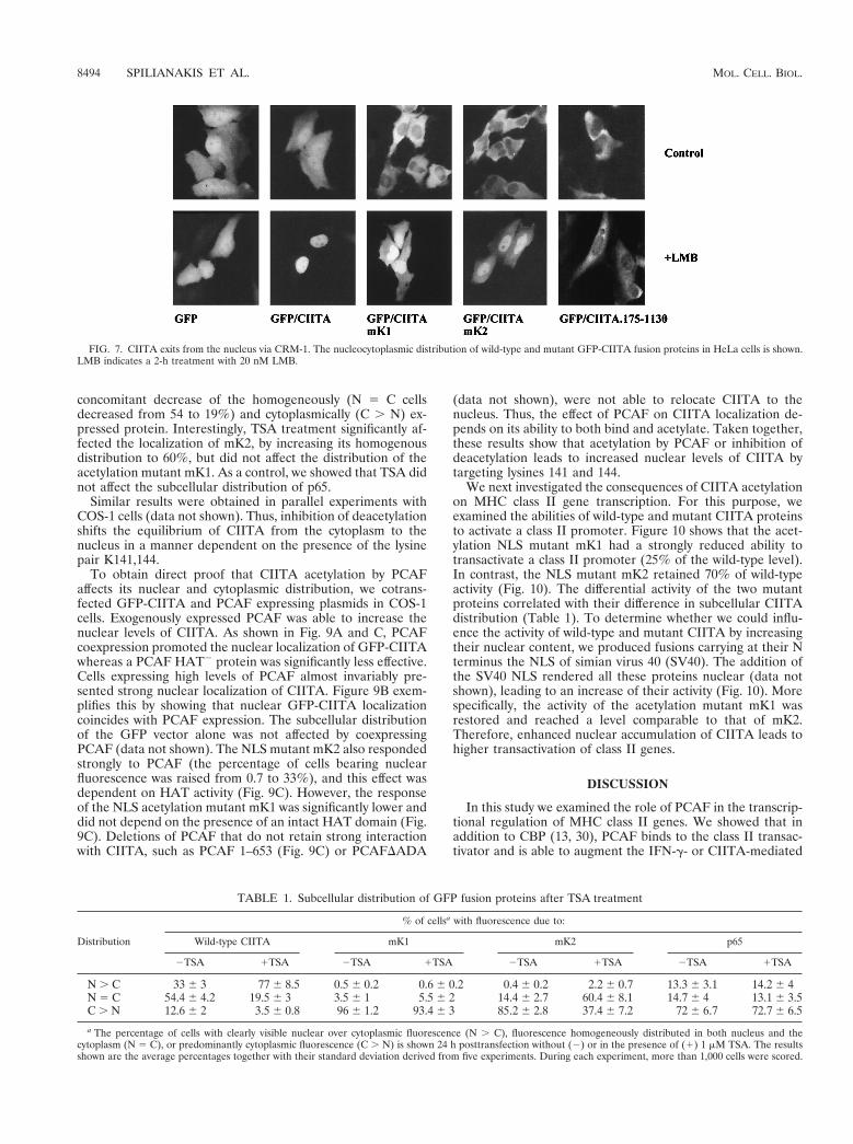
concomitant decrease of the homogeneously (N 5 C cellsdecreased from 54 to 19%) and cytoplasmically (C . N) ex-pressed protein. Interestingly, TSA treatment significantly af-fected the localization of mK2, by increasing its homogenousdistribution to 60%, but did not affect the distribution of theacetylation mutant mK1. As a control, we showed that TSA didnot affect the subcellular distribution of p65.
Similar results were obtained in parallel experiments withCOS-1 cells (data not shown). Thus, inhibition of deacetylationshifts the equilibrium of CIITA from the cytoplasm to thenucleus in a manner dependent on the presence of the lysinepair K141,144.
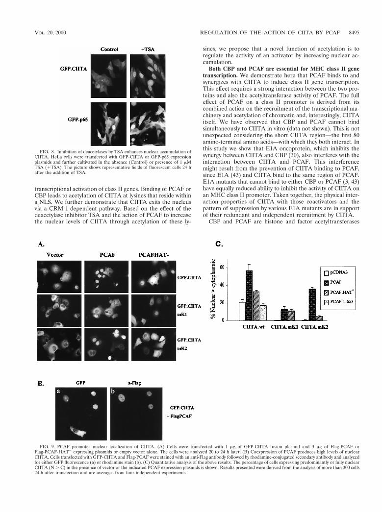
To obtain direct proof that CIITA acetylation by PCAFaffects its nuclear and cytoplasmic distribution, we cotrans-fected GFP-CIITA and PCAF expressing plasmids in COS-1cells. Exogenously expressed PCAF was able to increase thenuclear levels of CIITA. As shown in Fig. 9A and C, PCAFcoexpression promoted the nuclear localization of GFP-CIITAwhereas a PCAF HAT2 protein was significantly less effective.Cells expressing high levels of PCAF almost invariably pre-sented strong nuclear localization of CIITA. Figure 9B exem-plifies this by showing that nuclear GFP-CIITA localizationcoincides with PCAF expression. The subcellular distributionof the GFP vector alone was not affected by coexpressingPCAF (data not shown). The NLS mutant mK2 also respondedstrongly to PCAF (the percentage of cells bearing nuclearfluorescence was raised from 0.7 to 33%), and this effect wasdependent on HAT activity (Fig. 9C). However, the responseof the NLS acetylation mutant mK1 was significantly lower anddid not depend on the presence of an intact HAT domain (Fig.9C). Deletions of PCAF that do not retain strong interactionwith CIITA, such as PCAF 1–653 (Fig. 9C) or PCAFDADA
(data not shown), were not able to relocate CIITA to thenucleus. Thus, the effect of PCAF on CIITA localization de-pends on its ability to both bind and acetylate. Taken together,these results show that acetylation by PCAF or inhibition ofdeacetylation leads to increased nuclear levels of CIITA bytargeting lysines 141 and 144.
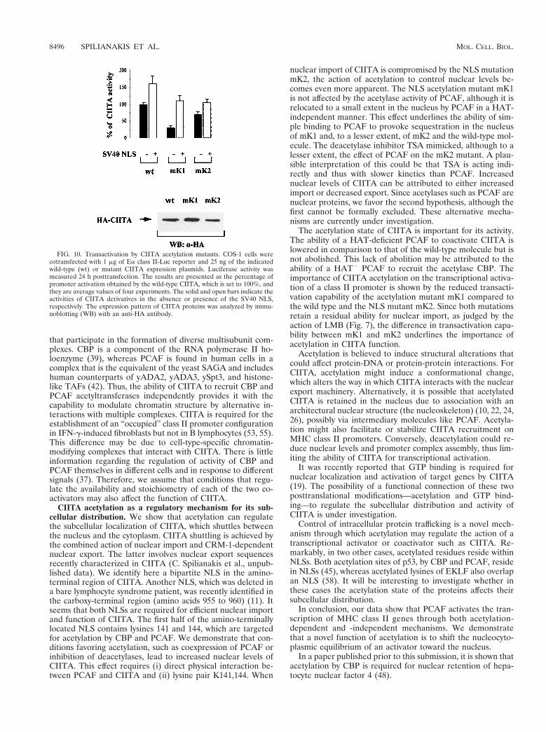
We next investigated the consequences of CIITA acetylationon MHC class II gene transcription. For this purpose, weexamined the abilities of wild-type and mutant CIITA proteinsto activate a class II promoter. Figure 10 shows that the acet-ylation NLS mutant mK1 had a strongly reduced ability totransactivate a class II promoter (25% of the wild-type level).In contrast, the NLS mutant mK2 retained 70% of wild-typeactivity (Fig. 10). The differential activity of the two mutantproteins correlated with their difference in subcellular CIITAdistribution (Table 1). To determine whether we could influ-ence the activity of wild-type and mutant CIITA by increasingtheir nuclear content, we produced fusions carrying at their Nterminus the NLS of simian virus 40 (SV40). The addition ofthe SV40 NLS rendered all these proteins nuclear (data notshown), leading to an increase of their activity (Fig. 10). Morespecifically, the activity of the acetylation mutant mK1 wasrestored and reached a level comparable to that of mK2.Therefore, enhanced nuclear accumulation of CIITA leads tohigher transactivation of class II genes.
DISCUSSION
In this study we examined the role of PCAF in the transcrip-tional regulation of MHC class II genes. We showed that inaddition to CBP (13, 30), PCAF binds to the class II transac-tivator and is able to augment the IFN-g- or CIITA-mediated
FIG. 7. CIITA exits from the nucleus via CRM-1. The nucleocytoplasmic distribution of wild-type and mutant GFP-CIITA fusion proteins in HeLa cells is shown.LMB indicates a 2-h treatment with 20 nM LMB.
TABLE 1. Subcellular distribution of GFP fusion proteins after TSA treatment
Distribution
% of cellsa with fluorescence due to:
Wild-type CIITA mK1 mK2 p65
2TSA 1TSA 2TSA 1TSA 2TSA 1TSA 2TSA 1TSA
N . C 33 6 3 77 6 8.5 0.5 6 0.2 0.6 6 0.2 0.4 6 0.2 2.2 6 0.7 13.3 6 3.1 14.2 6 4N 5 C 54.4 6 4.2 19.5 6 3 3.5 6 1 5.5 6 2 14.4 6 2.7 60.4 6 8.1 14.7 6 4 13.1 6 3.5C . N 12.6 6 2 3.5 6 0.8 96 6 1.2 93.4 6 3 85.2 6 2.8 37.4 6 7.2 72 6 6.7 72.7 6 6.5
a The percentage of cells with clearly visible nuclear over cytoplasmic fluorescence (N . C), fluorescence homogeneously distributed in both nucleus and thecytoplasm (N 5 C), or predominantly cytoplasmic fluorescence (C . N) is shown 24 h posttransfection without (2) or in the presence of (1) 1 mM TSA. The resultsshown are the average percentages together with their standard deviation derived from five experiments. During each experiment, more than 1,000 cells were scored.
8494 SPILIANAKIS ET AL. MOL. CELL. BIOL.
transcriptional activation of class II genes. Binding of PCAF orCBP leads to acetylation of CIITA at lysines that reside withina NLS. We further demonstrate that CIITA exits the nucleusvia a CRM-1-dependent pathway. Based on the effect of thedeacetylase inhibitor TSA and the action of PCAF to increasethe nuclear levels of CIITA through acetylation of these ly-
sines, we propose that a novel function of acetylation is toregulate the activity of an activator by increasing nuclear ac-cumulation.
Both CBP and PCAF are essential for MHC class II genetranscription. We demonstrate here that PCAF binds to andsynergizes with CIITA to induce class II gene transcription.This effect requires a strong interaction between the two pro-teins and also the acetyltransferase activity of PCAF. The fulleffect of PCAF on a class II promoter is derived from itscombined action on the recruitment of the transcriptional ma-chinery and acetylation of chromatin and, interestingly, CIITAitself. We have observed that CBP and PCAF cannot bindsimultaneously to CIITA in vitro (data not shown). This is notunexpected considering the short CIITA region—the first 80amino-terminal amino acids—with which they both interact. Inthis study we show that E1A oncoprotein, which inhibits thesynergy between CIITA and CBP (30), also interferes with theinteraction between CIITA and PCAF. This interferencemight result from the prevention of CIITA binding to PCAF,since E1A (43) and CIITA bind to the same region of PCAF.E1A mutants that cannot bind to either CBP or PCAF (3, 43)have equally reduced ability to inhibit the activity of CIITA onan MHC class II promoter. Taken together, the physical inter-action properties of CIITA with those coactivators and thepattern of suppression by various E1A mutants are in supportof their redundant and independent recruitment by CIITA.
CBP and PCAF are histone and factor acetyltransferases
FIG. 8. Inhibition of deacetylases by TSA enhances nuclear accumulation ofCIITA. HeLa cells were transfected with GFP-CIITA or GFP-p65 expressionplasmids and further cultivated in the absence (Control) or presence of 1 mMTSA (1TSA). The picture shows representative fields of fluorescent cells 24 hafter the addition of TSA.
FIG. 9. PCAF promotes nuclear localization of CIITA. (A) Cells were transfected with 1 mg of GFP-CIITA fusion plasmid and 3 mg of Flag-PCAF orFlag-PCAF-HAT2 expressing plasmids or empty vector alone. The cells were analyzed 20 to 24 h later. (B) Coexpression of PCAF produces high levels of nuclearCIITA. Cells transfected with GFP-CIITA and Flag-PCAF were stained with an anti-Flag antibody followed by rhodamine-conjugated secondary antibody and analyzedfor either GFP fluorescence (a) or rhodamine stain (b). (C) Quantitative analysis of the above results. The percentage of cells expressing predominantly or fully nuclearCIITA (N . C) in the presence of vector or the indicated PCAF expression plasmids is shown. Results presented were derived from the analysis of more than 300 cells24 h after transfection and are averages from four independent experiments.
VOL. 20, 2000 REGULATION OF THE ACTION OF CIITA BY PCAF 8495
that participate in the formation of diverse multisubunit com-plexes. CBP is a component of the RNA polymerase II ho-loenzyme (39), whereas PCAF is found in human cells in acomplex that is the equivalent of the yeast SAGA and includeshuman counterparts of yADA2, yADA3, ySpt3, and histone-like TAFs (42). Thus, the ability of CIITA to recruit CBP andPCAF acetyltransferases independently provides it with thecapability to modulate chromatin structure by alternative in-teractions with multiple complexes. CIITA is required for theestablishment of an “occupied” class II promoter configurationin IFN-g-induced fibroblasts but not in B lymphocytes (53, 55).This difference may be due to cell-type-specific chromatin-modifying complexes that interact with CIITA. There is littleinformation regarding the regulation of activity of CBP andPCAF themselves in different cells and in response to differentsignals (37). Therefore, we assume that conditions that regu-late the availability and stoichiometry of each of the two co-activators may also affect the function of CIITA.
CIITA acetylation as a regulatory mechanism for its sub-cellular distribution. We show that acetylation can regulatethe subcellular localization of CIITA, which shuttles betweenthe nucleus and the cytoplasm. CIITA shuttling is achieved bythe combined action of nuclear import and CRM-1-dependentnuclear export. The latter involves nuclear export sequencesrecently characterized in CIITA (C. Spilianakis et al., unpub-lished data). We identify here a bipartite NLS in the amino-terminal region of CIITA. Another NLS, which was deleted ina bare lymphocyte syndrome patient, was recently identified inthe carboxy-terminal region (amino acids 955 to 960) (11). Itseems that both NLSs are required for efficient nuclear importand function of CIITA. The first half of the amino-terminallylocated NLS contains lysines 141 and 144, which are targetedfor acetylation by CBP and PCAF. We demonstrate that con-ditions favoring acetylation, such as coexpression of PCAF orinhibition of deacetylases, lead to increased nuclear levels ofCIITA. This effect requires (i) direct physical interaction be-tween PCAF and CIITA and (ii) lysine pair K141,144. When
nuclear import of CIITA is compromised by the NLS mutationmK2, the action of acetylation to control nuclear levels be-comes even more apparent. The NLS acetylation mutant mK1is not affected by the acetylase activity of PCAF, although it isrelocated to a small extent in the nucleus by PCAF in a HAT-independent manner. This effect underlines the ability of sim-ple binding to PCAF to provoke sequestration in the nucleusof mK1 and, to a lesser extent, of mK2 and the wild-type mol-ecule. The deacetylase inhibitor TSA mimicked, although to alesser extent, the effect of PCAF on the mK2 mutant. A plau-sible interpretation of this could be that TSA is acting indi-rectly and thus with slower kinetics than PCAF. Increasednuclear levels of CIITA can be attributed to either increasedimport or decreased export. Since acetylases such as PCAF arenuclear proteins, we favor the second hypothesis, although thefirst cannot be formally excluded. These alternative mecha-nisms are currently under investigation.
The acetylation state of CIITA is important for its activity.The ability of a HAT-deficient PCAF to coactivate CIITA islowered in comparison to that of the wild-type molecule but isnot abolished. This lack of abolition may be attributed to theability of a HAT2 PCAF to recruit the acetylase CBP. Theimportance of CIITA acetylation on the transcriptional activa-tion of a class II promoter is shown by the reduced transacti-vation capability of the acetylation mutant mK1 compared tothe wild type and the NLS mutant mK2. Since both mutationsretain a residual ability for nuclear import, as judged by theaction of LMB (Fig. 7), the difference in transactivation capa-bility between mK1 and mK2 underlines the importance ofacetylation in CIITA function.
Acetylation is believed to induce structural alterations thatcould affect protein-DNA or protein-protein interactions. ForCIITA, acetylation might induce a conformational change,which alters the way in which CIITA interacts with the nuclearexport machinery. Alternatively, it is possible that acetylatedCIITA is retained in the nucleus due to association with anarchitectural nuclear structure (the nucleoskeleton) (10, 22, 24,26), possibly via intermediary molecules like PCAF. Acetyla-tion might also facilitate or stabilize CIITA recruitment onMHC class II promoters. Conversely, deacetylation could re-duce nuclear levels and promoter complex assembly, thus lim-iting the ability of CIITA for transcriptional activation.
It was recently reported that GTP binding is required fornuclear localization and activation of target genes by CIITA(19). The possibility of a functional connection of these twoposttranslational modifications—acetylation and GTP bind-ing—to regulate the subcellular distribution and activity ofCIITA is under investigation.
Control of intracellular protein trafficking is a novel mech-anism through which acetylation may regulate the action of atranscriptional activator or coactivator such as CIITA. Re-markably, in two other cases, acetylated residues reside withinNLSs. Both acetylation sites of p53, by CBP and PCAF, residein NLSs (45), whereas acetylated lysines of EKLF also overlapan NLS (58). It will be interesting to investigate whether inthese cases the acetylation state of the proteins affects theirsubcellular distribution.
In conclusion, our data show that PCAF activates the tran-scription of MHC class II genes through both acetylation-dependent and -independent mechanisms. We demonstratethat a novel function of acetylation is to shift the nucleocyto-plasmic equilibrium of an activator toward the nucleus.
In a paper published prior to this submission, it is shown thatacetylation by CBP is required for nuclear retention of hepa-tocyte nuclear factor 4 (48).
FIG. 10. Transactivation by CIITA acetylation mutants. COS-1 cells werecotransfected with 1 mg of Ea class II-Luc reporter and 25 ng of the indicatedwild-type (wt) or mutant CIITA expression plasmids. Luciferase activity wasmeasured 24 h posttransfection. The results are presented as the percentage ofpromoter activation obtained by the wild-type CIITA, which is set to 100%, andthey are average values of four experiments. The solid and open bars indicate theactivities of CIITA derivatives in the absence or presence of the SV40 NLS,respectively. The expression pattern of CIITA proteins was analyzed by immu-noblotting (WB) with an anti-HA antibody.
8496 SPILIANAKIS ET AL. MOL. CELL. BIOL.
ACKNOWLEDGMENTS
We thank T. Makatounakis and G. Vretzos for providing excellenttechnical assistance and T. Kouzarides and Y. Nakatani for providingthe indicated reagents. We are indebted to D. Thanos for helpfuldiscussions and critical reading of the manuscript. We also thank C.Mamalaki, I. Talianidis, and S. Georgatos for critical reading; NektariaKelaidi for help with secretarial work; L. Kalogeraki for photographicwork; and T. Makatounakis for assistance with the presentation.
The present work was supported by the Greek Secretariat Generalfor Research through Institutional funds and European Union (EPETII) grant 236.234.603 and National (PENED) grant 2016.
REFERENCES
1. Arany, Z., D. Newsome, E. Oldread, D. M. Livingston, and R. Eckner. 1995.A family of transcriptional adaptor proteins targeted by the E1A oncopro-tein. Nature 374:81–84.
2. Baldwin, A. S. 1996. The NF-kB and IkB proteins: new discoveries andinsights. Annu. Rev. Immunol. 14:649–683.
3. Bannister, A. J., and T. Kouzarides. 1995. CBP-induced stimulation of c-Fosactivity is abrogated by E1A. EMBO J. 14:4758–4762.
4. Bannister, A. J., and T. Kouzarides. 1996. The CBP co-activator is a histoneacetyltransferase. Nature 384:641–643.
5. Berger, S. L. 1999. Gene activation by histone and factor acetyltransferases.Curr. Opin. Cell Biol. 11:336–341.
6. Boss, J. M. 1997. Regulation of transcriptional of MHC class II genes. Curr.Opin. Immunol. 9:107–113.
7. Boyes, J., P. Byfield, Y. Nakatani, and V. Ogryzko. 1998. Regulation ofactivity of the transcription factor GATA-1 by acetylation. Nature 396:594–598.
8. Chang, C. H., J. D. Fontes, M. Peterlin, and R. A. Flavell. 1994. Class IItransactivator (CIITA) is sufficient for the inducible expression of majorhistocompatibility complex class II genes. J. Exp. Med. 180:1367–1374.
9. Chen, H., R. Lin, W. Xie, D. Wilpitz, and R. Evans. 1999. Regulation ofhormone-induced histone hyperacetylation and gene activation via acetyla-tion of an acetylase. Cell 98:675–686.
10. Cook, P. R. 1999. The organization of replication and transcription. Science284:1790–1795.
11. Cressman, D. E., K. C. Chin, D. J. Taxman, and J. P. Y. Ting. 1999. A defectin the nuclear translocation of CIITA causes a form of type II bare lympho-cyte syndrome. Immunity 10:163–171.
12. Fontes, J. D., B. Jiang, and B. M. Peterlin. 1997. The class II trans-activatorCIITA interacts with the TBP-associated factor TAFII32. Nucleic Acids Res.25:2522–2528.
13. Fontes, J. D., S. Kanazawa, D. Jean, and B. M. Peterlin. 1999. Interactionsbetween the class II transactivator and CREB binding protein increasetranscription of major histocompatibility complex class II genes. Mol. Cell.Biol. 19:941–947.
14. Fornerod, M., M. Ohno, M. Yoshida, and I. W. Mattaj. 1997. CRM1 is anexport receptor for leucine-rich nuclear export signals. Cell 90:1051–1060.
15. Fukuda, M., S. Asano, T. Nakamura, M. Adachi, M. Yoshida, M. Yanagida,and E. Nishida. 1997. CRM1 is responsible for intracellular transport me-diated by the nuclear export signal. Nature 390:308–311.
16. Glimcher, L. H., and C. J. Kara. 1992. Sequences and factors: a guide toMHC class-II transcription. Annu. Rev. Immunol. 10:13–49.
17. Gu, W., and R. Roeder. 1997. Activation of p53 sequence-specific DNAbinding by acetylation of the p53 C-terminal domain. Cell 90:595–606.
18. Harhaj, E. W., and S. C. Sun. 1999. Regulation of RelA subcellular local-ization by a putative nuclear export signal and p50. Mol. Cell. Biol. 19:7088–7095.
19. Harton, J., D. Cressman, K. Chin, C. Der, and J. Ting. 1999. GTP bindingby class II transactivator. Role in nuclear import. Science 285:1402–1405.
20. Hebbes, T. R., A. W. Thorne, and C. Crane-Robinson. 1988. A direct linkbetween core histone acetylation and transcriptionally active chromatin.EMBO J. 7:1395–1402.
21. Herrera, J., K. Sakagushi, M. Berger, L. Trieschmann, Y. Nakatani, and M.Bustin. 1999. Specific acetylation of chromosome protein HMG-17 by PCAFalters its interaction with nucleosomes. Mol. Cell. Biol. 19:3466–3473.
22. Hozak, P. 1996. The nucleoskeleton and attached activities. Exp. Cell Res.229:267–271.
23. Hung, H. L., J. Lau, A. Kim, M. Weiss, and G. Blobel. 1999. CREB-bindingprotein acetylates hematopoietic transcription factor GATA-1 at function-ally important sites. Mol. Cell. Biol. 19:3496–3505.
24. Iborra, F. J., A. Pombo, D. A. Jackson, and P. R. Cook. 1996. Active RNApolymerases are localized within discrete transcription “factories” in humannuclei. J. Cell Sci. 109:1427–1436.
25. Imhof, A., X. J. Yang, V. Ogryzko, Y. Nakatani, A. Wolffe, and H. Ge. 1997.Acetylation of general transcription factors by histone acetyltransferases.Curr. Biol. 7:689–692.
26. Jackson, D. A., A. B. Hassan, R. J. Errington, and P. R. Cook. 1993. Visu-
alization of focal sites of transcription within human nuclei. EMBO J. 12:1059–1065.
27. Kiernan, R., C. Vanhulle, L. Schiltz, E. Adam, H. Xiao, F. Maudoux, C.Calomme, A. Burny, Y. Nakatani, K.-T. Jeang, M. Benkirane, and C. VanLint. 1999. HIV-1 Tat transcriptional activity is regulated by acetylation.EMBO J. 18:6106–6118.
28. Korzus, E., J., Torchia, D. Rose, L. Xu, R. Kurokawa, E. McInerney, T.-M.Mullen, C. Glass, and M. Rosenfeld. 1998. Transcription factor-specificrequirements for coactivators and their associated acetyltransferase func-tions. Science 279:703–707.
29. Kouzarides, T. 1999. Histone acetylases and deacetylases in cell prolifera-tion. Curr. Opin. Genet. Dev. 9:40–48.
30. Kretsovali, A., T. Agalioti, C. Spilianakis, E. Tzortzakaki, M. Merika, and J.Papamatheakis. 1998. Involvement of CREB binding protein in expressionof major histocompatibility complex class II genes via interaction with theclass II transactivator. Mol. Cell. Biol. 18:6777–6783.
31. Kuo, M.-H., J. Zhou, P. Jampeck, M. Churchill, and D. Allis. 1998. Histoneacetyltransferase activity of yeast Gcn5p is required for the activation oftarget genes in vivo. Genes Dev. 12:627–639.
32. Liu, L., D. Scolnick, R. Trievel, H. B. Zhang, R. Marmorstein, T. Halazone-tis, and S. Berger. 1999. p53 acetylated in vitro by pCAF and p300 areacetylated in vivo in response to DNA damage. Mol. Cell. Biol. 19:1202–1209.
33. Lundblad, J. R., R. P. S. Kwok, M. E. Laurance, M. L. Harter, and R. H.Goodman. 1995. Adenoviral E1A-associated protein p300 as a functionalhomologue of the transcriptional co-activator CBP. Nature 374:85–88.
34. Mach, B., V. Steimle, E. Martinez-Soria, and W. Reith. 1996. Regulation ofMHC class II genes: lessons from a disease. Annu. Rev. Immunol. 14:301–331.
35. Mahanta, S. K., T. Scholl, F. C. Yang, and J. L. Strominger. 1997. Trans-activation by CIITA, the type II bare lymphocyte syndrome-associated fac-tor, requires participation of multiple regions of the TATA box bindingprotein. Proc. Natl. Acad. Sci. USA 94:6324–6329.
36. Martinez-Balbas, M., U.-M. Bauer, S. Nielsen, A. Brehm, and T. Kouzarides.2000. Regulation of E2F1 activity by acetylation. EMBO J. 19:662–671.
37. Masumi, A., I. M. Wang, B. Lefebvre, X. J. Yang, Y. Nakatani, and K. Ozato.1999. The histone acetylase PCAF is a phorbol-ester-inducible coactivator ofthe IRF family that confers enhanced interferon responsiveness. Mol. Cell.Biol. 19:1810–1820.
38. Munshi, N., M. Merika, J. Yie, K. Senger, G. Chen, and D. Thanos. 1998.Acetylation of HMG I (Y) by CBP turns off IFNb expression by disruptingthe enhanceosome. Mol. Cell 2:457–467.
39. Nakajima, T., C. Ushida, S. Anderson, J. Parvin, and M. Montminy. 1997.Analysis of a cAMP-responsive activator reveals a two-component mecha-nism for transcriptional induction via signal-dependent factors. Genes Dev.11:738–747.
40. Offringa, R., S. Gebel, H. van Dam, M. Timmers, A. Smits, R. Zwart, B.Stein, J. L. Boss, A. van der Eb, and P. Herrlich. 1990. A novel function ofthe transforming domain of E1A: repression of AP-1 activity. Cell 62:527–538.
41. Ogryzko, V., L. Schiltz, V. Russanova, B. H. Howard, Y. Nakatani. 1996. Thetranscriptional coactivators p300 and CBP are histone acetyltransferases.Cell 87:953–959.
42. Ogryzko, V., T. Kotani, X. Zhang, R. L. Schiltz, T. Howard, X. J. Yang, B.Howard, J. Qin, and Y. Nakatani. 1998. Histone-like TAFs within the PCAFhistone acetylase complex. Cell 94:35–44.
43. Reid, J. L., A. J. Bannister, P. Zegerman, M. A. Martinez-Balbas, and T.Kouzarides. 1998. E1A directly binds and regulates the P/CAF acetyltrans-ferases. EMBO J. 17:4469–4477.
44. Riley, J. L., S. D. Westerheide, J. A. Price, J. A. Brown, and J. M. Boss. 1995.Activation of class II MHC genes requires both the X box region and theclass II transactivator (CIITA). Immunity 2:533–543.
45. Sakaguchi, K., J. Herrera, S. Saito, T. Miki, M. Bustin, A. Vassilev, C.Anderson, and E. Appella. 1998. DNA damage activates p53 through aphosphorylation-acetylation cascade. Genes Dev. 12:2831–2841.
46. Sartorelli, V., P. L. Puri, Y. Hamamori, V. Ogryzko, G. Chung, Y. Nakatani,J. Wang, and L. Kedes. 1999. Acetylation of MyoD directed by PCAF isnecessary for the execution of the muscle program. Mol. Cell 4:725–734.
47. Sims, T., J. Elliot, V. Ramassar, D. Denney, and P. Halloran. 1997. Mouseclass II transactivator: cDNA sequence and amino acid comparison with thehuman class II transactivator. Immunogenetics 45:220–222.
48. Soutoglou, E., N. Katrakili, and I. Talianidis. 2000. Acetylation regulatestranscription factor activity at multiple levels. Mol. Cell 5:745–751.
49. Steimle, V., C. A. Siegrist, A. Mottet, B. Lisowska-Grospierre, and B. Mach.1994. Regulation of MHC class II expression by interferon-gamma mediatedby the transactivator gene CIITA. Science 265:106–109.
50. Struhl, K. 1998. Histone acetylation and transcriptional regulatory mecha-nisms. Genes Dev. 12:599–606.
51. Thanos, D., G. Mavrothalassitis, and J. Papamatheakis. 1988. Multipleregulatory regions on the 59 side of the mouse E alpha gene. Proc. Natl.Acad. Sci. USA 85:3075–3079.
VOL. 20, 2000 REGULATION OF THE ACTION OF CIITA BY PCAF 8497
52. Ting, J. P., and A. S. Baldwin. 1993. Regulation of MHC gene expression.Curr. Opin. Immunol. 5:8–16.
53. Villard, J., A. Muhtethaler-Mottet, S. Bontron, B. Mach, and W. Reith. 1999.CIITA-induced occupation of MHC class II promoters is independent of thecooperative stabilization of the promoter-bound multi-protein complexes.Int. Immunol. 11:461–469.
54. Wolff, B., J. J. Sanglier, and Y. Wang. 1997. Leptomycin B is an inhibitor ofnuclear export: inhibition of nucleo-cytoplasmic translocation of the humanimmunodeficiency virus type 1 (HIV-1) Rev protein and Rev-dependentmRNA. Chem. Biol. 4:139–147.
55. Wright, K., K. Chin, M. Linhoff, C. Skinner, J. Brown, J. Boss, G. Stark, andJ. Ting. 1998. CIITA stimulation of transcription factor binding to majorhistocompatibility complex class II and associated promoters in vivo. Proc.
Natl. Acad. Sci. USA 95:6267–6272.56. Yang, X. J., V. Ogryzko, J. Nishikawa, B. Howard, and Y. Nakatani. 1996. A
p300/CBP-associated factor that competes with the adenoviral oncoproteinE1A. Nature 382:319–324.
57. Yoshida, M., M. Kijima, M. Akita, and T. Beppu. 1990. Potent and specificinhibition of mammalian histone deacetylase both in vivo and in vitro bytrichostatin A. J. Biol. Chem. 265:17174–17179.
58. Zhang, W., and J. Bieker. 1998. Acetylation and modulation of erythroidKruppel-like factor (EKLF) activity by interaction with histone acetyltrans-ferases. Proc. Natl. Acad. Sci. USA 95:9855–9860.
59. Zhou, H., and L. H. Glimcher. 1995. Human class II gene transcriptiondirected by the carboxy terminus of CIITA, one of the defective genes in typeII MHC combined immune deficiency. Immunity 2:545–553.
8498 SPILIANAKIS ET AL. MOL. CELL. BIOL.
Related Documents