Safe Blood and Blood Products Screening for HIV and Other Infectious Agents Module 2

Welcome message from author
This document is posted to help you gain knowledge. Please leave a comment to let me know what you think about it! Share it to your friends and learn new things together.
Transcript
Safe
Blood
and
Blood
Products
Screening forHIV and OtherInfectiousAgents
Module 2
Safe
Blood
and
Blood
Products
Screening forHIV and OtherInfectiousAgents
Module 2
Conversion of electronic files for the website edition was supported by Cooperative Agreement Number PS001426 from the Centers for Disease Control and Prevention (CDC), Atlanta, United States of America. Its contents are solely the responsibility of the authors and do not necessarily represent the official views of CDC.
© World Health Organization, reprinted 2009
All rights reserved. Publications of the World Health Organization can be obtained from WHO Press, World Health Organization, 20 Avenue Appia, 1211 Geneva 27, Switzerland (tel.: +41 22 791 3264; fax: +41 22 791 4857; e-mail: [email protected]). Requests for permission to reproduce or translate WHO publications – whether for sale or for noncommercial distribution – should be addressed to WHO Press, at the above address (fax: +41 22 791 4806; e-mail: [email protected]).
The designations employed and the presentation of the material in this publication do not imply the expression of any opinion whatsoever on the part of the World Health Organization concerning the legal status of any country, territory, city or area or of its authorities, or concerning the delimitation of its frontiers or boundaries. Dotted lines on maps represent approximate border lines for which there may not yet be full agreement.
The mention of specific companies or of certain manufacturers’ products does not imply that they are endorsed or recommended by the World Health Organization in preference to others of a similar nature that are not mentioned. Errors and omissions excepted, the names of proprietary products are distinguished by initial capital letters.
All reasonable precautions have been taken by the World Health Organization to verify the information contained in this publication. However, the published material is being distributed without warranty of any kind, either expressed or implied. The responsibility for the interpretation and use of the material lies with the reader. In no event shall the World Health Organization be liable for damages arising from its use.
Contents
1 INTRODUCTION TO MODULE 2 1
1.1 The Distance Learning Materials 2
1.2 Before You Begin This Module 3
1.3 Module 2: Screening for HIV and Other InfectiousAgents 4
1.4 Module Objectives 5
1.5 Planning Your Study 7
2 INFECTION AND INFECTIOUS AGENTS 10
2.1 Infectious Agents 11
2.2 Transmission of Infectious Agents by BloodTransfusion 14
2.3 The Basic Immunology of Infection 15
2.4 Introduction to Screening for Infectious Agents 18
3 THE HUMAN IMMUNODEFICIENCY VIRUSES 22
3.1 The Background to HIV Infection 23
3.2 The Structure of HIV 23
3.3 Entry of HIV Into Cells 25
3.4 The Clinical Presentation of HIV Infection and AIDS 26
3.5 Laboratory Testing for HIV Infection in BloodDonors 27
3.6 Epidemiology of HIV Infection 29
3.7 Transmission of HIV Infection 30
3.8 Preventing the Spread of HIV Infection 32
4 PRINCIPLES OF SCREENING ASSAYS FOR 36TRANSFUSION-TRANSMISSIBLE INFECTIONS
4.1 Screening Blood Donors for Enzyme ImmunoAssay 37
4.2 Types of Screening Assays 37
4.3 Enzyme ImmunoAssays 39
4.4 Combination Antigen/Antibody Assays 51
4.5 Determining the Results of EIAs 52
4.6 Particle Agglutination Assays 55
4.7 Simple Rapid Assays 57
5 SELECTING SCREENING ASSAYS FOR TRANSFUSION- 63TRANSMISSIBLE INFECTIONS
5.1 Sensitivity and Specificity 64
5.2 Predictive Values 65
5.3 Selecting an Assay 66
5.4 Factors Influencing Screening Programmes 68
6 USING SCREENING ASSAYS FOR TRANSFUSION- 81TRANSMISSIBLE INFECTIONS
6.1 Using Screening Assays 82
6.2 Confirmatory Testing 83
6.3 Recording Test Results 86
6.4 Storing Screening Results 88
6.5 Handling TTI-Positive Donations 89
6.6 Health and Safety Aspects of Commercial Assays 91
7 QUALITY SYSTEMS IN SCREENING FOR 95TRANSFUSION-TRANSMISSIBLE INFECTIONS
7.1 The Need for Quality in TTI Screening 96
7.2 Quality Systems 96
7.3 Standard Operating Procedures (SOPs) 98
7.4 Laboratory Worksheets 101
7.5 Quality Audits 101
8 SCREENING FOR OTHER TRANSFUSION- 104TRANSMISSIBLE INFECTIONS
8.1 Hepatitis B Virus 106
8.2 Hepatitis C Virus (HCV) 111
8.3 Human T Cell Leukaemia Viruses I + II 114
8.4 Syphilis (Treponema Pallidum Infection) 116
8.5 Malaria 120
8.6 Chagas Disease 124
9 ACTION PLAN 129
9.1 Reviewing Your Progress 130
9.2 Making Your Action Plan 131
9.3 Implementing Your Action Plan 134
Activity Checklists and Answers 137
Answers to Self-Assessment Questions 157
Glossary 162
Appendices 167
Safe Blood and Blood Products is a series of interactive learningmaterials developed by the World Health Organization (WHO). They havebeen designed for use in distance learning programmes in blood safety,although they can also be used for independent study or as resourcematerials in conventional training courses and in-service trainingprogrammes.
The learning materials have been produced for staff with responsibilityfor donor recruitment and retention, and for the collection, testing andissue of blood for transfusion. They comprise the following modules:
■ Introductory Module: Guidelines and Principles for Safe BloodTransfusion Practice
■ Module 1: Safe Blood Donation
■ Module 2: Screening for HIV and Other Infectious Agents
■ Module 3: Blood Group Serology.
The English edition was first published in 1993. French, Spanish,Russian, Chinese and Portuguese editions have since been producedand the materials have also been translated into a number of nationallanguages.
This second, updated edition of the materials has been developed toreflect changes in transfusion medicine and laboratory technology sincethe publication of the first edition.
Distance learning in blood safety
Since the publication of Safe Blood and Blood Products, WHO has helda series of regional and sub-regional workshops for senior bloodtransfusion service personnel from over 100 countries on establishingnational distance learning programmes in blood safety. Programmeshave since been established in every region of the world, using the WHOlearning materials.
Part of the follow-up to the workshops has been the production ofEstablishing a Distance Learning Programme in Blood Safety: A Guide forProgramme Coordinators. This provides a practical guide to the planning,implementation and evaluation of a distance learning programme inblood safety.
Preface
Other WHO learning materials
The Clinical Use of Blood consists of an open learning module and pockethandbook which provide comprehensive guidance on transfusion andalternatives to transfusion in the areas of general medicine, obstetrics,paediatrics and neonatology, surgery and anaesthesia, trauma andacute surgery, and burns. They are designed to promote a reduction inunnecessary transfusions through the wider use of plasma substitutesand more effective prevention and treatment of the conditions that maymake transfusion necessary.
WHO has also published recommendations on Developing a NationalPolicy and Guidelines on the Clinical Use of Blood which encourage theuse of the learning materials in education and training programmes topromote effective clinical decisions on transfusion.
Additional learning materials in the Safe Blood and Blood Products seriesthat are available or in development by the WHO Department of BloodSafety and Clinical Technology (WHO/BCT) include:
■ Costing Blood Transfusion Services
■ The Blood Cold Chain
■ Blood Collection
■ Blood Components Production.
More detailed information on these materials and other documents andpublications related to blood transfusion is available from WHO/BCT,which also issues regular reports on evaluations of the operationalcharacteristics of many commercially available screening assays fortransfusion-transmissible infections.
Information can be obtained from the BCT section of the WHO websiteat http://www.who.int/bct or by contacting WHO/BCT at WHOHeadquarters or WHO Regional Offices.
Dr Jean C. EmmanuelDr Jean C. EmmanuelDr Jean C. EmmanuelDr Jean C. EmmanuelDr Jean C. Emmanuel
Director, Blood Safety and Clinical TechnologyDirector, Blood Safety and Clinical TechnologyDirector, Blood Safety and Clinical TechnologyDirector, Blood Safety and Clinical TechnologyDirector, Blood Safety and Clinical Technology
World Health OrganizationWorld Health OrganizationWorld Health OrganizationWorld Health OrganizationWorld Health Organization
I N T R O D U C T I O N T O M O D U L E 2
1
Introduction to Module 2
The purpose of this section is to introduce you to Module 2: Screeningfor HIV and Other Infectious Agents, which focuses on developing aneffective screening programme to detect infectious agents in donatedblood.
LEARNING OBJECTIVES
When you have completed this section, you should be able to:
1 Explain the purpose of Module 2.
2 Identify a personal “supporter” who can assist youthroughout your work on this module.
3 Assess your current knowledge, skills and experience inrelation to the objectives of this module.
4 Make a realistic Study Plan for your work on this module.
1
S E C T I O N 1
2
1.1 THE DISTANCE LEARNING MATERIALS
Module 2: Screening for HIV and Other Infectious Agents is part of aseries of distance learning materials, Safe Blood and Blood Products,developed by the World Health Organization Blood Transfusion SafetyTeam (WHO/BTS). These materials have been designed to provideaccess to training for staff working in blood transfusion services, hospitalblood banks and public health laboratories who have limited opportunitiesto attend conventional training courses.
The other modules in this series are:
Introductory Module: Guidelines and Principles for SafeBlood Transfusion Practice
Module 1: Safe Blood Donation
Module 3: Blood Group Serology.
You should already be familiar with the use of these distance learningmaterials from your work on the Introductory Module. If you have not yetread it, it is essential to do so before studying this module so that youunderstand how the programme is organized. In particular, make surethat you read Section 1 which explains the role of your trainer andsupporter and how to use the learning materials, especially Section 1.2on pages 5–8 which describes the following features of the modules:
module objectives
sections
learning objectives
activities
action list and action plan
summary
self-assessment
progress check
glossary
appendices
offprints.
Using Module 2
You should find this module useful if you work in a blood transfusionservice laboratory, hospital blood bank or public health laboratory andare involved in any aspect of screening blood for transfusion-transmissibleinfections (TTIs).
You may also find Module 2 of interest if you are a member of the medicalor technical laboratory staff, such as a medical superintendent or asenior technologist, and are responsible for training or supervising staffwho are involved in any aspect of screening blood. In this case, themodule will offer basic refresher and updating material for your own useas well as a comprehensive resource that you can integrate into your owntraining programmes.
I N T R O D U C T I O N T O M O D U L E 2
3
Module 2 contains some material which is quite complex, so you may findit hard to understand everything at first. Don’t worry about this. Take asmuch time as you need to read through each section and mark anythingthat you find difficult. Then go back to those parts and reread them untilyou can understand them. If you still find them complicated or are unableto complete any of the activities, seek help from your trainer, yoursupporter or another senior colleague. Don’t be afraid to ask forassistance since what you are learning is extremely important and willdirectly benefit the centre in which you work.
Some sections may contain material which is already familiar to you. Ifso, read them through as a means of revision and to make sure that yourknowledge is completely up to date. There have been many recentdevelopments in laboratory practice, particularly in relation to screeningfor transfusion-transmissible infections, and it is important that you arefully aware of them. However, some sections, particularly Sections 3, 4and 8, contain a great deal of information which is primarily provided asreference material. It is not necessary to memorize all the details.
There may also be some parts of the module that are not relevant to yourown work. For example, Chagas disease, which is described in Section8, does not occur in many parts of the world. Even if you do not need toknow about it in detail, you may still find it interesting to read about it andcompare it with the diseases that are prevalent in your country.
1.2 BEFORE YOU BEGIN THIS MODULE
You should already have completed the Introductory Module and mayalso have worked through other modules in this distance learningprogramme. During this period, you should have been in regular contactwith your trainer. You should already have had an opportunity to discussthe work you will be undertaking on Module 2, but if this has not yet beenpossible, contact your trainer before you begin this module.
When you started working through the Introductory Module, you wereasked to identify someone, ideally your supervisor, who would act as yourpersonal “supporter”. Hopefully, you were able to find someone who waswilling to meet with you regularly to discuss your progress and provideassistance and support, particularly when you were developing andimplementing your Action Plan. You now need to choose a supporter foryour work on this module – perhaps the same person or another seniorcolleague who has specialist experience in screening for transfusion-transmissible infections.
ACTIVITY 1
Think about the people with whom you work, particularly yoursupervisor and other senior colleagues, who could support you whileyou are working through Module 2. Identify one person whom youthink would be willing to spend some time talking to you periodicallyabout your work on this module and helping you with any problemsthat you might face. Remember that it is important to choosesomeone who is prepared to discuss your ideas about ways of
S E C T I O N 1
4
improving the service and to assist you in planning and implementingany changes that you identify as being needed as a result of yourwork on this module.
Check that the person you have identified is prepared to help you. Ifyou have chosen a different supporter from the person you selectedfor your work on other modules, explain how the learning programmeoperates and what the role of the supporter involves. Show thismodule to your supporter so that he or she becomes familiar with itscontent and approach. When you are preparing your Study Plan,arrange regular meetings to discuss your progress.
If you have any difficulty in finding a suitable supporter in yourworkplace, talk to your trainer who will help to find someone tosupport you.
Even though your supporter will be your main source of assistance, youwill also find it helpful to discuss your work on this module with yourcolleagues, particularly those who are involved in your laboratory'sscreening programme.
1.3 MODULE 2: SCREENING FOR HIV AND OTHERINFECTIOUS AGENTS
With the rapid spread of the AIDS pandemic, there is an urgent need toensure the safety of all blood and blood products. Module 1: Safe BloodDonation addresses the first step in reducing the risk of transmission ofinfectious agents through blood, which is to select low-risk donors andscreen them thoroughly before they donate blood. Voluntary non-remunerated donors who give blood on a regular basis are more likely tobe free from transfusion-transmissible infections (TTIs) than family orreplacement donors, or commercial donors.
Even with the most careful screening of donors, however, some donorswill prove to be seropositive for TTIs and rigorous screening of all donatedblood is required to ensure the safety of the blood supply. This modulesets out to strengthen your own knowledge and skills in screening bloodfor infectious agents.
Section 1: Introduction to Module 2 outlines the contents of the moduleand contains activities that are designed to help you to prepare for yourwork on it.
Section 2: Infection and Infectious Agents looks at four types ofinfectious agent and the transmission of these agents by blood transfusion.It also provides an introduction to the basic immunology of infection andto screening for infectious agents, such as HIV.
Section 3: The Human Immunodeficiency Viruses examines HIV infectionand the consequences of infection. It describes the structural featuresof HIV and the basic stages of infection and considers routes oftransmission and the prevention of its spread. It will provide you withbackground information which you can use for reference purposes.
I N T R O D U C T I O N T O M O D U L E 2
5
Section 4: Principles of Screening Assays for Transfusion-TransmissibleInfections focuses on the possible approaches to screening for TTIs andexplains the principles behind the different types of assay.
Section 5: Selecting Screening Assays for Transfusion-TransmissibleInfections will help you to select the most suitable type of screeningassay for your particular circumstances. It explains the importance ofsensitivity and specificity and considers a number of factors to take intoaccount when developing a screening programme.
Section 6: Using Screening Assays for Transfusion-TransmissibleInfections deals with the performance of screening assays, includinghandling screening results, confirmatory testing, recording and storingtest results and safety procedures in handling positive donations, assaycomponents and waste.
Section 7: Quality in Screening for Transfusion-Transmissible Infectionsemphasizes the importance of quality assurance in the maintenance ofan effective screening programme, and covers quality systems, standardoperating procedures, laboratory worksheets and audit trails.
Section 8: Screening for Other Transfusion-Transmissible Infectionsdescribes the basic features of infection with hepatitis B virus, hepatitisC virus, HTLV-I and II, syphilis, malaria and Chagas disease, and explainstheir significance for blood transfusion practice.
Section 9: Action Plan is the final section in which you are asked to reviewall the ideas you have included in your Action List and to prepare an ActionPlan as a basis for improving working practices in your laboratory. You willfind the Action List for Module 2 on page 132.
1.4 MODULE OBJECTIVES
There are seven overall objectives for this module which specify what youshould be able to do as a result of reading the text, answering the self-assessment questions, completing the activities and preparing yourindividual Action Plan.
When you have finished working through Module 2, you should be ableto achieve the following objectives:
Section 2Explain the role of microorganisms as infectious agents inhuman disease and their significance for blood transfusion.
Section 3Describe HIV infection and the significance of infection forblood transfusion practice.
Section 4Outline the principles of the diagnostic assays most commonlyused to detect transfusion-transmissible infections and thedifferences between them.
Section 5Select the most suitable type of screening assay for transfusion-transmissible infections for use in your own laboratory.
S E C T I O N 1
6
Section 6Develop an effective screening programme for transfusion-transmissible infections and maintain accurate records of thescreening results.
Section 7Help to develop an appropriate quality system for your laboratoryto maintain an effective screening programme.
Section 8Recognize the basic features of other infectious agents andtheir significance for blood transfusion practice.
Some sections of the module may not be directly relevant to your work.Before starting work on this module, therefore, you should identify anysections that cover tasks that you do not currently perform. Discussthem with your trainer before you draw up your Study Plan. You maydecide to work through every section and attempt all the activities;alternatively, you may agree that you should complete only the sectionsthat relate directly to your work and simply read the remainder.
ACTIVITY 2
Before you begin work on Section 2, you will find it helpful to assessyour current level of knowledge, skill and experience in relation to themodule objectives and decide what you want to achieve by workingthrough the module. Look carefully at the module objectives and, foreach one, decide whether you have:
1 A high level of knowledge, skill and experience.
2 A reasonably good level of knowledge, skill and experience.
3 Some knowledge, skill and experience.
4 Little or no knowledge, skill or experience.
The objectives are repeated in the table on page 7. Note down yourrating (1, 2, 3 or 4) for each objective and add any comments youwish to make. Note any objectives that relate to areas of work thatyou do not undertake.
You have now identified the areas which will be mostly revision for youand the areas to which you need to pay particular attention. Theobjectives are designed to help you to assess your own progress. Whenyou reach the end of the module, you will be asked to look back at themto check whether you feel that you have achieved them. The mostimportant question to ask yourself then is whether you feel that you cando your job better as a result of your work on this module. If you feel thatyou would like to improve your knowledge, understanding and skillsfurther, think carefully about the topics you would like to learn moreabout. Then talk to your supporter, supervisor or trainer about how youcan achieve this.
I N T R O D U C T I O N T O M O D U L E 2
7
1.5 PLANNING YOUR STUDY
Since you should already have completed the Introductory Module and,perhaps, other modules, you should be able to make a reasonableestimate of the amount of time that you will need to spend on Module 2.The activities are likely to be time-consuming, but remember that you willbe able to complete most of them during the course of your normal work.
ACTIVITY 3
Look quickly at the other sections in this module to get an idea of thecontent, level and approach and to assess how much of the materialis likely to be new to you. Also look at some of the activities toassess the kind of work that will be involved.
Section 2
Explain the role of microorganisms as infectious
agents in human disease and their significance
for blood transfusion.
Section 3
Describe HIV infection and the significance of
infection for blood transfusion.
Section 4
Outline the principles of the diagnostic assays
most commonly used to detect transfusion-
transmissible infections and the differences
between them.
Section 5
Select the most suitable type of screening assay
for transfusion-transmissible infections for use in
your own laboratory.
Section 6
Develop an effective screening programme for
transfusion-transmissible infections and maintain
accurate records of the screening results.
Section 7
Help to develop an appropriate quality system for
your laboratory to maintain an effective screening
programme.
Section 8
Recognize the basic features of other infectious
agents and their significance for blood transfusion
practice.
Rating
(1 to 4)Module objective Comments

S E C T I O N 1
8
Section 2Section 2Section 2Section 2Section 2Infection and InfectiousAgents
Section 3Section 3Section 3Section 3Section 3The HumanImmunodeficiency Viruses
Section 4Section 4Section 4Section 4Section 4Principles of ScreeningAssays for Transfusion-Transmissible Infections
Section 5Section 5Section 5Section 5Section 5Selecting Screening Assaysfor Transfusion-Transmissible Infections
Section 6Section 6Section 6Section 6Section 6Using Screening Assays forTransfusion-TransmissibleInfections
Section 7Section 7Section 7Section 7Section 7Quality in Screening forTransfusion-TransmissibleInfections
Section 8Section 8Section 8Section 8Section 8Screening for OtherTransfusion-TransmissibleInfections
Section 9Section 9Section 9Section 9Section 9Action Plan
STUDY PLANSTUDY PLANSTUDY PLANSTUDY PLANSTUDY PLAN
Meeting dates
Notes
Rating
(1–4)Section with trainer with supporter
Planned completion
dates
I N T R O D U C T I O N T O M O D U L E 2
9
Try to estimate how much time you will need to study each section,including answering the self-assessment questions and completingthe activities. Remember that you will also need to allocate time tomeet with your supporter and trainer and to prepare your Action Plan.Then talk to your supervisor about the amount of time you could beallocated each week, or each month, for your work on Module 2.
Now fill in the Study Plan on page 8. Copy the ratings of yourknowledge, skills and experience from Activity 2 as they are anindication of how much time you will need to spend on each section.Then add the dates by which you plan to complete each section,taking into account your current knowledge, skills and experience inrelation to each module objective and the time you are likely to haveavailable for study. When you have arranged dates for meetings orother contact with your trainer and supporter, add these to your StudyPlan.
SUMMARY
1 Module 2 focuses on the screening of donated blood foranti-HIV and other transfusion-transmissible infections.
2 You should identify a personal supporter to provideongoing support while you work through this module.
3 Before starting work on Module 2, you should review yourknowledge, skills and experience in relation to the moduleobjectives.
4 A realistic Study Plan will help you to organize your workon this module.
PROGRESS CHECK
Before moving on to Section 2, spend a few minutes thinkingabout whether you have achieved the learning objectives forSection 1. These were to:
1 Explain the purpose of Module 2.
2 Identify a personal "supporter" who can assist youthroughout your work on this module.
3 Assess your current knowledge, skills and experience inrelation to the objectives of this module.
4 Make a realistic Study Plan for your work on this module.
If you feel confident that you have understood everything, turnto Section 2. If you feel that you need more information aboutModule 2 or the learning programme as a whole, contact yourtrainer to discuss anything you are unsure about or talk to yoursupporter.
S E C T I O N 2
10
2Infection and InfectiousAgents
The purpose of this section is:
to review the various types of infectious agent
to examine the features of certain infectious agents thatenable transmission by blood transfusion
to help you to understand the role of transfusion-transmitted infections in modern blood banking
to review the basic immunology of infection.
Simple descriptions of the four main types of pathogenic microorganismpathogenic microorganismpathogenic microorganismpathogenic microorganismpathogenic microorganismare given but, if you are already familiar with these infectious agents,simply use this part of the section to revise your knowledge.
LEARNING OBJECTIVES
When you have completed this section, you should be able to:
1 Identify examples of the four main types of infectiousagent.
2 Understand the term “latency”, as applied to viralinfections, and its significance in blood transfusionpractice.
3 Identify infectious agents which can be transmitted byblood transfusion.
4 Use your knowledge of immunology to predict likelymarkers of infection at different stages following infection.
pathogenic microorganismpathogenic microorganismpathogenic microorganismpathogenic microorganismpathogenic microorganism:::::Any disease-causingmicroorganism.
I N F E C T I O N A N D I N F E C T I O U S A G E N T S
11
2.1 INFECTIOUS AGENTS
There are four main types of infectious agent known. They are:
viruses
bacteria
protozoa
fungi.
Only the first three types of infectious agent – viruses, bacteria andprotozoa – have been reported to be transmitted by blood transfusion;the extent of the transmission of each varies from country to country.Serious fungal infections usually make people too ill to be accepted asblood donors.
There is however, a fifth type of infectious agent, the prion,prion,prion,prion,prion, which hasbeen known about for some while but which has unusual properties andis still not fully understood. The term “prion” was first used to describethe then unknown infectious agent responsible for a number ofneurodegenerative diseases found in mammals. Prions are unique asinfectious agents because they do not contain any nucleic acidnucleic acidnucleic acidnucleic acidnucleic acid, butappear to consist only of a protein which is found in the membranes ofnormal cells; the protein however, has an altered shape or conformation.The current hypothesis is that this protein is able to bind to other proteinsof the same type with a normal structure and cause them to change theirconformation. This seems to cause a chain reaction that causes thedisease process. At present, it seems that all the infections thought tobe due to prions affect the central nervous system. At this time, there isno conclusive evidence to suggest that they are transmitted by bloodtransfusion.
Viruses
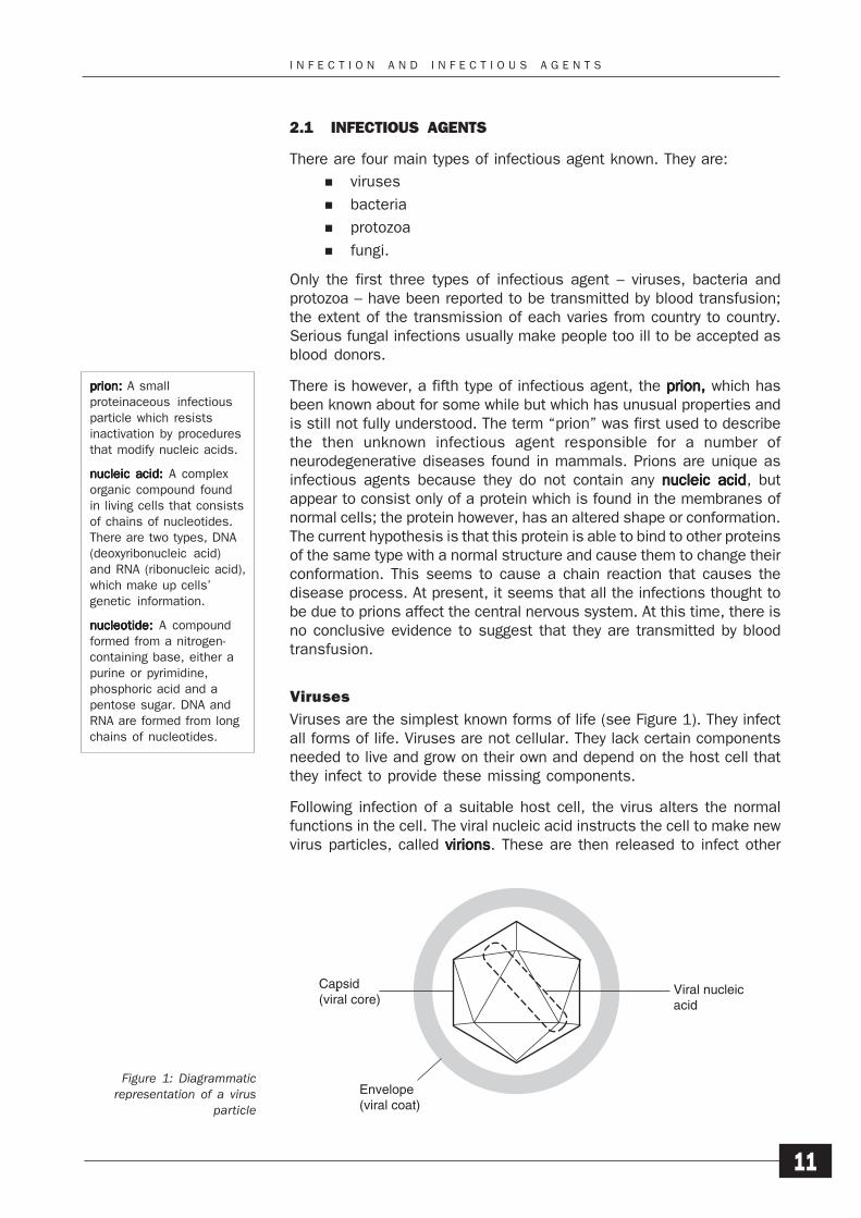
Viruses are the simplest known forms of life (see Figure 1). They infectall forms of life. Viruses are not cellular. They lack certain componentsneeded to live and grow on their own and depend on the host cell thatthey infect to provide these missing components.
Following infection of a suitable host cell, the virus alters the normalfunctions in the cell. The viral nucleic acid instructs the cell to make newvirus particles, called virionsvirionsvirionsvirionsvirions. These are then released to infect other
Figure 1: Diagrammaticrepresentation of a virus
particle
prionprionprionprionprion::::: A smallproteinaceous infectiousparticle which resistsinactivation by proceduresthat modify nucleic acids.
nucleic acidnucleic acidnucleic acidnucleic acidnucleic acid::::: A complexorganic compound foundin living cells that consistsof chains of nucleotides.There are two types, DNA(deoxyribonucleic acid)and RNA (ribonucleic acid),which make up cells’genetic information.
nucleotide:nucleotide:nucleotide:nucleotide:nucleotide: A compoundformed from a nitrogen-containing base, either apurine or pyrimidine,phosphoric acid and apentose sugar. DNA andRNA are formed from longchains of nucleotides.
Viral nucleicacid
Envelope(viral coat)
Capsid(viral core)
S E C T I O N 2
12
cells. Proteins present in the viral coat and the viral core are recognizedby the immune response of the organism.
Some examples of common viruses are:
hepatitis A virus
hepatitis B virus
human immunodeficiency virus (HIV)
measles virus
hepatitis C virus
Varicella zoster (chickenpox) virus.
Some viruses have the property of latencylatencylatencylatencylatency. This is the ability of a virusto join its own nucleic acid with the nucleic acid of the host cell withouttaking complete control of the cell as a virus would normally do. Latencyusually occurs after an active infection when the individual has recoveredand immunity is building up. The viral nucleic acid exists in an inactiveform that does not seem to harm the host cell. When the host cell divides,the cell nucleic acid is copied, together with the viral nucleic acid. In thisway, the viral nucleic acid becomes part of the cell nucleic acid and iscopied every time the cell divides.
Latency is usually indefinite and without any harmful effects on the hostcell. However, at any time, the latent nucleic acid could become activeand take over the cell functions, resulting in an active infection.
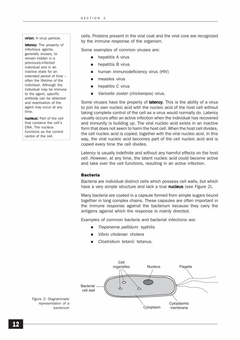
Bacteria
Bacteria are individual distinct cells which possess cell walls, but whichhave a very simple structure and lack a true nucleusnucleusnucleusnucleusnucleus (see Figure 2).
Many bacteria are coated in a capsule formed from simple sugars boundtogether in long complex chains. These capsules are often important inthe immune response against the bacterium because they carry theantigens against which the response is mainly directed.
Examples of common bacteria and bacterial infections are:
Treponema pallidum: syphilis
Vibrio cholerae: cholera
Clostridium tetanii: tetanus.
virion:virion:virion:virion:virion: A virus particle.
latencylatencylatencylatencylatency::::: The property ofinfectious agents,generally viruses, toremain hidden in apreviously-infectedindividual and in aninactive state for anextended period of time –often the lifetime of theindividual. Although theindividual may be immuneto the agent, specificantibody can be detectedand reactivation of theagent may occur at anytime.
nucleusnucleusnucleusnucleusnucleus::::: Part of the cellthat contains the cell’sDNA. The nucleusfunctions as the controlcentre of the cell.
Figure 2: Diagrammaticrepresentation of a
bacterium
Cellorganelles Nucleus Flagella
CytoplasmicmembraneCytoplasm
Bacterialcell wall
I N F E C T I O N A N D I N F E C T I O U S A G E N T S
13
Most bacteria are not of medical importance. Few bacteria can actuallysurvive or grow in animals or humans. Often the bacteria are alreadypresent on the external surfaces of the body without causing any adverseeffects. Damage to the tissues can subsequently lead to infection.Following infection, the disease process is often caused by the productionand release of toxinstoxinstoxinstoxinstoxins by the bacteria.
Protozoa
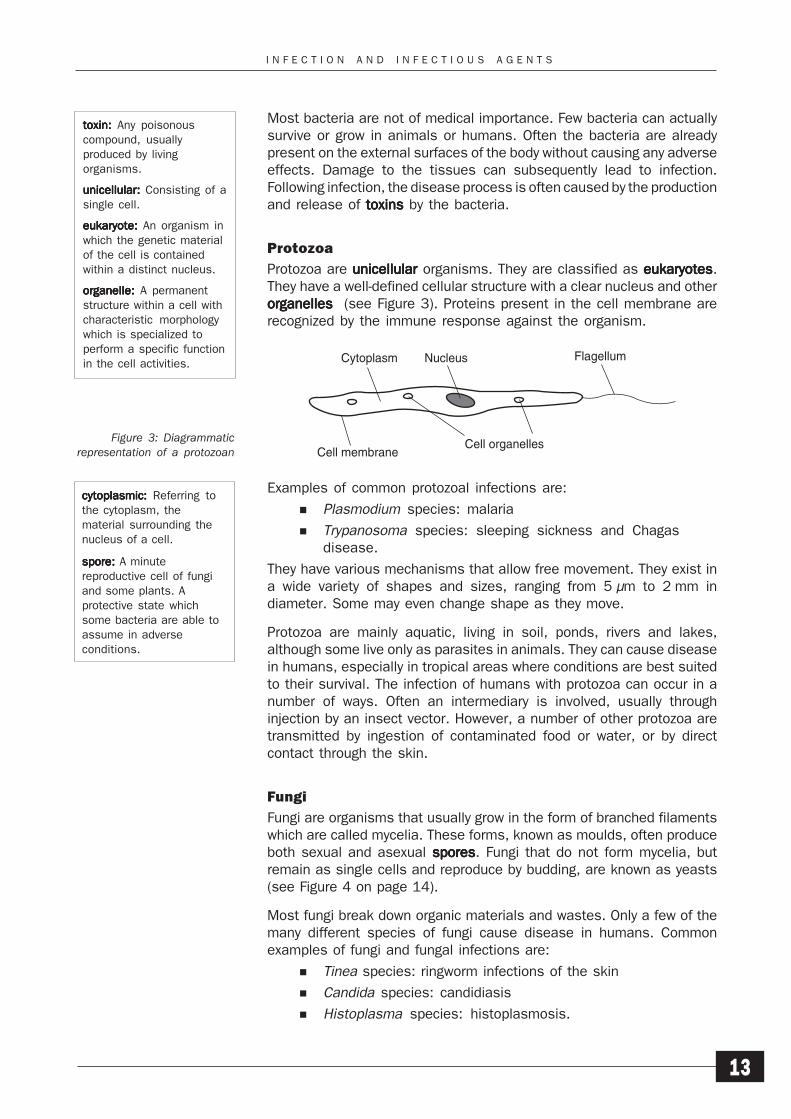
Protozoa are unicellularunicellularunicellularunicellularunicellular organisms. They are classified as eukaryoteseukaryoteseukaryoteseukaryoteseukaryotes.They have a well-defined cellular structure with a clear nucleus and otherorganellesorganellesorganellesorganellesorganelles (see Figure 3). Proteins present in the cell membrane arerecognized by the immune response against the organism.
Examples of common protozoal infections are:
Plasmodium species: malaria
Trypanosoma species: sleeping sickness and Chagasdisease.
They have various mechanisms that allow free movement. They exist ina wide variety of shapes and sizes, ranging from 5 μm to 2 mm indiameter. Some may even change shape as they move.
Protozoa are mainly aquatic, living in soil, ponds, rivers and lakes,although some live only as parasites in animals. They can cause diseasein humans, especially in tropical areas where conditions are best suitedto their survival. The infection of humans with protozoa can occur in anumber of ways. Often an intermediary is involved, usually throughinjection by an insect vector. However, a number of other protozoa aretransmitted by ingestion of contaminated food or water, or by directcontact through the skin.
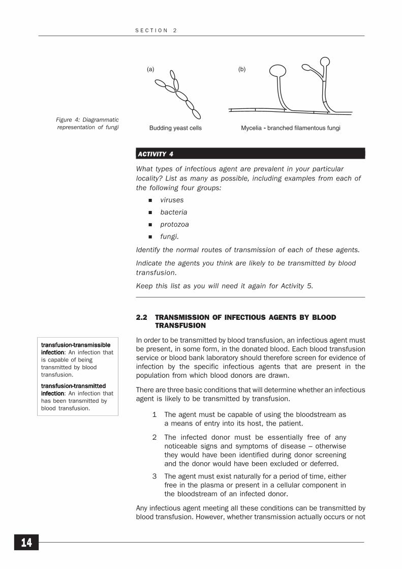
Fungi
Fungi are organisms that usually grow in the form of branched filamentswhich are called mycelia. These forms, known as moulds, often produceboth sexual and asexual sporessporessporessporesspores. Fungi that do not form mycelia, butremain as single cells and reproduce by budding, are known as yeasts(see Figure 4 on page 14).
Most fungi break down organic materials and wastes. Only a few of themany different species of fungi cause disease in humans. Commonexamples of fungi and fungal infections are:
Tinea species: ringworm infections of the skin
Candida species: candidiasis
Histoplasma species: histoplasmosis.
Figure 3: Diagrammaticrepresentation of a protozoan
toxintoxintoxintoxintoxin::::: Any poisonouscompound, usuallyproduced by livingorganisms.
unicellularunicellularunicellularunicellularunicellular::::: Consisting of asingle cell.
eukaryoteeukaryoteeukaryoteeukaryoteeukaryote::::: An organism inwhich the genetic materialof the cell is containedwithin a distinct nucleus.
organelleorganelleorganelleorganelleorganelle::::: A permanentstructure within a cell withcharacteristic morphologywhich is specialized toperform a specific functionin the cell activities.
cytoplasmiccytoplasmiccytoplasmiccytoplasmiccytoplasmic::::: Referring tothe cytoplasm, thematerial surrounding thenucleus of a cell.
sporesporesporesporespore::::: A minutereproductive cell of fungiand some plants. Aprotective state whichsome bacteria are able toassume in adverseconditions.
Flagellum
Cell organellesCell membrane
NucleusCytoplasm
S E C T I O N 2
14
ACTIVITY 4
What types of infectious agent are prevalent in your particularlocality? List as many as possible, including examples from each ofthe following four groups:
viruses
bacteria
protozoa
fungi.
Identify the normal routes of transmission of each of these agents.
Indicate the agents you think are likely to be transmitted by bloodtransfusion.
Keep this list as you will need it again for Activity 5.
2.2 TRANSMISSION OF INFECTIOUS AGENTS BY BLOODTRANSFUSION
In order to be transmitted by blood transfusion, an infectious agent mustbe present, in some form, in the donated blood. Each blood transfusionservice or blood bank laboratory should therefore screen for evidence ofinfection by the specific infectious agents that are present in thepopulation from which blood donors are drawn.
There are three basic conditions that will determine whether an infectiousagent is likely to be transmitted by transfusion.
1 The agent must be capable of using the bloodstream asa means of entry into its host, the patient.
2 The infected donor must be essentially free of anynoticeable signs and symptoms of disease – otherwisethey would have been identified during donor screeningand the donor would have been excluded or deferred.
3 The agent must exist naturally for a period of time, eitherfree in the plasma or present in a cellular component inthe bloodstream of an infected donor.
Any infectious agent meeting all these conditions can be transmitted byblood transfusion. However, whether transmission actually occurs or not
transfusion-transmissibletransfusion-transmissibletransfusion-transmissibletransfusion-transmissibletransfusion-transmissibleinfectioninfectioninfectioninfectioninfection: An infection thatis capable of beingtransmitted by bloodtransfusion.
transfusion-transmittedtransfusion-transmittedtransfusion-transmittedtransfusion-transmittedtransfusion-transmittedinfectioninfectioninfectioninfectioninfection: An infection thathas been transmitted byblood transfusion.
Figure 4: Diagrammaticrepresentation of fungi
(a) (b)
Budding yeast cells Mycelia - branched filamentous fungi
I N F E C T I O N A N D I N F E C T I O U S A G E N T S
15
depends on a number of other factors, particularly on the immune statusof the patient and the amount of infectious agent transfused.
Although blood transfusion can be an efficient route of transmission ofan infectious agent, it is important to remember that, under normalcircumstances, it is not the primary route of infection of any infectiousagent. This is because, no matter how infectious any individual agentmay be, most people do not have a blood transfusion during theirlifetime. Any agent that depends solely on transfusion for transmissionwill therefore not persist in the population.
It is known that the transmission of certain infectious agents throughblood transfusion can, and does, occur, however, and that it can be animportant route of infection. The key point to remember here is thattransmission can be avoided in most cases by the design and use ofsuitable selection and screening programmes.
ACTIVITY 5
Look back at your answers to Activity 4 where you identified theinfectious agents that you think could be transmitted by transfusion.
From the information that you now have, would you make anychanges to your list of agents that can be transmitted bytransfusion? If so, amend your list and check it with your supervisoror another senior member of staff.
2.3 THE BASIC IMMUNOLOGY OF INFECTION
Human immunology is concerned with the study of how the bodyresponds to foreign proteins and the mechanisms by which the bodyprotects itself from attack by infectious agents.
The main function of the immune system is to protect our bodies fromattack by the many infectious agents that we all meet during our lives.It consists of two parts:
a detection system that recognizes foreign proteins in thebody
a number of different mechanisms that prevent thegrowth of the infectious agent in the body.
Importantly, the immune response is specific.
Unfortunately, however, these systems are not perfect and they may failor take some time to respond. When this happens, it can sometimesresult in infection by an infectious agent and can also lead to damage tothe body which is caused by components of the immune system itself.
Foreign substances that enter the body and induce the immune responseare called antigensantigensantigensantigensantigens (Ag). One possible response to these antigens is theproduction of a protein called an antibodyantibodyantibodyantibodyantibody (Ab). Another possible
antigen:antigen:antigen:antigen:antigen: Any substancerecognized as foreign bythe body and whichstimulates the immunesystem to mount aresponse against it.
antibody:antibody:antibody:antibody:antibody: A protectiveprotein produced by theimmune response of anindividual to stimulation bya foreign substance. Itplays a role in the defenceagainst pathogens, oftenby neutralization or byidentifying the pathogen asforeign and to beeliminated by the immunesystem.
S E C T I O N 2
16
response is the activation of cells that kill infected cells by direct contactor by activating other cell-killing mechanisms, or by both means.
Antibody response
Antibodies are molecules that are made up of proteins and carbohydrates.They are produced by lymphoid cellslymphoid cellslymphoid cellslymphoid cellslymphoid cells in response to stimulation byantigen. They are specific to a particular antigen and they bind to theantigen to enable the organism to eliminate it. They belong to a group ofhuman serum proteins known as gammaglobulinsgammaglobulinsgammaglobulinsgammaglobulinsgammaglobulins, and are known asimmunoglobulinsimmunoglobulinsimmunoglobulinsimmunoglobulinsimmunoglobulins.
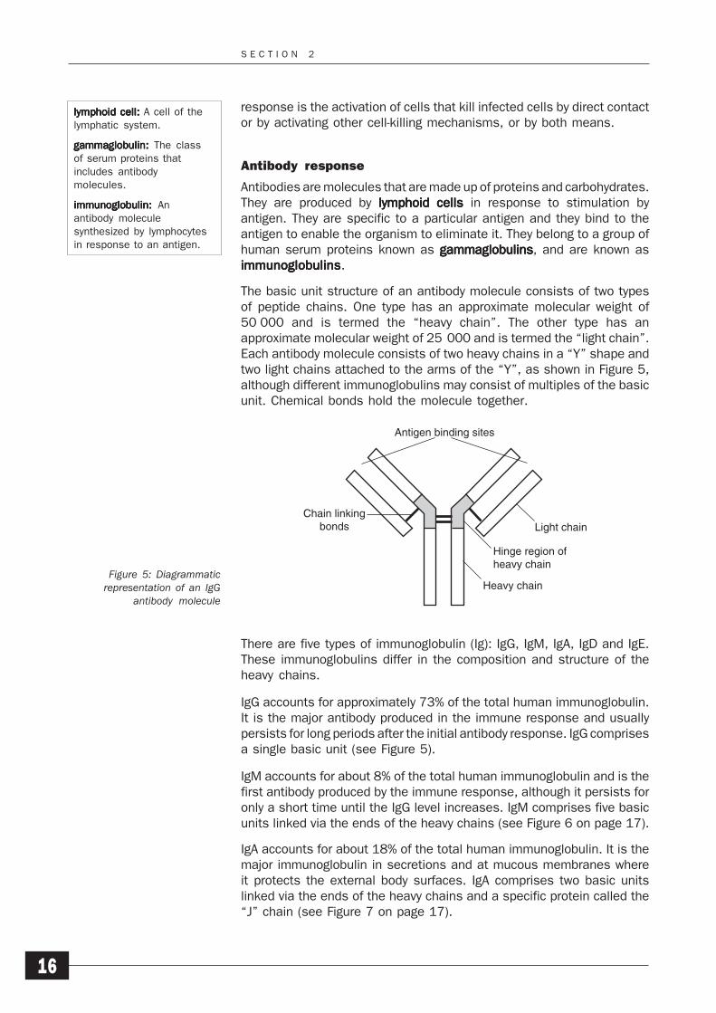
The basic unit structure of an antibody molecule consists of two typesof peptide chains. One type has an approximate molecular weight of50 000 and is termed the “heavy chain”. The other type has anapproximate molecular weight of 25 000 and is termed the “light chain”.Each antibody molecule consists of two heavy chains in a “Y” shape andtwo light chains attached to the arms of the “Y”, as shown in Figure 5,although different immunoglobulins may consist of multiples of the basicunit. Chemical bonds hold the molecule together.
Figure 5: Diagrammaticrepresentation of an IgG
antibody molecule
There are five types of immunoglobulin (Ig): IgG, IgM, IgA, IgD and IgE.These immunoglobulins differ in the composition and structure of theheavy chains.
IgG accounts for approximately 73% of the total human immunoglobulin.It is the major antibody produced in the immune response and usuallypersists for long periods after the initial antibody response. IgG comprisesa single basic unit (see Figure 5).
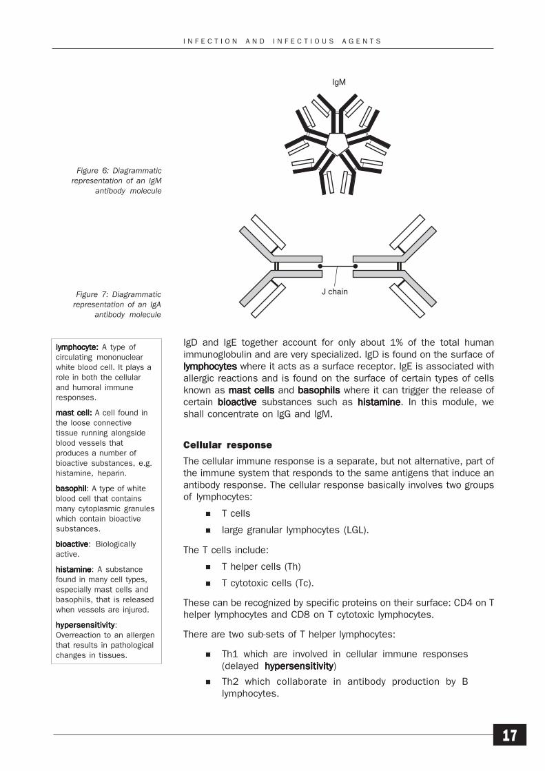
IgM accounts for about 8% of the total human immunoglobulin and is thefirst antibody produced by the immune response, although it persists foronly a short time until the IgG level increases. IgM comprises five basicunits linked via the ends of the heavy chains (see Figure 6 on page 17).
IgA accounts for about 18% of the total human immunoglobulin. It is themajor immunoglobulin in secretions and at mucous membranes whereit protects the external body surfaces. IgA comprises two basic unitslinked via the ends of the heavy chains and a specific protein called the“J” chain (see Figure 7 on page 17).
lymphoid cell:lymphoid cell:lymphoid cell:lymphoid cell:lymphoid cell: A cell of thelymphatic system.
gammaglobulin:gammaglobulin:gammaglobulin:gammaglobulin:gammaglobulin: The classof serum proteins thatincludes antibodymolecules.
immunoglobulin:immunoglobulin:immunoglobulin:immunoglobulin:immunoglobulin: Anantibody moleculesynthesized by lymphocytesin response to an antigen.
Antigen binding sites
Chain linkingbonds Light chain
Hinge region ofheavy chain
Heavy chain
I N F E C T I O N A N D I N F E C T I O U S A G E N T S
17
IgD and IgE together account for only about 1% of the total humanimmunoglobulin and are very specialized. IgD is found on the surface oflymphocyteslymphocyteslymphocyteslymphocyteslymphocytes where it acts as a surface receptor. IgE is associated withallergic reactions and is found on the surface of certain types of cellsknown as mast cellsmast cellsmast cellsmast cellsmast cells and basophilsbasophilsbasophilsbasophilsbasophils where it can trigger the release ofcertain bioactivebioactivebioactivebioactivebioactive substances such as histaminehistaminehistaminehistaminehistamine. In this module, weshall concentrate on IgG and IgM.
Cellular response
The cellular immune response is a separate, but not alternative, part ofthe immune system that responds to the same antigens that induce anantibody response. The cellular response basically involves two groupsof lymphocytes:
T cells
large granular lymphocytes (LGL).
The T cells include:
T helper cells (Th)
T cytotoxic cells (Tc).
These can be recognized by specific proteins on their surface: CD4 on Thelper lymphocytes and CD8 on T cytotoxic lymphocytes.
There are two sub-sets of T helper lymphocytes:
Th1 which are involved in cellular immune responses(delayed hypersensitivityhypersensitivityhypersensitivityhypersensitivityhypersensitivity)
Th2 which collaborate in antibody production by Blymphocytes.
Figure 6: Diagrammaticrepresentation of an IgM
antibody molecule
Figure 7: Diagrammaticrepresentation of an IgA
antibody molecule
lymphocyte:lymphocyte:lymphocyte:lymphocyte:lymphocyte: A type ofcirculating mononuclearwhite blood cell. It plays arole in both the cellularand humoral immuneresponses.
mast cell: mast cell: mast cell: mast cell: mast cell: A cell found inthe loose connectivetissue running alongsideblood vessels thatproduces a number ofbioactive substances, e.g.histamine, heparin.
basophilbasophilbasophilbasophilbasophil: A type of whiteblood cell that containsmany cytoplasmic granuleswhich contain bioactivesubstances.
bioactivebioactivebioactivebioactivebioactive: Biologicallyactive.
histaminehistaminehistaminehistaminehistamine: A substancefound in many cell types,especially mast cells andbasophils, that is releasedwhen vessels are injured.
hypersensitivityhypersensitivityhypersensitivityhypersensitivityhypersensitivity:Overreaction to an allergenthat results in pathologicalchanges in tissues.
IgM
J chain
S E C T I O N 2
18
T cytotoxic cells (Tc) are the effectors in the destruction of cells infectedwith intracellular micro-organisms, such as virus.
Large granular lymphocytes comprise 5–10% of the lymphocyte population.They include natural killer cells (NK cells) which are capable of lysing avariety of tumour and virus-infected cells without overt antigenicstimulation. In response to high levels of Interleukin 2, NK cellsdifferentiate into lymphokine-activated killer cells (LAK cells) which killtarget cells in a relatively indiscriminate way. There is a further subsetcalled killer cells (K cells) which lyse infected cells but only recognizethose cells coated with IgG. Both of these cell types act in a similarmanner to Tc cells.
One cell type, macrophagesmacrophagesmacrophagesmacrophagesmacrophages, plays a central role in the immuneresponse, both in the antibody response and the cellular response.Macrophages phagocytosephagocytosephagocytosephagocytosephagocytose infectious material and infected cells. Theydestroy the infectious agent and also present it to other cells of theimmune system, both T cells for the cellular response and B cells for theantibody response. The results of the cellular response are not used forblood screening purposes.
2.4 INTRODUCTION TO SCREENING FOR INFECTIOUS AGENTS
The extent and range of the screening performed on the blood supply varygreatly from country to country. Sometimes this is simply because ofdifferences in the countries’ needs, but it is sometimes due to financialconstraints. As a result, the effectiveness of screening programmes alsovaries.
Whatever level of service is provided, however, the main purpose ofscreening blood is to ensure that the available blood supply is as free aspossible from infectious agents by detecting any that may be presentbefore the blood is issued for transfusion.
Blood transfusion is an ideal route for the transmission of certaininfectious agents from the donor to the recipient of the blood. The riskcan, however, be reduced in the following ways.
1 The careful selection of donors to ensure that, whereverpossible, blood is not collected from people who are likelyto be carriers of infectious agents. Module 1: Safe BloodDonation shows that building a panel of regular, voluntarynon-remunerated donors is the first step towards ensuringa safe and adequate supply of blood. In countries wheremuch of the blood is collected from family or familyreplacement donors or from commercial or professionaldonors, the risk of transfusion-transmitted infection ishigher.
2 The direct screening of the donated blood for evidence ofthe presence of infectious agents.
3 The removal of specific components of blood thought toharbour infectious agents: for example, by the filtration ofblood to remove white blood cells.
macrophagemacrophagemacrophagemacrophagemacrophage: A phagocyticcell type found in thebloodstream as well astissues. It ingests bacteriaand cell debris.
phagocytosis:phagocytosis:phagocytosis:phagocytosis:phagocytosis: Theprocess by which cellsingest solid matter,especially cell debris andpathogens.
I N F E C T I O N A N D I N F E C T I O U S A G E N T S
19
4 The physical inactivation of any contaminating agentsthat may be present: for example, by heat treatment ofFactor VIII concentrates (used in the treatment ofhaemophilia A) during production.
Not all infectious agents can be detected directly in donated blood. Bloodis most often screened for evidence of previous infection by looking forthe presence of specific antibody raised against the infectious agent.
As we have already mentioned, some organisms possess the propertyof latency. In such cases, while antibody has been produced and acuteinfection has resolved, the organism remains in a dormant dormant dormant dormant dormant state withinhost cells. Although dormant, the agent is capable of reactivating andproducing an acute infection at any time. The transfusion of blood cellscontaining latent organism could, therefore, lead to the transmission ofinfection.
Markers of infection
Markers of infection are the detectable signs of infection appearing in thebloodstream during, or following, infection. These can be detected by thepresence of the agent itself but, more commonly, by the presence ofspecific antibodies against the infectious agent.
Clearly, it is only by understanding which markers of infection areproduced by an infectious agent that screening for the correct marker canbe introduced.
ACTIVITY 6
Think about the following situation. A donor is infected with aninfectious agent that can be transmitted by blood transfusion. Thedonor has an acute infection, but with no symptoms, that lasts forone month. After this period, immunity immunity immunity immunity immunity to the agent develops andthe agent is destroyed and cleared from the body. The naturalimmunity lasts for at least one year.
What markers of infection are most likely to be found in donationstaken at the following periods after the donor was first infected?
2 weeks
4 weeks
6 weeks
8 weeks
18 months.
If this infectious agent was likely to be transmitted only during theperiod of acute infection, would it be better to screen donors forthe agent itself or for antibody produced against the agent?
Write down your answers and then check them with the answersgiven in the Activity Checklists and Answers on page 139.
dormantdormantdormantdormantdormant: : : : : An inactiveperiod in the life-cycle oforganisms in which growthslows or ceases.
markers of infectionmarkers of infectionmarkers of infectionmarkers of infectionmarkers of infection: Thedetectable signs ofinfection, including thebody’s own response tothe infectious agent,appearing in thebloodstream during, orfollowing, infection.
immunity:immunity:immunity:immunity:immunity: The state ofbeing resistant to infectionby an infectious agent dueto previous exposure tothe agent, with resultantproduction of a protectingimmune response.
S E C T I O N 2
20
You should recognize that it would be better to screen donors for theagent itself, rather than for antibody. In this case, the presence ofantibody only indicates immunity – not an infectious state.
In the case of viral hepatitis B, screening for the protein known as surfaceantigen (HBsAg) (see page 106) is used to identify infected individuals.In the cases of the human immunodeficinecy virus (HIV) and hepatitis Cvirus (HCV), the presence of the antibody to the virus is used to screendonated blood for infection.
With HBV infection, the virus causes the shedding of large amounts ofsurface antigen into the bloodstream during acute infection. This canpersist for long periods. The detection of this HBsAg is therefore used toidentify infected donors. The presence of antibody to HBsAg (HBsAb)indicates immunity following infection and protects against furtherinfections.
With HIV or HCV infection, although there is a period during which onlyviral antigen can be detected, this period is very short, possibly no morethan a few days in some cases, as the antigen is very quickly complexedwith circulating antibody as antibody levels rise. Detection of viral antigenis therefore generally not a suitable approach. The presence of specificantibody is the primary approach used to identify HIV or HCV infecteddonors. The virus is still present in the unit of blood, in the white cellsfor HIV and free in the plasma for HCV, and the antibody does notgenerally protect against reinfection. The presence of antibody thereforeidentifies donors carrying the virus.
SUMMARY
1 There are four main types of infectious agent:
viruses
bacteria
protozoa
fungi.
Only the first three types are known to be transmitted byblood transfusion.
2 Prions are an unusual type of infectious agent whosetransmission by blood transfusion has not yet beenconclusively demonstrated.
3 Infectious agents can be transmitted by blood transfusiononly if they are present in the donated blood. A screeningprogramme should therefore focus on the infectiousagents present in the donor population.
4 Blood transfusion can be a significant, but not theprimary, route of transmission of infectious agents.
5 The immune response to infectious agents consists oftwo parts:
the antibody response
the cellular response.
I N F E C T I O N A N D I N F E C T I O U S A G E N T S
21
6 Screening tests must detect the different markers ofinfection that different infectious agents produce.
SELF-ASSESSMENT
1 What does the term “latency” mean?
2 What are the three conditions required for transmission ofinfectious agents by blood transfusion?
3 Why do you think that blood transfusion cannot be theprimary route of infection of an infectious agent?
4 What are antigens and antibodies?
5 Name the five types of immunoglobulin.
6 What is the main reason for screening blood for thepresence of infectious agents?
7 What are the four main ways of minimizing the risk oftransmission of infectious agents by blood transfusion?
8 In some cases, the presence of antibody as a result of aprevious infection may indicate that the blood is stillinfectious. Why is this the case?
PROGRESS CHECK
Before moving on to Section 3, spend a few minutes thinkingabout whether you have achieved the learning objectives forSection 2. These were to:
1 Recognize examples of the four types of infectious agent.
2 Understand the term “latency”, as applied to viralinfections, and its significance in blood transfusionpractice.
3 Identify infectious agents which can be transmitted byblood transfusion.
4 Use your knowledge of immunology to predict likelymarkers of infection at different stages following infection.
If you feel confident that you have understood everything inthis section, turn to Section 3.
If you feel that you need to spend more time on this section,go back to the parts that are most unfamiliar or that you finddifficult. You may find it helpful to talk to other people, suchas your supporter or other senior colleagues, if there isanything you are still not sure about.

S E C T I O N 3
22
The HumanImmunodeficiency Viruses
The purpose of this section is to help you to understand HIV and theconsequences of infection. You will study various aspects of the humanimmunodeficiency viruses, including:
history
physical characteristics
epidemiology
clinical infection
prevention of transmission
the role of HIV as the causative agent of AIDS.
LEARNING OBJECTIVES
When you have completed this section, you should be able to:
1 Identify the structural features of HIV.
2 Describe the basic stages of HIV infection and the entryof the virus into susceptible cells.
3 Describe the most common clinical course of HIV infectionand its progression to AIDS in your country.
4 Describe the measures that are being taken to reduce thetransmission of HIV infection in your country.
3
T H E H U M A N I M M U N O D E F I C I E N C Y V I R U S E S
23
3.1 THE BACKGROUND TO HIV INFECTION
HIV is the primary cause of acquired immunodeficiency syndrome (AIDS).Although the way in which HIV infection causes AIDS is not fullyunderstood, it is clear that it damages part of the immune system. Thiscan lead to serious infections by agents that would normally be easilyovercome by the immune system.
HIV was first isolated from the cells of an infected patient in 1983(HIV-1). The virus was subsequently identified as the causative agent ofAIDS. In 1986 a second type of HIV, HIV-2, was identified in certain areasof West Africa. HIV-2 appears to cause the same diseases as HIV-1, butmay be less pathogenic. It is morphologicallymorphologicallymorphologicallymorphologicallymorphologically similar to HIV-1. The twotypes can be distinguished by the presence or absence of an antibodythat is specific to a protein found only on HIV-2. Although cross-reactivitycross-reactivitycross-reactivitycross-reactivitycross-reactivityoccurs between the core protein of both viruses, the envelope envelope envelope envelope envelope proteinsare different.
Cross-reactivity
Cross-reactivity is the situation that occurs when an antibody recognizesnot only its own antigen, but also other unrelated antigens that havecertain similarities. In the case of HIV-1 and HIV-2, this means that anindividual infected with HIV-1 would produce antibodies that recognizeboth core and envelope proteins of HIV-1 and core proteins of HIV-2.Similarly, an individual infected with HIV-2 would produce antibodies thatrecognize both core and envelope proteins of HIV-2 and core proteins ofHIV-1. However, the cross-reactivity commonly seen in anti-HIV and otherTTI screening assays is usually between specific antibody and unrelatedhuman proteins that are non-specifically bound during the sampleincubation phase of the assay.
3.2 THE STRUCTURE OF HIV
There are two types of nucleic acid:
ribonucleic acid (RNARNARNARNARNA)
deoxyribonucleic acid (DNADNADNADNADNA).
DNA is usually double-stranded. It is the genetic material passed todaughter cells when a cell divides. It is DNA that is responsible for thetransmission of hereditary characteristics from parents to children.
The nucleic acid in HIV is RNA. There is no DNA present. Instead, the virususes the machinery of the human cells that it enters to convert its RNAto DNA so that the virus can replicate or integrate itself in the cell’s DNA.
The viral RNA is condensed in a cylindrical core together with two closely-associated structural proteins and an important enzyme called RNA-dependent DNA polymerase. This is more commonly known as reversereversereversereversereversetranscriptasetranscriptasetranscriptasetranscriptasetranscriptase. This enzyme is found in all retrovirusesretrovirusesretrovirusesretrovirusesretroviruses as it is needed tocopy the viral RNA into DNA.
The way that viral and other such proteins are described is based on theirmolecular weight (measured in daltons) and on whether they are proteinsor glycoproteins. The two proteins associated with the RNA of HIV are
morphology: morphology: morphology: morphology: morphology: The study ofthe shape or form oforganisms.
cross-reactivity:cross-reactivity:cross-reactivity:cross-reactivity:cross-reactivity: When anantibody recognizes notonly its correspondingspecific antigen, but alsoother antigens that mayhave certain similarities.
envelope (viral):envelope (viral):envelope (viral):envelope (viral):envelope (viral): Anexternal protein coat thatsurrounds the viral capsid.Not all viruses areenveloped.
RNA (ribonucleic acid):RNA (ribonucleic acid):RNA (ribonucleic acid):RNA (ribonucleic acid):RNA (ribonucleic acid): Acomplex chemical found inthe cytoplasm andconcerned with proteinsynthesis. In someviruses, it is the hereditarymaterial.
DNA (deoxyribonucleicDNA (deoxyribonucleicDNA (deoxyribonucleicDNA (deoxyribonucleicDNA (deoxyribonucleicacid):acid):acid):acid):acid): The genetic materialof most living organismsthat determines hereditarycharacteristics by thecontrol of proteinsynthesis.
reverse transcriptase: reverse transcriptase: reverse transcriptase: reverse transcriptase: reverse transcriptase: Anaturally-occurring enzymewhich translates RNA intoDNA.
retrovirus:retrovirus:retrovirus:retrovirus:retrovirus: A virus familythat is characterized byRNA as the nucleic acid, aunique morphology, thepresence of a uniqueenzyme (reversetranscriptase) and latency.
S E C T I O N 3
24
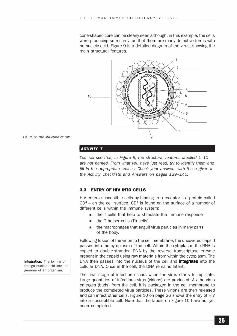
7000 daltons (7 kDa) and 9000 daltons (9 kDa). These are abbreviatedto p7 and p9 respectively. The reverse transcriptase enzyme is a 66 kDaprotein, which is abbreviated to p66. Glycoproteins Glycoproteins Glycoproteins Glycoproteins Glycoproteins are similarlyabbreviated to “gp”. We shall now use this notation throughout thismodule.
The core is totally enclosed in a cone-shaped shell of p24 protein. Thisis called the major core protein and appears to be the same in bothHIV-1 and HIV-2. The whole unit is called the viral capsidcapsidcapsidcapsidcapsid.
The capsid is itself covered by two layers. The first of these is a shell ofp17 matrix protein to which proteins that project from the surface of thevirus particle are attached. This is covered by a lipid bilayer. Projectingthrough the lipid are many transmembrane proteins. These proteins,gp41, are attached to the p17 matrix and themselves attach the gp120envelope proteins. These appear as small projections on the surface ofthe virus particle. It is the structure of these small projections and theirattaching proteins that appears to be the major difference betweenHIV-1 and HIV-2. The corresponding HIV-2 proteins are gp110/130 andgp36 respectively. Antibodies to these two specific sets of proteins donot cross-react.
The entire virus particle is called the virion. This is the infectious particlethat is secreted and transmitted between individuals. The completevirion is 100–120 μm in diameter.
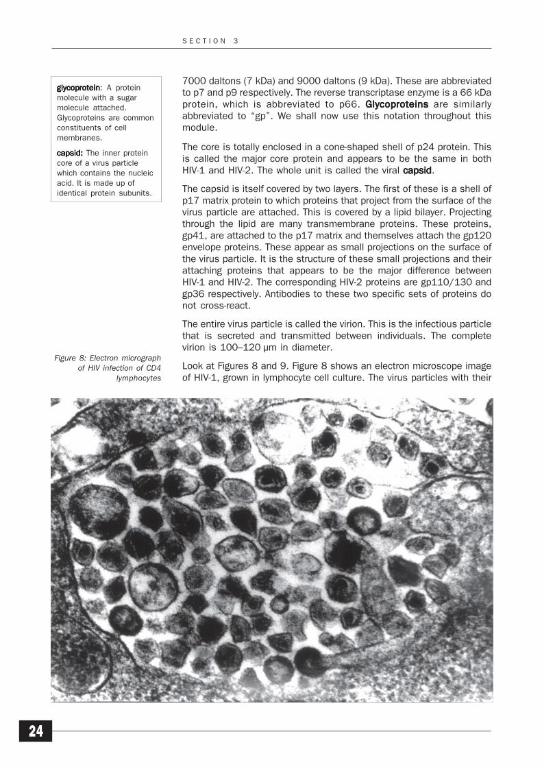
Look at Figures 8 and 9. Figure 8 shows an electron microscope imageof HIV-1, grown in lymphocyte cell culture. The virus particles with their
Figure 8: Electron micrographof HIV infection of CD4
lymphocytes
glycoproteinglycoproteinglycoproteinglycoproteinglycoprotein: A proteinmolecule with a sugarmolecule attached.Glycoproteins are commonconstituents of cellmembranes.
capsidcapsidcapsidcapsidcapsid: : : : : The inner proteincore of a virus particlewhich contains the nucleicacid. It is made up ofidentical protein subunits.
T H E H U M A N I M M U N O D E F I C I E N C Y V I R U S E S
25
cone-shaped core can be clearly seen although, in this example, the cellswere producing so much virus that there are many defective forms withno nucleic acid. Figure 9 is a detailed diagram of the virus, showing themain structural features.
ACTIVITY 7
You will see that, in Figure 9, the structural features labelled 1–10are not named. From what you have just read, try to identify them andfill in the appropriate spaces. Check your answers with those given inthe Activity Checklists and Answers on pages 139–140.
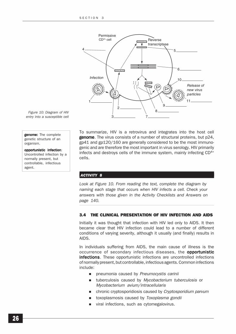
3.3 ENTRY OF HIV INTO CELLS
HIV enters susceptible cells by binding to a receptor – a protein calledCD4 – on the cell surface. CD4 is found on the surface of a number ofdifferent cells within the immune system:
the T cells that help to stimulate the immune response
the T helper cells (Th cells)
the macrophages that engulf virus particles in many partsof the body.
Following fusion of the virion to the cell membrane, the uncovered capsidpasses into the cytoplasm of the cell. Within the cytoplasm, the RNA iscopied to double-stranded DNA by the reverse transcriptase enzymepresent in the capsid using raw materials from within the cytoplasm. TheDNA then passes into the nucleus of the cell and integratesintegratesintegratesintegratesintegrates into thecellular DNA. Once in the cell, the DNA remains latent.
The final stage of infection occurs when the virus starts to replicate.Large quantities of infectious virus (virions) are produced. As the virusemerges (buds) from the cell, it is packaged in the cell membrane toproduce the completed virus particles. These virions are then releasedand can infect other cells. Figure 10 on page 26 shows the entry of HIVinto a susceptible cell. Note that the labels on Figure 10 have not yetbeen completed.
integrationintegrationintegrationintegrationintegration: : : : : The joining offoreign nucleic acid into thegenome of an organism.
Figure 9: The structure of HIV
1
9
5
10 8
6
3
7
4
2
S E C T I O N 3
26
To summarize, HIV is a retrovirus and integrates into the host cellgenomegenomegenomegenomegenome. The virus consists of a number of structural proteins, but p24,gp41 and gp120/160 are generally considered to be the most immuno-genic and are therefore the most important in virus serology. HIV primarilyinfects and destroys cells of the immune system, mainly infecting CD4+
cells.
ACTIVITY 8
Look at Figure 10. From reading the text, complete the diagram bynaming each stage that occurs when HIV infects a cell. Check youranswers with those given in the Activity Checklists and Answers onpage 140.
3.4 THE CLINICAL PRESENTATION OF HIV INFECTION AND AIDS
Initially it was thought that infection with HIV led only to AIDS. It thenbecame clear that HIV infection could lead to a number of differentconditions of varying severity, although it usually (and finally) results inAIDS.
In individuals suffering from AIDS, the main cause of illness is theoccurrence of secondary infectious diseases, the opportunisticopportunisticopportunisticopportunisticopportunisticinfectionsinfectionsinfectionsinfectionsinfections. These opportunistic infections are uncontrolled infectionsof normally present, but controllable, infectious agents. Common infectionsinclude:
pneumonia caused by Pneumocystis carinii
tuberculosis caused by Mycobacterium tuberculosis orMycobacterium avium/intracellularis
chronic cryptosporidiosis caused by Cryptosporidium parvum
toxoplasmosis caused by Toxoplasma gondii
viral infections, such as cytomegalovirus.
genomegenomegenomegenomegenome: : : : : The completegenetic structure of anorganism.
opportunistic infectionopportunistic infectionopportunistic infectionopportunistic infectionopportunistic infection:Uncontrolled infection by anormally present, butcontrollable, infectiousagent.
Figure 10: Diagram of HIVentry into a susceptible cell
1
4
3 7
8
9
6
5
11
10
2
Infection
Release ofnew virusparticles
Reversetranscriptase
PermissiveCD4+ cellCD4+ cell
T H E H U M A N I M M U N O D E F I C I E N C Y V I R U S E S
27
Secondary cancers such as Kaposi's sarcoma and non-Hodgkinslymphoma are other conditions commonly found in AIDS patients. Thesecancers are usually aggressive and do not respond very well to standardchemotherapy. Kaposi's sarcoma, as originally described, was a benignmalignancy found in elderly men that had no adverse affect on theindividual. However, the Kaposi's sarcoma found in AIDS patients is afast-growing, and usually fatal, malignancy now known to be associatedwith the more recently identified human herpes virus 8 (HHV8). Most HIV-infected individuals with Kaposi sarcoma are also infected with HHV8. Inmany parts of the world, patients with developing ARC (AIDS-relatedcomplex) or AIDS often present simply with severe diarrhoea. Thepresence of opportunistic infections or secondary cancers is only thendetermined following clinical and laboratory investigation.
ACTIVITY 9
Try to find out the following information about HIV infection and AIDSin your country:
the prevalence prevalence prevalence prevalence prevalence of HIV infection and the number of AIDScases
the most common clinical course of HIV infection and itsprogression to AIDS
the average time taken for AIDS to manifest following theinitial infection
the main opportunistic infections found in AIDS patients,apart from Pneumocystis carinii. Are these opportunisticinfections found in all areas of your country or are there localvariations?
the incidence of Kaposi’s sarcoma in your region or countryabout 25 years ago and today. Compare the two figures andrelate them to the current prevalence of AIDS in yourpopulation.
You may need to consult your Ministry of Health, a larger public healthlaboratory or a specialized AIDS counsellor in order to obtain thisinformation.
This information should give you an approximate picture of the situationin your country. It is important to find out this information so that you canunderstand the extent of the problem.
3.5 LABORATORY TESTING FOR HIV INFECTION IN BLOODDONATIONS
Before HIV was identified, AIDS was diagnosed by its clinical appearance.Any laboratory tests that were used were surrogate tests; they measuredthe results of HIV infection rather than specifically detecting either viralantigen or specific antibody against the virus.
prevalenceprevalenceprevalenceprevalenceprevalence: : : : : The proportionof a specific populationthat is infected with theinfectious agent at anyparticular time.
S E C T I O N 3
28
Surrogate testingSurrogate testingSurrogate testingSurrogate testingSurrogate testing is testing for a specific marker which is not a part ofthe agent under investigation, but which is thought to indicate thepresence of the infectious agent. For example, there is now a specificassay to detect antibody to the hepatitis C virus (anti-HCV) the majorcause of the previously termed non-A, non-B hepatitis (NANB). However,prior to the development of this assay, some countries used testing foranti-HBc (hepatitis B core antibody) and measuring levels of ALT (alanineaminotransferase, a measure of liver damage) to try to identify thosedonors who may have been more likely to transmit NANB hepatitis.
The production of specific tests for HIV has helped us to understand bothHIV infection and AIDS. The finding of anti-HIV in patients with previouslyunexplained immunodeficiency is diagnostic of AIDS.
The confirmed presence of anti-HIV in an asymptomatic individual (anindividual without symptoms) means that the individual has beenexposed to the virus. It is accepted that, in almost all cases, the viruswill still be present in the individual. SeroconversionSeroconversionSeroconversionSeroconversionSeroconversion (a change inserostatus serostatus serostatus serostatus serostatus from negative to positive) in sequential samples means thatthe infection is recent. The continued absence of antibody, lack ofseroconversion following possible exposure to the virus, means that it ishighly unlikely that the individual has been infected.
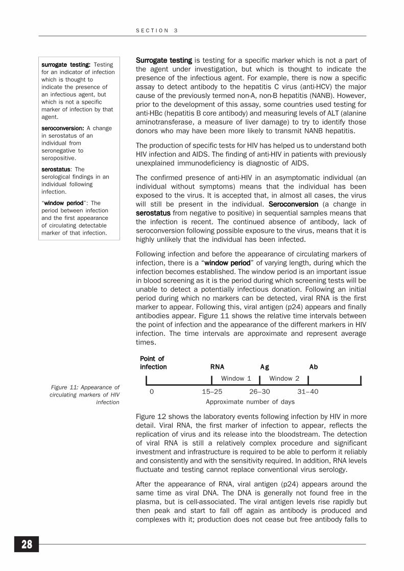
Following infection and before the appearance of circulating markers ofinfection, there is a “window periodwindow periodwindow periodwindow periodwindow period” of varying length, during which theinfection becomes established. The window period is an important issuein blood screening as it is the period during which screening tests will beunable to detect a potentially infectious donation. Following an initialperiod during which no markers can be detected, viral RNA is the firstmarker to appear. Following this, viral antigen (p24) appears and finallyantibodies appear. Figure 11 shows the relative time intervals betweenthe point of infection and the appearance of the different markers in HIVinfection. The time intervals are approximate and represent averagetimes.
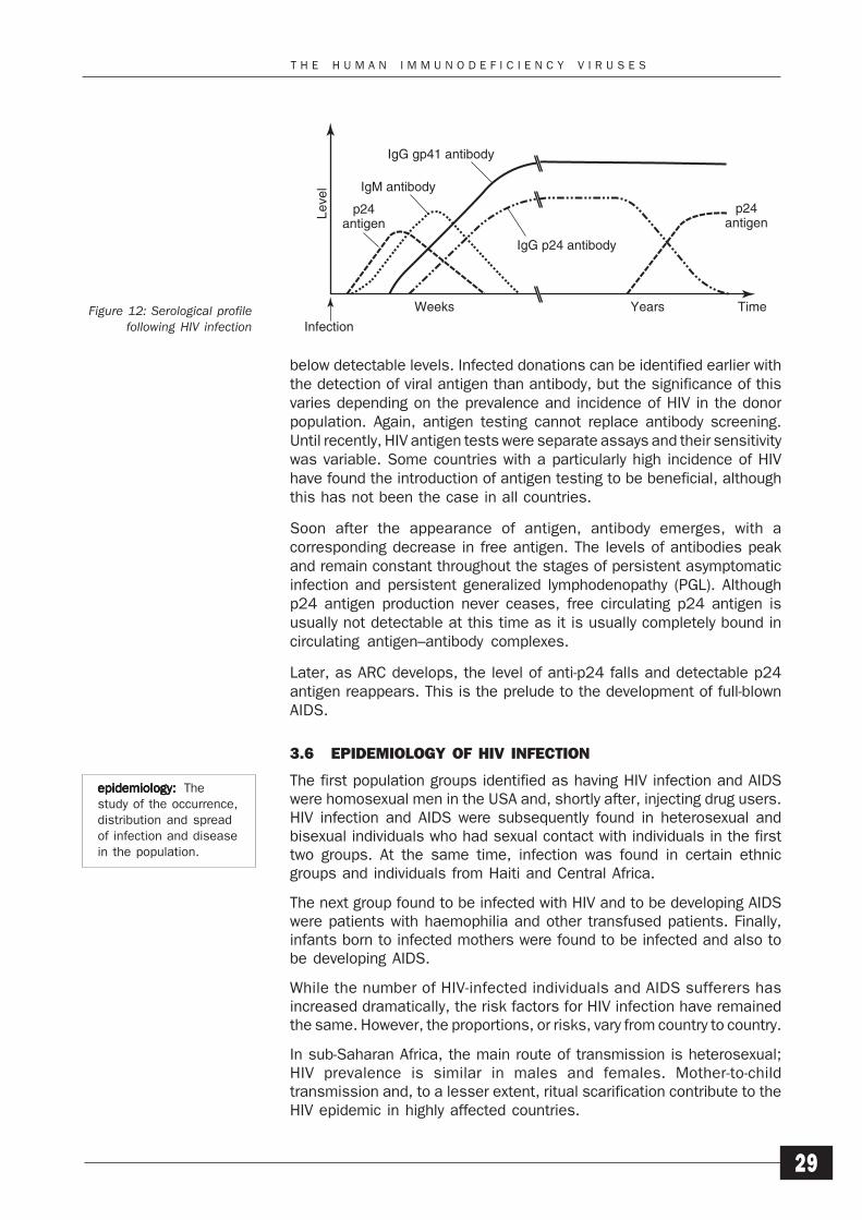
Figure 12 shows the laboratory events following infection by HIV in moredetail. Viral RNA, the first marker of infection to appear, reflects thereplication of virus and its release into the bloodstream. The detectionof viral RNA is still a relatively complex procedure and significantinvestment and infrastructure is required to be able to perform it reliablyand consistently and with the sensitivity required. In addition, RNA levelsfluctuate and testing cannot replace conventional virus serology.
After the appearance of RNA, viral antigen (p24) appears around thesame time as viral DNA. The DNA is generally not found free in theplasma, but is cell-associated. The viral antigen levels rise rapidly butthen peak and start to fall off again as antibody is produced andcomplexes with it; production does not cease but free antibody falls to
Figure 11: Appearance ofcirculating markers of HIV
infection
Point ofPoint ofPoint ofPoint ofPoint ofinfectioninfectioninfectioninfectioninfection RNARNARNARNARNA A gA gA gA gA g AbAbAbAbAb
0 15–25 26–30 31–40
Window 1 Window 2
Approximate number of days
surrogate testingsurrogate testingsurrogate testingsurrogate testingsurrogate testing: : : : : Testingfor an indicator of infectionwhich is thought toindicate the presence ofan infectious agent, butwhich is not a specificmarker of infection by thatagent.
seroconversion:seroconversion:seroconversion:seroconversion:seroconversion: A changein serostatus of anindividual fromseronegative toseropositive.
serostatusserostatusserostatusserostatusserostatus: Theserological findings in anindividual followinginfection.
“window periodwindow periodwindow periodwindow periodwindow period”: Theperiod between infectionand the first appearanceof circulating detectablemarker of that infection.
T H E H U M A N I M M U N O D E F I C I E N C Y V I R U S E S
29
below detectable levels. Infected donations can be identified earlier withthe detection of viral antigen than antibody, but the significance of thisvaries depending on the prevalence and incidence of HIV in the donorpopulation. Again, antigen testing cannot replace antibody screening.Until recently, HIV antigen tests were separate assays and their sensitivitywas variable. Some countries with a particularly high incidence of HIVhave found the introduction of antigen testing to be beneficial, althoughthis has not been the case in all countries.
Soon after the appearance of antigen, antibody emerges, with acorresponding decrease in free antigen. The levels of antibodies peakand remain constant throughout the stages of persistent asymptomaticinfection and persistent generalized lymphodenopathy (PGL). Althoughp24 antigen production never ceases, free circulating p24 antigen isusually not detectable at this time as it is usually completely bound incirculating antigen–antibody complexes.
Later, as ARC develops, the level of anti-p24 falls and detectable p24antigen reappears. This is the prelude to the development of full-blownAIDS.
3.6 EPIDEMIOLOGY OF HIV INFECTION
The first population groups identified as having HIV infection and AIDSwere homosexual men in the USA and, shortly after, injecting drug users.HIV infection and AIDS were subsequently found in heterosexual andbisexual individuals who had sexual contact with individuals in the firsttwo groups. At the same time, infection was found in certain ethnicgroups and individuals from Haiti and Central Africa.
The next group found to be infected with HIV and to be developing AIDSwere patients with haemophilia and other transfused patients. Finally,infants born to infected mothers were found to be infected and also tobe developing AIDS.
While the number of HIV-infected individuals and AIDS sufferers hasincreased dramatically, the risk factors for HIV infection have remainedthe same. However, the proportions, or risks, vary from country to country.
In sub-Saharan Africa, the main route of transmission is heterosexual;HIV prevalence is similar in males and females. Mother-to-childtransmission and, to a lesser extent, ritual scarification contribute to theHIV epidemic in highly affected countries.
Figure 12: Serological profilefollowing HIV infection
epidemiologyepidemiologyepidemiologyepidemiologyepidemiology: : : : : Thestudy of the occurrence,distribution and spreadof infection and diseasein the population.
Leve
lInfection
Weeks
p24antigen
p24antigen
IgM antibody
IgG gp41 antibody
IgG p24 antibody
Years Time
S E C T I O N 3
30
Blood transfusion is a potentially significant route of transmission in allcountries, although the risk may be reduced by the selection of donorsbefore donation and the screening of the donated blood.
An important factor to be taken into account when considering theprevalence of both HIV infection and AIDS is that, for many reasons, thereis still considerable under-reporting in many parts of the world.
ACTIVITY 10
Make a list of any reasons for possible under-reporting of HIVinfection and AIDS in your country. Consider the following reasons:
economic: your country may be unable to afford to test
political: your country may be unwilling to test because anyaction subsequently required may overload its health caresystem
practical: it may not be possible to set up a suitable samplecollection programme that is sufficiently representative ofthe population
technical: the expertise may not exist in your country forwide-scale testing to be performed with any reliability
cultural: individuals may be unwilling to be tested.
Note that all these reasons are interrelated.
3.7 TRANSMISSION OF HIV INFECTION
The modes of transmission of HIV infection are now well established.While virus can be isolated from many body secretions, infection istransmitted in only a limited number of ways.
There are three principal modes of transmission of HIV infection.
1 Unprotected penetrative sexual contact with an infectedperson, either between men or between men and women.
2 Inoculation of infected blood, either by blood transfusionor as the result of the use of contaminated needles,syringes or knives used, for example, in injecting druguse, ritual scarification or tattooing.
3 From an infected mother to her child, in the uterus, duringbirth or by breastfeeding.
ACTIVITY 11
Which of these three routes is the main route of transmission in yourcountry and why?
T H E H U M A N I M M U N O D E F I C I E N C Y V I R U S E S
31
The transmission of HIV by sexual contact is an extremely importantroute. In Africa, the effects of heterosexual transmission can be clearlyseen and are increasing as women who become infected pass on theirinfection to any children they have. Thus, infection of the populationincreases from two sides:
in adults (horizontal transmission)
in infants (vertical transmission).
In this way, HIV can spread very quickly in a population.
As you will remember from Section 2, blood transfusion can be asignificant route of infection. The efficiency of the transmission of HIVthrough blood transfusion is estimated to be virtually 100%. WHO reportsthat the viral dose in HIV transmission through blood is so large that oneHIV-positive transfusion leads to death, on the average, after two yearsin children and after three to five years in adults. Nevertheless, the extentto which blood transfusion is an actual route of transmission dependson the prevalence of infected individuals in the population and on theeffectiveness of the screening programme used. In a population with alow prevalence of infected individuals and a good screening programme,transmission by blood transfusion should be extremely rare; transfusionwould therefore not be a significant route of transmission. In a populationwith a high prevalence of infected individuals and a poor or nonexistentscreening programme, transmission by blood transfusion is likely to berelatively common and would be an important route of infection in thepopulation.
The following activity should help you to understand the potentialsignificance of transfusion-transmitted HIV infection.
ACTIVITY 12
A recently-married, previously healthy, HIV-negative young man has anaccident and is taken to hospital. There he receives a transfusion oftwo units of whole blood and recovers normally. HIV testing is notperformed on donated blood at the hospital and one of the units wasanti-HIV positive. What are the likely consequences of this situation,both short-term and long-term?
Check your answer with that given in the Activity Checklists andAnswers on pages 141–142.
This activity highlights the importance of testing as it is clear that bloodtransfusion can spread HIV infection very widely if blood is notsystematically screened.
While we are looking at the routes by which infection is transmitted, weshould clarify that there are a number of potential infection routes thatare not implicated in the transmission of HIV or AIDS.
1 There is no evidence that nonsexual, social or domesticcontact leads to the transmission of infection. Indeed, thevirus itself is not very stable and can easily be destroyedby environmental conditions.

S E C T I O N 3
32
2 There is no evidence that infection is spread by any insectvector in Africa or any other part of the world. Importantly,there is no evidence that HIV replicates in any arthropods;in fact, the evidence appears to be against any suchtransmission.
If insect transmission did occur, a higher incidence of HIVinfection in otherwise low-risk individuals should be seenin areas with a high incidence of insect-borne infection,such as malaria. This is not the case.
3 There is no evidence that health care workers have agreater risk of infection than any other individual with noother specific risk factors. If the correct safety proceduresfor handling pathological material are applied at all times,and good laboratory practice is followed, any risks areminimized.
3.8 PREVENTING THE SPREAD OF HIV INFECTION
The transmission of infectious diseases has been a problem for mankindfor hundreds, if not thousands, of years; epidemics of different kindshave swept across the world at various times in history. In order to findeffective ways of preventing the further spread of an infection, it is crucialto identify the routes of transmission.
The prevention of the spread of HIV infection essentially relies on twoapproaches that are closely interlinked and that are both related to theprovision of a safe blood supply:
education to enable people to avoid situations in whichthere is a risk of transmission occurring
the physical prevention of infection.
Let us now look at some ways in which the transmission of HIV infectioncan be prevented or reduced.
Sexual transmission
The sexual transmission of HIV infection can be dramatically reduced bythe avoidance of unsafe sexual practices, the use of condoms and areduction in the number of sexual partners. These measures have beenshown to be very effective in slowing the spread of infection amonghomosexual men. Among heterosexual contacts, the simple use ofcondoms has helped to reduce infection rates.
Transmission by injecting drug use
Transmission among injecting drug users can be reduced by the use ofdisposable syringes and needles and through schemes where usedsyringes are exchanged for new ones. Unfortunately, this approach israrely feasible or practical. Whilst disposable syringes can be madefreely available in some countries, in others they cannot even be providedfor clinical use because of their cost. Furthermore, as the use of
T H E H U M A N I M M U N O D E F I C I E N C Y V I R U S E S
33
injectable drugs is illegal in most countries, injecting drug users aregenerally reluctant to come forward to obtain clean equipment.
Transmission by blood transfusion
Transmission by blood transfusion, or the infusion of blood products, canalso be avoided. The first approach to the prevention of transmission bytransfusion is the selection of donors who are at low risk for transfusion-transmissible infections. Remember that a safe donor provides a saferdonation.
Module 1 focuses in detail on safe blood donation and, in particular, onthe importance of:
identifying low-risk donor groups
avoiding unsuitable blood donors
recruiting voluntary non-remunerated blood donors
promoting self-exclusion by individuals at risk through aneffective donor education programme
effective donor screening through:
— predonation counselling, including an assessment ofrisk factors and an opportunity for self-exclusion orconfidential unit exclusion
— a brief medical history, including possible signs andsymptoms related to transfusion-transmissibleinfections
— a basic health check, including a brief examination ofthe arm for needle marks
promoting regular, voluntary non-remunerated blooddonation.
Self-exclusion is probably the most effective approach in preventingtransmission, but is dependent on the education of potential donorsabout risk behaviour. It is particularly important to encourage self-exclusion by people such as sex workers, homosexual and bisexual men,injecting drug users, those who have any unprotected sexual contactother than with a regular partner, and the sexual contacts of any of thesepeople.
Ultimately, however, screening tests for anti-HIV are needed to enableinfected donors to be identified and the donated blood to be discarded.We shall look at this in more detail in Section 4.
ACTIVITY 13
Briefly list the various approaches that have been taken in yourcountry to reduce the spread of HIV infection and its transmission byblood transfusion.
Find out what advice is given in your country about the transmissionof HIV by sexual routes. Are condoms widely available? If so, havethey been effective or are people generally reluctant to use them?

S E C T I O N 3
34
Paediatric infection
The transmission of HIV from mother to child is a significant route ofinfection. Transmission may occur by infection of the developing fetusduring pregnancy, by infection of the infant at birth through virus presentin cervical secretions and via breastfeeding.
The prevention of mother-to-child transmission is a very complex areaand it is not appropriate to include it in this module.
SUMMARY
1 HIV is the primary cause of AIDS. HIV damages part of theimmune system, which can lead to serious infections byagents that would normally be easily overcome by theimmune system.
2 HIV is a retrovirus and integrates into the host cellgenome. The virus consists of a number of structuralproteins, but p24, gp41 and gp120/160 are in generalthe most immunogenic and are therefore among the mostimportant in virus serology. HIV primarily infects anddestroys cells of the immune system, mainly infectingCD4+ positive cells.
3 In individuals suffering from AIDS, the main cause ofillness is the occurrence of secondary infectious diseases.
4 The spread of HIV infection has followed different patternsin different countries.
5 There is considerable under-reporting of the prevalence ofHIV infection and AIDS in some countries.
6 The transmission of HIV through blood transfusion ismore than 90% efficient.
7 The prevention of the spread of HIV infection essentiallyrelies on two approaches:
public education to reduce risk behaviour
the physical prevention of infection.
SELF-ASSESSMENT
9 How does HIV enter susceptible cells?
10 What are the two HIV-specific antibodies that have beenfound to be the best confirmation of HIV infection?
11 How long after infection will antibodies first appear?
12 What are the three main routes of transmission for HIVinfection?
13 What are the three other routes of transmission of infectionthat are not implicated in HIV transmission?
T H E H U M A N I M M U N O D E F I C I E N C Y V I R U S E S
35
PROGRESS CHECK
Before moving on to Section 4, spend a few minutes thinkingabout whether you have achieved the learning objectives forSection 3. These were to:
1 Identify the structural features of HIV.
2 Identify the basic stages of HIV infection and the entry ofthe virus into susceptible cells.
3 Describe the most common clinical course of HIV infectionand its progression to AIDS in your country.
4 Explain the measures that are being taken to reduce thetransmission of HIV infection in your country.
If you feel confident that you have understood everything inthis section, turn to Section 4.
If you feel that you need to spend more time on this section,go back to the parts that are most unfamiliar or that you havefound difficult. You may find it helpful to talk to other people,such as your supporter or other senior colleagues, if there isanything you are still not sure about.
S E C T I O N 4
36
4Principles of ScreeningAssays for Transfusion-Transmissible Infections
The purpose of this section is to examine the possible approaches toscreening for transfusion-transmissible infections (TTIs) and to explainthe principles behind the different types of assay available.
The section contains a considerable amount of information which youmay wish to use as reference material in the future. At this stage,however, we suggest that you read through the text, concentrating on theassays to which you have access but completing all the activities.
LEARNING OBJECTIVES
When you have completed this section, you should be able to:
1 Outline the principles of the diagnostic assays mostcommonly used to detect the specific TTI markers used toidentify infected donations.
2 Explain the differences between the main types ofscreening assay.
P R I N C I P L E S O F S C R E E N I N G A S S A Y S F O R T T I s
37
4.1 SCREENING BLOOD DONATIONS FOR TTIs
In considering the screening of blood donations and the specificscreening tests that are available to detect TTIs, it is important toremember that a safe donor provides a safer donation. As Module 1emphasizes, the safest donors are regular, voluntary and non-remunerated. Clearly, donors specifically at risk of any blood-borneinfection (or any other infectious diseases) should be encouraged not todonate blood.
There are a number of other specific points to remember.
1 There is a risk of transmission of infection if donatedblood is not tested before the blood is transfused.
2 The collection of a unit of blood from an infected donorwastes precious resources in terms of consumables andstaff time.
3 If many TTI-positive donations are found, the number ofrepeat tests and confirmatory tests needed increases.This raises the total cost of testing.
An effective donor education and selection programme that promotesself-exclusion by donors at risk of transfusion-transmissible infectionstherefore makes your job a great deal easier, saves time and money andalso results in a safer donor population.
4.2 TYPES OF SCREENING ASSAY
The choice of specific markers to screen for depends on the infectiousagent that you are screening for. Screening for specific antibody isusually most appropriate for HIV, HCV and syphilis. Screening for specificantigen (HBsAg) is necessary for HBV. This section is concerned with theprinciples of the assays most commonly used for the screening of blooddonations: for both antibody and antigen.
Three main kinds of primary screening assay are available to detectinfectious disease markers in donated blood:
1 Enzyme Linked ImmunoSorbent Assays (ELISA)/EnzymeImmunoAssay (EIA).
2 Particle agglutination assays.
3 Simple rapid assays.
Note that the abbreviation ELISA is often replaced with the abbreviationEIA. This is a simpler description which we shall use throughout theremainder of the module.
There are a number of features that need to be taken into account whenselecting the most appropriate assay to use:
scientific principle of the assay
complexity of the assay
incubation times
sensitivity
specificity
suitability for different situations
S E C T I O N 4
38
availability
cost.
ACTIVITY 14
List the assays used in your laboratory when screening donated bloodfor TTIs. Note whether they are EIA, particle agglutination or simplerapid assays.
All three types of assay are based on the same biological principle andinvolve the same two basic elements.
1 The presence of specific antigen or antibody in a sampleis demonstrated in an immunological reaction involvingantigen/antibody (immune) complex formation, with oneof the components bound to a solid phase.
2 The formation of the immune complex is subsequentlydetected by an indicator system.
Test samples
Most of the assays in use today can be used with either serum or plasma.However, there are still some that work with serum only. The instructionswith the assay must be followed.
Terminology
Before we consider the assays in detail, it is important to be clear aboutthe use of the following terms in classifying the actual screening results:
positive/negative
reactive/non-reactive
equivocal/indeterminate.
Although the terms “positive” and “negative” are the main terminologyused in this module, this is for reasons of simplicity. Many people usethese words to report screening results, but it is not really correct to doso. “Positive” and “negative” should really be used only to describe thefinal status of the donation or the donor. This is really of minorimportance in the case of “non-reactive” screening results as long as itis clearly understood that only when all testing is completed and noevidence of infection is found should the overall donation status bedescribed as “negative”. In the case of “positive” screening test results,however, the results should be classified as “reactive” until they havebeen confirmed. The reason for this is that frequently a high percentageof the screen “reactive” results are nonspecific and are finally confirmedas “negative”. There are no real fixed rules concerning the use of thesewords, but you should be aware of the differences in meaning andsignificance.
The WHO terminology for a result that cannot be classified as clearlypositive or negative (usually around the cut-off value) is equivocal.equivocal.equivocal.equivocal.equivocal.
equivocal/indeterminate:equivocal/indeterminate:equivocal/indeterminate:equivocal/indeterminate:equivocal/indeterminate:A result that cannot beclassified as clearlypositive or negative.
P R I N C I P L E S O F S C R E E N I N G A S S A Y S F O R T T I s
39
ACTIVITY 15
List the words that are used in your laboratory to classify screeningresults. Check the instructions from the assay manufacturers to seewhat words they use. If you find that different terms are used, youneed to decide on the terminology to be used in your laboratory inorder to establish uniformity and avoid confusion. Discuss this withyour supervisor and colleagues. Note down your recommendations onyour Action List.
4.3 ENZYME IMMUNOASSAYS
Although EIA is the most complex of the three types of assay, we shallconsider it first since the principles involved are basically the same asin the other two types. Once you understand the principles of EIA,therefore, you should quickly be able to understand the principles ofparticle agglutination and simple rapid assays.
EIAs exist in many forms and can be used to detect both antigen andantibody. In general, the simplest and most commonly used antibodydetection assays are based on the use of immobilized antigen whichcaptures any specific antibody present in the test sample. The mostcommonly used antigen detection assays are based on the use ofimmobilized antibody.
There are a number of different presentations of EIA using different solidphases to immobilize the antigen or antibody. Most commonly the solidphases currently in use are:
1 The bottom and sides of the inside of a polystyrenemicrowell.
2 The surface of polystyrene, or other material,microparticles.
3 The surfaces of specific, dedicated, disposable devicesused in automated, self-contained assay systems. Thesevary according to the manufacturer, but are usuallypolystyrene and generally work on the same basicprinciples.
4 Strips of nylon or nitrocellulose membrane. These arespecifically used in western blots and line assays.
Four main types of EIA are commonly used in blood screening: threespecifically to detect antibody and one that can be used to detectantibody or antigen, depending on the specific design format of theassay.
To help you understand the basic principles of EIAs, we shall consider thefour basic types of EIA in the microwell format. However, these types canjust as easily be presented on microparticles, dedicated systems or stripformats. Indeed, today, these four types may be found in a wide rangeof commercial assays using a variety of different solid phases and in

S E C T I O N 4
40
Antigen
different formats. In addition, apart from assays specifically manufacturedto work on a specific, dedicated, automated system (closed systemassays), assays may be performed manually or on non-dedicated,automated assay processing systems (open system assays).
1 Antiglobulin EIA: this is the simplest form of EIA, in whichany specific antibody present in a test sample is bound toimmobilized specific antigen and is detected by enzyme-labelled anti-human antibody.
2 Competitive EIA: this is a slightly more complex assay, inwhich specific antibody that may be present in a testsample competes with enzyme-labelled specific antibodyfor binding sites on immobilized specific antigen.
3 Sandwich EIA: this is a highly specific type of EIA whichcan be used in either antibody or antigen sandwichformat. Specific antibody (or antigen) in the test sampleis bound to immobilized specific antigen (or antibody) andthen detected by enzyme-labelled specific antigen (orantibody).
4 Antibody capture EIA: this is a type of EIA in which anyantibody in the test sample, irrespective of specificity, iscaptured by immobilized anti-human immunoglobulins(IgG) and any captured specific antibody is detected byenzyme-labelled specific antigen.
The descriptions that follow explain the principles of the assays. You mayfind, however, that they differ from the manufacturers’ instructions. Youshould always follow the instructions supplied with the assays.
Type 1: Antiglobulin EIA (screening for specific antibody:e.g. anti-HIV)
Methodology1 The basis of the simplest form of antibody immunoassay
is an immobilized antigen (see Figure 13) which is boundto the surface of the microwell.
Figure 13
2 This binding can be achieved simply by adding a solutionof antigen in bicarbonate buffer to each well and incubatingat 4°C for 12–16 hours. The plastic used to produce themicrowells, polystyrene, is designed to bind proteins.After incubation, the antigen solution is removed, thewells are washed with distilled water or a specific bufferand are immediately dried. They are then stored at 4°C,often for long periods, without any loss of antigen. Theseplates are provided by the manufacturer, ready-coatedand standardized for use.
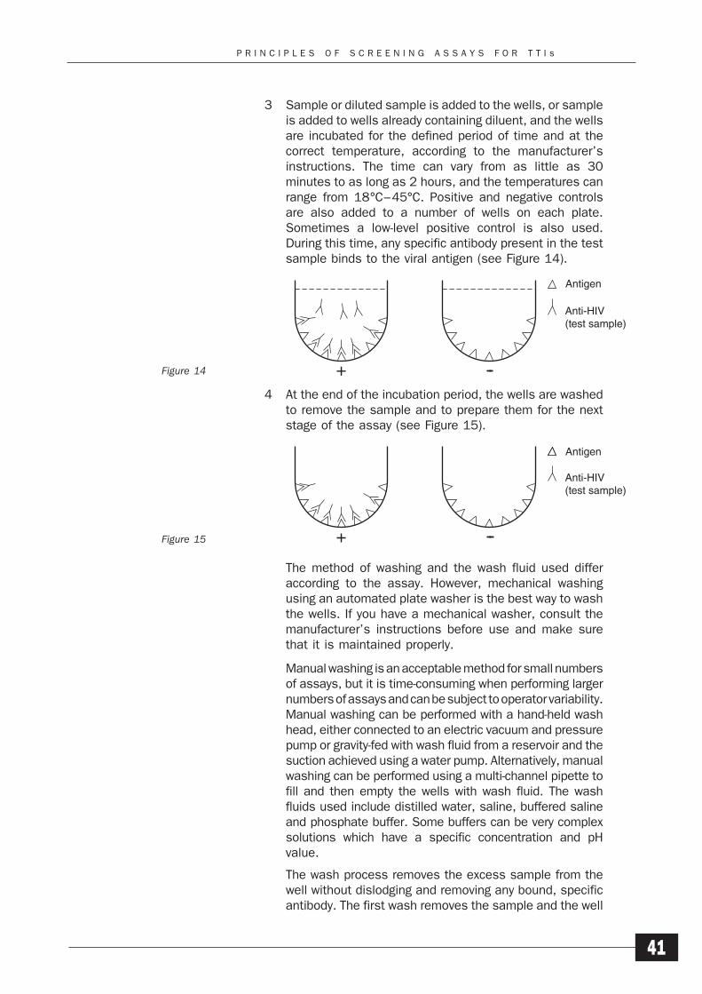
P R I N C I P L E S O F S C R E E N I N G A S S A Y S F O R T T I s
41
3 Sample or diluted sample is added to the wells, or sampleis added to wells already containing diluent, and the wellsare incubated for the defined period of time and at thecorrect temperature, according to the manufacturer’sinstructions. The time can vary from as little as 30minutes to as long as 2 hours, and the temperatures canrange from 18°C–45°C. Positive and negative controlsare also added to a number of wells on each plate.Sometimes a low-level positive control is also used.During this time, any specific antibody present in the testsample binds to the viral antigen (see Figure 14).
4 At the end of the incubation period, the wells are washedto remove the sample and to prepare them for the nextstage of the assay (see Figure 15).
The method of washing and the wash fluid used differaccording to the assay. However, mechanical washingusing an automated plate washer is the best way to washthe wells. If you have a mechanical washer, consult themanufacturer’s instructions before use and make surethat it is maintained properly.
Manual washing is an acceptable method for small numbersof assays, but it is time-consuming when performing largernumbers of assays and can be subject to operator variability.Manual washing can be performed with a hand-held washhead, either connected to an electric vacuum and pressurepump or gravity-fed with wash fluid from a reservoir and thesuction achieved using a water pump. Alternatively, manualwashing can be performed using a multi-channel pipette tofill and then empty the wells with wash fluid. The washfluids used include distilled water, saline, buffered salineand phosphate buffer. Some buffers can be very complexsolutions which have a specific concentration and pHvalue.
The wash process removes the excess sample from thewell without dislodging and removing any bound, specificantibody. The first wash removes the sample and the well
Figure 14
Figure 15
Antigen
Anti-HIV(test sample)
Antigen
Anti-HIV(test sample)
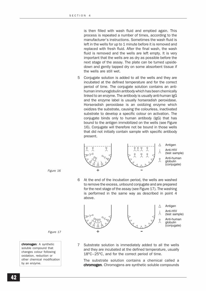
S E C T I O N 4
42
is then filled with wash fluid and emptied again. Thisprocess is repeated a number of times, according to themanufacturer’s instructions. Sometimes the wash fluid isleft in the wells for up to 1 minute before it is removed andreplaced with fresh fluid. After the final wash, the washfluid is removed and the wells are left empty. It is veryimportant that the wells are as dry as possible before thenext stage of the assay. The plate can be turned upside-down and gently tapped dry on some absorbent tissue ifthe wells are still wet.
5 Conjugate solution is added to all the wells and they areincubated at the defined temperature and for the correctperiod of time. The conjugate solution contains an anti-human immunoglobulin antibody which has been chemicallylinked to an enzyme. The antibody is usually anti-human IgGand the enzyme label is usually horseradish peroxidase.Horseradish peroxidase is an oxidizing enzyme whichoxidizes the substrate, causing the colourless, inactivatedsubstrate to develop a specific colour on activation. Theconjugate binds only to human antibody (IgG) that hasbound to the antigen immobilized on the wells (see Figure16). Conjugate will therefore not be bound in those wellsthat did not initially contain sample with specific antibodypresent.
6 At the end of the incubation period, the wells are washedto remove the excess, unbound conjugate and are preparedfor the next stage of the assay (see Figure 17). The washingis performed in the same way as described in point 4above.
7 Substrate solution is immediately added to all the wellsand they are incubated at the defined temperature, usually18°C–25°C, and for the correct period of time.
The substrate solution contains a chemical called achromogenchromogenchromogenchromogenchromogen. Chromogens are synthetic soluble compounds
chromogenchromogenchromogenchromogenchromogen: A syntheticsoluble compound thatchanges colour followingoxidation, reduction orother chemical modificationby an enzyme.
Figure 16
Figure 17
Antigen
Anti-HIV(test sample)
Anti-humanglobulin(conjugate)
Antigen
Anti-HIV(test sample)
Anti-humanglobulin(conjugate)
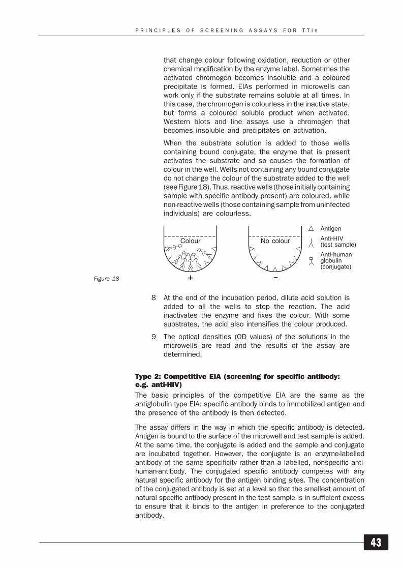
P R I N C I P L E S O F S C R E E N I N G A S S A Y S F O R T T I s
43
that change colour following oxidation, reduction or otherchemical modification by the enzyme label. Sometimes theactivated chromogen becomes insoluble and a colouredprecipitate is formed. EIAs performed in microwells canwork only if the substrate remains soluble at all times. Inthis case, the chromogen is colourless in the inactive state,but forms a coloured soluble product when activated.Western blots and line assays use a chromogen thatbecomes insoluble and precipitates on activation.
When the substrate solution is added to those wellscontaining bound conjugate, the enzyme that is presentactivates the substrate and so causes the formation ofcolour in the well. Wells not containing any bound conjugatedo not change the colour of the substrate added to the well(see Figure 18). Thus, reactive wells (those initially containingsample with specific antibody present) are coloured, whilenon-reactive wells (those containing sample from uninfectedindividuals) are colourless.
8 At the end of the incubation period, dilute acid solution isadded to all the wells to stop the reaction. The acidinactivates the enzyme and fixes the colour. With somesubstrates, the acid also intensifies the colour produced.
9 The optical densities (OD values) of the solutions in themicrowells are read and the results of the assay aredetermined.
Type 2: Competitive EIA (screening for specific antibody:e.g. anti-HIV)
The basic principles of the competitive EIA are the same as theantiglobulin type EIA: specific antibody binds to immobilized antigen andthe presence of the antibody is then detected.
The assay differs in the way in which the specific antibody is detected.Antigen is bound to the surface of the microwell and test sample is added.At the same time, the conjugate is added and the sample and conjugateare incubated together. However, the conjugate is an enzyme-labelledantibody of the same specificity rather than a labelled, nonspecific anti-human-antibody. The conjugated specific antibody competes with anynatural specific antibody for the antigen binding sites. The concentrationof the conjugated antibody is set at a level so that the smallest amount ofnatural specific antibody present in the test sample is in sufficient excessto ensure that it binds to the antigen in preference to the conjugatedantibody.
Figure 18
Antigen
Anti-HIV(test sample)
Anti-humanglobulin(conjugate)
Colour No colour
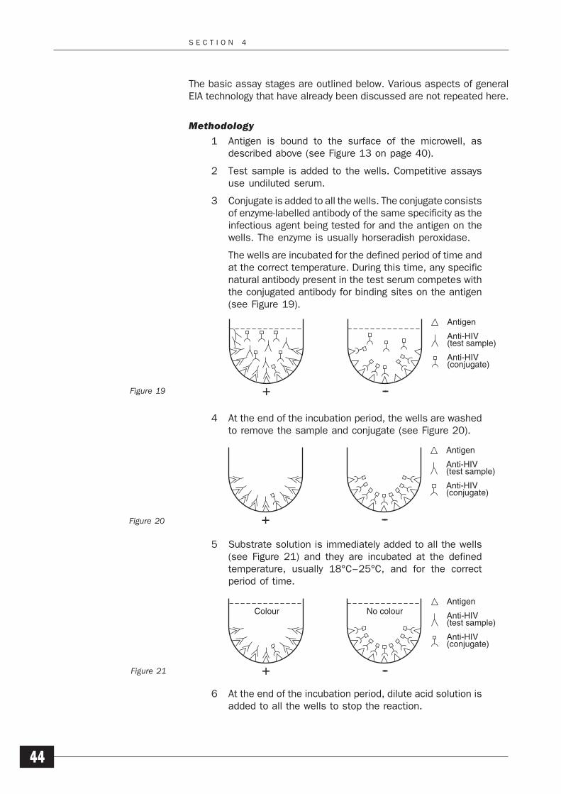
S E C T I O N 4
44
The basic assay stages are outlined below. Various aspects of generalEIA technology that have already been discussed are not repeated here.
Methodology
1 Antigen is bound to the surface of the microwell, asdescribed above (see Figure 13 on page 40).
2 Test sample is added to the wells. Competitive assaysuse undiluted serum.
3 Conjugate is added to all the wells. The conjugate consistsof enzyme-labelled antibody of the same specificity as theinfectious agent being tested for and the antigen on thewells. The enzyme is usually horseradish peroxidase.
The wells are incubated for the defined period of time andat the correct temperature. During this time, any specificnatural antibody present in the test serum competes withthe conjugated antibody for binding sites on the antigen(see Figure 19).
4 At the end of the incubation period, the wells are washedto remove the sample and conjugate (see Figure 20).
5 Substrate solution is immediately added to all the wells(see Figure 21) and they are incubated at the definedtemperature, usually 18°C–25°C, and for the correctperiod of time.
6 At the end of the incubation period, dilute acid solution isadded to all the wells to stop the reaction.
Figure 20
Figure 19
Figure 21
Antigen
Anti-HIV(test sample)
Anti-HIV(conjugate)
Antigen
Anti-HIV(test sample)
Anti-HIV(conjugate)
No colourColourAntigen
Anti-HIV(test sample)
Anti-HIV(conjugate)
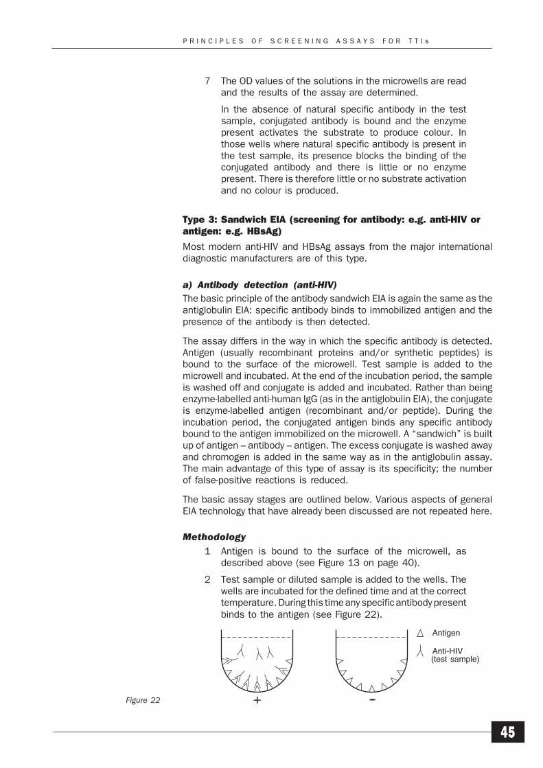
P R I N C I P L E S O F S C R E E N I N G A S S A Y S F O R T T I s
45
7 The OD values of the solutions in the microwells are readand the results of the assay are determined.
In the absence of natural specific antibody in the testsample, conjugated antibody is bound and the enzymepresent activates the substrate to produce colour. Inthose wells where natural specific antibody is present inthe test sample, its presence blocks the binding of theconjugated antibody and there is little or no enzymepresent. There is therefore little or no substrate activationand no colour is produced.
Type 3: Sandwich EIA (screening for antibody: e.g. anti-HIV orantigen: e.g. HBsAg)
Most modern anti-HIV and HBsAg assays from the major internationaldiagnostic manufacturers are of this type.
a) Antibody detection (anti-HIV)The basic principle of the antibody sandwich EIA is again the same as theantiglobulin EIA: specific antibody binds to immobilized antigen and thepresence of the antibody is then detected.
The assay differs in the way in which the specific antibody is detected.Antigen (usually recombinant proteins and/or synthetic peptides) isbound to the surface of the microwell. Test sample is added to themicrowell and incubated. At the end of the incubation period, the sampleis washed off and conjugate is added and incubated. Rather than beingenzyme-labelled anti-human IgG (as in the antiglobulin EIA), the conjugateis enzyme-labelled antigen (recombinant and/or peptide). During theincubation period, the conjugated antigen binds any specific antibodybound to the antigen immobilized on the microwell. A “sandwich” is builtup of antigen – antibody – antigen. The excess conjugate is washed awayand chromogen is added in the same way as in the antiglobulin assay.The main advantage of this type of assay is its specificity; the numberof false-positive reactions is reduced.
The basic assay stages are outlined below. Various aspects of generalEIA technology that have already been discussed are not repeated here.
Methodology1 Antigen is bound to the surface of the microwell, as
described above (see Figure 13 on page 40).
2 Test sample or diluted sample is added to the wells. Thewells are incubated for the defined time and at the correcttemperature. During this time any specific antibody presentbinds to the antigen (see Figure 22).
Figure 22
Antigen
Anti-HIV(test sample)
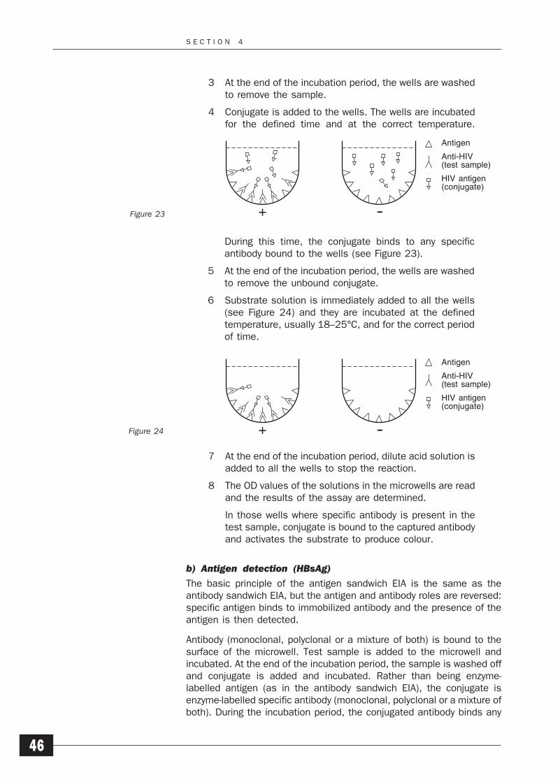
S E C T I O N 4
46
3 At the end of the incubation period, the wells are washedto remove the sample.
4 Conjugate is added to the wells. The wells are incubatedfor the defined time and at the correct temperature.
During this time, the conjugate binds to any specificantibody bound to the wells (see Figure 23).
5 At the end of the incubation period, the wells are washedto remove the unbound conjugate.
6 Substrate solution is immediately added to all the wells(see Figure 24) and they are incubated at the definedtemperature, usually 18–25°C, and for the correct periodof time.
7 At the end of the incubation period, dilute acid solution isadded to all the wells to stop the reaction.
8 The OD values of the solutions in the microwells are readand the results of the assay are determined.
In those wells where specific antibody is present in thetest sample, conjugate is bound to the captured antibodyand activates the substrate to produce colour.
b) Antigen detection (HBsAg)
The basic principle of the antigen sandwich EIA is the same as theantibody sandwich EIA, but the antigen and antibody roles are reversed:specific antigen binds to immobilized antibody and the presence of theantigen is then detected.
Antibody (monoclonal, polyclonal or a mixture of both) is bound to thesurface of the microwell. Test sample is added to the microwell andincubated. At the end of the incubation period, the sample is washed offand conjugate is added and incubated. Rather than being enzyme-labelled antigen (as in the antibody sandwich EIA), the conjugate isenzyme-labelled specific antibody (monoclonal, polyclonal or a mixture ofboth). During the incubation period, the conjugated antibody binds any
Figure 23
Figure 24
Antigen
Anti-HIV(test sample)
HIV antigen(conjugate)
Antigen
Anti-HIV(test sample)
HIV antigen(conjugate)

P R I N C I P L E S O F S C R E E N I N G A S S A Y S F O R T T I s
47
specific antigen bound to the antibody immobilized on the microwell. A“sandwich” is built up of antibody – antigen – antibody. The excessconjugate is washed away and chromogen is added in the same way asin the antibody sandwich assay.
The basic assay stages are outlined below. Various aspects of generalEIA technology that have already been discussed are not repeated here.
Methodology
1 Antibody is bound to the surface of the microwell in asimilar manner to antigen (see Figure 25).
2 Test sample or diluted sample is added to the wells. Thewells are incubated for the defined time and at the correcttemperature. During this time any specific antigen presentbinds to the antibody (see Figure 26).
5 At the end of the incubation period, the wells are washedto remove the unbound conjugate.
6 Substrate solution is immediately added to all the wells(see Figure 28 on page 48) and they are incubated at thedefined temperature, usually 18–25°C, and for the correctperiod of time.
3 At the end of the incubation period, the wells are washedto remove the sample.
4 Conjugate is added to the wells. The wells are incubatedfor the defined time and at the correct temperature.During this time, the conjugate binds to any specificantigen bound to the wells (see Figure 27).
Figure 25
Figure 26
Figure 27
AntibodyAntibody
Anti-HBs
Antigen
Anti-HBs
Antigen(test sample)
Anti-HBS
Antigen(test sample)
Anti-HBs(conjugate)
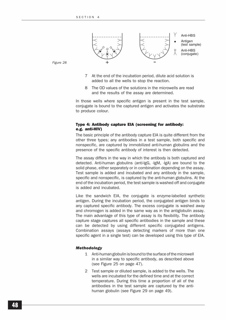
S E C T I O N 4
48
7 At the end of the incubation period, dilute acid solution isadded to all the wells to stop the reaction.
8 The OD values of the solutions in the microwells are readand the results of the assay are determined.
In those wells where specific antigen is present in the test sample,conjugate is bound to the captured antigen and activates the substrateto produce colour.
Type 4: Antibody capture EIA (screening for antibody:e.g. anti-HIV)
The basic principle of the antibody capture EIA is quite different from theother three types; any antibodies in a test sample, both specific andnonspecific, are captured by immobilized anti-human globulins and thepresence of the specific antibody of interest is then detected.
The assay differs in the way in which the antibody is both captured anddetected. Anti-human globulins (anti-IgG, -IgM, -IgA) are bound to thesolid phase, either separately or in combination depending on the assay.Test sample is added and incubated and any antibody in the sample,specific and nonspecific, is captured by the anti-human globulins. At theend of the incubation period, the test sample is washed off and conjugateis added and incubated.
Like the sandwich EIA, the conjugate is enzyme-labelled syntheticantigen. During the incubation period, the conjugated antigen binds toany captured specific antibody. The excess conjugate is washed awayand chromogen is added in the same way as in the antiglobulin assay.The main advantage of this type of assay is its flexibility. The antibodycapture stage captures all specific antibodies in the sample and thesecan be detected by using different specific conjugated antigens.Combination assays (assays detecting markers of more than onespecific agent in a single test) can be developed using this type of EIA.
Methodology
1 Anti-human globulin is bound to the surface of the microwellin a similar way to specific antibody, as described above(see Figure 25 on page 47).
2 Test sample or diluted sample, is added to the wells. Thewells are incubated for the defined time and at the correcttemperature. During this time a proportion of all of theantibodies in the test sample are captured by the anti-human globulin (see Figure 29 on page 49).
Figure 28
Anti-HBS
Antigen(test sample)
Anti-HBS(conjugate)
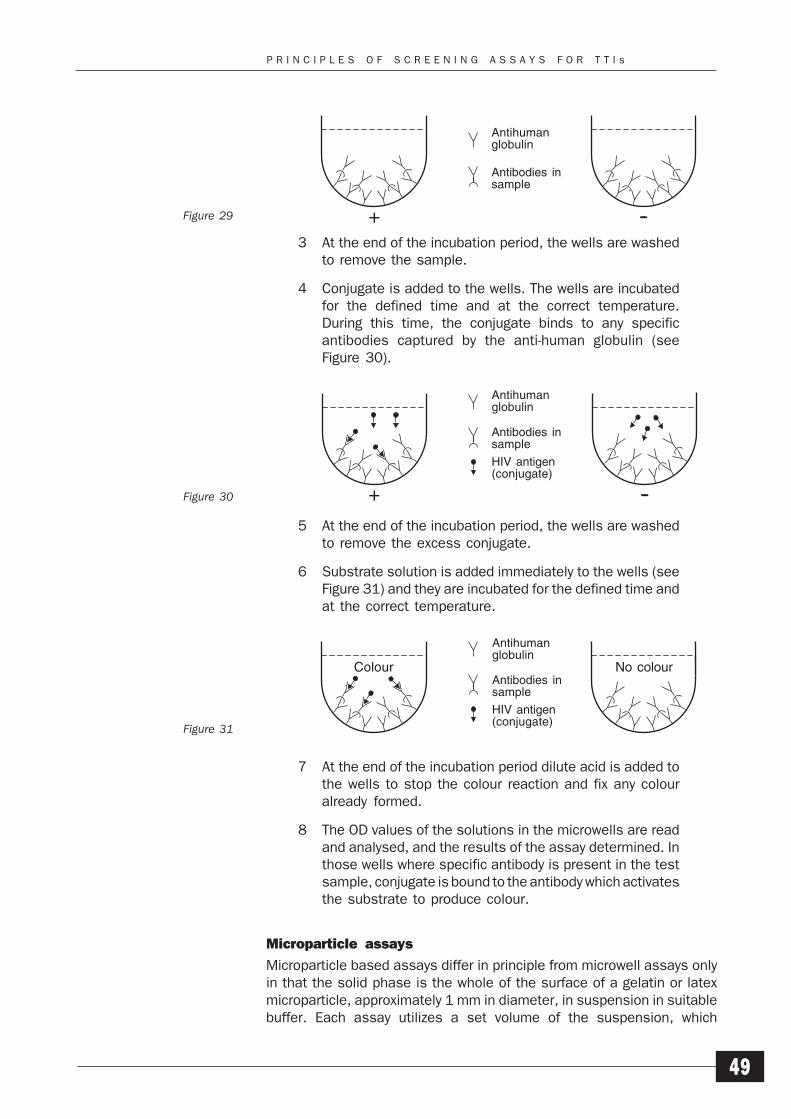
P R I N C I P L E S O F S C R E E N I N G A S S A Y S F O R T T I s
49
3 At the end of the incubation period, the wells are washedto remove the sample.
4 Conjugate is added to the wells. The wells are incubatedfor the defined time and at the correct temperature.During this time, the conjugate binds to any specificantibodies captured by the anti-human globulin (seeFigure 30).
5 At the end of the incubation period, the wells are washedto remove the excess conjugate.
6 Substrate solution is added immediately to the wells (seeFigure 31) and they are incubated for the defined time andat the correct temperature.
7 At the end of the incubation period dilute acid is added tothe wells to stop the colour reaction and fix any colouralready formed.
8 The OD values of the solutions in the microwells are readand analysed, and the results of the assay determined. Inthose wells where specific antibody is present in the testsample, conjugate is bound to the antibody which activatesthe substrate to produce colour.
Microparticle assays
Microparticle based assays differ in principle from microwell assays onlyin that the solid phase is the whole of the surface of a gelatin or latexmicroparticle, approximately 1 mm in diameter, in suspension in suitablebuffer. Each assay utilizes a set volume of the suspension, which
Figure 29
Figure 30
Figure 31
Antihumanglobulin
Antibodiesin sample
Antihumanglobulin
Antibodies insample
Antihumanglobulin
Antibodiesin sample
Antigenconjugate
Antihumanglobulin
Antibodies insample
HIV antigen(conjugate)
colour no colourAntihumanglobulin
Antibodiesin sample
Antigenconjugate
Colour No colour
Antihumanglobulin
Antibodies insample
HIV antigen(conjugate)
S E C T I O N 4
50
contains millions of these particles. This provides a large overall surfaceon which the assay reactions take place. Most microparticle assays aredesigned for dedicated systems where the microparticles are containedin small wells, which simply act as “test-tubes”, in which the reactiontakes place. The assay principles and methodology are broadly the sameas microwell EIAs. The Abbott PRISM and AxSYM are examples ofsystems that use microparticles.
Dedicated device/system assays
In addition to the PRISM and AxSYM dedicated assay systems mentionedabove, there are other systems that use basic EIA principles, but indifferent ways as part of dedicated systems. The bioMerieux VIDASsystem is an example of such a system. The solid phase is the insidesurface of a polystyrene tip and the sample and other solutions aresucked inside the tip and incubated there. All the reagents used in thissystem are provided in a sealed strip of wells, similar to microwells,which is loaded onto the machine at the start of the assay procedure.
Chemiluminescent assays
Although the substrate used in most microwell EIAs is a chromogen thatturns from a colourless to a coloured solution in the presence of thespecific enzyme catalyst, many microparticle assays and dedicatedassay systems now use a chemiluminescent substrate. Chemi-luminescence is the chemical generation of light at a certain wavelengthand is an enzyme-catalysed reaction that works in broadly the same wayas a chromogenic substrate. In this case, however, the conjugatedenzyme catalyses the generation of small quantities of light rather thana coloured solution. This type of assay may be more sensitive in somecases as the reaction does not need to be stopped at a certain time, themeasurement of light generated can be continuous and the readers candetect very low levels of emitted light. However, chemiluminescentassays need more specialized and expensive equipment (microplateformat luminometers) to detect the light generated and the overallbenefits of such assays are not significant in most situations. While anumber of commercial dedicated assay systems use this technology withsuccess, most of these systems are not ideal nor designed for bloodscreening.
Blots/line assays
Western blots and line assays are yet other presentations of the samebasic EIA principles, but in a different format. Blots and line assays areprimarily confirmatory assays which are used as part of appropriateconfirmatory algorithms to confirm reactivity: for example, in samplesfound reactive in screening assays. Most blots or line assays are for thedetection of antibody and the specific antigens are immobilized on a stripof nitrocellulose or nylon membrane. The assays are performed in smallindividual troughs which contain the strip and into which the sample/reagents/wash fluid are added at the appropriate time. A major differencebetween blots and other EIAs is that the chromogen used is not solublewhen activated by the enzyme conjugate. On activation, the chromogenforms a coloured precipitate at each position along the strip where theconjugate is bound.
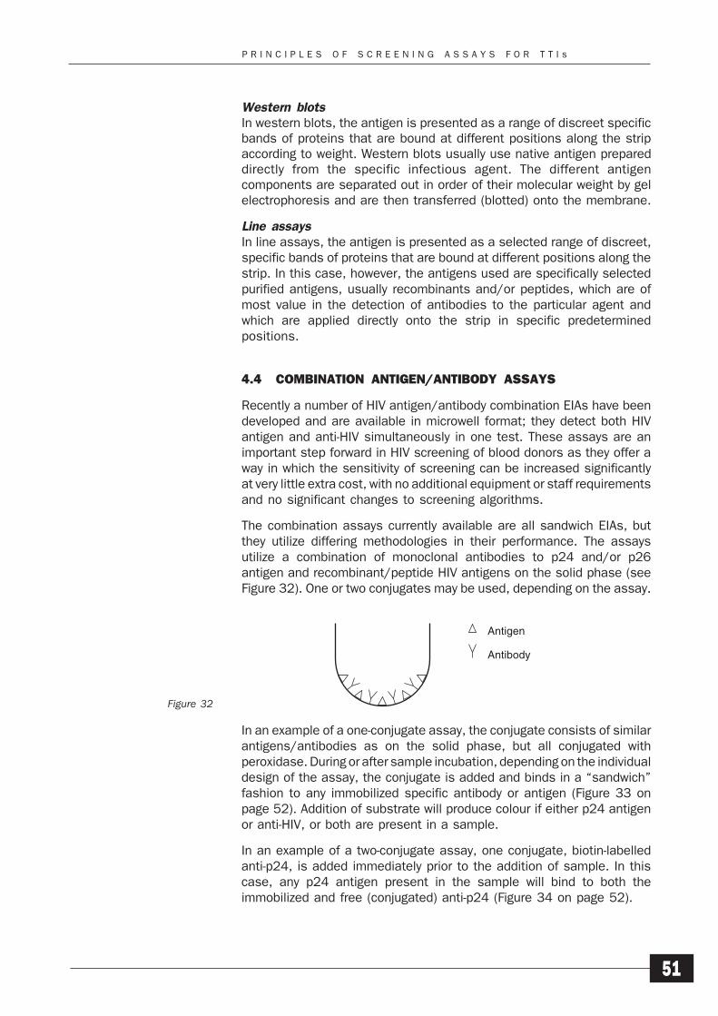
P R I N C I P L E S O F S C R E E N I N G A S S A Y S F O R T T I s
51
Western blotsIn western blots, the antigen is presented as a range of discreet specificbands of proteins that are bound at different positions along the stripaccording to weight. Western blots usually use native antigen prepareddirectly from the specific infectious agent. The different antigencomponents are separated out in order of their molecular weight by gelelectrophoresis and are then transferred (blotted) onto the membrane.
Line assaysIn line assays, the antigen is presented as a selected range of discreet,specific bands of proteins that are bound at different positions along thestrip. In this case, however, the antigens used are specifically selectedpurified antigens, usually recombinants and/or peptides, which are ofmost value in the detection of antibodies to the particular agent andwhich are applied directly onto the strip in specific predeterminedpositions.
4.4 COMBINATION ANTIGEN/ANTIBODY ASSAYS
Recently a number of HIV antigen/antibody combination EIAs have beendeveloped and are available in microwell format; they detect both HIVantigen and anti-HIV simultaneously in one test. These assays are animportant step forward in HIV screening of blood donors as they offer away in which the sensitivity of screening can be increased significantlyat very little extra cost, with no additional equipment or staff requirementsand no significant changes to screening algorithms.
The combination assays currently available are all sandwich EIAs, butthey utilize differing methodologies in their performance. The assaysutilize a combination of monoclonal antibodies to p24 and/or p26antigen and recombinant/peptide HIV antigens on the solid phase (seeFigure 32). One or two conjugates may be used, depending on the assay.
In an example of a one-conjugate assay, the conjugate consists of similarantigens/antibodies as on the solid phase, but all conjugated withperoxidase. During or after sample incubation, depending on the individualdesign of the assay, the conjugate is added and binds in a “sandwich”fashion to any immobilized specific antibody or antigen (Figure 33 onpage 52). Addition of substrate will produce colour if either p24 antigenor anti-HIV, or both are present in a sample.
In an example of a two-conjugate assay, one conjugate, biotin-labelledanti-p24, is added immediately prior to the addition of sample. In thiscase, any p24 antigen present in the sample will bind to both theimmobilized and free (conjugated) anti-p24 (Figure 34 on page 52).
Figure 32
Antigen
Antibody
Antigen
Antibody
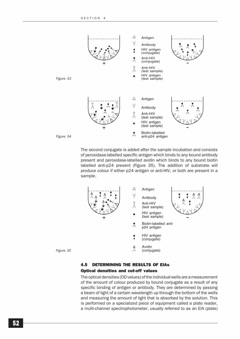
S E C T I O N 4
52
The second conjugate is added after the sample incubation and consistsof peroxidase-labelled specific antigen which binds to any bound antibodypresent and peroxidase-labelled avidin which binds to any bound biotinlabelled anti-p24 present (Figure 35). The addition of substrate willproduce colour if either p24 antigen or anti-HIV, or both are present in asample.
4.5 DETERMINING THE RESULTS OF EIAs
Optical densities and cut-off values
The optical densities (OD values) of the individual wells are a measurementof the amount of colour produced by bound conjugate as a result of anyspecific binding of antigen or antibody. They are determined by passinga beam of light of a certain wavelength up through the bottom of the wellsand measuring the amount of light that is absorbed by the solution. Thisis performed on a specialized piece of equipment called a plate reader,a multi-channel spectrophotometer, usually referred to as an EIA (plate)
Figure 34
Figure 35
HIV antigen(in sample)
Biotin-labelledanti-p24 antigen
Figure 33
Antigen
Antibody
Anti-HIV(in sample)
HIV Antigen(in sample)
Antigenconjugate
Antibodyconjugate
Antigen
Antibody
HIV antigen(conjugate)
Anti-HIV(conjugate)
Anti-HIV(test sample)HIV antigen(test sample)
Antigen
Antibody
Anti-HIV(in sample)
HIV Antigen(in sample)
Biotin labeledanti-p24 Antigen
Antigen
Antibody
Anti-HIV(test sample)HIV antigen(test sample)
Biotin-labelledanti-p24 antigen
A
A
A
A AA
A
A
Antigen
Antibody
Anti-HIV(in sample)
HIV Antigen(in sample)
Antigenconjugate
Biotin labeledanti-p24 Antigen
Avidinconjugate
HIV antigen(in sample)
Biotin-labelledanti-p24 antigen
Antigen
Antibody
Anti-HIV(test sample)
HIV antigen(test sample)
Biotin-labelled anti-p24 antigen
HIV antigen(conjugate)
Avidin(conjugate)
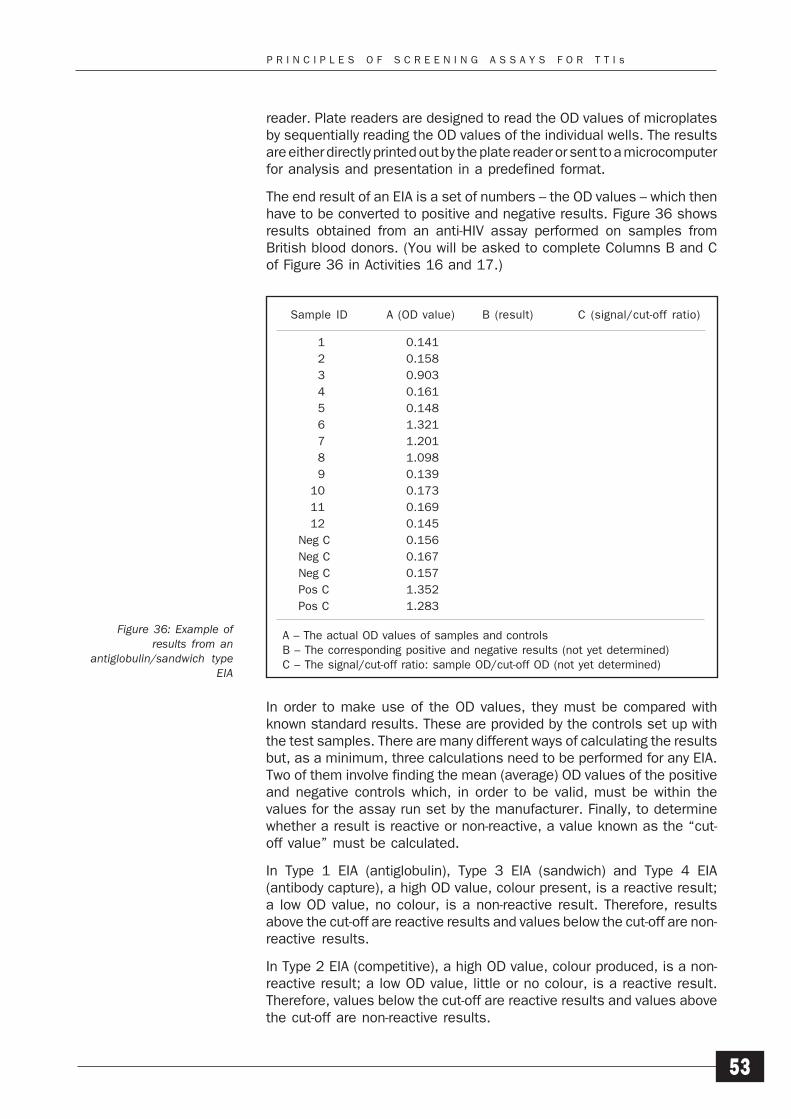
P R I N C I P L E S O F S C R E E N I N G A S S A Y S F O R T T I s
53
reader. Plate readers are designed to read the OD values of microplatesby sequentially reading the OD values of the individual wells. The resultsare either directly printed out by the plate reader or sent to a microcomputerfor analysis and presentation in a predefined format.
The end result of an EIA is a set of numbers – the OD values – which thenhave to be converted to positive and negative results. Figure 36 showsresults obtained from an anti-HIV assay performed on samples fromBritish blood donors. (You will be asked to complete Columns B and Cof Figure 36 in Activities 16 and 17.)
Sample ID A (OD value) B (result) C (signal/cut-off ratio)
1 0.141
2 0.158
3 0.903
4 0.161
5 0.148
6 1.321
7 1.201
8 1.098
9 0.139
10 0.173
11 0.169
12 0.145
Neg C 0.156
Neg C 0.167
Neg C 0.157
Pos C 1.352
Pos C 1.283
A – The actual OD values of samples and controlsB – The corresponding positive and negative results (not yet determined)C – The signal/cut-off ratio: sample OD/cut-off OD (not yet determined)
Figure 36: Example ofresults from an
antiglobulin/sandwich typeEIA
In order to make use of the OD values, they must be compared withknown standard results. These are provided by the controls set up withthe test samples. There are many different ways of calculating the resultsbut, as a minimum, three calculations need to be performed for any EIA.Two of them involve finding the mean (average) OD values of the positiveand negative controls which, in order to be valid, must be within thevalues for the assay run set by the manufacturer. Finally, to determinewhether a result is reactive or non-reactive, a value known as the “cut-off value” must be calculated.
In Type 1 EIA (antiglobulin), Type 3 EIA (sandwich) and Type 4 EIA(antibody capture), a high OD value, colour present, is a reactive result;a low OD value, no colour, is a non-reactive result. Therefore, resultsabove the cut-off are reactive results and values below the cut-off are non-reactive results.
In Type 2 EIA (competitive), a high OD value, colour produced, is a non-reactive result; a low OD value, little or no colour, is a reactive result.Therefore, values below the cut-off are reactive results and values abovethe cut-off are non-reactive results.
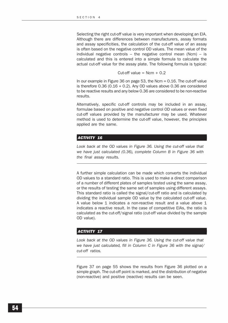
S E C T I O N 4
54
Selecting the right cut-off value is very important when developing an EIA.Although there are differences between manufacturers, assay formatsand assay specificities, the calculation of the cut-off value of an assayis often based on the negative control OD values. The mean value of theindividual negative controls – the negative control mean (Ncm) – iscalculated and this is entered into a simple formula to calculate theactual cut-off value for the assay plate. The following formula is typical:
Cut-off value = Ncm + 0.2
In our example in Figure 36 on page 53, the Ncm = 0.16. The cut-off valueis therefore 0.36 (0.16 + 0.2). Any OD values above 0.36 are consideredto be reactive results and any below 0.36 are considered to be non-reactiveresults.
Alternatively, specific cut-off controls may be included in an assay,formulae based on positive and negative control OD values or even fixedcut-off values provided by the manufacturer may be used. Whatevermethod is used to determine the cut-off value, however, the principlesapplied are the same.
ACTIVITY 16
Look back at the OD values in Figure 36. Using the cut-off value thatwe have just calculated (0.36), complete Column B in Figure 36 withthe final assay results.
A further simple calculation can be made which converts the individualOD values to a standard ratio. This is used to make a direct comparisonof a number of different plates of samples tested using the same assay,or the results of testing the same set of samples using different assays.This standard ratio is called the signal/cut-off ratio and is calculated bydividing the individual sample OD value by the calculated cut-off value.A value below 1 indicates a non-reactive result and a value above 1indicates a reactive result. In the case of competitive EIAs, the ratio iscalculated as the cut-off/signal ratio (cut-off value divided by the sampleOD value).
ACTIVITY 17
Look back at the OD values in Figure 36. Using the cut-off value thatwe have just calculated, fill in Column C in Figure 36 with the signal/cut-off ratios.
Figure 37 on page 55 shows the results from Figure 36 plotted on asimple graph. The cut-off point is marked, and the distribution of negative(non-reactive) and positive (reactive) results can be seen.
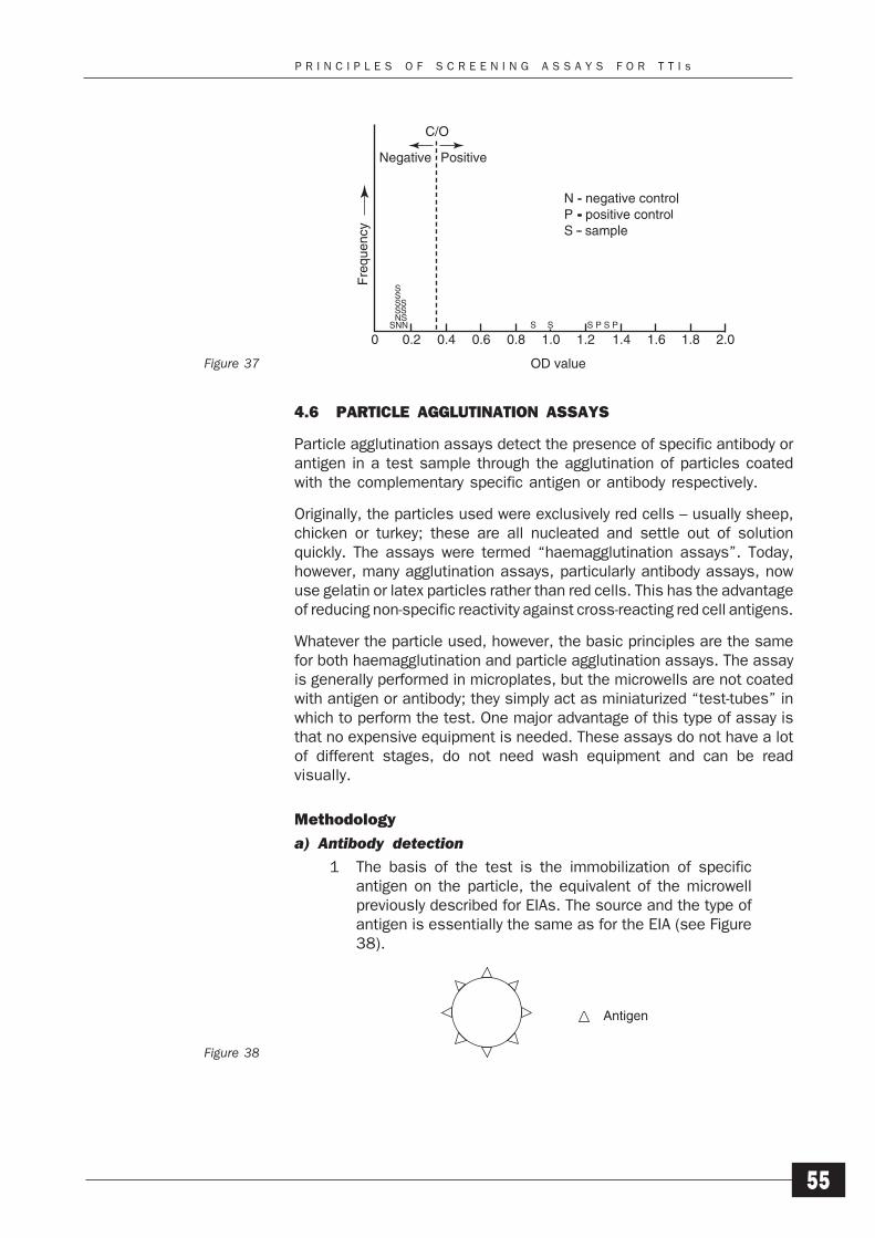
P R I N C I P L E S O F S C R E E N I N G A S S A Y S F O R T T I s
55
4.6 PARTICLE AGGLUTINATION ASSAYS
Particle agglutination assays detect the presence of specific antibody orantigen in a test sample through the agglutination of particles coatedwith the complementary specific antigen or antibody respectively.
Originally, the particles used were exclusively red cells – usually sheep,chicken or turkey; these are all nucleated and settle out of solutionquickly. The assays were termed “haemagglutination assays”. Today,however, many agglutination assays, particularly antibody assays, nowuse gelatin or latex particles rather than red cells. This has the advantageof reducing non-specific reactivity against cross-reacting red cell antigens.
Whatever the particle used, however, the basic principles are the samefor both haemagglutination and particle agglutination assays. The assayis generally performed in microplates, but the microwells are not coatedwith antigen or antibody; they simply act as miniaturized “test-tubes” inwhich to perform the test. One major advantage of this type of assay isthat no expensive equipment is needed. These assays do not have a lotof different stages, do not need wash equipment and can be readvisually.
Methodology
a) Antibody detection
1 The basis of the test is the immobilization of specificantigen on the particle, the equivalent of the microwellpreviously described for EIAs. The source and the type ofantigen is essentially the same as for the EIA (see Figure38).
Figure 37
Figure 38
S S S P S P
0 0.2 0.4 0.6 0.8 1.0 1.2 1.4 1.6 1.8 2.0
OD value
C/O
PositiveNegative
Fre
quen
cy
S S SS SS NSSNN
Antigen
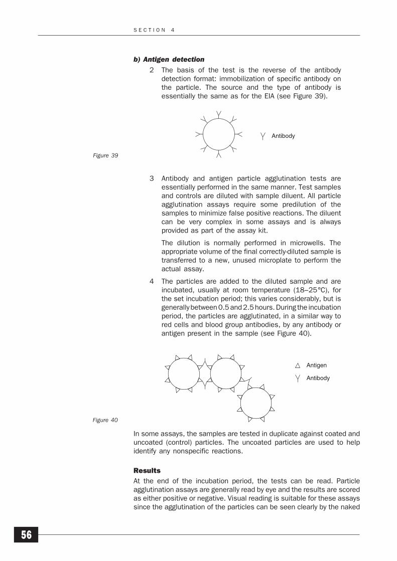
S E C T I O N 4
56
b) Antigen detection2 The basis of the test is the reverse of the antibody
detection format: immobilization of specific antibody onthe particle. The source and the type of antibody isessentially the same as for the EIA (see Figure 39).
3 Antibody and antigen particle agglutination tests areessentially performed in the same manner. Test samplesand controls are diluted with sample diluent. All particleagglutination assays require some predilution of thesamples to minimize false positive reactions. The diluentcan be very complex in some assays and is alwaysprovided as part of the assay kit.
The dilution is normally performed in microwells. Theappropriate volume of the final correctly-diluted sample istransferred to a new, unused microplate to perform theactual assay.
4 The particles are added to the diluted sample and areincubated, usually at room temperature (18–25°C), forthe set incubation period; this varies considerably, but isgenerally between 0.5 and 2.5 hours. During the incubationperiod, the particles are agglutinated, in a similar way tored cells and blood group antibodies, by any antibody orantigen present in the sample (see Figure 40).
In some assays, the samples are tested in duplicate against coated anduncoated (control) particles. The uncoated particles are used to helpidentify any nonspecific reactions.
Results
At the end of the incubation period, the tests can be read. Particleagglutination assays are generally read by eye and the results are scoredas either positive or negative. Visual reading is suitable for these assayssince the agglutination of the particles can be seen clearly by the naked
Figure 40
Figure 39
Antibody
Antibody
Antigen
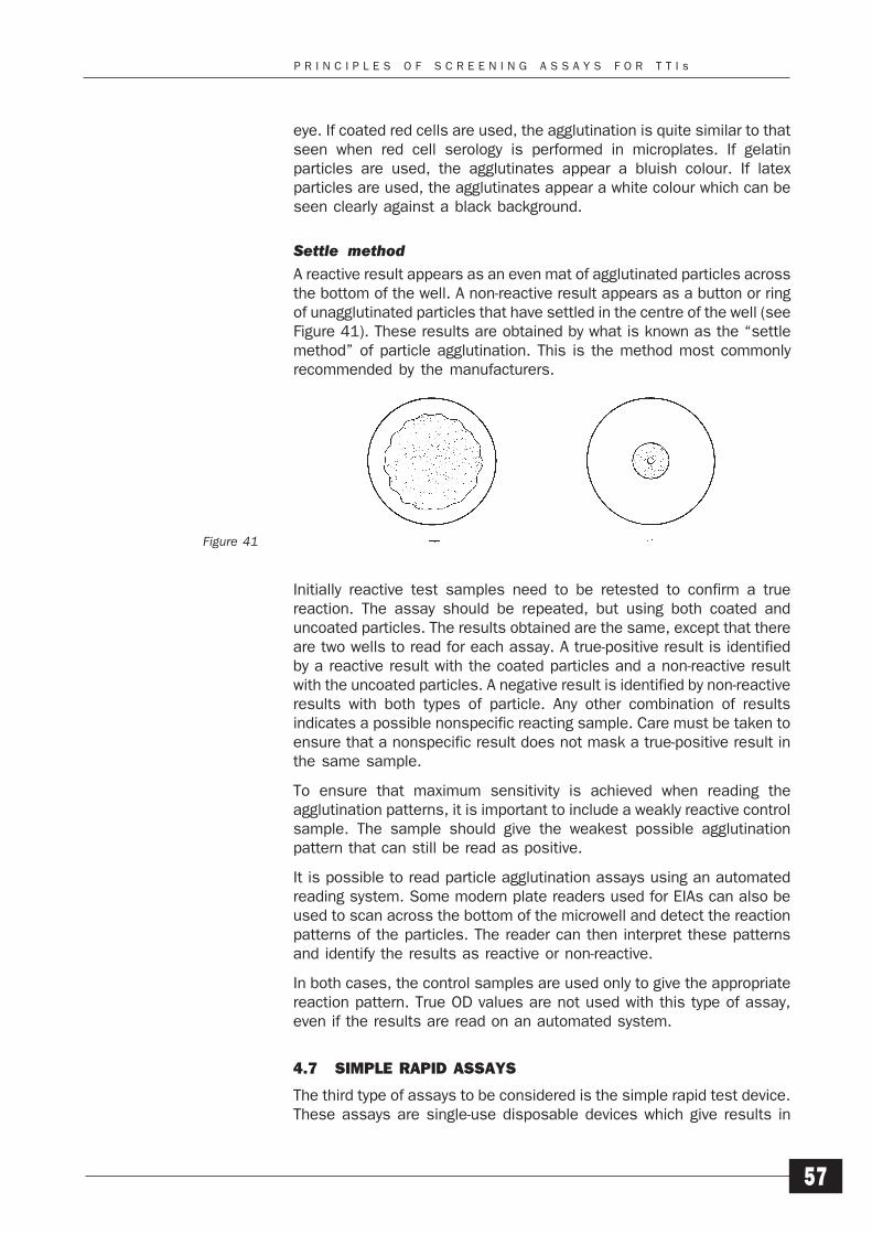
P R I N C I P L E S O F S C R E E N I N G A S S A Y S F O R T T I s
57
eye. If coated red cells are used, the agglutination is quite similar to thatseen when red cell serology is performed in microplates. If gelatinparticles are used, the agglutinates appear a bluish colour. If latexparticles are used, the agglutinates appear a white colour which can beseen clearly against a black background.
Settle methodA reactive result appears as an even mat of agglutinated particles acrossthe bottom of the well. A non-reactive result appears as a button or ringof unagglutinated particles that have settled in the centre of the well (seeFigure 41). These results are obtained by what is known as the “settlemethod” of particle agglutination. This is the method most commonlyrecommended by the manufacturers.
Figure 41
Initially reactive test samples need to be retested to confirm a truereaction. The assay should be repeated, but using both coated anduncoated particles. The results obtained are the same, except that thereare two wells to read for each assay. A true-positive result is identifiedby a reactive result with the coated particles and a non-reactive resultwith the uncoated particles. A negative result is identified by non-reactiveresults with both types of particle. Any other combination of resultsindicates a possible nonspecific reacting sample. Care must be taken toensure that a nonspecific result does not mask a true-positive result inthe same sample.
To ensure that maximum sensitivity is achieved when reading theagglutination patterns, it is important to include a weakly reactive controlsample. The sample should give the weakest possible agglutinationpattern that can still be read as positive.
It is possible to read particle agglutination assays using an automatedreading system. Some modern plate readers used for EIAs can also beused to scan across the bottom of the microwell and detect the reactionpatterns of the particles. The reader can then interpret these patternsand identify the results as reactive or non-reactive.
In both cases, the control samples are used only to give the appropriatereaction pattern. True OD values are not used with this type of assay,even if the results are read on an automated system.
4.7 SIMPLE RAPID ASSAYS
The third type of assays to be considered is the simple rapid test device.These assays are single-use disposable devices which give results in
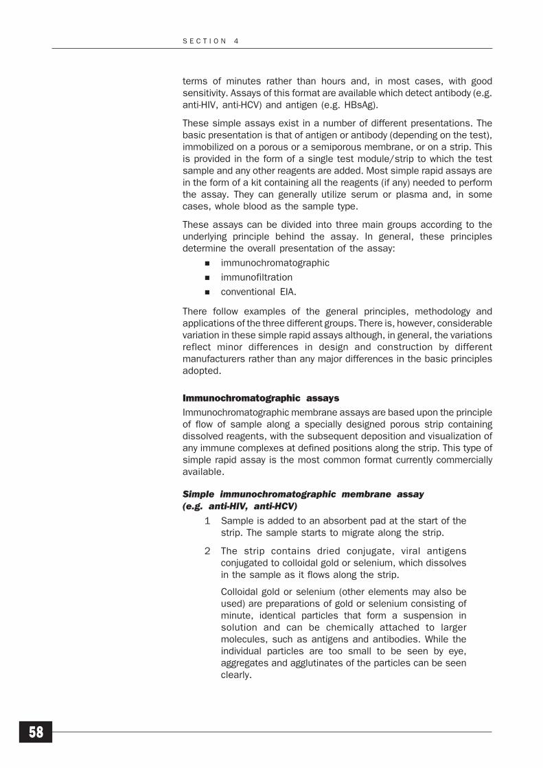
S E C T I O N 4
58
terms of minutes rather than hours and, in most cases, with goodsensitivity. Assays of this format are available which detect antibody (e.g.anti-HIV, anti-HCV) and antigen (e.g. HBsAg).
These simple assays exist in a number of different presentations. Thebasic presentation is that of antigen or antibody (depending on the test),immobilized on a porous or a semiporous membrane, or on a strip. Thisis provided in the form of a single test module/strip to which the testsample and any other reagents are added. Most simple rapid assays arein the form of a kit containing all the reagents (if any) needed to performthe assay. They can generally utilize serum or plasma and, in somecases, whole blood as the sample type.
These assays can be divided into three main groups according to theunderlying principle behind the assay. In general, these principlesdetermine the overall presentation of the assay:
immunochromatographic
immunofiltration
conventional EIA.
There follow examples of the general principles, methodology andapplications of the three different groups. There is, however, considerablevariation in these simple rapid assays although, in general, the variationsreflect minor differences in design and construction by differentmanufacturers rather than any major differences in the basic principlesadopted.
Immunochromatographic assays
Immunochromatographic membrane assays are based upon the principleof flow of sample along a specially designed porous strip containingdissolved reagents, with the subsequent deposition and visualization ofany immune complexes at defined positions along the strip. This type ofsimple rapid assay is the most common format currently commerciallyavailable.
Simple immunochromatographic membrane assay(e.g. anti-HIV, anti-HCV)
1 Sample is added to an absorbent pad at the start of thestrip. The sample starts to migrate along the strip.
2 The strip contains dried conjugate, viral antigensconjugated to colloidal gold or selenium, which dissolvesin the sample as it flows along the strip.
Colloidal gold or selenium (other elements may also beused) are preparations of gold or selenium consisting ofminute, identical particles that form a suspension insolution and can be chemically attached to largermolecules, such as antigens and antibodies. While theindividual particles are too small to be seen by eye,aggregates and agglutinates of the particles can be seenclearly.
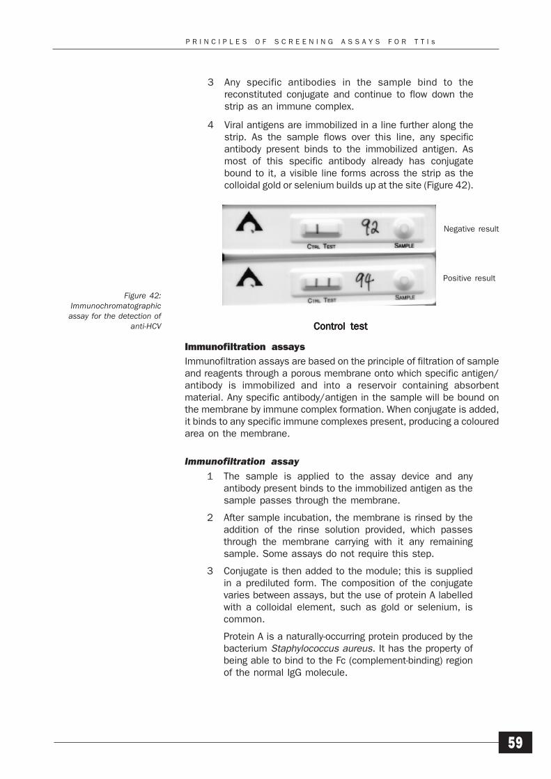
P R I N C I P L E S O F S C R E E N I N G A S S A Y S F O R T T I s
59
3 Any specific antibodies in the sample bind to thereconstituted conjugate and continue to flow down thestrip as an immune complex.
4 Viral antigens are immobilized in a line further along thestrip. As the sample flows over this line, any specificantibody present binds to the immobilized antigen. Asmost of this specific antibody already has conjugatebound to it, a visible line forms across the strip as thecolloidal gold or selenium builds up at the site (Figure 42).
Immunofiltration assays
Immunofiltration assays are based on the principle of filtration of sampleand reagents through a porous membrane onto which specific antigen/antibody is immobilized and into a reservoir containing absorbentmaterial. Any specific antibody/antigen in the sample will be bound onthe membrane by immune complex formation. When conjugate is added,it binds to any specific immune complexes present, producing a colouredarea on the membrane.
Immunofiltration assay1 The sample is applied to the assay device and any
antibody present binds to the immobilized antigen as thesample passes through the membrane.
2 After sample incubation, the membrane is rinsed by theaddition of the rinse solution provided, which passesthrough the membrane carrying with it any remainingsample. Some assays do not require this step.
3 Conjugate is then added to the module; this is suppliedin a prediluted form. The composition of the conjugatevaries between assays, but the use of protein A labelledwith a colloidal element, such as gold or selenium, iscommon.
Protein A is a naturally-occurring protein produced by thebacterium Staphylococcus aureus. It has the property ofbeing able to bind to the Fc (complement-binding) regionof the normal IgG molecule.
Negative result
Positive result
Figure 42:Immunochromatographicassay for the detection of
anti-HCV Control testControl testControl testControl testControl test
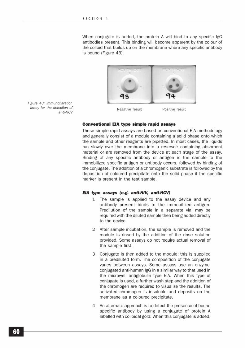
S E C T I O N 4
60
When conjugate is added, the protein A will bind to any specific IgGantibodies present. This binding will become apparent by the colour ofthe colloid that builds up on the membrane where any specific antibodyis bound (Figure 43).
Conventional EIA type simple rapid assays
These simple rapid assays are based on conventional EIA methodologyand generally consist of a module containing a solid phase onto whichthe sample and other reagents are pipetted. In most cases, the liquidsrun slowly over the membrane into a reservoir containing absorbentmaterial or are removed from the device at each stage of the assay.Binding of any specific antibody or antigen in the sample to theimmobilized specific antigen or antibody occurs, followed by binding ofthe conjugate. The addition of a chromogenic substrate is followed by thedeposition of coloured precipitate onto the solid phase if the specificmarker is present in the test sample.
EIA type assays (e.g. anti-HIV, anti-HCV)1 The sample is applied to the assay device and any
antibody present binds to the immobilized antigen.Predilution of the sample in a separate vial may berequired with the diluted sample then being added directlyto the device.
2 After sample incubation, the sample is removed and themodule is rinsed by the addition of the rinse solutionprovided. Some assays do not require actual removal ofthe sample first.
3 Conjugate is then added to the module; this is suppliedin a prediluted form. The composition of the conjugatevaries between assays. Some assays use an enzyme-conjugated anti-human IgG in a similar way to that used inthe microwell antiglobulin type EIA. When this type ofconjugate is used, a further wash step and the addition ofthe chromogen are required to visualize the results. Theactivated chromogen is insoluble and deposits on themembrane as a coloured precipitate.
4 An alternate approach is to detect the presence of boundspecific antibody by using a conjugate of protein Alabelled with colloidal gold. When this conjugate is added,
Figure 43: Immunofiltrationassay for the detection of
anti-HCVNegative result Positive result
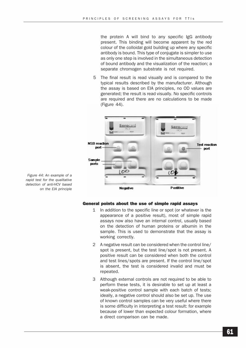
P R I N C I P L E S O F S C R E E N I N G A S S A Y S F O R T T I s
61
Figure 44: An example of arapid test for the qualitativedetection of anti-HCV based
on the EIA principle
the protein A will bind to any specific IgG antibodypresent. This binding will become apparent by the redcolour of the colloidal gold building up where any specificantibody is bound. This type of conjugate is simpler to useas only one step is involved in the simultaneous detectionof bound antibody and the visualization of the reaction; aseparate chromogen substrate is not required.
5 The final result is read visually and is compared to thetypical results described by the manufacturer. Althoughthe assay is based on EIA principles, no OD values aregenerated; the result is read visually. No specific controlsare required and there are no calculations to be made(Figure 44).
General points about the use of simple rapid assays
1 In addition to the specific line or spot (or whatever is theappearance of a positive result), most of simple rapidassays now also have an internal control, usually basedon the detection of human proteins or albumin in thesample. This is used to demonstrate that the assay isworking correctly.
2 A negative result can be considered when the control line/spot is present, but the test line/spot is not present. Apositive result can be considered when both the controland test lines/spots are present. If the control line/spotis absent, the test is considered invalid and must berepeated.
3 Although external controls are not required to be able toperform these tests, it is desirable to set up at least aweak-positive control sample with each batch of tests;ideally, a negative control should also be set up. The useof known control samples can be very useful where thereis some difficulty in interpreting a test result: for examplebecause of lower than expected colour formation, wherea direct comparison can be made.
S E C T I O N 4
62
4 In general, the whole procedure for these tests, fromadding sample to obtaining a usable result, usually takes10–15 minutes. However, the manufacturer’s timingsmust be adhered to as over-incubation can lead to falsepositive reactions in some assays.
We have considered the principles of the three main types of TTIscreening assay available. Although you may only use one type of assay,it is important that you understand the principles of all three basic types.It is also useful if you can assess for yourself the different types of assaysthat are available to you.
WHO assesses on a continuing basis the operational characteristics ofmany commercially available HIV, HCV and HBV antibody and antigenassays. Reports of these WHO evaluations are issued regularly and arealso posted on the WHO website. Copies of these reports and additionalinformation can be obtained by visiting the BCT section of the WHOwebsite (http://www.who.int/bct) or by contacting the Department ofBlood Safety and Clinical Technology, Geneva, or the WHO RegionalOffices.
SUMMARY
1 The time and costs involved in screening donated bloodfor any TTI can be reduced by an effective donor educationand selection programme that promotes self-exclusion bydonors at risk of transfusion-transmissible infection.
2 The three principal types of screening assay involve thesame two basic steps:
demonstrating the presence of specific antigen orantibody in a sample in an immunological reactioninvolving antigen/antibody (immune) complexformation, with one of the components bound to afixed surface
detecting the formation of the immune complex in alinked procedure.
SELF-ASSESSMENT
14 What are the three main types of screening assay?
15 What is the difference between the term “positive” andthe term “reactive”?
S E L E C T I N G S C R E E N I N G A S S A Y S F O R T T I s
63
5Selecting Screening Assaysfor Transfusion-TransmissibleInfections
The purpose of this section is to help you to determine the most suitablescreening strategy for your own circumstances and identify the basicequipment required.
LEARNING OBJECTIVES
When you have completed this section, you should be able to:
1 Define the terms “sensitivity” and “specificity” and explaintheir relevance to TTI screening assays.
2 Identify the most suitable type of primary screening assayfor use in your own laboratory.
3 Explain the factors that have to be considered whenchoosing a screening assay.
4 Assess the factors that contribute to the overall cost ofscreening in your laboratory.
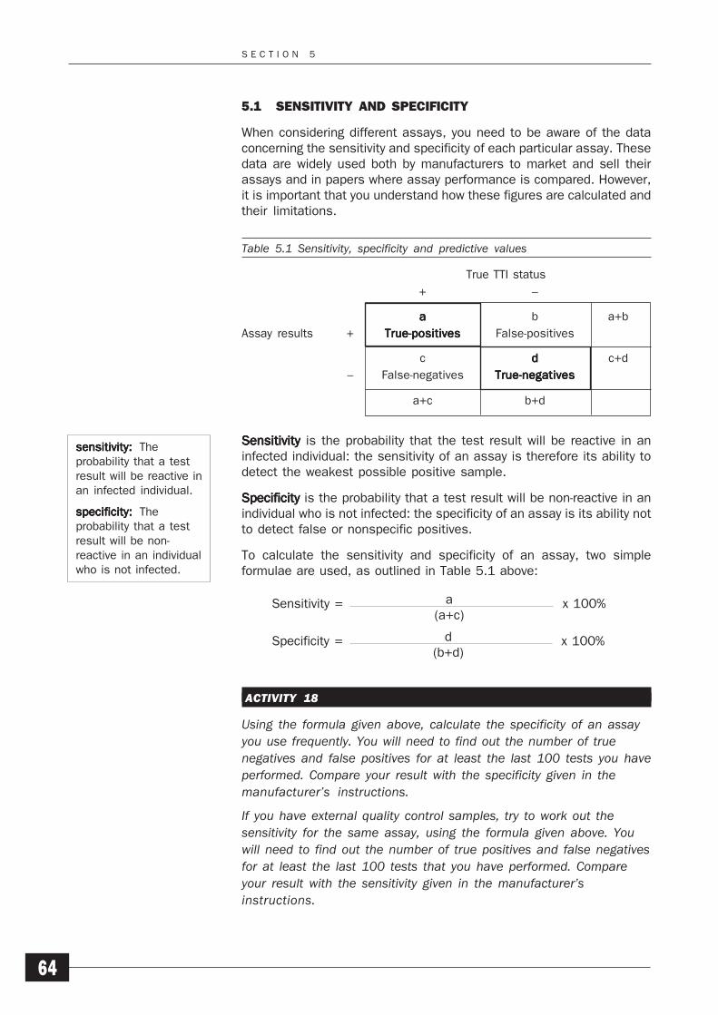
S E C T I O N 5
64
5.1 SENSITIVITY AND SPECIFICITY
When considering different assays, you need to be aware of the dataconcerning the sensitivity and specificity of each particular assay. Thesedata are widely used both by manufacturers to market and sell theirassays and in papers where assay performance is compared. However,it is important that you understand how these figures are calculated andtheir limitations.
Table 5.1 Sensitivity, specificity and predictive values
True TTI status
+ –
aaaaa b a+b
Assay results + True-positivesTrue-positivesTrue-positivesTrue-positivesTrue-positives False-positives
c ddddd c+d
– False-negatives True-negativesTrue-negativesTrue-negativesTrue-negativesTrue-negatives
a+c b+d
Sensitivity Sensitivity Sensitivity Sensitivity Sensitivity is the probability that the test result will be reactive in aninfected individual: the sensitivity of an assay is therefore its ability todetect the weakest possible positive sample.
SpecificitySpecificitySpecificitySpecificitySpecificity is the probability that a test result will be non-reactive in anindividual who is not infected: the specificity of an assay is its ability notto detect false or nonspecific positives.
To calculate the sensitivity and specificity of an assay, two simpleformulae are used, as outlined in Table 5.1 above:
ACTIVITY 18
Using the formula given above, calculate the specificity of an assayyou use frequently. You will need to find out the number of truenegatives and false positives for at least the last 100 tests you haveperformed. Compare your result with the specificity given in themanufacturer’s instructions.
If you have external quality control samples, try to work out thesensitivity for the same assay, using the formula given above. Youwill need to find out the number of true positives and false negativesfor at least the last 100 tests that you have performed. Compareyour result with the sensitivity given in the manufacturer’sinstructions.
sensitivity:sensitivity:sensitivity:sensitivity:sensitivity: Theprobability that a testresult will be reactive inan infected individual.
specificity:specificity:specificity:specificity:specificity: Theprobability that a testresult will be non-reactive in an individualwho is not infected.
Sensitivity = x 100%a(a+c)
Specificity = x 100%d(b+d)
S E L E C T I N G S C R E E N I N G A S S A Y S F O R T T I s
65
If you do not have external quality control samples, leave out thesecond part of this activity.
If your results differ from the specificity or sensitivity given in themanufacturer’s instructions, talk to your supervisor about yourfindings. Note down your recommendations on your Action List.
An important point to remember when considering the sensitivity andspecificity of assays is that they are generally inversely related; that is,as the sensitivity is increased, the specificity decreases and as thespecificity is increased, the sensitivity decreases.
The importance of sensitivity in an assay should be clear and needs nofurther discussion. Specificity is a more complicated issue, however, butit is essential that all staff are aware of the importance of using specificassays. One of the main problems is to determine what causes the lackof specificity. At the very simplest level, the specificity of an assay isrelated to the particular assay format and the source of bound antigenor antibody. For example, the use of purer antigen preparations (such assynthetic peptides) reduces nonspecific reactions because there arevery few, if any, non-peptide molecules present.
A final point is that, no matter how good the reported sensitivity andspecificity of an assay, its final performance depends largely on theskilful handling of the assay by the operator. Figures quoted bymanufacturers are usually very accurate, but often reflect the performanceof an assay in an experienced dedicated laboratory under very closelycontrolled conditions. It is important for you to know how the assay willperform in your laboratory using this equipment and staff available.
5.2 PREDICTIVE VALUES
Another way of looking at the performance of an assay is to look at itspredictive values. The predictive values, positive predictive value (PPV)and negative predictive value (NPV) are measures of the actualperformance of an assay in a laboratory and therefore can be helpful ingiving some idea of the sort of performance that you can expect from theassay in routine use.
The positive predictive value is the probability of a positive result beinga true positive. The negative predictive value is the probability of anegative result being a true negative.
As with sensitivity and specificity two simple formulae are used tocalculate the basic positive and negative predictive values, as outlinedin Table 5.1 on page 64.
Positive predictive value = x 100%a(a+b)
Negative predictive value = x 100%d(c+d)
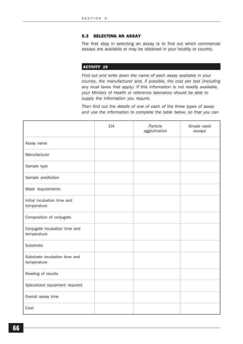
S E C T I O N 5
66
5.3 SELECTING AN ASSAY
The first step in selecting an assay is to find out which commercialassays are available or may be obtained in your locality or country.
ACTIVITY 19
Find out and write down the name of each assay available in yourcountry, the manufacturer and, if possible, the cost per test (includingany local taxes that apply). If this information is not readily available,your Ministry of Health or reference laboratory should be able tosupply the information you require.
Then find out the details of one of each of the three types of assayand use the information to complete the table below, so that you can
EIA Particle Simple rapidagglutination assays
Assay name
Manufacturer
Sample type
Sample predilution
Wash requirements
Initial incubation time and
temperature
Composition of conjugate
Conjugate incubation time and
temperature
Substrate
Substrate incubation time and
temperature
Reading of results
Specialized equipment required
Overall assay time
Cost
S E L E C T I N G S C R E E N I N G A S S A Y S F O R T T I s
67
make a direct comparison of these assays. You can return to thistable later on to help you decide which would be the most suitableassay to use for the routine screening of donated blood.
There is a great deal of misleading information available about theimportance of using the most sensitive assays. It is more important touse the most appropriate assay for your circumstances – those withwhich you will be able to obtain the most accurate and reliable results.This is not always the assay that is theoretically the most “sensitive”available. We shall therefore provide some simple guidelines to help youdetermine which assay is suitable for your own situation. In mostcircumstances, it would be inappropriate, for instance, for a smalllaboratory that tests 10–20 units of blood each week to use EIAs. Theexpense of obtaining the necessary equipment and the time needed toset it up and maintain it, as well as the amount of staff training required,are not justified.
A suggested and approximate guide is as follows:
laboratories testing 1–10 donations a day or 1–60 perweek should consider the use of simple rapid assays orparticle agglutination assays
laboratories testing 20 or more donations a day or 100 ormore a week should consider the use of EIAs.
Manufacturers and suppliers often use the claim of greater sensitivity toencourage laboratories to use EIAs. In many cases, however, thesensitivity of some EIAs is not greater than some of the simple rapidassays or particle agglutination assays. Even if it is greater, the figuresquoted to support these claims are often those obtained in largelaboratories with highly skilled staff and with optimal testing conditions.As mentioned previously, the most important thing is to use an assay thatgives reliable and accurate results in your own laboratory.
ACTIVITY 20
Look back at the table that you completed in Activity 19. Then notedown any additional factors that specifically relate to your laboratoryand that you feel are relevant to the choice of a TTI assay. Youshould consider:
the number of donations each week
the facilities and equipment available in your laboratory
the support service available: for example, water supply,electricity and so on.
Assess the different types of assay on their appropriateness to yoursituation and consider which type of assay you would select. If youfeel that your laboratory is not currently using the most appropriatetype of assay, note down your recommendations on your Action Listand discuss your findings with your supervisor.
S E C T I O N 5
68
Be wary of making the wrong decision simply because you cannot see theimmediate solution to a problem. Instead, consider all the possibilitiesfirst. For example, you may decide that EIAs would be the most suitabletype of assay to use in your laboratory, but feel you have to choose anotheroption because you have no suitable equipment. However, you couldperhaps ask the supplier of the assay to provide the equipment. Buyingor hiring the equipment is often too expensive and you may end up withequipment that you have difficulty in getting serviced and maintained. Amore common approach today is to lease the equipment as a packagewith the purchase of the kits. A major advantage of this is that theequipment remains the property of the supplier and it is in its interest tomake sure that it is kept in good condition and is regularly maintained andserviced. This is both because of the financial value of the equipment andalso because the equipment is needed to perform the screening tests.You will not continue to buy tests if the equipment is not working.
It is important that you select the type of assay that you feel is best suitedto your particular circumstances. The information provided so far shouldhave helped you to understand what choices have to be made.
5.4 FACTORS INFLUENCING SCREENING PROGRAMMES
Whatever type of assay is chosen as being the most appropriate for yourcircumstances, there are a number of other factors to be considered insetting up and maintaining an effective TTI screening programme.
ACTIVITY 21
What other important factors do you think need to be considered inorder to maintain a successful TTI screening programme?
Consider the factors listed below, and any others you can think of,and note down how they relate to your particular circumstances.
staff training
the equipment and reagents required
the supply of assay kits
the storage of assay kits
the suitability and condition of the samples to be tested
the supply of external quality control (QC) samples
time constraints on screening
the cost of screening.
Staff training
All staff need training to improve their effectiveness, even if they aretrained to do only one specific job. To be able to perform TTI testingsuccessfully, staff need to be trained not only in the performance of theassay, but also in the other actions necessary as a result of TTI testing,
S E L E C T I N G S C R E E N I N G A S S A Y S F O R T T I s
69
such as recording the results accurately. A successful TTI screeningprogramme does not simply mean being able to perform the assaywithout mistakes or failures. What training programmes are there in yourlaboratory?
ACTIVITY 22
Make some notes on the training provided in your laboratory onscreening for TTIs, including:
who provides the training
whether there is a member of staff who is in charge oftraining
the kind of training that is provided
the duration of training
the way in which trainees’ skills are assessed
whether regular training updates are held.
How would you develop a training programme for new staff working inthe area of screening for TTIs or for staff who are already performingTTI testing and testing for other infectious agents? Note down yourideas for a training scheme on your Action List. It need only be verysimple, covering the most important areas. Then discuss your ideaswith your supervisor.
It is important to keep a record of all training provided so that the trainerknows what the trainees should be capable of and the trainees understandwhat they are expected to know. Good training records not only enablemonitoring of the actual training sessions, but also provide a log of whenthe training was provided so that regular updates can be held. Thisshould help new staff to feel confident because it demonstrates that theywill not be expected to undertake tasks for which they have not beentrained. In addition, if the laboratory is likely to be inspected at sometime, training records not only show that staff have been trained, but canalso give the inspector confidence that the laboratory is run properly andthat the quality of the results is important to the staff in the laboratory.
Equipment and reagents
All assays, except the simple rapid assays, require the use of certainpieces of equipment or of reagents that are not supplied with the assaykit. The reagents are usually only simple items, but they increase the costof the assay and there may be problems if their supply cannot beguaranteed. The necessary equipment and reagents may not be readilyavailable in all laboratories, especially in small laboratories in ruralhospitals or those that are not part of a hospital.
S E C T I O N 5
70
ACTIVITY 23
From what you have already learned about EIAs and particleagglutination assays, list the items of equipment that are necessaryto perform the assays correctly. Although the assays are verydifferent, some items of equipment are common to both of them,while some are specific to each type of assay. Check your answerswith those in the Activity Checklists and Answers on page 146.
When you have checked your answers, identify the items ofequipment needed to perform the assays correctly that are alreadyavailable in your laboratory. Then identify any equipment to whichyou have unrestricted access within the hospital or the institution towhich your laboratory belongs.
If it is possible to use equipment in another part of your hospital orinstitution, it is important to check that access to it is not restricted. Forexample, equipment in another laboratory may be available during theweek. However, if that laboratory is locked at weekends, the equipmentwould not be suitable for use since blood donations may need to betested at any time if emergencies arise.
If you do not already have unrestricted access to the equipment neededto perform certain assays, the supplier of the assay may be willing toprovide the required equipment. If your laboratory is setting up a largescreening programme, this may be the best method of obtaining theappropriate equipment and support needed to maintain the programme.Remember, however, that the choice of assay should depend on whetherit is appropriate to your circumstances and should not be influenced bythe promise of the supply of equipment.
In addition to the equipment needed to perform the assay correctly, it isalso necessary to ensure that there is an adequate supply of the otherreagents and disposables that are required but that are not supplied withthe assay. Simple rapid assays are usually supplied with all the requiredreagents and disposables, and the same may also apply to the reagentsrequired for particle agglutination assays, although disposables are notusually provided. However, your laboratory will need to supply its ownadditional reagents and disposables for EIAs.
The disposables needed will vary, depending on the assays used, butthey will include such items as:
disposable pipette tips for multi-channel pipettes
reagent troughs
plastic bottles for the preparation of small volumes ofreagents supplied in concentrated or two-part solutions
microplates for particle agglutination assays.
The reagents required will also vary, depending on the assays used, butthey will include such items as:
S E L E C T I N G S C R E E N I N G A S S A Y S F O R T T I s
71
deionized or distilled water to prepare the wash bufferand, in some cases, to wash the plates directly: apossible source would be injection or irrigation water fromthe pharmacy
suitable acid solution to stop the final reaction and fix thedeveloped colour.
Where the supply of these disposables and reagents cannot be guaranteedwithin the laboratory, it is again worth approaching the supplier of yourassay kits for the necessary disposables and reagents to perform theassays. You will then have a complete supply system and all that will beneeded are the staff to perform the assay and the samples to test. Asystem of this kind will obviously cost money and may take some timeto set up and operate efficiently. You may find that buying the requireditems through the assay supplier is cheaper than using other sourcesand it will certainly save valuable staff time in trying to set up separatedelivery schedules to coincide with the delivery of the kits.
Supply of assay kits
The supply of assay kits needs to be well organized in order to ensure thatthe screening strategy is maintained efficiently. Section 7 in theIntroductory Module explains how to maintain a simple stock controlsystem, but there are four important issues that also need to beconsidered.
1 Calculating the actual usage of kitsWhen calculating the number of kits that are required, youmust take the following factors into account:
the number of donations that you would expect to testin a specified period of time
the repeat testing of initially reactive samples (youmay feel that it is necessary to repeat the test induplicate on these donations)
any internal quality control (QC) samples (preparedwithin the laboratory) or external QC samples (preparedby a separate laboratory or institution) that you use
the number of assay failures.
2 Determining an appropriate stock levelThe actual stock level that it is possible to hold isdetermined by the following factors:
the physical space available under suitable storageconditions
the financial constraints: how much money is availableand whether the supplier requires payment beforedelivery
the expiry dates of the assays: the usual length ofremaining shelf-life on the assays when they aredelivered.
S E C T I O N 5
72
3 Delivery timesThe length of time between ordering the assays and theirdelivery will depend on a number of factors, many of whichmay be outside the control of the supplier. For example,transportation may become impossible for a period duringrainy seasons. An allowance should be made for thiswhen ordering kits; for instance, it may be advisable toorganize a delivery schedule that normally ensures deliveryof a new supply of assay kits when you have used about80% of the previous stock.
4 Control of the batches of kits suppliedIf your laboratory uses a large number of test kits, it is worthassessing a particular batch before delivery and reservingsufficient kits from that batch for subsequent use.
ACTIVITY 24
Imagine that you are working in a laboratory that tests about 100donations per month using a simple rapid assay. The incidence of HIVis 10% in the population from which the blood donors are drawn. Theanti-HIV kits contain 20 tests each and arrive with a two-month shelf-life. Approximately 2% of assays fail. The kits usually take two weeksto arrive from the date of ordering, except in the rainy season whenthey take three weeks. There is one rainy season a year that lasts fortwo months. You have room to store only 8 kits for each marker.
When would you order more kits and how many would you order oneach occasion? Design a suitable ordering schedule, covering awhole year, that ensures that you do not have less than 20 tests instock at any time. Assume that you have seven kits in store at thebeginning of the year. Check your answer with the suggestions givenin the Activity Checklists and Answers on pages 146–147.
Then review the ordering schedule for assay kits in your laboratory. Ifyou can suggest any ways in which it could be improved, note downyour recommendations on your Action List and discuss them with yoursupervisor.
Storage of assay kits
It is vital that all assay kits are stored in appropriate conditions to ensurethat the assay performs correctly and that the results are reliable.Whenever any kits are delivered, it is the responsibility of the user tostore them exactly according to the manufacturer’s instructions.
Most assays require storage at a temperature of +2°C to +6°C. Ideally,a walk-in, well-insulated cold store should be used, preferably not onethat is also used to store clinical products, such as blood. The powersupply should be reliable and the temperature should be monitoredregularly. The kits should be stored on shelves above floor level andaccess should be restricted to authorized personnel.
S E L E C T I N G S C R E E N I N G A S S A Y S F O R T T I s
73
In most countries, however, storage facilities of this kind are rarelyavailable and a large laboratory (or even domestic) refrigerator is oftenused instead. It is important to ensure that any refrigerator used to storeassay kits is reliable, is large enough to store the required number of kitsand that the temperature is monitored regularly, at least twice a day.Temperature monitoring and the maintenance of refrigerators are coveredin detail in Section 5 of the Introductory Module.
An unreliable power supply is a major problem for many laboratories,even if suitable storage facilities are available. It is important to try tomaintain the temperature if the power fails by increasing the insulationof the refrigerator and not opening the door unnecessarily. However, thepower may be cut frequently and for long periods and, in this case, thesemeasures will not be sufficient to maintain the required storage conditionsand long-term storage of assay kits may then not be advisable. A possiblesolution is simply to store sufficient kits for a short period of time so thatthey are not subjected to too many temperature fluctuations. However,this approach is only possible if the kit supplier can be relied on to supplykits regularly.
Temperature monitoring is an important part of a laboratory qualitysystem. If continuous monitoring is not possible, regular checks using asuitable thermometer are sufficient. It is important to monitor thetemperature so that assay failures that are the result of changes instorage temperature outside the manufacturer’s recommended rangecan be identified. In some cases, manufacturers or other users of certainassays will say that the storage temperature is not particularly importantand that the kits can be safely stored at room temperature. There are twopoints to remember, however:
1 Room temperature is a defined temperature range of18°C–25°C, but the actual room temperature inlaboratories in many countries is often higher than this.
2 It is essential to follow the manufacturer’s instructionsrather than to follow conflicting advice from anyone else.
ACTIVITY 25
Examine the assays used in your laboratory and note down theconditions in which they are stored. Are these storage conditions thesame as those recommended by the manufacturers? If they are not,note down on your Action List your recommendations on how thestorage conditions might be improved.
Sample type and quality
All commercial assays clearly state whether plasma or serum is requiredfor the assay. It is very important to use only the type specified by themanufacturer. All commercial TTI assays are validated for use with serumand most with plasma. In effect, this means that, wherever possible, aclotted sample should be used for testing. A number of assays are also
S E C T I O N 5
74
validated for use with plasma samples. In addition, some of the simplerapid assays are validated for use with whole blood samples.
It is clear, however, that there is considerable variation in the collectionprocedures in different countries and sometimes even between differentblood collection centres within the same country. In some situations,extra samples are not taken after the blood is collected from the donor,and any laboratory testing has to be performed using the bleed lineattached to the blood pack. If this is the case in your own laboratory, itis advisable to change the system and to collect separate samples. Thecollection of a single clotted sample does not cost much money oradversely affect the donor, and it provides the laboratory with its ownsample for testing without the need to sample the blood pack. Even if onlyone sample is available, TTI screening can be performed first andserology or any other testing can then be performed on the remainingsample.
ACTIVITY 26
What types of samples are collected from your blood donors and whatare they each used for?
If testing in your laboratory is performed using the bleed line attachedto the blood pack, talk to your supervisor about changing the systemand collecting separate samples from donors. Note down yourrecommendations on your Action List.
The standardization of sample collection is very important in order toensure that all samples are in optimal condition for use and are correctlyidentified. If either serum or plasma is suitable for an assay, it is criticalto ensure that, if serum is used, the serum is fully clotted first. Assaysare validated for use on fully clotted blood, not on clotting blood. The useof samples that are not fully clotted may lead to false positive results.
In general, samples should be stored at 2°C–6°C for 12–16 hours beforetesting. If it is not possible to wait this long before testing and serum isrequired for the assay, incubation at 37°C for 2 hours is usually sufficientto ensure that the sample has fully clotted. If a centrifuge is available,it is best to spin the samples first to remove any microclots from theserum.
Haemolysed or lipaemic samples are not suitable for use in most assaysbecause they may give false-positive or false-negative results due toinhibition or, in some cases, obscure the results.
In all cases, the general rule is that good quality samples should be used.Assay results are better when using fresh, fully clotted, correctly storedsamples.
S E L E C T I N G S C R E E N I N G A S S A Y S F O R T T I s
75
Quality control samples
Every laboratory using a TTI screening assay should also use whateverexternal or internal QC samples are available for each marker. QCsamples are important because they offer a way of monitoring theperformance of an assay, both in terms of overall sensitivity andspecificity and in highlighting gradual trends in the results that would notnormally be noticed on a day-to-day basis.
External QC samples are stabilized samples provided by an independentlaboratory or institution. Internal QC samples are prepared within thehome laboratory/institution. They are sera of which the status is knownand characterized against a number of assays, and which give reactionsthat can be reproduced and the results from different assays compared.They are generally diluted strongly reactive positive samples or lesscommonly weak true reactive samples. It is important that the samplesare stabilized so that the reactions can be reproduced, even afterextended storage.
External samples are not available in all countries. They may, however,be available through the supplier of the assay kits. If QC samples areobtained from the kit supplier, it is important to make sure that they arenot simply commercial samples provided to validate the manufacturer’sassay. Such samples may not represent true samples as they may havebeen selected just to give a range of different results with the one assay.They may therefore not react with other assays which may actually bemore sensitive. Care must be taken to ensure that supplied samples donot contain preservatives which may interfere with the performance ofthe assay. Sodium azide, for example, is often used to preserve serumsamples, especially if long-term storage at 4°C is intended, but willinactivate the enzyme horseradish peroxidase which is commonly usedas the enzyme label in commercial EIAs.
If external QC samples are not readily available, internal QC samples can– and should – be prepared. These can be prepared by diluting confirmedpositive sera, although it must be remembered that some assays do notwork well with diluted samples. Standard negative QC sera should alsobe prepared.
ACTIVITY 27
If external QC samples are available to your laboratory, note down:
the markers that they are available for
where they are obtained from
how many samples are available
whether you have to pay for them
the assays with which they are meant to be used.
Are internal QC samples used in your laboratory? If they are, how arethey prepared and standardized?

S E C T I O N 5
76
Can you suggest any ways of improving the system for obtainingexternal QC samples or preparing and standardizing internal QCsamples? If you can, note them down on your Action List and discussthem with your supervisor.
Time constraints
To be effective, the TTI screening of donations must take place beforethe blood is transfused. This may seem to be a very obvious statement.However, in some areas where a formal blood transfusion service doesnot exist, stocks of blood are not always maintained. Blood is often onlydonated when required in emergency, usually by the family or friends ofthe patient, and is transfused into the patient immediately. It is in suchsituations that the greatest risk of transfusion-transmitted infectionexists. The use of untested blood must therefore be stopped if there isto be any hope of reducing the transmission of TTIs by blood transfusion.
In situations of this kind, it is generally most appropriate to use simplerapid assays. Even if time is short, there should be time to perform anassay, and blood or plasma could be sampled from the bleed line fortesting. In a well-organized blood programme, a target period of 24 hoursstorage would provide sufficient time for routine TTI screening prior totransfusion.
The only way to develop an effective screening programme to reduce therisk of post-transfusion infection is, however, to use voluntary donorswho provide blood for stock rather than for immediate use. All theevidence clearly indicates that, in all countries, the use of regular,voluntary, non-remunerated donors results in a safer blood supply. Thecollection and storage of their blood enables an efficient screeningprogramme to be maintained and a safe blood supply to be provided.
To guarantee that this system works at all times, however, a plannedblood collection programme is necessary. It is never easy to plan bloodcollection to avoid having either inadequate or excessive supplies ofblood, and it is even harder to predict when blood will be required,particularly if a lot of blood is used for obstetric patients who often do notpresent more than a few days before they are due to deliver. Module 1provides detailed guidance on building up a panel of voluntary non-remunerated donors who are willing to give blood regularly. Ways ofestimating blood requirements are covered in Section 3 of Module 1 andSection 6 of Module 3.
ACTIVITY 28
In your blood bank, what is the current average storage time for a unitof donated blood before it is issued for transfusion?
Is untested blood ever transfused in your hospital? If it is, why is itnecessary to use this blood so quickly that testing cannot beperformed?
S E L E C T I N G S C R E E N I N G A S S A Y S F O R T T I s
77
How do you think a storage period of at least 24 hours could bemaintained to provide sufficient time for routine screening for TTIsbefore transfusion and prevent the use of untested blood? Note downyour recommendations on your Action List and discuss them with yoursupervisor.
The cost of screening
It is difficult to make an accurate assessment of the cost of screeningbecause there are so many factors that affect it and the prices ofindividual assays change frequently.
The actual cost of the assay is only part of the total cost of a screeningprogramme, although the proportion of the overall cost varies in differentcountries. In many countries, little or no money is available for testing.Probably the best way to estimate the cost of screening is to consider allthe factors that contribute to the overall cost; these factors can then beapplied to any assay in order to determine the relative cost.
Number of donations to be tested
The number of assays required will be determined by the number ofdonations to be tested in a specified period. The expected number ofdonations to be tested therefore forms the basis for estimating the costof a screening programme. The more assays that are performed on agiven number of donations, the greater the overall cost of the screeningprogramme.
Type of assay
The type of assay selected is important because each type has differentneeds in terms of equipment and reagents. Simple rapid assays aremore expensive than EIAs and particle agglutination assays, but all therequired reagents and disposables are supplied with the kit.
Additional equipment and reagents
The purchase of additional equipment and reagents can clearly increasethe cost of testing, depending on the type of assay used. The cost ofadditional equipment increases with the complexity of the assay. Simplerapid assays usually come with all the equipment and reagents requiredto perform the assay. None of the other assays is self-contained in thisway. These non-simple assays, however, are generally much cheaperthan the simple rapid assays. This is particularly true where suppliersprovide equipment “free of charge” with the assay rather than selling orleasing it, as no additional costs are involved. This may sometimes alsoapply to the supply of additional reagents. If this is not possible, and ifthe required reagents are not readily available within the laboratory, theymust be purchased, again increasing the cost of the assay.
Reliability of the assay
The reliability of the assay is important because it is necessary to repeatfailed assays. This increases the actual number of assays used to testthe same group of donors and therefore also increases the cost.
S E C T I O N 5
78
Specificity of the assay
To some extent, the specificity of the assay determines the number ofinitially reactive samples that need to be repeated in order to confirm theresult. Assays with lower specificity will result in a larger number ofinitially reactive samples that need to be repeated, even though they maybe negative on repeat. Too low a specificity will therefore result inincreased costs due to both unnecessary repeat testing and the loss ofrepeatedly reactive but confirmed negative donations.
Prevalence of the infectious agent in the donor population
The prevalence of each infectious agent screened for in the donorpopulation plays an important part in the cost of screening because allinitially reactive samples need to be repeated, ideally in duplicate, toconfirm the results. Where there is a high prevalence of one or more TTIs,more repeat testing will be required and consequently more assays willbe used. A larger number of units of infected or potentially infected bloodwill also have to be discarded following testing.
Collection of donated blood
The cost of the collection of donated blood depends on a number of otherfactors, such as the number of mobile blood collection sessions and thenumber of staff involved. Where there is a high prevalence of TTI in thedonor population, the cost of blood collection will be proportionatelyhigher because a larger number of units of blood will need to bediscarded.
Staffing
Staff costs must always be considered, particularly if a new screeningprogramme is to be set up and additional staff have to be employed.
ACTIVITY 29
Make brief notes on how you think each of the factors listed belowmight affect the overall cost of your screening programme:
the number of donations to be tested
the types of assay used
the additional equipment and reagents needed
the reliability of the assays
the specificity of the assays
the prevalence of TTIs in the donor population
the cost of collecting the donated blood
staffing.
The expected number of donations to be tested is the most obviousfactor to consider when estimating the cost of a screening programme.
S E L E C T I N G S C R E E N I N G A S S A Y S F O R T T I s
79
All the other factors are also important, however, because they involveexpenditure, either to purchase the items required or to cover othercosts, such as staff salaries. Remember that a further factor to take intoaccount when calculating the overall cost of screening is the cost of theblood that has to be discarded as a direct result of screening.
SUMMARY
1 The sensitivity of an assay is the probability that the testresult will be reactive in an infected individual. Thespecificity of an assay is the probability that the test resultwill be non-reactive in an individual who is not infected.
2 It is important to use the most appropriate assay for yourparticular circumstances.
3 All staff need appropriate training to be able to performassays accurately.
4 Assays must always be stored in appropriate conditionsto ensure that they perform correctly and that the resultsare reliable.
5 Fresh, fully clotted, or properly anticoagulated, and correctlystored samples should always be used for assays.
6 If external quality control samples are not available,internal quality control samples should be prepared.
7 Whatever the time constraints, TTI screening shouldalways be performed before transfusion. When there islittle time available, simple rapid assays may be the mostappropriate assays to use.
8 The cost of screening is dependent on a combination offactors, not simply the cost of the assay.
SELF-ASSESSMENT
16 What is the relationship between sensitivity and specificity?
17 What factors contribute to the design and maintenance of aneffective anti-TTI screening programme?
18 What are the important conditions in the storage of assaykits?
19 What is the difference between external and internal QCsamples?
PROGRESS CHECK
Before moving on to Section 6, spend a few minutes thinkingabout whether you have achieved the learning objectives forSection 5. These were to:
S E C T I O N 5
80
1 Define the terms “sensitivity” and “specificity” and explaintheir relevance to TTI screening assays.
2 Identify the most suitable type of primary screening assayfor use in your own laboratory.
3 Explain the factors that have to be considered whenchoosing a screening assay.
4 Assess the factors that contribute to the overall cost ofscreening in your laboratory.
If you feel confident that you have understood everything inthis section, turn to Section 6.
If you feel that you need to spend more time on this section,go back to the parts that are most unfamiliar or that you havefound difficult. You may find it helpful to talk to other people,such as your supporter or other senior colleagues, if there isanything you are still not sure about.
U S I N G S C R E E N I N G A S S A Y S F O R T T I s
81
Using Screening Assays forTransfusion-TransmissibleInfections
The purpose of this section is to help you to develop an effective TTIscreening programme by considering:
the performance of screening assays and the resultsobtained
the confirmation of assay results
the permanent recording of assay results
the importance of good record-keeping as an integral partof any screening programme
potential safety hazards associated with commercial TTIscreening assays.
This section considers the actual use of whatever assay is used as theprimary screening assay. It does not include details on the assaymethods themselves, but focuses on general aspects of screening thatare important in ensuring that an assay is performed correctly.
LEARNING OBJECTIVES
When you have completed this section, you should be able to:
1 Use assays correctly to develop an effective screeningprogramme for transfusion-transmissible infections.
2 Determine the correct screening results for a set of donorsamples.
3 Maintain accurate and complete records of the screeningresults.
4 Review the health and safety procedures in your laboratoryfor handling and disposing of TTI-positive donations,assay components and waste.
6
S E C T I O N 6
82
6.1 USING SCREENING ASSAYS
Assuming that the right choice of assay has been made and that thenecessary equipment is available, performing the assay is simply amatter of following the manufacturer’s instructions exactly.
Reliable results can be obtained only if the instructions are followedprecisely. If it is later found that the test has failed to detect a positivesample, it is essential to identify the cause of the failure so that actioncan be taken to prevent it from happening again. There are a number ofpossible reasons for such a failure and many are due to error by theperson performing the assay. However, assay failure may also resultfrom a defect in one or more components of the assay kit.
It is important to keep an error log to record assay failures and the actionstaken to prevent them from happening in the future. The log should recordthe following information:
the assay
the operator
the reason for the failure
the consequences of the failure
the changes made to laboratory practice to prevent asimilar failure in the future.
ACTIVITY 30
Is an error log kept in your laboratory to record assay failures? If youcan suggest any ways that it can be improved, note yourrecommendations on your Action List.
If an error log is not kept in your laboratory, talk to your supervisorand your colleagues about introducing one and identify the preciseinformation that will need to be recorded. Note yourrecommendations on your Action List.
Handling screening results
After performing the assay and obtaining a set of initial screening results,the next stage should be to repeat the assay on all initially reactivesamples. The reason for this is to confirm the initial result. Whether yourepeat the assay singly or in duplicate depends on the type, specificityand cost of the assay. Initially reactive samples detected by simple rapidassays are usually repeated singly, while those from particle agglutinationassays and EIAs are repeated in duplicate. From the results of the repeatassays, the final test results can be determined. Repeatedly reactivesamples should be considered to be TTI-positive and the donation shouldbe discarded. It is also important to test the unit of blood itself to makesure that the correct pack has been identified. This can normally be doneby taking a sample from the bleed line. Remember that this sample willbe plasma and not serum.
U S I N G S C R E E N I N G A S S A Y S F O R T T I s
83
It is important to understand that the screening results can be consideredfrom two standpoints:
the result that determines the status of the donation
the result that determines the status of the donor.
Although the results may appear to be the same, this is not always thecase.
A repeatedly reactive sample is considered to be TTI-positive and thedonation is discarded. If confirmatory testing is available, however, itmay subsequently show that the screening result was a false positiveresult and that the donor is actually TTI-negative. The donation would becorrectly discarded as TTI-reactive using the screening test, but the donorwould subsequently be cleared as being TTI-negative. This situation iscommon in countries with a low prevalence of TTIs where the predictivepredictivepredictivepredictivepredictivevaluevaluevaluevaluevalue of a positive result is relatively low because of the relatively highincidence of false positive reactions.
6.2 CONFIRMATORY TESTING
Confirmatory testing should be performed by a separate referencelaboratory and using different assays from that used for the originalscreening. The level of reference service varies greatly from country tocountry, but laboratories offering a confirmatory service are expected towork to a high standard and to have experienced scientists on their staff.They should also be willing and able to give advice on the performanceof, and problems associated with, a range of diagnostic assays,including aspects of quality control and local evaluation.
If no confirmatory service is available in your country, the results mustbe accepted on the basis of the screening tests performed, although itis important to be aware of their potential limitations, such as thepossibility of repeatedly false positive reactions using the primary assay.If your laboratory does not have access to a confirmatory service, apossible solution is to keep a small stock of an alternative assay that isat least as sensitive as the primary assay, for use in confirming thestatus of samples that are found to be repeatedly reactive by the primaryassay. This is not a particularly cheap solution, but it does allow a basicconfirmation of screening results, using an alternative assay format.
An excessive number of false positive reactions may be caused by anumber of factors, including:
poor quality test kits
poor quality samples
poor washing
dirty reagent containers
dispensing of incorrect volumes of samples or reagents
incorrect reader set-up (for EIAs).
predictive valuepredictive valuepredictive valuepredictive valuepredictive value::::: Thelikelihood of a result beinga true result. Both positiveand negative predictivevalues are used.
S E C T I O N 6
84
ACTIVITY 31
Do you have access to a reference laboratory? If you do, what assaysare used?
Look at your records for the last 25 or 50 assays. How many of themwere confirmed as positive by your reference service (if one isavailable) or by an alternative assay? How many assays were falsepositives? Can you suggest any ways of reducing the number of falsepositives? Note down your recommendations on your Action List anddiscuss them with your supervisor.
Confirmatory assays
In theory, a confirmatory assay can be any reliable TTI assay as long as itis different from the assay that was used as the original screening assay.There is often discussion about a standard assay, and the term “goldstandard” has been widely used to describe what is considered to be thedefinitive assay for each TTI, both in terms of sensitivity and specificity.
The assay that was at one time considered to be the “gold standard” forantibody assays is the Western blot. The technique is one that combinesthe basic principles of the antibody EIA with the separation andimmobilization of specific proteins from the infectious agent on an inertsupport membrane. Western blotting is, however, a relatively specializedand expensive technique which is not appropriate when screening blooddonations. Consequently, it is not recommended as a routine confirmatorytest, except in central reference laboratories where there is a high levelof technical expertise and a wide range of techniques are available.
WHO has a set of recommendations for specific anti-HIV testing strategieswhich you may find helpful if you do not already have a strategy for bloodscreening. They are outlined in Revised Recommendations for theSelection and Use of HIV Antibody Tests (WHO, 1997), which isreproduced in Appendix 1. Read this now.
Dispatch of samples for confirmatory testing
The regulations for the dispatch of samples for confirmatory testing varyfrom country to country, but the basic approach is the same in all cases.As you saw in Section 3 of the Introductory Module, all blood and serumsamples and other pathological material should be packed in such a waythat if the sample container is damaged or breaks, the sample itself willbe completely contained with no risk of leakage out of the package. Thefollowing five precautions should be observed.
1 Use a sample container that is strong and watertight andhas a leak-proof screw lid. Clearly label the container.
2 Wrap the container in sufficient absorbent material tosoak up the sample in case of spillage.
3 Pack the wrapped container in a second watertightcontainer or seal it in a leak-proof plastic bag. Heat-
U S I N G S C R E E N I N G A S S A Y S F O R T T I s
85
sealing is the best method, although the necessaryequipment may not be available. Snap-tight bags or well-taped bags are alternatives. Seal the accompanyingdocumentation in a protective pouch and attach it to theoutside of the container or plastic bag.
4 Put this in an outer package that is capable of protectingthe contents from physical damage while they are intransit.
5 Label the outer packaging to indicate that it containspathological material. On the outside, write the name andaddress of your own laboratory as well as the name andaddress of the reference laboratory.
In addition to the precautions taken to safeguard against leakage, it maybe necessary to take special measures to ensure that samples arrive ina suitable condition for testing. Delays in delivery are a common problemwhich may be difficult to overcome because they can be due to many,often unpredictable, reasons. The use of special delivery services wouldsolve the problem, but they tend to be too expensive for most laboratoriesto use.
A second common problem is the exposure of samples to too high or toolow a temperature during transportation. This can be overcome to acertain extent by using well-insulated containers that can preventfluctuations in temperature for short periods. If serum samples are beingsent, low temperatures are generally not a problem. The addition of solidcarbon dioxide to the container can prevent a rise in temperature, butonly for a limited period, and this will depend on the actual temperaturesto which the samples are exposed. If whole blood samples are beingsent, it is important to maintain a very specific temperature range(normally 2°C–6°C). Again, this can be managed for short periods, butnot for long periods or in extreme temperatures.
ACTIVITY 32
If there is no reference service in your locality, how do you ensurethat samples arrive at the reference laboratory that you use in asuitable state to give reliable confirmatory results?
What are the local or national regulations about sending pathologicalmaterial by post? If there are no regulations, how do you think youshould send samples so that there is no risk of leakage duringtransit?
From your records of the last 25 or 50 samples sent to yourreference service, note down the following:
the time it takes for samples to arrive at the referenceservice
the proportion of samples that arrive damaged
the time it takes for you to receive the results.
S E C T I O N 6
86
Note down on your Action List any improvements that could be madein the safe dispatch of samples.
6.3 RECORDING TEST RESULTS
The recording of the assay results is important to ensure that the rightresults are linked to each sample. This may appear to be a very obviousstatement, but it is important to remember that the majority of errors inclinical laboratories are clerical.
The first step in recording test results is obviously to have a suitablerecord sheet. We shall consider the overall requirements of laboratoryworksheets in Section 7 when we consider quality assurance in TTItesting. At this stage, we shall simply focus on the recording of theindividual test results.
In the case of simple rapid tests and particle agglutination tests, theresults sheet is the only record of the results. EIAs may give a printoutof the OD values obtained, depending on the plate reader used.
In order to record testing data adequately, a results sheet should recordthe following information:
the donation numbers
the date the donations were tested
the test used including lot/batch number
the initial results
the final results on samples that needed repeat testing
the fate of the donations: whether they were issued fortransfusion or discarded
the person who performed the testing.
Whatever the design of the record, the important rule is to make it assimple and as clear as possible. Figure 45 on page 87 is an example ofa record sheet which gives a clear indication of the results and,importantly, whether the donation is to be accepted or rejected. It alsoincludes two columns for signatures, one to be completed by the personwho tests the samples and enters the details, and the other by a secondperson who checks all the details. This helps to minimize clerical errors.
ACTIVITY 33
Look at the record sheet used in your laboratory for test results. Doesit enable you to record all essential information? Compare it withFigure 45. Note down on your Action List any improvements that youthink could be made to your record sheets.
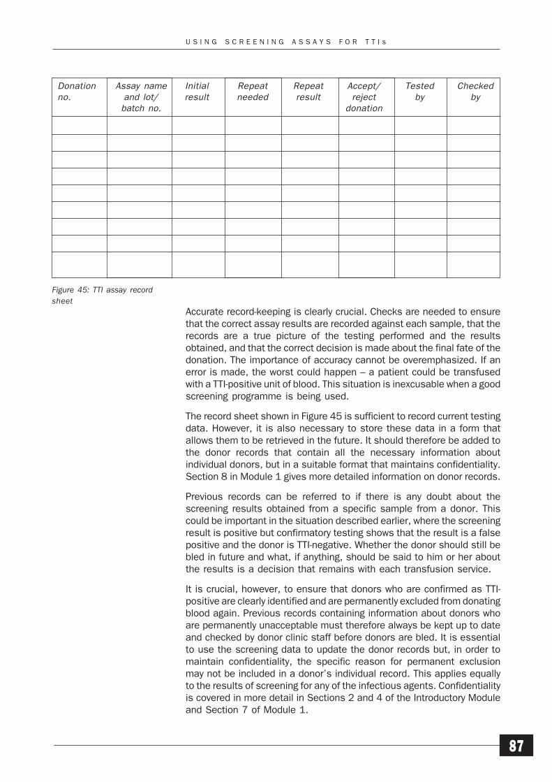
U S I N G S C R E E N I N G A S S A Y S F O R T T I s
87
Figure 45: TTI assay recordsheet
Accurate record-keeping is clearly crucial. Checks are needed to ensurethat the correct assay results are recorded against each sample, that therecords are a true picture of the testing performed and the resultsobtained, and that the correct decision is made about the final fate of thedonation. The importance of accuracy cannot be overemphasized. If anerror is made, the worst could happen – a patient could be transfusedwith a TTI-positive unit of blood. This situation is inexcusable when a goodscreening programme is being used.
The record sheet shown in Figure 45 is sufficient to record current testingdata. However, it is also necessary to store these data in a form thatallows them to be retrieved in the future. It should therefore be added tothe donor records that contain all the necessary information aboutindividual donors, but in a suitable format that maintains confidentiality.Section 8 in Module 1 gives more detailed information on donor records.
Previous records can be referred to if there is any doubt about thescreening results obtained from a specific sample from a donor. Thiscould be important in the situation described earlier, where the screeningresult is positive but confirmatory testing shows that the result is a falsepositive and the donor is TTI-negative. Whether the donor should still bebled in future and what, if anything, should be said to him or her aboutthe results is a decision that remains with each transfusion service.
It is crucial, however, to ensure that donors who are confirmed as TTI-positive are clearly identified and are permanently excluded from donatingblood again. Previous records containing information about donors whoare permanently unacceptable must therefore always be kept up to dateand checked by donor clinic staff before donors are bled. It is essentialto use the screening data to update the donor records but, in order tomaintain confidentiality, the specific reason for permanent exclusionmay not be included in a donor’s individual record. This applies equallyto the results of screening for any of the infectious agents. Confidentialityis covered in more detail in Sections 2 and 4 of the Introductory Moduleand Section 7 of Module 1.
Donation Assay name Initial Repeat Repeat Accept/ Tested Checkedno. and lot/ result needed result reject by by
batch no. donation
S E C T I O N 6
88
ACTIVITY 34
What system is used in your laboratory for retrieving data fromprevious tests on donors? Can you suggest any ways of improving theretrieval system? Note any recommendations down on your ActionList.
Very often, the TTI screening results have to be entered onto a separatesheet, such as an overall worksheet that contains all the current testinginformation. This may include:
blood grouping
antibody screening
relevant medical information
the results of screening the blood for infectious agents.
If your screening programme includes screening for several infectiousagents, a number of different systems may be used to report thescreening results which then all need to be coordinated. Two basicapproaches can be used to record the screening results. A worksheetcan be used for each different assay and the results gathered togetherat the completion of screening. Alternatively, a single worksheet can beused that enables the results of all the different assays to be enteredagainst each donation. The number of different assays performed andthe total number of samples screened will determine which of theseapproaches would be most suitable in your laboratory.
The results from the screening laboratory can be reported either asindividual assay results or as a single positive or negative result. Usinga single result is a very effective way of reporting because the screeninglaboratory is then solely responsible for gathering and assessing all thescreening results and thereby determining the overall suitability of thedonation for issue.
6.4 STORING SCREENING RESULTS
The screening results provide a permanent record of the testing of eachdonation and need to be stored confidentially for future retrieval, ifnecessary. What does storage mean? It does not mean putting recordsin a room or cupboard, shutting the door and forgetting them. If there isno intention of ever referring to the records, there is no point in keepingany records at all. The purpose of record-keeping is to ensure thataccurate records are stored in a logical way so that a particular set ofresults can be retrieved quickly and easily and there is full traceability.
There are a number of factors that need to be considered before anyrecords can be stored correctly:
the records must be complete
the length of storage time must be decided
a suitable storage area must be selected
U S I N G S C R E E N I N G A S S A Y S F O R T T I s
89
the storage area must be kept secure in order to maintainconfidentiality
the storage system must enable the data to be retrievedeasily
the conditions must be suitable for the long-term storageof records
a suitable disposal system must be available.
In some countries, the decision on how long to store records is made atnational level and guidelines are issued which must be followed in allcentres. In other countries, there is no policy on the length of time forwhich records must be stored, or the results are only kept until the bloodhas been transfused.
In countries which produce plasma for the preparation of other therapeuticproducts, records need to be stored for a longer period of time than inthose which simply collect and transfuse essentially unprocessed blood.Red cells are usually used within one month, but some of the otherproducts prepared from harvested plasma may not be used for a numberof years; immunoglobulin preparations, for example, may have an expirydate up to five years after the date of donation. Clearly, some laboratoriesare unable to store records for such long periods, but it is still importantto provide facilities for long-term storage, even if only for about one year.
ACTIVITY 35
How are records of screening results stored in your centre so thatthey are kept confidential, but are easy to retrieve? Think carefullyabout the factors listed above. If you can suggest any improvementsthat could be made to the storage system, note them on your ActionList.
6.5 HANDLING TTI-POSITIVE DONATIONS
Once a TTI-positive donation has been identified, it must be removed anddestroyed as soon as possible. When handling positive material,remember that there is always a risk of accidental infection. All positivedonations must be clearly marked to avoid them re-entering the issuableblood stocks and the appropriate safe handling procedures should beobserved at all times. Each laboratory must design and enforce suitableguidelines to ensure that the risks of infection of laboratory staff from thematerial that they handle, both pathological samples and units of blood,are reduced to a minimum.
As you saw in Section 3 of the Introductory Module, the method used todispose of positive donations safely has to be determined by eachlaboratory and depends on the facilities available. However, in order toensure that a safe system is in operation, you should take the followingsteps.
S E C T I O N 6
90
1 Confirm the identity of the positive donation. Make asecond check of the assay worksheets to ensure that thecorrect donation has been identified.
2 Physically retrieve and remove for disposal the donationand any products that have been prepared from it. Ifunsuitable or unsafe donations are stored for a short timebefore disposal, the storage area must be separate fromthe main blood bank. Mark all unsuitable donations insome way to make it clear that they should not betransfused.
3 Safely dispose of the donation. Ideally, it should bedestroyed by autoclaving, followed by incineration. Ifautoclaving is not possible, incineration is the next bestmethod of disposal. Avoid cutting the bag open andwashing the blood away or burying the blood unless nosuitable facilities for disposal are available; these methodscan be dangerous and do not destroy the potentialinfectivity of the blood. If there is no alternative to burying,make sure that the infected blood bag is securely packagedand buried deeply so that it cannot be dug up by animals.If blood bags are opened, the infected blood must bepoured into a suitable deep pit with strong disinfectant,such as sodium hypochlorite in concentrated form. Thebags should then be burned immediately.
You will find WHO guidelines for the safe disposal of HIV-infected blood in Biosafety Guidelines for Diagnostic andResearch Laboratories Working with HIV (WHO AIDSSeries, No. 9, 1991).
4 Maintain adequate completed records to demonstratethat the donation was actually removed from stock anddestroyed.
5 Make all staff aware of the importance of the correctidentification and safe disposal of all unsuitable blood.The procedure should be reviewed regularly.
ACTIVITY 36
What procedure is used in your centre to ensure that TTI-positivedonations are correctly identified, removed from stock and disposedof safely? If you can suggest any improvements to the procedure,note them down on your Action List.
If there is no standard procedure in your laboratory, it is important todevelop a system which is followed by all staff. Talk to yoursupervisor and colleagues about designing and implementing asuitable system for your laboratory. Note down your recommendationson your Action List.

U S I N G S C R E E N I N G A S S A Y S F O R T T I s
91
6.6 HEALTH AND SAFETY ASPECTS OF COMMERCIAL ASSAYS
Health and safety are covered in detail in Section 3 of the IntroductoryModule. We shall focus here on the health and safety aspects of thecommercial TTI screening assays.
Assay components
ACTIVITY 37
From what you have already learnt about TTI assays in general, listthe components of the assays that you think might present a healthand safety risk. In what way might they present a risk?
It is often difficult to identify the reagents that present a genuine risk asvirtually all chemicals can be considered dangerous. However, we needto consider only certain components of the commercial assays here.Clearly, these components are not found in every kind of assay, and theassays are presented in a number of different formats.
The components that could present health and safety risks are:
wash buffer
substrate
acid stopping solution
control sera.
Particle agglutination assays do not produce wash fluid waste or useacid.
Wash buffer
Wash buffer is supplied as a concentrated solution and therefore ispotentially far more hazardous than it is when at working dilution. Sincethere is a high concentration of chemicals in the stock solution, it maybe caustic or cause other skin irritation or it may be toxic. Azide-containing solutions have potentially toxic levels of azide and should notbe disposed of through copper waste pipes because explosive compoundsmay result.
Substrate
Some of the synthetic substrates (chromogens) used in modern assayshave been investigated for possible carcinogenic or teratogenic effects.Although the volumes in each assay kit are small, repeated exposure tothese chemicals could be harmful.
Acid stopping solution
Stopping solution is usually dilute acid solution, but at a concentrationthat can still cause irritation or skin damage. All acid solutions should behandled with care and neutralized with sodium bicarbonate beforedisposal.
S E C T I O N 6
92
Control sera
Control sera are screened for the presence of other infectious agents andare treated to inactivate any potentially infectious material present.However, they are human in origin and should therefore always behandled as if they are capable of transmitting infection.
It is important to realize that these components present what are onlypotential risks and that they need not be hazardous if basic laboratorysafety procedures are observed. Unless specifically stated in themanufacturer’s instruction leaflet, no specialized facilities are requiredto handle the reagents in the assays. Remember that the safe disposalof the used assay components is important to protect not only laboratorystaff but also any other individual who may come into contact with theused assay components or other waste material.
ACTIVITY 38
List the assay materials that may present a hazard in their disposaland suggest ways of disposing of them safely.
Disposal of potentially hazardous materials
There are a number of components that require safe handling anddisposal after screening has been performed. The subject of safehandling has been covered extensively in the WHO publication BiosafetyGuidelines for Diagnostic and Research Laboratories Working with HIV(WHO AIDS Series, No. 9, 1991). There are however, some additionalpoints to consider.
Acid
Laboratories that use EIAs invariably end up with a number of finishedassay plates containing acid. These should be put into bicarbonatesolution to neutralize the acid. If disinfectant is to be used, it can beadded later when the acid has been completely neutralized. Care mustbe taken when adding hypochlorite solutions to acids as chlorine gas canbe released. Excess bicarbonate must be used to ensure that all the acidis neutralized.
Fluid waste
Laboratories using EIAs also accumulate a large volume of infectiouswaste wash fluid. Remember that the wash fluid contains the originalserum from the first stage of the assay procedure. Disinfectant may beadded to this to the correct final concentration before disposal.
Solid waste
All assays generate an amount of contaminated solid waste. Do notattempt to reuse any disposable items, but dispose of them correctly andsafely.
U S I N G S C R E E N I N G A S S A Y S F O R T T I s
93
ACTIVITY 39
If there is a health and safety policy in your laboratory, check that itcovers all the necessary areas and working practices in yourscreening programme.
If it does not, talk to the person who is responsible for health andsafety and ask how regularly the policy is rewritten and updated. Ifyou can suggest any ways of improving the handling and disposal ofpotentially hazardous waste, note them down on your Action List.
Remember that laboratories contain many potential hazards because ofthe nature of the substances used and the tasks performed. All membersof staff must be appropriately trained so that they are aware of all thesepotential hazards and follow the safety procedures at all times.
SUMMARY
1 The instructions provided by the manufacturer of ascreening assay must always be followed exactly toensure that the assay has been performed correctly andthe results are reliable.
2 Initially reactive samples should be retested to confirmthe result, using a different assay from the initial test.
3 Safety precautions must always be observed whendispatching samples for confirmatory testing.
4 Accurate and complete records must be kept of screeningresults. They should be stored safely, using a system thatenables easy retrieval for future reference to ensure thatTTI-positive donors can be identified and excluded fromdonating blood in the future, while maintainingconfidentiality.
5 TTI-positive donations must be correctly identified, removedfrom storage and disposed of safely. An accurate recordshould always be kept of the disposal of positive donations.
6 Health and safety procedures should be followed at alltimes when handling assay components and waste.
SELF-ASSESSMENT
20 How does confirmatory testing differ from repeat testing?
21 What are the five precautions that should be taken whendespatching pathological specimens through the post?
22 Why is it important to keep accurate records of assayresults?
S E C T I O N 6
94
23 Why is it important to be able to retrieve previous testingdata?
24 If autoclaving is not possible, what is the next best methodof safely disposing of TTI-positive donations?
PROGRESS CHECK
Before moving on to Section 7, spend a few minutes thinkingabout whether you have achieved the learning objectives forSection 6. These were to:
1 Use assays correctly to develop an effective TTI screeningprogramme.
2 Determine the correct screening results for a set of donorsamples.
3 Maintain accurate and complete records of the screeningresults.
4 Review the health and safety procedures in your laboratoryfor handling and disposing of TTI-positive donations,assay components and waste.
If you feel confident that you have understood everything inthis section, turn to Section 7.
If you feel that you need to spend more time on this section,go back to the parts that are most unfamiliar or that you havefound difficult. You may find it helpful to talk to other people,such as your supporter or other senior colleagues, if there isanything you are still not sure about.

Q U A L I T Y S Y S T E M S I N S C R E E N I N G F O R T T I s
95
Quality Systems in Screeningfor Transfusion-TransmissibleInfections
The purpose of this section is to consider the importance of qualitysystems in the maintenance of an effective TTI screening programme,regardless of how many tests are performed or how many staff areemployed.
LEARNING OBJECTIVES
When you have completed this section, you should be able to:
1 Review the overall quality system in your laboratory.
2 Review the system for documenting screening proceduresin your laboratory.
3 Contribute to the preparation of standard operatingprocedures (SOPs) and follow them correctly.
4 Identify any weak areas in your own laboratory qualitysystem.
7
S E C T I O N 7
96
7.1 THE NEED FOR QUALITY IN TTI SCREENING
Section 4 of the Introductory Module deals in detail with quality andimplementing a quality system. Reread that section before continuingwith this module, particularly if you are still unclear about the terminologyused, and are not familiar with standard operating procedures.
As you saw from the Introductory Module, there are many aspects of thecollection and processing of donated blood which must be constantlymonitored to ensure quality throughout the blood transfusion service. Inthis section, we shall focus on quality in the specific context of thescreening of blood to ensure that the blood and blood products are safefor transfusion or other specified purposes.
No matter what assay is used or how many tests are performed, a quality-orientated approach is essential in the successful operation of anyscreening programme. Quality systems must be developed and appliedin all blood transfusion centres, laboratories and hospital blood banks.However it is important to develop a quality system that is appropriateto the individual BTS/laboratory and its specific activities.
Let us consider some of the constraints that a screening laboratory mayface. These will vary from laboratory to laboratory, but in the area ofscreening for infectious agents they are most likely to include technical,financial and time constraints.
1 Technical constraints include the ability of a laboratory toperform a certain type of assay and to ensure thesensitivity of the assay that is used.
2 Financial constraints include the amount of money availableto use for screening in relation to the actual costs ofscreening for the laboratory.
3 Time constraints include the amount of time available totest the blood before it is required for transfusion to apatient.
It may be difficult to overcome some of these constraints, but it shouldstill be possible to develop and maintain a quality system that isappropriate and feasible in your particular circumstances.
7.2 QUALITY SYSTEMS
The introduction of a quality based approach, or even the improvementof an existing quality system, starts with a recognition of the importanceof a consistent quality focused approach to procedures and workingpractices in order to provide a safe and effective product at all times. Asyou saw in the Introductory Module, introducing a quality systemessentially involves five stages.
1 Seeking and obtaining high-level support and resources.
2 Assessing what is required in order to achieve quality.
3 Planning the action that needs to be taken and identifyingthe best way to do it.

Q U A L I T Y S Y S T E M S I N S C R E E N I N G F O R T T I s
97
4 Implementing the changes required, including establishingsystems for monitoring and control.
5 Monitoring the system to assess how well it is operatingand to identify any further changes needed in order toensure that quality is maintained.
The basis for any quality system is that procedures are defined anddocumented and can then be monitored or recorded in some way as, forexample, in the case of an assay failure. Improvements can then bemade to the procedure because there is a full record of its operation.Many aspects of laboratory work can be documented and form the basisfor a quality system, including:
1 The sample collection date.
2 The sample test date.
3 The identity of the test run in which each sample wasincluded.
4 The identity of all samples in each testing run and a recordof the position of these samples in any testing system.
5 The manufacturer, product number, lot or batch numberand expiry date of the assay kits used.
6 The name of the operator and the supervisor.
7 The assay procedure.
8 The results obtained (eye-read and manually-transcribedresults, if necessary).
9 The preparation of any reagents or buffer solutions usedin the assay which were prepared in the laboratory.
10 The preparation and maintenance of any equipment usedto perform the assay. If incubators are used, temperaturecalibration checks should be included.
11 Temperature monitoring records for the laboratory itself(this is important if room-temperature incubations arerequired), incubators, water-baths and refrigerators usedfor the storage of assay kits or reagents.
12 Maintenance and calibration records for the equipmentused, including any mechanical pipettes.
13 Records of the disposal of any positive donations, withdetails of the retrieval of the packs and any productsprepared from them, and the actual destruction of thepacks.
These are some of the main areas where written records are important.You may be able to think of some more.
ACTIVITY 40
Look at the list above of areas where written records areimportant. Are they all documented in your laboratory?
S E C T I O N 7
98
If any of these aspects of laboratory work are not documented,talk to your supervisor about the importance of recording them inthe future and about any suggestions you may have aboutadditional procedures that should be documented. Note down yourrecommendations on your Action List.
7.3 STANDARD OPERATING PROCEDURES
Standard operating procedures (SOPs) are an important part of thewritten quality system. An SOP is a set of written instructions on how toperform a specific task. Thus, any laboratory should have a number ofSOPs covering all the important tasks within the laboratory.
A laboratory that tests 60 donations per day using an EIA, for example,should at least have SOPs for the following:
the reception and handling of samples
performing the assay
use and maintenance of the plate washer
use and maintenance of the plate reader
use and maintenance of the pipettes
issuing the results
retrieving and disposing of positive donations
cleaning and decontaminating the laboratory.
It is important to understand that the instructions given in an SOP are notprovided simply as a guide to help someone perform the particular task,but that they set out the only way that the task is to be performed in thatlaboratory. They are therefore a set of instructions to be followed at alltimes by all staff.
It seems sensible to be strict about the way tasks are performed whenyou consider all the tasks that are necessary to ensure that the screeningprogramme runs smoothly. However, all commercial assays are providedwith a set of instructions and all equipment comes with the necessaryinstructions. Why then do we need SOPs for these aspects of screening?
The reason is that the manufacturer’s instructions are instructions forgeneral use and they may not be sufficient or appropriate in everysituation because they do not relate to each laboratory’s specificrequirements. For example, a laboratory’s SOP covering the use of aparticular assay may include such additional information as:
the types of samples tested
the preparation of any reagents
the checking and recording procedures needed
the handling of samples before testing
the use of the kit controls and any external controls
the use of the equipment required to perform the assay(this would simply be mentioned briefly in the assay SOP,
Q U A L I T Y S Y S T E M S I N S C R E E N I N G F O R T T I s
99
with references to separate SOPs for the use of eachpiece of equipment)
the use of the assay itself
the recording of any testing data on laboratory worksheets
the method of reading the assay results
the calculation of the final results
the use of these results (this would simply be mentionedbriefly in the assay SOP with reference to a separate SOPfor overall result handling)
clearing and cleaning up at the completion of testing.
As you can see, an SOP provides detailed instructions on the way in whichthe assay should be performed in your particular laboratory and withinyour screening requirements. Furthermore, the same applies to anyequipment used; the SOP covers the way in which the equipment shouldbe used as part of your assay procedure. In all cases where manufacturers’instructions are provided, they should be incorporated into the SOP andcan be attached as an appendix to it.
You will find an example of an SOP covering the performance of a simpleEIA anti-HIV screening assay in Appendix 2. Further examples of SOPs areincluded in the Introductory Module and Module 1.
Preparing an SOP
Ideally, the preparation of an SOP should be a team effort. The first draftshould be written by the people performing that particular job. The finaldraft should then be prepared by the head of the laboratory, but thisshould be rechecked by a member of staff who performs that job, beforethe SOP is issued. In order to ensure an SOP is written in the correct way,a set of general guidelines should be provided so that all staff understandexactly what should be included in the draft, and in what order.
The contents of an SOP obviously depend on the particular procedurethat it covers but, as the Introductory Module showed, SOPs can usuallybe broken down into six main sections including:
purposepurposepurposepurposepurpose: the purpose for which the SOP has been written
responsibilitiesresponsibilitiesresponsibilitiesresponsibilitiesresponsibilities: the responsibilities of the different gradesof staff who may use the SOP or be in charge of the workarea in which the SOP is used
restrictionsrestrictionsrestrictionsrestrictionsrestrictions: who may and who may not have use of theSOP, where the SOP is to be used, what the SOP is to beused for
definitionsdefinitionsdefinitionsdefinitionsdefinitions: the definitions of words, phrases orabbreviations used in the SOP
items requireditems requireditems requireditems requireditems required: items required to be able to follow theSOP, including other documentation required (such asSOPs, forms, health and safety guidelines), equipment
procedureprocedureprocedureprocedureprocedure: precise details of the procedure, clearlydescribed in numbered steps that logically follow the
S E C T I O N 7
100
working sequence, including any quality control proceduresinvolved, the procedure for interpreting and reporting theresults and the action to be taken if problems occur.
In addition to the basic framework and contents, the SOP should beuniquely identified with:
a unique SOP identity number including version number
the date when the SOP was written or revised
the name of the person who prepared or revised the SOP.
Appendices may be included which provide any additional relevantdocumentation or information, such as copies of any standard forms orlabels to be completed or used during the procedure, operating instructionsand methods of use recommended by manufacturers of equipment anddiagnostic reagents. However, they should only be included if there is aclear benefit to the user of the SOP.
Instructions in the SOP should be written in the imperative.
Each SOP should be reviewed regularly, ideally at least once each year, incase any modifications are required as a result of direct changes in theprocedure, such as amendments in the manufacturer’s instructions forperforming an assay, or indirect changes in the procedure, such asalterations in an associated procedure that affect certain parts of the SOP.
ACTIVITY 41
Are SOPs used in your laboratory? If they are, assess how effectivethey are in contributing to a quality system, by answering thefollowing questions:
Are there SOPs for all the major activities in the screeningprogramme, such as those listed on page 98?
Do the SOPs cover all aspects of the procedures?
Are they followed at all times by all members of staffinvolved in performing the procedures?
How frequently are they reviewed and, where necessary,updated?
If you can suggest any ways in which the preparation and use ofSOPs could be improved in your laboratory, note them on yourAction List.
If SOPs are not yet used in your laboratory, identify an importanttask that you perform regularly as part of your job and try todevelop a simple SOP. Discuss it with any colleagues who alsoperform the same task and then ask your supervisor to review itand amend it, where necessary. Once it has been finalized, ensurethat all members of staff follow it at all times.
Note down on your Action List any other areas where you think thatSOPs are required and discuss them with your supervisor.
Q U A L I T Y S Y S T E M S I N S C R E E N I N G F O R T T I s
101
7.4 LABORATORY WORKSHEETS (FORMS)
It is clear that there are many areas within the laboratory where a writtenrecord is either essential or would be beneficial. Some laboratories,however, face the problem of having insufficient staff to maintaincomplete records since ideally at least two people are required: one toperform the action and the other to check the action. In a laboratory withonly one main or regular staff member, how can a quality system bemaintained in the screening programme?
It can be argued that in a very small laboratory, even though only smallnumbers of donations may be screened, quality is even more importantsince all the responsibility rests with just one person. It could be easierfor mistakes to go unnoticed. Some sort of quality system is thereforeessential.
In this kind of situation, laboratory checklists that can also serve aslaboratory worksheets are perhaps the simplest and most appropriatemeans of maintaining a quality system. All the assays performed in thelaboratory could be included on one checklist or a different checklistcould be used for each assay. The checklist should detail all the requiredactions in the correct sequence, and should have space or boxes forrecording whatever other information may be required, including signaturesand dates.
The checklist can then be used to record the actions that have beentaken when performing an assay, as well as to record any other detailsrequired. After the screening has been performed, the completedworksheet becomes the record of what has happened and thus completesthe quality circle providing part of the traceability through the laboratory.It becomes the quality record of that batch of testing.
ACTIVITY 42
Design a simple checklist that would be suitable for use for anyscreening assay in your own laboratory.
7.5 QUALITY AUDITS
The use of SOPs and laboratory worksheets forms the basis of a qualitysystem, but there are many areas within the laboratory that also requireregular monitoring to ensure that a full quality system is in place. Thepurpose of a quality audit is to check the integrity of the quality system,that it is comprehensive, effective and, importantly, actually followed. Akey part of this is traceability, the ability to trace what has happened toa unit of blood from donor to patient. If, for example, a particular packof blood was selected that had been fully tested and cleared fortransfusion, could the testing details of that donation be traced backthroughout the laboratory screening programme and, ultimately, to thedonor? As well as checking the screening results for the donation, thevalidity of the testing itself would be examined: for example, did thecontrols perform as expected and were the results obtained within thedefined ranges? Was the test run that included that particular donationvalid?
S E C T I O N 7
102
Clearly, it would not be possible to trace the testing details of a donationunless all the relevant information were recorded together with thescreening results. What other information is needed to demonstrate thatthe assay was not only performed correctly, but that all the associatedactions were performed and checked, and the complete testing run wasvalid? The answer to this question depends on the type of assay in use.The more complex assays require more complex associated actions.However, the list on page 97 covers the basic information that would beexpected to be available.
If these records are maintained, they will be able to validate the test runand thus complete the microbiology part of the audit. As well as providingvalidation for quality audits, they are important in the continuousmonitoring of the performance of the laboratory so that appropriateaction can be taken if any problems occur.
ACTIVITY 43
From your transfusion records, select a donation that was testedthree or four months ago. Follow the testing records back to thedonor.
Were you able to do this? If you could not follow the records of thedonation back to the donor, at what point did the record-keepingsystem fail?
Talk to your supervisor about your findings and try to suggest waysof improving the record-keeping system to ensure that an audittrail could always be followed. Note down your recommendationson your Action List.
SUMMARY
1 A quality based approach must be applied to everyscreening programme, even in the smallest laboratories.
2 Written records are the basis of a quality system becausethey enable procedures to be monitored.
3 SOPs should be prepared for all the main tasks undertakenin the laboratory.
4 A simple means of maintaining a quality system is to uselaboratory checklists that also serve as worksheets forrecording any actions taken and then become the finalrecords for the work performed.
5 Quality audits enable the quality system to be assessedand its effectiveness determined.
Q U A L I T Y S Y S T E M S I N S C R E E N I N G F O R T T I s
103
SELF-ASSESSMENT
25 Why are SOPs required for the use of assays wheninstructions are provided by the manufacturer?
26 Why is a quality system particularly important in a smalllaboratory with only one member of staff?
PROGRESS CHECK
Before moving on to Section 8, spend a few minutes thinkingabout whether you have achieved the learning objectives forSection 7. These were to:
1 Review the system for documenting screening proceduresin your laboratory.
2 Contribute to the preparation of standard operatingprocedures (SOPs) and follow them correctly.
3 Identify any weak areas in your own laboratory qualitysystem.
If you feel confident that you have understood everything inthis section, turn to Section 8.
If you feel that you need to spend more time on this section,go back to the parts that are most unfamiliar or that you havefound difficult. You may find it helpful to talk to other people,such as your supporter or other senior colleagues, if there isanything you are still not sure about.

104
8Screening for OtherTransfusion-TransmissibleInfections
The purpose of this section is to consider the other most significantinfectious agents that can be transmitted by blood and blood products:
hepatitis B virus (HBV)
hepatitis C virus (HCV)
human T cell leukaemia viruses I + II (HTLV I + II)
Treponema pallidum (syphilis)
Plasmodium species (malaria)
Trypanosoma cruzi (Chagas disease).
The detailed aspects of the main screening tests used have already beenconsidered in earlier sections. In this section, therefore, we shall simplyoutline the natural history of these infectious agents, including theroutes of transmission and the risks of transmission by blood transfusion.Although some of these agents may not be a problem in your country, theyrepresent those commonly considered to be both transmissible andtransmitted by transfusion. You may wish to study only those agents thatare known to be prevalent in your country although, with changes in theglobal distribution of infectious diseases, it is important that bloodtransfusion services should be aware of potential threats to the bloodsupply and have monitoring programmes in place to identify any emergingtransmissible diseases in their country or region.
Please note that there are no activities in this section although you maywish to repeat some of the relevant activities in Section 3: The HumanImmunodeficiency Viruses, but applying them to these other infectiousagents.
The principles discussed in this module apply to screening for anyinfectious agent that may be transmitted by blood transfusion. However,the range of commercially available screening tests varies according tothe agent.
S C R E E N I N G F O R O T H E R T T I s
105
In countries where the prevalence and incidence of certain infectiousagents are very low, it may not be necessary to screen routinely for them.In other countries, where infection by an agent is endemic, screening maynot be considered to be sensible or practical. Such considerations areimportant because, although blood safety is important, the resourcesavailable to ensure the safety of the blood supply are often limited andmust be used carefully and wisely to ensure maximum effectiveness.
The information given in this section about these infectious agents is notintended to be comprehensive, but should enable you to understand theagents, the mechanisms of infection, the laboratory screening neededand the significance for transfusion practice. The figures in this sectionare included to provide additional information, but you do not need tomemorize them. If you need further information, consult an appropriatetextbook.
LEARNING OBJECTIVES
When you have completed this section, you should be able to:
1 Describe the basic features of infection with HBV, HCV,HTLV, T. pallidum, Plasmodium species and T. cruzi.
2 Explain the significance of these agents for bloodtransfusion practice.
3 Describe the laboratory screening needed to identifydonations from infected donors.
S E C T I O N 8
106
8.1 HEPATITIS B VIRUS
The infectious agent
Hepatitis B virus (HBV) is a DNA virus of the hepadnavirus family. Thevirus is transmitted parenterally, including through intimate contact, andinfection may follow one of two courses:
acute infection with the subsequent clearance of the virusand development of immunity
chronic infection with the persistence of virus replicationfor extended periods, even the lifetime of the individual.
HBV infection can lead to severe disease: cirrhosis, hepatocellularcarcinoma (HCC) and ultimately liver failure. Asymptomatic infections arevery common, however, with most individuals resolving infection anddeveloping immunity without any symptoms or with only mild symptoms.The prevalence of HBV varies across the world from 0.1–0.2% in lowendemic countries, up to 3% in some Mediterranean countries and up to15% in Africa and the Western Pacific region. The rates are even higherin some isolated communities. It is estimated that there are at least250 million chronic carriers worldwide and that approximately half of theglobal population has been infected by HBV.
Structure
Two main types of circulating virus particles are identified following HBVinfection: the “infectious” virion, the Dane particle, and a vast excess ofsmaller particles that consist only of viral surface protein with no DNA andthat are therefore not considered to be “infectious”.
The virus particle (virion) is 42 nm in diameter and is known as the “Daneparticle” after the scientist who first identified it in the serum of infectedindividuals. This is the “infectious particle” in HBV infection. In the centreof the virion is the capsid core, which is 27 nm in diameter. It containsboth the viral nucleic acid and the enzyme DNA polymerase which isessential for successful infection by the virus.
A number of important proteins are found in the particle. The majorprotein of the virion is known as hepatitis B “surface” antigen (HBsAg)which completely encloses the capsid. HBsAg is produced in excess bycells infected with the virus and is released into the circulation as thesmall “noninfectious” particles of pure HBsAg. The capsid contains twomajor proteins: hepatitis B “core” antigen (HBcAg), and hepatitis B “e”antigen (HBeAg). HBcAg itself cannot be detected in the circulation, butis detectable in infected liver cells. Circulating HBeAg appears fairly earlyon in infection and, in general, indicates viral replication and the releaseof infectious virions.
The smaller particles (either spherical particles of 22 nm diameter orsmall tubular particles) containing only the major viral protein, HBsAg arereleased into the serum of infected individuals. They are produced in alarge excess to that needed to form new virions and this excess iscontinually released into the circulation during infection. These smallerparticles are therefore considered uninfectious, although they act as amarker of infection – both acute and chronic.
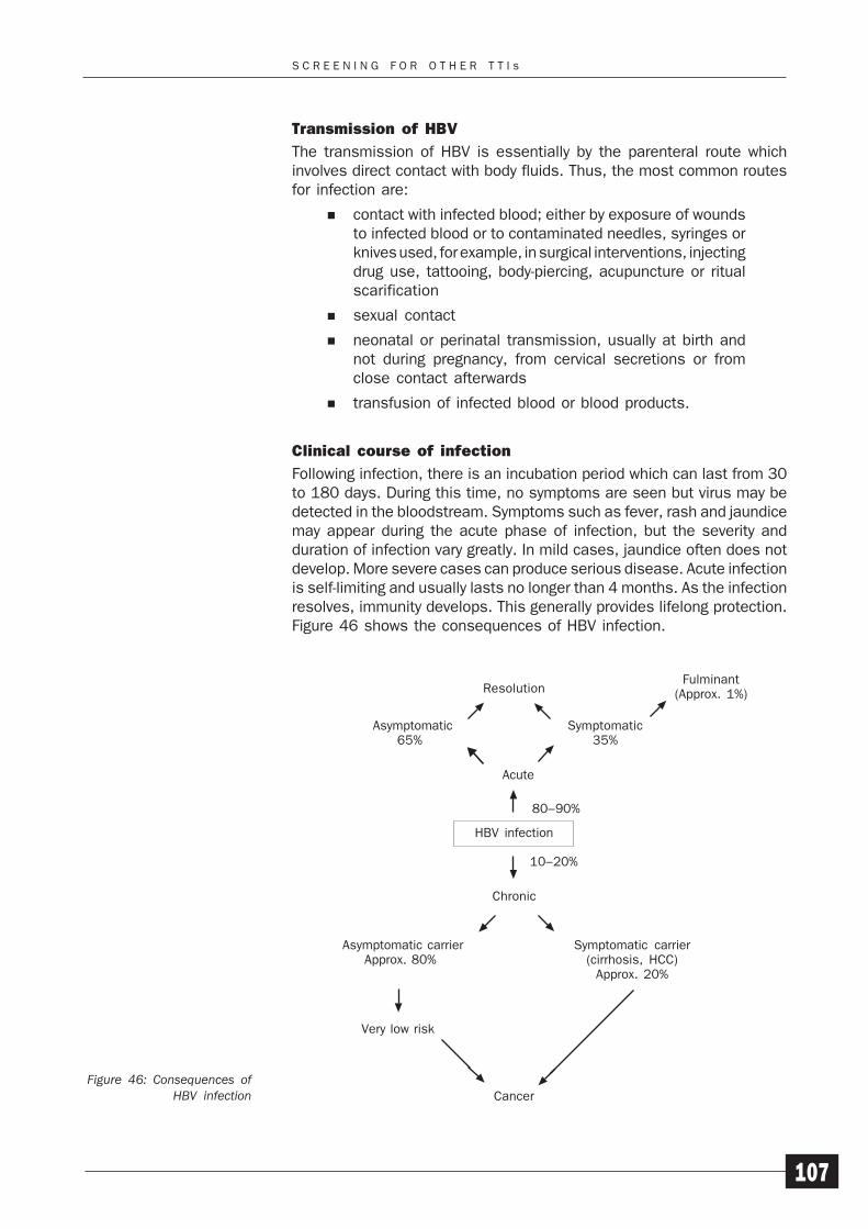
S C R E E N I N G F O R O T H E R T T I s
107
Transmission of HBV
The transmission of HBV is essentially by the parenteral route whichinvolves direct contact with body fluids. Thus, the most common routesfor infection are:
contact with infected blood; either by exposure of woundsto infected blood or to contaminated needles, syringes orknives used, for example, in surgical interventions, injectingdrug use, tattooing, body-piercing, acupuncture or ritualscarification
sexual contact
neonatal or perinatal transmission, usually at birth andnot during pregnancy, from cervical secretions or fromclose contact afterwards
transfusion of infected blood or blood products.
Clinical course of infection
Following infection, there is an incubation period which can last from 30to 180 days. During this time, no symptoms are seen but virus may bedetected in the bloodstream. Symptoms such as fever, rash and jaundicemay appear during the acute phase of infection, but the severity andduration of infection vary greatly. In mild cases, jaundice often does notdevelop. More severe cases can produce serious disease. Acute infectionis self-limiting and usually lasts no longer than 4 months. As the infectionresolves, immunity develops. This generally provides lifelong protection.Figure 46 shows the consequences of HBV infection.
Figure 46: Consequences ofHBV infection
80–90%
Asymptomatic carrier Approx. 80%
Symptomatic carrier(cirrhosis, HCC)
Approx. 20%
10–20%
Very low risk
Cancer
Resolution
Acute
HBV infection
Chronic
Asymptomatic65%
Symptomatic35%
Fulminant(Approx. 1%)
S E C T I O N 8
108
However, approximately 10–20% of individuals who have been clinicallydiagnosed as having HBV infection do not resolve their acute infectionand a period of chronic infection begins. In developing countries with ahigh incidence of HBV infection, as many as 90% of perinatally-infectedinfants and 20%–50% of childhood infections develop into chronicinfection. Chronic infection may last from months to years and then:
spontaneously resolve with the development of immunity
reactivate with a new acute episode and the possibility offulminant hepatitis.
In some individuals, chronicity may simply persist for life. It is in thesepersistently chronically-infected individuals that serious consequencesof infection may develop, often slowly over a period of years. In someindividuals, no ill-effects are seen. In many cases, however, chronic liverdisease eventually develops which may lead to cirrhosis, primary livercancer and then death. The risk of death from cirrhosis or liver cancer isapproximately 40% in male HBV carriers and at least 250 000 cases ofhepatocellular carcinoma are reported annually, making it one of themost common cancers in the world.
Laboratory testing for HBV infection in blood donations
Following infection with HBV, a range of circulating markers are detectable,which reflect the replication of the virus and the body’s immune responseto it. These markers are not static but change over time as the infectionprogresses. Figure 47 on page 109 shows the usual profiles of thesemarkers in acute and chronic infections. However, these normal patternsmay not be seen in all infected individuals, atypical patterns are seen.The stage and the progression of infection can usually be determined byidentifying the markers present in a serum/plasma sample or insequential samples from the infected individual. Specific test kits areavailable for all circulating markers of HBV infection and these are usedin the laboratory to determine the stage of infection at any point in time.
The first circulating marker to appear is viral DNA, which appears asreplication begins and virus is released from infected cells. This isfollowed soon afterwards, sometimes simultaneously, by the appearanceof HBsAg which very quickly reaches high titres. HBeAg appears next andthe presence of HBsAg and HBeAg are considered to indicate active viralreplication. Within 1–2 weeks, IgM anti-HBc usually appears and this isused as an indicator of a recent infection. IgM levels drop within about4 weeks and IgG anti-HBc persists, usually lifelong, irrespective ofsubsequent acute or chronic infection. The end of the acute stage ismarked by the gradual fall in titre of HBsAg, with the subsequentappearance, 4–6 weeks later, of anti-HBs. Persistence of HBsAg for atleast 6 months is considered to indicate chronic infection. The appearanceof anti-HBs indicates resolution of infection and immunity. HBeAg levelsfall as anti-HBe appears, but may persist if the infection becomeschronic; this is known as infectious carriage. Chronic infections that areanti-HBe positive are termed noninfectious carriage.
The detection of HBV infection in donated blood is achieved primarily byscreening for HBsAg as it is the first serological marker to appear in the
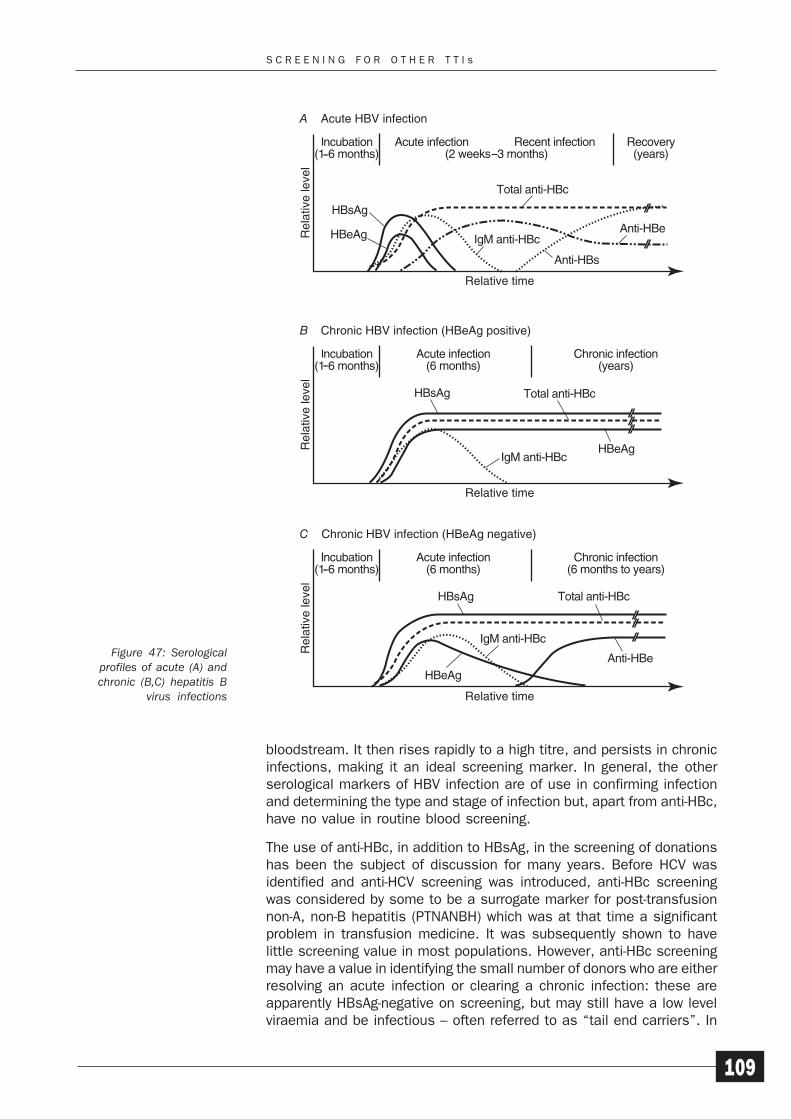
S C R E E N I N G F O R O T H E R T T I s
109
Figure 47: Serologicalprofiles of acute (A) andchronic (B,C) hepatitis B
virus infections
bloodstream. It then rises rapidly to a high titre, and persists in chronicinfections, making it an ideal screening marker. In general, the otherserological markers of HBV infection are of use in confirming infectionand determining the type and stage of infection but, apart from anti-HBc,have no value in routine blood screening.
The use of anti-HBc, in addition to HBsAg, in the screening of donationshas been the subject of discussion for many years. Before HCV wasidentified and anti-HCV screening was introduced, anti-HBc screeningwas considered by some to be a surrogate marker for post-transfusionnon-A, non-B hepatitis (PTNANBH) which was at that time a significantproblem in transfusion medicine. It was subsequently shown to havelittle screening value in most populations. However, anti-HBc screeningmay have a value in identifying the small number of donors who are eitherresolving an acute infection or clearing a chronic infection: these areapparently HBsAg-negative on screening, but may still have a low levelviraemia and be infectious – often referred to as “tail end carriers”. In
A Acute HBV infection
Rel
ativ
e le
vel
Relative time
Recovery(years)
HBsAg
Total anti-HBc
IgM anti-HBc
Anti-HBs
Anti-HBeHBeAg
B Chronic HBV infection (HBeAg positive)
Rel
ativ
e le
vel
Relative time
Chronic infection(years)
Acute infection(6 months)
HBsAg
HBeAg
Total anti-HBc
IgM anti-HBc
C Chronic HBV infection (HBeAg negative)
Rel
ativ
e le
vel
Relative time
Chronic infection(6 months to years)
Acute infection(6 months)
Anti-HBe
Total anti-HBc
IgM anti-HBc
HBsAg
HBeAg
S E C T I O N 8
110
such individuals, anti-HBc may be the only detectable circulating markerof infection and thus may be identifiable only by anti-HBc screening. Thiscould explain the occasional reported cases of post-transfusion HBVresulting from transfusion of donations screened as HBsAg-negative; inthese situations, unfortunately, it is often difficult to demonstrate thatthe patient had no other risks of infection.
Significance for transfusion practice
In the past, transmission via the transfusion of blood and blood productswas a significant route of HBV infection. A clear example was the incidentin which a large number of American army personnel were vaccinatedagainst yellow fever using a vaccine prepared from plasma from individualswho had recovered from the disease but at least one of whom also hadacute HBV infection. This was subsequently transmitted to most of therecipients of the vaccine. The screening of donations for the presence ofHBsAg is now routine in many countries and this has helped to reduceconsiderably the occurrence of post-transfusion hepatitis. It has beenshown that the severity of infection is related to a number of factors, oneof which is the size of the infecting dose. Blood transfusion can thereforebe an efficient route of transmission of HBV because a large amount ofinfected material is passed directly into the bloodstream of the recipient.
HBsAg mutants
Variants of HBV have been described which result from mutations in theviral genome. Very broadly, these can be divided into two groups: thoseaffecting the core region of the genome and those affecting the surfaceantigen region. The mutations in the core region of the genome havenormal HBsAg production and are currently not a major concern in bloodtransfusion. However, the mutations in the HBsAg region are of concernto blood screening since HBsAg expression may be altered such thatsome assays may fail to detect some HBsAg mutant forms. The extentof the problem is hard to assess critically and, although mutants arebeing identified and there are reports of variable reactivity with assays,their frequency is very low; a proportion of those that have been identifiedwere as a result of specific searches for such mutants and not as a resultof transmissions from donations screened as HBsAg-negative.
Hepatitis delta virus (HDV)
Hepatitis delta virus (HDV) is a small RNA virus, currently not firmlyclassified, that requires co-infection with HBV, replicating only in the livercells of individuals already infected with HBV. Although transmissible bytransfusion, screening for HBsAg will also prevent transmission of HDVbecause the virus needs the presence of HBV to replicate.
Prevention of spread of infection
Vaccination is the approach that is now being used globally to reduce therate of transmission, especially through childhood vaccinationprogrammes. This is a long-term approach, however, and the programmestake some time to reach full effectiveness. The screening of all blood andblood products, tissues and organs is therefore an important means of
S C R E E N I N G F O R O T H E R T T I s
111
reducing transmission by these routes. General public health educationprogrammes help to minimize transmission by the sexual and injectingdrug use routes. Specific HBV immunoglobulin is still also used in somesituations where immediate protection is required, such as the preventionof infection in infants born to infected mothers or resulting fromneedlestick injuries where there is a significant risk of HBV infection andthe injured person has not been vaccinated for HBV.
8.2 HEPATITIS C VIRUS
The infectious agent
Hepatitis C virus (HCV) is an RNA virus of the flavivirus family. Althoughinfections with HCV have been recognized for many years, the virus wasfinally identified and characterized only in 1989. The virus is transmittedparenterally, mainly through injecting drug use and via transfusion,although there are still a significant number of cases where parenteralroutes cannot be identified. Currently, like HBV, infection is known tofollow one of two courses: acute infection which is both fairly rapid andrelatively mild in its course or chronic infection which may persist formany years with a significant number of cases progressing to cirrhosisand hepatocellular carcinoma.
The main difference between HBV and HCV is that only about 20–40%of HCV cases are acute; the majority progress to chronic infection. Thecarrier rate varies across the world from 0.05–0.5% in low endemiccountries such as western Europe, north America and Australasia; 1–5%in parts of southern Europe, south America, Asia and Africa; and as highas 20% in Egypt and specific regions of some Asian and African countries.Current estimates are that at least 200 million people are infectedworldwide and, in many areas, the long-term significance of subsequentdisease due to cirrhosis and HCC in these individuals is greater than thatof HBV.
Structure
Although HCV was finally identified in 1989, the intact virion has still notbeen specifically isolated and characterized. The virus is a single-stranded, enveloped, RNA virus 50–80 nm in diameter with a genome ofapproximately 9 400 nucleotides. Work with the virus has all arisen fromthe sequencing of the viral RNA and the expression of viral proteins fromthat RNA. Proteins expressed from the nucleic acid sequence of the virusare used to develop assays to detect the presence of antibody to thevirus and, more recently, viral antigen.
Transmission of HCV
The transmission of HCV is essentially by the parenteral route, whichinvolves direct contact with body fluids. Thus, the most common routesfor infection are:
contact with infected blood; by exposure of wounds toinfected blood or to contaminated needles, syringes orknives used, for example, in surgical interventions, injectingdrug use, tattooing, body-piercing, acupuncture or ritualscarification
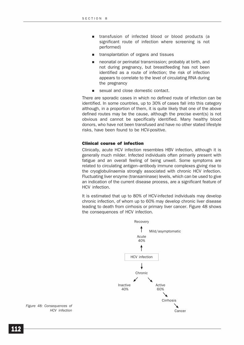
S E C T I O N 8
112
transfusion of infected blood or blood products (asignificant route of infection where screening is notperformed)
transplantation of organs and tissues
neonatal or perinatal transmission; probably at birth, andnot during pregnancy, but breastfeeding has not beenidentified as a route of infection; the risk of infectionappears to correlate to the level of circulating RNA duringthe pregnancy
sexual and close domestic contact.
There are sporadic cases in which no defined route of infection can beidentified. In some countries, up to 30% of cases fall into this categoryalthough, in a proportion of them, it is quite likely that one of the abovedefined routes may be the cause, although the precise event(s) is notobvious and cannot be specifically identified. Many healthy blooddonors, who have not been transfused and have no other stated lifestylerisks, have been found to be HCV-positive.
Clinical course of infection
Clinically, acute HCV infection resembles HBV infection, although it isgenerally much milder. Infected individuals often primarily present withfatigue and an overall feeling of being unwell. Some symptoms arerelated to circulating antigen–antibody immune complexes giving rise tothe cryoglobulinaemia strongly associated with chronic HCV infection.Fluctuating liver enzyme (transaminase) levels, which can be used to givean indication of the current disease process, are a significant feature ofHCV infection.
It is estimated that up to 80% of HCV-infected individuals may developchronic infection, of whom up to 60% may develop chronic liver diseaseleading to death from cirrhosis or primary liver cancer. Figure 48 showsthe consequences of HCV infection.
Figure 48: Consequences ofHCV infection
Acute40%
Mild/asymptomatic
Cancer
Recovery
Inactive 40%
Active60%
HCV infection
Chronic
Cirrhosis
S C R E E N I N G F O R O T H E R T T I s
113
Laboratory testing
It is important to understand how the tests for HCV have been developedbecause this process has been quite unique to HCV and has had animpact on diagnostic testing. As mentioned above, to date, the virusitself has not been isolated intact or cultured in vitro. The nucleic acidsequence has been used to construct recombinant and peptide antigenswhich have then been used in the laboratory tests available. Thisapproach differs from the usual development of viral serology tests whichgenerally originate with the isolation of the virus itself, from which crudeantigen preparations are then made. Following this, recombinant andpeptide antigens are subsequently produced, but with knowledge of thebehaviour of the crude “native” antigens being used in the design of therecombinant and peptide antigens. This has not been the case with HCV;“native” antigens have not been isolated and the tests used jumpedstraight to the use of recombinant and peptide antigens. The firstscreening tests for anti-HCV were relatively basic and used only one HCVrecombinant antigen, which was not very specific. These tests havedeveloped significantly since the first commercially available screeningtest was developed in 1990 and now use a combination of antigens fromdifferent regions of the genome.
Currently, following infection with HCV, three specific circulating markersof infection can be used to identify infected individuals. The first markeris viral RNA, which appears as replication begins and virus is releasedfrom infected cells. This is followed soon afterwards, sometimessimultaneously, by viral antigen (HCVAg) which reaches reasonabletitres, but not as high as with HBV infection. Within 1–4 weeks, specificantibody appears. IgM can be demonstrated very early in the immuneresponse, but only at low titres and these fall rapidly, and appears topersist for life in most individuals although this may subsequently notprove to be the case. Cases of seroreversion have been reported inindividuals who have resolved their infection and chronicity has notdeveloped. As antibody titres rise, antigen titres fall as the free antigenis rapidly complexed with the antibody. Viral antigen can still be detectedin individuals in whom viral replication is continuing if an antigen testutilizing an immune complex dissociation step (ICD) is used.
The detection of HCV infection in donated blood was initially achieved byscreening for anti-HCV and this remains the main test performed today.The detection of antibody indicates both current and past infection. Astechnology has developed, however, some countries now augment theserology with the detection of HCV RNA. Because there can be a relativelylong window period for HCV, it is possible to encounter a donor who hascirculating viral RNA, but no detectable antibody. While tests for viral RNAwill detect such donors, this type of testing requires a much higher levelof staff skills and expertise, infrastructure and resources than serology,and is significantly more expensive. In many countries, any benefits ofsuch testing may actually be outweighed by the costs. A test for HCVantigen has now been developed and made commercially available forblood screening; the test is a standard EIA format. Although not quite assensitive as HCV RNA, testing for HCV Ag can reduce the window periodsignificantly and provides a far simpler and less expensive alternative toHCV RNA testing in situations in which nucleic acid testing is tooexpensive or not feasible.
S E C T I O N 8
114
Significance for blood transfusion
In the past, transmission via the transfusion of blood and blood productswas a significant route of HCV infection. This is clearly demonstrated bythe large numbers of individuals in multi-transfused groups with evidenceof active or past HCV infection – haemophiliacs and thalassaemics beingtwo of the groups most severely affected. The screening of donations forthe presence of antibody to HCV (anti-HCV) is now routine in manycountries with developed healthcare systems, and this has reducedsignificantly the occurrence of PTHNANB. In many countries, cases ofPTHNANB are now virtually unknown, but blood transfusion remains asignificant route of infection in countries that do not screen blooddonations for HCV.
Prevention of spread
Apart from the screening of all blood and blood products, tissues andorgans, the only other intervention currently available to prevent HCVtransmission is the education of infected and at-risk individuals, mainlyinjecting drug users in whom transmission via shared needles and otherdevices is still common in many countries. The risk of transmission ofHCV from mother to infant is not clear; nonetheless, advice aboutbreastfeeding may be appropriate. Specific HCV immunoglobulin is notavailable as the antibodies do not appear to confer immunity, althoughneutralizing the viral antigen so far detected.
8.3 HUMAN T CELL LEUKAEMIA VIRUSES I + II
The infectious agentHuman T cell leukaemia virus (HTLV-I) was the first identified humanretrovirus. It is an oncogenic virusoncogenic virusoncogenic virusoncogenic virusoncogenic virus causing adult T cell leukaemia andlymphoma (ATLL or ATL) and tropical spastic paraparesis (TSP), alsoknown as HTLV-I associated myelopathy (HAM). A second virus, HTLV-II,has also been identified in specific groups of individuals, such asinjecting drug users, although no significant disease process has yetbeen associated with this virus. HTLV-I and -II are distinct, but closelyrelated, viruses with a number of proteins in common as well as uniqueindividual proteins. The distribution of both viruses is very localized, bothgeographically and in specific populations within defined areas acrossthe world – mainly the tropics, Japan, Caribbean, parts of south America,Africa and Australia.
StructureBoth HTLV-I and HTLV-II are enveloped RNA viruses with essentiallyidentical structures surrounding a central core. The virus is approximately100 nm in diameter and has a spikey appearance formed by projectionsof viral glycoprotein from the surface of the viral envelope. The genomeconsists of two single-stranded linked RNA molecules that are containedwithin the core together with the enzyme reverse transcriptase, which isessential for viral replication and integration.
TransmissionIt is thought that virus is virtually always cell-associated, infecting theCD4+ lymphocytes, and is transmitted in these cells parenterally via
oncogenic virusoncogenic virusoncogenic virusoncogenic virusoncogenic virus: A virusthat causes the formationof tumours.
S C R E E N I N G F O R O T H E R T T I s
115
blood or semen, or from mother to infant via breast milk. Once in the host,the virus integrates into the cell DNA and remains there for life; theappearance of antibody marks the resolution of acute infection, but notimmunity. The transmission by breast milk is a major route of infectionin some areas where HTLV-I is endemic. Studies on the transmission ofHTLV-I indicate that the virus is not normally transmitted in utero, but istransmitted in early life through breast milk equally to both male andfemale children. Later in life, however, sexual transmission is almostexclusively from male to female.
Blood transfusion is another potentially significant route of infection.Early studies demonstrated the efficiency of transfusion transmission,but indicated that cell-free products, such as plasma, did not transmitinfection. Fresher components from infected individuals are those mostlikely to transmit the virus. Additionally, recent findings appear toindicate that transfusion transmission may have a much shorter incubationperiod for the development of ATLL – as short as 1 year in some cases.
Clinical course
Most infections with HTLV-I in otherwise healthy individuals areasymptomatic and remain so. However, there is small risk that diseasemay develop at any time up to 40 years after infection. ATLL can presentas an acute leukaemia of CD4+ lymphocytes and death usually occurswithin a year of the onset of symptoms. TSP is a progressive diseaseinvolving the degeneration of neurones in the spinal cord, leading togradual paralysis of the lower limbs.
Similarly, virtually all HTLV-II infections in healthy individuals areasymptomatic. More recently HTLV-I and II infection have been associatedwith certain inflammatory diseases.
Laboratory testing
Following infection with HTLV, there is an incubation period from 30 to90 days before seroconversion. Prior to seroconversion, viral RNA can bedetected in lymphocytes. At seroconversion, antibody to HTLV appears;this is the major target for the diagnosis of HTLV infection. Afterseroconversion, the antibody generally persists at high titres for life,even if clinical disease subsequently develops only much later in life,although it is not protective. The serological responses to HTLV-I andHTLV-II are very similar but, like HIV-1 and 2, there are sufficientdifferences to require specific tests for the detection of anti-HTLV-I andanti-HTLV-II.
Significance for transfusion
The potential significance of blood transfusion as a route of transmissionhas meant that, in a number of endemic countries, the screening ofdonations for anti-HTLV-I and II has been carried out for some time.However, because the virus is cell-associated, only cellular products areimplicated in transmissions; virus has not been found in either acellularproducts, such as fresh plasma, or in any fractionated products that havebeen prepared in accordance with modern fractionation procedures andfollowing current internationally agreed guidelines.
S E C T I O N 8
116
Screening has also been introduced in some nonendemic developedcountries with mixed populations; in some instances, it is restricted topreviously untested donors. In many other countries, debate continueson the need and value of screening donations.
Prevention of spread
The main focus in the prevention of spread of HTLV in endemic areas isthe education of infected and at-risk individuals. Breastfeeding is knownto transmit HTLV, a significant route in endemic areas, and advice mustbe given to infected mothers not to breastfeed. General public healthawareness programmes can also help to limit the risk of sexualtransmission and transmission through injecting drug use. Blood andorgan screening is important in endemic areas and in nonendemic areaswhere the donor population may contain infected individuals. Specificimmunoglobulin is not available, but anti-retroviral therapy can beeffective if given early enough in infection.
8.4 SYPHILIS (TREPONEMA PALLIDUM INFECTION)
The infectious agent
Syphilis is the disease caused by infection with the bacterium Treponemapallidum which is a member of the class of bacteria known as spirochaetes.There are four major human treponemal pathogens which are so closelyrelated that they are described as subspecies of T. pallidum:
T. pallidum subspecies pallidum (syphilis)
T. pallidum subspecies pertenue (yaws)
T. pallidum subspecies carateum (pinta)
T. pallidum subspecies endemicum (bejel).
Here we are primarily concerned with infection due to T. pallidumpallidum (which we shall simply refer to as T. pallidum) as it is the mostimportant pathogen in the group. The other subspecies, which we shallalso consider briefly, are notably different in their routes of transmissionand their clinical significance and sequelae. There are also manynonpathogenic species that commonly infect all mammals.
StructureSpirochaetes are long, thin, highly motile bacteria that have flexible,Gram-negativeGram-negativeGram-negativeGram-negativeGram-negative type, cell walls, composed of an outer membrane, apeptidoglycan layer and an inner cytoplasmic membrane. The outermembrane contains the antigenic proteins of the bacterium. Spirochaetesare generally relatively fragile organisms that are very sensitive totemperature and die rapidly outside their host. They cannot be cultivatedon artificial media, although they can be cultivated in cell culture or inanimals. They are very characteristic organisms and can easily be seenunder dark-field microscopy.
Transmission
Syphilis is endemic in many parts of the world. In some areas, theincidence of disease is increasing as a result of the breakdown of public
Gram-negative:Gram-negative:Gram-negative:Gram-negative:Gram-negative: TheGram stain is a methodof staining bacteriawhich is used to classifythem. Bacteria areeither Gram-negative orGram-positive.
S C R E E N I N G F O R O T H E R T T I s
117
health systems and treatment programmes as a result of naturaldisasters, war and other civil unrest.
Syphilis is essentially a sexually-transmitted disease, although it canalso be spread by other forms of close contact with mucous-membranelesions. Congenital infection is a significant route, especially in endemiccountries with poor healthcare systems and minimal or absent treatmentprogrammes. The organism can pass across the placenta and infect thefetus. If this does not kill the fetus directly, the infant will be born withcongenital syphilis. Since part of the life cycle of treponemal infectioninvolves release into the bloodstream, parenteral transmission mayoccur and, in the past, blood transfusion was a potential route ofinfection, especially if fresh blood was transfused. However, afterdonated blood is stored for 24–48 hours at 4°C, any infection risk isessentially eliminated as the organism is very sensitive to temperatureand is killed quickly at low temperatures, including freezing.
Clinical course of infection
The normal course of syphilis can be divided into early and late infection,the division being approximately two years from initial infection. Infectionthen follows the stages of primary, secondary and tertiary infections.
1 Primary syphilis: following initial contact, the spirochaetespass through the mucous membranes and enter thelymphatic system, leaving a lesion full of treponemes(chancre) at the original site of entry. The normal incubationperiod ranges from 9 to 90 days. Without treatment, thisheals in 4 to 8 weeks but the disease progresses to thesecondary stage.
2 Secondary syphilis: 6 to 8 weeks after the chancre firstappears, new secondary lesions begin to appear on theskin and mucous membranes. These are highly infectiousas they are full of treponemes. There may also be a rashand/or generalized lymphadenopathy. Non-venerealspread of the infection can occur at this stage. Withouttreatment, latent syphilis follows; this is classed as earlylatent until the 2-year mark has passed when it thenbecomes late latent; late latency may last for many years.
3 Tertiary syphilis: this may occur any time from 5 to 40years after the initial infection. This is the most destructivestage of the disease. Lesions may occur in the centralnervous system, cardiovascular system, bones, skin,viscera and eyes where they can cause severe damage.Cases of tertiary syphilis, even in endemic areas, are notcommonly seen.
In cases of congenital infection, the disease can lead to a number ofsevere disabilities, including blindness, deafness and bone disease ifthe fetus does not die during pregnancy and if the disease is untreated.
Laboratory testing
Although direct observation of spirochaetes in fluid from the lesions ispossible using dark-field microscopy, this can be performed only at
S E C T I O N 8
118
certain stages of infection. Serology is therefore the major diagnosticmethod. After infection, specific IgM antibodies appear after about twoweeks, with IgG levels rising from three to four weeks. IgG antibodiestend to persist for life, whether or not treatment has been given.Detectable antibody levels drop only in late tertiary syphilis, possiblybecause of the spread of the disease throughout the body with thesignificant tissue destruction that then occurs.
Two groups of tests are available: nonspecific tests and specific tests.The particular screening strategy developed needs to reflect whether thetesting is for blood donors alone as well as the incidence of infection inthe population from which the donors are drawn.
Nonspecific tests, such as the Venereal Disease Reference Laboratorytest (VDRL) or Rapid Plasma Reagin test (RPR ) use a mixture of highlypurified cardiolipin/lecithin/cholesterol to detect a substance calledreagin which circulates in the bloodstream of recently/actively infectedindividuals. The tests are based on flocculation or aggregation of thecardiolipin reagent, either free in suspension or absorbed to carbonmicroparticles, by the reagin. Reagin is a mixture of specific IgM and IgGantibodies and is a fairly sensitive marker of recent/active syphilisinfection, although it is not particularly specific. Approximately 1–2% ofnormal adults produce nonspecific antibodies which may lead to falsepositive reactions.
Specific tests, such as the Treponema pallidum HaemagglutinationAssay (TPHA) or conventional EIAs, use native T. pallidum or T. pallidumproteins (native or recombinant) as the antigens in a number of differenttypes of test, all of which are aimed at the detection of specific antibodyto the organism.
Many particle agglutination tests, using red cells, latex or gelatinparticles, have been developed which are inexpensive, specific andsensitive. Since most of these tests can be used in a microwell format,they are suitable for the testing of a large number of samples. Many highquality EIAs have also been developed, but the cost of these assays isgenerally higher and most transfusion services in developed countriescurrently use particle agglutination assays to screen for syphilis.
Significance for transfusion
Syphilis was the first infectious agent shown to be transmitted by bloodtransfusion (1910) and, in the past, there was a reasonably significantnumber of transmissions. Occasional cases still occur even today insome countries with a high incidence of syphilis. However, it is veryunlikely that transfusion has ever been a major factor in the spread of thedisease.
In low-incidence countries, the vast majority of cases of syphilis identifiedin blood donors are due to “old” infections that have been treatedsuccessfully and present no risk of transfusion-transmission, althoughcases of recent primary acute syphilis are occasionally identified. Withthe exclusion of at-risk donors, screening for T. pallidum and the storageof most blood components at or below +4°C before transfusion, the riskof post-transfusion syphilis is generally very low and is almost negligible
S C R E E N I N G F O R O T H E R T T I s
119
in many countries. The only potential risk occurs with products that arenot stored at or below +4°C prior to transfusion. The only product notroutinely stored at low temperatures is platelet concentrates, but wholeblood is sometimes transfused fresh and without testing. However,syphilis is a disease that can be treated effectively and completely sothat, if transmission does occur, effective clinical intervention is available.
The infection of donors with syphilis is commonly used as a marker ofdonor suitability. Although syphilis is not a specific marker of HIVinfection, it indicates donors who are at risk of sexually transmitteddisease because of their particular sexual behaviour. Since this mayincrease their risk of exposure to HIV, donors with such patterns of riskbehaviour should be deferred.
Prevention of spread
Prevention of the spread of syphilis is primarily by education and by thedevelopment of effective screening and treatment programmes. Sexually-transmitted diseases in general are a major cause of significant diseasein many populations throughout the world and many countries aredeveloping such programmes through STD (Sexually-Transmitted Disease)or GUM (Genito-Urinary Medicine) clinics. While the screening of allblood, tissues and organs is important, transfusion is only a very minorroute of infection.
Other pathogenic treponemal infections
Yaws – infection due to T. pallidum pertenue
T. pallidum pertenue is the causative agent of yaws and is morphologicallyindistinguishable from T. pallidum pallidum. Yaws is restricted to tropicalareas where it is spread by direct contact with open ulcers or by vectorssuch as flies. Bone involvement with considerable disfigurement of theface is commonly found in the tertiary stage. In areas where yaws isendemic, diagnosis is based on the clinical findings. The tests used forsyphilis will give the same results with yaws.
Pinta – infection due to T. pallidum carateum
T. pallidum carateum is the causative agent of pinta, which occurs mainlyin central and south America. It is also indistinguishable from T. pallidumpallidum, although the skin lesions are flat red or blue areas that do notulcerate and eventually lose their pigment. The lesions are confined tothe skin. Transmission is by direct person-to-person contact, notablythrough open sores, and probably via insect vectors. In areas where pintais endemic, diagnosis is based on the clinical findings. The tests usedfor syphilis will give the same results with pinta.
Bejel – infection due to T. pallidum endemicum
T. pallidum endemicum is the causative agent of bejel, a non-venerealdisease occurring in Africa and the eastern Mediterranean region. Thedisease occurs mainly in children living under poor standards of hygieneand is spread from person to person through the use of common eatingand drinking utensils. The primary and secondary lesions are usually in
S E C T I O N 8
120
the mouth. Tertiary lesions are often more widespread and can be foundon the skin and bones. In areas where bejel is endemic, diagnosis isbased on the clinical findings.
Yaws and pinta may potentially be transmitted by transfusion, but fewdata exist. Transmission will generally occur only in endemic areas wherepatients may already be infected. Bejel is unlikely to be transmitted andinfected individuals usually have obvious symptoms that would lead todonor deferral.
8.5 MALARIA
The infectious agentMalaria is a systemic disease caused by infection with one of a numberof members of the class of protozoa called sporozoa. Approximately 150to 200 million individuals each year are infected with malaria and abouttwo million die from the disease.
The four species of sporozoa recognized as causative agents of malariain humans are:
Plasmodium vivax
Plasmodium ovale
Plasmodium malariae
Plasmodium falciparum.
Although there are some basic similarities in the lifecycles of theorganisms and symptoms arising, there are also significant differences.
Structure and life cycleSporozoa are transmitted in various ways, but they are all parasitic forone or more animal species. An unusual finding is that they require twodifferent animal hosts to complete their reproductive cycle: humans andmosquitoes. The natural history of infection with Plasmodium can bedivided into two parts: within the human host and within the mosquitocarrier. Figures 49 and 50 on page 121 show the life cycle in each host.We shall consider the life cycle within the human host first.
Life cycle in the human host
Infection starts when the individual is bitten by a carrier female Anophelesmosquito and the sporozoite form of the parasite is injected into thebloodstream. The sporozoites invade the liver where, depending on thespecies, they undergo a number of cycles of asexual reproduction beforebeing released back into the bloodstream. After entering the liver cell, thesporozoite changes shape to form a round trophozoite. This enlarges andthe nucleus divides to form thousands of individual nuclear bodies thatform a schizont. The individual nuclear bodies are surrounded bycytoplasm and a membrane and are released as merozoites. These canthen infect either red blood cells or other liver cells. This cycle of asexualreproduction in the liver cells is known as the pre-erythrocyte cycle.
The erythrocyte cycle starts when a merozoite infects a red blood cell. Thecycle is very similar to that in the liver cell. The merozoite changes form
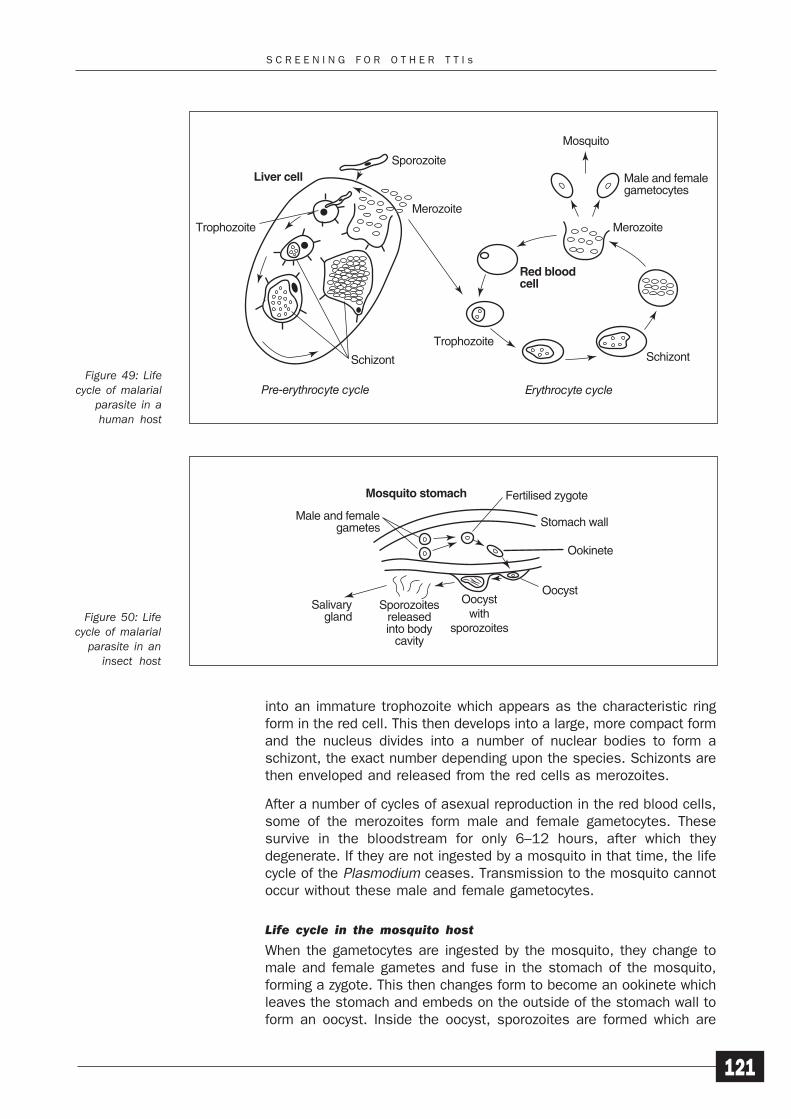
S C R E E N I N G F O R O T H E R T T I s
121
Figure 49: Lifecycle of malarial
parasite in ahuman host
Figure 50: Lifecycle of malarial
parasite in aninsect host
Sporozoite
Merozoite
Trophozoite
Trophozoite
Erythrocyte cycle
Schizont
Pre-erythrocyte cycle
Liver cell
Red bloodcell
Schizont
Merozoite
Male and femalegametocytes
Mosquito
Male and femalegametes
Mosquito stomach
Salivarygland
Sporozoitesreleasedinto body
cavity
Oocystwith
sporozoites
Oocyst
Ookinete
Stomach wall
Fertilised zygote
into an immature trophozoite which appears as the characteristic ringform in the red cell. This then develops into a large, more compact formand the nucleus divides into a number of nuclear bodies to form aschizont, the exact number depending upon the species. Schizonts arethen enveloped and released from the red cells as merozoites.
After a number of cycles of asexual reproduction in the red blood cells,some of the merozoites form male and female gametocytes. Thesesurvive in the bloodstream for only 6–12 hours, after which theydegenerate. If they are not ingested by a mosquito in that time, the lifecycle of the Plasmodium ceases. Transmission to the mosquito cannotoccur without these male and female gametocytes.
Life cycle in the mosquito host
When the gametocytes are ingested by the mosquito, they change tomale and female gametes and fuse in the stomach of the mosquito,forming a zygote. This then changes form to become an ookinete whichleaves the stomach and embeds on the outside of the stomach wall toform an oocyst. Inside the oocyst, sporozoites are formed which are
S E C T I O N 8
122
eventually released and spread throughout the body of the mosquito.Those that reach the salivary gland can then be transmitted to the nexthuman host. The whole cycle within the mosquito generally takes 10–12days.
Transmission
The parasite is spread to humans primarily through the bite of an infectedfemale Anopheles mosquito. There are over 200 species of Anopheles,of which approximately 60 are known to be vectors of the malariaparasite. Humans themselves are the reservoirs of infection; a mosquitothat has bitten an infected individual can pass malaria to noninfectedindividuals when feeding on them. The transmissibility of malaria throughblood transfusion has long been recognized because of the phase of thelife cycle of the parasite that involves the red cells. Donations frominfected individuals may transmit malaria if the parasite is present in redcells in the donation.
Clinical course of infection
The clinical picture of infection varies with each species, but is usuallycyclic in nature. The common symptoms are chills and fever at regularintervals and profuse sweating. Recurrent episodes can lead to severeanaemia. The incubation period ranges from 12 days for P. falciparum to15 days for P. vivax and P. ovale and to as long as 30 days for P. malariae.Infection caused by P. falciparum is generally considered to have themost serious clinical consequences in humans and may be fatal in asignificant number of cases when cerebral involvement occurs. Only P.malariae persists for extended periods in humans (up to 30 years); ingeneral, plasmodia of the other three species are finally destroyed by theimmune system after 1–3 years and die. The individual, if not reinfected,is then free from malaria.
Laboratory testing
Laboratory testing can be performed to detect either the malariaparasites in the bloodstream or specific antibody produced in responseto infection. Parasites can be detected in the bloodstream as quickly as48 hours after infection in heavily infected individuals. Antibody levelsstart to rise 3–4 weeks after infection and all infected individualsgenerally have specific antibody within 5–6 months. The development ofspecific antibodies may provide some degree of protection andamelioration of the disease process in multiply-infected individuals. Theantibody response usually differs between individuals who have lived inan endemic area for a significant amount of time and who have beenmultiply-infected, and those from nonendemic countries who have beeninfected through a single exposure episode. Infected individuals fromendemic areas are referred to as semi-immune and generally have low-titre antibody. This reflects the repeat exposure episodes in suchindividuals with frequent, if not persistent, low-level parasitaemia.Immunity is quickly lost on moving to a nonendemic area. Infectedindividuals from nonendemic areas, however, produce much higher titreantibody which then declines over the next one to two years, more quicklyif treated, and in many cases disappears totally.
S C R E E N I N G F O R O T H E R T T I s
123
The direct detection of parasites in blood is possible by looking at bloodfilms, using a specific diagnostic test for parasitaemia or even nucleicacid detection. In most cases, however, these are not sensitive enoughto identify infected donors, except in endemic countries where infecteddonors may have high numbers of circulating parasites (parasitaemia).These approaches are also impractical for the mass screening of blooddonations in most countries. However, donors with a high parasitaemiaare usually symptomatic and should be identified through the donorselection procedures.
Screening for specific antibody can therefore be used to identify currentlyor recently infected donors – those likely to donate blood that maytransmit malaria. Any donor who last returned from a nonendemic areaat least six months previously can be tested for specific antibody. Donorsfrom endemic areas can be treated similarly although the six-monthdeferral may need to be increased, depending on the country involved.
Significance for transfusion
Blood transfusion has been clearly documented as the cause of manycases of malaria, most of which have been P. falciparum. The extent towhich transmission by transfusion is a problem depends on the endemicityof malaria in that area and the number of infected individuals who arelikely to donate. In countries where malaria is endemic, it is highly likelythat most individuals are infected during childhood and that most donorsand most patients would already be infected. The significance oftransmission of malaria by blood transfusion is therefore very differentfrom that in nonendemic countries where most patients are not infectedand in whom infection would may be serious and even fatal.
Most nonendemic countries protect against malaria by donor selection,identifying those donors who have been to malarious areas and deferringthem for a suitable period before accepting them again. Whilst this isgenerally effective, the number of donors being deferred is rising and maybe too high to sustain in some countries. There is an increase in travelto malaria risk areas as well as the spread of malaria itself into new areasas climate changes occur or back into old areas as eradication programmesbreak down. The use of antibody testing in addition to donor deferral isnow becoming more acceptable in nonendemic countries in an effort toprevent transmission of malaria without significantly affecting thesufficiency of the blood supply.
Prevention of spread
Malaria is a significant problem worldwide and, for the last 30 years ormore, prevention of spread in endemic areas has focused on the removalof the mosquito vector by attacking its breeding sites. Prophylaxis is usedto prevent infection in travellers from nonendemic countries. This isgenerally effective, although not always totally successful, usually due toindividuals' failure to follow the regimen or to the presence of resistantPlasmodium strains. Treatment of malaria in individuals from nonendemicareas is usually successful if diagnosed early enough.
Transmission via blood and organs is minimized through a combinationof donor exclusion and specific testing.
S E C T I O N 8
124
8.6 CHAGAS DISEASE
The infectious agentChagas disease, also known as American trypanosomiasis, is a systemicdisease caused by infection with the protozoan Trypanosoma cruzi (T.cruzi) which can be severe and even fatal. It is transmitted to thevertebrate host by blood-sucking insects known as triatomine or reduviidbugs which transmit it via their faeces which they deposit as they bite andfeed. The disease is endemic in the Americas where it is thought that 16–18 million people are infected, most of these living in the poorer areasof south and central America. Infection of nonhuman vertebrates is alsocommon from the southern areas of Chile and Argentina to as far northas California. In the USA, however, the better living conditions mean thathuman infection by natural transmission is uncommon, although it doesoccur. It is estimated that in most central and south American countriesbetween 4% and 8% of the population are infected, although the figurecan be as low as 1.2% and as high as 20% in certain countries.
Structure and life cycleT. cruzi is one of a group of protozoa that are called haemoflagellates.These are flagellated protozoa that are transmitted to humans throughthe bites of infected insects. They are further grouped into two categories,Trypanosoma and Leishmania, although the differences between the twogroups are not well defined. Here, we shall only consider Trypanosoma,although cases of transfusion-transmission of leishmaniasis have beensuspected in some groups of military personnel serving abroad inendemic areas.
The life cycle of T. cruzi and the different forms of the haemoflagellatesduring the cycle are shown in Figures 51 and 52 on page 125. Theessential difference between the life cycles of T. cruzi and the Africanforms of trypanosomiasis is that T. cruzi cannot multiply in the bloodstreamof the human host. Instead it replicates inside the cells of almost everyorgan of the human body.
Infection is usually acquired by contamination, through contact with thefaeces of the triatomine bug. The trypanosomes grow in the gut of the bugand are excreted in the faeces. As the bugs defecate while feeding, thetrypanosomes in the faeces are able to infect the human host throughthe site of the bite. After entering the bloodstream, the organismschange their shape from the trypomastigote form and produce therounded amastigote form. The organisms first infect the lymph nodesclose to the site of infection where they initially multiply as the amastigoteform. From there, they subsequently spread to other organs via thebloodstream. The liver, splenic macrophages and, importantly, the heartare most commonly infected. The infected cells eventually rupture andrelease the amastigotes into the bloodstream, where they change totrypomastigotes. Although infectious, the protozoa do not multiply duringthis stage. If the infected individual is then bitten at this stage, thetriatomine ingests the trypomastigotes. The trypomastigotes multiply inthe mid-gut of the insect as epimastigotes and then pass through theinsect to be finally excreted in the faeces as the trypomastigote form. Thecycle then starts again.
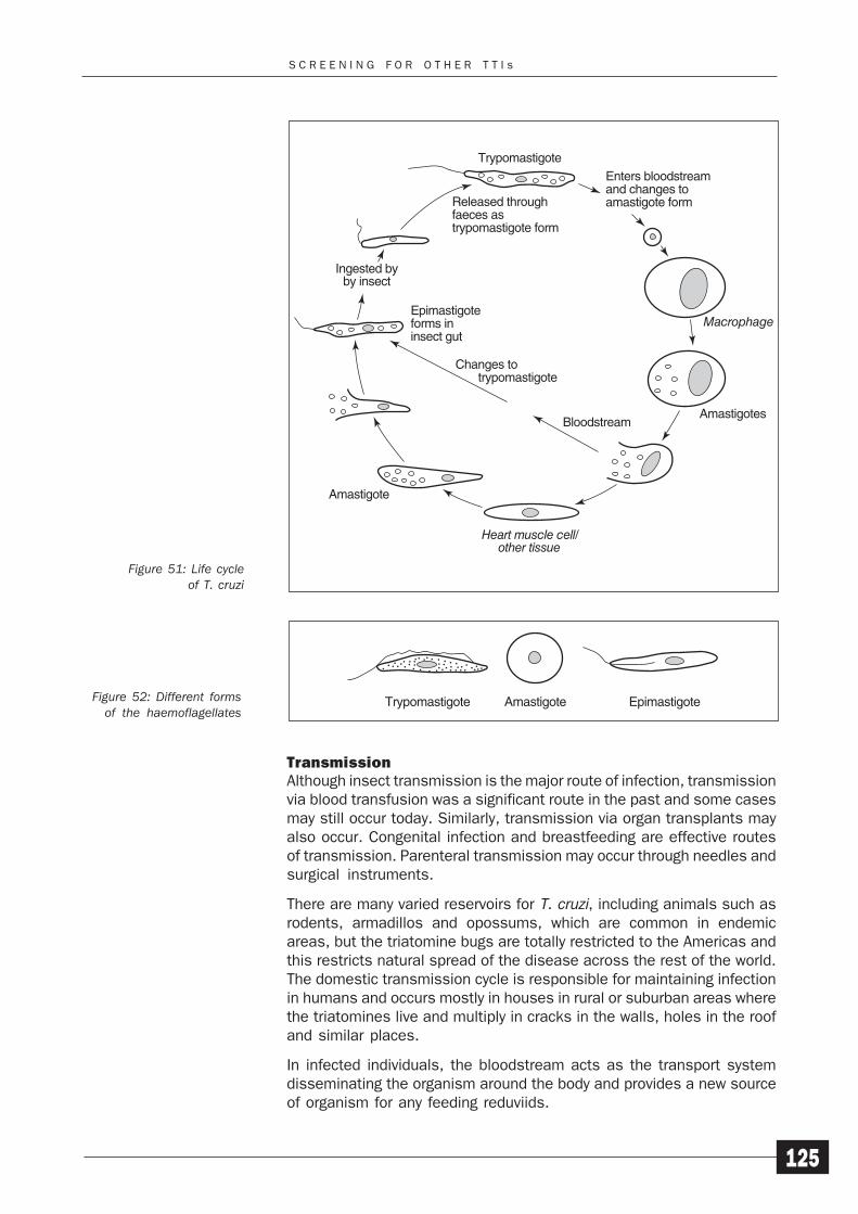
S C R E E N I N G F O R O T H E R T T I s
125
TransmissionAlthough insect transmission is the major route of infection, transmissionvia blood transfusion was a significant route in the past and some casesmay still occur today. Similarly, transmission via organ transplants mayalso occur. Congenital infection and breastfeeding are effective routesof transmission. Parenteral transmission may occur through needles andsurgical instruments.
There are many varied reservoirs for T. cruzi, including animals such asrodents, armadillos and opossums, which are common in endemicareas, but the triatomine bugs are totally restricted to the Americas andthis restricts natural spread of the disease across the rest of the world.The domestic transmission cycle is responsible for maintaining infectionin humans and occurs mostly in houses in rural or suburban areas wherethe triatomines live and multiply in cracks in the walls, holes in the roofand similar places.
In infected individuals, the bloodstream acts as the transport systemdisseminating the organism around the body and provides a new sourceof organism for any feeding reduviids.
TrypomastigoteEnters bloodstreamand changes toamastigote form
Macrophage
Amastigotes
Heart muscle cell/other tissue
Amastigote
by insect
Released throughfaeces astrypomastigote form
Epimastigoteforms ininsect gut
Changes to trypomastigote
Bloodstream
Trypomastigote Amastigote EpimastigoteFigure 52: Different formsof the haemoflagellates
Figure 51: Life cycleof T. cruzi
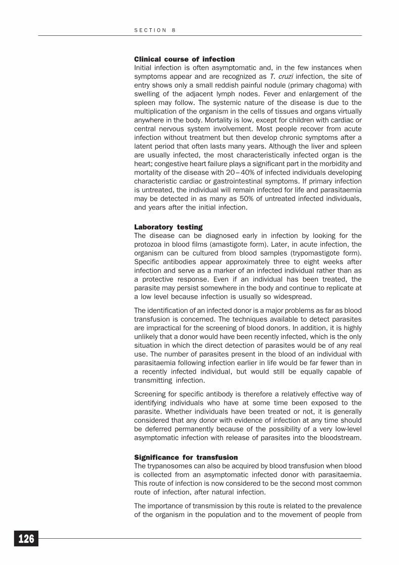
S E C T I O N 8
126
Clinical course of infectionInitial infection is often asymptomatic and, in the few instances whensymptoms appear and are recognized as T. cruzi infection, the site ofentry shows only a small reddish painful nodule (primary chagoma) withswelling of the adjacent lymph nodes. Fever and enlargement of thespleen may follow. The systemic nature of the disease is due to themultiplication of the organism in the cells of tissues and organs virtuallyanywhere in the body. Mortality is low, except for children with cardiac orcentral nervous system involvement. Most people recover from acuteinfection without treatment but then develop chronic symptoms after alatent period that often lasts many years. Although the liver and spleenare usually infected, the most characteristically infected organ is theheart; congestive heart failure plays a significant part in the morbidity andmortality of the disease with 20– 40% of infected individuals developingcharacteristic cardiac or gastrointestinal symptoms. If primary infectionis untreated, the individual will remain infected for life and parasitaemiamay be detected in as many as 50% of untreated infected individuals,and years after the initial infection.
Laboratory testingThe disease can be diagnosed early in infection by looking for theprotozoa in blood films (amastigote form). Later, in acute infection, theorganism can be cultured from blood samples (trypomastigote form).Specific antibodies appear approximately three to eight weeks afterinfection and serve as a marker of an infected individual rather than asa protective response. Even if an individual has been treated, theparasite may persist somewhere in the body and continue to replicate ata low level because infection is usually so widespread.
The identification of an infected donor is a major problems as far as bloodtransfusion is concerned. The techniques available to detect parasitesare impractical for the screening of blood donors. In addition, it is highlyunlikely that a donor would have been recently infected, which is the onlysituation in which the direct detection of parasites would be of any realuse. The number of parasites present in the blood of an individual withparasitaemia following infection earlier in life would be far fewer than ina recently infected individual, but would still be equally capable oftransmitting infection.
Screening for specific antibody is therefore a relatively effective way ofidentifying individuals who have at some time been exposed to theparasite. Whether individuals have been treated or not, it is generallyconsidered that any donor with evidence of infection at any time shouldbe deferred permanently because of the possibility of a very low-levelasymptomatic infection with release of parasites into the bloodstream.
Significance for transfusionThe trypanosomes can also be acquired by blood transfusion when bloodis collected from an asymptomatic infected donor with parasitaemia.This route of infection is now considered to be the second most commonroute of infection, after natural infection.
The importance of transmission by this route is related to the prevalenceof the organism in the population and to the movement of people from
S C R E E N I N G F O R O T H E R T T I s
127
endemic areas to nonendemic areas. Migration by people from centraland south America into north America and Europe has resulted in thepresence of infectious individuals in previously nonendemic areas. Whileit is very unlikely that these individuals will provide a reservoir fordomestic cycles of transmission involving the triatomines, they dopresent a source of transfusion-transmitted infection. Transmission bythis route into nonendemic areas is now well documented.
The actual transmission rate of Chagas disease by blood transfusion isdifficult to assess as there are few reliable data available, although it isestimated that, until recently, there were up to 10 000 cases per yearin Brazil. The exclusion of infected donors can help to reduce the risksof infection but, since many cases are largely asymptomatic, it may notbe clear which donors are infected or are potential infection risks.Furthermore, in many countries the rate of natural infection is high andcannot be ruled out as the source of infection.
Prevention of spread
Control of infection in endemic areas generally focuses on improvinghousing and the elimination of the triatomine bugs from homes, and theprevention of spread via blood or organs. In nonendemic areas, spreadby blood and organs is limited, depending on the risks within the donorpopulation, such as donors from endemic countries or who have travelledextensively in endemic areas. Public health education programmes playa part in the control of the disease, especially in the early identificationand reduction of risk of infection via breastfeeding.
In endemic countries, the screening of blood donors is a good controlmeasure that can help to reduce the spread of infection. In nonendemiccountries, screening may not be very useful unless there is a largenumber of donors from, or who have travelled in, endemic countries andrecipients of the blood are likely to be immunosuppressed. A moresuitable approach may be to exclude potentially infected donors bypredonation questioning although, with the increase in world travel, thismay result in the deferral of too many donors and selective screening mayhave to be introduced.
An important method of preventing transmission by transfusion ispractised in parts of Latin America where the disease is still endemic andthe exclusion of infected donors would significantly decrease the bloodsupply. The addition of a drug that kills trypanosomes to the donatedblood has proved very effective. Crystal violet (also known as gentianviolet) at a concentration of 200 μg/ml in blood stored at +4°C for 24hours before use prevents transfusion-transmitted infection. Thousandsof transfusions have been performed using blood treated in this waywithout any serious side-effects being reported.
Since the transmission of Chagas disease by blood transfusion has beenclearly demonstrated, there is no reason to suppose that other membersof this protozoan group would not be transmitted under the correctconditions. The other organisms that infect humans are:
T. gambiense which causes west African sleeping sickness(Gambian trypanosomiasis)
S E C T I O N 8
128
T. rhodesiense which causes east African sleepingsickness (Rhodesian trypanosomiasis).
However, no cases of transmission by transfusion of either T. gambienseor T. rhodesiense have yet been reported.
The spread of infection and the disease process caused by T. cruzi arevery different from the African types of trypanosomiasis.
SUMMARY
1 The basic principles that apply to the screening of bloodfor anti-HIV also apply to screening for other transfusion-transmitted infections.
2 The range of screening tests that are available for anti-HIVis not always available for other infectious agents.
SELF-ASSESSMENT
27 What is the significance for transfusion practice of HBVinfection?
28 What is currently the most effective method of screeningfor malaria?
29 What is the significance for transfusion practice of Chagasdisease?
PROGRESS CHECK
Before moving on to Section 9, spend a few minutes thinkingabout whether you have achieved the learning objectives forSection 8. These were to:
1 Describe the basic features of infection with HBV, HCV,HTLV, syphilis, malaria and Chagas disease.
2 Explain the significance of these agents for bloodtransfusion practice.
If you feel confident that you have understood everything inthis section, turn to Section 9.
If you feel that you need to spend more time on this section,go back to the parts that are most unfamiliar or that you havefound difficult. You may find it helpful to talk to other people,such as your supporter or other senior colleagues, if there isanything you are still not sure about.
A C T I O N P L A N
129
Action Plan
This final section focuses on the Action List you have been building upas you have worked through this module. You have probably identified anumber of improvements that you think could be made in your programmefor screening for anti-HIV and other infectious agents and it is now timeto identify priorities and begin putting your ideas into action.
LEARNING OBJECTIVES
When you have completed this section, you should be able to:
1 Reassess your knowledge and skills in relation to themodule objectives now that you have completed Module2.
2 Review your Action List, identifying improvements thatyou can implement and those that will require action byothers.
3 Prepare and implement a realistic Action Plan to introducechanges that will improve the quality of the screeningservice provided by your laboratory.
9
S E C T I O N 9
130
9.1 REVIEWING YOUR PROGRESS
Before you start making your Action Plan, think carefully about themodule objectives and the progress you have made since you startedworking through this module.
ACTIVITY 44
Complete the table below. You will notice that it is the same asthe one you filled in for Activity 2. Use it to review the knowledgeyou have gained and the skills you have developed as a result ofyour work on this module. Have you changed your rating in relationto each module objective?
You should have made some identifiable progress in each areacovered by this module. If there is anything you still do not feelconfident about, however, reread the appropriate section and then
Section 2
Explain the role of microorganisms as infectious
agents in human disease and their significance for
blood transfusion
Section 3
Describe HIV infection and the significance of
infection for blood transfusion
Section 4
Outline the principles of the diagnostic assays
most commonly used to detect transfusion-
transmissible infections and the differences
between them
Section 5
Select the most suitable type of screening assay
for transfusion-transmissible infections for use in
your own laboratory
Section 6
Develop an effective screening programme for
transfusion-transmissible infections and maintain
accurate records of the screening results
Section 7
Help to develop an appropriate quality system for
your laboratory to maintain an effective screening
programme
Section 8
Recognize the basic features of other infectious
agents and their significance for blood transfusion
practice
Rating(1–4)Module objective Comments
A C T I O N P L A N
131
discuss any remaining problems with your supporter or trainerbefore continuing with your Action Plan.
9.2 MAKING YOUR ACTION PLAN
The Action Plan provides you with an opportunity to make practicalimprovements in your own workplace, within any financial, resource orstaffing constraints that exist. As you worked through this module, youhave been noting down your ideas on the Action List on page 132. Youshould have listed the number of the activity in Column 1 and noted yourideas for improvement in Column 2. You should also have discussed yourideas with your supporter.
You may have tried out some of your ideas already, but some may requiremore time and effort and so it is important to identify priorities. You maynot be in a position to put all your ideas into action yourself and it maybe necessary to convince other members of staff to take the actions thatyou have identified as being both necessary and feasible.
ACTIVITY 45
Look carefully at all the suggestions for improvements that youhave written down on your Action List. Mark those where you havenot yet been able to take any action. Then divide them into twocategories:
1 Actions that you can takeActions that you can takeActions that you can takeActions that you can takeActions that you can take. Choose the ones that you thinkare most important and put them in order of priority. Notethem down in Column 1 of the Action Plan on page 133. InColumn 2, briefly summarize the action that you plan to take.In Column 3, note down the results that you would expectafter taking this action.
2 Actions that others could takeActions that others could takeActions that others could takeActions that others could takeActions that others could take. Note them down in Column 1of the Action Plan on page 133. In Column 2, write down thename of the person who would be responsible for making thechanges that you are recommending and, in Column 3,summarize the results that you would expect.
Then show your plan to your supervisor and supporter and discussit with them. Your ideas for improvement may need to be modifiedas a result of these discussions. Other senior staff may also needto be consulted before your Action Plan can be agreed. You shouldalso discuss it with your trainer at this stage.
When you have reached agreement about the actions you are goingto take, set a date by which you hope to complete each of themand note this in Column 4. Also use Column 4 to set a date bywhich you expect the completion of any actions taken by others.
Your Action Plan is now ready.

S E C T I O N 9
132
ACTION LISTACTION LISTACTION LISTACTION LISTACTION LIST
Ideas for improvementActivity number

A C T I O N P L A N
133
Pla
nned
com
ple
tion
date
Act
ual
com
ple
tion
date
AC
TIO
N P
LA
N
Pla
nned a
ctio
nId
eas
for
impro
vem
ent
Exp
ect
ed r
esu
lts
Act
ual
resu
lts
S E C T I O N 9
134
9.3 IMPLEMENTING YOUR ACTION PLAN
You should now begin to implement your Action Plan along the linesagreed with your supervisor and supporter. It will probably take youseveral weeks or months to put your all plans into action and you mayneed more time than you expected. In fact, you will probably start the nextmodule in the programme before you are able to complete everything.You may also find that some of your ideas for improvement are moredifficult to put into action than you expected and you may need to revisesome of your plans if they are too ambitious or are not working as wellas you hoped. However, if you have thought carefully about how you couldapply what you have learned from this module and have discussed yourideas with the appropriate people, you should be able to put most of theminto practice. You may even find that there are some unexpectedbenefits. If you have any problems during this time, talk to your supporteror supervisor and ask them for any assistance you need. You should alsogive them regular reports on your progress.
ACTIVITY 46
Once you have completed each action you included in your ActionPlan, note down the date in Column 5 and the final results inColumn 6. Then review the implementation of your Action Plan bycomparing the actual results with the results that you expected.Also compare the planned completion dates with the actualcompletion dates. Discuss the outcomes with your supporter andsupervisor.
Identify any further actions required to ensure the implementationof the improvements you have identified as being necessary.
Over the next few months, monitor the effectiveness of anychanges you have been able to introduce and be prepared to makeany further changes or take any follow-up action needed to ensurethat they continue to lead to improved quality in your screeningprogramme.
As you work through the remainder of the learning programme, you willbe asked to complete an Action List and Action Plan for each module.This approach can be applied to almost any situation and you may decideto use it in other areas of your work to improve the quality of the servicethat you provide.
PROGRESS CHECK
Now that you have completed this module, spend some timethinking about whether you have achieved the learningobjectives for this section. These were to:
1 Reassess your knowledge and skills in relation to themodule objectives now that you have completed Module 2.

A C T I O N P L A N
135
2 Review your Action List, identifying improvements that youcan implement and those that will require action by others.
3 Prepare and implement a realistic Action Plan to introducechanges that will improve the quality of the screeningservice provided by your laboratory.

137
Activity Checklists andAnswers
SECTION 1
Activity 1
Purpose
To identify a personal “supporter” for your work on Module 2.
Checklist
You should have:
Identified senior colleagues in your workplace who couldprovide assistance to you as you work through thismodule
Selected one particular person, ideally your supervisor, tobe your supporter for Module 2 and checked that he or sheis willing to assist you
Explained how the learning programme operates andwhat the role of the supporter involves
Agreed how frequently you will meet to discuss your workon this module
Showed Module 2 to your supporter
Informed your trainer about who your supporter is
Asked your trainer for assistance if you have any difficultyin finding a supporter in your workplace.
Activity 2
Purpose
To assess your knowledge, skills and experience in relation to themodule objectives before you start work on Module 2.
A C T I V I T Y C H E C K L I S T S A N D A N S W E R S
138
Checklist
You should have:
Looked carefully at the module objectives and, for eachone, assessed your current knowledge, skills andexperience, using the rating 1, 2, 3 or 4
Completed the table on page 7
Noted any additional comments you wish to make, suchas any module objectives that are not relevant to your ownwork.
Activity 3
Purpose
To make a realistic Study Plan for your work on Module 2.
Checklist
You should have:
Quickly looked at other sections to get an idea of thecontent, level and approach of the module and assesshow much of the material is likely to be new to you
Estimated the amount of time you think you will need tostudy each section, including completing the activitiesand answering the self-assessment questions
Discussed with your supervisor how much time can beallocated for study on a regular basis
Completed the Study Plan on page 8, adding the dateswhen you plan to complete each section and the dates ofmeetings with your trainer and supporter.
SECTION 2
Activity 4
Purpose
To identify the infectious agents in your locality which you think can betransmitted by blood transfusion.
Checklist
You should have:
Listed the infectious agents that are prevalent in yourlocality, including at least one example of each of the fourtypes of infectious agent
Identified the normal routes of transmission of each ofthese agents
A C T I V I T Y C H E C K L I S T S A N D A N S W E R S
139
Indicated the agents that you think are likely to betransmitted by blood transfusion.
Activity 5
Purpose
To identify the infectious agents which can be transmitted by bloodtransfusion.
Checklist
You should have:
Made any amendments you now think necessary to yourlist from Activity 4 of infectious agents that can betransmitted by blood transfusion
Checked your answers with your supervisor or anothersenior member of staff.
Activity 6
Purpose
To check your understanding of the markers of infection at differentstages following infection.
Answers
2 weeks: antigen
4 weeks: antigen and possibly low-titre antibody
6 weeks: antibody, with possibly some remaining antigen
8 weeks: antibody
18 months: low-titre antibody or no remaining markers.
If an infectious agent is likely to be transmitted only during the period ofacute infection, it would be better to screen donors for the agent itselfbecause the presence of antibody only indicates immunity – not aninfectious state.
SECTION 3
Activity 7
Purpose
To check your understanding of the structural features of HIV.
Answers
1 = nucleic acid
2 = capsid
A C T I V I T Y C H E C K L I S T S A N D A N S W E R S
140
3 = p24
4 = p7/p9
5 = reverse transcriptase (p66)
6 = p17 matrix protein
7 = gp41
8 = gp120
9 = lipid bilayer
10 = complete virion
Activity 8
Purpose
To check your understanding of the entry of HIV into a susceptible cell.
Answers
1 = HIV
2 = CD4 receptor
3 = uncoated virus (capsid)
4 = viral RNA
5 = DNA copy of viral RNA
6 = viral DNA integrated into genome
7 = viral proteins
8 = viral RNA
9 = virus capsid
10 = budding virus particle
11 = new HIV virion
Activity 9
Purpose
To investigate the clinical presentation of HIV and AIDS in your country.
Checklist
You should have tried to find out:
The prevalence of HIV infection and the number of AIDScases in your country
The most common clinical course of HIV infection and itsprogression to AIDS
The average time taken for AIDS to manifest following theinitial infection
A C T I V I T Y C H E C K L I S T S A N D A N S W E R S
141
The main opportunistic infections found in AIDS patientsand whether they are found in all areas of your country orthere are local variations
How the incidence of Kaposi’s sarcoma 25 years agocompares with the incidence today and how it relates tothe current prevalence of AIDS in your population.
Activity 10
Purpose
To consider possible reasons for any under-reporting of HIV infection andAIDS in your country.
Checklist
You should have:
Identified any possible reasons for under-reporting of HIVinfection and AIDS in your country, using the followingcategories:
— economic
— political
— practical
— technical
— cultural.
Activity 11
Purpose
To identify the principal route of transmission of HIV infection in yourcountry.
Checklist
You should have:
Identified the main route of transmission of HIV in yourcountry
Identified the reason why this is the main mode oftransmission.
Activity 12
Purpose
To illustrate the importance of screening for transfusion-transmitted HIVinfection.
A C T I V I T Y C H E C K L I S T S A N D A N S W E R S
142
Answers
1 Short-/long-term: the patient is infected with HIV and islikely to die within three to five years.
2 Short-/long-term: his wife (and any other sexual partner)is likely to be infected following normal sexual contact.
3 Long-term: his wife may infect any subsequent children.
4 Long-term: a recipient of any blood donated by thehusband or wife is likely to be infected if the blood is nottested before transfusion.
Activity 13
Purpose
To investigate the approaches taken to reduce the spread of HIV in yourcountry.
Checklist
You should have:
Listed all the approaches taken in your country to reduce:
— the spread of HIV infection
— its transmission through blood transfusion
Found out what advice is given about avoiding the sexualtransmission of HIV
Noted whether condoms are widely available
Noted how effective condom distribution has been inpreventing the transmission of HIV infection or whetherpeople are reluctant to use condoms.
SECTION 4
Activity 14
Purpose
To identify the types of TTI screening assay used in your laboratory.
Checklist
You should have:
Listed the names of all the assay kits used in yourlaboratory for screening donated blood for TTIs
Correctly identified each assay type.
Activity 15
Purpose
To standardize the terminology used in your laboratory.
A C T I V I T Y C H E C K L I S T S A N D A N S W E R S
143
Checklist
You should have:
Listed the words used in your laboratory to classifyscreening results
Noted any words that are different from those used by themanufacturers of the assays used in your laboratory
Discussed the terminology to be used in your laboratorywith your supervisor and colleagues and noted down yourrecommendations on your Action List.
Activity 16
Purpose
To calculate the results of an antiglobulin/sandwich-type EIA.
Checklist
You should have:
Used the cut-off value of 0.36 to complete Column B ofFigure 36 with the final assay results
Looked at Figure 37 to see the results from Figure 36plotted on a simple graph.
Activity 17
Purpose
To calculate the signal/cut-off ratios of an antiglobulin/sandwich-typeEIA.
You should have:
Used the cut-off value of 0.36 to complete Column C ofFigure 36 with the signal/cut-off ratios
Looked at Figure 37 to see the results from Figure 36plotted on a simple graph.
SECTION 5
Activity 18
Purpose
To calculate the specificity and, if possible, the sensitivity of an assayused in your laboratory.
Checklist
You should have:
Found out the number of true-negatives and false-positivesfor the last 100 assays performed in your laboratory
A C T I V I T Y C H E C K L I S T S A N D A N S W E R S
144
Used these figures correctly in the formula given tocalculate the specificity of the assay
Compared the answer with the specificity given in themanufacturer’s instructions
Found out the number of true-positives and false-negativesfor the last 100 assays performed, if external qualitycontrol samples are used in your laboratory
Used these figures correctly in the formula given tocalculate the sensitivity of the assay
Compared the answer with the sensitivity given in themanufacturer’s instructions
Talked to your supervisor about your results if theydiffered from the specificity or sensitivity given in themanufacturer’s instructions and noted down yourrecommendations on your Action List.
Activity 19
Purpose
To collect data on the assays available in your country.
Checklist
You should have:
Listed all the assays available in your country, togetherwith the names of the manufacturers and, if possible, thecost per test
Selected one assay from each of the three types: EIA,particle agglutination assay and simple rapid assay
Completed the table on page 66 with the relevantinformation about each of these three assays.
Activity 20
Purpose
To identify the most appropriate type of assay for use in your laboratory.
Checklist
You should have:
Noted any additional factors relevant to the choice of a TTIassay that specifically relate to your laboratory, including:
— the number of donations per week
— the facilities and equipment available in your laboratory
— the support services available in your laboratory, suchas water supply and electricity
A C T I V I T Y C H E C K L I S T S A N D A N S W E R S
145
Assessed the different types of assay on theirappropriateness to your situation
Identified the most suitable type of assay for use in yourlaboratory
Noted down your recommendations on your Action List, ifyou feel that your laboratory is currently not using themost appropriate type of assay, and discussed them withyour supervisor.
Activity 21
Purpose
To examine other factors that need to be considered in setting up andmaintaining an effective TTI screening programme.
Checklist
You should have:
Noted how each of the factors listed in the activity relateto the particular circumstances in your laboratory
Added any other factors that you think might be relevantto setting up and maintaining an effective TTI screeningprogramme.
Activity 22
Purpose
To review the training provided in your laboratory on screening for TTIs.
Checklist
You should have:
Made notes on the training provided in your laboratoryincluding:
— the name of the person who provides the training
— the name of the person who is in charge of training
— the kind of training that is provided
— the duration of training
— how trainees’ skills are assessed
— whether regular training updates are held
Outlined how you would develop a simple trainingprogramme for new staff or for staff who are alreadyperforming testing for TTIs
Noted down your ideas for a training scheme on yourAction List and discussed them with your supervisor.
A C T I V I T Y C H E C K L I S T S A N D A N S W E R S
146
Activity 23
Purpose
To identify the items of equipment that are needed to perform TTIscreening assays correctly.
Checklist
You should have:
Listed the following items of equipment needed to performEIAs and particle agglutination assays:
— suitable mechanical pipettes (ideally multi-channel)
— EIA plate reader
— EIA plate washer (manual/automatic)
— reagent troughs
Noted the items of equipment that are already availablein your laboratory
Identified any equipment to which you have unrestrictedaccess elsewhere in your hospital or institution.
Activity 24
Purpose
To plan an ordering schedule for the supply of assay kits.
Answer
Five or six kits would be required each month. Assuming that seven kitsare in stock at the beginning of the year, a suitable ordering schedulemight be as shown on page 147.
Checklist
You should have:
Reviewed the ordering schedule for assay kits used inyour laboratory
Identified any ways in which it could be improved
Noted down your recommendations on your Action Listand discussed them with your supervisor.
Activity 25
Purpose
To review the storage conditions for assays in your laboratory.
Checklist
You should have:
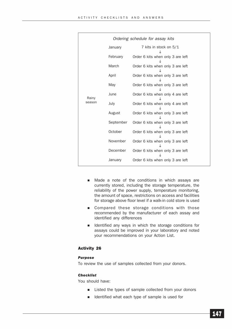
A C T I V I T Y C H E C K L I S T S A N D A N S W E R S
147
Made a note of the conditions in which assays arecurrently stored, including the storage temperature, thereliability of the power supply, temperature monitoring,the amount of space, restrictions on access and facilitiesfor storage above floor level if a walk-in cold store is used
Compared these storage conditions with thoserecommended by the manufacturer of each assay andidentified any differences
Identified any ways in which the storage conditions forassays could be improved in your laboratory and notedyour recommendations on your Action List.
Activity 26
Purpose
To review the use of samples collected from your donors.
Checklist
You should have:
Listed the types of sample collected from your donors
Identified what each type of sample is used for
7 kits in stock on 5/1
Order 6 kits when only 3 are left
Order 6 kits when only 3 are left
Order 6 kits when only 3 are left
Order 6 kits when only 3 are left
Order 6 kits when only 4 are left
Order 6 kits when only 4 are left
Order 6 kits when only 3 are left
Order 6 kits when only 3 are left
Order 6 kits when only 3 are left
Order 6 kits when only 3 are left
Order 6 kits when only 3 are left
Order 6 kits when only 3 are left
January
February
March
April
May
June
July
August
September
October
November
December
January
Rainyseason
Ordering schedule for assay kits
A C T I V I T Y C H E C K L I S T S A N D A N S W E R S
148
Talked to your supervisor about collecting separatesamples from donors, if testing is currently performedusing the bleed line attached to the blood pack, and notedyour recommendations on your Action List.
Activity 27
Purpose
To review the use of external and internal QC samples in your screeningprogramme.
Checklist
You should have:
Noted the external QC samples available, including:
— where they are obtained from
— how many samples are available
— whether you have to pay for them
— the assays with which they are meant to be used
Explained how any internal QC samples used in yourlaboratory are prepared and standardized
Suggested possible improvements in the system forobtaining external QC samples or preparing andstandardizing internal QC samples
Noted your recommendations on your Action List anddiscussed them with your supervisor.
Activity 28
Purpose
To identify ways of preventing the use of untested blood.
Checklist
You should have:
Noted the average time that a unit of blood is currentlystored in your blood bank before it is issued for transfusion
Noted whether untested blood is ever transfused in yourhospital and, if it is, the reasons why this is necessary
Suggested ways of maintaining a storage period of atleast 24 hours to provide sufficient time for routinescreening for TTIs before transfusion
Noted your recommendations on your Action List anddiscussed them with your supervisor.
A C T I V I T Y C H E C K L I S T S A N D A N S W E R S
149
Activity 29
Purpose
To assess how various factors might affect the overall cost of yourscreening programme.
Checklist
You should have considered the following factors:
the number of donations to be tested and therefore thenumber of assays that will need to be performed
the cost of the different types of assay
the cost of additional equipment and reagents required
the reliability of the assays
the specificity of the assays
the prevalence of TTIs in your donor population
the costs of collecting the donated blood
staff costs.
SECTION 6
Activity 30
Purpose
To review the system used in your laboratory to record assay failures.
Checklist
You should have:
Suggested any ways in which an error log kept in yourlaboratory could be improved and noted yourrecommendations on your Action List
Discussed introducing an error log with your supervisorand colleagues, if one is not kept at present
Identified the following information that should be recordedin an error log:
— the name of the assay
— the name of the operator
— the reason for the failure
— the consequence of the failure
— the changes made to laboratory practice to prevent asimilar failure in the future
Noted your recommendations on your Action List.
A C T I V I T Y C H E C K L I S T S A N D A N S W E R S
150
Activity 31
Purpose
To identify ways of reducing the number of false-positives in yourscreening programme.
Checklist
You should have:
Noted the assays used by your reference service
Consulted the records for the last 25 or 50 assays thatwere subjected to confirmatory testing, either by a referencelaboratory or by use of an alternative assay
Noted the number of these assays that were confirmedpositive
Noted the number of false-positives
Suggested ways of reducing the number of false-positivesby ensuring that:
— washing is performed correctly
— clean reagent containers are used
— the correct volumes of samples and reagents aredispensed
— the reader set-up (for EIAs) is correct
— good quality samples are used
Noted your recommendations on your Action List anddiscussed them with your supervisor.
Activity 32
Purpose
To review the procedures used for the dispatch of samples to a referencelaboratory.
Checklist
You should have:
Noted the procedures used in your laboratory to ensurethat samples arrive in a suitable state to give reliableconfirmatory results
Noted any local or national regulations for sendingpathological material by post
Suggested ways of sending samples so that there is norisk of leakage during transit
Reviewed the records of the last 25 or 50 samples sentto your reference service and noted the time taken for
A C T I V I T Y C H E C K L I S T S A N D A N S W E R S
151
delivery, the proportion that arrive damaged and the timetaken to receive the results
Identified any ways of improving the safe dispatch ofsamples and noted your recommendations on your ActionList.
Activity 33
Purpose
To review the TTI assay record sheet used in your laboratory.
Checklist
You should have:
Examined the record sheet used in your laboratory for testresults
Compared it with the example given in Figure 45
Identified any ways of improving your TTI assay recordsheets and noted your recommendations on your ActionList.
Activity 34
Purpose
To review the system used in your laboratory for retrieving previous testdata.
Checklist
You should have:
Described the system used in your laboratory for retrievingdata from previous tests on donors
Identified any ways of improving the system for retrievingprevious test data and noted your recommendations onyour Action List.
Activity 35
Purpose
To review the system used in your centre for storing screening results.
Checklist
You should have:
Assessed your centre’s system for the storage of recordsin relation to the factors listed on pages 88–89
Identified any ways of improving the system for storingscreening results and noted your recommendations onyour Action List.
A C T I V I T Y C H E C K L I S T S A N D A N S W E R S
152
Activity 36
Purpose
To review the system used in your centre for identifying, removing fromstock and disposing of TTI-positive donations.
Checklist
You should have:
Compared the procedure used in your centre with theguidelines given on page 90
Identified any ways of improving the current procedureand noted your recommendations on your Action List
Talked to your supervisor and colleagues about designingand implementing an appropriate system, if none currentlyexists, and noted your recommendations on your ActionList.
Activity 37
Purpose
To recognize the importance of the safe handling and disposal of assaycomponents and waste.
Checklist
You should have:
Listed the components of TTI screening assays that mightpresent a health and safety risk
Suggested why these components might present a healthand safety risk.
Activity 38
Purpose
To identify methods for the safe disposal of potentially hazardous assaymaterials.
Checklist
You should have:
Listed the materials used in TTI screening assays thatmay be hazardous in their disposal
Suggested ways of disposing of these materials safely.
Activity 39
Purpose
To review the handling and disposal of potentially hazardous waste fromTTI screening assays in your laboratory.
A C T I V I T Y C H E C K L I S T S A N D A N S W E R S
153
Checklist
You should have:
Read the health and safety policy for your workplace andchecked whether it covers all the necessary areas andworking practices in your screening programme
Contacted the person who is responsible for health andsafety and found out how regularly the health and safetypolicy is rewritten and updated
Identified any improvements you would like to see in thehandling and disposal of potentially hazardous waste andnoted your recommendations on your Action List.
SECTION 7
Activity 40
Purpose
To review the documentation of procedures in your laboratory.
Checklist
You should have:
Identified any of the areas listed on page 97 for whichwritten records are not kept in your laboratory
Discussed with your supervisor the importance of recordingany of these aspects of laboratory work that are notcurrently documented
Identified any further procedures that you think should bedocumented and noted your recommendations on yourAction List.
Activity 41
Purpose
To review the use of SOPs in your laboratory.
Checklist
If SOPs are used in your laboratory, you should have:
Noted whether there are SOPs for all the major activitiesin the screening programme, such as those listed on page98
Assessed whether each SOP covers all aspects of theprocedure
Noted whether SOPs are followed by all members of staffinvolved in performing those procedures
A C T I V I T Y C H E C K L I S T S A N D A N S W E R S
154
Noted how frequently SOPs are reviewed and updated,where necessary
Suggested any ways in which the preparation and use ofSOPs could be improved in your laboratory and noted yourrecommendations on your Action List.
If SOPs are not used in your laboratory, you should have:
Identified an important task that you perform regularly
Developed a simple SOP
Showed it to your colleagues and your supervisor foramendment, where necessary
Once it was finalized, ensured that all members of stafffollow it at all times
Identified any additional areas where SOPs are required,noted your recommendations on your Action List anddiscussed them with your supervisor.
Activity 42
Purpose
To design a checklist for performing an assay.
Checklist
You should have:
Designed a checklist for a screening assay with which youare familiar
Listed all the required actions in the correct order
Included space or boxes for recording the actions takenand adding any other information that may be required,including signatures and dates.
Activity 43
Purpose
To review the efficiency of the record-keeping system used in yourlaboratory.
Checklist
You should have:
Used your transfusion records to select a donation thatwas tested three or four months earlier
Attempted to follow the testing records back to the donor
Noted any point at which it was impossible to follow therecords back to the donor
Discussed your findings with your supervisor
A C T I V I T Y C H E C K L I S T S A N D A N S W E R S
155
Suggested ways of improving the record-keeping systemto ensure that an audit trail could be followed in the futureand noted your recommendations on your Action List.
SECTION 9
Activity 44
Purpose
To assess the progress you have made as a result of your work on Module2.
Checklist
You should have:
Assessed your knowledge, skills and experience in relationto each of the module objectives now that you havereached the end of the module
Completed the table on page 130
Identified any areas in which you still do not feel fullyconfident about your knowledge or skills
Reread the appropriate sections of the module anddiscussed any remaining problems with your supporter ortrainer before continuing with your Action Plan.
Activity 45
Purpose
To plan how to implement the improvements that you have identified asbeing necessary to ensure quality in your screening programme.
Checklist
You should have:
Looked at all the suggestions for improvements that youhave included on your Action List and marked those whereyou have not yet been able to take any action
Divided them into two categories:
— actions that you can take
— actions that others could take
Identified priorities for action
Filled in your Action Plan, as follows:
— Column 1: the improvements you have identified asbeing necessary
— Column 2: the action you plan to take or the name ofthe person who would be responsible for taking action
A C T I V I T Y C H E C K L I S T S A N D A N S W E R S
156
— Column 3: the results you would expect as a result ofimplementing your planned actions
Discussed your plan with your supervisor, supporter,trainer and any other appropriate senior staff
Modified your plan, where necessary
Filled in Column 4 with the dates by which you hope eachplanned action will be completed.
Activity 46
Purpose
To review the implementation of your Action Plan and identify any follow-up action required.
Checklist
You should have:
Filled in Column 5 with the dates on which you completedeach planned action and compared them with the plannedcompletion dates
Summarized the results of your planned action in Column6 and compared them with the results you had expected
Discussed the outcomes with your supporter
Discussed the outcomes with your supervisor
Identified any further actions required to ensure theimplementation of the improvements you identified asbeing necessary
Monitored the effectiveness of the changes you havebeen able to introduce
Identified any further changes or follow-up action required.
A N S W E R S T O S E L F - A S S E S S M E N T Q U E S T I O N S
157
Answers to Self-assessmentQuestions
SECTION 2
1 Latency is the property of infectious agents, generallyviruses, to remain hidden in a previously-infected individualand in an inactive state for an extended period of time –often the lifetime of the individual. Although the individualmay be immune to the agent, specific antibody can bedetected and reactivation of the agent may occur at anytime.
2 The three conditions that will determine whether aninfectious agent is likely to be transmitted by transfusionare:
the agent must be capable of using the bloodstreamas a means of entry into its host, the patient
the infected donor will be essentially free of any signsand symptoms of disease
the agent must exist naturally for a period of time,either free in the plasma or present in a cellularcomponent in the bloodstream of an infected donor.
3 Blood transfusion can never be the primary route ofinfection because most people do not have a bloodtransfusion during their lifetime. Any agent that dependssolely on transfusion for transmission will therefore notpersist in the population.
4 Antigens are foreign substances that enter the body andinduce an immune response against them. Antibodies aremolecules that are made up of proteins and carbohydrates.They are produced by the immune response of an individualas a reaction to stimulation by a foreign protein – antigen.
5 The five types of immunoglobulin are: IgG, IgM, IgA, IgDand IgE.
A N S W E R S T O S E L F - A S S E S S M E N T Q U E S T I O N S
158
6 The main reason for screening blood is to ensure that theavailable blood supply is as free as possible from anyinfectious agents.
7 The risk of transmission of infection by transfusion can beminimized by:
the careful selection of donors
the direct screening of the donated blood for infectiousagents
the removal of specific components of blood thoughtto harbour infectious agents
the physical inactivation of any contaminating agentsthat may be present.
8 The presence of antibody does not necessarily protectagainst infection and the infectious agent may still bepresent in the unit of blood.
SECTION 3
9 HIV enters susceptible cells by binding to a receptor (CD4)on the cell surface.
10 The two HIV-specific antibodies that have been found tobe the best confirmation of HIV infection are anti-p24 andanti-gp41.
11 Antibodies may be present as early as 14 days followinginfection with HIV, but may not appear until 28 days ormore after infection.
12 The three main routes of transmission for HIV infectionare:
unprotected penetrative sexual contact with an infectedperson, either between men or between men andwomen
the inoculation of infected blood by transfusion or theuse of contaminated needles, syringes or knives
from an infected mother to her child, either in theuterus, during birth or by breastfeeding.
13 Three routes of potential transmission that are notimplicated in the transmission of HIV are:
non-sexual social or domestic contact
insect vectors
handling pathological material, provided that the correctsafety procedures are applied at all times and goodlaboratory practice is followed.
SECTION 4
14 The three main types of screening assay are:
A N S W E R S T O S E L F - A S S E S S M E N T Q U E S T I O N S
159
enzyme linked immunosorbent assays (ELISA) orenzyme immunoassay (EIA)
particle agglutination assays
simple rapid assays.
15 The term “positive” describes the status of the donationor donor after the initial result has been confirmed byfurther assays. The term “reactive” is used until theresults are confirmed.
SECTION 5
16 Sensitivity and specificity are inversely related. As thesensitivity increases, so the specificity decreases.Similarly, as the specificity increases, the sensitivitydecreases.
17 The factors that contribute to the design and maintenanceof an effective TTI screening programme are:
staff training
provision of necessary equipment and reagents
regular supply of assay kits
suitable facilities for the storage of assay kits
availability of fresh, fully clotted, correctly storedsamples
supply of external or internal quality control samples
adequate time for TTI testing
adequate finance.
18 Assay kits should be stored according to themanufacturer’s instructions in a walk-in cold store or arefrigerator. The following conditions are required:
a temperature of +2°C to +6°C
a reliable power supply
a facility for temperature monitoring
sufficient capacity
restricted access
facilities for storing kits above floor level in a walk-in coldstore.
19 External QC samples are stabilized samples provided byan independent laboratory or institution. Internal QCsamples are prepared within the home laboratory.
SECTION 6
20 Repeat testing means that when a sample is initiallyreactive, the assay is repeated in order to confirm theinitial result. A repeatedly reactive sample is consideredto be TTI-positive and the donation should be discarded.
A N S W E R S T O S E L F - A S S E S S M E N T Q U E S T I O N S
160
Confirmatory testing is performed by a separate laboratory,using a different assay from that used in the originalscreening, in order to confirm the test result from theprimary assay. Confirmatory testing may show, however,that the screening result was a false-positive result andthat the donor is actually TTI-negative.
21 When pathological specimens are dispatched through thepost:
the sample container should be strong and watertightand have a leak-proof screw lid; the container shouldbe clearly labelled
the container should be wrapped in sufficientabsorbent material to soak up the sample in case ofspillage
the wrapped container should be packed in a secondwatertight container or sealed in a leak-proof plasticbag, with the accompanying documentation attachedto the outside of the container or bag
the package should be packed in a strong containerthat will protect the contents from physical damagewhile they are in transit
the outer packaging should be labelled to indicatethat it contains pathological material. The name andaddress of the laboratory sending the sample shouldbe written on the outside, as well as the name andaddress of the reference laboratory.
22 Accurate records of assay results ensure that the correctassay results are recorded against the right samples, thatthe records are a true picture of the testing performed andthe results obtained, and that the correct decision ismade about the final fate of the donation. This is essentialto prevent patients from being transfused with TTI-positiveblood.
23 It is important to be able to retrieve previous testing datain order to check previous screening results obtainedfrom a specific sample from a donor and to ensure thatdonors who have previously been confirmed as TTI-positive are clearly identified and permanently excludedfrom donating blood again.
24 If autoclaving is not possible, incineration is the next bestmethod of safely disposing of TTI-positive donations.
SECTION 7
25 SOPs are required for the use of assays becausemanufacturers’ instructions are intended for general use.These may not be sufficient or appropriate in everysituation because they do not relate to each laboratory’sspecific requirements.

A N S W E R S T O S E L F - A S S E S S M E N T Q U E S T I O N S
161
26 Quality assurance is particularly important in a smalllaboratory with only one member of staff because all theresponsibility falls on one person and mistakes couldeasily go unnoticed.
SECTION 8
27 Blood transfusion is an efficient route for the transmissionof HBV because a large amount of infected material ispassed directly into the bloodstream of the recipient.
28 Currently the most effective method of screening blood formalaria is the examination of blood films for parasiteswithin the red cells. However, this is not suitable forscreening large numbers of blood donations.
29 Many cases of Chagas disease are largely asymptomaticand it may therefore not be clear which donors areinfected or are potential infection risks.
162
Glossary
Antibody
A protective protein produced by the immune response of an individualto stimulation by a foreign substance. It plays a role in the defenceagainst pathogens, often by neutralization or by identifying the pathogenas foreign and to be eliminated by the immune system.
Antigen
Any substance recognized as foreign by the body and which stimulatesthe immune system to mount a response against it.
Basophil
A type of white blood cell that contains many cytoplasmic granules whichcontain bioactive substances.
Bioactive
Biologically active.
Capsid
The inner protein core of a virus particle which contains the nucleic acid.It is made up of identical protein sub-units.
Chromogen
A synthetic soluble compound that changes colour following oxidation,reduction or other chemical modification by an enzyme.
Cross-reactivity
When an antibody recognizes not only its corresponding specific antigen,but also other antigens that may have certain similarities.
Cytoplasmic
Referring to the cytoplasm, the material surrounding the nucleus of a cell.
DNA (deoxyribonucleic acid)
The genetic material of most living organisms which determines hereditarycharacteristics by the control of protein synthesis.
Dormant
An inactive period in the life-cycle of organisms in which growth slows orceases.
G L O S S A R Y
163
Envelope (viral)
An external protein coat that surrounds the viral capsid. Not all virusesare enveloped.
Epidemiology
The study of the occurrence, distribution and spread of infection anddisease in the population.
Equivocal
A result that cannot be classified as clearly positive or negative.
Eukaryote
An organism in which the genetic material of the cell is contained withina distinct nucleus.
Gammaglobulin
The class of serum proteins that includes antibody molecules.
Genome
The complete genetic structure of an organism.
Glycoprotein
A protein molecule with a sugar molecule attached. Glycoproteins arecommon constituents of cell membranes.
Gram-negative
The Gram stain is a method of staining bacteria which is used to classifythem. Bacteria are either Gram-negative or Gram-positive.
Histamine
A substance found in many cell types, especially mast cells andbasophils, that is released when vessels are injured.
Hypersensitivity
Overreaction to an allergen that results in pathological changes in tissues.
Immunity
The state of being resistant to infection by an infectious agent due toprevious exposure to the agent, with resultant production of a protectingimmune response.
Immunoglobulin
An antibody molecule synthesized by lymphocytes in response to anantigen.
Incidence
The number of new infections in a population.
Integration
The joining of foreign nucleic acid into the genome of an organism.
G L O S S A R Y
164
Latency
The property of infectious agents, generally viruses, to remain hidden ina previously-infected individual and in an inactive state for an extendedperiod of time – often the lifetime of the individual. Although the individualmay be immune to the agent, specific antibody can be detected andreactivation of the agent may occur at any time.
Lymphocyte
A type of circulating mononuclear white blood cell. It plays a role in boththe cellular and humoral immune responses.
Lymphoid cell
A cell of the lymphatic system.
Macrophage
A phagocytic cell type found in the bloodstream as well as tissues. Itingests bacteria and cell debris.
Markers of infection
The detectable signs of infection, including the body's own response tothe infectious agent, appearing in the bloodstream during, or following,infection.
Mast cell
A cell found in the loose connective tissue running alongside bloodvessels that produces a number of bioactive substances, e.g. histamine,heparin.
Morphology
The study of the shape or form of organisms.
Nucleic acid
A complex organic compound found in living cells that consists of chainsof nucleotides. There are two types, DNA (deoxyribonucleic acid) and RNA(ribonucleic acid), which make up the cells' genetic information.
Nucleotide
A compound formed from a nitrogen-containing base, either a purine orpyrimidine, phosphoric acid and a pentose sugar. DNA and RNA areformed from long chains of nucleotides.
Nucleus
Part of the cell that contains the cell’s DNA. The nucleus functions as thecontrol centre of the cell.
Oncogenic virus
A virus that causes the formation of tumours.
G L O S S A R Y
165
Opportunistic infection
Uncontrolled infection by a normally present, but controllable, infectiousagent.
Organelle
A permanent structure within a cell with characteristic morphology whichis specialized to perform a specific function in the cell activities.
Pathogenic microorganism
Any disease-causing microorganism.
Phagocytosis
The process by which cells ingest solid matter, especially cell debris andpathogens.
Predictive value
The likelihood of a result being a true result. Both positive and negativepredictive values are used.
Prevalence
The proportion of a specific population that is infected with the infectiousagent at any particular time.
Prion
A small proteinaceous infectious particle that resists inactivation byprocedures that modify nucleic acids.
Quality assurance
The overall range of activities and systems that provide confidence withinthe organization and authorities that all quality requirements are met.
Quality control
Checks put in place to ensure that processes, procedures and productsmeet the quality requirements.
Quality system
Organizational structure, processes, procedures and resources neededto implement quality requirements.
Retrovirus
A virus family that is characterized by RNA as the nucleic acid, a uniquemorphology, the presence of a unique enzyme (reverse transcriptase),and latency.
Reverse transcriptase
A naturally-occurring enzyme which translates RNA into DNA.
RNA (ribonucleic acid)
A complex chemical found in the cytoplasm and concerned with proteinsynthesis. In some viruses, it is the hereditary material.
G L O S S A R Y
166
Sensitivity
The probability that a test result will be reactive in an infected individual.
Seroconversion
A change in serostatus of an individual from seronegative to seropositive.
Serostatus
The serological findings in an individual following infection.
SOP
Standard operating procedure.
Specificity
The probability that a test result will be non-reactive in an individual whois not infected.
Spore
A minute reproductive cell of fungi and some plants. A protective statewhich some bacteria are able to assume in adverse conditions.
Surrogate testing
Testing for an indicator of infection which is thought to indicate thepresence of an infectious agent, but which is not a specific marker ofinfection by that agent.
Transfusion-transmissible infection
An infection that is capable of being transmitted by blood transfusion.
Transfusion-transmitted infection
An infection that has been transmitted by blood transfusion.
Toxin
Any poisonous compound, usually produced by living organisms.
Unicellular
Consisting of a single cell.
Virion
A virus particle.
Window period
The period between infection and the first appearance of circulatingdetectable marker of that infection.

Appendices

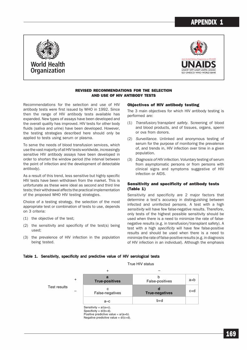
169
APPENDIX 1
Recommendations for the selection and use of HIVantibody tests were first issued by WHO in 1992. Sincethen the range of HIV antibody tests available hasexpanded. New types of assays have been developed andthe overall quality has improved. HIV tests for other bodyfluids (saliva and urine) have been developed. However,the testing strategies described here should only beapplied to tests using serum or plasma.
To serve the needs of blood transfusion services, whichuse the vast majority of all HIV tests worldwide, increasinglysensitive HIV antibody assays have been developed inorder to shorten the window period (the interval betweenthe point of infection and the development of detectableantibody).
As a result of this trend, less sensitive but highly specificHIV tests have been withdrawn from the market. This isunfortunate as these were ideal as second and third linetests; their withdrawal affects the practical implementationof the proposed WHO HIV testing strategies.
Choice of a testing strategy, the selection of the mostappropriate test or combination of tests to use, dependson 3 criteria:
(1) the objective of the test;
(2) the sensitivity and specificity of the test(s) beingused;
(3) the prevalence of HIV infection in the populationbeing tested.
Objectives of HIV antibody testing
The 3 main objectives for which HIV antibody testing isperformed are:
(1) Transfusion/transplant safety. Screening of bloodand blood products, and of tissues, organs, spermor ova from donors.
(2) Surveillance. Unlinked and anonymous testing ofserum for the purpose of monitoring the prevalenceof, and trends in, HIV infection over time in a givenpopulation.
(3) Diagnosis of HIV infection. Voluntary testing of serumfrom asymptomatic persons or from persons withclinical signs and symptoms suggestive of HIVinfection or AIDS.
Sensitivity and specificity of antibody tests(Table 1)
Sensitivity and specificity are 2 major factors thatdetermine a test’s accuracy in distinguishing betweeninfected and uninfected persons. A test with a highsensitivity will have few false-negative results. Therefore,only tests of the highest possible sensitivity should beused when there is a need to minimize the rate of false-negative results (e.g. in transfusion/transplant safety). Atest with a high specificity will have few false-positiveresults and should be used when there is a need tominimize the rate of false-positive results (e.g. in diagnosisof HIV infection in an individual). Although the emphasis
REVISED RECOMMENDATIONS FOR THE SELECTION
AND USE OF HIV ANTIBODY TESTS
True HIV status
–+
a ba+b+ True-positives False-positives
c d c+d– False-negatives True-negatives
Sensitivity = a/(a+c).Specificity = d/(b+d).Positive predictive value = a/(a+b).Negative predictive value = d/(c+d).
Test results
a–c b+d
Table 1.Table 1.Table 1.Table 1.Table 1. Sensitivity, specificity and predictive value of HIV serological testsSensitivity, specificity and predictive value of HIV serological testsSensitivity, specificity and predictive value of HIV serological testsSensitivity, specificity and predictive value of HIV serological testsSensitivity, specificity and predictive value of HIV serological tests
170
APPENDIX 1
might shift slightly towards sensitivity or specificity,depending on the objective of testing, both should meetminimum standards (>99%, >95%, respectively).
Prevalence of HIV infection
The probability that a test will accurately determine thetrue infection status of a person being tested varies withthe prevalence of HIV infection in the population fromwhich the person comes. In general, the higher theprevalence of HIV infection in the population, the greaterthe probability that a person testing positive is trulyinfected (i.e. the greater the positive predictive value[PPV]). Thus, with increasing prevalence, the proportionof serum samples testing false-positive decreases;conversely, the likelihood that a person showing negativetest results is truly uninfected (i.e. the negative predictivevalue [NPV]), decreases as prevalence increases.Therefore, as prevalence increases, so does the proportionof samples testing false negative.
Quality assurance
All laboratories carrying out HIV tests should have aquality assurance programme. It is most important thatquality control procedures be stringently complied with soas to maximize the accuracy of the laboratory results.Procedures for detecting both technical and clericalerrors must be included in all protocols. For example,procedures that guarantee the correct identification ofinitially reactive units of donated blood, which must bediscarded, are essential to the maintenance of a safeblood supply. It is recommended that laboratories submitto an external quality assessment at least once a year.
Strategies for HIV antibody testing
Several studies and field experiences have shown thatminor modifications to the 3 testing strategiesrecommended in 1992 are needed. Both the selection ofand the order in which the assays are used are of theutmost importance for the final outcome of the strategy.As HIV antibody assays have become more sensitive overthe years, the probability of a false-positive reaction in 2assays based on a different principle is not negligible.Therefore, if test combinations are not carefully selected,individuals may be wrongly diagnosed as HIV seropositive.Conversely, the more specific assays are presently slightlyless sensitive as compared to the average HIV antibodytest, which may result in a false-negative diagnosis.These observations cause concern with regard tostrategies II and III of the 1992 recommendations. Thechoice of the most appropriate HIV tests also depends onthe HIV variants present in a particular geographicalregion (e.g. HIV-1 group O). Therefore, test combinationsTherefore, test combinationsTherefore, test combinationsTherefore, test combinationsTherefore, test combinationsshould always be evaluated in the context in which theyshould always be evaluated in the context in which theyshould always be evaluated in the context in which theyshould always be evaluated in the context in which theyshould always be evaluated in the context in which theywill be used before wide-scale implementation.will be used before wide-scale implementation.will be used before wide-scale implementation.will be used before wide-scale implementation.will be used before wide-scale implementation.
Studies have shown that combinations of ELISA and/ orsimple/rapid assays such as dot immunoassays andagglutination tests can provide results as reliable as, andin some instances more reliable than, the ELISA/Western
blot (WB) combination, and at a much lower cost. Atpresent, a number of countries use a selection of differentHIV screening assays in a particular order, to minimize thenumber of costly confirmatory assays.1 Confirmatoryassays should only be used to resolve indeterminateresults for diagnostic purposes. UNAIDS and WHOtherefore recommend that countries consider testingstrategies for HIV antibody detection which use ELISAand/or simple/ rapid assays rather than ELISA/WB.
Recommendations
UNAIDS and WHO recommend 3 testing strategies tomaximize accuracy while minimizing cost. Which strategyis most appropriate will depend on the objectives of thetest and the prevalence of HIV in the sample population,as shown in Table 2 and Fig. 1.
Strategy I
All serum/plasma is tested with one ELISA or simple/rapid assay. Serum that is reactive is considered HIVantibody positive. Serum that is non-reactive isconsidered HIV antibody negative.
Transfusion/transplant safety
When applied for safeguarding the blood supply, the testselected for this strategy should preferably be a combinedcombinedcombinedcombinedcombinedHIV-1/HIV-2 assay HIV-1/HIV-2 assay HIV-1/HIV-2 assay HIV-1/HIV-2 assay HIV-1/HIV-2 assay which is highly sensitivehighly sensitivehighly sensitivehighly sensitivehighly sensitive. Units ofdonated blood yielding reactive reactive reactive reactive reactive or indeterminate indeterminate indeterminate indeterminate indeterminate testresults must be considered as probably infected with HIVand should be discarded according to universal safetyinstructions. 2 Strategy I is meant for testing the donations,but must not be used for notifying donors of a positive testresult. If a blood or tissue donor is to be notified of a testIf a blood or tissue donor is to be notified of a testIf a blood or tissue donor is to be notified of a testIf a blood or tissue donor is to be notified of a testIf a blood or tissue donor is to be notified of a testresult, testing strategies II or III for diagnosis must beresult, testing strategies II or III for diagnosis must beresult, testing strategies II or III for diagnosis must beresult, testing strategies II or III for diagnosis must beresult, testing strategies II or III for diagnosis must beapplied applied applied applied applied (Table 2, Fig. 1). Whatever the final diagnosis,donations which were initially reactive should not be usedfor transfusion or transplants. Several studies haveshown that careful selection of donors is more efficientthan HIV antigen testing to minimize the risk of transfusionrelated infections.
Surveillance
Sensitivity is less crucial for surveillance purposes;however, for this and the above application the assaychosen should have a specificity of at least 95%. It isrecommended that the same assay(s) be used over timeto monitor fluctuations in HIV prevalence.
Diagnosis (see below)
1 Western blot or similar assays based on recombinant proteins and/or synthetic peptides capable of detecting antibodies to specific HIV-1 and/or HIV-2 proteins.
2 See WHO AIDS SERIES 9, Biosafety guidelines for diagnostic and research
laboratories working with HIV.
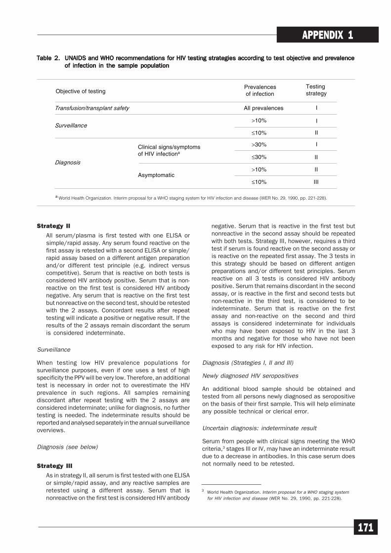
171
APPENDIX 1
Strategy II
All serum/plasma is first tested with one ELISA orsimple/rapid assay. Any serum found reactive on thefirst assay is retested with a second ELISA or simple/rapid assay based on a different antigen preparationand/or different test principle (e.g. indirect versuscompetitive). Serum that is reactive on both tests isconsidered HIV antibody positive. Serum that is non-reactive on the first test is considered HIV antibodynegative. Any serum that is reactive on the first testbut nonreactive on the second test, should be retestedwith the 2 assays. Concordant results after repeattesting will indicate a positive or negative result. If theresults of the 2 assays remain discordant the serumis considered indeterminate.
Surveillance
When testing low HIV prevalence populations forsurveillance purposes, even if one uses a test of highspecificity the PPV will be very low. Therefore, an additionaltest is necessary in order not to overestimate the HIVprevalence in such regions. All samples remainingdiscordant after repeat testing with the 2 assays areconsidered indeterminate; unlike for diagnosis, no furthertesting is needed. The indeterminate results should bereported and analysed separately in the annual surveillanceoverviews.
Diagnosis (see below)
Strategy III
As in strategy II, all serum is first tested with one ELISAor simple/rapid assay, and any reactive samples areretested using a different assay. Serum that isnonreactive on the first test is considered HIV antibody
negative. Serum that is reactive in the first test butnonreactive in the second assay should be repeatedwith both tests. Strategy III, however, requires a thirdtest if serum is found reactive on the second assay oris reactive on the repeated first assay. The 3 tests inthis strategy should be based on different antigenpreparations and/or different test principles. Serumreactive on all 3 tests is considered HIV antibodypositive. Serum that remains discordant in the secondassay, or is reactive in the first and second tests butnon-reactive in the third test, is considered to beindeterminate. Serum that is reactive on the firstassay and non-reactive on the second and thirdassays is considered indeterminate for individualswho may have been exposed to HIV in the last 3months and negative for those who have not beenexposed to any risk for HIV infection.
Diagnosis (Strategies I, II and III)
Newly diagnosed HIV seropositives
An additional blood sample should be obtained andtested from all persons newly diagnosed as seropositiveon the basis of their first sample. This will help eliminateany possible technical or clerical error.
Uncertain diagnosis: indeterminate result
Serum from people with clinical signs meeting the WHOcriteria,3 stages III or IV, may have an indeterminate resultdue to a decrease in antibodies. In this case serum doesnot normally need to be retested.
Objective of testingPrevalences of infection
Transfusion/transplant safety All prevalences
Surveillance>
≤10%
Clinical signs/symptomsof HIV infectiona
>30%
Diagnosis≤30%
>10% IIAsymptomatic
≤10% III
a World Health Organization. Interim proposal for a WHO staging system for HIV infection and disease (WER No. 29, 1990, pp. 221-228).
Testingstrategy
I
II
I
I
II
Table 2.Table 2.Table 2.Table 2.Table 2. UNAIDS and WHO recommendations for HIV testing strategies according to test objective and prevalenceUNAIDS and WHO recommendations for HIV testing strategies according to test objective and prevalenceUNAIDS and WHO recommendations for HIV testing strategies according to test objective and prevalenceUNAIDS and WHO recommendations for HIV testing strategies according to test objective and prevalenceUNAIDS and WHO recommendations for HIV testing strategies according to test objective and prevalenceof infection in the sample populationof infection in the sample populationof infection in the sample populationof infection in the sample populationof infection in the sample population
3 World Health Organization. Interim proposal for a WHO staging system
for HIV infection and disease (WER No. 29, 1990, pp. 221-228).
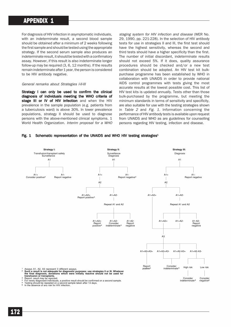
172
APPENDIX 1
For diagnosis of HIV infection in asymptomatic individuals,with an indeterminate result, a second blood sampleshould be obtained after a minimum of 2 weeks followingthe first sample and should be tested using the appropriatestrategy. If the second serum sample also produces anindeterminate result, it should be tested with a confirmatoryassay. However, if this result is also indeterminate longerfollow-up may be required (3, 6, 12 months). If the resultsremain indeterminate after 1 year, the person is consideredto be HIV antibody negative.
General remarks about Strategies I-II-III
Strategy I can only be used to confirm the clinicalStrategy I can only be used to confirm the clinicalStrategy I can only be used to confirm the clinicalStrategy I can only be used to confirm the clinicalStrategy I can only be used to confirm the clinicaldiagnosis of individuals meeting the WHO criteria ofdiagnosis of individuals meeting the WHO criteria ofdiagnosis of individuals meeting the WHO criteria ofdiagnosis of individuals meeting the WHO criteria ofdiagnosis of individuals meeting the WHO criteria ofstage III or IV of HIV infection stage III or IV of HIV infection stage III or IV of HIV infection stage III or IV of HIV infection stage III or IV of HIV infection and when the HIVprevalence in the sample population (e.g. patients froma tuberculosis ward) is above 30%. In lower prevalencepopulations, strategy II should be used to diagnosepersons with the above-mentioned clinical symptoms. 1World Health Organization. Interim proposal for a WHO
staging system for HIV infection and disease (WER No.29, 1990, pp. 221-228). In the selection of HIV antibodytests for use in strategies II and III, the first test shouldhave the highest sensitivity, whereas the second andthird tests should have a higher specificity than the first.The number of initial discordant, indeterminate resultsshould not exceed 5%. If it does, quality assuranceprocedures should be checked and/or a new testcombination should be adopted. An HIV test kit bulk-purchase programme has been established by WHO incollaboration with UNAIDS in order to provide nationalAIDS control programmes with tests giving the mostaccurate results at the lowest possible cost. This list ofHIV test kits is updated annually. Tests other than thosebulk-purchased by the programme, but meeting theminimum standards in terms of sensitivity and specificity,are also suitable for use with the testing strategies shownin Table 2 and Fig. 1. Information concerning theperformance of HIV antibody tests is available upon requestfrom UNAIDS and WHO as are guidelines for counsellingpersons regarding HIV testing, infection and disease.
Strategy I:
Transfusion/transplant safetySurveillance
A1
1 Assays A1, A2, A3 represent 3 different assays.2 Such a result is not adequate for diagnostic purposes; use strategies II or III. Whatever
the final diagnosis, donations which were initially reactive should not be used fortransfusion or transplants.
3 Report: result may be reported.4 For newly diagnosed individuals, a positive result should be confirmed on a second sample.5 Testing should be repeated on a second sample taken after 14 days. 6 In the absence of any risk for HIV infection.
A1+Consider positive2
A1-Report negative
Strategy II:
SurveillanceDiagnosis
A1
A1+ A1-Report negative3
A2
A1+A2+Report positive4
A1+A2-
Repeat A1 and A2
A1+A2+Report
positive4
A1+A2-Consider
indeterminate 5
A1-A2-Report
negative
Strategy III:
Diagnosis
A1
A1+ A1-Report negative
A2
A1+A2+ A1+A2-
Repeat A1 and A2
A1+A2+ A1+A2- A1-A2-Report
negative
A3
A1+A2+A3+ A1+A2+A3- A1+A2-A3+ A1+A2-A3-
Reportpositive4
Considerindeterminate 5 High risk Low risk
Considerindeterminate 5
Considernegative6
Fig. 1Fig. 1Fig. 1Fig. 1Fig. 1 Schematic representation of the UNAIDS and WHO HIV testing strategies Schematic representation of the UNAIDS and WHO HIV testing strategies Schematic representation of the UNAIDS and WHO HIV testing strategies Schematic representation of the UNAIDS and WHO HIV testing strategies Schematic representation of the UNAIDS and WHO HIV testing strategies11111
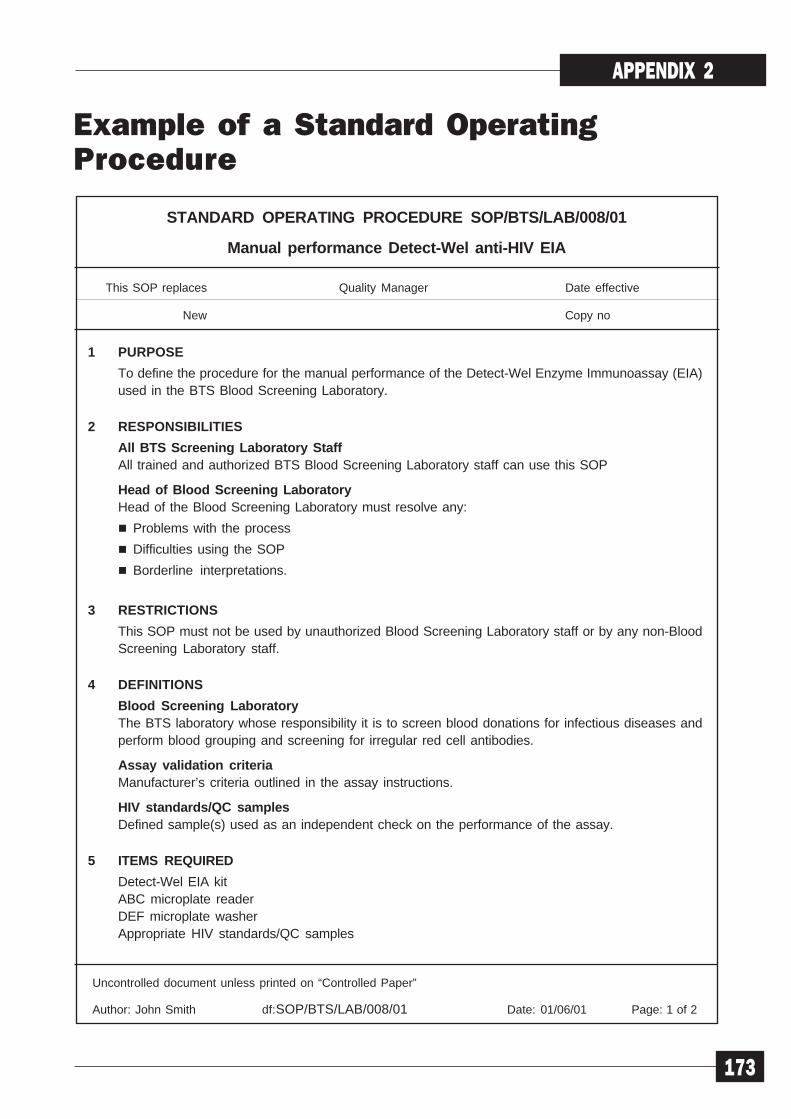
173
APPENDIX 2
Example of a Standard OperatingProcedure
1 PURPOSETo define the procedure for the manual performance of the Detect-Wel Enzyme Immunoassay (EIA)used in the BTS Blood Screening Laboratory.
2 RESPONSIBILITIESAll BTS Screening Laboratory StaffAll trained and authorized BTS Blood Screening Laboratory staff can use this SOP
Head of Blood Screening LaboratoryHead of the Blood Screening Laboratory must resolve any:
Problems with the process
Difficulties using the SOP
Borderline interpretations.
3 RESTRICTIONSThis SOP must not be used by unauthorized Blood Screening Laboratory staff or by any non-BloodScreening Laboratory staff.
4 DEFINITIONSBlood Screening LaboratoryThe BTS laboratory whose responsibility it is to screen blood donations for infectious diseases andperform blood grouping and screening for irregular red cell antibodies.
Assay validation criteriaManufacturer’s criteria outlined in the assay instructions.
HIV standards/QC samplesDefined sample(s) used as an independent check on the performance of the assay.
5 ITEMS REQUIREDDetect-Wel EIA kitABC microplate readerDEF microplate washerAppropriate HIV standards/QC samples
STANDARD OPERATING PROCEDURE SOP/BTS/LAB/008/01
Manual performance Detect-Wel anti-HIV EIA
This SOP replaces Quality Manager Date effective
New Copy no
Uncontrolled document unless printed on “Controlled Paper”
Author: John Smith df:SOP/BTS/LAB/008/01 Date: 01/06/01 Page: 1 of 2
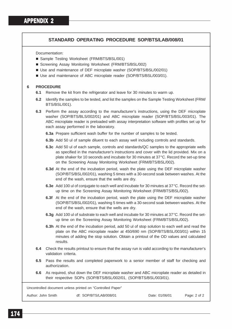
174
APPENDIX 2
STANDARD OPERATING PROCEDURE SOP/BTS/LAB/008/01
Uncontrolled document unless printed on “Controlled Paper”
Author: John Smith df: SOP/BTS/LAB/008/01 Date: 01/06/01 Page: 2 of 2
Documentation:Sample Testing Worksheet (FRM/BTS/BSL/001)Screening Assay Monitoring Worksheet (FRM/BTS/BSL/002)Use and maintenance of DEF microplate washer (SOP/BTS/BSL/002/01)Use and maintenance of ABC microplate reader (SOP/BTS/BSL/003/01).
6 PROCEDURE6.1 Remove the kit from the refrigerator and leave for 30 minutes to warm up.
6.2 Identify the samples to be tested, and list the samples on the Sample Testing Worksheet (FRM/BTS/BSL/001).
6.3 Perform the assay according to the manufacturer’s instructions, using the DEF microplatewasher (SOP/BTS/BLS/002/01) and ABC microplate reader (SOP/BTS/BSL/003/01). TheABC microplate reader is preloaded with assay interpretation software with profiles set up foreach assay performed in the laboratory.
6.3a Prepare sufficient wash buffer for the number of samples to be tested.
6.3b Add 50 ul of sample diluent to each assay well including controls and standards.
6.3c Add 50 ul of each sample, controls and standards/QC samples to the appropriate wellsas specified in the manufacturer's instructions and cover with the lid provided. Mix on aplate shaker for 10 seconds and incubate for 30 minutes at 37°C. Record the set-up timeon the Screening Assay Monitoring Worksheet (FRM/BTS/BSL/002).
6.3d At the end of the incubation period, wash the plate using the DEF microplate washer(SOP/BTS/BSL/002/01), washing 5 times with a 30-second soak between washes. At theend of the wash, ensure that the wells are dry.
6.3e Add 100 ul of conjugate to each well and incubate for 30 minutes at 37°C. Record the set-up time on the Screening Assay Monitoring Worksheet (FRM/BTS/BSL/002).
6.3f At the end of the incubation period, wash the plate using the DEF microplate washer(SOP/BTS/BSL/002/01), washing 5 times with a 30-second soak between washes. At theend of the wash, ensure that the wells are dry.
6.3g Add 100 ul of substrate to each well and incubate for 30 minutes at 37°C. Record the set-up time on the Screening Assay Monitoring Worksheet (FRM/BTS/BSL/002).
6.3h At the end of the incubation period, add 50 ul of stop solution to each well and read theplate on the ABC microplate reader at 450/690 nm (SOP/BTS/BSL/003/01) within 15minutes of adding the stop solution. Obtain a printout of the OD values and calculatedresults.
6.4 Check the results printout to ensure that the assay run is valid according to the manufacturer’svalidation criteria.
6.5 Pass the results and completed paperwork to a senior member of staff for checking andauthorization.
6.6 As required, shut down the DEF microplate washer and ABC microplate reader as detailed intheir respective SOPs (SOP/BTS/BSL/002/01, (SOP/BTS/BSL/003/01).
Related Documents