Module 06 - Classification and Diversity Checklist 06.01 Classification of Living Organisms

Module 06_video Review
Dec 14, 2015
Classification of living organisms
Welcome message from author
This document is posted to help you gain knowledge. Please leave a comment to let me know what you think about it! Share it to your friends and learn new things together.
Transcript

Module 06 - Classification and Diversity Checklist
06.01 Classification of Living Organisms


The image shows a cladogram. The first clade divides amphibians from other animals because amphibians lack amniotic eggs. The second clade divides the turtles because they lack the opening in their skull for muscles. At the third clade, the crocodiles, birds, dinosaurs, lizards, and snakes are divided from the other animals because they lack fur or hair. This clade is further divided. The crocodiles are on a opposite branch from lizards and snakes. Another branch divides crocodiles from birds and yet another

branch divides birds from dinosaurs. A fourth clade divides the monotremes from other fur or haired animals because monotremes do not birth their young. Of those organisms that do have live births, marsupials are divided from placental mammals.
Cladogram 1
Groups G and H are most closely related, while group A is the most distant. Clades are represented by numbers 1 through 7.
Cladogram 2Groups A and B are more closely related to each other than to any other group, as are C and D, E and F, and G and H. Clade 4 is more closely related to 5, and clade 6 is more closely related to clade 7.
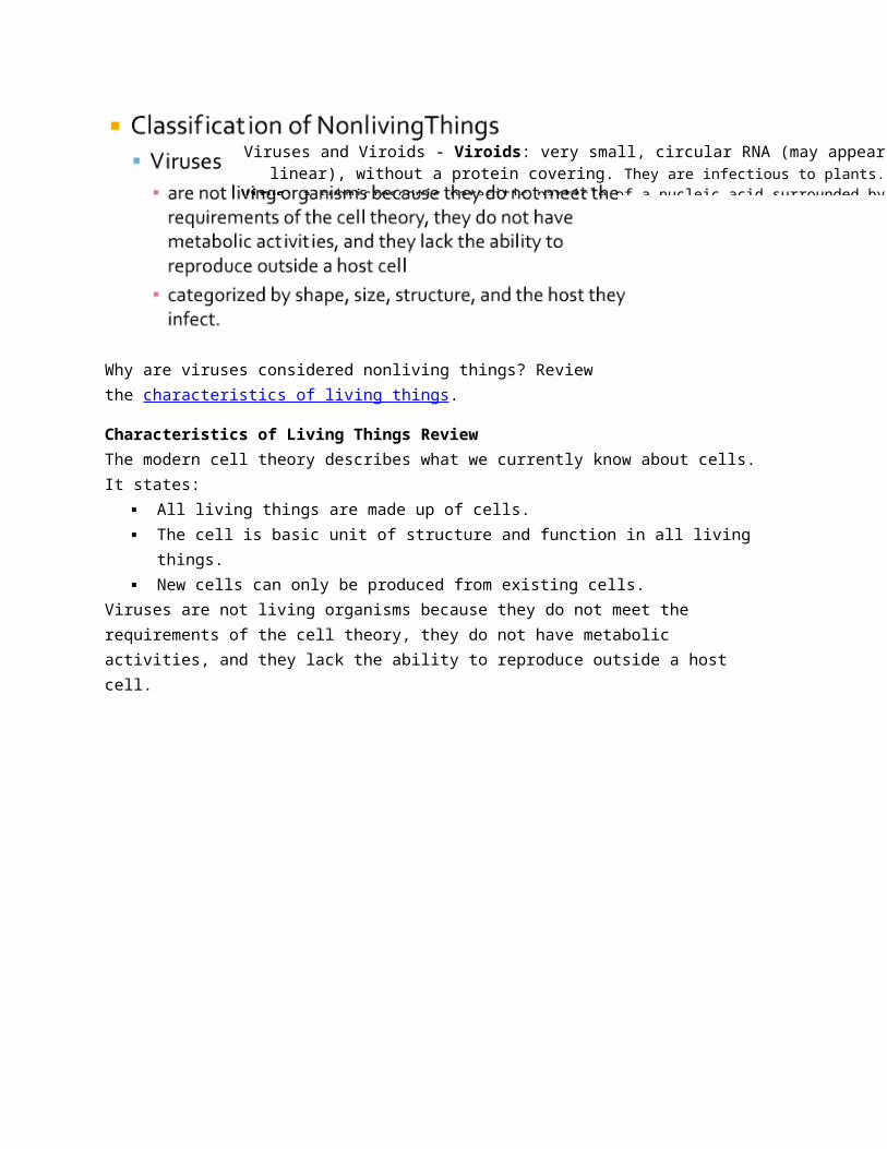
Why are viruses considered nonliving things? Review the characteristics of living things.
Characteristics of Living Things ReviewThe modern cell theory describes what we currently know about cells. It states:
All living things are made up of cells. The cell is basic unit of structure and function in all living things. New cells can only be produced from existing cells.
Viruses and Viroids - Viroids: very small, circular RNA (may appear linear), without a protein covering. They are infectious to plants. Virus: a submicroscopic parasitic particle of a nucleic acid surrounded by protein
that can only replicate within a host cell.

Viruses are not living organisms because they do not meet the requirements of the cell theory, they do not have metabolic activities, and they lack the ability to reproduce outside a host cell.
Basic Virus Shapes
Viruses come in all shapes and microscopic sizes. Some have lipid envelopes and some do not. Some have circular RNA, while others have linear DNA. If we consider the organisms they infect, the classification can get complex. However, there are some basic shapes for the protein coats of viruses. Let’s review these shapes below.
Lipid: a broad group of naturally occurring molecules, which includes fats and phospholipids

Viral Infections
There are two types of viral infections: lytic and lysogenic. In a lytic infection, the virus enters the host cell, replicates many copies, and destroys the cell in order to get out and spread to others. In a lysogenic infection, the virus incorporates its DNA into the DNA of the cell. It stays dormant and replicates with the cell DNA until the cell is in danger of dying. At that time, the lysogenic infection will become lytic in order to spread to other host cells.
For both types of infections, viruses attach to host cells and use the cells’ resources to replicate their genetic material and parts. Sometimes, the cell recognizes the protein as something it needs and brings it into the cell through active transport or endocytosis. In a few instances, the virus contains enzymes specific for breaking down cell walls in bacteria and plants. Once the enzyme provides an opening, the virus injects its DNA or RNA into the cell.
Lytic and Lysogenic InfectionsA slideshow depicting the life cycles of lytic and lysogenic infections.Lytic Cycle Steps:Slide 1: A little virus is shown attaching to a large cell.When a virus, like a bacteriophage, infects a host cell, it follows a lytic or lysogenic cycle. Both cycles begin with the virus attaching to the cell membrane of a host cell.Slide 2: Multiple circles of viral DNA are shown within the host cell, along with many new capsids and virus stems.In the lytic cycle, viral genetic material is replicated and the viral genes direct the cell to construct new viral parts.Slide 3: These newly formed pieces are shown moving together to form new viruses.The viral genetic material and parts come together to make new viruses.Slide 4: The little viruses are shown escaping from the broken cell.The cell lyses, releasing the new viruses.
Lysogenic Steps:Slide 5: A different colored piece of DNA is shown attaching itself to the host DNA.In the lysogenic cycle, the genetic material of the virus becomes part of the host DNA through transduction.Slide 6: The host DNA is shown replicating, each copy containing the integrated viral DNA.The integrated DNA (prophage DNA) is replicated with the host DNA for several cycles.Slide 7: Multiple circles of viral DNA are shown within the host cell, along with many new capsids and virus stems. These pieces come together to form new viruses which then escape the broken cell.When the cell is stressed or near death, the integrated DNA begins the lytic cycle.
Related Documents


