Modulation of Serotonin Transporter Function during Fetal Development Causes Dilated Heart Cardiomyopathy and Lifelong Behavioral Abnormalities Cornelle W. Noorlander 1 , Frederique F. T. Ververs 2 , Peter G. J. Nikkels 3 , Cees J. A. van Echteld 4 , Gerard H. A. Visser 5 , Marten P. Smidt 1 * 1 Rudolf Magnus Institute of Neuroscience, Department of Neuroscience and Pharmacology, University Medical Center Utrecht, Utrecht, The Netherlands, 2 Department of Pharmacy, University Medical Center Utrecht, Utrecht, The Netherlands, 3 Department of Pathology, University Medical Center Utrecht, Utrecht, The Netherlands, 4 Department of Cardiology, University Medical Center Utrecht, Utrecht, The Netherlands, 5 Department of Obstetrics, Neonatology and Gynaecology, University Medical Center Utrecht, Utrecht, The Netherlands Abstract Background: Women are at great risk for mood and anxiety disorders during their childbearing years and may become pregnant while taking antidepressant drugs. In the treatment of depression and anxiety disorders, selective serotonin reuptake inhibitors (SSRIs) are the most frequently prescribed drugs, while it is largely unknown whether this medication affects the development of the central nervous system of the fetus. The possible effects are the product of placental transfer efficiency, time of administration and dose of the respective SSRI. Methodology/Principal Findings: In order to attain this information we have setup a study in which these parameters were measured and the consequences in terms of physiology and behavior are mapped. The placental transfer of fluoxetine and fluvoxamine, two commonly used SSRIs, was similar between mouse and human, indicating that the fetal exposure of these SSRIs in mice is comparable with the human situation. Fluvoxamine displayed a relatively low placental transfer, while fluoxetine showed a relatively high placental transfer. Using clinical doses of fluoxetine the mortality of the offspring increased dramatically, whereas the mortality was unaffected after fluvoxamine exposure. The majority of the fluoxetine- exposed offspring died postnatally of severe heart failure caused by dilated cardiomyopathy. Molecular analysis of fluoxetine-exposed offspring showed long-term alterations in serotonin transporter levels in the raphe nucleus. Furthermore, prenatal fluoxetine exposure resulted in depressive- and anxiety-related behavior in adult mice. In contrast, fluvoxamine-exposed mice did not show alterations in behavior and serotonin transporter levels. Decreasing the dose of fluoxetine resulted in higher survival rates and less dramatic effects on the long-term behavior in the offspring. Conclusions: These results indicate that prenatal fluoxetine exposure affects fetal development, resulting in cardiomyopathy and a higher vulnerability to affective disorders in a dose-dependent manner. Citation: Noorlander CW, Ververs FFT, Nikkels PGJ, van Echteld CJA, Visser GHA, et al. (2008) Modulation of Serotonin Transporter Function during Fetal Development Causes Dilated Heart Cardiomyopathy and Lifelong Behavioral Abnormalities. PLoS ONE 3(7): e2782. doi:10.1371/journal.pone.0002782 Editor: Dawn N. Albertson, Minnesota State University Mankato, United States of America Received May 6, 2008; Accepted July 1, 2008; Published July 23, 2008 Copyright: ß 2008 Noorlander et al. This is an open-access article distributed under the terms of the Creative Commons Attribution License, which permits unrestricted use, distribution, and reproduction in any medium, provided the original author and source are credited. Funding: ‘‘Universtity of Utrecht’’ HIPO grant (M.P. Smidt), ‘‘Hersenstichting’’ grant (15F07(2).22) (C.W. Noorlander/M.P. Smidt) Competing Interests: The authors have declared that no competing interests exist. * E-mail: [email protected] Introduction Mood and anxiety disorders such as depression, panic disorder and obsessive-compulsive disorder are common in women during their childbearing years [1,2]. The prevalence of depression has been reported to be between 10% and 16% during pregnancy and is becoming a major health issue [3,4]. In the treatment of depression and anxiety disorders during pregnancy, selective serotonin reuptake inhibitors (SSRIs) are the most frequently prescribed drugs nowadays. SSRIs, like fluoxetine and fluvox- amine, inhibit the reuptake of serotonin (5-hydroxytryptamine or 5-HT) into the presynaptic neuron by binding to the serotonin transporter (5-HTT), which results in an increase of synaptic serotonin levels. SSRIs have no effects on other monoamine transporters, which differentiates them from the previously prescribed tricyclic antidepressants. SSRIs are considered much safer than tricyclic antidepressants, since the toxic dose threshold is much higher and they are believed to have fewer and weaker side effects. Nevertheless, there is still uncertainty concerning the safety of the offspring after antidepressant exposure during pregnancy. Although several studies have reported no associations between congenital malformations and prenatal SSRI exposure, it has been recently shown that fetal exposure to SSRIs results in an increased risk of adverse neonatal effects, including neurological abnormal- ities, cardiac malformations and persistent pulmonary hyperten- sion [5–11]. Furthermore, lower birth weight and an increased risk of preterm birth have been observed after prenatal SSRI treatment [12,13]. However, it is unknown whether this medication affects the development of the central nervous system of the fetus. Therefore, we have setup a study design to evaluate prenatal SSRI exposure on fetal development and the long-term consequences in terms of behavioral pathology. In the mouse PLoS ONE | www.plosone.org 1 July 2008 | Volume 3 | Issue 7 | e2782

Welcome message from author
This document is posted to help you gain knowledge. Please leave a comment to let me know what you think about it! Share it to your friends and learn new things together.
Transcript
Modulation of Serotonin Transporter Function duringFetal Development Causes Dilated HeartCardiomyopathy and Lifelong Behavioral AbnormalitiesCornelle W. Noorlander1, Frederique F. T. Ververs2, Peter G. J. Nikkels3, Cees J. A. van Echteld4,
Gerard H. A. Visser5, Marten P. Smidt1*
1 Rudolf Magnus Institute of Neuroscience, Department of Neuroscience and Pharmacology, University Medical Center Utrecht, Utrecht, The Netherlands, 2 Department
of Pharmacy, University Medical Center Utrecht, Utrecht, The Netherlands, 3 Department of Pathology, University Medical Center Utrecht, Utrecht, The Netherlands,
4 Department of Cardiology, University Medical Center Utrecht, Utrecht, The Netherlands, 5 Department of Obstetrics, Neonatology and Gynaecology, University Medical
Center Utrecht, Utrecht, The Netherlands
Abstract
Background: Women are at great risk for mood and anxiety disorders during their childbearing years and may becomepregnant while taking antidepressant drugs. In the treatment of depression and anxiety disorders, selective serotoninreuptake inhibitors (SSRIs) are the most frequently prescribed drugs, while it is largely unknown whether this medicationaffects the development of the central nervous system of the fetus. The possible effects are the product of placental transferefficiency, time of administration and dose of the respective SSRI.
Methodology/Principal Findings: In order to attain this information we have setup a study in which these parameters weremeasured and the consequences in terms of physiology and behavior are mapped. The placental transfer of fluoxetine andfluvoxamine, two commonly used SSRIs, was similar between mouse and human, indicating that the fetal exposure of theseSSRIs in mice is comparable with the human situation. Fluvoxamine displayed a relatively low placental transfer, whilefluoxetine showed a relatively high placental transfer. Using clinical doses of fluoxetine the mortality of the offspringincreased dramatically, whereas the mortality was unaffected after fluvoxamine exposure. The majority of the fluoxetine-exposed offspring died postnatally of severe heart failure caused by dilated cardiomyopathy. Molecular analysis offluoxetine-exposed offspring showed long-term alterations in serotonin transporter levels in the raphe nucleus.Furthermore, prenatal fluoxetine exposure resulted in depressive- and anxiety-related behavior in adult mice. In contrast,fluvoxamine-exposed mice did not show alterations in behavior and serotonin transporter levels. Decreasing the dose offluoxetine resulted in higher survival rates and less dramatic effects on the long-term behavior in the offspring.
Conclusions: These results indicate that prenatal fluoxetine exposure affects fetal development, resulting incardiomyopathy and a higher vulnerability to affective disorders in a dose-dependent manner.
Citation: Noorlander CW, Ververs FFT, Nikkels PGJ, van Echteld CJA, Visser GHA, et al. (2008) Modulation of Serotonin Transporter Function during FetalDevelopment Causes Dilated Heart Cardiomyopathy and Lifelong Behavioral Abnormalities. PLoS ONE 3(7): e2782. doi:10.1371/journal.pone.0002782
Editor: Dawn N. Albertson, Minnesota State University Mankato, United States of America
Received May 6, 2008; Accepted July 1, 2008; Published July 23, 2008
Copyright: � 2008 Noorlander et al. This is an open-access article distributed under the terms of the Creative Commons Attribution License, which permitsunrestricted use, distribution, and reproduction in any medium, provided the original author and source are credited.
Funding: ‘‘Universtity of Utrecht’’ HIPO grant (M.P. Smidt), ‘‘Hersenstichting’’ grant (15F07(2).22) (C.W. Noorlander/M.P. Smidt)
Competing Interests: The authors have declared that no competing interests exist.
* E-mail: [email protected]
Introduction
Mood and anxiety disorders such as depression, panic disorder
and obsessive-compulsive disorder are common in women during
their childbearing years [1,2]. The prevalence of depression has
been reported to be between 10% and 16% during pregnancy and
is becoming a major health issue [3,4]. In the treatment of
depression and anxiety disorders during pregnancy, selective
serotonin reuptake inhibitors (SSRIs) are the most frequently
prescribed drugs nowadays. SSRIs, like fluoxetine and fluvox-
amine, inhibit the reuptake of serotonin (5-hydroxytryptamine or
5-HT) into the presynaptic neuron by binding to the serotonin
transporter (5-HTT), which results in an increase of synaptic
serotonin levels. SSRIs have no effects on other monoamine
transporters, which differentiates them from the previously
prescribed tricyclic antidepressants. SSRIs are considered much
safer than tricyclic antidepressants, since the toxic dose threshold is
much higher and they are believed to have fewer and weaker side
effects. Nevertheless, there is still uncertainty concerning the safety
of the offspring after antidepressant exposure during pregnancy.
Although several studies have reported no associations between
congenital malformations and prenatal SSRI exposure, it has been
recently shown that fetal exposure to SSRIs results in an increased
risk of adverse neonatal effects, including neurological abnormal-
ities, cardiac malformations and persistent pulmonary hyperten-
sion [5–11]. Furthermore, lower birth weight and an increased risk
of preterm birth have been observed after prenatal SSRI
treatment [12,13]. However, it is unknown whether this
medication affects the development of the central nervous system
of the fetus. Therefore, we have setup a study design to evaluate
prenatal SSRI exposure on fetal development and the long-term
consequences in terms of behavioral pathology. In the mouse
PLoS ONE | www.plosone.org 1 July 2008 | Volume 3 | Issue 7 | e2782
model in which clinical doses were applied to the mothers during
pregnancy, we found that the offspring suffered in a dose-
dependent manner from a severe form of dilated cardiomyopathy
and that surviving mice had severe behavioral abnormalities which
may relate to anxiety disorders.
Materials and Methods
AnimalsPregnant C57Bl/6-JIco mice (Charles River Laboratory,
France) were housed individually on day 6 of pregnancy.
Pregnancy was determined by observation of a vaginal plug.
The plug date is considered to be day 0 of gestation (embryonic
day 0 (E0)). From day 8 until day 18 of pregnancy, mice were
injected intraperitonally with either fluoxetine (0.3, 0.6, 0.8 mg/
kg/day), fluvoxamine (4.2 mg/kg/day) or with equal volumes of
sterile saline. Mice were allowed ad libitum access to food and
water. Light/dark cycle (dark phase 19:00–07:00 h), temperature
(21uC) and humidity (60%) were kept constant. In cross fostering
experiments, pups were placed with another mother a few hours
after birth. Offspring was studied at E18, postnatal day 3 (P3), P20
and adult stage (P90). For the adult stage, pups were weaned at
P25 and remained group-housed (2–4 animals per cage) with
same-sex littermates until experimentation at adulthood. All
experiments were approved by the Animal Ethics Committee of
the University Medical Center Utrecht and were conducted in
agreement with Dutch laws (Wet op de Dierproeven, 1996) and
European regulations (Guideline 86/609/EEC).
High Performance Liquid Chromatography AnalysisFor determining placental transfer of SSRIs, pregnant mice
were sacrificed at E16 by decapitation five hours after drug
administration. Maternal blood was collected and embryos were
quickly removed. Embryonic tissue was homogenized with 1 ml of
0.9% NaCl. Maternal plasma and embryonic tissue was stored at
220uC until analysis. For determining placental transfer of SSRIs
in humans, maternal blood was obtained during delivery at the
end stage of labour of woman who used antidepressants
throughout pregnancy. Venous cord blood serum was obtained
within the first hour after delivery. Samples were stored at 220uCtill analysis.
Fluoxetine and fluvoxamine concentrations were determined by
a high-performance liquid chromatography (HPLC) method with
UV/Fluorescence detection. Desmethylmaprotyline was used as
internal standard. The analytes were extracted from plasma and
embryonic tissue using a liquid/liquid extraction procedure with
isoamylalcohol/n-hexane/borate buffer pH 9.3 (0.05/5/0.25 v/
v/v). Chromatography was performed on a PolarisH (Varian Inc.
Palo Alto, CA, USA) C18, 5 m, 50 mm62 mm analytical column
at a flow rate of 1,0 ml/min using acetonitrile/phosphoric acid
buffer pH 2 (15/45 v/v) at 30uC. The range where the assay was
linear (50 and 500 ug/L), the coefficient of variation was less than
10% and the accuracy was between 95 and 105% for both
fluoxetine and fluvoxamine.
Pathological examinationMice were sacrificed at P20 and adulthood, and heart, lungs,
liver, stomach, kidneys, intestines and spleen were dissected.
Tissue was fixed in 4% paraformaldehyde, dehydrated in an
ethanol series, embedded in paraffin. Paraffin-embedded sections
(7 mm) were mounted on SuperFrost plus slides (Menzel Glaser),
deparaffinated, stained with haematoxylin and eosin, and analyzed
by two independent observers blinded for the treatment.
Magnetic Resonance ImagingThe MRI scanning of the hearts was performed as described
before [14].
AutoradiographyP20 and adult brains were collected and immediately frozen on
dry ice. Sections (20 mm) were cut and collected on SuperFrost
Plus slides (Menzel Glaser). Brain sections were stored at 280uCuntil use. Sections were dried with a stream of air and washed for
20 min in Tris-HCl buffer (120 mM NaCl, 50 mM Tris-HCl
(Sigma-Aldrich), pH 7.4) at room temperature. For specific
binding to the serotonin transporter, sections were incubated for
1 hour with 1 nM [N-methyl-3H]-citalopram (77.0 Ci/mmol;
Amersham Bioscience, UK) in Tris-HCl buffer at room temper-
ature. Non-specific binding was determined by adding 10 mM of
the selective serotonin transporter inhibitor, fluoxetine (Sigma-
Aldrich) in addition to 1 nM [N-methyl-3H]-citalopram. Sections
were then washed three times (1, 10 and 10 min, respectively) in
ice cold Tris-HCl buffer and rinsed in ice cold water. Sections
were dried with a stream of air and exposed to BAS-TR2040
phosphoimaging plates (Fuji Imaging Plates) for 2 weeks at room
temperature. Autoradiographic BAS-TR2040 imaging plates were
scanned using the FLA-5000 imaging system (Fuji) and quantita-
tive analysis was performed using the AIDA Image Analyzer
Software (Raytest). Serotonin transporter binding was analyzed in
the dorsal raphe nucleus of mice at P20 and P90 treated with
saline (N = 10), fluvoxamine (N = 10) and fluoxetine (N = 4).
Elevated Plus MazeThe elevated plus maze (black Plexiglas) was elevated 100 cm
above floor level and consisted of two open and two closed arms
(3065 cm). All arms radiated from a common central open square
(565 cm). The floor of the enclosed arms was the same size as the
open, but these arms had side walls of 15 cm high. Each mouse
was placed in the plus-maze facing an open arm and was allowed
to explore freely the plus-maze for 10 min. All the experiments
were performed under normal ambient overhead lighting and
were carried out during the light phase of the cycle, between 13:00
and 18:00 h. Each experimental session was recorded by
videotracking software (Ethovision, Noldus Information Technol-
ogy, The Netherlands). Between trials, the apparatus was
thoroughly rinsed with 70% ethanol and dried with clean towels.
The following parameters were obtained: total distance moved,
velocity, entries into open and closed arms, time spent on open
and closed arms and distance moved on open and closed arms.
Mice were tested at P20 and adulthood.
Large Open FieldThe open field consisted of a dark-gray PVC cylinder with a
diameter of 80 cm and 30 cm in height. The open field was
divided in two parts, a central area with a diameter of 55 cm and
an outer ring. Adult mice were individually transported from the
adjacent room to the experimental room and immediately placed
near the wall, in the outer ring of the open field. All the
experiments were performed under normal ambient overhead
lighting and were carried out during the light phase of the cycle,
between 13:00 and 18:00 h. Locomotor activity was recorded by
videotracking software (Ethovision, Noldus Information Technol-
ogy, The Netherlands) for 30 min. Between trials, the apparatus
was thoroughly rinsed with 70% ethanol and dried with clean
towels. The following parameters were obtained: total distance
moved, velocity, time spent in the center and in the outer ring and
distance moved in the center and in the outer ring.
5-HT Influences Development
PLoS ONE | www.plosone.org 2 July 2008 | Volume 3 | Issue 7 | e2782
Novelty suppressed feeding testAdult mice were weighed and food was removed from the cage,
while water remained available ad libitum. Twenty-four hours
after food restriction, mice were transferred to the testing room,
placed in a novel arena with in the center a petridish containing a
pre-weighed quantity of food pellets. Each subject was placed in
the corner of the testing area, and the latency to feed, time spent
feeding, and total food consumption were recorded over 10 min.
All the experiments were performed under normal ambient
overhead lighting and were carried out during the light phase of
the cycle, between 13:00 and 18:00 h.
StatisticsData of body weight, Wt/r ratios, 5-HTT binding and
behavioral performance were analyzed using an one-way analysis
of variance (ANOVA), followed by Bonferroni multiple compar-
ison test when appropriate. Within-group comparisons were
performed using Student’s paired t test. P,0.05 was considered
significant.
Results
Placental transfer of fluoxetine and fluvoxamine iscomparable between mouse and human
The level of fetal exposure to certain SSRIs may influence the
effects of SSRIs on fetal development, and may differ due to
specific placental transfer efficiency. Therefore, we determined the
placental transfer of two commonly used SSRIs (fluoxetine and
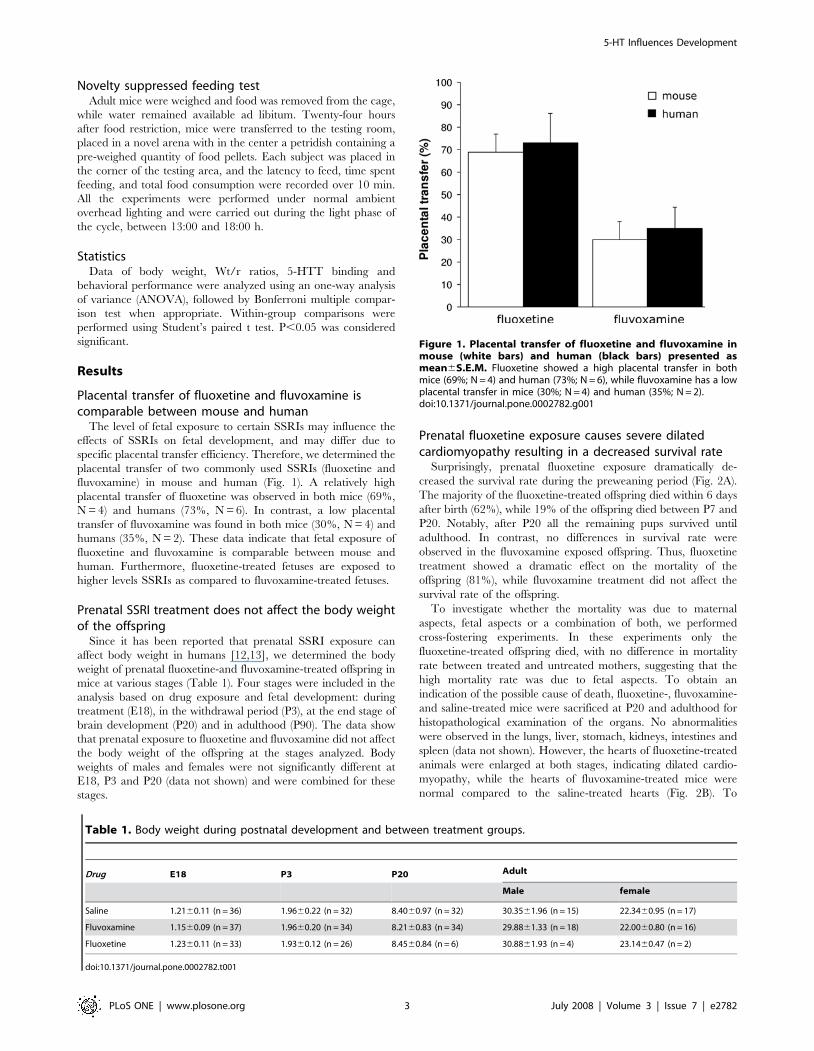
fluvoxamine) in mouse and human (Fig. 1). A relatively high
placental transfer of fluoxetine was observed in both mice (69%,
N = 4) and humans (73%, N = 6). In contrast, a low placental
transfer of fluvoxamine was found in both mice (30%, N = 4) and
humans (35%, N = 2). These data indicate that fetal exposure of
fluoxetine and fluvoxamine is comparable between mouse and
human. Furthermore, fluoxetine-treated fetuses are exposed to
higher levels SSRIs as compared to fluvoxamine-treated fetuses.
Prenatal SSRI treatment does not affect the body weightof the offspring
Since it has been reported that prenatal SSRI exposure can
affect body weight in humans [12,13], we determined the body
weight of prenatal fluoxetine-and fluvoxamine-treated offspring in
mice at various stages (Table 1). Four stages were included in the
analysis based on drug exposure and fetal development: during
treatment (E18), in the withdrawal period (P3), at the end stage of
brain development (P20) and in adulthood (P90). The data show
that prenatal exposure to fluoxetine and fluvoxamine did not affect
the body weight of the offspring at the stages analyzed. Body
weights of males and females were not significantly different at
E18, P3 and P20 (data not shown) and were combined for these
stages.
Prenatal fluoxetine exposure causes severe dilatedcardiomyopathy resulting in a decreased survival rate
Surprisingly, prenatal fluoxetine exposure dramatically de-
creased the survival rate during the preweaning period (Fig. 2A).
The majority of the fluoxetine-treated offspring died within 6 days
after birth (62%), while 19% of the offspring died between P7 and
P20. Notably, after P20 all the remaining pups survived until
adulthood. In contrast, no differences in survival rate were
observed in the fluvoxamine exposed offspring. Thus, fluoxetine
treatment showed a dramatic effect on the mortality of the
offspring (81%), while fluvoxamine treatment did not affect the
survival rate of the offspring.
To investigate whether the mortality was due to maternal
aspects, fetal aspects or a combination of both, we performed
cross-fostering experiments. In these experiments only the
fluoxetine-treated offspring died, with no difference in mortality
rate between treated and untreated mothers, suggesting that the
high mortality rate was due to fetal aspects. To obtain an
indication of the possible cause of death, fluoxetine-, fluvoxamine-
and saline-treated mice were sacrificed at P20 and adulthood for
histopathological examination of the organs. No abnormalities
were observed in the lungs, liver, stomach, kidneys, intestines and
spleen (data not shown). However, the hearts of fluoxetine-treated
animals were enlarged at both stages, indicating dilated cardio-
myopathy, while the hearts of fluvoxamine-treated mice were
normal compared to the saline-treated hearts (Fig. 2B). To
Figure 1. Placental transfer of fluoxetine and fluvoxamine inmouse (white bars) and human (black bars) presented asmean6S.E.M. Fluoxetine showed a high placental transfer in bothmice (69%; N = 4) and human (73%; N = 6), while fluvoxamine has a lowplacental transfer in mice (30%; N = 4) and human (35%; N = 2).doi:10.1371/journal.pone.0002782.g001
Table 1. Body weight during postnatal development and between treatment groups.
Drug E18 P3 P20 Adult
Male female
Saline 1.2160.11 (n = 36) 1.9660.22 (n = 32) 8.4060.97 (n = 32) 30.3561.96 (n = 15) 22.3460.95 (n = 17)
Fluvoxamine 1.1560.09 (n = 37) 1.9660.20 (n = 34) 8.2160.83 (n = 34) 29.8861.33 (n = 18) 22.0060.80 (n = 16)
Fluoxetine 1.2360.11 (n = 33) 1.9360.12 (n = 26) 8.4560.84 (n = 6) 30.8861.93 (n = 4) 23.1460.47 (n = 2)
doi:10.1371/journal.pone.0002782.t001
5-HT Influences Development
PLoS ONE | www.plosone.org 3 July 2008 | Volume 3 | Issue 7 | e2782
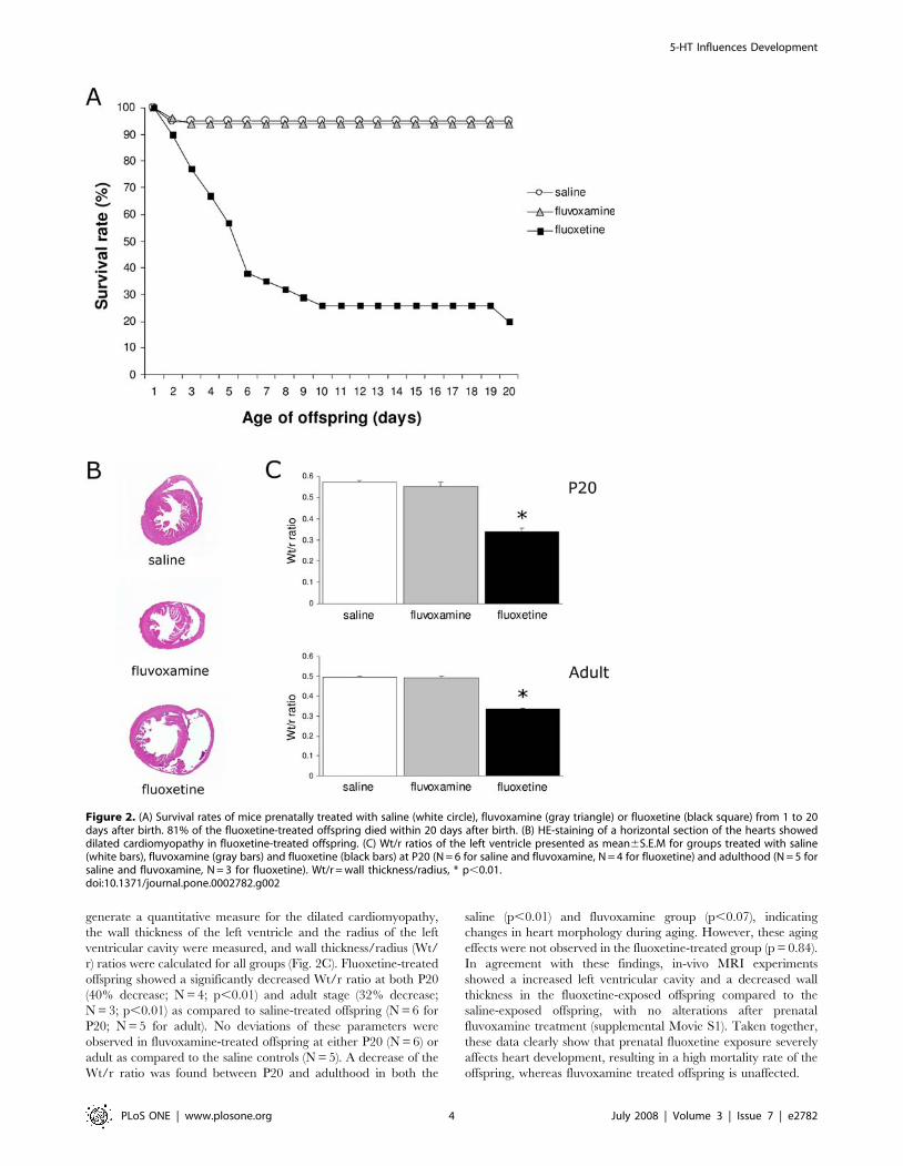
generate a quantitative measure for the dilated cardiomyopathy,
the wall thickness of the left ventricle and the radius of the left
ventricular cavity were measured, and wall thickness/radius (Wt/
r) ratios were calculated for all groups (Fig. 2C). Fluoxetine-treated
offspring showed a significantly decreased Wt/r ratio at both P20
(40% decrease; N = 4; p,0.01) and adult stage (32% decrease;
N = 3; p,0.01) as compared to saline-treated offspring (N = 6 for
P20; N = 5 for adult). No deviations of these parameters were
observed in fluvoxamine-treated offspring at either P20 (N = 6) or
adult as compared to the saline controls (N = 5). A decrease of the
Wt/r ratio was found between P20 and adulthood in both the
saline (p,0.01) and fluvoxamine group (p,0.07), indicating
changes in heart morphology during aging. However, these aging
effects were not observed in the fluoxetine-treated group (p = 0.84).
In agreement with these findings, in-vivo MRI experiments
showed a increased left ventricular cavity and a decreased wall
thickness in the fluoxetine-exposed offspring compared to the
saline-exposed offspring, with no alterations after prenatal
fluvoxamine treatment (supplemental Movie S1). Taken together,
these data clearly show that prenatal fluoxetine exposure severely
affects heart development, resulting in a high mortality rate of the
offspring, whereas fluvoxamine treated offspring is unaffected.
Figure 2. (A) Survival rates of mice prenatally treated with saline (white circle), fluvoxamine (gray triangle) or fluoxetine (black square) from 1 to 20days after birth. 81% of the fluoxetine-treated offspring died within 20 days after birth. (B) HE-staining of a horizontal section of the hearts showeddilated cardiomyopathy in fluoxetine-treated offspring. (C) Wt/r ratios of the left ventricle presented as mean6S.E.M for groups treated with saline(white bars), fluvoxamine (gray bars) and fluoxetine (black bars) at P20 (N = 6 for saline and fluvoxamine, N = 4 for fluoxetine) and adulthood (N = 5 forsaline and fluvoxamine, N = 3 for fluoxetine). Wt/r = wall thickness/radius, * p,0.01.doi:10.1371/journal.pone.0002782.g002
5-HT Influences Development
PLoS ONE | www.plosone.org 4 July 2008 | Volume 3 | Issue 7 | e2782
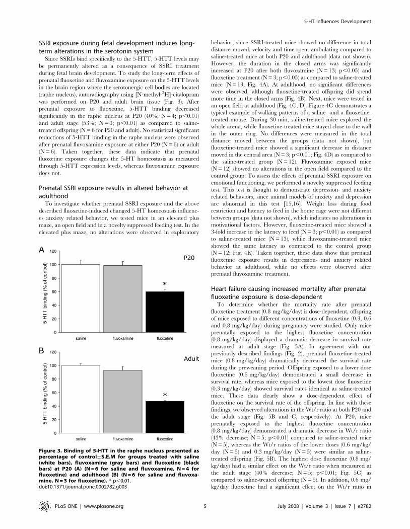
SSRI exposure during fetal development induces long-term alterations in the serotonin system
Since SSRIs bind specifically to the 5-HTT, 5-HTT levels may
be permanently altered as a consequence of SSRI treatment
during fetal brain development. To study the long-term effects of
prenatal fluoxetine and fluvoxamine exposure on the 5-HTT levels
in the brain region where the serotonergic cell bodies are located
(raphe nucleus), autoradiography using [N-methyl-3H]-citalopram
was performed on P20 and adult brain tissue (Fig. 3). After
prenatal exposure to fluoxetine, 5-HTT binding decreased
significantly in the raphe nucleus at P20 (40%; N = 4; p,0.01)
and adult stage (53%; N = 3; p,0.01) as compared to saline-
treated offspring (N = 6 for P20 and adult). No statistical significant
reductions of 5-HTT binding in the raphe nucleus were observed
after prenatal fluvoxamine exposure at either P20 (N = 6) or adult
(N = 6). Taken together, these data indicate that prenatal
fluoxetine exposure changes the 5-HT homeostasis as measured
through 5-HTT expression levels, whereas fluvoxamine exposure
does not.
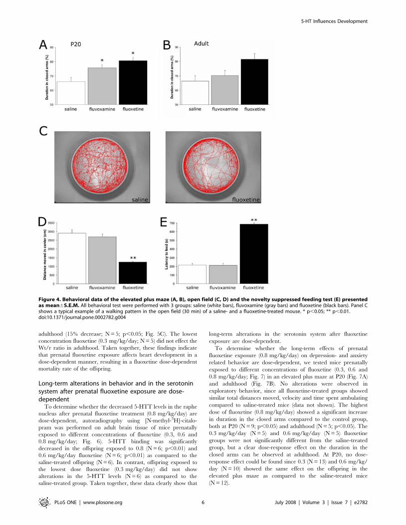
Prenatal SSRI exposure results in altered behavior atadulthood
To investigate whether prenatal SSRI exposure and the above
described fluoxetine-induced changed 5-HT homeostasis influenc-
es anxiety related behavior, we tested mice in an elevated plus
maze, an open field and in a novelty suppressed feeding test. In the
elevated plus maze, no alterations were observed in exploratory
behavior, since SSRI-treated mice showed no difference in total
distance moved, velocity and time spent ambulating compared to
saline-treated mice at both P20 and adulthood (data not shown).
However, the duration in the closed arms was significantly
increased at P20 after both fluvoxamine (N = 13; p,0.05) and
fluoxetine treatment (N = 3; p,0.05) as compared to saline-treated
mice (N = 13; Fig. 4A). At adulthood, no significant differences
were observed, although fluoxetine-treated offspring did spend
more time in the closed arms (Fig. 4B). Next, mice were tested in
an open field at adulthood (Fig. 4C, D). Figure 4C demonstrates a
typical example of walking patterns of a saline- and a fluoxetine-
treated mouse. During 30 min, saline-treated mice explored the
whole arena, while fluoxetine-treated mice stayed close to the wall
in the outer ring. No differences were measured in the total
distance moved between the groups (data not shown), but
fluoxetine-treated mice showed a significant decrease in distance
moved in the central area (N = 3; p,0.01; Fig. 4D) as compared to
the saline-treated group (N = 12). Fluvoxamine exposed mice
(N = 12) showed no alterations in the open field compared to the
control group. To assess the effects of prenatal SSRI exposure on
emotional functioning, we performed a novelty suppressed feeding
test. This test is thought to demonstrate depression- and anxiety
related behaviors, since animal models of anxiety and depression
are abnormal in this test [15,16]. Weight loss during food
restriction and latency to feed in the home cage were not different
between groups (data not shown), which indicates no alterations in
motivational factors. However, fluoxetine-treated mice showed a
3-fold increase in the latency to feed (N = 3; p,0.01) as compared
to saline-treated mice (N = 13), while fluvoxamine-treated mice
showed the same latency as compared to the control group
(N = 12; Fig. 4E). Taken together, these data show that prenatal
fluoxetine exposure results in depression- and anxiety related
behavior at adulthood, while no effects were observed after
prenatal fluvoxamine treatment.
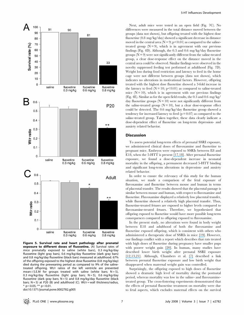
Heart failure causing increased mortality after prenatalfluoxetine exposure is dose-dependent
To determine whether the mortality rate after prenatal
fluoxetine treatment (0.8 mg/kg/day) is dose-dependent, offspring
of mice exposed to different concentrations of fluoxetine (0.3, 0.6
and 0.8 mg/kg/day) during pregnancy were studied. Only mice
prenatally exposed to the highest fluoxetine concentration
(0.8 mg/kg/day) displayed a dramatic decrease in survival rate
measured at adult stage (Fig. 5A). In agreement with our
previously described findings (Fig. 2), prenatal fluoxetine-treated
mice (0.8 mg/kg/day) dramatically decreased the survival rate
during the preweaning period. Offspring exposed to a lower dose
fluoxetine (0.6 mg/kg/day) demonstrated a small decrease in
survival rate, whereas mice exposed to the lowest dose fluoxetine
(0.3 mg/kg/day) showed survival rates identical as saline-treated
mice. These data clearly show a dose-dependent effect of
fluoxetine on the survival rate of the offspring. In line with these
findings, we observed alterations in the Wt/r ratio at both P20 and
the adult stage (Fig. 5B and C, respectively). At P20, mice
prenatally exposed to the highest fluoxetine concentration
(0.8 mg/kg/day) demonstrated a dramatic decrease in Wt/r ratio
(43% decrease; N = 5; p,0.01) compared to saline-treated mice
(N = 5), whereas the Wt/r ratios of the lower doses (0.6 mg/kg/
day (N = 5) and 0.3 mg/kg/day (N = 5) were similar as saline-
treated offspring (Fig. 5B). The highest dose fluoxetine (0.8 mg/
kg/day) had a similar effect on the Wt/r ratio when measured at
the adult stage (40% decrease; N = 5; p,0.01; Fig. 5C) as
compared to saline-treated offspring (N = 5). In addition, 0.6 mg/
kg/day fluoxetine had a significant effect on the Wt/r ratio in
Figure 3. Binding of 5-HTT in the raphe nucleus presented aspercentage of control6S.E.M for groups treated with saline(white bars), fluvoxamine (gray bars) and fluoxetine (blackbars) at P20 (A) (N = 6 for saline and fluvoxamine, N = 4 forfluoxetine) and adulthood (B) (N = 6 for saline and fluvoxa-mine, N = 3 for fluoxetine). * p,0.01.doi:10.1371/journal.pone.0002782.g003
5-HT Influences Development
PLoS ONE | www.plosone.org 5 July 2008 | Volume 3 | Issue 7 | e2782
adulthood (15% decrease; N = 5; p,0.05; Fig. 5C). The lowest
concentration fluoxetine (0.3 mg/kg/day; N = 5) did not effect the
Wt/r ratio in adulthood. Taken together, these findings indicate
that prenatal fluoxetine exposure affects heart development in a
dose-dependent manner, resulting in a fluoxetine dose-dependent
mortality rate of the offspring.
Long-term alterations in behavior and in the serotoninsystem after prenatal fluoxetine exposure are dose-dependent
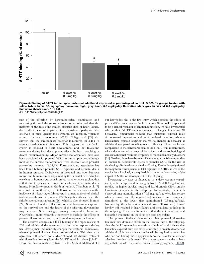
To determine whether the decreased 5-HTT levels in the raphe
nucleus after prenatal fluoxetine treatment (0.8 mg/kg/day) are
dose-dependent, autoradiography using [N-methyl-3H]-citalo-
pram was performed on adult brain tissue of mice prenatally
exposed to different concentrations of fluoxetine (0.3, 0.6 and
0.8 mg/kg/day; Fig. 6). 5-HTT binding was significantly
decreased in the offspring exposed to 0.8 (N = 6; p,0.01) and
0.6 mg/kg/day fluoxetine (N = 6; p,0.01) as compared to the
saline-treated offspring (N = 6). In contrast, offspring exposed to
the lowest dose fluoxetine (0.3 mg/kg/day) did not show
alterations in the 5-HTT levels (N = 6) as compared to the
saline-treated group. Taken together, these data clearly show that
long-term alterations in the serotonin system after fluoxetine
exposure are dose-dependent.
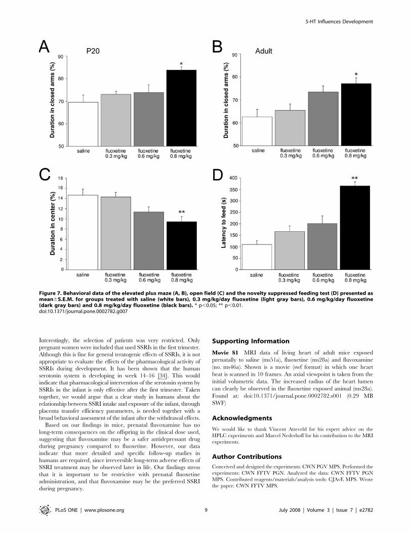
To determine whether the long-term effects of prenatal
fluoxetine exposure (0.8 mg/kg/day) on depression- and anxiety
related behavior are dose-dependent, we tested mice prenatally
exposed to different concentrations of fluoxetine (0.3, 0.6 and
0.8 mg/kg/day; Fig. 7) in an elevated plus maze at P20 (Fig. 7A)
and adulthood (Fig. 7B). No alterations were observed in
exploratory behavior, since all fluoxetine-treated groups showed
similar total distances moved, velocity and time spent ambulating
compared to saline-treated mice (data not shown). The highest
dose of fluoxetine (0.8 mg/kg/day) showed a significant increase
in duration in the closed arms compared to the control group,
both at P20 (N = 9; p,0.05) and adulthood (N = 5; p,0.05). The
0.3 mg/kg/day (N = 5) and 0.6 mg/kg/day (N = 5) fluoxetine
groups were not significantly different from the saline-treated
group, but a clear dose-response effect on the duration in the
closed arms can be observed at adulthood. At P20, no dose-
response effect could be found since 0.3 (N = 13) and 0.6 mg/kg/
day (N = 10) showed the same effect on the offspring in the
elevated plus maze as compared to the saline-treated mice
(N = 12).
Figure 4. Behavioral data of the elevated plus maze (A, B), open field (C, D) and the novelty suppressed feeding test (E) presentedas mean6S.E.M. All behavioral test were performed with 3 groups: saline (white bars), fluvoxamine (gray bars) and fluoxetine (black bars). Panel Cshows a typical example of a walking pattern in the open field (30 min) of a saline- and a fluoxetine-treated mouse. * p,0.05; ** p,0.01.doi:10.1371/journal.pone.0002782.g004
5-HT Influences Development
PLoS ONE | www.plosone.org 6 July 2008 | Volume 3 | Issue 7 | e2782
Next, adult mice were tested in an open field (Fig. 7C). No
differences were measured in the total distance moved between the
groups (data not shown), but offspring treated with the highest dose
fluoxetine (0.8 mg/kg/day) showed a significant decrease in distance
moved in the central area (N = 9; p,0.01) as compared to the saline-
treated group (N = 9), which is in agreement with our previous
findings (Fig. 4D). Although, the 0.3 and 0.6 mg/kg/day fluoxetine
groups (N = 9) were not significantly different from the saline-treated
group, a clear dose-response effect on the distance moved in the
central area could be observed. Similar findings were observed in the
novelty suppressed feeding test performed at adulthood (Fig. 7D).
Weight loss during food restriction and latency to feed in the home
cage were not different between groups (data not shown), which
indicates no alterations in motivational factors. However, offspring
treated with the highest dose fluoxetine showed a 3-fold increase in
the latency to feed (N = 10; p,0.01) as compared to saline-treated
mice (N = 10), which is in agreement with our previous findings
(Fig. 4E). Similar as for the open field results, the 0.3 and 0.6 mg/kg/
day fluoxetine groups (N = 10) were not significantly different from
the saline-treated group (N = 10), but a clear dose-response effect
could be detected. The 0.6 mg/kg/day fluoxetine group showed a
tendency for increased latency to feed (p = 0.07) as compared to the
saline-treated group. Taken together, these data clearly indicate a
dose-dependent effect of fluoxetine on long-term depression- and
anxiety related behavior.
Discussion
To assess potential long-term effects of prenatal SSRI exposure,
we administered clinical doses of fluvoxamine and fluoxetine to
pregnant mice. Embryos were exposed to SSRIs between E8 and
E18, when the 5-HTT is present [17,18]. After prenatal fluoxetine
exposure, we found a dose-dependent increase in neonatal
mortality in the offspring, a permanent decreased 5-HTT binding
and significant long-term alterations in depression- and anxiety
related behavior.
In order to ensure the relevancy of this study for the human
situation, we made a comparison of the fetal exposure of
fluvoxamine and fluoxetine between mouse and human in terms
of placental transfer. The results showed that the placental passage is
similar between mouse and human, with respect to fluvoxamine and
fluoxetine. Fluvoxamine displayed a relatively low placental transfer,
while fluoxetine showed a relatively high placental transfer. Thus,
fluoxetine-treated fetuses are exposed to higher levels compared to
fluvoxamine-treated fetuses. Therefore, we hypothesized that
offspring exposed to fluoxetine would have more possible long-term
consequences compared to offspring exposed to fluvoxamine.
In the present study, no alterations were found in body weight
between E18 and adulthood of both the fluvoxamine and
fluoxetine exposed offspring, which is consistent with others who
administered a therapeutic dose of SSRIs in mice [19]. However,
our findings conflict with a report which describes that rats treated
with high doses of fluoxetine during pregnancy have smaller pups
with poorer weight gain [20]. In human, many studies have
described lower birth weight after prenatal SSRI exposure
[12,13,21]. Although, Chambers et al. [7] described a link
between prenatal fluoxetine exposure and low birth weight that
disappeared when maternal weight gain was controlled.
Surprisingly, the offspring exposed to high doses of fluoxetine
showed a dramatic high level of mortality during the postnatal
period, whereas mortality was low in the saline- and fluvoxamine-
treated group. The cross-fostering experiments demonstrated that
the effects of prenatal fluoxetine treatment on mortality were due
to fetal aspects, which excludes maternal effects on the survival
Figure 5. Survival rate and heart pathology after prenatalexposure to different doses of fluoxetine. (A) Survival rates ofmice prenatally exposed to saline (white bars), 0.3 mg/kg/dayfluoxetine (light gray bars), 0.6 mg/kg/day fluoxetine (dark gray bars)and 0.8 mg/kg/day fluoxetine (black bars) measured at adulthood. 67%of the offspring exposed to the highest dose fluoxetine (0.8 mg/kg/day)died during the preweaning period as compared to 9% of the saline-treated offspring. Wt/r ratios of the left ventricle are presentedmean6S.E.M for groups treated with saline (white bars; N = 5),0.3 mg/kg/day fluoxetine (light gray bars; N = 5), 0.6 mg/kg/dayfluoxetine (dark gray bars; N = 5) and 0.8 mg/kg/day fluoxetine (blackbars; N = 5) at P20 (B) and adulthood (C). Wt/r = wall thickness/radius,* p,0.05; ** p,0.01.doi:10.1371/journal.pone.0002782.g005
5-HT Influences Development
PLoS ONE | www.plosone.org 7 July 2008 | Volume 3 | Issue 7 | e2782
rate of the offspring. By histopathological examination and
measuring the wall thickness/radius ratio, we observed that the
majority of the fluoxetine-treated offspring died of heart failure,
due to dilated cardiomyopathy. Dilated cardiomyopathy was also
observed in mice lacking the serotonin 2B receptor, which is
required for heart development [22,23]. Nebigil et al. [22] also
showed that the serotonin 2B receptor is required for 5-HT to
regulate cardiovascular functions. This suggests that the 5-HT
system is involved in heart development and that fluoxetine
treatment during fetal development affects the heart, resulting in
dilated cardiomyopathy. Major cardiac malformations have also
been associated with prenatal SSRIs in human practice, although
most of the cardiac malformations were observed after prenatal
paroxetine treatment [4,24,25]. Fortunately, no association has
been found between prenatal SSRI exposure and neonatal death
in human practice. Differences in neonatal mortality between
mouse and human can be explained by the neonatal care, which is
excellent in humans but poor in mice. An alternative explanation
is that, due to species differences in development, neonatal death
in mice is similar to prenatal death in humans. Chambers et al. [7]
observed that mothers exposed to fluoxetine had an increase in the
incidence of miscarriages. Moreover, in a meta-analysis of clinical
trials it was shown that prenatal SSRIs significantly increased the
risk for spontaneous abortion [26], which is also observed in mice
[27]. Since we found no effects of prenatal fluvoxamine exposure
on the survival rate and the hearts of the offspring, fluvoxamine
may be a safer SSRI during pregnancy compared to fluoxetine.
Nevertheless, more research is necessary to exclude the effects of
prenatal fluoxetine exposure on heart development in humans.
The observed changes in 5-HTT binding in the raphe nucleus at
P20 and adulthood demonstrates that fluoxetine exposure during
fetal development permanently changes the serotonin homeostasis,
whereas prenatal fluvoxamine exposure did not. This data is in
agreement with other reports, which showed that chronic treatment
with fluoxetine downregulates the 5-HTT in adult rodents [28–30].
However, these animals were treated with SSRIs at adulthood. To
our knowledge, this is the first study which describes the effects of
prenatal SSRI treatment on 5-HTT density. Since 5-HTT appeared
to be a critical regulator of emotional function, we have investigated
whether these 5-HTT alterations resulted in changes of behavior. All
behavioral experiments showed that fluoxetine exposed mice
demonstrated depression- and anxiety-related behavior, whereas
fluvoxamine exposed offspring showed no changes in behavior at
adulthood compared to saline-treated offspring. These results are
comparable to the behavioral data of the 5-HTT null mutant mice,
which demonstrated a range of behavioral and neurophysiological
abnormalities that resemble symptoms of mood and anxiety disorders
[31]. To date, there have been insufficient long-term follow-up studies
in human to demonstrate effects of prenatal SSRI on the risk of
developing affective disorders in the offspring. Further investigation of
the long-term consequences of fetal exposure to SSRIs, as well as the
mechanisms involved, are required for a better understanding of the
impact of SSRIs on development of the offspring.
Decreasing the dose of fluoxetine in a dose-response experi-
ment, with therapeutic doses ranging from 0.3 till 0.8 mg/kg/day,
resulted in higher survival rates and less dramatic effects on the
long-term behavior in the offspring. Interestingly, the effects
observed after administration of 0.8 mg/kg/day were less severe
when a lower dose (0.6 mg/kg/day) was used, and completely
diminished at the lowest dose administered (0.3 mg/kg/day).
Noteworthy, the sub-maximal clinical dose of fluoxetine (0.6 mg/
kg/day) still resulted in heart failure and behavioral pathology in
the offspring. These results indicate that the effects of prenatal
fluoxetine treatment on the fetus are dose-dependent.
The present findings demonstrate that prenatal fluoxetine
treatment has dramatic effects on the survival rate of the offspring,
alter the 5-HT system homeostasis at adulthood and indicate that
fluoxetine exposed mice are more vulnerable to anxiety disorders at
adulthood. Ultimately, clinical studies will be required to determine
whether our findings have applicability to the risks for anxiety or
affective disorders in humans. Two recent papers on this subject
argue that it is safe to use antidepressants during pregnancy [32,33].
Figure 6. Binding of 5-HTT in the raphe nucleus at adulthood expressed as percentage of control6S.E.M. for groups treated withsaline (white bars), 0.3 mg/kg/day fluoxetine (light gray bars), 0.6 mg/kg/day fluoxetine (dark gray bars) and 0.8 mg/kg/dayfluoxetine (black bars). * p,0.01.doi:10.1371/journal.pone.0002782.g006
5-HT Influences Development
PLoS ONE | www.plosone.org 8 July 2008 | Volume 3 | Issue 7 | e2782
Interestingly, the selection of patients was very restricted. Only
pregnant women were included that used SSRIs in the first trimester.
Although this is fine for general teratogenic effects of SSRIs, it is not
appropriate to evaluate the effects of the pharmacological activity of
SSRIs during development. It has been shown that the human
serotonin system is developing in week 14–16 [34]. This would
indicate that pharmacological intervention of the serotonin system by
SSRIs in the infant is only effective after the first trimester. Taken
together, we would argue that a clear study in humans about the
relationship between SSRI intake and exposure of the infant, through
placenta transfer efficiency parameters, is needed together with a
broad behavioral assessment of the infant after the withdrawal effects.
Based on our findings in mice, prenatal fluvoxamine has no
long-term consequences on the offspring in the clinical dose used,
suggesting that fluvoxamine may be a safer antidepressant drug
during pregnancy compared to fluoxetine. However, our data
indicate that more detailed and specific follow-up studies in
humans are required, since irreversible long-term adverse effects of
SSRI treatment may be observed later in life. Our findings stress
that it is important to be restrictive with prenatal fluoxetine
administration, and that fluvoxamine may be the preferred SSRI
during pregnancy.
Supporting Information
Movie S1 MRI data of living heart of adult mice exposed
prenatally to saline (ms51a), fluoxetine (ms28a) and fluvoxamine
(no. ms46a). Shown is a movie (swf format) in which one heart
beat is scanned in 10 frames. An axial viewpoint is taken from the
initial volumetric data. The increased radius of the heart lumen
can clearly be observed in the fluoxetine exposed animal (ms28a).
Found at: doi:10.1371/journal.pone.0002782.s001 (0.29 MB
SWF)
Acknowledgments
We would like to thank Vincent Atteveld for his expert advice on the
HPLC experiments and Marcel Nederhoff for his contribution to the MRI
experiments.
Author Contributions
Conceived and designed the experiments: CWN PGV MPS. Performed the
experiments: CWN FFTV PGN. Analyzed the data: CWN FFTV PGN
MPS. Contributed reagents/materials/analysis tools: CJAvE MPS. Wrote
the paper: CWN FFTV MPS.
Figure 7. Behavioral data of the elevated plus maze (A, B), open field (C) and the novelty suppressed feeding test (D) presented asmean6S.E.M. for groups treated with saline (white bars), 0.3 mg/kg/day fluoxetine (light gray bars), 0.6 mg/kg/day fluoxetine(dark gray bars) and 0.8 mg/kg/day fluoxetine (black bars). * p,0.05; ** p,0.01.doi:10.1371/journal.pone.0002782.g007
5-HT Influences Development
PLoS ONE | www.plosone.org 9 July 2008 | Volume 3 | Issue 7 | e2782
References
1. Llewellyn AM, Stowe ZN, Nemeroff CB (1997) Depression during pregnancy
and the puerperium. J Clin Psychiatry 58: 26–32.
2. Laine K, Heikkinen T, Ekblad U, Kero P (2003) Effects of exposure to selective
serotonin reuptake inhibitors during pregnancy on serotonergic symptoms in
newborns and cord blood monoamine and prolactin concentrations. Arch Gen
Psychiatry 60: 720–726.
3. Bennett HA, Einarson A, Taddio A, Koren G, Einarson TR (2007) Prevalence
of depression during pregnancy: systematic review. Obstet Gynecol 103:
698–709 [Erratum, Obstet Gynecol (2007) 103: 1344].
4. Kallen B, Otterblad-Olausson P (2006) Antidepressant drugs during pregnancy
and infant congenital heart defect. Reprod Toxicol 21: 221–222.
5. Kallen B (2004) Fluoxetine use in early pregnancy. Birth Defects Res B Dev
Reprod Toxicol 71: 395–396.
6. Zeskind PS, Stephens LE (2004) Maternal selective serotonin reuptake inhibitor
use during pregnancy and newborn neurobehavior. Pediatrics 113: 368–375.
7. Chambers CD, Johnson KA, Dick LM, Felix RJ, Jones KL (1996) Birth
outcomes in pregnant women taking fluoxetine. N Engl J Med 335: 1010–1015.
8. Chambers CD, Hernandez-Diaz S, Van Marter LJ, Werler MM, Louik C, et al.
(2006) Selective serotonin-reuptake inhibitors and risk of persistent pulmonary
hypertension of the newborn. N Engl J Med 354: 579–587.
9. Moses-Kolko EL, Bogen D, Perel J, Bregar A, Uhl K, et al. (2005) Neonatal signs
after late in utero exposure to serotonin reuptake inhibitors: literature review and
implications for clinical applications. JAMA 293: 2372–2383.
10. Hallberg P, Odlind V, Sjoblom V (2006) Selective serotonin-reuptake inhibitors
and persistent pulmonary hypertension of the newborn. N Engl J Med 354:
2188–2190.
11. Louik C, Lin AE, Werler MM, Hernandez-Dıaz S, Mitchell AA (2007) First-
trimester use of selective serotonin-reuptake inhibitors and the risk of birth
defects. N Engl J Med 356: 2675–2683.
12. Nonacs R, Cohen LS (2003) Assessment and treatment of depression during
pregnancy: an update. Psychiatr Clin North Am 26: 547–562.
13. Sanz EJ, De-las-Cuevas C, Kiuru A, Bate A, Edwards R (2005) Selective
serotonin reuptake inhibitors in pregnant women and neonatal withdrawal
syndrome: a database analysis. Lancet 365: 482–487.
14. van Laake LW, Passier R, Monshouwer-Kloots J, Nederhoff MG, Ward-van
Oostwaard D, et al. (2007) Monitoring of cell therapy and assessment of cardiac
function using magnetic resonance imaging in a mouse model of myocardial
infarction. Nature Protoc 2: 2551–2567.
15. Santarelli L, Saxe M, Gross C, Surget A, Battaglia F, et al. (2003) Requirement
of hippocampal neurogenesis for the behavioral effects of antidepressants.
Science 301: 805–809.
16. Bodnoff SR, Suranyi-Cadotte B, Quirion R, Meaney MJ (1989) A comparison of
the effects of diazepam versus several typical and atypical anti-depressant drugs
in an animal model of anxiety. Psychopharmacology (Berl) 97: 277–279.
17. Shuey DL, Sadler TW, Lauder JM (1992) Serotonin as a regulator of
craniofacial morphogenesis: site specific malformations following exposure to
serotonin uptake inhibitors. Teratology 46: 367–378.
18. Moiseiwitsch JR, Lauder JM (1995) Serotonin regulates mouse cranial neural
crest migration. Proc Natl Acad Sci USA 92: 7182–7186.19. Bairy KL, Madhyastha S, Ashok KP, Bairy I, Malini S (2007) Developmental
and behavioral consequences of prenatal fluoxetine. Pharmacology 79: 1–11.20. Vorhees CV, Acuff-Smith KD, Schilling MA, Fisher JE, Moran MS, et al. (1994)
A developmental neurotoxicity evaluation of the effects of prenatal exposure to
fluoxetine in rats. Fundam Appl Toxicol 23: 194–205.21. Hendrick V, Smith LM, Suri R, Hwang S, Haynes D, et al. (2003) Birth
outcomes after prenatal exposure to antidepressant medication. Am J ObstetGynecol 188: 812–815.
22. Nebigil CG, Choi DS, Dierich A, Hickel P, Le Meur M, et al. (2000) Serotonin
2B receptor is required for heart development. Proc Natl Acad Sci USA 97:9508–9513.
23. Nebigil CG, Hickel P, Messaddeq N, Vonesch JL, Douchet MP, et al. (2001)Ablation of serotonin 5-HT(2B) receptors in mice leads to abnormal cardiac
structure and function. Circulation 103: 2973–2979.24. Cuzzell JZ (2006) Paroxetine may increase risk for congenital malformations.
Dermatol Nurs 18: 6.
25. Berard A, Ramos E, Rey E, Blais L, St-Andre M, et al. (2007) First trimesterexposure to paroxetine and risk of cardiac malformations in infants: the
importance of dosage. Birth Defects Res B Dev Reprod Toxicol 80: 18–27.26. Rahimi R, Nikfar S, Abdollahi M (2006) Pregnancy outcomes following
exposure to serotonin reuptake inhibitors: a meta-analysis of clinical trials.
Reprod Toxicol 22: 571–575.27. Lisboa SF, Oliveira PE, Costa LC, Venancio EJ, Moreira EG (2007) Behavioral
evaluation of male and female mice pups exposed to fluoxetine duringpregnancy and lactation. Pharmacology 80: 49–56.
28. Benmansour S, Ramos E, Rey E, Blais L, St-Andre M, et al. (1999) Effects ofchronic antidepressant treatments on serotonin transporter function, density,
and mRNA level. J Neurosci 19: 10494–10501.
29. Hirano K, Kimura R, Sugimoto Y, Yamada J, Uchida S, et al. (2005)Relationship between brain serotonin transporter binding, plasma concentration
and behavioural effect of selective serotonin reuptake inhibitors. Br J Pharmacol144: 695–702.
30. Mirza NR, Nielsen EØ, Troelsen KB (2007) Serotonin transporter density and
anxiolytic-like effects of antidepressants in mice. Progr Neuro-PsychopharmacolBiol Psychiatry 31: 858–866.
31. Holmes A, Murphy DL, Crawley JN (2003) Abnormal behavioral phenotypes ofserotonin transporter knockout mice: parallels with human anxiety and
depression. Biol Psychiatry 54: 953–959.32. Alwan S, Reefhuis J, Rasmussen SA, Olney RS, Friedman JM (2007) Use of
selective serotonin-reuptake inhibitors in pregnancy and the risk of birth defects.
N Engl J Med 356: 2732–2733.33. Greene MF (2007) Teratogenicity of SSRIs — Serious concern or much ado
about little. N Engl J Med 356: 26.34. Verney C, Lebrand C, Gaspar P (2002) Changing distribution of monoamin-
ergic markers in the developing human cerebral cortex with special emphasis on
the serotonin transporter. Anat Rec 267: 87–93.
5-HT Influences Development
PLoS ONE | www.plosone.org 10 July 2008 | Volume 3 | Issue 7 | e2782
Related Documents