Biochemistry 1994, 33, 10313-103 18 10313 Modulation of P-Glycoprotein by Protein Kinase Ca! in a Baculovirus Expression System+ Shakeel Ahmad,: Ahmad R. Safa,l and Robert I. Glazer'.: Department of Pharmacology and Lombardi Cancer Research Center, Georgetown University Medical Center, Washington, D.C. 20007, and Department of Medicine and Cancer Research Center, University of Chicago School of Medicine, Chicago, Illinois 60637 Received April 8, 1994; Revised Manuscript Received June 6, 1994" ABSTRACT: The modulation of P-glycoprotein by protein kinase Ca (PKCa) was examined in a baculovirus expression system. PGP was phosphorylated in membrane vesicle preparations in vitro only when coexpressed with PKCa, and phosphorylation was Ca2+-dependentand inhibited by the PKC inhibitor Ro 3 1-8220. PGP and PKCa were tightly associated in membrane vesicles and were coimmunoprecipitated with antibodies against either PGP or PKCa. Photoaffinity labeling of membrane vesicles with [3H]azidopine indicated that drug binding to PGP was slightly increased in the presence of PKCa. In contrast, PGP ATPase activity was increased by PKCa as well as by verapamil, but only PKC-stimulated activity in the presence of verapamil was inhibited by Ro 31-8220. Mutation of serine-671 to asparagine in the linker region of PGP abolished PKCa-stimulated ATPase activity, and also inhibited to a lesser degree verapamil-stimulated ATPase activity. These results indicate that PKCa in a positive regulator of PGP ATPase activity and suggest that this mechanism may account for the increased multidrug resistance observed in MDR1 - expressing cells when PKCa activity is elevated. The hallmarkof multidrugresistance (MDR)' is the reduced intracellular accumulation of structurally unrelated natural product anticancer drugs such as the anthracyclines, Vinca alkaloids, actinomycin D, and epipodophyllotoxins (Beck, 1987; Gottesman & Pastan, 1988). The MDR phenotype is most often associated with the elevated expression of a plasma membrane-associated ATP-dependent unidirectional drug transporter, P-glycoprotein (PGP) (Hamada & Tsuruo, 1986, 1988; Kamimoto et al., 1989; Naito et al., 1989). PGP is the product of the MDRl gene, a highly conserved multigene family that includes two genes in man termed MDRl and MDR3 (also called MDR2) (Croop et al., 1988; Ng et al., 1989;Chinet al., 1989). In MDRcell lines, MDRl expression is increased as a result of gene amplification or increased transcription (Fairchildet al., 1987; Fojoet al., 1985; Riordan et al., 1985; Roninson et al., 1986; Scotto et al., 1986), and overexpression of the MDRl gene, but not the MDR3 gene, confers MDR (Schinkel et al., 1991). Several studies have shown that expression of either the genomic or the cDNA sequence of MDRl confers the MDR phenotype to the recipient cells (Ueda et al., 1987;Croop et al., 1987;Debenham et al., 1982; Gros et al., 1986; Shen et al., 1986; Sugimoto & Tsuruo, 1987), although the degree of resistance is generally less when compared to drug-selected MDR cells. Another phenotypic characteristic of all human MDR cell lines is an increase in PKC activity (Aquino et al., 1988,1990; Fineetal., 1988;Leeetal., 1992;Mellonietal., 1989;O'Brian et al., 1989; Palayoor et al., 1987; Posada et al., 1989a,b). This work was supported in part by National Institutes of Health Grants lR55CA57244 to R.I.G. and lROlCA56078 to A.R.S., by a grant from the Bristol-Myers Squibb Co. to R.I.G.,and by a grant from the Leukemia Research Foundation to S.A. Addresscorrespondence to this author at the Georgetown University Medical Center, 4 Research Court, Room 208, Rockville, MD 20850. t Georgetown University Medical Center. *Abstract published in Advance ACS Abstracts, August 1, 1994. I Abbreviations: MDR, multidrug-resistant or multidrug resistance; PKC, proteinkinase C;PGP, P-glycoprotein; SDS, sodiumdodecylsulfate. University of Chicago School of Medicine. 0006-2960/94/0433-103 13$04.50/0 This attribute has been found for PKCa (Lee et al., 1992; Posadaetal., 1989a;Blobeetal., 1993;Yuetal., 1991;O'Brian et al., 1991), PKCP (Fan et al., 1992), and PKCy (Aquino et al., 1990). Since PGP is phosphorylated in several MDR cell lines (Carlsen et al., 1977; Center, 1983, 1985; Mellado etal., 1987;Myersetal., 1989;Schurret al., 1989)andphorbol esters stimulate PGP phosphorylation in intact cells (Yu et al., 1991; Bates et al., 1992; Hamada et al., 1987; Chambers et al., 1990a,b) and reduce drug accumulation (Chambers et ai., 1990a, 1992), it may be inferred that PKC is involved in this process. PGP serves as a substrate for PKC in vitro (Chambers et al., 1992), and overexpression of PKCa (Yu et al., 1991), but not PKCy (Ahmad et al., 1992), increases MDR, decreases drug accumulation, and increases phorbol ester-dependent phosphorylation of PGP. In contrast, anti- sense expressionof PKCa in MDR MCF-7/ADR cells reduces MDR (Ahmad & Glazer, 1993). Therefore, a substantial body of data supports the thesis that PKCa is involved in modulating PGP, but the mechanism for this effect has not been determined. To investigate the possible modulatory role of PKCa on PGP function, a baculovirus expression system was developed in which we could examine the interaction of these proteins in the absence of other pleiotropic changes which occur in MDR cells. MATERIALS AND METHODS Baculovirus Expression of PGP and PKCa in Sj9 Cells. The human MDRl cDNA was generously provided by Dr. Merrill Goldsmith, National Cancer Institute, as plasmid pMTAdr (Fairchild et al., 1987), and consists of the 3.84 kb open reading frame and 424 bp of 5'-untranslated and 382 bp of 3'-untranslatedsequences. TheMDR1 cDNA was isolated from plasmid pMTAdr by digestion with BamHI followed by gel purification. Baculovirustransfer vector pVLl392 (kindly provided by Dr. Max Summers, Texas A&M University) was digested with BamHI and the MDRl insert ligated into the vector with T4 ligase. The orientation of the MDR1 insert downstream from the polyhedrin promoter was confirmed by 0 1994 American Chemical Society

Welcome message from author
This document is posted to help you gain knowledge. Please leave a comment to let me know what you think about it! Share it to your friends and learn new things together.
Transcript

Biochemistry 1994, 33, 103 13-103 18 10313
Modulation of P-Glycoprotein by Protein Kinase Ca! in a Baculovirus Expression System+
Shakeel Ahmad,: Ahmad R. Safa,l and Robert I. Glazer'.:
Department of Pharmacology and Lombardi Cancer Research Center, Georgetown University Medical Center, Washington, D.C. 20007, and Department of Medicine and Cancer Research Center, University of Chicago School of Medicine,
Chicago, Illinois 60637
Received April 8, 1994; Revised Manuscript Received June 6, 1994"
ABSTRACT: The modulation of P-glycoprotein by protein kinase Ca (PKCa) was examined in a baculovirus expression system. PGP was phosphorylated in membrane vesicle preparations in vitro only when coexpressed with PKCa, and phosphorylation was Ca2+-dependent and inhibited by the PKC inhibitor Ro 3 1-8220. PGP and PKCa were tightly associated in membrane vesicles and were coimmunoprecipitated with antibodies against either PGP or PKCa. Photoaffinity labeling of membrane vesicles with [3H]azidopine indicated that drug binding to PGP was slightly increased in the presence of PKCa. In contrast, PGP ATPase activity was increased by PKCa as well as by verapamil, but only PKC-stimulated activity in the presence of verapamil was inhibited by Ro 31-8220. Mutation of serine-671 to asparagine in the linker region of PGP abolished PKCa-stimulated ATPase activity, and also inhibited to a lesser degree verapamil-stimulated ATPase activity. These results indicate that PKCa in a positive regulator of PGP ATPase activity and suggest that this mechanism may account for the increased multidrug resistance observed in MDR1 - expressing cells when PKCa activity is elevated.
The hallmarkof multidrugresistance (MDR)' is the reduced intracellular accumulation of structurally unrelated natural product anticancer drugs such as the anthracyclines, Vinca alkaloids, actinomycin D, and epipodophyllotoxins (Beck, 1987; Gottesman & Pastan, 1988). The MDR phenotype is most often associated with the elevated expression of a plasma membrane-associated ATP-dependent unidirectional drug transporter, P-glycoprotein (PGP) (Hamada & Tsuruo, 1986, 1988; Kamimoto et al., 1989; Naito et al., 1989). PGP is the product of the MDRl gene, a highly conserved multigene family that includes two genes in man termed MDRl and MDR3 (also called MDR2) (Croop et al., 1988; Ng et al., 1989; Chinet al., 1989). In MDRcell lines, MDRl expression is increased as a result of gene amplification or increased transcription (Fairchildet al., 1987; Fojoet al., 1985; Riordan et al., 1985; Roninson et al., 1986; Scotto et al., 1986), and overexpression of the MDRl gene, but not the MDR3 gene, confers MDR (Schinkel et al., 1991). Several studies have shown that expression of either the genomic or the cDNA sequence of MDRl confers the MDR phenotype to the recipient cells (Ueda et al., 1987; Croop et al., 1987; Debenham et al., 1982; Gros et al., 1986; Shen et al., 1986; Sugimoto & Tsuruo, 1987), although the degree of resistance is generally less when compared to drug-selected MDR cells.
Another phenotypic characteristic of all human MDR cell lines is an increase in PKC activity (Aquino et al., 1988,1990; Fineetal., 1988;Leeetal., 1992;Melloniet al., 1989;O'Brian et al., 1989; Palayoor et al., 1987; Posada et al., 1989a,b).
This work was supported in part by National Institutes of Health Grants lR55CA57244 to R.I.G. and lROlCA56078 to A.R.S., by a grant from the Bristol-Myers Squibb Co. to R.I.G., and by a grant from the Leukemia Research Foundation to S.A.
Address correspondence to this author at the Georgetown University Medical Center, 4 Research Court, Room 208, Rockville, MD 20850.
t Georgetown University Medical Center.
*Abstract published in Advance ACS Abstracts, August 1 , 1994. I Abbreviations: MDR, multidrug-resistant or multidrug resistance;
PKC, protein kinase C; PGP, P-glycoprotein; SDS, sodium dodecyl sulfate.
University of Chicago School of Medicine.
0006-2960/94/0433-103 13$04.50/0
This attribute has been found for PKCa (Lee et al., 1992; Posadaetal., 1989a;Blobeetal., 1993;Yuetal., 1991;O'Brian et al., 1991), PKCP (Fan et al., 1992), and PKCy (Aquino et al., 1990). Since PGP is phosphorylated in several MDR cell lines (Carlsen et al., 1977; Center, 1983, 1985; Mellado etal., 1987;Myersetal., 1989;Schurret al., 1989)andphorbol esters stimulate PGP phosphorylation in intact cells (Yu et al., 1991; Bates et al., 1992; Hamada et al., 1987; Chambers et al., 1990a,b) and reduce drug accumulation (Chambers et ai., 1990a, 1992), it may be inferred that PKC is involved in this process. PGP serves as a substrate for PKC in vitro (Chambers et al., 1992), and overexpression of PKCa (Yu et al., 1991), but not PKCy (Ahmad et al., 1992), increases MDR, decreases drug accumulation, and increases phorbol ester-dependent phosphorylation of PGP. In contrast, anti- sense expression of PKCa in MDR MCF-7/ADR cells reduces MDR (Ahmad & Glazer, 1993). Therefore, a substantial body of data supports the thesis that PKCa is involved in modulating PGP, but the mechanism for this effect has not been determined. To investigate the possible modulatory role of PKCa on PGP function, a baculovirus expression system was developed in which we could examine the interaction of these proteins in the absence of other pleiotropic changes which occur in MDR cells.
MATERIALS AND METHODS
Baculovirus Expression of PGP and PKCa in Sj9 Cells. The human MDRl cDNA was generously provided by Dr. Merrill Goldsmith, National Cancer Institute, as plasmid pMTAdr (Fairchild et al., 1987), and consists of the 3.84 kb open reading frame and 424 bp of 5'-untranslated and 382 bp of 3'-untranslatedsequences. TheMDR1 cDNA was isolated from plasmid pMTAdr by digestion with BamHI followed by gel purification. Baculovirus transfer vector pVLl392 (kindly provided by Dr. Max Summers, Texas A&M University) was digested with BamHI and the MDRl insert ligated into the vector with T4 ligase. The orientation of the MDR1 insert downstream from the polyhedrin promoter was confirmed by
0 1994 American Chemical Society

10314 Biochemistry, Vol. 33, No. 34, 1994 Ahmad et al.
A Membrane, PGP B Membrane, PKCa C Cytosol, PKCa
200-
97-
12 24 4 8 h r 9 7-
68-
-,. r . 97-
68-
12 2 4 4 8 hr
12 24 4 8 hr
FIGURE 1: Time course of the expression of PGP and PKCa in Sf9 cells. At 12-48 h after infection, Sf9 cells were disrupted by nitrogen cavitation, and membrane vesicle and cytosol fractions were prepared by differential centrifugation. Membrane vesicle (20 pg) and cytosol (20 pg) fractions were separated by SDS-PAGE, and immunoblotting was carried out as described under Materials and Methods. (A) PGP levels in the membrane fraction; (B) PKCa levels in the membrane fraction; (C) PKC levels in the cytosol fraction. PGP and PKCa were visualized with monoclonal antibody C219 or with a polyclonal antibody against PKCa, respectively.
restriction enzyme analysis. The resulting plasmid, pVL- MDRl, and linearized mutated proviral DNA from Au- tographa californica multiply enveloped nuclear polyhedrosis virus (BaculoGold, Pharmingen) were used to cotransfect Sf9 cells by lipofection (Lipofectin, BRL). Recombinant plaques expressing PGP were identified by their occlusion-negative morphology and by immunoblotting with monoclonal antibody C219 (Centocor). Immunoreactive bands werevisualized with the alkaline phosphatase/BCIP/NBT detection system. A recombinant bacuiovirus expressing PKCa was prepared as described previously (Goswami & Glazer, 199 1).
Site-Directed Mutagenesis. Plasmid pVL-MDR 1 was mutated by the procedure of Deng and Nickoloff (1 992) using the protocol supplied with the Transformer size-directed mutagenesis kit (Clontech). Codon 67 1, AGT, was mutated to AAT to produce a Ser-Asn mutation using the oligo- nucleotide 5'-TCC-ACG-GAC-AAT-CCT-ACG-AGT-3'. Plasmid selection was based on mutation of the unique Not1 site, GCGGCCGC to GCTGCCGC, with oligonucleotide 5'- TGG-AGC-GGC-AGC-TGC-AGA-TCT-3'. Mutations were confirmed by double-stranded sequencing using Sequenase (USB).
Membrane Vesicle Preparation. Cells (2 X lo7) were infected with each recombinant virus at 2.5 pfu/cell in 10 mL of SF900 medium (Gibco/BRL), and after 1 h, an additional 10 mL of medium was added. After 48 h, cells were suspended in 5 mL of hypotonic buffer (10 mM Tris-HCI, pH 7.5, 10 mM NaCI, 1.5 mM MgC12, and 2 mM PMSF) and incubated on ice for 30 min. Cells were pressurized at 800 psi under nitrogen in a Parr cell disruption bomb for 10 min and after decompression were centrifuged for 10 min at 500g at 4 "C. The supernatant was centrifuged at 1 OOOOOg for 1 h, and the cell pellet containing the membrane vesicle fraction was suspended in the same buffer containing 0.25 M sucrose, divided into aliquots, and kept at -80 "C.
Phosphorylation of PGP. Membranevesicles were prepared from Sf9 cells infected with MDR1 or MDR1 and PKCa. Phosphorylation assays were carried out in vitro as described by Yu et al. (1991), except that exogenous substrate was omitted. In some experiments, the staurosporine analog Ro 31-8220 (kindly provided by Dr. Geoffrey Lawton, Roche Products, Ltd., England) was used to inhibit PKC activity (Davis et al., 1992).
Immunoprecipitation of PGP and PKC. After in vitro phosphorylation, the reaction was terminated by the addition of 500 pL of immunoprecipitation buffer [ 10 mM Tris-HC1 (pH 7.4), 150 mM NaCI, 5 mM EDTA, 10% glycerol, and
1% Triton X- 1001. Immunoprecipitation was carried out with the following antibodies: 5 pg of PGP rabbit polyclonal antibody 4007 (kindly provided by Dr. Michael Gottesman, National Cancer Institute), 1 pg of PGP monoclonal antibody MRK16 (kindly provided by Dr. Takashi Tsuruo, Japanese Foundation for Cancer Research, Japan), 5 pg of PKCa/f? rabbit polyclonal antibody C4 (Aquino et al., 1987) and 1.25 pg of PKCa rabbit polyclonal antibody (BRL). Immuno- conjugates were adsorbed with 20 pL of Protein G-Plus/ Protein A-agarose (Oncogene Science) at 4 "C for 1 h on a rocking platform,centrifuged at 12000gfor 2 min, and washed 3 times with immunoprecipitation buffer. Adsorbed immu- nocomplexes were suspended in Laemmli sample buffer without boiling and separated by SDS-PAGE in an 8% minigel (Novex). Samples were transferred to nitrocellulose by electroblotting, and antigens were visualized by autoradiog- raphy.
ATPase Activity. ATPase activity was measured by the procedure of Sarkadi et al. (1 992) in the presence and absence of 10 pM verapamil. In order to determine only PGP- associated ATPase activity, the assay mix contained 2 mM EGTA (an inhibitor of Ca2+-dependent ATPase), 5 mM sodium azide (an inhibitor of F1,F2-ATPase), and 1 mM ouabain (an inhibitor of Na+,K+-ATPase).
Photoaffinity Labeling with [ 3 H j Azidopine. Membrane vesicles were incubated in a buffer containing 10 mM Tris- HCI (pH 7.4), 10% PBS, 0.3 mM MgC12, and 3 mM ATP containing 4% DMSO and 0.25 pM [3H]azidopine (specific activity 44 Ci/mmol; Amersham Corp.) in a final volume of 0.05 mL as described previously (Safa et al., 1990). The reaction mixture was preincubated for 30 min at 25 "C in the absence or presence of 50 pM nonradioactive competing ligand and then irradiated for 20 min with a UV lamp equipped with two 15-W self-filtering 302-nm lamps. Photolabeled mem- branes (75 pg of protein/lane) were analyzed directly by SDS- PAGEon a 5-1 5% gradient gel containing 4.5 M urea, followed by fluorography.
RESULTS
Time Course of Expression and Translocation of PGP and PKCa. PGP was distributed exclusively in the membrane fraction at 12-48 h after infection (Figure 1A). PKCa was also distributed in the membrane fraction 24 h after infection when expressed either alone or with PGP (Figure lB), but PKCa was present mainly in the cytosol at 48 h after infection (Figure 1C). PKC activity 48 h after infection was 99.3%
P-Glycoprotein Modulation by Protein Kinase C a Biochemistry, Vol. 33, No. 34, 1994 10315
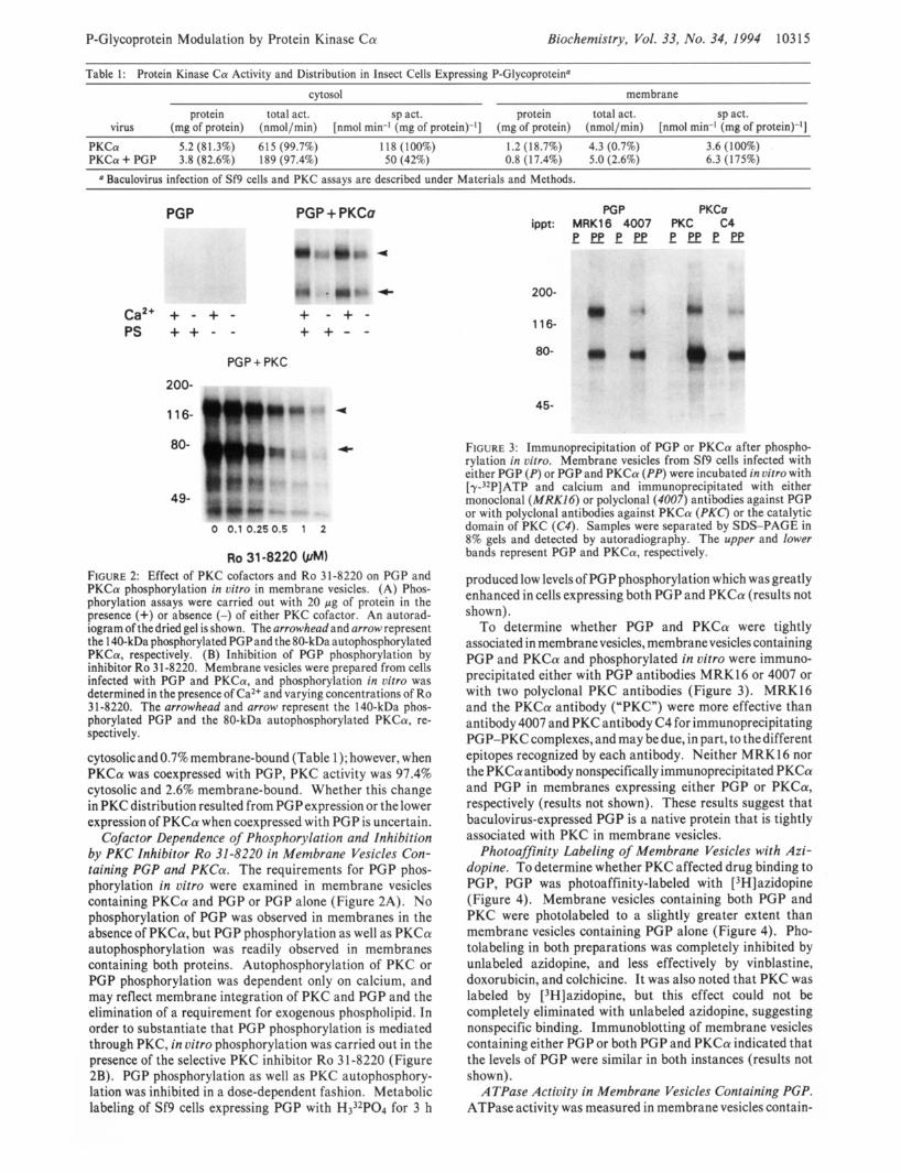
Table 1 : Protein Kinase Ca Activity and Distribution in Insect Cells Expressing P-Glycoprotein4 cytosol membrane
protein total act. sp act. protein total act. sp act. virus (mg of protein) (nmoI/min) [nmol min-’ (mg of protein)-’] (mg of protein) (nmol/min) [nmol min-’ (mg of protein)-’]
PKCa 5.2 (81.3%) 615 (99.7%) 118 (100%) 1.2 (1 8.7%) 4.3 (0.7%) 3.6 (100%) PKCa + PGP 3.8 (82.6%) 189 (97.4%) 50 (42%) 0.8 (1 7.4%) 5.0 (2.6%) 6.3 (175%)
Baculovirus infection of Sf9 cells and PKC assays are described under Materials and Methods.
PGP PGP + PKCa
a
c
Ca2+ + - + - + - + - PS + + - - + + - -
PGP + PKC
200-
116- 4
80- *
49-
0 0.1 0.250.5 1 2
Ro 31-8220 FIGURE 2: Effect of PKC cofactors and Ro 31-8220 on PGP and PKCa phosphorylation in uitro in membrane vesicles. (A) Phos- phorylation assays were carried out with 20 pg of protein in the presence (+) or absence (-) of either PKC cofactor. An autorad- iogram of thedried gel is shown. The arrowhead and arrow represent the 1 40-kDa phosphorylated PGPand the 80-kDa autophosphorylated PKCa, respectively. (B) Inhibition of PGP phosphorylation by inhibitor Ro 3 1-8220. Membrane vesicles were prepared from cells infected with PGP and PKCa, and phosphorylation in vitro was determined in the presence of Ca2+ and varying concentrations of Ro 31-8220. The arrowhead and arrow represent the 140-kDa phos- phorylated PGP and the 80-kDa autophosphorylated PKCa, re- spectively.
cytosolicand 0.7% membrane-bound (Table 1); however, when PKCa was coexpressed with PGP, PKC activity was 97.4% cytosolic and 2.6% membrane-bound. Whether this change in PKC distribution resulted from PGPexpression or the lower expression of PKCa when coexpressed with PGP is uncertain.
Cofactor Dependence of Phosphorylation and Inhibition by PKC Inhibitor Ro 31-8220 in Membrane Vesicles Con- taining PCP and PKCa. The requirements for PGP phos- phorylation in vitro were examined in membrane vesicles containing PKCa and PGP or PGP alone (Figure 2A). No phosphorylation of PGP was observed in membranes in the absence of PKCa, but PGP phosphorylation as well as PKCa autophosphorylation was readily observed in membranes containing both proteins. Autophosphorylation of PKC or PGP phosphorylation was dependent only on calcium, and may reflect membrane integration of PKC and PGP and the elimination of a requirement for exogenous phospholipid. In order to substantiate that PGP phosphorylation is mediated through PKC, in vitro phosphorylation was carried out in the presence of the selective PKC inhibitor Ro 3 1-8220 (Figure 2B). PGP phosphorylation as well as PKC autophosphory- lation was inhibited in a dose-dependent fashion. Metabolic labeling of Sf9 cells expressing PGP with H332P04 for 3 h
PGP PKCa ippt: MRK16 4007 PKC C4
- P P P E P P
200-
116-
80-
45-
FIGURE 3: Immunoprecipitation of PGP or PKCa after phospho- rylation in vitro. Membrane vesicles from Sf9 cells infected with either PGP (P) or PGP and PKCa (PP) were incubated in vitro with [y3*P]ATP and calcium and immunoprecipitated with either monoclonal (MRK16) or polyclonal (4007) antibodies against PGP or with polyclonal antibodies against PKCa (PKC) or the catalytic domain of PKC (C4). Samples were separated by SDS-PAGE in 8% gels and detected by autoradiography. The upper and lower bands represent PGP and PKCa, respectively.
produced low levels of PGP phosphorylation which was greatly enhanced in cells expressing both PGP and PKCa (results not shown).
To determine whether PGP and PKCa were tightly associated in membranevesicles, membrane vesicles containing PGP and PKCa and phosphorylated in vitro were immuno- precipitated either with PGP antibodies MRK16 or 4007 or with two polyclonal PKC antibodies (Figure 3). MRK16 and the PKCa antibody (“PKC”) were more effective than antibody 4007 and PKC antibody C4 for immunoprecipitating PGP-PKCcomplexes, and may be due, in part, to thedifferent epitopes recognized by each antibody. Neither MRK16 nor the PKCaantibody nonspecifically immunoprecipitated PKCa and PGP in membranes expressing either PGP or PKCa, respectively (results not shown). These results suggest that baculovirus-expressed PGP is a native protein that is tightly associated with PKC in membrane vesicles.
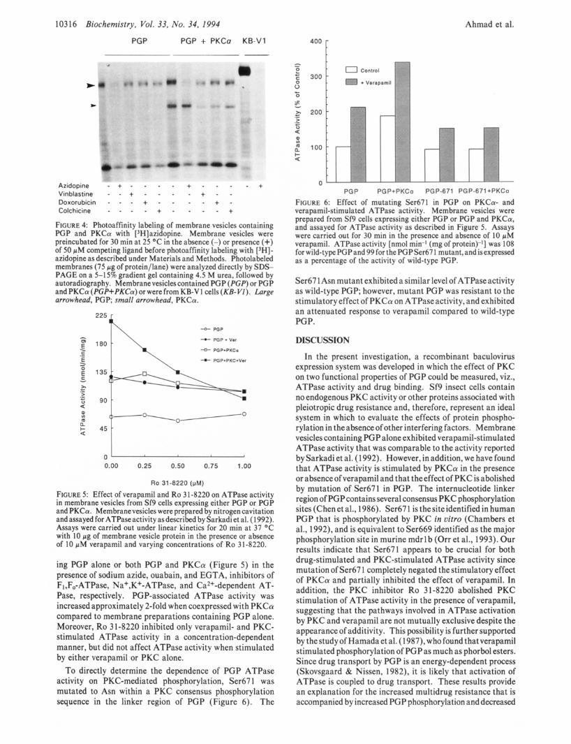
Photoaffinity Labeling of Membrane Vesicles with Azi- dopine. To determine whether PKC affected drug binding to PGP, PGP was photoaffinity-labeled with [3H]azidopine (Figure 4). Membrane vesicles containing both PGP and PKC were photolabeled to a slightly greater extent than membrane vesicles containing PGP alone (Figure 4). Pho- tolabeling in both preparations was completely inhibited by unlabeled azidopine, and less effectively by vinblastine, doxorubicin, and colchicine. It was also noted that PKC was labeled by [3H]azidopine, but this effect could not be completely eliminated with unlabeled azidopine, suggesting nonspecific binding. Immunoblotting of membrane vesicles containing either PGP or both PGP and PKCa indicated that the levels of PGP were similar in both instances (results not shown).
A TPase Activity in Membrane Vesicles Containing PCP. ATPase activity was measured in membrane vesicles contain-
10316 Biochemistry, Vol. 33, No. 34, 1994
PGP PGP + PKCa KB-V1
>
8
Atidopine - + - - Vinblastine - - + - Doxorubicin - - - + Colchicine - - - -
+
FIGURE 4: Photoaffinity labeling of membrane vesicles containing PGP and PKCa with [3H]azidopine. Membrane vesicles were preincubated for 30 min at 25 O C in the absence (-) or presence (+) of 50 pM competing ligand before photoaffinity labeling with [3H]- azidopine as described under Materials and Methods. Photolabeled membranes (75 pg of protein/lane) were analyzed directly by SDS- PAGE on a 5-15% gradient gel containing 4.5 M urea, followed by autoradiography. Membrane vesicles contained PGP (PGP) or PGP and PKCa (PGP+PKCa) or were from KB-V 1 cells (KB-VZ). Large arrowhead, PGP; small arrowhead, PKCa.
-0- PGP
4- POP + Ver
4 PGP+PKCa
4 PGP+PKC+Ver
180 h
-r .- E
U
< + 4 5 t 0 0.00 0.25 0.50 0.75 1.00
RO 31-8220 (pM)
FIGURE 5: Effect of verapamil and Ro 3 1-8220 on ATPase activity in membrane vesicles from Sf9 cells expressing either PGP or PGP and PKCa. Membranevesicles were prepared by nitrogen cavitation and assayed for ATPase activity as described by Sarkadi et al. (1 992). Assays were carried out under linear kinetics for 20 min at 37 "C with 10 pg of membrane vesicle protein in the presence or absence of 10 pM verapamil and varying concentrations of Ro 31-8220.
ing PGP alone or both PGP and PKCa (Figure 5 ) in the presence of sodium azide, ouabain, and EGTA, inhibitors of F,,F,-ATPase, Na+,K+-ATPase, and Ca2+-dependent AT- Pase, respectively. PGP-associated ATPase activity was increased approximately 2-fold when coexpressed with PKCa compared to membrane preparations containing PGP alone. Moreover, Ro 31-8220 inhibited only verapamil- and PKC- stimulated ATPase activity in a concentration-dependent manner, but did not affect ATPase activity when stimulated by either verapamil or PKC alone.
To directly determine the dependence of PGP ATPase activity on PKC-mediated phosphorylation, Ser67 1 was mutated to Asn within a PKC consensus phosphorylation sequence in the linker region of PGP (Figure 6). The
Ahmad et al.
400 F
" t s
PGP PGP+PKCo PGP-671 PGP-671+PKCo
FIGURE 6: Effect of mutating Ser671 in PGP on PKCa- and verapamil-stimulated ATPase activity. Membrane vesicles were prepared from Sf9 cells expressing either PGP or PGP and PKCa, and assayed for ATPase activity as described in Figure 5. Assays were carried out for 30 min in the presence and absence of 10 pM verapamil. ATPase activity [nmol min-I (mg of protein)-'] was 108 for wild-type PGP and 99 for the PGP Ser67 1 mutant, and is expressed as a percentage of the activity of wild-type PGP.
Ser67 1 Asn mutant exhibited a similar level of ATPaseactivity as wild-type PGP; however, mutant PGP was resistant to the stimulatory effect of PKCa on ATPaseactivity, and exhibited an attenuated response to verapamil compared to wild-type PGP.
DISCUSSION
In the present investigation, a recombinant baculovirus expression system was developed in which the effect of PKC on two functional properties of PGP could be measured, viz., ATPase activity and drug binding. Sf9 insect cells contain no endogenous PKC activity or other proteins associated with pleiotropic drug resistance and, therefore, represent an ideal system in which to evaluate the effects of protein phospho- rylation in theabsenceof other interfering factors. Membrane vesicles containing PGP alone exhibited verapamil-stimulated ATPase activity that was comparable to the activity reported by Sarkadi et al. (1 992). However, in addition, we have found that ATPase activity is stimulated by PKCa in the presence or absence of verapamil and that the effect of PKC is abolished by mutation of Ser671 in PGP. The internucleotide linker region of PGPcontains several consensus PKC phosphorylation sites (Chen et al., 1986). Ser671 is thesiteidentified in human PGP that is phosphorylated by PKC in vitro (Chambers et al., 1992), and is equivalent to Ser669 identified as the major phosphorylation site in murine mdrl b (Orr et al., 1993). Our results indicate that Ser671 appears to be crucial for both drug-stimulated and PKC-stimulated ATPase activity since mutation of Ser67 1 completely negated the stimulatory effect of PKCa and partially inhibited the effect of verapamil. In addition, the PKC inhibitor Ro 31-8220 abolished PKC stimulation of ATPase activity in the presence of verapamil, suggesting that the pathways involved in ATPase activation by PKC and verapamil are not mutually exclusive despite the appearance of additivity. This possibility is further supported by the study of Hamada et al. (1 987), who found that verapamil stimulated phosphorylation of PGP as much as phorbol esters. Since drug transport by PGP is an energy-dependent process (Skovsgaard & Nissen, 1982), it is likely that activation of ATPase is coupled to drug transport. These results provide an explanation for the increased multidrug resistance that is accompanied by increased PGP phosphorylation and decreased

P-Glycoprotein Modulation by Protein Kinase C a
drug retention in MCF-7 breast carcinoma cells overexpressing MDRl andPKCa(Yuet al., 1991),andfor thepartialreversal of resistance by antisense PKCa (Ahmad & Glazer, 1993).
Several studies have used PKC inhibitors to reverse MDR (O’Brian et al., 1989; Posada et al., 1989a; Sampson et al., 1993; Dong et al., 1991), but these effects have been difficult to interpret since all of these inhibitors are known to interact with PGP and prevent drug binding (Miyamoto et al., 1992; Sato et al., 1990; Wakusawa et al., 1992). In addition, inhibitors such as staurosporine and H-7 lack PKC selectivity (Meyer et al., 1989) and produce cytotoxicity that may be unrelated to inhibition of protein kinase activity (Smith et al., 1988). Despite the fact that inhibitors such as Ro 31-8220 show greater PKC isoform selectivity (Dieter & Fitzke, 1991), their ability to serve as substrates for PGP may pose an insurmountable limitation to studies trying to dissect out the role of PKC in regulating PGP function. Therefore, the major advantage of the baculovirus system is that it permits the assessment of the drug binding and ATPase functions of PGP independently of its transport function and the influence of other pleiotropic factors associated with MDR.
PGP phosphorylation by PKCa in membrane vesicles was Ca2+- but not phospholipid-dependent. This is not an unexpected finding since other studies have shown that the cofactor requirements of PKC for catalytic activity and substrate binding vary considerably depending on the substrate (Bazzi & Nelsestuen, 1987; Hyatt et al., 1994) and that membrane-associated PKC exhibits changes in its cofactor requirements (Bazzi & Nelsestuen, 1988, 1990). Our data also indicate that PGP strongly associates with PKCa, and although this has not been demonstrated previously in MDR cells, PGP was found to cofractionate with an uncharacterized phospholipid-dependent protein kinase with the characteristics of Ca2+-independent PKC isoforms (Staats et al., 1990). Whether this occurs in MDR cells remains to be established.
Our data suggest that PKC has a slight stimulatory effect on substrate binding as measured by photoaffinity labeling with azidopine. Germann et al. (1990) reported that Sf9 cell membranes containing PGP exhibited less azidopine binding than KB-V1 cells, and that vinblastine and vincristine were less effective competitors of binding in insect cell membrane vesicles compared to KB-V1 cells. We did not observe differences in the ability of vinblastine, doxorubicin, or colchicine to compete with azidopine in Sf9 cell membranes containing either PGP or PGP and PKCa, although they were less effective competitors than azidopine. The relative order of competition of these drugs for azidopine binding to membranes containing PGP and PKCa was azidopine > vinblastine > doxorubicin > colchicine, a result that is similar towhat was observed previously (Safa et al., 1990). Although azidopine binding was lower in Sf9 membrane vesicles than in membranes from KB-V1 cells, this effect may be a result of either membrane lipid composition or hypoglycosylation of PGP in insect cells. A recent study by Schinkel et al. (1993) indicated that hypoglycosylation of PGP did not affect its ability to confer MDR in mammalian cells, and therefore the lipid composition of insect cell vs mammalian cell membranes is the most likely cause of the differences observed in drug binding (Higgins & Gottesman, 1992).
Thus, the additive effects of verapamil and PKC suggest that PGP can be activated by two, but not necessarily, mutually exclusive processes: substrate binding and PGP phosphory- lation. Although the mechanism for these effects has not been determined, it is likely that PGP phosphorylation results in a conformational change in PGP in the linker region which
Biochemistry, Vol. 33, No. 34, 1994 10317
results in enhanced ATP hydrolysis and energy-dependent drug transport. We have, in fact, observed increased vin- blastine accumulation in vitro using membrane vesicles from Sf9 cells phosphorylated by PKC (unpublished results). There is precedent for this model for another member of the ATPase binding cassette (ABC) protein family, the cystic fibrosis transmembrane regulator (Ames & Lecar, 1992), where PKC phosphorylation of the R domain (analogous to the linker region in PGP) activates its C1- conductance activity (Hwang et al., 1989; Picciotto et al., 1992). Thus, PGP becomes the second ABC protein to be modulated by PKC.
ACKNOWLEDGMENT
We thank Michael Agresti and Scott Roberts for their technical assistance.
REFERENCES
Ahmad, S., & Glazer, R. I. (1993) Mol. Pharmacol. 43, 858-
Ahmad, S., Trepel, J. B., Ohno, S., Suzuki, K., Tsuruo, T., &
Ames, G. F.-L., & Lecar, H. (1992) FASEB J . 6, 2660-2666. Aquino, A., Hartman, K. D., Knode, M. C., Huang, K.-P., Niu,
C.-H., & Glazer, R. I. (1988) Cancer Res. 48, 3324-3329. Aquino, A,, Warren, B., Omichinski, J., Hartman, K. D., &
Glazer, R. 1. (1990) Biochem. Biophys. Res. Commun. 166,
Bates, S. E., Currier, S. J., Alvarez, M., & Fojo, A. T. (1992)
Bazzi, M. D., & Nelsestuen, G. L. (1 987) Biochemistry 26,1974-
Bazzi, M. D., & Nelsestuen, G. L. (1988) Biochem. Biophys.
Bazzi, M. D., & Nelsestuen, G. L. (1990) Biochemistry 29,7624
Beck, W. T. (1987) Biochem. Pharmacol. 36, 2879-2887. Blobe, G. C., Sachs, C. W., Khan, W. A., Fabbro, D., Stabel, S.,
Wetsel, W. C., Obeid, L. M., Fine, R. L., & Hannun, Y. A. (1993) J . Biol. Chem. 268, 658-664.
Carlsen, S. A., Till, J. E., & Ling, V. (1977) Biochim. Biophys. Acta 467, 238-250.
Center, M. S. (1983) Biochem. Biophys. Res. Commun. 115,
Center, M. S. (1985) Biochem. Pharmacol. 34, 1471-1476. Chambers, T., Chalikonda, I., & Eilon, G. (1990a) Biochem.
Chambers, T. C., McAvoy, E. M., Jacobs, J . W., & Eilon, G.
Chambers, T., Zheng, B., & Kuo, J. F. (1992) Mol. Pharmacol.
Chen, C., Chin, J. E., Ueda, K., Clark, D. P., Pastan, I., Gottesman,
Chin, J. E., Soffir, R., Noonan, K. E., Choi, K., & Roninson, I.
Croop, J. M., Guild, B. C., Gros, P., & Housman, D. E. (1987)
Croop, J. M., Gros, P., & Housman, D. E. (1988) J . Clin. Invest.
Davis, P. D., Elliott, L. H., Harris, W., Hill, C. H., Hurst, S. A., Keech, E., Kumar, M. K. H., Lawton, G., Nixon, J . S., & Wilkinson, S. E. (1992) J . Med. Chem. 35, 994-1001.
Debenham, P. G., Kartner, N., Siminovitch, L., Riordan, J. R., & Ling, V. (1982) Mol. Cell. Biol. 2, 881-889.
Deng, W. P., & Nickoloff, J. A. (1992) Anal. Biochem. 200,
Dieter,P., & Fitzke, E. (1991) Biochem. Biophys. Res. Commun.
Dong, Z., Ward, N. E., Fan, D., Gupta, K. P., & O’Brian, C. A.
862.
Glazer, R. I. (1992) Mol. Pharmacol. 42, 1004-1009.
123-728.
Biochemistry 31, 6366-6372.
1982.
Res. Commun. 152, 336-343.
7630.
159-1 66.
Biophys. Res. Commun. 169, 253-259.
(1990b) J . Biol. Chem. 265, 7679-7686.
41, 1008-1015.
M. M., & Roninson, I. B. (1986) Cell 47, 381-389.
B. (1989) Mol. Cell. Biol. 9, 3808-3820.
Cancer Res. 47, 5982-5988.
81, 1303-1309.
81-88.
181, 396-401.
(1991) Mol. Pharmacol. 39, 563-569.
10318 Biochemistry, Vol. 33, No. 34, 1994
Fairchild, C. R., Ivy, S. P., Kao-Shan, C.-S., Whang-Peng, J., Rosen, N., Israel, M. A., Melera, P. W., Cowan, K. H., & Goldsmith, M. E. (1987) Cancer Res. 47, 5141-5148.
Fan, D., Fidler, I. J., Ward, N. E., Seid, C., Earnest, L. E., Housey, G. M., & O’Brian, C. A. (1992) Anticancer Res. 12,661-668.
Fine, R. L., Patel, J., & Chabner, B. A. (1988) Proc. Natl. Acad. Sci. U.S.A. 85, 582-586.
Fojo, A. T., Whang-Peng, J., Gottesman, M. M., & Pastan, I. (1985) Proc. Natl. Acad. Sci. U.S.A. 82, 7661-1665.
Germann, U. A., Willingham, M. C., Pastan, I., & Gottesman, M. M. (1990) Biochemistry 29, 2295-2303.
Goswami, B. B., & Glazer, R. I. (1991) BioTechniques 10,626- 630.
Gottesman, M. M., & Pastan, I. (1988) J. Biol. Chem. 263,
Gros, P., Neriah, Y. B., Croop, J. M., & Housman, D. E. (1986)
Hamada, H., & Tsuruo, T. (1986) Proc. Natl. Acad. Sci. U.S.A.
Hamada, H., & Tsuruo, T. (1988) J . Biol. Chem. 263, 1454-
Hamada, H., Hagiwara, K.-I., Nakajima,T., & Tsuruo, T. (1987)
Higgins, C. F., & Gottesman, M. M. (1992) Trends Biochem.
Horio, M., Gottesman, M. M., & Pastan, I. (1988) Proc. Natl.
Hwang, T. C., Lu, L., Zeitlin, P. L., Gruenert, D. C., Huganir,
Hyatt, S. L., Liao, L., Chapline, C., & Jaken, S. (1994)
Juranka, P. F., Zastawny, R. L., & Ling, V. (1989) FASEB J.
Kamimoto, Y., Gatmaitan, Z . , Hsu, J., & Arias, I. M. (1989) J . Biol. Chem. 264, 11693-1 1698.
Lee, S. A., Karaszkiewicz, J. W., & Anderson, W. B. (1992) Cancer Res. 52, 3150-3759.
Mellado, W., & Horwitz, S. B. (1987) Biochemistry 26, 6900- 6904.
Melloni, E., Pontremoli, S., Viotti, P. L., Patrone, M., Marks, P. A., & Rifkind, R. A. (1989) J . Biol. Chem. 264, 18414- 18418.
Meyer, T., Regenass, U., Fabbro, D., Alteri, E., Rosel, J., Muller, M., Caravatti, G., & Matter, A. (1989) Znt. J. Cancer 43,
Miyamoto, K. I., Wakusawa, S., Inoko, K., Takagi, K., &
Myers, M. B., Rittmann-Grauer, L., O’Brien, J. P., & Safa, A.
Naito, M., Hamada, H., & Tsuruo, T. (1988) J . Biol. Chem.
Ng, W. F., Sarangi, F., Zastawny, R. L., Veinot-Drebot, L., &
O’Brian, C. A., Fan, D., Ward, N. E., Seid, C., & Fidler, I. J.
12163-1 21 66.
Nature 323, 128-73 1.
83, 7185-7189.
1458.
Cancer Res. 47, 2860-2865.
Sci. 17, 18-21.
Acad. Sci. U.S.A. 85, 3580-3584.
R., & Guggino, W. B. (1989) Science 244, 1351-1353.
Biochemistry 33, 1223-1228.
3, 2583-2592.
851-856.
Koyama, M. (1992) Cancer Lett. 64, 177-183.
R. (1989) Cancer Res. 49, 3209-3214.
263, 11887-11891.
Ling, V. (1989) Mol. Cell. Biol. 9, 1224-1232.
(1989) FEBS Lett. 246, 78-82.
Ahmad et al.
OBrian, C. A., Fan, D., Ward, N. E., Dong, Z., Iwamoto, L., Gupta, K. P., Earnest, L. E., & Fidler, I. J. (1991) Biochem. Pharmacol. 41, 791-806.
Orr, G. A., Han, E. K. H., Browne, P. C., Nieves, E., O’Connor, B. M., Yang, C. P. H., & Horwitz, S. B. (1993) J. Biol. Chem. 268, 25054-25062.
Palayoor, S. T., Stein, J. M., & Hait, W. N. (1987) Biochem. Biophys. Res. Commun. 148,118-125.
Picciotto, M. R., Cohn, J. A., Bertuzzi, G., Greengard, P., & Nairn, A. C. (1992) J. Biol. Chem. 267, 12742-12752.
Posada, J. A., McKeegan, E. M., Worthington, K. F., Morin, M. J., Jaken, S., & Tritton, T. R. (1989a) Cancer Commun. 1,
Posada, J., Vichi, P., & Tritton, T. R. (1989b) Cancer Res. 49, 6634-6639.
Riordan, J. R., Deuchars, K., Kartner, N., Alan, N., Trent, J., & Ling, V. (1985) Nature 16, 811-819.
Roninson, I. B., Chin, J. E., Choi, K., Gros, P., Housman, D. E., Fojo,A.,Shen, D.-W., Gottesman, M. M., & Pastan, I. (1986) Proc. Natl. Acad. Sci. U.S.A. 83, 4538-4542.
Safa, A. R., Stern, R. K., Choi, K., Agresti, M., Tamai, I., Mehta, N. D., & Roninson, I. B. (1990) Proc. Natl. Acad. Sci. U.S.A.
Sampson, K. E., Wolf, C. L., & Abraham, I. (1993) Cancer Lett.
Sarkadi, B., Price, E. M., Boucher, R. C., Germann, U. A,, & Scarborough, G. A. (1992) J. Biol. Chem. 267, 4854-4858.
Sato, W., Yusa, K., Naito, M., & Tsuruo, T. (1990) Biochem. Biophys. Res. Commun. 173, 1252-1251.
Schinkel, A. H., Roelofs, M. E. M., & Borst, P. (1991) Cancer Res. 51, 2628-2635.
Shinkel, A. H., Kemp, S., Dolle, M., Rudenko, G., & Wagenaar, E. (1993) J . Biol. Chem. 268, 7474-7481.
Schurr, E., Raymond, M., Bell, J. C., & Gros, P. (1 989) Cancer Res. 49, 2729-2734.
Scotto, K. W., Biedler, J. L., & Melera, P. W. (1986) Science
Shen, D.-W., Fojo, A., Roninson, I. B., Chin, J. E., Soffir, R., Pastan, I., & Gottesman, M. M. (1986) Mol. Cell. Biol. 6, 4039-4045.
Skovsgaard, T., & Nissen, N. I. (1982) Pharmacol. Ther. 18,
Smith, C. D., Glickman, J. F., & Chang, K.-J. (1988) Biochem.
Staats, J., Marquardt, D., & Center, M. S. (1990) J. Biol. Chem.
Sugimoto, Y. , & Tsuruo, T. (1981) Cancer Res. 47,2620-2625. Ueda, K., Cardarelli, C., Gottesman, M. M., & Pastan, I. (1987)
Proc. Natl. Acad. Sci. U.S.A. 84, 3004-3008. Wakusawa, S., Nakamura, S., Tajima, K., Miyamoto, K. I.,
Hagiwara, M., & Hidaka, H. (1992) Mol. Pharmacol. 41, 1034-1038.
Yu, G., Ahmad, S., Aquino, A,, Fairchild, C. R., Trepel, J. B., Cowan, K. H., Tsuruo, T., Ohno, S., & Glazer, R. I. (1991) Cancer Commun. 3, 181-189.
285-292.
87, 7225-7229.
68, 7-14.
232,151-755.
293-3 1 1.
Biophys. Res. Commun. 156, 1250-1256.
265,4084-4090.
Related Documents


