Modulation of Macrophage Activation State Protects Tissue from Necrosis during Critical Limb Ischemia in Thrombospondin-1-Deficient Mice Nicolas Bre ´ chot 1,2 , Elisa Gomez 1,2 , Marine Bignon 1,2 , Jamila Khallou-Laschet 3,4 , Michael Dussiot 3,4 , Aure ´ lie Cazes 1,2 , Ce ´ cile Alanio-Bre ´ chot 5 , Me ´ lanie Durand 1,2 , Josette Philippe 1,2 , Jean-Se ´ bastien Silvestre 6,7 , Nico Van Rooijen 8 , Pierre Corvol 1,2 , Antonino Nicoletti 3,4 , Be ´ne ´ dicte Chazaud 9,10 , Ste ´ phane Germain 1,2,11 * 1 INSERM, U833, Paris, France, 2 Laboratoire Angiogene `se Embryonnaire et Pathologique, Colle `ge de France, Paris, France, 3 INSERM, U872, Paris, France, 4 Centre de recherche des Cordeliers, Universite ´ Pierre et Marie Curie, Paris, France, 5 Laboratoire d’He ´ matologie, Ho ˆ pital Bice ˆtre, AP-HP, Le Kremlin-Bice ˆtre, France, 6 INSERM, U689, Paris, France, 7 Centre de Recherche Cardiovasculaire de Lariboisie ` re, Universite ´ Denis Diderot, Paris, France, 8 Department of Molecular Cell Biology, Free University Medical Center, Amsterdam, The Netherlands, 9 INSERM, U 567, CNRS UMR 8104, Paris, France, 10 Institut Cochin, Universite ´ Paris 5, Paris, France, 11 Service d’He ´ matologie Biologique A, Ho ˆ pital Europe ´en Georges Pompidou, AP-HP, Paris, France Abstract Background: Macrophages, key regulators of healing/regeneration processes, strongly infiltrate ischemic tissues from patients suffering from critical limb ischemia (CLI). However pro-inflammatory markers correlate with disease progression and risk of amputation, suggesting that modulating macrophage activation state might be beneficial. We previously reported that thrombospondin-1 (TSP-1) is highly expressed in ischemic tissues during CLI in humans. TSP-1 is a matricellular protein that displays well-known angiostatic properties in cancer, and regulates inflammation in vivo and macrophages properties in vitro. We therefore sought to investigate its function in a mouse model of CLI. Methods and Findings: Using a genetic model of tsp-1 2/2 mice subjected to femoral artery excision, we report that tsp-1 2/2 mice were clinically and histologically protected from necrosis compared to controls. Tissue protection was associated with increased postischemic angiogenesis and muscle regeneration. We next showed that macrophages present in ischemic tissues exhibited distinct phenotypes in tsp-1 2/2 and wt mice. A strong reduction of necrotic myofibers phagocytosis was observed in tsp-1 2/2 mice. We next demonstrated that phagocytosis of muscle cell debris is a potent pro-inflammatory signal for macrophages in vitro. Consistently with these findings, macrophages that infiltrated ischemic tissues exhibited a reduced postischemic pro-inflammatory activation state in tsp-1 2/2 mice, characterized by a reduced Ly-6C expression and a less pro- inflammatory cytokine expression profile. Finally, we showed that monocyte depletion reversed clinical and histological protection from necrosis observed in tsp-1 2/2 mice, thereby demonstrating that macrophages mediated tissue protection in these mice. Conclusion: This study defines targeting postischemic macrophage activation state as a new potential therapeutic approach to protect tissues from necrosis and promote tissue repair during CLI. Furthermore, our data suggest that phagocytosis plays a crucial role in promoting a deleterious intra-tissular pro-inflammatory macrophage activation state during critical injuries. Finally, our results describe TSP-1 as a new relevant physiological target during critical leg ischemia. Citation: Bre ´chot N, Gomez E, Bignon M, Khallou-Laschet J, Dussiot M, et al. (2008) Modulation of Macrophage Activation State Protects Tissue from Necrosis during Critical Limb Ischemia in Thrombospondin-1-Deficient Mice. PLoS ONE 3(12): e3950. doi:10.1371/journal.pone.0003950 Editor: Yihai Cao, Karolinska Institutet, Sweden Received September 1, 2008; Accepted November 17, 2008; Published December 16, 2008 Copyright: ß 2008 Brechot et al. This is an open-access article distributed under the terms of the Creative Commons Attribution License, which permits unrestricted use, distribution, and reproduction in any medium, provided the original author and source are credited. Funding: The funders had no role in study design, data collection and analysis, decision to publish, or preparation of the manuscript. This work emanates from the European Vascular Genomics Network (http://www.evgn.org) (contract LSHMCT-2003-503254) and is supported by a grant from Agence Nationale de la recherche (ANR-06-JCJC-0160). NB is supported by Fondation pour la Recherche Me ´dicale. Competing Interests: The authors have declared that no competing interests exist. * E-mail: [email protected] Introduction Peripheral artery disease affects up to 15% of people over 55 years [1] and may lead to critical limb ischemia (CLI) as the disease progresses. Despite percutaneous transluminal angioplasty or vascular surgery, major amputation occurs in about 13% of patients suffering from CLI [2], thereby emphasizing the crucial need for alternative efficient pharmacological treatments. Several studies in humans were designed with the aim of restoring proangiogenic signals in ischemic legs, but led to contradictory results [3–5]. Physiopathology of CLI is indeed complex and not restricted to solely a lack of tissue perfusion. Inflammation is also a crucial component of critical leg ischemia in humans since ischemic tissues exhibit large inflammatory infiltrates, rich in macrophages [6,7], which are known to be key regulators of healing/regeneration processes [8–10]. However pro-inflammato- ry markers independently correlate with disease progression (relative risk = 2.9), risk of amputation and 1-year mortality [11], PLoS ONE | www.plosone.org 1 December 2008 | Volume 3 | Issue 12 | e3950

Welcome message from author
This document is posted to help you gain knowledge. Please leave a comment to let me know what you think about it! Share it to your friends and learn new things together.
Transcript

Modulation of Macrophage Activation State ProtectsTissue from Necrosis during Critical Limb Ischemia inThrombospondin-1-Deficient MiceNicolas Brechot1,2, Elisa Gomez1,2, Marine Bignon1,2, Jamila Khallou-Laschet3,4, Michael Dussiot3,4,
Aurelie Cazes1,2, Cecile Alanio-Brechot5, Melanie Durand1,2, Josette Philippe1,2, Jean-Sebastien
Silvestre6,7, Nico Van Rooijen8, Pierre Corvol1,2, Antonino Nicoletti3,4, Benedicte Chazaud9,10, Stephane
Germain1,2,11*
1 INSERM, U833, Paris, France, 2 Laboratoire Angiogenese Embryonnaire et Pathologique, College de France, Paris, France, 3 INSERM, U872, Paris, France, 4 Centre de
recherche des Cordeliers, Universite Pierre et Marie Curie, Paris, France, 5 Laboratoire d’Hematologie, Hopital Bicetre, AP-HP, Le Kremlin-Bicetre, France, 6 INSERM, U689,
Paris, France, 7 Centre de Recherche Cardiovasculaire de Lariboisiere, Universite Denis Diderot, Paris, France, 8 Department of Molecular Cell Biology, Free University
Medical Center, Amsterdam, The Netherlands, 9 INSERM, U 567, CNRS UMR 8104, Paris, France, 10 Institut Cochin, Universite Paris 5, Paris, France, 11 Service
d’Hematologie Biologique A, Hopital Europeen Georges Pompidou, AP-HP, Paris, France
Abstract
Background: Macrophages, key regulators of healing/regeneration processes, strongly infiltrate ischemic tissues frompatients suffering from critical limb ischemia (CLI). However pro-inflammatory markers correlate with disease progressionand risk of amputation, suggesting that modulating macrophage activation state might be beneficial. We previouslyreported that thrombospondin-1 (TSP-1) is highly expressed in ischemic tissues during CLI in humans. TSP-1 is amatricellular protein that displays well-known angiostatic properties in cancer, and regulates inflammation in vivo andmacrophages properties in vitro. We therefore sought to investigate its function in a mouse model of CLI.
Methods and Findings: Using a genetic model of tsp-12/2 mice subjected to femoral artery excision, we report that tsp-12/2
mice were clinically and histologically protected from necrosis compared to controls. Tissue protection was associated withincreased postischemic angiogenesis and muscle regeneration. We next showed that macrophages present in ischemic tissuesexhibited distinct phenotypes in tsp-12/2 and wt mice. A strong reduction of necrotic myofibers phagocytosis was observed intsp-12/2 mice. We next demonstrated that phagocytosis of muscle cell debris is a potent pro-inflammatory signal formacrophages in vitro. Consistently with these findings, macrophages that infiltrated ischemic tissues exhibited a reducedpostischemic pro-inflammatory activation state in tsp-12/2 mice, characterized by a reduced Ly-6C expression and a less pro-inflammatory cytokine expression profile. Finally, we showed that monocyte depletion reversed clinical and histologicalprotection from necrosis observed in tsp-12/2 mice, thereby demonstrating that macrophages mediated tissue protection inthese mice.
Conclusion: This study defines targeting postischemic macrophage activation state as a new potential therapeuticapproach to protect tissues from necrosis and promote tissue repair during CLI. Furthermore, our data suggest thatphagocytosis plays a crucial role in promoting a deleterious intra-tissular pro-inflammatory macrophage activation stateduring critical injuries. Finally, our results describe TSP-1 as a new relevant physiological target during critical leg ischemia.
Citation: Brechot N, Gomez E, Bignon M, Khallou-Laschet J, Dussiot M, et al. (2008) Modulation of Macrophage Activation State Protects Tissue from Necrosisduring Critical Limb Ischemia in Thrombospondin-1-Deficient Mice. PLoS ONE 3(12): e3950. doi:10.1371/journal.pone.0003950
Editor: Yihai Cao, Karolinska Institutet, Sweden
Received September 1, 2008; Accepted November 17, 2008; Published December 16, 2008
Copyright: � 2008 Brechot et al. This is an open-access article distributed under the terms of the Creative Commons Attribution License, which permitsunrestricted use, distribution, and reproduction in any medium, provided the original author and source are credited.
Funding: The funders had no role in study design, data collection and analysis, decision to publish, or preparation of the manuscript. This work emanates fromthe European Vascular Genomics Network (http://www.evgn.org) (contract LSHMCT-2003-503254) and is supported by a grant from Agence Nationale de larecherche (ANR-06-JCJC-0160). NB is supported by Fondation pour la Recherche Medicale.
Competing Interests: The authors have declared that no competing interests exist.
* E-mail: [email protected]
Introduction
Peripheral artery disease affects up to 15% of people over 55
years [1] and may lead to critical limb ischemia (CLI) as the
disease progresses. Despite percutaneous transluminal angioplasty
or vascular surgery, major amputation occurs in about 13% of
patients suffering from CLI [2], thereby emphasizing the crucial
need for alternative efficient pharmacological treatments. Several
studies in humans were designed with the aim of restoring
proangiogenic signals in ischemic legs, but led to contradictory
results [3–5]. Physiopathology of CLI is indeed complex and not
restricted to solely a lack of tissue perfusion. Inflammation is also a
crucial component of critical leg ischemia in humans since
ischemic tissues exhibit large inflammatory infiltrates, rich in
macrophages [6,7], which are known to be key regulators of
healing/regeneration processes [8–10]. However pro-inflammato-
ry markers independently correlate with disease progression
(relative risk = 2.9), risk of amputation and 1-year mortality [11],
PLoS ONE | www.plosone.org 1 December 2008 | Volume 3 | Issue 12 | e3950

suggesting a potential deleterious effect of the pro-inflammatory
state observed in patients.
Thrombospondin-1 (TSP-1) is a 450 kDa matricellular protein
synthesized by various cell types, that interacts with a wide range
of integrin and non-integrin receptors, thus exhibiting pleiotropic
activities [12]. It accumulates during various situations of tissue
injury, and acts as a regulator of tissue remodeling [13,14]. In
particular it displays potent angiostatic properties in cancer
[14,15] and limits ischemic tissue survival in a myocutaneous flap
model through inhibition of NO-mediated post-ischemic vasore-
laxation [16]. Moreover TSP-1 is a key regulator of inflammation
in mice in vivo [17,18], and is a strong regulator of macrophage
properties in vitro. It is a potent pro-inflammatory [19] and pro-
migratory [20] signal for macrophages, and phagocytosis of
various cell types depends on TSP-1 [21–25].
We previously showed that TSP-1 is strongly overexpressed
during critical hind limb ischemia in humans and secreted by both
endothelial cells and macrophages [26]. Considering its complex
roles in regulating both angiogenesis and inflammation, we here
hypothesized that TSP-1 may play a deleterious role in CLI, and
therefore sought to investigate its function in a mouse model of
CLI. We report that tsp-12/2 mice were clinically and
histologically protected from tissue necrosis induced by limb
ischemia. Tissue protection was associated with increased
postischemic angiogenesis and muscle regeneration. We next
showed that macrophages in ischemic tissues exhibited distinct
phenotypes in tsp-12/2 and wt mice: phagocytosis of necrotic
myofibers was strongly reduced in tsp-12/2 mice. Consistently
with our findings that phagocytosis of muscle cell debris is a potent
pro-inflammatory signal, macrophages exhibited a reduced pro-
inflammatory activation state in these mice. Finally, using a model
of monocyte depletion, we demonstrated that this distinct
macrophage phenotype was responsible for the tissue protection
observed in tsp-12/2 mice.
Results
TSP-1 is expressed by macrophages and endothelial cellsduring critical hind limb ischemia in mice
We analyzed tsp-1 mRNA expression pattern in gastrocnemius
muscle during CLI in mice at d4, d6, d16 and d21 following
femoral artery excision. No expression was observed in non
ischemic tissues (fig 1A&B). At early time points (d4), tissue
architecture was strongly disorganized in necrotic areas (fig. 1C &
fig. S1A), replaced by an inflammatory infiltrate rich in
macrophages (fig. S1B). A dense network of capillaries developed
at this stage (fig. S1C), not covered by smooth muscle cells (not
shown). Thrombospondin-1 mRNA was highly expressed in necrotic
areas (fig. 1D), expressed by macrophages (fig. 1K&1L), endothe-
lial cells (fig. 1L&1M), and to a lesser extent by myofibers (fig. 1D).
In contrast to heart or brain [9,27], skeletal muscle displays
regenerative properties during ischemia [28]. At d6 regenerating
basic myofibers appeared in the healing area (fig. 1E & fig. S1D).
At this stage, tsp-1 mRNA was still expressed by macrophages
localized in the healing border zone (fig. 1F & fig. S1E). From d16
to d21, regenerating myofibers with central nuclei developed
(fig. 1G & fig. S1G) and gastrocnemius muscle healed almost
normally, except small lipidic deposits observed locally (fig. 1I &
fig. S1J). Macrophages gradually disappeared (fig. S1H&1K) at
these later stages and tsp-1 mRNA expression strongly decreased,
nevertheless persisting in endothelial cells (see arrowheads
fig. 1H&1J). As previously described [28], capillary density
decreased during the regeneration process (fig. S1C-1L).
Western blot analysis of TSP-1 protein expression at similar time
points confirmed protein expression in muscles, that peaked at early
stages (d4 to d7) and then decreased at d16 until d21 (fig. 1N).
We thus found that TSP-1 is expressed during postischemic
healing/regeneration in macrophages and endothelial cells in
mice, as we previously observed in humans [26].
Thrombospondin-12/2 mice are protected from ischemia-induced necrosis
In order to analyze the functional role of TSP-1 during critical
hind limb ischemia, we performed femoral artery excision in tsp-
12/2 mice and their wt littermates and followed macroscopic
clinical necrosis during 21 days. Figure 2A shows two represen-
tative pictures of either tissue protection or necrosis. Necrosis
developed between d2 and d7 (fig. 2B). Wild-type mice were highly
affected since 86% of mice exhibited macroscopic necrosis (fig. 2B).
Conversely, only 50% of tsp-12/2 mice were affected (p,0,05 vs.
wt, n = 19).
We then performed histological analyses of gastrocnemius
muscles from tsp-12/2 and wt mice at d4 (d4 was chosen because
necrosis is an ongoing process at this time point (fig. 2B)). Tissue
protection was confirmed in tsp-12/2 mice (fig. 3). Three different
types of area were observed at this stage as described in the
methods section and in fig. S2 : i) a preserved area presenting an
Figure 1. Thrombospondin-1 is expressed during critical hindlimb ischemia in mice. Thrombospondin-1 mRNA expression wasanalyzed in gastrocnemius muscle using in situ hybridization at d4 (D),d6 (F), d16 (H) and d21 (J) after ischemia. D0 (B) represents non ischemictissue. Adjacent sections (respectively C, E, G, I and A were stained withH&E (Scale bar = 200 mm). (L) Gastrocnemius muscle analyzed at d4 fortsp-1 mRNA expression at a higher magnification (scale bar = 25 mm). (K,M) Adjacent sections were stained for macrophages (K) and endothelialcells (M), showing expression of tsp-1 mRNA in macrophages (seearrows in K&L) and endothelial cells (see arrowheads in L&M).Arrowheads in H&J show tsp-1 mRNA expression in endothelial cellsat d16 and d21 respectively. (N) Western blot analysis of TSP-1expression in gastrocnemius muscle at similar time points. Arro-whead = 150 kD.doi:10.1371/journal.pone.0003950.g001
Macrophage Polarization in CLI
PLoS ONE | www.plosone.org 2 December 2008 | Volume 3 | Issue 12 | e3950
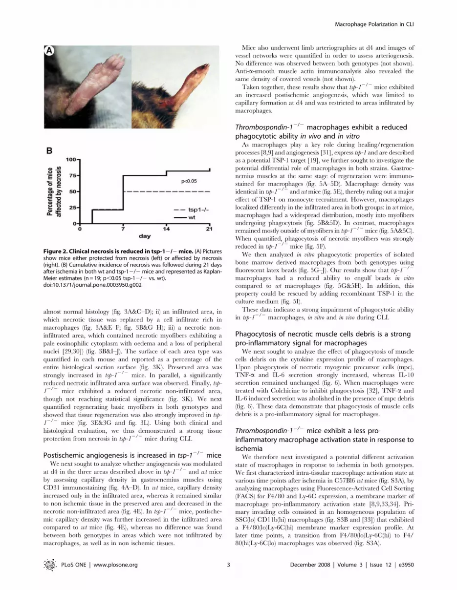
almost normal histology (fig. 3A&C–D); ii) an infiltrated area, in
which necrotic tissue was replaced by a cell infiltrate rich in
macrophages (fig. 3A&E–F; fig. 3B&G–H); iii) a necrotic non-
infiltrated area, which contained necrotic myofibers exhibiting a
pale eosinophilic cytoplasm with oedema and a loss of peripheral
nuclei [29,30]) (fig. 3B&I–J). The surface of each area type was
quantified in each mouse and reported as a percentage of the
entire histological section surface (fig. 3K). Preserved area was
strongly increased in tsp-12/2 mice. In parallel, a significantly
reduced necrotic infiltrated area surface was observed. Finally, tsp-
12/2 mice exhibited a reduced necrotic non-infiltrated area,
though not reaching statistical significance (fig. 3K). We next
quantified regenerating basic myofibers in both genotypes and
showed that tissue regeneration was also strongly improved in tsp-
12/2 mice (fig. 3E&3G and fig. 3L). Using both clinical and
histological evaluation, we thus demonstrated a strong tissue
protection from necrosis in tsp-12/2 mice during CLI.
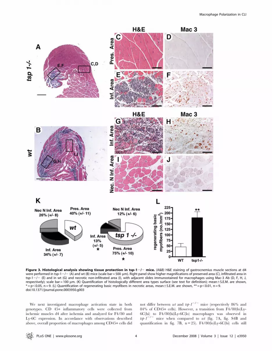
Postischemic angiogenesis is increased in tsp-12/2 miceWe next sought to analyze whether angiogenesis was modulated
at d4 in the three areas described above in tsp-12/2 and wt mice
by assessing capillary density in gastrocnemius muscles using
CD31 immunostaining (fig. 4A–D). In wt mice, capillary density
increased only in the infiltrated area, whereas it remained similar
to non ischemic tissue in the preserved area and decreased in the
necrotic non-infiltrated area (fig. 4E). In tsp-12/2 mice, postische-
mic capillary density was further increased in the infiltrated area
compared to wt mice (fig. 4E), whereas no difference was found
between both genotypes in areas which were not infiltrated by
macrophages, as well as in non ischemic tissues.
Mice also underwent limb arteriographies at d4 and images of
vessel networks were quantified in order to assess arteriogenesis.
No difference was observed between both genotypes (not shown).
Anti-a-smooth muscle actin immunoanalysis also revealed the
same density of covered vessels (not shown).
Taken together, these results show that tsp-12/2 mice exhibited
an increased postischemic angiogenesis, which was limited to
capillary formation at d4 and was restricted to areas infiltrated by
macrophages.
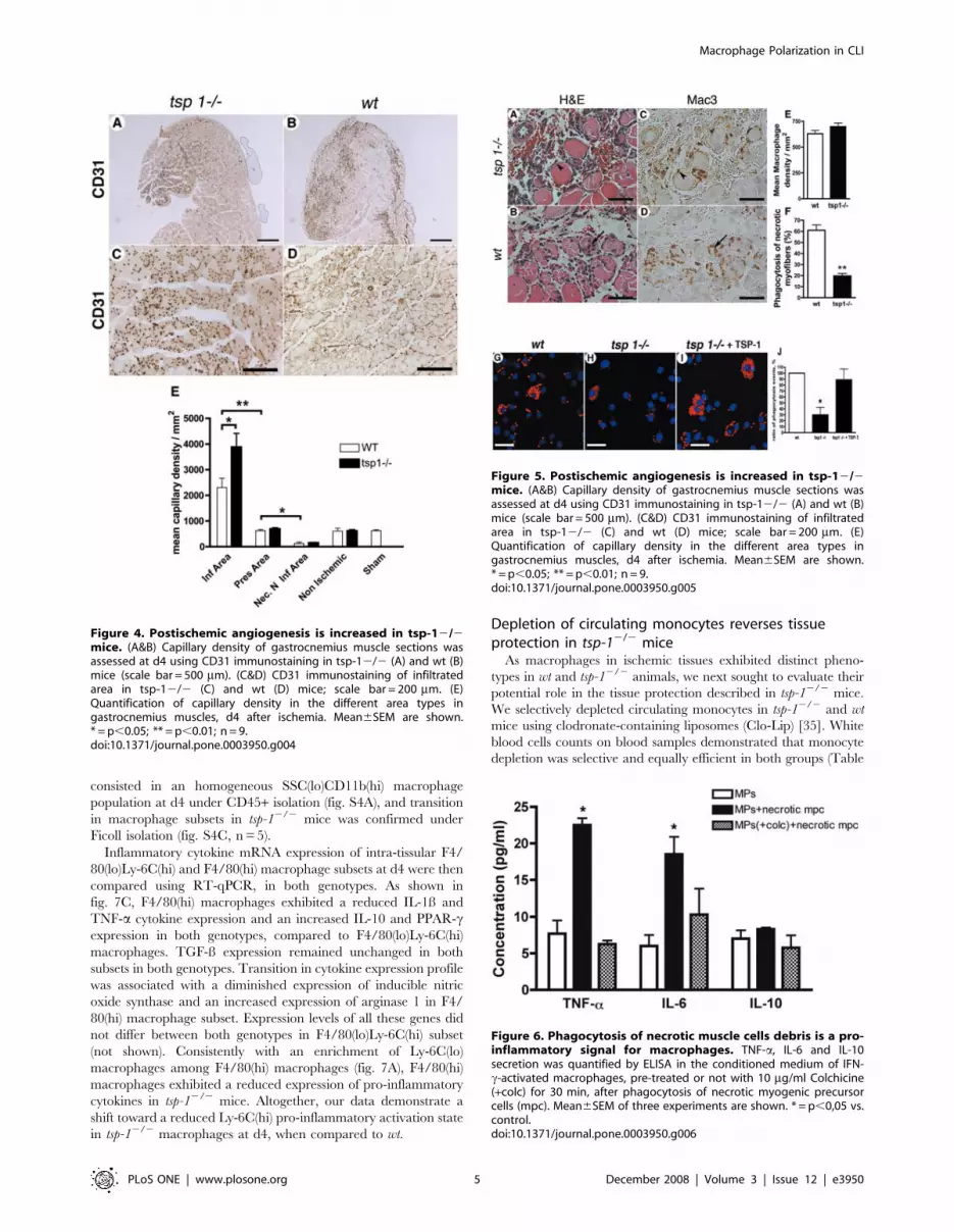
Thrombospondin-12/2 macrophages exhibit a reducedphagocytotic ability in vivo and in vitro
As macrophages play a key role during healing/regeneration
processes [8,9] and angiogenesis [31], express tsp-1 and are described
as a potential TSP-1 target [19], we further sought to investigate the
potential differential role of macrophages in both strains. Gastroc-
nemius muscles at the same stage of regeneration were immuno-
stained for macrophages (fig. 5A–5D). Macrophage density was
identical in tsp-12/2 and wt mice (fig. 5E), thereby ruling out a major
effect of TSP-1 on monocyte recruitment. However, macrophages
localized differently in the infiltrated area in both groups: in wt mice,
macrophages had a widespread distribution, mostly into myofibers
undergoing phagocytosis (fig. 5B&5D). In contrast, macrophages
remained mostly outside of myofibers in tsp-12/2 mice (fig. 5A&5C).
When quantified, phagocytosis of necrotic myofibers was strongly
reduced in tsp-12/2 mice (fig. 5F).
We then analyzed in vitro phagocytotic properties of isolated
bone marrow derived macrophages from both genotypes using
fluorescent latex beads (fig. 5G–J). Our results show that tsp-12/2
macrophages had a reduced ability to engulf beads in vitro
compared to wt macrophages (fig. 5G&5H). In addition, this
property could be rescued by adding recombinant TSP-1 in the
culture medium (fig. 5I).
These data indicate a strong impairment of phagocytotic ability
in tsp-12/2 macrophages, in vitro and in vivo during CLI.
Phagocytosis of necrotic muscle cells debris is a strongpro-inflammatory signal for macrophages
We next sought to analyze the effect of phagocytosis of muscle
cells debris on the cytokine expression profile of macrophages.
Upon phagocytosis of necrotic myogenic precursor cells (mpc),
TNF-a and IL-6 secretion strongly increased, whereas IL-10
secretion remained unchanged (fig. 6). When macrophages were
treated with Colchicine to inhibit phagocytosis [32], TNF-a and
IL-6 induced secretion was abolished in the presence of mpc debris
(fig. 6). These data demonstrate that phagocytosis of muscle cells
debris is a pro-inflammatory signal for macrophages.
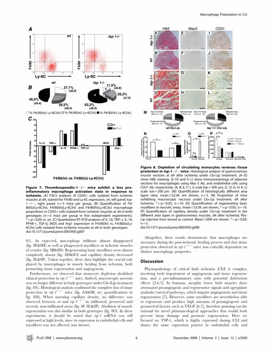
Thrombospondin-12/2 mice exhibit a less pro-inflammatory macrophage activation state in response toischemia
We therefore next investigated a potential different activation
state of macrophages in response to ischemia in both genotypes.
We first characterized intra-tissular macrophage activation state at
various time points after ischemia in C57Bl6 wt mice (fig. S3A), by
analyzing macrophages using Fluorescence-Activated Cell Sorting
(FACS) for F4/80 and Ly-6C expression, a membrane marker of
macrophage pro-inflammatory activation state [8,9,33,34]. Pri-
mary invading cells consisted in an homogeneous population of
SSC(lo) CD11b(hi) macrophages (fig. S3B and [33]) that exhibited
a F4/80(lo)Ly-6C(hi) membrane marker expression profile. At
later time points, a transition from F4/80(lo)Ly-6C(hi) to F4/
80(hi)Ly-6C(lo) macrophages was observed (fig. S3A).
Figure 2. Clinical necrosis is reduced in tsp-12/2 mice. (A) Picturesshow mice either protected from necrosis (left) or affected by necrosis(right). (B) Cumulative incidence of necrosis was followed during 21 daysafter ischemia in both wt and tsp-12/2 mice and represented as Kaplan-Meier estimates (n = 19; p,0.05 tsp-12/2 vs. wt).doi:10.1371/journal.pone.0003950.g002
Macrophage Polarization in CLI
PLoS ONE | www.plosone.org 3 December 2008 | Volume 3 | Issue 12 | e3950
We next investigated macrophage activation state in both
genotypes. CD 45+ inflammatory cells were collected from
ischemic muscles d4 after ischemia and analyzed for F4/80 and
Ly-6C expression. In accordance with observations described
above, overall proportion of macrophages among CD45+ cells did
not differ between wt and tsp-12/2 mice (respectively 86% and
84% of CD45+ cells). However, a transition from F4/80(lo)Ly-
6C(hi) to F4/80(hi)Ly-6C(lo) macrophages was observed in
tsp-12/2 mice when compared to wt (fig. 7A, fig. S4B and
quantification in fig. 7B, n = 25). F4/80(lo)Ly-6C(hi) cells still
Figure 3. Histological analysis showing tissue protection in tsp-12/2 mice. (A&B) H&E staining of gastrocnemius muscle sections at d4were performed in tsp-12/2 (A) and wt (B) mice (scale bar = 500 mm). Right panel show higher magnifications of preserved area (C), infiltrated area intsp-12/2 (E) and in wt (G) and necrotic non-infiltrated area (I), with adjacent slides immunostained for macrophages using Mac-3 Ab (D, F, H, J,respectively); scale bar = 100 mm. (K) Quantification of histologically different area types surface (see text for definition); mean6S.E.M. are shown,* = p,0.05, n = 9. (L) Quantification of regenerating basic myofibers in necrotic areas, mean6S.E.M. are shown, ** = p,0.01, n = 9.doi:10.1371/journal.pone.0003950.g003
Macrophage Polarization in CLI
PLoS ONE | www.plosone.org 4 December 2008 | Volume 3 | Issue 12 | e3950
consisted in an homogeneous SSC(lo)CD11b(hi) macrophage
population at d4 under CD45+ isolation (fig. S4A), and transition
in macrophage subsets in tsp-12/2 mice was confirmed under
Ficoll isolation (fig. S4C, n = 5).
Inflammatory cytokine mRNA expression of intra-tissular F4/
80(lo)Ly-6C(hi) and F4/80(hi) macrophage subsets at d4 were then
compared using RT-qPCR, in both genotypes. As shown in
fig. 7C, F4/80(hi) macrophages exhibited a reduced IL-1ß and
TNF-a cytokine expression and an increased IL-10 and PPAR-cexpression in both genotypes, compared to F4/80(lo)Ly-6C(hi)
macrophages. TGF-ß expression remained unchanged in both
subsets in both genotypes. Transition in cytokine expression profile
was associated with a diminished expression of inducible nitric
oxide synthase and an increased expression of arginase 1 in F4/
80(hi) macrophage subset. Expression levels of all these genes did
not differ between both genotypes in F4/80(lo)Ly-6C(hi) subset
(not shown). Consistently with an enrichment of Ly-6C(lo)
macrophages among F4/80(hi) macrophages (fig. 7A), F4/80(hi)
macrophages exhibited a reduced expression of pro-inflammatory
cytokines in tsp-12/2 mice. Altogether, our data demonstrate a
shift toward a reduced Ly-6C(hi) pro-inflammatory activation state
in tsp-12/2 macrophages at d4, when compared to wt.
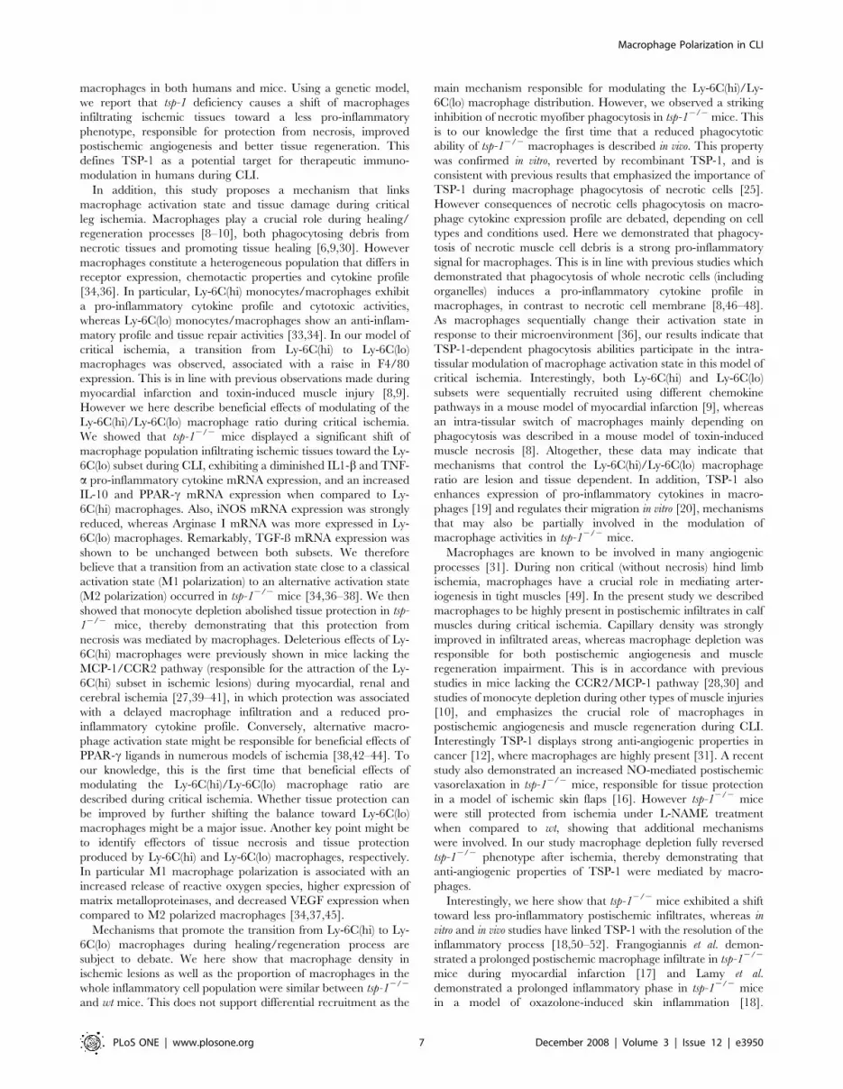
Depletion of circulating monocytes reverses tissueprotection in tsp-12/2 mice
As macrophages in ischemic tissues exhibited distinct pheno-
types in wt and tsp-12/2 animals, we next sought to evaluate their
potential role in the tissue protection described in tsp-12/2 mice.
We selectively depleted circulating monocytes in tsp-12/2 and wt
mice using clodronate-containing liposomes (Clo-Lip) [35]. White
blood cells counts on blood samples demonstrated that monocyte
depletion was selective and equally efficient in both groups (Table
Figure 4. Postischemic angiogenesis is increased in tsp-12/2mice. (A&B) Capillary density of gastrocnemius muscle sections wasassessed at d4 using CD31 immunostaining in tsp-12/2 (A) and wt (B)mice (scale bar = 500 mm). (C&D) CD31 immunostaining of infiltratedarea in tsp-12/2 (C) and wt (D) mice; scale bar = 200 mm. (E)Quantification of capillary density in the different area types ingastrocnemius muscles, d4 after ischemia. Mean6SEM are shown.* = p,0.05; ** = p,0.01; n = 9.doi:10.1371/journal.pone.0003950.g004
Figure 5. Postischemic angiogenesis is increased in tsp-12/2mice. (A&B) Capillary density of gastrocnemius muscle sections wasassessed at d4 using CD31 immunostaining in tsp-12/2 (A) and wt (B)mice (scale bar = 500 mm). (C&D) CD31 immunostaining of infiltratedarea in tsp-12/2 (C) and wt (D) mice; scale bar = 200 mm. (E)Quantification of capillary density in the different area types ingastrocnemius muscles, d4 after ischemia. Mean6SEM are shown.* = p,0.05; ** = p,0.01; n = 9.doi:10.1371/journal.pone.0003950.g005
Figure 6. Phagocytosis of necrotic muscle cells debris is a pro-inflammatory signal for macrophages. TNF-a, IL-6 and IL-10secretion was quantified by ELISA in the conditioned medium of IFN-c-activated macrophages, pre-treated or not with 10 mg/ml Colchicine(+colc) for 30 min, after phagocytosis of necrotic myogenic precursorcells (mpc). Mean6SEM of three experiments are shown. * = p,0,05 vs.control.doi:10.1371/journal.pone.0003950.g006
Macrophage Polarization in CLI
PLoS ONE | www.plosone.org 5 December 2008 | Volume 3 | Issue 12 | e3950
S1). As expected, macrophage infiltrate almost disappeared
(fig. 8F&8H) as well as phagocyted myofibers in ischemic muscles
of wt mice (fig. 8B&8D). Regenerating basic myofibers were almost
completely absent (fig. 8D&8O) and capillary density decreased
(fig. 8L&8P). Taken together, these data highlight the crucial role
played by macrophages in muscle healing from ischemia, both
promoting tissue regeneration and angiogenesis.
Furthermore, we observed that monocyte depletion abolished
clinical protection in tsp-12/2 mice. Indeed, macroscopic necrosis
was no longer different in both genotypes under Clo-Lip treatment
(fig. 8N). Histological analysis confirmed the complete loss of tissue
protection in tsp-12/2 mice (fig. 8A&8B; see quantifications in
fig. 8M). When assessing capillary density, no difference was
observed between wt and tsp-12/2 in infiltrated, preserved and
necrotic non-infiltrated areas (fig. 8I–8L&8P). Abolition of muscle
regeneration was also similar in both genotypes (fig. 8O). In these
experiments, it should be noted that tsp-1 mRNA was still
expressed at high levels, since its expression in endothelial cells and
myofibers was not affected (not shown).
Altogether, these results demonstrate that macrophages are
necessary during the post-ischemic healing process and that tissue
protection observed in tsp-12/2 mice was critically dependent on
distinct macrophage properties.
Discussion
Physiopathology of critical limb ischemia (CLI) is complex,
involving both impairment of angiogenesis and tissue regenera-
tion, and a pro-inflammatory state with potential deleterious
effects [2,6,7]. In humans, atrophic lower limb muscles show
attenuated proangiogenic and regenerative signals and upregulate
anabolic/survival pathways, which impairs angiogenesis and tissue
regeneration [7]. However, some myofibers are nevertheless able
to regenerate and produce high amounts of proangiogenic and
prosurvival factors, such as VEGF [6,7], therefore pointing out the
rational for novel pharmacological approaches that would both
prevent tissue damage and promote regeneration. Here we
focused on TSP-1, which is highly expressed during CLI and
shares the same expression pattern in endothelial cells and
Figure 7. Thrombospondin-12/2 mice exhibit a less pro-inflammatory macrophage activation state in response toischemia. (A) FACS analyses of CD45+ cells isolated from ischemicmuscles at d4, stained for F4/80 and Ly-6C expression, wt, left panel; tsp-12/2, right panel; n = 5 mice per group. (B) Quantification of F4/80(lo)Ly-6C(hi), F4/80(hi)Ly-6C(hi) and F4/80(hi)Ly-6C(lo) macrophageproportions in CD45+ cells isolated from ischemic muscles at d4 in bothgenotypes (n = 5 mice per group in five independent experiments).* = p,0,05 vs. wt. (C) Quantitative RT-PCR analyses of IL-1b, TNF-a, IL-10,PPAR-c, TGF-b, iNOS and Arg1 expression in F4/80(hi) vs. F4/80(lo)Ly-6C(hi) cells isolated from ischemic muscles at d4 in both genotypes.doi:10.1371/journal.pone.0003950.g007
Figure 8. Depletion of circulating monocytes reverses tissueprotection in tsp-12/2 mice. Histological analysis of gastrocnemiusmuscle sections at d4 after ischemia under Clo-Lip treatment. (A–D)show H&E staining. (E–H) and (I–L) show immunostainings of adjacentsections for macrophages using Mac-3 Ab, and endothelial cells usingCD31 Ab, respectively. (A, B; E, F; I, J) scale bar = 500 mm; (C, D; G, H; K, L)scale bar = 200 mm. (M) Quantification of histologically different areatypes ratio; mean6S.E.M. are shown, n = 5. (N) Proportion of miceexhibiting macroscopic necrosis under Clo-Lip treatment, d4 afterischemia. * = p,0.05, n = 10. (O) Quantification of regenerating basicmyofibers in necrotic areas, mean6S.E.M. are shown, * = p,0.05, n = 10.(P) Quantification of capillary density under Clo-Lip treatment in thedifferent area types in gastrocnemius muscles, d4 after ischemia; Pbs-Lip injected mice served as control. Mean6SEM are shown. * = p,0.05;n = 5.doi:10.1371/journal.pone.0003950.g008
Macrophage Polarization in CLI
PLoS ONE | www.plosone.org 6 December 2008 | Volume 3 | Issue 12 | e3950
macrophages in both humans and mice. Using a genetic model,
we report that tsp-1 deficiency causes a shift of macrophages
infiltrating ischemic tissues toward a less pro-inflammatory
phenotype, responsible for protection from necrosis, improved
postischemic angiogenesis and better tissue regeneration. This
defines TSP-1 as a potential target for therapeutic immuno-
modulation in humans during CLI.
In addition, this study proposes a mechanism that links
macrophage activation state and tissue damage during critical
leg ischemia. Macrophages play a crucial role during healing/
regeneration processes [8–10], both phagocytosing debris from
necrotic tissues and promoting tissue healing [6,9,30]. However
macrophages constitute a heterogeneous population that differs in
receptor expression, chemotactic properties and cytokine profile
[34,36]. In particular, Ly-6C(hi) monocytes/macrophages exhibit
a pro-inflammatory cytokine profile and cytotoxic activities,
whereas Ly-6C(lo) monocytes/macrophages show an anti-inflam-
matory profile and tissue repair activities [33,34]. In our model of
critical ischemia, a transition from Ly-6C(hi) to Ly-6C(lo)
macrophages was observed, associated with a raise in F4/80
expression. This is in line with previous observations made during
myocardial infarction and toxin-induced muscle injury [8,9].
However we here describe beneficial effects of modulating of the
Ly-6C(hi)/Ly-6C(lo) macrophage ratio during critical ischemia.
We showed that tsp-12/2 mice displayed a significant shift of
macrophage population infiltrating ischemic tissues toward the Ly-
6C(lo) subset during CLI, exhibiting a diminished IL1-b and TNF-
a pro-inflammatory cytokine mRNA expression, and an increased
IL-10 and PPAR-c mRNA expression when compared to Ly-
6C(hi) macrophages. Also, iNOS mRNA expression was strongly
reduced, whereas Arginase I mRNA was more expressed in Ly-
6C(lo) macrophages. Remarkably, TGF-ß mRNA expression was
shown to be unchanged between both subsets. We therefore
believe that a transition from an activation state close to a classical
activation state (M1 polarization) to an alternative activation state
(M2 polarization) occurred in tsp-12/2 mice [34,36–38]. We then
showed that monocyte depletion abolished tissue protection in tsp-
12/2 mice, thereby demonstrating that this protection from
necrosis was mediated by macrophages. Deleterious effects of Ly-
6C(hi) macrophages were previously shown in mice lacking the
MCP-1/CCR2 pathway (responsible for the attraction of the Ly-
6C(hi) subset in ischemic lesions) during myocardial, renal and
cerebral ischemia [27,39–41], in which protection was associated
with a delayed macrophage infiltration and a reduced pro-
inflammatory cytokine profile. Conversely, alternative macro-
phage activation state might be responsible for beneficial effects of
PPAR-c ligands in numerous models of ischemia [38,42–44]. To
our knowledge, this is the first time that beneficial effects of
modulating the Ly-6C(hi)/Ly-6C(lo) macrophage ratio are
described during critical ischemia. Whether tissue protection can
be improved by further shifting the balance toward Ly-6C(lo)
macrophages might be a major issue. Another key point might be
to identify effectors of tissue necrosis and tissue protection
produced by Ly-6C(hi) and Ly-6C(lo) macrophages, respectively.
In particular M1 macrophage polarization is associated with an
increased release of reactive oxygen species, higher expression of
matrix metalloproteinases, and decreased VEGF expression when
compared to M2 polarized macrophages [34,37,45].
Mechanisms that promote the transition from Ly-6C(hi) to Ly-
6C(lo) macrophages during healing/regeneration process are
subject to debate. We here show that macrophage density in
ischemic lesions as well as the proportion of macrophages in the
whole inflammatory cell population were similar between tsp-12/2
and wt mice. This does not support differential recruitment as the
main mechanism responsible for modulating the Ly-6C(hi)/Ly-
6C(lo) macrophage distribution. However, we observed a striking
inhibition of necrotic myofiber phagocytosis in tsp-12/2 mice. This
is to our knowledge the first time that a reduced phagocytotic
ability of tsp-12/2 macrophages is described in vivo. This property
was confirmed in vitro, reverted by recombinant TSP-1, and is
consistent with previous results that emphasized the importance of
TSP-1 during macrophage phagocytosis of necrotic cells [25].
However consequences of necrotic cells phagocytosis on macro-
phage cytokine expression profile are debated, depending on cell
types and conditions used. Here we demonstrated that phagocy-
tosis of necrotic muscle cell debris is a strong pro-inflammatory
signal for macrophages. This is in line with previous studies which
demonstrated that phagocytosis of whole necrotic cells (including
organelles) induces a pro-inflammatory cytokine profile in
macrophages, in contrast to necrotic cell membrane [8,46–48].
As macrophages sequentially change their activation state in
response to their microenvironment [36], our results indicate that
TSP-1-dependent phagocytosis abilities participate in the intra-
tissular modulation of macrophage activation state in this model of
critical ischemia. Interestingly, both Ly-6C(hi) and Ly-6C(lo)
subsets were sequentially recruited using different chemokine
pathways in a mouse model of myocardial infarction [9], whereas
an intra-tissular switch of macrophages mainly depending on
phagocytosis was described in a mouse model of toxin-induced
muscle necrosis [8]. Altogether, these data may indicate that
mechanisms that control the Ly-6C(hi)/Ly-6C(lo) macrophage
ratio are lesion and tissue dependent. In addition, TSP-1 also
enhances expression of pro-inflammatory cytokines in macro-
phages [19] and regulates their migration in vitro [20], mechanisms
that may also be partially involved in the modulation of
macrophage activities in tsp-12/2 mice.
Macrophages are known to be involved in many angiogenic
processes [31]. During non critical (without necrosis) hind limb
ischemia, macrophages have a crucial role in mediating arter-
iogenesis in tight muscles [49]. In the present study we described
macrophages to be highly present in postischemic infiltrates in calf
muscles during critical ischemia. Capillary density was strongly
improved in infiltrated areas, whereas macrophage depletion was
responsible for both postischemic angiogenesis and muscle
regeneration impairment. This is in accordance with previous
studies in mice lacking the CCR2/MCP-1 pathway [28,30] and
studies of monocyte depletion during other types of muscle injuries
[10], and emphasizes the crucial role of macrophages in
postischemic angiogenesis and muscle regeneration during CLI.
Interestingly TSP-1 displays strong anti-angiogenic properties in
cancer [12], where macrophages are highly present [31]. A recent
study also demonstrated an increased NO-mediated postischemic
vasorelaxation in tsp-12/2 mice, responsible for tissue protection
in a model of ischemic skin flaps [16]. However tsp-12/2 mice
were still protected from ischemia under L-NAME treatment
when compared to wt, showing that additional mechanisms
were involved. In our study macrophage depletion fully reversed
tsp-12/2 phenotype after ischemia, thereby demonstrating that
anti-angiogenic properties of TSP-1 were mediated by macro-
phages.
Interestingly, we here show that tsp-12/2 mice exhibited a shift
toward less pro-inflammatory postischemic infiltrates, whereas in
vitro and in vivo studies have linked TSP-1 with the resolution of the
inflammatory process [18,50–52]. Frangogiannis et al. demon-
strated a prolonged postischemic macrophage infiltrate in tsp-12/2
mice during myocardial infarction [17] and Lamy et al.
demonstrated a prolonged inflammatory phase in tsp-12/2 mice
in a model of oxazolone-induced skin inflammation [18].
Macrophage Polarization in CLI
PLoS ONE | www.plosone.org 7 December 2008 | Volume 3 | Issue 12 | e3950

Postischemic infiltrates mostly contain macrophages and necrotic
myofibers at d4, and very few CD3+ cells (data not shown).
Conversely oxazolone-induced skin infiltrates and post-ischemic
tissues at later stages contain a high proportion of lymphocytes
[18]. This could explain a differential role for TSP-1 in
modulating these different types of infiltrates: TSP-1-dependent
phagocytosis of necrotic lymphocytes induces an anti-inflamma-
tory response in macrophages [25], whereas phagocytosis of
necrotic muscle cells promotes a pro-inflammatory response.
In conclusion we describe here TSP-1 as a new relevant
physiological target during critical leg ischemia in humans.
Furthermore, we describe the modulation of postischemic macro-
phage activation state as a new potential therapeutic approach to
protect tissues from necrosis and promote tissue repair during critical
ischemia. Finally our data also suggest that phagocytosis of necrotic
muscle debris plays a crucial role in regulating the intra-tissular
macrophage activation state during critical leg ischemia.
Methods
AnimalsThrombospondin-12/2 mice were on a C57/Bl6 background as
described previously [53]. All experiments were performed on 12
to 18 weeks old males.
This study conforms to the standards of INSERM (the French
National Institute of Health) regarding the care and use of
laboratory animals, was performed in accordance with European
Union Council Directives (86/609/EEC) and was approved by the
institutional research ethics committee IDF - Paris - Comite 1 (ref :
2008004).
Hind limb ischemia proceduresUnilateral critical ischemia was generated by ligation/excision
of the femoral artery as previously described [54]. Mice were
anesthetized with i.p. injection of ketamin 2 mg (Imalgene) and
xylazine 0.2 mg (Rompun). After skin incision, the superficial
epigastric artery was ligated (Ethicon 6-0, Vicryl). The proximal
end of the left femoral artery and the distal portion of the
saphenous artery were ligated. The femoral artery was then
excised. Femoral vein and nerve were not preserved during
surgical procedures. After surgery, the skin was closed with
interrupted 6.0 proline sutures.
Macroscopic necrosisThe incidence of hind limb macroscopic necrosis was
determined at d2, d7, d14 and d21 after femoral artery excision
(n = 19 mice per group).
Histological analyses and immunohistochemistryAfter sacrifice at d4, gastrocnemius muscles were fixed,
dehydrated and paraffin-embedded. Serial adjacent 7 mm cross
sections were generated through the midportion of the muscle for
H&E staining and Mac-3 immunostaining (that labels macro-
phages). Myofibers with pale cytoplasm and loss of peripheral
nuclei were defined as necrotic as previously described [30].
Macrophages were stained using a rat anti-mouse Mac-3
monoclonal Ab (BD Biosciences, dilution 1/75) and revealed with
a secondary biotin-conjugated goat anti-rat Ab (Jackson Immu-
noresearch, dilution 1/200). ABC-peroxydase complex (Vector
Laboratories) was used for signal amplification.
Three types of area were observed (fig. S2A): an infiltrated area
(that display necrosis and macrophage infiltrate) (fig. S2B&2C), a
preserved area (normal histology) (fig. S2D&2E) and a necrotic
non-infiltrated area (that display necrosis without macrophage
infiltrate) (fig. S2F&2G). Images of each area were digitally
captured on H&E slides using a Leica DM 4000B microscope
equipped with a DFC 420 camera and the Application Suite 2.7.1
software. The surface of each area type was quantified in each
mouse using Metamorph software and reported as percentage of
the entire section surface (n = 9) (see fig. S2A). Regenerating basic
myofibers were quantified on whole muscle section as previously
described [8] for each mouse (n = 9). For macrophage density,
macrophages were counted on 5 digitally captured non-overlap-
ping 620 magnification fields in the infiltrated area, and reported
as a single value/mm2 in each animal (n = 9). Necrotic myofibers
containing two or more macrophages were defined as phagocyted
and expressed as a percentage of total necrotic myofibers, on
digitally captured images of non-overlapping fields (620 magni-
fication, 4 fields per animal, n = 9).
Assessment of capillary density. Biotin-conjugated rat anti-
mouse CD31 Ab (BD Biosciences, dilution 1:50) and Cy3-
conjugated mouse monoclonal anti a-smooth muscle actin Ab
(Sigma-Aldrich, dilution 1/100) were used to identify endothelial
cells and vascular smooth muscle cells, respectively. For capillary
density quantification, nonoverlapping620 fields (5 per area) were
digitally captured. Capillaries were counted using the software IP
lab 3.2.4.
Western Blot analyses of TSP-1 expression were performed
as previously described [26] excepted that attophos substrate
(Promega) was used. The specificity of antibody binding was
verified in tsp-12/2 mice.
In situ hybridization procedures were performed as
previously described [55]. A 1186 bp cDNA fragment (nucleotide
151-1336) was used to generate a 35S-RNA antisense mouse tsp-1
probe. A sense probe was used as a negative control.
MicroangiographyMice were anesthetized (40 ml i.p. sodium pentobarbital) and
longitudinal laparotomy was performed to introduce a polyethyl-
ene catheter into the abdominal aorta to inject contrast medium
(barium sulfate, 1 g/mL). Angiography of hind limbs was then
performed and images (two per animal) were acquired using a
high-definition digital X-ray transducer. Computerized quantifi-
cation of vessel density was then performed and expressed as a
ratio from ischemic to non ischemic leg of the percentage of pixels
per image occupied by vessels in the quantification area.
Bone marrow-derived macrophage isolation anddifferentiation
Primary culture of murine bone marrow macrophages were
harvested from femur of 12 to 18 week-old tsp-12/2 and wt mice
as described previously [36]. Briefly, the marrow cells were flushed
from the bone with a 26G needle connected to a syringe filled with
RPMI 1640 supplemented with 1% antibiotic mixture (penicillin,
streptomycin, neomycin). Following centrifugation over Ficoll, the
cells were cultured in DMEM medium (Gibco 31885) supple-
mented with 1% antibiotic mixture, 10% FBS and 20% of L-929-
conditioned medium (source of M-CSF-1). Non-adherent cells
were collected after 24 h, seeded on coverslips, and differentiated
for 7 days in polystyrene culture plates, changing the medium once
on d4. The resulting cell population was .95% F4/80 and 98%
Mac-3 positive as assessed by flow cytometry.
PhagocytosisAfter overnight priming with IFN-c (100 U/ml), bone marrow
macrophages cells were washed and activated overnight with
100 ng/ml LPS. Fluorescent latex beads (Sigma L3030, 2 mm)
Macrophage Polarization in CLI
PLoS ONE | www.plosone.org 8 December 2008 | Volume 3 | Issue 12 | e3950
were seeded on macrophages (three beads per one macrophage)
for 3 h at 37uC with or without 2,5 mg/ml recombinant TSP-1
(US Biological). macrophage cultures were washed four times to
remove noningested material and fixed using 4% paraformalde-
hyde. Coverslips were mounted with Moviol/ToPro (Invitrogen)
and analyzed in sequential scanning mode using a 640 objective
lens with a Leica TCS SP2 confocal microscope equipped with
three external lasers (488, 543 and 633 nm) (Leica Microsystems).
The number of Latex-containing cells was expressed as the
percentage of total cells. Five nonoverlapping fields were averaged
and reported as a single value for each well.
For inflammation assays, necrotic myogenic precursor cells (lyzed
by three cycles of freeze-thawing) were seeded on IFN-c-activated
macrophages (ratio 3:1) for 3 h at 37uC. macrophage cultures were
washed three times to remove noningested material and further
cultured in serum-free medium (GIBCO 31885) for 24 h prior to
collection of conditioned media. TNF-a, IL-6 and IL-10 were
quantified in the supernatant by enzyme-linked immunoabsorbent
assay (Quantikine, R&D systems). To inhibit phagocytosis, macro-
phages were pre-treated with 10 mg/ml colchicine (Sigma-Aldrich)
for 30 min, as previously described [32].
Isolation of monocytes/macrophages from muscleand RNA preparation were performed as previously described
[8]. Briefly, muscles were dissociated in DMEM containing
collagenase B 0.2% (Roche Diagnostics Gmbh) at 37uC for
90 min, filtered and counted. Inflammatory cells cells were
isolated using CD45+ magnetic sorting (Milteny Biotec) or
centrifugation over Ficoll and stained with the following
combinations of antibodies : {PE-conjugated F4/80 (AbD Serotec)
and FITC-conjugated Ly-6G/ Ly-6C (only Ly-6C is expressed by
macrophages [8]) (eBioscience)} antibodies, or {APC-conjugated
F4/80 (eBioscience), PE-Cy7-conjugated CD45 (eBioscience),
FITC-conjugated Ly-6G/ Ly-6C (eBioscience) and PE-conjugated
CD11b (BD Pharmingen)} antibodies, in association with a
CD16/CD32 Fc block antibody (BD Pharmigen). Analysis was
performed using a cytometer (MoFlo and Cyan, Dako-Cytoma-
tion). Using either CD45+ magnetic sorting or Ficoll isolation, F4/
80(lo) Ly-6C(hi) cells consisted at d4 in an homogeneous
monocyte/macrophage population, as indicated by their mono-
nuclearity read as low orthogonal (side) scatter in the flow
cytometer and their myeloid nature, as indicated by high level of
CD11b expression [33] (fig. S3B & fig. S4A).
RT-qPCR0.5 mg of total RNA was reverse transcribed using Superscript II
reverse transcriptase. Each cDNA preparation was amplified using
a iQTM SYBR green supermix (Bio-Rad) and the following specific
primers (sense and antisense, respectively): b2 microglobulin, 59-
CAGTTCCACCCGCCTCAC-39 and 59-CACATGTCTCGA-
TCCCAG-39; TNF-a, 59-AAAGATGGGGGGCTTCCAGAA-
CTC-39 and 59-TGAGATAGCAAATCGGCTGACGG-39; IL-
1b, 59-TGACGTTCCCATTAGACAACTG-39 and 59-CCG-
TCTTTCATTACACAGGACA-39; IL-10, 59-ACCAGCTGGA-
CAACATACTGC-39 and 59-TCACTCTTCACCTGCTCCA-
CT-39; PPAR-c, 59-AGGCCGAGAAGGAGAAGCTGTTG -39
and 59-TGGCCACCTCTTTGCTCTGCTC-39, TFG-ß1, 59-
TGCGCTTGCAGAGATTAAAA-39 and 59-CGTCAAAAGA-
CAGCCACTCA-39; iNOS, 59-GAAGAAAACCCCTTGTG-
CTG-39 and 59-TCCAGGGATTCTGGAACATT-39; Arg I, 59-
CAGAAGAATGGAAGAGTCAG-39 and 59-CAGATATGCA-
GGGAGTCACC-39. PCR conditions were: 95uC for 15 min and
then 95uC for 15 s, 60uC for 30 s and 72uC for 45 s repeated for
40 cycles. Melting curves were obtained. Amplification was
performed using a iCycler equipped with a MyiQTM optical
module (Bio-Rad). Analysis of the relative expression of each target
gene was related to b2 microglobulin expression using the iQTM5
Optical system software (Bio-Rad).
Macrophage depletion was achieved using clodronate (Cl2-
MDP)-containing liposomes as previously described [35,56]. This
method allows the specific depletion in monocytes and macrophages,
which undergo apoptosis upon phagocytosis of Cl2MDP liposomes.
Clo-Lip were injected i.v. 12 h before femoral artery ligation in tsp-
12/2 and wt mice (250 ml, 10 mice per group). Considering the half-
life of this product (48 h), injection was repeated at d2 after ligation
and mice were sacrificed at d4 for clinical, histological and
immunohistochemical analyses. PBS-containing liposomes (Pbs-
Lip) were used as a negative control in wt mice (10 mice). Clodronate
was a gift of Roche Diagnostics GmbH.
White blood cell countsBlood samples were taken from tsp-12/2 and wt mice prior to
Clo-Lip injection and at d4 after femoral artery ligation (n = 10 per
group). After May Grunwald Giemsa staining, percentage of
monocyte, lymphocyte and neutrophil were quantified by a
hematologist. At least 200 cells were counted for each sample.
Statistical analysesAll experiments were conducted in a blinded-manner for both
genotype and groups of mice, and performed using cultures or
animals in at least three independent experiments. Staview (SAS
institute Inc.) and Prism 4.0 (GaphPad software, Inc) were used for
statistical analyses. Continuous variables are reported as Mean6-
SEM. Incidence of macroscopic necrosis was estimated by the
Kaplan-Meier method, and differences were assessed by means of
the log-rank test. Wilcoxon test was used to compare proportions
of macrophage subsets in both genotypes (fig. 7), and Mann-
Whitney was used to compare tsp-12/2 from wt mice in other
experiments. Statistical significance was set at p,0.05.
Supporting Information
Figure S1 Description of the healing/regeneration process in
gastrocnemius muscle in response to ischemia. Histological
analyses of gastrocnemius muscles sections at d4, d6, d16 and
d21 after ischemia. (A, D, G, J) show H&E staining. (B, E, H, K)
and (C, F, I, L) show immunostainings of adjacent sections for
macrophages using a Mac-3 Ab, and endothelial cells using a
CD31 Ab, respectively. Scale bar = 200 mm.
Found at: doi:10.1371/journal.pone.0003950.s001 (8.83 MB TIF)
Figure S2 Three different types of area are observed in
gastrocnemius muscle at d4 after ischemia. (A) Cross sections
were generated through the midportion of gastrocnemius d4 after
ischemia and stained with H&E. (A) representative section of the
right part of wt mice gastrocnemius is shown (scale bar = 500 mm).
Adjacent sections were immunostained for macrophages using
Mac-3 Ab. (B–G) Higher magnification of infiltrated area (necrotic
myofibers+macrophage infiltrate) (B, C), preserved area (normal
histology) (D, E) and necrotic non-infiltrated area (necrotic
myofibers+absence of macrophage infiltrate) (F, G), either stained
with H&E (B, D, F) or immunostained for Mac-3 (C, E, G); scale
bar = 100 mm. Quantification. Surfaces of infiltrated area (black
stroke) and preserved area (green stroke) were quantified and
reported as percentage of the entire section surface in each mouse.
The remainder of surface percentage was attributed to necrotic
non-infiltrated area ( = 100%- infiltrated area (%)- preserved area
(%) in each mouse).
Found at: doi:10.1371/journal.pone.0003950.s002 (14.85 MB
TIF)
Macrophage Polarization in CLI
PLoS ONE | www.plosone.org 9 December 2008 | Volume 3 | Issue 12 | e3950

Figure S3 Kinetic analysis of intra-tissular macrophage activa-
tion state during ischemia. (A) Mononuclear cells were isolated
from ischemic muscles of C57Bl6 mice (Charles-River) using
centrifugation over Ficoll, and analyzed by FACS for F4/80 and
Ly-6C expression (n = 3 per time point). (B) SSC/FSC character-
istics and CD11b expression of Ficoll-isolated F4/80(lo) Ly-6C(hi)
cells at d4, showing an homogeneous SSC(lo) CD11b(hi)
macrophage population.
Found at: doi:10.1371/journal.pone.0003950.s003 (4.71 MB TIF)
Figure S4 Thrombospondin-12/2 mice exhibit a less pro-
inflammatory macrophage activation state in response to ischemia.
Additional experiments. (A) SSC/FSC characteristics and CD11b
expression of CD45+ F4/80(lo) Ly-6C(hi) cells isolated from
ischemic muscles at d4, in both genotypes, showing an
homogeneous SSC(lo)CD11b(hi) macrophage population. (B)
FACS analyses of CD45+ cells isolated from ischemic muscles at
d4, stained for F4/80 and Ly-6C expression. Additional examples
of two independent experiments are shown (n = 5 mice). (C) Upper
panel, representative FACS analyses of Ficoll-isolated mononu-
clear cells from ischemic muscles of one mouse from both
genotypes at d4, stained for F4/80 and Ly-6C. Lower panel,
quantification of F4/80(lo)Ly-6C(hi), F4/80(hi)Ly-6C(hi) and F4/
80(hi)Ly-6C(lo) macrophage proportions in both genotypes (n = 5).
Found at: doi:10.1371/journal.pone.0003950.s004 (6.79 MB TIF)
Table S1 Clo-Lip induced monocyte depletion is identical in
both genotypes. Percentage of monocytes, neutrophils and
lymphocytes in total white blood cells, counted on blood samples
in both genotypes, at baseline (before injection) and d4 after
ischemia under Clo-Lip treatment. Mean6SEM are reported.
** = p,0.01 Clo-Lip vs. baseline; n = 10.
Found at: doi:10.1371/journal.pone.0003950.s005 (0.02 MB XLS)
Acknowledgments
We thank Jack Lawler for providing tsp-12/2 mice, Anne Eichmann,
Steven Suchting and Ariane Galaup for critical reading of the manuscript,
Lionel Leroux and Thierry Couffinhal for sharing expertise in the animal
model, and Eric Etienne for confocal microscopy. We also thank Thomas
Mathivet, Madly Brigitte and Rana Abou-Khalil for their help, the
Cytometry facility of IFR10 (IM3) - Universite Paris XII, Faculte de
medecine, Creteil- for FACS analyses and Fabrice Chretien for help with
clodronate liposomes injection. Cl2MDP (or clodronate) was a gift of
Roche Diagnostics GmbH, Mannheim, Germany.
Author Contributions
Conceived and designed the experiments: NB AN BC SG. Performed the
experiments: NB EG MB JKL MD AC MD JP JSS AN BC SG. Analyzed
the data: NB EG MB AC CAB JSS PC AN BC SG. Contributed reagents/
materials/analysis tools: NB JKL MD NvR AN BC SG. Wrote the paper:
NB SG.
References
1. Dormandy JA, Rutherford RB (2000) Management of peripheral arterial disease
(PAD). TASC Working Group. TransAtlantic Inter-Society Consensus (TASC).
J Vasc Surg 31: S1–S296.
2. Barani J, Nilsson JA, Mattiasson I, Lindblad B, Gottsater A (2005) Inflammatory
mediators are associated with 1-year mortality in critical limb ischemia. J Vasc
Surg 42: 75–80.
3. Baumgartner I, Pieczek A, Manor O, Blair R, Kearney M, et al. (1998)
Constitutive expression of phVEGF165 after intramuscular gene transfer
promotes collateral vessel development in patients with critical limb ischemia.
Circulation 97: 1114–1123.
4. Lederman RJ, Mendelsohn FO, Anderson RD, Saucedo JF, Tenaglia AN, et al.
(2002) Therapeutic angiogenesis with recombinant fibroblast growth factor-2 for
intermittent claudication (the TRAFFIC study): a randomised trial. Lancet 359:
2053–2058.
5. Rajagopalan S, Mohler E 3rd, Lederman RJ, Saucedo J, Mendelsohn FO, et al.
(2003) Regional Angiogenesis with Vascular Endothelial Growth Factor (VEGF)
in peripheral arterial disease: Design of the RAVE trial. Am Heart J 145:
1114–1118.
6. Rissanen TT, Vajanto I, Hiltunen MO, Rutanen J, Kettunen MI, et al. (2002)
Expression of vascular endothelial growth factor and vascular endothelial growth
factor receptor-2 (KDR/Flk-1) in ischemic skeletal muscle and its regeneration.
Am J Pathol 160: 1393–1403.
7. Tuomisto TT, Rissanen TT, Vajanto I, Korkeela A, Rutanen J, et al. (2004)
HIF-VEGF-VEGFR-2, TNF-alpha and IGF pathways are upregulated in
critical human skeletal muscle ischemia as studied with DNA array.
Atherosclerosis 174: 111–120.
8. Arnold L, Henry A, Poron F, Baba-Amer Y, van Rooijen N, et al. (2007)
Inflammatory monocytes recruited after skeletal muscle injury switch into
antiinflammatory macrophages to support myogenesis. J Exp Med 204:
1057–1069.
9. Nahrendorf M, Swirski FK, Aikawa E, Stangenberg L, Wurdinger T, et al.
(2007) The healing myocardium sequentially mobilizes two monocyte subsets
with divergent and complementary functions. J Exp Med 204: 3037–3047.
10. Summan M, Warren GL, Mercer RR, Chapman R, Hulderman T, et al. (2006)
Macrophages and skeletal muscle regeneration: a clodronate-containing
liposome depletion study. Am J Physiol Regul Integr Comp Physiol 290:
R1488–1495.
11. Engstrom G, Site-Flondell D, Lindblad B, Janzon L, Lindgarde F (2004) Risk of
treatment of peripheral arterial disease is related to inflammation-sensitive
plasma proteins: a prospective cohort study. J Vasc Surg 40: 1101–1105.
12. Esemuede N, Lee T, Pierre-Paul D, Sumpio BE, Gahtan V (2004) The role of
thrombospondin-1 in human disease. J Surg Res 122: 135–142.
13. Agah A, Kyriakides TR, Lawler J, Bornstein P (2002) The lack of
thrombospondin-1 (TSP1) dictates the course of wound healing in double-
TSP1/TSP2-null mice. Am J Pathol 161: 831–839.
14. Lawler J (2002) Thrombospondin-1 as an endogenous inhibitor of angiogenesis
and tumor growth. J Cell Mol Med 6: 1–12.
15. Lawler J, Detmar M (2004) Tumor progression: the effects of thrombospondin-1
and -2. Int J Biochem Cell Biol 36: 1038–1045.
16. Isenberg JS, Hyodo F, Matsumoto K, Romeo MJ, Abu-Asab M, et al. (2007)
Thrombospondin-1 limits ischemic tissue survival by inhibiting nitric oxide-
mediated vascular smooth muscle relaxation. Blood 109: 1945–1952.
17. Frangogiannis NG, Ren G, Dewald O, Zymek P, Haudek S, et al. (2005) Critical
role of endogenous thrombospondin-1 in preventing expansion of healing
myocardial infarcts. Circulation 111: 2935–2942.
18. Lamy L, Foussat A, Brown EJ, Bornstein P, Ticchioni M, et al. (2007)
Interactions between CD47 and thrombospondin reduce inflammation.
J Immunol 178: 5930–5939.
19. Yamauchi Y, Kuroki M, Imakiire T, Abe H, Uchida H, et al. (2002)
Thrombospondin-1 differentially regulates release of IL-6 and IL-10 by human
monocytic cell line U937. Biochem Biophys Res Commun 290: 1551–1557.
20. Mansfield PJ, Suchard SJ (1994) Thrombospondin promotes chemotaxis and
haptotaxis of human peripheral blood monocytes. J Immunol 153: 4219–4229.
21. Moodley Y, Rigby P, Bundell C, Bunt S, Hayashi H, et al. (2003) Macrophage
recognition and phagocytosis of apoptotic fibroblasts is critically dependent on
fibroblast-derived thrombospondin 1 and CD36. Am J Pathol 162: 771–779.
22. Ren Y, Savill J (1995) Proinflammatory cytokines potentiate thrombospondin-
mediated phagocytosis of neutrophils undergoing apoptosis. J Immunol 154:
2366–2374.
23. Stern M, Savill J, Haslett C (1996) Human monocyte-derived macrophage
phagocytosis of senescent eosinophils undergoing apoptosis. Mediation by alpha
v beta 3/CD36/thrombospondin recognition mechanism and lack of phlogistic
response. Am J Pathol 149: 911–921.
24. Ren Y, Stuart L, Lindberg FP, Rosenkranz AR, Chen Y, et al. (2001)
Nonphlogistic clearance of late apoptotic neutrophils by macrophages: efficient
phagocytosis independent of beta 2 integrins. J Immunol 166: 4743–4750.
25. Bottcher A, Gaipl US, Furnrohr BG, Herrmann M, Girkontaite I, et al. (2006)
Involvement of phosphatidylserine, alphavbeta3, CD14, CD36, and comple-
ment C1q in the phagocytosis of primary necrotic lymphocytes by macrophages.
Arthritis Rheum 54: 927–938.
26. Favier J, Germain S, Emmerich J, Corvol P, Gasc JM (2005) Critical
overexpression of thrombospondin 1 in chronic leg ischaemia. J Pathol 207:
358–366.
27. Dimitrijevic OB, Stamatovic SM, Keep RF, Andjelkovic AV (2007) Absence of
the chemokine receptor CCR2 protects against cerebral ischemia/reperfusion
injury in mice. Stroke 38: 1345–1353.
28. Contreras-Shannon V, Ochoa O, Reyes-Reyna SM, Sun D, Michalek JE, et al.
(2007) Fat accumulation with altered inflammation and regeneration in skeletal
muscle of CCR22/2 mice following ischemic injury. Am J Physiol Cell Physiol
292: C953–967.
29. Ochoa O, Sun D, Reyes-Reyna SM, Waite LL, Michalek JE, et al. (2007)
Delayed angiogenesis and VEGF production in CCR22/2 mice during
impaired skeletal muscle regeneration. Am J Physiol Regul Integr Comp Physiol
293: R651–661.
Macrophage Polarization in CLI
PLoS ONE | www.plosone.org 10 December 2008 | Volume 3 | Issue 12 | e3950
30. Shireman PK, Contreras-Shannon V, Ochoa O, Karia BP, Michalek JE, et al.
(2007) MCP-1 deficiency causes altered inflammation with impaired skeletalmuscle regeneration. J Leukoc Biol 81: 775–785.
31. Dirkx AE, Oude Egbrink MG, Wagstaff J, Griffioen AW (2006) Monocyte/
macrophage infiltration in tumors: modulators of angiogenesis. J Leukoc Biol 80:1183–1196.
32. Lucas M, Stuart LM, Zhang A, Hodivala-Dilke K, Febbraio M, et al. (2006)Requirements for apoptotic cell contact in regulation of macrophage responses.
J Immunol 177: 4047–4054.
33. Sunderkotter C, Nikolic T, Dillon MJ, Van Rooijen N, Stehling M, et al. (2004)Subpopulations of mouse blood monocytes differ in maturation stage and
inflammatory response. J Immunol 172: 4410–4417.34. Gordon S, Taylor PR (2005) Monocyte and macrophage heterogeneity. Nat Rev
Immunol 5: 953–964.35. van Amerongen MJ, Harmsen MC, van Rooijen N, Petersen AH, van Luyn MJ
(2007) Macrophage depletion impairs wound healing and increases left
ventricular remodeling after myocardial injury in mice. Am J Pathol 170:818–829.
36. Stout RD, Jiang C, Matta B, Tietzel I, Watkins SK, et al. (2005) Macrophagessequentially change their functional phenotype in response to changes in
microenvironmental influences. J Immunol 175: 342–349.
37. Gordon S (2003) Alternative activation of macrophages. Nat Rev Immunol 3:23–35.
38. Odegaard JI, Ricardo-Gonzalez RR, Goforth MH, Morel CR, Subramanian V,et al. (2007) Macrophage-specific PPARgamma controls alternative activation
and improves insulin resistance. Nature 447: 1116–1120.39. Furuichi K, Wada T, Iwata Y, Kitagawa K, Kobayashi K, et al. (2003) CCR2
signaling contributes to ischemia-reperfusion injury in kidney. J Am Soc Nephrol
14: 2503–2515.40. Dewald O, Zymek P, Winkelmann K, Koerting A, Ren G, et al. (2005) CCL2/
Monocyte Chemoattractant Protein-1 regulates inflammatory responses criticalto healing myocardial infarcts. Circ Res 96: 881–889.
41. Hayasaki T, Kaikita K, Okuma T, Yamamoto E, Kuziel WA, et al. (2006) CC
chemokine receptor-2 deficiency attenuates oxidative stress and infarct sizecaused by myocardial ischemia-reperfusion in mice. Circ J 70: 342–351.
42. Michalik L, Wahli W (2006) Involvement of PPAR nuclear receptors in tissueinjury and wound repair. J Clin Invest 116: 598–606.
43. Ricote M, Li AC, Willson TM, Kelly CJ, Glass CK (1998) The peroxisomeproliferator-activated receptor-gamma is a negative regulator of macrophage
activation. Nature 391: 79–82.
44. Alleva DG, Johnson EB, Lio FM, Boehme SA, Conlon PJ, et al. (2002)
Regulation of murine macrophage proinflammatory and anti-inflammatory
cytokines by ligands for peroxisome proliferator-activated receptor-gamma:
counter-regulatory activity by IFN-gamma. J Leukoc Biol 71: 677–685.
45. Chan G, Bivins-Smith ER, Smith MS, Smith PM, Yurochko AD (2008)
Transcriptome analysis reveals human cytomegalovirus reprograms monocyte
differentiation toward an M1 macrophage. J Immunol 181: 698–711.
46. Brouckaert G, Kalai M, Krysko DV, Saelens X, Vercammen D, et al. (2004)
Phagocytosis of necrotic cells by macrophages is phosphatidylserine dependent
and does not induce inflammatory cytokine production. Mol Biol Cell 15:
1089–1100.
47. Scaffidi P, Misteli T, Bianchi ME (2002) Release of chromatin protein HMGB1
by necrotic cells triggers inflammation. Nature 418: 191–195.
48. Fadok VA, Bratton DL, Guthrie L, Henson PM (2001) Differential effects of
apoptotic versus lysed cells on macrophage production of cytokines: role of
proteases. J Immunol 166: 6847–6854.
49. Heil M, Eitenmuller I, Schmitz-Rixen T, Schaper W (2006) Arteriogenesis
versus angiogenesis: similarities and differences. J Cell Mol Med 10: 45–55.
50. Demeure CE, Tanaka H, Mateo V, Rubio M, Delespesse G, et al. (2000) CD47
engagement inhibits cytokine production and maturation of human dendritic
cells. J Immunol 164: 2193–2199.
51. Grimbert P, Bouguermouh S, Baba N, Nakajima T, Allakhverdi Z, et al. (2006)
Thrombospondin/CD47 interaction: a pathway to generate regulatory T cells
from human CD4+ CD252 T cells in response to inflammation. J Immunol
177: 3534–3541.
52. Doyen V, Rubio M, Braun D, Nakajima T, Abe J, et al. (2003)
Thrombospondin 1 is an autocrine negative regulator of human dendritic cell
activation. J Exp Med 198: 1277–1283.
53. Lawler J, Sunday M, Thibert V, Duquette M, George EL, et al. (1998)
Thrombospondin-1 is required for normal murine pulmonary homeostasis and
its absence causes pneumonia. J Clin Invest 101: 982–992.
54. Couffinhal T, Silver M, Zheng LP, Kearney M, Witzenbichler B, et al. (1998)
Mouse model of angiogenesis. Am J Pathol 152: 1667–1679.
55. Le Jan S, Amy C, Cazes A, Monnot C, Lamande N, et al. (2003) Angiopoietin-
like 4 is a proangiogenic factor produced during ischemia and in conventional
renal cell carcinoma. Am J Pathol 162: 1521–1528.
56. Van Rooijen N, Sanders A (1994) Liposome mediated depletion of
macrophages: mechanism of action, preparation of liposomes and applications.
J Immunol Methods 174: 83–93.
Macrophage Polarization in CLI
PLoS ONE | www.plosone.org 11 December 2008 | Volume 3 | Issue 12 | e3950
Related Documents