This content has been downloaded from IOPscience. Please scroll down to see the full text. Download details: This content was downloaded by: msl1 IP Address: 193.136.33.133 This content was downloaded on 10/03/2014 at 11:07 Please note that terms and conditions apply. Modulation of human dermal microvascular endothelial cell and human gingival fibroblast behavior by micropatterned silica coating surfaces for zirconia dental implant applications View the table of contents for this issue, or go to the journal homepage for more 2014 Sci. Technol. Adv. Mater. 15 025001 (http://iopscience.iop.org/1468-6996/15/2/025001) Home Search Collections Journals About Contact us My IOPscience

Welcome message from author
This document is posted to help you gain knowledge. Please leave a comment to let me know what you think about it! Share it to your friends and learn new things together.
Transcript

This content has been downloaded from IOPscience. Please scroll down to see the full text.
Download details:
This content was downloaded by: msl1
IP Address: 193.136.33.133
This content was downloaded on 10/03/2014 at 11:07
Please note that terms and conditions apply.
Modulation of human dermal microvascular endothelial cell and human gingival fibroblast
behavior by micropatterned silica coating surfaces for zirconia dental implant applications
View the table of contents for this issue, or go to the journal homepage for more
2014 Sci. Technol. Adv. Mater. 15 025001
(http://iopscience.iop.org/1468-6996/15/2/025001)
Home Search Collections Journals About Contact us My IOPscience
National Institute for Materials Science Science and Technology of Advanced Materials
Sci. Technol. Adv. Mater. 15 (2014) 025001 (11pp) doi:10.1088/1468-6996/15/2/025001
Modulation of human dermal microvascularendothelial cell and human gingivalfibroblast behavior by micropatterned silicacoating surfaces for zirconia dental implantapplicationsMarta S Laranjeira1,2, Ângela Carvalho1,2, Alejandro Pelaez-Vargas3,Derek Hansford4, Maria Pia Ferraz1,5, Susana Coimbra6,7, Elísio Costa6,8,Alice Santos-Silva6,8, Maria Helena Fernandes9
and Fernando Jorge Monteiro1,2
1 INEB—Instituto Engenharia Biomédica, Universidade do Porto, Porto, Portugal2 Faculdade de Engenharia, DEMM, Universidade do Porto, Porto, Portugal3 Facultad de Odontología, Universidad Cooperativa de Colombia, Medellín, Colombia4 Department of Biomedical Engineering (BME), Ohio State University, Columbus, OH, USA5 Laboratory CEBIMED—Centro de Estudos em Biomedicina, Universidade Fernando Pessoa, Porto,Portugal6 IBMC—Instituto de Biologia Molecular e Celular, Universidade do Porto, Porto, Portugal7 CESPU—Instituto de Investigação e Formação Avançada em Ciências e Tecnologias da Saúde,Gandra-PRD, Portugal8 Departamento de Ciências Biológicas-Serviço de Bioquímica, Faculdade de Farmácia, Universidade doPorto, Porto, Portugal9 Laboratory for Bone Metabolism and Regeneration, Faculdade de Medicina Dentária, Universidade doPorto, Porto, Portugal
E-mail: [email protected]
Received 19 April 2013Accepted for publication 9 February 2014Published 7 March 2014
AbstractDental ceramic implants have shown superior esthetic behavior and the absence of inducedallergic disorders when compared to titanium implants. Zirconia may become a potentialcandidate to be used as an alternative to titanium dental implants if surface modifications areintroduced. In this work, bioactive micropatterned silica coatings were produced on zirconiasubstrates, using a combined methodology of sol–gel processing and soft lithography. The aimof the work was to compare the in vitro behavior of human gingival fibroblasts (HGFs) andhuman dermal microvascular endothelial cells (HDMECs) on three types of silica-coatedzirconia surfaces: flat and micropatterned (with pillars and with parallel grooves). Our resultsshowed that cells had a higher metabolic activity (HGF, HDMEC) and increased geneexpression levels of fibroblast-specific protein-1 (FSP-1) and collagen type I (COL I) onsurfaces with pillars. Nevertheless, parallel grooved surfaces were able to guide cell growth.Even capillary tube-like networks of HDMEC were oriented according to the surfacegeometry. Zirconia and silica with different topographies have shown to be blood compatibleand silica coating reduced bacteria adhesion. All together, the results indicated thatmicrostructured bioactive coating seems to be an efficient strategy to improve soft tissue
Content from this work may be used under the terms of the Creative Commons Attribution-NonCommercial-ShareAlike 3.0 licence. Anyfurther distribution of this work must maintain attribution to the author(s) and the title of the work, journal citation and DOI.
1468-6996/14/025001+11$33.00 1 © 2014 National Institute for Materials Science

Sci. Technol. Adv. Mater. 15 (2014) 025001 M S Laranjeira et al
integration on zirconia implants, protecting implants from peri-implant inflammation andimproving long-term implant stabilization. This new approach of micropatterned silica coatingon zirconia substrates can generate promising novel dental implants, with surfaces that providephysical cues to guide cells and enhance their behavior.
Keywords: biomaterials, micropatterning, soft lithography, human dermal microvascularendothelial cells (HDMECs), human gingival fibroblasts (HGFs), hemolysis, bacteria adhesion
1. Introduction
Micro- and nano-fabricated substrates with different surfacepatterns have shown to be able to induce changes inmorphology, orientation and alignment of adherent cells ina variety of materials for several biomedical applications.With the combination of adequate material chemistry andthe right patterns, the cell contact guidance phenomenoncan occur [1–3] in different cell types such as neurons,osteoblasts, myoblasts, fibroblasts, endothelial cells, epithelialcells, macrophages and neutrophils which are able to adjusttheir orientation and alignment [1, 3] to a wide varietyof topographic geometries such as pillars, fibers, pits andgrooves [1, 3]. In addition, Kolind et al have shown that microand nanoscale surface patterns influence mesenchymal stemcell (MSC) differentiation. Changes induced in the cell shapeand cytoskeleton may rearrange cell components and alterthe gene/protein response leading stem cell commitment toa specific lineage. However, to understand these interactionsbetween materials and cells much more studies are needed [4].
Surface patterning can be helpful in tissue engineeringapproaches defining an oriented cell growth which isimportant in nerve and tendon repair and regeneration [3],cardiac constructs [5] and new bone formation. The rightsurface pattern allows cell orientation leading to functionaltissue architectures [5].
For dental implant success it is essential to have a goodosseointegration, where the presence of patterned surfaces canhelp to achieve this goal in combination with an adequatematerial.
Titanium and its alloys are currently the most used metalsin dental implants due to their high resistance to corrosionin a physiological environment, excellent biocompatibilityand mechanical properties, thus being a choice materialfor intraosseous use [6, 7]. However, recent studies havereported clinical cases of patients allergic to titanium. Severalsymptoms have been described such as skin rashes, implantfailure, non-specific immune suppression or even facialeczema [8]. Released products from titanium were foundin higher concentration inside tissues that surrounded dentalimplants. Furthermore, these released products can combinewith proteins resulting in a protein–metal complex withimmunogenic characteristics [8, 9]. In addition, titaniumshows poor esthetic behavior since it has gray color, whichmay become a problem when visible, by tarnishing thegingiva. This can happen with time if patients have athin gingival biotype or undergo peri-implant recession. Toovercome the described problems, ceramics can be considered
as an alternative, since its natural color is similar to the teethroot [10–12].
Among the ceramic materials, zirconia is a goodcandidate because it is biocompatible and shows high fracturetoughness and bending strength. It is used as a dental implantmaterial to replace anterior teeth and molars because incontrast to other ceramic materials it has a good long-termstrength. Previous studies showed that zirconia ceramicshave mechanical properties similar to stainless steel witha compression resistance of 2000 MPa and a resistanceto traction between 900 and 1200 MPa [13–15]. Severalbiological studies in vitro have been performed and haveshown that zirconia is non-cytotoxic [16, 17].
In vivo studies demonstrated that zirconia implantshave a good osseointegration under unloading and loadingconditions and no adverse effects were reported when itwas inserted into bones or muscles. Zirconia is a chemicallyinert material; therefore different strategies can be used toimprove the implant bioactivity and for a higher long-termimplant performance success rate. For these reasons, in thiswork, zirconia was chosen to be used as a model materialto study the interaction between micropatterned surfaces andcells. To improve the bioactive implant properties, sampleswere covered with silica and a well-defined and controllablesurface topography was obtained using the soft lithographytechnology, having reproducible patterns whose elementshave sizes within the ‘micron’ range (1–10 µm) [18].
The success of dental implants involves wound healingof hard and soft tissues, where endothelial cells assumea primordial role in angiogenesis and inflammation duringthe wound healing process [19]. Furthermore, the mouthenvironment has several types of pathogenic bacteria that canlead to peri-implantitis. To avoid bacteria access to the newlyformed bone, it is important to establish a soft tissue barriercapable of protecting the peri-implant structure [20]. Our mainaim was to improve the adhesion of soft tissue to the implantso that this can be used as a first barrier against infection. Thestrategy used to achieve this goal was to modify the implantsurface topography and chemical composition and study cellbehavior on the surfaces. Endothelial and fibroblast cellswere chosen for the biological study since soft tissue is highlyvascularized and consists of fibroblasts which maintain thestructural integrity of connective tissues [21, 22].
Taking these issues into account, micropatterned silicacoatings on zirconia substrates were produced using acombined methodology of sol–gel and soft lithography withdifferent geometric shaped patterns. The in vitro behaviorof human dermal microvascular endothelial cells (HDMECs)and human gingival fibroblasts (HGFs) was compared ondifferent silica microfabricated surfaces.
2
Sci. Technol. Adv. Mater. 15 (2014) 025001 M S Laranjeira et al
2. Materials and methods
2.1. Material preparation
2.1.1. Zirconia sample preparation. Dense cylindricalzirconia samples were prepared using 3 mol% Y2O3–ZrO2
(3Y-TZP) powder (TZ-3YS-E, Tosoh, Japan) and compressedby uniaxial pressing. Afterwards, samples were pre-sinteredat 1100 ◦C with a heating rate of 5 ◦C min−1. The sampleswere polished and sintered at 1400 ◦C with a heating rate of5 ◦C min−1 with 30 min dwelling time, followed by naturalcooling inside the furnace.
2.1.2. Silica coating preparation. Silica coatings wereproduced via a sol–gel method described elsewhere [11, 23,24]. Briefly, tetraethylorthosilicate (TEOS, Aldrich, USA)and methyltriethoxysilane (MTES, Aldrich, USA) were usedas silicate precursors in a single stage process with acidcatalysis. The silica solution was aged for 24 h before beingapplied on cylindrical zirconia substrates. Flat surfaces wereproduced using the dip coating technique.
2.1.3. Micropatterned surface production. To producemicropatterned surfaces, two different lithographic methodswere used. By photolithography, silicon masters weregenerated with the desired patterns (parallel grooves andpillars) in a clean room (Class 100) facility. Furthermore,polydimethylsiloxane (PDMS, Sigma, USA) was pouredonto the silicon master and cured for 2 h. Negative moldswith ordered geometric patterns were created with wells of5 µm diameter, 5 µm depth and 10 µm edge-to-edge spacing.Patterns with parallel grooves were also produced with 5 µmdepth and 10 µm edge-to-edge spacing. Zirconia sampleswere covered with silica gel and negative PDMS molds wereused to stamp the pattern of interest. After 2 h the molds wereremoved and samples were heat treated up to 500 ◦C with1 h plateau at 500 ◦C. The samples were characterized byscanning electron microscopy (SEM).
2.2. Biological characterization
2.2.1. Human gingival fibroblasts. HGFs were purchasedfrom Sciencell and cultured in cell culture plates withfibroblast medium (Sciencell, USA) containing 2% of fetalbovine serum (FBS, Sciencell, USA), 10 units ml−1 ofpenicillin, 10 µg ml−1 of streptomycin (1% of P/S solution,Sciencell, USA) and fibroblast growth supplement (1% ofFGS, Sciencell, USA). Incubation was carried out in ahumidified atmosphere of 95% air and 5% CO2 at 37 ◦C.
2.2.2. Human dermal microvascular endothelial cells.HDMECs were purchased from Sciencell. The cell suspensionwas cultured in cell culture plates with endothelial (EC)culture basal medium (EGM-2, Lonza, USA) containing 2%of FBS (Lonza, USA), 10 units ml−1 of penicillin, 10 µg ml−1
of streptomycin (1% of P/S solution, Lonza, USA) andendothelial growth supplement (Lonza, USA). Incubationwas carried out in a humidified atmosphere of 95% air and5% CO2 at 37 ◦C.
2.2.3. Cells cultured on the material. Zirconia silica coatedsamples were pre-incubated for 1 h in the respective cellculture medium, at 37 ◦C, in a humidified atmosphere of95% air and 5% CO2. HGFs and HDMECs from passage3 were seeded on four types of surfaces: cell culturetreated-surface (TCP), silica flat surface and silica surfaceswith pillars and parallel grooves, respectively, at a cell densityof 104 cells cm−2. HGF and HDMEC cultures on the differentsurfaces were characterized throughout the incubation time (1,3 and 7 days), as hereby indicated.
2.2.4. Biological assays. At each time point, the metabolicactivity and viability of HGFs and HDMECs wereevaluated on the different silica coated surfaces using theMTT (3-(4,5-dimethylthiazol-2-yl)-2,5-diphenyltetrazoliumbromide, Sigma, USA) assay. This assay was also performedin cultures growing on standard tissue-treated surfaces ofpolystyrene in the absence and in the presence of 1% phenol(Sigma, USA), as negative/positive controls, respectively.Cell culture plates with treated surfaces are hydrophilic andnegatively charged, better for cell attachment and spreading.The positive control is in accordance with ISO 10993-5and is frequently used as a positive control. It inhibitsthe cell proliferation rate and leads to cell morphologicalchanges [25]. Briefly, 10 µl of MTT solution (5 mg ml−1 inphosphate buffered saline (PBS)) was added to 100 µl ofcell medium and incubated for 3 h at 37 ◦C. Afterwards,the solution was removed, and the formed formazan saltswere dissolved in dimethylsulfoxide (200 µl, Sigma, USA).Finally, the solution was transferred to a 96-well plate, andthe absorbance was read at 550 nm on an ELISA reader(Synergy HT, Bio tek, USA). The absorbance of the sampleswas normalized for the sample area.
To evaluate the HGF and HDMEC cell morphologyon the different surfaces, immunostaining of the F-actincytoskeleton and nucleus was carried out. Thus, cells werefixed for 15 min (4% formaldehyde, Sigma, USA) andpermeabilized with 0.1% Triton X-100 for 5 min. Both typesof cells were stained for F-actin with Alexa-Fluor-conjugatedphalloidin (1:100 in 1% solution of bovine serum albuminand PBS, bovine serum albumin/PBS; Alexa Fluor R© 488Phalloidin, Molecular Probes, USA) for 1 h. Nuclei werestained with Dapi (Sigma, USA) diluted in PBS for 10 min.Samples were covered with Vectashield (Vector Laboratories,USA). Fluorescence images were acquired with an invertedmicroscope (Axiovert 200M, Zeiss). Also the cell morphologyand distribution was assessed using a FEI Quanta 400FEGESEM/EDAX Genesis X4M scanning electron microscopewhere cells were fixed and dehydrated in a graded series ofalcohols and hexamethyldisilazane (HMDS, Ted Pella, USA).Samples were collected at days 1, 3 and 7.
To assess the RNA expression of some HGF andHDMEC characteristic genes, a reverse-transcriptionpolymerase chain reaction (RT-PCR) was performed. Thetotal RNA was extracted from the cell culture using theRiboPureTM Kit (Life Technologies, USA) according to themanufacturer´s instructions. RT-PCR amplification (TitanOne Tube RT-PCR system, Roche, New Jersey, USA) was
3
Sci. Technol. Adv. Mater. 15 (2014) 025001 M S Laranjeira et al
Table 1. Primers for PCR amplification.
Gene Primer sequences TM (◦C)
GAPDH Forward 5′-TAACTCTGGTAAAGTGGATATTG-3′ Reverse 5′-GAAGATGGTGATGGGATTTC-3′ 58COL1 Forward 5′-GGAATGAGGAGACTCGCAACC-3′ Reverse 5′-TCAGCACCACCGATGTCCAAA-3′ 58vWF Forward 5′-AAGAAAATAACACAGGTGAA-3′ Reverse 5′-TACTCTCCTCTCTCATTGAC-3′ 58FSP-1 Forward 5′-TTCTTCCTGGGCTGCTTATC-3′ Reverse 5′-CTCTGGAGAAGGCCCTGGAT-3′ 58β-Actin Forward 5′-TGA AGT GTG ACG TGG ACA TC-3′ Reverse 5′-GGAGGAGCAATGATCTTGAT-3′ 58
performed for 30 cycles. RT reaction mixtures consisted ofRNA extracted, Titan RT-PCR buffer, dithiothreitol (DTT),deoxynucleoside triphosphate, primers for each gene tested,avian myeloblastosis virus RT (AMV-RT) and water, in a totalvolume of 25 µl. Total RNA was reverse transcribed withAMV reverse transcriptase, at 50 ◦C for 30 min, followedby a 2 min denaturation at 94 ◦C. The cDNAs were thenamplifiled with recombinant Taq-DNA polymerase under thefollowing conditions: 30 cycles of denaturation (94 ◦C per30 s), annealing (58 ◦C per 30 s), elongation (68 ◦C per 45 s),followed by a prolonged elongation of 7 min at 68 ◦C. Table 1shows the primer sequences used for PCR amplification.The PCR products were separated by 2% agarose gelelectrophoresis, and visualized by ethidium bromide staining.The images of the gel were captured with a camera andanalyzed with Image J software. A semi-quantitativeassessment of gene expression was performed and data werenormalized with the density of glyceraldehyde-3-phosphatedehydrogenase (GAPDH). Samples were collected at day 7.
The in vitro capillary tube-like formation assay wasperformed using the BD Matrigel Matrix (BD Biosciences,Franklin Lakes, New Jersey, USA) on samples of glasssubstrates covered with micropatterned silica coatings.HDMECs were cultured on samples with supplementedendothelial (EC) culture basal medium (EGM-2, Lonza,USA) at a concentration of 104 cells cm−2. After 6 days ofculture, the medium was removed and 500 µl of Matrigelwere added and incubated for 60 min at 37 ◦C. Afterwards,500 µl of EC culture medium were added to each well of a24-well culture plate. After 3 days of culture with Matrigel,capillary tube-like formation was assessed under an invertedmicroscope (Axiovert 200M, Zeiss, Germany). Phase contrastimages were obtained.
2.2.5. Hemolysis assay and hemostatic studies. Twentymilliliters of blood were collected by venipunction from fourhealthy human volunteers into tubes with sodium citrate asan anticoagulant to perform hemolysis assays and hemostaticstudies.
Hemolysis assay. The hemolysis assay was performedaccording to the Standard Practice for Assessment ofHemolytic Properties of Materials from the American Societyfor Testing and Materials (ASTMF756-00, 2000). Thehemoglobin concentration was measured using an automaticblood cell counter (Sysmex K1000; Sysmex, Hamburg,Germany). Subsequently, the blood was diluted with PBS,pH 7.4, to obtain a concentration of 8 g dl−1 of hemoglobin.A calibration curve for the hemoglobin concentration wasperformed. The different material samples (zirconia, silica
flat surface, silica grooves and silica pillars) and the negativecontrol (polypropylene) were placed in contact with blood,in duplicate (265 mg of biomaterial with 400 µl of dilutedblood). During the incubation the samples were gentlyagitated every 60 min for 3 h. Afterwards, blood sampleswere centrifuged at 1600g for 10 min and the supernatantwas carefully removed for spectroscopic analysis. Theabsorbance of the supernatant was measured at 540 nmand the hemolysis percentage was calculated according tohemoglobin concentrations estimated by the calibration curve.
Hemostatic studies. Blood was centrifuged at 200g for10 min in order to obtain platelet-rich plasma (PRP). Thefour materials and negative control were incubated at 37 ◦Cwith PRP for 2 h, in duplicate. Thereafter, platelet countingwas performed in the supernatant using an automatic bloodcell counter (Sysmex K1000; Sysmex, Hamburg, Germany),in order to evaluate the difference in platelet adherence tothe biomaterials. The incubated biomaterials were rinsedfour times with PBS, fixed with Karnovsky solution anddehydrated in graded series of alcohols (70, 90 and 100%).Finally, the samples were mounted on aluminum stubsusing double-side adhesive tape and sputter coated with agold/palladium thin film. The SEM evaluation was performedusing a high-resolution environmental SEM combined withx-ray microanalysis and electron backscattered diffractionanalysis (Quanta 400 FEG ESEM/EDAX Genesis X4M) toevaluate platelet adhesion and activation. Moreover, this PRPwas centrifuged in order to obtain platelet-poor plasma toevaluate the activated partial thromboplastin time (APTT) andprothrombin time (PT).
2.2.6. Bacterial culture. Streptococcus sobrinus DSM20742(DSMZ, Germany) bacteria were harvested onto brain heartinfusion (BHI, Liofilchem, Italy) culture plates at 37 ◦C. After48 h, bacteria cells were re-suspended in physiologic salinesolution (0.9% NaCl in PBS) to obtain a concentration of1.5 × 108 colony forming unit cells per ml, according tothe McFarland standard, using a densitometer (BioMerieux,France). The materials tested (zirconia, silica flat surface,silica with grooves and silica with pillars) were incubatedwith 1 ml of the bacterial suspension at 37 ◦C for 90 min intriplicate. Following incubation, materials were rinsed twotimes with PBS in order to remove non-adherent or looselyadherent bacteria. Thereafter, the samples were transferred tosterile borosilicate glass tubes with PBS and sonicated for1 s at 20 kHz (MS 73 probe, Sonopuls HD 2200, Bandelin,Germany). Finally, serial dilutions of the sonicated solutionswere inoculated onto BHI (Liofilchem, Italy) culture plates at37 ◦C under microaerofilic conditions. After 24 h, the number
4

Sci. Technol. Adv. Mater. 15 (2014) 025001 M S Laranjeira et al
Figure 1. SEM images of the silica coated zirconia substrate. (A) Flat silica coating; micropatterned silica coatings with pillars (B) andparallel grooves (C). To evaluate the cell viability and metabolic activity of HGFs and HDMECs on the surfaces, the MTT assay was used.Data showed that the metabolic activity increased with culture time for both types of cells on all surfaces tested. Scale bar = 50 µm.
of adherent bacteria was counted. Bacteria morphology anddistribution were assessed using a FEI Quanta 400FEGESEM/EDAX Genesis X4M SEM where bacteria weredehydrated in a graded series of alcohols.
For statistical analysis, results were expressed asarithmetic mean±standard deviation (SD) and triplicateexperiments were performed. Analysis of results was carriedout using the Graphpad Prism and statistical analysis wascarried out using one-way ANOVA, with a significance levelof p < 0.05.
3. Results and discussion
Recent studies showed that textured surfaces at themicro- and nano-scale are important to modulate andunderstand cell/material interactions. They can be usedto achieve a higher long-term implant success rateand to produce functional biomedical devices (such asbiosensors) [18]. Considerable research has been directedtoward controlling the spatial organization of cells in definedmicroarchitectures. A multitude of techniques have beendescribed to control two-dimensional cellular alignmenton micro- and nanostructured surfaces produced either bychemical or topographic patterning [26–28].
Recently, Pelaez-Vargas et al [11] proposed a newmethod to produce micropatterned silica thin films onzirconia substrates through a synergy of sol–gel chemistryand soft lithography technology. The aim of the work wasto explore zirconia ceramics as a competitive alternativematerial for dental implant application, with a modifiedsurface to guide tissue growth, diminish bacterial adhesionand eliminate the use of titanium having disadvantagessuch as gingival tarnishing problems. Pelaez-Vargas et al[11, 29] characterized the samples with several techniques.Using a combination of SEM and energy-dispersive x-rayspectroscopy techniques it was verified that the ZrO2
polycrystalline ceramics used were homogeneous and finelyequiaxed. Furthermore, the additive (Y2O3) was uniformlydistributed on zirconia [11]. Silica coating was alsocharacterized by Fourier transform infrared spectroscopywhere it was possible to confirm the existence of SiO2 lattice.X-ray photoelectron spectroscopy analyses also confirmed
Figure 2. MTT activity assay for HGFs (A) and HDMECs (B) onflat, grooved and pillar surfaces. Negative and positive controlresults are also shown. Values reported are the mean ± SD. Assayswere performed after 1, 3 and 7 days of culture; ∗ indicatessignificant statistical difference (p < 0.05; n = 3).
through the elemental percentage analysis of the surface thatit was composed of 63.5% of O and 36.5% of Si [30].Contact angles were determined for all types of the studiedsurfaces using the sessile drop method. Patterned silicasurfaces presented hydrophobic behavior, while the flat silicasurface was hydrophilic showing that the surface wettabilityof the samples was greatly influenced by the patterns [29, 30].Pelaez-Vargas et al [11] performed in vitro studies with theosteoblastic cell line MG-63 and showed that cells were ableto adhere and proliferate. Furthermore, the surface topographymodulated their morphology where it was observed that cellswere able to elongate and migrate throughout the patterns. Areproducible cell response was achieved, nevertheless these
5

Sci. Technol. Adv. Mater. 15 (2014) 025001 M S Laranjeira et al
Figure 3. RT-PCR analysis of fibroblast and endothelial-associated markers: FSP-1, COL-1, CD31 and vWF, after 7 days of cell culture onTCP, flat, grooved and pillar surfaces. Panels (A) and (C) are representative agarose gel of PCR products. Panels (B) and (D) showdensiometric analyses of PCR products, normalized to the corresponding GAPDH and β-actin value. Values reported are the mean ± SD;∗, statistically significantly different (p < 0.05; n = 3).
Figure 4. SEM images showing HFG and HDMEC morphology, after 1 day of culture on flat, grooved and pillar surfaces. Scalebar = 20 µm.
observations cannot be extrapolated to all types of cellsbecause it depends on their origins, size and functions [11].
The same methodology (sol–gel and soft lithography)was used in this work aiming to study the influence of differentshape topographies on cells that are present in soft gingivaltissues.
In this study, silica coated surfaces were analyzed bySEM, and it was observed that parallel grooves and pillarfeatures of PDMS molds were reproduced with success
on zirconia silica coating samples (figure 1). Pillar shapeswere reproduced in silica coatings with ∼5 µm diameter and∼10 µm edge–edge spacing (figure 1(b)). Patterns withparallel shaped grooves were also reproduced with ∼5 µmwidth and ∼10 µm edge-to-edge spacing (figure 1(c)).
MTT values were markedly higher on pillar surfaces after7 days of culture compared to other surfaces (figure 2).
A semi-quantitative RT-PCR analysis was performed toevaluate the expression of phenotype markers for HGFs and
6
Sci. Technol. Adv. Mater. 15 (2014) 025001 M S Laranjeira et al
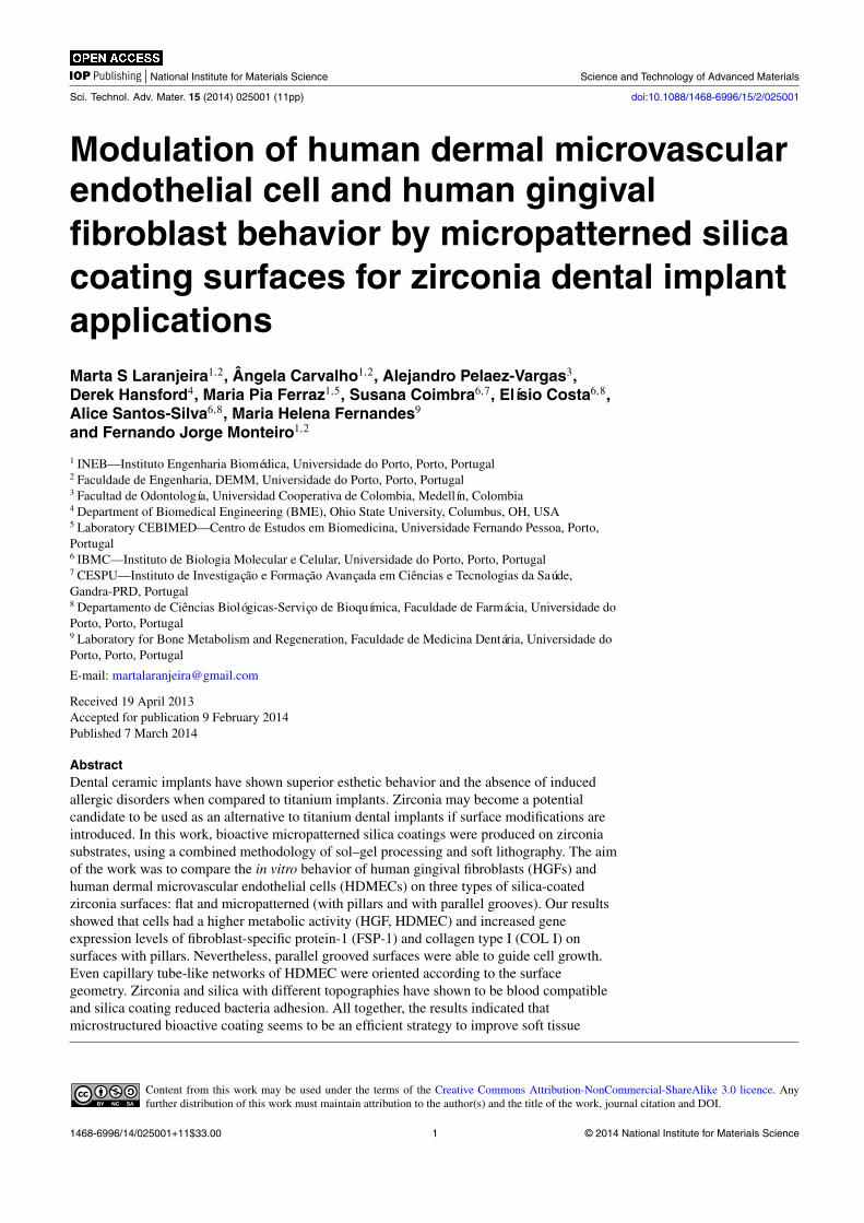
Figure 5. Inverted fluorescence microscopy images of HFGs and HDMECs after 7 days of culture, on flat, grooved and pillar surfaces.Scale bar = 50 µm.
A B C
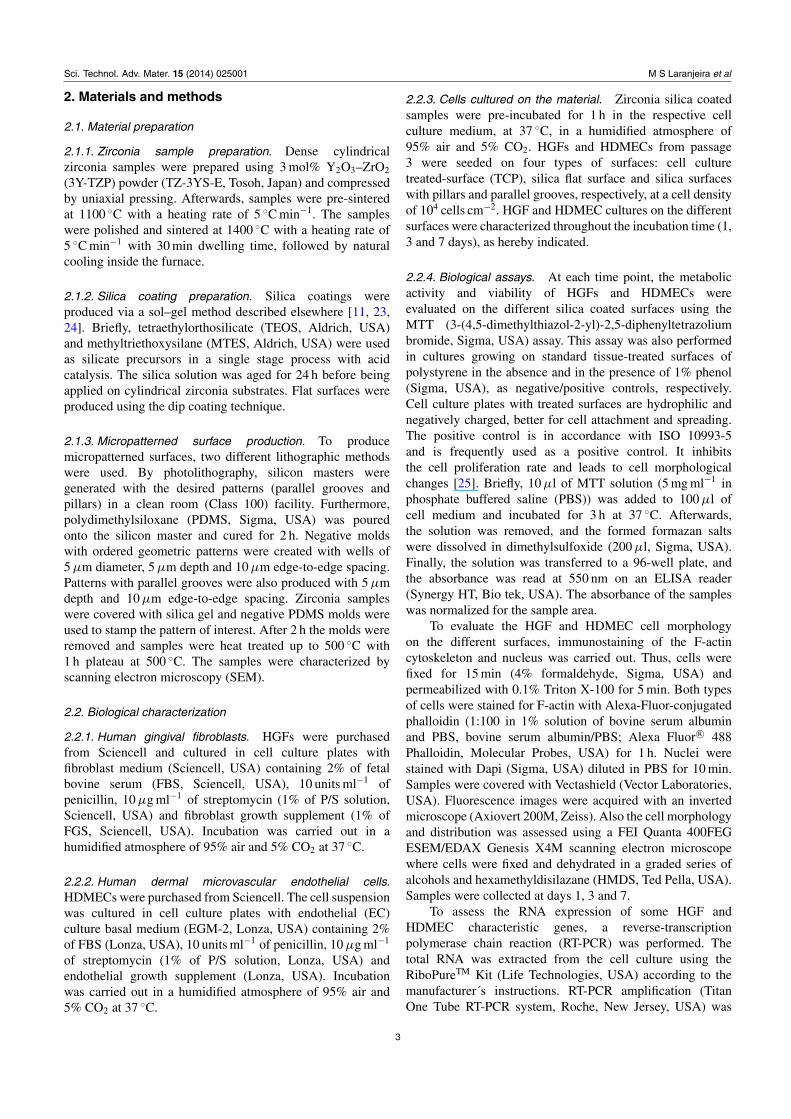
Figure 6. Matrigel assay images of capillary-like tube formation of HDMECs after 3 days of culture, on flat (a), grooved surfaces (b) andpillars (c). The direction of the parallel microtopographic features is indicated by the white arrow. Scale bar = 100 µm.
HDMECs on all the studied surfaces (figure 3). Cultureswere assessed for the genes fibroblast-specific protein-1(FSP-1) and collagen-type 1 (COL-1) and genes expressed byfibroblast cells and von Willebrand factor (vWF) and CD31as characteristic endothelial cell markers. Expression of thesegenes was evaluated at day 7, the same time point wherethe higher metabolic activity and cell viability was achieved.HDMECs were able to express vWF and CD31 for all thesurfaces. Regarding HGF culture, these cells expressed FSP-1and COL1 for all the surfaces with a significantly higherexpression in pillar surfaces (figure 3(b)).
Collagen type 1 is a very important protein that is presentin the extracellular matrix and shows different chemistry andphysical cues that can dictate cell response. It is not onlythe diffusible factors that are important for cell behaviorbut also the adhesive and mechanical interactions with thematerial [2, 31]. Cell behavior can be modulated by thesurface roughness and topography via integrin receptors.Integrin expression can be different according to the cellstate of differentiation or the type of surface where cellsare seeded. The activation of integrin receptors leads toa series of reactions (signal transduction cascades) [2].The activation of signaling pathways can lead to different
synergistic responses. For example, it was described thatsurface roughness increases MG63 expression levels ofprostaglandins which are necessary for normal osteoblastdifferentiation [31].
We hypothesized that a well-defined geometricalsurface could promote cell organization and guided growthdepending on the geometric pattern in question and thecell type. Furthermore, previous studies showed that thecell shape and size can influence the MSC differentiationfate [32]. Microtextured substrates with different geometriesinfluence cytoskeletal dynamics and motility as well as thefocal adhesion localization/distribution [3, 33]. The degreeof cytoskeletal tension affects cell regulatory mechanismsof mechanically stimulated differentiation, not yet explained[32, 34]. Furthermore, previous studies showed thatcytoskeletal tension regulates the cell shape and promoteshigher metabolic activity as well as overexpression of someproteins required for cell functionality [35].
Also, previous studies on endothelial cells showedthat this type of cell responds to ridge-groove surfacesthrough alignment (contact guidance), elongation, reducedproliferation and enhanced migration compared with smoothsurfaces [2, 36]. However, Uttayarat et al [3] cultured bovine
7
Sci. Technol. Adv. Mater. 15 (2014) 025001 M S Laranjeira et al
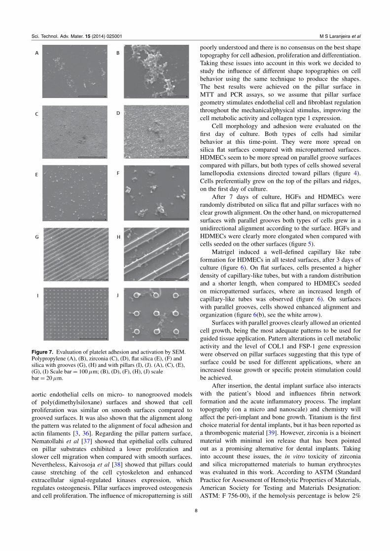
Figure 7. Evaluation of platelet adhesion and activation by SEM.Polypropylene (A), (B), zirconia (C), (D), flat silica (E), (F) andsilica with grooves (G), (H) and with pillars (I), (J). (A), (C), (E),(G), (I) Scale bar = 100 µm; (B), (D), (F), (H), (J) scalebar = 20 µm.
aortic endothelial cells on micro- to nanogrooved modelsof poly(dimethylsiloxane) surfaces and showed that cellproliferation was similar on smooth surfaces compared togrooved surfaces. It was also shown that the alignment alongthe pattern was related to the alignment of focal adhesion andactin filaments [3, 36]. Regarding the pillar pattern surface,Nematollahi et al [37] showed that epithelial cells culturedon pillar substrates exhibited a lower proliferation andslower cell migration when compared with smooth surfaces.Nevertheless, Kaivosoja et al [38] showed that pillars couldcause stretching of the cell cytoskeleton and enhancedextracellular signal-regulated kinases expression, whichregulates osteogenesis. Pillar surfaces improved osteogenesisand cell proliferation. The influence of micropatterning is still
poorly understood and there is no consensus on the best shapetopography for cell adhesion, proliferation and differentiation.Taking these issues into account in this work we decided tostudy the influence of different shape topographies on cellbehavior using the same technique to produce the shapes.The best results were achieved on the pillar surface inMTT and PCR assays, so we assume that pillar surfacegeometry stimulates endothelial cell and fibroblast regulationthroughout the mechanical/physical stimulus, improving thecell metabolic activity and collagen type 1 expression.
Cell morphology and adhesion were evaluated on thefirst day of culture. Both types of cells had similarbehavior at this time-point. They were more spread onsilica flat surfaces compared with micropatterned surfaces.HDMECs seem to be more spread on parallel groove surfacescompared with pillars, but both types of cells showed severallamellopodia extensions directed toward pillars (figure 4).Cells preferentially grew on the top of the pillars and ridges,on the first day of culture.
After 7 days of culture, HGFs and HDMECs wererandomly distributed on silica flat and pillar surfaces with noclear growth alignment. On the other hand, on micropatternedsurfaces with parallel grooves both types of cells grew in aunidirectional alignment according to the surface. HGFs andHDMECs were clearly more elongated when compared withcells seeded on the other surfaces (figure 5).
Matrigel induced a well-defined capillary like tubeformation for HDMECs in all tested surfaces, after 3 days ofculture (figure 6). On flat surfaces, cells presented a higherdensity of capillary-like tubes, but with a random distributionand a shorter length, when compared to HDMECs seededon micropatterned surfaces, where an increased length ofcapillary-like tubes was observed (figure 6). On surfaceswith parallel grooves, cells showed enhanced alignment andorganization (figure 6(b), see the white arrow).
Surfaces with parallel grooves clearly allowed an orientedcell growth, being the most adequate patterns to be used forguided tissue application. Pattern alterations in cell metabolicactivity and the level of COL1 and FSP-1 gene expressionwere observed on pillar surfaces suggesting that this type ofsurface could be used for different applications, where anincreased tissue growth or specific protein stimulation couldbe achieved.
After insertion, the dental implant surface also interactswith the patient’s blood and influences fibrin networkformation and the acute inflammatory process. The implanttopography (on a micro and nanoscale) and chemistry willaffect the peri-implant and bone growth. Titanium is the firstchoice material for dental implants, but it has been reported asa thrombogenic material [39]. However, zirconia is a bioinertmaterial with minimal ion release that has been pointedout as a promising alternative for dental implants. Takinginto account these issues, the in vitro toxicity of zirconiaand silica micropatterned materials to human erythrocyteswas evaluated in this work. According to ASTM (StandardPractice for Assessment of Hemolytic Properties of Materials,American Society for Testing and Materials Designation:ASTM: F 756-00), if the hemolysis percentage is below 2%
8

Sci. Technol. Adv. Mater. 15 (2014) 025001 M S Laranjeira et al
Figure 8. Platelet counts (A); APTT and PT (B), (C) for negative control (polypropylene membrane), zirconia, silica flat andmicropatterned (grooves and pillars) surfaces.
the material is considered as non-hemolytic, if between 2 and5% it is considered as slightly hemolytic and if higher than5% it is considered as hemolytic. Zirconia samples presenteda hemolysis percentage of 0.15 ± 0.04% very similar tosilica flat and micropatterned (grooves and pillars) surfaceswhich had the percentages of 0.16 ± 0.04, 0.15 ± 0.05 and0.16 ± 0.03%, respectively. All samples showed no hemolyticactivity, since the percentage of hemolysis was less than 1%and similar to that found in the negative control (0.16 ±
0.03%).Platelet adhesion and activation and coagulation cascade
activation assessments are very important to evaluate thebiomaterial blood compatibility, namely to evaluate theprobability of thrombus formation. As shown in figures 7and 8 the platelet counts were similar in PRP incubated withdifferent biomaterials, including the polypropylene used asa non-thrombogenic control material. SEM evaluation of thematerial surface showed that platelets adhere to all materials,in small groups and limited to small areas of the samplesurface (figure 7). We can also see that platelets presenteda flattened morphology with some pseudopodial processes.This extended morphology of platelets has been demonstratedas an indicator of platelet activation; however, similar resultswere found in all tested materials and in polypropyleneused as a non-thrombogenic control material. Moreover, ascreening for coagulation cascade activation was performedby measuring TP and APTT in the plasma samples incubatedwith the different materials, and no alterations were found(figures 8(A) and (B)). These results suggest that zirconia andsilica with different morphologies are blood compatible andnon-thrombogenic materials, as the results are similar to thatfound for polypropylene material.
Dental caries and periodontal diseases have been relatedto bacterial accumulation. The early colonizer bacteria arethe first to adhere to enamel or to implants and provideattachment substrates for the next colonizers, influencingthe subsequent biofilm formation. This type of bacteriaspecies plays an important role in dental biofilm formationwhich is decisive in determining oral health or disease [40].Several Streptoccoccus species such as S. sanguinis, S. oralis,S. gordonii, S. mitis and S. sobrinus constitute 60–80% ofthe early colonizers [41]. For this reason S. sobrinus waschosen to test the bacteria adhesion to zirconia and silica
Figure 9. SEM images show bacterial morphology and distributionon zirconia (A), (B), flat silica (C), (D), silica with grooves (E), (F)and silica with pillars (G), (H). (A), (C), (E), (G) Scalebar = 40 µm; (B), (D), (F), (H) scale bar = 10 µm.
micropatterned surfaces. Our results showed that after 90 minof incubation, bacteria were able to adhere and grow onall types of surfaces, maintaining their morphology andorganizing themselves in small clusters (figure 9). S. sobrinuswas able to colonize the top of ridges and pillars, being
9

Sci. Technol. Adv. Mater. 15 (2014) 025001 M S Laranjeira et al
Figure 10. Bacterial adhesion on zirconia, flat silica, silica withgrooves and silica with pillars. Values reported are the mean±SD.Assays were performed after 90 min of culture; ∗ indicatessignificant statistical difference (p < 0.05; n = 3).
present in a higher number in the space between pillarssince they had more area available to grow (figure 10).Zirconia samples showed significantly higher values ofadherent bacteria when compared to silica flat and groovedsurfaces (figure 10) what might be related to the higherporosity and irregular morphology previously observed inzirconia samples [11]. Moreover, previous studies showedthat bacteria adhere preferentially to more irregular surfacemorphologies, in order to maximize the bacteria surface area.Furthermore, more roughness surface areas leave a highersurface area available for their adhesion and protection fromshear forces [36]. Nevertheless, Rimondini et al [42] havereported an in vivo study showing that zirconia stabilized withyttria accumulated less bacteria when compared to titanium.Silica flat and micropatterned cover did not significantlyincrease the number of adherent bacteria compared to thezirconia substrate. These last results, once more, reinforcedthe beneficial potential use of the developed materials fordental implant applications.
4. Conclusions
In this work, reproducible materials were produced usinga combined methodology of sol–gel and soft lithography.Different surface topographies with pillar and groovepatterns were obtained on silica thin films coating zirconiasubstrates. HGFs and HDMECs were able to adhere,migrate and proliferate on the micropatterned coatings. Themicropatterned surfaces were able to modulate the cellresponse, playing a crucial role in orientation of cell growth,metabolic activity and expression of proteins, which areimportant for the cell functionality. Zirconia and silica withdifferent morphologies were shown to be blood compatible.Silica coatings reduce bacterial adhesion compared to zirconiasubstrates. Our results suggest that the newly developedmaterials can improve dental implants.
Acknowledgments
The authors would like to thank FCT—Fundaçãopara a Ciência e Tecnologia (Portugal) for grant
FCT/PTDC/CTM/100120/2008 ‘Bonamidi’ and NSEC(Nanoscale Science and Engineering Center, USA). This workwas also financed by FEDER funds through the ProgramaOperacional Factores de Competitividade—COMPETEand by Portuguese funds through FCT—Fundação paraa Ciência e a Tecnologia in the framework of the projectPEst-C/SAU/LA0002/2011.
References
[1] Zhou F, Yuan L, Huang H and Chen H 2009 Phenomenon of‘contact guidance’ on the surface withnano-micro-groove-like pattern and cell physiologicaleffects Chin. Sci. Bull. 54 3200–5
[2] Bettinger C J, Zhang Z, Gerecht S, Borenstein J T andLanger R 2008 Enhancement of in vitro capillary tubeformation by substrate nanotopography Adv. Mater.20 99–103
[3] Uttayarat P, Toworfe G K, Dietrich F, Lelkes P I andComposto R J 2005 Topographic guidance of endothelialcells on silicone surfaces with micro-to nanogrooves:orientation of actin filaments and focal adhesions J. Biomed.Mater. Res. A 75 668–80
[4] Kolind K, Leong K W, Besenbacher F and Foss M 2012Guidance of stem cell fate on 2D patterned surfacesBiomaterials 33 6626–33
[5] Gerberich B G and Bhatia S K 2013 Tissue scaffold surfacepatterning for clinical applications Biotechnol. J. 8 73–84
[6] Özcan M and Hämmerle C 2012 Titanium as a reconstructionand implant material in dentistry: advantages and pitfallsMaterials 5 1528–45
[7] Werner S et al 2009 The effect of microstructured surfaces andlaminin-derived peptide coatings on soft tissue interactionswith titanium dental implants Biomaterials 30 2291–301
[8] Sicilia A et al 2008 Titanium allergy in dental implantpatients: a clinical study on 1500 consecutive patients. Clin.Oral Impl. Res. 19 823–35
[9] Egusa H, Ko N, Shimazu T and Yatani H 2008 Suspectedassociation of an allergic reaction with titanium dentalimplants: a clinical report J. Prosthet. Dent. 100 344–7
[10] Gahlert M, Röhling S, Wieland M, Sprecher C M, Kniha Hand Milz S 2009 Osseointegration of zirconia and titaniumdental implants: a histological and histomorphometricalstudy in the maxilla of pigs Clin. Oral Impl. Res.20 1247–53
[11] Pelaez-Vargas A et al 2011 Isotropic micropatterned silicacoatings on zirconia induce guided cell growth for dentalimplants Dent. Mater. 27 581–9
[12] Adatia N D, Bayne S C, Cooper L F and Thompson J Y 2009Fracture resistance of yttria-stabilized zirconia dentalimplant abutments J. Prosthodont. 18 17–22
[13] Manicone P F, Rossi Iommetti P and Raffaelli L 2007 Anoverview of zirconia ceramics: basic properties and clinicalapplications J. Dent. 35 819–26
[14] Sundh A, Molin M and Sjögren G 2005 Fracture resistance ofyttrium oxide partially-stabilized zirconia all-ceramicbridges after veneering and mechanical fatigue testing Dent.Mater. 21 476–82
[15] Att W, Grigoriadou M and Strub J 2007 ZrO2 three-unit fixedpartial dentures: comparison of failure load before and afterexposure to a mastication simulator J. Oral Rehabil.34 282–90
[16] Dion I et al 1994 Physico-chemistry and cytotoxicity ofceramics J. Mater. Sci.: Mater. Med. 5 18–24
[17] Josset Y, Oum’Hamed Z, Zarrinpour A, Lorenzato M,Adnet J-J and Laurent-Maquin D 1999 In vitro reactions ofhuman osteoblasts in culture with zirconia and aluminaceramics J. Biomed. Mater. Res. 47 481–93
10

Sci. Technol. Adv. Mater. 15 (2014) 025001 M S Laranjeira et al
[18] Pelaez-Vargas A et al 2012 Micropatterned coatings for guidedtissue regeneration in dental implantology Cell Interactioned S Gowder (Intech) ch 11
[19] An N, Schedle A, Wieland M, Andrukhov O, Matejka M andRausch-Fan X 2010 Proliferation, behavior, and cytokinegene expression of human umbilical vascular endothelialcells in response to different titanium surfaces J. Biomed.Mater. Res. A 93 364–72
[20] Myshin H L and Wiens J P 2005 Factors affecting soft tissuearound dental implants: a review of the literature J. Prosthet.Dent. 94 440–4
[21] Moule A J, Li H and Bartold P M 1995 Donor variability inthe proliferation of human dental pulp fibroblasts Aust.Dent. J. 40 110–4
[22] Darveau R P 2010 Periodontitis: a polymicrobial disruption ofhost homeostasis Nature Rev. Micro. 8 481–90
[23] Durán A, Conde A, Coedo A G, Dorado T, García C andCeré S 2004 Sol–gel coatings for protection andbioactivation of metals used in orthopaedic devices.J. Mater. Chem. 14 2282–90
[24] Garcia C, Cere S and Duran A 2004 Bioactive coatingsprepared by sol–gel on stainless steel 316L J. Non-Cryst.Solids 348 218–24
[25] Illeperuma R P et al 2012 Immortalized gingival fibroblasts asa cytotoxicity test model for dental materials J. Mater. Sci.,Mater. Med. 23 753–62
[26] Aubin H et al 2010 Directed 3D cell alignment and elongationin microengineered hydrogels Biomaterials 31 6941–51
[27] Lu J, Rao M P, MacDonald N C, Khang D and Webster T J2008 Improved endothelial cell adhesion and proliferationon patterned titanium surfaces with rationally designed,micrometer to nanometer features Acta Biomater. 4 192–201
[28] Nikkhah M, Edalat F, Manoucheri S and Khademhosseini A2012 Engineering microscale topographies to control thecell–substrate interface Biomaterials 33 5230–46
[29] Pelaez-Vargas A, Gallego-Perez D, Carvalho A,Fernandes M H, Hansford D J and Monteiro F J 2013Effects of density of anisotropic microstamped silica thinfilms on guided bone tissue regeneration—in vitro studyJ. Biomed. Mater. Res. B, Appl. Biomater. 101B 762–9
[30] Carvalho A et al 2012 Micropatterned silica thin films withnanohydroxyapatite micro-aggregates for guided tissueregeneration Dent. Mater. 28 1250–60
[31] D’Lima D D, Lemperle S M, Chen P C, Holmes R E andColwell C W Jr 1998 Bone response to implant surfacemorphology J. Arthroplasty. 13 928–34
[32] Kilian K A, Bugarija B, Lahn B T and Mrksich M 2010Geometric cues for directing the differentiation ofmesenchymal stem cells Proc. Natl Acad. Sci. USA107 4872–7
[33] von der Mark K, Park J, Bauer S and Schmuki P 2010Nanoscale engineering of biomimetic surfaces: cues fromthe extracellular matrix Cell Tissue Res. 339 131–53
[34] Sunters A et al 2010 Mechano-transduction in osteoblasticcells involves strain-regulated estrogen receptor α-mediatedcontrol of insulin-like growth factor (IGF) I receptorsensitivity to ambient IGF, leading to phosphatidylinositol3-kinase/AKT-dependent Wnt/LRP5 receptor-independentactivation of β-catenin signaling J. Bio. Chem. 285 8743–58
[35] Mendonça G et al 2009 The effects of implant surfacenanoscale features on osteoblast-specific gene expressionBiomaterials 30 4053–62
[36] Anselme K, Davidson P, Popa A, Giazzon M, Liley M andPloux L 2010 The interaction of cells and bacteria withsurfaces structured at the nanometre scale Acta Biomater.6 3824–46
[37] Nematollahi M, Hamilton D, Jaeger N and Brunette D 2009Hexagonal micron scale pillars influence epithelial celladhesion, morphology, proliferation, migration, andcytoskeletal arrangement J. Biomed. Mater. Res. A91 149–57
[38] Kaivosoja E et al 2013 Cell adhesion and osteogenicdifferentiation on three-dimensional pillar surfacesJ. Biomed. Mater. Res. A 101 842–52
[39] Traini T, Caputi S, Gherlone E, Degidi M and Piattelli A 2013Fibrin clot extension on zirconia surface for dental implants:a quantitative in vitro study Clin. Impl. Dent. Relat. Res.at press
[40] Li J et al 2004 Identification of early microbial colonizers inhuman dental biofilm J. Appl. Microbiol. 97 1311–8
[41] Avila M, Ojcius D M and Yilmaz Ö 2009 The oral microbiota:living with a permanent guest DNA Cell Biol. 28 405–11
[42] Rimondini L, Cerroni L, Carrassi A and Torricelli P 2002Bacterial colonization of zirconia ceramic surfaces: anin vitro and in vivo study Int. J. Oral Maxillofac. Impl.17 793–8
11
Related Documents