Modulating the cytocompatibility of tridimensional carbon nanotube-based scaffolds† Stefania Nardecchia, Mar´ ıa Concepci´ on Serrano, * Mar´ ıa Concepci´ on Guti´ errez, Mar´ ıa Luisa Ferrer and Francisco del Monte * Carbon nanotubes (CNTs) have lately attracted significant attention in the field of biomedicine. Although a wide repertoire of CNT-based composites has been explored as substrates for cell growth, the fabrication of 3D scaffolds has been more rarely accomplished. Additionally, concerns referred to CNT biocompatibility make their use in biomaterials still controversial. Herein we explore the interaction of three types of CNT-based 3D scaffolds – prepared with multi-walled CNTs and processed to show different architectural and morphological features at the microscale by using three different polymers (i.e., chitosan, chondroitin sulphate and gelatin) – with three types of mammalian cells displaying different sizes and adhesion patterns. Cell–material interaction has been assessed by studying cell viability, adhesion, morphology, and apoptosis. By means of time-lapse confocal laser scanning microscopy, we investigate, for the first time in CNT-based scaffolds, cell migration processes in real time. Scaffolds displaying both a pore size in range with that of cells and lower surface roughness reveal the highest viability values. In contrast, those with a smaller pore size and higher surface roughness account for the lowest cytocompatibility. Results from these studies benefit the fabrication of optimized biomaterials by varying scaffold-dependent parameters in accordance with those of target cells. Furthermore, they may serve to anticipate the response of other cell types sharing similar characteristics to those described herein when in contact with CNT-based scaffolds. 1 Introduction Carbon nanotubes (CNTs) are dened as hollow cylinders exclusively composed of graphitic carbon sheets generally rolled as either single-walled structures (SWCNTs) or multi-walled structures (MWCNTs). Since their discovery by Iijima and others, 1,2 an extensive progress has been done in an attempt to use CNTs as brick elements for the development of advanced materials with remarkable electrical, thermal and mechanical properties. In this sense, the fabrication of diverse CNT-based composites has opened their use in a wide repertoire of appli- cations, from exible electrodes in displays to high-perfor- mance composites for aircra and automotive industries. 3,4 CNTs have also attracted signicant attention in the emerging eld of nanobiotechnology, thus being already exploited for the preparation of biosensors, 5 molecular transporters for drug, gene and protein delivery, 6 and fuel-powered articial muscles. 7 In the area of tissue engineering, 8–11 they have been extensively explored for bone regeneration 12 and nerve tissue repair. 13 In this context, a wide repertoire of methodologies has been explored and optimized for the preparation of 2D CNT-based composites (e.g., lms and bers). 3 However, the fabrication of 3D architectures has been more rarely accomplished, with some approaches including chitosan-based matrices, 14–18 gelatin and/ or methacrylate hydrogels 19,20 or collagen scaffolds. 21 Further- more, diverse polymeric systems have been exploited for CNT dispersion, such as poly(lactic-co-glycolic acid) 22 or poly(methyl methacrylates). 23 Nanomaterial-induced cytotoxicity has been related to size/ mass, shape, surface charge, and functionalization, 24 with both size-dependent 25 and composition-dependent 26 toxicity mecha- nisms. In the particular case of CNTs, an increasing number of studies have focused on their use as “brick” elements of 2D and 3D materials envisioned for biomedical applications. 27–31 Nevertheless, the toxicity of CNTs remains controversial. 32 In order to bring some insights into the still uncertain interaction of 3D structures composed of CNTs with mammalian cells, we explored herein the interaction of three cell types (i.e., murine broblasts, human bone sarcoma cells and porcine endothelial progenitor cells) with MWCNT-based 3D scaffolds that showed different architectural and morphological features at the microscale. The introduction of MWCNTs in these structures not only allowed the acquisition of 3D architectures, but also conferred superior mechanical properties and electrical Instituto de Ciencia de Materiales de Madrid (ICMM), Consejo Superior de Investigaciones Cient´ ıcas (CSIC), Calle Sor Juana In´ es de la Cruz 3, 28049-Madrid, Spain. E-mail: [email protected]; [email protected]; Fax: +34 91 3720623; Tel: +34 91 3349000 † Electronic supplementary information (ESI) available. See DOI: 10.1039/c3tb20253d Cite this: J. Mater. Chem. B, 2013, 1, 3064 Received 20th February 2013 Accepted 26th April 2013 DOI: 10.1039/c3tb20253d www.rsc.org/MaterialsB 3064 | J. Mater. Chem. B, 2013, 1, 3064–3072 This journal is ª The Royal Society of Chemistry 2013 Journal of Materials Chemistry B PAPER Published on 26 April 2013. Downloaded by Pontifícia Universidade Catolica do Rio de Janeiro on 10/07/2013 16:09:26. View Article Online View Journal | View Issue

Welcome message from author
This document is posted to help you gain knowledge. Please leave a comment to let me know what you think about it! Share it to your friends and learn new things together.
Transcript

Journal ofMaterials Chemistry B
PAPER
Publ
ishe
d on
26
Apr
il 20
13. D
ownl
oade
d by
Pon
tifíc
ia U
nive
rsid
ade
Cat
olic
a do
Rio
de
Jane
iro
on 1
0/07
/201
3 16
:09:
26. View Article Online
View Journal | View Issue
Instituto de Ciencia de Materiales de
Investigaciones Cientıcas (CSIC), Calle Sor
Spain. E-mail: [email protected]
3720623; Tel: +34 91 3349000
† Electronic supplementary informa10.1039/c3tb20253d
Cite this: J. Mater. Chem. B, 2013, 1,3064
Received 20th February 2013Accepted 26th April 2013
DOI: 10.1039/c3tb20253d
www.rsc.org/MaterialsB
3064 | J. Mater. Chem. B, 2013, 1, 30
Modulating the cytocompatibility of tridimensionalcarbon nanotube-based scaffolds†
Stefania Nardecchia, Marıa Concepcion Serrano,* Marıa Concepcion Gutierrez,Marıa Luisa Ferrer and Francisco del Monte*
Carbon nanotubes (CNTs) have lately attracted significant attention in the field of biomedicine. Although a
wide repertoire of CNT-based composites has been explored as substrates for cell growth, the fabrication of
3D scaffolds has been more rarely accomplished. Additionally, concerns referred to CNT biocompatibility
make their use in biomaterials still controversial. Herein we explore the interaction of three types of
CNT-based 3D scaffolds – prepared with multi-walled CNTs and processed to show different architectural
and morphological features at the microscale by using three different polymers (i.e., chitosan,
chondroitin sulphate and gelatin) – with three types of mammalian cells displaying different sizes and
adhesion patterns. Cell–material interaction has been assessed by studying cell viability, adhesion,
morphology, and apoptosis. By means of time-lapse confocal laser scanning microscopy, we investigate,
for the first time in CNT-based scaffolds, cell migration processes in real time. Scaffolds displaying both a
pore size in range with that of cells and lower surface roughness reveal the highest viability values. In
contrast, those with a smaller pore size and higher surface roughness account for the lowest
cytocompatibility. Results from these studies benefit the fabrication of optimized biomaterials by
varying scaffold-dependent parameters in accordance with those of target cells. Furthermore, they may
serve to anticipate the response of other cell types sharing similar characteristics to those described
herein when in contact with CNT-based scaffolds.
1 Introduction
Carbon nanotubes (CNTs) are dened as hollow cylindersexclusively composed of graphitic carbon sheets generally rolledas either single-walled structures (SWCNTs) or multi-walledstructures (MWCNTs). Since their discovery by Iijima andothers,1,2 an extensive progress has been done in an attempt touse CNTs as brick elements for the development of advancedmaterials with remarkable electrical, thermal and mechanicalproperties. In this sense, the fabrication of diverse CNT-basedcomposites has opened their use in a wide repertoire of appli-cations, from exible electrodes in displays to high-perfor-mance composites for aircra and automotive industries.3,4
CNTs have also attracted signicant attention in the emergingeld of nanobiotechnology, thus being already exploited for thepreparation of biosensors,5 molecular transporters for drug,gene and protein delivery,6 and fuel-powered articial muscles.7
In the area of tissue engineering,8–11 they have been extensivelyexplored for bone regeneration12 and nerve tissue repair.13 In
Madrid (ICMM), Consejo Superior de
Juana Ines de la Cruz 3, 28049-Madrid,
s; [email protected]; Fax: +34 91
tion (ESI) available. See DOI:
64–3072
this context, a wide repertoire of methodologies has beenexplored and optimized for the preparation of 2D CNT-basedcomposites (e.g., lms and bers).3 However, the fabrication of3D architectures has been more rarely accomplished, with someapproaches including chitosan-based matrices,14–18 gelatin and/or methacrylate hydrogels19,20 or collagen scaffolds.21 Further-more, diverse polymeric systems have been exploited for CNTdispersion, such as poly(lactic-co-glycolic acid)22 or poly(methylmethacrylates).23
Nanomaterial-induced cytotoxicity has been related to size/mass, shape, surface charge, and functionalization,24 with bothsize-dependent25 and composition-dependent26 toxicity mecha-nisms. In the particular case of CNTs, an increasing number ofstudies have focused on their use as “brick” elements of 2D and3D materials envisioned for biomedical applications.27–31
Nevertheless, the toxicity of CNTs remains controversial.32 Inorder to bring some insights into the still uncertain interactionof 3D structures composed of CNTs with mammalian cells, weexplored herein the interaction of three cell types (i.e., murinebroblasts, human bone sarcoma cells and porcine endothelialprogenitor cells) with MWCNT-based 3D scaffolds that showeddifferent architectural and morphological features at themicroscale. The introduction of MWCNTs in these structuresnot only allowed the acquisition of 3D architectures, but alsoconferred superior mechanical properties and electrical
This journal is ª The Royal Society of Chemistry 2013

Fig. 1 SEM images of LNCHI (A) and SNCHI (B) scaffolds, along with their 3D
Paper Journal of Materials Chemistry B
Publ
ishe
d on
26
Apr
il 20
13. D
ownl
oade
d by
Pon
tifíc
ia U
nive
rsid
ade
Cat
olic
a do
Rio
de
Jane
iro
on 1
0/07
/201
3 16
:09:
26.
View Article Online
conductivity to the resulting scaffolds. These matrices wereobtained by the application of the ice segregation induced self-assembly (ISISA) process into MWCNT aqueous suspensionscontaining different polymers (i.e., chitosan, chondroitinsulphate and gelatin) that acted as dispersant agents.33 Thechemical nature of every polymer contributed to the formationof a particular patterned porous structure.34–36 Besides the use ofdifferent polymers, the scaffold structure was modied by theincorporation of two different types of functionalized MWCNTs(i.e., long MWCNTs and short MWCNTs; abbreviated as LN andSN, respectively), as well as by the application of two differenttemperatures during the ISISA process (i.e., �196 and �65 �C).Cell–material interaction was assessed by studying cell viability,adhesion, morphology, and apoptosis. Time-lapse confocallaser scanning microscopy (CLSM) was also used to explore, forthe rst time in CNT-based scaffolds, cell migration processesin real time.
reconstructed images (C and D, respectively). Scale bars: 10 mm (A) and 20 mm (B).
Fig. 2 SEM images of LNCHO (A), SNCHO (B), LNGEL (C), and SNGEL (D) scaffolds.3D reconstructed image from LNGEL (E). Scale bars: 20 mm (A and B) and 10 mm(C and D).
2 Results and discussion2.1 Scaffold characterization
CNT-based scaffolds displaying different architectural andmorphological features were obtained by the application of theISISA process, a simple and versatile bottom-up techniquebased on the unidirectional immersion of an aqueous solution,a colloidal suspension or even a hydrogel into a liquid nitrogenbath and subsequent freeze-drying. This process has demon-strated its suitability for the preparation of inorganic, organicand hybrid macroporous monoliths and bers.37–39 In thisparticular case, we applied the ISISA process to MWCNTaqueous suspensions that were prepared using different poly-mers as dispersant agents. The polymers of choice were chito-san (CHI),40 chondroitin sulphate (CHO),41,42 and gelatin(GEL),43 all well-known biocompatible polymers. For scaffoldcross-linking, we used chemical agents that have also beenextensively explored for the fabrication of biocompatiblematerials (e.g., glutaraldehyde and hexamethylene diisocya-nate).44,45 Besides the use of different polymers, the scaffoldmorphology was modied by the incorporation of two differenttypes of MWCNTs. The rst type, dened as LN, was obtainedupon acid treatment with HNO3 that resulted in MWCNTfunctionalization with carboxylic groups without signicantmodication of their original length (i.e., 5–9 mm). For thesecond type (i.e., SN), MWCNTs were exposed to a moreaggressive acid treatment with a mixture of HNO3 and H2SO4
that not only promoted the functionalization with carboxylicgroups, but also shortened their original length to ca. 3 mm. Theresulting six scaffolds (i.e., LNCHI, SNCHI, LNCHO, SNCHO,LNGEL, and SNGEL) exhibited a homogeneous structurethroughout the entire monolith that differed depending on theparticular polymer and MWCNTs used (Fig. 1, ESI†). Thus, theuse of CHI and CHO promoted the formation of scaffolds with awell-patterned cross-sectional structure, whereas those scaf-folds prepared from GEL suspensions exhibited one with amore disordered pattern (Fig. 1 and 2). We also prepared anadditional type of LNCHI scaffold where both the pore diameterand the width of the walls that support the porous structure
This journal is ª The Royal Society of Chemistry 2013
were modied by the application of the ISISA process at �65 �C(rather than �196 �C) (Fig. 3).46 These scaffolds were abbrevi-ated as LNCHI-65.
The morphology of the different scaffolds also differed interms of porosity and surface roughness (Table 1). In regard toporosity, we analyzed from SEMmicrographs of cross-sectionedscaffolds the pore area per cross-sectioned area (AP) and theaveraged pore width (WP). The most signicant differences werefound between LNCHI-65 and the rest of scaffolds because ofthe well-known inuence that the freezing temperature has onscaffold morphology.46 Nonetheless, AP and WP of scaffoldsprepared at�196 �C from different polymers also experienced anoticeable change, both parameters decreasing from CHI– toCHO– up to GEL-based scaffolds. Finally, SN-type scaffolds also
J. Mater. Chem. B, 2013, 1, 3064–3072 | 3065

Fig. 3 SEM images showing the macroporous structure of LNCHI-65 (A) and adetail of MWCNTs accommodating within scaffold walls (B). 3D reconstructedimages from (A) and (B) are shown (C and D, respectively). Scale bars: 40 mm (A)and 5 mm (B).
Fig. 4 Light optical microscopy images of L929 fibroblasts (A and B), Saos-2osteoblasts (C and D) and ECPC cells (E and F). Cell size was estimated from the celllength (l) and width (w), as indicated in representative cells (white arrows). Scalebars: 20 mm.
Journal of Materials Chemistry B Paper
Publ
ishe
d on
26
Apr
il 20
13. D
ownl
oade
d by
Pon
tifíc
ia U
nive
rsid
ade
Cat
olic
a do
Rio
de
Jane
iro
on 1
0/07
/201
3 16
:09:
26.
View Article Online
exhibited signicant differences in both parameters ascompared to their respective LN-type analogues. Interestingly,AP increased for every SN-type scaffold whereasWP increased forSNCHI and SNCHO, but decreased for SNGEL.
When analyzing the roughness of the walls that dene thecross-sectioned structures on which cells were cultured(Fig. 1–3, and Table 1), the root mean square (RMS) roughnessincreased from scaffolds with a well-patterned cross-sectionstructure (i.e., CHI and CHO) to those with a disordered one(i.e., GEL). This behavior was ascribed to both the dimensions ofMWCNTs and their rigid nature, which determined a betteraccommodation of MWCNTs within the walls of LNCHI andLNCHO rather than on LNGEL scaffolds. The relevance of thedimensions of MWCNTs and their rigid nature on this param-eter was corroborated on SN-type scaffolds. In this case, RMSroughness experienced a signicant decrease in every scaffoldbecause the use of shorter MWCNTs was obviously facilitatingtheir accommodation and the subsequent formation of morecompact walls. Additionally, the dimensions of the walls alsoplayed a role as lower RMS roughness values were found in thethick walls of LNCHI-65 scaffolds than in the thinner ones ofLNCHI.
Table 1 Scaffold characterization including porosity, surface roughness, mechanic
ScaffoldPore areaper mm2 (AP)
Pore width(WP) (mm)
LNCHI 0.60 � 0.14 1.38 � 2.8LNCHI-65 0.69 � 0.05 62.8 � 43.9SNCHI 0.69 � 0.04 21.3 � 4.4LNCHO 0.51 � 0.03 8.9 � 2.7SNCHO 0.58 � 0.03 9.4 � 3.0LNGEL 0.45 � 0.13 7.4 � 3.6SNGEL 0.51 � 0.13 5.8 � 1.8
3066 | J. Mater. Chem. B, 2013, 1, 3064–3072
Finally, the mechanical properties and conductivity of thedifferent scaffolds were found to be strongly inuenced by theabove-mentioned differences in architecture and morphology(Table 1). For instance, GEL-based scaffolds showed the highestYoung's modulus and the lowest conductivity likely because of amore decient connection between MWCNTs in their poorlycompacted walls.
2.2 Cell type rationale and size considerations
We rst used murine L929 broblasts (Fig. 4A and B) as areference cell line for preliminary biocompatibility studies invitro.47 They are considered as slow-moving cells with a typicallyelongated morphology in culture. Human Saos-2 osteoblastswere selected as a mature cell type displaying a more polygonalmorphology in culture (Fig. 4C and D) and were previously usedin the cytocompatibility assessment of CNT-based materials.28,29
Finally, we explored the behavior of porcine endothelial cellsderived from peripheral blood progenitors (ECPC cells), asprevious studies have reported a higher susceptibility of somestem cells to CNT-induced cellular damage.48 Since theirdiscovery by Asahara et al. in 1997,49 ECPC cells have alreadydemonstrated an excellent therapeutic potential for diverseapplications, such as vascular repair and bone healing.50,51 Incontrast to L929 broblasts and Saos-2 osteoblasts, ECPC cellsare larger (both in length and width) and tend to spread on thesubstrate acquiring a pavement-like morphology to imitate thevascular endothelial lining (Fig. 4E and F). Table 2 summarizes
al properties, and electrical conductivity
RMS roughness(Rq) (nm)
Young'smodulus (MPa)
Conductivity(S cm�1)
220 5.2 1.70190 1.2 0.70120 4.0 0.32220 7.0 1.57115 5.3 0.13380 8.4 0.05280 12.1 n/a
This journal is ª The Royal Society of Chemistry 2013

Table 2 Size comparison among the different cell types used in these studies.Cell size was measured and averaged after either trypsin treatment (in suspen-sion) or culture for 48 h on TCP (adhered in culture). The ratio l/w was calculatedas cell length divided by cell width. Statistics: ***p < 0.005
L929 broblasts Saos-2 osteoblasts ECPC cells
In suspensionDiameter (mm) 5.2 � 0.69 4.7 � 0.76 5.5 � 1.01***
Adhered in cultureLength (mm) 16.0 � 3.87 13.5 � 4.04 24.6 � 7.12***
Width (mm) 3.8 � 0.86 4.7 � 1.37 7.6 � 2.35***
l/w ratio 4.2 2.9 3.2 Fig. 5 L929 fibroblast cell viability on LNCHI (A and B), LNCHI-65 (C and D),LNCHO (E and F), and LNGEL (G and H) scaffolds. Dead cells appear stained in red,while alive cells stained in bright green. Representative culture images at 48 h areshown. Scale bars: 75 mm (top) and 25 mm (bottom).
Paper Journal of Materials Chemistry B
Publ
ishe
d on
26
Apr
il 20
13. D
ownl
oade
d by
Pon
tifíc
ia U
nive
rsid
ade
Cat
olic
a do
Rio
de
Jane
iro
on 1
0/07
/201
3 16
:09:
26.
View Article Online
dimensions of the different cells used in this study. Forcomparative purposes, data were represented as the ratiobetween cell length and width (l/w), so that elongated cells werecharacterized by higher l/w ratios than rounded cells and viceversa. We assumed that the ability of the three different celltypes to initially colonize the scaffold was similar, as cellsshowed a comparable size when trypsinized.
Table 3 Cell viability on the different substratesa
Cell type ScaffoldViable cells(green) (%)
Dead cells(red) (%)
Apoptotic cells(green + red) (%)
L929 TCP 99 � 1 1 � 0 0 � 0CHI 99 � 1 1 � 0 0 � 0LNCHI 88 � 2(i) 10 � 2(i) 2 � 1(i)
LNCHI-65 80 � 4(i) 20 � 4(i) 0 � 0CHO 99 � 1 1 � 0 0 � 0LNCHO 96 � 2 4 � 2 0 � 0GEL 99 � 1 1 � 0 0 � 0LNGEL 72 � 8(i) 25 � 9(i) 3 � 1(i)
Saos-2 TCP 99 � 1 1 � 0 0 � 0CHI 99 � 1 1 � 1 0 � 0LNCHI 76 � 5(i) 19 � 5(i),(iii) 5 � 2SNCHI 76 � 2(i) 23 � 2(i) 1 � 1CHO 99 � 1 1 � 0 0 � 0LNCHO 82 � 8 14 � 9 4 � 2SNCHO 83 � 2(i) 15 � 2(i) 2 � 1GEL 98 � 1 2 � 1 0 � 0LNGEL 61 � 15(i) 36 � 17(i) 3 � 3SNGEL 94 � 1(ii) 5 � 2(ii) 0 � 0
ECPC TCP 98 � 1 2 � 2 0 � 0CHI 99 � 1 1 � 1 0 � 0LNCHI 50 � 15(i),(iii) 11 � 5 39 � 10(i),(iii)
SNCHI 58 � 14 14 � 3(i),(iii) 28 � 13(iii)
CHO 97 � 2 2 � 2 1 � 1LNCHO 73 � 13 4 � 6 23 � 13(iii)
SNCHO 90 � 3(iii) 5 � 1c 5 � 2(ii),(iii)
GEL 97 � 2 2 � 1 1 � 1LNGEL 0 � 0(i),(iii) 46 � 25(i) 54 � 25(i),(iii)
SNGEL 16 � 7(i),(ii),(iii) 5 � 5(ii) 79 � 8(i),(iii)
a Statistically signicant differences ( p < 0.05) (i) with respect to TCP fora particular cell type, (ii) between LN and SN for a particular scaffold andcell type and (iii) among different cell types for a particular scaffold.
2.3 Cell viability and apoptosis studies
Cell viability was investigated on every scaffold for the threedifferent cell types mentioned above. For this purpose, twodifferent probes were used simultaneously: calcein, for alivecells, and ethidium homodimer-1 (EthD-1), for dead cells.Initial studies with L929 broblasts revealed that cultures onLNCHI and LNCHO scaffolds exhibited superior viabilities thanthose on LNGEL (Fig. 5 and Table 3). This behaviour could beascribed to either the presence of polymers with differentchemical functionalities or to the different scaffoldmorphology. To discard the rst hypothesis, control cultureswere carried out on glass coverslips homogeneously coated witha thin cross-linked layer of either polymer alone (CHI, CHO orGEL) (Fig. 2, ESI† and Table 3) or MWCNT/polymer (in the sameproportion used for scaffold preparation, Fig. 3–5 and Table 1,ESI†). Cell viability values on the three polymers were compa-rable to those obtained on tissue culture polystyrene (TCP,control surface) for the three cell types, thus conrming theabsence of a relevant role played by polymer chemistry on thisparameter. However, remarkable differences were observed onviability results on MWCNT/polymer thin layers according totheir different surface roughness (Fig. 6 and 7, ESI†). Inconsequence, we further focused on scaffold architecture andmorphology as key responsible parameters for cell behavior.Interestingly, previous studies by Giannona et al. evidenced asignicant inuence of CNT organization on cell morphology,orientation and growth of osteoblast-like cells.52 For thispurpose, L929 broblasts were seeded on LNCHI-65 scaffolds,which displayed signicant architectural and morphologicaldifferences with respect to the rest of scaffolds. Cells on LNCHIweremostly suspended in air occupying the pore space (Fig. 5B),whereas those on LNCHI-65 spread on the thick walls thatcharacterize its structure (Fig. 5D). However, despite the archi-tectural differences between scaffolds, similar viability valueswere obtained.
This journal is ª The Royal Society of Chemistry 2013
The lack of conclusive results from L929 broblast culturesdecided us to investigate the role of morphological scaffoldfeatures in the viability of more selective cell types (e.g., Saos-2and ECPC cells). While some cytocompatibility studies of Saos-2cells cultured on CNT-based scaffolds exist,28,29 the behavior ofECPC cultures on this type of material has not been explored yet.CLSM studies demonstrated a similar trend on the viabilityof Saos-2 cells than that described for L929 broblasts (i.e.,LNCHO ¼ LNCHI [ LNGEL). However, the viability of ECPC
J. Mater. Chem. B, 2013, 1, 3064–3072 | 3067
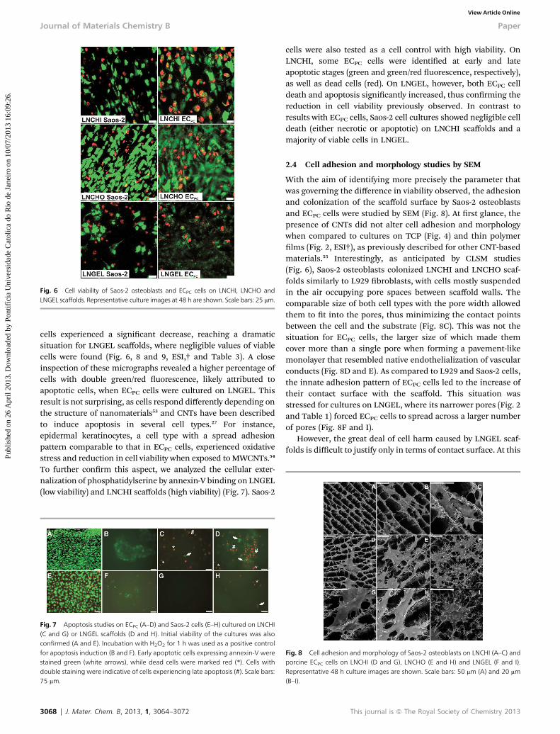
Fig. 6 Cell viability of Saos-2 osteoblasts and ECPC cells on LNCHI, LNCHO andLNGEL scaffolds. Representative culture images at 48 h are shown. Scale bars: 25 mm.
Journal of Materials Chemistry B Paper
Publ
ishe
d on
26
Apr
il 20
13. D
ownl
oade
d by
Pon
tifíc
ia U
nive
rsid
ade
Cat
olic
a do
Rio
de
Jane
iro
on 1
0/07
/201
3 16
:09:
26.
View Article Online
cells experienced a signicant decrease, reaching a dramaticsituation for LNGEL scaffolds, where negligible values of viablecells were found (Fig. 6, 8 and 9, ESI,† and Table 3). A closeinspection of these micrographs revealed a higher percentage ofcells with double green/red uorescence, likely attributed toapoptotic cells, when ECPC cells were cultured on LNGEL. Thisresult is not surprising, as cells respond differently depending onthe structure of nanomaterials53 and CNTs have been describedto induce apoptosis in several cell types.27 For instance,epidermal keratinocytes, a cell type with a spread adhesionpattern comparable to that in ECPC cells, experienced oxidativestress and reduction in cell viability when exposed toMWCNTs.54
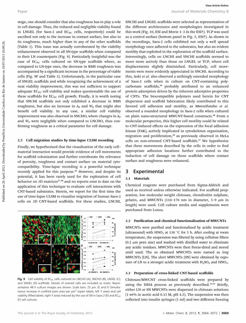
To further conrm this aspect, we analyzed the cellular exter-nalization of phosphatidylserine by annexin-V binding on LNGEL(low viability) and LNCHI scaffolds (high viability) (Fig. 7). Saos-2
Fig. 7 Apoptosis studies on ECPC (A–D) and Saos-2 cells (E–H) cultured on LNCHI(C and G) or LNGEL scaffolds (D and H). Initial viability of the cultures was alsoconfirmed (A and E). Incubation with H2O2 for 1 h was used as a positive controlfor apoptosis induction (B and F). Early apoptotic cells expressing annexin-V werestained green (white arrows), while dead cells were marked red (*). Cells withdouble staining were indicative of cells experiencing late apoptosis (#). Scale bars:75 mm.
3068 | J. Mater. Chem. B, 2013, 1, 3064–3072
cells were also tested as a cell control with high viability. OnLNCHI, some ECPC cells were identied at early and lateapoptotic stages (green and green/red uorescence, respectively),as well as dead cells (red). On LNGEL, however, both ECPC celldeath and apoptosis signicantly increased, thus conrming thereduction in cell viability previously observed. In contrast toresults with ECPC cells, Saos-2 cell cultures showed negligible celldeath (either necrotic or apoptotic) on LNCHI scaffolds and amajority of viable cells in LNGEL.
2.4 Cell adhesion and morphology studies by SEM
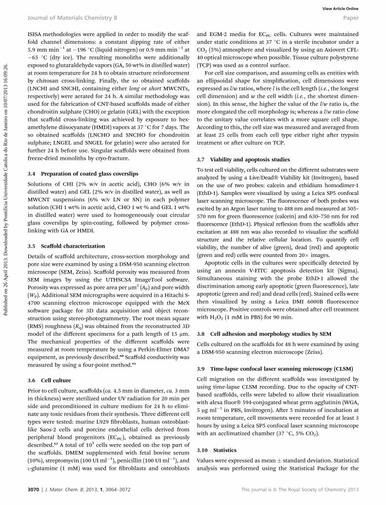
With the aim of identifying more precisely the parameter thatwas governing the difference in viability observed, the adhesionand colonization of the scaffold surface by Saos-2 osteoblastsand ECPC cells were studied by SEM (Fig. 8). At rst glance, thepresence of CNTs did not alter cell adhesion and morphologywhen compared to cultures on TCP (Fig. 4) and thin polymerlms (Fig. 2, ESI†), as previously described for other CNT-basedmaterials.55 Interestingly, as anticipated by CLSM studies(Fig. 6), Saos-2 osteoblasts colonized LNCHI and LNCHO scaf-folds similarly to L929 broblasts, with cells mostly suspendedin the air occupying pore spaces between scaffold walls. Thecomparable size of both cell types with the pore width allowedthem to t into the pores, thus minimizing the contact pointsbetween the cell and the substrate (Fig. 8C). This was not thesituation for ECPC cells, the larger size of which made themcover more than a single pore when forming a pavement-likemonolayer that resembled native endothelialization of vascularconducts (Fig. 8D and E). As compared to L929 and Saos-2 cells,the innate adhesion pattern of ECPC cells led to the increase oftheir contact surface with the scaffold. This situation wasstressed for cultures on LNGEL, where its narrower pores (Fig. 2and Table 1) forced ECPC cells to spread across a larger numberof pores (Fig. 8F and I).
However, the great deal of cell harm caused by LNGEL scaf-folds is difficult to justify only in terms of contact surface. At this
Fig. 8 Cell adhesion and morphology of Saos-2 osteoblasts on LNCHI (A–C) andporcine ECPC cells on LNCHI (D and G), LNCHO (E and H) and LNGEL (F and I).Representative 48 h culture images are shown. Scale bars: 50 mm (A) and 20 mm(B–I).
This journal is ª The Royal Society of Chemistry 2013
Paper Journal of Materials Chemistry B
Publ
ishe
d on
26
Apr
il 20
13. D
ownl
oade
d by
Pon
tifíc
ia U
nive
rsid
ade
Cat
olic
a do
Rio
de
Jane
iro
on 1
0/07
/201
3 16
:09:
26.
View Article Online
stage, one should consider that also roughness has to play a rolein cell damage. Thus, the reduced and negligible viability foundin LNGEL (for Saos-2 and ECPC cells, respectively) could beascribed not only to the increase in contact surface, but also toits roughness, much higher than in any of the other scaffolds(Table 1). This issue was actually corroborated by the viabilityenhancement observed in all SN-type scaffolds when comparedto their LN counterparts (Fig. 9). Particularly insightful was thecase of ECPC cells cultured on SN-type scaffolds where, ascompared to LN-type ones, the decrease in RMS roughness wasaccompanied by a signicant increase in the percentage of viablecells (Fig. 9F and Table 3). Unfortunately, in the particular caseof SNGEL scaffolds and while recognizing the achievement of aneat viability improvement, this was not sufficient to supportadequate ECPC cell viability and makes questionable the use ofthese scaffolds for ECPC cell growth. Finally, it is worth notingthat SNCHI scaffolds not only exhibited a decrease in RMSroughness, but also an increase in AP and WP that might alsobenet cell viability. In any case, a similar cell viabilityimprovement was also observed in SNCHO, where changes in APand WP were negligible when compared to LNCHO, thus con-rming roughness as a critical parameter for cell damage.
2.5 Cell migration studies by time-lapse CLSM recording
Finally, we hypothesized that the visualization of the early cell–material interaction would provide evidence of cell movementsfor scaffold colonization and further corroborate the relevanceof porosity, roughness and contact surface on material cyto-compatibility. Time-lapse recording is a powerful techniquerecently applied for this purpose.56 However, and despite itspotential, it has been rarely used for the exploration of cellbehavior on 3D matrices57,58 and no reports exist to date on theapplication of this technique to evaluate cell interactions withCNT-based substrates. Herein, we report for the rst time theuse of time-lapse CLSM to visualize migration of human Saos-2cells on 3D CNT-based scaffolds. For these studies, LNCHI,
Fig. 9 Cell viability of ECPC cells cultured on LNCHO (A), SNCHO (B), LNGEL (C),and SNGEL (D) scaffolds. Details of stained cells are included as insets. Repre-sentative 48 h culture images are shown. Scale bars: 25 mm. (E and F) Simulta-neous increase in scaffold pore area per mm2 (open labels, left Y axes) and cellviability (filled labels, right Y axes) induced by the use of SN in Saos-2 (E) and ECPC
(F) cell cultures.
This journal is ª The Royal Society of Chemistry 2013
SNCHI and LNGEL scaffolds were selected as representatives ofthe different architectures and morphologies investigated inthis work (Fig. 10, ESI and Movie 1–3 in the ESI†). TCP was usedas a control surface (bottom panel in Fig. 5, ESI†). As shown inthe recordings, Saos-2 cells exhibited not only a well spreadmorphology once adhered to the substrates, but also an evidentmotility that exploited in the exploration of the scaffold surface.Interestingly, cells on LNCHI and SNCHI scaffolds seemed tomove more actively than those on LNGEL or TCP, where celldisplacements slightly diminished. Particularly, cell move-ments were more evidently appreciated in SNCHI. According tothis, Aoki et al. also observed a strikingly extended morphologyof Saos-2 cells when in culture on CNT-containing poly-carbonate scaffolds,29 probably attributed to an enhancedprotein adsorption driven by the inherent adsorptive propertiesof CNTs. The biocompatible polymers used herein for CNTdispersion and scaffold fabrication likely contributed to thisfavored cell adhesion and motility, as Mwenifumbo et al.observed a rounded morphology in Saos-2 cells when culturedon plain nano-structured MWCNT-based constructs.28 From amolecular perspective, this higher cell motility could be relatedto CNT-induced effects on the expression of the focal adhesionkinase (FAK), actively implicated in cytoskeleton organization,migration and proliferation,59 as previously observed in HeLacells on non-oriented CNT-based scaffolds.27 We hypothesizethat these movements described by the cells in order to ndappropriate adhesion locations further contributed to theinduction of cell damage on those scaffolds where contactsurface and roughness were enhanced.
3 Experimental3.1 Materials
Chemical reagents were purchased from Sigma-Aldrich andused as received unless otherwise indicated. For scaffold prep-aration, low molecular weight chitosan, chondroitin sulphate,gelatin, and MWCNTs (110–170 nm in diameter, 5–9 mm inlength) were used. Cell culture media and supplements werepurchased from Lonza.
3.2 Purication and chemical functionalization of MWCNTs
MWCNTs were puried and functionalized by acidic treatment(ultrasound) with HNO3 at 130 �C for 5 h. Aer cooling at roomtemperature, the suspension was ltered by using cellulose lters(0.2 mm pore size) and washed with distilled water to eliminateany acidic residues. MWCNTs were then freeze-dried and storeduntil used. The so obtained MWCNTs were named as longMWCNTs (LN). The short MWCNTs (SN) were obtained by expo-sure of LN to a stronger acidic treatment with H2SO4 and HNO3.
3.3 Preparation of cross-linked CNT-based scaffolds
Chitosan/MWCNT cross-linked scaffolds were prepared byusing the ISISA process as previously described.15,35 Briey,either LN or SN MWCNTs were dispersed in chitosan solutions(1 wt% in acetic acid 0.15 M, pH 4.5). The suspension was thencollected into insulin syringes (1 ml) and two different freezing
J. Mater. Chem. B, 2013, 1, 3064–3072 | 3069
Journal of Materials Chemistry B Paper
Publ
ishe
d on
26
Apr
il 20
13. D
ownl
oade
d by
Pon
tifíc
ia U
nive
rsid
ade
Cat
olic
a do
Rio
de
Jane
iro
on 1
0/07
/201
3 16
:09:
26.
View Article Online
ISISA methodologies were applied in order to modify the scaf-fold channel dimensions: a constant dipping rate of either5.9 mm min�1 at �196 �C (liquid nitrogen) or 0.9 mm min�1 at�65 �C (dry ice). The resulting monoliths were additionallyexposed to glutaraldehyde vapors (GA, 50 wt% in distilled water)at room temperature for 24 h to obtain structure reinforcementby chitosan cross-linking. Finally, the so obtained scaffolds(LNCHI and SNCHI, containing either long or short MWCNTs,respectively) were aerated for 24 h. A similar methodology wasused for the fabrication of CNT-based scaffolds made of eitherchondroitin sulphate (CHO) or gelatin (GEL) with the exceptionthat scaffold cross-linking was achieved by exposure to hex-amethylene diisocyanate (HMDI) vapors at 37 �C for 7 days. Theso obtained scaffolds (LNCHO and SNCHO for chondroitinsulphate; LNGEL and SNGEL for gelatin) were also aerated forfurther 24 h before use. Singular scaffolds were obtained fromfreeze-dried monoliths by cryo-fracture.
3.4 Preparation of coated glass coverslips
Solutions of CHI (2% w/v in acetic acid), CHO (6% w/v indistilled water) and GEL (2% w/v in distilled water), as well asMWCNT suspensions (6% w/v LN or SN) in each polymersolution (CHI 1 wt% in acetic acid, CHO 1 wt % and GEL 1 wt%in distilled water) were used to homogeneously coat circularglass coverslips by spin-coating, followed by polymer cross-linking with GA or HMDI.
3.5 Scaffold characterization
Details of scaffold architecture, cross-section morphology andpore size were examined by using a DSM-950 scanning electronmicroscope (SEM, Zeiss). Scaffold porosity was measured fromSEM images by using the UTHSCSA ImageTool soware.Porosity was expressed as pore area per mm2 (AP) and pore width(WP). Additional SEMmicrographs were acquired in a Hitachi S-4700 scanning electron microscope equipped with the MeXsoware package for 3D data acquisition and object recon-struction using stereo-photogrammetry. The root mean square(RMS) roughness (Rq) was obtained from the reconstructed 3Dmodel of the different specimens for a path length of 15 mm.The mechanical properties of the different scaffolds weremeasured at room temperature by using a Perkin-Elmer DMA7equipment, as previously described.60 Scaffold conductivity wasmeasured by using a four-point method.61
3.6 Cell culture
Prior to cell culture, scaffolds (ca. 4.5 mm in diameter, ca. 3 mmin thickness) were sterilized under UV radiation for 20 min perside and preconditioned in culture medium for 24 h to elimi-nate any toxic residues from their synthesis. Three different celltypes were tested: murine L929 broblasts, human osteoblast-like Saos-2 cells and porcine endothelial cells derived fromperipheral blood progenitors (ECPC), obtained as previouslydescribed.62 A total of 105 cells were seeded on the top part ofthe scaffolds. DMEM supplemented with fetal bovine serum(10%), streptomycin (100 UI ml�1), penicillin (100 UI ml�1), andL-glutamine (1 mM) was used for broblasts and osteoblasts
3070 | J. Mater. Chem. B, 2013, 1, 3064–3072
and EGM-2 media for ECPC cells. Cultures were maintainedunder static conditions at 37 �C in a sterile incubator under aCO2 (5%) atmosphere and visualized by using an Axiovert CFL-40 optical microscope when possible. Tissue culture polystyrene(TCP) was used as a control surface.
For cell size comparison, and assuming cells as entities withan ellipsoidal shape for simplication, cell dimensions wereexpressed as l/w ratios, where l is the cell length (i.e., the longestcell dimension) and w the cell width (i.e., the shortest dimen-sion). In this sense, the higher the value of the l/w ratio is, themore elongated the cell morphology is; whereas a l/w ratio closeto the unitary value correlates with a more square cell shape.According to this, the cell size was measured and averaged fromat least 25 cells from each cell type either right aer trypsintreatment or aer culture on TCP.
3.7 Viability and apoptosis studies
To test cell viability, cells cultured on the different substrates wereanalyzed by using a Live/Dead� Viability kit (Invitrogen), basedon the use of two probes: calcein and ethidium homodimer-1(EthD-1). Samples were visualized by using a Leica SP5 confocallaser scanning microscope. The uorescence of both probes wasexcited by an Argon laser tuning to 488 nm and measured at 505–570 nm for green uorescence (calcein) and 630–750 nm for reduorescence (EthD-1). Physical reexion from the scaffolds aerexcitation at 488 nm was also recorded to visualize the scaffoldstructure and the relative cellular location. To quantify cellviability, the number of alive (green), dead (red) and apoptotic(green and red) cells were counted from 20� images.
Apoptotic cells in the cultures were specically detected byusing an annexin V-FITC apoptosis detection kit (Sigma).Simultaneous staining with the probe EthD-1 allowed thediscrimination among early apoptotic (green uorescence), lateapoptotic (green and red) and dead cells (red). Stained cells werethen visualized by using a Leica DMI 6000B uorescencemicroscope. Positive controls were obtained aer cell treatmentwith H2O2 (1 mM in PBS) for 90 min.
3.8 Cell adhesion and morphology studies by SEM
Cells cultured on the scaffolds for 48 h were examined by usinga DSM-950 scanning electron microscope (Zeiss).
3.9 Time-lapse confocal laser scanning microscopy (CLSM)
Cell migration on the different scaffolds was investigated byusing time-lapse CLSM recording. Due to the opacity of CNT-based scaffolds, cells were labeled to allow their visualizationwith alexa uor� 594-conjugated wheat germ agglutinin (WGA,5 mg ml�1 in PBS, Invitrogen). Aer 5 minutes of incubation atroom temperature, cell movements were recorded for at least 3hours by using a Leica SP5 confocal laser scanning microscopewith an acclimatized chamber (37 �C, 5% CO2).
3.10 Statistics
Values were expressed as mean� standard deviation. Statisticalanalysis was performed using the Statistical Package for the
This journal is ª The Royal Society of Chemistry 2013
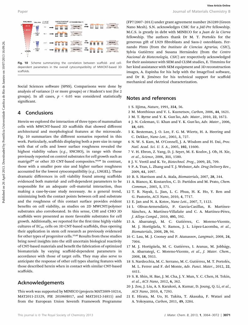
Fig. 10 Scheme summarizing the correlation between scaffold- and cell-dependent parameters in the overall cytocompatibility of MWCNT-based 3Dscaffolds.
Paper Journal of Materials Chemistry B
Publ
ishe
d on
26
Apr
il 20
13. D
ownl
oade
d by
Pon
tifíc
ia U
nive
rsid
ade
Cat
olic
a do
Rio
de
Jane
iro
on 1
0/07
/201
3 16
:09:
26.
View Article Online
Social Sciences soware (SPSS). Comparisons were done byanalysis of variance (3 or more groups) or t Student's test (for 2groups). In all cases, p < 0.05 was considered statisticallysignicant.
4 Conclusions
Herein we explored the interaction of three types of mammaliancells with MWCNT-based 3D scaffolds that showed differentarchitectural and morphological features at the microscale.Fig. 10 summarizes the different scenarios reported in thiswork. Particularly, scaffolds displaying both a pore size in rangewith that of cells and lower surface roughness revealed thehighest viability values (e.g., SNCHO), in range with thosepreviously reported on control substrates for cell growth such asmatrigel63 or other 3D CNT-based composites.63,64 In contrast,those with a smaller pore size and higher surface roughnessaccounted for the lowest cytocompatibility (e.g., LNGEL). Thesedramatic differences in cell viability found among scaffoldsevidenced both material- and cell-dependent parameters to beresponsible for an adequate cell–material interaction, thusmaking a case-by-case study necessary. As a general trend,minimizing both the contact points between cell and substrateand the roughness of this contact surface provides evidentbenets on cell viability, as studies on 2D MWCNT/polymersubstrates also corroborated. In this sense, CHI and CHO 3Dscaffolds were presented as more favorable substrates for cellgrowth. Additionally, we reported for the rst time highly viablecultures of ECPC cells on 3D CNT-based scaffolds, thus openingtheir application in stem cell research as previously evidencedfor other types of progenitor cells.13,65 Results from these studiesbring novel insights into the still uncertain biological reactivityof CNT-based materials and benet the fabrication of optimizedbiomaterials by varying scaffold-dependent parameters inaccordance with those of target cells. They may also serve toanticipate the response of other cell types sharing features withthose described herein when in contact with similar CNT-basedscaffolds.
Acknowledgements
This work was supported by MINECO (projects MAT2009-10214,MAT2011-25329, PIE 201060I017, and MAT2012-34811) andfrom the European Union Seventh Framework Programme
This journal is ª The Royal Society of Chemistry 2013
(FP7/2007–2013) under grant agreement number 263289 (GreenNano Mesh). S.N. acknowledges CSIC for a JAE-Pre fellowship.M.C.S. is greatly in debt with MINECO for a Juan de la Ciervafellowship. The authors thank Dr M. T. Portoles for thegenerous gi of L929 broblasts and Saos-2 osteoblasts. Fer-nando Pinto (from the Instituto de Ciencias Agrarias, CSIC),Sylvia Gutierrez and Susana Hernandez (from the CentroNacional de Biotecnologıa, CSIC) are respectively acknowledgedfor their assistance with SEM and CLSM studies, E. Timmins forher kind assistance with SEM equipment and 3D reconstructionimages, A. Espinha for his help with the ImageTool soware,and Dr R. Jimenez for his technical support for scaffoldmechanical and electrical characterization.
Notes and references
1 S. Iijima, Nature, 1991, 354, 56.2 M. Monthioux and V. L. Kuznetsov, Carbon, 2006, 44, 1621.3 M. T. Byrne and Y. K. Gun'ko, Adv. Mater., 2010, 22, 1672.4 J. N. Coleman, U. Khan and Y. K. Gun'ko, Adv. Mater., 2006,18, 689.
5 K. Besteman, J. O. Lee, F. G. M. Wiertz, H. A. Heering andC. Dekker, Nano Lett., 2003, 3, 727.
6 N. W. S. Kam, M. O'Connell, J. A. Wisdom and H. Dai, Proc.Natl. Acad. Sci. U. S. A., 2005, 102, 11600.
7 V. H. Ebron, Z. Yang, D. J. Seyer, M. E. Kozlov, J. Oh, H. Xie,et al., Science, 2006, 311, 1580.
8 J. V. Veetil and K. Ye, Biotechnol. Prog., 2009, 25, 709.9 P. A. Tran, L. Zhang and T. J. Webster, Adv. Drug Delivery Rev.,2009, 61, 1097.
10 B. S. Harrison and A. Atala, Biomaterials, 2007, 28, 344.11 A. Bianco, K. Kostarelos, C. D. Partidos and M. Prato, Chem.
Commun., 2005, 5, 571.12 T. R. Nayak, L. Jian, L. C. Phua, H. K. Ho, Y. Ren and
G. Pastorin, ACS Nano, 2010, 4, 7717.13 E. Jan and N. A. Kotov, Nano Lett., 2007, 7, 1123.14 I. Olivas-Armendariz, P. Garcıa-Casillas, R. Martınez-
Sanchez, A. Martınez-Villafa~ne and C. A. Martınez-Perez,J. Alloys Compd., 2010, 495, 592.
15 A. Abarrategi, M. C. Gutierrez, C. Moreno-Vicente,M. J. Hortiguela, V. Ramos, J. L. Lopez-Lacomba, et al.,Biomaterials, 2008, 29, 94.
16 C. Lau, M. J. Cooney and P. Atanassov, Langmuir, 2008, 24,7004.
17 M. J. Hortiguela, M. C. Gutierrez, I. Aranaz, M. Jobbagy,A. Abarrategi, C. Moreno-Vicente, et al., J. Mater. Chem.,2008, 18, 5933.
18 S. Nardecchia, M. C. Serrano, M. C. Gutierrez, M. T. Portoles,M. L. Ferrer and F. del Monte, Adv. Funct. Mater., 2012, 22,4411.
19 S. R. Shin, H. Bae, J. M. Cha, J. Y. Mun, Y. C. Chen, H. Tekin,et al., ACS Nano, 2012, 6, 362.
20 J. Zou, J. Liu, A. S. Karakoti, A. Kumar, D. Joung, Q. Li, et al.,ACS Nano, 2010, 4, 7293.
21 E. Hirata, M. Uo, H. Takita, T. Akasaka, F. Watari andA. Yokoyama, Carbon, 2011, 49, 3284.
J. Mater. Chem. B, 2013, 1, 3064–3072 | 3071

Journal of Materials Chemistry B Paper
Publ
ishe
d on
26
Apr
il 20
13. D
ownl
oade
d by
Pon
tifíc
ia U
nive
rsid
ade
Cat
olic
a do
Rio
de
Jane
iro
on 1
0/07
/201
3 16
:09:
26.
View Article Online
22 H. Zhang and Z. J. Chen, J. Bioact. Compat. Polym., 2010, 25,241.
23 K. Rege, N. R. Raravikar, D. Y. Kim, L. S. Schadler,P. M. Ajayan and J. S. Dordick, Nano Lett., 2003, 3, 829.
24 L. Ding, J. Stilwell, T. Zhang, O. Elboudwarej, H. Jiang,J. P. Selegue, et al., Nano Lett., 2005, 5, 2448.
25 V. M. Silva, N. Corson, A. Elder and G. Oberdorster, Toxicol.Sci., 2005, 85, 983.
26 C. M. Sayes, J. D. Fortner, W. Guo, D. Lyon, A. M. Boyd,K. D. Ausman, et al., Nano Lett., 2004, 4, 1881.
27 X. Zhang, X. Wang, Q. Lu and C. Fu, Carbon, 2008, 46,453.
28 S. Mwenifumbo, M. S. Shaffer and M. M. Stevens, J. Mater.Chem., 2007, 17, 1894.
29 N. Aoki, A. Yokoyama, Y. Nodasaka, T. Akasaka, M. Uo,Y. Sato, et al., Chem. Lett., 2006, 35, 508.
30 T. J. Webster, M. C. Waid, J. L. McKenzie, R. L. Price andJ. U. Ejiofor, Nanotechnology, 2004, 15, 48.
31 M. A. Correa-Duarte, N. Wagner, J. Rojas-Chapana,C. Morsczeck, M. Thie and M. Giersig, Nano Lett., 2004, 4,2233.
32 H. F. Cui, S. K. Vashist, K. Al-Rubeaan, J. H. T. Luong andF. S. Sheu, Chem. Res. Toxicol., 2010, 23, 1131.
33 S. Nardecchia, D. Carriazo, M. L. Ferrer, M. C. Gutierrez andF. del Monte, Chem. Soc. Rev., 2013, 42, 794.
34 M. C. Gutierrez, M. L. Ferrer and F. del Monte, Chem. Mater.,2008, 20, 634.
35 M. J. Hortiguela, I. Aranaz, M. C. Gutierrez, M. L. Ferrer andF. del Monte, Biomacromolecules, 2011, 12, 179.
36 H. Zhang, I. Hussain, M. Brust, M. F. Butler, S. P. Rannardand A. I. Cooper, Nat. Mater., 2005, 4, 787.
37 M. C. Gutierrez, M. Jobbagy, N. Rapun, M. L. Ferrer andF. del Monte, Adv. Mater., 2006, 18, 1137.
38 S. Deville, E. Saiz, R. K. Nalla and A. P. Tomsia, Science, 2006,311, 515.
39 S. R. Mukai, H. Nishihara and H. Tamon, Chem. Commun.,2004, 10, 874.
40 M. Dash, F. Chiellini, R. M. Ottenbrite and E. Chiellini, Prog.Polym. Sci., 2011, 36, 981.
41 A. D. Baldwin and K. L. Kiick, Pept. Sci., 2010, 94, 128.42 D. J. Mooney and E. A. Silva, Nat. Mater., 2007, 6, 327.43 K. Ulubayram, E. Aksu, S. I. D. Gurhan, K. Serbetci and
N. Hasirci, J. Biomater. Sci., Polym. Ed., 2002, 13, 1203.44 J. Berger, M. Reist, J. M. Mayer, O. Felt, N. A. Peppas and
R. Gurny, Eur. J. Pharm. Biopharm., 2004, 57, 19.
3072 | J. Mater. Chem. B, 2013, 1, 3064–3072
45 W. Cao, M. Cheng, Q. Ao, Y. Gong, N. Zhao and X. Zhang, J.Biomater. Sci., Polym. Ed., 2005, 16, 791.
46 H. Nishihara, S. Iwamura and T. Kyotani, J. Mater. Chem.,2008, 18, 3662.
47 M. C. Serrano, R. Pagani, M. Vallet-Regı, J. Pe~na, A. Ramila,I. Izquierdo, et al., Biomaterials, 2004, 25, 5603.
48 L. Zhu, D. W. Chang, L. Dai and Y. Hong, Nano Lett., 2007, 7,3592.
49 T. Asahara, T. Murohara, A. Sullivan, M. Silver, R. van derZee, T. Li, et al., Science, 1997, 27, 964.
50 A. Zampetaki, J. P. Kirton and Q. Xu, Cardiovasc. Res., 2008,78, 413.
51 T. Matsumoto, R. Kuroda, Y. Mifune, A. Kawamoto, T. Shoji,M. Miwa, et al., Bone, 2008, 43, 434.
52 S. Giannona, I. Firkowska, J. Rojas-Chapana and M. Giersig,J. Nanosci. Nanotechnol., 2007, 7, 1679.
53 L. Ding, J. Stilwell, T. Zhang, O. Elboudwarej, H. Jiang,J. P. Selegue, et al., Nano Lett., 2005, 5, 2448.
54 A. Shvedova, V. Castranova, E. R. Kisin, D. Schwegler-Berry,A. R. Murray, V. Z. Gandelsman, et al., J. Toxicol. Environ.Health, Part A, 2003, 66, 1909.
55 R. A. MacDonald, B. F. Laurenzi, G. Viswanathan,P. M. Ajayan and J. P. Stegemann, J. Biomed. Mater. Res.,Part A, 2005, 74, 489.
56 R. Gatti, G. Orlandini, J. Uggeri, S. Belletti, C. Galli,M. Raspanti, et al., Micron, 2008, 39, 137.
57 T. A. Ulrich, A. Jain, K. Tanner, J. L. MacKay and S. Kumar,Biomaterials, 2010, 31, 1875.
58 G. P. Raeber, M. P. Lutolf and J. A. Hubbell, Acta Biomater.,2007, 3, 615.
59 J. T. Parsons, K. H. Martin, J. K. Slack, J. M. Taylor andS. A. Weed, Oncogene, 2000, 19, 5606.
60 M. C. Serrano, M. C. Gutierrez, R. Jimenez, M. L. Ferrer andF. del Monte, Chem. Commun., 2012, 48, 579.
61 M. C. Gutierrez, D. Carriazo, A. Tamayo, R. Jimenez, F. Pico,J. M. Rojo, et al., Chem.–Eur. J., 2011, 17, 10537.
62 J. Allen, S. Khan, M. C. Serrano and G. Ameer, Tissue Eng.,2008, 14, 183.
63 A. Tiwari, Y. Sharma, S. Hattori, D. Terada, A. K. Sharma,A. P. F. Turner and H. Kobayashi, Biopolymers, 2013, 99, 334.
64 M. Mattioli-Belmonte, G. Vozzi, Y. Whulanza, M. Seggiani,V. Fantauzzi, G. Orsini and A. Ahluwalia, Mater. Sci. Eng.,C, 2012, 32, 152.
65 S. Y. Park, S. Namgung, B. Kim, J. Im, J. Y. Kim, K. Sun, et al.,Adv. Mater., 2007, 19, 2530.
This journal is ª The Royal Society of Chemistry 2013
Related Documents


![Cytocompatibility of Chitosan and Collagen-Chitosan ...forms the highly porous structure of the scaffolds[13] Two percent (w/v) of chitosan was prepared by dissolving chitosan in 0.2](https://static.cupdf.com/doc/110x72/5e3f1725786dcc56c068fc16/cytocompatibility-of-chitosan-and-collagen-chitosan-forms-the-highly-porous.jpg)