Modulating inhibitory control with direct current stimulation of the superior medial frontal cortex Tzu-Yu Hsu a,b,c , Lin-Yuan Tseng a,b,c , Jia-Xin Yu a,b,c , Wen-Jui Kuo a,b,c , Daisy L. Hung a,b,c , Ovid J.L. Tzeng a,b,c,d , Vincent Walsh e , Neil G. Muggleton b,e , Chi-Hung Juan b,c, ⁎ a Institute of Neuroscience, National Yang-Ming University, Taipei 112, Taiwan b Institute of Cognitive Neuroscience, National Central University, Jhongli 320, Taiwan c Laboratories for Cognitive Neuroscience, National Yang-Ming University, Taipei 112, Taiwan d Institute of Linguistics, Academia Sinica, Taipei 115, Taiwan e Institute of Cognitive Neuroscience and Department of Psychology, University College London, London WC1N 3AR, UK abstract article info Article history: Received 7 January 2011 Revised 6 March 2011 Accepted 25 March 2011 Available online 1 April 2011 Keywords: Inhibitory control Pre-SMA tDCS Stop signal task The executive control of voluntary action involves not only choosing from a range of possible actions but also the inhibition of responses as circumstances demand. Recent studies have demonstrated that many clinical populations, such as people with attention-deficit hyperactivity disorder, exhibit difficulties in inhibitory control. One prefrontal area that has been particularly associated with inhibitory control is the pre- supplementary motor area (Pre-SMA). Here we applied non-invasive transcranial direct current stimulation (tDCS) over Pre-SMA to test its role in this behavior. tDCS allows for current to be applied in two directions to selectively excite or suppress the neural activity of Pre-SMA. Our results showed that anodal tDCS improved efficiency of inhibitory control. Conversely, cathodal tDCS showed a tendency towards impaired inhibitory control. To our knowledge, this is the first demonstration of non-invasive intervention tDCS altering subjects' inhibitory control. These results further our understanding of the neural bases of inhibitory control and suggest a possible therapeutic intervention method for clinical populations. © 2011 Elsevier Inc. All rights reserved. Introduction Efficient and timely control over behavioral urges is vital to the executive control of voluntary action, and inhibitory control in the context of prepotent actions is critical in a variety of scenarios. Oft mentioned examples of such behavior include driving towards a crossroads, where the choice of executing or withholding an action (acceleration or braking) must be made in a very brief period of time. This behavior can be investigated experimentally using stop-signal tasks that have been shown to reliably estimate the response time of an internally generated act of control (Logan, 1994). Stop-signal task The stop-signal task is widely used to investigate the processes of motor inhibition (Boucher et al., 2007; Chen et al., 2008, 2009; Chao et al., 2009; Li et al., 2006a, 2008a; Logan et al., 1997; Logan and Cowan, 1984; Logan et al., 1984; Schachar et al., 1993; Schachar et al., 1995; Schachar and Logan, 1990). This task consists of ‘go’ and ‘stop’ trials. In the go trials, subjects are required to respond to a stimulus as soon as possible. In the stop trials, an additional stop signal is displayed to instruct the subject to withhold response. The interval between the go and stop signal is known as the stop signal delay (SSD), which is estimated by considering the distribution of go reaction times and the probability of responding correctly in those trials with a stop signal. This estimation effectively predicts the time required to inhibit a planned response, called the stop-signal reaction time (SSRT). The neural correlates of inhibitory control When performing a manual inhibitory control task, the signal from the retina is projected from the visual areas to brain regions that are related to inhibitory control, such as frontal eye fields (FEF, Curtis et al., 2005; Hanes and Schall, 1996; Muggleton et al., 2010), supple- mentary eye fields (SEF, Isoda and Hikosaka, 2007; Stuphorn et al., 2000; Stuphorn and Schall, 2006), and anterior cingulate cortex (ACC, Chevrier et al., 2007; Ito et al., 2003). Information converges at the primary motor cortex (M1), which executes motor commands by transmitting them to the spinal cord and muscles. M1 is therefore considered to be a part of the ‘final common path’ for voluntary action (Sherrington, 1906). More important, M1 also receives input from the pre-supplementary motor area (Pre-SMA), which is involved in initiating self-paced actions (Deiber et al., 1999; Jenkins et al., 2000) NeuroImage 56 (2011) 2249–2257 ⁎ Corresponding author at: Institute of Cognitive Neuroscience, No. 300, Jhongda Rd. Jhongli City, 320, Taiwan. Fax: +886 3 426 3502. E-mail address: [email protected] (C.-H. Juan). 1053-8119/$ – see front matter © 2011 Elsevier Inc. All rights reserved. doi:10.1016/j.neuroimage.2011.03.059 Contents lists available at ScienceDirect NeuroImage journal homepage: www.elsevier.com/locate/ynimg

Welcome message from author
This document is posted to help you gain knowledge. Please leave a comment to let me know what you think about it! Share it to your friends and learn new things together.
Transcript
NeuroImage 56 (2011) 2249–2257
Contents lists available at ScienceDirect
NeuroImage
j ourna l homepage: www.e lsev ie r.com/ locate /yn img
Modulating inhibitory control with direct current stimulation of the superior medialfrontal cortex
Tzu-Yu Hsu a,b,c, Lin-Yuan Tseng a,b,c, Jia-Xin Yu a,b,c, Wen-Jui Kuo a,b,c, Daisy L. Hung a,b,c, Ovid J.L. Tzeng a,b,c,d,Vincent Walsh e, Neil G. Muggleton b,e, Chi-Hung Juan b,c,⁎a Institute of Neuroscience, National Yang-Ming University, Taipei 112, Taiwanb Institute of Cognitive Neuroscience, National Central University, Jhongli 320, Taiwanc Laboratories for Cognitive Neuroscience, National Yang-Ming University, Taipei 112, Taiwand Institute of Linguistics, Academia Sinica, Taipei 115, Taiwane Institute of Cognitive Neuroscience and Department of Psychology, University College London, London WC1N 3AR, UK
⁎ Corresponding author at: Institute of Cognitive NeurJhongli City, 320, Taiwan. Fax: +886 3 426 3502.
E-mail address: [email protected] (C.-H. Juan).
1053-8119/$ – see front matter © 2011 Elsevier Inc. Aldoi:10.1016/j.neuroimage.2011.03.059
a b s t r a c t
a r t i c l e i n f oArticle history:Received 7 January 2011Revised 6 March 2011Accepted 25 March 2011Available online 1 April 2011
Keywords:Inhibitory controlPre-SMAtDCSStop signal task
The executive control of voluntary action involves not only choosing from a range of possible actions but alsothe inhibition of responses as circumstances demand. Recent studies have demonstrated that many clinicalpopulations, such as people with attention-deficit hyperactivity disorder, exhibit difficulties in inhibitorycontrol. One prefrontal area that has been particularly associated with inhibitory control is the pre-supplementary motor area (Pre-SMA). Here we applied non-invasive transcranial direct current stimulation(tDCS) over Pre-SMA to test its role in this behavior. tDCS allows for current to be applied in two directions toselectively excite or suppress the neural activity of Pre-SMA. Our results showed that anodal tDCS improvedefficiency of inhibitory control. Conversely, cathodal tDCS showed a tendency towards impaired inhibitorycontrol. To our knowledge, this is the first demonstration of non-invasive intervention tDCS altering subjects'inhibitory control. These results further our understanding of the neural bases of inhibitory control andsuggest a possible therapeutic intervention method for clinical populations.
oscience, No. 300, Jhongda Rd.
l rights reserved.
© 2011 Elsevier Inc. All rights reserved.
Introduction
Efficient and timely control over behavioral urges is vital to theexecutive control of voluntary action, and inhibitory control in thecontext of prepotent actions is critical in a variety of scenarios. Oftmentioned examples of such behavior include driving towards acrossroads, where the choice of executing or withholding an action(acceleration or braking) must be made in a very brief period of time.This behavior can be investigated experimentally using stop-signaltasks that have been shown to reliably estimate the response time ofan internally generated act of control (Logan, 1994).
Stop-signal task
The stop-signal task is widely used to investigate the processesof motor inhibition (Boucher et al., 2007; Chen et al., 2008, 2009; Chaoet al., 2009; Li et al., 2006a, 2008a; Logan et al., 1997; Logan andCowan, 1984; Logan et al., 1984; Schachar et al., 1993; Schachar et al.,1995; Schachar and Logan, 1990). This task consists of ‘go’ and ‘stop’trials. In the go trials, subjects are required to respond to a stimulus as
soon as possible. In the stop trials, an additional stop signal isdisplayed to instruct the subject to withhold response. The intervalbetween the go and stop signal is known as the stop signal delay(SSD), which is estimated by considering the distribution of goreaction times and the probability of responding correctly in thosetrials with a stop signal. This estimation effectively predicts the timerequired to inhibit a planned response, called the stop-signal reactiontime (SSRT).
The neural correlates of inhibitory control
When performing amanual inhibitory control task, the signal fromthe retina is projected from the visual areas to brain regions that arerelated to inhibitory control, such as frontal eye fields (FEF, Curtiset al., 2005; Hanes and Schall, 1996; Muggleton et al., 2010), supple-mentary eye fields (SEF, Isoda and Hikosaka, 2007; Stuphorn et al.,2000; Stuphorn and Schall, 2006), and anterior cingulate cortex (ACC,Chevrier et al., 2007; Ito et al., 2003). Information converges at theprimary motor cortex (M1), which executes motor commands bytransmitting them to the spinal cord and muscles. M1 is thereforeconsidered to be a part of the ‘final common path’ for voluntary action(Sherrington, 1906). More important, M1 also receives input from thepre-supplementary motor area (Pre-SMA), which is involved ininitiating self-paced actions (Deiber et al., 1999; Jenkins et al., 2000)
2250 T.-Y. Hsu et al. / NeuroImage 56 (2011) 2249–2257
and mediating motor inhibition that is required for voluntary musclerelaxation (Toma et al., 1999). Neuroimaging studies have shownstronger activation of the Pre-SMA for self-paced actions than forexternally triggered actions. Such a function has been confirmed bypatient studies, where patients with Pre-SMA lesions showed primedspeeded motor responses regardless of the relevance of the primes tothe targets (Sumner et al., 2007). In contrast, speeded responses wereonly observed when the mask was compatible with the targets in ahealthy control group (Eimer and Schlaghecken, 1998). This suggeststhat a normally functioning Pre-SMA should suppress automaticresponses to environmental stimulation, and those patients with Pre-SMA damage are therefore hyper-responsive. Indeed, Ball et al. (1999)found that Pre-SMA decreases its activity level in response toactivation of the primary motor cortex before voluntary movements(Ball et al., 1999). In the context of the stop-signal task, Li et al.(2006a) systematically investigated the neural correlates of motorinhibition with the stop-signal task and found a linear correlationbetween the BOLD activation of Pre-SMA and SSRTs. They found thatgreater activation in Pre-SMA led to shorter SSRTs, suggesting anefficient stop-signal processing. In contrast, IFG did not show thisassociation (Chao et al., 2009), although it is also considered as thecortical site for inhibitory motor control (Aron and Poldrack, 2006;Rubia et al., 2003; Rubia et al., 2005; Verbruggen and Logan, 2008).These results suggest that Pre-SMA mediates motor inhibition (Aronet al., 2007b; Aron and Poldrack, 2006; Kenner et al., 2010; Nachevet al., 2005). Recently, Chen et al. (2009) used TMS to probe thefunctional role of Pre-SMA in the stop-signal paradigm. They observedelevated SSRTs and increased error rates when TMS was deliveredover Pre-SMA. Their results implied that Pre-SMA plays a direct andcausal role in response inhibition and response selection.
Modulation of neural activity using tDCS
Although elevated SSRTs and increased error rates were observedas a consequence of TMS stimulation of Pre-SMA, it is unclear whetherthe underlying neurons were facilitated or inhibited by TMS (Chenet al., 2009). Thus, the present study employed transcranial directcurrent stimulation (tDCS) to disambiguate the role of Pre-SMA andits vicinity (for simplicity, Pre-SMA will be used to denote Pre-SMAand its vicinity throughout the text) in the stop-signal task. tDCS is atechnique that can either facilitate or suppress cortical excitability byusing anodal or cathodal electrical stimulation. This modulation wasfirst introduced in animal studies, where subthreshold DC stimulationincreased cerebral excitability beneath anodal stimulation by depo-larizing cell membranes and increasing firing rates, while cathodalstimulation resulted in the opposite effect by hyperpolarization anddecreasing firing rates (Bindman et al., 1964; Creutzfeldt et al., 1962;Nitsche et al., 2009b; Nitsche et al., 2009b; Purpura and McMurtry,1965; Scholfield, 1990). Neural excitability has also been observedusing functional near infrared optical brain imaging (fNIR) withincreasing oxyhemoglobin (HbO2), indicating extra oxygen deliveryand a raised CBF signal after anodal stimulation (Merzagora et al.,2010). Nitsche and Paulus (2001) also found consistent results bymeasuring humanmotor-evoked potentials (MEP). HigherMEPswereelicited after anodal stimulation than cathodal stimulation, indicatingthat neural excitability in the primary motor cortex was varied bytDCS. Therefore, the current study used anodal and cathodal tDCS torespectively facilitate or inhibit neural activity in Pre-SMA in order toinvestigate the functional role of the Pre-SMA in motor inhibition.
Materials and methods
Participants
Fourteen students (aged 20 to 26 years, mean 22.1, 8 males, 6females and all right handed) from the National Central University
took part in the Pre-SMA tDCS experiment. An additional group offourteen volunteers (aged 18 to 27 years, mean 21.79, 8 males and 6females) participated in the left primary motor cortex tDCS expe-riments (M1 tDCS) that served as a control condition. All gave infor-med consent prior to participation. The experiments were approvedby local ethical committee.
Apparatus
Testing took place in a sound attenuated room. Stimuli werepresented on a 19-inch CRT screen using a video resolution of800×600 pixels and a vertical refresh rate of 100 Hz. Subjects sat70 cm in front of the screen, which was positioned at an eye level. Thetask was programmed using E-prime running on a Pentium IV PC,which controlled the presentation of the stimuli as well as recordingresponse information. tDCS was delivered with a Magstim Eldith DC-stimulator and a pair of electrodes housed in 4×4 cm saline-soakedsponge coverings. The center of the stimulation electrode was placedover the target site (Pre-SMA or M1). The reference electrode wasplaced over the left cheek of the subject. In the tDCS conditions thecurrent was applied for 10 min with an intensity of 1.5 mA.
Procedure
Stop-signal taskIn the stop-signal task, the stop signal delay (SSD) is the most
critical independent variable and it is manipulated by adjusting thetime between the onset of the go stimulus and the stop signal. Thenoncancelled rate denotes the error rate when the stop signal ispresented but subjects fail to inhibit their responses. The outcome ofthe race between the go and the stop processes is reflected by theinhibition function, which describes the probability of responding fora given a stop signal delay in accordance with the race model of Loganand Cowan (1984). The stop signal reaction time (SSRT) representsthe latency of the stop process and it is another dependent variable inthe task. The SSRT can be estimated from the observed distribution ofRTs in no-stop signal trials in combinationwith the inhibition function(Logan, 1994). In the current study, SSRTs for each SSD wereestimated using the integration method, and one summary SSRTwas calculated by averaging the three SSRTs acquired from the threeSSDs in our experiments (Logan, 1994; Band et al., 2003).
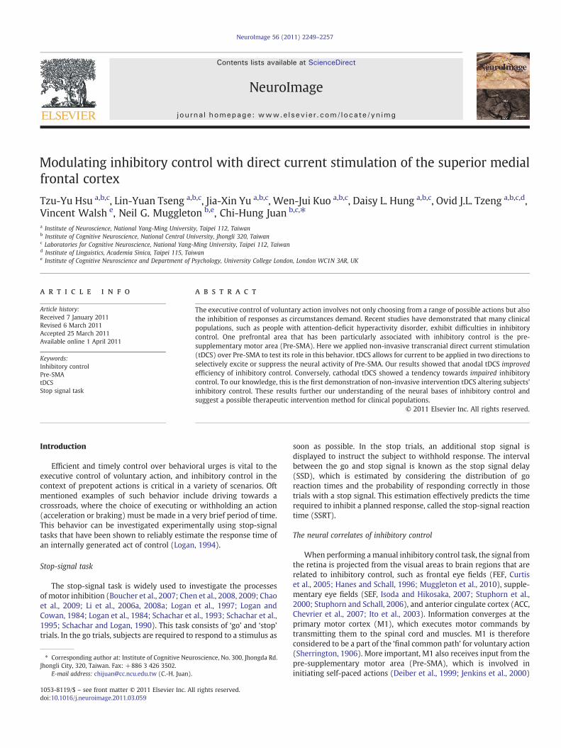
In the present study, each trial of the stop-signal task began with a500 ms central fixation dot. Following the offset of this dot, a whitetarget dot was presented to the left or right of the fixation at 9°eccentricity on the horizontal meridian (see Fig. 1). On 75% of thetrials (go trials) subjects were required to make a key-press responseindicating whether the dot was presented on the left or right withtheir left and right index fingers, respectively. On 25% of the trials(stop trials), the central fixation dot reappeared and served as a signalto withhold responses to the peripheral target.
Baseline parametersIn order to reduce the number of trials in the formal experiments,
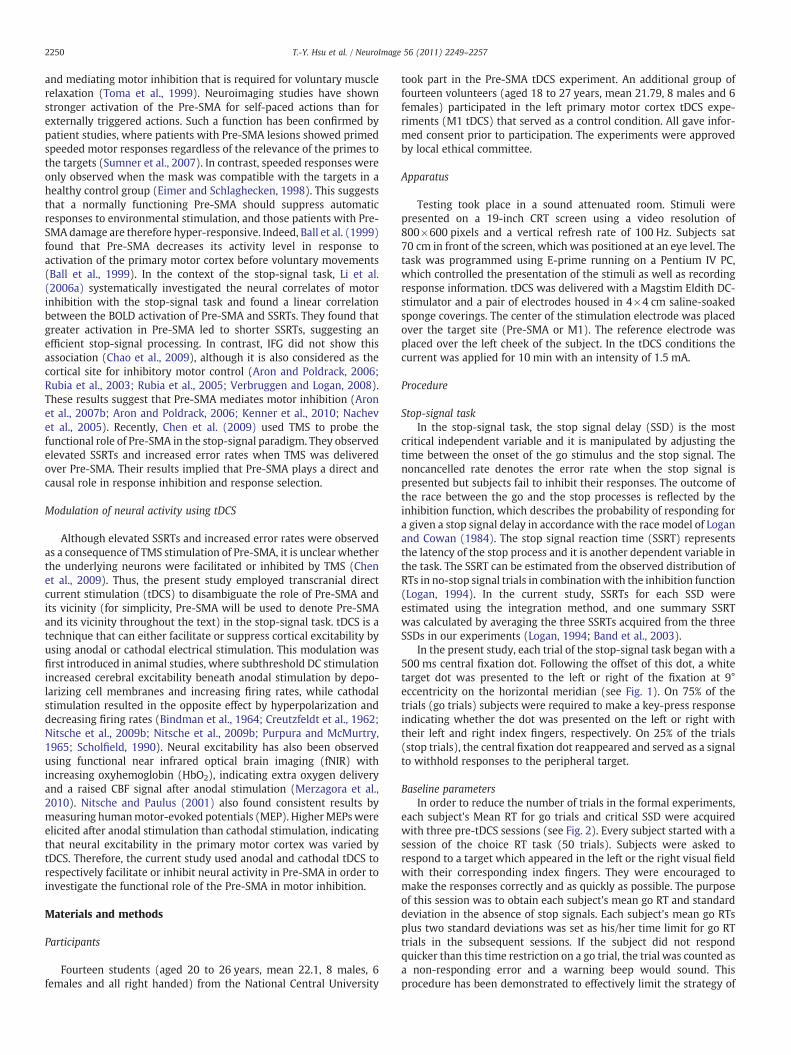
each subject's Mean RT for go trials and critical SSD were acquiredwith three pre-tDCS sessions (see Fig. 2). Every subject started with asession of the choice RT task (50 trials). Subjects were asked torespond to a target which appeared in the left or the right visual fieldwith their corresponding index fingers. They were encouraged tomake the responses correctly and as quickly as possible. The purposeof this session was to obtain each subject's mean go RT and standarddeviation in the absence of stop signals. Each subject's mean go RTsplus two standard deviations was set as his/her time limit for go RTtrials in the subsequent sessions. If the subject did not respondquicker than this time restriction on a go trial, the trial was counted asa non-responding error and a warning beep would sound. Thisprocedure has been demonstrated to effectively limit the strategy of
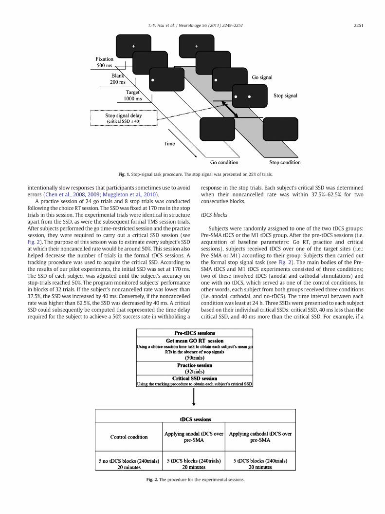
Fig. 1. Stop-signal task procedure. The stop signal was presented on 25% of trials.
2251T.-Y. Hsu et al. / NeuroImage 56 (2011) 2249–2257
intentionally slow responses that participants sometimes use to avoiderrors (Chen et al., 2008, 2009; Muggleton et al., 2010).
A practice session of 24 go trials and 8 stop trials was conductedfollowing the choice RT session. The SSDwas fixed at 170 ms in the stoptrials in this session. The experimental trials were identical in structureapart from the SSD, as were the subsequent formal TMS session trials.After subjects performed the go time-restricted session and the practicesession, they were required to carry out a critical SSD session (seeFig. 2). The purpose of this session was to estimate every subject's SSDatwhich their noncancelled ratewould be around 50%. This session alsohelped decrease the number of trials in the formal tDCS sessions. Atracking procedure was used to acquire the critical SSD. According tothe results of our pilot experiments, the initial SSD was set at 170 ms.The SSD of each subject was adjusted until the subject's accuracy onstop-trials reached 50%. The programmonitored subjects' performancein blocks of 32 trials. If the subject's noncancelled rate was lower than37.5%, the SSD was increased by 40 ms. Conversely, if the noncancelledrate was higher than 62.5%, the SSD was decreased by 40ms. A criticalSSD could subsequently be computed that represented the time delayrequired for the subject to achieve a 50% success rate in withholding a
Fig. 2. The procedure for the
response in the stop trials. Each subject's critical SSD was determinedwhen their noncancelled rate was within 37.5%–62.5% for twoconsecutive blocks.
tDCS blocks
Subjects were randomly assigned to one of the two tDCS groups:Pre-SMA tDCS or the M1 tDCS group. After the pre-tDCS sessions (i.e.acquisition of baseline parameters: Go RT, practice and criticalsessions), subjects received tDCS over one of the target sites (i.e.:Pre-SMA or M1) according to their group. Subjects then carried outthe formal stop signal task (see Fig. 2). The main bodies of the Pre-SMA tDCS and M1 tDCS experiments consisted of three conditions;two of these involved tDCS (anodal and cathodal stimulations) andone with no tDCS, which served as one of the control conditions. Inother words, each subject from both groups received three conditions(i.e. anodal, cathodal, and no-tDCS). The time interval between eachconditionwas least at 24 h. Three SSDswere presented to each subjectbased on their individual critical SSDs: critical SSD, 40 ms less than thecritical SSD, and 40 ms more than the critical SSD. For example, if a
experimental sessions.
2252 T.-Y. Hsu et al. / NeuroImage 56 (2011) 2249–2257
subject's critical SSDwas 130 ms (acquired in the critical SSD session),the other two conditions were 90 ms and 170 ms. Five experimentalblocks were presented for each condition, and each block included 48trials, lasting approximately 4 min; the occurrence and order of thethree stop signal presentation conditions were randomized withineach block. For each tDCS site an ABBA design was used to control forsequence effects. Subjects only received one tDCS session on the firstday and received the second tDCS session on the next day. Subjectswere randomly assigned to receive either anodal or cathodal tDCS fortheir first tDCS session (see Fig. 2).
tDCS parameters and site localizationSince tDCS requires two electrodes that simultaneously stimulate
one brain region and inhibit another, the left cheek was selected asthe region for the ‘reference’ electrode, based on previous reports(Nitsche and Paulus, 2001; Nitsche et al., 2003a; Nitsche et al., 2003b).Three conditions were analyzed: Pre-SMA anodal/left cheek cathodal,left cheek anodal/Pre-SMA cathodal, and a control group with no tDCSstimulation. The stimulation site for the Pre-SMA was localized withthe EEG 10-20 system, with the center of the tDCS electrode placedover the site of Fz. The accuracy of this localization method wasconfirmed in 6 subjects using magnetic resonance image (MRI)-guided frameless stereotaxy system (Brainsight, Rogue Research,Montreal, Canada). Briefly, identification of the Pre-SMA site on thestructural scans was achieved by the following procedure. IndividualMRIs were normalized against a standard template using the FSLsoftware package (FMRIB, Oxford). This produced a matrix of valuesdescribing the transformation from the structural scan to thenormalized brain. This was then reverse-applied to the coordinatesof Pre-SMA (−4, 32, 51; Li et al., 2006a; see Fig. 3) to obtain thelocation of the site in the original structural scan for each subject. Thelocation was then marked on the MRI scan in the Brainsight system.
After the Pre-SMA location had been identified in each subject'sstructural MRI scan, a Polaris infra-red tracking system (NorthernDigital, Waterloo, Canada) was then used to co-register the positionsof anatomical landmarks on each subject's head, which were alsovisible on each MRI scan (bridge of nose, nose tip, left and right intra-tragal notches). Another infra-red tracker was placed on a pointer toidentify the scalp point over the identified Pre-SMA site.
In addition to the no-tDCS control condition, we also applied tDCSover the primary motor cortex (M1) to better control for physicalsensations, if any, that may be associated with tDCS. M1 has beeninvestigated in many tDCS studies (Nitsche and Paulus, 2000; Nitscheet al., 2003a; Nitsche et al., 2003b; Reis et al., 2009) and tDCS has been
Fig. 3. The pre-SMA stimulation site shown in one participant. The locationcorresponding to the standard coordinates of −4, 32, 51.
shown to affect the motor-evoked potential (MEP) from M1, MEP is asensitive index to measure the tDCS modulation of M1. Nitsche et al.(2003a) found that M1 tDCS affects reaction times in an implicitmotor learning task. However, the current study focuses on inhibitorycontrol and mostly utilizes behavioral indexes (i.e. go RT, SSRT andSSD), therefore a weak functional influence of M1 on inhibitorycontrol is expected, except for any non-experimental factors such asfatigue (Gandiga et al., 2006), nor any effect from the current flowingthrough the brain (Priori et al., 2009) that the M1 tDCS condition actsas a control for. For the localization of the primary motor cortex, weused transcranial magnetic stimulation (TMS) to functionally localizeeach subject's M1. Based on previous studies (Wassermann et al.,1996; Kozel et al., 2000), the initial left M1 was marked as 5 cm leftrelative to the vertex. After we marked the left M1, a figure-of-eightcoil was used to stimulate that point initially. The experimenter thenmoved the coil around that point to search for the ‘hot spot’ whichwas defined when the largest visible twitch in the right hand thumband index finger was observed. In the tDCS conditions, the center ofthe tDCS electrode was placed over the target site (Pre-SMA or M1).The reference electrode was placed over left cheek of the subject. Thecurrent was applied for 10 min with an intensity of 1.5 mA. Based onthe safety criteria proposed by Nitsche et al. (2003c), both currentdensity and stimulation strength are important in determining thesafety limits of stimulation. In the present study, both indexes arebelow the safety criterion. The criterion for current densities is 25 mA/cm2 (McCreery et al., 1990). In our protocol, the current density was0.0937 mA/cm2, well below the criterion. With regard to stimulationstrength, tissue damage has been detected at a minimum total chargeof 216 C/cm2 (Yuen et al., 1981). The total charge in our currentexperiment was 0.0056 C/cm2, which is also below the safetycriterion. Therefore, the parameters in the present study were inaccordance with the literature and safe for the participants.
Results
Results summary
The primary finding was that noncancelled response rates weresignificantly modulated by tDCS delivered over Pre-SMA. Specifically,these rates were reduced by anodal stimulation and increased bycathodal stimulation. No effects on go RT, noncancelled RT or SSRTwere observed. Below we detail the results for all conditions.
Pre-SMA tDCS condition
Repeated measures analysis of variance (ANOVA) was carried outfor correct go RT, noncancelled go RT, noncancelled rate and SSRTwithfactors of Pre-SMA tDCS condition (anode, cathode and no tDCS), andresponse hand. As there was no significant effect of response hand(t(13)=1.259 and PN0.05) (see Supplementary Information A for acomparison of the performance of the two hands), datawere collapsedfor this factor and the analysis was repeated with it omitted. Sincemany tDCS studies have demonstrated that anodal tDCS has excitatoryeffects and cathodal tDCS has inhibitory effects (e.g.: Nitsche et al.,2003a;Nitsche et al., 2003b; Fecteau et al., 2007a; Fecteau et al., 2007b;Hecht et al., 2010), we use one tailed corrected t-tests in the post hocanalysis as specified by the a priori directionally-specific predictions.
Go RTs (correct responses)There were no significant effects of tDCS condition on go RTs
(Anode tDCS: 321.41±15.31, Cathode tDCS: 317.4±16.55, no tDCS:323.77±13.24, F(2,6)=0.161, MSE=904.192, and PN0.05).
Go RTs (noncancelled responses)To reiterate, noncancelled go RTs aremean go reaction times when
responses were not inhibited appropriately. There was no significant
2253T.-Y. Hsu et al. / NeuroImage 56 (2011) 2249–2257
effect of tDCS on go RTs for these responses (Anode tDCS: 284.37±15.24, Cathode tDCS: 288.55±13.68, no tDCS: 294.94±8.82, F(2,26)=0.383 and PN0.05).
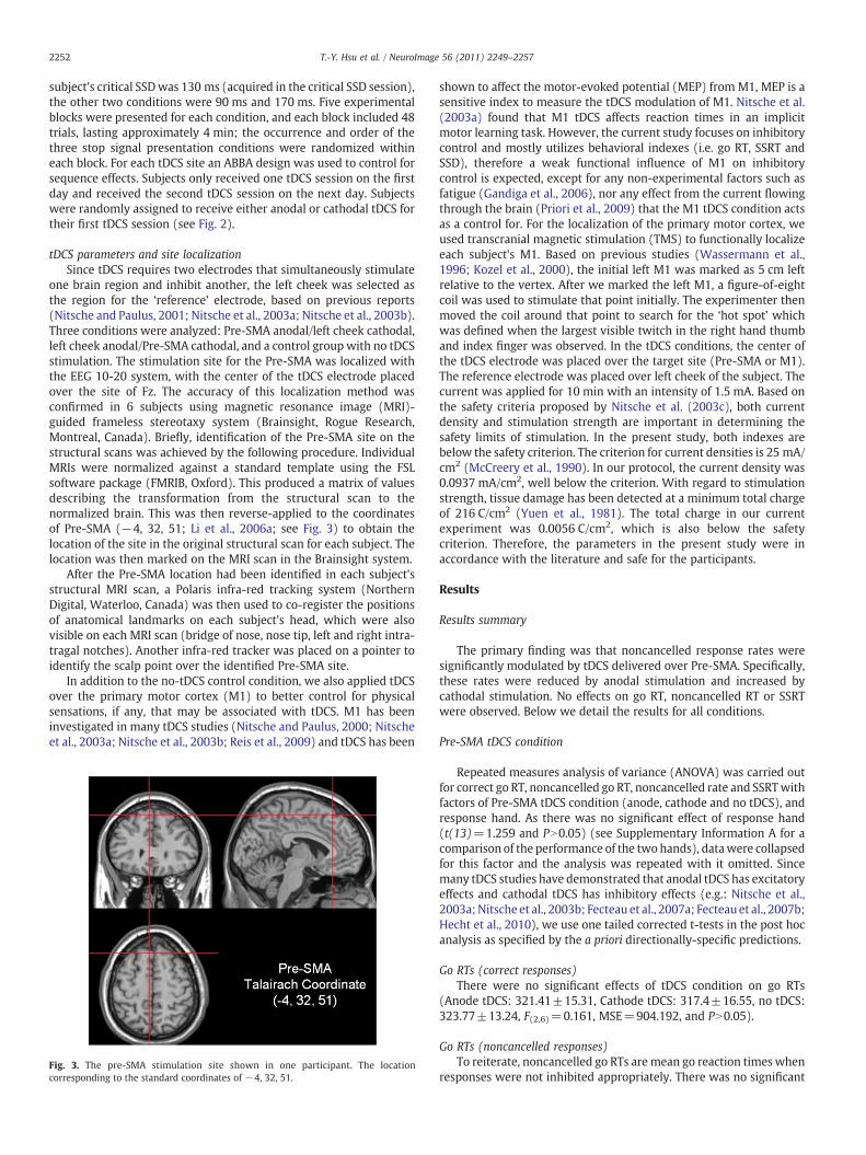
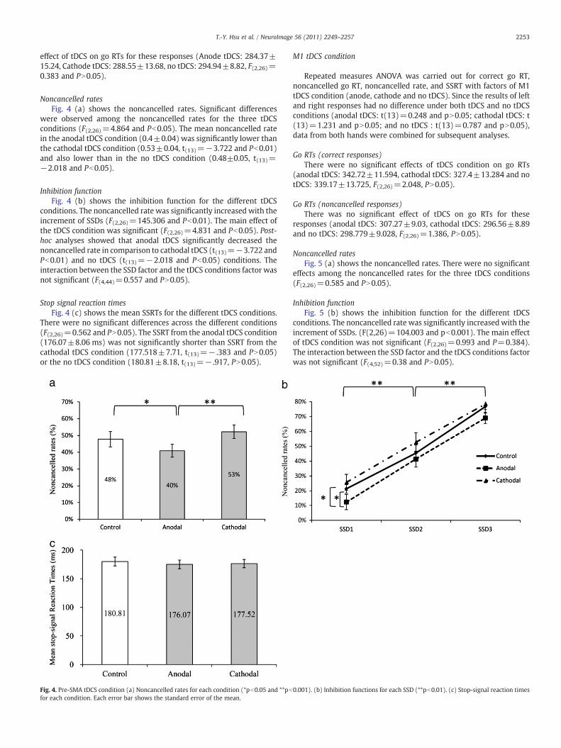
Noncancelled ratesFig. 4 (a) shows the noncancelled rates. Significant differences
were observed among the noncancelled rates for the three tDCSconditions (F(2,26)=4.864 and Pb0.05). The mean noncancelled ratein the anodal tDCS condition (0.4±0.04) was significantly lower thanthe cathodal tDCS condition (0.53±0.04, t(13)=−3.722 and Pb0.01)and also lower than in the no tDCS condition (0.48±0.05, t(13)=−2.018 and Pb0.05).
Inhibition functionFig. 4 (b) shows the inhibition function for the different tDCS
conditions. The noncancelled rate was significantly increased with theincrement of SSDs (F(2,26)=145.306 and Pb0.01). The main effect ofthe tDCS condition was significant (F(2,26)=4.831 and Pb0.05). Post-hoc analyses showed that anodal tDCS significantly decreased thenoncancelled rate in comparison to cathodal tDCS (t(13)=−3.722 andPb0.01) and no tDCS (t(13)=−2.018 and Pb0.05) conditions. Theinteraction between the SSD factor and the tDCS conditions factor wasnot significant (F(4,44)=0.557 and PN0.05).
Stop signal reaction timesFig. 4 (c) shows the mean SSRTs for the different tDCS conditions.
There were no significant differences across the different conditions(F(2,26)=0.562 and PN0.05). The SSRT from the anodal tDCS condition(176.07±8.06 ms) was not significantly shorter than SSRT from thecathodal tDCS condition (177.518±7.71, t(13)=− .383 and PN0.05)or the no tDCS condition (180.81±8.18, t(13)=− .917, PN0.05).
Fig. 4. Pre-SMA tDCS condition (a) Noncancelled rates for each condition (*pb0.05 and **pbfor each condition. Each error bar shows the standard error of the mean.
M1 tDCS condition
Repeated measures ANOVA was carried out for correct go RT,noncancelled go RT, noncancelled rate, and SSRT with factors of M1tDCS condition (anode, cathode and no tDCS). Since the results of leftand right responses had no difference under both tDCS and no tDCSconditions (anodal tDCS: t(13)=0.248 and pN0.05; cathodal tDCS: t(13)=1.231 and pN0.05; and no tDCS : t(13)=0.787 and pN0.05),data from both hands were combined for subsequent analyses.
Go RTs (correct responses)There were no significant effects of tDCS condition on go RTs
(anodal tDCS: 342.72±11.594, cathodal tDCS: 327.4±13.284 and notDCS: 339.17±13.725, F(2,26)=2.048, PN0.05).
Go RTs (noncancelled responses)There was no significant effect of tDCS on go RTs for these
responses (anodal tDCS: 307.27±9.03, cathodal tDCS: 296.56±8.89and no tDCS: 298.779±9.028, F(2,26)=1.386, PN0.05).
Noncancelled ratesFig. 5 (a) shows the noncancelled rates. There were no significant
effects among the noncancelled rates for the three tDCS conditions(F(2,26)=0.585 and PN0.05).
Inhibition functionFig. 5 (b) shows the inhibition function for the different tDCS
conditions. The noncancelled rate was significantly increasedwith theincrement of SSDs. (F(2,26)=104.003 and pb0.001). The main effectof tDCS condition was not significant (F(2,26)=0.993 and P=0.384).The interaction between the SSD factor and the tDCS conditions factorwas not significant (F(4,52)=0.38 and PN0.05).
0.001). (b) Inhibition functions for each SSD (**pb0.01). (c) Stop-signal reaction times
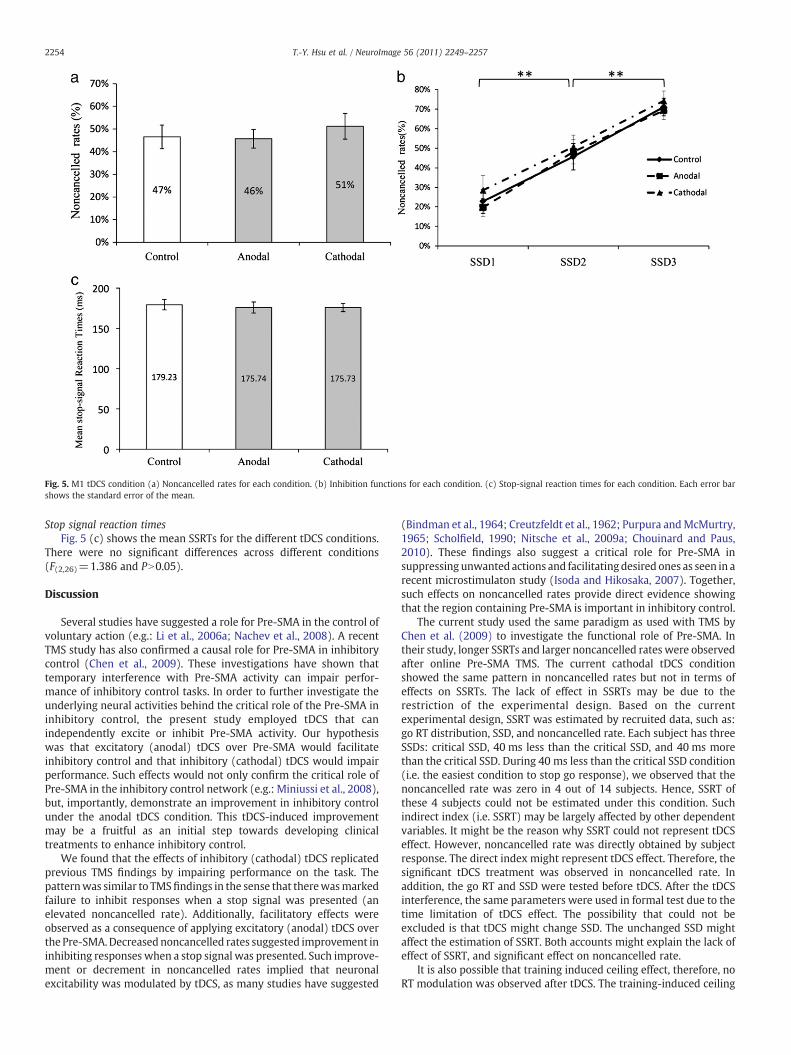
Fig. 5. M1 tDCS condition (a) Noncancelled rates for each condition. (b) Inhibition functions for each condition. (c) Stop-signal reaction times for each condition. Each error barshows the standard error of the mean.
2254 T.-Y. Hsu et al. / NeuroImage 56 (2011) 2249–2257
Stop signal reaction timesFig. 5 (c) shows the mean SSRTs for the different tDCS conditions.
There were no significant differences across different conditions(F(2,26)=1.386 and PN0.05).
Discussion
Several studies have suggested a role for Pre-SMA in the control ofvoluntary action (e.g.: Li et al., 2006a; Nachev et al., 2008). A recentTMS study has also confirmed a causal role for Pre-SMA in inhibitorycontrol (Chen et al., 2009). These investigations have shown thattemporary interference with Pre-SMA activity can impair perfor-mance of inhibitory control tasks. In order to further investigate theunderlying neural activities behind the critical role of the Pre-SMA ininhibitory control, the present study employed tDCS that canindependently excite or inhibit Pre-SMA activity. Our hypothesiswas that excitatory (anodal) tDCS over Pre-SMA would facilitateinhibitory control and that inhibitory (cathodal) tDCS would impairperformance. Such effects would not only confirm the critical role ofPre-SMA in the inhibitory control network (e.g.: Miniussi et al., 2008),but, importantly, demonstrate an improvement in inhibitory controlunder the anodal tDCS condition. This tDCS-induced improvementmay be a fruitful as an initial step towards developing clinicaltreatments to enhance inhibitory control.
We found that the effects of inhibitory (cathodal) tDCS replicatedprevious TMS findings by impairing performance on the task. Thepatternwas similar to TMSfindings in the sense that therewasmarkedfailure to inhibit responses when a stop signal was presented (anelevated noncancelled rate). Additionally, facilitatory effects wereobserved as a consequence of applying excitatory (anodal) tDCS overthe Pre-SMA.Decreasednoncancelled rates suggested improvement ininhibiting responses when a stop signal was presented. Such improve-ment or decrement in noncancelled rates implied that neuronalexcitability was modulated by tDCS, as many studies have suggested
(Bindman et al., 1964; Creutzfeldt et al., 1962; Purpura andMcMurtry,1965; Scholfield, 1990; Nitsche et al., 2009a; Chouinard and Paus,2010). These findings also suggest a critical role for Pre-SMA insuppressing unwanted actions and facilitating desired ones as seen in arecent microstimulaton study (Isoda and Hikosaka, 2007). Together,such effects on noncancelled rates provide direct evidence showingthat the region containing Pre-SMA is important in inhibitory control.
The current study used the same paradigm as used with TMS byChen et al. (2009) to investigate the functional role of Pre-SMA. Intheir study, longer SSRTs and larger noncancelled rates were observedafter online Pre-SMA TMS. The current cathodal tDCS conditionshowed the same pattern in noncancelled rates but not in terms ofeffects on SSRTs. The lack of effect in SSRTs may be due to therestriction of the experimental design. Based on the currentexperimental design, SSRT was estimated by recruited data, such as:go RT distribution, SSD, and noncancelled rate. Each subject has threeSSDs: critical SSD, 40 ms less than the critical SSD, and 40 ms morethan the critical SSD. During 40 ms less than the critical SSD condition(i.e. the easiest condition to stop go response), we observed that thenoncancelled rate was zero in 4 out of 14 subjects. Hence, SSRT ofthese 4 subjects could not be estimated under this condition. Suchindirect index (i.e. SSRT) may be largely affected by other dependentvariables. It might be the reason why SSRT could not represent tDCSeffect. However, noncancelled rate was directly obtained by subjectresponse. The direct index might represent tDCS effect. Therefore, thesignificant tDCS treatment was observed in noncancelled rate. Inaddition, the go RT and SSD were tested before tDCS. After the tDCSinterference, the same parameters were used in formal test due to thetime limitation of tDCS effect. The possibility that could not beexcluded is that tDCS might change SSD. The unchanged SSD mightaffect the estimation of SSRT. Both accounts might explain the lack ofeffect of SSRT, and significant effect on noncancelled rate.
It is also possible that training induced ceiling effect, therefore, noRT modulation was observed after tDCS. The training-induced ceiling
2255T.-Y. Hsu et al. / NeuroImage 56 (2011) 2249–2257
effect activates maximum individual ability level. Each subjectperformed three experimental conditions that might result insignificant practice effects on RT, possibly leading to no significantRT differences being observed. Another possibility is the variabilitybetween individuals. Comparing the mean SSRT from the currentstudy with Chen et al. (2009), mean SSRT here (anodal=176.07 ms;cathodal=177.51; and no tDCS=180.81) was faster than mean SSRT(Pre-SMA TMS=210.5; vertex TMS=193.5; and no TMS=196.9) inChen et al. (2009). Although no statistical index can be provided dueto different sample sizes, mean SSRT may provide another potentialexplanation that subjects in the current study had better inhibitorycontrol than subjects in the Chen et al. (2009) experiment. Thus, thevariability between different groups of subjects may also explain whyno RT modulation was observed under different tDCS conditions.
In addition, the different findings on the two dependent variables(i.e. noncancelled rates and SSRT) suggest that TMS and tDCS mayresult in different modulation on neurons even though both are activeinterference techniques. TMS may result in modulation of neuronalthresholds, or a change in synaptic efficiency, and these mechanismsare generally expressed as a form of functional plasticity ormetaplasticity (Siebner and Rothwell, 2003; Siebner et al., 2009;Silvanto et al., 2007a; Silvanto et al., 2007b). In contrast, tDCS changesneuron excitability. Anodal and cathodal stimulations show polarity-specific depolarizing or hyperpolarizing effects on neuron firing rates(Bindman et al., 1964; Creutzfeldt et al., 1962; Purpura andMcMurtry,1965; Scholfield, 1990; Nitsche et al., 2009b). In addition, with regardto focality, tDCS stimulates a relatively large area (1600 mm2) incontrast to TMS (25 mm2) (Priori et al., 2009). Thus, there is thepossibility that tDCS stimulation affected other nearby regions such asthe SMA. Imaging studies have demonstrated a functional role forSMA in complex sequences of finger movements (Roland et al., 1980;Goldberg, 1985; Verwey et al., 2002; Serrien et al., 2002; Steyverset al., 2003). However, the current experiment required only simpleone-button responses, which is unlikely to be heavily dependent uponSMA functioning. Furthermore, even if tDCS did stimulate SMA in thepresent study, changes in RTs and accuracywould also be expected forthe go condition, instead of merely affecting noncancelled rates. Thus,it seems unlikely that a lack of specificity can account for the effects onthe noncancelled rates.
Previous fMRI experiments have indicated that greater Pre-SMAactivity is associatedwithmore efficient inhibitory control as indicatedby shortened SSRTs (Li et al., 2006a; Chao et al., 2009). Moreover, thedirect anatomical and functional connections between Pre-SMA andthe basal ganglia (both caudate and STN) have been linked to a brakingsystem for prepotent responses of the type observed in the currentstudy (Aron et al., 2007a; Aron et al., 2007b; Aron and Poldrack., 2006;Madsen et al., 2010;Duann et al., 2009;Haggard, 2008; for a review seeNachev et al., 2008). This connection is further supported by severalEEG studieswhich have also demonstrated activation of Pre-SMApriorto the initiation of a planned action (e.g.: Brinkman and Porter, 1979;for a review see Nachev et al., 2008; c.f.: Swann et al., 2009) and thatthe amplitude of the lateralized readiness potential from the primarymotor cortex is correlated with the efficiency of inhibitory control(Dimoska and Johnstone, 2007). This evidence confirms a critical rolefor Pre-SMAwithin a neural network for inhibitory control in the stop-signal paradigm. It is important to note that a remote effect of tDCS onthe regions neighbouring the stimulation site is also possible. Thisremote effect can be mediated via cortico-cortical and cortico-subcortical connections. Anodal stimulation inducesmorewidespreadincreases in rCBF in remote brain regions whereas cathodal stimula-tion would have the opposite effect (Lang et al., 2005). Thus, althoughthe current study confirms a critical role for Pre-SMA and its vicinity ininhibitory control, it does not mean that Pre-SMA is the only neurallocus for such inhibitory processes, and should not be taken asevidence ruling out other regions such as IFG, basal ganglia, ACC, andSMA in the neural network.
Several studies have indicated that the cingulate cortex isconnected to Pre-SMA and motor cortex. A simple effect of alertnessand arousal might occur due to the current spread from Pre-SMA andmotor cortex to cingulate cortex. If this was driving the effects seen,the same level of alertness and arousal should be reflected on alldependent variables. However, the present results showed that onlynoncancelled rate, a specific index of inhibitory control, was affected.In addition, performance on this measurewas only affected in the Pre-SMA tDCS condition but not the M1 tDCS condition.
Indeed, Pre-SMA is not the only region that has been implicated toplay a key role in inhibitory processes. Other brain regions includingFEF (Hanes and Schall, 1996; Curtis et al., 2005; Muggleton et al.,2010), SEF (Stuphorn et al., 2000; Stuphorn and Schall, 2006; Isodaand Hikosaka, 2007; Chen et al., 2009), ACC (Ito et al., 2003; Chevrieret al., 2007), and IFG (Aron et al., 2003; Leung and Cai, 2007) have alsobeen reported to be involved in the cognitive processes required forthe stop-signal task. However, the precise role of each region, as wellas their interactions with each other in the context of inhibitorycontrol, remains to be investigated. For example, the basal ganglia/subthalamic route has been proposed to mediate action inhibition(Isoda and Hikosaka, 2008). However, this pathway was ruled out byMars et al. (2009) using paired-pulse transcranial magnetic stimula-tion. Mars et al. (2009) found that the M1 induced motor-evokedpotential (MEP) was affected 6 ms following the conditional pre-SMATMS. The duration is too short to pass through basal ganglia/subthalamic route. However, Neubert et al. (2010) provided newevidence that M1 induced MEP is not only affected 6 ms after byconditional pre-SMA TMS, but also 12 ms after conditional pre-SMAand conditional rIFG TMS. This raises the possibility that informationrelating to action inhibition may indeed pass through the basalganglia/subthalamic route. Additionally, IFG has been demonstratedto be necessary in the stop-signal paradigm (Chambers et al., 2006,2007) due to its function in inhibitory motor control (Aron andPoldrack, 2006; Rubia et al., 2003; Rubia et al., 2005; Verbruggen andLogan, 2008). The interactions between rIFG with M1 were furtherinvestigated with using paired-pulse transcranial magnetic stimula-tion. At 175 ms after a visual cue, rIFG inhibits M1 corticospinalactivity during action reprogramming (Mars et al., 2009; Neubertet al., 2010). But a fMRI study did not observe any correlation betweenIFG activation and SSRTs (Chao et al., 2009). In contrast, Li et al.(2006a) observed a linear correlation between the BOLD activation ofPre-SMA and SSRTs. They found that greater activation in the Pre-SMAled to shorter SSRTs, suggesting more efficient stop-signal processing.Converging evidence from other paradigms also suggests a criticalinvolvement of Pre-SMA in inhibitory control. Rushworth et al. (2002)applied TMS over Pre-SMA to demonstrate the role of the area in taskswitching. They showed that the involvement of Pre-SMA in their taskwas temporally precise and related to the time of switching (Rush-worth et al., 2002; Taylor et al., 2007). Similar results with Pre-SMATMS experiments in the stop signal task have been reported by Chenet al. (2009). They also applied temporally precise TMS over the leftPre-SMA by delivering one pulse concurrent with the appearance ofthe GO signal and another one at 100 ms after its appearance. Theydemonstrated that the TMS pulses impaired inhibitory control andthat the involvement of the Pre-SMA was early, consistent with a rolein conflict resolution rather than error monitoring.
Conclusion
Despite evidence from magnetic stimulation experiments andlesion studies showing impaired inhibition when Pre-SMA activity isdisrupted, no study has demonstrated that human inhibitory controlcan be improved with increased Pre-SMA activity. To the best of ourknowledge, this study is the first to demonstrate both inhibition andfacilitation effects by stimulating this area in healthy participants. Thefinding that it is possible to improve and impair inhibitory control
2256 T.-Y. Hsu et al. / NeuroImage 56 (2011) 2249–2257
using anodal and cathodal tDCS is important, as it suggests that thistype of stimulation may offer a potential clinical intervention forindividuals exhibiting difficulties with inhibitory control. This in-cludes a range of neuropsychiatric conditions, including but notlimited to ADHD (e.g. Durston, 2008, 2010; Li e al., 2006b, 2008b; seeSchachar and Logan, 1990, for an example in the stop-signal tasks),autism (Kana et al., 2007), and obsessive–compulsive disorder(Chamberlain et al., 2005). This is an area that may be fruitful forfuture research both in terms of stimulation effectiveness as well asdevelopment for longer-term clinical applications. In sum, by showingeffects of anodal and cathodal tDCS over Pre-SMA in stop-signalperformance, the current study further confirms that Pre-SMA has aunique role inmediating inhibitory control with the noncancelled rateeither increased or reduced by facilitatory or inhibitory tDCS.
Supplementarymaterials related to this article can be found onlineat doi:10.1016/j.neuroimage.2011.03.059.
Acknowledgments
This work was sponsored by the National Science Council, Taiwan(99-2410-H-008-022-MY3, 99-2911-I-008-025, 97-2511-S-008-005-MY3, 98-2410-H-008-010-MY3, 98-2517-S-004-001-MY3 and 97-2511-S-008-008-MY5). VW and NGM were supported by the UKMedical Research Council and VW was also supported by a RoyalSociety Wolfson Merit Award. CHJ was supported by the NationalScience Council, Taiwan (98-2918-I-008-011) and the Fulbrightscholarship, Taiwan-USA.
References
Aron, A.R., Poldrack, R.A., 2006. Cortical and subcortical contributions to stop signalresponse inhibition: role of the subthalamic nucleus. J. Neurosci. 26 (9), 2424–2433.
Aron, A.R., Fletcher, P.C., Bullmore, E.T., Sahakian, B.J., Robbins, T.W., 2003. Stop-signalinhibition disrupted by damage to right inferior frontal gyrus in humans. Nat.Neurosci. 6 (2), 115–116.
Aron, A.R., Behrens, T.E., Smith, S., Frank, M.J., Poldrack, R.A., 2007a. Triangulating acognitive control network using diffusion-weighted magnetic resonance imaging(MRI) and functional MRI. J. Neurosci. 27 (14), 3743–3752.
Aron, A.R., Durston, S., Eagle, D.M., Logan, G.D., Stinear, C.M., Stuphorn, V., 2007b.Converging evidence for a fronto–basal–ganglia network for inhibitory control ofaction and cognition. J. Neurosci. 27 (44), 11860–11864.
Ball, T., Schreiber, A., Feige, B., Wagner, M., Lucking, C.H., Kristeva-Feige, R., 1999. Therole of higher-order motor areas in voluntary movement as revealed by high-resolution EEG and fMRI. Neuroimage 10 (6), 682–694.
Band, G.P., van der Molen, M.W., Logan, G.D., 2003. Horse-race model simulations of thestop-signal procedure. Acta Psychol. 112 (2), 105–142.
Bindman, L.J., Lippold, O.C., Redfearn, J.W., 1964. The action of brief polarizing currentson the cerebral cortex of the rat (1) during current flow and (2) in the production oflong-lasting after-effects. J. Physiol. 172, 369–382.
Boucher, L., Palmeri, T.J., Logan, G.D., Schall, J.D., 2007. Inhibitory control in mind andbrain: an interactive race model of countermanding saccades. Psychol. Rev. 114 (2),376–397.
Brinkman, C., Porter, R., 1979. Supplementary motor area in the monkey: activity ofneurons during performance of a learned motor task. J. Neurophysiol. 42 (3),681–709.
Chamberlain, S.R., Blackwell, A.D., Fineberg, N.A., Robbins, T.W., Sahakian, B.J., 2005. Theneuropsychology of obsessive compulsive disorder: the importance of failures incognitive and behavioural inhibition as candidate endophenotypic markers.Neurosci. Biobehav. Rev. 29 (3), 399–419.
Chambers, C.D., Bellgrove, M.A., Stokes, M.G., Henderson, T.R., Garavan, H., Robertson, I.H., et al., 2006. Executive “brake failure” following deactivation of human frontallobe. J. Cogn. Neurosci. 18 (3), 444–455.
Chambers, C.D., Bellgrove, M.A., Gould, I.C., English, T., Garavan, H., McNaught, E., et al.,2007. Dissociable mechanisms of cognitive control in prefrontal and premotorcortex. J. Neurophysiol. 98 (6), 3638–3647.
Chao, H.H., Luo, X., Chang, J.L., Li, C.S., 2009. Activation of the pre-supplementary motorarea but not inferior prefrontal cortex in association with short stop signal reactiontime—an intra-subject analysis. BMC Neurosci. 10, 75.
Chen, C.Y., Muggleton, N.G., Juan, C.H., Tzeng, O.J., Hung, D.L., 2008. Time pressure leadsto inhibitory control deficits in impulsive violent offenders. Behav. Brain Res. 187(2), 483–488.
Chen, C.Y., Muggleton, N.G., Tzeng, O.J., Hung, D.L., Juan, C.H., 2009. Control of prepotentresponses by the superior medial frontal cortex. Neuroimage 44 (2), 537–545.
Chevrier, A.D., Noseworthy, M.D., Schachar, R., 2007. Dissociation of response inhibitionand performance monitoring in the stop signal task using event-related fMRI. Hum.Brain Mapp. 28 (12), 1347–1358.
Chouinard, P.A., Paus, T., 2010. What have we learned from “perturbing” the humancortical motor system with transcranial magnetic stimulation? Front. Hum.Neurosci. 4, 173.
Creutzfeldt, O.D., Fromm, G.H., Kapp, H., 1962. Influence of transcortical d-c currents oncortical neuronal activity. Exp. Neurol. 5, 436–452.
Curtis, C.E., Cole, M.W., Rao, V.Y., D'Esposito, M., 2005. Canceling planned action: anFMRI study of countermanding saccades. Cereb. Cortex 15 (9), 1281–1289.
Deiber, M.P., Honda, M., Ibanez, V., Sadato, N., Hallett, M., 1999. Mesial motor areas inself-initiated versus externally triggered movements examined with fMRI: effect ofmovement type and rate. J. Neurophysiol. 81 (6), 3065–3077.
Dimoska, A., Johnstone, S.J., 2007. Neural mechanisms underlying trait impulsivity innon-clinical adults: stop-signal performance and event-related potentials. Prog.Neuropsychopharmacol. Biol. Psychiatry 31 (2), 443–454.
Duann, J.R., Ide, J.S., Luo, X., Li, C.S., 2009. Functional connectivity delineates distinctroles of the inferior frontal cortex and presupplementary motor area in stop signalinhibition. J. Neurosci. 29 (32), 10171–10179.
Durston, S., 2008. Converging methods in studying attention-deficit/hyperactivitydisorder: what can we learn from neuroimaging and genetics? Dev. Psychopathol.20 (4), 1133–1143.
Durston, S., 2010. Imaging genetics in ADHD. Neuroimage 53 (3), 832–838.Eimer, M., Schlaghecken, F., 1998. Effects of masked stimuli on motor activation:
behavioral and electrophysiological evidence. J. Exp. Psychol. Hum. Percept.Perform. 24 (6), 1737–1747.
Fecteau, S., Knoch, D., Fregni, F., Sultani, N., Boggio, P., Pascual-Leone, A., 2007a.Diminishing risk-taking behavior by modulating activity in the prefrontal cortex: adirect current stimulation study. J. Neurosci. 27 (46), 12500–12505.
Fecteau, S., Pascual-Leone, A., Zald, D.H., Liguori, P., Theoret, H., Boggio, P.S., et al., 2007b.Activation of prefrontal cortex by transcranial direct current stimulation reducesappetite for risk during ambiguous decision making. J. Neurosci. 27 (23), 6212–6218.
Gandiga, P.C., Hummel, F.C., Cohen, L.G., 2006. Transcranial DC stimulation (tDCS): atool for double-blind sham-controlled clinical studies in brain stimulation. Clin.Neurophysiol. 117 (4), 845–850.
Goldberg, G., 1985. Supplementary motor area structure and function: review andhypotheses. Brain Res. 8 (159).
Haggard, P., 2008. Human volition: towards a neuroscience of will. Nat. Rev. Neurosci. 9(12), 934–946.
Hanes, D.P., Schall, J.D., 1996. Neural control of voluntary movement initiation. Science274 (5286), 427–430.
Hecht, D., Walsh, V., Lavidor, M., 2010. Transcranial direct current stimulationfacilitates decision making in a probabilistic guessing task. J. Neurosci. 30 (12),4241–4245.
Isoda, M., Hikosaka, O., 2007. Switching from automatic to controlled action by monkeymedial frontal cortex. Nat. Neurosci. 10 (2), 240–248.
Isoda, M., Hikosaka, O., 2008. Role for subthalamic nucleus neurons in switching fromautomatic to controlled eye movement. J. Neurosci. 28 (28), 7209–7218.
Ito, S., Stuphorn, V., Brown, J.W., Schall, J.D., 2003. Performance monitoring by theanterior cingulate cortex during saccade countermanding. Science 302 (5642),120–122.
Jenkins, I.H., Jahanshahi, M., Jueptner, M., Passingham, R.E., Brooks, D.J., 2000. Self-initiated versus externally triggered movements. II. The effect of movementpredictability on regional cerebral blood flow. Brain 123 (Pt 6), 1216–1228.
Kana, R.K., Keller, T.A., Minshew, N.J., Just, M.A., 2007. Inhibitory control in high-functioning autism: decreased activation and underconnectivity in inhibitionnetworks. Biol. Psychiatry 62 (3), 198–206.
Kenner, N.M., Mumford, J.A., Hommer, R.E., Skup, M., Leibenluft, E., Poldrack, R.A., 2010.Inhibitory motor control in response stopping and response switching. J. Neurosci.30 (25), 8512–8518.
Kozel, F.A., Nahas, Z., deBrux, C., Molloy, M., Lorberbaum, J.P., Bohning, D., et al., 2000.How coil-cortex distance relates to age, motor threshold, and antidepressantresponse to repetitive transcranial magnetic stimulation. J. Neuropsychiatry Clin.Neurosci. 12 (3), 376–384.
Lang, N., Siebner, H.R., Ward, N.S., Lee, L., Nitsche, M.A., Paulus, W., et al., 2005. Howdoes transcranial DC stimulation of the primary motor cortex alter regionalneuronal activity in the human brain? Eur. J. Neurosci. 22 (2), 495–504.
Leung, H.C., Cai, W., 2007. Common and differential ventrolateral prefrontal activityduring inhibition of hand and eye movements. J. Neurosci. 27 (37), 9893–9900.
Li, C.S., Huang, C., Constable, R.T., Sinha, R., 2006a. Imaging response inhibition in a stop-signal task: neural correlates independent of signal monitoring and post-responseprocessing. J. Neurosci. 26 (1), 186–192.
Li, C.S., Chang, H.L., Hsu, Y.P., Wang, H.S., Ko, N.C., 2006b. Motor response inhibition inchildren with Tourette's disorder. J. Neuropsychiatry Clin. Neurosci. 18, 417–419.
Li, C.S., Yan, P., Sinha, R., Lee, T.W., 2008a. Subcortical processes of motor responseinhibition during a stop signal task. Neuroimage 41 (4), 1352–1363.
Li, C.S., Huang, C., Yan, P., Bhagwagar, Z., Milivojevic, V., Sinha, R., 2008b. NeuralCorrelates of Impulse Control During Stop Signal Inhibition in Cocaine-DependentMen. Neuropsychopharmacology 33 (8), 1798–1806.
Logan, G.D., 1994. On the ability to inhibit thought and action: a users' guide to the stopsignal paradigm. In: Dagenbach, D., Carr, T.H. (Eds.), Inhibitory Processes inAttention, Memory, and Language. Academic Press, San Diego, pp. 189–239.
Logan, G.D., Cowan, W.B., 1984. On the ability to inhibit thought and action: a theory ofac act of control of ac act of control. Psychol. Rev. 295–327.
Logan, G.D., Cowan, W.B., Davis, K.A., 1984. On the ability to inhibit simple and choicereaction time responses: a model and a method. J. Exp. Psychol. Hum. Percept.Perform. 10 (2), 276–291.
Logan, G.D., Schachar, R.J., Tannock, R., 1997. Impulsivity and inhibitory control.Psychol. Sci. 8 (1), 60–64.
2257T.-Y. Hsu et al. / NeuroImage 56 (2011) 2249–2257
Madsen, K.S., Baare, W.F., Vestergaard, M., Skimminge, A., Ejersbo, L.R., Ramsoy, T.Z., etal., 2010. Response inhibition is associated with white matter microstructure inchildren. Neuropsychologia 48 (4), 854–862.
Mars, R.B., Klein, M.C., Neubert, F.X., Olivier, E., Buch, E.R., Boorman, E.D., Rushworth, M.F., 2009. Short-latency influence of medial frontal cortex on primary motor cortexduring action selection under conflict. J. Neurosci. 29 (21), 6926–6931.
McCreery, D.B., Agnew, W.F., Yuen, T.G., Bullara, L., 1990. Charge density and charge perphase as cofactors in neural injury induced by electrical stimulation. IEEE Trans.Biomed. Eng. 37 (10), 996–1001.
Merzagora, A.C., Foffani, G., Panyavin, I., Mordillo-Mateos, L., Aguilar, J., Onaral, B., et al.,2010. Prefrontal hemodynamic changes produced by anodal direct currentstimulation. Neuroimage 49 (3), 2304–2310.
Miniussi, C., Cappa, S.F., Cohen, L.G., Floel, A., Fregni, F., Nitsche, M.A., et al., 2008.Efficacy of repetitive transcranial magnetic stimulation/transcranial direct currentstimulation in cognitive neurorehabilitation. Brain Stimul. 1 (4), 326–336.
Muggleton, N.G., Chen, C.Y., Tzeng, O.J., Hung, D.L., Juan, C.H., 2010. Inhibitory controland the frontal eye fields. J. Cogn. Neurosci. 22 (12), 2804–2812.
Nachev, P., Rees, G., Parton, A., Kennard, C., Husain, M., 2005. Volition and conflict inhuman medial frontal cortex. Curr. Biol. 15 (2), 122–128.
Nachev, P., Kennard, C., Husain, M., 2008. Functional role of the supplementary and pre-supplementary motor areas. Nat. Rev. Neurosci. 9 (11), 856–869.
Neubert, F.X., Mars, R.B., Buch, E.R., Olivier, E., Rushworth, M.F., 2010. Cortical andsubcortical interactions during action reprogramming and their related whitematter pathways. Proc. Natl Acad. Sci. U. S. A. 107 (30), 13240–13245.
Nitsche, M.A., Paulus,W., 2000. Excitability changes induced in the humanmotor cortexby weak transcranial direct current stimulation. J. Physiol. 527 (3), 633–639.
Nitsche,M.A., Paulus,W., 2001. Sustained excitability elevations induced by transcranialDC motor cortex stimulation in humans. Neurology 57 (10), 1899–1901.
Nitsche, M.A., Fricke, K., Henschke, U., Schlitterlau, A., Liebetanz, D., Lang, N., et al.,2003a. Pharmacological modulation of cortical excitability shifts induced bytranscranial direct current stimulation in humans. J. Physiol. 553 (Pt 1), 293–301.
Nitsche, M.A., Liebetanz, D., Lang, N., Antal, A., Tergau, F., Paulus, W., 2003b. Safetycriteria for transcranial direct current stimulation (tDCS) in humans. Clin.Neurophysiol. 114 (11), 2220–2222 author reply 2222–2223.
Nitsche, M.A., Schauenburg, A., Lang, N., Liebetanz, D., Exner, C., Paulus, W., et al., 2003c.Facilitation of implicit motor learning byweak transcranial direct current stimulationof the primary motor cortex in the human. J. Cogn. Neurosci. 15 (4), 619–626.
Nitsche, M.A., Boggio, P.S., Fregni, F., Pascual-Leone, A., 2009a. Treatment of depressionwith transcranial direct current stimulation (tDCS): a Review. Exp. Neurol. 219 (1),14–19.
Nitsche, M.A., Kuo, M.F., Grosch, J., Bergner, C., Monte-Silva, K., Paulus, W., 2009b. D1-receptor impact on neuroplasticity in humans. J. Neurosci. 29 (8), 2648–2653.
Priori, A., Hallett, M., Rothwell, J.C., 2009. Repetitive transcranial magnetic stimulationor transcranial direct current stimulation? Brain Stimul. 2 (4), 241–245.
Purpura, D.P., McMurtry, J.G., 1965. Intracellular activities and evoked potentialchanges during polarization of motor cortex. J. Neurophysiol. 28, 166–185.
Reis, J., Schambra, H.M., Cohen, L.G., Buch, E.R., Fritsch, B., Zarahn, E., et al., 2009. Nonin-vasive cortical stimulation enhances motor skill acquisition over multiple daysthrough an effect on consolidation. Proc. Natl Acad. Sci. U. S. A. 106 (5), 1590–1595.
Roland, P.E., Larsen, B., Lassen, N.A., Skinhoj, E., 1980. Supplementary motor areaand other cortical areas in organization of voluntary movements in man. J.Neurophysiol. 43 (1), 118–136.
Rubia, K., Smith, A.B., Brammer, M.J., Taylor, E., 2003. Right inferior prefrontal cortexmediates response inhibition while mesial prefrontal cortex is responsible for errordetection. Neuroimage 20 (1), 351–358.
Rubia, K., Smith, A.B., Brammer, M.J., Toone, B., Taylor, E., 2005. Abnormal brainactivation during inhibition and error detection in medication-naive adolescentswith ADHD. Am. J. Psychiatry 162 (6), 1067–1075.
Rushworth, M.F., Passingham, R.E., Nobre, A.C., 2002. Components of switchingintentional set. J. Cogn. Neurosci. 14 (8), 1139–1150.
Schachar, R., Logan, G., 1990. Impulsivity and inhibitory control in normal developmentand childhood psychopathology. Dev. Psychol. 26, 710–720.
Schachar, R., Tannock, R., Logan, G.D., 1993. Inhibitory control, impulsiveness, andattention deficit hyperactivity disorder. Clin. Psychol. Rev. 13 (8), 721–739.
Schachar, R., Tannock, R., Marriott, M., Logan, G., 1995. Deficient inhibitory control inattention deficit hyperactivity disorder. J. Abnorm. Child Psychol. 23 (4),411–437.
Scholfield, C.N., 1990. Properties of K-currents in unmyelinated presynaptic axons ofbrain revealed revealed by extracellular polarisation. Brain Res. 507 (1),121–128.
Serrien, D.J., Strens, L.H., Oliviero, A., Brown, P., 2002. Repetitive transcranial magneticstimulation of the supplementarymotor area (SMA) degrades bimanual movementcontrol in humans. Neurosci. Lett. 328 (2), 89–92.
Sherrington, C.S., 1906. The Integrative Action of the Nervous System. Yale universitypress, New Haven.
Siebner, H.R., Rothwell, J., 2003. Transcranial magnetic stimulation: new insights intorepresentational cortical plasticity. Exp. Brain Res. 148 (1), 1–16.
Siebner, H., Hartwigsen, H., Kassuba, T., Rothwell, J., 2009. How does transcranialmagnetic stimulation modify neuronal activity in the brain? — implications forstudies of cognition. Cortex 45 (9), 1035–1042.
Silvanto, J., Muggleton, N.G., Cowey, A., Walsh, V., 2007a. Neural activation statedetermines behavioral susceptibility to modified theta burst transcranial magneticstimulation. Eur. J. Neurosci. 26 (2), 523–528.
Silvanto, J., Muggleton, N.G., Cowey, A., Walsh, V., 2007b. Neural adaptation revealsstate-dependent effects of transcranial magnetic stimulation. Eur. J. Neurosci. 25(6), 1874–1881.
Steyvers, M., Etoh, S., Sauner, D., Levin, O., Siebner, H.R., Swinnen, S.P., et al., 2003. High-frequency transcranial magnetic stimulation of the supplementary motor areareduces bimanual coupling during anti-phase but not in-phase movements. Exp.Brain Res. 151 (3), 309–317.
Stuphorn, V., Schall, J.D., 2006. Executive control of countermanding saccades by thesupplementary eye field. Nat. Neurosci. 9 (7), 925–931.
Stuphorn, V., Taylor, T.L., Schall, J.D., 2000. Performance monitoring by thesupplementary eye field. Nature 408 (6814), 857–860.
Sumner, P., Nachev, P., Morris, P., Peters, A.M., Jackson, S.R., Kennard, C., et al., 2007.Human medial frontal cortex mediates unconscious inhibition of voluntary action.Neuron 54 (5), 697–711.
Swann, N., Tandon, N., Canolty, R., Ellmore, T.M., McEvoy, L.K., Dreyer, S., et al., 2009.Intracranial EEG reveals a time- and frequency-specific role for the right inferiorfrontal gyrus and primary motor cortex in stopping initiated responses. J. Neurosci.29 (40), 12675–12685.
Taylor, P.C., Nobre, A.C., Rushworth, M.F., 2007. Subsecond changes in top down controlexerted by human medial frontal cortex during conflict and action selection: acombined transcranial magnetic stimulation electroencephalography study. J.Neurosci. 27 (42), 11343–11353.
Toma, K., Honda, M., Hanakawa, T., Okada, T., Fukuyama, H., Ikeda, A., et al., 1999.Activities of the primary and supplementary motor areas increase in preparationand execution of voluntary muscle relaxation: an event-related fMRI study. J.Neurosci. 19 (9), 3527–3534.
Verbruggen, F., Logan, G.D., 2008. Response inhibition in the stop-signal paradigm.Trends Cogn. Sci. 12 (11), 418–424.
Verwey, W.B., Lammens, R., van Honk, J., 2002. On the role of the SMA in the discretesequence production task: a TMS study. Transcranial Magnetic Stimulation.Neuropsychologia 40 (8), 1268–1276.
Wassermann, E.M., Wang, B., Zeffiro, T.A., Sadato, N., Pascual-Leone, A., Toro, C., et al.,1996. Locating the motor cortex on the MRI with transcranial magnetic stimulationand PET. Neuroimage 3 (1), 1–9.
Yuen, T.G., Agnew, W.F., Bullara, L.A., Jacques, S., McCreery, D.B., 1981. Histologicalevaluation of neural damage from electrical stimulation: considerations for theselection of parameters for clinical application. Neurosurgery 9 (3), 292–299.
Related Documents