Modulating Gradients in Regulatory Signals within Mesenchymal Stem Cell Seeded Hydrogels: A Novel Strategy to Engineer Zonal Articular Cartilage Stephen D. Thorpe 1,2 , Thomas Nagel 1,2 , Simon F. Carroll 1,2 , Daniel J. Kelly 1,2 * 1 Trinity Centre for Bioengineering, Trinity Biomedical Sciences Institute, Trinity College Dublin, Dublin, Ireland, 2 Department of Mechanical and Manufacturing Engineering, School of Engineering, Trinity College Dublin, Dublin, Ireland Abstract Engineering organs and tissues with the spatial composition and organisation of their native equivalents remains a major challenge. One approach to engineer such spatial complexity is to recapitulate the gradients in regulatory signals that during development and maturation are believed to drive spatial changes in stem cell differentiation. Mesenchymal stem cell (MSC) differentiation is known to be influenced by both soluble factors and mechanical cues present in the local microenvironment. The objective of this study was to engineer a cartilaginous tissue with a native zonal composition by modulating both the oxygen tension and mechanical environment thorough the depth of MSC seeded hydrogels. To this end, constructs were radially confined to half their thickness and subjected to dynamic compression (DC). Confinement reduced oxygen levels in the bottom of the construct and with the application of DC, increased strains across the top of the construct. These spatial changes correlated with increased glycosaminoglycan accumulation in the bottom of constructs, increased collagen accumulation in the top of constructs, and a suppression of hypertrophy and calcification throughout the construct. Matrix accumulation increased for higher hydrogel cell seeding densities; with DC further enhancing both glycosaminoglycan accumulation and construct stiffness. The combination of spatial confinement and DC was also found to increase proteoglycan-4 (lubricin) deposition toward the top surface of these tissues. In conclusion, by modulating the environment through the depth of developing constructs, it is possible to suppress MSC endochondral progression and to engineer tissues with zonal gradients mimicking certain aspects of articular cartilage. Citation: Thorpe SD, Nagel T, Carroll SF, Kelly DJ (2013) Modulating Gradients in Regulatory Signals within Mesenchymal Stem Cell Seeded Hydrogels: A Novel Strategy to Engineer Zonal Articular Cartilage. PLoS ONE 8(4): e60764. doi:10.1371/journal.pone.0060764 Editor: Hani A. Awad, University of Rochester, United States of America Received January 3, 2013; Accepted March 2, 2013; Published April 16, 2013 Copyright: ß 2013 Thorpe et al. This is an open-access article distributed under the terms of the Creative Commons Attribution License, which permits unrestricted use, distribution, and reproduction in any medium, provided the original author and source are credited. Funding: Funding was provided by Science Foundation Ireland (President of Ireland Young Researcher Award: 08/Y15/B1336) and the European Research Council (StemRepair – Project number 258463). The funders had no role in study design, data collection and analysis, decision to publish, or preparation of the manuscript. Competing Interests: The authors have declared that no competing interests exist. * E-mail: [email protected] Introduction Adult articular cartilage consists of three separate structural zones; the superficial tangential, middle and deep zones. This depth dependent composition and organisation is fundamental to the normal physiological function of articular cartilage [1,2]. Not only does cell morphology and arrangement change with depth, but each zone has distinct extra-cellular matrix (ECM) composi- tion, architecture and mechanical properties. The dominant load carrying structural components of the ECM are collagen (,75% tissue by dry weight) and proteoglycan (20%–30% tissue by dry weight), the concentrations of which vary with depth from the articular surface [1,3,4]. Collagen content is highest in the superficial zone, decreasing by ,20% in the middle and deep zones [1,3]. Proteoglycan content is lowest at the surface, increasing by as much as 50% into the middle and deep zones [3,4]. The zonal composition and structural organisation of the ECM determine the biomechanical properties which also vary through the tissue depth; such that the compressive modulus increases from the superficial zone to the deep zone [2,5,6], while the tensile modulus decreases from the superficial surface to the deep zone [7]. An on-going challenge in the field of articular cartilage regeneration is the attainment of this stratified zonal structure. Classical tissue engineering approaches focus primarily on forming homogeneous tissues by embedding chondrocytes or stem cells in various scaffolds and do not attempt to mimic the organised zonal architecture of articular cartilage. One approach toward this aim is to utilise chondrocytes from specific zones of articular cartilage in the corresponding regions of an engineered construct [8–10]. It has been shown that chondrocytes from different zones demon- strate different biosynthetic activities [9,10]. Layering such zonal chondrocytes in a photo-polymerising hydrogel has been shown to result in increased sulphated glycosaminoglycan (sGAG) accumu- lation in the bottom of the construct, although collagen content was also significantly higher in the bottom when compared to the top [10]. Another approach to engineering zonal cartilage is to vary biomaterial properties such as pore size [11], stiffness [12,13] or composition [14,15] through the depth of the scaffold or hydrogel. For example, combining layers of 2% and 3% agarose leads to zonal differences in the initial mechanical properties of the construct, however chondrocyte matrix elaboration in such bi- layered constructs was inferior to that in uniform 2% agarose [12]. Further improvements were observed with the application of PLOS ONE | www.plosone.org 1 April 2013 | Volume 8 | Issue 4 | e60764

Welcome message from author
This document is posted to help you gain knowledge. Please leave a comment to let me know what you think about it! Share it to your friends and learn new things together.
Transcript
Modulating Gradients in Regulatory Signals withinMesenchymal Stem Cell Seeded Hydrogels: A NovelStrategy to Engineer Zonal Articular CartilageStephen D. Thorpe1,2, Thomas Nagel1,2, Simon F. Carroll1,2, Daniel J. Kelly1,2*
1 Trinity Centre for Bioengineering, Trinity Biomedical Sciences Institute, Trinity College Dublin, Dublin, Ireland, 2Department of Mechanical and Manufacturing
Engineering, School of Engineering, Trinity College Dublin, Dublin, Ireland
Abstract
Engineering organs and tissues with the spatial composition and organisation of their native equivalents remains a majorchallenge. One approach to engineer such spatial complexity is to recapitulate the gradients in regulatory signals thatduring development and maturation are believed to drive spatial changes in stem cell differentiation. Mesenchymal stemcell (MSC) differentiation is known to be influenced by both soluble factors and mechanical cues present in the localmicroenvironment. The objective of this study was to engineer a cartilaginous tissue with a native zonal composition bymodulating both the oxygen tension and mechanical environment thorough the depth of MSC seeded hydrogels. To thisend, constructs were radially confined to half their thickness and subjected to dynamic compression (DC). Confinementreduced oxygen levels in the bottom of the construct and with the application of DC, increased strains across the top of theconstruct. These spatial changes correlated with increased glycosaminoglycan accumulation in the bottom of constructs,increased collagen accumulation in the top of constructs, and a suppression of hypertrophy and calcification throughoutthe construct. Matrix accumulation increased for higher hydrogel cell seeding densities; with DC further enhancing bothglycosaminoglycan accumulation and construct stiffness. The combination of spatial confinement and DC was also found toincrease proteoglycan-4 (lubricin) deposition toward the top surface of these tissues. In conclusion, by modulating theenvironment through the depth of developing constructs, it is possible to suppress MSC endochondral progression and toengineer tissues with zonal gradients mimicking certain aspects of articular cartilage.
Citation: Thorpe SD, Nagel T, Carroll SF, Kelly DJ (2013) Modulating Gradients in Regulatory Signals within Mesenchymal Stem Cell Seeded Hydrogels: A NovelStrategy to Engineer Zonal Articular Cartilage. PLoS ONE 8(4): e60764. doi:10.1371/journal.pone.0060764
Editor: Hani A. Awad, University of Rochester, United States of America
Received January 3, 2013; Accepted March 2, 2013; Published April 16, 2013
Copyright: � 2013 Thorpe et al. This is an open-access article distributed under the terms of the Creative Commons Attribution License, which permitsunrestricted use, distribution, and reproduction in any medium, provided the original author and source are credited.
Funding: Funding was provided by Science Foundation Ireland (President of Ireland Young Researcher Award: 08/Y15/B1336) and the European ResearchCouncil (StemRepair – Project number 258463). The funders had no role in study design, data collection and analysis, decision to publish, or preparation of themanuscript.
Competing Interests: The authors have declared that no competing interests exist.
* E-mail: [email protected]
Introduction
Adult articular cartilage consists of three separate structural
zones; the superficial tangential, middle and deep zones. This
depth dependent composition and organisation is fundamental to
the normal physiological function of articular cartilage [1,2]. Not
only does cell morphology and arrangement change with depth,
but each zone has distinct extra-cellular matrix (ECM) composi-
tion, architecture and mechanical properties. The dominant load
carrying structural components of the ECM are collagen (,75%
tissue by dry weight) and proteoglycan (20%–30% tissue by dry
weight), the concentrations of which vary with depth from the
articular surface [1,3,4]. Collagen content is highest in the
superficial zone, decreasing by ,20% in the middle and deep
zones [1,3]. Proteoglycan content is lowest at the surface,
increasing by as much as 50% into the middle and deep zones
[3,4]. The zonal composition and structural organisation of the
ECM determine the biomechanical properties which also vary
through the tissue depth; such that the compressive modulus
increases from the superficial zone to the deep zone [2,5,6], while
the tensile modulus decreases from the superficial surface to the
deep zone [7].
An on-going challenge in the field of articular cartilage
regeneration is the attainment of this stratified zonal structure.
Classical tissue engineering approaches focus primarily on forming
homogeneous tissues by embedding chondrocytes or stem cells in
various scaffolds and do not attempt to mimic the organised zonal
architecture of articular cartilage. One approach toward this aim
is to utilise chondrocytes from specific zones of articular cartilage
in the corresponding regions of an engineered construct [8–10]. It
has been shown that chondrocytes from different zones demon-
strate different biosynthetic activities [9,10]. Layering such zonal
chondrocytes in a photo-polymerising hydrogel has been shown to
result in increased sulphated glycosaminoglycan (sGAG) accumu-
lation in the bottom of the construct, although collagen content
was also significantly higher in the bottom when compared to the
top [10]. Another approach to engineering zonal cartilage is to
vary biomaterial properties such as pore size [11], stiffness [12,13]
or composition [14,15] through the depth of the scaffold or
hydrogel. For example, combining layers of 2% and 3% agarose
leads to zonal differences in the initial mechanical properties of the
construct, however chondrocyte matrix elaboration in such bi-
layered constructs was inferior to that in uniform 2% agarose [12].
Further improvements were observed with the application of
PLOS ONE | www.plosone.org 1 April 2013 | Volume 8 | Issue 4 | e60764
dynamic compressive strain, or seeding zone specific chondrocytes
into layered agarose hydrogels [13,16]. While promising, there are
potential limitations associated with such an approach, including
the development of a distinct boundary between gel layers which
could delineate as a result of shear stress [17]. Furthermore,
isolation of chondrocytes from separate zones of articular cartilage
can be difficult, particularly in damaged and diseased human
tissue where the zonal differences are less distinct and potential
biopsies are limited in size.
An alternative strategy to engineering zonal cartilage is to
attempt to recapitulate aspects of the tissue micro-environment
which may be responsible for the creation of depth-dependent
properties in articular cartilage during development and matura-
tion. In this respect, undifferentiated mesenchymal stem cells
(MSCs) could prove to be a more appropriate cell source, as when
provided with suitable stimuli they may differentiate into
chondrocytes with specific zonal phenotypes. For example, low
oxygen tension, a characteristic of avascular articular cartilage, has
been shown to enhance chondrogenesis of mesenchymal stem cells
(MSCs) [18–25]. In addition to oxygen, it has long been proposed
that mechanical signals guide the differentiation of mesenchymal
stem cells [26–30]. Hydrostatic pressure has been shown to
promote a chondrogenic phenotype [31–34], enhancing collagen
and sulphated glycosaminoglycan (sGAG) accumulation [35] and
increasing the mechanical stiffness [36] of cartilaginous grafts
engineered using MSCs encapsulated in agarose hydrogels.
Dynamic compressive strain, another key component of the
mechanical environment of articular cartilage, has also been
shown to promote MSC chondrogenesis [30,37–42]; positively
modulating the functional development of cartilaginous constructs
engineered using MSCs [43]. It has also been demonstrated that
intermittent cyclic tensile strain applied to MSC seeded constructs
increases collagen accumulation [44,45].
The objective of this study was to engineer a cartilaginous
construct with native-like zonal composition using MSCs by
controlling both the oxygen tension and mechanical environment
thorough the depth of the developing tissue. In an attempt to
create a gradient in oxygen tension through the depth of the
construct mimicking that in normal articular cartilage [46], the
bottom halves of MSC seeded agarose constructs were radially
confined; limiting oxygen transport into this region of the
construct. Furthermore, by subjecting these radially confined
constructs to dynamic compression it is possible to modulate the
mechanical environment throughout the depth of the tissue, with
higher levels of fluid pressure in the bottom of the construct and
greater strains across the top of the construct. It is hypothesised
that such a depth dependent microenvironment will lead to the
development of zonal cartilage tissues with a composition mim-
icking that of normal articular cartilage.
Materials and Methods
Cell Isolation and ExpansionPorcine MSCs were isolated and maintained as previously
described [42]. Animals were bred and raised for food and not
research purposes and were not subject to any procedures prior to
their sacrifice, hence no specific ethical approval was required for
this study. Briefly, mononuclear cells were isolated from the
femora of 4 month old pigs (,50 kg) within 2 hours of sacrifice
and plated at 106106 mononuclear cells per 75 cm2 culture flask
(Nunclon; Nunc, VWR, Dublin, Ireland) allowing colony forma-
tion. MSCs were maintained in high-glucose Dulbecco’s modified
eagle medium (4.5 mg/mL D-Glucose; hgDMEM) supplemented
with 10% foetal bovine serum (FBS), penicillin (100 U/mL)-
streptomycin (100 mg/mL) (all Gibco, Invitrogen, Dublin, Ireland)
and amphotericin B (0.25 mg/mL; Sigma-Aldrich, Arklow, Ire-
land). Cultures were washed in Dulbecco’s phosphate buffered
saline (PBS) after 72 hrs. When ,75% confluent, MSCs were re-
plated at 56103 cells/cm2 and expanded to passage two in
a humidified atmosphere at 37uC and 5% CO2.
Agarose Hydrogel Encapsulation and ConstructConfinementMSCs were suspended in defined chondrogenic medium
consisting of hgDMEM supplemented with penicillin (100 U/
mL)-streptomycin (100 mg/mL) (both Gibco), 0.25 mg/mL am-
photericin B, 100 mg/ml sodium pyruvate, 40 mg/mL L-proline,
1.5 mg/mL bovine serum albumin, 4.7 mg/mL linoleic acid, 16insulin–transferrin–selenium, 50 mg/mL L-ascorbic acid-2-phos-
phate, 100 nM dexamethasone (all Sigma-Aldrich) and 10 ng/mL
TGF-b3 (Pro Spec-Tany TechnoGene Ltd., Rehovot, Israel). This
cell suspension was mixed with agarose (Type VII; Sigma-Aldrich)
in PBS at a ratio of 1:1 at approx. 40uC, to yield a final agarose
concentration of 2% and a cell density of either 206106 cells/mL
or 506106 cells/mL. The agarose-cell suspension was cast
between stainless steel plates, one of which was overlaid with
a patterned PDMS layer, allowed cool to 21uC for 30 min., and
cored to produce cylindrical constructs (Ø 6 mm64 mm thick-
ness) which were patterned on one surface. Constructs remained
patterned side up throughout culture and were maintained in
,1 mL chondrogenic medium per 16106 cells/day with medium
exchanged every 3 or 4 days and sampled for biochemical analysis.
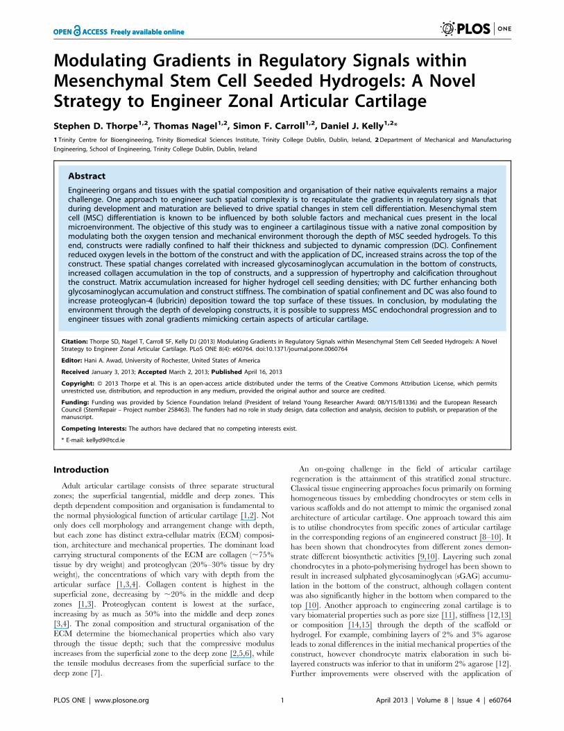
Either directly after fabrication or at day 21 of culture, constructs
were press-fitted into custom made PTFE confinement chambers
(Fig. 1) where they remained for the outstanding culture duration.
Dynamic Compression ApplicationDynamic compressive loading was applied as described pre-
viously [42] to constructs from day 21 to day 42 of culture.
Unconfined intermittent dynamic compression (DC) was carried
out in an incubator-housed, dynamic compression bioreactor and
consisted of a sine wave of 10% strain amplitude superimposed
upon a 1% pre-strain, with a 0.01 N per construct preload at
a frequency of 1 Hz for 4 hours/day, 5 days/week.
Mechanical Testing and Analysis of Physical ParametersOn removal from culture, construct diameter and wet weight
(ww) were recorded. Constructs were mechanically tested in
unconfined compression between impermeable platens using
a standard materials testing machine (Bose Electroforce 3100;
Figure 1. Experimental design. Constructs were press-fitted intocustom made PTFE wells such that the bottom 2 mm of the constructthickness was confined.doi:10.1371/journal.pone.0060764.g001
Tissue Engineering Zonal Cartilage
PLOS ONE | www.plosone.org 2 April 2013 | Volume 8 | Issue 4 | e60764
Bose Corporation, Gillingham, UK) as previously described [47].
A preload of 0.01 N was applied to ensure that the construct
surface was in direct contact with the impermeable loading
platens. Stress relaxation tests were performed consisting of a ramp
displacement of 0.025%/s up to 10% strain, which was
maintained until equilibrium was reached (,30 minutes). This
was followed by a dynamic test where cyclic strain amplitude of
1% (10%–11% total strain) was applied for 10 cycles at 1 Hz.
Samples were subsequently sliced into top and bottom regions
using a custom built rig and each region was tested simultaneously
on separate, randomly assigned material testing machines (Zwick
Roell Z005; Zwick Testing Machines Ltd., Herefordshire, UK) as
above.
Biochemical ConstituentsAfter mechanical testing of top and bottom construct regions,
the wet weight (ww) of each was recorded and the constructs
frozen at 285uC for further analyses. The biochemical content of
constructs was assessed as previously described [42]. Samples were
digested with papain (125 mg/ml) in 0.1 M sodium acetate, 5 mM
L-cysteine HCl, 0.05 M EDTA (all Sigma-Aldrich), pH 6.0 at
60uC under constant rotation for 18 hours. DNA content was
quantified using the Hoechst Bisbenzimide 33258 dye assay
(Sigma-Aldrich) as previously described [48]. The sulphated
glycosaminoglycan (sGAG) content was quantified using the
dimethylmethylene blue dye-binding assay (Blyscan; Biocolor
Ltd., Carrickfergus, Northern Ireland). Total collagen content
was determined by measuring orthohydroxyproline via the
dimethylaminobenzaldehyde and chloramine T assay [49]. A
hydroxyproline-to-collagen ratio of 1:7.69 was used [50]. Cell
culture media was analysed for sGAG and collagen secreted.
Histology and ImmunohistochemistryConstructs were fixed in 4% paraformaldehyde, paraffin
embedded and sectioned at 5 mm to produce a cross section
perpendicular to the disc face. Sections were stained for sGAG
with 1% alcian blue 8 GX in 0.1 M HCl, for collagen with
picro-sirius red and for calcific deposition with 1% alizarin red
(all Sigma-Aldrich). Immunohistochemistry was performed as
described previously [42]. Sections were enzymatically treated
with chondroitinase ABC (Sigma-Aldrich) in a humidified
environment at 37uC. Slides were blocked with goat serum
(Sigma-Aldrich) and sections were incubated for 1 hour with
a primary antibody diluted in blocking buffer specific to either
collagen type I (1:400, 1 mg/mL), collagen type II (1:100;
1 mg/mL), collagen type X (1:200; 1.4 mg/mL) (all Abcam,
Cambridge, UK) or proteoglycan-4 (PRG4; 1:200; 1 mg/mL)
(Sigma-Aldrich). After washing in PBS, sections were incubated
for 1 hour in the secondary antibody, anti-mouse IgG biotin
antibody produced in goat (1:200; 2 mg/mL; Sigma-Aldrich).
Colour was developed using the Vectastain ABC kit followed by
exposure to peroxidase DAB substrate kit (both Vector
Laboratories, Peterborough, UK). Immunofluorescent staining
was employed for collagen type X, which involved permeabi-
lisation of the cell membrane with 0.1% Triton-X100. In place
of colour development, sections were incubated with ExtrA-
vidin-FITC (1:100; Sigma-Aldrich) for 1 hour, washed several
times in PBS, nuclei counterstained with DAPI (1:500; 1 mg/
mL; VWR), and sections mounted using Vectashield (Vector
Laboratories). Sections were imaged with an Olympus IX51
inverted fluorescent microscope fitted with an Olympus DP70
camera. Sections of porcine cartilage, ligament and/or growth
plate were included as controls.
Theoretical Prediction of Mechanical Environment withinAgarose ConstructsTo estimate the effect of semi-confinement on the mechanical
environment within the construct, pore pressure and maximum
principal strain were predicted for day 0 constructs. The loading
protocol of the bioreactor was simulated with material properties
derived from a sample specific fit to constructs mechanically tested
at day 0. Cell-seeded constructs were modelled as fluid saturated
porous media using a finite strain formulation applied previously
to agarose in compression [51]. Surfaces in contact with media
were modelled as free draining, while fluid flow was prohibited
across surfaces in contact with the loading platens or the
confinement well. Nonlinear permeability was modelled following
Gu et al. [52]. Briefly, the solid matrix was modelled as nonlinearly
viscoelastic based on a multiplicative decomposition of the
deformation gradient into elastic and viscous parts F=FeFv. An
evolution equation was defined for the viscous right Cauchy-Green
tensor Cv:
_CCv~1
gvCvTovC
where gv is the viscosity function, Tov is the viscoelastic overstress
and C is the right Cauchy-Green tensor [51,53]. While the
equilibrium free Helmholtz energy potentials where based on
a Neo-Hookean formulation
yeq~C1 I1 Cð Þ{ ln I3 Cð Þð Þ{3½ �zD2 ln I3 Cð Þð Þ2
the viscoelastic overstresses where derived from the exponential
potential
yov~C1v
aveav I1 CC{1
vð Þ{ ln I3 CC{1vð Þð Þ{3½ �{1
h izD2v ln I3 CC{1
v
� �� �2
The equilibrium properties C1 and D2 were determined
analytically from the relaxed part of the ramp and hold test. D2v
was set to C1vD2/C1. The remaining parameters in the viscoelastic
potential and the viscosity were fit to unconfined ramp and hold
force relaxation curves using a differential evolution algorithm
developed by Storn and Price [54]. For further details see Gorke
et al. [51].
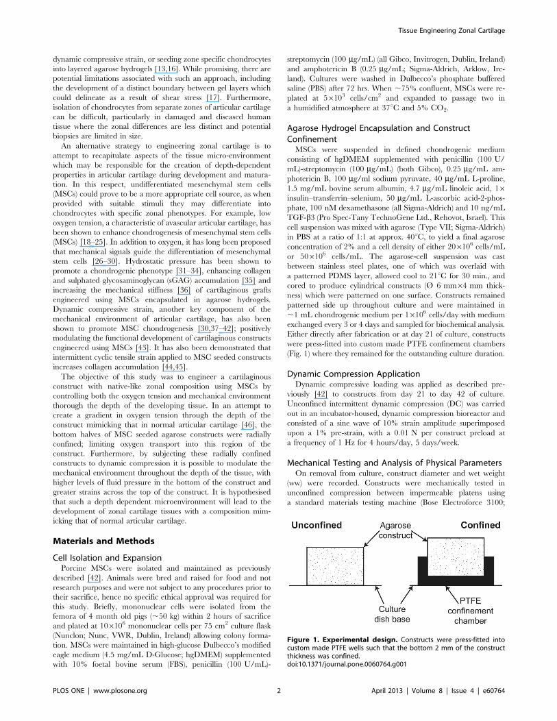
Measurement and Prediction of Oxygen Concentrationwithin Agarose ConstructsLocal oxygen concentration was assessed using implantable
fibre optic oxygen micro-sensors (Microx TX; PreSens – Precision
Sensing GmbH, Regensburg, Germany). Media was added to day
5 constructs seeded with 206106 cells/mL in confined and
unconfined configurations so that the surface of the media was
1 mm above the top surface of the construct. The sensor tip was
positioned at the media surface above the centre of the construct.
Constructs were allowed equilibrate for$24 hours in an incubator
at 18.5% oxygen prior to oxygen measurement. A linear actuator
(NA0830; Zaber Technologies Inc., Vancouver, Canada) was used
to control the movement of the sensor tip which was moved
vertically downward along the construct axis at 1.8 mm/s to within
,1 mm of the construct base; to avoid collision of the probe with
the culture dish or confinement chamber. Oxygen concentration
was sampled every 5 s.
Tissue Engineering Zonal Cartilage
PLOS ONE | www.plosone.org 3 April 2013 | Volume 8 | Issue 4 | e60764
Oxygen concentration in the constructs was modelled using
a diffusion-reaction type equation. The reaction term followed
Michaelis-Menten kinetics:
LcLt
~D+2c{nQmc
Kmzc
Here, c is the oxygen concentration, D the diffusion coefficient, n
the cell density, Qm the maximum consumption rate and Km the
concentration at half the maximum consumption rate. The
diffusion coefficient in 2% agarose was determined using the
Mackie and Meares relation
Dag
DH2O~
q2f
2{qf� �
so that Dag=2.7761023 mm2/s and Qf is the fluid phase volume
fraction [55]. The cellular consumption value was set to
Qm=13.5610218 mol/cell/s as determined by the model fit to
the experimental data for unconfined constructs with 206106
cells/mL (Fig. 2A). This value was similar to previously reported
values for MSC oxygen consumption while undergoing chondro-
genic differentiation [56]. The Michaelis-Menten constant, Km was
set to 561025 mmol/mm3. The sensitivity of the simulation
outcome to this value is very low. The construct was modelled as
axisymmetric. Oxygen diffusion through the culture media was
accounted for by setting Dmedia=3.061023 mm2/s. The oxygen
concentration at the media surface was prescribed as 185 mM,
while at surfaces in contact with the bottom of the well, the
confining chamber and the symmetry axis the flux was set to zero.
The simulations were performed for cell concentrations of
n=206106 cells/mL and n=506106 cells/mL.
Statistical AnalysisPresented are results from one of two replicate studies with
unique donors where n refers to the number of constructs analysed
for each assay within a given replicate (n numbers provided in
figure legends). Statistics were performed using MINITAB 15.1
software package (Minitab Ltd., Coventry, UK). Where necessary,
a Box-Cox transformation was used to normalise data sets.
Construct groups were analysed for significant differences using
a general linear model for analysis of variance with factors of
group, confinement, dynamic compression, construct region and
interactions between these factors examined. Tukey’s test for
multiple comparisons was used to compare conditions. Signifi-
cance was accepted at a level of p#0.05. Numerical and graphical
results are presented as mean 6 standard error. Statistical results
displayed in figures are from the post hoc tests and represent
differences between specific treatment groups. p values in the text
may refer to either main effects or post hoc tests.
Results
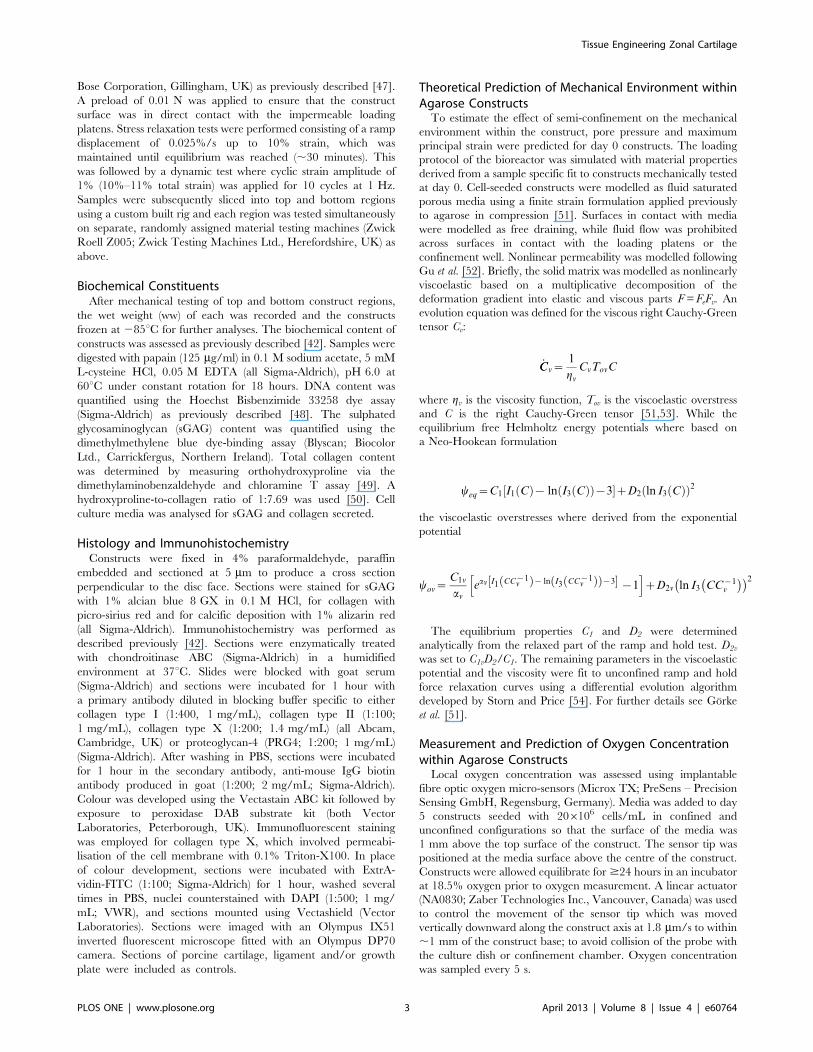
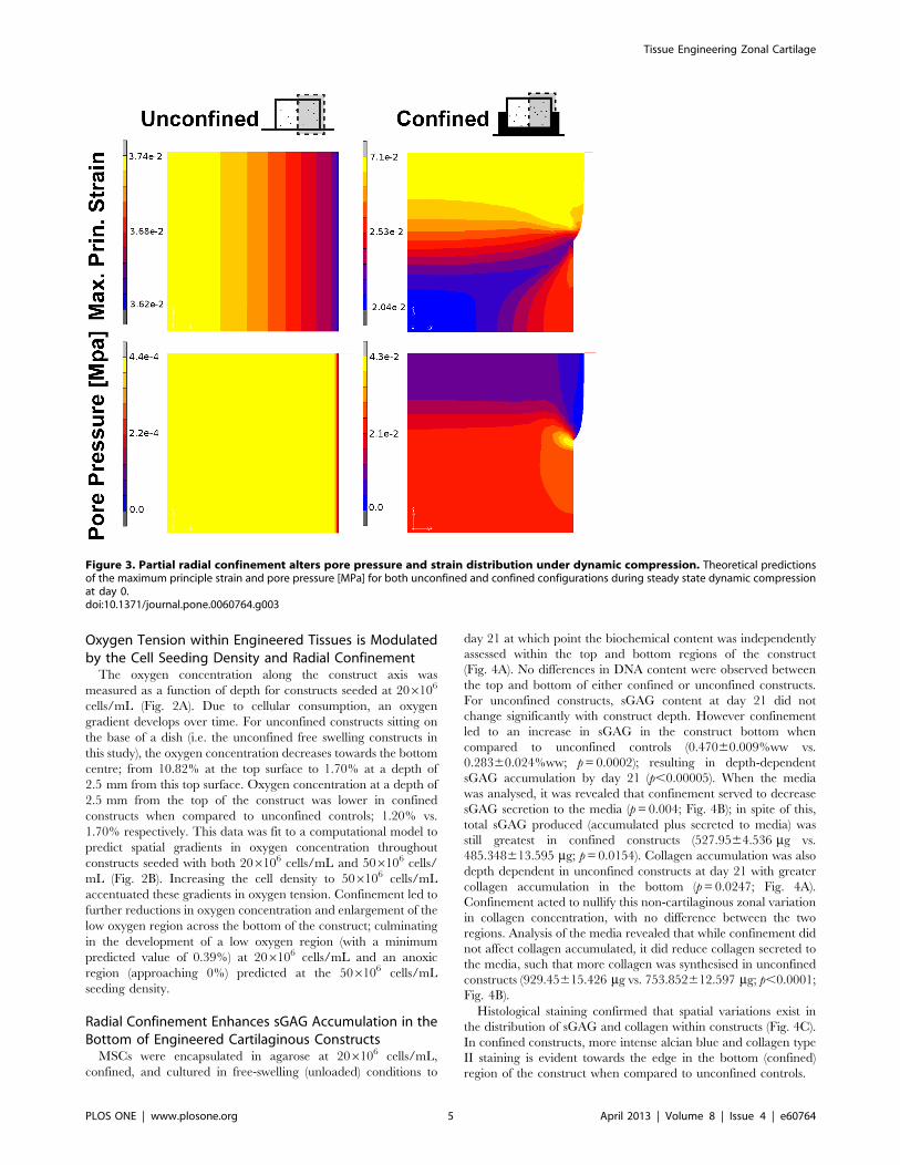
Radial Confinement Spatially Alters the Pore Pressure andTensile Strain within Agarose Hydrogels during DynamicCompressionThe mechanical environment within an agarose hydrogel
during dynamic compression was predicted for both unconfined
and confined configurations (Fig. 1). A relatively homogenous
strain environment is predicted within the unconfined construct
while the confined constructs experience higher tensile strains in
the top of the construct with predominantly compressive strains of
lower magnitude present in the bottom (Fig. 3). The model
predicts pore pressures in the bottom of the confined constructs
which are about an order of magnitude greater than that in the
unconfined configuration where a relatively homogenous pressure
environment exists.
Figure 2. Oxygen tension is modulated by the cell seeding density and radial confinement. MSCs encapsulated in agarose at 206106
cells/mL were cultured for 4 days, at which point constructs were confined and allowed to equilibrate for 24 hours. (A) Oxygen concentration throughthe media and along the construct axis was measured for unconfined and confined constructs with representative samples of each presented (Exp).Model predictions were fit to experimental data obtained from the surface of the culture media through the depth of the construct. For clarity, onlythe fit through the construct is shown (Model). (B) Predicted gradients in oxygen volume fraction for unconfined and confined constructs withseeding densities of 206106 and 506106 cells/mL. Oxygen volume fraction corresponds to molar fraction (610 pmol/mm3).doi:10.1371/journal.pone.0060764.g002
Tissue Engineering Zonal Cartilage
PLOS ONE | www.plosone.org 4 April 2013 | Volume 8 | Issue 4 | e60764
Oxygen Tension within Engineered Tissues is Modulatedby the Cell Seeding Density and Radial ConfinementThe oxygen concentration along the construct axis was
measured as a function of depth for constructs seeded at 206106
cells/mL (Fig. 2A). Due to cellular consumption, an oxygen
gradient develops over time. For unconfined constructs sitting on
the base of a dish (i.e. the unconfined free swelling constructs in
this study), the oxygen concentration decreases towards the bottom
centre; from 10.82% at the top surface to 1.70% at a depth of
2.5 mm from this top surface. Oxygen concentration at a depth of
2.5 mm from the top of the construct was lower in confined
constructs when compared to unconfined controls; 1.20% vs.
1.70% respectively. This data was fit to a computational model to
predict spatial gradients in oxygen concentration throughout
constructs seeded with both 206106 cells/mL and 506106 cells/
mL (Fig. 2B). Increasing the cell density to 506106 cells/mL
accentuated these gradients in oxygen tension. Confinement led to
further reductions in oxygen concentration and enlargement of the
low oxygen region across the bottom of the construct; culminating
in the development of a low oxygen region (with a minimum
predicted value of 0.39%) at 206106 cells/mL and an anoxic
region (approaching 0%) predicted at the 506106 cells/mL
seeding density.
Radial Confinement Enhances sGAG Accumulation in theBottom of Engineered Cartilaginous ConstructsMSCs were encapsulated in agarose at 206106 cells/mL,
confined, and cultured in free-swelling (unloaded) conditions to
day 21 at which point the biochemical content was independently
assessed within the top and bottom regions of the construct
(Fig. 4A). No differences in DNA content were observed between
the top and bottom of either confined or unconfined constructs.
For unconfined constructs, sGAG content at day 21 did not
change significantly with construct depth. However confinement
led to an increase in sGAG in the construct bottom when
compared to unconfined controls (0.47060.009%ww vs.
0.28360.024%ww; p=0.0002); resulting in depth-dependent
sGAG accumulation by day 21 (p,0.00005). When the media
was analysed, it was revealed that confinement served to decrease
sGAG secretion to the media (p=0.004; Fig. 4B); in spite of this,
total sGAG produced (accumulated plus secreted to media) was
still greatest in confined constructs (527.9564.536 mg vs.
485.348613.595 mg; p=0.0154). Collagen accumulation was also
depth dependent in unconfined constructs at day 21 with greater
collagen accumulation in the bottom (p=0.0247; Fig. 4A).
Confinement acted to nullify this non-cartilaginous zonal variation
in collagen concentration, with no difference between the two
regions. Analysis of the media revealed that while confinement did
not affect collagen accumulated, it did reduce collagen secreted to
the media, such that more collagen was synthesised in unconfined
constructs (929.45615.426 mg vs. 753.852612.597 mg; p,0.0001;
Fig. 4B).
Histological staining confirmed that spatial variations exist in
the distribution of sGAG and collagen within constructs (Fig. 4C).
In confined constructs, more intense alcian blue and collagen type
II staining is evident towards the edge in the bottom (confined)
region of the construct when compared to unconfined controls.
Figure 3. Partial radial confinement alters pore pressure and strain distribution under dynamic compression. Theoretical predictionsof the maximum principle strain and pore pressure [MPa] for both unconfined and confined configurations during steady state dynamic compressionat day 0.doi:10.1371/journal.pone.0060764.g003
Tissue Engineering Zonal Cartilage
PLOS ONE | www.plosone.org 5 April 2013 | Volume 8 | Issue 4 | e60764

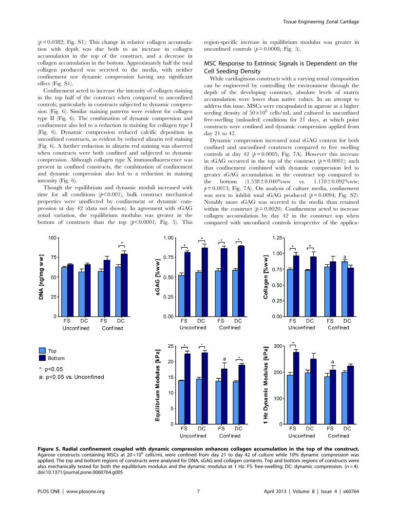
Radial Confinement Coupled with Dynamic CompressionEnhances Collagen Accumulation in the Top of theConstructMSCs were encapsulated in agarose at 206106 cells/mL and
cultured in unconfined free-swelling (unloaded) conditions for 21
days, at which point constructs were confined and dynamic
compression applied from day 21 to 42. This delayed application
of dynamic compression was motivated by our previous finding
that dynamic compression application from day 0 inhibited
chondrogenesis of MSCs [30,42,57,58].
While there was no difference in sGAG content between top
and bottom construct regions for unconfined constructs at day 21,
ensuring constructs remained the same way up over 42 days of
culture did eventually lead to greater sGAG accumulation in the
construct bottom when compared to the top (p,0.0001; Fig. 5).
Confinement from day 21 did not further enhance sGAG
accumulation in either region. When normalised to DNA content,
only unconfined constructs exhibited a significant difference in
sGAG/DNA with depth (Fig. S1). Neither dynamic compression
nor confinement had a significant effect on total sGAG
accumulation, although compression did increase sGAG secretion
to the media (p=0.0344; Fig. S1). By day 42 unconfined constructs
also exhibited zonal variation in collagen content with greater
accumulation in the bottom of the construct (p=0.0016; Fig. 5).
However, construct confinement again acted to reduce this zonal
variation such that there was no longer a significant difference
between top and bottom. Moreover, when confinement was
combined with dynamic compression, this zonal gradient was
reversed such that collagen content in the top of confined
compressed constructs was greater than that in unconfined
controls (0.86860.033 vs. 0.73660.011; p=0.0252; Fig. 5). When
normalised to DNA, collagen synthesis in the top of confined
constructs was significantly higher than that in the bottom
Figure 4. Radial confinement enhances sGAG accumulation in the bottom of engineered cartilaginous constructs. MSCs encapsulatedin agarose at 206106 cells/mL were cultured for 21 days in unconfined or confined conditions. (A) The top and bottom regions of unconfined andconfined free-swelling constructs were analysed for DNA, sGAG and collagen contents. (n=4) (B) Total sGAG and collagen accumulated in theconstructs (sum of top and bottom) and secreted to the media. Media data is presented as mg accumulated and secreted (n= 4) (C) Unconfined andconfined constructs were stained with alcian blue for sulphated mucins, picro-sirius red for collagen and immunohistochemically for collagen type II.Representative full-depth half construct sections are shown as indicated. (n= 2) Scale bar 500 mm.doi:10.1371/journal.pone.0060764.g004
Tissue Engineering Zonal Cartilage
PLOS ONE | www.plosone.org 6 April 2013 | Volume 8 | Issue 4 | e60764
(p=0.0382; Fig. S1). This change in relative collagen accumula-
tion with depth was due both to an increase in collagen
accumulation in the top of the construct, and a decrease in
collagen accumulation in the bottom. Approximately half the total
collagen produced was secreted to the media, with neither
confinement nor dynamic compression having any significant
effect (Fig. S1).
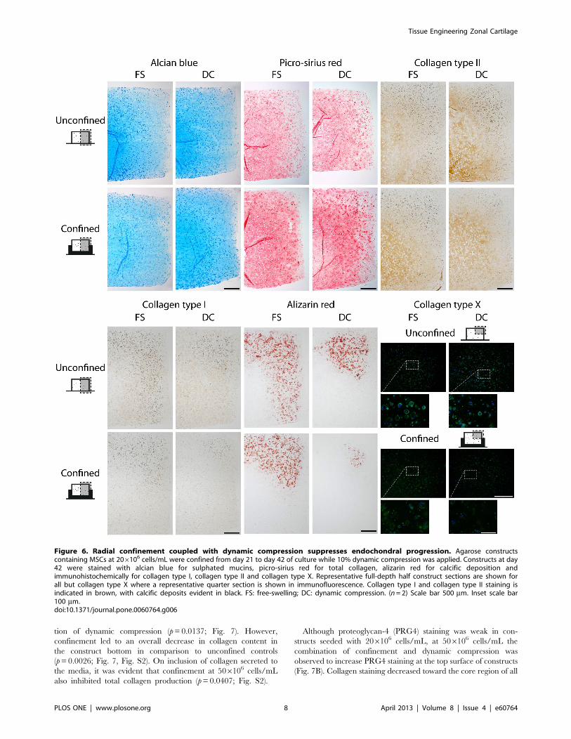
Confinement acted to increase the intensity of collagen staining
in the top half of the construct when compared to unconfined
controls; particularly in constructs subjected to dynamic compres-
sion (Fig. 6). Similar staining patterns were evident for collagen
type II (Fig. 6). The combination of dynamic compression and
confinement also led to a reduction in staining for collagen type I
(Fig. 6). Dynamic compression reduced calcific deposition in
unconfined constructs, as evident by reduced alizarin red staining
(Fig. 6). A further reduction in alizarin red staining was observed
when constructs were both confined and subjected to dynamic
compression. Although collagen type X immunofluorescence was
present in confined constructs, the combination of confinement
and dynamic compression also led to a reduction in staining
intensity (Fig. 6).
Though the equilibrium and dynamic moduli increased with
time for all conditions (p,0.001), bulk construct mechanical
properties were unaffected by confinement or dynamic com-
pression at day 42 (data not shown). In agreement with sGAG
zonal variation, the equilibrium modulus was greater in the
bottom of constructs than the top (p,0.0001; Fig. 5). This
region-specific increase in equilibrium modulus was greater in
unconfined controls (p=0.0008; Fig. 5).
MSC Response to Extrinsic Signals is Dependent on theCell Seeding DensityWhile cartilaginous constructs with a varying zonal composition
can be engineered by controlling the environment through the
depth of the developing construct, absolute levels of matrix
accumulation were lower than native values. In an attempt to
address this issue, MSCs were encapsulated in agarose at a higher
seeding density of 506106 cells/mL and cultured in unconfined
free-swelling (unloaded) conditions for 21 days, at which point
constructs were confined and dynamic compression applied from
day 21 to 42.
Dynamic compression increased total sGAG content for both
confined and unconfined constructs compared to free swelling
controls at day 42 (p=0.0005; Fig. 7A). However this increase
in sGAG occurred in the top of the construct (p=0.0001); such
that confinement combined with dynamic compression led to
greater sGAG accumulation in the construct top compared to
the bottom (1.53860.040%ww vs. 1.17060.092%ww;
p=0.0013; Fig. 7A). On analysis of culture media, confinement
was seen to inhibit total sGAG produced (p=0.0094; Fig. S2).
Notably more sGAG was secreted to the media than retained
within the construct (p=0.0020). Confinement acted to increase
collagen accumulation by day 42 in the construct top when
compared with unconfined controls irrespective of the applica-
Figure 5. Radial confinement coupled with dynamic compression enhances collagen accumulation in the top of the construct.Agarose constructs containing MSCs at 206106 cells/mL were confined from day 21 to day 42 of culture while 10% dynamic compression wasapplied. The top and bottom regions of constructs were analysed for DNA, sGAG and collagen contents. Top and bottom regions of constructs werealso mechanically tested for both the equilibrium modulus and the dynamic modulus at 1 Hz. FS: free-swelling; DC: dynamic compression. (n=4).doi:10.1371/journal.pone.0060764.g005
Tissue Engineering Zonal Cartilage
PLOS ONE | www.plosone.org 7 April 2013 | Volume 8 | Issue 4 | e60764
tion of dynamic compression (p=0.0137; Fig. 7). However,
confinement led to an overall decrease in collagen content in
the construct bottom in comparison to unconfined controls
(p=0.0026; Fig. 7, Fig. S2). On inclusion of collagen secreted to
the media, it was evident that confinement at 506106 cells/mL
also inhibited total collagen production (p=0.0407; Fig. S2).
Although proteoglycan-4 (PRG4) staining was weak in con-
structs seeded with 206106 cells/mL, at 506106 cells/mL the
combination of confinement and dynamic compression was
observed to increase PRG4 staining at the top surface of constructs
(Fig. 7B). Collagen staining decreased toward the core region of all
Figure 6. Radial confinement coupled with dynamic compression suppresses endochondral progression. Agarose constructscontaining MSCs at 206106 cells/mL were confined from day 21 to day 42 of culture while 10% dynamic compression was applied. Constructs at day42 were stained with alcian blue for sulphated mucins, picro-sirius red for total collagen, alizarin red for calcific deposition andimmunohistochemically for collagen type I, collagen type II and collagen type X. Representative full-depth half construct sections are shown forall but collagen type X where a representative quarter section is shown in immunofluorescence. Collagen type I and collagen type II staining isindicated in brown, with calcific deposits evident in black. FS: free-swelling; DC: dynamic compression. (n= 2) Scale bar 500 mm. Inset scale bar100 mm.doi:10.1371/journal.pone.0060764.g006
Tissue Engineering Zonal Cartilage
PLOS ONE | www.plosone.org 8 April 2013 | Volume 8 | Issue 4 | e60764

constructs and was more heterogeneous than alcian blue staining
(Fig. S3).
Dynamic compression acted to enhance both the equilibrium
and dynamic modulus for the whole construct (p,0.05; data not
shown). Regionally, this increase was only evident in the construct
top, which was stiffer than the corresponding region in FS controls
(p,0.05; Fig. 7). Confinement had a negative effect on whole
construct stiffness (p,0.05); attributed to a decrease in both
equilibrium and dynamic moduli in the bottom of confined
constructs corresponding to lower matrix accumulation in this
region (p,0.01; Fig. 7).
Discussion
Significant developments have been made regarding the use of
MSCs for functional cartilage tissue engineering [59–61]. How-
ever, attempts to engineer grafts with a zonal composition and
organisation mimicking normal articular cartilage have been
limited. Creating such zonal variations in tissue composition
within engineered grafts may be critical to regenerating hyaline
cartilage with a normal Benninghoff architecture [62]. Here we
show that the depth dependent properties of cartilaginous grafts
engineered using MSCs can be modulated through spatial
alteration of the oxygen tension and the mechanical environment
within the developing construct. MSCs were encapsulated in
agarose hydrogel and confined up to half their thickness. This
Figure 7. MSC response to extrinsic signals is dependent on the cell seeding density. Agarose constructs containing MSCs at 506106 cells/mL were confined from day 21 to day 42 of culture while 10% dynamic compression was applied. (A) The top and bottom regions of constructs wereanalysed for DNA, sGAG and collagen contents. Top and bottom regions of constructs were also mechanically tested for both the equilibriummodulus and the dynamic modulus at 1 Hz. (n=4) (B) Constructs at day 42 were stained immunohistochemically for proteoglycan-4 (PRG4). FS: free-swelling; DC: dynamic compression. (n= 2) Scale bar 100 mm.doi:10.1371/journal.pone.0060764.g007
Tissue Engineering Zonal Cartilage
PLOS ONE | www.plosone.org 9 April 2013 | Volume 8 | Issue 4 | e60764
reduced oxygen levels in the bottom of the construct and when
combined with dynamic compression, increased tensile strains
across the top of the construct. These spatial changes in the local
environment correlated with increased sGAG accumulation in the
bottom of constructs, increased collagen accumulation in the top
of the construct, and a near complete suppression of MSC
hypertrophy and tissue calcification throughout the depth of the
engineered tissue. Consequently, a tissue with depth dependent
variation in sGAG, collagen and mechanical properties similar,
but not identical to native articular cartilage was established. In an
effort to increase extracellular matrix (ECM) accumulation and
mechanical properties, constructs were seeded at a higher cell
density (506106 cells/mL). This failed to instigate the creation of
a native-like zonal composition in biochemical constituents and
mechanical properties, demonstrating that the spatial environment
within constructs seeded at very high densities was not conducive
to engineering zonal constructs with cartilage-like properties.
Implantable fibre-optic oxygen micro-sensors and computation-
al modelling were used to assess the spatial alterations in oxygen
tension and the mechanical environment induced by confinement
and dynamic compression respectively. Due to the uncertainty
associated with material parameter identification for constructs
later in culture where elaborated extracellular matrix may alter
nutrient transfer and inhomogeneously alter construct mechanics,
the simulations were performed for the initial cell seeded agarose
constructs only. Despite this limitation, the measurement of
oxygen availability and the finite element simulations provided
insight into the spatial gradients in the mechanical environment
and the oxygen tension throughout the construct and its de-
pendence on cell density.
Initially, the effect of confinement was examined over 21 days.
Confinement enhanced sGAG accumulation in the bottom
(confined) region of the construct. One explanation for this was
that confinement was simply acting to reduce secretion of ECM
components into the media. While less sGAG was secreted from
confined constructs, the total produced remained significantly
higher with confinement (Fig. 4B), supporting the hypothesis that
the low oxygen microenvironment predicted within the bottom of
confined constructs may be enhancing chondrogenesis. Regions of
faint alcian blue and collagen type II staining (Fig. 4C) in both
confined and unconfined constructs also appeared to correlate
with predicted regions of higher oxygen availability (Fig. 2B).
While confinement lowers the availability of oxygen in the bottom
of confined constructs, it could also act to reduce the availability of
other regulatory factors such as TGF-b3 or glucose. However, in
contrast to the abundance of data available in the literature
supporting the argument that lower oxygen levels enhance
chondrogenesis of MSCs [18–25], to the author’s knowledge,
there is no data available in the literature demonstrating that lower
levels of other factors such as TGF-b3 or glucose will enhance
cartilage matrix specific ECM synthesis in bone marrow derived
MSCs. While we believe the lower levels of oxygen within the
bottom of confined constructs are acting to enhance chondrogen-
esis in this region of the construct, we cannot rule out the
possibility that gradients in other factors may also be playing a role.
In contrast to the findings over the first three weeks of culture,
confinement from day 21 to day 42 did not lead to enhanced
sGAG accumulation. In fact, sGAG accumulation was concen-
trated in the bottom of both confined and unconfined constructs at
day 42. Given that care was taken to prevent constructs from
flipping over during culture (thereby ensuring that the top surface
of the construct remained face upwards for the culture duration),
a low oxygen region will naturally develop in the bottom of the
construct due to cellular consumption (Fig. 2), which may explain
why sGAG accumulation in the long term is higher in the bottom
of both confined and unconfined constructs. It should be noted
that the oxygen levels within the engineered tissue will also
theoretically depend on the depth of media above the construct,
and this should be considered when designing culture strategies to
engineer zonal tissues.
When confinement was combined with dynamic compression,
collagen accumulation increased in the top of the construct
resulting in a depth-dependent zonal variation in both the
biochemical composition and compressive properties of the
engineered tissue. The increased levels of collagen synthesis in
the top of mechanically stimulated confined constructs may be due
to the higher levels of strain within this region of the engineered
tissue (Fig. 3). Intermittent cyclic tension has specifically been
shown to increase collagen content within MSC seeded constructs
[44,45]. In the present study, tensile strains across the top of
dynamically compressed constructs were predicted to approxi-
mately double due to radial confinement (Fig. 3); perhaps
implicating this stimulus in promoting the higher levels of collagen
accumulation in this region of the construct. Interestingly, the
combination of confinement and dynamic compression also acted
to inhibit hypertrophy (as evident by reduced type X collagen
staining) and calcification (as evident by reduced alizarin red
staining) of the engineered tissue. We have previously demon-
strated that a low oxygen tension supresses the endochondral
phenotype of chondrogenically primed MSCs [25], and here we
provide further support for the role of dynamic compression in
ensuring the development of a stable chondrogenic phenotype
[30,63], suggesting that gradients in oxygen levels and mechanical
cues may together regulate hypertrophy in developing articular
cartilage. Although fluid pressures in the bottom of confined
constructs were increased due to dynamic compression, this did
not lead to a corresponding increase in sGAG accumulation in this
region of the construct at either cell seeding density. This may be
due to the fact that the pressure generated remains over an order
of magnitude lower than that reported to elicit increases in
chondrogenic gene expression for MSCs [31,32,35].
The effect of dynamic compression was also dependent on the
cell seeding density. At a seeding density of 506106 cells/mL
dynamic compression acted to enhance sGAG accumulation and
the bulk construct mechanical properties in both unconfined and
confined conditions. Long-term dynamic compression of MSC
seeded agarose applied after 21 days of unloaded pre-culture has
previously been shown to augment sGAG accumulation [42] and
enhance construct stiffness [43]. This finding that MSC response
to dynamic compression is dependent on cell seeding density has
also recently been reported [63]. At the higher seeding density of
506106 cells/mL, it is possible that greater cell-cell contact and
signalling may enhance MSC response to dynamic compression. It
is also possible that a threshold level of ECM accumulation is
required for dynamic compression to stimulate additional synthe-
sis, as increasing the cell density was seen to increase total matrix
accumulation. Demarteau et al. demonstrated that chondrocyte
response to dynamic compression was positively correlated to the
level of ECM accumulation [64]. Additionally, dynamic compres-
sion may facilitate enhanced transport of nutrients and other
regulatory factors within the construct [65–67]. Increasing cell
density acts to increase the severity of nutrient gradients due to
cellular consumption in the construct periphery, which may be
somewhat overcome through the application of dynamic com-
pression.
At 506106 cells/mL, the combination of confinement and
loading also led to an increase in PRG4 staining across the top of
the engineered tissue, which may be due to the high levels of
Tissue Engineering Zonal Cartilage
PLOS ONE | www.plosone.org 10 April 2013 | Volume 8 | Issue 4 | e60764
deformation present in this region of the construct. This protein is
known to play an important role in joint lubrication, and its
expression has previously been shown to increase with mechanical
stimulation [68,69].
While a construct with a zonal variation in mechanical
properties and biochemical composition mimicking certain aspects
of normal articular cartilage was achieved with a seeding density of
206106 cells/mL, this was not the case at the higher seeding
density of 506106 cells/mL. Increasing the cell seeding density
presumably increased consumption of oxygen and other solutes
within the construct, resulting in a more acute decrease in nutrient
availability away from the periphery. Confinement of constructs
added to the severity of this gradient, such that an anoxic region
was predicted in the bottom of confined constructs at the 506106
cells/mL seeding density (Fig. 2B); lower than that predicted
within the deep zone of native articular cartilage [46]. It is
expected that gradients similar to that predicted for oxygen will
develop for other solutes such as ascorbate and glucose and that
one, or a combination of these may compromise cell metabolism
culminating in the reduced ECM production observed at 506106
cells/mL. While matrix accumulation was significantly reduced in
regions of nutrient limitation, DNA content did not follow the
same trend, with no differences between construct regions or
conditions (Fig. 7A), suggesting that cells were viable but in
a quiescent state. When cultured at low oxygen, MSCs have
demonstrated a robust glycolytic potential [56,70] which may
explain the survival of these cells in this potentially anoxic
environment. It has recently been shown that optimal nutrient
supply is crucial to the production of functional cartilage matrix
using MSCs [71]. Vascularised cartilage canals are present in
developing cartilage [72,73] and may provide a route for oxygen
and nutrient supply. Mimicking such nutrient paths in engineered
constructs [74,75] may provide a means to overcome transport
limitations in such tissues.
Recently, through incorporation of specific natural and
synthetic components into polyethylene glycol hydrogels, it has
been demonstrated that it is possible to direct MSC differentiation
into zone-specific phenotypes [15]; and through layering of these
zone-specific hydrogels, engineer a depth-dependent tissue [76].
This approach was successful in the induction of cartilage-like
zonal variations in collagen type II, type X, and sGAG production.
The strategy adopted in this study, namely to modulate gradients
in biomechanical and biochemical signals within the developing
tissue, also has the potential to induce cartilage-like zonal
variations in ECM content in one cohesive construct. Further-
more, this system may better recapitulate the spatial patterns of
regulatory cues determining articular cartilage organisation during
postnatal development, and may prime the construct for in vivo
implantation as it will be subject to similar environmental cues
within a load bearing defect. By coupling this zonal approach with
strategies that attempt to spatially regulate endochondral ossifica-
tion within MSC seeded hydrogels [77], it may be possible to also
engineer functional osteochondral grafts.
ConclusionEngineering cartilaginous grafts with structural composition and
organisation is crucial to the long term repair of cartilage lesions.
By controlling the oxygen tension and mechanical environment
through the depth of the developing tissue, MSC differentiation
was modulated such that a construct with depth-dependent sGAG
and collagen content somewhat akin to that of articular cartilage
was engineered. This paper represents a novel approach toward
engineering an organ or tissue with zonal variations in biochemical
composition and mechanical properties. While there are still
challenges to be overcome in order to engineer a native-like zonal
articular cartilage tissue, and whether such bioreactor systems are
ultimately used to engineer cartilaginous grafts for the clinic
remains an open question [78], the results of this study help us to
elucidate how environmental factors regulate MSC differentiation
and in this case, the development and organisation of articular
cartilage; knowledge that will be central to developing new
therapies for damaged and diseased joints.
Supporting Information
Figure S1 Radial confinement coupled with dynamiccompression enhances collagen accumulation in the topof the construct. Agarose constructs containing MSCs at
206106 cells/mL were confined from day 21 to day 42 of culture
while 10% dynamic compressive strain was applied. The top and
bottom regions of constructs were analysed for sGAG and collagen
contents which were normalised to DNA content. sGAG and
collagen accumulated within the construct and secreted to culture
media was also measured. FS: free-swelling; DC: dynamic
compression. Dynamic compression as a main effect led to
enhanced sGAG secretion to the media; p=0.0344. n=4. *:
p,0.05; a: p,0.05 vs. Unconfined.
(TIF)
Figure S2 MSC response to extrinsic signals is de-pendent on the cell seeding density. Agarose constructs
containing MSCs at 506106 cells/mL were confined from day 21
to day 42 of culture while 10% dynamic compressive strain was
applied. The top and bottom regions of constructs were analysed
for sGAG and collagen contents which were normalised to DNA
content. sGAG and collagen accumulated within the construct and
secreted to culture media was measured. FS: free-swelling; DC:
dynamic compression. n=4. *: p,0.01; a: p,0.05 vs. Unconfined;
b: p,0.05 vs. FS.
(TIF)
Figure S3 MSC response to extrinsic signals is de-pendent on the cell seeding density. Constructs at day 42
were stained with alcian blue for sulphated mucins, picro-sirius red
for total collagen, and immunohistochemically for collagen type I
and type II. Representative full-depth half construct sections are
shown. FS: free-swelling; DC: dynamic compression. (n=2) Scale
bar 500 mm.
(TIF)
Author Contributions
Conceived and designed the experiments: ST TN DK. Performed the
experiments: ST TN SC. Analyzed the data: ST TN SC DK. Wrote the
paper: ST DK.
References
1. Mow VC, Hung CT (2001) Biomechanics of articular cartilage. In: Nordin M,Frankel VH, editors. Basic biomechanics of the musculoskeletal system. 3rd ed.
Philadelphia: Lippincott Williams & Wilkins. 60–101.
2. Gannon AR, Nagel T, Kelly DJ (2012) The role of the superficial region in
determining the dynamic properties of articular cartilage. Osteoarthritis
Cartilage 20: 1417–1425.
3. Mow VC, Guo XE (2002) Mechano-electrochemical properties of articular
cartilage: their inhomogeneities and anisotropies. Annu Rev Biomed Eng 4:
175–209.
4. Brocklehurst R, Bayliss MT, Maroudas A, Coysh HL, Freeman MA, et al.
(1984) The composition of normal and osteoarthritic articular cartilage from
Tissue Engineering Zonal Cartilage
PLOS ONE | www.plosone.org 11 April 2013 | Volume 8 | Issue 4 | e60764

human knee joints. With special reference to unicompartmental replacementand osteotomy of the knee. J Bone Joint Surg Am 66: 95–106.
5. Schinagl RM, Gurskis D, Chen AC, Sah RL (1997) Depth-dependent confined
compression modulus of full-thickness bovine articular cartilage. J Orthop Res
15: 499–506.
6. Laasanen MS, Toyras J, Korhonen RK, Rieppo J, Saarakkala S, et al. (2003)
Biomechanical properties of knee articular cartilage. Biorheology 40: 133–140.
7. Akizuki S, Mow VC, Muller F (1986) Tensile properties of human knee joint
cartilage: I. Influence of ionic conditions, weight bearing, and fibrillation on thetensile modulus. J Orthop Res 4: 379–392.
8. Kim TK, Sharma B, Williams CG, Ruffner MA, Malik A, et al. (2003)
Experimental model for cartilage tissue engineering to regenerate the zonalorganization of articular cartilage. Osteoarthritis Cartilage 11: 653–664.
9. Klein TJ, Schumacher BL, Schmidt TA, Li KW, Voegtline MS, et al. (2003)
Tissue engineering of stratified articular cartilage from chondrocyte subpopula-tions. Osteoarthritis Cartilage 11: 595–602.
10. Sharma B, Williams CG, Kim TK, Sun D, Malik A, et al. (2007) Designing
zonal organization into tissue-engineered cartilage. Tissue Eng 13: 405–414.
11. Woodfield TB, Van Blitterswijk CA, De Wijn J, Sims TJ, Hollander AP, et al.(2005) Polymer scaffolds fabricated with pore-size gradients as a model for
studying the zonal organization within tissue-engineered cartilage constructs.Tissue Eng 11: 1297–1311.
12. Ng KW, Wang CCB, Mauck RL, Kelly TAN, Chahine NO, et al. (2005) A
layered agarose approach to fabricate depth-dependent inhomogeneity inchondrocyte-seeded constructs. J Orthop Res 23: 134–141.
13. Ng KW, Mauck RL, Statman LY, Lin EY, Ateshian GA, et al. (2006) Dynamic
deformational loading results in selective application of mechanical stimulationin a layered, tissue-engineered cartilage construct. Biorheology 43: 497–507.
14. Hwang NS, Varghese S, Lee HJ, Theprungsirikul P, Canver A, et al. (2007)
Response of zonal chondrocytes to extracellular matrix-hydrogels. FEBS Lett
581: 4172–4178.
15. Nguyen LH, Kudva AK, Guckert NL, Linse KD, Roy K (2011) Unique
biomaterial compositions direct bone marrow stem cells into specific
chondrocytic phenotypes corresponding to the various zones of articularcartilage. Biomaterials 32: 1327–1338.
16. Ng KW, Ateshian GA, Hung CT (2009) Zonal chondrocytes seeded in a layered
agarose hydrogel create engineered cartilage with depth-dependent cellular andmechanical inhomogeneity. Tissue Eng Part A 15: 2315–2324.
17. Lee CS, Gleghorn JP, Won Choi N, Cabodi M, Stroock AD, et al. (2007)
Integration of layered chondrocyte-seeded alginate hydrogel scaffolds. Bioma-terials 28: 2987–2993.
18. Kanichai M, Ferguson D, Prendergast PJ, Campbell VA (2008) Hypoxia
promotes chondrogenesis in rat mesenchymal stem cells: a role for AKT andhypoxia-inducible factor (HIF)-1alpha. J Cell Physiol 216: 708–715.
19. Krinner A, Zscharnack M, Bader A, Drasdo D, Galle J (2009) Impact of oxygen
environment on mesenchymal stem cell expansion and chondrogenic differen-tiation. Cell Prolif 42: 471–484.
20. Meyer EG, Buckley CT, Thorpe SD, Kelly DJ (2010) Low oxygen tension is
a more potent promoter of chondrogenic differentiation than dynamic
compression. J Biomech 43: 2516–2523.
21. Buckley CT, Meyer EG, Kelly DJ (2012) The influence of construct scale on the
composition and functional properties of cartilaginous tissues engineered using
bone marrow-derived mesenchymal stem cells. Tissue Eng Part A 18: 382–396.
22. Baumgartner L, Arnhold S, Brixius K, Addicks K, Bloch W (2010) Human
mesenchymal stem cells: Influence of oxygen pressure on proliferation and
chondrogenic differentiation in fibrin glue in vitro. J Biomed Mater Res A 93:930–940.
23. Buckley CT, Vinardell T, Kelly DJ (2010) Oxygen tension differentially
regulates the functional properties of cartilaginous tissues engineered frominfrapatellar fat pad derived MSCs and articular chondrocytes. Osteoarthritis
Cartilage 18: 1345–1354.
24. Li J, He F, Pei M (2011) Creation of an in vitro microenvironment to enhancehuman fetal synovium-derived stem cell chondrogenesis. Cell Tissue Res 345:
357–365.
25. Sheehy EJ, Buckley CT, Kelly DJ (2012) Oxygen tension regulates theosteogenic, chondrogenic and endochondral phenotype of bone marrow derived
mesenchymal stem cells. Biochem Biophys Res Commun 417: 305–310.
26. Pauwels F (1960) A new theory on the influence of mechanical stimuli on the
differentiation of supporting tissue. The tenth contribution to the functionalanatomy and causal morphology of the supporting structure. Z Anat
Entwicklungsgesch 121: 478–515.
27. Carter DR, Blenman PR, Beaupre GS (1988) Correlations between mechanicalstress history and tissue differentiation in initial fracture healing. J Orthop Res 6:
736–748.
28. Prendergast PJ, Huiskes R, Soballe K (1997) ESB Research Award 1996.Biophysical stimuli on cells during tissue differentiation at implant interfaces.
J Biomech 30: 539–548.
29. Kelly DJ, Jacobs CR (2010) The role of mechanical signals in regulatingchondrogenesis and osteogenesis of mesenchymal stem cells. Birth Defects
Res C Embryo Today 90: 75–85.
30. Thorpe SD, Buckley CT, Steward AJ, Kelly DJ (2012) European Society ofBiomechanics S.M. Perren Award 2012: The external mechanical environment
can override the influence of local substrate in determining stem cell fate.
J Biomech 45: 2483–2492.
31. Angele P, Yoo JU, Smith C, Mansour J, Jepsen KJ, et al. (2003) Cyclichydrostatic pressure enhances the chondrogenic phenotype of human mesen-
chymal progenitor cells differentiated in vitro. J Orthop Res 21: 451–457.
32. Miyanishi K, Trindade MC, Lindsey DP, Beaupre GS, Carter DR, et al. (2006)
Effects of hydrostatic pressure and transforming growth factor-beta 3 on adulthuman mesenchymal stem cell chondrogenesis in vitro. Tissue Eng 12: 1419–
1428.
33. Steward AJ, Thorpe SD, Vinardell T, Buckley CT, Wagner DR, et al. (2012)Cell-matrix interactions regulate mesenchymal stem cell response to hydrostatic
pressure. Acta Biomater 8: 2153–2159.
34. Vinardell T, Rolfe RA, Buckley CT, Meyer EG, Ahearne M, et al. (2012)Hydrostatic pressure acts to stabilise a chondrogenic phenotype in porcine joint
tissue derived stem cells. Eur Cell Mater 23: 121–132; discussion 133–124.
35. Meyer EG, Buckley CT, Steward AJ, Kelly DJ (2011) The effect of cyclichydrostatic pressure on the functional development of cartilaginous tissues
engineered using bone marrow derived mesenchymal stem cells. J Mech Behav
Biomed Mater 4: 1257–1265.
36. Liu Y, Buckley CT, Downey R, Mulhall KJ, Kelly DJ (2012) The role of
environmental factors in regulating the development of cartilaginous grafts
engineered using osteoarthritic human infrapatellar fat pad-derived stem cells.Tissue Eng Part A 18: 1531–1541.
37. Angele P, Schumann D, Angele M, Kinner B, Englert C, et al. (2004) Cyclic,
mechanical compression enhances chondrogenesis of mesenchymal progenitorcells in tissue engineering scaffolds. Biorheology 41: 335–346.
38. Campbell JJ, Lee DA, Bader DL (2006) Dynamic compressive strain influences
chondrogenic gene expression in human mesenchymal stem cells. Biorheology43: 455–470.
39. Huang CY, Hagar KL, Frost LE, Sun Y, Cheung HS (2004) Effects of cyclic
compressive loading on chondrogenesis of rabbit bone-marrow derivedmesenchymal stem cells. Stem Cells 22: 313–323.
40. Kisiday JD, Frisbie DD, McIlwraith CW, Grodzinsky AJ (2009) Dynamic
compression stimulates proteoglycan synthesis by mesenchymal stem cells in theabsence of chondrogenic cytokines. Tissue Eng Part A 15: 2817–2824.
41. Mauck RL, Byers BA, Yuan X, Tuan RS (2007) Regulation of cartilaginous
ECM gene transcription by chondrocytes and MSCs in 3D culture in responseto dynamic loading. Biomech Model Mechanobiol 6: 113–125.
42. Thorpe SD, Buckley CT, Vinardell T, O’Brien FJ, Campbell VA, et al. (2010)
The response of bone marrow-derived mesenchymal stem cells to dynamic
compression following tgf-b3 induced chondrogenic differentiation. Ann BiomedEng 38: 2896–2909.
43. Huang AH, Farrell MJ, Kim M, Mauck RL (2010) Long-term dynamic loading
improves the mechanical properties of chondrogenic mesenchymal stem cell-laden hydrogel. Eur Cell Mater 19: 72–85.
44. Connelly JT, Vanderploeg EJ, Mouw JK, Wilson CG, Levenston ME (2010)
Tensile loading modulates bone marrow stromal cell differentiation and thedevelopment of engineered fibrocartilage constructs. Tissue Eng Part A 16:
1913–1923.
45. Baker BM, Shah RP, Huang AH, Mauck RL (2011) Dynamic tensile loadingimproves the functional properties of mesenchymal stem cell-laden nanofiber-
based fibrocartilage. Tissue Eng Part A 17: 1445–1455.
46. Zhou S, Cui Z, Urban JP (2004) Factors influencing the oxygen concentrationgradient from the synovial surface of articular cartilage to the cartilage-bone
interface: a modeling study. Arthritis Rheum 50: 3915–3924.
47. Buckley CT, Thorpe SD, O’Brien FJ, Robinson AJ, Kelly DJ (2009) The effectof concentration, thermal history and cell seeding density on the initial
mechanical properties of agarose hydrogels. J Mech Behav Biomed Mater 2:
512–521.
48. Kim YJ, Sah RL, Doong JY, Grodzinsky AJ (1988) Fluorometric assay of DNAin cartilage explants using Hoechst 33258. Anal Biochem 174: 168–176.
49. Kafienah W, Sims TJ (2004) Biochemical methods for the analysis of tissue-
engineered cartilage. Methods Mol Biol 238: 217–230.
50. Ignat’eva NY, Danilov NA, Averkiev SV, Obrezkova MV, Lunin VV, et al.
(2007) Determination of hydroxyproline in tissues and the evaluation of the
collagen content of the tissues. J Anal Chem 62: 51–57.
51. Gorke U-J, Gunther H, Nagel T, Wimmer MA (2010) A Large Strain MaterialModel for Soft Tissues With Functionally Graded Properties. J Biomech Eng
132: 074502.
52. Gu WY, Yao H, Huang CY, Cheung HS (2003) New insight into deformation-dependent hydraulic permeability of gels and cartilage, and dynamic behavior of
agarose gels in confined compression. J Biomech 36: 593–598.
53. Lion A (1997) A physically based method to represent the thermo-mechanicalbehaviour of elastomers. Acta Mechanica 123: 1–25.
54. Storn R, Price K (1997) Differential Evolution – A Simple and Efficient
Heuristic for Global Optimization over Continuous Spaces. J of GlobalOptimization 11: 341–359.
55. Sengers BG, Heywood HK, Lee DA, Oomens CWJ, Bader DL (2005) Nutrient
Utilization by Bovine Articular Chondrocytes: A Combined Experimental andTheoretical Approach. J Biomech Eng 127: 758–766.
56. Pattappa G, Heywood HK, de Bruijn JD, Lee DA (2011) The metabolism of
human mesenchymal stem cells during proliferation and differentiation. J CellPhysiol 226: 2562–2570.
57. Thorpe SD, Buckley CT, Vinardell T, O’Brien FJ, Campbell VA, et al. (2008)
Dynamic compression can inhibit chondrogenesis of mesenchymal stem cells.
Biochem Biophys Res Commun 377: 458–462.
Tissue Engineering Zonal Cartilage
PLOS ONE | www.plosone.org 12 April 2013 | Volume 8 | Issue 4 | e60764
58. Haugh MG, Meyer EG, Thorpe SD, Vinardell T, Duffy GP, et al. (2011)
Temporal and Spatial Changes in Cartilage-Matrix-Specific Gene Expression inMesenchymal Stem Cells in Response to Dynamic Compression. Tissue Eng
Part A 17: 3085–3093.
59. Khan WS, Johnson DS, Hardingham TE (2010) The potential of stem cells inthe treatment of knee cartilage defects. Knee 17: 369–374.
60. Vinatier C, Bouffi C, Merceron C, Gordeladze J, Brondello JM, et al. (2009)Cartilage tissue engineering: towards a biomaterial-assisted mesenchymal stem
cell therapy. Curr Stem Cell Res Ther 4: 318–329.
61. Huang AH, Farrell MJ, Mauck RL (2010) Mechanics and mechanobiology ofmesenchymal stem cell-based engineered cartilage. J Biomech 43: 128–136.
62. Nagel T, Kelly DJ (2012) The Composition of Engineered Cartilage at the Timeof Implantation Determines the Likelihood of Regenerating Tissue with
a Normal Collagen Architecture. Tissue Eng Part A: In press.63. Bian L, Zhai DY, Zhang EC, Mauck RL, Burdick JA (2012) Dynamic
compressive loading enhances cartilage matrix synthesis and distribution and
suppresses hypertrophy in hMSC-laden hyaluronic acid hydrogels. Tissue EngPart A 18: 715–724.
64. Demarteau O, Wendt D, Braccini A, Jakob M, Schafer D, et al. (2003) Dynamiccompression of cartilage constructs engineered from expanded human articular
chondrocytes. Biochem Biophys Res Commun 310: 580–588.
65. Albro MB, Chahine NO, Li R, Yeager K, Hung CT, et al. (2008) Dynamicloading of deformable porous media can induce active solute transport.
J Biomech 41: 3152–3157.66. Mauck RL, Hung CT, Ateshian GA (2003) Modeling of neutral solute transport
in a dynamically loaded porous permeable gel: implications for articularcartilage biosynthesis and tissue engineering. J Biomech Eng 125: 602–614.
67. Albro MB, Banerjee RE, Li R, Oungoulian SR, Chen B, et al. (2011) Dynamic
loading of immature epiphyseal cartilage pumps nutrients out of vascular canals.J Biomech 44: 1654–1659.
68. Kupcsik L, Stoddart MJ, Li Z, Benneker LM, Alini M (2010) Improvingchondrogenesis: potential and limitations of SOX9 gene transfer and mechanical
stimulation for cartilage tissue engineering. Tissue Eng Part A 16: 1845–1855.
69. Candrian C, Vonwil D, Barbero A, Bonacina E, Miot S, et al. (2008) Engineered
cartilage generated by nasal chondrocytes is responsive to physical forces
resembling joint loading. Arthritis Rheum 58: 197–208.
70. Pattappa G, Thorpe SD, Jegard NC, Heywood H, de Bruijn JD, et al. (2013)
Continuous and uninterrupted oxygen tension influences the colony formation
and oxidative metabolism of human mesenchymal stem cells. Tissue Eng
Part C Methods 19: 68–79.
71. Farrell MJ, Comeau ES, Mauck RL (2012) Mesenchymal stem cells produce
functional cartilage matrix in three-dimensional culture in regions of optimal
nutrient supply. Eur Cell Mater 23: 425–440.
72. Hall BK (2005) Bones and cartilage: developmental and evolutionary skeletal
biology. London: Elsevier Academic. xxviii, 760 p.
73. Lecocq M, Girard CA, Fogarty U, Beauchamp G, Richard H, et al. (2008)
Cartilage matrix changes in the developing epiphysis: early events on the
pathway to equine osteochondrosis? Equine Vet J 40: 442–454.
74. Buckley CT, Thorpe SD, Kelly DJ (2009) Engineering of large cartilaginous
tissues through the use of microchanneled hydrogels and rotational culture.
Tissue Eng Part A 15: 3213–3220.
75. Sheehy EJ, Buckley CT, Kelly DJ (2011) Chondrocytes and bone marrow-
derived mesenchymal stem cells undergoing chondrogenesis in agarose
hydrogels of solid and channelled architectures respond differentially to dynamic
culture conditions. J Tissue Eng Regen Med 5: 747–758.
76. Nguyen LH, Kudva AK, Saxena NS, Roy K (2011) Engineering articular
cartilage with spatially-varying matrix composition and mechanical properties
from a single stem cell population using a multi-layered hydrogel. Biomaterials
32: 6946–6952.
77. Sheehy EJ, Vinardell T, Buckley CT, Kelly DJ (2013) Engineering osteochon-
dral constructs through spatial regulation of endochondral ossification. Acta
Biomater 9: 5484–5492.
78. Pelttari K, Wixmerten A, Martin I (2009) Do we really need cartilage tissue
engineering? Swiss Med Wkly 139: 602–609.
Tissue Engineering Zonal Cartilage
PLOS ONE | www.plosone.org 13 April 2013 | Volume 8 | Issue 4 | e60764
Related Documents