This article appeared in a journal published by Elsevier. The attached copy is furnished to the author for internal non-commercial research and education use, including for instruction at the authors institution and sharing with colleagues. Other uses, including reproduction and distribution, or selling or licensing copies, or posting to personal, institutional or third party websites are prohibited. In most cases authors are permitted to post their version of the article (e.g. in Word or Tex form) to their personal website or institutional repository. Authors requiring further information regarding Elsevier’s archiving and manuscript policies are encouraged to visit: http://www.elsevier.com/copyright

Welcome message from author
This document is posted to help you gain knowledge. Please leave a comment to let me know what you think about it! Share it to your friends and learn new things together.
Transcript

This article appeared in a journal published by Elsevier. The attachedcopy is furnished to the author for internal non-commercial researchand education use, including for instruction at the authors institution
and sharing with colleagues.
Other uses, including reproduction and distribution, or selling orlicensing copies, or posting to personal, institutional or third party
websites are prohibited.
In most cases authors are permitted to post their version of thearticle (e.g. in Word or Tex form) to their personal website orinstitutional repository. Authors requiring further information
regarding Elsevier’s archiving and manuscript policies areencouraged to visit:
http://www.elsevier.com/copyright

Author's personal copy
Modular plastic chip for one-shot human papillomavirus diagnostic analysis
G. Vecchio a,1, S. Sabella b,1, L. Tagliaferro c, P. Menegazzi c, M.P. Di Bello a, V. Brunetti a, R. Cingolani b,R. Rinaldi a, P.P. Pompa b,*
a National Nanotechnology Laboratory of CNR–INFM, University of Salento, Scuola Superiore ISUFI, 73100 Lecce, Italyb Italian Institute of Technology, Center for Bio-Molecular Nanotechnology, 73010 Arnesano (Lecce), Italyc Virology and Molecular Biology Department, Pignatelli Laboratory, 73100 Lecce, Italy
a r t i c l e i n f o
Article history:Received 4 August 2009Available online 18 September 2009
Keywords:PCRHPVBiochipsPolymeric materials
a b s t r a c t
In this article, we report the design and development of a plastic modular chip suitable for one-shothuman papillomavirus (HPV) diagnostics, namely detection of the viral presence and relative genotyping,by two sequential steps performed directly on the same device. The device is composed of two modularand disposable plastic units that can be assembled or used separately. The first module is represented bya polydimethylsiloxane (PDMS) microreactor that is exploited for real-time polymerase chain reaction(PCR) and, thus, is suitable for detecting the presence of virus. The second unit is a PDMS microwell arraythat allows virus genotyping by a colorimetric assay, based on DNA hybridization technology developedon plastic, requiring simple inspection by the naked eye. The two modules can be easily coupled to reus-able hardware, enabling the heating/cooling processes and the real-time detection of HPV. By couplingreal-time assay and colorimetric genotyping on the same chip, the assembled device may provide alow-cost tool for HPV diagnostics, thereby favoring the prediction of cancer risk in patients.
� 2009 Elsevier Inc. All rights reserved.
Human papillomavirus (HPV)2 infections are widely consideredto be an important risk factor for the genesis of cervical cancer [1–4], which is the second most common tumor disease in womenworldwide [5]. Moreover, such infections are etiologically associatedwith certain types of carcinomas in the head and neck [6]. To date,more than 100 different genotypes have been identified on the basisof the DNA sequence of the L1, E6, and upstream regulatory regions[2,6,7], and depending on the association of the virus with mucosalinfections, high-risk (HR), probable medium-risk (MR), and low-risk(LR) categories have been defined [8]. Because of the strong phyloge-netic relationship of HPV genotypes to malignant lesions, the knowl-edge of HPV types is crucial for a correct prognosis, therapy, andformulation of effective vaccines (e.g., virus-like particle HPV vac-cine) [9,10] that so far are sensitive, for example, to only specificgenotypes of the virus [11,12]. Thereby, several molecular biologytechniques, based on the molecular detection of HPV infections, havebeen developed recently. These usually represent important assaysbecause, unlike the classical histological and immunological meth-
ods alone, they may lead to unequivocal diagnostics of the presenceof infections. Such techniques comprise mainly polymerase chainreaction (PCR) and hybridization tests. Consensus PCR protocolshave been conveniently used for large-scale screening clinical anal-yses, but they can assess only the presence of infections. Althoughinteresting attempts to develop genotype-specific PCR have beencarried out and several PCR primer sets enabling the simultaneousamplification of a broad range of HPV fragments have been devel-oped [13–16], the maximum number of detectable types still re-mains restricted. For this reason, other molecular tests, such as thehybridization-based methodologies (e.g., restriction fragment lengthpolymorphism [17,18], type-specific probe hybridization [19], re-verse hybridization [20–22]) and sequencing [23–25], must be ap-plied to the amplified solutions to achieve the unambiguousidentification of the highest number of genotypes. Importantly, be-cause all of such tests show advantages and disadvantages, a combi-nation of amplification methods and molecular recognition onesand/or sequencing may be desirable for in-depth diagnostics. Nota-bly, due to their high parallelism of analysis, miniaturized biochipssuitable for HPV analysis (e.g., microarray-based lab-on-a-chip)may offer an appealing chance to perform highly sensitive diagnostictests. Moreover, the intrinsic advantages provided by biochips(e.g., portability, facile handling, savings of biological samples,point-of-care applicability) may permit their diffusion as diagnosticdevices for large-scale screening programs not only in centralizedlaboratories but also in peripheral centers such as medical officesand public centers. For example, DNA microarray biochips have
0003-2697/$ - see front matter � 2009 Elsevier Inc. All rights reserved.doi:10.1016/j.ab.2009.09.025
* Corresponding author. Fax: +39 0832 295708.E-mail address: [email protected] (P.P. Pompa).
1 These authors contributed equally to this work.2 Abbreviations used: HPV, human papillomavirus; HR, high-risk; MR, medium-risk;
LR, low-risk; PCR, polymerase chain reaction; PDMS, polydimethylsiloxane; BSA,bovine serum albumin; SDS, sodium dodecyl sulfate; ssDNA, single-stranded DNA;APTES, 3-aminopropyltriethoxysilane; GTA, glutaraldehyde; SSC, sodium chloride–sodium citrate; PMT, photomultiplier tube; TBS-T, Tris-buffered saline with Tween;NBT, nitro blue tetrazolium; FRET, fluorescence resonance energy transfer.
Analytical Biochemistry 397 (2010) 53–59
Contents lists available at ScienceDirect
Analytical Biochemistry
journal homepage: www.elsevier .com/locate /yabio
Author's personal copy
demonstrated the detection of single infections and even multipleinfections simultaneously with high sensitivity [26–30]. Such de-vices have been developed on different materials (e.g., nylon mem-brane, glass), and a discrete number of genotypes have beencharacterized by a simple colorimetric detection of the hybridizationbetween type-specific probes and target.
In this article, we describe the development of a biochip withthe ability to conjugate more molecular HPV tests (e.g., real-timePCR, reverse hybridization) on a small-sized device, showing theunique advantage to providing more diagnostic information atonce. The device has two plastic modular units, namely a poly-dimethylsiloxane (PDMS) microreactor (for real-time PCR) and aPDMS microwell array that allows virus genotyping (if the infec-tion has been detected in the first module) by a simple colorimetricassay. Being disposable, the modular units can be used in associa-tion or separately on supporting hardware, providing a novel inex-pensive tool for HPV diagnostics, likely favoring the prediction ofcancer risk in patients.
Materials and methods
PDMS microreactors
The PDMS microreactors were produced by means of a simplesoft lithography technique, based on replica molding, as describedby Sabella and coworkers [31]. To produce microreactors entirelyof PDMS, the method was modified slightly as described briefly be-low. A solution consisting of PDMS (Sylgard 184, Dow Corning,Midland, MI, USA) and curing agent in a 10:1 (w/w) ratio was thor-oughly mixed, sonicated for degassing, and poured into formingmolds and punchers, conferring the desired shape and dimensionsto microreactors. After curing in an oven at 70 �C for 2 h, the PDMSmicroreactors were peeled off. Very thin PDMS layers, acting as thebottom of the microreactors, were then produced by spin-coating.The thickness of the bottom layer was investigated extensively andoptimized (it was in the range of a few hundred micrometers) toguarantee a proper thermal cycling of the PCR solution. Afterward,PDMS microreactors were made hydrophilic by means of oxygenplasma (PICO, Diener Electronic, Nagold, Germany; experimentalconditions: 25 W for 2 min) and immediately blocked with bovineserum albumin (BSA) and sodium dodecyl sulfate (SDS), both at aconcentration of 0.1%. Microreactors were then washed exten-sively with Milli-Q water. The BSA-based treatment was found tobe extremely effective and stable (the microreactors typically pre-served their stability and biocompatibility over several months).Interestingly, after passivation, PDMS microreactors can be steril-ized by conventional autoclaves without losing any surface featureand biocompatibility. The efficiency of thermal cycling was evalu-ated experimentally by using different thermocouples inside thePCR solution in the plastic microreactors. Specifically, the experi-ments reported in this article (including real-time PCR and PCRwith end-point detection by gel electrophoresis) were performedon square- and cylinder-shaped single-well microreactors with avolume capacity of 30 ll.
PDMS microwell array for HPV genotyping
The PDMS microwell array consists of two assembled PDMSslides. To obtain a controlled thickness (�500 lm) of the slides,the PDMS/curing agent mixture (1:10 ratio) was spin-coated (Lau-rell WS 650S-6NPP/Lite, 300 rpm � 20 s) on rigid flat masters. Aftercuring at 70 �C for 2 h, the polymeric films were peeled off. A num-ber of wells (typically 6–35 wells with a capacity ranging from 2 to5 ll) were produced on the first slide by micropunching the sur-
face, whereas the second slide, acting as the bottom, was assem-bled with the first one by oxygen plasma [32].
The polymeric wells were chemically modified to permit thecovalent immobilization of different types of single-strandedDNA (ssDNA) probe molecules according to a published procedure[33] that was slightly modified by us. Briefly, after oxygen plasmatreatment, the microwell array was immediately immersed in a10% (v/v) aqueous solution of 3-aminopropyltriethoxysilane(APTES, Sigma–Aldrich, St. Louis, MO, USA) at 25 �C for 3 h, rinsedwith water, and finally dried with a stream of N2. APTES reacts withthe hydroxyl groups on PDMS to yield primary amino functionalgroups on the surface. The aminosilanized PDMS surface (PDMS–NH2) was activated with the homo-bifunctional linker glutaralde-hyde (GTA, Sigma–Aldrich), suitable to react with aminated probessuch as 50-aminated ssDNA. We used solutions of GTA at a concen-tration of 2.5% (v/v) in 100 mM phosphate buffer, and reactionswere carried out at 4 �C in the dark for 2 h. Afterward, substrateswere washed abundantly with Milli-Q water and dried with astream of N2.
We immobilized in the microwells two classes of molecules:probes that were type specific for four types of HPV (HPV probes)and molecule used as a standard control (HPV test). Specifically,the HPV probes were amino-modified at the 50 end. Such probeshad sequences that were specific for HPV 6, 16, 31, and 51, hence-forth referred to as HPV-6 probe (50-TAT TTG TTG GGG TAA TCAACT G-30), HPV-16 probe (50-CAT TTG TTG GGG TAA CCA ACT A-30), HPV-31 probe (50-TAT TTG TTG GGG CAA TCA GTT A-30), andHPV-51 probe (50-CAT TTG CTG GAA CAA TCA GCT T-30), respec-tively. The HPV test was indeed amino-modified at the 50 end,and biotin was modified at the 30 end. Such sequence, referred toas HPV-test (50-CGG AAG CAG GCC CAA TCT GCA A-30), was usedas a standard control for the colorimetric assay because it selec-tively binds the streptavidinated enzyme. All probes were pur-chased from Thermo Fisher. ssDNA molecules (30 lM in 3� SSC[sodium chloride–sodium citrate] buffer) were immobilized onthe bottom of each well by soaking solutions at 25 �C for 30 min,followed by another incubation at 70 �C for 60 min. Afterward,wells were rinsed thoroughly with 0.2% SDS solution at 25 �C for2 min, washed with Milli-Q water for 2 min, and then blocked witha solution of NaBH4 at 37 �C for 1 h, followed by extensive washingwith a solution of 0.2% SDS and water.
The optimal surface density of the immobilized ssDNA withinthe wells was assessed and quantified by fluorescence measure-ments. Standard solutions of Cy3-modified ssDNA HPV probesranging from 0.001 to 50 lM were spotted on GTA-functionalizedPDMS slides. By confocal microscopy analyses (Leica, TCS-SP5AOBS), we found out that the highest fluorescence signal was ob-tained with the 30-lM value (data not shown).
Assembling of the HPV prototype device
The overall HPV diagnostics was carried out on a homemadeassembled system. The plastic microreactors and microwell arraywere fixed on the system by simply contacting them with the sur-face of a thermal plate for PCR thermal cycling (flat plate, Techne).Fluorescence detection of real-time PCR was performed with alow-power excitation source (e.g., a blue LED) and a bandpass filterand photomultiplier tube (PMT) detector in a classical right-anglegeometry. Microreactors were used as the disposable part of thediagnostic system so that they were changed for each analysis.
Validation tests of PDMS microreactors and HPV real-time PCR
Prior to performing the amplification experiments with HPVDNA sequences extracted from biological samples, the plastic mic-roreactors were validated extensively in terms of biocompatibility
54 Chip for one-shot HPV diagnostic analysis / G. Vecchio et al. / Anal. Biochem. 397 (2010) 53–59

Author's personal copy
and efficiency by carrying out several PCRs on human genomicDNA with end-point gel electrophoresis detection. In particular,we used as a model target a 206-bp sequence of the CDK11 genefrom human genomic DNA (length � 109 bp) that was amplifiedby the following PCR master mix: approximately 45 ll or less ofMegaMix (Microzone), 0.4 lM of primer mix (forward: 50-AGTGCTTCCCTTGCTTTCAA-30; reverse: 50-TGTGCTGTACATTTCCTGTGTG-30), and 1.5 ng/ll of DNA template. All reagents were purchasedfrom Sigma–Aldrich. PCR experiments with end-point detection(250 independent analyses) were performed simultaneously invials (used as reference) and in microreactors inside a thermal cy-cler (T 3000, Biometra, Goettingen, Germany) using aliquots of thesame master mix. Solutions were analyzed by agarose gel electro-phoresis by using O’GeneRuler 1 kb DNA (Fermentas Life Sciences)as marker. Validation was carried out by analyzing two analyticalparameters, namely specificity and efficiency (band position andPCR yield, respectively). Quantification of DNA, migrated as a bandin the gel, was calculated by means of one-dimensional gel analysissoftware (UVIpro Platinum 1.1).
Real-time PCR measurements were performed on real biologicalsamples, that is, DNA extracted from patients affected by varioustypes of HPV (6, 16, 31, and 51) kindly provided by the PignatelliLaboratory (Lecce, Italy). Such samples were previously character-ized by commercial instrumentation and tested in our plastic de-vice without knowing the virus type a priori. Results were thencompared with standard protocols only at the end of the colori-metric response of our plastic multiwell array. In real-time PCRexperiments, a gene fragment containing a sequence of approxi-mately 65 bp was amplified. We used the same master mix ofthe PCR with end-point detection (see above) except for the fourforward primers (HP 1E, HP 1F, HP 1G, and HP 1H) and three bio-tinylated reverse primers (HP 2Cbiot, HP 2Dbiot, and HP 2Ebiot)[34], enabling the amplification of fragments related to the L1 re-gion of 39 HPV genotypes. The fluorescent dye SYBR Green I(kex = 488 nm, kem = 522 nm) was added to the master mix. Forreal-time PCR analyses, we employed a square-shaped PDMS mic-roreactor. We typically collected the fluorescence signal of SYBRGreen during the annealing phase of each PCR cycle by using0.5 s excitation and simultaneous detection by a PMT. To assessthe yield and specificity of PCR amplification, we carried out addi-tional end-point detection (after 40 cycles) by means of agarose gelelectrophoresis. The marker used to analyze amplicon solutionswas the PCR 20-bp Low Ladder (Sigma–Aldrich), presenting a sizerange of 20–1000 bp.
HPV genotyping by PDMS microwell array
In the case of positive detection of HPV, the final PCR productscontaining the biotinylated amplimers (obtained during the PCRreaction [see above]) were characterized by hybridization withthe HPV type-specific probes immobilized in the wells of the PDMSarray. The array was soaked with the PCR solution, which was di-luted 10-fold with a buffer solution (9� SSC and 0.1% SDS), dena-tured at 95 �C for 5 min, and finally maintained at 42 �C for30 min to allow hybridization with the solid-state HPV probes.After incubation, the solution was gently removed and the PDMSarray was washed thoroughly by three washing cycles with solu-tions composed of 2� SSC/0.2% SDS, 2� SSC, and 0.2� SSC, respec-tively, at 25 �C for 10 min. Afterward, the microwell array wasrinsed immediately with streptavidin–alkaline phosphatase en-zyme solution in 0.05% TBS-T (Tris-buffered saline with Tween,Sigma–Aldrich) at a concentration ratio of 1:1000 for 10 min. Afterwashing the substrates with the same buffer for 2 min, wells wereincubated for 30 min at 25 �C with the BCIP/NBT (nitro blue tetra-zolium) Liquid Substrate System (Sigma–Aldrich). Finally, the BCIP/NBT was removed and the array was dried in an oven. The reaction
mechanism of such colorimetric substrate is based on the produc-tion of an insoluble product (NBT diformazan), which deposits as acolored spot in proximity of dsDNA spots on interaction of the en-zyme with dsDNA by a biotin–streptavidin bond. Also in this case,to assess the best concentration values, we carried out severalexperiments by immobilizing in the wells solutions of 50 amino-and 30 biotin-modified ssDNA probes, following the same proce-dures described for the HPV probes. We tested enzyme solutionsranging from a concentration ratio of 1:50 up to 1:5000 with differ-ent incubation times (10, 20, and 30 min), finding out that the bestvalue corresponded to a 1:1000 concentration with 10 min of incu-bation time.
Results and discussion
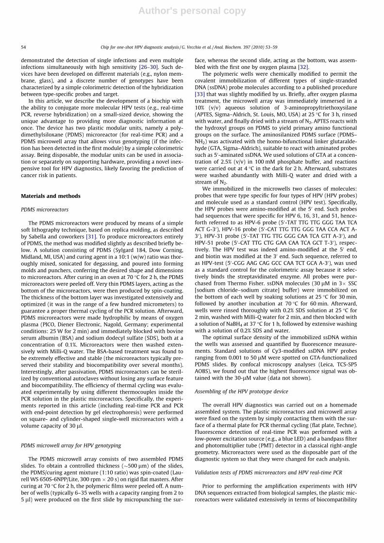
In this article, we show the realization of a plastic, disposable,and modular chip suitable for one-shot HPV diagnostics, namelydetection of the viral infection and relative genotyping, by twosequential steps performed directly on the same device. Fig. 1shows the modular chip we realized, which is composed of twodisposable plastic units (i.e., a microreactor and a microwell array)combined with reusable hardware consisting of a heating/coolingsurface and a simple optical apparatus for fluorescence detection.Plastic transparent microreactors and PDMS microwell arrays canbe produced easily and cheaply by means of soft lithography pro-cedures such as replica molding and micropunching (see Materialsand methods for details). Depending on sizes and shapes, the dis-posable plastic reactors can be easily adapted and used for ampli-fication reactions or as colorimetric plastic microwell arrays(Fig. 1A). By setting up the fabrication parameters as well as thesurface treatments (physical and chemical) to obtain hydrophilicand biocompatible surface properties, we produced plastic unitspresenting very good versatility and applicability for genomic anal-ysis. Such plastic modules are ideal for real-time PCR and virus rec-ognition and genotyping. In particular, for the experimentspresented here, we used square- and cylinder-shaped single-cham-ber PDMS microreactors with a volume capacity of 30 ll and PDMSmultiwell arrays with 2.5 ll of working solution (6–35 wells), asreported in Fig. 1B. Microreactors were stably modified and madebiocompatible toward PCR by passivating surfaces with oxygenplasma treatments [35] and protein blocking procedures [36](see Materials and methods for details). On the other hand, micro-well arrays were chemically functionalized with HPV probes bymeans of a surface chemistry modification exploiting APTES reac-tivity on PDMS and the use of GTA as a linker between the silanizedsurface and the amino-modified probes (see Materials and meth-ods for details).
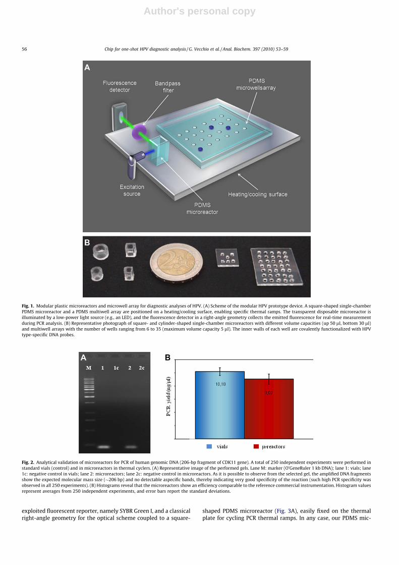
Before testing HPV microreactors for real-time PCR of HPV sam-ples, we investigated their performance by an extensive analyticalvalidation, carrying out PCR on human genomic DNA (250 inde-pendent experiments) with end-point detection. Notably, we ob-tained very good results in terms of specificity and efficiency ofthe microreactors as compared with standard vials in thermal cy-clers (Fig. 2). Experimental data show that microreactors well per-mit the selective amplification of the expected DNA band whileavoiding the formation of any aspecific fragments (Fig. 2A). PCRyields are comparable in the two cases (Fig. 2B). Also, these mic-roreactors, which are nearly transparent in the whole visible spec-tral range, can be used with a wide variety of fluorescent reporters(e.g., molecular beacons, fluorescence resonance energy transfer[FRET] probes) to monitor the real-time progress of the amplifica-tion reaction of nucleic acid (real-time PCR). Also, there are virtu-ally no specific constraints on the implementation of the opticalsetup, with several possible geometries of the excitation/detectionscheme. In this work, for HPV detection, we used a widely
Chip for one-shot HPV diagnostic analysis / G. Vecchio et al. / Anal. Biochem. 397 (2010) 53–59 55
Author's personal copy
exploited fluorescent reporter, namely SYBR Green I, and a classicalright-angle geometry for the optical scheme coupled to a square-
shaped PDMS microreactor (Fig. 3A), easily fixed on the thermalplate for cycling PCR thermal ramps. In any case, our PDMS mic-
Fig. 1. Modular plastic microreactors and microwell array for diagnostic analyses of HPV. (A) Scheme of the modular HPV prototype device. A square-shaped single-chamberPDMS microreactor and a PDMS multiwell array are positioned on a heating/cooling surface, enabling specific thermal ramps. The transparent disposable microreactor isilluminated by a low-power light source (e.g., an LED), and the fluorescence detector in a right-angle geometry collects the emitted fluorescence for real-time measurementduring PCR analysis. (B) Representative photograph of square- and cylinder-shaped single-chamber microreactors with different volume capacities (up 50 ll, bottom 30 ll)and multiwell arrays with the number of wells ranging from 6 to 35 (maximum volume capacity 5 ll). The inner walls of each well are covalently functionalized with HPVtype-specific DNA probes.
Fig. 2. Analytical validation of microreactors for PCR of human genomic DNA (206-bp fragment of CDK11 gene). A total of 250 independent experiments were performed instandard vials (control) and in microreactors in thermal cyclers. (A) Representative image of the performed gels. Lane M: marker (O’GeneRuler 1 kb DNA); lane 1: vials; lane1c: negative control in vials; lane 2: microreactors; lane 2c: negative control in microreactors. As it is possible to observe from the selected gel, the amplified DNA fragmentsshow the expected molecular mass size (�206 bp) and no detectable aspecific bands, thereby indicating very good specificity of the reaction (such high PCR specificity wasobserved in all 250 experiments). (B) Histograms reveal that the microreactors show an efficiency comparable to the reference commercial instrumentation. Histogram valuesrepresent averages from 250 independent experiments, and error bars report the standard deviations.
56 Chip for one-shot HPV diagnostic analysis / G. Vecchio et al. / Anal. Biochem. 397 (2010) 53–59

Author's personal copy
roreactors are compatible with a wide range of heating/coolingminiaturized systems (e.g., commercially available systems, Peltieror miniaturized microheaters, whose technology is currently wellestablished [37,38]) to perform efficient and fast PCR reactions.Real-time PCR was carried out on human DNA extracted from bio-logical samples of patients with HPV infections (Fig. 3). We focusedon the L1 HPV region, which was used to amplify a target sequenceof approximately 65 bp specific for at least 39 HPV types. In thisexperiment, we typically collected the fluorescence signal of SYBRGreen during the annealing phase of each PCR cycle. Due to the pri-mer specificity for HPV fragments, the typical sigmoid-like shape ofthe real-time PCR curve revealed the presence of the infection, asshown in Fig. 3B. Importantly, our plastic device allows meltingcurve analysis, providing the interesting opportunity to assessthe specificity of the amplification in situ, as shown in Fig. 3C. Inthis case, we collected the SYBR Green fluorescence signal in thetemperature range from 50 to 94 �C, observing a melting tempera-ture of approximately 78 �C, in line with the expected meltingtemperature for HPV amplicons [34]. Importantly, real-time PCRdata were consistent with gel electrophoresis analysis performedat the end of the reaction (typically after 40 PCR cycles [seeFig. 3D]), further confirming the DNA band specificity and PCRyield.
If virus presence is detected by real-time PCR in the microreac-tor module, the second plastic unit may be used for genotypinganalysis. Hence, the just amplified PCR products were directly ana-
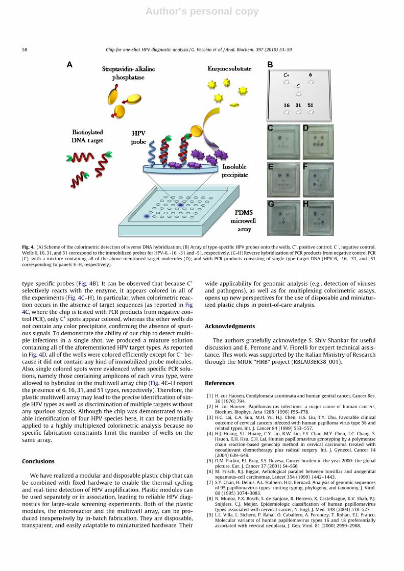
lyzed by the PDMS microwell array, which allows colorimetricdetection of various HR or LR HPV types. A schematic illustrationof the detection principle is reported in Fig. 4A. Reverse DNAhybridization between specific PCR products (which are biotinyla-ted amplimers) and HPV probes (which are selectively immobi-lized in the PDMS microwell array) is detected by means of anenzymatic reaction producing colored spots. In particular, the reac-tion occurs between streptavidin–alkaline phosphatase, whichselectively binds only the hybridized target DNA by the biotin–streptavidin bond, and the specific substrate, namely the BCIP/NBT Liquid Substrate System. This leads to the formation of local-ized colored spots on the array, visible by the naked eye, in lessthan 10 min due to the deposition of NBT diformazan precipitate[39]. Therefore, by analyzing the pattern of the colored spot onthe basis of the immobilized DNA probe sequences, specific forsome HR, MR, and LR HPV types, it is possible to genotype theDNA target. As proof of principle, we immobilized HR types 16,31, and 51 and LR type 6. To optimize the colored spot signals,we carried out an extensive optimization in terms of optimal probedensity, enzyme concentration, and time of reaction. In Fig. 4B, C+
represents the positive control (i.e., biotin-modified DNA probeshowing high specificity for the enzyme and no affinity for theDNA target), which was used as a standard control for the enzymereactivity (see Materials and methods), and C� represents the neg-ative control, which does not contain any immobilized DNAprobes. The numbered wells 6, 16, 31, and 51 correspond to HPV
Fig. 3. Real-time PCR and melting curve analysis by HPV prototype device. (A) Plastic microreactors for real-time PCR. (B) Real-time PCR curve by using SYBR Green I as thefluorescent reporter (details of the reaction are described in Materials and methods). The curve is a representative example of the results obtained for the amplification ofextracted DNA HPV from biological samples (in particular, in this case the 16 HPV genotype amplification is shown). A total of 10 replicates for each biological samplecontaining 6, 16, 31, or 51 HPV types were carried out, and high reproducibility was observed. (C) On-chip melting curve analysis. (D) End-point agarose gel detection of a 65-bp HPV DNA target (typically after 40 PCR cycles). The gel is a representative result of all the performed experiments. Lane M: marker (PCR 20-bp Low Ladder); lane 1:negative control in vials; lane 2: vials; lane 3: negative control in HPV prototype device; lane 4: HPV prototype device. (For interpretation of the references to color in thisfigure legend, the reader is referred to the web version of this paper.)
Chip for one-shot HPV diagnostic analysis / G. Vecchio et al. / Anal. Biochem. 397 (2010) 53–59 57
Author's personal copy
type-specific probes (Fig. 4B). It can be observed that because C+
selectively reacts with the enzyme, it appears colored in all ofthe experiments (Fig. 4C–H). In particular, when colorimetric reac-tion occurs in the absence of target sequences (as reported in Fig4C, where the chip is tested with PCR products from negative con-trol PCR), only C+ spots appear colored, whereas the other wells donot contain any color precipitate, confirming the absence of spuri-ous signals. To demonstrate the ability of our chip to detect multi-ple infections in a single shot, we produced a mixture solutioncontaining all of the aforementioned HPV target types. As reportedin Fig. 4D, all of the wells were colored efficiently except for C� be-cause it did not contain any kind of immobilized probe molecules.Also, single colored spots were evidenced when specific PCR solu-tions, namely those containing amplicons of each virus type, wereallowed to hybridize in the multiwell array chip (Fig. 4E–H reportthe presence of 6, 16, 31, and 51 types, respectively). Therefore, theplastic multiwell array may lead to the precise identification of sin-gle HPV types as well as discrimination of multiple targets withoutany spurious signals. Although the chip was demonstrated to en-able identification of four HPV species here, it can be potentiallyapplied to a highly multiplexed colorimetric analysis because nospecific fabrication constraints limit the number of wells on thesame array.
Conclusions
We have realized a modular and disposable plastic chip that canbe combined with fixed hardware to enable the thermal cyclingand real-time detection of HPV amplification. Plastic modules canbe used separately or in association, leading to reliable HPV diag-nostics for large-scale screening experiments. Both of the plasticmodules, the microreactor and the multiwell array, can be pro-duced inexpensively by in-batch fabrication. They are disposable,transparent, and easily adaptable to miniaturized hardware. Their
wide applicability for genomic analysis (e.g., detection of virusesand pathogens), as well as for multiplexing colorimetric assays,opens up new perspectives for the use of disposable and miniatur-ized plastic chips in point-of-care analysis.
Acknowledgments
The authors gratefully acknowledge S. Shiv Shankar for usefuldiscussion and E. Perrone and V. Fiorelli for expert technical assis-tance. This work was supported by the Italian Ministry of Researchthrough the MIUR ‘‘FIRB” project (RBLA03ER38_001).
References
[1] H. zur Hausen, Condylomata acuminata and human genital cancer, Cancer Res.36 (1976) 794.
[2] H. zur Hausen, Papillomavirus infections: a major cause of human cancers,Biochim. Biophys. Acta 1288 (1996) F55–F78.
[3] H.C. Lai, C.A. Sun, M.H. Yu, H.J. Chen, H.S. Liu, T.Y. Chu, Favorable clinicaloutcome of cervical cancers infected with human papilloma virus type 58 andrelated types, Int. J. Cancer 84 (1999) 553–557.
[4] H.J. Huang, S.L. Huang, C.Y. Lin, R.W. Lin, F.Y. Chao, M.Y. Chen, T.C. Chang, S.Hsueh, K.H. Hsu, C.H. Lai, Human papillomavirus genotyping by a polymerasechain reaction-based genechip method in cervical carcinoma treated withneoadjuvant chemotherapy plus radical surgery, Int. J. Gynecol. Cancer 14(2004) 639–649.
[5] D.M. Parkin, F.I. Bray, S.S. Devesa, Cancer burden in the year 2000: the globalpicture, Eur. J. Cancer 37 (2001) S4–S66.
[6] M. Frisch, R.J. Biggar, Aetiological parallel between tonsillar and anogenitalsquamous-cell carcinomas, Lancet 354 (1999) 1442–1443.
[7] S.Y. Chan, H. Delius, A.L. Halpern, H.U. Bernard, Analysis of genomic sequencesof 95 papillomavirus types: uniting typing, phylogeny, and taxonomy, J. Virol.69 (1995) 3074–3083.
[8] N. Munoz, F.X. Bosch, S. de Sanjose, R. Herrero, X. Castellsague, K.V. Shah, P.J.Snijders, C.J. Meijer, Epidemiologic classification of human papillomavirustypes associated with cervical cancer, N. Engl. J. Med. 348 (2003) 518–527.
[9] L.L. Villa, L. Sichero, P. Rahal, O. Caballero, A. Ferenczy, T. Rohan, E.L. Franco,Molecular variants of human papillomavirus types 16 and 18 preferentiallyassociated with cervical neoplasia, J. Gen. Virol. 81 (2000) 2959–2968.
Fig. 4. (A) Scheme of the colorimetric detection of reverse DNA hybridization. (B) Array of type-specific HPV probes onto the wells. C+, positive control; C�, negative control.Wells 6, 16, 31, and 51 correspond to the immobilized probes for HPV-6, -16, -31 and -51, respectively. (C–H) Reverse hybridization of PCR products from negative control PCR(C); with a mixture containing all of the above-mentioned target molecules (D); and with PCR products consisting of single type target DNA (HPV-6, -16, -31, and -51corresponding to panels E–H, respectively).
58 Chip for one-shot HPV diagnostic analysis / G. Vecchio et al. / Anal. Biochem. 397 (2010) 53–59

Author's personal copy
[10] D.M. Cerqueira, T. Raiol, N.M. Veras, N. von Gal Milanezi, F.A. Amaral, M. deMacedo Brigido, C.R. Martins, New variants of human papillomavirus type 18identified in central Brazil, Virus Genes 37 (2008) 282–287.
[11] A. Ames, P. Gravitt, Human papillomavirus vaccine update, Curr. Infect. Dis.Rep. 9 (2007) 151–158.
[12] J.T. Schiller, X. Castellsague, L.L. Villa, A. Hildesheim, An update of prophylactichuman papillomavirus L1 virus-like particle vaccine clinical trial results,Vaccine 26 (2008) K53–K61.
[13] W. Qu, G. Jiang, Y. Cruz, C.J. Chang, G.Y. Ho, R.S. Klein, R.D. Burk, PCR detectionof human papillomavirus: Comparison between MY09/MY11 and GP5+/GP6+primer systems, J. Clin. Microbiol. 35 (1997) 1304–1310.
[14] P.E. Gravitt, C.L. Peyton, T.Q. Alessi, C.M. Wheeler, F. Coutlee, A. Hildesheim,M.H. Schiffman, D.R. Scott, R.J. Apple, Improved amplification of genital humanpapillomaviruses, J. Clin. Microbiol. 38 (2000) 357–361.
[15] B. Kleter, L.J. van Doorn, J. ter Schegget, L. Schrauwen, K. van Krimpen, M.Burger, B. ter Harmsel, W. Quint, Novel short-fragment PCR assay for highlysensitive broad-spectrum detection of anogenital human papillomaviruses,Am. J. Pathol. 153 (1998) 1731–1739.
[16] M.V. Jacobs, P.J. Snijders, A.J. van den Brule, T.J. Helmerhorst, C.J. Meijer, J.M.Walboomers, A general primer GP5+/GP6(+)-mediated PCR–enzymeimmunoassay method for rapid detection of 14 high-risk and 6 low-riskhuman papillomavirus genotypes in cervical scrapings, J. Clin. Microbiol. 35(1997) 791–795.
[17] T. Meyer, R. Arndt, E. Stockfleth, H.T. Flammann, H. Wolf, U. Reischl, Strategyfor typing human papillomaviruses by RFLP analysis of PCR products andsubsequent hybridization with a generic probe, BioTechniques 19 (1995) 632–639.
[18] M. Grce, K. Husnjak, M. Skerlev, J. Lipozencic, K. Pavelic, Detection and typingof human papillomaviruses by means of polymerase chain reaction andfragment length polymorphism in male genital lesions, Anticancer Res. 20(2000) 2097–2102.
[19] N. Ylitalo, T. Bergstrom, U. Gyllensten, Detection of genital humanpapillomavirus by single-tube nested PCR and type-specific oligonucleotidehybridization, J. Clin. Microbiol. 33 (1995) 1822–1828.
[20] P.E. Gravitt, C.L. Peyton, R.J. Apple, C.M. Wheeler, Genotyping of 27 humanpapillomavirus types by using L1 consensus PCR products by a single-hybridization, reverse line blot detection method, J. Clin. Microbiol. 36(1998) 3020–3027.
[21] B. Kleter, L.J. van Doorn, L. Schrauwen, A. Molijn, S. Sastrowijoto, J. terSchegget, J. Lindeman, B. ter Harmsel, M. Burger, W. Quint, Development andclinical evaluation of a highly sensitive PCR–reverse hybridization line probeassay for detection and identification of anogenital human papillomavirus, J.Clin. Microbiol. 37 (1999) 2508–2517.
[22] A.J. van den Brule, R. Pol, N. Fransen-Daalmeijer, L.M. Schouls, C.J. Meijer, P.J.Snijders, GP5+/6+ PCR followed by reverse line blot analysis enables rapid andhigh-throughput identification of human papillomavirus genotypes, J. Clin.Microbiol. 40 (2002) 779–787.
[23] S.D. Vernon, E.R. Unger, D. Williams, Comparison of human papillomavirusdetection and typing by cycle sequencing, line blotting, and hybrid capture, J.Clin. Microbiol. 38 (2000) 651–655.
[24] S. Kosel, S. Burggraf, J. Mommsen, W. Engelhardt, B. Olgemoller, Type-specificdetection of human papillomaviruses in a routine laboratory setting:improved sensitivity and specificity of PCR and sequence analysis comparedto direct hybridisation, Clin. Chem. Lab. Med. 41 (2003) 787–791.
[25] N. Speich, C. Schmitt, R. Bollmann, M. Bollmann, Human papillomavirus (HPV)study of 2916 cytological samples by PCR and DNA sequencing: genotypespectrum of patients from the West German area, J. Med. Microbiol. 53 (2004)125–128.
[26] N.H. Cho, H.J. An, J.K. Jeong, S. Kang, J.W. Kim, Y.T. Kim, T.K. Park, Genotyping of22 human papillomavirus types by DNA chip in Korean women: comparisonwith cytologic diagnosis, Am. J. Obstet. Gynecol. 188 (2003) 56–62.
[27] T.J. Oh, C.J. Kim, S.K. Woo, T.S. Kim, D.J. Jeong, M.S. Kim, S. Lee, H.S. Cho, S. An,Development and clinical evaluation of a highly sensitive DNA microarray fordetection and genotyping of human papillomaviruses, J. Clin. Microbiol. 42(2004) 3272–3280.
[28] Y.D. Choi, W.W. Jung, J.H. Nam, H.S. Choi, C.S. Park, Detection of HPV genotypesin cervical lesions by the HPV DNA chip and sequencing, Gynecol. Oncol. 98(2005) 369–375.
[29] H. Lin, J.S. Moh, Y.C. Ou, S.Y. Shen, Y.M. Tsai, C.C. ChangChien, J.M. Liu, Y.Y. Ma,A simple method for the detection and genotyping of high-risk humanpapillomavirus using seminested polymerase chain reaction and reversehybridization, Gynecol. Oncol. 96 (2005) 84–91.
[30] C.Y. Lin, H.C. Chen, R.W. Lin, S.L. You, C.M. You, L.C. Chuang, M.H. Pan, M.H. Lee,Y.C. Chou, C.J. Chen, Quality assurance of genotyping array for detection andtyping of human papillomavirus, J. Virol. Methods 140 (2007) 1–9.
[31] S. Sabella, G. Vecchio, R. Cingolani, R. Rinaldi, P.P. Pompa, Real-time PCR in aplastic chip based on solid state FRET, Langmuir 24 (2008) 13266–13269.
[32] D.C. Duffy, J.C. McDonald, O.J.A. Schueller, G.M. Whitesides, Rapid prototypingof microfluidic systems in poly(dimethylsiloxane), Anal. Chem. 70 (1998)4974–4984.
[33] A. Fung, C.Y. Yang, S. Freire, C. Montemagno, B. Brough, C.M. Ho, F. Gu, W. Shi,Fluorescent detection of oral pathogens by a solid-phase immunoassay onPDMS, Conf. Proc. IEEE Eng. Med. Biol. Soc. 3 (2005) 2630–2633.
[34] C. Payan, A. Ducancelle, M.H. Aboubaker, J. Caer, M. Tapia, A. Chauvin, D.Peyronnet, E. Le Hen, Z. Arab, M.-C. Legrand, A. Tran, E. Postec, F. Tourmen, M.Avenel, C. Malbois, M.-A. De Brux, P. Descamps, F. Lunel, Humanpapillomavirus quantification in urine and cervical samples by using theMx4000 and LightCycler general real-time PCR systems, J. Clin. Microbiol. 45(2007) 897–901.
[35] S. Bhattacharya, A. Datta, J.M. Berg, S. Gangopadhyay, Studies on surfacewettability of poly(dimethyl) siloxane (PDMS) and glass under oxygen–plasmatreatment and correlation with bond strength, J. Microelectromech. Syst. 14(2005) 590–597.
[36] C. Consolandi, M. Severgnini, A. Frosini, G. Caramenti, M. De Fazio, F. Ferrara, A.Zocco, A. Fischetti, M. Palmieri, G. De Bellis, Polymerase chain reaction of 2-kbcyanobacterial gene and human anti-a1-chymotrypsin gene from genomicDNA on the In-Check single-use microfabricated silicon chip, Anal. Biochem.353 (2006) 191–197.
[37] C.S. Liao, G.B. Lee, H.S. Liu, T.M. Hsieh, C.H. Luo, Miniature RT-PCR system fordiagnosis of RNA-based viruses, Nucleic Acids Res. 33 (2005) e156.
[38] C.S. Liao, G.B. Lee, J.J. Wu, C.C. Chang, T.M. Hsieh, F.C. Huang, C.H. Luo,Micromachined polymerase chain reaction system for multiple DNAamplification of upper respiratory tract infectious diseases, Biosens.Bioelectron. 20 (2005) 1341–1348.
[39] M.S. Blake, K.H. Johnston, G.J. Russell-Jones, E.C. Gotschlich, A rapid sensitivemethod for detection of alkaline phosphatase-conjugated anti-antibody onWestern blots, Anal. Biochem. 136 (1984) 175–179.
Chip for one-shot HPV diagnostic analysis / G. Vecchio et al. / Anal. Biochem. 397 (2010) 53–59 59
Related Documents