Modification of Laser Modification of Laser Alignment of Cranial Alignment of Cranial X-Rays X-Rays Intraoperatively Intraoperatively Pamela Tebebi Peter Konrad, MD, Ph.D (Advisor)

Modification of Laser Alignment of Cranial X-Rays Intraoperatively This presentation will probably involve audience discussion, which will create action.
Dec 29, 2015
Welcome message from author
This document is posted to help you gain knowledge. Please leave a comment to let me know what you think about it! Share it to your friends and learn new things together.
Transcript
Modification of Laser Modification of Laser Alignment of Cranial X-Rays Alignment of Cranial X-Rays
IntraoperativelyIntraoperatively
Pamela TebebiPeter Konrad, MD, Ph.D (Advisor)

Functional Stereotactic Functional Stereotactic NeurosurgeryNeurosurgery Stereotaxy is technique used in surgery for
directing an electrode to a specific locus in the brain
Intracerebral site is selected for destruction, stimulation, or physiological monitoring
Diseases associated: movement disorders (including Parkinson's Disease, tremor, and torticollis), epilepsy, pain and spasticity

Deep Brain Stimulation (DBS) for Deep Brain Stimulation (DBS) for Movement DisordersMovement Disorders
A surgical procedure whereby electrodes are permanently implanted in particular parts of the brain and continuous stimulation administered by a small electric unit (generator) that is implanted under the skin of the chest
Goals of DBS are to improve the control of movement on the side opposite the stimulator by modifying the brain cells in area of interest
Depending on the type of movement disorder, this may result in loss of tremor, loss of rigidity, loss of dyskinesia or dystonia
Risk of DBS– Most serious is bleeding into the brain causing headache,
paralysis, coma, or death– Infection (10%)– Malfunction of stimulator– Movement of the electrode
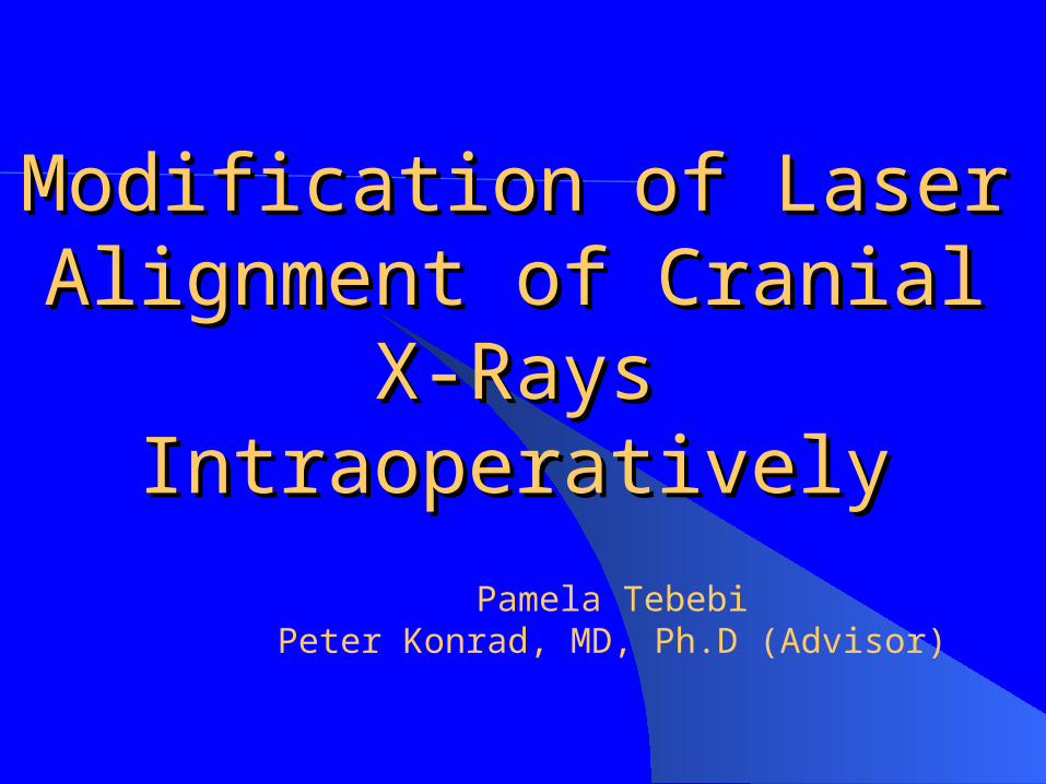
Current Device for DBSCurrent Device for DBS Many surgeon use CRW
frame Stereotactic frame is fixed in
the skull and the stimulating electrode is driven through a burrhole
Can only be aligned once Does not provide an accurate
X-ray laser sighting alignment system for the comparison of the electrode stimulation site to the final site of the implant so as to verify that the implant is at the designated site
Cosman-Roberts-Wells (CRW) apparatus; Photo from R. Galloway, R Maciunas. Stereotactic Neurosurery. Biomedical Engineering, Vol 18(3), 1990.

New Technology in DBS: mT New Technology in DBS: mT Platform SystemPlatform System Developed by Frederick
Haer & Co. (FHC), Inc (specialize in metal and glass microelectrode, needles and instrumentation for cellular research)
microTargeting Platform System requires no stereotactic frame or image guidance system
Vanderbilt is the first and only institution in the nation testing the device
microTargeting (mT Platform) Platform System; Photo provided by FHC

mT Platform is design from importing CT and MR scans of the head into the mT Platform Planning and Design Software
Software allows neurosurgeon to design custom fixture based on the target, entry, references points, and etc.
Software virtually build the Platform in place on patient, including trajectory and target
New Technology in DBS: mT Platform System
microTargeting (mT Platform) ; Photos provided by FHC

If virtual representation is satisfactory, data file is send to FHC for fabrication of Platform
No method for alignment Does not provide an
accurate X-ray laser sighting alignment system for the comparison of the electrode stimulation site to the final site of the implant so as to verify that the implant is at the designated site
microTargeting (mT Platform) Platform System; Photo provided by FHC
New Technology in DBS: mT New Technology in DBS: mT Platform SystemPlatform System

Project GoalProject Goal
Design an accurate sighting system by means of laser sighting alignment for intraoperative X-rays taken of brain stimulator implants

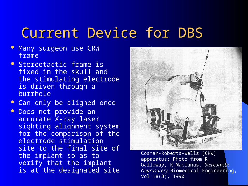
Proposed Alignment DeviceProposed Alignment Device
Mount an aiming tube to a rectangular platform that is attached to a leg on mT Platform
Aiming tube is parallel to mid-sagittal plane
Center of tube is normal to mid-sagittal plane

Problem & Soloution to Problem & Soloution to Proposed Alignment DeviceProposed Alignment Device PROBLEM: Too expensive for FHC to modify
the software interface to allow attachment of alignment device to leg
Legs are based on a component that is loaded once and copied to other two position
Changing one leg would result in complete redesign
SOLUTION by FHC: Modify software to create a component for second hub
Alignment Device Alignment Device Requires modification
of the mT Platform Requires modification
of of the software model file written by FHC
Modification is less expensive
Both hubs point to the target site, and hub with tube is parallel to mid-sagittal plane
Image provided by FHC

Alignment Device : componentsAlignment Device : components
mT Platform with second hub on the fixture
Aiming tube for X-ray with cross hair on both ends is attached to second hub
Image provided by FHC
Image provided by FHC
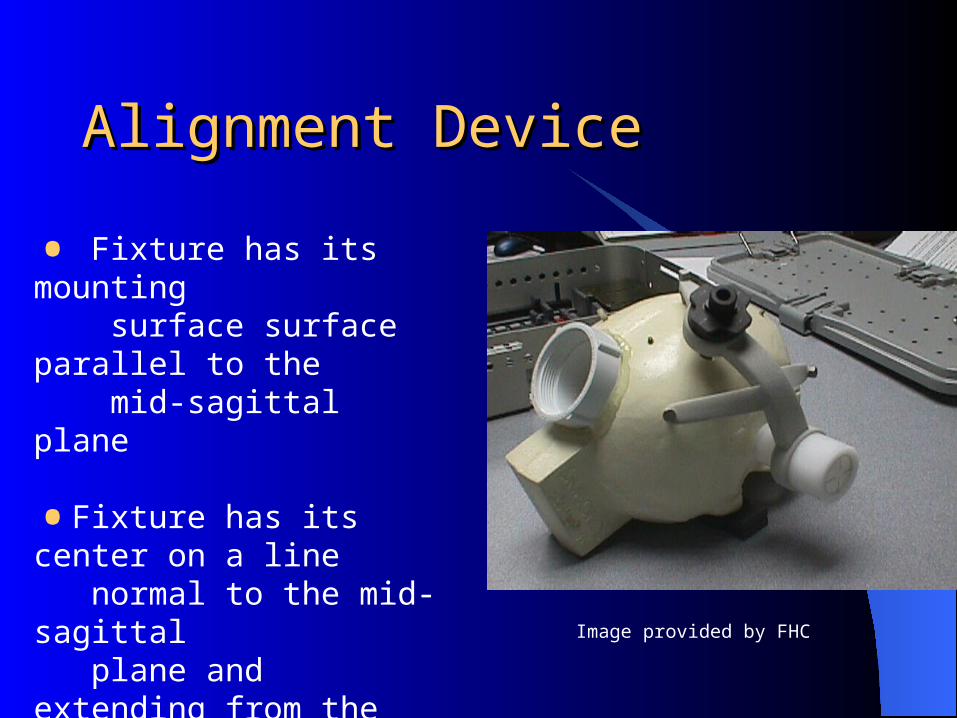
Alignment DeviceAlignment Device
• Fixture has its mounting surface surface parallel to the mid-sagittal plane
•Fixture has its center on a line normal to the mid-sagittal plane and extending from the target point
•Allows for a sagittal view through the aiming tube
Image provided by FHC

Alignment DeviceAlignment Device
When cross hairs at the ends of the tube are in line in the image, C-arm is aligned with predicted target point
Beam of the C-arm passes through the tube pointed at target point
All calculations and localization is done by mT Platform Planning and Design Software with out neurosurgeon having to align tube
Image provided by FHC
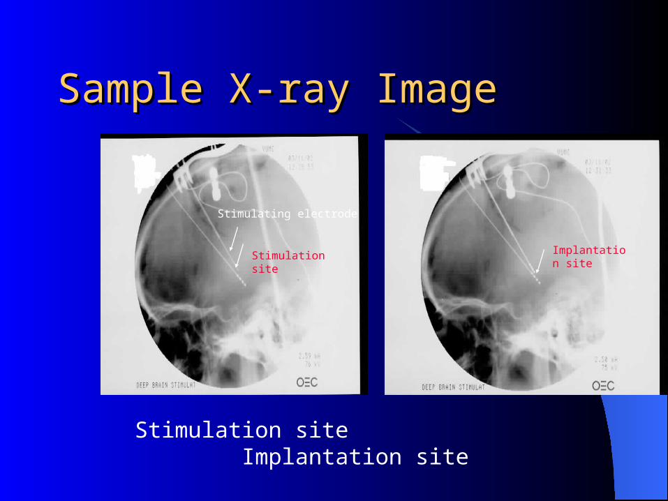
Sample X-ray ImageSample X-ray Image
Stimulation site Implantation site
Stimulating electrode
Implantation siteStimulation site

ToleranceTolerance
Target site area: 4X6mmAllowable distance implant can be from
site of stimulation: 0.5mmIf implant is placed more than 0.5mm from
targeted stimulation result in no stimulation or undesirable side effects

Technology: Alignment DeviceTechnology: Alignment DeviceFirst of its kind; ~48 procedures/year Vanderbilt University is the first
institution to consider this new approachHelpful tool for neurosurgeon; feel more
comfortable about accurately placing the stimulating implants
X-ray is useful in lawsuits and beneficial to insurance company to verify correct placement of implant to stimulation site by neurosurgeon

Market SizeMarket Size
Neurosurgery: -tool for neurosurgeon Medical and Research Institution: -prevent lawsuits -receive better insurance coverage

Device StatusDevice Status
Prototype model will be provided by FHCOnce completed the alignment device will
be tested on patients here at Vanderbilt

AcknowledgementsAcknowledgements
Peter Konrad, MD, Ph.D (Vanderbilt University) Ron Franklin, Senior Engineer (Frederick Haer & Co.) Chris Koa, MD, Ph.D (Vanderbilt University) Robert Galloway, Ph.D (Vanderbilt University) John Song, MD (Vanderbilt University)

ReferencesReferences1. Surgery for Deep Brain Stimulation, Anderson MA, RN. Vanderbilt University Medical
Center, Jan 2002. (pamphlet)2. microTargeting Platform System incorporating STarFix guidance. User Manual A989-02,
FHC, Inc, 2001.3. Galloway RL, Cleary K, Peter T. Image Guided Procedures 2001 A Snapshot View. July 31
2001.4. Galloway RL, Macinus RJ. Stereotactic Neurosurgery. Biomedical Engineering, Vol. 18(3), pp.
181-205, 1990.5. Stereotactic Neurosurgery. Concepts in Neurosurgery. Heilbrum MP, Ed., Baltimore: William
& Wilkins, Vol. 2., 1988.6. Movement Disorder Surgery. Progress in Neurological Surgery. Lozano AM, Ed., New York:
Karger, Vol. 15, 2000.7. Shrivastava, RK, Germano IM. Deep Brain Stimulation for the Treatment of Parkinson’s
Disease. Contemporary Neurosurgery, Vol.. 23(16), Aug 15, 2001.
Related Documents