Modern Instrumental Methods of Analysis Prof. J. R. Mudakavi Department of Chemical Engineering Indian Institute of Technology, Bangalore Lecture No. # 15 X-Ray Analytical Techniques-1 Instrumentation Welcome to the fifteenth lecture of our course. In the last class, we had discussed about X-rays fluorescence; just for continuity sake I will quickly go through about some properties of the X-rays what we had discussed earlier only. So, basically X-rays are short wave length electromagnetic radiations produced by the deceleration of high energy electrons or by it can also be produced by transition electronic transition of the electrons in the inner orbital’s of the atoms. The X-rays have a wavelength range of 10 raise to minus 5 angstrom units to 100 angstrom. Conventional X-rays spectroscopy is largely confined to 0.1 angstrom to 25 angstrom units. (Refer Slide Time: 01:16) So, you can generate the X-rays by bombarding a metal target a beam of high energy electrons. You can also expose a substance to a primary beam of X-rays to generate secondary beam of X-rays of lower energy. You can use radioactive sources, some of the

Welcome message from author
This document is posted to help you gain knowledge. Please leave a comment to let me know what you think about it! Share it to your friends and learn new things together.
Transcript

Modern Instrumental Methods of Analysis Prof. J. R. Mudakavi
Department of Chemical Engineering Indian Institute of Technology, Bangalore
Lecture No. # 15
X-Ray Analytical Techniques-1 Instrumentation
Welcome to the fifteenth lecture of our course. In the last class, we had discussed about
X-rays fluorescence; just for continuity sake I will quickly go through about some
properties of the X-rays what we had discussed earlier only. So, basically X-rays are
short wave length electromagnetic radiations produced by the deceleration of high
energy electrons or by it can also be produced by transition electronic transition of the
electrons in the inner orbital’s of the atoms. The X-rays have a wavelength range of 10
raise to minus 5 angstrom units to 100 angstrom. Conventional X-rays spectroscopy is
largely confined to 0.1 angstrom to 25 angstrom units.
(Refer Slide Time: 01:16)
So, you can generate the X-rays by bombarding a metal target a beam of high energy
electrons. You can also expose a substance to a primary beam of X-rays to generate
secondary beam of X-rays of lower energy. You can use radioactive sources, some of the

radioactive sources continuously emit X-rays like curium. By during it is and the
adequate process and you can also generate X-rays from a synchrotron radiation source.
So, the X-ray tube generates both continuum spectra as well as line spectra. Electrons are
produced at a heated cathode and accelerated towards a metal anode with a potential
difference of about 100 kilo volts.
And upon colliding with the anode part of the energy of the incident beam is converted
into X-rays, and the continuum X-rays spectrum exhibits a well defined short wave
length limit, which is characteristic of the applied voltage, but not of that of the chemical
what you have element. So, you can see that if you use 50 kV, you will get 0.2; and 40
kV, we can get X-rays only starting from about 0.3 kV etcetera. These things we have
seen in the last class with different applied potentials.
(Refer Slide Time: 02:50)
So, the continuum radiation results basically from the collision between the electrons and
the atoms of the target. Each collision results in the emission of a photon. The energy of
the photon must equal to the loss of the energy when it collides with an electron. So, the
number of collision with decreasing energy may occur for each electron as it bounces
from one atom to another atom and this gives raise to continuum spectrum. So, the
maximum photon energy generated corresponds to the instantaneous deceleration of the
electron to zero kinetic energy - that is the basic difference. And the kinetic energy of all
the electrons can be expressed as Duane hunt law - that is v e that is product of the

applied potential multiplied by the electronic charge gives you radiation X radiation and
the same h nu zero say notations.
(Refer Slide Time: 04:14)
You use to calculate the wavelength h nu 0 is nothing but h c by lambda; where h is
planks constant and c is the velocity of light and a is the lambda is the wave length. So,
the kinetic energy is basically the product…
(Refer Slide Time: 04:30)
Now, let us learn some more about the X-rays that is the X-rays lines spectra results from
electronic transitions of the innermost atomic orbital’s, they occur in the longer

wavelength range of 4 to 6 angstrom. The line spectra occur for all elements having
atomic numbers of 12 and above below that you do not get line spectrum. So, usually
atomic numbers having less than 23 show only two lines called as K series and
designated as K alpha and K beta. These are of short wavelengths for continuum, for
examples, K series for tungsten appears at 0.18 and 0.21 angstrom.
(Refer Slide Time: 05:14)
So, this is the figure we had shown you in the last class that there are two lines in this
range, which are line spectra in super imposed on the continuum spectra like this.
(Refer Slide Time: 05:30)
So, elements having atomic numbers more than 23 show L series which also designated
as alpha 1 and beta 1. There is a threshold voltage for each element below which line
spectra do not appear. For example, below 50 kV, no line spectra are obtained for the
molybdenum; however, above 70 kV it produces line spectra.
(Refer Slide Time: 05:57)
Now, here are some elements and X-ray emission lines for some typical elements, for
example, sodium - atomic number is 11; alpha 1 occur at 11.9 naught 9 and beta 1 occurs
at 11.617. You will be surprised to see that we are able to calculate the wavelengths at
such accuracy, only in the case of X-rays. Similarly, for potassium we have 3.74 to 3.454
etcetera. Above this level above atomic number 23 we have K alpha as well as L alpha L
beta. So, we get four lines maximum, same with the case with rubidium, cesium,
tungsten, uranium etcetera, where I have listed some of the X-ray lines obtained for these
things.

(Refer Slide Time: 06:54)
Now, mostly has shown that there is a real linear relationship between the square root of
the frequency for a given K or L line and the atomic number as shown below. For
example, c by lambda is equal to a in to Z minus sigma whole square, where Z is the
atomic weight. And you can see that the as the atomic number increases, there is a linear
relationship in L alpha 1 as well as in the K alpha line frequency.
(Refer Slide Time: 07:39)
So, this is the very important property of all the X-rays and the short wavelength X-rays
K series is produced when the high energy electron beam removes an electron the from

the K orbit. This produces an exited ion which emits a quanta of the radiation as
electrons from outer most orbital’s come back to the ground state. So, K series lines arise
from transitions between the higher energy levels and the K shell. Similarly, L series of
lines results when an electron is lost from the second principal quantum level directly or
from the transition of element L electron from the K level.
(Refer Slide Time: 08:24)
Now, this is the partial energy level diagram causing X-rays production, here I have
shown you here alpha 1, alpha 2, beta 1, beta 2 etcetera. And these are all this series and
the left part of this figure is K series, and the right part is the L series. They also contain
some of the fluorescence lines.

(Refer Slide Time: 08:53)
So, in this figure you can see that, the energy levels that is on the left axis versus or and
the logarithmic scale. So, as the energy level increases the separation between the lines
they also becomes less and they become crowded at higher energy levels at rather lower
energy level, but between K shell L shell the energy levels are well separated. So, the
wavelengths of characteristic X-ray emissions are also independent of the chemical
element as the transitions do not involve bonding or non bonding electrons. For
examples, the position K alpha line for tungsten or any element whether is exactly same
whether it is an oxide, or whether it is sulphide or whether it is a pure metal or it could be
an organic complex. So, whatever be the state chemical state of the atom the wavelength
of characteristic X-ray spectrum, X-ray are always same; that means, irrespective of the
state of the atom you should be able to determine the element by X-ray spectrum.

(Refer Slide Time: 10:39)
Now, based on the properties of X-rays, a number of analytical methodologies have been
developed and these are based on the atomic emission, for example, that is known as X-
ray emission spectroscopy - XES, and then you can study the emission or surface
emission that is known as auger emission spectroscopy. You can study the X-ray
fluorescence spectroscopy, where the emitted X-rays have a wavelength longer than the
excitation wavelength - that is X-ray fluorescence spectroscopy. Then you can have
electron spectroscopy - that is ESCA. So, the here you are studying only the specific
portion of the surface of the sample, what elements it contains etcetera using the X-ray
line spectrum.
And you can also have X-ray absorption spectrometry - that is nothing but, you take a
sample pass the X-rays whatever comes out the loss in energy of the X-rays can be
determined as a function of a known element concentration. So, that is known as X-rays
absorption. Another way of using another analytical technique very useful for X-rays is
X-ray diffraction, that is you can study the crystal structure of the substance, where the
elements, where the atoms are located. So, this you can do and we are not going into
details of each of this technique, but we are going to concentrate only on the X-ray
fluorescence technique, but this much of information is essential before you proceed into
the analytical aspect. Now, here I am continuing where I left my last slide, that is I had
described number of analytical technique that XES, AES, XFS, ESCA, X-rays
absorption etcetera.

(Refer Slide Time: 13:06)
The phenomenon that is occurring there, I am trying to show here. Here the X-ray is
coming in the top figure – a, you can see that radiation is falling and then it gets excited
and then it emits the radiation. And b is as hay the radiation is the falling on to the
example, and again depending upon the X-ray properties you get as hay emission. And
then X-ray fluorescence - the radiation X radiation falls on the sample, but the radiation
emitted has a different wavelength.
Similarly, other electron spectroscopy: you take the electrons and then measure the
wavelength of the different K lines L likes of the different elements that also you can
take it to determine the concentration on the surface. Now, X-ray absorption like I was
telling you to take the basic radiation, total energy of the bean passed through the sample
loss of energy is determined just like molecular absorption. And electron spectra X-ray
diffraction it gives you the atoms at different positions of the crystal and the crystal
structure can be determined.

(Refer Slide Time: 14:34)
Now, the X-ray absorption and emission spectra are quite simple, but because they
consists of very few lines, K alpha K beta L alpha and L beta. So, X-ray emission
spectrum of an element may be obtained by using the sample itself as the target element.
And this approach is not really convenient for all types of samples, because sometimes
you may have a liquid sample, then it is not possible to fit it on to the position of the
target directly. So, the sometimes you may have a powder that is also not easy that is
unless you have a metal, which you can paste it and focus the incoming electronic beam
on to the element.
Now, instead what we can do is? We can excite, you can achieve the same excitation by
irradiating the sample with beam of X-rays from an X-ray tube or a radioactive source.
And in this method all the elements in the sample are excited by the absorption of the
primary beam which emit their own characteristic X-rays as usual fluorescence X-ray
fluorescence. Now, therefore, the X-ray fluorescence is a powerful tool for the
qualitative analysis that is pass fail test. Suppose, you are producing a metal in a foundry
and then you would like to add some of the alloying elements and you would like to
determine the concentration of the alloying element within a particular level.
All you have got to do is let the furnace run let the metal melt take a small amount of the
melt and take it to a X-ray fluorescence just take the spectrum. If it shows you the X-ray
fluorescence which is pre calibrated to the known concentration then you have a pass fail
test ready, very quick method and very easy to control. So, most of the high Tec foundry
have this kind of X-ray fluorescence lab attach to the foundry itself where they can
control the composition of the metal. So, X-rays is also an non destructive technique,
because the material does not get destroyed. So, unlike other element techniques where
the, we have to either dissolve the sample or then extract the original nature of the
sample is lost. So, there are two types of X-rays fluorescence spectrometers, one is you
can monitor the wavelength of the dispersed red X-rays or you can monitor the energy of
the X-rays.
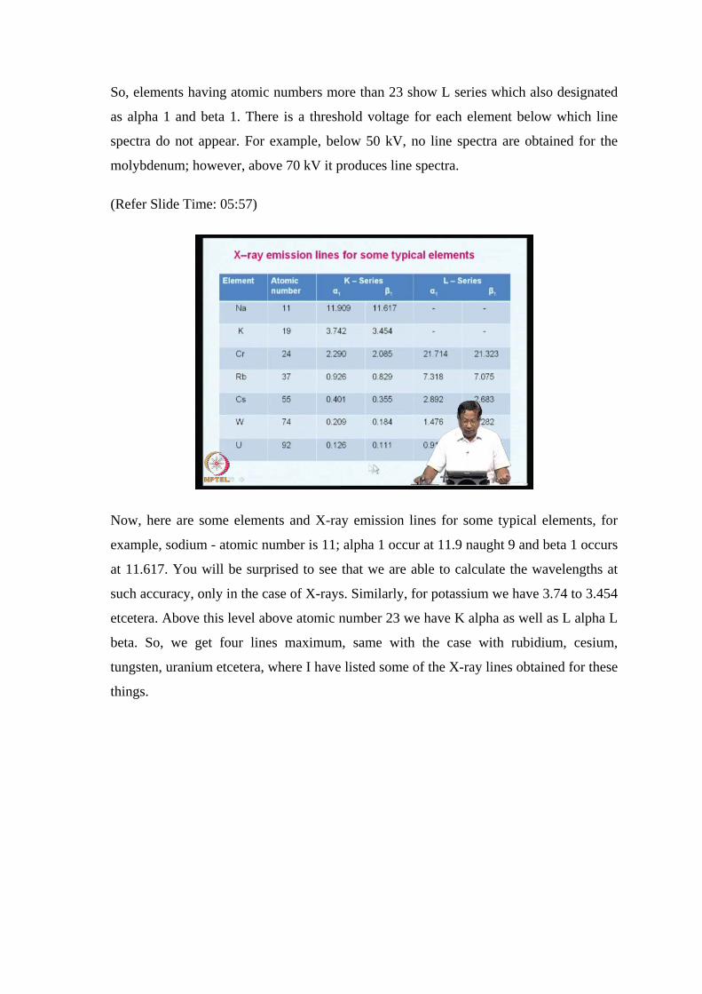
(Refer Slide Time: 17:41)
So, important instrument components of a source X-ray XRF, I have shown here, here is
an X-ray source that is X-rays will fall on the sample and then you have a detector
followed by electronics and the computer. Then you can have another kind of
arrangement, where X-ray source will fall on the sample and then you have a analyzing
crystal through and the emitted X-rays are focused using metal thin metal films here. The
one and two into and then on to the analyzing crystal from there again it is focused on to
the detector followed by it is electronic and the computer to control the measurement.

(Refer Slide Time: 18:31)
So, what are the different types of sources for X-rays, how do you generate? The X-rays
and other instrumental aspects are same whether it is auger, whether it is ESCA whether
it is a X-ray absorption, emission etcetera. So, we are going to study the basic
instrumentation for rays and then we will go on to the analysis of X-ray fluorescence
technique other techniques. So, we are not studying right now. So, what does an X-ray
tube has? Basically an X-ray tube is a high vacuum sealed off tube containing a the
containing heated filament from, which we lead the electrons flow and these electrons
need to be accelerated using hypo tension that is the requirement.
So, the filament cathode can be filaments which can simple generate the electrons when
it is heated and the anode has to be a 5 by 10 mm heavy block of copper with a metal
target plated or embedded on it is surface. And the target materials could be any element
like tungsten chromium copper molybdenum rhodium scandium silver ion cobalt any
element what you would like to study. So, you need two circuits one is for controlling
the heating of the filament and another to accelerate the electrons and make them go and
hit the cathode. So, the heat anode not the cathode, but it is the anode so, the heater
circuit controls, the intensity of the emitted X-rays and the accelerating voltage
determines the energy of the electrons or the wavelength.
So, both and current and the voltage must be stable up to point one percent accuracy that
is very important otherwise, the energy of X-rays will not be constant and most of the

measurements will go ovary. So, X-ray generation by this method is basically a
inefficient process and much energy is wasted as heat so, cooling of the anodes is a must.
So, the you will have to have a water cooling system to cool the anode and modern
equipments what they do is they use highly sensitive transducers and hence they avoid
cooling, cooling is not necessary. Some times in the such cases the X-ray beam
generated in the source has to come out and fall on to the sample. So, we use a beryllium
window or aluminum window or Parlodion films for this purpose, because you need to
take out the X radiation from the source out of the source out of the tube and focus them
on to the sample.
(Refer Slide Time: 22:14)
So, I am going to show you an X-ray tube here. The cooling water, there is cooling water
in and cooling water out. So, the whole thing is cooled continuously during operation,
and then we have a metal target anode here and this is the filament tungsten filament is
we have used. And this is a focusing cup whatever are the electrons generated will be
focused on to the target anode and then you have a circuit here, for accelerating the
voltage, another for heating the element; this is for the heating, this is for the another
circuit for the high voltage. And we have a beryllium window here through which X-rays
are coming out you can have beryllium or any other element.

(Refer Slide Time: 23:17)
So, the current is basically regulated by monitoring the X-rays tube DC current and
controlling the filament voltage. When the full wave is rectified? The voltage reaches it
is speak value 120 times per second and persisted for a only a small fraction of the time;
that means, 120 times per second you are going to get X-rays in per minute which is
available to you. When the tube is operated at 50 kV the gain is approximately two fold
for elements up to atomic number 25 and it increase to fourfold at beryllium. That is you
keep on increasing the atomic number up to 25 increase, this to fold and then you take
some other metal and reach up to other metals one by one.
And when you reach the atomic number 56 that is barium, the energy reaching the
detector will be fourfold compare to bromine. So, normally X-ray tubes are operated
between 50 to 60 kV source, power source, but nowadays you can operate tubes with 100
kV also for these are for high end elements. So, this is this I have shown you, another
way of producing X-ray is to use radioactive substances such as hydrogen tritium and
iron, cobalt, cadmium 1 naught 948, 1 naught 9 top numbers refer to top numbers refer to
the atomic weight here I have written cadmium 1 naught 948.
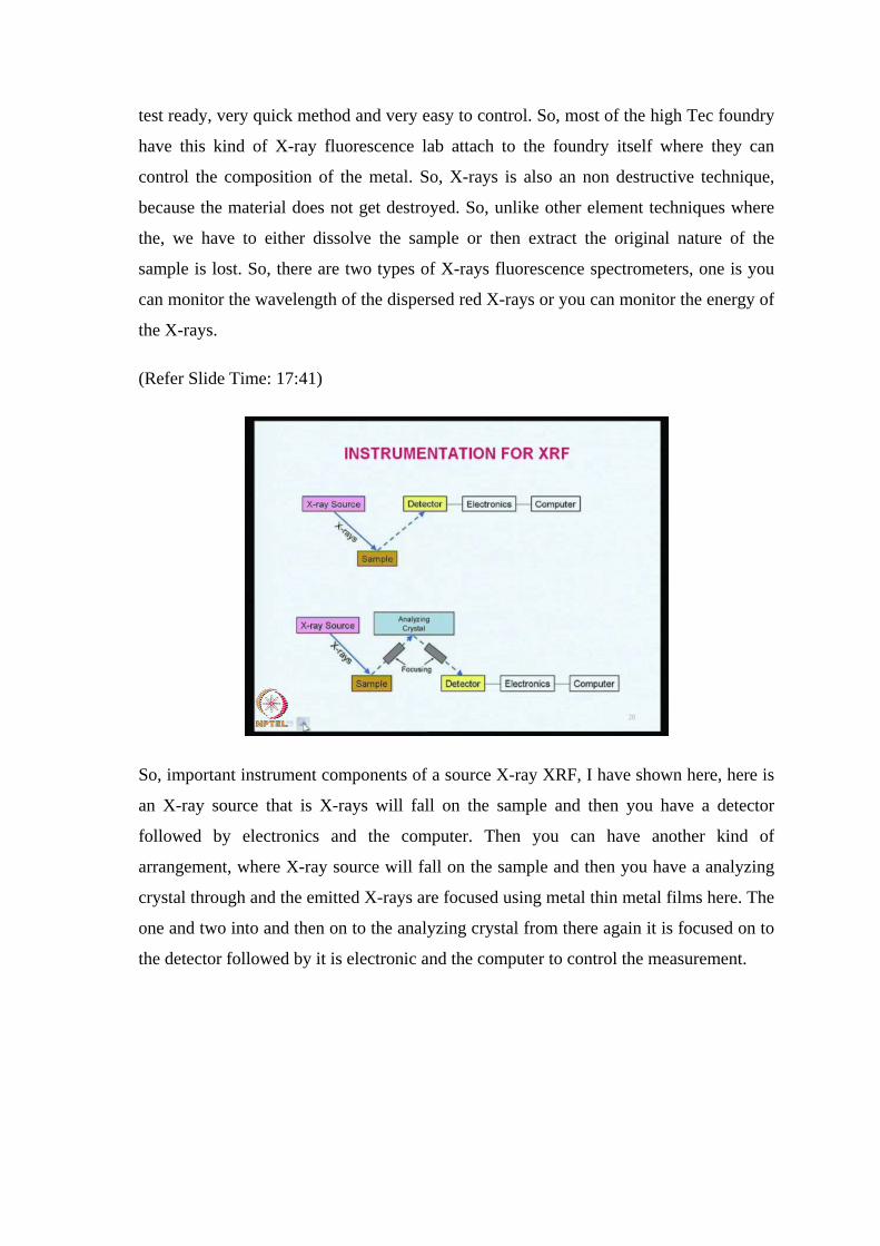
(Refer Slide Time: 25:08)
And then iodine is 125 53 like that there are number of elements which are radioactive,
but they generate X-rays. So, some elements produce simple line spectra others produce
a continuum a given isotope will be suitable for existing a number of limits for example,
a source giving X-ray in 0.3 to 0.47 angstrom is suitable for a X-rays studies for all
elements up to silver. The sensitivity improves as the wavelength of the source line
approaches the absorption edge.
(Refer Slide Time: 25:56)
Now, in some applications X-ray tubes with tungsten target is used to excite K alpha and

K beta lines of molybdenum. The resulting flow since spectrum would also contain only
line spectrum which can be used for the excitation of the analyte. So, after you have X-
rays generated from an X-ray tube or radioactive source and they come out you need to
filter unwanted X-rays. So, just like in other electromagnetic radiation when you have
you want to remove the unwanted the radiations here, also you need to use filters. So, the
in some applications narrow ranges are required wavelength are required you do not need
all the kinds of X-rays. So, you do not need continuum also so, both filters and
monochromator are used for this purpose. So, zirconium filters are very ideal for these
and they can be used in films for about 0.01 centimeter thickness to isolate K alpha line.
They not only eliminate the continuum, but they eliminate K alpha line also and you can
see here in this figure that wave length is plotted again absorption of emission. So, here I
have a filter in this range that is between 0.06 and 0.08 approximately around 0.07 there
is continuum X-ray coming like this, and then there is K alpha line and then K beta line.
And you need to use only K beta line, emitted by molybdenum target that is this line, but
you do not need the other part. So, what I do is? I use a zirconium filter which will cut
off all the radiation that is coming in this range including the K alpha line. So, this is
what this is the job of the filter so, you what is available after using the filter is only the
K line K beta line. So, several other filters are also available and basically thin strips of
varies metal and you can choose their characteristics cut off wavelength is different for
example, in this case.
(Refer Slide Time: 27:29)

It is around 0.65 or 0.7, but for some other elements it may be something else. So,
depending up on your requirement you can choose the filter.
(Refer Slide Time: 28:59)
Now, after choosing the filter you also need just like in you visible spectrophotometry
monochromator systems. In X-rays monochromator, if you remember what I had thought
you earlier? We had used the we had defined an X-ray monochromators system
containing lens focusing lens a slate prism and the or a grating and then again refocusing
slit etcetera, this is known as monochromator system. So, similarly, in X-rays also you
need to have a monochromator system and this consists of a pair of beam of beam
collimators that is to make the beam parallel. One serves usually you can use two filters
one for as a slit one to serve as a slit and the other serves as the dispersing element.
So, the dispersing element this is basically a single crystal mounted on a goniometer.
What is a goniometer? A goniometer is a rotating table that permits the determination of
the angle of theta that is the angle at which the radiation falls on the crystal and the then
it gets reflected diffraction florist etcetera. And this angle you are you suppose your
sample is on a rotating term stable the radiation will fall like this and then this angle can
be varied from zero to ninety one eighty followed by three sixty. So, all the three sixty
degrees you can vary the angle of incidence of X-rays.
So, the goniometer permits you disc arrangement it is nothing, but a circular arrangement
on which the electrons are coming. And this angle of incidence from where the angle is

from where the X-ray is coming is can be varied precisely for determining the X-ray and
the detection and detector has to be located at twice the angle let us if I use Bragg’s law
L lambda is equal 2 d sine theta. Now, we have pour we have studied earlier, but it is
also coming further; that means, for every angle of theta that the X radiation falls on the
sample the detector has to be rotated to twice. Suppose, you are angle of incidence is 35
degrees the detector has to rotate at 70 degrees.
So, the both the turn table as well as the detector have to be rotated exactly one at half
the speed one and another at twice the quantity of the theta, that is the between the
crystal phase and the collimated incident beam. So, Bragg’s equation shows, that for any
given angular setting goniometer only a few lens are diffracted according to this
equation, it is n lambda is equal to two d sine theta which I have explained just now. And
to produce the spectrum again what you need? The exit beam also has to the collimated
once again and the detector must be mounted on the another second table, that rotates at
the twice the speed that I have told you.
And the modern X-ray monochromator systems have microprocessor controlled motors
to drive the crystal to rotate the crystal and the detector independent of each other
without gear based mechanism. What is the problem with gear based mechanism? If you
use the gear based mechanism to rotate something gears can wear out. So, subsequently
after you use it for sometime the gears can wear out and the precision of the angle would
not be there at all. So, gear less mechanical systems is more comfortable in this kind of
arrangements. So, they can scan rapidly if there are gearless we can scan the whole 360
degrees in a very short time because there are no gears involved. So, you can scan them
at the rate of 240 degrees per minute, so these is the advantage of gearless this thing.
(Refer Slide Time: 34:31)
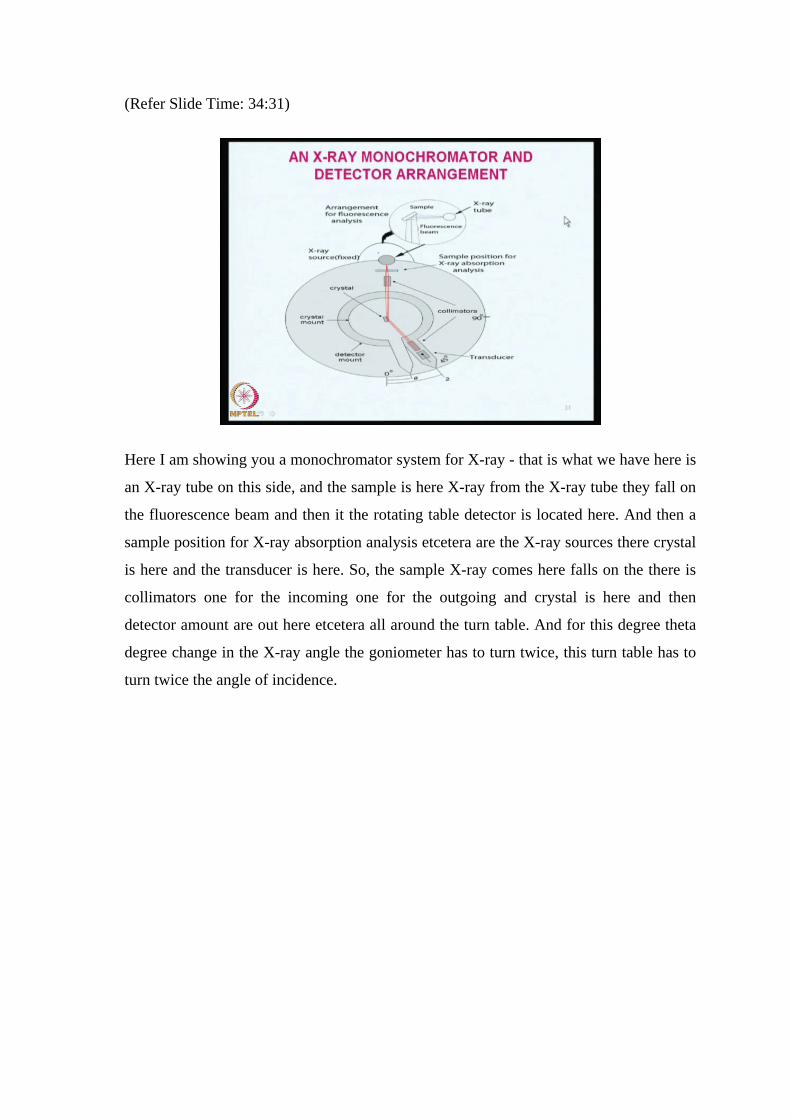
Here I am showing you a monochromator system for X-ray - that is what we have here is
an X-ray tube on this side, and the sample is here X-ray from the X-ray tube they fall on
the fluorescence beam and then it the rotating table detector is located here. And then a
sample position for X-ray absorption analysis etcetera are the X-ray sources there crystal
is here and the transducer is here. So, the sample X-ray comes here falls on the there is
collimators one for the incoming one for the outgoing and crystal is here and then
detector amount are out here etcetera all around the turn table. And for this degree theta
degree change in the X-ray angle the goniometer has to turn twice, this turn table has to
turn twice the angle of incidence.

(Refer Slide Time: 35:46)
So, this is what you have been talking about? The collimators basically consist of series
of closely spaced metal plates that absorb all the radiations expect the parallel beams.
Because the radiation scattered in other directions have no meaning for the chemical
analysis what is coming out through a thin film of radiation, which is parallel that only
use full. So, X radiation longer than two angstrom is absorbed the atmospheric gases this
is another important concept you should understand. That means, longer than any X
radiation longer than two angstroms should not be opened to atmospheric gases; that
means, you need to have a vacuum system if longer wavelength are required.
So, the sample compartment must be fleshed with either helium or some other gas which
does not absorb X-rays or it must be evacuated. So, with flat crystals, up to ninety nine
percent of the X radiation is divergent which is absorbed by the collimators waste
wastage twenty ninety nine percent. This loss can be minimized by about ten percent
using a curved crystal surface suppose, your crystal is not exactly a ninety degree this
thing we can machine it and shape it into a curved surface like this, then the loss can be
minimized by about ten percent. So, and this divergent beam the curved surface also will
help in accomplishing the divergent focusing further focusing it will any curved surface.
If it X-ray falls on that it will act like a concave mirror or convex mirror, where it will try
to focus the divergent beam from the source on to the exit collimators.

(Refer Slide Time: 38:03)
So, here there are some diffracting crystal properties like topaz, lithium fluoride, sodium
chloride and then ethylene diamine tartrate ditartrate ammonium dyhydrogen phosphate
etcetera, there lattice spacing’s are given here 1.356, 2.014 etcetera.
The wavelength ranges are given here, the dispersion is approximately in the third fourth
column at lambda max and at lambda minimum. So, if you want to get X-rays of
between two and between 2.67 angstroms and 0.24 you should go for topaz - this is how
the table can be utilized. Suppose, you want 8.6 between 8 and 0.7 and 8 angstrom unit
X-rays then you should go to this table, and look which of the material gives you the
radiation corresponding to between 0.77 and 8.67 that corresponds to EDDT. So,
depending upon the angle a wavelength, you should be able to choose the wavelength
material crystal for X-ray collection.

(Refer Slide Time: 39:27)
So, the useful wavelength range for a crystal is determined by it is lattice spacing d - that
is m lambda is equal to 2 d sine theta that what you mean by d; d corresponds to the
lattice spacing in between the spacing in between the two element, two atoms rather.
And this happens; the detector problems are more if the angle of incident is less than 10
degrees. So, if the angle of sometimes the angle may be 35 degrees, 90 degrees, 120
degrees like that below if it is almost parallel approximately 10 degree angle, the detector
will have problems in collecting the collecting the X-ray emanating from the crystal,
same thing happens when the detect angle approaches 180 degrees.
So, less between 170 and 180 detector will have problem in collecting the radiations
quantitatively, between zero and ten as you have started from one end to the other end if
this is zero and then slowly you will increase. First ten degrees there is a problem of
collection, then it improves detection collection, it reaches 90 and then slowly when it
comes to 170 degrees again it is almost parallel between 170 and 180 again the detection
problem exists.
So, what we do is in between? We tried to work between only up to 160 degrees in an X-
ray work. So, crystals with large spacing’s offer better kind of d spacing a lattice spacing
offer better control over the scatter control, but it results in lower dispersion also, this is a
consequence of the X-ray Bragg’s laws diffraction this thing.

(Refer Slide Time: 41:52)
If you differentiate Bragg’s equation what do you get? Is d theta by lambda is equal to n
over 2d sine theta and this 2d comes the denominator. Therefore, d theta by d lambda
this dispersion is the measure of the dispersion that is inversely proportional to d, on the
one hand when the d is large we get a better dispersion better collection. But because it is
a measure of the dispersion which is inversely proportional you get other problems. So,
low dispersion prohibits the use of shorter wavelength, hence topaz are lithium fluoride
must be substituted in most of the X-ray fluorescence work.
(Refer Slide Time: 42:39)
So, X-ray, now we move on to we had discussed so far about X production of X-rays we
had discussed so far about the use of filters. We had discussed about the monochromator
system and how the sample has to be mounted, and how the angles and goniometer have
to be arranged. Now, let us discuss about the detectors. In the X-ray detectors are usually
operated as photon counter, in this mode individual pulses of the charges are produced as
quanta of radiation, which are absorbed by the transducer. Basically, X-ray is nothing but
the X-radiation has to come and hit a particular sensitive transducer and amount of
energy has to be released quanta of energy that has to be counted.
So, this requires rapid response time because X-rays are very fast electrons are also very
fast moving, X-rays also are very fast. And therefore, the transducers as well as a signal
processor have to work in within micro seconds, the moment that hits within a few micro
seconds the whole measurement and counting and all other related system must be
completed before another photon comes and hits the detector. So, this technique is useful
for low intensity X-ray beams, because in high intensity X-ray beams there is not much
time for the each photon to be counted individually.
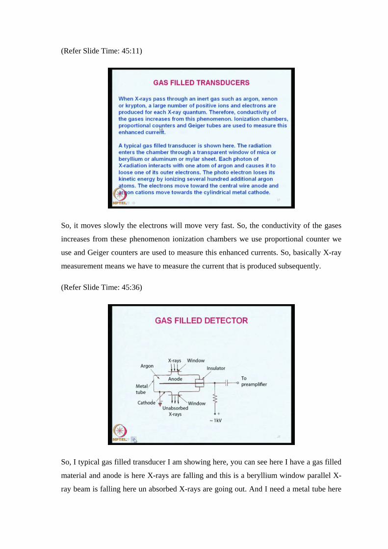
So, when X-rays pass through an inert gas such as argon and anon a or krypton a large
number of positive ions are also generated electrons are there and then X-ray quantum is
there. Therefore, the conductivity of the gases increases, because the positive ions and
electrons have generated argon cation and electrons. So, electron is very light weight and
argon cation is a higher rate samples.
(Refer Slide Time: 45:11)
So, it moves slowly the electrons will move very fast. So, the conductivity of the gases
increases from these phenomenon ionization chambers we use proportional counter we
use and Geiger counters are used to measure this enhanced currents. So, basically X-ray
measurement means we have to measure the current that is produced subsequently.
(Refer Slide Time: 45:36)
So, I typical gas filled transducer I am showing here, you can see here I have a gas filled
material and anode is here X-rays are falling and this is a beryllium window parallel X-
ray beam is falling here un absorbed X-rays are going out. And I need a metal tube here
argon is filled and the window another window is here and the amount of radiation that is
generated has two there is a cathode here there is a anode here. So, the electrons cations
will move towards this side and electrons will move towards the cathode and the amount
of current generated is to be amplified and used. So, this is how a gas filled detector
works?
(Refer Slide Time: 46:31)
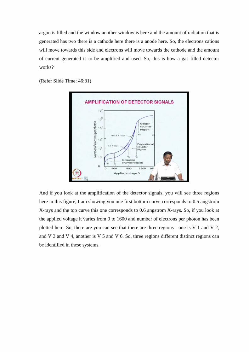
And if you look at the amplification of the detector signals, you will see three regions
here in this figure, I am showing you one first bottom curve corresponds to 0.5 angstrom
X-rays and the top curve this one corresponds to 0.6 angstrom X-rays. So, if you look at
the applied voltage it varies from 0 to 1600 and number of electrons per photon has been
plotted here. So, there are you can see that there are three regions - one is V 1 and V 2,
and V 3 and V 4, another is V 5 and V 6. So, three regions different distinct regions can
be identified in these systems.

(Refer Slide Time: 47:24)
So, the first region shows the number of electrons reaching the anode which is fairly
constant for each photon; that means the amount of energy is constant and it can be
detected very easily. In the second region that is V 3-V 4 in this region. There is you can
see that the slope has changed and it is it has increased enormously in this region range
current. So, now this region number of electron release increase rapidly with applied
voltage and here the secondary ion pair production occurs due to the collisions between
the accelerated electrons and gas molecules.
Therefore, under these conditions amplification of the current occurs. So, the third region
that is V 5-V 6 again here, there is a sort of saturation occurring and the amplification is
enormous, but the current is independent of the type and energy of the incoming
radiation. The current is actually governed by the geometry of then and gas pressure of
the tube this region is known as Geiger region.

(Refer Slide Time: 48:53)
So, scintillation counters are another type of electron of other types of detectors, where
we use a sodium iodide crystal containing 0.2 percent thallium iodide shaped in the form
of a cylinder and in about 3 to 4 inch in dimension. So, one surface is plane and the
which phases is the cathode of a PMT as the incoming radiation passes through passes
through the crystal its energy is lost to the scintillation, which is subsequently released in
the form of photons of fluorescence radiation. So, the flashes of light produced are
transmitted to the photo cathode which in turn produce an electrical pulse that can be
amplified.
So, in X-ray now we come to these are the so far what I have covered is the basic
instrumentation for X-ray fluorescence for all X-rays. Now, we come to X-ray
fluorescence as I have explained to earlier, we have wavelength dispersive X-ray,
WDXRF rays, energy dispersive. So, in both these systems radioactive sources are
collimated and dispersed into its component wavelength both single channel and
multichannel detection systems are possible. In what do we mean by single channel and
double channel, multichannel and single channel elements you are going to determine
only one element, and in multi channel elements you are going to determine number of
elements simultaneously. So, the angles for each element have to be set specific for each
element. So, the counting progresses until sufficient counts are obtained for précised
measurements, the instruments can be automated to cover the entire three sixty range in
such instruments.

(Refer Slide Time: 50:52)
The moment of the crystal and the detector and synchronized such instruments usually,
cost around 25 lakhs of rupees and in multi channel instruments they are costlier the cost
approximately 150,000 dollar’s and there are useful for the determination of up to 24
elements simultaneously.
(Refer Slide Time: 51:05)
In these instruments, individual channels for each crystal for each element are arranged
radially around the X-ray tube and sample holder. The crystals for most of the channels
are fixed at an appropriate angle for a given analytic. That means if you want to

determine chromium you need one crystal and then one detector system one channel one
amplifier 1 pulse side detector etcetera. And the similarly, every element to be
determined there have to be so many different channels and they analysis the whole
analysis would be completed within a few seconds or maximum a couple of minutes.
(Refer Slide Time: 52:03)
So, in energy dispersive X-ray fluorescence it has a poly chromatic source that is an X-
ray source or a radioactive material a sample holder a semi conductor detector and
various electronic components. So, the analytical methodology is also very simple here
that is there are no moving parts - that is no collimators are crystal diffractions.
Therefore, the entire energy reaching the detector is approximately 100 times more than
wavelength dispersive hex rays instruments.
So, low power energy X-ray tubes or weaker radioactive materials can be used as
radiation sources. Which are cost effective and safer to use why I am telling you all this
is when you want to buy an instruments for your laboratory you should know what are
the different sources? What are the detectors? What are their causes? What their
collimators? What are their lines etcetera. So, this information would be very useful
when you want to go for an instrument of this type.

(Refer Slide Time: 53:19)
So, here you can see the amplifier is there, sample is here then helium cooling and then
beryllium window is here, this is the beryllium window and then detector and then
amplifier and multichannel pulse I am pulse height analyzer. So, fluorescence radiation
keeps on coming like this; for each element you will have different kinds of the crystals
and the detectors. So, this is the mars and rover detector mars machine rover detector,
here we can see that X-ray detectors are all located around these six head APXS. This is
a front view and alpha source is there, contact ring is there, and then door etcetera. Other
things are all a set here this we had discussed in the first lecture also.
So, the sensor head the rower contains curium 244 sources that emits X-rays and five
point eighty one m e v alpha particles. So, the multiple element determination can be
obtained by particle induced X-ray emission method. So, the X-ray detector is a new
room temperature type that exhibits very low noise the if the detector is at operating at
very high temperature; that means, it is going for higher noise; that means, the detection
limit will be lower.
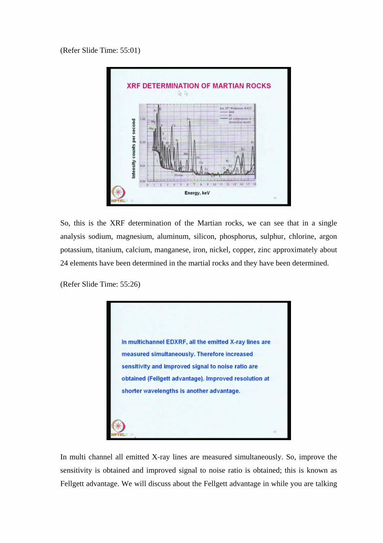
(Refer Slide Time: 55:01)
So, this is the XRF determination of the Martian rocks, we can see that in a single
analysis sodium, magnesium, aluminum, silicon, phosphorus, sulphur, chlorine, argon
potassium, titanium, calcium, manganese, iron, nickel, copper, zinc approximately about
24 elements have been determined in the martial rocks and they have been determined.
(Refer Slide Time: 55:26)
In multi channel all emitted X-ray lines are measured simultaneously. So, improve the
sensitivity is obtained and improved signal to noise ratio is obtained; this is known as
Fellgett advantage. We will discuss about the Fellgett advantage in while you are talking
about infrared radiation also. So, improved resolution a shorter wavelengths is possible
nowadays, a bench top EDXRF is available for routine determination of up to twelve
elements, ranging from sodium to uranium. The fluorescence signal is obtained by using
appropriate filters and that strikes the bottom of the rotating table where the detectors is
rotated located. And the rhodium X-ray tube five programmable filters helium per
system is available twelve positive sample changer is there and a spinner to rotate.
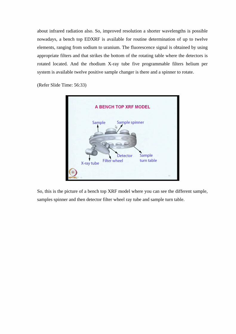
(Refer Slide Time: 56:33)
So, this is the picture of a bench top XRF model where you can see the different sample,
samples spinner and then detector filter wheel ray tube and sample turn table.
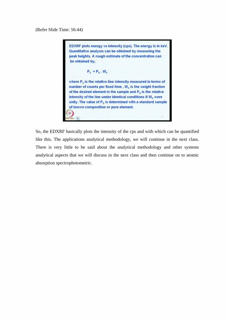
(Refer Slide Time: 56:44)
So, the EDXRF basically plots the intensity of the cps and with which can be quantified
like this. The applications analytical methodology, we will continue in the next class.
There is very little to be said about the analytical methodology and other systems
analytical aspects that we will discuss in the next class and then continue on to atomic
absorption spectrophotometric.
Related Documents