Moderators – Dr M M Singh Dr Deepak Agrawal

Welcome message from author
This document is posted to help you gain knowledge. Please leave a comment to let me know what you think about it! Share it to your friends and learn new things together.
Transcript

� Moderators –
Dr M M Singh
Dr Deepak Agrawal

� History
� Equipment and its working
� Indications
� Results
� Complications

� The Gamma Knife (stereotactic radiosurgery)
in which narrow beams of highly focused and destructive dose of radiation is given in a single session using an external reference frame fixed to the head.
• Term introduced by Lars Leksell in 1951(Professor of Neurosurgery, Karolinska University)
• 1967 , 1st GKRS installed by Leksell & Larsson at Stockholm • 1967 , 1st GKRS installed by Leksell & Larsson at Stockholm (For treatment of intractable pain managements & functional
procedure using 179 CO -60 source)
• 1975 , 2nd generation GKRS used at Karolinska institute using round collimetors (4,8,14,18mm) for treatment of vascular malformation & tumors
• Focal distance from radiation source to target is 40.3 cms

� Four models are used –
� Model U : uses a hemispherical array – dose profile greater in superior / inferior extent
� model B/ C: uses circular array of radiation � model B/ C: uses circular array of radiation sources –dose profile greater in right/left
� New model C: contain an automatic positioning system
� latest version (perfexion ):was first installed at Mayo Clinic in September 2007.

� Radiation does not remove the tumor or tissue abnormality.
� Radiation distorts DNA (ionizing induces mutations and other forms of DNA damage & cell cycle arrest).cycle arrest).
� Radiation induce apoptosis to proliferating cells.
� The cell lose its ability to retain fluids.
� For arteriovenous malformations, radiation induces the thickening and closing off the blood vessels.

� Tumor loses its ability to grow
� Remains the same size
�Never grow again
Benign tumors take up to 2 years to disappear
Metastatic (Cancerous) tumors take only months to disappear

� Three part of the procedure
� 3 D Stereotactic localization
� Dose planning- Most crucial
� Radiation exposure
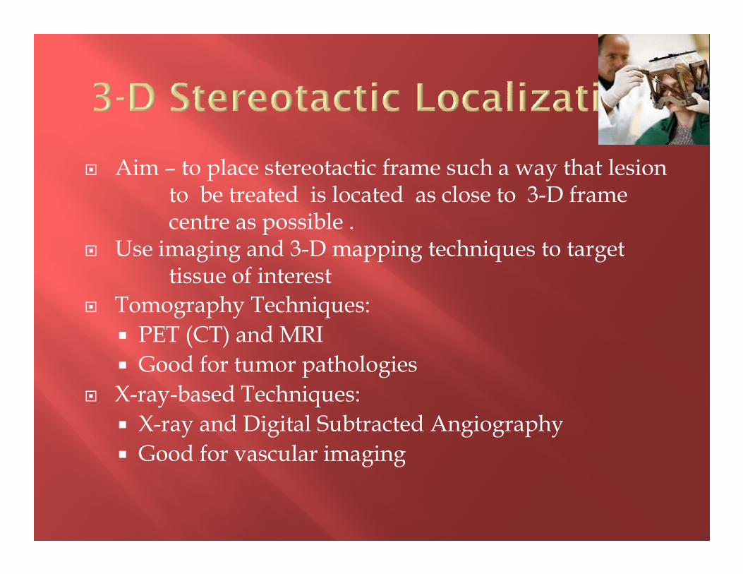
� Aim – to place stereotactic frame such a way that lesion to be treated is located as close to 3-D frame centre as possible .
� Use imaging and 3-D mapping techniques to targettissue of interesttissue of interest
� Tomography Techniques:
� PET (CT) and MRI
� Good for tumor pathologies
� X-ray-based Techniques:
� X-ray and Digital Subtracted Angiography
� Good for vascular imaging

Gamma Plan 4.12 software used to plan GK treatment of a frontal lobe meningioma. Note abrupt fall off of radiation at tumor edge. The tumor volume is red and the yellow lines represent the 50% isodose curves.

� Image transferred & using Leksell Gamma Knife 3-D planning software, a treatment protocol is planned
� Surgeon, radiation oncologist and radiation physicist decide dose plansdecide dose plans� different collimator sizes and added "shots“ required
� Tumors may not be exactly spherical
� Require additional "shots"

� High dose delivered to tumor core
Tumor tissue less oxygenated at
center
� Normal surrounding parenchyma spared � Normal surrounding parenchyma spared
� Dose most lethal when surrounding tissue receives a higher dose of radiation

� Dose rate
� Volume of tumor-
maximal tumor volume that could be
radiated is around 25 ccradiated is around 25 cc
� Cranial nerves sensitivity
� Effect on surrounding vessels
Usually not a limiting factor
Endothelial damage in large vessels does not leads to thrombosis in most of the cases

� Factors determining cranial nerve dysfunction
� Nature of the nerve -Optic and Acoustic
� Length of nerve irradiated� Length of nerve irradiated� Volume of nerve roots exposed� Mechanical stretching of the nerve by tumor

� Optic nerve- 8 Gy
� Cavernous sinus nerves- 30-40 Gy
� Trigeminal nerve- 10-25 Gy
� Facial nerve- 10-25 Gy
� Lower cranial nerves- 10-25 Gy
Tishler CA, loeffler JS et al :Tolerance of cranal nerve to radiosurgery :Int J Radiate Oncol Biol Phys, 1193

� Noninvasive method of treating inoperable lesions� Eliminates the risk of open surgery
� patients experience little discomfort.� Low immediate procedural morbidity� Low immediate procedural morbidity� Patients can immediately resume their previous activities
� Short hospitalization
� The lesion being treated receives a high dose of radiation with minimum risk to nearby tissue and structures

� Delayed complication of radiation
� Use is limited to tumor located adjacent to critical neurovascular structure

� Lesions characteristic� small -less than 3 cms in diameter
� Larger not appropriate � require two or more "shots“� radiation dose to surrounding brain is more - damage tissue� Radiation Necrosis� Radiation Necrosis
� Geometrically � Regular- spherical, ovoid or cylindrical is good
� irregular shaped i.e. star or crescent shaped- not good candidates � Difficult to plan radiation dose volume
or� it deliver lethal dose of radiation to the surrounding brain

� Tumors
� Acoustic neuromas
� Pituitary adenomas
� Meningiomas
� Skull-based tumors � Meningiomas of cavernous sinus � Meningiomas of cavernous sinus
� Chordomas and chondrosarcomas
� Craniopharyngiomas
� Metastases
� Gliomas and other primary intra-axial tumors
� Pineal tumors
� Haemagioblastoma
� Glomus tumors

� Vascular abnormalities:
� Arteriovenous malformations� Cavernous malformations
� Functional problems:
� Trigeminal neuralgia � Parkinson’s disease ( pallidotomy)� OCD � Radiosurgical thalamotomy
� For epilepsy
� Cavernous malformation� Arteriovenous malformation� Hypothelamic hemartoma� -Mesial temporal epilepsy
� Ocular tumors
� Uveal melamona � Orbital metastases � Optic nerve sheath meningioma
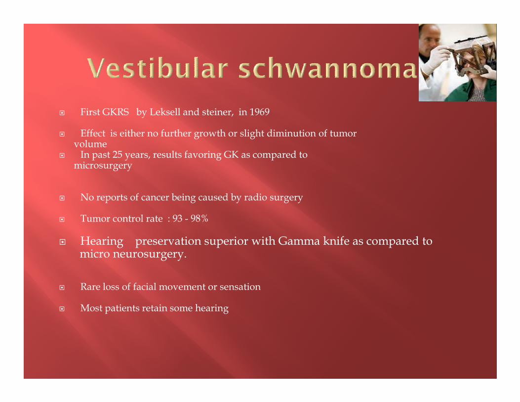
� First GKRS by Leksell and steiner, in 1969
� Effect is either no further growth or slight diminution of tumorvolume
� In past 25 years, results favoring GK as compared to microsurgery
� No reports of cancer being caused by radio surgery� No reports of cancer being caused by radio surgery
� Tumor control rate : 93 - 98%
� Hearing preservation superior with Gamma knife as compared tomicro neurosurgery.
� Rare loss of facial movement or sensation
� Most patients retain some hearing

� Small tumors with maximum intracisternal diameter : < 3 cm
� Residual / Recurrent tumor after microsurgery
� In elderly population
High risk patients refuse microsurgery. � High risk patients refuse microsurgery.
� In younger subset, hearing preservation is an important issue
Contraindication :
Tumor causing mass effect on brain stem with clinical deficit

� Marginal dose for small tumors -14 Gy
medium tumors - 12 Gy
large tumors -10 Gy
� Margin dose 11 to 15Gy at 50% isodose curve.

Procedure Cure/ Control
Death Hearing Preservation
Facial Nerve Preservation
GK Radiosurgery
90-96% 0% 70% 100%
Microsurgery 98% 1.5% 7% 63%
Regis J,Delsanti C,Roche P-H,Thomassin J-M,Pellete W. Functional outcomes of radiosurgical treatment of vestibular schwannomas:1000 successive cases and review of the literature.Neurochirurgie 2004:50(2-3):301-311
J. Neurosurg94(5):1091-1100,2002,Modern management of vestibular schwannomas

� Earliest change includes loss of contrast or gadolinium enhancement, most marked in center of tumor
� Poorer response in NF-2-life long follow up required
� SRS – high rate of tumor control, higher hearing preservation , low morbidity low morbidity
� Initial loss of contrast enhancement may be followed by period of increased volume and dense enhancement
� Decrease in tumor volume with increased length of follow up-up to 90 % at 10 years
Chopra R, Kondzoilka D,Niranjan A,Lunsford LD,Flickinger JC. Long term follow-up of acoustic schwannoma radiosurgery with marginal tumor doses of 12 to 14 Gy.Int J Radiat Oncol Biol phys 2007:68:845-851

� Microsurgery is the initial treatment of choice
� First Meningioma radiated in 1975
� GKRS used as an alternative to post operative radiotherapy for residual meningiomas
� Advantages include unique accuracy and precision ; minimizing risk

� Indications � primary treatment (for difficult to operate )
� cavernous sinus / basal meningioma
� petroclival tumors of the posterior fosse
� Medical illness /advanced age
For residual meningiomas� For residual meningiomas
� Recurrent tumor after open surgery
� Contraindicated � Tumor with symptomatic optic nerve or chiasmal compression
� optic nerve sheath tumor with preserved vision
� > 3 cms in diameter with mass effect
� Mean dose to tumor margin :14 Gy
� Mean tumor volume: 7.4 ml
� mean 7.5 isocenter used
� Results � 94% tumor control rate � 94% tumor control rate
� 10% edema risk
� Subsequent surgery - 5.2 %,
� Additional RT :2.9%
� Morbidity –7.7%
Kondziolka,Niranjan A,long term result after radiosurgery for benign intra cranil tumors, Neurosurgery 2003, 53

� SRS recommended mainly for Pilocytic astrocytomas located in thalamus, hypothalamus, brain stem, optic tract etc.
� Low grade gliomas (grade 2) are usually diffusely infiltrating hence role of radiosurgery limited for residuallesion
� Low grade glioma in medial temporal lobe is usuallywell circumscribed and amendable to SRS
� Two groups where it can be used patients with low Karnofsky's scorewell localized small lesions

� Primary Low Grade Gliomas
� 95% response rate.
� 10-year follow-ups in most patients treated show no evidence of residual tumor
Kida Y,Kobayashi T,Mori Y,Gamma knife radiosurgery for low-grade astrocytomas:results of long term follow-up. J Neurosurg 2000:93

� Cytoreduction followed by GK
� MR scan within 48 hours of surgery
� residual, enhancing tissue is boosted with GK followed by conventional radiation therapyresidual, enhancing tissue is boosted with GK followed by conventional radiation therapy
� recurrence treated with GK, if tumor nidus is small
� High grade gliomas are usually bulky tumors;SRS again has a limited role

� Controlled studies need to be completed toconclusively demonstrate the role of GKRS
� Recent studies indicate radiosurgery is useful inextending survival in patients with recurrent glioblastomaglioblastoma
� Median tumor progression free interval of 12 months
Hsieh PC et al, Adjuvant Gamma Knife stereotactic radioosurgery , treatment option for recurrent GBM, Neurosurgery 2005,57

� Metastatic tumors are usually well circumscribed; hence amendable to GKRS
� GKRS used in two forms� As local boost in combination with WBRT
� As exclusive primary therapy� As exclusive primary therapy
� As a primary therapy can be used in patients with low Karnofsky’s score where WBRT is not desirable
� Dose recommendationsadjunctive therapy 15-20 Gy primary therapy 25-30 Gy

� Surgical removal + radiation
� benefits patients’ quality of life and survival � must control primary tumor first
� Gamma Knife radiosurgery � As effective as open surgery combined with brainradiotherapyradiotherapy
J Neurosurgery / Volume 93 / Dec 2000
� Effective even for tumors relatively resistant to external beamradiation therapy
� In selected individuals, whole brain radiotherapy not necessary� Some multiple brain metastases are also candidates for GK
J Neurosurgery / Volume 93 / Dec 2000
� Recurrent or new tumor deposits can be retreated by GK

� Success rate up to 90% reported
� Increase in tumor volume in some cases can be due to radionecrosis
� Recurrence rate is 7-15% Vs 20% for craniotomy + WBRT; hence Gamma knife superiorWBRT; hence Gamma knife superior
� Best results are obtained in patients with Melanoma and Renal cell carcinoma
� Results are poor with Bronchogenic carcinoma
� Hence, SRS recommended as primary modality for metastasis except in very large tumors
Hasegawa ,et al,Brain metastasis treated with radiosurgery alone, an alternative to WBRT,Neurosurgery 2003,52

� Best managed by microsurgery
� Gamma Knife radiosurgery useful in � recurrent
� residual tumors, especially in the cavernous sinus. � residual tumors, especially in the cavernous sinus.
� Excellent preservation of cranial nerve and pituitary function.

� Goal :to stabilize or reduce adenoma volume
� SRS indicated:� failure of total resection / parasellar region
� recurrence after surgery and radiotherapy
� Dose required is less as compared to functional tumors� Dose required is less as compared to functional tumors
� Edge dose to tumor ranges from 10-15 Gy
� Dose to optic apparatus should be kept below 8 Gy
� Excellent tumor control rate 93% ( 68 - 100 % )
� Relatively safe
no neuro-opthalmological complications reported
1 case of pan- hypopituitarism reported
Sheehan JP ,Kondziolka,Radiosurgery for residual/recurrent nonfunctioning pituitary adenoma ,J Neurosurgery 97,2002

� Complete surgical resection not possible in many cases because of frequent parasellar growth
� SRS indicated in these casesSRS indicated in these cases
� Also used as an initial modality by some and in recurrence after surgery
� Edge dose to tumor ranges from 10-25 Gy
� In 70% cases GH levels fall below 5 ng /ml
Pollock BE Radiosurgery of GH producing pituitary adenoma :factor associated with biochemical remission ,J Neurosurgery 2007 ,106

� Microsurgery considered gold standard for microadenomas - excellent results
� SRS indicated - Recurrent cases - Inaccessible tumor
� dose to tumor margin 20 Gy.� dose to tumor margin 20 Gy.� Normal 24 hour UFC:63% at 1 year� Hypocortisolism/recurrent disease not correlated
with radiation doses.� Tumor volume decreased in 73% cases not
significantly correlated with endocrine outcome
Sheehan JM.,Vance ML,Sheehan JP,et al Radiosurgery for cushing`s disease after failed transphenidal surgery. J.Neurosurg 93(5):738-42 2000

� GK - primary
- unsuccessful surgery (residual )
- failed medical treatments
� Mean dose to tumor margin 13-30 GY� Mean dose to tumor margin 13-30 GY
� Endocrine cure rate 52 %
� Remission rate poor in pt receiving antisecretory medication at time of GK
Pan L,zhang N,Wang EM.Wang BJ,Gamma knife radiosurgery as a priary treatment for prolactinomas.J Neurosurgery 2000:93

� Complete surgical resection not possible
� Multimodality therapeutic approach
-Surgery + post op radiotherapy-Surgery + post op radiotherapy
-Stereotactic cyst aspiration& injection of
radioactive Yttrium-90
- GKRS + Stereotactic single dose
radiation
Chiou SM,Lunsford LD,Niranjan A,et al:Steriotactic radiosurgery of residual or recurrent craniopharygioma, after surgery,with or without radiation therapy.Neurooncol 3:159-166,2001.

� GKRS used� Primary treatment
� adjunctive treatment alone
� combining with intralesional brachytherapy
� Salvage treatment for recurrence.� Salvage treatment for recurrence.
� Optimum dose to achieve local control was 12 Gy.(10 – 20 )
� Tumor control: 79%, complete response:19%, at 65 months mean follow-up.
� Results are better with squamous histology as
compared to adamantinomatous type.
Kobayanshi et al. Japan Long-term results of Gamma Knife surgery for the treatment of craniopharyngioma in 98 cinsecutive cases.J Neurosurg 2005:103(6 suppl)

� Role of GKRS remains controversial� Indication
� Elderly patients with symptomatic tumor� Residual / recurrent tumor � Smaller tumors
� Contraindication� Contraindication� Young patient with large tumors causing significant mass effect� Patient with catecholamine secreting tumor
� Tumor growth control rate of 94%
� Mean dose of 16.4 Gy.� Margin dose 16.4 – Maximum dose 30� Mean follow-up 46 months.� Tumor control rate : 94%

� Low morbidity-zero mortality.
� Hearing lose: 54%
� Lower cranial dysfunction is extremely rare
� Nausea,vomitting and dizziness/vestibular Nausea,vomitting and dizziness/vestibular dysfunction
� Transient facial or glossopharyngeal neuropathies
� Neither target volume nor the radiation dose associated with the incidence of cranial neuropathies.
� Severe vertigo after radiosurgery

� Gold standard treatment – surgical resection of the solid components
� GKRS can be given to solid portion ,with max dose 28 - 60 Gy and peripheral dose 11 – 20 Gymax dose 28 - 60 Gy and peripheral dose 11 – 20 Gy
� Tumor size – Decrease in size - 70 %
No change in size – 30 %
Niemela M young J et al ,Gamma knife radiosurgery in 11 Hemangioblastoma,in 7th international meeting fLeksell Gamma Knife Society,1995

� Rare skull base tumors
� Both highly invasive locally
� Challenges to treat due to their invasiveness, proximity to vital structure proximity to vital structure
� Post op GKRS – modest reduction in tumor size and long term survival
Muthukumar N,Kondzoilka D,Lundsford LD,et al: Stereotactic radiosurgery for chrdoma and chondrosarcoma: Further experiences. Int J Radiat Oncol Biol Phys 41:387-392,1998.

� Technical issue
� Indication:
� Pilocytic astrocytomas / low grade glioma
� High grade glioma
� Medulloblastomas
� Ependymomas� Ependymomas
� Craniopharyngioma
� Pituitary Adenomas
� Pineal region tumors
� Gangliogliomas
� Choroid plexus papillomas
� Neurocytomas

� No initial effects of radiosurgery
� Few patients have experienced seizures• almost always with established seizure disorder• adjust anticonvulsant prior to treatment • adjust anticonvulsant prior to treatment
� Local pain in the scalp • simple, oral pain medication

� -Brain edema
cells lose ability to retain fluid,
swelling within the adjacent brain
Rx -oral steroids/ self-limiting
Psychological side effects: � Psychological side effects:
loss of memory, decreased cognitive abilities
� Cranial nerve dysfunction
• double vision /visual loss
• facial numbness, weakness
• hearing loss
� Rare with modern gamma ray doses

� Radiation Necrosis
� radiosurgery turns a live tumor into a dead tumor
� large tumors are not good candidates ??� large tumors are not good candidates ??� risk of radiation necrosis increases with size
� dead tissue cleared from brain by an inflammatory reaction
� bigger the mass of dead tissue = greater the inflammation
� high doses of steroids
� 6 and 18 months after procedure
� Sometimes open surgery to remove dead tissue

� MR/CT imaging • every 3 months to every year • assure control of tumor
� Arteriovenous malformations-MR angiograms � Arteriovenous malformations-MR angiograms
� Follow-up protocols vary from center to center

.
Neurol India. 2008 Jan-Mar;56(1):57-61.
Gamma knife radiosurgery for glomus jugulare tumors: therapeutic advantages of minimalism in the skull base.
1601 patients who underwent GKS from 1997 to 2006, 24 patients with GJ underwent 25 procedures.
J Neurooncol. 2010 Jun;98(2):265-70. Epub 2010 Apr 20.Tumor control and hearing preservation after Gamma Knife radiosurgery for vestibular schwannomas in neurofibromatosis type 2.
Tumor control and hearing preservation after Gamma Knife radiosurgery for management of acoustic neuromas Journal of Neurosurgery - 1999/05/01
Related Documents