CLINICAL PATHWAY Page 1 of 14 Traumatic Brain Injury (TBI): Moderate/Severe ALGORITHM 1: Post-Resuscitation *Post Cardiac Arrest Clinical Pathway Inclusion Criteria: Patient with traumatic brain injury and Glasgow Coma Scale (GCS) less than or equal to 12 Exclusion Criteria: Patients found down without clear traumatic brain injury (TBI) (Can be used in conjunction with Post Cardiac Arrest Pathway* if considered beneficial) Glasgow Coma Scale (GCS) less than or equal to 8 Post-resuscitation Emergency Department Management Trauma and Neurosurgery Consult Head Computed Tomography (CT) scan- if not already obtained (STAT upload) 2 IVs with Normal Saline (NS) as maintenance Modified Rapid sequence intubation (RSI) if not already performed For patients with signs of elevated intracranial pressure (ICP) empirically treat 3% Hypertonic saline (HTS) bolus 5mL/kg/dose via peripheral IV or central line If transporting to Operating Room: Consider mild hyperventilation to PCO 2 of 32-35 mmHg Standard Neuroprotective measures** Standard neuroprotective monitoring *** Neurocritical Care (NCC) Consult Surgery as indicated Surgery Indicated? Yes Glasgow Coma Scale (GCS) 9-12 Post resuscitation (Consider for children under 2 years old with concern for acute abusive head trauma) Monitor GCS. GCS decrease to less than or equal to 8? Consider comfort sedation and standard neuroprotective measures. Complete frequent neurologic exams. Improving or stable? **Standard Neuroprotective Measures: Head of bed at 30⁰, midline Non-restrictive cervical-collar Sodium greater than 140 mmol/L Normothermia 36.8 ⁰C Oxygen Saturation 93%-97% PCO 2 35-40 mmHg Normoglycemia 80-180 mg/dL Levetiracetam 10mg/kg/dose BID x 7 days (Max dose 1,000mg BID) Appropriate sedation Continue clinical monitoring and consult Physical Medicine & Rehabilitation (PM&R) If external ventricular drain (EVD)/intracranial pressure (ICP) monitor placed per neurosurgery, follow Intracranial pressure (ICP) management algorithm (Next page) No Yes No No ***Neuroprotective Monitoring: End Tidal CO2 (ETCO2), core temperature If needed, central access and arterial line Intracranial pressure (ICP) monitor or external ventricular drain (EVD) placed if needed per neurosurgery in emergency department, Operating room, or PICU Continuous EEG (cEEG) when stabilized Yes Rehab Considerations for Severe TBI Patients Consult Physical Medicine & Rehabilitation (PM&R) for patients that have stabilized or have been hospitalized greater than 5 days Consult PT/OT within 48 hours of admission for early passive range of motion, bracing & splinting, and out of bed when appropriate Consult speech therapy when patient stabilized or have been hospitalized for greater than 5 days Consult rehab neuropsychology when PM&R is consulted

Welcome message from author
This document is posted to help you gain knowledge. Please leave a comment to let me know what you think about it! Share it to your friends and learn new things together.
Transcript

CLINICAL PATHWAY
Page 1 of 14
Traumatic Brain Injury (TBI): Moderate/Severe
ALGORITHM 1: Post-Resuscitation
*Post Cardiac Arrest Clinical Pathway
Inclusion Criteria:
Patient with traumatic
brain injury and Glasgow Coma Scale
(GCS) less than or equal to 12
Exclusion Criteria:
Patients found down without clear
traumatic brain injury (TBI)
(Can be used in conjunction with Post
Cardiac Arrest Pathway* if
considered beneficial)
Glasgow Coma Scale (GCS)
less than or equal to 8
Post-resuscitation
Emergency Department Management
Trauma and Neurosurgery Consult
Head Computed Tomography (CT) scan- if not already obtained (STAT
upload)
2 IVs with Normal Saline (NS) as maintenance
Modified Rapid sequence intubation (RSI) if not already performed
For patients with signs of elevated intracranial pressure (ICP)
empirically treat
3% Hypertonic saline (HTS) bolus 5mL/kg/dose via peripheral IV or
central line
If transporting to Operating Room: Consider mild hyperventilation to
PCO2 of 32-35 mmHg
Standard Neuroprotective
measures**
Standard neuroprotective
monitoring ***
Neurocritical Care (NCC)
Consult
Surgery as indicated
Surgery
Indicated?Yes
Glasgow Coma Scale (GCS) 9-12
Post resuscitation(Consider for children under 2 years old with
concern for acute abusive head trauma)
Monitor GCS. GCS
decrease to less than
or equal to 8?
Consider comfort sedation
and standard neuroprotective
measures.
Complete frequent
neurologic exams.
Improving or
stable?
**Standard Neuroprotective
Measures:
Head of bed at 30⁰, midline
Non-restrictive cervical-collar
Sodium greater than 140
mmol/L
Normothermia 36.8 ⁰C
Oxygen Saturation 93%-97%
PCO2 35-40 mmHg
Normoglycemia 80-180 mg/dL
Levetiracetam 10mg/kg/dose
BID x 7 days (Max dose
1,000mg BID)
Appropriate sedation
Continue clinical monitoring and
consult Physical Medicine &
Rehabilitation (PM&R)
If external ventricular drain
(EVD)/intracranial pressure (ICP)
monitor placed per neurosurgery,
follow
Intracranial pressure (ICP)
management algorithm
(Next page)
No
Yes
No
No
***Neuroprotective Monitoring:
End Tidal CO2 (ETCO2), core
temperature
If needed, central access and
arterial line
Intracranial pressure (ICP)
monitor or external ventricular
drain (EVD) placed if needed per
neurosurgery in emergency
department, Operating room, or
PICU
Continuous EEG (cEEG) when
stabilized
Yes
Rehab Considerations for
Severe TBI Patients
Consult Physical Medicine &
Rehabilitation (PM&R) for
patients that have stabilized
or have been hospitalized
greater than 5 days
Consult PT/OT within 48
hours of admission for early
passive range of motion,
bracing & splinting, and out
of bed when appropriate
Consult speech therapy
when patient stabilized or
have been hospitalized for
greater than 5 days
Consult rehab
neuropsychology when
PM&R is consulted

CLINICAL PATHWAY
Page 2 of 14
Inclusion Criteria:
Patients with Moderate/severe
TBI with an ICP monitor in
place
Exclusion Criteria:
Patient without an ICP
monitor in place
Intracranial pressure (ICP)
monitor placed at the discretion
of neurosurgery
Elevated ICP?
External
ventricular drain
(EVD) in place?
Start deep sedation with goal
State Behavioral Scale (SBS)
equal to -3
Elevated ICP?
Hyperosmolar Therapy
Bolus hypertonic saline (HTS) if sodium
less than 165 mmol/L
If fluid overloaded or not responsive to
HTS, bolus mannitol 0.5-1g/kg/dose if
osmolarity (Osm) less than 340 MOSM/
Kg. Can Repeat after 15 minutes if ICP is
elevated
Elevated ICP?
Notify neurosurgery (NSYG),
trauma and Neurology critical
care (NCC)
Neuromuscular Blockade
Consider Repeat CT Scan
Elevated ICP?
Consider Salvage Therapies via PICU and
Neurosurgery attending to attending call:
Decompressive Craniectomy
Hypothermia to 32-34⁰C
Barbiturate Coma
Continue current ICP directed
Therapies 24 hours. Then wean
per pathway.
Ensure appropriate
cerebral spinal fluid
(CSF) drainage. See
CSF Drainage box.
Neuroprotective Monitoring:
End Tidal CO2 (ETCO2), core
temperature
If needed, central access and arterial
line
Intracranial pressure (ICP) monitor or
external ventricular drain (EVD)
placed if needed per neurosurgery in
emergency department, operating
room, or PICU
Continuous EEG (cEEG) when
stabilized
Standard Neuroprotective Measures:
Head of bed at 30⁰, midline
Non-restrictive cervical-collar
Sodium greater than 140 mmol/L
Normothermia 36.8 ⁰C
O2 Sat 93%-97%
PCO2 35-40 mmHg
Normoglycemia 80-180 mg/dL
Levetiracetam 10mg/kg BID x 7 days
(Max dose 1,000mg BID)
Age MAP (mmHg)
ICP (mmHg)
CPP (mmHg)
0-2 50-70 <15 35-55
2-5 60-80 <20 40-60
6-8 65-85 <20 45-65
9 + 70-95 <20 50-75
Fluid Goals and Vasoactives
Maintain cerebral perfusion pressure
(CPP) and euvolemia
If euvolemic, use ionotropic/
vasopressor support
Cerebral Spinal Fluid (CSF) Drainage:
For patients with external ventricular
drain (EVD) inform neurosurgery if
ICP elevated
If clamped, unclamp and place at 15
mmHg, do not raise if already lower
then 15 mmHg
Recheck ICP every 10 minutes
Additional adjustments per
neurosurgery
Comfort Sedation
State Behavioral Scale (SBS) less
than or equal to -1
Comfort score 17-24
With a goal of preserving a
neurologic exam and safety for the
patient
Deep Sedation
Midazolam, fentanyl, or morphine
Avoid hypotension
Consider neuromuscular blockade if
not ventilating or for refractory ICP
Hyperosmolar Therapy
Hypertonic Saline (HTS) is first line
in patient with central venous line
and not fluid overloaded
3% HTS may be administered via
peripheral IV in emergent situations
12% (2mL/kg; Max=60mL) and
23.4% (1mL/kg; max=30mL)
boluses via central venous line for
fluid overloaded patients
Mannitol 0.5-1g/kg/dose IV if HTS
not available or refractory to HTS
Monitor sodium and osmolarity
(Osm) every 4 hours
Sodium less than 165 mmol/L and
osmolarity less than 340 MOSM/Kg
No
No
No
Yes
ICP
less than 20
ICP
greater than 20
Sedate for comfort.
Responsive to touch or
name. See sedation
section for guidance.
Moderate/Severe Traumatic Brain Injury (TBI) Intercranial Pressure (ICP) Management Algorithm:
Once neuromuscular blockade has
started, maintain sedatives and
analgesics at current rate.
If greater than 3 PRN doses in 1
hour call PICU provider to discuss.
Continue sedation for ICP
management if effective.
Maximized
External ventricular
drain (EVD) drainage
and hyperosmolar
therapy?
Maximize external ventricular
drain (EVD) drainage and
Hyperosmolar therapy.
Discuss with Neurology if EEG
remains reactive, if so could
consider further sedation.
No
Yes
Elevated intracranial pressure
(ICP) Parameters
For patients that are calm,
unstimulated and outside of cares:
Actively treat for an ICP of greater
than 20-25 mmHg for more than
10 minutes
For a patient that is in pain, agitated
or receiving cares:
Ensure appropriate sedation/
analgesia
Consider treating ICP greater
then 30 mmHg for longer than 10
minutes after appropriate sedation
or the end of cares
When treating, treat to a goal ICP
of less than 20 mmHg
Neuroprotective
monitoring including ICP
No
No
Yes
Yes
Yes
Yes
Mean Arterial Pressure (MAP), Cerebral Perfusion pressure (CPP)

CLINICAL PATHWAY
Page 3 of 14
TABLE OF CONTENTS
Algorithm 1: Post Resuscitation
Algorithm 2: ICP Management
Target Population
Background | Definitions
Initial Evaluation & Clinical Management
Therapeutics
Systemic Management of Patients with Moderate/Severe Brain Injury
Special Considerations for Suspected Abusive Head Trauma
Appendix A: Overview of State Behavioral Scale (SBS)
Appendix B: VTE Prophylaxis for TBI Algorithm
References
Clinical Improvement Team
TARGET POPULATION
Inclusion Criteria
All patients cared for at Children’s Hospital Colorado with known or suspected brain injury secondary to trauma with a post-resuscitation Glasgow Coma Score (GCS) of less than 8 (Severe Traumatic Brain Injury) or GCS 9-12 (Moderate Traumatic Brain Injury) with concern for potential decompensation. This includes patients less than 2 years of age with concern for abusive head trauma.
Exclusion Criteria
Patient found down without clear trauma
BACKGROUND | DEFINITIONS
This protocol is based on “The Guidelines for the Acute Medical Management of Severe Traumatic Brain Injury in Infants, Children, and Adolescents” and on the existing literature and is intended to provide standardization in the care of traumatic brain injury (TBI) patients at Children’s Hospital Colorado.
Scope: Children’s Hospital Colorado – Anschutz Medical Campus (CHCO-Anschutz)
Severe Traumatic Brain Injury: Glasgow Coma Score (GCS) of 3 – 8
Moderate TBI: GCS of 9-12
Mild TBI: GCS of 13-15

CLINICAL PATHWAY
Page 4 of 14
INITIAL EVALUATION & CLINICAL MANAGEMENT
Emergency Department Management:
Obtaining the best neurologic exam safely is critical for neurosurgical decision-making and appropriate patient management. In all patients, Glasgow Coma Scale (GCS) is assessed. When possible, consider avoiding further sedatives and paralytics until neurosurgery resident or fellow examines patient. Call neurosurgery early when sedation or intubation may be necessary.
When sedation or analgesia are necessary, use short acting intermittent agents, including fentanyl (1- 2 mcg/kg) and/or ketamine 0.5-1 mg/kg IV. Note that Propofol is not appropriate for long-term sedation in children, but is a reasonable option for the first hours. Midazolam may also be used, but can alter the neurologic exam for hours.
Primary Survey
Airway control
o Intubate for GCS of eight (8) or less or if unable to maintain stable airway.
Rapid sequence intubation (RSI)
Assess GCS prior to administering sedation.
o Maintain head of patient in neutral position, use rigid cervical collar (e.g. Aspen collar).
Breathing
o Maintain oxygen saturations at 93-97%
o Maintain PCO2 35-40 mmHg
o Monitor end tidal CO2 (ETCO2) and obtain blood gases to correlate ETCO2 to PCO2 when indicated.
Circulation
o See Table 1 for minimum acceptable blood pressure by age group.
o Avoid hypotension by ensuring adequate circulating blood volume using isotonic fluid (Normal Saline) or Trauma Packed red blood cell (PRBC) units as indicated.
o Avoid hypotonic fluids as they exacerbate brain swelling.
o May use pressor support once intravascular volume has been repleted.
o Once euvolemia is attained, use Normal Saline (avoid hypotonic fluids) at age-appropriate maintenance intravenous (IV) rate.
Secondary Survey:
Full neurologic exam including cranial nerve exam
Trauma labs and x-rays per protocol, including electrolytes, glucose, ionized calcium, full CBC, and PT/PTT
Obtain head computed tomography (CT) and other indicated imaging. STAT upload prior imaging if available
If head of bed elevation is preferred, utilize reverse Trendelenburg (Revere T) positioning rather than just raising the head of the bed until thoracic spine cleared. Place Foley and orogastric tube (OG) unless contraindicated. Establish adequate IV access. Obtain CVL when indicated.
Neuroprotective Measures
Glucose Management
Check serum glucose measurements on admission to PICU and minimum every 6 hours for first 48 hours
Maintain serum blood glucose 80-180 mg/dL. Persistent hyperglycemia (greater than 180 mg/dL) and severe hypoglycemia (less than 40 mg/dL) are associated with increased mortality/worse neurological outcome.
Avoid bolus insulin dose in trauma patients.

CLINICAL PATHWAY
Page 5 of 14
Ventilation
Goal PCO2 of 35-40 mmHg
Monitor end-tidal CO2 (ETCO2).
Obtain blood gases to correlate ETCO2 with PCO2 as indicated.
Aggressive, rapid hyperventilation must be reserved for acute ICP crisis as adjunct with therapy escalation. Effect is temporary (less than 30 min), and rebound ICP elevation will occur with normalization of CO2.
Seizures
Seizure prophylaxis with levetiracetam 10 mg/kg BID x 7 days (max dose 1 g BID) should be administered to all severe TBI patients of all ages.
Seizure monitoring:
o Initiate EEG as early as feasible to evaluate for subclinical seizures, encephalopathy and reactivity. Seizures are common in patients with TBI.
o For all patients with TBI admitted to the PICU, monitor with EEG for a minimum of 24 hours, or longer if clinically indicated.
Infants and patients with suspected abusive head trauma have a high risk for seizures and may benefit from prolonged monitoring of 48-72 hours.
GCS is an unreliable marker of the risk for seizures in children less than 2 years of age.
o If the patient develops seizures, discuss treatment options with the Neurocritical Care Consult team and reference the Status Epilepticus Clinical Pathway as needed.
Sedation/Analgesia
Provide analgesia and sedation to achieve a COMFORT Pain Score of 17-24 and a State Behavioral Scale (SBS) of -1, unless deep sedation is instituted for treatment of elevated ICP. If Deep sedation is needed to manage elevated ICP, then change administration instructions within analgesia and sedation order to reflect new metric of ICP goal.
Initial dosing of sedative continuous infusions:
o Titrate sedative continuous infusions, including fentanyl, midazolam, and dexmedetomidine (be careful with dexmedetomidine in first 72 hours).
Indications for ICP Monitoring
The decision to monitor ICP is based on Glasgow Coma Score (GCS), CT scan findings, and clinical scenario at the discretion of the on-call neurosurgery attending.
Strongly consider an ICP monitor in patients with GCS of eight (8) or less after initial resuscitation, especially if CT scan shows mass lesions (e.g. hematoma, edema), effaced basilar cisterns, or diffuse edema.
o An ICP monitor may be placed in the setting of a borderline GCS when neurologic exam is unavailable for a prolonged period of time, e.g. emergent non-cranial surgery.
For those going to operating room for removal of an intracranial mass (bleed), the need for monitoring will be assessed at surgery.
The decision for or against the placement of an ICP monitoring device should be communicated by the neurosurgery attending to the trauma service and PICU attendings as well as their level of concern for anticipated ICP elevations and threshold for interventions (i.e. ICP, exam changes, EVD output).
External ventricular drainage is preferred in cases when ventricles are deemed adequate without significant anatomical distortion, are enlarged or where CSF diversion is likely to be beneficial for ICP control.
If GCS is marginal (9-13), and ICP monitor is not placed, follow exam with minimum necessary sedation/analgesia.

CLINICAL PATHWAY
Page 6 of 14
GCS is assessed hourly by ICU nursing staff, with any decline in exam communicated to the PICU provider who will communicate with the neurosurgical services. Subacute development of elevated ICP with need for medical or surgical interventions is possible from ongoing brain swelling.
If signs of herniation are present, consider giving hypertonic saline 3% (5 mL/kg) or mannitol (1 gm/kg IV) or mild hyperventilation (ETCO2 32-35) while monitor is being placed.
THERAPEUTICS
Treatment of Elevated ICP
Treatments are initiated for intracranial hypertension as defined by elevated ICP. Table 1 defines threshold values for treatment by age group. ICP units are mmHg (20 cm H20 = 14.7 mmHg).
Treat if ICP is above stated values. Treat Cerebral perfusion pressure (CPP) only when ICP cannot be controlled.
Table 1: Age Appropriate Pressure Ranges
Age MAP (mmHg) ICP (mmHg) ICP Treatment Threshold
0-23 months 50-70 Less than or equal to15 Greater than or equal to 20
2-5 years 60-80 Less than or equal to 15 Greater than or equal to 20
6-8 years 65-85 Less than or equal to 20 Greater than or equal to 20
Greater than or equal to 9 years 70-95 Less than or equal to 20 Greater than or equal to 20
Mean Arterial Pressure (MAP)
Elevate head of bed to 30 degrees – This is done in moderate to severe TBI patients.
Check that cervical collar is loose enough to allow jugular venous return (able to easily slip a finger between collar and neck).
Comfort sedation – Sedate as needed to a State Behavioral Scale (SBS) of -1. Short-acting agents are preferred. All intubated patients should receive comfort sedation.
Cerebral spinal fluid (CSF) diversion – open external ventricular drain (EVD) if clamped and notify the PICU provider who will notify neurosurgery.
o Consider placing an EVD or replacement of ICP monitor with EVD if ICP becomes refractory unless ventricles are slit-like or casted with blood
Hyperosmolar Therapy
Hypertonic Saline (3% saline) is the first-line osmotic in any patient who is not fluid overloaded.
o Administer a bolus of 5-10 mL/kg IV over 15 minutes for acutely elevated ICP.
o Infuse 0.1 - 1 mL/kg/hour continuously for maintenance of ICP control.
Check serum sodium and osmolality every 4 hours while actively administering hyperosmolar therapy.
o If sodium is greater than 165, be cautious and carefully consider administration hypertonic saline.
If limitation of total fluid volume is required (eg. in a patient receiving large amounts of fluid from drips and antibiotics), 12% or 23.4% saline solution may be substituted for 3%.
Equivalent hypertonic saline (HTS) bolus volumes and expected mEq/L increase
HTS % Max Dose mmol/L 5 mL/kg equivalent
Expected increase in serum sodium
3% NaCl ~235mL 513 5 mL/kg 5 mEq/L
12% NaCl 60 mL 2052 1.25 mL/kg 5 mEq/L
23.4% NaCl 30mL 4001 0.64 mL/kg 5 mEq/L
CLINICAL PATHWAY
Page 7 of 14
Mannitol
o Consider mannitol if:
Hypertonic saline is not immediately available.
Patient is fluid overloaded.
ICP does not respond to initial 3% hypertonic saline (HTS) bolus.
Mannitol should be used with caution if serum OSM is greater than 340.
o Dosing: Refer to Formulary
o Check serum osmolality (OSM) and calculated OSM prior to repeat bolusing after infusion. If OSM gap is greater than 10 mmol, would not administer more mannitol since it is still circulating.
o Osmolality (OSM) = (2 × Na+) + (glucose ÷ 18) + (BUN ÷ 3); OSM gap = Serum OSM – Calculated OSM
Cerebral hypoperfusion
The below therapies are instituted in the following order when ICP cannot be controlled and CPP is low as defined by Table 1. They may also be considered when ICP is normal but mean arterial pressure (MAP) is low. Cerebral perfusion pressure (CPP) is calculated as follows: CPP=MAP-ICP.
o Bolus IV fluid – Normal saline, colloid, or blood.
o Repeat boluses to maintain euvolemia
o Norepinephrine– Titrate to goal cerebral perfusion pressure (CPP)
o Additional pressors as necessary– Titrate to goal cerebral perfusion pressure (CPP)
Sedation
Comfort sedation:
State Behavioral Scale (SBS) less than or equal to -1
Comfort score 17-24
With a goal of preserving a neurologic exam and safety for the patient
Deep Sedation:
May be used concurrently with hyperosmolar therapy or prior to hyperosmolar therapy, according to provider discretion.
Deep sedation for control of ICP refractory to osmotic therapy. Nursing maneuvers are minimized and neurologic exams should be minimized during deep sedation to minimize any stimulation to the patient.
Neuromuscular blockade
Add neuromuscular blockade if ICP is refractory to sedation.
Rocuronium – 1 mg/kg bolus or cisatracurium 1 to 4 mcg/kg/minute
Titrate infusion to ICP control. Monitor paralysis with trains of four with maximal therapy being reached when there is either ICP control or an absence of a train of four twitches. Other neuromuscular blockers may be used if indicated.
Institute EEG monitoring when neuromuscular blockade is ongoing.
Sedation and neuromuscular blockade may acutely raise pCO2 (and hence ICP) if patient has been over-breathing the ventilator prior to administration. End Tidal CO2 (ETCO2) monitoring diagnoses this and allows appropriate monitoring of ventilator changes.
Evaluate responsiveness to sedation boluses given for ICP as additional sedation may not be beneficial if there are no clinical signs of responsiveness to additional boluses

CLINICAL PATHWAY
Page 8 of 14
Second-line ICP therapies
Pentobarbital Coma
o Consider in severe, non-lateralized, potentially salvageable cases.
o Initial bolus of 10 mg/kg bolus over 30 min (can use a smaller bolus of 3-5mg/kg or slower infusion rate if hypotension develops).
o Follow the initial bolus with 5 mg/kg/hr of pentobarbital for 3 hours
o Continue pentobarbital infusion at 1 mg/kg/hr
o Titrate to ICP effect and consider monitoring with continuous EEG monitoring with a target of 90% suppression (or a 1 second burst per 10 seconds of suppression) as a maximal therapy.
o Close blood pressure monitoring will be needed. Order pressors to bedside prior to administration of pentobarbital bolus.
o Monitor for pancreatitis (lipase every 3 days) and infection (daily blood cultures).
Decompressive Craniectomy
PICU attending and neurosurgery attending discussion of indication for decompressive craniectomy:
o Consider in lateralized cases or bifrontal craniectomy in non-lateralized cases with some likelihood of survival
Order of ICP Therapy weaning
Normocapnia
Barbiturate withdrawal as tolerated (ICP/ CPP)
Paralytic withdrawal as tolerated (ICP/CPP)
Sedation withdrawal as tolerated (Long-acting opiate/benzodiazepine as indicated)
Normalize serum sodium and osmolarity
SYSTEMIC MANAGEMENT OF PATIENTS WITH MODERATE/SEVERE BRAIN INJURY
Nutrition
Traumatic brain injury is associated with significant elevation in mean energy expenditure above predicted levels.
o Adult data suggests that early feeding after TBI (in first five-seven days) is associated with lower mortality rates.
Institute enteral, parenteral, or combination nutrition within 48 hours after injury
Parenteral nutrition may be delayed up to one week in compromised patients
Nutrition consult to assist in determining optimal volume and content of feeds/TPN
Use PICU Nutrition Guidelines
Venous Thromboembolism (VTE) prophylaxis and Treatment:
Due to the complexity of the Moderate/Severe TBI population please use the following VTE prophylaxis recommendations. Do not use the standard VTE Prevention clinical pathway recommendations for these patients.
Prophylaxis:
For VTE prophylaxis in TBI there are special consideration please see the VTE prophylaxis algorithm
Treatment:

CLINICAL PATHWAY
Page 9 of 14
The risk: benefit ratio of pharmacologic treatment of a documented venous thromboses, regardless of location (superficial vs deep DVT), is poorly defined for TBI patients and management decisions should be multidisciplinary between the involved surgical, critical care, and hematology services.
Any intracranial hemorrhage is a relative contraindication to treatment-dose anticoagulation
Any intracranial foreign body (external ventricular drain (EVD), ICP monitor) should be considered a relative contraindication to treatment-dose anticoagulation given an increased risk of associated hemorrhage in the acute post traumatic period. Multidisciplinary discussion is highly recommended.
If pharmacologic treatment is deemed necessary in a patient with intracranial hemorrhage, strong consideration should be given to the following approaches:
o Use a standard heparin infusion, without loading dose.
o Target lower heparin assay levels (0.2-0.3 U/mL) for 2-4 days before escalating to full treatment dose
o Repeat brain imaging before escalating to full treatment doses of heparin and/or transitioning to enoxaparin.
Coagulopathy
Correct coagulopathy, as clinically indicated, in the initial 48 hours following injury, particularly when intracranial hemorrhage is present or intracranial procedures may be necessary.
Rehabilitation
Consult Physical Medicine & Rehabilitation (PM&R) for patients that have stabilized or have been hospitalized for greater than 5 days
Consult PT/OT within 48 hours of admission for early passive range of motion, bracing & splinting, and out of bed when appropriate.
Consult speech therapy when patient stabilized or have been hospitalized for greater than 5 days to assist with assessing swallow abilities and communication even while intubated.
Consult rehab neuropsychology when PM&R is consulted to assist with TBI education, coping with rehab process and adjustment to cognitive changes.
SPECIAL CONSIDERATIONS FOR SUSPECTED ABUSIVE HEAD TRAUMA
EEG
Consider prolonged EEG monitoring (48-72 hours), since the majority of patients with abusive head trauma will have subclinical seizures which may be present on admission or delayed 24-72 hours.
MRI Imaging
Cases of suspected abusive head trauma may be of unknown timing and mechanism. As a result, they warrant aggressive monitoring and oftentimes further imaging. Definitive MRI brain imaging should be performed no earlier than 48 hours after admission and preferably 72 hours after admission, but no later than 7 days following admission. The order should be for: “MRI brain with and without contrast, with venography”. Also request “MRI cervical spine without contrast.” Additional MRIs may be obtained outside the 72 hour-7 day window to assist with medical management, as needed.
CLINICAL PATHWAY
Page 10 of 14
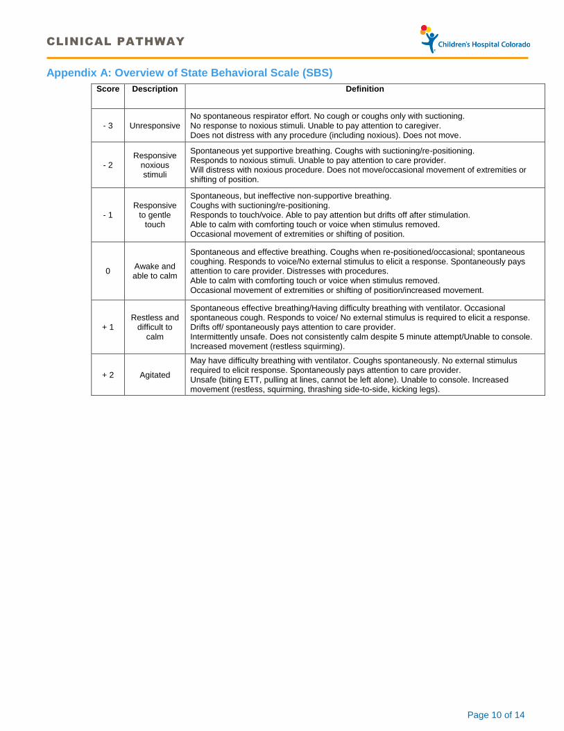
Appendix A: Overview of State Behavioral Scale (SBS)
Score Description Definition
- 3 Unresponsive No spontaneous respirator effort. No cough or coughs only with suctioning. No response to noxious stimuli. Unable to pay attention to caregiver. Does not distress with any procedure (including noxious). Does not move.
- 2 Responsive
noxious stimuli
Spontaneous yet supportive breathing. Coughs with suctioning/re-positioning. Responds to noxious stimuli. Unable to pay attention to care provider. Will distress with noxious procedure. Does not move/occasional movement of extremities or shifting of position.
- 1 Responsive
to gentle touch
Spontaneous, but ineffective non-supportive breathing. Coughs with suctioning/re-positioning. Responds to touch/voice. Able to pay attention but drifts off after stimulation. Able to calm with comforting touch or voice when stimulus removed. Occasional movement of extremities or shifting of position.
0 Awake and able to calm
Spontaneous and effective breathing. Coughs when re-positioned/occasional; spontaneous coughing. Responds to voice/No external stimulus to elicit a response. Spontaneously pays attention to care provider. Distresses with procedures. Able to calm with comforting touch or voice when stimulus removed. Occasional movement of extremities or shifting of position/increased movement.
+ 1 Restless and
difficult to calm
Spontaneous effective breathing/Having difficulty breathing with ventilator. Occasional spontaneous cough. Responds to voice/ No external stimulus is required to elicit a response. Drifts off/ spontaneously pays attention to care provider. Intermittently unsafe. Does not consistently calm despite 5 minute attempt/Unable to console. Increased movement (restless squirming).
+ 2 Agitated
May have difficulty breathing with ventilator. Coughs spontaneously. No external stimulus required to elicit response. Spontaneously pays attention to care provider. Unsafe (biting ETT, pulling at lines, cannot be left alone). Unable to console. Increased movement (restless, squirming, thrashing side-to-side, kicking legs).

CLINICAL PATHWAY
Page 11 of 14
Appendix B: VTE Prophylaxis for TBI
TBI Patient in the PICU
Mechanical Prophylaxis
Patient Age?
Assess Bleeding Risk
Risk Factors:
Craniotomy
EVD/ICP monitor in place
Moderate/large (greater than 5 mm)
subdural, epidural, or IVH
Moderate/large (greater than 1 cm) contusion
INR greater than 1.5
Platelets less than 100K
Other site of bleeding (liver, spleen, pelvis)
Any of the following:
Known clotting disorder?
Personal history venous
thromboembolism (VTE)? OR
Strong family history VTE?
Mechanical Prophylaxis
only
High Bleeding
risk? (any risk
factors present)
Reassess 24 hours after
injury
Reassess 72 hours after
injury
Clinically stable?
Repeat imaging (if
obtained) stable?
Clinically stable?
Repeat imaging (if
obtained) stable?
Complete CHCO VTE risk
assessment
VTE Risk level?
Mechanical Prophylaxis
only Enoxaparin Prophylaxis
Notes:
For TBI patients with known clotting
disorders, personal history of venous
thromboembolism (VTE), or strong
family history of VTE, consult
Hematology for discussion of VTE
prophylaxis and/or treatment.
Reassessment for bleeding risk should
occur at 24 and 72 hours after injury
and/or instrumentation (including EVD/
ICP monitor placement and
craniotomies), whichever is most
recent.
For reassessment of patients at 24
and 72 hours, repeat brain imaging
should be considered but is not
required to start VTE prophylaxis.
For patients with low bleeding risk and
high VTE risk, start enoxaparin
prophylaxis per CHCO VTE guideline
Age less than 12 years Age grater than or equal to 12 years
Yes No
Yes
Yes
Moderate/Low
VTE Risk High VTE Risk
No
No
No
Yes

CLINICAL PATHWAY
Page 12 of 14
REFERENCES
1. Guidelines for the management of severe traumatic brain injury. Bullock R, Chesnut RM, Clifton G, et al: J Neurotrauma 2000; 17:451–55
2. Reiter PD, Pietras M, Dobyns EL. Prolonged dexmedetomidine infusions in critically ill infants and children. Indian
Pediatr. 2009 Sep;46(9):767-73 3. Ogden AT, Mayer SA, Connolly ES Jr. Hyperosmolar Agents in Neurosurgical Practice: The evolving role of
hypertonic saline Neurosurgery. 2005 Aug;57(2):207-15 4. Oddo, Levine JM, Frangos S, Carrera E, Maloney-Wilensky E, Pascual JL, Kofke WA, Mayer SA, LeRoux PD.
Effect of mannitol and hypertonic saline on cerebral oxygenation in patients with severe traumatic brain injury and refractory intracranial hypertension. J Neurol Neurosurg Psychiatry. 2009 Aug;80(8):916-20
5. AJ Kerwin, Schinco MA, Tepas JJ 3rd, Renfro WH, Vitarbo EA, Muehlberger M. The Use of 23.4% Hypertonic
Saline for the Management of Elevated Intracranial Pressure in Patients With Severe Traumatic Brain Injury: A Pilot Study. J Trauma. 2009;67: 277–282
6. Fivez T, Kerklaan D, Mesotten D, Verbruggen S, Wouters P, Vanhorebeek I, Debaveye Y, Vlasselaers D, Desmet
L, Casaer M, Guerra G, Hanot J, Joffe A, Tibboel D, Joosten K and Berghe G. Early vs Late Parenteral Nutrition in Critically Ill Children. NEJM. 2016;374(12):1111-1122.
7. Borzotta AP, Pennings J, Papasadero B, et al: Enteral vs Parenteral Nurtrition After Severe Closed Head Injury. J
Trauma. 1994;37:459-468
8. Bennett KS, DeWitt PE, Harlaar N, Bennett TD. Seizures in Children With Severe Traumatic Brain Injury. Pediatr
Crit Care Med. 2017;18(1):54–63.
9. Kochanek PM, Carney N, Adelson PD, et al. Guidelines for the acute medical management of severe traumatic brain injury in infants, children, and adolescents--second edition. Pediatr Crit Care Med. 2012;13 Suppl 1:S1–82.
10. Carney N, Totten AM, O’Reilly C, et al. Guidelines for the Management of Severe Traumatic Brain Injury, Fourth Edition.Neurosurgery. 2017;80(1):6–15.

CLINICAL PATHWAY
Page 13 of 14
Clinical pathways are intended for informational purposes only. They are current at the date of publication and are reviewed on a regular basis to align with the best available evidence. Some information and links may not be available to external viewers. External viewers are encouraged to consult other available sources if needed to confirm and supplement the content presented in the clinical pathways. Clinical pathways are not intended to take the place of a physician’s or other health care provider’s advice, and is not intended to diagnose, treat, cure or prevent any disease or other medical condition. The information should not be used in place of a visit, call, consultation or advice of a physician or other health care provider. Furthermore, the information is provided for use solely at your own risk. CHCO accepts no liability for the content, or for the consequences of any actions taken on the basis of the information provided. The information provided to you and the actions taken thereof are provided on an “as is” basis without any warranty of any kind, express or implied, from CHCO. CHCO declares no affiliation, sponsorship, nor any partnerships with any listed organization, or its respective directors, officers, employees, agents, contractors, affiliates, and representatives.
CLINICAL IMPROVEMENT TEAM MEMBERS
Craig Press, MD | Neurology Todd Carpenter, MD| Critical Care
Tell Bennett, MD | Critical Care Matthew Mayer, MD| Physical Medicine & Rehabilitation
Brent O’Neill, MD | Neurosurgery Brian Branchford, MD | Hematology
Ricka Messer, MD | Neurology John Recicar, RN| Trauma
Amy Clevenger, MD| Critical Care Joni Mackenzie, PNP | Emergency Medicine
Steven Moulton, MD| Trauma Beth Wathen, MSN, PNP| Critical Care
Todd Hankinson, MD| Neurosurgery Pam Reiter, PharmD | Clinical Pharmacist
Patrick Mahar, MD| Emergency Medicine Elise Rolison RRT-NPS| Clinical Effectiveness
APPROVED BY
Clinical Care Guideline and Measures Review Committee – March 13, 2018
Pharmacy & Therapeutics Committee – March 1, 2018
MANUAL/DEPARTMENT Clinical Care Guidelines/Quality
ORIGINATION DATE March 13, 2018
LAST DATE OF REVIEW OR REVISION March 13, 2018
APPROVED BY
Lalit Bajaj, MD, MPH Medical Director, Clinical Effectiveness
REVIEW | REVISION SCHEDULE
Scheduled for full review on date here March 13, 2022.

CLINICAL PATHWAY
Page 14 of 14
Related Documents