Journal of Magnetism and Magnetic Materials 293 (2005) 639–646 Modelling magnetic carrier particle targeting in the tumor microvasculature for cancer treatment Ovidiu Rotariu , Norval J.C. Strachan School of Biological Sciences, University of Aberdeen, Cruickshank Building, St. Machar Drive, Aberdeen, UK Available online 3 March 2005 Abstract Magnetic drug targeting of tumors situated in the cavity of the human body is difficult because the magnetic gradients decrease rapidly with the distance from the magnets. Here computer simulations are used to investigate different techniques to focus small MPs within the microvasculature of tumors. Non-invasive methods were found to have range o15 cm whilst minimal invasive methods could be applied specifically to small tumors (o18 mm). r 2005 Published by Elsevier B.V. Keywords: Magnetic drug targeting; Magnetic particles; Blood flow; Arterioles; Capillaries; Mathematical modeling; Simulation; Tumor physiology; Magnetic focusing; Magnetic needle 1. Introduction The delivery of anticancer agents to specific target sites with minimum side effects is an important challenge in chemo-, radio- and gene- therapy. Among many existing methods, the active targeting of tumors with particulate drug carriers has gained increased interest in the last 15 years [1]. Of particular interest is drug delivery via carrier magnetic particles (MPs) of nano- and micrometre size using magnetic field gradients. This has been demonstrated to be an efficient method to concentrate anticancer agents into tumors [2] and has achieved permanent remission of squamous cell carcinoma implanted in New Zealand White Rabbits [3]. Further, encouraging clinical trials have been performed on humans for treatment of breast cancer [4]. The study of the magnetic capture process of MPs and their release of the drug into the tumors has been comprehensively investigated for small animal models or surface tumors [5–7]. The physical aspects of the MP concentration into microvasculature have been experimentally simu- lated using magnetic sources positioned in the immediate proximity of the artificial blood vessels ARTICLE IN PRESS www.elsevier.com/locate/jmmm 0304-8853/$ - see front matter r 2005 Published by Elsevier B.V. doi:10.1016/j.jmmm.2005.01.081 Corresponding author. Tel.: +44 1224 272256; fax: +44 1224 272703. E-mail address: [email protected] (O. Rotariu).

Welcome message from author
This document is posted to help you gain knowledge. Please leave a comment to let me know what you think about it! Share it to your friends and learn new things together.
Transcript
ARTICLE IN PRESS
Journal of Magnetism and Magnetic Materials 293 (2005) 639–646
0304-8853/$
doi:10.1016
�Correspfax: +4412
E-mail a
www.elsevier.com/locate/jmmm
Modelling magnetic carrier particle targeting in the tumormicrovasculature for cancer treatment
Ovidiu Rotariu�, Norval J.C. Strachan
School of Biological Sciences, University of Aberdeen, Cruickshank Building, St. Machar Drive, Aberdeen, UK
Available online 3 March 2005
Abstract
Magnetic drug targeting of tumors situated in the cavity of the human body is difficult because the magnetic
gradients decrease rapidly with the distance from the magnets. Here computer simulations are used to investigate
different techniques to focus small MPs within the microvasculature of tumors. Non-invasive methods were found to
have range o15 cm whilst minimal invasive methods could be applied specifically to small tumors (o18mm).r 2005 Published by Elsevier B.V.
Keywords: Magnetic drug targeting; Magnetic particles; Blood flow; Arterioles; Capillaries; Mathematical modeling; Simulation;
Tumor physiology; Magnetic focusing; Magnetic needle
1. Introduction
The delivery of anticancer agents to specifictarget sites with minimum side effects is animportant challenge in chemo-, radio- and gene-therapy. Among many existing methods, the activetargeting of tumors with particulate drug carriershas gained increased interest in the last 15 years[1]. Of particular interest is drug delivery viacarrier magnetic particles (MPs) of nano- andmicrometre size using magnetic field gradients.
- see front matter r 2005 Published by Elsevier B.V.
/j.jmmm.2005.01.081
onding author. Tel.: +441224 272256;
24 272703.
ddress: [email protected] (O. Rotariu).
This has been demonstrated to be an efficientmethod to concentrate anticancer agents intotumors [2] and has achieved permanent remissionof squamous cell carcinoma implanted in NewZealand White Rabbits [3]. Further, encouragingclinical trials have been performed on humans fortreatment of breast cancer [4].The study of the magnetic capture process of
MPs and their release of the drug into the tumorshas been comprehensively investigated for smallanimal models or surface tumors [5–7]. Thephysical aspects of the MP concentration intomicrovasculature have been experimentally simu-lated using magnetic sources positioned in theimmediate proximity of the artificial blood vessels
ARTICLE IN PRESS
O. Rotariu, N.J.C. Strachan / Journal of Magnetism and Magnetic Materials 293 (2005) 639–646640
[8–10]. Targeting sites deep within the body hasbeen successfully investigated for a swine model[11]. These experimental data confirm the potentialof targeting only large (0.5–5.0 mm) iron-carbonparticles into the liver of these animals, atdistances o13 cm. The disadvantages in usinglarge particles (�5 mm) is that they may blockhealthy vessels/arteries prior to reaching the tumorand may also not reach the tumor located withinparticular organs (e.g. brain) because they are toolarge to cross the endothelial barrier [12].Focusing magnetic drug carriers requires
magnetic field gradients. These magnetic fieldgradients drop off very steeply with the distancefrom the magnet. Therefore, targeting tumors atdeep sites is extremely difficult. This inevitablyresults in poor concentration of small magneticdrug carriers (e.g. ferrofluids) within tumorshence decreasing the efficiency of treatment.However, it is very important to focus particlesof reduced size specifically at the tumor, wherethey will have the advantage of crossing theendothelial barrier [13].This paper presents computer simulations of
minimal invasive and non-invasive methods totarget small MPs at deep tumor sites within thehuman body. This is achieved by modellingdifferent magnetic configurations and determiningthe trajectory and capture of MPs within healthyand diseased blood vessels.
2. Specifications
A specification of what is needed for intratu-moral magnetic drug targeting was made else-where [5] but several additional features should beconsidered. The main requirements are:
�
the MPs must be biocompatible and biodegrad-able,�
the MPs should be of an appropriate size,magnetic material and concentration that en-ables: sufficient attraction by the magnetic field;ease of passage into the tumor microvasculatureand transmission through the endothelial tissue,�
the magnetic field should be of sufficientstrength and high gradient to enable attractionand retention of the magnetic particles in thetarget area of either surface or deep tumors,
�
the magnetic field should be adjustable inaccordance with the properties of the MPs andthe blood flow to avoid the MPs concentrationin the normal tissue,�
magnetic targeting devices should permit thefocussing of MPs within either small or largetumors,�
the MPs should contain and release an appro-priate dose of anticancer agent and should befunctionalised at their surface to avoid clearanceby the reticuloendothelial system,�
the method of delivery should have good accessto the tumor vasculature and should be minimalinvasive.In this study the following specificationsof the magnets, the MPs and the blood vesselsare:
Magnets: Three sources of magnetic field wereinvestigated: cylindrical magnet magnetised alongits diameter, magnetic circuit with parabolicshaped confocal poles (MCPSCP) and needlemagnet magnetised perpendicularly on its long-itudinal axis (Fig. 1). The physical and geometricalcharacteristics of the magnets used in the simula-tions are as follows:
–
all magnets are made from NdFeB with anenergy product of 319 kJm�3 (40MGOe);–
the direction of magnetisation (Ox) is orientedperpendicular to the direction of the main bloodvessel feeding the tumor tissue;–
the pole surfaces that come into contact with thebody (tumor) have a curvature (lowest curva-ture–cylindrical magnet (radius a ¼ 2:5 cm; Fig.1a); medium curvature—MCPSCP (pole withsmallest focal parameter2b ¼ 1:5 cm; Fig. 1b);highest curvature—magnetic needle (radius a ¼0:75mm; Fig. 1c));
– the curvature of the pole surface that faces thetarget site creates an increased magnetic fieldgradient. Hence, the magnetic force acting onthe MPs increases and this makes possible theretention of MPs.–
cylindrical and MCPSCP magnets are largerenabling high magnetic fields (4105Am�1) at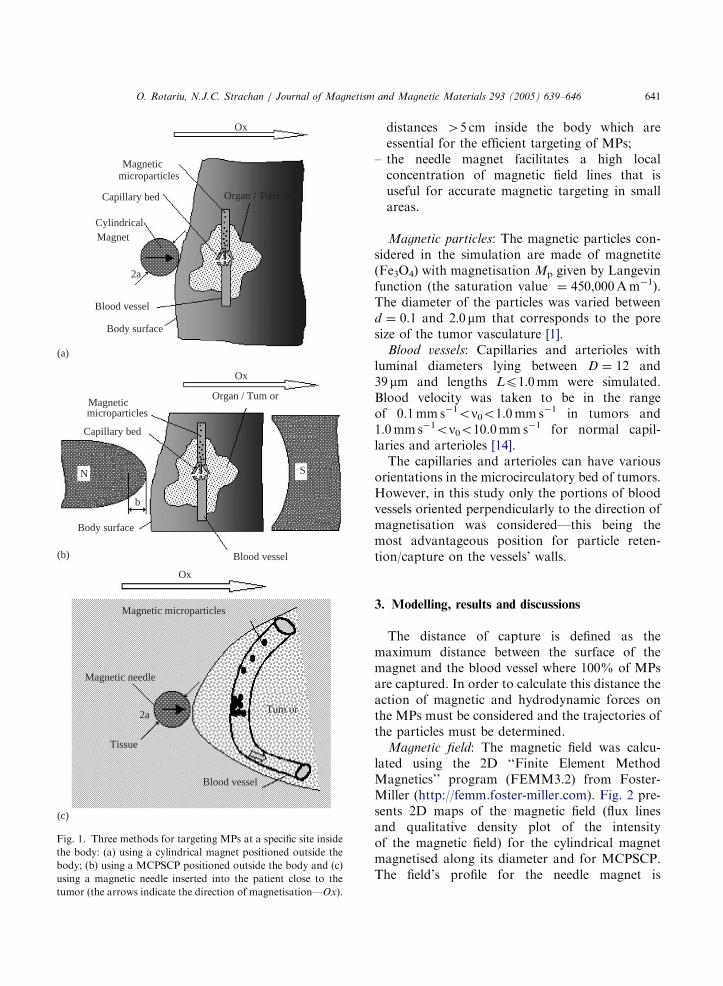
ARTICLE IN PRESS
Ox
Magnetic
Magnetic
microparticles
microparticles
Magnetic microparticles
Capillary bed
Capillary bed
Blood vessel
Blood vessel
Body surface
Body surface
Organ / Tum or
Organ / Tum or
Ox
Ox
b
N
Magnetic needle
2a
Tissue
Blood vessel
Tum or
CylindricalMagnet
2a
S
(a)
(b)
(c)
Fig. 1. Three methods for targeting MPs at a specific site inside
the body: (a) using a cylindrical magnet positioned outside the
body; (b) using a MCPSCP positioned outside the body and (c)
using a magnetic needle inserted into the patient close to the
tumor (the arrows indicate the direction of magnetisation—Ox).
O. Rotariu, N.J.C. Strachan / Journal of Magnetism and Magnetic Materials 293 (2005) 639–646 641
distances 45 cm inside the body which areessential for the efficient targeting of MPs;
–
the needle magnet facilitates a high localconcentration of magnetic field lines that isuseful for accurate magnetic targeting in smallareas.Magnetic particles: The magnetic particles con-sidered in the simulation are made of magnetite(Fe3O4) with magnetisation Mp given by Langevinfunction (the saturation value ¼ 450,000Am�1).The diameter of the particles was varied betweend ¼ 0:1 and 2:0mm that corresponds to the poresize of the tumor vasculature [1].
Blood vessels: Capillaries and arterioles withluminal diameters lying between D ¼ 12 and39mm and lengths Lp1:0mm were simulated.Blood velocity was taken to be in the rangeof 0.1mm s�1on0o1.0mm s�1 in tumors and1.0mm s�1on0o10.0mm s�1 for normal capil-laries and arterioles [14].The capillaries and arterioles can have various
orientations in the microcirculatory bed of tumors.However, in this study only the portions of bloodvessels oriented perpendicularly to the direction ofmagnetisation was considered—this being themost advantageous position for particle reten-tion/capture on the vessels’ walls.
3. Modelling, results and discussions
The distance of capture is defined as themaximum distance between the surface of themagnet and the blood vessel where 100% of MPsare captured. In order to calculate this distance theaction of magnetic and hydrodynamic forces onthe MPs must be considered and the trajectories ofthe particles must be determined.
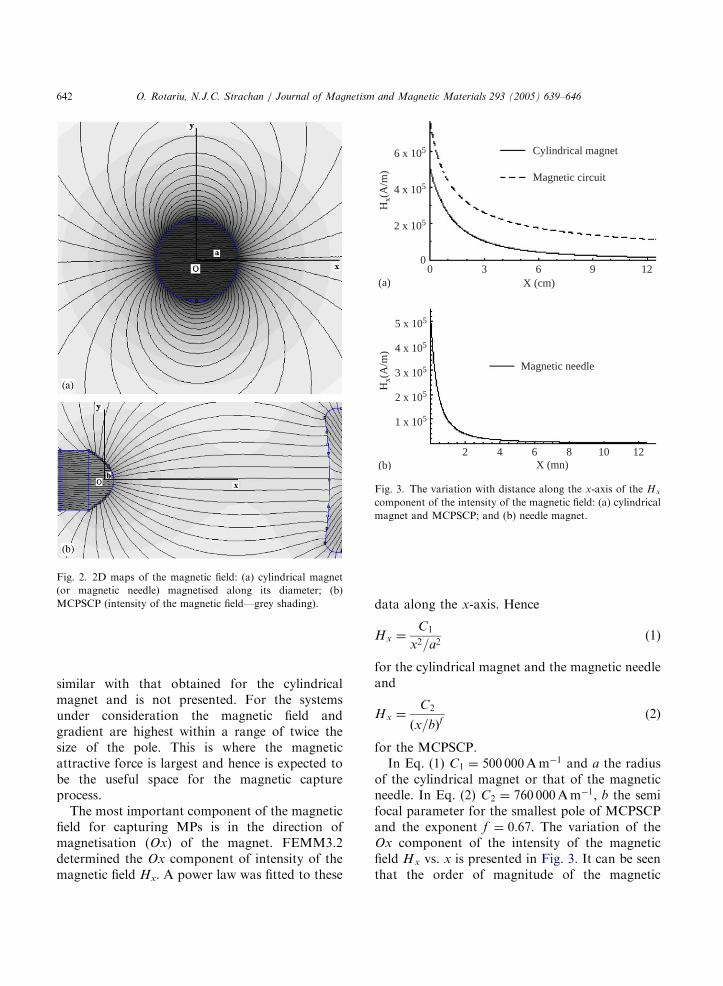
Magnetic field: The magnetic field was calcu-lated using the 2D ‘‘Finite Element MethodMagnetics’’ program (FEMM3.2) from Foster-Miller (http://femm.foster-miller.com). Fig. 2 pre-sents 2D maps of the magnetic field (flux linesand qualitative density plot of the intensityof the magnetic field) for the cylindrical magnetmagnetised along its diameter and for MCPSCP.The field’s profile for the needle magnet is
ARTICLE IN PRESS
Fig. 2. 2D maps of the magnetic field: (a) cylindrical magnet
(or magnetic needle) magnetised along its diameter; (b)
MCPSCP (intensity of the magnetic field—grey shading).
6 x 10⁵
4 x 10⁵
5 x 10⁵
4 x 10⁵
3 x 10⁵
1 x 10⁵
2 x 10⁵
2 x 10⁵
00 3 6 9 12
2 4 6 8 10 12X (mn)
Magnetic needle
X (cm)
Hx(
A/m
)H
x(A
/m)
Magnetic circuit
Cylindrical magnet
(a)
(b)
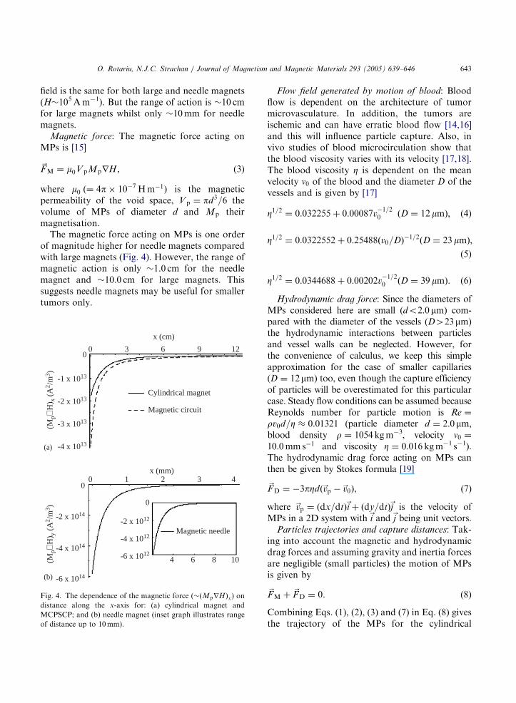
Fig. 3. The variation with distance along the x-axis of the Hx
component of the intensity of the magnetic field: (a) cylindrical
magnet and MCPSCP; and (b) needle magnet.
O. Rotariu, N.J.C. Strachan / Journal of Magnetism and Magnetic Materials 293 (2005) 639–646642
similar with that obtained for the cylindricalmagnet and is not presented. For the systemsunder consideration the magnetic field andgradient are highest within a range of twice thesize of the pole. This is where the magneticattractive force is largest and hence is expected tobe the useful space for the magnetic captureprocess.The most important component of the magnetic
field for capturing MPs is in the direction ofmagnetisation (Ox) of the magnet. FEMM3.2determined the Ox component of intensity of themagnetic field Hx. A power law was fitted to these
data along the x-axis. Hence
Hx ¼C1
x2=a2(1)
for the cylindrical magnet and the magnetic needleand
Hx ¼C2
ðx=bÞf(2)
for the MCPSCP.In Eq. (1) C1 ¼ 500 000Am
�1 and a the radiusof the cylindrical magnet or that of the magneticneedle. In Eq. (2) C2 ¼ 760 000Am
�1; b the semifocal parameter for the smallest pole of MCPSCPand the exponent f ¼ 0:67: The variation of theOx component of the intensity of the magneticfield Hx vs. x is presented in Fig. 3. It can be seenthat the order of magnitude of the magnetic
ARTICLE IN PRESS
O. Rotariu, N.J.C. Strachan / Journal of Magnetism and Magnetic Materials 293 (2005) 639–646 643
field is the same for both large and needle magnets(H�105Am�1). But the range of action is �10 cmfor large magnets whilst only �10mm for needlemagnets.
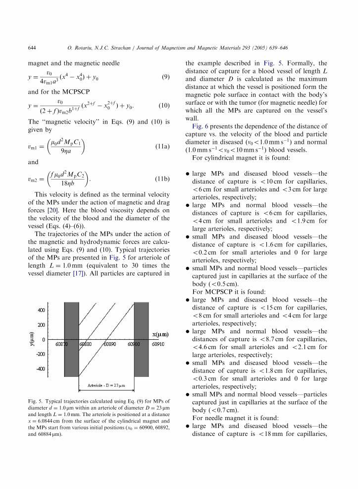
Magnetic force: The magnetic force acting onMPs is [15]
~FM ¼ m0VpMprH, (3)
where m0 ð¼ 4p� 10�7 Hm�1Þ is the magneticpermeability of the void space, Vp ¼ pd3=6 thevolume of MPs of diameter d and Mp theirmagnetisation.The magnetic force acting on MPs is one order
of magnitude higher for needle magnets comparedwith large magnets (Fig. 4). However, the range ofmagnetic action is only �1.0 cm for the needlemagnet and �10.0 cm for large magnets. Thissuggests needle magnets may be useful for smallertumors only.
-1 x 10¹³
-2 x 10¹³
-3 x 10¹³
-4 x 10¹³
0
-2 x 10¹⁴
-4 x 10¹⁴
-6 x 10¹⁴
-6 x 10¹²
-4 x 10¹²
-2 x 10¹²
4 6 8 10
00 3 6 9 12
0 1 2 3 4
x (cm)
Cylindrical magnet
Magnetic circuit
0
Magnetic needle
x (mm)
(Mp∇
H) y
(A
2 /m
3 )(M
p∇H
) x (
A2 /
m3 )
(a)
(b)
Fig. 4. The dependence of the magnetic force ð�ðMprHÞxÞ on
distance along the x-axis for: (a) cylindrical magnet and
MCPSCP; and (b) needle magnet (inset graph illustrates range
of distance up to 10mm).
Flow field generated by motion of blood: Bloodflow is dependent on the architecture of tumormicrovasculature. In addition, the tumors areischemic and can have erratic blood flow [14,16]and this will influence particle capture. Also, invivo studies of blood microcirculation show thatthe blood viscosity varies with its velocity [17,18].The blood viscosity Z is dependent on the meanvelocity n0 of the blood and the diameter D of thevessels and is given by [17]
Z1=2 ¼ 0:032255þ 0:00087v�1=20 ðD ¼ 12mmÞ; (4)
Z1=2 ¼ 0:0322552þ 0:25488ðv0=DÞ�1=2
ðD ¼ 23mmÞ;
(5)
Z1=2 ¼ 0:0344688þ 0:00202v�1=20 ðD ¼ 39mmÞ: (6)
Hydrodynamic drag force: Since the diameters ofMPs considered here are small (do2:0mm) com-pared with the diameter of the vessels (D423mm)the hydrodynamic interactions between particlesand vessel walls can be neglected. However, forthe convenience of calculus, we keep this simpleapproximation for the case of smaller capillaries(D ¼ 12mm) too, even though the capture efficiencyof particles will be overestimated for this particularcase. Steady flow conditions can be assumed becauseReynolds number for particle motion is Re ¼
rv0d=Z 0:01321 (particle diameter d ¼ 2:0mm;blood density r ¼ 1054kgm�3, velocity n0 ¼10:0mms�1 and viscosity Z ¼ 0:016 kgm�1 s�1).The hydrodynamic drag force acting on MPs canthen be given by Stokes formula [19]
~FD ¼ �3pZdð~vp �~v0Þ, (7)
where ~vp ¼ ðdx=dtÞ~i þ ðdy=dtÞ~j is the velocity ofMPs in a 2D system with~i and~j being unit vectors.
Particles trajectories and capture distances: Tak-ing into account the magnetic and hydrodynamicdrag forces and assuming gravity and inertia forcesare negligible (small particles) the motion of MPsis given by
~FM þ ~FD ¼ 0. (8)
Combining Eqs. (1), (2), (3) and (7) in Eq. (8) givesthe trajectory of the MPs for the cylindrical
ARTICLE IN PRESS
O. Rotariu, N.J.C. Strachan / Journal of Magnetism and Magnetic Materials 293 (2005) 639–646644
magnet and the magnetic needle
y ¼v0
4vm1a3ðx4 � x40Þ þ y0 (9)
and for the MCPSCP
y ¼v0
ð2þ f Þvm2b1þf
ðx2þf � x2þf0 Þ þ y0. (10)
The ‘‘magnetic velocity’’ in Eqs. (9) and (10) isgiven by
vm1 ¼m0d
2MpC1
9Za
� �(11a)
and
vm2 ¼f m0d
2MpC2
18Zb
� �. (11b)
This velocity is defined as the terminal velocityof the MPs under the action of magnetic and dragforces [20]. Here the blood viscosity depends onthe velocity of the blood and the diameter of thevessel (Eqs. (4)–(6)).The trajectories of the MPs under the action of
the magnetic and hydrodynamic forces are calcu-lated using Eqs. (9) and (10). Typical trajectoriesof the MPs are presented in Fig. 5 for arteriole oflength L ¼ 1:0mm (equivalent to 30 times thevessel diameter [17]). All particles are captured in
Fig. 5. Typical trajectories calculated using Eq. (9) for MPs of
diameter d ¼ 1:0mm within an arteriole of diameter D ¼ 23 mmand length L ¼ 1:0mm: The arteriole is positioned at a distancex ¼ 6:0844 cm from the surface of the cylindrical magnet and
the MPs start from various initial positions (x0 ¼ 60900; 60892,and 60884mm).
the example described in Fig. 5. Formally, thedistance of capture for a blood vessel of length L
and diameter D is calculated as the maximumdistance at which the vessel is positioned form themagnetic pole surface in contact with the body’ssurface or with the tumor (for magnetic needle) forwhich all the MPs are captured on the vessel’swall.Fig. 6 presents the dependence of the distance of
capture vs. the velocity of the blood and particlediameter in diseased (n0o1:0mms�1) and normal(1:0mms�1on0o10mms�1) blood vessels.For cylindrical magnet it is found:
�
large MPs and diseased blood vessels—thedistance of capture is o10 cm for capillaries,o6 cm for small arterioles and o3 cm for largearterioles, respectively;�
large MPs and normal blood vessels—thedistances of capture is o6 cm for capillaries,o4 cm for small arterioles and o1.9 cm forlarge arterioles, respectively;�
small MPs and diseased blood vessels—thedistance of capture is o1.6 cm for capillaries,o0.2 cm for small arterioles and 0 for largearterioles, respectively;�
small MPs and normal blood vessels—particlescaptured just in capillaries at the surface of thebody (o0.5 cm).For MCPSCP it is found:�
large MPs and diseased blood vessels—thedistance of capture is o15 cm for capillaries,o8 cm for small arterioles and o4 cm for largearterioles, respectively;�
large MPs and normal blood vessels—thedistances of capture is o8.7 cm for capillaries,o4.6 cm for small arterioles and o2.1 cm forlarge arterioles, respectively;�
small MPs and diseased blood vessels—thedistance of capture is o1.8 cm for capillaries,o0.3 cm for small arterioles and 0 for largearterioles, respectively;�
small MPs and normal blood vessels—particlescaptured just in capillaries at the surface of thebody (o0.7 cm).For needle magnet it is found:�
large MPs and diseased blood vessels—thedistance of capture is o18mm for capillaries,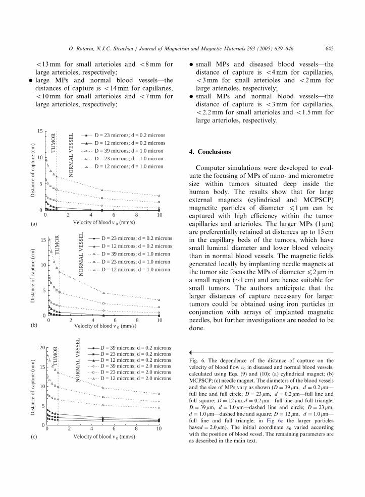
ARTICLE IN PRESS
(a
(b
(c
O. Rotariu, N.J.C. Strachan / Journal of Magnetism and Magnetic Materials 293 (2005) 639–646 645
o13mm for small arterioles and o8mm forlarge arterioles, respectively;
�
large MPs and normal blood vessels—thedistances of capture is o14mm for capillaries,o10mm for small arterioles and o7mm forlarge arterioles, respectively;0
5
10
15
0 4 8 10Velocity of blood v 0 (mm/s)
Dis
tanc
e of
cap
ture
(cm
)
D = 23 microns; d = 0.2 microns
D = 12 microns; d = 0.2 microns
D = 39 microns; d = 1.0 micron
D = 23 microns; d = 1.0 micron
D = 12 microns; d = 1.0 micron
TU
MO
R
NO
RM
AL
VE
SSE
L
)
0
5
10
15
0 6 10Velocity of blood v 0 (mm/s)
Dis
tanc
e of
cap
ture
(cm
)
D = 23 microns; d = 0.2 microns
D = 12 microns; d = 0.2 microns
D = 39 microns; d = 1.0 micron
D = 23 microns; d = 1.0 micron
D = 12 microns; d = 1.0 micron
TU
MO
R
NO
RM
AL
VE
SSE
L
)
0
5
10
15
20
0 4 8 10
Velocity of blood v 0 (mm/s)
Dis
tanc
e of
cap
ture
(m
m)a
D = 39 microns; d = 0.2 micronsD = 23 microns; d = 0.2 micronsD = 12 microns; d = 0.2 micronsD = 39 microns; d = 2.0 micronsD = 23 microns; d = 2.0 micronsD = 12 microns; d = 2.0 microns
TU
MO
R
NO
RM
AL
VE
SSE
L
2
62
4 8
2 6
)
�
Fi
ve
ca
M
an
ful
ful
D
d ¼
ful
ha
wi
as
small MPs and diseased blood vessels—thedistance of capture is o4mm for capillaries,o3mm for small arterioles and o2mm forlarge arterioles, respectively;
�
small MPs and normal blood vessels—thedistance of capture is o3mm for capillaries,o2.2mm for small arterioles and o1.5mm forlarge arterioles, respectively.4. Conclusions
Computer simulations were developed to eval-uate the focusing of MPs of nano- and micrometresize within tumors situated deep inside thehuman body. The results show that for largeexternal magnets (cylindrical and MCPSCP)magnetite particles of diameter p1 mm can becaptured with high efficiency within the tumorcapillaries and arterioles. The larger MPs (1 mm)are preferentially retained at distances up to 15 cmin the capillary beds of the tumors, which havesmall luminal diameter and lower blood velocitythan in normal blood vessels. The magnetic fieldsgenerated locally by implanting needle magnets atthe tumor site focus the MPs of diameterp2 mm ina small region (�1 cm) and are hence suitable forsmall tumors. The authors anticipate that thelarger distances of capture necessary for largertumors could be obtained using iron particles inconjunction with arrays of implanted magneticneedles, but further investigations are needed to bedone.
g. 6. The dependence of the distance of capture on the
locity of blood flow v0 in diseased and normal blood vessels,
lculated using Eqs. (9) and (10): (a) cylindrical magnet; (b)
CPSCP; (c) needle magnet. The diameters of the blood vessels
d the size of MPs vary as shown ðD ¼ 39mm; d ¼ 0:2mm—l line and full circle; D ¼ 23mm; d ¼ 0:2mm—full line andl square; D ¼ 12mm; d ¼ 0:2mm—full line and full triangle;¼ 39mm; d ¼ 1:0mm—dashed line and circle; D ¼ 23mm;1:0mm—dashed line and square; D ¼ 12mm; d ¼ 1:0mm—l line and full triangle; in Fig 6c the larger particles
ved ¼ 2:0mmÞ: The initial coordinate x0 varied according
th the position of blood vessel. The remaining parameters are
described in the main text.
ARTICLE IN PRESS
O. Rotariu, N.J.C. Strachan / Journal of Magnetism and Magnetic Materials 293 (2005) 639–646646
Acknowledgements
This work was funded by EU Marie CurieFellowship (contract QLK6-CT-2002-51544).
References
[1] F. Marcucci, F. Lefoulon, Drug Disc. Today 9 (2004) 219.
[2] U.O. Hafeli, Int. J. Pharm. 277 (2004) 19.
[3] C. Alexiou, W. Arnold, R.J. Klein, et al., Cancer Res. 60
(2000) 6641.
[4] A.S. Lubbe, C. Bergemann, H. Riess, et al., Cancer Res. 56
(1996) 4686.
[5] C. Alexiou, W. Arnold, R.J. Klein, et al., Cancer Res. 60
(2000) 6641.
[6] E. Viroonchatapan, H. Sato, M. Ueno, et al., Life Sci. 58
(1996) 2251.
[7] A.S. Lubbe, C. Alexiou, C. Bergemann, J. Surg. Res. 95
(2001) 200.
[8] R. Sheng, G.A. Flores, J. Liu, J. Magn. Magn. Mater. 194
(1999) 167.
[9] J. Liu, G.A. Flores, R. Sheng, J. Magn. Magn. Mater. 225
(2001) 209.
[10] G.A. Flores, J. Liu, Eur. Cells Mater. 3 (Suppl. 2)
(2002) 9.
[11] S. Goodwin, C. Peterson, C. Hoh, et al., J. Magn. Magn.
Mater. 194 (1999) 132.
[12] S. Jain, V. Mishra, P. Singh, et al., Int. J. Pharm. 261
(2003) 43.
[13] D.J.A. Crommelin, G. Scherphof, G. Storm, Adv. Drug
Delivery Rev. 17 (1995) 49.
[14] R.K. Jain, Cancer Res. 48 (1988) 2641.
[15] J.D. Jackson, Classical Electrodynamics, Wiley, New
York, 1975, p. 185.
[16] R.K. Jain, Adv. Drug Delivery Rev. 46 (2001) 149.
[17] H.H. Lipowsky, S. Kovalcheck, B.W. Zweifach, Circ. Res.
43 (1978) 738.
[18] A.R. Pries, T.W. Secomb, P. Gaehtgens, Cardiovasc. Res.
32 (1996) 654.
[19] G.K. Batchelor, An Introduction in Fluid Dynamics,
Cambridge University Press, London, 1970, p. 233.
[20] J.H.P. Watson, J. Appl. Phys. 44 (1973) 4209.
Related Documents