LETTER TO THE EDITOR Open Access Modeling tumor development and metastasis using paired organoids derived from patients with colorectal cancer liver metastases He Li 1,2† , Weixing Dai 3,4† , Xi Xia 1† , Renjie Wang 3,4† , Jing Zhao 1,2 , Lingyu Han 3,4 , Shaobo Mo 3,4 , Wenqiang Xiang 3,4 , Lin Du 1 , Guangya Zhu 1 , Jingjing Xie 1 , Jun Yu 5 , Nan Liu 1 , Mingzhu Huang 4,6* , Jidong Zhu 1,7* and Guoxiang Cai 3,4* Abstract Tumor metastasis accounts for the majority of cancer-related deaths; it is therefore important to develop preclinical models that faithfully recapitulate disease progression. Here, we generated paired organoids derived from primary tumors and matched liver metastases in the same colorectal cancer (CRC) patients. Despite the fact that paired organoids exhibit comparable gene expression and cell morphology, organoids from metastatic lesions demonstrate more aggressive phenotypes, tumorigenesis, and metastatic capacity than those from primary lesions. Transcriptional analyses of the paired organoids reveal signature genes and pathways altered during the progression of CRC, including SOX2. Further study shows that inducible knockdown of SOX2 attenuated invasion, proliferation, and liver metastasis outgrowth. Taken together, we use patient-derived paired primary and metastatic cancer organoids to model CRC metastasis and illustrate that SOX2 is associated with CRC progression and may serve as a potential prognostic biomarker and therapeutic target of CRC. Keywords: Colorectal cancer, Tumor metastasis, Preclinical model, Paired organoids, SOX2 To the Editor, Tumor heterogeneity plays a key role in cancer progression and therapy resistance [1]. However, knowledge of how tumor heterogeneity arises and contributes to disease progression is still limited [2]. Recent advances in organoid culture have been suc- cessfully established in a variety of solid tumors [3–5]. Tumor organoids retain the histological complexity and genetic heterogeneity of parental tumors, even after many passages [6], providing a wide range of ap- plications for cancer research. Organoids have enor- mous potential for the identification of optimal treatment strategies in individual patients [6]. For example, human CRC organoids derived from primary tumors [5] and liver metastases [7] have been reported as precision medical models for assessing drug re- sponses. However, paired organoids have not been studied as a model for CRC progression. In the present study, we used paired organoids derived from primary and liver metastatic tumors of CRC patients to model cancer metastasis. Through in vitro and in vivo studies and transcriptional analyses of the paired organoids, we revealed key genes associated © The Author(s). 2020 Open Access This article is licensed under a Creative Commons Attribution 4.0 International License, which permits use, sharing, adaptation, distribution and reproduction in any medium or format, as long as you give appropriate credit to the original author(s) and the source, provide a link to the Creative Commons licence, and indicate if changes were made. The images or other third party material in this article are included in the article's Creative Commons licence, unless indicated otherwise in a credit line to the material. If material is not included in the article's Creative Commons licence and your intended use is not permitted by statutory regulation or exceeds the permitted use, you will need to obtain permission directly from the copyright holder. To view a copy of this licence, visit http://creativecommons.org/licenses/by/4.0/. The Creative Commons Public Domain Dedication waiver (http://creativecommons.org/publicdomain/zero/1.0/) applies to the data made available in this article, unless otherwise stated in a credit line to the data. * Correspondence: [email protected]; [email protected]; [email protected] † He Li, Weixing Dai, Xi Xia and Renjie Wang contributed equally to this work. 4 Department of Oncology, Shanghai Medical College, Fudan University, Shanghai, China 1 Interdisciplinary Research Center on Biology and Chemistry, Shanghai Institute of Organic Chemistry, Chinese Academy of Sciences, Shanghai, China 3 Department of Colorectal Surgery, Fudan University Shanghai Cancer Center, Shanghai, China Full list of author information is available at the end of the article Li et al. Journal of Hematology & Oncology (2020) 13:119 https://doi.org/10.1186/s13045-020-00957-4

Welcome message from author
This document is posted to help you gain knowledge. Please leave a comment to let me know what you think about it! Share it to your friends and learn new things together.
Transcript

LETTER TO THE EDITOR Open Access
Modeling tumor development andmetastasis using paired organoids derivedfrom patients with colorectal cancer livermetastasesHe Li1,2†, Weixing Dai3,4†, Xi Xia1†, Renjie Wang3,4†, Jing Zhao1,2, Lingyu Han3,4, Shaobo Mo3,4, Wenqiang Xiang3,4,Lin Du1, Guangya Zhu1, Jingjing Xie1, Jun Yu5, Nan Liu1, Mingzhu Huang4,6*, Jidong Zhu1,7* and Guoxiang Cai3,4*
Abstract
Tumor metastasis accounts for the majority of cancer-related deaths; it is therefore important to develop preclinicalmodels that faithfully recapitulate disease progression. Here, we generated paired organoids derived from primarytumors and matched liver metastases in the same colorectal cancer (CRC) patients. Despite the fact that pairedorganoids exhibit comparable gene expression and cell morphology, organoids from metastatic lesionsdemonstrate more aggressive phenotypes, tumorigenesis, and metastatic capacity than those from primary lesions.Transcriptional analyses of the paired organoids reveal signature genes and pathways altered during theprogression of CRC, including SOX2. Further study shows that inducible knockdown of SOX2 attenuated invasion,proliferation, and liver metastasis outgrowth. Taken together, we use patient-derived paired primary and metastaticcancer organoids to model CRC metastasis and illustrate that SOX2 is associated with CRC progression and mayserve as a potential prognostic biomarker and therapeutic target of CRC.
Keywords: Colorectal cancer, Tumor metastasis, Preclinical model, Paired organoids, SOX2
To the Editor,Tumor heterogeneity plays a key role in cancer
progression and therapy resistance [1]. However,knowledge of how tumor heterogeneity arises andcontributes to disease progression is still limited [2].Recent advances in organoid culture have been suc-cessfully established in a variety of solid tumors [3–5].
Tumor organoids retain the histological complexityand genetic heterogeneity of parental tumors, evenafter many passages [6], providing a wide range of ap-plications for cancer research. Organoids have enor-mous potential for the identification of optimaltreatment strategies in individual patients [6]. Forexample, human CRC organoids derived from primarytumors [5] and liver metastases [7] have been reportedas precision medical models for assessing drug re-sponses. However, paired organoids have not beenstudied as a model for CRC progression. In thepresent study, we used paired organoids derived fromprimary and liver metastatic tumors of CRC patientsto model cancer metastasis. Through in vitro andin vivo studies and transcriptional analyses of thepaired organoids, we revealed key genes associated
© The Author(s). 2020 Open Access This article is licensed under a Creative Commons Attribution 4.0 International License,which permits use, sharing, adaptation, distribution and reproduction in any medium or format, as long as you giveappropriate credit to the original author(s) and the source, provide a link to the Creative Commons licence, and indicate ifchanges were made. The images or other third party material in this article are included in the article's Creative Commonslicence, unless indicated otherwise in a credit line to the material. If material is not included in the article's Creative Commonslicence and your intended use is not permitted by statutory regulation or exceeds the permitted use, you will need to obtainpermission directly from the copyright holder. To view a copy of this licence, visit http://creativecommons.org/licenses/by/4.0/.The Creative Commons Public Domain Dedication waiver (http://creativecommons.org/publicdomain/zero/1.0/) applies to thedata made available in this article, unless otherwise stated in a credit line to the data.
* Correspondence: [email protected]; [email protected];[email protected]†He Li, Weixing Dai, Xi Xia and Renjie Wang contributed equally to this work.4Department of Oncology, Shanghai Medical College, Fudan University,Shanghai, China1Interdisciplinary Research Center on Biology and Chemistry, ShanghaiInstitute of Organic Chemistry, Chinese Academy of Sciences, Shanghai,China3Department of Colorectal Surgery, Fudan University Shanghai CancerCenter, Shanghai, ChinaFull list of author information is available at the end of the article
Li et al. Journal of Hematology & Oncology (2020) 13:119 https://doi.org/10.1186/s13045-020-00957-4
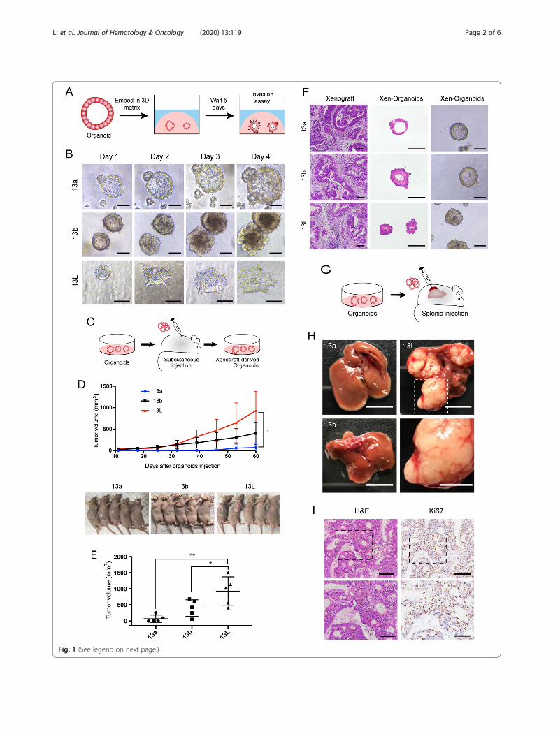
Fig. 1 (See legend on next page.)
Li et al. Journal of Hematology & Oncology (2020) 13:119 Page 2 of 6

with CRC liver metastasis, which could be translatedinto therapeutic targets or prognostic biomarkers fordisease treatment. A total of 24 organoids have beenestablished (Table S1). The library contained 2-pairedorganoid lines from patients P13 and P21. Particularly,P13 carried two primary tumors. The 13a and 13borganoids were established from the primary tumor,while 13L organoid was established from a synchron-ous liver metastatic tumor. Organoids of 21a and 21Lwere established from primary tumor and synchronousliver metastasis of the patient P21, which data demon-strated in Additional files (Supplementary Table S1and Fig. S1, S2, S4, and S5). Histopathological struc-tures and the intestinal epithelial marker CDX2 ofparental tumor were well preserved in organoids (Fig.S1).Invasion is a fundamental step in tumor progression
toward metastasis. To study collective invasion, wecultured paired organoids in a 3D invasion matrix(Fig. 1a). Although we did not observe collectiveprotrusive migration in organoids derived fromprimary lesions, metastatic organoids exhibited robustprotrusive migration into 3D invasion matrix (Fig. 1band Fig. S2A and B). Besides, the expression level ofMMP-2 (matrix metalloproteinase 2) and Ki67 wassignificantly higher in metastatic organoids than thatin the primary organoids (Fig. S2C-E). In subcutane-ous xenotransplantation of paired organoids (Fig. 1c),the growth rate and volume of 13L organoids derivedxenograft tumors was significantly higher than that of13a and 13b organoids derived tumors (Fig. 1d and e).Furthermore, we successfully generated organoidsfrom xenografts, histology, and Ki-67 expression ana-lysis of xenografts, as well as organoids derived fromthese xenografts, demonstrated similarity to theoriginal parental tumors (Fig. 1f and Fig. S2F). Wenext performed splenic injection of the paired orga-noids to assess the development of liver metastases(Fig. 1g). The 13L organoids formed macrometastatic
tumors in the livers (Fig. 1h and i), whereas 13b orga-noids and 13a organoids failed to colonize and had anegative expression of Ki67 in the liver (Fig. S2G).We then performed gene expression analysis in
the paired organoids and tumor tissue from patient13. There were 33 genes (P < 0.05; fold change >2.5) that were significantly upregulated in metastaticorganoids (Fig. 2a and Fig. S3A and B), includingthe transcription factor SOX2. Previous studies haveshown that SOX2 plays critical roles in embryonicpluripotent stem cells [8] and that SOX2 is abnor-mally expressed in many types of cancer [9–12].The differential expression of SOX2 in paired orga-noids was consistent with the RNA-seq data (Fig. 2band c), and SOX2 was also highly expressed in themetastatic tissues (Fig. 2d), while relatively low ex-pression in normal colon tissues (Fig. S3C-F). SOX2is also highly expressed in metastatic organoids andtissues of the other paired organoids (Fig. S4A and B).To investigate the role of SOX2 in CRC progres-
sion, doxycycline (Dox) inducible expression ofshRNA targeting SOX2 was established in metastaticorganoids (Fig. S4C and D). SOX2− organoids exhib-ited the reduced ability of invasion, colony-formingefficiency, and cell viability in metastatic organoidlines (Fig. 2e-g and Fig. S4E-G). Furthermore, themetastatic organoids efficiently formed large meta-static tumors in control groups (Dox untreated),whereas the SOX2− organoids showed no or fewengraftments (Fig. 2h). The downregulation of SOX2and Ki67 was further confirmed by immunohisto-chemistry (Fig. 2i and Fig. S4H). We then overex-pressed SOX2 in primary organoids and found thatorganoids with overexpressed SOX2 exhibited in-creased ability of invasion and proliferation whencompared with control organoids (Fig. S5). Takentogether, these findings demonstrate that SOX2expression is sufficient and necessary for CRC orga-noids to exhibit the metastatic potential.
(See figure on previous page.)Fig. 1 Patient-derived paired organoids provide invasion and transplantable models for human CRC progression. a Schematic of 3Dinvasion assay using paired organoids. b Representative micrographs of organoids in 3D invasion assay. Tumor organoids showed thesmooth and protrusive leading fronts, respectively. The scale bar represents 100 μm. c Schematic of the subcutaneous organoidinjection. d Organoids were injected subcutaneously into the flank region of nude mice for 60 days. Tumor volumes were monitoredover time (top). n = 5 mice per group. Error bars indicate SEMs. *p = 0.0146 (one-way ANOVA). e Tumor volume of the mice at day 60after inoculation. Each dot indicates individual mice. n = 5 mice per group. Error bars indicate SEMs. *p = 0.0433, **p = 0.002 (one-wayANOVA). f Representative bright-field images of organoids together with H&E staining of xenografts generated from organoids, andorganoids derived from the xenografts. The scale bar represents 100 μm. g Schematic of the hepatic metastasis assay by splenicorganoid injection. h Representative macroscopic photographs of the whole liver. n = 5 mice per group. Top, scale bar of the wholeliver represents 1 cm. Bottom, high magnification of inset. Scale bar, 5 mm. i Representative histopathology and Ki67 staining of livermetastatic lesions generated from splenic injection of 13L organoids. Top, the scale bar represents 100 μm; bottom, the scale barrepresents 50 μm
Li et al. Journal of Hematology & Oncology (2020) 13:119 Page 3 of 6

Fig. 2 (See legend on next page.)
Li et al. Journal of Hematology & Oncology (2020) 13:119 Page 4 of 6

In summary, the present study highlights the poten-tial of patient-derived paired primary and metastaticcancer organoids as an experimental model for inves-tigating CRC progression. We identified a significantlydysregulated gene between paired organoids, SOX2,which could be a prognostic biomarker, and perhapsa potent therapeutic target in the treatment of CRC.
Supplementary informationSupplementary information accompanies this paper at https://doi.org/10.1186/s13045-020-00957-4.
Additional file 1: Supplementary Table S1. Summary of patient-derived CRC organoid lines and corresponding clinical data.
Additional file 2: Supplementary Figures. Figure S1. Pairedorganoids derived from primary and metastatic CRC recapitulate thehistopathological structure of parental tumor. A and B, Organoidsarchitecture resembles parental tumor epithelium. Representative bright-field images of organoids together with H&E staining of parental tumorsand patient-derived organoids. The scale bar represents 100 μm. C andD, Representative IHC sections for the intestinal epithelial marker CDX2.The scale bar represents 100 μm. Figure S2. Organoids derived fromliver metastatic lesions exhibited the most aggressive phenotypes withsignificant high tumorigenic and metastatic potential. A, Representativemicrographs of organoids in 3D invasion assay. Tumor organoids showedthe smooth and protrusive leading fronts, respectively. B, Micrographs ofthe paired organoids stained with phalloidin–F-actin (right). The scale barrepresents 100 μm. C, qRT-PCR analysis of MMP-2 in paired organoidlines. Error bars indicate SEMs. *P=0.0207, **P =0.0083, ***P=0.0008 (one-way ANOVA; left), ***P=0.0006 (Unpaired t test; right). D, Western blotanalysis of the MMP-2 protein expression in paired organoid lines. α-tubulin was used as a loading control. E, Representative IHC sections forKi67 in human colorectal tumor tissues and paired organoid lines. Thescale bar represents 100 μm. F, Representative IHC sections for Ki67 inorganoid xenografts and organoids derived from xenografts. The scalebar represents 100 μm. G, Representative gross image, histopathologyand Ki67 staining of whole liver from primary organoid xenografts. Thescale bar of the whole liver represents 1cm. The black scale bar repre-sents 100 μm. Figure S3. A, Representation of the up-regulated genes in13L organoids. Error bars indicate SEMs. *P = 0.0283, ***P = 0.0004, ****P< 0.0001 (one-way ANOVA). B, qRT-PCR analysis of the up-regulatedgenes in 13L organoids. Values were normalized to mean levels in 13aorganoids. Error bars indicate SEMs. *P=0.02, ***P=0.0008 ****P < 0.0001(one-way ANOVA). C, RNA sequencing analysis of SOX2 in human colo-rectal cancer tissues and normal colon tissues. D, qRT-PCR analysis ofSOX2 in tumor tissues and paired normal colon tissues. Values were nor-malized to mean levels in normal colon tissues. Error bars indicate SEMs.**P = 0.0018, ****P < 0.0001 (one-way ANOVA). E, Western blot analysisof the SOX2 protein expression in tumor tissues and paired normal colontissues. α-tubulin was used as a loading control. F, Representative IHC
sections for SOX2 in human colorectal tumor tissues and paired normalcolon tissues. The scale bar represents 100 μm. Figure S4. SilencingSOX2 in metastatic organoids attenuated cell invasion and proliferationand suppressed liver metastasis in vivo. A, qRT-PCR analysis of SOX2 inpaired organoid lines (left). Values were normalized to mean levels in 21aorganoids. Error bars indicate SEMs. ****P < 0.0001(Unpaired t test). West-ern blot analysis of the SOX2 protein expression in paired organoid lines.α-tubulin was used as a loading control (right). B, Representative IHC sec-tions for SOX2 in human colorectal tumor tissues and organoids. Thescale bar represents 100 μm. C, qRT-PCR analysis of SOX2 in the 13L-shRNA-1/2 (left) and 21L-shRNA-1/2 (right) organoids after 3 days of Dox-induction. The level of SOX2 was compared to that in the untreated sam-ple. Error bars indicate SEMs. ***P=0.0001, ****P < 0.0001 (two-wayANOVA). D, Western blot analysis of the expression of SOX2 in the 13L-shRNA-1/2 and 21L-shRNA-1/2 organoids after 3 days of Dox-induction.α-tubulin was used as a loading control. E, Representative micrographs ofDox-transduced the 21L-shRNA-1/2 organoids after 5 days and stainedwith phalloidin–F-actin. The scale bar represents 100 μm. F, Proliferationof the 21L-shRNA-1/2 organoids were examined by CTG cell viability as-says following 3- or 5-days growth in the presence or absence of Dox.Error bars indicate SEMs. **p=0.0019, ****P < 0.0001 (two-way ANOVA). G,Representative micrographs of colonies arising from the 21L-shRNA-1/2organoids (top), with magnified insets showing colonies (bottom). Thescale bar represents 200 μm. Colony forming efficiency in G was calcu-lated and compared (right). Error bars indicate SEMs. **P=0.0015 (two-way ANOVA). H, Representative Ki67 immunostaining of liver metastasesof the 13L-shRNA-1/2 organoids (left) and ratio of Ki67-positive tumorcells in liver metastases (right). Scale bars, 200 μm (top) and 100 μm (bot-tom). Each dot indicates individual mice. Error bars indicate SEMs. *p=0.0197, **P=0.0044 (two-way ANOVA). Figure S5. SOX2 is overexpressedin primary organoids. A, qRT-PCR analysis of SOX2 in primary organoidlines. Values were normalized to mean levels in control organoids. Errorbars indicate SEMs. ****P < 0.0001 (one-way ANOVA). B, Western blotanalysis of the SOX2 protein expression in primary organoid lines. α-tubulin was used as a loading control. C, Representative micrographs ofcolonies arising from the control, LV-Vector and LV-SOX2 primary orga-noid lines. The scale bar represents 200 μm. D-F, Colony forming effi-ciency in C was calculated and compared. Error bars indicate SEMs. ***P=0.0008, ****P < 0.0001 (one-way ANOVA). G, Proliferation of the control,LV-Vector and LV-SOX2 primary organoid lines were examined by CTGcell viability assays. Error bars indicate SEMs. ***P=0.0008, ****P < 0.0001(one-way ANOVA). H, Representative micrographs of organoids in 3D in-vasion assay. Arrowheads, protrusive leading fronts. Micrographs of thepaired organoids stained with phalloidin–F-actin. The scale bar represents100 μm.
Additional file 3. Supplementary Materials and Methods.
AbbreviationsCRC: Colorectal cancer; 3D: Three-dimensional; MMP-2: Matrixmetalloproteinase 2; qRT-PCR: Quantitative reverse transcription PCR;Dox: Doxycycline; CTG: CellTiter-Glo Luminescent
(See figure on previous page.)Fig. 2 SOX2 plays an important role in colorectal metastasis. a Hierarchical clustering heatmaps of the 33 significantly upregulated genes in 13Lorganoids. b qRT-PCR analysis of SOX2 in paired organoid lines. Values were normalized to mean levels in 13a organoids. Error bars indicate SEMs.**p = 0.0050, ****p < 0.0001 (one-way ANOVA). c Western blot analysis of the SOX2 protein expression in paired organoid lines. α-tubulin wasused as a loading control. d Representative IHC sections for SOX2 in human colorectal tumor tissues and organoids. The scale bar represents100 μm. e Representative micrographs of Dox-transduced the 13 L-shRNA-1/2 organoids after 5 days and stained with phalloidin–F-actin. Thescale bar represents 100 μm. f Representative micrographs of colonies arising from the 13L-shRNA-1/2 organoids (top), with magnified insetsshowing colonies (bottom). The scale bar represents 200 μm. Colony forming efficiency in f was calculated and compared (right). Error barsindicate SEMs. **p = 0.0078, (two-way ANOVA). g Proliferation of the 13L-shRNA-1/2 organoids were examined by CTG cell viability assaysfollowing 3- or 5-days growth in the presence or absence of Dox. Error bars indicate SEMs. **p = 0.0019, ****p < 0.0001 (two-way ANOVA). hRepresentative macroscopic photographs (top) and histology sections (H&E, bottom) of the livers of NOD mice transplanted with the 13L-shRNA-1/2 organoids. Arrowheads, metastatic foci. n = 5 mice per group. Scale bars, 1 cm (top) and 4mm (bottom). i Representative SOX2immunostaining of liver metastases of the 13L-shRNA-1/2 organoids. Scale bars, 200 μm (top) and 100 μm (bottom)
Li et al. Journal of Hematology & Oncology (2020) 13:119 Page 5 of 6

AcknowledgementsWe thank all of the patients who consented to donate their tumor tissuesfor this study, as well as the surgical teams who facilitated this work.
Authors’ contributionsConception and design: MH, HL, JZ, and GC; development of methodology:HL, WD, RW, XX, and JZ; acquisition of data (provided animals, acquired andmanaged patients, provided facilities, etc.): HL, JZ, LH, RW, SM, WX, and LD;analysis and interpretation of data (e.g., statistical analysis, biostatistics,computational analysis): HL, WD, RW, XX, JX, GZ, JY, and JZ; manuscriptwriting: HL; manuscript revision: HL, XX, MH, NL, JZ, and GC; administrative,technical, or material support (i.e., reporting or organizing data, constructingdatabases): MH, HL, JZ, and GC; study supervision: JZ and GC. All authorsread and approved the final manuscript.
FundingThis work was supported by the National Key R&D Program of China(2016YFA0501900), the Shanghai Municipal Science and Technology MajorProject (Grant No. 2019SHZDZX02), the Grant of National Natural ScienceFoundation of China (No. 81871958 and No. 81572351), the Grant of Scienceand Technology Commission of Shanghai Municipality (No. 16401970502and No.17411951100 and No. 19140902100).
Availability of data and materialsThe datasets used and/or analyzed during the current study are availablefrom the corresponding author on reasonable request.
Ethics approval and consent to participateThis study was approved by the Shanghai Cancer Center of Fudan UniversityEthics Committee. Written informed consent was obtained prior to theacquisition of tissue from all patients. All animal procedures were performedunder guidelines approved by the Institutional Animal Care and UseCommittee of the Shanghai Institute of Organic Chemistry, ChineseAcademy of Sciences.
Consent for publicationNot applicable.
Competing interestsThe authors declare that they have no competing interests.
Author details1Interdisciplinary Research Center on Biology and Chemistry, ShanghaiInstitute of Organic Chemistry, Chinese Academy of Sciences, Shanghai,China. 2University of the Chinese Academy of Sciences, Beijing, China.3Department of Colorectal Surgery, Fudan University Shanghai CancerCenter, Shanghai, China. 4Department of Oncology, Shanghai MedicalCollege, Fudan University, Shanghai, China. 5Department of Surgery, JohnsHopkins University School of Medicine, Baltimore, MD, USA. 6Department ofMedical Oncology, Fudan University Shanghai Cancer Center, Shanghai,China. 7Center for Excellence in Molecular Synthesis, Shanghai Institute ofOrganic Chemistry, Chinese Academy of Sciences, Shanghai, China.
Received: 12 June 2020 Accepted: 27 August 2020
References1. Stratton MR, Campbell PJ, Futreal PA. The cancer genome. Nature. 2009;
458(7239):719–24.2. McGranahan N, Swanton C. Clonal heterogeneity and tumor evolution: past,
present, and the future. Cell. 2017;168(4):613–28.3. Broutier L, Mastrogiovanni G, Verstegen MM, Francies HE, Gavarró LM,
Bradshaw CR, et al. Human primary liver cancer-derived organoid culturesfor disease modeling and drug screening. Nat Med. 2017;23(12):1424–35.
4. Sachs N, de Ligt J, Kopper O, Gogola E, Bounova G, Weeber F, et al. A livingbiobank of breast cancer organoids captures disease heterogeneity. Cell.2018;172(1-2):373–86 e10.
5. van de Wetering M, Francies HE, Francis JM, Bounova G, Iorio F, Pronk A,et al. Prospective derivation of a living organoid biobank of colorectalcancer patients. Cell. 2015;161(4):933–45.
6. Yoshida GJ. Applications of patient-derived tumor xenograft models andtumor organoids. J Hematol Oncol. 2020;13(1):4.
7. Vlachogiannis G, Hedayat S, Vatsiou A, Jamin Y, Fernández-Mateos J, Khan K,et al. Patient-derived organoids model treatment response of metastaticgastrointestinal cancers. Science. 2018;359:920–6.
8. Avilion AA, Nicolis SK, Pevny LH, Perez L, Vivian N, Lovell-Badge R.Multipotent cell lineages in early mouse development depend on SOX2function. Genes Dev. 2003;17(1):126–40.
9. Leis O, Eguiara A, Lopez-Arribillaga E, Alberdi MJ, Hernandez-Garcia S,Elorriaga K, et al. Sox2 expression in breast tumours and activation in breastcancer stem cells. Oncogene. 2012;31(11):1354–65.
10. Chou YT, Lee CC, Hsiao SH, Lin SE, Lin SC, Chung CH, et al. The emergingrole of SOX2 in cell proliferation and survival and its crosstalk withoncogenic signaling in lung cancer. Stem Cells. 2013;31(12):2607–19.
11. Lundberg IV, Löfgren Burström A, Edin S, Eklof V, Oberg A, Stenling R, et al.SOX2 expression is regulated by BRAF and contributes to poor patientprognosis in colorectal cancer. PLoS One. 2014;9(7):e101957.
12. Neumann J, Bahr F, Horst D, Kriegl L, Engel J, Luque RM, et al. SOX2expression correlates with lymph-node metastases and distant spread inright-sided colon cancer. BMC Cancer. 2011;11:518.
Publisher’s NoteSpringer Nature remains neutral with regard to jurisdictional claims inpublished maps and institutional affiliations.
Li et al. Journal of Hematology & Oncology (2020) 13:119 Page 6 of 6
Related Documents