journal homepage: www.elsevier.com/locate/yexcr Available online at www.sciencedirect.com Research Article Modeling neurogenesis impairment in down syndrome with induced pluripotent stem cells from Trisomy 21 amniotic fluid cells Huai-En Lu a,b , Yao-Chen Yang a , Sheng-Mei Chen c , Hong-Lin Su c , Pai-Cheng Huang d , Ming-Song Tsai e,f , Tzu-Hao Wang d , Ching-Ping Tseng b,n , Shiaw-Min Hwang a,nn a Bioresource Collection and Research Center, Food Industry Research and Development Institute, Hsinchu 30062, Taiwan, ROC b Department of Biological Science and Technology, National Chiao Tung University, Hsinchu 30068, Taiwan, ROC c Department of Life Sciences, National Chung Hsing University, Taichung 40227, Taiwan, ROC d Department of Obstetrics and Gynecology, Chang Gung Memorial Hospital, Lin-Kou Medical Center, Taoyuan 33305, Taiwan, ROC e Prenatal Diagnosis Center, Cathay General Hospital, Taipei 10630, Taiwan, ROC f School of Medicine, Fu Jen Catholic University, Taipei 24205, Taiwan, ROC articleinformation Article Chronology: Received 10 August 2012 Received in revised form 28 September 2012 Accepted 30 September 2012 Available online 4 October 2012 Keywords: Down syndrome Trisomy 21 Neurogenesis impairment Induced pluripotent stem cells miRNA abstract Down syndrome (DS), or Trisomy 21 (T21) syndrome, one of the most common chromosomal abnormalities, is caused by an extra duplication of chromosome 21. In studies of neuron development, experimental models based on human cells are considered to be the most desired and accurate for basic research. The generation of diseased induced pluripotetn stem (iPS) cell is a critical step in understanding the developmental stages of complex neuronal diseases. Here, we generated human DS iPS cell lines from second trimester amniotic fluid (AF) cells with T21 by co-expressing Yamanaka factors through lentiviral delivery and subsequently differentiated them into neuronal progenitor cells (NPCs) for further analyses. T21 AF-iPS cells were characterized for the expression of pluripotent markers and for their ability to differentiate into all three germ layers by forming embryoid bodies in vitro and teratomas in vivo. The T21 AF-iPS cells maintained their unique pattern of chromosomal karyotypes: three pairs of chromosome 21. The level of amyloid precursor protein was significantly increased in NPCs derived from T21 AF-iPS cells compared with NPCs from normal AF-iPS cells. The expression levels of miR-155 and miR-802 in T21 AF-iPS-NPCs were highly elevated in the presence of low expression of MeCP2. We observed that T21 iPS-NPCs generated fewer neurons compared with controls. T21 iPS-NPCs exhibit developmental defects during neurogenesis. Our findings suggest that T21 AF-iPS cells serve as a good source to further elucidate the impairment neurogenesis of DS and the onset of Alzheimer’s disease. & 2012 Elsevier Inc. All rights reserved. 0014-4827/$ - see front matter & 2012 Elsevier Inc. All rights reserved. http://dx.doi.org/10.1016/j.yexcr.2012.09.017 n Correspondence to: Department of Biological Science and Technology, National Chiao Tung University, 75, Bo-ai Street, Hsinchu 30068, Taiwan, ROC Fax: þ886 3 5729 288. nn Correspondence to: Bioresource Collection and Research Center, Food Industry Research and Development Institute, 331, Shih-pin Road, Hsinchu 30062, Taiwan, ROC Fax: þ886 3 5224 171. E-mail addresses: [email protected] (C.-P. Tseng), hsm@firdi.org.tw (S.-M. Hwang). EXPERIMENTAL CELL RESEARCH 319 (2013) 498 –505

Welcome message from author
This document is posted to help you gain knowledge. Please leave a comment to let me know what you think about it! Share it to your friends and learn new things together.
Transcript
Available online at www.sciencedirect.com
journal homepage: www.elsevier.com/locate/yexcr
E X P E R I M E N T A L C E L L R E S E A R C H 3 1 9 ( 2 0 1 3 ) 4 9 8 – 5 0 5
0014-4827/$ - see frohttp://dx.doi.org/10.1
nCorrespondence t
Taiwan, ROC Fax: þ8nnCorrespondence
Hsinchu 30062, Taiw
E-mail addresses
Research Article
Modeling neurogenesis impairment in down syndrome with
induced pluripotent stem cells from Trisomy 21 amniotic
fluid cells
Huai-En Lua,b, Yao-Chen Yanga, Sheng-Mei Chenc, Hong-Lin Suc, Pai-Cheng Huangd,Ming-Song Tsaie,f, Tzu-Hao Wangd, Ching-Ping Tsengb,n, Shiaw-Min Hwanga,nn
aBioresource Collection and Research Center, Food Industry Research and Development Institute, Hsinchu 30062, Taiwan, ROCbDepartment of Biological Science and Technology, National Chiao Tung University, Hsinchu 30068, Taiwan, ROCcDepartment of Life Sciences, National Chung Hsing University, Taichung 40227, Taiwan, ROCdDepartment of Obstetrics and Gynecology, Chang Gung Memorial Hospital, Lin-Kou Medical Center, Taoyuan 33305, Taiwan, ROCePrenatal Diagnosis Center, Cathay General Hospital, Taipei 10630, Taiwan, ROCfSchool of Medicine, Fu Jen Catholic University, Taipei 24205, Taiwan, ROC
a r t i c l e i n f o r m a t i o n
Article Chronology:
Received 10 August 2012
Received in revised form
28 September 2012
Accepted 30 September 2012
Available online 4 October 2012
Keywords:
Down syndrome
Trisomy 21
Neurogenesis impairment
Induced pluripotent stem cells
miRNA
nt matter & 2012 Elsevier016/j.yexcr.2012.09.017
o: Department of Biologic
86 3 5729 288.
to: Bioresource Collection
an, ROC Fax: þ886 3 5224
: [email protected] (C.-
a b s t r a c t
Down syndrome (DS), or Trisomy 21 (T21) syndrome, one of the most common chromosomal
abnormalities, is caused by an extra duplication of chromosome 21. In studies of neuron
development, experimental models based on human cells are considered to be the most desired
and accurate for basic research. The generation of diseased induced pluripotetn stem (iPS) cell is
a critical step in understanding the developmental stages of complex neuronal diseases. Here,
we generated human DS iPS cell lines from second trimester amniotic fluid (AF) cells with T21
by co-expressing Yamanaka factors through lentiviral delivery and subsequently differentiated
them into neuronal progenitor cells (NPCs) for further analyses. T21 AF-iPS cells were
characterized for the expression of pluripotent markers and for their ability to differentiate
into all three germ layers by forming embryoid bodies in vitro and teratomas in vivo. The T21
AF-iPS cells maintained their unique pattern of chromosomal karyotypes: three pairs of
chromosome 21. The level of amyloid precursor protein was significantly increased in NPCs
derived from T21 AF-iPS cells compared with NPCs from normal AF-iPS cells. The expression
levels of miR-155 and miR-802 in T21 AF-iPS-NPCs were highly elevated in the presence of low
expression of MeCP2. We observed that T21 iPS-NPCs generated fewer neurons compared with
controls. T21 iPS-NPCs exhibit developmental defects during neurogenesis. Our findings suggest
that T21 AF-iPS cells serve as a good source to further elucidate the impairment neurogenesis of
DS and the onset of Alzheimer’s disease.
& 2012 Elsevier Inc. All rights reserved.
Inc. All rights reserved.
al Science and Technology, National Chiao Tung University, 75, Bo-ai Street, Hsinchu 30068,
and Research Center, Food Industry Research and Development Institute, 331, Shih-pin Road,
171.
P. Tseng), [email protected] (S.-M. Hwang).

E X P E R I M E N T A L C E L L R E S E A R C H 3 1 9 ( 2 0 1 3 ) 4 9 8 – 5 0 5 499
Introduction
Down syndrome (DS), or Trisomy 21 (T21) syndrome, one of the
most common chromosomal abnormalities, is caused by an extra
duplication of chromosome 21 [1]. The characteristics of DS include
cognitive impairment, congenital heart defects, craniofacial abnorm-
alities, gastrointestinal anomalies, leukemia, mental retardation,
seizures and early onset Alzheimer’s disease (AD) [2].
The Ts65Dn mouse used to study DS is an invaluable tool, but
it provides a limited representation of human pathophysiology
[3]. In this case, Ts65Dn mice are trisomic at chromosome 16,
which is only homologous to approximately 55% of the human
chromosome 21 [4]. This shows that Ts65Dn mouse is not always
a faithful mimic of humans for human T21 syndrome. Studying
human cells in vitro is the backbone of basic research, and
numerous findings regarding both normal and pathologic cellular
processes have been reported. Human cell culture provides an
essential complement to research with animal models, where
induced pluripotent stem (iPS) cells can serve as in vitro human
disease models [5–7]. Disease-specific pluripotent cells capable
of differentiating into various tissues; in this specific case,
differentiation into neurons can provide new insights into dis-
ease pathophysiology that were not possible with murine models
[8–11].
Here, we generated human DS iPS cell lines from human
second trimester amniotic fluid (AF) cells with T21 by co-
expressing Yamanaka factors through lentiviral delivery and
subsequently differentiated them into neuronal progenitor cells
(NPCs) for further analyses. We report that T21 AF-iPS cells were
positive for pluripotent markers and differentiate into three germ
layers, as shown by the generation of embryoid bodies (EBs)
in vitro and teratomas in vivo. The T21 AF-iPS cells maintained
the abnormal chromosomal karyotype signature. The human
gene for amyloid precursor protein (APP) is located on chromo-
some 21. APP is a ubiquitously expressed transmembrane protein
whose cleavage product, the b-amyloid (Ab) protein, is deposited
as amyloid plaques in the neurodegenerative conditions of AD
and DS. We found that the expressed protein level of APP in NPCs
derived from T21 AF-iPS cells was significantly overexpressed. In
addition, several studies reported that developing DS cortical
neurons have shorter dendrites, fewer dendritic spines and
abnormal dendritic morphology [12]. New insight was also
provided by a recent study that described the developing DS
neurons on the basis of miRNA regulation [13]. Human chromo-
some 21 harbors 5 miRNAs: miR-99a, let-7c, miR-125b-2,
miR-155 and miR-802 [14]. In DS neurons, over expression of
miR-155 and miR-802 inhibit the expression of the target,
methyl-CpG-binding protein 2 (MeCP2) and in turn cause aber-
rant expression of two downstream target genes, CREB1 and
MEF2C, both which play important roles in neurogenesis. Sub-
sequent impairments in neuronal maturation also appear to be
reflected in the DS brain [13]. We found that over expression of
miR-155 and miR-802 result in the degradation of MeCP2 in T21
iPS-NPCs. We observed that T21 iPS-NPCs generated fewer
neurons compared with controls. T21 iPS-NPCs exhibited a
developmentally regulated defect in neurogenesis. Our findings
demonstrate that T21 AF-iPS cells serve as a good source to
further elucidate the impairment neurogenesis of DS and the
onset of Alzheimer’s disease.
Materials and methods
Cell culture and iPS cell generation
T21 and normal AF samples were obtained with written
informed consents from pregnant donors. The protocols of this
study were approved under the Institutional Review Board (IRB)
of Cathay General Hospital, Taipei, Taiwan. AF cells were isolated
from second-trimester amniocenteses according to our previous
report [15]. T21 AF cells were cultured and passaged routinely
at 80–90% confluence with alpha-modified minimum essential
medium (a-MEM, Hyclone, Logan, UT) containing 20% fetal
bovine serum (FBS, Hyclone), 4 ng/mL basic fibroblast growth
factor (bFGF, Peprotech, London, UK), 50 U/mL penicillin and
50 mg/mL streptomycin (Sigma, St. Louis, MO) at 37 1C and 5%
CO2 in a humidified incubator. Briefly, pre-prepared lentivirus
packaged with Yamanaka factors (OCT4, SOX2, KLF4 and c-MYC)
was purchased from Vectorite Biomedica Inc. (VBI, Taipei,
Taiwan). The virus titer used for transduction was 1�109 with
a multiplicity of infection (MOI) of 20. For viral transduction,
1�105 T21 AF cells were seeded per well in 12-well culture
plates. The viral mixture was then added into the culture
medium with 2 mg/mL polybrene (Sigma) and incubated for
two days. On day 2, the cells were passaged and plated onto
100 mm tissue culture dishes coated with 1�106 irradiated
mouse embryonic fibroblasts (MEFs). The next day, the culture
medium was exchanged with human iPS cell culture medium
composed of Dulbecco’s Modified Eagle Medium/Nutrient Mix-
ture F-12 (DMEM/F12, Invitrogen, Carlsbad, CA) medium supple-
mented with 20% KnockOut serum replacement (KOSR,
Invitrogen), 0.1 mM nonessential amino acids (NEAA, Invitrogen),
1 mM GlutaMax-1 (Invitrogen), 0.1 mM 2-mercaptoethanol
(Invitrogen), 10 ng/mL bFGF, 50 U/mL penicillin and 50 mg/mL
streptomycin. On day 10 post-transduction, embryonic stem
cell(ES)-like colonies were picked based on morphology and
cultured on irradiated MEF feeder layers with iPS culture
medium. On day 17, T21 AF-iPS colonies were transferred for
expansion and further characterization. The workflow chart is
shown in Fig. 1A.
Alkaline phosphatase and immunofluorescence staining
The alkaline phosphatase (AP) activity detection kit used in this
study was obtained from Millipore (Millipore, Billerica, MA) and
used following the manufacturer’s instructions. For immuno-
fluorescence staining, cells were fixed with 4% paraformaldehyde
(Sigma), permeabilized with 0.05% Triton X-100 (Sigma) if
necessary and blocked with 10% donkey serum. For the identi-
fication of ES markers in T21 AF-iPS cells, colonies were stained
with primary antibodies against OCT4 (1:400, Abcam), SOX2
(1:200, Millipore), NANOG (1:500, Abcam, Cambridge, MA),
SSEA-1 (1:200, Millipore), SSEA-3 (1:100, Abcam), SSEA-4
(1:100, Abcam), TRA-1–60 (1:200, Abcam) and TRA-1–81
(1:200, Abcam). For the detection of lineage markers in differ-
entiated T21 AF-iPS cells in vitro, cells were stained with primary
antibodies against TUJ1 (1:400, Sigma), NESTIN (1:100, R&D
Systems, Minneapolis, MN), a-SMA (1:100, Abcam), TROPONIN
(1:100, Santa Cruz Biotechnology, Santa Cruz, CA) and ALBUMIN
(1:200, Abcam). For the detection of lineage markers in NPCs
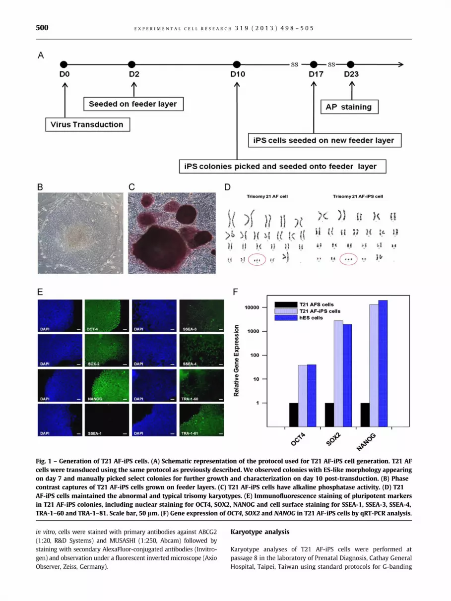
Fig. 1 – Generation of T21 AF-iPS cells. (A) Schematic representation of the protocol used for T21 AF-iPS cell generation. T21 AF
cells were transduced using the same protocol as previously described. We observed colonies with ES-like morphology appearing
on day 7 and manually picked select colonies for further growth and characterization on day 10 post-transduction. (B) Phase
contrast captures of T21 AF-iPS cells grown on feeder layers. (C) T21 AF-iPS cells have alkaline phosphatase activity. (D) T21
AF-iPS cells maintained the abnormal and typical trisomy karyotypes. (E) Immunofluorescence staining of pluripotent markers
in T21 AF-iPS colonies, including nuclear staining for OCT4, SOX2, NANOG and cell surface staining for SSEA-1, SSEA-3, SSEA-4,
TRA-1–60 and TRA-1–81. Scale bar, 50 lm. (F) Gene expression of OCT4, SOX2 and NANOG in T21 AF-iPS cells by qRT-PCR analysis.
E X P E R I M E N T A L C E L L R E S E A R C H 3 1 9 ( 2 0 1 3 ) 4 9 8 – 5 0 5500
in vitro, cells were stained with primary antibodies against ABCG2
(1:20, R&D Systems) and MUSASHI (1:250, Abcam) followed by
staining with secondary AlexaFluor-conjugated antibodies (Invitro-
gen) and observation under a fluorescent inverted microscope (Axio
Observer, Zeiss, Germany).
Karyotype analysis
Karyotype analyses of T21 AF-iPS cells were performed at
passage 8 in the laboratory of Prenatal Diagnosis, Cathay General
Hospital, Taipei, Taiwan using standard protocols for G-banding

E X P E R I M E N T A L C E L L R E S E A R C H 3 1 9 ( 2 0 1 3 ) 4 9 8 – 5 0 5 501
of chromosomes. The karyotype description follows the Interna-
tional System for Human Cytogenetic Nomenclature (http://
atlasgeneticsoncology.org/ISCN09/ISCN09.html).
RT-PCR and qRT-PCR
Total RNA was extracted from each sample using TRIzol reagent
(Invitrogen) and reverse transcription was performed using the
RevertAidTM H Minus First Strand cDNA Synthesis Kit (Fermentas,
Glen Burnie, MD). Specific cDNA was amplified by PCR using
DreamTaq PCR Master Mix (Fermentas) and subsequently ana-
lyzed by gel electrophoresis. The primers and conditions used
were reported previously [16,17]. For qRT-PCR, we detected the
expression levels of APP and MeCP2 in T21 and normal AF-iPS-
NPCs and the expression levels of OCT4, SOX2 and NANOG in T21
AF-iPS, T21 AF and human ES cells, H9. For evaluating the miRNA
profile, single-stranded cDNA was synthesized from total RNA
samples using the TaqMans MicroRNA Reverse Transcription Kit.
We detected the expression levels of the miRNAs located on
chromosome 21 (Hsa21-derived miRNA), including miR-99a,
miR-125b-2, let-7c, miR-155 and miR-802. Amplification data
were collected using the ABI sequence detection system 7700.
In vitro differentiation
For the generation of EBs, T21 AF-iPS cells were treated with
200 U/mL collagenase IV (Biochrom, Cambridge, UK) in DMEM/F12
basal medium and suspended in ultralow attachment 6-well plates
(Corning, Lowell. MA) with DMEM/F12 supplemented with 20%
FBS, 0.1 mM NEAA, 1 mM GlutaMax-1, 0.1 mM 2-mercaptoethanol,
50 U/mL penicillin and 50 mg/mL streptomycin for 7 days. EBs were
then transferred onto gelatin-coated plates and cultured for another
10 days. The spontaneously differentiating T21 AF-iPS cells were
analyzed for ectodermal (NESTIN and TUJ1), mesodermal (TROPONIN
and a-SMA), and endodermal (ALBUMIN) markers by immunofluor-
escence staining as described above.
In vivo differentiation
The animal study conducted conformed to the Animal Protection
Law (2010.01.10 Amended) published by the Council of Agricul-
ture, Taiwan and approved by the Animal Care and Use Committee
of the Food Industry Research and Development Institute,
Hsinchu, Taiwan. For teratoma formation, 2�106 T21 AF-iPS cells
were injected intramuscularly into non-obese/severe combined
immunodeficiency (NOD-SCID) mice (Biolasco, Taipei, Taiwan). Six
to eight weeks post-injection, teratomas were harvested and fixed
with 10% formaldehyde (Sigma). Tissue sections were embedded
in paraffin and stained with hematoxylin and eosin. Histopatho-
logical analysis was performed by the Taipei Institute of Pathology
(Taipei, Taiwan).
Neuronal progenitor cell and neuronal differentiation
For the generation of NPCs, normal AF-iPS cells and T21 AF-iPS cells
were differentiated into EBs for 4 days as described above. EBs were
plated onto poly-ornithine/laminin-coated (Sigma) dishes in DMEM/
F12 supplemented with 0.5X N2. After 7 days, rosette-like cells
formed and were dissociated with accutase (Chemicon) and then
seeded onto coated dishes with NPC culture media (DMEM/F12,
0.5X N2, 0.5X B27, 10 mM SB431542 and 10 ng/mL bFGF). To induce
neuronal differentiation, NPCs were plated onto poly-ornithine/
laminin-coated dishes and cultured with NPC culture media without
bFGF for 15 days, and further analyses were subsequently performed.
Western blot analysis
The cells were harvested and resuspended in lysis buffer (20 mM
HEPES at pH 7.6 containing 7.5 mM NaCl, 2.5 mM MgCl2, 0.1 mM
EDTA, 0.1% Triton X-100 and protease inhibitor cocktail). The cell
lysates were centrifuged at 10,000 g for 5 min, and the supernatant
was retained for further analysis. The supernatant was separated
by sodium dodecyl sulfatepolyacrylamide gel electrophoresis (SDS-
PAGE) (Expedeon, Cambridge, UK) and transferred to polyvinyli-
dene difluoride (PVDF) transfer membranes (Millipore). The mem-
brane was probed with anti-beta-ACTIN (1:7000, Abcam), anti-APP
(1:400, Millipore) and anti-MeCP2 (1:200, Novus). The secondary
antibodies utilized were goat anti-mouse IgG-HRP (1:10,000, R&D)
or goat anti-rabbit IgG-HRP (1:10,000, Millipore).
Results
Generation of iPS cells with trisomy 21
T21 AF cells were transduced and processed as shown in the
flowchart in Fig. 1A. We observed colonies with ES-like morphol-
ogy appearing on day 7 and manually picked select colonies for
further growth and characterization on day 10 post-transduction.
The T21 AF-iPS colonies maintained their ES-like morphology
(Fig. 1B) and the chromosome 21 trisomy karyotype (Fig. 1D) on
feeder cells. T21 AF-iPS cells were further characterized for AP
activity (Fig. 1C) and the expression of human ES cell markers by
immunofluorescence staining analyses for OCT4, SOX2, NANOG,
SSEA-3, SSEA-4, TRA-1–60 and TRA-1–81 (a typical example is
shown in Fig. 1E). Gene expression levels of OCT4, SOX2 and
NANOG in T21 AF-iPS cells were also confirmed as being similar
to the levels in hES cells by qRT-PCR analyses (Fig. 1F).
Differentiation potential of T21 AF-iPS colonies
To determine the pluripotency of the T21 AF-iPS cells, their ability to
differentiate into the three germ layers was assayed in vitro and
in vivo. EBs were formed in suspension on ultralow attachment
plates for one week and then seeded on gelatin-coated plates for
adherence and further differentiation in vitro. The resulting cell
populations stained positively for lineage markers NESTIN and
TUJ1 (ectoderm), a-SMA and TROPONIN (mesoderm) and ALBUMIN
(endoderm), indicating the presence of all three germ layers (Fig. 2A).
The gene expression levels of genes specific to the three germ layers
were also confirmed by an RT-PCR analysis for the expression of
RUNX1 and COL.II (mesoderm), GATA4 (endoderm), NESTIN and SOX1
(ectoderm) (Fig. 2B). To investigate the teratoma-forming ability of
T21 AF-iPS cells in vivo, we injected 2�106 cells intramuscularly into
NOD/SCID mice. Typical teratoma-like masses were observed and
harvested for histopathological analysis six to eight weeks post-
transplantation. We observed that the teratomas comprised of
tissues from all three germ layers, such as the respiratory epithelium
(endoderm), cartilage (mesoderm), muscle (mesoderm), and neural
tube (ectoderm) (Fig. 2C).

E X P E R I M E N T A L C E L L R E S E A R C H 3 1 9 ( 2 0 1 3 ) 4 9 8 – 5 0 5502
Differentiation of T21-iPS into neuronalprogenitor cells
T21 AF-iPS cells and AF-iPS cells were differentiated into NPCs as
described in the Materials and Methods. NPCs were cultured on
poly-ornithine/laminin-coated plates and expressed NPC mar-
kers, including ABCG2, NESTIN, SOX2 and MUSASHI (Fig. 2D).
Homogeneous populations of NPCs could be generated after 1–2
passages for further analyses.
Hsa21-derived miRNA, gene and protein expression levels
Quantitative analyses of miRNAs were performed using primer sets
specific for the 5 Hsa21-derived miRNAs. The results demonstrated

Fig. 3 – Hsa21-derived miRNA, gene and protein expression levels in T21 iPS-NPCs. Expression levels of Hsa21-derived miRNAs
and mRNAs are elevated in T21 iPS-NPCs relative to normal iPS-NPCs based on qRT-PCR results (from three clones per iPS line).
The relative expression levels of (A) miR-99a, (B) let-7c, (C) miR-125b-2, (D) miR-155, (E) miR-802, (F) APP, and (G) MeCP2.�: po0.05, ��: po0.01 between each group. (H) Protein expression of APP and MeCP2 in T21 iPS-NPCs compared with normal
iPS-NPCs by Western blot analysis.
E X P E R I M E N T A L C E L L R E S E A R C H 3 1 9 ( 2 0 1 3 ) 4 9 8 – 5 0 5 503
that the 5 Hsa21-derived miRNAs were elevated by at least 1.5 fold
in T21 iPS-NPCs when compared with normal iPS-NPCs (Fig. 3A–E).
qRT-PCR experiments demonstrated that the APP mRNA levels in
the T21 iPS-NPCs were elevated by 6.7 fold compared with normal
iPS-NPCs (Fig. 3F). MeCP2 mRNA levels were significantly low
expressed in T21 iPS-NPCs relative to normal iPS-NPCs (Fig. 3G).
To support the mRNA qRT-PCR data, Western blot analyses of the
same samples showed that differences in APP and MeCP2 protein
levels were consistent with the mRNA results in the T21 iPS-NPCs
relative to controls (Fig. 3H).
Differentiation of T21 iPS-NPCs into neuronal cells
To compare neuronal differentiation abilities, normal iPS-NPCs
and T21 iPS-NPCs were further differentiated in bFGF-free
medium. Although both normal and T21 iPS-NPCs differentiated
Fig. 2 – Differentiation potentials of T21 AF-iPS colonies in vit
(A) Immunofluorescence staining of EB differentiation. EBs were
seeded on gelatin-coated plates for 10 days of growth in vitro. The resu
layers, including a-SMA and TROPONIN (mesoderm), NESTIN and TUJ1
expression of the markers for the three germ layers in T21 AF and A
and SOX1 (both for ectoderm), RUNX1 and COL.II (both for mesoderm
cells in NOD/SCID mice in vivo. Mice were intramuscularly injected w
harvested for histopathological analyses 6 to 8 weeks post-transplan
Hematoxylin and eosin staining of teratoma sections. (D) T21 AF-iPS
attachment plates for one week and then seeded on gelatin-coated p
immunofluorescence staining for NPC markers. The resulting cell popu
Scale bar, 50 lm.
into neurons, determined by positive staining of TUJ1 and APP on
day 15 (Fig. 4A and B), we observed that T21 iPS-NPCs generated
fewer neurons compared with controls. We quantified the
efficiency in neuronal differentiation of normal iPS-NPCs and
T21 iPS-NPCs by counting the TUJ1þ/total cells (n¼3). The results
demonstrated that the T21 iPS-NPCs revealed significantly lower
efficiency in neuronal differentiation when compared with
normal iPS-NPCs (Fig. 4C). This result showed that T21 iPS-NPCs
exhibited a defect in the neuron development.
Discussion
The generation of iPS cells has a unique value for the development
of in vitro human genetic disease models. This technology holds
the promise of increased understanding for the development
of complex diseases. Currently, there are several established
ro and in vivo and neuronal progenitor cell differentiation.
formed on ultralow attachment plates for 7 days and then
lting cell populations were stained for markers of the three germ
(ectoderm) and ALBUMIN (endoderm). Scale bar, 50 lm. (B) Gene
F-iPS EB-differentiated cells by RT-PCR analysis, including NESTIN
) and GATA4 (endoderm). (C) Teratoma formation of T21 AF-iPS
ith 2�106 T21 AF iPS cells. Typical teratoma-like masses were
tation. The tissues of all three germ layers were present.
cells and AF-iPS cells were formed in suspension on ultra-low
lates for adherence and further differentiation, followed by
lations stained positively for ABCG2, NESTIN, SOX2, and MUSASHI.

Fig. 4 – T21 iPS-NPCs exhibit a developmentally regulated defect in neurogenesis. T21 iPS-NPCs and normal iPS-NPCs were
differentiated into neuronal cells and subsequently processed for immunofluorescence staining. (A) Neurons stained for TUJ1.
(B) Neurons double-stained for TUJ1 and APP. (C) Quantification of the efficiency in neuronal differentiation of normal iPS-NPCs
and T21 iPS-NPCs.
E X P E R I M E N T A L C E L L R E S E A R C H 3 1 9 ( 2 0 1 3 ) 4 9 8 – 5 0 5504
patient-specific iPS cells that pertain to genetic disorders [8,11].
Our study represents the first report of iPS cells from second
trimester AF cells with T21. In practice, amniotic fluid is routinely
collected from second trimester amniocenteses for karyotype
analysis. The AF cells can be easily isolated and cultured without
interrupting prenatal diagnostic procedures and thus do not
present significant ethical concerns [18,19]. AF cells have been
recently tested for reprogramming to a pluripotent state and have
exhibited higher efficiency of conversion than fibroblasts [20]. It
has been reported that key senescence-related genes in fibroblasts
from older donors are hypermethylated, which in turn has been
shown to be a major barrier in iPS cell generation, suggesting that
donor age may affect the efficiency and quality of iPS cells [21,22].
Prenatal AF cells, which can be easily isolated, stored and
reprogrammed suggests that they may be the applicable source
for iPS cells and future patient-specific therapies, especially for
studying some lethal fetal diseases [20,23].
In this study, we generated human DS iPS cell lines from
human second trimester T21 AF cells by lentiviral delivery of 4
Yamanaka factors. We report that T21 AF-iPS cells expressed
pluripotent markers and had the ability to differentiate into
the three germ layers by forming EBs in vitro and teratomas
in vivo. The T21 AF-iPS cells maintained their unique pattern of
chromosomal karyotype, trisomy 21. The miRNAs, especially
miR-155 and miR-802 in NPCs derived from T21 AF-iPS cells
were overexpressed, contributing to the decreased expression
of their specific target, MeCP2. These results are consis-
tent with previous studies demonstrating that miR-155 and
miR-802 overexpressed result in subsequent impairments in
neuronal differentiation [13].
Porayette et al. reported that progesterone directs APP proces-
sing towards the non-amyloidogenic pathway, and then pro-
motes hESC differentiation into NPCs [25]. In our study, we
observed that the T21 iPS-NPCs revealed significantly lower
efficiency in neuronal differentiation when compared with
normal iPS-NPCs. This result demonstrated that T21 iPS-NPCs
exhibited a defect in the neuron development accompany with
the overexpressed APP level. That suggests that overexpressed
APP was a factor to impairments during neurogenesis.
DS is also considered to be linked with the early onset of AD,
due to constitutively overexpressed APP gene on chromosome 21
that encodes APP protein. Shi et al. reported AD pathology could
be developed in cortical neurons differentiated from iPS cells
derived from DS patient fibroblasts in vitro, including secretion of
the pathogenic peptide fragment Ab42 and tau protein produc-
tion, suggesting strong correlations between DS and AD devel-
opment [24]. In our study, we demonstrated that APP levels in
T21 iPS-NPCs were significantly overexpressed in comparison
with NPCs derived from normal AF-iPS cells. It would be
interesting to carry out for the future direction, especially the
maturation of cortical neurons derived from T21 AF-iPS cells.
Conclusion
In conclusion, we report that T21 AF-iPS cells may provide the
consistent quality and quantity of cells needed for the basic
study of underlying molecular processes and study of various
genetic disorders. The NPCs derived from T21 AF-iPS cells serve

E X P E R I M E N T A L C E L L R E S E A R C H 3 1 9 ( 2 0 1 3 ) 4 9 8 – 5 0 5 505
as a valuable disease model to further understand the role of
neuronal development in individuals with DS.
Conflicts of interest
The authors indicate no potential conflicts of interest.
Acknowledgments
We thank Ming-Yuan Hung and Yi-Ting Chen of the Food
Industry Research and Development Institute (Hsinchu, Taiwan)
for assistance in NOD/SCID mouse care. We also thank Davey Leu
for critically reading and commenting on the manuscript. This
work was supported by a grant from the Ministry of Economic
Affairs, Taiwan (100-EC-17-A-17-R7-0525) and National
Research program of Biopharmaceutics (NRPB, DOH100-TD-PB-
111-TM020).
r e f e r e n c e s
[1] B.S. Scott, L.E. Becker, T.L. Petit, Neurobiology of Down’s syn-
drome, Prog. Neurobiol. 21 (1983) 199–237.
[2] J.T. Coyle, M.L. Oster-Granite, J.D. Gearhart, The neurobiologic
consequences of Down syndrome, Brain. Res. Bull. 16 (1986)
773–787.
[3] M.T. Davisson, C. Schmidt, R.H. Reeves, N.G. Irving, E.C. Akeson,
B.S. Harris, R.T. Bronson, Segmental trisomy as a mouse model
for Down syndrome, Prog. Clin. Biol. Res. 384 (1993) 117–133.
[4] K. Gardiner, A.C. Costa, The proteins of human chromosome 21,
Am. J. Med. Genet. C Semin. Med. Genet. 142C (2006) 196–205.
[5] M.C. Marchetto, C. Carromeu, A. Acab, D. Yu, G.W. Yeo, Y. Mu,
G. Chen, F.H. Gage, A.R. Muotri, A model for neural development
and treatment of Rett syndrome using human induced plur-
ipotent stem cells, Cell 143 (2010) 527–539.
[6] A.L. Lahti, V.J. Kujala, H. Chapman, A.P. Koivisto, M. Pekkanen-
Mattila, E. Kerkela, J. Hyttinen, K. Kontula, H. Swan, B.R. Conklin,
S. Yamanaka, O. Silvennoinen, K. Aalto-Setala, Model for long QT
syndrome type 2 using human iPS cells demonstrates arrhyth-
mogenic characteristics in cell culture, Dis. Model. Mech. (2011).
[7] T. Yagi, D. Ito, Y. Okada, W. Akamatsu, Y. Nihei, T. Yoshizaki,
S. Yamanaka, H. Okano, N. Suzuki, Modeling familial Alzheimer’s
disease with induced pluripotent stem cells, Hum. Mol. Genet.
20 (2011) 4530–4539.
[8] I.H. Park, N. Arora, H. Huo, N. Maherali, T. Ahfeldt, A. Shimamura,
M.W. Lensch, C. Cowan, K. Hochedlinger, G.Q. Daley, Disease-
specific induced pluripotent stem cells, Cell 134 (2008) 877–886.
[9] I. Gunaseeli, M.X. Doss, C. Antzelevitch, J. Hescheler, A. Sachinidis,
Induced pluripotent stem cells as a model for accelerated patient-
and disease-specific drug discovery, Curr. Med. Chem. 17 (2010)
759–766.
[10] J. Jang, J.E. Yoo, J.A. Lee, D.R. Lee, J.Y. Kim, Y.J. Huh, D.S. Kim, D.Y.
Hwang, H.S. Kim, H.C. Kang, D.W. Kim, Disease-specific induced
pluripotent stem cells: a platform for human disease modeling
and drug discovery, Exp Mol Med. (2011).
[11] D. Ito, H. Okano, N. Suzuki, Accelerating progress in induced
pluripotent stem cell research for neurological diseases, Ann.
Neurol. 72 (2012) 167–174.
[12] L. Becker, T. Mito, S. Takashima, K. Onodera, Growth and
development of the brain in Down syndrome, Prog. Clin. Biol.
Res. 373 (1991) 133–152.
[13] D.E. Kuhn, G.J. Nuovo, A.V. Terry Jr., M.M. Martin, G.E. Malana,
S.E. Sansom, A.P. Pleister, W.D. Beck, E. Head, D.S. Feldman,
T.S. Elton, Chromosome 21-derived microRNAs provide an
etiological basis for aberrant protein expression in human Down
syndrome brains, J. Biol. Chem. 285 (2010) 1529–1543.
[14] D.E. Kuhn, G.J. Nuovo, M.M. Martin, G.E. Malana, A.P. Pleister,
J. Jiang, T.D. Schmittgen, A.V. Terry Jr., K. Gardiner, E. Head,
D.S. Feldman, T.S. Elton, Human chromosome 21-derived
miRNAs are overexpressed in down syndrome brains
and hearts, Biochem. Biophys. Res. Commun. 370 (2008)
473–477.
[15] M.S. Tsai, S.M. Hwang, K.D. Chen, Y.S. Lee, L.W. Hsu, Y.J. Chang,
C.N. Wang, H.H. Peng, Y.L. Chang, A.S. Chao, S.D. Chang, K.D. Lee,
T.H. Wang, H.S. Wang, Y.K. Soong, Functional network analysis
of the transcriptomes of mesenchymal stem cells derived from
amniotic fluid, amniotic membrane, cord blood, and bone
marrow, Stem. Cells 25 (2007) 2511–2523.
[16] K. Takahashi, K. Tanabe, M. Ohnuki, M. Narita, T. Ichisaka,
K. Tomoda, S. Yamanaka, Induction of pluripotent stem cells
from adult human fibroblasts by defined factors, Cell 131 (2007)
861–872.
[17] B.K. Kim, S.E. Kim, J.H. Shim, D.H. Woo, J.E. Gil, S.K. Kim, J.H. Kim,
Neurogenic effect of vascular endothelial growth factor during
germ layer formation of human embryonic stem cells, FEBS Lett.
580 (2006) 5869–5874.
[18] M.S. Tsai, J.L. Lee, Y.J. Chang, S.M. Hwang, Isolation of human
multipotent mesenchymal stem cells from second-trimester
amniotic fluid using a novel two-stage culture protocol, Hum.
Reprod. 19 (2004) 1450–1456.
[19] P. De Coppi, G. Bartsch Jr., M.M. Siddiqui, T. Xu, C.C. Santos,
L. Perin, G. Mostoslavsky, A.C. Serre, E.Y. Snyder, J.J. Yoo, M.E.
Furth, S. Soker, A. Atala, Isolation of amniotic stem cell
lines with potential for therapy, Nat. Biotechnol. 25 (2007)
100–106.
[20] C. Li, J. Zhou, G. Shi, Y. Ma, Y. Yang, J. Gu, H. Yu, S. Jin, Z. Wei,
F. Chen, Y. Jin, Pluripotency can be rapidly and efficiently
induced in human amniotic fluid-derived cells, Hum. Mol.
Genet. 18 (2009) 4340–4349.
[21] C.M. Koch, C.V. Suschek, Q. Lin, S. Bork, M. Goergens, S. Joussen,
N. Pallua, A.D. Ho, M. Zenke, W. Wagner, Specific age-associated
DNA methylation changes in human dermal fibroblasts, PLoS.
One 6 (2011) e16679.
[22] A. Banito, S.T. Rashid, J.C. Acosta, S. Li, C.F. Pereira, I. Geti,
S. Pinho, J.C. Silva, V. Azuara, M. Walsh, L. Vallier, J. Gil,
Senescence impairs successful reprogramming to pluripotent
stem cells, Genes. Dev. 23 (2009) 2134–2139.
[23] E. Galende, I. Karakikes, L. Edelmann, R.J. Desnick, T. Kerenyi,
G. Khoueiry, J. Lafferty, J.T. McGinn, M. Brodman, V. Fuster,
R.J. Hajjar, K. Polgar, Amniotic fluid cells are more efficiently
reprogrammed to pluripotency than adult cells, Cell. Repro-
gram. 12 (2010) 117–125.
[24] Y. Shi, P. Kirwan, J. Smith, G. MacLean, S.H. Orkin, F.J. Livesey,
A human stem cell model of early Alzheimer’s disease pathology
in Down syndrome, Sci. Transl. Med. 4 (2012) 124ra129.
[25] P. Porayette, M.J. Gallego, M.M. Kaltcheva, R.L. Bowen,
S. Vadakkadath Meethal, C.S. Atwood, Differential processing of
amyloid-beta precursor protein directs human embryonic stem
cell proliferation and differentiation into neuronal precursor
cells, J. Biol. Chem. 284 (2009) 23806–23817.
Related Documents


![Stem/progenitor cells in the cerebral cortex of the human ... · disease and Parkinson’s disease [14]. Stem/progenitor cells and neurogenesis in the . cerebral cortex during pre-](https://static.cupdf.com/doc/110x72/5f17ee643585122f2e3c70e7/stemprogenitor-cells-in-the-cerebral-cortex-of-the-human-disease-and-parkinsonas.jpg)