ARTICLE Modeling alpha-synuclein pathology in a human brain-chip to assess blood-brain barrier disruption Iosif Pediaditakis 1,9,12 ✉ , Konstantia R. Kodella 1,11 , Dimitris V. Manatakis 1,11 , Christopher Y. Le 1 , Chris D. Hinojosa 1 , William Tien-Street 1 , Elias S. Manolakos 2,3 , Kostas Vekrellis 4 , Geraldine A. Hamilton 1 , Lorna Ewart 1 , Lee L. Rubin 5,6,7 & Katia Karalis 1,8,10,12 ✉ Parkinson’s disease and related synucleinopathies are characterized by the abnormal accu- mulation of alpha-synuclein aggregates, loss of dopaminergic neurons, and gliosis of the substantia nigra. Although clinical evidence and in vitro studies indicate disruption of the Blood-Brain Barrier in Parkinson’s disease, the mechanisms mediating the endothelial dys- function is not well understood. Here we leveraged the Organs-on-Chips technology to develop a human Brain-Chip representative of the substantia nigra area of the brain con- taining dopaminergic neurons, astrocytes, microglia, pericytes, and microvascular brain endothelial cells, cultured under fluid flow. Our αSyn fibril-induced model was capable of reproducing several key aspects of Parkinson’s disease, including accumulation of phos- phorylated αSyn (pSer129-αSyn), mitochondrial impairment, neuroinflammation, and com- promised barrier function. This model may enable research into the dynamics of cell-cell interactions in human synucleinopathies and serve as a testing platform for target identifi- cation and validation of novel therapeutics. https://doi.org/10.1038/s41467-021-26066-5 OPEN 1 Emulate Inc., 27 Drydock Avenue, Boston, MA, USA. 2 Department of Informatics and Telecommunications, National and Kapodistrian University of Athens, Athens, Greece. 3 Northeastern University, Bouvé College of Health Sciences, Boston, MA, USA. 4 Biomedical Research Foundation of Academy of Athens, Athens, Greece. 5 Department of Stem Cell and Regenerative Biology, Harvard University, Cambridge, MA, USA. 6 Harvard Stem Cell Institute, Cambridge, MA, USA. 7 Broad Institute of Massachusetts Institute of Technology and Harvard, Cambridge, MA, USA. 8 Endocrine Division, Children’s Hospital, Harvard Medical School, Boston, MA, USA. 9 Present address: Serqet Therapeutics, Inc. 55 Cambridge Parkway, Suite 800E, Boston, MA 02142, USA. 10 Present address: Regeneron Pharmaceuticals, 777 Old Saw Mill River Rd, Tarrytown, NY 10591, USA. 11 These authors contributed equally: Konstantia R. Kodella, Dimitris V. Manatakis. 12 These authors jointly supervised this work: Iosif Pediaditakis, Katia Karalis. ✉ email: [email protected]; [email protected] NATURE COMMUNICATIONS | (2021)12:5907 | https://doi.org/10.1038/s41467-021-26066-5 | www.nature.com/naturecommunications 1 1234567890():,;

Welcome message from author
This document is posted to help you gain knowledge. Please leave a comment to let me know what you think about it! Share it to your friends and learn new things together.
Transcript
ARTICLE
Modeling alpha-synuclein pathology in a humanbrain-chip to assess blood-brain barrier disruptionIosif Pediaditakis 1,9,12✉, Konstantia R. Kodella 1,11, Dimitris V. Manatakis 1,11, Christopher Y. Le1,
Chris D. Hinojosa1, William Tien-Street1, Elias S. Manolakos2,3, Kostas Vekrellis 4, Geraldine A. Hamilton1,
Lorna Ewart 1, Lee L. Rubin 5,6,7 & Katia Karalis1,8,10,12✉
Parkinson’s disease and related synucleinopathies are characterized by the abnormal accu-
mulation of alpha-synuclein aggregates, loss of dopaminergic neurons, and gliosis of the
substantia nigra. Although clinical evidence and in vitro studies indicate disruption of the
Blood-Brain Barrier in Parkinson’s disease, the mechanisms mediating the endothelial dys-
function is not well understood. Here we leveraged the Organs-on-Chips technology to
develop a human Brain-Chip representative of the substantia nigra area of the brain con-
taining dopaminergic neurons, astrocytes, microglia, pericytes, and microvascular brain
endothelial cells, cultured under fluid flow. Our αSyn fibril-induced model was capable of
reproducing several key aspects of Parkinson’s disease, including accumulation of phos-
phorylated αSyn (pSer129-αSyn), mitochondrial impairment, neuroinflammation, and com-
promised barrier function. This model may enable research into the dynamics of cell-cell
interactions in human synucleinopathies and serve as a testing platform for target identifi-
cation and validation of novel therapeutics.
https://doi.org/10.1038/s41467-021-26066-5 OPEN
1 Emulate Inc., 27 Drydock Avenue, Boston, MA, USA. 2Department of Informatics and Telecommunications, National and Kapodistrian University of Athens,Athens, Greece. 3 Northeastern University, Bouvé College of Health Sciences, Boston, MA, USA. 4 Biomedical Research Foundation of Academy of Athens,Athens, Greece. 5 Department of Stem Cell and Regenerative Biology, Harvard University, Cambridge, MA, USA. 6 Harvard Stem Cell Institute, Cambridge,MA, USA. 7 Broad Institute of Massachusetts Institute of Technology and Harvard, Cambridge, MA, USA. 8 Endocrine Division, Children’s Hospital, HarvardMedical School, Boston, MA, USA. 9Present address: Serqet Therapeutics, Inc. 55 Cambridge Parkway, Suite 800E, Boston, MA 02142, USA. 10Presentaddress: Regeneron Pharmaceuticals, 777 Old Saw Mill River Rd, Tarrytown, NY 10591, USA. 11These authors contributed equally: Konstantia R. Kodella,Dimitris V. Manatakis. 12These authors jointly supervised this work: Iosif Pediaditakis, Katia Karalis. ✉email: [email protected];[email protected]
NATURE COMMUNICATIONS | (2021) 12:5907 | https://doi.org/10.1038/s41467-021-26066-5 | www.nature.com/naturecommunications 1
1234
5678
90():,;
In Parkinson’s disease (PD) and related synucleinopathies, theaccumulation of alpha-synuclein (αSyn) plays a key role indisease pathogenesis. Assessment of the pathology in post-
mortem brains from PD patients has demonstrated abnormalinclusions, enriched in misfolded and aggregated forms of αSyn,including fibrils1,2. These findings together with a wealth ofexperimental data, support the hypothesis for a central role ofαSyn aggregation in the formation of the Lewy bodies and,therefore in the pathogenesis of synucleinopathies3–5. Recently,αSyn has been identified in body fluids, such as blood and cere-brospinal fluid6,7, and has been postulated that it is also producedby peripheral tissues8,9. However, the ability of αSyn to cross theblood-brain barrier (BBB) in either direction and its potentialcontribution to the endothelial dysfunction described in patientswith PD10–13, remains unclear.
Experimental models of PD, such as animal models14,15 orconventional cell culture systems16–18, have advanced ourunderstanding of the role of αSyn and its aggregated forms in thedevelopment of the disease and the induction of neuronal toxi-city. However, these models have not been able to uncover thedynamics of the specific interactions between the brain par-enchymal cells and the BBB, in normal or pathological states.Animal models of synucleopathies have so far shown a minimalability for translation of their findings to human patients,including the cascade of tissue responses, that require specializedimaging and frequent sampling19. Conventional cell culture sys-tems, including co-culture in Transwells, also have limitationssuch as difficulty to maintain nutrient concentrations, lack offluid flow, and compromised ability, if any, to recapitulate thecell–cell interactions and cytoarchitecture at the neurovascularunit20,21.
Recently, microengineered Organs-on-Chips22,23 have beensuccessfully developed for multiple complex organs, includingintestine, lung, liver, heart, and brain24–30. Organs-on-Chipsenable the recreation of a more physiological mircroenvirone-ment, including co-culture of relevant cells on tissue-specificextracellular matrices (ECM), exposure to continuous flow, and invivo-relevant mechanical forces such as the fluidic sheer stress.Lately, there have been several approaches to model the BBB in achip, with models designed to reconstitute the cerebrovascularinterface, however, they often do not include combinations of allkey cell types, such as region-specific neurons, astrocytes, andmicroglia, critical to reconstruct the complex physiology of theneurovascular unit31–35.
In the present study, we describe an approach where we havepopulated the Brain-Chip with human dopaminergic neurons acharacteristic cell type of the substantia nigra, the area pre-dominantly affected in PD (referred to here as the “SubstantiaNigra Brain-Chip”). Our Substantia Nigra Brain-Chip recreatesthe vascular-neuronal interface, and it is populated with humaniPSC-derived brain endothelial cells, pericytes, astrocytes,microglia, and dopaminergic neurons. In order to model states ofexposure to abnormal αSyn aggregation and confirm the cap-ability of the Substantia Nigra Brain-Chip to generate clinicallyrelevant endpoints, we reconstructed a model of synucleinopathyby introducing human αSyn pre-formed fibrils (PFFs or “αSynfibrils”) within the brain channel. We provide evidence that thismodel replicates pathological hallmarks observed in human PDbrains, including pSer129-αSyn accumulation, mitochondrialdysfunction, and progressive neuronal death36. In parallel, weshow activation of astrocytes and microglia, in line with the activeinflammatory process in the substantia nigra in patients withPD37. Further, we provide evidence that the worsening in brainpathology over time impacts the whole neurovascular unit, asevidenced by the compromised BBB permeability.
When taken together, these data suggest that the human αSynfibril-induced disease model we have established in the SubstantiaNigra Brain-Chip provides a model for recapitulating complexpathophysiological features of PD, including the BBB dysfunc-tion. This model can be employed as a platform for target iden-tification, target validation, and for evaluating the efficacy oftherapies against PD and other synucleinopathies.
ResultsReconstitution and characterization of a human SubstantiaNigra Brain-Chip. For the Substantia Nigra Brain-Chip, weleveraged our previously described Organs-Chip design25, whichconsists of two microfluidic channels fabricated from poly-dimethylsiloxane (PDMS) elastomer separated by a thin (50 μm)PDMS membrane containing multiple pores (7 μm diameter,40 μm spacing). Each channel has dedicated inlet and outlet portsfor the seeding of the human cells, that are maintained undercontrolled, independent for each channel, laminar flow applied(Fig. 1a). The membrane that separates the two channels is coatedon both sides with a tissue-specific ECM cocktail, optimized forthe Brain-Chip to contain collagen type IV, fibronectin, andlaminin. First (D0), we seeded in the brain channel human iPSC-derived dopaminergic (DA) neurons derived from a healthydonor, as well as human primary brain astrocytes, microglia, andpericytes at respective seeding densities, as described in theMaterials section. The next day (D1), we seeded human iPSC-derived brain microvascular endothelial cells (HBMECs) on theopposite surface of the membrane (Fig. 1b). Glia (astrocytes andmicroglia) and pericytes cultured in the brain channel support theproper development and maintenance of the BBB function, aspreviously reported34,38,39. To the best of our knowledge (basedon data provided by the selling company) these donors do notcarry any of the mutations in genes and SNPs associated with PDsusceptibility. Further analysis of the transcriptomic profiling andSNPs genotyping of the Brain-Chip, showed expression of PD-related genes40 at levels similar to those in the healthy adulthuman substantia nigra tissue. Common LRRK2 polymorphisms,such as rs34637584 and rs33949390, were not detected in any ofthe donors used (Supplementary Fig. 1a, b).
The vascular and brain channels were perfused with endothelialcell medium supplemented with 2% platelet-poor plasma-derivedserum, and specific Dopaminergic Neurons Media, respectively(see “Methods”). We used cell morphology and metabolic activityassays (see “Methods” for details), to confirm that the cultureconditions and the DA neurons medium used, were able tosupport the cellular composition of the brain channel throughoutthe length of the study (Supplementary Fig. 2a–d). The SubstantiaNigra Brain-Chip was maintained for 2 days (D1-D2) in staticculture, to promote the formation of the endothelial lumen andacclimate the cells to the microenvironment, before switched tocontinuous medium flow at 60 μL/h. Double-label immunofluor-escence with antibodies against tyrosine hydroxylase (TH) andmicrotubule-associated protein 2 (MAP2) after 8 days in culture,revealed the vast majority of neurons as TH-positive (~80%),confirming their midbrain dopaminergic neuronal identity(Fig. 1c). Furthermore, the majority of the TH-positive neuronswere positive for both FOXA2 (day 2: 89.5 ± 1.7 and day 8:92.8 ± 1.6%), and LMX1A (day 2: 88.3 ± 0.8 and day 8: 90 ± 1.5%)over the course of the culture, indicative of their midbrainfloorplate origin (Supplementary Fig. 2e, f). Similarly, the othercells of the co-culture in the brain channel of the Substantia NigraBrain-Chip were stained positive on D8 of the culture for the cell-specific markers glial fibrillary acidic protein (GFAP; astrocytes),or transmembrane protein 119 (TMEM119; resting microglia), or
ARTICLE NATURE COMMUNICATIONS | https://doi.org/10.1038/s41467-021-26066-5
2 NATURE COMMUNICATIONS | (2021) 12:5907 | https://doi.org/10.1038/s41467-021-26066-5 | www.nature.com/naturecommunications
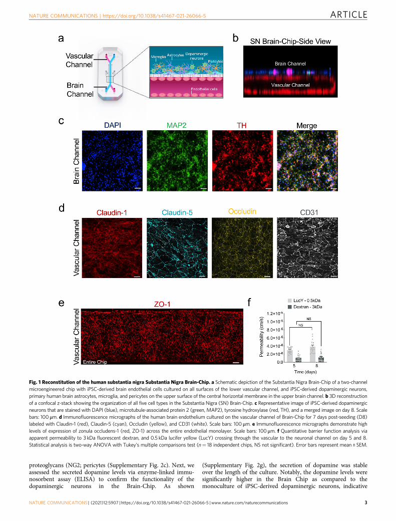
proteoglycans (NG2; pericytes (Supplementary Fig. 2c). Next, weassessed the secreted dopamine levels via enzyme-linked immu-nosorbent assay (ELISA) to confirm the functionality of thedopaminergic neurons in the Brain-Chip. As shown
(Supplementary Fig. 2g), the secretion of dopamine was stableover the length of the culture. Notably, the dopamine levels weresignificantly higher in the Brain Chip as compared to themonoculture of iPSC-derived dopaminergic neurons, indicative
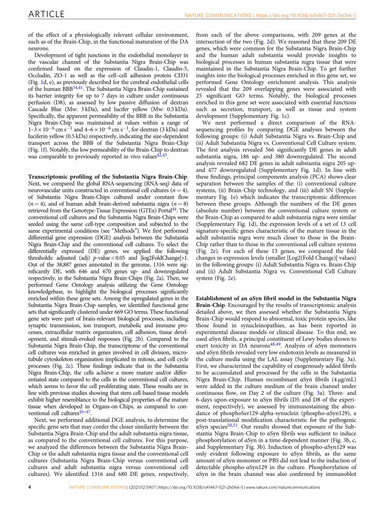
Fig. 1 Reconstitution of the human substantia nigra Substantia Nigra Brain-Chip. a Schematic depiction of the Substantia Nigra Brain-Chip of a two-channelmicroengineered chip with iPSC-derived brain endothelial cells cultured on all surfaces of the lower vascular channel, and iPSC-derived dopaminergic neurons,primary human brain astrocytes, microglia, and pericytes on the upper surface of the central horizontal membrane in the upper brain channel. b 3D reconstructionof a confocal z-stack showing the organization of all five cell types in the Substantia Nigra (SN) Brain-Chip. c Representative image of iPSC-derived dopaminergicneurons that are stained with DAPI (blue), microtubule-associated protein 2 (green, MAP2), tyrosine hydroxylase (red, TH), and a merged image on day 8. Scalebars: 100 μm. d Immunofluorescence micrographs of the human brain endothelium cultured on the vascular channel of Brain-Chip for 7 days post-seeding (D8)labeled with Claudin-1 (red), Claudin-5 (cyan), Occludin (yellow), and CD31 (white). Scale bars: 100 μm. e Immunofluorescence micrographs demonstrate highlevels of expression of zonula occludens-1 (red, ZO-1) across the entire endothelial monolayer. Scale bars: 100 μm. f Quantitative barrier function analysis viaapparent permeability to 3 kDa fluorescent dextran, and 0.5 kDa lucifer yellow (LucY) crossing through the vascular to the neuronal channel on day 5 and 8.Statistical analysis is two-way ANOVA with Tukey’s multiple comparisons test (n= 18 independent chips, NS not significant). Error bars represent mean ± SEM.
NATURE COMMUNICATIONS | https://doi.org/10.1038/s41467-021-26066-5 ARTICLE
NATURE COMMUNICATIONS | (2021) 12:5907 | https://doi.org/10.1038/s41467-021-26066-5 | www.nature.com/naturecommunications 3
of the effect of a physiologically relevant cellular environment,such as of the Brain-Chip, in the functional maturation of the DAneurons.
Development of tight junctions in the endothelial monolayer inthe vascular channel of the Substantia Nigra Brain-Chip wasconfirmed based on the expression of Claudin-1, Claudin-5,Occludin, ZO-1 as well as the cell–cell adhesion protein CD31(Fig. 1d, e), as previously described for the cerebral endothelial cellsof the human BBB34,41. The Substantia Nigra Brain-Chip sustainedits barrier integrity for up to 7 days in culture under continuousperfusion (D8), as assessed by low passive diffusion of dextranCascade Blue (Mw: 3 kDa), and lucifer yellow (Mw: 0.5 kDa).Specifically, the apparent permeability of the BBB in the SubstantiaNigra Brain-Chip was maintained at values within a range of1–3 × 10⁻⁶ cm s−1 and 4–6 × 10⁻⁶ cm s−1, for dextran (3 kDa) andluciferin yellow (0.5 kDa) respectively, indicating the size-dependenttransport across the BBB of the Substantia Nigra Brain-Chip(Fig. 1f). Notably, the low permeability of the Brain-Chip to dextranwas comparable to previously reported in vivo values42,43.
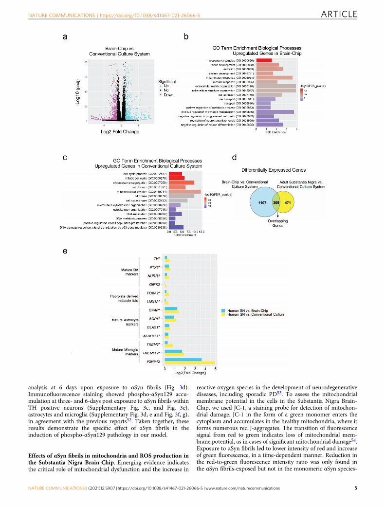
Transcriptomic profiling of the Substantia Nigra Brain-Chip.Next, we compared the global RNA-sequencing (RNA-seq) data ofneurovascular units constructed in conventional cell cultures (n= 4),of Substantia Nigra Brain-Chips cultured under constant flow(n= 4), and of human adult brain-derived substantia nigra (n= 8)retrieved from the Genotype-Tissue Expression (GTEx) Portal44. Theconventional cell cultures and the Substantia Nigra Brain-Chips wereseeded using the same cell-type composition and subjected to thesame experimental conditions (see “Methods”). We first performeddifferential gene expression (DGE) analysis between the SubstantiaNigra Brain-Chip and the conventional cell cultures. To select thedifferentially expressed (DE) genes, we applied the followingthresholds: adjusted (adj) p-value < 0.05 and |log2FoldChange|>1.Out of the 38,887 genes annotated in the genome, 1316 were sig-nificantly DE, with 646 and 670 genes up- and downregulatedrespectively, in the Substantia Nigra Brain-Chips (Fig. 2a). Then, weperformed Gene Ontology analysis utilizing the Gene Ontologyknowledgebase, to highlight the biological processes significantlyenriched within these gene sets. Among the upregulated genes in theSubstantia Nigra Brain-Chip samples, we identified functional genesets that significantly clustered under 669 GO terms. These functionalgene sets were part of brain-relevant biological processes, includingsynaptic transmission, ion transport, metabolic and immune pro-cesses, extracellular matrix organization, cell adhesion, tissue devel-opment, and stimuli-evoked responses (Fig. 2b). Compared to theSubstantia Nigra Brain-Chip, the transcriptome of the conventionalcell cultures was enriched in genes involved in cell division, micro-tubule cytoskeleton organization implicated in mitosis, and cell cycleprocesses (Fig. 2c). These findings indicate that in the SubstantiaNigra Brain-Chip, the cells achieve a more mature and/or differ-entiated state compared to the cells in the conventional cell cultures,which seems to favor the cell proliferating state. These results are inline with previous studies showing that stem cell-based tissue modelsexhibit higher resemblance to the biological properties of the maturetissue when developed in Organs-on-Chips, as compared to con-ventional cell cultures45–47.
Next, we performed additional DGE analysis, to determine thespecific gene sets that may confer the closer similarity between theSubstantia Nigra Brain-Chip and the adult substantia nigra tissue,as compared to the conventional cell cultures. For this purpose,we analyzed the differences between the Substantia Nigra Brain-Chip or the adult substantia nigra tissue and the conventional cellcultures (Substantia Nigra Brain-Chip versus conventional cellcultures and adult substantia nigra versus conventional cellcultures). We identified 1316 and 680 DE genes, respectively,
from each of the above comparisons, with 209 genes at theintersection of the two (Fig. 2d). We reasoned that these 209 DEgenes, which were common for the Substantia Nigra Brain-Chipand the human adult substantia would provide insights tobiological processes in human substantia nigra tissue that weremaintained in the Substantia Nigra Brain-Chip. To get furtherinsights into the biological processes enriched in this gene set, weperformed Gene Ontology enrichment analysis. This analysisrevealed that the 209 overlapping genes were associated with25 significant GO terms. Notably, the biological processesenriched in this gene set were associated with essential functionssuch as secretion, transport, as well as tissue and systemdevelopment (Supplementary Fig. 1c).
We next performed a direct comparison of the RNA-sequencing profiles by comparing DGE analyses between thefollowing groups: (i) Adult Substantia Nigra vs. Brain-Chip and(ii) Adult Substantia Nigra vs. Conventional Cell Culture system.The first analysis revealed 566 significantly DE genes in adultsubstantia nigra, 186 up- and 380 downregulated. The secondanalysis revealed 682 DE genes in adult substantia nigra 205 up-and 477 downregulated (Supplementary Fig. 1d). In line withthese findings, principal components analysis (PCA) shows clearseparation between the samples of the: (i) conventional culturesystems, (ii) Brain-Chip technology, and (iii) adult SN (Supple-mentary Fig. 1e) which indicates the transcriptomic differencesbetween these groups. Although the numbers of the DE genes(absolute number) between the conventional culture system orthe Brain-Chip as compared to adult substantia nigra were similar(Supplementary Fig. 1d), the expression levels of a set of 13 cellsignature-specific genes characteristic of the mature tissue in theadult substantia nigra were much closer to those in the Brain-Chip rather than to those in the conventional cell culture systems(Fig. 2e). For each of these 13 genes, we compared the foldchanges in expression levels (smaller |Log2(Fold Change)| values)in the following groups: (i) Adult Substantia Nigra vs. Brain-Chipand (ii) Adult Substantia Nigra vs. Conventional Cell Culturesystem (Fig. 2e).
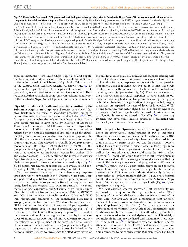
Establishment of an αSyn fibril model in the Substantia NigraBrain-Chip. Encouraged by the results of transcriptomic analysisdetailed above, we then assessed whether the Substantia NigraBrain-Chip would respond to abnormal, toxic protein species, likethose found in synucleinopathies, as has been reported inexperimental disease models or clinical disease. To this end, weused αSyn fibrils, a principal constituent of Lewy bodies shown toexert toxicity in DA neurons48,49. Analysis of αSyn monomersand αSyn fibrils revealed very low endotoxin levels as measured inthe culture media using the LAL assay (Supplementary Fig. 3a).First, we characterized the capability of exogenously added fibrilsto be accumulated and processed by the cells in the SubstantiaNigra Brain-Chip. Human recombinant αSyn fibrils (4 μg/mL)were added in the culture medium of the brain channel undercontinuous flow, on Day 2 of the culture (Fig. 3a). Three- and6-days upon-exposure to αSyn fibrils (D5 and D8 of the experi-ment, respectively), we assessed by immunostaining the abun-dance of phosphoSer129-alpha-synuclein (phospho-αSyn129), apost-translational modification characteristic for the pathogenicαSyn species50,51. Our results showed that exposure of the Sub-stantia Nigra Brain-Chip to αSyn fibrils was sufficient to inducephosphorylation of αSyn in a time-dependent manner (Fig. 3b, c,and Supplementary Fig. 3b). Induction of phospho-αSyn129 wasonly evident following exposure to αSyn fibrils, as the sameamount of αSyn monomer or PBS did not lead to the induction ofdetectable phospho-αSyn129 in the culture. Phosphorylation ofαSyn in the brain channel was also confirmed by immunoblot
ARTICLE NATURE COMMUNICATIONS | https://doi.org/10.1038/s41467-021-26066-5
4 NATURE COMMUNICATIONS | (2021) 12:5907 | https://doi.org/10.1038/s41467-021-26066-5 | www.nature.com/naturecommunications
analysis at 6 days upon exposure to αSyn fibrils (Fig. 3d).Immunofluorescence staining showed phospho-αSyn129 accu-mulation at three- and 6-days post exposure to αSyn fibrils withinTH positive neurons (Supplementary Fig. 3c, and Fig. 3e),astrocytes and microglia (Supplementary Fig. 3d, e and Fig. 3f, g),in agreement with the previous reports52. Taken together, theseresults demonstrate the specific effect of αSyn fibrils in theinduction of phospho-αSyn129 pathology in our model.
Effects of αSyn fibrils in mitochondria and ROS production inthe Substantia Nigra Brain-Chip. Emerging evidence indicatesthe critical role of mitochondrial dysfunction and the increase in
reactive oxygen species in the development of neurodegenerativediseases, including sporadic PD53. To assess the mitochondrialmembrane potential in the cells in the Substantia Nigra Brain-Chip, we used JC-1, a staining probe for detection of mitochon-drial damage. JC-1 in the form of a green monomer enters thecytoplasm and accumulates in the healthy mitochondria, where itforms numerous red J-aggregates. The transition of fluorescencesignal from red to green indicates loss of mitochondrial mem-brane potential, as in cases of significant mitochondrial damage54.Exposure to αSyn fibrils led to lower intensity of red and increaseof green fluorescence, in a time-dependent manner. Reduction inthe red-to-green fluorescence intensity ratio was only found inthe αSyn fibrils-exposed but not in the monomeric αSyn species-
NATURE COMMUNICATIONS | https://doi.org/10.1038/s41467-021-26066-5 ARTICLE
NATURE COMMUNICATIONS | (2021) 12:5907 | https://doi.org/10.1038/s41467-021-26066-5 | www.nature.com/naturecommunications 5
exposed Substantia Nigra Brain-Chips (Fig. 4a, b, and Supple-mentary Fig. 4a). Next, we measured the intracellular ROS levelsin the brain channel of the Substantia Nigra Brain-Chip, on day 8of the culture, using CellROX reagent. As shown (Fig. 4c, d),exposure to αSyn fibrils led to a significant increase in ROSproduction, as compared to exposure to αSyn monomers. Thus,we conclude that αSyn fibrils compromise mitochondrial functionin the Substantia Nigra Brain-Chip, in a time dependent manner.
αSyn fibrils induce cell death and neuroinflammation in theSubstantia Nigra Brain-Chip. Several studies have shown thatαSyn fibrils initiate a series of secondary processes leading toneuroinflammation, neurodegeneration, and cell death48,55. Wefirst questioned whether the cells in the Substantia Nigra Brain-Chip would respond to αSyn fibrils by induction of apoptosis.Three days (experimental D5) following exposure to αSyn, eithermonomeric or fibrillar, there was no effect in cell survival, asreflected by the similar percentage of live cells in all the experi-mental groups. In contrast, 6 days post exposure (experimentalD8), there was significant reduction of the live cells in the Sub-stantia Nigra Brain-Chip exposed to αSyn fibrils compare to αSynmonomers or PBS (50.63 ± 3.9 vs 87.02 ± 0.87 vs 91.2 ± 1.05)(Supplementary Fig. 4b, c). Confocal immunocytochemical ana-lysis using antibodies against MAP2, tyrosine hydroxylase (TH),and cleaved caspase-3 (CC3), confirmed the increase in caspase3-positive dopaminergic neurons at day 6 post exposure to αSynfibrils, as compared to those exposed to monomeric αSyn (Fig. 5a,b). Dopaminergic neuron apoptosis was also evaluated and con-firmed by TUNEL staining (Supplementary Fig. 4d, e).
Next, we assessed the extent of the inflammatory responseupon exposure to αSyn fibrils in the Substantia Nigra Brain-Chip.We performed quantitative analysis on the gene expression ofmarkers for reactive astrocyte and activated microglia, that areupregulated in pathological conditions. In particular, we foundthat 6 days post exposure of the Substantia Nigra Brain-Chip toαSyn fibrils, both reactive astrocyte markers (GFAP, VIMENTIN,and LCN2) and activated microglia markers (CD68 and EDA)were upregulated compared to the monomeric αSyn-treatedgroup (Supplementary Fig. 5a). We also observed increasedGFAP staining in the αSyn fibrils-exposed chips suggestive ofreactive astrogliosis (Fig. 5c and Supplementary Fig. 5b), acomponent of the brain inflammatory response37. In parallel,there was activation of the microglia, as indicated by the increasein CD68 immunoreactivity (Fig. 5d and Supplementary Fig. 5c).Interestingly, a large number of activated microglia formedclusters around the apoptotic neurons (Supplementary Fig. 5d),suggesting that the microglia response may be linked to theneuronal injury. Finally, we investigate the effect αSyn fibrils on
the proliferation of glial cells. Immunocytochemical staining withthe proliferation marker Ki67 showed no significant increase inproliferation following exposure to αSyn fibrils as compared tomonomeric αSyn (Supplementary Fig. 5e, f). In line, there wereno differences in the number of cells between the control andtreated groups (Supplementary Fig. 5g). Thus, we conclude thatthe astrocytic and microglial responses to αSyn fibrils in ourmodel are due largely due to changes in the existing resting glialcells, rather than due to the generation of new glial cells from glialprecursors. As expected, the secreted levels of interleukin-6 (IL-6), and tumor necrosis factor-alpha (TNF-α) in the effluent of theneuronal channel, were significantly increased following exposureto αSyn fibrils versus monomeric αSyn (Fig. 5e, f), providingevidence that αSyn fibrils-induced pathology is associated withthe development of neuroinflammation.
BBB disruption in αSyn-associated PD pathology. As the evi-dence on extraneuronal manifestations of PD is increasing,attention has been drawn to the effects of the disease on the BBBfunction. Measurable levels of αSyn have been identified in thebrain and in the systemic circulation, and the current hypothesisis that they are implicated in disease onset and/or progression.The origin of peripheral αSyn remains a subject of discussion, aswell as the possibility that αSyn could cross the BBB in eitherdirection12. Recent data argue that there is BBB dysfunction inPD as proposed for other neurodegenerative diseases, and that therole of BBB in the pathogenesis and progression of PD may bepivotal56. Thus, we ran BBB permeability assays on the SubstantiaNigra Brain-Chip upon exposure to αSyn fibrils, or αSynmonomers or PBS. Our data indicate significantly increasedpermeability to 160 kDa Immunoglobulins (IgG), 3 kDa dextran,and 0.5 kDa lucifer in the brain channel of the Substantia NigraBrain-Chip 6 days after exposure to αSyn fibrils (Fig. 6a, b, andSupplementary Fig. 6a).
We next assessed whether increased BBB permeability wasassociated to disruption of the tight junction protein ZO-1.Staining of the endothelial cells in the vascular channel of theBrain-Chip with anti-ZO1 at D8, demonstrated tight junctionsdamage following exposure to αSyn fibrils, but not to monomericαSyn (Supplementary Fig. 6b, c). Next, we examined theimplication of mechanisms attributable to BBB impairment, suchas changes in the expression of TOM20, which prevents α-synuclein-induced mitochondrial dysfunction57, and ICAM-1, akey molecule in immune-mediated and inflammatory processesin endothelial cells58. Immunofluorescence analysis showedattenuated expression of the TOM20 and increased expressionof ICAM-1 at 6 days (experimental D8) post exposure to αSynfibrils compared to monomeric group (Supplementary Fig. 6b, c),
Fig. 2 Differentially Expressed (DE) genes and enriched gene ontology categories in Substantia Nigra Brain-Chip or conventional cell cultures ascompared to the adult substantia nigra. a The volcano plot resulted by the differentially gene expression (DGE) analysis between Substantia Nigra Brain-Chip and conventional cell cultures. For the selection of the DE genes we used the following thresholds: adjusted (adj) p-value < 0.05 and |Log2(foldchange)| >1. The identified up- (down-) regulated genes are highlighted in cyan (magenta) color respectively. Sample sizes were as follows:Substantia Nigra Brain-Chip, n= 4, conventional cell culture system, n=4. The p-values attained by the two-sided Wald test and corrected for multipletesting using the Benjamini and Hochberg method. b, c List of biological processes identified by Gene Ontology (GO) enrichment analysis using the up- anddownregulated genes respectively resulted by the differentially gene expression analysis between Substantia Nigra Brain-Chip and conventional cellcultures. d DGE analysis identified up- and downregulated genes in Substantia Nigra Brain-Chip compared to conventional cell cultures (cyan circle), andhuman adult substantia nigra compared to conventional cell cultures (yellow circle). Sample sizes were as follows: Substantia Nigra Brain-Chip, n= 4,Conventional cell culture system, n= 4, and adult substantia nigra, n= 8 (independent biological specimens). Culture in Brain-Chips and conventional cellcultures were done in parallel. Samples were collected and processed for analyses 8 days post-seeding (D8). e Gene expression pattern analysis betweenthe following groups: i) Adult Substantia Nigra vs. Brain-Chip and ii) Adult Substantia Nigra vs. Conventional Cell Culture system. In the Brain-Chip, most ofthese genes (those with an asterisk) exhibit statistically significantly smaller fold changes (P < 0.05) in their expression levels as compared to theconventional cell culture system. Statistical analysis is two-sided Wald test and corrected for multiple testing using the Benjamini and Hochberg method.The adjusted P value per gene is contained in Supplementary Table 1.
ARTICLE NATURE COMMUNICATIONS | https://doi.org/10.1038/s41467-021-26066-5
6 NATURE COMMUNICATIONS | (2021) 12:5907 | https://doi.org/10.1038/s41467-021-26066-5 | www.nature.com/naturecommunications
suggesting a possible role for αSyn fibrils in BBB damagecorrelated with mitochondria dysfunction and inflammation inthe vasculature. To check whether αSyn fibrils were directlydeposited on the endothelial cells in the Brain-Chip, weperformed immunofluorescent staining with phospho-αSyn129in the vascular channel. Our results show that exposure of theSubstantia Nigra Brain-Chip to αSyn fibrils-induced phosphor-ylation of αSyn localized in the endothelial cells in the vascularchannel, 6 days post exposure to αSyn fibrils (Fig. 6c, d). Theeffective phosphorylation of αSyn in the vascular channel wasconfirmed by immunoblot analysis at 6 days post exposure toαSyn fibrils as compared to the PBS group (SupplementaryFig. 6d). These results indicate that αSyn fibrils affect thepermeability of the BBB in the Brain-Chip, although our
experiment cannot distinguish between a direct role of αSynfibrils or indirect via effects on other contributing molecules incausing BBB impairment.
To further characterize the endothelium in the SubstantiaNigra Brain-Chip model and to determine whether the exposureto αSyn fibrils leads to transcriptomic changes in these cells, weran RNA-seq analysis. PCA revealed differences in the tran-scriptome profiles between the two conditions, i.e., exposure toαSyn fibrils or to αSyn monomers (Fig. 6e). This analysis resultedin the identification of 1280 DE genes, either significantlyupregulated (739 genes) or downregulated (541 genes) (Fig. 6f)in the αSyn fibril-exposed Substantia Nigra Brain-Chips. This setof 1280 DE genes includes several genes that have been implicatedin BBB dysfunction in a number of diseases59. Multiple members
Fig. 3 Pathological αSyn accumulation in the brain channel following exposure to human αSyn fibrils. a Experimental design for assessing the effects ofαSyn toxic aggregates (fibrils) in the Substantia Nigra Brain-Chip, including the seeding in the Brain-Chip, the timeline for medium changes, as well assampling times. b Immunofluorescence micrographs show the accumulation of phosphorylated αSyn (green, phospho-αSyn129 staining; blue, DAPI) at day6 post-exposure (D8). Pathology is absent in the brain channel following exposure to monomer or PBS. Scale bars: 100 μm. c Quantitative analysis offluorescence intensity in each group at day 3 and 6 post-exposure (D5 and D8, respectively). Statistical analysis is two-way ANOVA with Tukey’s multiplecomparisons test (n= 3–4 independent chips with 3~5 randomly selected different areas per chip, *P= 0.0103, ****P < 0.0001 compared to monomericgroup). Error bars represent mean ± SEM. d Western blotting analysis of cell lysates from the brain channel shows significant intracellular phosphorylationof αSyn at Ser129 (MW: 18 kDa) following exposure to αSyn fibrils, whereas there was no effect upon exposure to the PBS. For loading control, equalamounts of protein were immunoblotted with GAPDH antibody (MW: 37 kDa). e Confocal images of double immunostaining for phospho-αSyn129 (green)and tyrosine hydroxylase (red, TH), in the brain channel at day 6 post-exposure (D8) to αSyn fibrils (white arrow). Scale bars: 50 μm. f, g αSyn fibrils aretaken up by astrocytes and microglia (white arrow), as evidenced by double immunostaining for phospho-αSyn129 (green) and either glial fibrillary acidicprotein (red, GFAP) for astrocytes or ionized calcium-binding adaptor molecule 1 (red, IBA1) for microglia in the brain channel at day 6 post-exposure (D8)to αSyn fibrils. Scale bars: 50 μm.
NATURE COMMUNICATIONS | https://doi.org/10.1038/s41467-021-26066-5 ARTICLE
NATURE COMMUNICATIONS | (2021) 12:5907 | https://doi.org/10.1038/s41467-021-26066-5 | www.nature.com/naturecommunications 7
of specific gene families were upregulated, such as extracellularproteases of the Serpin family (SERPINA1), collagens (COL3A1),centromere proteins (CENPE), and kinesins (KIF15). In addition,we identified multiple genes in the αSyn fibrils-exposed chipsimplicated in key cellular processes associated with PDpathogenesis (Table 1, Fig. 6g) such as autophagy, oxidativestress, mitochondrial function, inflammation, and vesiculartrafficking, highlighting the potential for brain endothelial cellsto contribute to molecular mechanisms and functional deficits inPD. Examples of associated genes include leucine-rich repeatkinase 2 (LRRK2)60, synphilin-1 (SNCAIP)61, monoamineoxidase A (MAOA)62, complement 5 (C5)63, and apolipoproteinA-1 (APOA1)64. The altered expression of PD-associated geneswas confirmed by quantitative RT-PCR (qPCR) (SupplementaryFig. 6e). Other BBB-related genes with altered expression are low-density lipoprotein receptor-related protein 1B (LRP1B)65 andATP-binding cassette (ABC) transporters (ABCB1)66. Theupregulation of the LRP1 gene is consistent with previousfindings, where dysfunction of LRP1B has been associated withPD67. Further, positive and negative associations between specificABCB1 haplotypes associated with P‑glycoprotein activity PDincidence have been reported66. Endothelial genes downregulatedupon exposure to αSyn fibrils included the tight junctions claudingene family (CLDN1, CLDN4, and CLDN9), and the gap junctionprotein alpha 4 (GJA4). All the above suggest that the SubstantiaNigra Brain-Chip could be used to assess novel treatments that
protect from vascular dysfunction or improve vascular remodel-ing in the brain.
Trehalose ameliorates αSyn induced pathology. To this purpose,we tested whether the disrupted BBB in the αSyn fibril-exposedSubstantia Nigra Brain-Chip, could be restored by therapeutic agentstargeting mechanisms for clearing of the accumulated αSyn protein.Recent reports of trehalose, a disaccharide approved by FDA, haveshown beneficial effects against the accumulation of neurotoxic,aggregated proteins, and neurodegeneration68. The study of Hoff-mann et al.69 provided evidence that trehalose prevents or halts thepropagation of αSyn pathology by targeting lysosomes. Also, studiesin aged mice suggest that oral supplementation of the autophagy‐stimulating disaccharide trehalose, restored vascular autophagy andameliorated age‐related endothelial dysfunction70. On the basis ofthese results, we speculate that trehalose may disturb lysosomeintegrity and function, which could subsequently hinder the BBBdisruption induced by αSyn fibrils. In this regard, we administeredtrehalose (10mM) in the Substantia Nigra Brain-Chip, via the vas-cular channel on experimental day 5, 3 days after adding αSyn fibrils(Fig. 7a).
To evaluate whether trehalose crosses the BBB and attenuatesαSyn accumulation within αSyn-exposed cells, we first measuredtrehalose penetration in the brain channel. Using the trehaloseassay kit, we found that more than 60% of trehalose had cross the
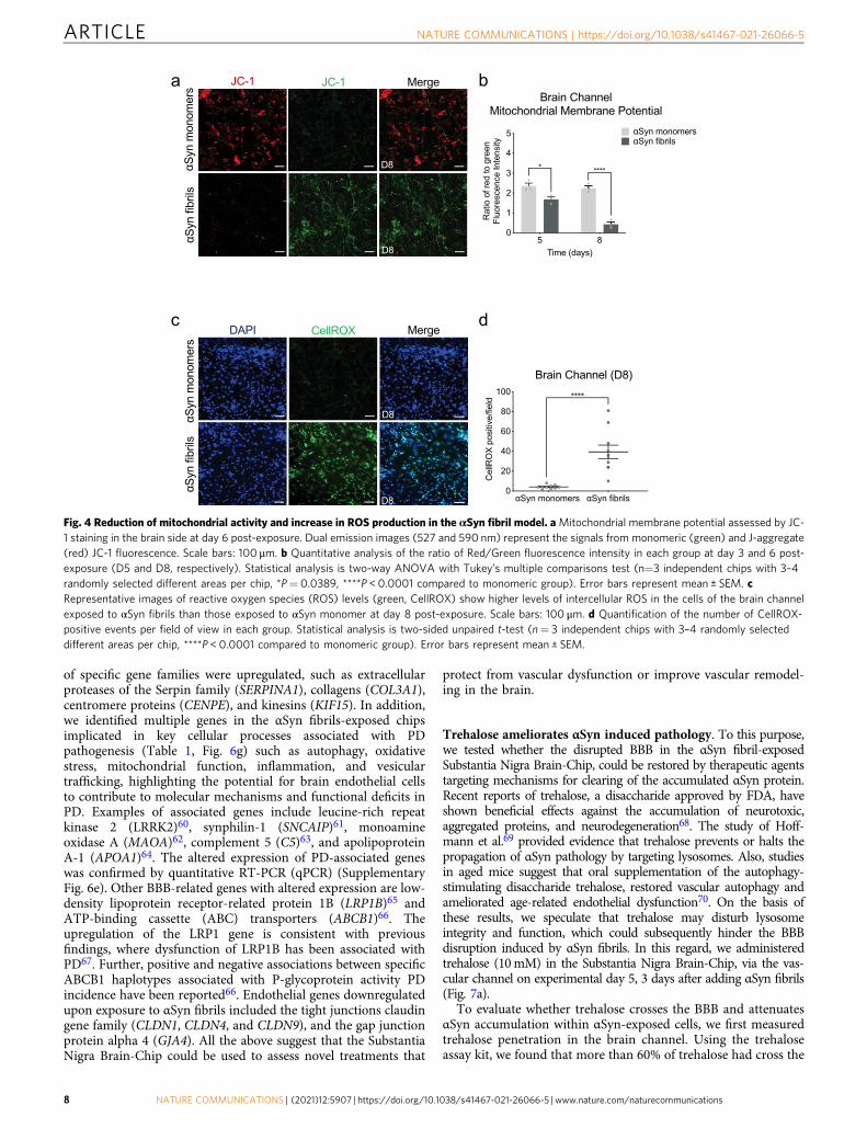
Fig. 4 Reduction of mitochondrial activity and increase in ROS production in the αSyn fibril model. aMitochondrial membrane potential assessed by JC-1 staining in the brain side at day 6 post-exposure. Dual emission images (527 and 590 nm) represent the signals from monomeric (green) and J-aggregate(red) JC-1 fluorescence. Scale bars: 100 μm. b Quantitative analysis of the ratio of Red/Green fluorescence intensity in each group at day 3 and 6 post-exposure (D5 and D8, respectively). Statistical analysis is two-way ANOVA with Tukey’s multiple comparisons test (n=3 independent chips with 3–4randomly selected different areas per chip, *P= 0.0389, ****P < 0.0001 compared to monomeric group). Error bars represent mean ± SEM. cRepresentative images of reactive oxygen species (ROS) levels (green, CellROX) show higher levels of intercellular ROS in the cells of the brain channelexposed to αSyn fibrils than those exposed to αSyn monomer at day 8 post-exposure. Scale bars: 100 μm. d Quantification of the number of CellROX-positive events per field of view in each group. Statistical analysis is two-sided unpaired t-test (n= 3 independent chips with 3–4 randomly selecteddifferent areas per chip, ****P < 0.0001 compared to monomeric group). Error bars represent mean ± SEM.
ARTICLE NATURE COMMUNICATIONS | https://doi.org/10.1038/s41467-021-26066-5
8 NATURE COMMUNICATIONS | (2021) 12:5907 | https://doi.org/10.1038/s41467-021-26066-5 | www.nature.com/naturecommunications
BBB (Fig. 7b). As accumulation of phosphorylated αSyn in fibril-exposed cells was readily detectable in the brain channel (Fig. 3),we next validated the efficacy of trehalose in preventing lysosomalalteration and accumulation of aggregated αSyn. In agreementwith previous findings69, lysosomal cathepsin D activitydecreased significantly in αSyn fibrils-exposed cells, indicativeof impaired lysosomal activity (Supplementary Fig. 7a). Ourresults showed that trehalose prevented lysosomal alterationswithin αSyn-exposed cells in the brain channel (SupplementaryFig. 7a) and significantly decreased the abundance of phospho-αSyn129 compared to the non-treated chips (Fig. 7c andSupplementary Fig. 7b). Taken together, these findings
demonstrate the protective effects of trehalose on the αSyn-mediated impairment of lysosomes and on the clearance ofaccumulated αSyn in the cells within the brain channel. Next, weexamined whether trehalose suppressed the secretion of proin-flammatory cytokines detected in αSyn fibrils-exposed cells in thebrain channel (Fig. 5e, f). We found that the TNF-α and IL-6levels were significantly decreased in the brain channel’s effluentfollowing exposure to trehalose compared to the non-treatedchips, in support of its protective effects against neuroinflamma-tion (Fig. 7d).
At the vascular channel, trehalose exerted similar effects as itreduced the levels of phospho-αSyn129 (Fig. 7e and Supplementary
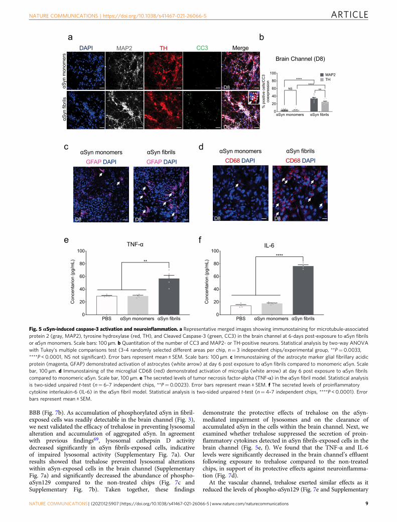
Fig. 5 αSyn-induced caspase-3 activation and neuroinflammation. a Representative merged images showing immunostaining for microtubule-associatedprotein 2 (gray, MAP2), tyrosine hydroxylase (red, TH), and Cleaved Caspase-3 (green, CC3) in the brain channel at 6-days post-exposure to αSyn fibrilsor αSyn monomers. Scale bars: 100 μm. b Quantitation of the number of CC3 and MAP2- or TH-positive neurons. Statistical analysis by two-way ANOVAwith Tukey’s multiple comparisons test (3–4 randomly selected different areas per chip, n= 3 independent chips/experimental group, **P= 0.0033,****P < 0.0001, NS not significant). Error bars represent mean ± SEM. Scale bars: 100 μm. c Immunostaining of the astrocyte marker glial fibrillary acidicprotein (magenta, GFAP) demonstrated activation of astrocytes (white arrow) at day 6 post exposure to αSyn fibrils compared to monomeric αSyn. Scalebar, 100 μm. d Immunostaining of the microglial CD68 (red) demonstrated activation of microglia (white arrow) at day 6 post exposure to αSyn fibrilscompared to monomeric αSyn. Scale bar, 100 μm. e The secreted levels of tumor necrosis factor-alpha (TNF-α) in the αSyn fibril model. Statistical analysisis two-sided unpaired t-test (n= 6–7 independent chips, **P= 0.0023). Error bars represent mean ± SEM. f The secreted levels of proinflammatorycytokine interleukin-6 (IL-6) in the αSyn fibril model. Statistical analysis is two-sided unpaired t-test (n= 4–7 independent chips, ****P < 0.0001). Errorbars represent mean ± SEM.
NATURE COMMUNICATIONS | https://doi.org/10.1038/s41467-021-26066-5 ARTICLE
NATURE COMMUNICATIONS | (2021) 12:5907 | https://doi.org/10.1038/s41467-021-26066-5 | www.nature.com/naturecommunications 9
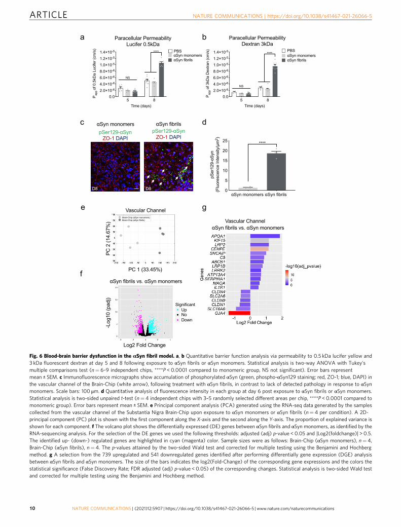
Fig. 6 Blood-brain barrier dysfunction in the αSyn fibril model. a, b Quantitative barrier function analysis via permeability to 0.5 kDa lucifer yellow and3 kDa fluorescent dextran at day 5 and 8 following exposure to αSyn fibrils or αSyn monomers. Statistical analysis is two-way ANOVA with Tukey’smultiple comparisons test (n= 6–9 independent chips, ****P < 0.0001 compared to monomeric group, NS not significant). Error bars representmean ± SEM. c Immunofluorescence micrographs show accumulation of phosphorylated αSyn (green, phospho-αSyn129 staining; red, ZO-1; blue, DAPI) inthe vascular channel of the Brain-Chip (white arrow), following treatment with αSyn fibrils, in contrast to lack of detected pathology in response to αSynmonomers. Scale bars: 100 μm. d Quantitative analysis of fluorescence intensity in each group at day 6 post exposure to αSyn fibrils or αSyn monomers.Statistical analysis is two-sided unpaired t-test (n= 4 independent chips with 3–5 randomly selected different areas per chip, ****P < 0.0001 compared tomonomeric group). Error bars represent mean ± SEM. e Principal component analysis (PCA) generated using the RNA-seq data generated by the samplescollected from the vascular channel of the Substantia Nigra Brain-Chip upon exposure to αSyn monomers or αSyn fibrils (n= 4 per condition). A 2D-principal component (PC) plot is shown with the first component along the X-axis and the second along the Y-axis. The proportion of explained variance isshown for each component. f The volcano plot shows the differentially expressed (DE) genes between αSyn fibrils and αSyn monomers, as identified by theRNA-sequencing analysis. For the selection of the DE genes we used the following thresholds: adjusted (adj) p-value < 0.05 and |Log2(foldchange)| > 0.5.The identified up- (down-) regulated genes are highlighted in cyan (magenta) color. Sample sizes were as follows: Brain-Chip (αSyn monomers), n= 4,Brain-Chip (αSyn fibrils), n= 4. The p-values attained by the two-sided Wald test and corrected for multiple testing using the Benjamini and Hochbergmethod. g A selection from the 739 upregulated and 541 downregulated genes identified after performing differentially gene expression (DGE) analysisbetween αSyn fibrils and αSyn monomers. The size of the bars indicates the log2(Fold-Change) of the corresponding gene expressions and the colors thestatistical significance (False Discovery Rate; FDR adjusted (adj) p-value < 0.05) of the corresponding changes. Statistical analysis is two-sided Wald testand corrected for multiple testing using the Benjamini and Hochberg method.
ARTICLE NATURE COMMUNICATIONS | https://doi.org/10.1038/s41467-021-26066-5
10 NATURE COMMUNICATIONS | (2021) 12:5907 | https://doi.org/10.1038/s41467-021-26066-5 | www.nature.com/naturecommunications
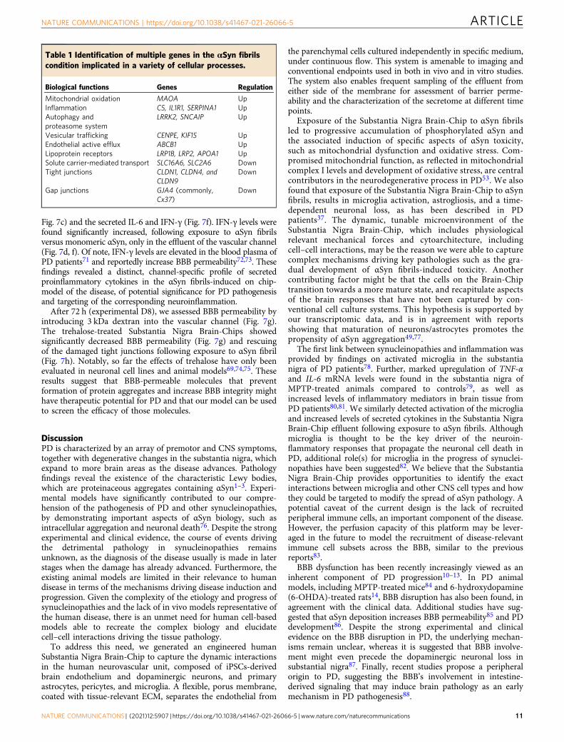
Fig. 7c) and the secreted IL-6 and IFN-γ (Fig. 7f). IFN-γ levels werefound significantly increased, following exposure to αSyn fibrilsversus monomeric αSyn, only in the effluent of the vascular channel(Fig. 7d, f). Of note, IFN-γ levels are elevated in the blood plasma ofPD patients71 and reportedly increase BBB permeability72,73. Thesefindings revealed a distinct, channel-specific profile of secretedproinflammatory cytokines in the αSyn fibrils-induced on chip-model of the disease, of potential significance for PD pathogenesisand targeting of the corresponding neuroinflammation.
After 72 h (experimental D8), we assessed BBB permeability byintroducing 3 kDa dextran into the vascular channel (Fig. 7g).The trehalose-treated Substantia Nigra Brain-Chips showedsignificantly decreased BBB permeability (Fig. 7g) and rescuingof the damaged tight junctions following exposure to αSyn fibril(Fig. 7h). Notably, so far the effects of trehalose have only beenevaluated in neuronal cell lines and animal models69,74,75. Theseresults suggest that BBB-permeable molecules that preventformation of protein aggregates and increase BBB integrity mighthave therapeutic potential for PD and that our model can be usedto screen the efficacy of those molecules.
DiscussionPD is characterized by an array of premotor and CNS symptoms,together with degenerative changes in the substantia nigra, whichexpand to more brain areas as the disease advances. Pathologyfindings reveal the existence of the characteristic Lewy bodies,which are proteinaceous aggregates containing αSyn1–3. Experi-mental models have significantly contributed to our compre-hension of the pathogenesis of PD and other synucleinopathies,by demonstrating important aspects of αSyn biology, such asintracellular aggregation and neuronal death76. Despite the strongexperimental and clinical evidence, the course of events drivingthe detrimental pathology in synucleinopathies remainsunknown, as the diagnosis of the disease usually is made in laterstages when the damage has already advanced. Furthermore, theexisting animal models are limited in their relevance to humandisease in terms of the mechanisms driving disease induction andprogression. Given the complexity of the etiology and progress ofsynucleinopathies and the lack of in vivo models representative ofthe human disease, there is an unmet need for human cell-basedmodels able to recreate the complex biology and elucidatecell–cell interactions driving the tissue pathology.
To address this need, we generated an engineered humanSubstantia Nigra Brain-Chip to capture the dynamic interactionsin the human neurovascular unit, composed of iPSCs-derivedbrain endothelium and dopaminergic neurons, and primaryastrocytes, pericytes, and microglia. A flexible, porus membrane,coated with tissue-relevant ECM, separates the endothelial from
the parenchymal cells cultured independently in specific medium,under continuous flow. This system is amenable to imaging andconventional endpoints used in both in vivo and in vitro studies.The system also enables frequent sampling of the effluent fromeither side of the membrane for assessment of barrier perme-ability and the characterization of the secretome at different timepoints.
Exposure of the Substantia Nigra Brain-Chip to αSyn fibrilsled to progressive accumulation of phosphorylated αSyn andthe associated induction of specific aspects of αSyn toxicity,such as mitochondrial dysfunction and oxidative stress. Com-promised mitochondrial function, as reflected in mitochondrialcomplex I levels and development of oxidative stress, are centralcontributors in the neurodegenerative process in PD53. We alsofound that exposure of the Substantia Nigra Brain-Chip to αSynfibrils, results in microglia activation, astrogliosis, and a time-dependent neuronal loss, as has been described in PDpatients37. The dynamic, tunable microenvironment of theSubstantia Nigra Brain-Chip, which includes physiologicalrelevant mechanical forces and cytoarchitecture, includingcell–cell interactions, may be the reason we were able to capturecomplex mechanisms driving key pathologies such as the gra-dual development of αSyn fibrils-induced toxicity. Anothercontributing factor might be that the cells on the Brain-Chiptransition towards a more mature state, and recapitulate aspectsof the brain responses that have not been captured by con-ventional cell culture systems. This hypothesis is supported byour transcriptomic data, and is in agreement with reportsshowing that maturation of neurons/astrocytes promotes thepropensity of αSyn aggregation49,77.
The first link between synucleinopathies and inflammation wasprovided by findings on activated microglia in the substantianigra of PD patients78. Further, marked upregulation of TNF-αand IL-6 mRNA levels were found in the substantia nigra ofMPTP-treated animals compared to controls79, as well asincreased levels of inflammatory mediators in brain tissue fromPD patients80,81. We similarly detected activation of the microgliaand increased levels of secreted cytokines in the Substantia NigraBrain-Chip effluent following exposure to αSyn fibrils. Althoughmicroglia is thought to be the key driver of the neuroin-flammatory responses that propagate the neuronal cell death inPD, additional role(s) for microglia in the progress of synuclei-nopathies have been suggested82. We believe that the SubstantiaNigra Brain-Chip provides opportunities to identify the exactinteractions between microglia and other CNS cell types and howthey could be targeted to modify the spread of αSyn pathology. Apotential caveat of the current design is the lack of recruitedperipheral immune cells, an important component of the disease.However, the perfusion capacity of this platform may be lever-aged in the future to model the recruitment of disease-relevantimmune cell subsets across the BBB, similar to the previousreports83.
BBB dysfunction has been recently increasingly viewed as aninherent component of PD progression10–13. In PD animalmodels, including MPTP-treated mice84 and 6-hydroxydopamine(6-OHDA)-treated rats14, BBB disruption has also been found, inagreement with the clinical data. Additional studies have sug-gested that αSyn deposition increases BBB permeability85 and PDdevelopment86. Despite the strong experimental and clinicalevidence on the BBB disruption in PD, the underlying mechan-isms remain unclear, whereas it is suggested that BBB involve-ment might even precede the dopaminergic neuronal loss insubstantial nigra87. Finally, recent studies propose a peripheralorigin to PD, suggesting the BBB’s involvement in intestine-derived signaling that may induce brain pathology as an earlymechanism in PD pathogenesis88.
Table 1 Identification of multiple genes in the αSyn fibrilscondition implicated in a variety of cellular processes.
Biological functions Genes Regulation
Mitochondrial oxidation MAOA UpInflammation C5, IL1R1, SERPINA1 UpAutophagy andproteasome system
LRRK2, SNCAIP Up
Vesicular trafficking CENPE, KIF15 UpEndothelial active efflux ABCB1 UpLipoprotein receptors LRP1B, LRP2, APOA1 UpSolute carrier-mediated transport SLC16A6, SLC2A6 DownTight junctions CLDN1, CLDN4, and
CLDN9Down
Gap junctions GJA4 (commonly,Cx37)
Down
NATURE COMMUNICATIONS | https://doi.org/10.1038/s41467-021-26066-5 ARTICLE
NATURE COMMUNICATIONS | (2021) 12:5907 | https://doi.org/10.1038/s41467-021-26066-5 | www.nature.com/naturecommunications 11
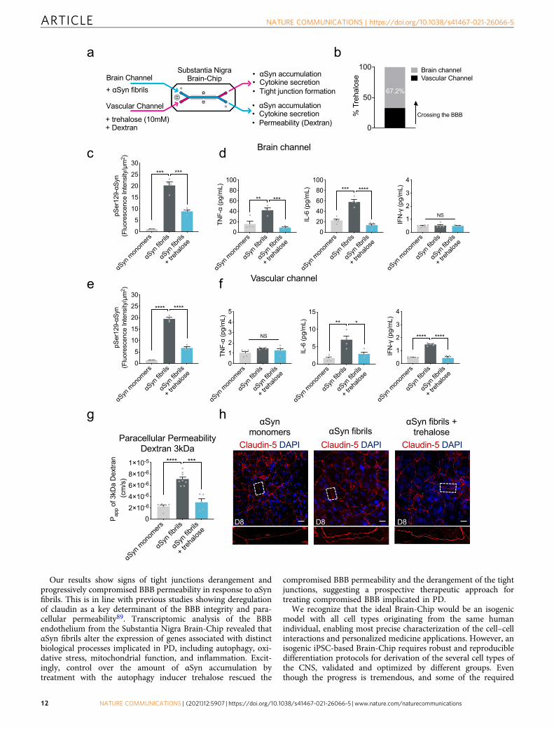
Our results show signs of tight junctions derangement andprogressively compromised BBB permeability in response to αSynfibrils. This is in line with previous studies showing deregulationof claudin as a key determinant of the BBB integrity and para-cellular permeability89. Transcriptomic analysis of the BBBendothelium from the Substantia Nigra Brain-Chip revealed thatαSyn fibrils alter the expression of genes associated with distinctbiological processes implicated in PD, including autophagy, oxi-dative stress, mitochondrial function, and inflammation. Excit-ingly, control over the amount of αSyn accumulation bytreatment with the autophagy inducer trehalose rescued the
compromised BBB permeability and the derangement of the tightjunctions, suggesting a prospective therapeutic approach fortreating compromised BBB implicated in PD.
We recognize that the ideal Brain-Chip would be an isogenicmodel with all cell types originating from the same humanindividual, enabling most precise characterization of the cell–cellinteractions and personalized medicine applications. However, anisogenic iPSC-based Brain-Chip requires robust and reproducibledifferentiation protocols for derivation of the several cell types ofthe CNS, validated and optimized by different groups. Eventhough the progress is tremendous, and some of the required
αSyn
mon
omers
αSyn
fibrils
αSyn
fibrils
+ treh
alose
02×10-64×10-66×10-68×10-61×10-5
P app
of 3
kDa
Dex
tran
(cm
/s)
**** ***
0
50
100
% T
reha
lose
Vascular ChannelBrain channel
67.2%
Crossing the BBB
αSyn
mon
omers
αSyn
fibrils
αSyn
fibrils
+ treh
alose
020406080
100
TNF-
α (p
g/m
L)
** ***
αSyn
mon
omers
αSyn
fibrils
αSyn
fibrils
+ treh
alose
0
5
10
15
20
25
30
pSer
129-
αSyn
(F
luor
esce
nce
Inte
nsity
/μm
2 )
*** ***
αSyn
mon
omers
αSyn
fibrils
αSyn
fibrils
+ treh
alose
020406080
100
IL-6
(pg/
mL) *** ****
αSyn
mon
omers
αSyn
fibrils
αSyn
fibrils
+ treh
alose
0
1
2
3
4
IFN
-γ (p
g/m
L)
NS
αSyn
mon
omers
αSyn
fibrils
αSyn
fibrils
+ treh
alose
012345
TNF-
α (p
g/m
L)
NS
αSyn
mon
omers
αSyn
fibrils
αSyn
fibrils
+ treh
alose
0
5
10
15
20
25
30
pSer
129-
αSyn
(F
luor
esce
nce
Inte
nsity
/μm
2 )
**** ****
αSyn
mon
omers
αSyn
fibrils
αSyn
fibrils
+ treh
alose
0
5
10
15IL
-6 (p
g/m
L) ** *
αSyn
mon
omers
αSyn
fibrils
αSyn
fibrils
+ treh
alose
0
1
2
3
4
IFN
-γ (p
g/m
L)
**** ****
ARTICLE NATURE COMMUNICATIONS | https://doi.org/10.1038/s41467-021-26066-5
12 NATURE COMMUNICATIONS | (2021) 12:5907 | https://doi.org/10.1038/s41467-021-26066-5 | www.nature.com/naturecommunications
differentiation protocols already showed high robustness, the fieldhas not yet reached the point where a reproducible, standardizedisogenic model is available for disease modeling studies90.Variability between models remains a considerable issue, asexemplified in a study comparing the differentiation capacity offour different iPSC lines to isogenic BBB models, includingendothelial cells and astrocytes91. A recent study successfullyincorporated isogenic microglia-like cells with brain region-specific organoids from hiPSCs, however, these structures lackedthe endothelial component required for BBB formation92.Therefore, for this study, we chose to employ well-characterizedand established cell sources to develop stable and standardizedassays that provide good cell quality and enable comparisonsbetween experimental groups based on clinically relevantendpoints.
In conclusion, we report the generation of a Substantia NigraBrain-Chip, that upon exposure to αSyn fibrils reproduces invivo-relevant aspects of synucleinopathies. The Substantia NigraBrain-Chip provides a promising model for the study of thespecific disease mechanisms underlying this unmet medical need,including the dynamics of BBB dysfunction. Moreover, the chipmay be useful to characterize the response to PD therapies andidentify and evaluate associated biomarkers of the disease.
MethodsCell culture. Commercial human iPSC-derived dopaminergic neurons (iCell®Neurons) were purchased from Cellular Dynamics International (CDI; R1108) andmaintained in complete maintenance media (iCell DopaNeurons Media). The cellshave been characterized by CDI to represent a pure neuronal population with>80% pure midbrain dopaminergic neurons. Primary human astrocytes isolatedfrom the cerebral cortex were obtained from ScienCell (Cat. No. 1800) andmaintained in astrocyte medium (ScienCell). Primary human brain pericytes werealso obtained from ScienCell (Cat. No. 1200) and maintained in the pericytemedium (ScienCell). Resting primary human brain microglia were purchased fromATCC (Cat. No. CRL-3304) and cultured according to the manufacturer’sinstructions. To confirm that the cell culture conditions applied were optimized,cell metabolic activity for each cell type in the brain channel medium was assessedusing a (3-(4,5-dimethylthiazol-2-yl)-5-(3-carboxymethoxyphenyl)-2-(4-sulfophe-nyl)-2H-tetrazolium) MTS assay (n= 6), that measures the formazan product as anindex of cell metabolic activity. The primary cells were used at passage 2–4.
Brain microvascular endothelial cell differentiation of hiPSCs. Human-inducedpluripotent stem cells (hiPSCs) obtained from the Rutgers University Cell andDNA Repository (RUCDR; ND50028) were maintained on Matrigel-coated tissue-culture treated six-well culture plates (Corning) in mTeSR™1 (Stem Cell Tech-nologies). The established hiPSC colonies displayed a normal karyotype in culture.For each independent experiment, we used the same cell passage (P49). Prior todifferentiation, hiPSCs were singularized using Accutase® (Invitrogen) and plated
onto Matrigel®-coated six-well culture plates in mTeSR™1 supplemented with10 μM Rho-associated protein kinase inhibitor Y27632 (ROCK inhibitor; Stem CellTechnologies) at a density between 25,000 and 50,000 cells cm−2. Directed dif-ferentiation of hiPSCs was adapted from a previously reported protocol39. Briefly,singularized hiPSCs were expanded for 3 days in mTeSR™1, then were treated with6 µM CHIR99021 (Stem Cell Technologies) in DeSR1. The DeSR1 medium iscomposed of DMEM/Ham’s F12 (Thermo Fisher Scientific), 1× MEM-NEAA(Thermo Fisher Scientific), 0.5× GlutaMAX (Thermo Fisher Scientific), and0.1 mM b-mercaptoethanol (Sigma). After 24 h, the medium was replaced byDeSR2 medium that is composed by DeSR1 plus 1× B27 (Thermo Fisher Scien-tific); the medium was refreshed every day for a period of another 5 days. On day 6,the medium was switched to hECSR1: hESFM (Thermo Fisher Scientific) supple-mented with 20 ng/mL bFGF (R&D Systems), 10 μM all-trans retinoic acid(Sigma), and 1 × B27. The medium was not changed for 48 h. On day 9 themedium was switched to hESCR2: hECSR1 lacking RA and bFGF. On day 10, cellswere dissociated with TrypLE™ (Thermo Fisher Scientific) and replated onto ahuman placenta-derived collagen IV/human plasma-derived fibronectin/humanplacenta-derived laminin-coated flasks. After 20 mins, the flasks were rinsed usinga medium composed of human serum-free endothelial medium supplemented with2% platelet-poor plasma-derived serum and 10 µM Y27632, as a selection step toremove any undifferentiated cells. Human brain microvascular endothelial cells(HBMECs) were then left in the same medium overnight to allow cell attachmentand growth before seeded into the Organ-Chips.
Brain-Chip microfabrication and Zoë® culture module. Organ-Chips (Chip-S1®,Emulate, Inc. Boston, MA, USA) were used to recreate the human Brain-Chip.Chip-S1® is made of a poly(dimethylsiloxane) (PDMS) flexible elastomer. It con-sists of two channels (1 × 1 mm and 1 × 0.2 mm, “Brain” and “Vascular” channel,respectively) separated by a porous flexible PDMS membrane25. Flow can beintroduced to each channel independently to continuously provide essentialnutrients to the cells, while effluent containing any secretion/waste componentsfrom cells is excreted/collected on the outlet of each channel separately. This allowsfor channel-specific and independent analysis and interpretation of results. TheZoë® culture module is the instrumentation designed to automate the maintenanceof these chips in a controlled and robust manner (Emulate, Inc.).
Human Brain-Chip and cell seeding. Prior to cell seeding, chips were functio-nalized using Emulate’s proprietary protocols and reagents. Briefly, ER-1 (Emulatereagent: 10461) and ER-2 (Emulate reagent: 10462) are mixed at a concentration of1 mg/mL before being added to the top and bottom microfluidic channels of thechip. The platform is then irradiated with high-power UV light having peakwavelength of 365 nm and intensity of 100 μJ/cm2 for 20 min using a UV oven(CL-1000 Ultraviolet Crosslinker AnalytiK-Jena: 95-0228-01). After surface func-tionalization, both channels of the human Brain-Chip were coated with collagen IV(400 μg/mL), fibronectin (100 μg/mL), and laminin (20 μg/mL) overnight-bothchannels of the chip and then with DopaNeurons medium before seeding cells. Amixture of dopaminergic neurons, astrocytes, microglia, and pericytes was seededin the upper brain channel of the Brain-Chips at the following concentrations: 2million cells/mL for dopaminergic neurons, 2 million cells/mL for astrocytes, 0.1million cells/mL for microglia, and 0.1 million cells/mL for pericytes. After cellseeding, the upper channel of the Brain-Chip was maintained in DopaNeuronsmedium and incubated overnight at 37 °C (Day 0). The following day (Day 1), thelower vascular channel was rinsed with human serum-free endothelial medium
Fig. 7 Effect of autophagic inducer, trehalose on the αSyn induced model. a Schematic of the experimental design for the perfusion of trehalose and αSynfibril in the Brain-Chip. b Percentage of trehalose crossing the blood-brain barrier (BBB) after 24 h of perfusion. c Quantitation of the accumulatedphosphorylated αSyn at the brain channel based on the fluorescence intensity at day 8 in the αSyn fibril model with or without trehalose treatment.Statistical analysis by two-way ANOVA with Tukey’s multiple comparisons test (n= 3–4 independent chips with 3–5 randomly selected different areas perchip, ***P= 0.001 compared to monomeric group, ***P= 0.0009 compared to αSyn fibrils). Error bars represent mean ± SEM. d Secreted levels of tumornecrosis factor-alpha (TNF-α), interleukin-6 (IL-6), and interferon gamma (IFN-γ) at the brain channel at D8. Statistical analysis by two-way ANOVA withTukey’s multiple comparisons test (n= 4 independent chips with 3–5 randomly selected different areas per chip, TNF-α: **P= 0.0011 compared tomonomeric group, ***P= 0.0003 compared to αSyn fibrils, IL-6: ***P= 0.0002 compared to monomeric group, ****P < 0.0001 compared to αSyn fibrils,NS not significant). Error bars represent mean ± SEM. e Quantitation of the accumulated phosphorylated αSyn at the vascular channel based on thefluorescence intensity at day 8 in the αSyn fibril model with or without trehalose treatment. Statistical analysis by two-way ANOVA with Tukey’s multiplecomparisons test (n= 4 independent chips with 3–5 randomly selected different areas per chip, ****P < 0.0001). Error bars represent mean ± SEM. fSecreted levels of TNF-α, IL-6, and IFN-γ at the vascular channel at day 8 in the αSyn fibril model with or without trehalose treatment. Statistical analysis istwo-way ANOVA with Tukey’s multiple comparisons test (n= 3–4 independent chips with 3–5 randomly selected different areas per chip, IL-6:**P= 0.0025 compared to monomeric group, *P= 0.0158 compared to αSyn fibrils, IFN-γ: ****P < 0.0001, NS not significant). Error bars representmean ± SEM. g Quantitative barrier function analysis via permeability to 3 kDa fluorescent dextran at day 8 in the αSyn fibril model with or withouttrehalose treatment. Statistical analysis is two-sided unpaired t-test (n= 5–8 independent chips, ****P < 0.0001 compared to monomeric group,***P= 0.0001 compared to αSyn fibrils). Error bars represent mean ± SEM. h Morphological analysis of tight junctions in endothelial cells at day 8 in theαSyn fibril model with or without trehalose treatment. The junction protein expression of Claudin-5 was visualized by immunofluorescence staining with aClaudin-5 antibody and DAPI for cell nuclei. Dashed boxes show the endothelial junctions. Scale bars: 50 μm.
NATURE COMMUNICATIONS | https://doi.org/10.1038/s41467-021-26066-5 ARTICLE
NATURE COMMUNICATIONS | (2021) 12:5907 | https://doi.org/10.1038/s41467-021-26066-5 | www.nature.com/naturecommunications 13
supplemented with 2% platelet-poor plasma-derived serum, 10 μM Y27632, andthen BMECs were seeded at a concentration of 16-20 million cells/mL to ensure thevery tight endothelial monolayer found in the human BBB, and the chips wereflipped immediately to allow BMECs to adhere to the ECM-coated part of themembrane. After 2 h incubation, the chips were flipped back to let the rest ofBMECs sit on the bottom and sides of the channel to form a capillary lumen. Thevascular channel of the Brain-Chip was maintained overnight. On Day 2, theBrain-Chips were connected to the Zoë® culture module and perfused continuouslythrough the brain and vascular channel at a flow rate of 30 and 60 μl h−1
respectively, using each channels’ respective media.
Conventional cell cultures. The conventional cell cultures and the Brain-Chipswere seeded using the same ECM composition as well as cell composition andseeding density. At the first experimental day (D0) the dopaminergic neurons,astrocytes, microglia, and pericytes were seeded on the apical side, followed by theseeding of the endothelial cells (D1) on the basolateral side of the 0.47 cm2
Transwell-Clear permeable inserts (0.4-μm pore size). For the apical compartmentwe used iCell DopaNeurons Media, while for the basolateral compartment we usedHBMEC medium. The cells maintained under static conditions throughout theduration of the experiment (D8). The culture medium was replaced daily in bothcompartments.
Addition of exogenous alpha-synuclein to brain channel. Human recombinantαSyn monomers and pre-formed fibrils were purchased from Abcam (Monomers;ab218819, Fibrils; ab218819), and were diluted in DopaNeurons Media to a finalconcentration of 4 μg/mL. On the day of use, αSyn fibrils were sonicated, and theiractivity was verified by Thioflavin T assay. Endotoxin levels were evaluated by theLimulus amebocyte lysate assay (Endotoxin Testing Services, Lonza Europe), andthe amount expressed was negligible. For treatment, freshly prepared monomersand fibrils were used. On Day 2, the upper channel of the Brain-Chip was exposedto monomeric or fibrillar αSyn. After 3 days of exposure (D5), the medium waschanged, and the culture was maintained using DopaNeurons Media (αSyn free)for three more days (D8). Effluents, lysates, and staining were collected/fixed at day3- and day 6 post-exposure (D5 and D8 respectively), and were analyzed by amicroplate reader, ELISA kits, western blot, and immunofluorescence microscopy.
Permeability assays. Culture medium containing 100 μg mL−1 of dextran (3 kDa)and 20 μg mL−1 of lucifer (0.5 kDa) tracers were dosed through the vascularchannels for 24 h, and concentration of the dextran and lucifer tracers in the outletsamples from both vascular and brain channels was determined by using BioTek(BioTek Instruments, Inc., Winooski, VT, USA). Then, the following Eq. (1) wasused to calculate Papp:
Papp ¼QR � QD
SA � ðQR þ QDÞ� ln 1� CR;0 � QR þ QD
� �
QR � CR;O þ QD � CD;O
� �
2
4
3
5 ð1Þ
Here, SA is the surface area of sections of the channels that overlap (0.17cm2),QD and QR are the fluid flow rates in the dosing and receiving channelsrespectively, in units of cm3/s, CD,O, and CR,O are the recovered concentrations inthe dosing and receiving channels respectively, in any consistent units. IgGpermeability was also evaluated after dosing the vascular channel and measuringthe IgG content on the brain channel. Detection and quantitation of serumimmunoglobulin G (IgG1; Abcam) was performed using the ELISA kit (Abcam),after 24 h of perfusion.
Immunofluorescence microscopy. Chips were fixed with 4% paraformaldehyde inPBS for 10 min and then washed with PBS. Immunostaining was performed afterpermeabilization in PBS with 0.1% Saponin and blocking for 30 min in 10% donkeyserum in PBS with 0.1% Saponin. Immunostaining was performed with specificprimary antibodies (Supplementary Table 2): rabbit anti-TH (1:500;abcam,ab6211), chicken anti-TH (1:100;abcam, ab76442), goat anti-GFAP (1:300;abcam,ab53554), mouse anti-TH (1:100;Sigma, T2928), Rabbit anti-FOXA2 (1:200;CellSignaling, 8186), rabbit anti-LMX1A (1:100;Sigma, ZRB1373), mouse ant-CD68(1:100;abcam, ab955), mouse anti-ICAM-1 (1:100;R&D Systems, NET30), rabbitMAP2 (1:100;abcam, ab32454), mouse anti-TOM20 (1:100;abcam, ab56783), rabbitanti-IBA1 (1:50;FCDI, 019-19741), mouse anti-phosphoSer129 (1:100;abcam,ab184674), rabbit anti-phosphoSer129 (1:100;Cell Signaling, 23706), rabbit anti-ki67 (1:10;abcam, ab197234), rabbit anti-CD68 (1:100;abcam, ab213363), rabbitanti-Cleaved Caspase-3 (1:100;abcam, ab32042), rabbit anti-NG2 (1:100;abcam,ab83178), rabbit anti-TMEM119 (1:100;abcam, ab185333), rabbit anti-pSer129-αSyn (1:100;abcam ab51253), mouse anti-MAP2 (1:200;Thermo Fisher Scientific,MA512823), rabbit anti-CD31 (1:25;Thermo Fisher Scientific, RB-10333-P1),mouse anti-Claudin-1 (1:25;Thermo Fisher Scientific, 37-4900), mouse anti-Claudin-5 (1:50;Thermo Fisher Scientific, 35-2500), mouse anti-Occludin(1:100;Thermo Fisher Scientific, 33-1500), rabbit anti-ZO-1 (1:200;Thermo FisherScientific, 40-2200), mouse anti-ZO-1 (1:200;Thermo Fisher Scientific, ZO1-1A12).Primary antibodies were diluted in 10% donkey serum in PBS with 0.1% Saponinand incubated overnight on the Brain-Chip at 4 °C. After three PBS washes, cellswere incubated with secondary antibodies in 10% donkey serum in PBS with 0.1%
Saponin (Alexa Fluor-488 (1:300;abcam, ab150105), Alexa Fluor- 568(1:300;abcam, ab175470), and Alexa Fluor-647 (1:300;abcam, ab150135) for 1 h atroom temperature when the primary antibodies are not conjugated, and cells werecounterstained with DAPI nuclear stain. Cells were then washed with PBS threetimes and were visualized. Images were acquired with either an Olympus fluor-escence microscope (IX83), Zeiss confocal microscope (AxiovertZ1 LSM880) orOpera Phenix (Perkin Elmer).
Western blotting. RIPA cell lysis buffer (50 mM Tris, pH 8.0, 150 mM NaCl,5 mM EDTA, 1% NP-40, 0.5% sodium deoxycholate, and 1% SDS) supplementedwith protease and phosphatase inhibitors (Sigma) was used for the extraction oftotal protein from either brain or vascular channel. The Auto Western TestingService was provided by RayBiotech, Inc. (Peachtree Corners, GA USA). 0.1 mg/mL sample concentration was loaded into the automated capillary electrophoresismachine. Phosphorylated αSyn at Ser129 was detected with a pSer129-α-synantibody (1:100;Cell Signaling, 23706). Gluceraldehyde-3-phosphate dehy-drogenase (GAPDH) antibody provided by RayBiotech (service library antibody)was used as the loading control. The uncropped WB images are provided in thesource data file.
Mitochondrial membrane potentials assay. JC-1 probe was employed to evaluatethe mitochondrial depolarization in cells seeded at the brain channel. Briefly, cellswere incubated with 2 μM of JC-1 dye at 37 °C for 20 min and rinsed twice withPBS, then replaced in fresh medium. Finally, images were taken in the green andred fluorescence channel by confocal laser scanning microscopy imaging. Theimages were obtained at 488 nm excitation and 530 nm emission for green (JC-1monomers) and 543 nm excitation and 590 nm emission for red fluorescence (JC-1aggregates). Four frames per chip at ×10 magnification were selected for eachtreatment, and fluorescence intensity was measured using Fiji/ImageJ.
Intracellular ROS measurement. Intracellular ROS production was measuredusing CellROX® Green Reagent (Thermo Fisher Scientific) according to themanufacturer’s protocol. At day 8, CellROX® reagent was added to the brainchannel at a final concentration of 5 μM, and cells were incubated for 60 min at37 °C in the dark, followed by triple washing with prewarmed PBS. Then, cells wereexamined with a confocal laser scanning microscope at an excitation/emissionwavelength of 485/520 nm. Four frames per chip at ×10 magnification wereselected for each treatment, and particles were counted using Fiji/ImageJ.
Viability and cell death assay. The cell viability was assessed using the LIVE/DEAD staining kit (Thermo Fisher Scientific). The neuronal channel of the Brain-Chips was incubated for 30 min in PBS containing 1 μM calcein AM and 2 μMethidium homodimer-1 (EthD1). The channel was then washed with PBS andimaged under a motorized fluorescent microscope (Zeiss confocal microscope).Four frames per chip at ×10 magnification were selected for each treatment, andparticles were counted using Fiji/ImageJ. Data were expressed as the average livecells/total number of cells (sum of calcein-AM positive and ethidium homodimerpositive). In order to confirm the efficiency and reliability of this assay, we set up apositive control (DMSO treatment) and negative control (no treatment) to do theparallel experiment with the αSyn treatment. In cell death analysis, TUNEL-positive cell density was measured for each treatment, and particles were countedusing Fiji/ImageJ.
Cytokine, dopamine, and trehalose measurements. The levels of TNF-α, IFN-γ,IL-6, dopamine, and trehalose were measured by commercial ELISA kits (cyto-kines; Abcam, dopamine; BioVision, trehalose; MyBioSource) according to themanufacturers’ instructions. The assays were performed in duplicate in 96-wellplates.
Cathepsin D activity. Cathepsin D activity was determined using the Cathepsin Dactivity assay Kit (Abcam). Each channel of the Brain-Chip was lysed in 200 µlchilled CD cell lysis buffer supplied in the kit. Cells and the reaction mix wereprepared according to manufacturer’s protocol. The fluorescence derived fromcathepsin D-mediated substrate cleavage was measured using a BioTek plate readerat excitation/emission= 328/460 nm.
TaqMan genotyping assay. Genomic DNA was isolated from each cell type usingPureLink genomic DNA purification kit (Thermo Fisher Scientific) following themanufacturer’s recommended protocol. DNA concentrations were determinedusing the NanoDrop 2000 UV/VIS spectrophotometer (Thermo Fisher Scientific).Genotyping by TaqMan was performed following as per manufacturer’s instruc-tions using 10 ng of DNA mixed with the TaqMan Genotyping Master Mix(Thermo Fisher Scientific) and custom TaqMan SNP assays (Thermo Fisher Sci-entific), Supplementary Table 3.
RNA isolation, reverse transcription, and qPCR. Each channel of the Brain-Chipwas lysed using TRI Reagent® (Sigma- Aldrich, T9424) and RNA was isolated using
ARTICLE NATURE COMMUNICATIONS | https://doi.org/10.1038/s41467-021-26066-5
14 NATURE COMMUNICATIONS | (2021) 12:5907 | https://doi.org/10.1038/s41467-021-26066-5 | www.nature.com/naturecommunications
the Direct-zol RNA Purification Kit (Zymo Research, R2060). Genomic DNA wasremoved using the TURBO DNA-freeTM Kit (ThermoFischer, AM1907) andreverse transcription to cDNA was performed with the SuperScript IV SynthesisSystem (ThermoFischer, 18091050). TaqMan Fast Advanced Master Mix (AppliedBiosystems, 4444963) and TaqMan Gene Expression Assays were used for theQuantitative Real-Time PCR (qPCR). The qPCR was detected on a QuantStudio 3PCR System (Fisher Scientific). The target genes were assessed using commerciallyavailable primers (GFAP: Hs00909233_m1, VIMENTIN: Hs00958111_m1, LCN2:Hs01008571_m1, CD68: Hs02836816_g1, EDA: Hs03025596_s1, APOA1:Hs00163641_m1, SNCAIP: Hs00917422_m1, LRRK2: Hs01115057_m1, MAOA:Hs00165140_m1). The results were quantified by the comparative Ct method. Ctvalues for samples were normalized to the expression of the housekeeping gene(18s: Hs99999901_s1; Applied Biosystems). All primers are listed in SupplementaryTable 4.
RNA isolation and sequencing. RNA was extracted using TRIzol (TRI reagent,Sigma) according to the manufacturer’s guidelines. The collected samples weresubmitted to GENEWIZ South Plainfield, NJ, for next-generation sequencing.After quality control and RNA-seq library preparation the samples were sequencedwith Illumina HiSeq 2 × 150 system using sequencing depth ~50M paired-endreads/sample.
RNA-sequencing bioinformatics. Using Trimmomatic v.0.36 we trimmed thesequence reads and filtered-out poor quality nucleotides and possible adaptersequences. The remained trimmed reads were mapped to the homo sapiencereference genome GRCh38 using the STAR aligner v2.5.2b which generated theBAM files. Using the BAM files, we calculated for each sample the unique gene hit-counts by applying the feature Counts from the subread package v.1.5.2. Note thatonly unique reads that fell within the exon region were counted. Finally, thegenerated hit-counts were used to perform differentially gene expression (DGE)analysis using the “DESeq2” R package (v.1.30.1) by Bioconductor.
GO term enrichment analysis. The gene sets resulted from the DGE analyses weresubjected to gene ontology (GO) enrichment analysis. The GO terms enrichmentanalysis was performed using the gene ontology knowledgebase (Gene OntologyResource http://geneontology.org/).
GTEx human adult substantia nigra samples selection procedure. GTEx Portalprovides 114 RNA-seq samples for human adult substantia nigra. Eight repre-sentative samples out of the 114 samples were selected and combined with oureight samples from our Brain-Chip and conventional cell culture samples togenerate a balanced dataset. For the selection of the eight representative samples weused the following criteria: (1) The samples belonged to donors who were rea-sonably healthy and they had fast and unexpected deaths from natural causes; and(2) Have the smaller transcriptomic distances93 from the average transcriptomicexpression profile of the samples that satisfy criterion (1). Next, we used the“removeBatchEffect” function of the “limma” R package (v.3.46.0)94 in order toremove shifts in the means between our samples (Brain-Chip and conventional cellcultures) and the 8 human substantia nigra samples that we retrieved from GTExportal95. The dataset was used to perform DGE analyses between the differentconditions. For the DGE analyses, we used the ‘DESeq2’ R package (v.1.30.1) bybioconductor96.
Statistics and reproducibility. All experiments were performed with controls(monomers or PBS) side-by- side and in random order and they were reproducedfor at least two times to confirm data reliability. All the attempts at replicationswere successful. The experiments in Figs. 1c–e; 3e–g; 5c–d; 7h and SupplementaryFig. 2a, c, e; 3b–e; 4a; 5d; 6b–c; 7b–c were performed in a minimal of two inde-pendent biological experiments. Western blots in Fig. 3d and in SupplementaryFig. 6d were performed once, using the antibodies and techniques listed in the“Methods” section. Statistical analyses were done using GraphPad Prism 9. Allnumeric results are shown as mean ± standard error of the mean (SEM) andrepresent data from a minimum of two independent experiments of distinct samplemeasurements. Analysis of significance was performed by using ANOVA withTukey’s multiple comparisons test or unpaired t-test depending on the data sets.Significant differences are depicted as follows: *P < 0.05, **P < 0.01, ***P < 0.001,and ****P < 0.0001.
Reporting summary. Further information on research design is available in the NatureResearch Reporting Summary linked to this article.
Data availabilityAll data generated or analyzed during this study are included in this published article(and its supplementary information files), and all additional information is availableupon reasonable request to the authors. The microscope images have been deposited inMendeley in a publicly available dataset (Pediaditakis et al.97). In addition, all theuncropped WB images are provided in the source data file. RNA-sequencing data have
been deposited in the Gene Expression Omnibus (GEO) under the accession numberGSE168870. Source data are provided with this paper.
Code availabilityAll the code used for the analysis in this report is derived from previously publishedreports. It is also explained and cited in the appropriate materials and methods orsupplementary experimental procedures sections.
Received: 17 August 2020; Accepted: 15 September 2021;
References1. Peng, C., Gathagan, R. J. & Lee, V. M. Y. Distinct α-Synuclein strains and
implications for heterogeneity among α-Synucleinopathies. Neurobiol. Dis.109, 209–218 (2018).
2. Peelaerts, W., Bousset, L., Baekelandt, V. & Melki, R. ɑ-Synuclein strains andseeding in Parkinson’s disease, incidental Lewy body disease, dementia withLewy bodies and multiple system atrophy: similarities and differences. CellTissue Res. 373, 195–212 (2018).
3. Luk, K. C. et al. Pathological α-synuclein transmission initiates Parkinson-likeneurodegeneration in nontransgenic mice. Science 338, 949–953 (2012).
4. Goedert, M., Jakes, R. & Spillantini, M. G. The Synucleinopathies: twenty yearson. J. Parkinson’s Dis. 7, S53–S71 (2017).
5. Ouzounoglou, E. et al. In silico modeling of the effects of alpha-synucleinoligomerization on dopaminergic neuronal homeostasis. BMC Syst. Biol. 8, 54(2014).
6. El‐Agnaf, O. M. A. et al. Detection of oligomeric forms of α‐synuclein proteinin human plasma as a potential biomarker for Parkinson’s disease. FASEB J.20, 419–425 (2006).
7. Tokuda, T. et al. Detection of elevated levels of α-synuclein oligomers in CSFfrom patients with Parkinson disease. Neurology 75, 1766–1772 (2010).
8. Beach, T. G. et al. Multi-organ distribution of phosphorylated α-synucleinhistopathology in subjects with Lewy body disorders. Acta Neuropathol. 119,689–702 (2010).
9. Donadio, V. et al. Skin nerve a-synuclein deposits A biomarker for idiopathicParkinson disease. Neurology 82, 1362–1369 (2014).
10. Kortekaas, R. et al. Blood-brain barrier dysfunction in Parkinsonian midbrainin vivo. Ann. Neurol. 57, 176–179 (2005).
11. Rektor, I. et al. Impairment of brain vessels may contribute to mortality inpatients with Parkinson’s disease. Mov. Disord. 27, 1169–1172 (2012).
12. Sui, Y.-T., Bullock, K. M., Erickson, M. A., Zhang, J. & Banks, W. A. Alphasynuclein is transported into and out of the brain by the blood-brain barrier.Peptides 62, 197–202 (2014).
13. Lee, H. & Pienaar, I. S. Disruption of the blood-brain barrier in parkinson’sdisease: Curse or route to a cure? Front. Biosci. - Landmark 19, 272–280(2014).
14. Carvey, P. M. et al. 6-Hydroxydopamine-induced alterations in blood-brainbarrier permeability. Eur. J. Neurosci. 22, 1158–1168 (2005).
15. Peelaerts, W. et al. α-Synuclein strains cause distinct synucleinopathies afterlocal and systemic administration. Nature 522, 340–344 (2015).
16. Stefanis, L., Larsen, K. E., Rideout, H. J., Sulzer, D. & Greene, L. A. Expressionof A53T mutant but not wild-type α-synuclein in PC12 cells inducesalterations of the ubiquitin-dependent degradation system, loss of dopaminerelease, and autophagic cell death. J. Neurosci. 21, 9549–9560 (2001).
17. Vekrellis, K., Xilouri, M. & Emmanouilidou E, S. L. Inducible over-expressionof wild type alpha-synuclein in human neuronal cells leads to caspase-dependent non-apoptotic death. J. Neurochem. 109, 1348–1362 (2009).
18. Kuan, W.-L. et al. α-Synuclein pre-formed fibrils impair tight junction proteinexpression without affecting cerebral endothelial cell function. Exp. Neurol.285, 72–81 (2016).
19. Lane, E. & Dunnett, S. Animal models of Parkinson’s disease and L-dopainduced dyskinesia: How close are we to the clinic? Psychopharmacology 199,303–312 (2008).
20. Banks, W. A., Kovac, A. & Morofuji, Y. Neurovascular unit crosstalk: pericytesand astrocytes modify cytokine secretion patterns of brain endothelial cells. J.Cereb. Blood Flow. Metab. 38, 1104–1118 (2018).
21. Kaisar, M. A. et al. New experimental models of the blood-brain barrier forCNS drug discovery. Expert Opin. Drug Discov. 12, 89–103 (2017).
22. Bhatia, S. N. & Ingber, D. E. Microfluidic organs-on-chips. Nat. Biotechnol.32, 760–772 (2014).
23. Haring, A. P., Sontheimer, H. & Johnson, B. N. Microphysiological humanbrain and neural systems-on-a-chip: potential alternatives to small animalmodels and emerging platforms for drug discovery and personalized medicine.Stem Cell Rev. Rep. 13, 381–406 (2017).
NATURE COMMUNICATIONS | https://doi.org/10.1038/s41467-021-26066-5 ARTICLE
NATURE COMMUNICATIONS | (2021) 12:5907 | https://doi.org/10.1038/s41467-021-26066-5 | www.nature.com/naturecommunications 15
24. Kasendra, M. et al. Duodenum Intestine-Chip for preclinical drug assessmentin a human relevant model. Elife 9, e50135 (2020).
25. Huh, D. et al. A human disease model of drug toxicity-induced pulmonaryedema in a lung-on-a-chip microdevice. Sci. Transl. Med. 4, 159ra147 (2012).
26. Jang, K. J. et al. Reproducing human and cross-species drug toxicities using aLiver-Chip. Sci. Transl. Med. 11, eaax5516 (2019).
27. Agarwal, A., Goss, J. A., Cho, A., McCain, M. L. & Parker, K. K. Microfluidicheart on a chip for higher throughput pharmacological studies. Lab Chip 13,3599–3608 (2013).
28. Moreno, E. L. et al. Differentiation of neuroepithelial stem cells into functionaldopaminergic neurons in 3D microfluidic cell culture. Lab Chip 15,2419–2428 (2015).
29. Freundt, E. C. et al. Neuron-to-neuron transmission of α-synuclein fibrilsthrough axonal transport. Ann. Neurol. 72, 517–524 (2012).
30. Maoz, B. M. et al. A linked organ-on-chip model of the human neurovascularunit reveals the metabolic coupling of endothelial and neuronal cells. Nat.Biotechnol. 36, 865–877 (2018).
31. Vatine, G. D. et al. Human iPSC-derived blood-brain barrier chips enabledisease modeling and personalized medicine applications. Cell Stem Cell 24,995–1005.e6 (2019).
32. Park, T. E. et al. Hypoxia-enhanced blood-brain barrier chip recapitulateshuman barrier function and shuttling of drugs and antibodies. Nat. Commun.10, 2621 (2019).
33. Shin, Y. et al. Blood-brain barrier dysfunction in a 3D in vitro model ofAlzheimer’s disease. Adv. Sci. 6, 1900962 (2019).
34. Ahn, S. I. et al. Microengineered human blood–brain barrier platform forunderstanding nanoparticle transport mechanisms. Nat. Commun. 11 (2020).
35. Choi, J. H., Santhosh, M. & Choi, J. W. In vitro blood-brain barrier-integratedneurological disorder models using a microfluidic device. Micromachines 11,21 (2020).
36. Ganjam, G. K. et al. Mitochondrial damage by α-synuclein causes cell death inhuman dopaminergic neurons. Cell Death Dis. 10, 865 (2019).
37. Hirsch, E. C., Vyas, S. & Hunot, S. Neuroinflammation in Parkinson’s disease.Park. Relat. Disord. 18, S210–S212 (2012).
38. Obermeier, B., Daneman, R. & Ransohoff, R. M. Development, maintenanceand disruption of the blood-brain barrier. Nat. Med. 19, 1584–1596 (2013).
39. Qian, T. et al. Directed differentiation of human pluripotent stem cells toblood-brain barrier endothelial cells. Sci. Adv. 3, e1701679 (2017).
40. Klein, C. & Westenberger, A. Genetics of Parkinson’s disease. Cold SpringHarb. Perspect. Med. 2, a008888 (2012).
41. Kniesel, U. & Wolburg, H. Tight junctions of the blood-brain barrier. Cell.Mol. Neurobiol. 20, 57–76 (2000).
42. Shi, L., Zeng, M., Sun, Y. & Fu, B. M. Quantification of blood-brain barriersolute permeability and brain transport by multiphoton microscopy. J.Biomech. Eng. 136, 031005 (2014).
43. Yuan, W., Lv, Y., Zeng, M. & Fu, B. M. Non-invasive measurement of solutepermeability in cerebral microvessels of the rat. Microvasc. Res. 77, 166–173(2009).
44. Battle, A., Brown, C. D. & Engelhardt BE, M. S. B. Genetic effects on geneexpression across human tissues. Nature 550, 204–213 (2017).
45. Hesari, Z. et al. A hybrid microfluidic system for regulation of neuraldifferentiation in induced pluripotent stem cells. J. Biomed. Mater. Res. - PartA 104, 1534–1543 (2016).
46. Samal, P., van Blitterswijk, C., Truckenmüller, R. & Giselbrecht, S. Grow withthe flow: when morphogenesis meets microfluidics. Adv. Mater. 31, 1805764(2019).
47. Sances, S. et al. Human iPSC-derived endothelial cells and microengineeredorgan-chip enhance neuronal development. Stem Cell Rep. 10, 1222–1236(2018).
48. Marques, O. & Outeiro, T. F. Alpha-synuclein: from secretion to dysfunctionand death. Cell Death Dis. 3, e350 (2012).
49. Volpicelli-Daley, L. A. et al. Exogenous α-synuclein fibrils induce lewy bodypathology leading to synaptic dysfunction and neuron death. Neuron 72,57–71 (2011).
50. Anderson, J. P. et al. Phosphorylation of Ser-129 is the dominant pathologicalmodification of α-synuclein in familial and sporadic lewy body disease. J. Biol.Chem. 281, 29739–29752 (2006).
51. Arawaka, S., Sato, H., Sasaki, A., Koyama, S. & Kato, T. Mechanismsunderlying extensive Ser129-phosphorylation in α-synuclein aggregates. ActaNeuropathol. Commun. 5, 48 (2017).
52. Courte, J. et al. The expression level of alpha-synuclein in different neuronalpopulations is the primary determinant of its prion-like seeding. Sci. Rep. 10,4895 (2020).
53. Moon, H. E. & Paek, S. Mitochondrial dysfunction in Parkinson’s disease. Exp.Neurobiol. 24, 103–116 (2015).
54. Sivandzade, F., Bhalerao, A. & Cucullo, L. Analysis of the MitochondrialMembrane Potential Using the Cationic JC-1 Dye as a Sensitive FluorescentProbe. Bio. Protoc. 9, e3128 (2019).
55. Gelders, G., Baekelandt, V. & Van der Perren, A. Linking neuroinflammationand neurodegeneration in Parkinson’s disease. J. Immunol. Res. 2018, 4784268(2018).
56. Desai, B. S., Monahan, A. J., Carvey, P. M. & Hendey, B. Blood-brain barrierpathology in Alzheimer’s and Parkinson’s disease: Implications for drugtherapy. Cell Transplant. 16, 285–299 (2007).
57. De Miranda, B. R., Rocha, E. M., Castro, S. L. & Greenamyre, J. T. Protectionfrom α-Synuclein induced dopaminergic neurodegeneration byoverexpression of the mitochondrial import receptor TOM20. npj Park. Dis. 6,38 (2020).
58. Marchetti, L. & Engelhardt, B. Immune cell trafficking across the blood-brainbarrier in the absence and presence of neuroinflammation. Vasc. Biol. 2,H1–H18 (2020).
59. Munji, R. N. et al. Profiling the mouse brain endothelial transcriptome inhealth and disease models reveals a core blood–brain barrier dysfunctionmodule. Nat. Neurosci. 22, 1892–1902 (2019).
60. Gandhi, P. N., Chen, S. G. & Wilson-Delfosse, A. L. Leucine-rich repeat kinase2 (LRRK2): A key player in the pathogenesis of Parkinson’s disease. J.Neurosci. Res. 87, 1283–1295 (2009).
61. Wakabayashi, K. et al. Synphilin-1 is present in lewy bodies in Parkinson’sdisease. Ann. Neurol. 47, 521–523 (2000).
62. Youdim, M. B. H. & Bakhle, Y. S. Monoamine oxidase: Isoforms andinhibitors in Parkinson’s disease and depressive illness. Br. J. Pharmacol. 147,S287–S296 (2006).
63. Carpanini, S. M., Torvell, M. & Morgan, B. P. Therapeutic inhibition of thecomplement system in diseases of the central nervous system. Front. Immunol.10, 362 (2019).
64. Del Giudice, R. et al. Amyloidogenic variant of apolipoprotein A-I elicitscellular stress by attenuating the protective activity of angiogenin. Cell DeathDis. 5, e1097 (2014).
65. Gosselet, F. et al. Transcriptional profiles of receptors and transportersinvolved in brain cholesterol homeostasis at the blood-brain barrier: use of anin vitro model. Brain Res 1249, 34–42 (2009).
66. Furuno, T. et al. Expression polymorphism of the blood-brain barriercomponent P-glycoprotein (MDR1) in relation to Parkinson’s disease.Pharmacogenetics 12, 529–534 (2002).
67. Jin, U., Park, S. J. & Park, S. M. Cholesterol metabolism in the brain and itsassociation with Parkinson’s disease. Exp. Neurobiol. 28, 554–567 (2019).
68. Yoon, Y.-S. et al. Is trehalose an autophagic inducer? Unraveling the roles ofnon-reducing disaccharides on autophagic flux and alpha-synucleinaggregation. Cell Death Dis. 8, e3091 (2017).
69. Hoffmann, A.-C. et al. Extracellular aggregated alpha synuclein primarilytriggers lysosomal dysfunction in neural cells prevented by trehalose. Sci. Rep.9, 544 (2019).
70. Larocca, T. J. et al. Translational evidence that impaired autophagycontributes to arterial ageing. J. Physiol. 590, 3305–3316 (2012).
71. Mount, M. P. et al. Involvement of interferon-γ in microglial-mediated loss ofdopaminergic neurons. J. Neurosci. 27, 3328–3337 (2007).
72. Chrobak, I., Lenna, S., Stawski, L. & Trojanowska, M. Interferon-γ promotesvascular remodeling in human microvascular endothelial cells by upregulatingendothelin (ET)-1 and transforming growth factor (TGF) β2. J. Cell. Physiol.228, 1774–1783 (2013).
73. Ng, C. T. et al. Interferon-gamma increases endothelial permeability bycausing activation of p38 MAP kinase and actin cytoskeleton alteration. J.Interf. Cytokine Res. 35, 513–522 (2015).
74. Rodríguez-Navarro, J. A. et al. Trehalose ameliorates dopaminergic and taupathology in parkin deleted/tau overexpressing mice through autophagyactivation. Neurobiol. Dis. 39, 423–438 (2010).
75. Lan, D. M. et al. Effect of trehalose on PC12 cells overexpressing wild-type orA53T mutant α-synuclein. Neurochem. Res. 37, 2025–2032 (2012).
76. Lázaro, D. F., Pavlou, M. A. S. & Outeiro, T. F. Cellular models as tools for thestudy of the role of alpha-synuclein in Parkinson’s disease. Exp. Neurol. 298,162–171 (2017).
77. Wood, S. J. et al. α-Synuclein fibrillogenesis is nucleation-dependent:Implications for the pathogenesis of Parkinson’s disease. J. Biol. Chem. 274,19509–19512 (1999).
78. McGeer, P. L., Itagaki, S., Boyes, B. E. & McGeer, E. G. Reactive microglia arepositive for HLA-DR in the: Substantia nigra of Parkinson’s and Alzheimer’sdisease brains. Neurology 38, 1285–1291 (1988).
79. Lofrumento, D. D. et al. MPTP-induced neuroinflammation increases theexpression of pro-inflammatory cytokines and their receptors in mouse brain.Neuroimmunomodulation 18, 79–88 (2010).
80. Mogi, M. et al. Tumor necrosis factor-α (TNF-α) increases both in the brainand in the cerebrospinal fluid from parkinsonian patients. Neurosci. Lett. 165,208–210 (1994).
81. Mogi, M. et al. Interleukin-1β, interleukin-6, epidermal growth factor andtransforming growth factor-α are elevated in the brain from parkinsonianpatients. Neurosci. Lett. 180, 147–150 (1994).
ARTICLE NATURE COMMUNICATIONS | https://doi.org/10.1038/s41467-021-26066-5
16 NATURE COMMUNICATIONS | (2021) 12:5907 | https://doi.org/10.1038/s41467-021-26066-5 | www.nature.com/naturecommunications
82. Lee, H. J., Suk, J. E., Bae, E. J. & Lee, S. J. Clearance and deposition ofextracellular α-synuclein aggregates in microglia. Biochem. Biophys. Res.Commun. 372, 423–428 (2008).
83. Man, S. et al. CXCL12-induced monocyte-endothelial interactions promotelymphocyte transmigration across an in vitro blood-brain barrier. Sci. Transl.Med. 4, 119ra14 (2012).
84. Zhao, C., Ling, Z., Newman, M. B., Bhatia, A. & Carvey, P. M. TNF-αknockout and minocycline treatment attenuates blood-brain barrier leakage inMPTP-treated mice. Neurobiol. Dis. 26, 36–46 (2007).
85. Jangula, A. & Murphy, E. J. Lipopolysaccharide-induced blood brain barrierpermeability is enhanced by alpha-synuclein expression. Neurosci. Lett. 551,23–27 (2013).
86. Gray, M. T. & Woulfe, J. M. Striatal blood-brain barrier permeability inParkinson’s disease. J. Cereb. Blood Flow. Metab. 35, 747–750 (2015).
87. Rite, I., Machado, A., Cano, J. & Venero, J. L. Blood-brain barrier disruptioninduces in vivo degeneration of nigral dopaminergic neurons. J. Neurochem.101, 1567–1582 (2007).
88. Logsdon, A. F., Erickson, M. A., Rhea, E. M., Salameh, T. S. & Banks, W. A.Gut reactions: How the blood-brain barrier connects the microbiome and thebrain. Exp. Biol. Med. 243, 159–165 (2018).
89. Gonçalves, A., Ambrósio, A. F. & Fernandes, R. Regulation of claudins inblood-tissue barriers under physiological and pathological states. TissueBarriers 1, e24782 (2013).
90. Rivetti di Val Cervo, P., Besusso, D., Conforti, P. & Cattaneo, E. hiPSCs forpredictive modelling of neurodegenerative diseases: dreaming the possible.Nat. Rev. Neurol. https://doi.org/10.1038/s41582-021-00465-0 (2021).
91. Patel, R., Page, S. & Al-Ahmad, A. J. Isogenic blood–brain barrier modelsbased on patient-derived stem cells display inter-individual differences in cellmaturation and functionality. J. Neurochem. 142, 74–88 (2017).
92. Song, L. et al. Functionalization of brain region-specific spheroids withisogenic microglia-like cells. Sci. Rep. 9, 11055 (2019).
93. Manatakis, D. V., VanDevender, A. & Manolakos, E. S. An information-theoretic approach for measuring the distance of organ tissue samples usingtheir transcriptomic signatures. Bioinformatics https://doi.org/10.1093/bioinformatics/btaa654 (2020).
94. Ritchie, M. E. et al. Limma powers differential expression analyses for RNA-sequencing and microarray studies. Nucleic Acids Res. 43, e47 (2015).
95. Lonsdale, J. et al. The Genotype-Tissue Expression (GTEx) project. Nat.Genet. 45, 580–585 (2013).
96. Love, M. I., Huber, W. & Anders, S. Moderated estimation of fold change anddispersion for RNA-seq data with DESeq2. Genome Biol. 15, 550 (2014).
97. Pediaditakis, I. et al. Modeling alpha-synuclein pathology in a human brain-chip to assess blood-brain barrier disruption. Mendeley Data, V1, https://doi.org/10.17632/bdnf2srwcg.1 (2021).
AcknowledgementsWe would like to thank Dr. Athanasia Apostolou for providing technical assistance. Thiswork was supported by The Michael J. Fox Foundation for Parkinson’s Research (16561;to I.P. and K.K.) and by the National Institute of Health, National Center for AdvancingTranslational Sciences (UG3TR002188; to C.D.H. and K.K.). The content is solely the
responsibility of the authors and does not necessarily represent the official views of theNational Institutes of Health. We also thank Brett Clair for scientific illustrations.
Author contributionsI.P. designed, performed the experiments, and analyzed the data. K.R.K. helped performexperiments and interpret data. D.V.M. processed and analyzed the transcriptomic data,and incorporate the associated data in the manuscript. C.Y.L. helped perform experi-ments. C.D.H. provided insightful input on the engineering aspects of the project. W.T.S.helped with image analysis. E.S.M. was involved in the bioinformatic analysis and pro-vided comments in the paper. L.E., K.V., and G.A.H. provided insightful comments onthe findings through the project and reviewed the manuscript. L.L.R. provided criticalfeedback and reviewed the manuscript. K.K. and I.P. initiated, supervised the study, andwrote the paper with input from all others.
Competing interestsI.P., K.R.K., D.V.M., C.Y.L., C.D.H., W.T.S., L.E., G.A.H., and K.K. are current or formeremployees of Emulate, Inc and may hold equity interests in Emulate, Inc. All otherauthors declare no competing interests.
Additional informationSupplementary information The online version contains supplementary materialavailable at https://doi.org/10.1038/s41467-021-26066-5.
Correspondence and requests for materials should be addressed to Iosif Pediaditakis orKatia Karalis.
Peer review information Nature Communications thanks Maeve Caldwell and the other,anonymous, reviewer(s) for their contribution to the peer review of this work. Peerreviewer reports are available.
Reprints and permission information is available at http://www.nature.com/reprints
Publisher’s note Springer Nature remains neutral with regard to jurisdictional claims inpublished maps and institutional affiliations.
Open Access This article is licensed under a Creative CommonsAttribution 4.0 International License, which permits use, sharing,
adaptation, distribution and reproduction in any medium or format, as long as you giveappropriate credit to the original author(s) and the source, provide a link to the CreativeCommons license, and indicate if changes were made. The images or other third partymaterial in this article are included in the article’s Creative Commons license, unlessindicated otherwise in a credit line to the material. If material is not included in thearticle’s Creative Commons license and your intended use is not permitted by statutoryregulation or exceeds the permitted use, you will need to obtain permission directly fromthe copyright holder. To view a copy of this license, visit http://creativecommons.org/licenses/by/4.0/.
© The Author(s) 2021
NATURE COMMUNICATIONS | https://doi.org/10.1038/s41467-021-26066-5 ARTICLE
NATURE COMMUNICATIONS | (2021) 12:5907 | https://doi.org/10.1038/s41467-021-26066-5 | www.nature.com/naturecommunications 17
Related Documents