Model Plants and Crop Improvement
Oct 26, 2014
Welcome message from author
This document is posted to help you gain knowledge. Please leave a comment to let me know what you think about it! Share it to your friends and learn new things together.
Transcript


MODEL PLANTSandCROPIMPROVEMENT
3063_book.fm Page i Thursday, August 24, 2006 1:23 PM
© 2007 by Taylor & Francis Group, LLC

CRC is an imprint of the Taylor & Francis Group,an informa business
Boca Raton London New York
MODEL PLANTSandCROPIMPROVEMENTEdited by
Rajeev K. VarshneyRobert M.D. Koebner
3063_book.fm Page iii Thursday, August 24, 2006 1:23 PM
© 2007 by Taylor & Francis Group, LLC
CRC PressTaylor & Francis Group6000 Broken Sound Parkway NW, Suite 300Boca Raton, FL 33487‑2742
© 2007 by Taylor & Francis Group, LLC CRC Press is an imprint of Taylor & Francis Group, an Informa business
No claim to original U.S. Government worksPrinted in the United States of America on acid‑free paper10 9 8 7 6 5 4 3 2 1
International Standard Book Number‑10: 0‑8493‑3063‑7 (Hardcover)International Standard Book Number‑13: 978‑0‑8493‑3063‑6 (Hardcover)
This book contains information obtained from authentic and highly regarded sources. Reprinted material is quoted with permission, and sources are indicated. A wide variety of references are listed. Reasonable efforts have been made to publish reliable data and information, but the author and the publisher cannot assume responsibility for the validity of all materials or for the conse‑quences of their use.
No part of this book may be reprinted, reproduced, transmitted, or utilized in any form by any electronic, mechanical, or other means, now known or hereafter invented, including photocopying, microfilming, and recording, or in any information storage or retrieval system, without written permission from the publishers.
For permission to photocopy or use material electronically from this work, please access www.copyright.com (http://www.copyright.com/) or contact the Copyright Clearance Center, Inc. (CCC) 222 Rosewood Drive, Danvers, MA 01923, 978‑750‑8400. CCC is a not‑for‑profit organization that provides licenses and registration for a variety of users. For organizations that have been granted a photocopy license by the CCC, a separate system of payment has been arranged.
Trademark Notice: Product or corporate names may be trademarks or registered trademarks, and are used only for identification and explanation without intent to infringe.
Library of Congress Cataloging‑in‑Publication Data
Model plants and crop improvement / editors, Rajeev Kumar Varshney and Robert Koebner.
p. cm.Includes bibliographical references and index.ISBN‑13: 978‑0‑8493‑3063‑6 (alk. paper)ISBN‑10: 0‑8493‑3063‑7 (alk. paper)1. Crop improvement. 2. Plant molecular biology. I. Varshney, R. K. (Rajeev
K.), 1973‑ II. Koebner, Robert.
SB106.I47M63 2006631.5’233‑‑dc22 2006017797
Visit the Taylor & Francis Web site athttp://www.taylorandfrancis.com
and the CRC Press Web site athttp://www.crcpress.com
© 2007 by Taylor & Francis Group, LLC

Foreword
Agriculture is vital to our existence: plants provide the oxygen we breathe, the foodwe eat, many of the fibers for our clothes, and some of the materials used to buildour homes, as well as fuel and fodder. Beyond these absolute necessities, a quarterof all medicinal drugs are derived from plant species. However, despite these manyvital contributions of plants, far less is known about their biology and, particularly,their genetics than has been gleaned from the mouse, the fruit fly, or the majorbacterial species that inhabit our intestines. We need to learn more about how plantsgrow and develop; how they produce useful chemicals; how they protect themselvesfrom pests; and how they sense, respond to, and even alter our environment. Inattempting to illuminate many of these important questions, the last 25 years or sohave been very rewarding; development and application of DNA technologies haveenabled a quantum leap in our ability to study such diverse topics as genomearchitecture, plant adaptation, and plant improvement.
To clarify the function of plant genes and to optimize crop improvement strat-egies, it was realized early that the generality of the central paradigm of molecularbiology meant that sophisticated applications such as genome sequencing and func-tional genomics could be carried out in simple plant species acting as models formore complex ones. As a result of its small size, diploid genetics, small genome,and relatively short life cycle, thale cress (
Arabidopsis thaliana
) was accepted asthe first model plant species, and its complete genome sequence was published in2000. Since then, other plant species—in particular, rice,
Medicago
,
Lotus
, andpoplar—have been promoted as complementary models. As DNA sequencing hasbecome less expensive, full genome sequences of these second-generation modelshave been or are soon to be completed. Though technological and scientific advancesreported over recent years continue to be important for basic research, consensus islimited as to whether an improved understanding of
Arabidopsis
or other modelshas contributed or can ever contribute materially to the breeding of commercial crops.
This book documents achievements (also failings) and prospects of model plantresearch in the context of its contribution to the advancement of crop science. Theeditors have commissioned a range of relevant and interesting reviews concerningmodel species research from leading authorities. I am sure the book will make asignificant contribution toward enhancing knowledge on the model-crop paradigmand help practitioners of plant genetics and breeding.
William D. Dar
Director GeneralInternational Crops Research Institute for the Semi-Arid Tropics (ICRISAT)
Patancheru, India
3063_book.fm Page v Thursday, August 24, 2006 1:23 PM
© 2007 by Taylor & Francis Group, LLC

Preface
The past two decades have seen significant research activity in model plant biology.In particular, the workhorse
Arabidopsis thaliana
has become a reference speciesfor plant biologists and has taken its place in the genomic universe alongside yeastand the animal models worm, fly, zebra-fish, and mouse as well. The vision at theoutset of plant molecular biology was that much of the biological, genetic, and latergenomic insights gained from the dissection of this small dicot plant would provetransferable to higher plant species in general and to crop species in particular.However, because many of the world’s staple crops are monocots, the age of themonocot/dicot divergence meant that this early optimism had to be tempered withthe realization that a separate monocot model was probably essential. The choicefell on rice, which also enjoys a small genome size (though not quite as small asthat of
Arabidopsis
) and is notably a crop plant in its own right (unlike the weed
Arabidopsis
), but is less well suited to model status in the context of its slowgeneration turnover and the large physical size of the adult plant.
Since then, the list of model species has continued to grow, capturing theuniqueness of the important legume–
Rhizobium
symbiosis and tackling the phenom-ena of perennial and juvenile characteristics of tree species. Although the givenmajor reason for using all these models has been to simplify research, an importantadditional justification has always been the promise of the flow of discoveries andtechnologies to crop improvement. As a result, far more is now known about thebiology and genetics of the models than of any single crop species (rice, of course,excluded).
The relevance of these models for crop improvement remains a horizon appli-cation that has yet to be tested adequately. In one scenario, greater use of modelsclosely associated with respective crop breeding programs will be a winning com-bination because it will enable many more hypotheses to be tested than is possibleusing a crop species in isolation, thus streamlining discovery of solutions to cropproblems. In the opposing scenario, the model research effort can be better describedas expenditure rather than as investment.
The need for improvement in all crops is so urgent and the volume of informationflowing from the models so large that closer associations between models andexpanded crop biology programs are a priority. Therefore, as the postgenomics eradawns, it has become timely to consider achievements and failings of the modelparadigm with respect to crop science and to ask how continued research in modelscan contribute to the goal of delivering the outputs of molecular biology to crops.We planned the present volume as a means to gather the opinions of “modelers”and “croppers,” along with those working at the model–crop transition. The bookincludes chapters covering the application of discoveries and research in majormodels (i.e.,
Arabidopsis
and rice) for crop improvement programs and provides
3063_book.fm Page vii Thursday, August 24, 2006 1:23 PM
© 2007 by Taylor & Francis Group, LLC
overviews of other model species such as
Medicago
,
Brachypodium
, and
Chlamy-domonas
and a critical assessment of their potential for understanding the moleculargenetics of crops.
The editors are grateful to the contributing authors (see “Contributors” section),who not only reviewed the published research work in their area of expertise but alsoshared their unpublished results to bring the chapters up to date. We also appreciatetheir patience and cooperation in meeting deadlines and revising their manuscripts,when required. We also acknowledge the strong support of the many collaborators(see “Reviewers” section), who willingly reviewed the manuscripts and gave usefulsuggestions for improvement. As editors, we take responsibility for errors (we hopefew in number!) that may have crept in as a result of our editorial work.
The cooperation and help received from David Fausel and John Sulzycki of CRCPress during various stages of the development and completion of this project areappreciated. Producing this book on the back of full-time research jobs has beendemanding of our time, and we thank family and friends for their forbearance inputting up with these demands. RKV particularly acknowledges the help and supportof his wife, Monika, who contributed directly to formatting the text, tables, andfigures in several chapters of the book.
The editors hope that the book will prove useful for our target audience and thatreaders will bring any errors or omissions to our notice, as well as offer suggestions,so that any future update in such a quickly changing field will be facilitated.
Rajeev K. Varshney
International Crops Research Institute for the Semi-Arid Tropics (ICRISAT)Patancheru, India
Robert M.D. Koebner
John Innes CentreNorwich, U.K.
3063_C000.fm Page viii Tuesday, August 29, 2006 7:50 AM
© 2007 by Taylor & Francis Group, LLC

Editors
Rajeev K. Varshney
,
Ph.D.
, works at the International Crops Research Institute forthe Semi-Arid Tropics (ICRISAT), Patancheru, India. Dr. Varshney has basic trainingin the areas of plant genetics, breeding, and biotechnology. After obtaining his Ph.D.from CCS University, Meerut, India, in January 2001, he took a position as a researchscientist at the Institute of Plant Genetics and Crop Plant Research (IPK) in Gaters-leben, Germany. Dr. Varshney recently joined the ICRISAT as a senior scientist inapplied genomics under the global theme of biotechnology.
Robert M.D. Koebner
,
Ph.D.
, has been active in the world of wheat genetics andcytogenetics since embarking on his Ph.D. at the University of Adelaide, Australia,in 1981. This period coincided with the beginning of molecular mapping in plants,and thus Dr. Koebner was involved from an early stage in the development andapplication of markers in wheat. In 1986, he took up a postdoctoral post at the PlantBreeding Institute, Cambridge, United Kingdom. When the PBI was privatized in1989, he transferred to the John Innes Institute (now the John Innes Centre) inNorwich, where he has worked in the Crop Genetics Department.
3063_book.fm Page ix Thursday, August 24, 2006 1:23 PM
© 2007 by Taylor & Francis Group, LLC

Reviewers
Kapil Bharti
National Institutes of HealthBethesda, Maryland, U.S.A.
Luigi Cattivelli
Experimental Institute for Cereal Research
Foggia, Italy
Isabelle Debeaujon
Institut Jean-Pierre Bourgin, INRAVersailles, France
John Draper
Institute of Biological SciencesUniversity of WalesAberystwyth, U.K.
Thomas Eulgem
University of California, RiversideRiverside, California, U.S.A.
Robert Hasterok
University of SilesiaKatowice, Poland
Etienne-Pascal Journet
Laboratoire des Interactions Plantes Micro-organismes, INRA
Tolosan, France
Gábor Kocksy
Hungarian Academy of SciencesMartonvasar, Hungary
Friedrich Kopisch-Obuch
Georg August UniversitätGöttingen, Germany
Deepak Pental
University of Delhi, South CampusNew Delhi, India
Karam Singh
CSIRO Plant IndustryCanberra, Australia
Nagendra K. Singh
National Research Centre on Plant Biotechnology (NRCPB)
New Delhi, India
Imre E. Sommssich
Max Planck Institute for Plant BreedingCologne, Germany
Yunbi Xu
Cornell UniversityIthaca, New York, U.S.A.
Shinjiro Yamaguchi
RIKEN Plant Science CenterYokohama, Japan
Tong Zhu
Syngenta Biotechnology Inc.Research Triangle Park,
North Carolina, U.S.A.
3063_book.fm Page xi Thursday, August 24, 2006 1:23 PM
© 2007 by Taylor & Francis Group, LLC

Contributors
Markku K. Aalto
Department of Biological and Environmental Sciences
Division of GeneticsUniversity of HelsinkiHelsinki, Finland
Baltazar A. Antonio
National Institute of Agrobiological Sciences
Tsukuba, Ibaraki, Japan
Tameka Bailey
Department of Plant Pathology and Program in Cell and Molecular Biology
University of ArkansasFayetteville, Arkansas, U.S.A.
Leónie Bentsink
Laboratory of GeneticsWageningen UniversityWageningen, the Netherlands
Wolfgang Busch
Department of Molecular BiologyMax Planck Institute for Plant Breeding
ResearchTübingen, Germany
Emilio Fernández
Departamento de Bioquímica y Biología Molecular
Facultad de CienciasUniversidad de Córdoba Córdoba, Spain
Pierre R. Fobert
Protein Research GroupPlant Biotechnology InstituteNational Research Council of Canada Saskatoon, Saskatchewan, Canada
Henk Franssen
Department of Plant ScienceLaboratory of Molecular BiologyWageningen UniversityWageningen, the Netherlands
Aurora Galván
Departamento de Bioquímica y Biología Molecular
Facultad de CienciasUniversidad de CórdobaCórdoba, Spain
David F. Garvin
USDA-ARS Plant Science Research UnitandDepartment of Agronomy and Plant
GeneticsUniversity of MinnesotaSt. Paul, Minnesota, U.S.A.
René Geurts
Department of Plant ScienceLaboratory of Molecular BiologyWageningen UniversityWageningen, the Netherlands
David González-Ballester
Departamento de Bioquímica y Biología Molecular
Facultad de CienciasUniversidad de CórdobaCórdoba, Spain
3063_book.fm Page xiii Thursday, August 24, 2006 1:23 PM
© 2007 by Taylor & Francis Group, LLC

Pekka Heino
Department of BiosciencesDivision of Genetics and Institute
of BiotechnologyUniversity of HelsinkiHelsinki, Finland
Michael Holdsworth
Division of Agricultural and Environmental Sciences
University of Nottingham, Sutton Bonington Campus
Loughborough, U.K.
Graham J. King
Rothamsted ResearchHarpenden, U.K.
Robert M.D. Koebner
Crop Genetics DepartmentJohn Innes Centre Norwich, U.K.
Maarten Koornneef
Laboratory of GeneticsWageningen UniversityWageningen, the NetherlandsandMax Planck Institute for Plant Breeding
ResearchCologne, Germany
Mukesh Kumar
Zentrum für Molekularbiologie der Pflanzen
Allgemeine GenetikEberhard Karls UniversitätTübingen, Germany
Vicente Mariscal
Departamento de Bioquímica y Biología Molecular
Facultad de CienciasUniversidad de CórdobaCórdoba, Spain
E. Tapio Palva
Department of BiosciencesDivision of Genetics and Institute of
BiotechnologyUniversity of HelsinkiHelsinki, Finland
Tressa J. Panikulangara
Zentrum für Molekularbiologie der Pflanzen
Allgemeine GenetikEberhard Karls UniversitätTübingen, Germany
Takuji Sasaki
National Institute of Agrobiological Sciences
Tsukuba, Ibaraki, Japan
Friedrich Schöffl
Zentrum für Molekularbiologie der Pflanzen
Allgemeine GenetikEberhard Karls UniversitätTübingen, Germany
Rajeev K. Varshney
International Crops Research Institute for the Semi-Arid Tropics
Patancheru, India
Yinong Yang
Department of Plant PathologyandProgram in Cell and Molecular BiologyUniversity of ArkansasFayetteville, Arkansas
Xiangjun Zhou
Department of Plant PathologyandProgram in Cell and Molecular BiologyUniversity of ArkansasFayetteville, Arkansas, U.S.A.
3063_book.fm Page xiv Thursday, August 24, 2006 1:23 PM
© 2007 by Taylor & Francis Group, LLC

Contents
Chapter 1
Development and Application of Genomic Modelsfor Large-Crop Plant Genomes............................................................ 1
Robert M.D. Koebner and Rajeev K. Varshney
Chapter 2
Conserved Mechanisms of Dormancy and Germinationas Targets for Manipulation of Agricultural Problems ..................... 11
Michael Holdsworth, Leónie Bentsink, and Maarten Koornneef
Chapter 3
Utilization of
Arabidopsis
and
Brassica
Genomic Resourcesto Underpin Genetic Analysis and Improvementof
Brassica
Crops............................................................................... 33
Graham J. King
Chapter 4
Characterization of the Completed Rice Genome Sequenceand Scope of Its Utilization in Cereal Improvement ........................ 71
Baltazar A. Antonio and Takuji Sasaki
Chapter 5
Model Legume
Medicago truncatula
................................................ 91
Henk Franssen and René Geurts
Chapter 6
Brachypodium distachyon
: A New Model Systemfor Structural and Functional Analysis of Grass Genomes ............ 109
David F. Garvin
Chapter 7
The Green Alga
Chlamydomonas
as a Tool to Study theNitrate Assimilation Pathway in Plants ........................................... 125
Aurora Galván, Vicente Mariscal, David González-Ballester,and Emilio Fernández
Chapter 8
Transcription Factors Regulating Plant Defense Responses........... 159
Pierre R. Fobert
3063_book.fm Page xv Thursday, August 24, 2006 1:23 PM
© 2007 by Taylor & Francis Group, LLC

Chapter 9
Defense Signaling and Pathway Interactions Involvedin Rice Disease Resistance .............................................................. 207
Xiangjun Zhou, Tameka Bailey, and Yinong Yang
Chapter 10
Identification of Heat-Shock Factor Regulated Genesand Pathways.................................................................................... 227
Friedrich Schöffl, Wolfgang Busch, Mukesh Kumar, and Tressa J. Panikulangara
Chapter 11
Improving Low-Temperature Tolerance in Plants........................... 247
Markku K. Aalto, Pekka Heino, and E. Tapio Palva
3063_book.fm Page xvi Thursday, August 24, 2006 1:23 PM
© 2007 by Taylor & Francis Group, LLC
1
1
Development and Application of Genomic Models for Large-Crop Plant Genomes
Robert M.D. Koebner and Rajeev K. Varshney
CONTENTS
1.1 Introduction ...................................................................................................... 11.1.1 Dicot Models........................................................................................ 3
1.1.1.1
Arabidopsis thaliana
(Thale Cress) ..................................... 31.1.1.2
Lotus japonicus
(Trefoil) and
Medicago truncatula
(Barrel Medic) ...................................................................... 31.1.1.3
Populus trichocarpa
(Poplar or Black Cottonwood) ........... 41.1.2 Monocot Models .................................................................................. 4
1.1.2.1
Oryza sativa
(Rice)............................................................... 41.1.2.2
Brachypodium
spp. (False Bromes) ..................................... 61.2 Harnessing Model Genomes for Crop Genetics and Improvement................ 61.3 Perspective........................................................................................................ 8References.................................................................................................................. 9
1.1 INTRODUCTION
Plant genomes vary enormously in size. A part of this variation is generated bypolyploidy, which is ubiquitous in the plant kingdom; however, even between closelyrelated, ostensibly diploid species, it can still vary by an order of magnitude. A notable,but not atypical example is the contrast between rice (1 C DNA content of 0.50 pg,equivalent to 450 Mbp) and barley (5.55 pg, 5300 Mbp). The gene content of thesetwo species is thought to be rather similar, numbering something under 40,000,depending on the gene prediction program employed [1]. Thus, much of the differencein DNA content is made up of nongenic DNA—in particular, retrotransposons.
When large-scale genome sequencing became possible in the 1990s, the largesize of the majority of the leading crop genomes was technically and financiallyprohibitive. This prompted the plant research community to identify species (in
3063_book.fm Page 1 Thursday, August 24, 2006 1:23 PM
© 2007 by Taylor & Francis Group, LLC

2 Model Plants and Crop Improvement
particular Arabidopsis thaliana) with more tractably sized genomes as genomicmodels. Technical improvements in the efficiency of sequencing achieved the fin-ishing of the Arabidopsis genome by 2000 (4 years ahead of schedule) and thesequence was released with some fanfare in Nature [2].
At the time, Arabidopsis represented one of the first eukaryotes to be sequencedfully (along with Saccharomyces cerevisiae, human, and Caenorhabditis elegans).Its protein-encoding gene content has been estimated to be about 25,000 [2]. In themeantime, the genome sequence of Arabidopsis has been joined by those of abewildering and ever-growing list of eukaryotic and prokaryotic organisms number-ing over 300 as of December 2005 (http://www.genomesonline.org). Of the 40 fullysequenced eukaryotic genomes, 25 belong to simple organisms (protozoans andfungi), 7 are vertebrates, 3 are insects, 2 are nematodes, and 3 are plants (of which2 are the indica and japonica subspecies of rice).
The divergence of the monocot from the dicot clade is an ancient event, currentlydated using molecular clock methods applied to the chloroplast genome at 140 to150 MYA during the late Jurassic to early Cretaceous periods [3]. Independentestimates based on mitochondrial sequences have placed it somewhat earlier, at 170to 235 MYA [4]. Dating of the time of speciation within each clade has beenattempted by applying molecular clock methodology to repetitive sequences suchas retrotransposons, but sequence homology in this class of element between cladesis insufficient to use this method to date the monocot–dicot divergence.
Thus, it was recognized at an early stage that the Arabidopsis genome sequencewould probably be of only partial relevance to monocot genomes. With a genomesize about three times larger than that of Arabidopsis, rice was rapidly identified asthe donor of a suitable model monocot genome. Before completion of the ricegenome sequence, it became apparent that only a poor level of commonality in geneorder existed between Arabidopsis and rice [5], thereby justifying post hoc the needfor a separate model for the two major plant clades.
Nevertheless, the two genomes do retain some similarity as a result of commondescent. Although some 85% of predicted Arabidopsis proteins were found to sharesignificant homology with those of rice, about a tenth of them show a strong levelof conservation [6]; in addition, most monocot–dicot homologs maintain exon orderas expected. Perhaps most surprisingly, in many homologs, intron number, position,and even relative size show a remarkable level of conservation [7]. Despite theapparent disparity in gene number between the two models (25,000 vs. 40,000), ithas recently been claimed that only a few hundred, or at most a few thousand, ricegenes appear to lack close homologs in Arabidopsis [1].
The infrastructure and efficiency of whole genome sequencing is now at a pointat which it has become much more realistic to undertake on a large scale. Currentcrop species targets include oat, Brassica spp., orange, coffee, barley, soybean,cotton, ryegrass, alfalfa, tomato, banana, bean, poplar, castor oil, sorghum, andmaize. A growing number of other species has been targeted for sequencing of thegene space (ESTs or similar). If these trends continue, it is likely that within 10years, most of the major crop genomes will have been fully sequenced. In themeantime, species that are nodal in crop phylogenies may be chosen to serve to
3063_book.fm Page 2 Thursday, August 24, 2006 1:23 PM
© 2007 by Taylor & Francis Group, LLC

Genomic Models for Large-Crop Plant Genomes 3
generate a network of submodels; a particular example of this lies behind the currentproposal to sequence the grass Brachypodium distachyon.
This chapter attempts to take stock of model genomes’ contribution to under-standing of the genomes of crop species to date. Perhaps other contributors to thisvolume will show the lasting value that model species biology has made to cropimprovement.
1.1.1 DICOT MODELS
1.1.1.1 Arabidopsis thaliana (Thale Cress)
Arabidopsis is by far the most well developed of the crop plant models. In additionto its completed genome sequence, it is easily transformable and enjoys a huge rangeof genetic (mutants, mapping populations, ecotypes) and genomic (cloned genes,libraries, arrays, markers, etc.) resources and an ever expanding database relatingphenotype to genotype. The closest crop relatives to Arabidopsis are the three diploidBrassica species rapa, nigra, and oleracea that carry, respectively, the A, B, and Cgenomes as described in Reference 8. Although all of these represent rather minorcrop species, the major contributor of Brassica spp. to agriculture is B. napus (oilseedrape or canola), which is an AC allotetraploid formed from the combination B.oleracea × B. rapa.
The lineages of Arabidopsis and Brassica are thought to have diverged from oneanother between 14 and 20 MYA [4]; this divergence has included a number ofdistinct polyploidization events because the present-day diploid Brassica spp. carrymultiple paralogous copies of chromosomal segments collinear with the Arabidopsisgenome. This copy number is most commonly three, so the inference is that thediploids must have evolved from a hexaploid ancestor [9,10]. Copy number isfrequently less than three, varying in 4× B. napus from four to seven [10]. Withinthe triplicated paralogs, a common pattern of interspersed gene loss is emerging,with the result that each paralog typically carries a slightly different spectrum of thefull gene set presumably present on the progenitor segment [11].
A further complication is that Arabidopsis, as revealed from its genomesequence, is a cryptic polyploid, carrying a sufficient number of large segmentalduplications for an evolutionary history of at least four different large-scale dupli-cation events to have been proposed [12]. Overall, an estimated 74 translocations,fusions, deletions, or inversions separate the genomes of Arabidopsis and B. napus[10], of which about one half are common to A and C genomes in present-dayoilseed rape.
1.1.1.2 Lotus japonicus (Trefoil) and Medicago truncatula (Barrel Medic)
The Fabaceae, one of the largest families of flowering plants with 650 genera andover 18,000 species, is distinguished from other dicot families by its symbioticrelationship with nitrogen-fixing Rhizobium. The economic and nutritional impor-tance of nitrogen fixation has been sufficient to justify targeting a model represen-tative, and two competitive species are currently being pursued. Medicago truncatula
3063_book.fm Page 3 Thursday, August 24, 2006 1:23 PM
© 2007 by Taylor & Francis Group, LLC

4 Model Plants and Crop Improvement
has some importance in its own right as a forage crop in Australia. It has a smalldiploid genome (1 C DNA 0.48 pg) and a rapid generation time, is self-fertile,transformable, and is a prolific seed producer. Lotus japonicus is a short-life-cycle,perennial wild legume that also has a small genome size (1 C DNA 0.48 pg).
The genomes of both species are currently being sequenced (see, respectively,http://www.medicago.org and http://www.kazusa.or.jp/lotus/index.html). The twosequences show a high degree of similarity to one another [13]. Collinearity betweenM. truncatula and pea at the level of coarse genetic maps appears to be encourag-ingly high [14], although there is significant sequence divergence between those ofLotus and the major legume crop species soybean [13]. In a computational approach,Lotus, Medicago, and Glycine unigenes were BLASTed against non-legume unigenesets and the rice genome sequence to define legume-specific gene motifs; thisdelivered some 2500 such contigs, of which less than 3% showed any homologyto any previously identified legume genes [15]. Such results underline the utilityof a model legume to define sequences specific to this group of agriculturallyimportant crop species.
1.1.1.3 Populus trichocarpa (Poplar or Black Cottonwood)
Conventional genetic approaches in trees are limited by the large size, long gener-ation interval, and outcrossing mating system of most species. The need for a treemodel reflects the importance of many traits that are not shared by an herbaceousannual plant such as Arabidopsis. Important among these are wood formation,longevity, seasonal growth, and hardiness. The genus Populus consists of 30 to 40species, 4 of which have significant commercial importance. Selection and hybrid-ization programs in poplars began in North America in the 1960s, and the mostcommonly exploited crosses have involved P. trichocarpa, P. deltoides, P. nigra, P.grandidentata, P. alba, P. tremuloides, and P. tremula.
Because the genomic resources of P. trichocarpa were the most developed atthe time that genome sequencing was proposed, this species became the acceptedtree model. It was chosen as the first tree for genome sequencing largely becauseof its modest genome size (0.6 pg)—about 40 times smaller than that of pine, themost important of all forestry species. It also has a number of other advantages overpotential alternative tree species specifically related to its rapid juvenile growth,which allows for phenotypic assessments to be made relatively quickly; its well-established transformation and regeneration protocols; and the pre-existence of abody of genetic mapping, which includes placement and tagging of a number ofquantitative trait loci (QTL). The final draft sequence was scheduled for release inearly 2005, but is still awaited at the time of writing. Current status is updated onhttp://genome.jgi-psf.org/Poptr1/Poptr1.home.html.
1.1.2 MONOCOT MODELS
1.1.2.1 Oryza sativa (Rice)
Rice is the pre-eminent monocot model and is uniquely both a model and a crop inits own right. The particular importance of this duality lies in the much greater
3063_book.fm Page 4 Thursday, August 24, 2006 1:23 PM
© 2007 by Taylor & Francis Group, LLC

Genomic Models for Large-Crop Plant Genomes 5
potential that this allows for transferring phenotype, as well as genotype, from modelto crop. Rice is a tropical species and thus more likely to share pathogens and/orabiotic stresses with its tropical crop relatives such as the millets (and, to a lesserextent, maize and sorghum) than with its important temperate small-grain and pas-ture-grass relatives (wheat, barley, rye, oat, and ryegrass). Nevertheless, sharedmorphology and crop architecture among all cereal species do allow many pheno-typic connections to be made. The dicot models, in contrast, are far removed in acrop morphology, making such transfers much less predictable.
The grasses belong to the Poaceae, which evolved from a common ancestorsome 50 to 60 MYA; together, they provide an estimated 60% of global humancalorific intake. The family includes at least 10,000 species, classified into 650 genera[16,17]. The crop species within the family fall into the three subfamilies: Pooideae(which includes the temperate cereals and ryegrass), Panicoideae (maize, sorghum,millets, sugar cane), and Bambusoideae (rice). Until the development of genericDNA technology, primarily in the 1990s, genetic research in each grass crop wasconducted in isolation from that in the others. Before this time there was no secureway of verifying what had already been suspected for some time: that because thesespecies were related by (albeit distant) descent, they were likely to share geneticcontent and, at least at a basic level, genetic mechanisms.
The first demonstration of what is now referred to as “comparative genetics”was carried out in the Solanaceae, where common RFLP linkage relationships intomato and potato were uncovered using DNA probes developed from a tomatotemplate [18]. The concept spread quickly to the Poaceae, and numerous cross-species comparisons began to appear in the literature during the early to mid-1990s[19–21]. These led to the construction of partial consensus maps linking maize withsorghum [22] and wheat with barley and rye [23]. A synthesis of these maps wasgenerated by relating them all to that of the rice genome [24]. The concept of“synteny” elaborated by these cross-species comparisons of gene order reflectsconservation over evolutionary time at the macroscale. Whether this was extendableto the microscale was questionable, given the large variation in genome size betweenindividual Poaceae species.
The outcome of sequence-based comparisons in selective syntenic regions is thatalthough gene structure and sequence are extremely well conserved between taxa,intergenic regions are highly divergent, even at the level of genotypes within a taxon[25]. Much of this intra- and interspecific divergence is generated by retroelementactivity and, in particular, helitron-like transposons composed of multiple gene-derived fragments [26]. In addition, the increasing body of evidence generated fromlarge-scale sequence comparisons between related taxa demonstrates how syntenyis also disturbed by the presence of species-specific localized duplications and otherforms of genome reorganization [27–29].
By the end of the 1990s, with the Arabidopsis genome project already well underway, rice became an increasingly attractive candidate for whole genome sequencing[30] in the private and public sectors. These efforts were combined to produce almostfull genomic sequences of japonica and indica subspecies [6,31,32], along with a near-complete compendium of full-length cDNA sequence [33]. The finished sequencecurrently covers about 95% of the genome, including most euchromatic regions and
3063_book.fm Page 5 Thursday, August 24, 2006 1:23 PM
© 2007 by Taylor & Francis Group, LLC

6 Model Plants and Crop Improvement
2 (out of 12) complete centromeres. Mirroring the situation in Arabidopsis, the genomesequence has revealed a history of polyploidization in the evolution of modern dayrice, with about half of the gene content duplicated as paralogs.
1.1.2.2 Brachypodium spp. (False Bromes)
The false bromes are a group of non-cultivated grasses, mostly regarded in agricultureas weeds rather than as beneficial plants. The perennial B. sylvaticum (slender falsebrome) and the annual B. distachyon (purple false brome) have been suggested asintermediate models for the temperate cereals. B. distachyon has been claimed tohave a genome size indistinguishable from that of Arabidopsis [34], but measurementof 1 C DNA content suggested that it is three times larger (0.36 pg; http://www.rbgkew.org.uk/cvalues/). The genome size of B. sylvaticum is slightly higher still(0.48 pg), but both genomes are smaller than that of rice.
The value of both as genomic models for the temperate grain cereals lies in theirmembership within the Pooideae clade and hence their much closer relationship to wheat,barley, rye, and oats than rice enjoys. The significance of this relationship has beenconfirmed in two recent positional cloning projects, one in wheat [35] and the other inbarley [36] since both the quality of probe hybridization to and prediction of overall genecontent in the target were superior in Brachypodium to that offered by rice [37]. AlthoughB. sylvaticum has been proposed to date only as a genomic and not a biological model,B. distachyon does have a number of generic advantages as a functional genomic andbiological model (self-fertility, in-breeding habit, short life cycle, small size [approxi-mately 20 cm at maturity], lack of seed-head shatter, and undemanding growth require-ments) [34]. At the time of writing, there is a concerted effort to develop B. distachyonas a fully functional genomic model, but this proposal remains controversial.
1.2 HARNESSING MODEL GENOMES FOR CROP GENETICS AND IMPROVEMENT
The impact of model genomes on crop species has been felt mainly in their deliveryof a strategy for gene isolation in the large genome crop species. This strategy relieson the maintenance of synteny, assuming that gene content in the model in a specificgenomic region is more or less conserved in the target crop genome. The model-to-crop paradigm follows a combination of:
Mapping a trait to a defined genetic interval in the cropIdentifying the corresponding genomic region in the model via the use of
common genic markers (because it is substantially only the gene content,not the nongenic, largely retrotransposon-containing, repetitive content thatis conserved across clades)
Identifying a potential candidate sequence in the model on the basis of arelationship between predicted gene function (derived from the annotationof the model genome) and the target trait
Validating the crop homolog of the candidate, demonstrated by allelic asso-ciation and/or mutation complementation
3063_book.fm Page 6 Thursday, August 24, 2006 1:23 PM
© 2007 by Taylor & Francis Group, LLC

Genomic Models for Large-Crop Plant Genomes 7
The first major success of the model-to-crop genomic approach in the monocotscame with the isolation of the “green revolution” wheat semidwarfing genes Rht-B1and Rht-D1 [38]. Together, these two genes have been responsible for probably themost far-reaching and widespread change in the appearance of any crop worldwide.Their incorporation into the breeding pool has generated shorter plants that enjoyan enhanced grain yield potential, thanks to the consequent increase in harvest index,and are responsive to higher application rates of fertilizer without becoming liableto straw collapse.
The isolation of these genes predated the availability of the full rice or Arabi-dopsis genome sequence, but nevertheless relied heavily on genomic informationfrom both model species. Critical to the success of their cloning was that thephysiological nature of the semidwarf variants of wheat was similar to that ofpreviously characterized mutants in maize and Arabidopsis. This allowed anapproach whereby the rice ortholog of the Arabidopsis gai gene was identified froma rice EST collection. When this rice sequence hybridized to wheat DNA at thegenomic locations of the Rht-1 genes, the rice probe was exploited to extract thefull genomic sequence of both of the wheat genes. Thereafter, the sequence andfunctional basis of these important semidwarf alleles were readily obtained.
Finishing the genome sequences of the models enabled the model-to-crop par-adigm to be tested. A textbook illustration was provided by the recent successfulcloning of the barley gene Ppd-H1, the major determinant of flowering time underlong photoperiods [36]. Unlike the situation with Rht-1, the physiological modelprovided by Arabidopsis was not informative because the candidate genes providedby Arabidopsis did not map to the genomic location of the barley gene target. Thus,the initial step was to fine-map Ppd-H1 in a conventional cross between parentscarrying contrasting alleles, and the linked markers thereby derived then allowed forconstruction of a physical contig based on the presence of key marker loci on barleybacterial artificial chromosomes (BACs). The gene content of the homologous regionin Brachypodium sylvaticum helped to define the matching region in rice, and thecritical barley recombinants finally identified a region in the homologous rice seg-ment that contained only a single candidate sequence.
This rice gene, Os-PRR, shares significant sequence homology with ArabidopsisAt-PRR7, which, when mutated, leads to delayed flowering under long day condi-tions, just as the inactive form of Ppd-H1 does in barley. Ppd-H1 and At-PRR alsoshare temporal patterns of expression. Finally, resequencing of the critical parts ofHv-PRR across varieties of known allelic status at Ppd-H1 was able to demonstratea correlation between a functional glycine to tryptophan change in a domain of thegene that is well conserved across taxa.
A more elaborate but essentially equivalent strategy was used to isolate thewheat gene responsible for determination of winter habit (vernalization require-ment) [39]. Once again, a large mapping population, this time in the diploid wheatTriticum monococcum, was used to delineate a genetic interval of <0.1 cM con-taining the target. Sequencing of the 324 kb represented by this segment identifiedtwo genes, with no additional candidates present in the homologous segments ofrice or sorghum. Both candidate genes had Arabidopsis homologs, but only one ofthem, AP1, is required for the transition between vegetative and reproductive phases
3063_book.fm Page 7 Thursday, August 24, 2006 1:23 PM
© 2007 by Taylor & Francis Group, LLC

8 Model Plants and Crop Improvement
in Arabidopsis; the other is a floral meristem identity gene. The association betweensequence variation at Tm-AP1 and phenotype was established by demonstration ofthree independent deletions distinguishing the promoter sequence of spring fromwinter accessions.
The most recent example of positional cloning in a monocot crop that has reliedon the availability of model genomes is the isolation of the Ph1 locus in wheat [35].This “gene” is responsible for the diploid-like inheritance of hexaploid wheat, andits isolation was hampered at the outset by a lack of any verifiable allelic variation.Because of this, it was not possible to generate a fine-scale genetic map as a firststep to defining the target genomic region. Instead, a series of overlapping deletionmutants was generated, and phenotype (loss of diploid-like chromosome pairing)was related with loss of genic markers in the Ph1 region, which had been derivedfrom synteny comparisons between wheat and rice and/or Brachypodium.
As a result, the number of genes present in the smallest genetic interval definedwas over 30, and because the effect of Ph1 is specific to polyploids, there were nosensible leads derived from the predicted function of any of these candidates. Toprogress beyond this point, it was necessary to sequence a substantial tract of wheatDNA directly; the identity of the locus was finally determined through an internalcomparison among the individual A, B, and D genome segments.
A reasonable level of synteny between Arabidopsis and Brassica exists, thecomplications of segmental duplication notwithstanding [40], and the finished Ara-bidopsis sequence has been available for longer than that of rice; however, geneisolation in Brassica has relied more on functional homology than on positionalcloning. Thus, having established function of a gene in Arabidopsis, primarily bymutation/complementation, homologs in Brassica have been extracted from genomicor cDNA libraries and function in Brassica established by associating variation inphenotype with polymorphism at the RFLP or sequence level. Beyond the Brassicaspp., high rates of sequence divergence have greatly inhibited the success of orthol-ogous cDNAs as hybridization probes against genomic DNA and restricted theapplicability of the model to its immediate relatives.
1.3 PERSPECTIVE
The value of model plants in a strictly genomic context is probably ephemeral. Thisis primarily because large genomes are increasingly considered practical to sequenceon cost or technical grounds. Within 10 years, it is likely that most of the majorcrops will have been sequenced, at least with respect to their gene space. At thesame time, comparative genomics is showing that although gene order at the mac-roscale is well conserved over large taxonomic distances, the microsynteny necessaryto predict sequence across species (and even, to a surprising extent, within species[25]) at the microscale is insufficient for a small number of models to be able toserve many diverse crop species. The cereals are exceptional in this respect, in thatso many cereal crop species are clustered within a narrow taxonomic clade, but evenfor these, the models have their limitations.
The more lasting value of models will surely lie in the insights into plant biologythat they will allow. Some of these will include the rapidly developing fields of
3063_book.fm Page 8 Thursday, August 24, 2006 1:23 PM
© 2007 by Taylor & Francis Group, LLC

Genomic Models for Large-Crop Plant Genomes 9
epigenetic and micro-RNA-directed gene regulation, where Arabidopsis is alreadyserving as a model organism for species well beyond the plant kingdom [41,42].Many of the more specifically plant-orientated areas of biology informed by modelspecies are covered by other contributions to this volume.
REFERENCES
1. Bennetzen, J.L. et al., Consistent overestimation of gene number in complex plantgenomes, Curr. Opin. Plant Biol., 7, 732, 2004.
2. Arabidopsis Genome Initiative, Analysis of the genome sequence of the floweringplant Arabidopsis thaliana, Nature 408, 796, 2000.
3. Chaw, S.M. et al., Dating the monocot–dicot divergence and the origin of coreeudicots using whole chloroplast genomes, J. Mol. Evol., 58, 424, 2004.
4. Yang, Y.W. et al., Rates of nucleotide substitution in angiosperm mitochondrial DNAsequences and dates of divergence between Brassica and other angiosperm lineages,J. Mol. Evol., 48, 597, 1999.
5. Liu, H., Sachidanandam, R., and Stein, L., Comparative genomics between rice andArabidopsis shows scant collinearity in gene order, Genome Res., 11, 2020, 2001.
6. Goff, S.A. et al., A draft sequence of the rice genome (Oryza sativa L. ssp. japonica),Science, 296, 92, 2002.
7. Carels, N. and Bernardi, G., Two classes of genes in plants, Genetics, 154, 1819, 2000.8. U, N., Genome analysis in Brassica with special reference to the experimental
formation of B. napus and peculiar mode of fertilization, Jpn. J. Bot., 7, 389, 1935.9. Lysak, M.A. et al., Chromosome triplication found across the tribe Brassiceae,
Genome Res., 15, 516, 2005. 10. Parkin, I.A.P. et al., Segmental structure of the Brassica napus genome based on
comparative analysis with Arabidopsis thaliana, Genetics, 171, 765, 2005.11. Rana, D. et al., Conservation of the microstructure of genome segments in Brassica
napus and its diploid relatives, Plant J., 40, 725, 2004.12. Vision, T.J., Brown, D.G., and Tanksley, S.D., The origins of genomic duplications
in Arabidopsis, Science, 290, 2114, 2000.13. Choi, H.K. et al., Estimating genome conservation between crop and model legume
species, Proc. Natl. Acad. Sci. USA, 101, 15289, 2004.14. Kalo, P. et al., Comparative mapping between Medicago sativa and Pisum sativum,
Mol. Genet. Genomics, 272, 235, 2004. 15. Graham, M.A. et al., Computational identification and characterization of novel genes
from legumes, Plant Physiol., 135, 1179, 2004.16. Bennetzen, J.L. and Freeling, M., Grasses as a single genetic system—genome com-
position, collinearity and compatibility, Trends Genet., 9, 259, 1993.17. Kellogg, E.A., Relationships of cereal crops and other grasses, Proc. Natl. Acad. Sci.
USA, 95, 2005, 1998. 18. Bonierbale, M.W., Plaisted, R.L., and Tanksley, S.D., RFLP maps based on a common
set of clones reveal modes of chromosomal evolution in potato and tomato, Genetics,120, 1095, 1988.
19. Ahn, S. and Tanksley, S.D., Comparative linkage maps of the rice and maize genomes,Proc. Natl. Acad. Sci. USA, 90, 7980, 1993.
20. Kurata, N. et al., Conservation of genome structure between rice and wheat, Bio-Technology, 12, 276, 1994.
3063_book.fm Page 9 Thursday, August 24, 2006 1:23 PM
© 2007 by Taylor & Francis Group, LLC

10 Model Plants and Crop Improvement
21. Devos, K.M. et al., Chromosomal rearrangements in the rye genome relative to thatof wheat, Theor. Appl. Genet., 85, 673, 1993.
22. Dufour, P. et al., Comparative genetic mapping between duplicated segments on maizechromosomes 3 and 8 and homoeologous regions in sorghum and sugarcane, Theor.Appl. Genet., 92, 1024, 1996.
23. VanDeynze, A.E. et al., Molecular-genetic maps for group-1 chromosomes of Trit-iceae species and their relation to chromosomes in rice and oat, Genome, 38, 45, 1995.
24. Moore, G. et al., Cereal genome evolution—grasses, line up and form a circle, Curr.Biol., 5, 737, 1995.
25. Brunner, S. et al., Evolution of DNA sequence nonhomologies among maize inbreds,Plant Cell, 17, 343, 2005.
26. Morgante, M. et al., Gene duplication and exon shuffling by helitron-like transposonsgenerate intraspecies diversity in maize, Nat. Genet., 37, 997, 2005.
27. Salse, J. et al. New in silico insight into the synteny between rice (Oryza sativa L.)and maize (Zea mays L.) highlights reshuffling and identifies new duplications in therice genome, Plant J., 38, 396, 2004.
28. Sorrells, M.E. et al., Comparative DNA sequence analysis of wheat and rice genomes,Genome Res., 13, 1818, 2003.
29. Klein, P.E. et al., Sequence-based alignment of sorghum chromosome 3 and ricechromosome 1 reveals extensive conservation of gene order and one major chromo-somal rearrangement, Plant J., 34, 605, 2003.
30. Goff, S.A., Rice as a model for cereal genomics, Curr. Opin. Plant Biol., 2, 86, 1999. 31. Yu, J. et al., A draft sequence of the rice genome (Oryza sativa L. ssp indica), Science,
296, 79, 2002. 32. International Rice Genome Sequencing Project, The map-based sequence of the rice
genome, Nature, 436, 793, 2005.33. Rice Full-Length cDNA Consortium, Collection, mapping, and annotation of over
28,000 cDNA clones from japonica rice, Science, 301, 376, 2003.34. Draper, J. et al., Brachypodium distachyon. A new model system for functional
genomics in grasses, Plant Physiol., 127, 1539, 2001. 35. Griffiths, S. et al., Molecular characterization of Ph1 as a major chromosome pairing
locus in polyploid wheat, Nature (London), 439(7077), 749, 2006.36. Turner, A. et al., The pseudo-response regulator Ppd-H1 provides adaptation to
photoperiod in barley, Science, 310, 1031, 2005.37. Foote, T.N. et al., Construction and analysis of a BAC library in the grass Brachy-
podium sylvaticum: its use as a tool to bridge the gap between rice and wheat inelucidating gene content, Funct. Integr. Genomics, 4, 26, 2004.
38. Peng, J.R. et al., “Green revolution” genes encode mutant gibberellin response mod-ulators, Nature, 400, 256, 1999.
39. Yan, L. et al., Positional cloning of the wheat vernalization gene VRN1, Proc. Natl.Acad. Sci. USA, 100, 6263, 2003.
40. Lukens, L. et al., Comparison of a Brassica oleracea genetic map with the genomeof Arabidopsis thaliana, Genetics, 164, 359, 2003.
41. Martienssen, R.A., Doerge, R.W., and Colot, V., Epigenomic mapping in Arabidopsisusing tiling microarrays, Chromosome Res., 13, 299, 2005.
42. Lippman, Z. et al., Role of transposable elements in heterochromatin and epigeneticcontrol, Nature, 430, 471, 2004.
3063_book.fm Page 10 Thursday, August 24, 2006 1:23 PM
© 2007 by Taylor & Francis Group, LLC

11
2 Conserved Mechanisms of Dormancy and Germination as Targets for Manipulation of Agricultural Problems
Michael Holdsworth, Leónie Bentsink,and Maarten Koornneef
CONTENTS
2.1 Dormancy and Germination of Seeds—Definitions and Ecological Significance .................................................................................................... 11
2.2 Dormancy as an Agricultural Problem .......................................................... 142.3 Role of the Model Organism Arabidopsis in Defining Genetic Control
of Dormancy .................................................................................................. 162.4 Mechanism of Dormancy and Germination in Cereals ................................ 202.5 Potential for Information Transfer from Model Systems
to Agronomically Important Systems............................................................ 21References................................................................................................................ 26
2.1 DORMANCY AND GERMINATION OF SEEDS—DEFINITIONS AND ECOLOGICAL SIGNIFICANCE
The seed is the structure in which a usually fully developed plant embryo is dispersedand that enables it to survive the period between seed maturation and establishmentof the next generation as a seedling after it has germinated. The dry, quiescent seedis well equipped to sustain extended periods of unfavorable conditions. Dormancy,defined as the failure of an intact viable seed to complete germination under condi-tions favorable for germination, is an adaptive trait optimizing germination to thebest suitable time that enables the species to complete its life cycle. The environ-mental conditions required for germination are not defined specifically, but in prac-tice refer to conditions that allow germination of a nondormant seed batch of thespecies under investigation [1].
3063_book.fm Page 11 Thursday, August 24, 2006 1:23 PM
© 2007 by Taylor & Francis Group, LLC

12 Model Plants and Crop Improvement
Dormancy varies in a quantitative way often described by deep and non-deepor strong and weak dormancy. Dormancy is the property of the seed, so the degreeof dormancy defines which conditions should be met to make the seeds germinate.Therefore, a more precise method of defining the dormancy status of a seed batchis to describe the environmental requirements for germination (temperature rangeor time of after-ripening required to overcome dormancy) [2].
A complication in seed dormancy research is that the germination assays usedare a measure of the integration of many events that happened in the history of theseed (dormancy) and the various environmental factors acting during germination.For a better understanding of seed dormancy and germination, it is important todistinguish between these two processes. Germination (or germination sensu strictoor visible germination) is defined as embryo protrusion, which depends on embryoexpansion (which is a growth, mainly cell expansion, process) driven by wateruptake. After radicle protrusion, seedling establishment takes place, which requiresmobilization of reserves and growth of the seedling. Seedling establishment is oftenincluded in the seed germination process (Figure 2.1).
Different types of dormancy, including primary, secondary, seasonal, and coat-imposed dormancy, have been defined. Primary dormancy is induced during seeddevelopment. Dormancy is most likely induced or initiated during the later phasesof seed development, as can be concluded by the absence of seed dormancy inmutants that have a strongly disturbed seed maturation, such as ABA-insensitive 3(abi3), fusca 3 (fus3), and leafy cotyledon (lec1 and lec2). Because virtually all ofthe cellular and metabolic events known to occur before the completion of germi-nation in nondormant seeds also occur in imbibed dormant seeds, failure of theembryo axis to elongate seems to represent the mechanism of dormancy [3]. Sec-ondary dormancy can be induced when imbibed seeds cannot germinate because ofan unfavorable environmental factor; this indicates that dormancy induction mech-anisms continue to operate even after loss of primary dormancy.
Dormancy and germination are determined by balance of the growth potentialof the embryo and the constraints imposed by the tissues surrounding it. The balancebetween these forces and their relative contribution as well as differences in theresponse to environmental conditions results in the fact that dormancy can be verydifferent between species. In many species, the seed envelope imposes a strongphysical constraint to radicle protrusion. This explains why envelope characters affectthe dormancy status of the seed and also why weakening these envelopes (whichcan be the testa, the endosperm layer, or both) leads to germination.
Dormancy and germination are strongly influenced by environmental factors,which are mainly light and temperature, as well as soil factors, among which nitrateis best known. These environmental factors can act during formation of the seed(maternal factors) and during the imbibed stage of the mature seed. According toVleeshouwers et al. [2], changes in dormancy levels of imbibed seeds depend onlyon temperature. In addition, some factors may act during conditions of low metabolicactivity due to low water content and explain the after-ripening effect, a strong factorinfluencing dormancy release. How endogenous and environmental factors interactis largely unknown, with the exception of the induction of gibberellin (GA) synthesisduring imbibition.
3063_book.fm Page 12 Thursday, August 24, 2006 1:23 PM
© 2007 by Taylor & Francis Group, LLC

Co
nserved
Mech
anism
s of D
orm
ancy an
d G
ermin
ation
as Targets13
FIGURE 2.1 (Color Figure 2.1 follows p. 144.) A general outline of dormancy and germination in Arabidopsis seeds.
3063_book.fm Page 13 T
hursday, August 24, 2006 1:23 PM
© 2007 by Taylor &
Francis Group, LLC
14 Model Plants and Crop Improvement
The ecological significance and large variation in dormancy characteristicsbetween species are described in reviews by Baskin and Baskin [1,4] and referencesthere. Seasonal dormancy can delay germination until conditions are appropriate forgrowth and consequently may influence fitness. In temperate climates, delayinggermination until spring (when the cold winter changes the dormancy status) canprevent winter mortality. However, autumn germination may have a selective advan-tage when the risk of winter mortality is low by enabling the plants to flower earlieror at a larger size [5].
2.2 DORMANCY AS AN AGRICULTURAL PROBLEM
Dormancy has been an agricultural problem since early farmers first started todomesticate wild plant species [3]. Those features of dormancy that provide ecolog-ical advantages present agronomic disadvantages within a farmed system. At oneextreme, many weed species show very high levels of dormancy when shed fromthe plant, thereby infesting land that subsequently requires long-term treatment toremove succeeding generations [6]. At the other extreme, lack of dormancy in cropsis considered part of the “domestication syndrome” that provides the benefit of earlyseedling establishment, but the disadvantage of possible germination before harvestand reduced quality of the seed [7].
Within crop species, competence for dormancy is associated with several agro-nomic problems. The capacity of oil seed rape (Brassica napus) to display secondarydormancy means that in successive harvests from fields originally sown with rape,“volunteer” plants can appear as germination is triggered following ploughing andlight-activated germination [8]. Wild rice types (including red rice) can also presentproblems as weeds, in part because of dormancy characteristics. Within the Triticeaetribe of the grass family, the important agricultural species barley, rye, and wheat(bread and pasta) can show extreme reductions in dormancy during grain develop-ment, leading to a complex set of traits together known as preharvest sprouting(PHS) [9]. Alternatively, seeds can display high levels of primary dormancy, thusrequiring heating and storage treatments before use, for example, in malting (whereuniform germination is essential) [10]. Storage and heating requirements to removemoisture (and the potential for sprouting) from wheat seeds harvested under dampconditions are major environmental costs because of the energy used during storage.
PHS is a particularly important problem associated with seed quality, and qualityissues for downstream processors can result from different phenomenology. Forexample, rape seed oil content is reduced in harvests containing seeds showing PHS;typically, the oil contains free fatty acids associated with increased cloudiness thatcan be green due to the presence of chlorophyll [11]. Typically, industries associatedwith crushing to extract oil will not buy seeds containing greater than 2% free fattyacid. Seed lots containing high levels of hydrolytic enzyme activity associated withsprouted wheat and rye grains (principally due to enzymatic activity of alpha-amylase) produce flour that, if used for baking, provides low-quality loaves of bread,with a typically sticky crumb structure and poor loaf volume.
Triticeae PHS is one component of a complex interrelated set of phenotypescharacterized by inappropriate preharvest alpha-amylase production by grains [9].
3063_book.fm Page 14 Thursday, August 24, 2006 1:23 PM
© 2007 by Taylor & Francis Group, LLC

Conserved Mechanisms of Dormancy and Germination as Targets 15
It is measured commercially using the Hagberg falling number test (HFN) thatprovides an indication of starch breakdown by endogenous enzymes [12]. In theUnited Kingdom, an HFN score above 250 is usually required for bread-makingwheat, and above 225 for grower contracts [13]. Sprouting damage is caused byinteraction between development within the maturing seed and prevailing environ-mental conditions. Typically, it occurs when cool, damp conditions are encounteredbefore harvest through excessive moisture remaining in intact ears or as a side effectof increased exposure of ears to ground moisture following lodging. Under theseconditions, seeds do not contain enough dormancy to prevent the onset of germina-tion; this phenomenon has also been termed postmaturity sprouting (PoMS; [9]).
In addition to physical environmental damage, biological damage of wheat seedsby larvae of orange wheat blossom midge (OBM; Sitodiplosis mosellana) can alsolead to sprouting, although this mechanism is of far less economic significance. Thisoccurs at an earlier stage of seed development than PoMS, when grains still have ahigh moisture content, and sprouting may be the result of damage to embryo sur-rounding structures, leading to a reduction in dormancy capacity [9]. PHS is asso-ciated with production of low- and high-pI alpha-amylase (amy2 and amy1, respec-tively), normally expressed in mature grains of harvested seeds as part of thegermination process [14]. From an economical perspective, overexpression of PHSin particular years can cause financial problems for farmers.
Throughout the world, where the capacity for cool, damp conditions during thelater stages of maturation of harvests exists, PHS has been an important problem[15,16]. In Germany, at least 5 of the last 15 years have seen PHS damage to ryecrops; in Poland 6.4% of wheat production and 8% of rye could not be sold forconsumption due to sprouting in 1998. In 2000, up to 45% of wheat production innorthern France was used as feed due to low quality as a result of sprouting. In theUnited Kingdom, where it is estimated that the average yearly loss due to PHSdamage of the wheat crop is ca. GBP 18M (J. Flintham, personal communication),2004 was a particularly bad year due to prevailing U.K. weather conditions. In thisyear almost three quarters of group 1 (high quality used for bread making) varietiesfailed to meet quality standards, with a national average HFN of 231—well belowthat required for bread making [17].
The unpredictable nature of the environmental input to PHS has meant that plantbreeders have found it very difficult to provide effective strategies for increasedgenetic resistance. Field testing of breeding lines and analysis of HFN are not veryuseful tools because they monitor a highly complex set of interacting subtraits.Although PHS is unpredictable at the microscale, evidence suggests that long-termglobal weather patterns can predict PHS occurrence. There is a high degree ofcorrelation between average HFN of the U.K. wheat crop and fluctuation of theNorth Atlantic oscillation (NAO), a measurement of the difference in air pressuresbetween the Azores Iberian peninsula region and Arctic Iceland region of the NorthAtlantic [13]. The NAO has a cycle of 8 years, mirroring the cycling of averageU.K. HFN. Although not helpful in preventing damage to crops, this correlationprovides evidence that continued breeding for resistance is important, even duringsustained periods of low environmentally induced damage.
3063_book.fm Page 15 Thursday, August 24, 2006 1:23 PM
© 2007 by Taylor & Francis Group, LLC

16 Model Plants and Crop Improvement
2.3 ROLE OF THE MODEL ORGANISM ARABIDOPSIS IN DEFINING GENETIC CONTROL OF DORMANCY
Seed dormancy has been studied in many plant species [18]. This happened partlybecause researchers were interested in comparison of species and ecological signif-icance of differences found between them. Weed scientists studied species withstrong dormancy where this had an impact on the weedy character [19]. Modelspecies have also been used for study where this has provided important insights toparticular mechanisms. This has been based on suitability for the analysis of specificphenomena, such as light-induced germination in the case of lettuce (because thisspecies shows a strong response to light).
In recent decades, as in other fields of biology, model species have been usedto provide genetic variants. The study of plant hormone mutants in Arabidopsis andtomato showed convincingly the importance of abscisic acid (ABA) and GA for seedgermination. In addition, the availability of other types of genetic variants and accessto the full genome sequence of Arabidopsis has made genetics and molecular biologyindispensable to the study of biological processes, including seed dormancy, becausethese resources allow identification of the genes and also processes that control thetrait. In addition, they allow the application of “omic” approaches including fullgenome gene expression analysis and efficient proteomics.
Because the required resources for these studies are presently only available inArabidopsis and rice, it is no surprise that biological research is focusing on thesespecies. Genetic research in other species, such as barley and Avena fatua, wheregenetic variation for dormancy is available will certainly benefit from studies onthese models. Sequence similarity and synteny between gene order in differentspecies will allow the transfer of knowledge from model species to other species.This requires that mechanisms involved in a process are similar across species.Although such similarities are seen (discussed later), one should not overlook thepossibility that some processes or subprocesses can be specific to species (or morelikely species group). This is considered likely for seed dormancy in which quali-tatively and quantitatively large differences in dormancy phenotypes (and also pos-sible mechanisms) exist [1].
Although earlier studies on the role of light quality during seed developmentand during germination used Arabidopsis, the use of plant hormone mutants hasstimulated research on seed biology in Arabidopsis. Much of the work on seeddormancy in Arabidopsis up to 2002 has been reviewed previously [20,21].
Several plant hormones affect seed dormancy. The importance of GA in seeddormancy in Arabidopsis was confirmed by the identification of nongerminatingmutants that could be restored by the application of GA to the imbibition medium[22]. That application of GA biosynthesis inhibitors such as paclobutrazol (PAC)and uniconazol during imbibition inhibited germination indicated that de novo syn-thesis of GA is needed; this has recently been confirmed by measuring GA levels[23]. The way in which light and cold signals promote the transcription of GAbiosynthesis genes (especially GA 3-oxidases) and how this signal is transduced toactivate genes that affect cell expansion are presently some of the best-known partsof hormone signaling related to germination.
3063_book.fm Page 16 Thursday, August 24, 2006 1:23 PM
© 2007 by Taylor & Francis Group, LLC

Conserved Mechanisms of Dormancy and Germination as Targets 17
A dormant genotype resembles a GA-deficient mutant because neither germi-nates in light on water. However, because differences between imbibed nongermi-nating GA mutants and wild-type at the protein level are very limited and onlybecome more obvious during radicle protrusion [24], it seems plausible that GA acts(only) at the stage of radicle protrusion when a growth potential should be developedthat allows protrusion of the radicle through the surrounding envelopes. GA is notabsolutely necessary for germination, which is indicated by the capacity of GA-deficient mutants to germinate when the seed coat is mechanically removed orgenetically weakened [25]. The late rise in GA levels after imbibition and the factthat germination can be inhibited rather late by uniconazol [26] suggest that the roleof GA is relatively late. That dormancy is different from lack of germination issuggested by the fact that dormancy-related genes are expressed during seed devel-opment and that some nondormant mutants such as fus3 and delay of germination1 (dog1) still require GA for germination [27; Bentsink et al., unpublished]. Thiswould be in agreement with the hypothesis that primary dormancy is induced duringseed maturation.
Dormancy can only be assessed after imbibition during which much metabolicactivity and also gene expression changes take place. Prolonged imbibition in con-ditions where germination does not occur can lead to secondary dormancy, whichindicates that dormancy reinduction can take place or that the developmental phaseof the seed can be reversed to pregermination (maturation phase) as has beensuggested [28]. This developmental process might be partially under epigeneticcontrol as indicated by the pickle mutant in which maturation is prevented and thatencodes a chromatin-modeling protein [29].
Another plant hormone that affects seed dormancy is ABA. The ABA biosyn-thesis mutants were identified based on the fact that nondormant ABA-deficientmutants do not require GA for germination [30]. ABA signal transduction mutantsalso show a dormancy phenotype similar to that of ABA biosynthesis mutants [31].ABA signal transduction mutants that also were characterized by lack of dormancycould be selected directly by their resistance to germination-inhibiting concentrationof ABA [32,33]. ABA-deficient mutants in all species studied thus far showed anabsence of dormancy. The role of ABA may be twofold. On one hand, it seems tobe required for induction of dormancy during seed development, where ABI3 is animportant downstream component.
Karssen et al. [34] concluded that ABA induces a dormancy state during seedmaturation; they observed that ABA levels are high halfway during seed develop-ment. This ABA is partly from maternal origin. Important for dormancy inductionwas a transient peak of ABA produced at a late stage during seed maturation by theembryo proper [34]. The conclusion that ABA was present during imbibition at levelsso low that it could not inhibit germination did not take into account the possibilitythat ABA could be resynthesized during imbibition. This was observed later fordormant seeds of the dormant accession Cape Verde Islands (Cvi) [35] and was alsosuggested by the fact that application of inhibitors of ABA biosynthesis promotesgermination [25,35,36].
ABA levels in mature seeds also contribute to the inhibition of germination andneed to be metabolized before germination takes place. The essential gene for this
3063_book.fm Page 17 Thursday, August 24, 2006 1:23 PM
© 2007 by Taylor & Francis Group, LLC
18 Model Plants and Crop Improvement
breakdown appears to be ABA 8′-hydroxylase (CYP707A). Null mutants of this geneshow a strongly reduced germination [37]. The way in which these effects onhormone metabolism are determined by the dormancy status of seeds set duringmaturation and the way in which GA and ABA levels depend on the levels of eachother is still not clear.
Among the other hormones, clear, although not decisive, roles were found forbrassinosteroids (BR) and ethylene. Ethylene seems to be required for normal fastgermination of seeds [38], which may also be related to the sensitivity of seeds toABA [39,40]. BR can promote germination of GA mutants by bypassing the GArequirement [41], which is also an effect of applied ethylene [42]. However, BRmutants germinate normally and BRs do not enhance germination of wild-type seeds[41]. Phytochrome mutants have allowed dissection of the role of different phyto-chrome species in seed germination [43]; phytochrome B was especially shown toinduce GA biosynthesis after exposure to red light [44].
The importance of the seed coat or testa could also be demonstrated usinggenetics because almost all mutants with an altered testa color or structure showedreduced germination. Wild-type Arabidopsis seeds are brown because of the presenceof brown proanthocyanindins (condensed tannins) that are flavonoid end productsin the inner layer of the inner integument. These compounds affect the structure ofthe cells in that layer and confer additional resistance to the protruding radicle [45].It is not clear whether the thick-walled single layer of endosperm (aleurone) inmature seeds [46] plays a significant role in preventing germination because nomutants affected specifically in this layer have been identified. However, the rele-vance of this layer, which needs to be weakened to allow germination in speciessuch as tomato and tobacco, is suggested by the expression of cell wall-weakeningenzymes in the aleurone layer at the onset of germination [23,47].
Mutants affected in storage reserve mobilization indicate that this process is notrequired for germination but is essential for seedling establishment [48]. However,the comatose mutant isolated as a nongerminating mutant has a defect in an ABCtransporter affecting lipid breakdown [49]. This suggests that, differently from otherlipid mobilization genes, this gene affects germination in stricto senso.
The application of microarray technology and proteomics added another dimen-sion to our understanding of seed germination. The relevance of transcription initi-ation for germination may be limited because the transcriptional inhibitor α-amanitindoes not inhibit germination, as does cycloheximide [28], and hardly affects thelevels of major proteins during imbibition. However, for storage mobilization andhexose metabolism during establishment, transcription certainly plays a role. Appar-ently, many changes in transcription observed during imbibition in microarray exper-iments before radicle protrusion could be related to seedling establishment and notto germination per se, which seems mainly driven by mRNAs already present in thedry seed [23,50]. However, a role of newly synthesized transcripts shortly afterimbibition cannot be excluded because, at this time point, α-amanitin may not befully effective.
Although progress has been made in understanding of dormancy and germina-tion, especially by using the tools available for Arabidopsis, many questions aboutboth processes remain unanswered. Because progress in the understanding of
3063_book.fm Page 18 Thursday, August 24, 2006 1:23 PM
© 2007 by Taylor & Francis Group, LLC

Conserved Mechanisms of Dormancy and Germination as Targets 19
dormancy and germination has been focused on the role of GA and ABA, thisemphasis might neglect the importance of other factors. These may be thosecontrolled by genes represented by mutants (e.g., reduced dormancy, rdo1 to rdo4[51]) that have not yet been cloned or for which only quantitative trait loci (QTL)positions are known [36,52].
The importance of maternal factors that are not directly related to the structureof the testa is not well understood. Indications about the importance of such factorscome from the fact that two DOF zinc finger genes, DAG1 and DAG2 (DOF affectedin germination), influence germination. These genes (DAG1 inhibits and DAG2promotes germination) are expressed in the vascular tissue of developing seeds butnot in mature seeds or during imbibition [53]. Furthermore, the mechanism of after-ripening and moist chilling (stratification) is not well understood. Although theeffect of the latter treatment on GA biosynthesis during imbibition is convincinglyshown [50], it seems unlikely that this is the only role of cold because this treatmentis far more effective than applied GA in breaking dormancy in strongly dormantgenotypes [35,36].
Genetic variation for seed dormancy within species is present between accessionsof wild plants and among varieties of cultivated plants. The large environmentaleffects on the expression of germination characteristics and the involvement of manygenes make dormancy genetically a typically quantitative trait. Such traits are nowmore amenable to genetic analysis because the position of individual QTL and therelative contribution of these loci can be determined. QTL analysis for seed dormancyrequires permanent or immortal mapping populations such as recombinant inbredlines (RILs) because it allows testing a large number of genetically identical seedsper genotype in different environmental conditions.
QTL analysis for seed dormancy has been reported for Arabidopsis [36,52,54].This analysis can be followed by study of the individual genes (or chromosomeregions containing specific dormancy QTL) by fine mapping and subsequent cloning.In this way genes can be identified that control seed dormancy; furthermore, genesthat control adaptation to specific environments can also be identified. The genesidentified in the study of natural variation can be the same as those identified inmutant screens. However, there are several reasons why this is not always the case.First the parent lines used for mutation experiments can be mutated for specificgenes. In the case of Arabidopsis, many natural accessions show much stronger seeddormancy than the commonly used laboratory accessions Landsberg erecta (Ler)and Columbia (Col). Also, mutants that show strong pleiotropic effects such as mostABA mutants will not survive in nature. Therefore, genes identified by analyzingnatural variation are expected to be ecologically relevant.
The analysis of different RIL populations thus far identified more than twelveregions on the Arabidopsis genome where QTLs associated with dormancy arelocated [36; Bentsink et al., unpublished]. Many of these do not colocate with knowndormancy loci. Depending on the parents of the progeny analyzed, the same ordifferent regions are identified [Bentsink et al., unpublished].
The feasibility to clone such genes has been demonstrated for the DOG1 locus,of which Ler contains a weak allele and the dormant accession Cvi a strong allele[36]. This gene, which is expressed during seed development and down-regulated
3063_book.fm Page 19 Thursday, August 24, 2006 1:23 PM
© 2007 by Taylor & Francis Group, LLC

20 Model Plants and Crop Improvement
during imbibition, encodes a gene of unknown function that is assumed to controlgenes responsible for dormancy induction (Bentsink, unpublished data). The cloningof additional QTL and the study of more dormant lines in which regions containingstrongly dormant alleles have been introgressed from other accessions into an Lerbackground is ongoing and should identify additional genes involved in dormancyinduction, dormancy maintenance, and dormancy breakage.
2.4 MECHANISM OF DORMANCY AND GERMINATION IN CEREALS
Much research into dormancy has been spurred by the need to define methods ofcontrol for highly dormant weed species. The possible usefulness of different weedspecies as models for investigation has been reviewed recently [55]. Wild oat (Avenafatua), which is a highly pernicious weed, has been used as the major model forphysiological investigations of grass dormancy for over 100 years (Graham Simpson’sdormancy publications database is available at http://library.usask.ca/dbs/seed.html).This species is a good physiological model, demonstrating deep dormancy imposedby embryo and surrounding maternal structures. Many studies have been carried outusing wild oat to define biochemical and physiological changes controlling dormancy.For example, an increase in glycolysis and/or the Krebs cycle has been shown to bean important determinant of dormancy breakage in this species [56].
Genetic studies have been attempted using wild oat. Many distinct genetic linesexist with differing dormancy characters [57–59] and these have provided the mate-rial for QTL analyses that have indicated interactions between loci that promotegermination or dormancy. However, because the wild oat genome is hexaploid, witha large genome size (approx. 11,000 Mbp) and very little physical genome informa-tion, it is a poor model to use at this level of investigation [55]. Several molecularstudies have also used this genetically defined material to study changes in geneexpression associated with dormancy and germination phases [60–62]. In one study,the expression of the wild oat ortholog of maize Vp1/Arabidopsis ABI3 was shownto be highly correlated with seed dormancy status [60].
The genetic components contributing to PHS have been investigated using avariety of methodologies from whole plant to single seed. Physiological studies ofdeveloping wheat caryopses have analyzed the relationship between ABA contentof seeds and dormancy capacity. It has been argued that it is important to define thetiming of dormancy induction during seed development in order to assess the influ-ence of applied hormones and changes in environment [63]. King [63] analyzed theenvironmental influence on in-ear sprouting and excised grain germination, compar-ing cold humid conditions with warm dry. These analyses established a window ofdormancy induction in wheat associated with grain drying and suggest that devel-opment favors germination if grain desiccation is prevented at maturity.
In addition, studies including this one have shown that dormancy impositionwas not related to embryo ABA content [63–65]. Other studies comparing dormantand nondormant seeds (of varieties or induced mutants) demonstrated large differ-ences in responsiveness of embryos to ABA [65,66]. Therefore, it is likely that
3063_book.fm Page 20 Thursday, August 24, 2006 1:23 PM
© 2007 by Taylor & Francis Group, LLC

Conserved Mechanisms of Dormancy and Germination as Targets 21
sensitivity to ABA at specific developmental periods, in association with the induc-tion of desiccation, are key processes related to dormancy induction and sensitivityto environmental conditions that may induce premature germination. The chromo-somal locations of wheat embryo sensitivity to ABA and dormancy have beeninvestigated [67]. A cross between wheat cv. Chinese spring (nondormant and ABAinsensitive) and line Kitakei-1354 (dormant, ABA sensitive) was used to demonstratethe importance of 4AL and 2D for these characters.
At present, the major genetic components used to increase resistance to sproutingin wheat are the red grain color (R) loci located on group 5 chromosomes [68].Dominant alleles promote expression of red pigments (phlobaphenes) within thematernally derived pericarp that tightly surrounds the embryo. It is not knownwhether the R genes provide increased dormancy or are very closely linked to otherdormancy-promoting loci (for example, Vp-1, discussed later).
Recently, it has been suggested that the R genes encode an myb-like transcriptionfactor that controls expression of genes of the flavonoid pathway within the pericarpduring grain development [69]. Although the R loci can be used to provide someresistance to PHS, they also provide characters that reduce the perceived quality ofthe flour for certain markets. These include color (red wheat seeds produce discoloredflour that is perceived negatively by the noodle-making industry), production energycosts, and taste perception. White wheat types contain no active R loci and in generaltherefore are more prone to PHS. Breeders, particularly in North America andAustralia, have concentrated on producing white wheats with increased dormancylevels that should have increased market potential [70,71].
2.5 POTENTIAL FOR INFORMATION TRANSFERFROM MODEL SYSTEMS TO AGRONOMICALLY IMPORTANT SYSTEMS
Seed dormancy of weeds and sprouting characteristics of crops provide ongoingproblems for plant breeders, agrochemical companies, farmers, and downstreamprocessors. Long-term durable solutions are an important target. Genetic alterationof crops to increase resistance to sprouting would provide benefits of quality assur-ance and sustainability. Because these phases of development are complex, manyconfounding factors need to be addressed to achieve useful alterations in traitsassociated with seed performance.
From the breeding perspective, PHS is phenotypically difficult to manipulatebecause it results from complex physiological and environmental inputs and theenvironmental inputs are difficult or impossible to control and influence. Multiplegenetic loci input into the trait at different levels (morphologically) and stages of seedgrowth; loci unassociated with seed development per se can influence susceptibility.In addition, mechanisms can be species specific, making it difficult to transfer phys-iological information from one example to another. Candidate genes that influencedormancy and germination, defined through genetic and molecular approaches, offerimportant potential tools that could be utilized as highly informative molecular mark-ers for selection and/or genes for manipulating development through transgenesis.
3063_book.fm Page 21 Thursday, August 24, 2006 1:23 PM
© 2007 by Taylor & Francis Group, LLC

22 Model Plants and Crop Improvement
Genetic approaches to understand PHS in cereals have used QTL and mutantstudies to define important regions of the genome [10,72–79], as well as introgressionstudies to add new genetic material with improved trait characteristics. The D-genome diploid progenitor Triticum tauschii has been used as a donor of embryoand maternal mechanisms for sprouting resistance, via construction of synthetichexaploid wheat with T. turgidum [80]. Similarly, T. monococcum types with highsprouting resistance have been used to introgress this character into triticale [81].Several QTL studies in wheat and rice have indicated regions of the genome thatinfluence traits of dormancy and sprouting resistance or susceptibility. In addition,a comparative genetic approach has been used to indicate QTLs fromwheat/rice/maize that have conserved syntenic relationships (i.e., that lie at the samechromosomal positions in the genome) [82]. These represent important potentialtargets for candidate loci having conserved functions associated with dormancy orgermination traits.
Genetic studies in cereal crops suffer the disadvantage of complexity of geneisolation. This is caused by the extreme difficulty of positional cloning of majorgene loci and QTLs—for the most part, because of genome size and polyploidy.Recently, several rice and wheat loci have been cloned through positional method-ologies [83,84], but in general this remains a long-term and problematic approach,especially for QTLs of small effect. Therefore, the definition of candidate genesdefined in simpler model systems provides a complementary approach to identifyand characterize regulatory molecules. Models can be utilized at different levels;in the study of mechanisms controlling dormancy these include physiological orbiochemical approaches relating biochemical changes to physiological characteris-tics, molecular or biochemical approaches providing mechanistic understanding ofgene expression regulation and subsequent function, and the use of “simpler”genetic systems.
Studies in tomato, Arabidopsis, and maize have identified processes and regulatormolecules controlling the initiation of germination and genetic loci regulating dor-mancy initiation and the transition to germination [85]. The alpha-amylase promoterhas been used as a model for transcriptional regulation in studies of aleurone functionand hormone responsiveness in association with postgermination events in cereals[86]. Results obtained from all these approaches have been integrated into modelsfor regulation from promoter–transcription factor interactions through developmentalchanges. These models have the potential to define function and usefulness of iden-tified candidate genes in agriculturally relevant species and environments. Severalexamples of candidate genes identified using model systems have been studied andreveal some highly conserved aspects of cell signaling and control of gene expressionin flowering plants in relation to post seed-shed development. However, it is likelythat because this phase of development is so complex and environmental interactionsspecies specific, many aspects of control may not be shared by all species.
An indication of the importance of specific candidate genes can be understoodby analysis of conserved function and structure. Here, the focus is on factors asso-ciated with ABA and GA signal transduction and synthesis and control of expressionof alpha-amylase, a key marker for germination and sprouting in cereals. A simplemodel (based on information obtained from monocot and dicot species) showing
3063_book.fm Page 22 Thursday, August 24, 2006 1:23 PM
© 2007 by Taylor & Francis Group, LLC

Conserved Mechanisms of Dormancy and Germination as Targets 23
candidate genes and possible interactions between key regulator molecules andpathways associated with alpha-amylase production is shown in Figure 2.2, andcomponents of the model are discussed next.
Mutants have been isolated in Arabidopsis, maize, and rice that exhibit a vivip-arous phenotype similar to that of PHS [87,88]. The phenotypes of several mutantsin Arabidopsis, including severe alleles of abi3, fus3, and lec2, are outwardly similarto those of mutation at the maize Vp-1 locus. In each case, dormancy is completelylost and seeds display a viviparous developmental pattern, including the absence ofseed maturation programs and the premature activation of germination- and post-germination-associated development [87]. These loci have all been cloned andencode highly similar proteins with DNA-binding and transcriptional activationfunctions, suggesting that they act in one part by repressing gene expression pro-grams associated with germination [14]. Conserved structure and function alsosuggest a central and conserved role in the transition to germination. The phenotypesof these mutants are superficially similar to that of seeds within ears displaying PHSin wheat, although it is also possible that vivipary in these mutants results fromdisruptions in other developmental pathways.
FIGURE 2.2 Conceptual framework of possible interactions between regulatory moleculesinvolved in ABA and GA control of alpha-amylase synthesis, dormancy, and germination.Solid lines indicate physical interactions, open arrows point to functional consequences.Activation is indicated by arrows, repression by blocked ends.
Vp1/ABI3
ABI5/TRAB/TaABF
α-amy
GAMYBGibberellin
PKABA1/SnRK2
B1 domain
B1 domain
Repression of GerminationABA
Phosphorylation
Rht/RGL2
Activation of Germination
GA20ox/SD-1
GA signaltransduction
3063_book.fm Page 23 Thursday, August 24, 2006 1:23 PM
© 2007 by Taylor & Francis Group, LLC

24 Model Plants and Crop Improvement
The role of Vp-1 in controlling resistance to PHS and in ABA sensitivity hasbeen investigated [89]. In this case, it was shown that wheat and closely relatedprogenitors exhibit significant alternative splicing of homolog transcripts (derivedfrom each of the single locus homologous genome positions) and it was not possibleto observe full-length Vp-1 protein in wheat embryo nuclei. The suggestion that onereason that wheat shows a propensity to sprout is caused by a lack of effective Vp-1 activity was tested using transgenic wheat containing a correctly spliced Vp-1cDNA derived from a highly dormant wild oat ecotype (where Vp-1 expression wasshown to be highly correlated with dormancy status) under the control of a consti-tutive promoter. Transgenic wheat plants showed increased resistance to sproutingin the ear, and isolated seeds also displayed increased sensitivity to applied ABA.Both results indicate, as suggested, that wheat embryos have the capacity to expressa higher level of dormancy (and resistance to PHS) through increased activity of“correctly” expressed Vp-1 protein.
Others have reported genetic differences in ABA sensitivity of wheat embryos[65]. One study has shown that a QTL associated with sprouting resistance is presentover the chromosomal region containing Vp-1 on the long arm of 3DL [70]. TheVp-1 gene is therefore a good candidate for development of markers associated withreduced splicing of transcripts or increased expression of specific homologs.
The Vp-1 protein has been shown to repress expression from a high-pI alpha-amylase promoter through the B1 domain located in the middle of the protein [90],providing one molecular explanation for Vp-1 repression of germination-associatedpathways. This region has also been shown to interact with another conserved tran-scription factor from Arabidopsis and rice (ABI5/TRAB1, respectively) [91,92] thatis highly similar to the wheat protein TaABF [93]. ABI3 and ABI5 have been studiedin detail in Arabidopsis, where their roles in ABA-related control of seed developmentand germination are well established [94]. The TaABF protein interacts with a wheatABA-induced Ser/Thr SnRK2 protein kinase PKABA1 [93], suggesting that phos-phorylation of this transcription factor is an important component of function.
Interestingly, ABI5 has also been shown to be phosphorylated in an ABA-dependant manner in imbibed Arabidopsis seeds, mirroring observations in wheatembryos [95], and TRAB1 is phosphorylated in response to ABA treatment in rice[96]. Similar kinases exist in Arabidopsis (that contains 10 SnRK2 genes)—two ofwhich exhibit expression related to ABA responses—and may be good candidatesfor functional analysis during seed development and germination [97]. In tomato,expression of the regulatory subunit of the SnRK1 complex (LeSNF4) is associatedwith lack of germination in ABA-treated imbibed seeds [98]. These observationsprovide evidence of a conserved pathway regulating the activation of ABA responses,induction of ABA-regulated molecular interactions, and repression of germination.
PKABA1 is induced by ABA in wheat and barley [99,100] and suppresses GA-inducible alpha-amylase gene expression in barley [100]. PKABA1 has been shownto down-regulate GAMYB—a transcription factor that is part of GA-regulatedresponses—and is required for activation of expression through a GA responseelement present in all GA-inducible alpha-amylase promoters [101–103]. Riceorthologs of PKABA1 appear to exist (SAPK 1 and 2) and expression of SAPK1was induced by ABA, although the protein is apparently not activated by ABA [104].
3063_book.fm Page 24 Thursday, August 24, 2006 1:23 PM
© 2007 by Taylor & Francis Group, LLC

Conserved Mechanisms of Dormancy and Germination as Targets 25
GAMYB was originally identified in barley aleurone cells, but has subsequentlybeen shown to play a role in rice aleurone by conferring GA responsiveness ofamylase production [105]. (These experiments used TOS17 transposon insertionlines to remove expression of the gene.) GAMYB is induced by GA [102], indi-cating that this transcription factor is used to integrate signals from GA and ABAsignal transduction. Three Arabidopsis MYB proteins have been identified withsimilarity to GAMYB that can substitute for barley GAMYB in transactivating thebarley alpha-amylase promoter [106], although their role in controlling germinationis not known.
A paradigm for demonstrating the conservation of function of agriculturallyimportant genes is that of the relationship between Arabidopsis GIBBERELLININSENSITIVE (GAI) and wheat Reduced height (Rht) [107]. Both encoded proteinsbelong to the DELLA subfamily of the GRAS family of plant regulatory proteins.In both cases, dominant mutant alleles produce plants of shortened stature andreduced fertility. Dominant alleles exert their effect by reducing the capacity forGA-induced degradation of the mutant protein; interestingly, alleles in Arabidopsiswheat and maize contain deletions of amino acids around a highly conserved sectionof the protein (the DELLA domain) [107]. These proteins are selected for degra-dation through the ubiquitin–proteasome pathway via interaction with the F-boxprotein SLEEPY (SLY); this degradation does not occur with dominant mutantalleles [108,109].
Arabidopsis contains five DELLA protein genes (RGA, GAI, RGL1, RGL2, andRGL3) that are expressed at different levels throughout development [110], whereaswheat contains only one homolog group on chromosomes 4B and 4D [111]. Of thefive Arabidopsis genes, RGL2 has been shown to be the major determinant ofrepression of the initiation of germination; mutant seeds show reduced sensitivityto PAC and enhanced germination potential [112]. Early work with the Rht3 alleleshowed that this reduces wheat aleurone amylase expression during germination andexogenous GA responsiveness of amylase production by the aleurone [113]. Inaddition, this allele has been shown to reduce susceptibility to prematurity alpha-amylase production [114] that occurs in the absence of sprouting [9].
Genes associated with many of the steps of GA metabolism have been isolatedand analyzed from a variety of species [115]. Individual enzymes may provide usefulcandidates for manipulation of grain GA biosynthesis capacity and hence sensitivityto sprouting. For example, one dwarfing locus in rice (analogous in importance to theRht locus in wheat) (sd-1) encodes a GA 20-oxidase gene that catalyses several of thelater stages of GA biosynthesis [116–118]. Recently, genes representing enzymes fromthis pathway have been mapped using comparative approaches in wheat, rice, andbarley [119]. This approach provides useful information that should allow analysis ofthe extent to which GA metabolism loci account for chromosomal regions regulatingGA-associated phenotypes (including germination) and an indication of the relation-ship of QTLs associated with GA phenotypes and GA metabolism loci.
As the function of candidate genes in Arabidopsis and other model systems isrevealed, their usefulness for the manipulation of agronomically important charactersassociated with dormancy and germination can be tested. The importance of dor-mancy induction mechanisms and ABA sensitivity in wheat embryos as determinants
3063_book.fm Page 25 Thursday, August 24, 2006 1:23 PM
© 2007 by Taylor & Francis Group, LLC

26 Model Plants and Crop Improvement
of susceptibility to PHS means that candidate loci affecting these responses inArabidopsis should be priority targets for further investigation. The recent identifi-cation in Arabidopsis of the key ABA catabolic enzyme ABA 8′-hydroxylase [37]offers one avenue for analysis of this process in wheat embryos, as mutants displayhyperdormancy. Several other novel regulators influencing ABA sensitivity haverecently been described, including SAD1 and ABH1 [120,121].
Analysis of crop orthologs of Arabidopsis genes associated with QTLs thatenhance dormancy potential will also offer new possibilities for manipulation ofPHS resistance and some indication of pathways important for dormancy in weedspecies. Large-scale bioinformatic comparisons of Arabidopsis gene sequences asso-ciated with dormancy and germination with rice genome and wheat/barley ESTinformation should provide candidate orthologous sequences for further analysis.
Conserved function allows easy integration of information from model studiesto practical application. However, there are several caveats to the use of this approach.It is notable that QTL studies in Arabidopsis have revealed loci previously undetectedin mutational screens [36]. Candidate gene studies use conservation as a major tool,and it may be that little variation will be present at loci with highly conservedfunctions, thereby reducing the usefulness of such genes for marker-assisted breed-ing. Lastly, it is of course important to identify variation within the species and trait.Candidate genes and pathways can provide a framework to detect species-specificcomponents; however, in some cases, pathways may be taxon specific and not heldin common with those present in distantly related model species. This latter pointis exemplified by the identification of vernalization proteins in wheat that do notappear to correspond to those used in vernalization pathways in Arabidopsis [83].
Several agronomic problems are associated with germination and dormancy.These can be caused by deterioration of quality of seeds in crops or survival of weedseeds in the soil contaminating and competing with subsequent crops. Understandingunderlying genetic and molecular mechanisms of the different processes that inputinto dormancy of the embryo and surrounding structures is an important componentof strategies designed for improving seed quality or controlling seed survival.Whether via marker-assisted selection or production of transgenic plants, basicresearch in plant science should provide compelling tools for plant breeders tomanipulate traits associated with seed development and subsequent dormancy andgermination, as well as for agrochemical companies to devise novel chemistries todisrupt the dormancy mechanism of weed seeds.
REFERENCES
1. Baskin, J.M. and Baskin, C.C. A classification system for seed dormancy. Seed Sci.Res. 14, 1–16, 2004.
2. Vleeshouwers, L.M. et al. Redefining seed dormancy: an attempt to integrate phys-iology and ecology. J. Ecol. 83, 1031–1037, 1995.
3. Bewley, J.D. Seed germination and dormancy. Plant Cell 9, 1055–1066, 1997.4. Baskin, C.C. and Baskin, J.M. Seeds: Ecological, Biogeography, and Evolution of
Dormancy and Germination. Academic Press, London, 1998.
3063_book.fm Page 26 Thursday, August 24, 2006 1:23 PM
© 2007 by Taylor & Francis Group, LLC
Conserved Mechanisms of Dormancy and Germination as Targets 27
5. Donohue, K. Germination timing influences natural selection on life-history charac-ters in Arabidopsis thaliana. Ecology 83, 1006–1016, 2002.
6. Dyer, W.E. Exploiting weed seed dormancy and germination requirements throughagronomic practices. Weed Sci. 43, 498–503, 1995.
7. Harlan, J.R. Crops and Man, 2nd ed. American Society of Agronomy, Madison, WI,1992.
8. Lutman, P.J.W. et al. The long-term persistence of seeds of oilseed rape (Brassicanapus) in arable fields. J. Agric. Sci. 141, 231–240, 2003.
9. Lunn, G.D. et al. Mechanisms leading to excess alpha-amylase activity in wheat(Triticum aestivum, L.) grain in the U.K. J. Cereal Sci. 33, 313–329, 2001.
10. Li, C.D. et al. A major QTL controlling seed dormancy and preharvest sprouting/grainalpha-amylase in two-rowed barley (Hordeum vulgare L.). Aust. J. Agric. Res. 54,1303–1313, 2003.
11. Staff. Arable news: high chitting hits rape. Farmers Wkly. Interactive, 17 August 2004.12. Hagberg, S. A rapid method for determining alpha-amylase activity. Cereal Chem.
37, 218–222, 1960.13. Kettlewell, P. et al. Development of a scheme for Pre-Harvest Sprouting prediction
of Hagberg Falling Number in wheat. In Eighth International Symposium on PreHarvest Sprouting. (D. Weipert, Ed.). Association of Cereal Research, Detmold,Germany, 1999, 9–14.
14. Holdsworth, M. et al. Genetic control mechanisms regulating the initiation of germi-nation. J. Plant Physiol. 158, 439–445, 2001.
15. Derera, N.F. A perspective of sprouting research. In Fifth International Symposiumon Preharvest Sprouting in Cereals. (K. Ringlund, E. Mosleth, and D.J. Mares, Eds.).Westview Press, Boulder, CO, 1990, 3–11.
16. Williams, P. European campaign approaching the last leg. Farmers Wkly., 25 August 2000.17. Staff. Arable news: majority of wheat fails the grade. Farmers Wkly. Interactive, 28
September 2004.18. Bewley, J.D. and Black, M. Seeds: Physiology of Development and Germination.
Plenum Press, New York, 1994.19. Basu, C. et al. Weed genomics: new tools to understand weed biology. Trends Plant
Sci. 9, 391–398, 2004.20. Bentsink, L. and Koornneef, M. Seed dormancy and germination. In The Arabidopsis
Book. (C.R. Somerville and E.M. Meyerowitz, Eds.). American Society of PlantBiologists, Rockville, MD, 2002, DOI 10.1199 tab.0050.
21. Koornneef, M. and Karssen, C.M. Seed dormancy and germination. In Arabidopsis.(E.M. Meyerowitz and C.R. Somerville, Eds.). Cold Spring Harbor Laboratory Press,Cold Spring Harbor, NY, 1994, 313–334.
22. Koornneef, M. and Van der Veen, J.H. Induction and analysis of gibberellin-sensitivemutants in Arabidopsis thaliana (L.) Heynh. Theor. Appl. Genet. 58, 257–263, 1980.
23. Ogawa, M. et al. Gibberellin biosynthesis and response during Arabidopsis seedgermination. Plant Cell 15, 1591–1604, 2003.
24. Gallardo, K. et al. Proteomics of Arabidopsis seed germination. A comparative studyof wild-type and gibberellin-deficient seeds. Plant Physiol. 129, 823–837, 2002.
25. Debeaujon, I. and Koornneef, M. Gibberellin requirement for Arabidopsis seed ger-mination is determined both by testa characteristics and embryonic abscisic acid.Plant Physiol. 122, 415–424, 2000.
26. Nambara, E. et al. Effects of the gibberellin biosynthetic inhibitor uniconazol onmutants of Arabidopsis. Plant Physiol. 97, 736–738, 1991.
3063_book.fm Page 27 Thursday, August 24, 2006 1:23 PM
© 2007 by Taylor & Francis Group, LLC

28 Model Plants and Crop Improvement
27. Raz, V. et al. Sequential steps for developmental arrest in Arabidopsis seeds. Devel-opment 128, 243–252, 2001.
28. Rajjou, L. et al. The effect of alpha-amanitin on the Arabidopsis seed proteomehighlights the distinct roles of stored and neosynthesized mRNAs during germination.Plant Physiol. 134, 1598–1613, 2004.
29. Ogas, J. et al. PICKLE is a CHD3 chromatin-remodeling factor that regulates thetransition from embryonic to vegetative development in Arabidopsis. Proc. Natl.Acad. Sci. USA 96, 13839–13844, 1999.
30. Léon-Kloosterziel, K.M. et al. Isolation and characterization of abscisic acid-deficientArabidopsis mutants at two new loci. Plant J. 10, 655–661, 1996.
31. Nambara, E. et al. A regulatory role for the ABI3 gene in the establishment of embryomaturation in Arabidopsis thaliana. Development 121, 629–636, 1995.
32. Koornneef, M. et al. The isolation and characterization of abscisic acid insensitivemutants of Arabidopsis thaliana. Physiol. Plant. 61, 377–383, 1984.
33. Nambara, E. et al. A screen for genes that function in abscisic acid signaling inArabidapsis thaliana. Genetics 161, 1247–1255, 2002.
34. Karssen, C.M. et al. Induction of dormancy during seed development by endogenousabscisic acid—studies on abscisic-acid deficient genotypes of Arabidopsis thaliana(L.) Heynh. Planta 157, 158–165, 1983.
35. Ali-Rachedi, S. et al. Changes in endogenous abscisic acid levels during dormancyrelease and maintenance of mature seeds: studies with the Cape Verde Islands ecotype,the dormant model of Arabidopsis thaliana. Planta 219, 479–488, 2004.
36. Alonso-Blanco, C. et al. Analysis of natural allelic variation at seed dormancy lociof Arabidopsis thaliana. Genetics 164, 711–729, 2003.
37. Kushiro, T. et al. The Arabidopsis cytochrome P450CYP707A encodes ABA 8′-hydroxylases: key enzymes in ABA catabolism. EMBO J. 23, 1647–1656, 2004.
38. Siriwitayawan, G. et al. Seed germination of ethylene perception mutants of tomatoand Arabidopsis. Seed Sci. Res. 13, 303–314, 2003.
39. Beaudoin, N. et al. Interactions between abscisic acid and ethylene signaling cascades.Plant Cell 12, 1103–1115, 2000.
40. Ghassemian, M. et al. Regulation of abscisic acid signaling by the ethylene responsepathway in Arabidopsis. Plant Cell 12, 1117–1126, 2000.
41. Steber, C.M. and McCourt, P. A role for brassinosteroids in germination in Arabi-dopsis. Plant Physiol. 125, 763–769, 2001.
42. Karssen, C.M. et al. Key role for endogenous gibberellins in the control of seedgermination. Ann. Bot. 63, 71–80, 1989.
43. Shinomura, T. et al. Mode of phytochrome B action in the photoregulation of seedgermination in Arabidopsis thaliana. Plant J. 13, 583–590, 1998.
44. Yamaguchi, S. et al. Phytochrome regulation and differential expression of gibberellin3 beta-hydroxylase genes in germinating Arabidopsis seeds. Plant Cell 10,2115–2126, 1998.
45. Debeaujon, I. et al. Influence of the testa on seed dormancy, germination, and lon-gevity in Arabidopsis. Plant Physiol. 122, 403–413, 2000.
46. Penfield, S. et al. Reserve mobilization in the Arabidopsis endosperm fuels hypocotylelongation in the dark, is independent of abscisic acid, and requires PHOSPHO-ENOLPYRUVATE CARBOXYKINASE1. Plant Cell 16, 2705–2718, 2004.
47. Dubreucq, B. et al. The Arabidopsis AtEPR1 extensin-like gene is specificallyexpressed in endosperm during seed germination. Plant J. 23, 643–652, 2000.
48. Eastmond, P.J. et al. Postgerminative growth and lipid catabolism in oilseeds lackingthe glyoxylate cycle. Proc. Natl. Acad. Sci. USA 97, 5669–5674, 2000.
3063_book.fm Page 28 Thursday, August 24, 2006 1:23 PM
© 2007 by Taylor & Francis Group, LLC

Conserved Mechanisms of Dormancy and Germination as Targets 29
49. Footitt, S. and Cohn, M.A. Developmental arrest: from sea urchins to seeds. SeedSci. Res. 11, 3–16, 2001.
50. Yamauchi, Y. et al. Activation of Gibberellin biosynthesis and response pathways bylow temperature during imbibition of Arabidopsis thaliana seeds. Plant Cell 16,367–378, 2004.
51. Peeters, A.J.M. et al. Characterization of mutants with reduced seed dormancy at twonovel rdo loci and a further characterization of rdo1 and rdo2 in Arabidopsis. Physiol.Plant. 115, 604–612, 2002.
52. Van Der Schaar, W. et al. QTL analysis of seed dormancy in Arabidopsis usingrecombinant inbred lines and MQM mapping. Heredity 79, 190–200, 1997.
53. Gualberti, G. et al. Mutations in the Dof zinc finger genes DAG2 and DAG1 influencewith opposite effects the germination of Arabidopsis seeds. Plant Cell 14, 1253–1263,2002.
54. Clerkx, E.J.M. et al. Analysis of natural allelic variation of Arabidopsis seed germi-nation and seed longevity traits between the accessions Landsberg erecta and Shakdara,using a new recombinant inbred line population. Plant Physiol. 135, 432–443, 2004.
55. Foley, M.E. Weeds, seeds, and buds—opportunities and systems for dormancy inves-tigations. Weed Sci. 50, 267–272, 2002.
56. Adkins, S.W. and Simpson, G.M. The physiological-basis of seed dormancy in Avenafatua. 9. Characterization of 2 dormancy states. Physiol. Plant. 73, 15–20, 1988.
57. Jana, S. et al. Genetic basis of dormancy and differential response to sodium azidein Avena fatua seeds. Can. J. Bot. 66, 635–641, 1988.
58. Symons, S.J. et al. Secondary dormancy in avena fatua—induction and characteristicsin genetically pure dormant lines. Physiol. Plant. 68, 27–33, 1986.
59. Foley, M.E. and Fennimore, S.A. Genetic basis for seed dormancy. Seed Sci. Res. 8,173–182, 1998.
60. Jones, H.D. et al. Genotype and environment interact to control dormancy and dif-ferential expression of the VIVIPAROUS 1 homologue in embryos of Avena fatua.Plant J. 12, 911–920, 1997.
61. Johnson, R.R. et al. Characterization of cDNA clones for differentially expressedgenes in embryos of dormant and nondormant Avena fatua L. caryopses. Plant Mol.Biol. 28, 113–122, 1995.
62. Li, B.L. and Foley, M.E. Cloning and characterization of differentially expressedgenes in imbibed dormant and after-ripened Avena fatua embryos. Plant Mol. Biol.29, 823–831, 1995.
63. King, R.W. Manipulation of grain dormancy in wheat. J. Ex. Bot. 44, 1059–1066, 1993.64. Black, M. et al. Water content, raffinose, and dehydrins in the induction of desiccation
tolerance in immature wheat embryos. Plant Physiol. 120, 463–471, 1999.65. Walker-Simmons, M.K. ABA levels and sensitivity in developing wheat embryos of
sprouting resistant and susceptible cultivars. Plant Physiol. 84, 61–66, 1987.66. Kawakami, N. et al. ABA insensitivity and low ABA levels during seed development
of nondormant wheat mutants. J. Ex. Bot. 48, 1415–1421, 1997.67. Noda, K. et al. Chromosomes responsible for sensitivity of embryo to abscisic acid
and dormancy in wheat. Euphytica 123, 203–209, 2002.68. Flintham, J.E. Different genetic components control coat-imposed and embryo-
imposed dormancy in wheat. Seed Sci. Res. 10, 43–50, 2000.69. Himi, E.N., and Noda, K. R gene for wheat grain colour might be a Myb-type
transcription factor. In Proceedings of the Tenth International Wheat Genetics Sym-posium. (N.E.R. Pogna, A. Pogna, and A. Galterio, Eds.). Instituto Sperimentale perla Cerealicoltura, Rome, Italy, 2003, 958–960.
3063_book.fm Page 29 Thursday, August 24, 2006 1:23 PM
© 2007 by Taylor & Francis Group, LLC

30 Model Plants and Crop Improvement
70. Mares, D. et al. Dormancy in white-grained wheat: progress towards identificationof genes and molecular markers. Euphytica 126, 47–53, 2002.
71. McCaig, T.N. and Depauw, R.M. Breeding for preharvest sprouting tolerance inwhite-seed-coat spring wheat. Crop Sci. 32, 19–23, 1992.
72. Swanston, J.S. et al. Germination and malting properties of mutants derived frommalting barley cv. triumph. Cereal Chem. 79, 392–396, 2002.
73. Warner, R.L. et al. Dormancy in white-grain mutants of Chinese spring wheat (Triti-cum aestivum L.). Seed Sci. Res. 10, 51–60, 2000.
74. Cai, H.W. and Morishima, H. Genomic regions affecting seed shattering and seeddormancy in rice. Theor. Appl. Genet. 100, 840–846, 2000.
75. Lin, S.Y. et al. Mapping quantitative trait loci controlling seed dormancy and headingdate in rice, Oryza sativa L., using backcross inbred lines. Theor. Appl. Genet. 96,997–1003, 1998.
76. Gu, X.Y. et al. Multiple loci and epistases control genetic variation for seed dormancyin weedy rice (Oryza sativa). Genetics 166, 1503–1516, 2004.
77. Anderson, J.A. et al. RFLP analysis of genomic regions associated with resistanceto preharvest sprouting. Crop Sci. 33, 453–459, 1993.
78. Zanetti, S. et al. Genetic analysis of preharvest sprouting resistance in a wheat × speltcross. Crop Sci. 40, 1406–1417, 2000.
79. Mares, D.J. and Mrva, K. Mapping quantitative trait loci associated with variation ingrain dormancy in Australian wheat. Aust. J. Agric. Res. 52, 1257–1265, 2001.
80. Gatford, K.T. et al. Novel resistance to pre-harvest sprouting in Australian wheatfrom the wild relative Triticum tauschii. Euphytica 126, 67–76, 2002.
81. Sodkiewicz, W. Diploid wheat—Triticum monococcum as a source of resistance genesto preharvest sprouting of triticale. Cereal Res. Commun. 30, 323–328, 2002.
82. Gale, M.D. et al. Cereal comparative genetics and preharvest sprouting. Euphytica126, 21–25, 2002.
83. Yan, L.L. et al. The wheat VRN2 gene is a flowering repressor down-regulated byvernalization. Science 303, 1640–1644, 2004.
84. Yano, M. et al. Hd1, a major photoperiod sensitivity quantitative trait locus in rice,is closely related to the Arabidopsis flowering time gene CONSTANS. Plant Cell 12,2473–2483, 2000.
85. Koornneef, M. et al. Seed dormancy and germination. Curr. Opin. Plant Biol. 5,33–36, 2002.
86. Lovegrove, A. and Hooley, R. Gibberellin and abscisic acid signalling in aleurone.Trends Plant Sci. 5, 102–110, 2000.
87. Holdsworth, M. et al. Molecular and genetic mechanisms regulating the transitionfrom embryo development to germination. Trends Plant Sci. 4, 275–280, 1999.
88. Miyoshi, K. et al. Characterization of viviparous mutants in rice. Breed. Sci. 50,207–213, 2000.
89. McKibbin, R.S. et al. Transcripts of Vp-1 homeologues are misspliced in modernwheat and ancestral species. Proc. Natl. Acad. Sci. USA 99, 10203–10208, 2002.
90. Hoecker, U. et al. Integrated control of seed maturation and germination programsby activator and repressor functions of viviparous-1 of maize. Genes Devel. 9,2459–2469, 1995.
91. Hobo, T. et al. A bZIP factor, TRAB1, interacts with VP1 and mediates abscisic acid-induced transcription. Proc. Natl. Acad. Sci. USA 96, 15348–15353, 1999.
92. Nakamura, S. et al. Physical interactions between ABA response loci of Arabidopsis.Plant J. 26, 627–635, 2001.
3063_book.fm Page 30 Thursday, August 24, 2006 1:23 PM
© 2007 by Taylor & Francis Group, LLC
Conserved Mechanisms of Dormancy and Germination as Targets 31
93. Johnson, R.R. et al. The abscisic acid-responsive kinase PKABA1 interacts with aseed-specific abscisic acid response element-binding factor, TaABF, and phosphory-lates TaABF peptide sequences. Plant Physiol. 130, 837–846, 2002.
94. Brocard-Gifford, I.M. et al. Regulatory networks in seeds integrating developmental,abscisic acid, sugar, and light signaling. Plant Physiol. 131, 78–92, 2003.
95. Lopez-Molina, L. et al. ABI5 acts downstream of ABI3 to execute an ABA-dependentgrowth arrest during germination. Plant J. 32, 317–328, 2002.
96. Kagaya, Y. et al. Abscisic acid-induced transcription is mediated by phosphorylationof an abscisic acid response element binding factor, TRAB1. Plant Cell 14,3177–3189, 2002.
97. Hrabak, E.M. et al. The Arabidopsis CDPK-SnRK superfamily of protein kinases.Plant Physiol. 132, 666–680, 2003.
98. Bradford, K.J. et al. Abscisic acid and gibberellin differentially regulate expressionof genes of the SNF1-related kinase complex in tomato seeds. Plant Physiol. 132,1560–1576, 2003.
99. Anderberg, R.J. and Walker-Simmons, M.K. Isolation of a wheat cDNA clone for anabscisic acid-inducible transcript with homology to protein-kinases. Proc. Natl. Acad.Sci. USA 89, 10183–10187, 1992.
100. Yamauchi, D. et al. Molecular analysis of the barley (Hordeum vulgare L.) geneencoding the protein kinase PKABA1 capable of suppressing gibberellin action inaleurone layers. Planta 215, 319–326, 2002.
101. Gubler, F. et al. Gibberellin-regulated expression of an Myb gene in barley aleuronecells—evidence for Myb Transactivation of a high-pl alpha-amylase gene promoter.Plant Cell 7, 1879–1891, 1995.
102. Gomez-Cadenas, A. et al. Gibberellin/abscisic acid antagonism in barley aleuronecells: site of action of the protein kinase PKABA1 in relation to gibberellin signalingmolecules. Plant Cell 13, 667–679, 2001.
103. Lanahan, M.B. et al. A gibberellin response complex in cereal alpha-amylase genepromoters. Plant Cell 4, 203–211, 1992.
104. Kobayashi, Y. et al. Differential activation of the rice sucrose nonfermenting1-relatedprotein kinase2 family by hyperosmotic stress and abscisic acid. Plant Cell 16,1163–1177, 2004.
105. Kaneko, M. et al. Loss-of-function mutations of the rice GAMYB gene impair alpha-amylase expression in aleurone and flower development. Plant Cell 16, 33–44, 2004.
106. Gocal, G.F.W. et al. GAMYB-like genes, flowering, and gibberellin signaling inArabidopsis. Plant Physiol. 127, 1682–1693, 2001.
107. Peng, J.R. et al. “Green revolution” genes encode mutant gibberellin response mod-ulators. Nature 400, 256–261, 1999.
108. Dill, A. et al. The DELLA motif is essential for gibberellin-induced degradation ofRGA. Proc. Natl. Acad. Sci. USA 98, 14162–14167, 2001.
109. Dill, A. et al. The Arabidopsis F-box protein SLEEPY1 targets gibberellin signalingrepressors for gibberellin-induced degradation. Plant Cell 16, 1392–1405, 2004.
110. Tyler, L. et al. DELLA proteins and gibberellin-regulated seed germination and floraldevelopment in Arabidopsis. Plant Physiol. 135, 1008–1019, 2004.
111. Borner, A. et al. The relationships between the dwarfing genes of wheat and rye.Euphytica 89, 69–75, 1996.
112. Lee, S.C. et al. Gibberellin regulates Arabidopsis seed germination via RGL2, aGAI/RGA-like gene whose expression is up-regulated following imbibition. GenesDevel. 16, 646–658, 2002.
3063_book.fm Page 31 Thursday, August 24, 2006 1:23 PM
© 2007 by Taylor & Francis Group, LLC

32 Model Plants and Crop Improvement
113. Fick, G.N. and Qualset, C.O. Genetic control of endosperm amylase activity andgibberellic-acid responses in standard-height and short-statured wheats. Proc. Natl.Acad. Sci. USA 72, 892–895, 1975.
114. Gale, M. The genetics of preharvest sprouting in cereals, particularly in wheat. InPreharvest Field Sprouting in Cereals. N.F. Derera, ed., CRC Press Inc., Boca Raton,FL, 1989, 85–110.
115. Hedden, P. and Kamiya, Y. Gibberellin biosynthesis: Enzymes, genes and their reg-ulation. Ann. Rev. Plant Physiol. Plant Mol. Biol. 48, 431–460, 1997.
116. Ashikari, M. et al. Loss of function of a rice gibberellin biosynthetic gene, GA20oxidase (GA20ox-2) led to the rice “green revolution.” Breed. Sci. 52, 143–150, 2002.
117. Monna, L. et al. Positional cloning of rice semidwarfing gene, sd-1: rice “greenrevolution gene” encodes a mutant enzyme involved in gibberellin synthesis. DNARes. 9, 11–17, 2002.
118. Spielmeyer, W. et al. Semidwarf (sd-1) “green revolution” rice contains a defectivegibberellin 20-oxidase gene. Proc. Natl. Acad. Sci. USA 99, 9043–9048, 2002.
119. Spielmeyer, W. et al. Isolation of gibberellin and metabolic pathway genes frombarley and comparative mapping in barley, wheat and rice. Theor. Appl. Genet. 109,847–855, 2004.
120. Hugouvieux, V. et al. An mRNA cap binding protein, ABH1, modulates early abscisicacid signal transduction in Arabidopsis. Cell 106, 477–487, 2001.
121. Xiong, L.M. et al. Modulation of abscisic acid signal transduction and biosynthesisby an Sm-like protein in Arabidopsis. Dev. Cell 1, 771–781, 2001.
3063_book.fm Page 32 Thursday, August 24, 2006 1:23 PM
© 2007 by Taylor & Francis Group, LLC

33
3 Utilization of Arabidopsis and Brassica Genomic Resources to Underpin Genetic Analysisand Improvementof Brassica Crops
Graham J. King
CONTENTS
3.1 Introduction .................................................................................................... 343.2 Comparative Genomics.................................................................................. 35
3.2.1 Genome Organization ........................................................................ 363.2.1.1 Collinearity and Chromosome Segmental Duplications...... 363.2.1.2 Genetic Markers ................................................................. 38
3.2.1.2.1 Marker Systems................................................ 383.2.1.2.2 Gene-Specific Markers and Comparative
Genomics .......................................................... 393.2.1.3 Karyotype and Physical Map Analysis .............................. 40
3.2.2 Genome Rearrangements ................................................................... 413.2.2.1 Mechanisms ........................................................................ 413.2.2.2 Timing................................................................................. 413.2.2.3 Recent Genome Rearrangements and Breeding
Introgression ....................................................................... 423.3 Gene Homology and Divergence of Function............................................... 42
3.3.1 Detecting Orthologous and Paralogous Gene Coding Sequences....... 423.3.2 Consequences of Genome Duplication ............................................. 43
3.3.2.1 Redundancy of Gene Function........................................... 443.3.2.2 Divergence of Gene Function............................................. 44
3.4 Accounting for Trait Variation in Brassica Crops ........................................ 453.4.1 QTL Analysis ..................................................................................... 463.4.2 Mutational Analysis ........................................................................... 47
3063_book.fm Page 33 Thursday, August 24, 2006 1:23 PM
© 2007 by Taylor & Francis Group, LLC

34 Model Plants and Crop Improvement
3.5 Characterizing Specific Gene Families.......................................................... 483.5.1 Development ...................................................................................... 48
3.5.1.1 Flowering ............................................................................ 483.5.1.2 Vernalization ....................................................................... 483.5.1.3 Floral Development ............................................................ 493.5.1.4 Gene Identification in Model and Crop ............................. 50
3.5.2 Oil and Fatty Acid Pathways ............................................................. 513.5.3 Glucosinolate Pathways ..................................................................... 533.5.4 Pollen–Stigma Interactions and Self-Incompatibility ....................... 543.5.5 Host Resistance to Pathogens ............................................................ 55
3.6 Understanding Gene Regulation.................................................................... 583.6.1 Gene Expression and Use of Transcriptional Arrays ........................ 583.6.2 Characterization of cis-Regulatory Regions...................................... 58
3.7 Future Developments and Applications......................................................... 603.7.1 Assessing and Exploiting Genetic Diversity
for Crop Improvement ....................................................................... 603.7.2 Information Management and Bioinformatics .................................. 61
Acknowledgment ..................................................................................................... 62References................................................................................................................ 62
3.1 INTRODUCTION
Brassica species are the closest crop relatives to the reference dicotyledon plantspecies, Arabidopsis thaliana; they contribute to a diverse range of agricultural andhorticultural crops worldwide, encompassing vegetable, salad, oil, mustard, fodderand nonfood uses. Throughout the genus, taxa are characterized by the wide rangeof developmental adaptations, many of which have been domesticated into cropsthat include oilseed rape/canola and swede (B. napus); cabbage, cauliflower, broccoli,Brussels sprout (B. oleracea); Chinese cabbage, pak choi, turnip, and oil (B. rapa);and the mustards and associated oils crops (B. nigra, B. juncea, B. carinata). Thesecrops contribute basic food energy, nutrients, and secondary metabolites to humanand livestock diets, as well as providing a potentially increasing number of nonfooduses. In addition, brassicas can be beneficial as break crops with soil remediationproperties and contribute to rotational cropping systems. Brassica species are natu-rally mostly outbreeding, with a strong and well described sporophytic self-incom-patibility system.
Among the major challenges facing crop improvement in changing economic,market, and climate conditions is the ability to harness genetic diversity through aninformation-led approach that maximizes information from model species. For bras-sicas, as with other crops, there is a wide range of valuable traits for which geneticvariation exists, but where understanding is required. These include improving har-vest index and yields in the context of changing climate and reduced inputs, opti-mizing harvestable and processed product quality fit for purpose, and identifyingthe scope for increased added value through nutritional, prophylactic health, ornonfood use.
3063_book.fm Page 34 Thursday, August 24, 2006 1:23 PM
© 2007 by Taylor & Francis Group, LLC

Genetic Analysis and Improvement of Brassica Crops 35
A wide range of genetic resources are available for Brassica. These may be usedin genetic analysis or as a source of alleles for introgression into new crop varieties.There is considerable scope to accelerate introgression by use of gene- or locus-specific genetic markers. An increasing amount of genomic information is accumu-lating, together with ready access to information derived from the related Brassi-caceae model plant Arabidopsis.
There has been considerable progress in the genetic analysis of agronomic andrelated plant traits in Brassica. However, compared with Arabidopsis, there has beenrelatively slow progress in identifying and characterizing the behavior of the under-lying genes, genomic regulatory networks, and associated metabolism. This haspartly resulted from the complexities of genome organization, based on segmentalchromosome duplication and divergence, existing within the diploid brassicas. Theseare compounded in the amphidiploid species such as B. napus and B. juncea.
For effective crop-based research, it is essential to be able to navigate betweentrait and gene and thus integrate information from agronomy, breeding, genetics,and genomics. Genomic information is the key to exploiting knowledge gained atthe level of gene expression, biochemistry, metabolism, and physiology. To manip-ulate crop traits based on genomic knowledge, genes need to be located in theirrelevant genomic context in order to understand their regulation and any evolutionaryselection pressures. In practical terms, this knowledge may then be used to developlocus-specific molecular genetic markers for use in marker-assisted selection or tointroduce novel alleles via site-selected mutagenesis or transgenic modification.
At the time of writing, developments within the international research commu-nities have led to establishment of the Multinational Brassica Genome Project, whichwas initiated in 2002. [1] This is a long-term project, an early output of which isthe Multinational Brassica rapa Genome Sequencing Project, which is generatingcontiguous sequence of gene-coding regions over the B. rapa “A” genome [137].This builds on previous initiatives to develop public-access genomic resources, whichhave included expressed sequence tags (ESTs), bacterial artificial chromosome(BAC) genomic libraries, BAC-based physical contigs, saturated sequence-taggedgenetic maps, and associated populations.
This chapter will concentrate on providing examples of current developmentsin Brassica research that have benefited from genetic and genomic approaches,highlighting areas well positioned to capitalize on data, information, and knowledgefrom the model Arabidopsis for the benefit of crop improvement. As such, it doesnot aim to provide a comprehensive review of Brassica research, which has a longand rich history based on diverse genetic resources and breeding systems amenableto genetic analysis [2,3]. The central role of comparative genomic approaches willbe highlighted, together with an assessment of current gaps in knowledge.
3.2 COMPARATIVE GENOMICS
The ability to carry out comprehensive comparative genomics depends upon theadoption of common standards and nomenclature for describing various constituententities or objects such as linkage group, locus, gene identity, and trait. It has takensome time for the linkage group nomenclature for Brassica species to converge
3063_book.fm Page 35 Thursday, August 24, 2006 1:23 PM
© 2007 by Taylor & Francis Group, LLC

36 Model Plants and Crop Improvement
because different researchers have developed linkage maps based upon diverse setsof arbitrary markers. The availability of sequence-tagged markers and subsequentdevelopment of maps anchored to the Arabidopsis genome is currently acceleratingconvergence in this area. The linkage group nomenclature established by Parkin etal. [4] and Sharpe et al. [5] for B. napus, by Bohuon et al. [6] for B. oleracea, andby Lagercrantz et al. [7] for B. nigra has now been adopted to describe other linkagemaps. This nomenclature scheme allows alignment of linkage groups for the amphi-diploid genomes with their constituent diploid linkage groups.
Thus, B. rapa (R1-R10) corresponds to the B. napus (N1-N10) A genome, andB. oleracea (O1-O9) corresponds to the B. napus (N11-N19) C genome. For B.oleracea, the linkage groups have now been aligned and oriented with respect to thekaryotype [8]. Recently, the pattern of chromosome segments conserved within theA and C genomes of B. napus has been aligned with collinear regions of theArabidopsis genome [129].
3.2.1 GENOME ORGANIZATION
The relationship between the canonical Brassica genomes has been characterized inthe schema commonly referred to as the “triangle of U.” [9] Three distinct diploidgenomes or “cytodemes” are recognized and each is represented by a type species(Table 3.1). The genome sizes vary and have been estimated to range from 470 Mbp(B. nigra) to 1540 Mbp (B. carinata) [10,11]. In contrast, Arabidopsis species havefive chromosomes, with a genome size for A. thaliana of ~120 Mbp [12].
3.2.1.1 Collinearity and Chromosome Segmental Duplications
Different hypotheses have been proposed to account for the origin of and relation-ships between contemporary crucifer genomes. Based on comparison of linkagemaps, Truco et al. [13] have proposed a possible chromosome phylogenetic pathwaybased on an ancestral genome of at least five, and no more than seven, chromosomes.A number of studies have focused on specific regions of Arabidopsis and comparedthem with the genome organization in Brassica species. For example, a comparison
TABLE 3.1Relationships among the Species of Brassica, with Chromosome Numbers (n) and Indicative Genome Sizes
Genome Species n = ~Genome size
A B. rapa 10 500–550 MbpB B. nigra 8 470C B. oleracea 9 600–650AB B. juncea 18 1100–1500AC B. napus 19 1130–1240BC B. carinata 17 1540
3063_book.fm Page 36 Thursday, August 24, 2006 1:23 PM
© 2007 by Taylor & Francis Group, LLC

Genetic Analysis and Improvement of Brassica Crops 37
of B. napus with Arabidopsis Chr 5 based on sequence-tagged (RFLP) markers [14]revealed six highly conserved copies in B. napus that corresponded to an 8-Mbsegment from Chr 5. This included a single inversion that appeared to be the primaryrearrangement that accounted for two lineages since divergence of the genera.
These results were used to suggest that the constituent genomes of B. napuswere generated from a fusion of three ancestral genomes that together had strongsimilarities to the contemporary Arabidopsis genome. This is consistent with thehypothesis that diploid Brassica genomes evolved from a common hexaploid ances-tor. For specific regions, a genetic distance of 1 cM in B. napus was found to beequivalent to 285 kb in Arabidopsis [14], although this figure may vary considerablyacross the genomes.
Within different regions of the genome, comparisons between Arabidopsis andBrassica suggest differential patterns of divergence [15,16], with reciprocal translo-cations described at a genetic level in natural and resynthesized amphidiploidB. napus [4,5]. The first evidence for this came from RFLP probes, which alloweddetection of a reciprocal chromosomal transposition found in several oilseed B. napusgenotypes, and it involved exchange of interstitial homologous regions on N7 andN16 [17]. This was confirmed by cytological analysis on synaptonemal complexes.Up to a third of the physical length of the N7 and N16 chromosomes appeared tobe involved, although only a few recombination events were detected in the region.
It is interesting that this region corresponds to an inverted segmental duplicationthat has been described in B. oleracea O6 [18], with the self-incompatibility locus(S) located at or near the junction of the duplication. Higher seed yields wereassociated with parental configurations of the B. napus rearrangement in segregatingprogenies [17], with complete complements of homologous chromosomes from thediploid A and C genome progenitors of B. napus.
A more comprehensive survey of mapped RFLP probes has shown that 73% ofgenomic clones detect two or more sequences in the Brassica A and C diploidgenomes [19]. Most duplicate loci appear to be in distinct linkage groups as collinearblocks of linked loci and display a variety of rearrangements, including inversionsand translocations, following duplication. The presence of some identical rearrange-ments can be taken as evidence that these occurred before divergence of the twospecies. For some of the linkage groups, their current organization appears to beconsistent with earlier centric fusion and/or fission processes that may have playedan important role in the evolution of Brassica genomes [19].
Overall, it appears that at least 16 gross chromosomal rearrangements canaccount for differences between these two diploid genomes since divergence froma common ancestor [19]. It also appears that there are homologous loci in the Cgenome for almost every mapped locus in the “A” genome. Given current efforts tosequence the complete B. rapa genome, this homologous conservation should facil-itate the inference of gene location in other Brassica genomes through comparisonof saturated physical maps and the complete genome sequence of B. rapa.
The ability to infer information about the organization of diploid Brassicagenomes has been complicated by the recognition that a large amount of internalduplication has taken place within the Arabidopsis genome. Up to 80% of thisgenome appears to comprise duplicated sequences with about 20% of genes tandemly
3063_book.fm Page 37 Thursday, August 24, 2006 1:23 PM
© 2007 by Taylor & Francis Group, LLC

38 Model Plants and Crop Improvement
duplicated [20]. Thus, the overall pattern of rearrangements and genome organizationmay not represent a simple one-to-one correspondence, as recognized by Lan et al.[21]. At a more detailed level, O’Neill and Bancroft [15] have demonstrated that thecollinearity between a neighboring set of 19 predicted gene sequences in Arabidopsisand the corresponding replicated loci in Brassica is characterized by local rearrange-ments and deletions.
The Brassica genome can be regarded as a mosaic of segments sharing a commonancestry with Arabidopsis. Parkin et al. [129] have recently compiled a comprehensivedata set that allows the pattern of duplicated chromosome segments in B. napus to becompared with the Arabidopsis genome. They used evidence from 359 sequencedBrassica RFLP probes to detect 1232 loci in B. napus. Comparative sequence analysiswith the Arabidopsis genome revealed 550 homologous sequences from which theycould infer relative chromosomal position. They were able to identify 21 blocksconserved within Arabidopsis that, following replication and rearrangement, canaccount for almost 90% of the genetic map of B. napus. They estimate that a minimumof 74 gross rearrangements (38 and 36 in the A and C genomes, respectively) mayhave separated the two lineages since their divergence 14 to 24 MYA [130].
Pairs or sets of genes are said to be orthologous when they have divergedfollowing a speciation event; paralogous genes arise as a result of duplication events.The organization of the diploid Brassica genomes can be represented as a mosaicof segments that align in varying degrees of collinearity with corresponding orthol-ogous regions of the Arabidopsis genome. The extent of collinearity between paral-ogous segments may differ significantly as a result of the time elapsed since theirdivergence and the degree to which they may have been subject to infection bytransposable elements.
3.2.1.2 Genetic Markers
The use of molecular markers in Brassica has recently been reviewed in detail [22].The emphasis here will be on developments in genetic markers that have the potentialto provide additional information about genome evolution between Brassica andother taxa, and Arabidopsis in particular. The increased availability of markersamenable to reproducible high-throughput screening, combined with locus-specificlinkage to genes underlying crop traits, provides a key technology for crop improve-ment involving marker-assisted selection [131].
3.2.1.2.1 Marker SystemsIn general, simple sequence repeats (SSRs, or microsatellites) have the advantagethat they are relatively often polymorphic, locus specific, sequence tagged, andtransferable between laboratories. They have been isolated and characterized fromArabidopsis and a range of Brassica species [23–26] and primer sets used to developinformative and locus-specific sequence-tagged genetic markers. The process ofmarker development has conventionally been based upon sequencing of clonesrandomly selected from SSR-enriched genomic libraries. When the original clonesequences are of sufficient quality and made available, the sequence flanking therepeat can be used for comparative genomic analysis.
3063_book.fm Page 38 Thursday, August 24, 2006 1:23 PM
© 2007 by Taylor & Francis Group, LLC

Genetic Analysis and Improvement of Brassica Crops 39
For B. rapa, up to 90% of 228 microsatellites identified in one study were foundto amplify corresponding regions in other Brassica species, and 40% of primer setsamplified Arabidopsis loci [24]. A related technology, inter simple sequence repeats(ISSRs), involves using one locus-specific primer flanking the SSR together with anonanchored primer. This has been used to amplify and sequence 44 fragments fromcauliflower [27], where the majority of the internal regions of the ISSRs had homol-ogies with known sequences (e.g., from Arabidopsis)—primarily with protein-codinggenes implicated in DNA interaction and gene expression. This is one line ofevidence to suggest that there are long and numerous regions conserved with theArabidopsis genome. More recently, the availability of genome survey sequence(GSS) and other data sets for the B. oleracea genome has allowed informaticdetection of SSRs [28,29].
A comparison of different sequence-tagged molecular markers has been carriedout in order to establish their relative ease of development [30]. This involved usingEST sequences from Brassica and Arabidopsis to design primer pairs to amplifygene sequences using sequence characterized amplified region (SCAR), cleavedamplified polymorphic sequence (CAPS), and PCR-RF-SSCP (a combination ofCAPS and single-strand conformation polymorphism) markers. The level of poly-morphism detected was assessed in two sets of B. oleracea breeding lines. Moresingle genes were amplified using primer pairs from B. oleracea data than from B.rapa or Arabidopsis. The PCR-RF-SSCP method was the most efficient, yieldinginformative assays between even closely related parent lines in situations whereCAPS assays were uninformative.
3.2.1.2.2 Gene-Specific Markers and Comparative GenomicsConsiderably more information about genome evolution and organization can beobtained by developing genetic linkage maps based upon markers directly associatedwith coding regions. For Brassica species, a number of technologies have beendeveloped that make use of genic information from Brassica or Arabidopsis.
The sequence-related amplified polymorphism (SRAP) technique [31] wasdeveloped to provide a relatively high level of information per assay. SRAP is basedon a two-primer system that involves core as well as filler sequences and providesa similar level of efficiency to AFLPs, while being technically simpler. This approachwas used to develop markers from B. oleracea cDNAs [32]. Of these, 169 hadsimilarity to genes in the Arabidopsis genome. Orthologous and paralogous geneswere identified by the clear differences in their similarity score values. In commonwith other comparative studies, the resulting genetic map [32] revealed extensivecollinearity between the two genomes over chromosomal segments, including manyinversions and segmental indels, as well as an uneven distribution of large-scaleduplications.
However, based on the particular markers used, it was found that most of theduplicated segments corresponded to Arabidopsis Chr 1 and Chr 5, whereas repre-sentation in Brassica of segments from Arabidopsis Chr 2 and Chr 4 was lower.SRAP has also been used to determine the level of diversity among oilseed rape (B.napus) cytoplasmic male sterility maintainer and restorer lines [33]. In this case,118 polymorphic loci were used to calculate similarity indices from between 0.46
3063_book.fm Page 39 Thursday, August 24, 2006 1:23 PM
© 2007 by Taylor & Francis Group, LLC

40 Model Plants and Crop Improvement
and 0.97. A subsequent cluster analysis was successful in grouping the lines andwas in agreement with existing pedigree data.
A more directed approach for gene marker development has involved develop-ment of amplified consensus genetic markers (ACGM) [34]. Based on sequenceanalysis and design of PCR primers, this technique allows rapid sequencing ofhomologous genes from species within the same phylogenetic family, as well asdetection of intragenomic polymorphism. As a demonstration, a set of 32 ACGMshas been used to amplify genes from Arabidopsis and B. napus [35]. The polymor-phism detected with ACGMs is primarily associated with intron sequences and, inthis study, allowed mapping of 43 genes, together with attribution of homologs toA or C genomes within B. napus. A similar approach has been used with primersthat amplified regions of 22 putatively orthologous functional loci in Arabidopsisspecies and B. oleracea [36].
3.2.1.3 Karyotype and Physical Map Analysis
Chromosome fluorescent in situ hybridization (FISH) has been successfully appliedto Brassica and Arabidopsis to locate genes, markers, and marker loci in the contextof existing karyotypes and chromosomal features. The linkage maps of B. oleraceahave been aligned and oriented to the karyotype using a combination of RFLP-,cosmid-, and BAC-labeled probes [8]. Comparative fiber-FISH mapping has beendemonstrated between B. rapa and Arabidopsis [37]. This technique allows directmeasurements to be made on DNA fibers isolated from chromatin and provides avaluable level of resolution between that achieved with chromosomal FISH andcurrently available genomic sequence or physical maps of Brassica. These resultssupported the hypothesis that chromosomal duplications played a major role inexpansion and evolution of B. rapa, which contrasts with the alternative of regionalexpansion due to accumulation of repetitive sequences in intergenic regions, suchas has been found in grass genomes [38].
Chromosome FISH mapping of low-copy sequences from Arabidopsis BACsonto B. oleracea allows investigation of patterns of chromosomal duplication andrelative physical distances. This has been used to demonstrate conserved organizationof two BACs on two B. oleracea chromosomes [39]. A combination of Arabidopsisand Brassica BACs has also enabled confirmation of conservation of the order ofgenomic DNA between a chromosomal segment of Arabidopsis Chr I and twoduplicated segments on B. oleracea linkage group O6 [40]. The FISH analysis wasable to resolve the inverted duplication on O6, together with a short inversion withinone duplicated copy. This study also demonstrated that although genetic distance(frequency of recombination) between the two segments diverged, the physicaldistance appeared relatively conserved.
Lysak et al. [132] recently used a multicolor FISH approach to investigate thepattern of rearrangements within the Brassicaceae. They fluorescently labeled adja-cent segments within an Arabidopsis thaliana BAC contig of ~8.7 Mb from chro-mosome 4 and used the visualization of three colors to trace homologous chromo-some regions in 21 species. Their data were consistent with the Brassiceae tribebeing a monophyletic group, with all species analyzed descending from a common
3063_book.fm Page 40 Thursday, August 24, 2006 1:23 PM
© 2007 by Taylor & Francis Group, LLC

Genetic Analysis and Improvement of Brassica Crops 41
hexaploid ancestor. They confirmed the presence of three copies of the segment inthe three diploid Brassica species and six in the amphidiploids. Detailed analysis ofthe amphidiploid B. juncea (AB genome) and its two component species, B. rapa(A) and B. nigra (B), indicated that two of the three A genome homologs in B.juncea had a structure that deviated from that of B. rapa. This most likely occurreddue to translocation and inversion event.
3.2.2 GENOME REARRANGEMENTS
3.2.2.1 Mechanisms
One of the primary mechanisms for genomic change among plants is whole genomeor segmental polyploidy. Superimposed on such variation are the ongoing processesand effects of transposition. Transposable elements (TEs) are a major component ofplant genomes and, together with chromosomal segmental duplications, are likelyto account for most of the differences in genome size between Brassica species.
It has been possible to determine some of the patterns of TE amplification,diversification and loss since divergence of TEs between Arabidopsis and B. oleraceaby making use of the shotgun genomic sequence data available for B. oleracea [41]and subjecting these to comparative analysis with the Arabidopsis genome [42,133].From this analysis, it appears that nearly all TE lineages are shared between thegenera, with the number of elements in each lineage larger for B. oleracea [42]. Inboth species, Class I retroelements are the most abundant, of which LTR and non-LTR elements are the most prevalent. Within B. oleracea several families of classII (DNA) elements are present in very high copy numbers and can account for muchof the observed genome expansion. The TIGR plant repeat database contains acollation of repeat sequences for Arabidopsis and Brassica [43], and these are codedinto superclasses, classes, and subclasses based on sequence and structure similarity.
The distribution of long terminal repeat (LTR) transposons has also been inves-tigated in the B. oleracea genome by probing specific elements onto gridded BAClibraries [133]. Analysis of these data provided estimates of between 90 and 320copies of individual Ty1 (copia-like) and Ty3 (gypsy-like) retrotransposons perhaploid genome. This was consistent with sequence analysis of the same elementsin available shotgun genome survey sequence, which indicated between 60 and 570elements. There was minimal evidence for clustering of the two retrotransposongroups, which was also substantiated by FISH analysis that showed that each had acharacteristic chromosomal distribution. Taken together, these results suggest thatpreferential sites, and perhaps control mechanisms, may exist for the insertion orexcision of the different retrotransposon groups. There was only evidence for a singleLINE element in the BAC analysis and none from the sequence analysis.
3.2.2.2 Timing
Recent estimates of the timing of the whole-genome duplication that has occurred inArabidopsis have been based on two alternative hypotheses. These are based eitheron the assumption that duplicated segments diverged from an autotetraploid form (38MYA) before divergence from Brassica [44] or that the ancestor was allotetraploid
3063_book.fm Page 41 Thursday, August 24, 2006 1:23 PM
© 2007 by Taylor & Francis Group, LLC

42 Model Plants and Crop Improvement
and the duplication occurred less than 38 MYA, thus contributing to the Arabidop-sis–Brassica divergence.
Duplicated blocks within the Arabidopsis genome [45] have been detected usingprotein sequence similarity, together with estimates of the level of synonymoussubstitution present between duplicated genes. This allows estimates of the relativeage of segments and suggests that Arabidopsis underwent two distinct episodes ofduplication. One of these would have occurred as a polyploidy prior to the Arabi-dopsis–Brassica divergence (14 to 40 MYA), and another, older set of duplicatedblocks would have been formed following the monocotyledon–dicotyledon diver-gence [45]. A comparative analysis involving the Arabidopsis and Capsella rubellagenomes [46] found that collinearity is more pronounced than that for Arabidopsisand different Brassica species.
Brassica genomes are remarkably plastic. In a comparison of the B. oleracealinkage map and the Arabidopsis genome sequence, Lukens et al. [47] found strongevidence for genome duplication and rearrangement within the diploid Brassicaspecies, but less evidence for triplication. It appears that large-scale translocationscombined with tetrasomic inheritance can account for some but not all genomicchanges observed in the more recent amphidiploid species formed from the basediploids. Superimposed on these large-scale events at the level of chromosomalsegment, transpositions and other small-scale sequence changes contribute to con-tinuing genomic novelty [47].
3.2.2.3 Recent Genome Rearrangements and Breeding Introgression
The increased and widespread production of canola and rapeseed over the past 30years has resulted from the development of modern low-glucosinolate cultivars thatproduce high-protein meal for animal feed. The low-glucosinolate trait was initiallyintroduced from ‘Bronowski,’ and residual segments of this genotype are present inmodern cultivars and may still contribute to reduced yield, poorer winter hardiness,and lower oil content. It has been found that at least 15 segments are still present inthe ‘Tapidor’ genotype [48], representing about 30% of the B. napus genome. Sharpeand Lydiate [48] have shown that just three ‘Bronowski’ donor segments contain locithat can explain more than 90% of the variation for total seed glucosinolates. Thislevel of linkage drag is common in breeding programs, reflecting a relatively low ordiscontinuous level of recombination. With knowledge of the location of such intro-gressed loci, there is considerable scope for using high-resolution marker-assistedselection (MAS) to eliminate any associated deleterious alleles.
3.3 GENE HOMOLOGY AND DIVERGENCEOF FUNCTION
3.3.1 DETECTING ORTHOLOGOUS AND PARALOGOUS GENE CODING SEQUENCES
The DNA sequences in coding regions of the Arabidopsis and Brassica genomesare highly conserved and have, on average, about 85% similarity [49]. As already
3063_book.fm Page 42 Thursday, August 24, 2006 1:23 PM
© 2007 by Taylor & Francis Group, LLC

Genetic Analysis and Improvement of Brassica Crops 43
described, collinearity of gene order exists between these two genomes over regionscovering as much as 30 cM in Arabidopsis [49]. Many studies indicate that a highproportion of loci in Arabidopsis are present in at least three copies in haploidBrassica genomes [50,51,129] For example, Lan et al. [160] presented a detailedcomparative map of B. oleracea and A. thaliana based primarily on RFLP mappingof Arabidopsis expressed sequence tags (ESTs). They found a one-to-one correspon-dence between linkage groups accounting for 57% of comparative loci.
A similar study by Babula et al. [52] used 110 Arabidopsis ESTs as molecularmarkers in B. oleracea and found that 95 were informative and suitable to use forconstruction of an RFLP genetic linkage map. These EST-based markers yielded212 new loci that covered all nine linkage groups and confirmed previous patternsof collinear organization, albeit with varying levels of sequence conservation. Bytaking into account the extensive duplication within the Arabidopsis genome, theywere able to identify long conserved regions covering entire chromosome arms inboth genomes, suggesting that these are probably shared by descent. This wasconsistent with other studies [129], as was the presence of extensive rearrangementsin many chromosome regions.
The difficulty associated with identifying the most closely related segmentsbetween Arabidopsis and Brassica genomes is exacerbated by the duplicationspresent within both species. This can occasionally lead to ambiguous criteria beingused to identify orthologous regions. To address this, Lukens et al. [53] comparedthe positions of genetically mapped and sequenced loci in B. oleracea to positionsof putative orthologs present within the Arabidopsis genome. They defined explicitcriteria to distinguish orthologous from paralogous loci and developed a conservativealgorithm to identify collinear loci between the genomes, as well as using a permu-tation test to evaluate the significance of the regions. This analysis enabled 34significant Arabidopsis regions to be identified that were collinear with up to 30%of the B. oleracea genetic map. The findings were consistent with a high level ofrearrangement in B. oleracea genome since divergence from Arabidopsis, probablyas a result of polyploidization.
Coding sequence divergence can be assessed through comparison of nonsynon-ymous (Ka) to synonymous (Ks) changes between coding regions [54]. This hasbeen carried out for a small sample of B. rapa ESTs and orthologous Arabidopsiscoding regions [55]. Among the 218 sequences sampled, the distribution of Ka:Ksratio was unimodal; substitution rates were more variable at nonsynonymous sitesand no evidence suggested that Ka and Ks were positively correlated. Therefore, itwould appear from this sample that there was no evidence for any of the geneshaving evolved in response to positive selection. As more complete data sets becomeavailable and are compared, it should become possible to relate any patterns andselective constraints to the evolutionary history of different chromosomal segments.
3.3.2 CONSEQUENCES OF GENOME DUPLICATION
Osborn et al. [56] have addressed some of the issues relating to understanding themechanisms associated with novel genetic variation and gene expression in poly-ploids. When genetic variation is achieved via the processes of gene duplication and
3063_book.fm Page 43 Thursday, August 24, 2006 1:23 PM
© 2007 by Taylor & Francis Group, LLC
44 Model Plants and Crop Improvement
intergenomic heterozygosity, this may increase variation in dosage-regulated geneexpression and changes in regulatory interactions, as well as rapid genetic andepigenetic changes. For crop plants, the consequences of gene duplication are likelyto be manifested in greater developmental plasticity and the ability to respond tochanging environments. Genetic crop improvement, therefore, may involve selectionof optimal combinations of alleles at duplicated loci and the avoidance of anyassociated linkage drag effects.
3.3.2.1 Redundancy of Gene Function
In Arabidopsis, many insertion mutants have no obvious phenotypic effect [57],which in part may be due to redundancy of function among duplicated genes. Anexample of this is found with the shatterproof genes SHP1 and SHP2, which areMADS box transcriptional regulators that must both be simultaneously down-regu-lated or removed to generate a silique nondehiscence phenotype [58]. These twogenes are located within a chromosomal block that appears to have duplicated about100 MYA [57].
A comparative study involving another related MADS box gene in Arabidopsisand Brassica has also indicated possible redundancy of gene function. The ap1/caldouble mutant in Arabidopsis displays an arrest at inflorescence initiation and formsa cauliflower-like curd. Such lines are also apetalous. In contrast, B. oleracea cau-liflowers that have recessive loci that contain the orthologous genes Bocal-a/Boap1-a arrest at the same stage, but have wild-type flowers. Because B. oleracea containsmultiple loci of each of these genes, it appears this could represent differentialfunction during ontogeny [59] consistent with the duplication–degeneration–com-plementation (DDC) model [60].
This model predicts that degenerative mutations in regulatory elements tend toincrease the probability of duplicate gene preservation rather than the converse. Italso predicts that the normal mechanism associated with preservation of duplicategenes involves partitioning of ancestral functions rather than the evolution of newfunctions.
3.3.2.2 Divergence of Gene Function
Studies of genes in isolation can provide useful insights into the processes drivingfunctional divergence and specificity. However, it is important to understand suchchanges within their genomic context because this may provide information aboutmechanisms underlying phenomena such as heterosis and epistasis.
For some classes of genes, especially those involved in recognition pathways,there is often a requirement for rapid divergence of specificity, as is the case withpathogen resistance recognition genes. Glycine-rich pollen surface proteins (GRPs)have diverged substantially between Arabidopsis and B. oleracea, and this makesidentification of homologous genes impossible, although they are only separated by~20 million years [61]. Fiebig et al. [61] have sequenced eight members of the GRPcluster in four related crucifer species, as well as 11 flanking genes. They found thatGRP genes change more rapidly than neighboring genes, are more repetitive, and
3063_book.fm Page 44 Thursday, August 24, 2006 1:23 PM
© 2007 by Taylor & Francis Group, LLC

Genetic Analysis and Improvement of Brassica Crops 45
have undergone more insertion and deletion events. However, this variation occursconcurrently with conservation of the repeat amino acid composition. The sequenceanalysis provided evidence that genes flanking the cluster had undergone strongpurifying selection, with relaxed selective constraints in the first exon of the GRP.As a result, these researchers conclude that rapid GRP evolution was due primarilyto the processes of duplication and deletion, together with divergence of repetitivesequences.
A comparison of sequence and gene content has been carried out in a targetedregion (ABI1-Rps2-CK1) in Arabidopsis and B. oleracea [62]. BoRps2 is present insingle copy, with a segment containing an additional N-myr gene between Rps2 andCK1. The Arabidopsis homologs for this gene are on different chromosomes, wherethey are linked with additional homologs for CK1. There are high levels of sequenceidentity for coding sequences of all genes. However, in most cases, Brassica haslarger intra- and intergenic noncoding spacers than does Arabidopsis. As has beenfound in some other studies, the promoters of these genes were poorly conserved,except for several sequences of a few nucleotides.
Comparison of duplicate copies of caseine kinase-like (CK1) genes in Arabi-dopsis and B. oleracea enabled separation into two major groups based on exonnumber and sequence identity [63] and thus assignment to orthology and parology.A more thorough examination has been carried out for recent (10,000 years for B.napus) and ancient (~20 MYA from Arabidopsis) divergence of blocks of genes withan intermediate set (A genome vs. C genome: 4 million years). This was based ona comparison of Arabidopsis genome sequence and clones from BAC libraries [64].
There appears to have been extensive divergence of gene content between B.rapa paralogous segments and homologous segments in Arabidopsis. It also appearsthat the pattern of gene loss in B. rapa and B. oleracea is similar, with a smallnumber of species-specific rearrangements. From such comparisons, it can be con-cluded that the evolution of genome microstructure is an ongoing process [64],although there has been little or no change in microstructure as a consequence ofthe hybridization event forming modern B. napus amphidiploids.
3.4 ACCOUNTING FOR TRAIT VARIATIONIN BRASSICA CROPS
Brassica species display a notable phenotypic plasticity in terms of morphology anddevelopmental adaptation, which is apparent in the diversity of crop morphotypes.To understand the genetic and genomic basis underlying phenotypic variation andits relevance to crop improvement fully, it is important to be able to assign varianceamong different components of environmental effects, as well as components ofgenetic variation. Comparative studies between Arabidopsis and Brassica can alsobe particularly informative, especially when a similar range of phenotypes may beobserved.
Forward genetic approaches involving analysis of natural or chemically inducedallelic variation have been widely used both in model and crop. Much of the currentunderstanding of genes underlying plant development initially arose from mutational
3063_book.fm Page 45 Thursday, August 24, 2006 1:23 PM
© 2007 by Taylor & Francis Group, LLC

46 Model Plants and Crop Improvement
and segregation analysis in Arabidopsis. Within Brassica, progress in identifyingmajor and quantitative gene effects has primarily relied on access to a range ofdifferent segregating populations and associated linkage maps based on molecularmarkers. Beyond this, progress has been made through candidate gene and somemap-based cloning efforts facilitated by development of large-insert BAC librariesand associated physical contigs anchored to the Arabidopsis genome [64].
The ease of transformation and establishment of the complete genomic sequencein Arabidopsis have led to an array of reverse genetic approaches, which haveprovided deep insights into gene function. There are large collections of T-DNAinsertion lines and transposon knock-out lines, as well as constitutive and inducibleRNAi lines that saturate the Arabidopsis genome. When combined with the readyability to up- and down-regulate specific genes and complement mutations, as wellas more sophisticated resources such as enhancer traps and cell lineage markers, apreviously unforeseen level of detail is being accumulated. Although many of theseresources and much of the information can be transferred to the related Brassicacrop species, there is a need to understand the most appropriate use of comparativeanalyses. This is particularly relevant in the context of the extent of genome-wideduplication events that characterize the Brassica species.
3.4.1 QTL ANALYSIS
Among the Brassica crops, many agronomic traits are comprised of componentshaving a polygenic inheritance, with varying degrees of genotype times environment(G × E) interaction. The availability of segregating populations with associated link-age maps provides the opportunity to detect quantitative trait loci (QTL). QTL maybe detected in F2 or derivative populations, although the estimates of variancecomponents associated with G × E interaction will not be as reliable as those obtainedfrom replicated populations of recombinant inbred or doubled haploid populationsof homozygous lines.
In Arabidopsis, QTL studies using recombinant inbred (RI) populations haveincreasingly been used to detect the genetic basis of more complex traits [65].Although the resolving power of QTL analysis is limited by the number of recom-binants available in a population or set of populations [66], for Arabidopsis, anyQTL detected can usually be readily resolved further through the screening ofadditional near isogenic lines (NILs) or STepped Aligned Inbred Recombinant(STAIR) lines [67]. This then allows resolution to a relatively small number ofcandidate genes, which may then be functionally analyzed using knock-out mutants,RNAi, or other resources.
For Brassica species, the availability of reference doubled haploid mappingpopulations with well-covered linkage maps has allowed initial detection and com-parison of many QTL effects. However, the lack of resolving power, in terms ofnumber of recombinants, presents a major limitation to widespread use of genomicinformation in Brassica crops. Further resolution can be achieved through use ofsubstitution lines [68,92], which provide similar resolving power to NILs. To date,little use has been made of association genetic approaches and linkage disequilibriumto detect or resolve trait loci in Brassica, although such approaches [134] could
3063_book.fm Page 46 Thursday, August 24, 2006 1:23 PM
© 2007 by Taylor & Francis Group, LLC

Genetic Analysis and Improvement of Brassica Crops 47
readily be applied to the wide range of genetic resources that represent the domes-ticated gene pool. This would provide a sound basis for extending the role of MASin crop breeding by enabling rapid screening of germplasm for novel variation,followed by introgression of specific alleles into breeding lines.
3.4.2 MUTATIONAL ANALYSIS
TILLING (targeted induced local lesions in genomes) allows screening pools ofPCR products from plants that have been chemically mutagenized, and it has suc-cessfully been applied to Arabidopsis [69]. The methodology allows identificationand isolation of mis-sense and non-sense mutant alleles within target (candidate)genes and is often more suitable than transgenic reverse genetic approaches for cropplants. In particular, genomes containing multiple duplicated copies of genes maybe able to withstand a high mutant load and thus require relatively smaller numbersof lines to be screened. In addition, TILLING allows identification of an allelic orparalogous series of mutations with which to study gene dosage effects and epistaticinteractions. As well as providing a tool for inducing variation and thereby identi-fying gene function in model and crop plants, there is considerable potential forusing this approach in generating and identifying natural variation.
EcoTILLING [136] has been adopted to identify natural genetic variants thatcan provide considerable information about gene function. The technique can alsobe useful for association mapping and linkage disequilibrium analysis [135] and islikely to become an important tool for crop improvement by allowing rapid identi-fication of allelic variation that can then be introgressed into new varieties usingMAS. In Arabidopsis, Henikoff and Comai [136] were able to detect small deletions,insertions, and microsatellite polymorphisms, as well as single nucleotide polymor-phisms (SNPs). EcoTILLING can also be used to establish the level of heterozygositywithin a gene [135], which could be important in screening crop lines to assessuniformity or the basis of hybrid vigor.
Earlier studies of EMS-induced mutation in B. napus [70] generated plants withincreased and decreased flowering times. In these experiments, there were no changesin seedling emergence with EMS concentrations between 0 to 1%. To date, no reportsof TILLING applied to Brassica species have been published, although a numberof screening programs are under way.
Other mutagens have been used effectively in Brassica to induce genetic orepigenetic variation. Dunnemann and Grunewaldt [71] used N-nitroso-N-methyl-urea(NMU), which is an alkylating agent that acts on DNA to affect base pairing andthus induces SNPs. They observed a range of developmental phenotypes at a rate of14 to 28% with 20 nM NMU. Epigenetic mutations may be induced by treatmentwith 5-azacytidine, which incorporates into DNA during replication in place of cyti-dine and appears to inhibit the subsequent action of methyltransferase, thus effectinga reduction of 5-methylcytosine (5mC), the major methylated nucleotide in eukaryotegenomes. By treating imbibing seeds of B. oleracea, a range of developmental variantscan be generated at high frequency [72]. Although mostly transmitted through mitosisand not meiosis, the phenotypes observed were similar in type to those observed asa result of somaclonal variation in tissue culture or with mutation by NMU.
3063_book.fm Page 47 Thursday, August 24, 2006 1:23 PM
© 2007 by Taylor & Francis Group, LLC

48 Model Plants and Crop Improvement
3.5 CHARACTERIZING SPECIFIC GENE FAMILIES
Comparative genomic approaches based on candidate genes from Arabidopsis havebeen applied to characterizing a range of traits in Brassica crop species. A numberof examples relating to different areas of biology and crop traits are sufficient toillustrate the progress that has been made and to highlight complexities that canemerge as a result of paralogous genes arising from historical segmental duplications.The relevance of such information for crop improvement lies in the ability todetermine the underlying genetic complexity and inter-relationships of particulartraits and to understand the scope for selecting allelic combinations likely to providepredictable crop phenotypes in particular growing conditions.
3.5.1 DEVELOPMENT
3.5.1.1 Flowering
QTLs controlling flowering time have been identified in Brassica species [68,73,74],some of which have been associated with genomic regions that contain orthologs ofCONSTANS, a regulator that plays a key role in the photoperiodic flowering pathway.CONSTANS is controlled by the circadian clock and in Arabidopsis promotes flow-ering in long days. Four orthologs from homologous loci have been isolated fromB. napus lines that had different flowering times [75]. These BnCO genes all appearto be expressed in B. napus, and the functional conservation of one allele (BnCOa1)has been confirmed by complementation of the Arabidopsis co-2 mutation in adosage-dependent manner [75].
Two different alleles of CONSTANS that possess identical DNA coding sequencehave also been obtained from B. nigra (BniCOa), although they were isolated fromearly flowering and late flowering lines [76]. Because these did not show anydifferential effect on flowering time, Osterberg et al. [76] deduced that the variationinfluencing flowering time must be outside the coding region (cis-regulation oranother gene). Further investigation showed that a B. nigra ortholog of the Arabi-dopsis CONSTANS-like1 gene (BniCOL1) was located 3.5 kb upstream of BniCOa,and this did display sequence divergence among alleles of early and late floweringlines. It was also found that a single indel polymorphism in the BniCOL1 codingregion was present in several natural populations of B. nigra and, in most cases, hada significant association with flowering time.
More detailed analysis showed that the intergenic sequence between BniCOL1 andBniCOa had a prominent peak of divergence 1 kb downstream of the BniCOL1 codingregion and may in fact contain regulatory elements for the downstream BnCOa gene.Further comparison of the indel among 41 sequences of complete BniCOL1 revealeda moderate rate of within-population recombination, with no evidence for selection [77].This is an exemplary example of the care and attention to detail required to interpretcandidate- and map-based cloning studies, especially in complex crop genomes.
3.5.1.2 Vernalization
Several QTLs have been detected that account for vernalization in Brassica species.VFR2 is a major QTL for vernalization-responsive flowering time [78] in B. rapa.
3063_book.fm Page 48 Thursday, August 24, 2006 1:23 PM
© 2007 by Taylor & Francis Group, LLC

Genetic Analysis and Improvement of Brassica Crops 49
The chromosome region in which VFR2 is located is syntenous with a region of B.napus that controls the same trait, as well as a region of Arabidopsis Chr 5 thatcontains several flowering time loci. Kole et al. [78] have backcrossed the late alleleinto an early flowering line and detected an additive effect attributable to the lateallele. FLC is a type II MADS-box repressor of flowering and is down-regulated inresponse to exposure to cold temperatures. This stable epigenetic switch is requiredfor the winter-annual habit of late flowering ecotypes of Arabidopsis [79]. It alsoappears to provide an explanation for cold vernalization in biennial brassicas andother species.
The expression patterns in B. rapa are consistent with those in Arabidopsis, andan RFLP detected by the Arabidopsis FLC sequence was found to cosegregate exactlywith the VFR2 QTL in 414 gametes [78]. In the B. rapa biennial parent, BrFLCRNA is up-regulated, whereas under cold treatment it is down-regulated. Four BrFLCorthologs have been cloned [80] and sequenced. There appears to be no evidencefor differential rates of evolution, with the Ka:Ks ratios of nonsynonymous tosynonymous substitutions suggesting they are not under strong purifying selection.The BrFLC1-3 gene has been mapped to regions that are collinear with the top ofArabidopsis Chr 5, which is consistent with a polyploid origin.
Another paralog, BrFLC5, maps near a junction of two collinear regions ofArabidopsis. One of these contains an FLG-like gene (AGL31). However, all BrFLCsequences appear more closely related to FLC than AGL31. Kole et al. [161] haveconcluded that the duplicated BrFLC genes appear to have a similar function andto interact in an additive manner to modulate flowering time. Thus, one of theconsequences of segmental chromosomal duplication appears to have been toincrease the sensitivity and adaptive range with respect to changes in location andenvironment.
Environmental and endogenous flowering signals in Arabidopsis are integratedby a range of transcriptional regulators. These include the MADS-box gene AGL20,which appears to be a flowering activator downstream of FLC. Knockouts of AGL20have a late flowering phenotype, whereas when activated, it promotes early floweringeven in the presence of strong expression of FLC [81]. The role of AGL20 appearsto have been conserved in the Brassicaceae, with the orthologs from B. rapa(BrAGL20 genes) at least 94% identical [82]. When the BrAGL20 genes wereconstitutively expressed in Cardamine felxuosa (a long-day Brassicaceae that doesnot respond to vernalization), it was found that although some transgenic plantsflowered very early, other antisense plants had delayed flowering.
3.5.1.3 Floral Development
It has been suggested that oilseed B. napus genotypes with reduced or no petals wouldpossess greater photosynthetic efficiency and activity. To manipulate this trait, hairpin(hnRNA) gene silencing has been used to silence the B-type MADS-box floral organidentity genes APETALA3 and PISTILLATA in Arabidopsis and in B. napus [83]. Thisengineering approach made use of an AP1 promoter that regulates transcription in asecond whorl-specific manner. The transgenic Arabidopsis plants had male fertileflowers in which the petals were converted into sepals. The corresponding transgenic
3063_book.fm Page 49 Thursday, August 24, 2006 1:23 PM
© 2007 by Taylor & Francis Group, LLC

50 Model Plants and Crop Improvement
B. napus plants also had male fertile flowers and sepaloid petals. In both cases, thephenotypes were stable and heritable, underlying the conservation of function. Thisstudy also underlines the experimental value of focusing on key transcriptional reg-ulators to understand and manipulate crop plant development.
It should be noted that natural variation present within the Brassica gene poolcan also result in a reduction or absence of petal tissue. For example, Fray et al.[84] have identified cosegregation of two loci (STAP) that control the production ofstamenoid petals in homologous positions in B. napus, and have isolated orthologsof the Arabidopsis CURLY LEAF (CLF) gene from the same genetic loci. Theyconsidered CLF, which pleiotropically affects leaf and flower development, to be acandidate gene for STAP. More recently, Jiang et al. [85] have established that theapetalous phenotype of a mutant B. napus line (ap-Tengbe) was regulated by cyto-plasmic genes interacting with two pairs of nuclear genes [85].
3.5.1.4 Gene Identification in Model and Crop
Such investigations highlight the need for parallel experimental approaches thatmake use of the functional genomic resources for a model plant, as well as detailedand exhaustive study of natural variation present at multiple loci within a cropplant. Simple knockout experiments often cannot provide full insight into thefunctioning of key regulatory genes that may exist in two or more copies in thegenome. The use of gene-silencing approaches such as RNAi may be effective insuch studies, but will not always reveal the subtlety of regulation or interactionsamong multiple loci.
A valuable demonstration of the suitability of crop plants for isolating keyregulatory genes that provide additional information about model systems is illus-trated by the characterization of the BABY BOOM (BBM) gene [86]. This was isolatedfollowing subtractive hybridization of RNA from embryogenesis-inducedmicrospores of B. napus, against a nonembryogenic sample. Such an approach wouldhave been challenging in Arabidopsis because no optimized procedure is currentlyavailable for production of microspore-generated embryos.
BBM is preferentially expressed in developing embryos and seeds and is similarto the AP2/ERF family of transcription factors. The Brassica (BnBBM) and Arabi-dopsis (AtBBM) sequences have a high level of similarity (85%) and conservedintron–exon boundaries. When the BBM gene was ectopically expressed in Arabi-dopsis and Brassica, it gave rise to spontaneous formation of somatic embryos andcotyledon-like structures on seedlings, as well as similar ranges of pleiotropic effects.
The preceding examples highlight the importance of understanding the functionof paralogous loci in their genomic context, as well as the need to identify andsequence multiple copies of gene-coding sequences from Brassica based on candi-date genes in Arabidopsis. Testing the conservation of gene action over a widertaxonomic range can also be informative. For example, the OsMADS1 gene fromrice is functional across angiosprem subclasses and has been successfully introducedinto B. rapa under a constitutive promoter [87]. The transgenes appeared to beexpressed, with one line notably involving homeotic replacement of a carpel withanother flower.
3063_book.fm Page 50 Thursday, August 24, 2006 1:23 PM
© 2007 by Taylor & Francis Group, LLC

Genetic Analysis and Improvement of Brassica Crops 51
3.5.2 OIL AND FATTY ACID PATHWAYS
Fatty acid synthesis and metabolism is of primary importance for production andgenetic improvement of Brassica oil crops. Understanding the basis of harvestableyield (oil content) and quality (fatty acid profile) has considerable potential to enablethese traits to be manipulated by transgenic or conventional means. Triacylglyerolsand proteins are used as the major seed storage reserves in the developing embryosof the Brassicaceae. The synthetic and modification pathways and products are wellconserved, and considerable information is now available about the genes codingfor key enzymatic steps in synthesis, elongation, and modification steps.
The accumulation of knowledge about plant lipid metabolism, including fattyacid synthesis and modification, has benefited from availability of information andfunctional genomic resources in Arabidopsis, as well as genetic variation in Brassicaspecies. Considerable progress has been made by mining the Arabidopsis genomesequence. This has already demonstrated that most of the genes encoding enzymesinvolved in lipid biosynthesis are represented by gene families that include a diversearray of isoform functions [88].
Quantitative genetic studies of oil synthesis in Arabidopsis have so far revealeda QTL that can account for some of the variation in seed oil content and fatty acidcomposition [89]. This study was based on a recombinant inbred population betweenLandsberg erecta and Cape Verdi Islands ecotypes, where the QTLs included twomajor and two minor loci accounting for 42% of variation in oil content. As withother areas of metabolism, there is interest in knowing whether some or all of thegenetic variation observed can be accounted for by specific candidate enzyme-codinggenes or whether regulatory genes make major contributions. In this study, significantQTLs for linoleic acid and linolenic acid appeared to colocate with the fatty aciddesaturase 3 (FAD3) locus in Arabidopsis, and one for oleic acid with FAD2 [89].
Within the plastid, synthesis of fatty acids requires carboxylation of acetyl-CoA,which is catalyzed by acetyl-CoA carboxylase (ACCase). The same enzyme is alsoused in a number of biosynthetic pathways within the cytosol for fatty acid elonga-tion. Two genes located in a tandem 25-kbp duplicated region near the centromereof Arabidopsis Chr1 code for two multifunctional ACCase isoforms [90].
Fatty acid desaturases important in plant lipid metabolism are located in theendoplasmic reticulum (ER) and other subcell compartments. B. juncea plants havebeen transformed with ADS1, which is an Arabidopsis homolog of yeast and mam-malian acyl-CoA Delta9 desaturases [91]. These had a significant decrease in thelevel of seed saturated fatty acids. These preliminary data suggested that ArabidopsisADS1 encoded a Delta9 desaturase. However, as well as altering the level of saturatedfatty acids in Brassica, it also affected the levels of monounsaturated fatty acids.This highlights some of the complexities associated with regulation of fatty acidmetabolism that may be compounded in amphidiploid Brassica species.
Substitution lines developed between two varieties of B. napus have been usedto identify a range of QTLs for oil content and fatty acid profile [92]. Of 13 QTLsdetected that affected fatty acid composition, 7 also affected total seed oil content.The approach of using substitution lines appeared to substantiate and provide addi-tional resolution over previous results from segregating DH populations [93].
3063_book.fm Page 51 Thursday, August 24, 2006 1:23 PM
© 2007 by Taylor & Francis Group, LLC
52 Model Plants and Crop Improvement
Erucic acid is the main component of storage fatty acids in oilseed Brassica,with the levels controlled by activity of the fatty acid elongation 1 (FAE1) gene.Fatty acid elongases have been cloned from Arabidopsis and Brassica, including B.rapa and B. juncea [94], and the gene sequences compared with corresponding lociin high- and low-erucic acid lines of B. rapa as well as from B. oleracea and B.napus. FAE1 sequences from Brassica and Arabidopsis have a high level of nucle-otide and amino acid sequence conservation. This study concluded that differencesin 13 amino acid positions in the central part of the protein were responsible fordifferences in erucic acid levels between low- and high-erucic acid lines. QTLanalysis in B. juncea identified loci on two linkage groups; SNP markers to FAE1.1and FAE1.3 cosegregated with the QTLs that accounted for 60 and 38% of the totalphenotypic variance, respectively.
Quantitative transcriptional analysis has shown that mRNAs of various compo-nents involved in lipid biosynthesis are expressed in a coordinated manner duringB. napus embryogenesis and are present in constant molar stoichometric ratios [95].For biotin carboxylase, similar amounts of RNA were found in Brassica embryos[95] and Arabidopsis siliques [96]. Although Girke et al. [97] found similar levelsof mRNAs for fatty acid synthase components between leaves and seeds using acDNA microarray, this was not substantiated by O’Hara et al. [95]. Using quantitativenorthern analysis and RT-PCR they found that embryos accumulated between 3- and15-fold more transcripts per unit total RNA than young leaf tissue. In cases wherethere appears to be divergence of expression pattern, it is necessary to distinguishbetween inherent behavior of the promoter and ancillary effects of transgene insertionsite or transitory silencing.
Oil within the developing embryo is accumulated in oil bodies that are smalldroplets containing mostly triacylglycerol and are surrounded by a phospholipid/ole-osin annulus. As the major protein component of developing embryos, oleosins havebeen suggested to play a structural role in stabilizing the lipid body during dessicationof the seed by preventing coalescence of the oil [98]. In Arabidopsis, pollen-specificoleosin-like proteins (olleopollenin) genes are located in a tandemly repeated cluster.
Comparative analysis between Arabidopsis and Brassica [98] of the completeset of oleosin genes confirmed that they were subject to rapid evolution, includingwhole gene duplication and loss events, as well as a high rate of nonsynonymousmutations and indels in coding sequence. Evidence suggested that lineages leadingto Arabidopsis and Brassica arose from independent duplications, consistent withthe overall pattern of variation deduced from collinearity studies. Based on the Ka:Ksratios of nonsynonymous to synonymous divergence, this class of gene appears tobe among the most rapidly evolving.
A survey of the Arabidopsis gene pool for seed oil content, very long chain fattyacids, and polyunsaturated fatty acids has demonstrated extensive natural allelicvariation [99]. A core collection derived from the original 360 accessions has beenselected and should be valuable for gene identification, as well as more detaileddissection of the genetic regulation of seed lipid traits. Comparative studies usingsimilar approaches are likely to be very effective, especially when applied to thewider gene pools beyond relatively modern mono- or oligophyletic oilseed cropssuch as B. napus.
3063_book.fm Page 52 Thursday, August 24, 2006 1:23 PM
© 2007 by Taylor & Francis Group, LLC

Genetic Analysis and Improvement of Brassica Crops 53
Interest in production of crop plants that possess enhanced or optimized nutritionalcapacity is increasing. One trait of particular interest is the accumulation of omega-3 very long chain polyunsaturated fatty acids (VLC-PUFAs). These have been shownto help prevent cardiovascular disease, metabolic syndrome, and progression towardsother prevalent Western pathologies such as type-2 diabetes and obesity. Unfortu-nately, VLC-PUFAs are not normally present in the oils of higher plants. However,it has been demonstrated that, by introducing genes encoding key enzymes originatingfrom marine microalgal, algal, and fungal species, heterologous reconstitution ofVLC-PUFA biosynthesis can be achieved in transgenic plants [138,139]. This workwas carried out in Arabidopsis and tobacco, as well as the oilseed crop linseed,demonstrating proof-of-concept accumulation (to low levels) of VLC-PUFAs.
Contrary to earlier expectation, recent reports have demonstrated that somegenotypes of Brassica juncea have the capacity to accumulate higher significantamounts of these valuable fatty acids [140]. This highly significant finding indicatesthat there may be considerable scope to engineer Brassica germplasm metabolicallyto produce economically viable amounts of VLC-PUFAs. At present, there is noknowledge of the inferred intrinsic species-specific variation in terms of combina-tions of naturally occurring alleles that may contribute to expression and regulationof the relevant pathways. This would provide information to guide genetic improve-ment through prebreeding of lines amenable to wide-scale production.
3.5.3 GLUCOSINOLATE PATHWAYS
The genes in the Brassicaceae uniquely enable production of glucosinolates (GLS),which break down to isothiocyanates such as sulphoraphane that are known toprovide some protection against a range of human cancers [100–102]. This has beenascribed to the ability of isothiocyanates to induce phase 1 and 2 detoxificationenzymes in mammalian cells, which can then lead to reduction in the rate of tumordevelopment. Glucosinolates also have a potentially significant role in some aspectsof herbivore defense and signaling mechanisms, as well as in phytoremediationthrough interactions with soil microorganisms.
The role of comparative studies with Arabidopsis in elucidating the various genesinvolved in glucosinolate synthetic, modification, and breakdown pathways has beenwell covered in recent reviews (e.g., Wittstock and Hailkier [103]). Different glu-cosinolate products vary considerably as a result of variation in activity of differentsteps in the synthetic and side-chain modification pathways. This variation occursthroughout the Brassica genepool and arises from mutations in loci controlling suchsteps. Genes involved in synthesis of glucosinolates have been isolated from Arabi-dopsis and Brassica. Those that regulate side-chain length of GSL-PRO result inthree carbon glucosinolates, whereas GSL-ELONG results in four-carbon glucosi-nolates. Segregation of GSL-PRO and GSL-ELONG has been shown to be indepen-dent in B. oleracea [104], and double recessive plants produce only trace amountsof aliphatic glucosinolate. Candidate genes from the Arabidopsis genome sequencehave been used to clone several GLS genes, including BoGSL-ELONG [105].
Comparative sequence analysis has provided a view of the conservation of geneorder between Arabidopsis and B. oleracea in a region containing several GLS genes.
3063_book.fm Page 53 Thursday, August 24, 2006 1:23 PM
© 2007 by Taylor & Francis Group, LLC

54 Model Plants and Crop Improvement
By comparing a region from Arabidopsis Chr 4 [106], Gao et al. demonstrated thata high level of collinearity existed, with 23 out of 37 genes present and in the sameorientation in B. oleracea. However, whereas three 2-oxoglutarate-dependent dioxy-genase (AOP) genes are present in the Arabidopsis region, together with an additionalAOP pseudogene, in B. oleracea two of these genes are locally duplicated and a third(AOP3) was not present. This arrangement was also conserved between different B.oleracea crop types (collard and broccoli). A more extensive survey did indicate thatthe copy number and sequence of the Brassica AOP2 gene varied across the gene pool.
QTL studies in B. napus have accounted for much of the phenotypic variationfor seed glucosinolate [107], with three QTLs detected in common between twopopulations were located in homologous regions. This suggested that seed glucosi-nolate accumulation is controlled by duplicated genes. In B. juncea [108], QTLshave been identified associated with 3-butenyl that were consistent across years andexperimental sites. Other QTLs for 2-propenyl and total GLS were detected, althoughsome of these were highly inconsistent in different environments.
GSL-ALK affects desaturation of side-chains. In Arabidopsis and B. oleracea[104], it cosegregates with the GSL-OH responsible for side-chain hydroxylation.An ortholog cloned from B. oleracea has been transformed into Arabidopsis [109].This resulted in detectable transcriptional activity and associated changes in theglucosinolate profiles of leaf and seed tissues.
Myrosinase is the only known S-glycosidase in plants and contributes to degra-dation of glucosinolates to isothiocyanates and nitriles [110]. Thangstad et al. [110]have carried out promoter-fusion experiments to determine tissue specificity ofmyrosinase expression in Brassica, Arabidopsis, and Nicotiana. By fusing myrosi-nase promoters from B. napus and A. thaliana to a GUS reporter gene and trans-forming into A. thaliana and B. napus as well as Nicotiana, Thangstad and colleagueswere able to determine the cell types in which they were expressed.
They found that the Arabidopsis TGG1 promoter directed expression withinguard cells and phloem myrosin cell idioblasts of the transgenic A. thaliana plants,whereas the B. napus Myr1.Bn1 promoter resulted in cell-specific expression inidioblast myrosin cells of immature and mature seeds, as well as the myrosin cellsof phloem in B. napus. The B. napus promoter resulted in an expression patternsimilar to TGG1 in the guard cells. This differential pattern of expression may resultfrom locus-specific divergence of cis-acting factors in the amphidiploid B. napusbecause only one promoter sequence was tested in this study.
In practical terms, advances have been made in manipulation of glucosinolatepathways and contents in Brassica harvestable products. For example, Faulkener etal. [111] describe a broccoli hybrid with a tenfold increase in the level of 4-methylsulphinylbutyl glucosinolate. Tissue from this was able to induce more thana 100-fold increase in quinone reductase within Hepa 1c1c7 cell lines.
3.5.4 POLLEN–STIGMA INTERACTIONS AND SELF-INCOMPATIBILITY
Brassica species have for many decades provided the model for understandingsporophytic pollen self-incompatibility (SI) systems. In diploid brassicas, self-incompatibility is controlled by genes at a single locus (S), with the transmembrane
3063_book.fm Page 54 Thursday, August 24, 2006 1:23 PM
© 2007 by Taylor & Francis Group, LLC

Genetic Analysis and Improvement of Brassica Crops 55
receptor kinase (SRK) gene expressed in the stigma and the S-locus cysteine-richSCR ligand gene is expressed in the pollen coat. A considerable body of work hasbeen carried out to characterize the allelic variation and function of the S-locus,particularly in B. oleracea and B. rapa [112]. In the case of self-incompatibility,relatively few functional insights have been achieved through experimental workwithin Arabidopsis. This is primarily because Arabidopsis thaliana is self-compatibleand has no functional S-locus, although a functioning and orthologous S-locussystem with similar levels of allelic variability as Brassica is present within theperennial species A. lyrata 113].
The lack of a functional S-locus in A. thaliana has enabled the genetic basis ofthe Brassicaceae SI system to be confirmed through transfer of the relevant compo-nents between Arabidopsis species. Nasrallah et al. [114] isolated the SRK and SCRgenes from one S-locus haplotype of A. lyrata and were able to demonstrate bycomplementation that these genes alone are sufficient to confer a self-incompatiblephenotype upon self-fertile A. thaliana. From this, they concluded that all the othercomponents of the relevant signaling cascade had been conserved within A. thaliana.This key experiment provided the impetus to analyze other aspects of self-incom-patibility with the relevant forward and reverse functional genomics tools availablefor A. thaliana.
It is apparent that the transition from inbreeding to outbreeding can occur rapidlyduring evolution and crop domestication. This has been demonstrated from studiesin Brassica [115] and Arabidopsis [114]. Ekuere et al. [115] analyzed the geneticcontrol of self-incompatibility in populations derived from crosses between resyn-thesized lines of B. napus and oilseed rape cultivars. They were able to detectevidence for latent S-alleles in at least two B. napus rapeseed cultivars and alsodemonstrated that the S-phenotype was masked by an unlinked suppressor systemcommon to oilseed rape. Based on this analysis, they suggested that similar latentS-alleles may be widespread throughout the domesticated rapeseed gene pool andthat, moreover, they may be associated with the highly conserved C-genome S-locusof these crop types.
3.5.5 HOST RESISTANCE TO PATHOGENS
Brassica crops are subject to attack by a range of parasitic organisms, includingviruses, bacterial, fungi, oomycetes, and various insect pests. This section will focuson a small number of examples in which knowledge of resistance mechanismsdetermined in Arabidopsis is likely to provide insights into crop-based resistance.There are, however, several caveats to a comparative approach that will lead togenetically determined durable field resistance. These include the different life his-tory and ecological context of Arabidopsis compared with Brassica crops, as wellas global issues relating to rate of change and spread of pathogen populations as aresult of international trade in seed and crop products.
The major Brassica pathogens include viruses such as turnip (TuMV) andcauliflower (CaMV) mosaics; bacteria such as Xanthomonas campestris pv. campes-tris (Xcc) and Pseudomonas spp.; fungi such as Leptosphaeria maculans, Pyreno-peziza brassicae, Altenaria brassicae, and Fusarium oxysporum; and the oomycetes
3063_book.fm Page 55 Thursday, August 24, 2006 1:23 PM
© 2007 by Taylor & Francis Group, LLC

56 Model Plants and Crop Improvement
Hyaloperonospora peronospora (formerly Peronospora parasitica) and Albugo can-didans, as well as the protozoan clubroot pathogen Plasmodiophora brassicae. Fora number of these pathogens, a large research effort has focused on the same speciesand their interaction with the model plant Arabidopsis [141,142]. This has enabledconsiderable progress to be made in elucidating the detailed mechanisms ofhost–parasite recognition, signal transduction, and response.
However, as Hammond-Kosack and Parker [143] have pointed out, most attemptsto harness this knowledge to engineer improved disease resistance in crops have sofar failed. In terms of transgenic approaches that introduce components of resistancemechanisms, commercial exploitation has not been possible because of detrimentaleffects on plant growth, development, and crop yield [143]. Crop improvement thatmakes use of biotechnological approaches is increasingly focused on marker-assistedbreeding, as well as a more targeted use of transgenes that involves vectors containinghighly regulated transgenes able to confer resistance in several distinct ways.
To develop comprehensive and effective marker-assisted strategies for cropimprovement, it is necessary to understand the underlying genetic and functionalmechanisms of host–parasite interaction. In terms of host-specific resistance, it hasbecome apparent that plants are able to resist pathogen attack by eliciting an activedefense response that mostly leads to cell death or hypersensitive response. Thisinvolves dramatic cellular reprogramming [143] with activation of a signal trans-duction cascade mediated by plant disease resistance recognition (R) genes [144].These have been well characterized and classified in Arabidopsis and other modelspecies [145].
In a compatible response, avirulence genes encode parasite elicitor moleculesthat interact directly or indirectly with the corresponding plant host R gene productthrough R gene recognition of elicitors in a ligand–receptor interaction. Dependingupon the class of R gene, there is then an interaction with different componentpathways in the signal transduction cascade [146]. To date, very little research hasbeen published that focuses on isolating the different components of resistancedirectly from Brassica crop species. Although there was initial progress in usingrapid cycling Brassicas as a tool for characterizing resistance genes [147,148], inrecent years the potential of these valuable resources has unfortunately not beenfully realized.
Major pathotype-specific resistance gene loci have been mapped in Brassica fora number of pathogens. These include TuMV [149,150] and Xanthomonas [151], aswell as markers to Albugo resistance [152,153]. Kole et al. [154] made use ofcomparative mapping in their characterization of Albugo white rust resistance lociin B. rapa and B. napus. By comparing map positions of resistance genes in thesetwo species, they were able to identify loci where additional resistance loci may belocated. Alignment of the Brassica maps to the physical map of the Arabidopsisgenome identified regions to target for comparative fine mapping.
As in other crops, a number of studies have focused on identifying resistancegene analogs (RGAs) in Brassica species. Such information can assist in under-standing the level of conservation between Arabidopsis and Brassica genomes eitherin terms of genome location or gene sequence. Most studies to date have focused
3063_book.fm Page 56 Thursday, August 24, 2006 1:23 PM
© 2007 by Taylor & Francis Group, LLC

Genetic Analysis and Improvement of Brassica Crops 57
on the NBS-LRR class of recognition genes rather than genes further down the signaltransduction pathway.
A combination of 103 Arabidopsis thaliana ESTs homologous to cloned plantR genes and 36 Brassica R-gene homologs has been mapped in Brassica napus toidentify candidate R-gene loci and explore collinearity with Arabidopsis [155]. Theseresults indicated no apparent rapid divergence of the R-gene containing loci betweenthe two genomes. As with many recognition genes, NBS-LRR genes are highlyvariable, although some conserved motifs are commonly used to amplify RGAs fromgenomic DNA.
Vicente and King [156] used this approach to isolate RGAs from B. oleracea,the sequences of which were highly variable, although most of them showed simi-larity to known disease-resistant genes. Brassica-specific primers were then used toamplify and map five groups of RGAs, and four locus-specific sequences wereconfirmed as being expressed. Probing the specific gene sequences onto a BAClibrary indicated that these specific genes may only be present in low copy number.In another study, 44 B. napus RGAs were identified, of which a third were expressedfrom a subset of 29 [162]. The sequence specificity allowed discrimination of eachgenotype within a B. napus collection.
Although examples of conservation of specific resistance recognition geneswithin the Brassicaceae exist, the pattern of changes in local organization and relativerate of evolution of resistance loci is likely to be highly complex. Grant et al. [157]investigated the conservation associated with homologs of the Pseudomonas syringaepathovar maculicola (RPM1) bacterial resistance gene, which is completely absentin Arabidopsis thaliana accessions that lack RPM1 function. Collinearity of genesflanking RPM1 is conserved between B. napus and Arabidopsis, with four additionalB. napus loci in which the flanking marker synteny is maintained, but RPM1 isabsent. The B. napus rpm1-null loci have no detectable nucleotide similarity to theArabidopsis rpm1-null allele; thus, it appears that RPM1 evolved before the diver-gence of the Brassicaceae and has been deleted independently in the Brassica andArabidopsis lineages. The general conclusion from such results is that functionalpolymorphism at R gene loci can arise from gene deletions.
Although much is now known of the diversity and interactions between planthost and parasites from work on Arabidopsis and genomic information about resis-tance recognition genes is being accumulated, at present little progress has beenmade on matching the molecular diversity to known functional resistances in cropbrassicas. For example, Malvas et al. [158] investigated homologs of the RPS2disease resistance gene and found that 2.5-kb fragments were conserved at a levelof 95 to 98% homology among Brassica species and that the homolog was consti-tutively expressed in Brassica oleracea. However, they found no linkage betweenthe gene and resistance to blackrot caused by Xanthomonas campestris pv. campes-tris. This underlines the need for careful genetic analysis and gene isolation in well-characterized resistant lines.
Given the progress being made in Brassica comparative genomics and the abilityto map-base clone genes rapidly, a large number of major resistance gene allelesshould soon be isolated. This will provide information to understand the relativerange and rate of resistance recognition gene variation in the domesticated Brassica
3063_book.fm Page 57 Thursday, August 24, 2006 1:23 PM
© 2007 by Taylor & Francis Group, LLC
58 Model Plants and Crop Improvement
species compared to Arabidopsis, as well as “in-gene” molecular markers to assistin breeding introgression and resistance gene pyrimiding strategies. However,although major gene resistance is valuable in some situations, the understanding ofmore durable “field” resistance is one of the ultimate goals in terms of integratedcrop disease management. In this area, relatively little progress has been madethrough use of the model species Arabidopsis, primarily because of differences interms of its ephemeral life history and unsuitability to the study of crop fieldpathogenesis and epidemiology.
3.6 UNDERSTANDING GENE REGULATION
3.6.1 GENE EXPRESSION AND USE OF TRANSCRIPTIONAL ARRAYS
Transcriptional analysis (transcriptomics) based on gridded arrays of gene sequenceshas provided a powerful approach to understand the coordinated expression of genesand deduce the regulatory networks associated with different cell types, stages ofdevelopment, and responses to changing environments. For Arabidopsis, a range oftechnological platforms exists, including those based on ESTs, amplified geneprobes, and short or long oligonucleotides. This has been made possible due to thecomprehensive whole-genome and EST data sets available for the model species.
For Brassica, transcriptomics has to date been less widely used, although theincreasing availability of sequence data and validation of Arabidopsis resources willenable more detailed analysis. There have been concerns that the presence of multiplecopies of closely related paralogous gene sequences may provide equivocal results.However, in any transcriptomic analysis, the initial identification of up- or down-regulated transcripts does require detailed quantitative verification, which requiresdevelopment of locus-specific PCR assays. Amagai et al. [116] have used an Ara-bidopsis cDNA macroarray to detect signals from hybridization with cDNA isolatedfrom anthers and pistils of B. oleracea. This resulted in identification of 53 putativeanther-specific genes, including a number that had already been characterized. Athird of the clones had RT-PCR expression patterns consistent with that detected inthe Arabidopsis macroarray.
More recently, Lee et al. [117] carried out a sensitivity analysis by comparingcDNAs and corresponding 60- to 70-mer oligonucleotides for 192 Arabidopsisgenes. In addition to demonstrating that the sources of variation were similar forArabidopsis and B. oleracea RNA, they showed that cDNA and oligonucleotideswere similar in their ability to detect changes in expression, with a common subsetof significant genes.
3.6.2 CHARACTERIZATION OF CIS-REGULATORY REGIONS
The behavior of promoters exchanged between Arabidopsis and Brassica providesa useful tool to investigate functional motifs and properties. In many cases, a verysimilar range of tissue or temporal expression is observed. For example, the promoterof an Arabidopsis late embryogenesis abundant protein (AtEm1), when fused to aGUS reporter, was found to be highly active in vascular tissues of B. napus embryoand pollen grains [118] and was also active in other late developing floral organs.
3063_book.fm Page 58 Thursday, August 24, 2006 1:23 PM
© 2007 by Taylor & Francis Group, LLC

Genetic Analysis and Improvement of Brassica Crops 59
In other cases, the function of cis-acting regions across species can provideconsiderable insights into the relationships between sequence divergence and spec-ificity of action. The promoter of AGAMOUS (AG) from Arabidopsis has beenintroduced into B. napus and shown to have an expression pattern limited primarilyto the reproductive organs and nectarium [119]. However, the tissue-specific patternin this case was not conserved between species. For example, the AG cis elementsdid not express in the ovules of B. napus, although the pattern was temporally similarto that observed during early development of Arabidopsis. Pylatuik et al. [119]conclude from these experiments that the regulatory factors controlling the general-ized local expression of AG were conserved between these species, although thosethat control the temporal and tissue-specific expression have not been conserved.Future enhancer traps lines may be able to provide the relevant level of informationon the subtlety of cell-specific control associated with cis-regulatory regions fromparalogous copies within Brassica genomes.
To date, a number of different approaches have been used to isolate cis-regulatoryregions from orthologous copies of candidate genes. Based on the Apetala3 gene,an ortholog was isolated from B. napus (BnAP3) and the 5′ region of the cDNA,then retrieved by rapid amplification of cDNA ends (RACE) [120]. Expressionanalysis of the multiple alleles from B. napus demonstrated that they are expressedin floral as well as nonfloral tissues. Further information was obtained by transform-ing BnAP3 into wild-type Arabidopsis and B. napus under the control of a repro-ductive organ specific promoter. Consistent with the behavior of the Arabidopsisortholog, the transgenic plants had carpels converted to stamens. As well as com-plementing the ap3 Arabidopsis mutant, the BnAP3 gene also directed the conversionof carpels to stamens in the fourth whorl.
A plant-wide survey searching for conserved regulatory sequence elements hasbeen carried out by Hulzink et al. [121]. This was based on the relative importanceof 5′ untranslated regions (UTR) of several pollen transcripts and the conservationof genetic programs in pollen. By analyzing 5′-UTR sequences of pollen and sporo-phytic expressed genes, they identified several pollen-specific elements containingvarious consensus sequences, several of which were preferentially associated withgenes from dicotyledons, wet-type stigma plants, or plants with bicellular pollen.The analysis included three sequence elements that were preferentially found in the5′-UTR of pollen-expressed genes in Arabidopsis and B. napus. This approach hasgeneric application for identifying functional motifs in cis-regulatory regions throughidentification of consensus sequence or by bioinformatic comparison of sequence-determined physical properties that might affect binding of trans-acting regulators.
Margulies et al. [122] have outlined a rationale for identifying multispeciesconserved sequences (MCSs) based on analyses between vertebrate genomes.Although they found that about 70% of the bases within MCSs are located withinnoncoding regions, they deduced that most of the sequences had no known function.Many MCSs corresponded to clusters of transcription factor-binding sites, noncodingRNA transcripts, and other candidate functional elements in vertebrates.
Although it should be noted that vertebrate genomes have a different distribu-tion of coding and noncoding sequence than dicotyledonous plants, Colinas et al.[123] used a similar comparative bioinformatic approach to analyze the degree of
3063_book.fm Page 59 Thursday, August 24, 2006 1:23 PM
© 2007 by Taylor & Francis Group, LLC

60 Model Plants and Crop Improvement
conservation between upstream noncoding regions of B. oleracea and Arabidopsis.This demonstrated that there is likely to be significant conservation of promoterregions between Arabidopsis and Brassica and that such sequence comparisonsbetween two species at this level of divergence could reveal functional cis-regulatoryelements.
Another approach has been suggested by Gilchrist and Haughn [135], whospeculated that by identifying blocks of conserved sequence within relatively uncon-served noncoding regions, TILLING could help identify regulatory domains. Thiswould have considerable application in the comparative analysis of Arabidopsis andBrassica.
3.7 FUTURE DEVELOPMENTS AND APPLICATIONS
As outlined in the examples given in the previous sections, there has been consid-erable progress in transferring information and experimental approaches from themodel Arabidopsis to the crop Brassica species. A number of experimental areasstill need additional focus of effort to exploit current knowledge fully. These includethe ability to identify and resolve gene effects, to distinguish between locus-specificgene sequences, and to understand fully the interactions that may occur in complexgenomes comprising multiple duplicated chromosomal segments. Crop improvementhas additional requirements to identify and characterize sources of genetic diversityand to understand the constraints and control of recombination. Gene identificationand haplotype reconstruction will benefit from developments in highly parallel locus-specific SNP detection, which should also allow high-throughput MAS for breeding.
Recombinational resolution currently limits inferences that can be made fromconventional genetic analysis. This may be addressed through concerted developmentof overlapping sets of near-isogenic, substitution, or STAIR lines. A resolution gapthen remains between trait loci (major gene or QTL) and existing BAC-basedphysical maps or the emerging genomic sequences. As more gene sequences areanchored in the context of Brassica genetic maps, so we will understand and be ableto reconstruct the detailed relationships with the Arabidopsis genome. For practicalpurposes this will allow more rapid interpolation and identification of additionalcandidates within any given region. Understanding the adaptive significance ofsegmental and whole-chromosome polyploidy, especially with regard to interactionswith the environment, is a further challenge. These may be mediated via variationin cis-acting regulatory sequences, with epigenetic mechanisms superimposed thatconfer locus or cell-specific variation in local or global patterns of gene regulation.
3.7.1 ASSESSING AND EXPLOITING GENETIC DIVERSITY FOR CROP IMPROVEMENT
The ability of genetic improvements in Brassica and other crops to meet complexand changing requirements of production and market conditions will be constrainedby the ability to generate and select breeding lines that contain optimal combinationsof alleles. As an increasing amount of information becomes available that contributesto understanding the interactions between genotype to phenotype, so will the ability
3063_book.fm Page 60 Thursday, August 24, 2006 1:23 PM
© 2007 by Taylor & Francis Group, LLC

Genetic Analysis and Improvement of Brassica Crops 61
to utilize a wider range of allelic variation within the gene pool. This places anemphasis on conserving, accessing, and understanding genetic diversity at thegenomic level.
There is considerable scope to use combinations of locus-specific markers toexplore the extensive genetic diversity within the Brassica gene pool. Althoughdevelopment of informative SNP markers can be problematic in some Brassicamaterial because of the requirement to distinguish paralogs from homologous loci,such approaches are likely to be increasingly valuable in addressing the relationshipsand limitations of natural and domesticated variation. The success of associationgenetics and linkage disequilibrium studies in other species indicates that this shouldbe a powerful approach for identifying alleles relevant for crop improvement. Devel-opment of “graphical genotypes” that display DNA marker data to create a graphicalimage for the genomic constitution of an individual [124] will provide an importantlink for breeding pedigrees, allelic variation, and crop phenotypes.
In general, modern crops have a relatively narrow genetic base that does notreflect the existence of extensive allelic variation within the wider gene pool orgermplasm collections. For Brassica crops, existing natural variation has beenexploited for particular target traits such as resistances, low glucosinolates, or mod-ified oil profiles. Despite this, a considerable degree of genetic erosion has occurredcompared with traditional land-race germplasm. Ex situ genetic resource collectionsfor Brassica exist throughout the world, and core collections have been developedfrom these to screen for novel sources of fungal or other resistance traits [125].
Although they are valuable in identifying hotspots of variation within the relevantgene pools, such collections consist of heterogeneous and heterozygous material.This limits their long-term use for correlating detailed genetic studies. As a result,the concept of diversity fixed foundation sets (DFFSs) has been devised [126]. Theseare defined as an informative set of genetically fixed lines representing a structuredsampling of diversity across a gene pool; they provide the advantage of being suitablefor replicated, coordinated, or distributed analysis at molecular and trait levels. DNAand seed of fixed lines are being made available in the public domain to provide acommon reference resource for B. oleracea, related C-genome species, and B. napus,with additional sets being prepared for B. rapa.
3.7.2 INFORMATION MANAGEMENT AND BIOINFORMATICS
The continuing development of crop genetics and genomics is increasing the require-ment for access to a wide range of relevant information and data. The establishmentof international community resources and generation of large amounts of intercon-nected and persistent data provide great opportunities for interdisciplinary researchand involvement of stakeholders (see, for example, http://www.oregin.info). A rangeof databases and other information resources exists for Brassica [1], but requireintegration. These include genetic resources, genetic mapping, genomic and func-tional genomics, as well as disparate sources of legacy trait and pedigree data.
In addition, the close relationship with Arabidopsis provides a large amount offunctional and reference information of direct relevance to the Brassica community.This is starting to be collated in a number of public-domain systems [29,159]. In all
3063_book.fm Page 61 Thursday, August 24, 2006 1:23 PM
© 2007 by Taylor & Francis Group, LLC

62 Model Plants and Crop Improvement
situations, it is important to be able to assess and determine provenance, status, andinformation content of data sets. The need for stable nomenclature for objects andentities such as chromosomes and linkage groups is becoming increasingly importantas data integration progresses. For crop genetics in general, there are opportunitiesto establish community-wide acceptance of trait and other ontologies [128].
Throughout this chapter, the emphasis has been on the complexity of genomeorganization that underlies the plasticity of Brassica crop phenotypes. Developinga deeper understanding of the ramifications and constraints arising from this will beimportant in the context of identifying and characterizing relevant genetic variationthat can then be exploited in future breeding programs.
ACKNOWLEDGMENT
The author is funded by the U.K. Biotechnology & Biological Sciences ResearchCouncil.
REFERENCES
1. http://www.brassica.info.2. Gómez-Campo, C., Ed., Biology of Brassica Coenospecies, Elsevier, Amsterdam,
1999.3. Paterson, A.H. et al., Brassica genomics: a complement to, and early beneficiary of,
the Arabidopsis sequence, Genome Biol., 2, 1011.1–1011.4, 2001.4. Parkin, I.A.P. et al., Identification of the A and C genomes of amphidiploid Brassica
napus (oilseed rape), Genome, 38, 1122–1131, 1995. 5. Sharpe, A.G. et al., Frequent nonreciprocal translocations in the amphidiploid genome
of oilseed rape (Brassica napus), Genome, 38, 1112–1121, 1995.6. Bohuon, E.J.R. et al., Alignment of the conserved C genomes of Brassica oleracea
and Brassica napus, Theor. Appl. Genet., 93, 883–839, 1996.7. Lagercrantz, U. and Lydiate, D.J., RFLP mapping in Brassica nigra indicates differing
recombination rates in male and female meioses, Genome, 38, 255–264, 1995.8. Howell, E.C. et al., Integration of the cytogenetic and genetic linkage maps of
Brassica oleracea, Genetics, 161, 1225–1234, 2002.9. U, N., Genomic analysis in Brassica with special reference to the experimental for-
mation of B. napus and peculiar mode of fertilization, Jpn. J. Bot., 7, 389–452, 1935.10. Arumuganathan, K. and Earle, E.D., Nuclear DNA content of some important plant
species, Plant Molec. Biol. Repr., 9, 211–215, 1991.11. Bennett, M. and Smith, J.B., Nuclear DNA amounts in angiosperms, Phil. Trans.
Royal Soc. Lond. B, 274, 227–274, 1976.12. The Arabidopsis Genome Initiative (AGI), Analysis of the genome of the flowering
plant Arabidopsis thaliana, Nature, 408, 816–820, 2000.13. Truco, M.J. et al., Inter and intragenomic homology of the Brassica genomes: impli-
cations on their origin and evolution, Theor. Appl. Genet., 93, 1225–1233, 1996.14. Parkin, I.A.P., Lydiate, D.J., and Trick, M., Assessing the level of collinearity between
Arabidopsis thaliana and Brassica napus for A. thaliana chromosome 5, Genome,45, 356, 2002.
3063_book.fm Page 62 Thursday, August 24, 2006 1:23 PM
© 2007 by Taylor & Francis Group, LLC

Genetic Analysis and Improvement of Brassica Crops 63
15. O’Neill, C.M. and Bancroft, I., Comparative physical mapping of segments of thegenome of Brassica oleracea var. alboglabra that are homologous to sequencedregions of chromosomes 4 and 5 of Arabidopsis thaliana, Plant J., 23, 233, 2000.
16. Ryder, C.R., Barker, G.C., and King, G.J., unpublished results.17. Osborn, T.C. et al., Detection and effects of a homologous reciprocal transposition
in Brassica napus, Genetics, 165, 1569, 2003.18. Ryder C.D. et al., Contrasting genome organization: two regions of the Brassica
oleracea genome compared with collinear regions of the Arabidopsis thalianagenome, Genome, 44, 808–817, 2001.
19. Parkin, I.A.P., Sharpe, A.G., and Lydiate, D.J., Patterns of genome duplication withinthe Brassica napus genome, Genome, 46, 291, 2003.
20. Blanc, G., Hokamp, K. and Wolfe, K.H., A recent polyploidy superimposed on olderlarge-scale duplications in the Arabidopsis genome, Genome Res., 13, 137–144, 2003.
21. Lan, T. et al., An EST-enriched comparative map of Brassica oleracea and Arabi-dopsis thaliana, Genome Res., 10, 776–788, 2000.
22. Snowdon, R.J. and Friedt, W., Molecular markers in Brassica oilseed breeding:current status and future possibilities, Plant Breed., 123, 1, 2004.
23. Suwabe, K. et al., Isolation and characterization of microsatellites in Brassica rapaL., Theor. Appl. Genet., 104, 1092, 2002.
24. Suwabe, K. et al., Characteristics of microsatellites in Brassica rapa genome andtheir potential utilization for comparative genomics in Cruciferae, Breed. Sci., 85,2004.
25. Saal, B. et al., Microsatellite markers for genome analysis in Brassica, II. Assignmentof rapeseed microsatellites to the A and C genomes and genetic mapping in Brassicaoleracea L., Theor. Appl. Genet., 102, 695, 2001.
26. Lowe, A.J. et al., Efficient large-scale development of microsatellites for marker andmapping applications in Brassica crop species, Theor. Appl. Genet., 108, 1103, 2004.
27. Bornet, B. et al., Highly informative nature of inter simple sequence repeat (ISSR)sequences amplified using tri- and tetra-nucleotide primers from DNA of cauliflower(Brassica oleracea var. botrytis L.), Genome, 45, 890, 2002.
28. Tonguc, M. and Griffiths, P.D., Genetic relationships of Brassica vegetables determinedusing database-derived simple sequence repeats, Euphytica, 137, 193–201, 2004.
29. Love, C.G. et al., New computational tools for Brassica genome research, Comp.Func. Genom., 5, 276–280, 2004.
30. Inoue, H. and Nishio, T., Efficiency of PCR-RF-SSCP marker production in Brassicaoleracea using Brassica EST sequences, Euphytica, 137, 233, 2004.
31. Li, G. and Quiros, C.F., Sequence-related amplified polymorphism (SRAP), a newmarker system based on a simple PCR reaction: its application to mapping and genetagging in Brassica, Theor. Appl. Genet., 103, 455, 2001.
32. Li, G. et al., Gene for gene alignment between the Brassica and Arabidopsis genomesby direct transcriptome mapping, Theor. Appl. Genet., 107, 168, 2003.
33. Riaz, A. et al., Genetic diversity of oilseed Brassica napus inbred lines based onsequence-related amplified polymorphism and its relation to hybrid performance,Plant Breed., 120, 411, 2001.
34. Brunel, D., Froger, N., and Pelletier, G., Development of amplified consensus geneticmarkers (ACGM) in Brassica napus from Arabidopsis thaliana sequences of knownbiological function, Genome, 42, 387–402, 1999.
35. Fourmann, M. et al., From Arabidopsis to Brassica napus: development of amplifiedconsensus genetic markers (ACGM) for construction of a gene map, Theor. Appl.Genet., 105, 1196, 2002.
3063_book.fm Page 63 Thursday, August 24, 2006 1:23 PM
© 2007 by Taylor & Francis Group, LLC

64 Model Plants and Crop Improvement
36. Kuittinen, H. et al., Primers for 22 candidate genes for ecological adaptations inBrassicaceae, Molec. Ecol. Not., 2, 258, 2002.
37. Jackson, S.A. et al., Comparative fluorescence in situ hybridization mapping of a431-kb Arabidopsis thaliana bacterial artificial chromosome contig reveals the roleof chromosomal duplications in the expansion of the Brassica rapa genome, Genetics,156, 833–838, 2000.
38. Messing, J. and Llaca, V., Importance of anchor genomes for any plant genomeproject, Proc. Natl. Acad. Sci. USA, 95, 2017–2020, 1998.
39. Ziolkowski, P.A. and Sadowski, J., FISH-mapping of rDNAs and Arabidopsis BACson pachytene complements of selected Brassicas, Genome, 45, 189, 2002.
40. Howell, E.C. et al., Physical organisation of the major duplication on Brassicaoleracea chromosome O6 revealed through fluorescence in situ hybridisation withArabidopsis and Brassica BACs, Genome, 48, 1093, 2005.
41. Ayele, M. et al., Whole genome shotgun sequencing of Brassica oleracea and itsapplication to gene discovery and annotation in Arabidopsis, Genome Res., 15,487–495, 2005.
42. Zhang, X.Y. and Wessler, S.R., Genome-wide comparative analysis of the transpos-able elements in the related species Arabidopsis thaliana and Brassica oleracea, Proc.Natl. Acad. Sci. USA, 101, 5589, 2004.
43. Ouyang, S. and Buell, C.R., The TIGR plant repeat database: a collective resource forthe identification of repetitive sequences in plants, Nucl. Acids Res., 32, D360, 2004.
44. Ermolaeva, M.D. et al., The age of the Arabidopsis thaliana genome duplication,Plant. Mol. Biol., 51, 859, 2003.
45. Blanc, G., Hokamp, K., and Wolfe, K.H., A recent polyploidy superimposed on olderlarger-scale duplications in the Arabidopsis genome, Genome Res., 13, 137, 2003.
46. Bolvin, K. et al., The Arabidopsis genome sequence as a tool for genome analysisin Brassicaceae. A comparison of the Arabidopsis and Capsella rubella genomes,Plant Physiol., 135, 735, 2004.
47. Lukens, L.N. et al., Genome redundancy and plasticity within ancient and recentBrassica crop species, Biol. Linnean Soc., 82, 665, 2004.
48. Sharpe, A.G. and Lydiate, D.J., Mapping the ancestral genotypes in a cultivar ofoilseed rape (Brassica napus) selected via pedigree breeding, Genome, 46, 461, 2003.
49. Cavell, A.C. et al., Collinearity between a 30-centimorgan segment of Arabidopsisthaliana chromosome 4 and duplicated regions within the Brassica napus genome,Genome, 41, 62, 1998.
50. KowalskiI, S.T. et al., Comparative mapping of Arabidopsis thaliana and Brassicaoleracea chromosomes reveal islands of conserved organization, Genetics, 138,499–510, 1994.
51. Lagercrantz, U., Comparative mapping between Arabidopsis thaliana and Brassicanigra indicates that Brassica genomes have evolved through extensive genome rep-lication accompanied by chromosome fusions and frequent rearrangements, Genetics,150, 1217–1228, 1998.
52. Babula, D. et al., Chromosomal mapping of Brassica oleracea based on ESTs fromArabidopsis thaliana: complexity of the comparative map, Molec. Genet. Genom.,268, 656, 2003.
53. Lukens, L. et al., Comparison of a Brassica oleracea genetic map with the genomeof Arabidopsis thaliana, Genetics, 164, 359, 2003.
54. Yang, Z. and Nielsen, R., Estimating synonymous and nonsynonymous nucleotidesubstitution rates under realistic evolutionary models, Mol. Biol. Evol., 17, 32–43,2000.
3063_book.fm Page 64 Thursday, August 24, 2006 1:23 PM
© 2007 by Taylor & Francis Group, LLC
Genetic Analysis and Improvement of Brassica Crops 65
55. Tiffin, P. and Hahn, M.W., Coding sequence divergence between two closely relatedplant species: Arabidopis thaliana and Brassica rapa ssp. pekinensis, J. Molec. Evol.,54, 746, 2002.
56. Gao, M.J. et al., regulation and characterization of four CBF transcription factorsfrom Brassica napus, Plant Molec. Biol., 49, 459, 2002.
57. Vision, T.J., Brown, D.G., and Tanksley, S.D., The origins of genomic duplicationsin Arabidopsis, Science, 290, 2114–2117, 2000.
58. Liljegren, S.J. et al., SHATTERPROOF MADS-box genes control seed dispersal inArabidopsis, Nature, 404, 766–770, 2000.
59. Smith, L.B. and King, G.J., The distribution of BoCAL-a alleles in Brassica oleraceais consistent with a genetic model for curd development and domestication of thecauliflower, Mol. Breed., 6, 603–613, 2000.
60. Force, A. et al., Preservation of duplicate genes by complementary, degenerativemutations, Genetics, 151, 1531–1545, 1999.
61. Fiebig, A., Kimport, R., and Preuss, D., Comparisons of pollen coat genes acrossBrassicaceae species reveal rapid evolution by repeat expansion and diversification,Proc. Natl. Acad. Sci. USA, 101, 3286, 2004.
62. Quiros, C.F. et al., Arabidopsis and Brassica comparative genomics: sequence, struc-ture and gene content in the ABI1-Rps2-Ck1 chromosomal segment and relatedregions, Genetics, 157, 1321, 2001.
63. Suzuki, T. et al., Structure, sequence and phylogeny of the members of the Ck1 genefamily in Brassica oleracea and Arabidopsis thaliana (Brassicaceae), Plant Sci., 164,735, 2003.
64. Rana, D. et al., Conservation of the microstructure of genome segments in Brassicanapus and its diploid relatives, Plant J., 40, 725, 2004.
65. El-Lithy, M.E., Quantitative trait locus analysis of growth-related traits in a newArabidopsis recombinant inbred population, Plant Phys., 135, 444–458, 2004.
66. Kearsey M.J. and Farquhar, A.G.L., QTL analysis in plants. Where are we now?Heredity, 80, 137–142, 1998.
67. Koumprogolou, R. et al., STAIRS: a new genetic resource for functional genomicstudies of Arabidopsis, Plant J., 31, 355–364, 2002.
68. Rae, A.M., Howell, E.C., and Kearsey, M.J., More QTL for flowering time revealedby substitution lines in Brassica oleracea, Heredity, 83, 586,1999.
69. Greene, E.A. et al., Spectrum of chemically induced mutations from a large-scalereverse-genetic screen in Arabidopsis, Genetics, 164, 731–40, 2003.
70. Thurling, N. and Depittayanan, V., EMS induction of early flowering mutants inspring rape (Brassica napus), Plant Breed., 108, 177–184, 1992.
71. Dunemann, F. and Grunewaldt, J., In vitro mutagenesis in Brassica oleracea var.italica Plenck (Broccoli), Gartenbauwissenschaft, 55, 155–8, 1990.
72. King, G.J., Morphological development in Brassica oleracea is modulated by in vivotreatment with 5-azacytidine, J. Hort. Sci., 70, 333–342, 1995.
73. Bohuon, E.J.R. et al., The association of flowering time quantitative trait loci withduplicated regions and candidate loci in Brassica oleracea, Genetics, 150, 393, 1998.
74. Sebastian, R.L., Kearsey, M.J., and King, G.J., Identification of quantitative trait locicontrolling developmental characteristics of Brassica oleracea L., Theor. Appl.Genet., 104, 601–609, 2002.
75. Robert, L.S. et al., Conserved structure and function of the Arabidopsis floweringtime gene CONSTANS in Brassica napus, Plant Molec. Biol., 37, 763, 1998.
76. Osterberg, M.K. et al., Naturally occurring indel variation in the Brassica nigra COL1gene is associated with variation in flowering time, Genetics, 161, 299, 2002.
3063_book.fm Page 65 Thursday, August 24, 2006 1:23 PM
© 2007 by Taylor & Francis Group, LLC

66 Model Plants and Crop Improvement
77. Lagercrantz, U., Osterberg, M.K., and Lascoux, M., Sequence variation and haplotypestructure at the putative flowering-time locus COL1 of Brassica nigra, Mol. Biol.Evol., 19, 1474, 2002.
78. Kole, C. et al., Evidence for homology of flowering-time genes VFR2 from Brassicarapa and FLC from Arabidopsis thaliana, Theor. Appl. Genet., 102, 425, 2001.
79. Amasino, R., Vernalization, competence, and the epigenetic memory of winter, PlantCell, 16, 2553–2559, 2004.
80. Schranz, M.E. et al., Characterization and effects of the replicated flowering timegene FLC in Brassica rapa, Genetics, 162, 1457, 2002.
81. Lee H. et al., The AGAMOUS-LIKE 20 MADS domain integrates floral inductivepathways in Arabidopsis, Genes Dev., 14, 2366–2376, 2000.
82. Kim, K.W. et al., The function of the flowering time gene AGL20 is conserved incrucifers, Molec. Cells, 16, 136, 2003.
83. Byzova, M. et al., Transforming petals into sepaloid organs in Arabidopsis and oilseedrape: implementation of the hairpin RNA-mediated gene silencing technology in anorgan-specific manner, Planta, 218, 379–87, 2004.
84. Fray, M.J. et al., The genetics of stamenoid petal production in oilseed rape (Brassicanapus) and equivalent variation in Arabidopsis thaliana, Theor. Appl. Genet., 94,731–736, 1997.
85. Jiang, L. and Becker, H.C., Inheritance of apetalous flowers in a mutant of oilseedrape, Crop Sci., 43, 508–510, 2003.
86. Boutilier, K. et al., Ectopic expression of BABY BOOM triggers a conversion fromvegetative to embryonic growth, Plant Cell, 8, 1737–49, 2002.
87. Shin, J. et al., Transformation of rice OsMADS1 gene causes homeotic mutations infloral organs of Chinese cabbage (Brassica campestris), J. Plant. Biol., 46, 46, 2003.
88. Delseny, M. et al., Genomics and plant lipid metabolism, OCL-Oleagineux CorpsGras Lipides, 9, 130, 2002.
89. Hobbs, D.H., Flintham, J.E., and Hills, M.J., Genetic control of storage oil synthesisin seeds of Arabidopsis, Plant Physiol., 136, 1–9, 2004.
90. Baud, B. et al., Multifunctional acetyl-CoA carboxylase 1 is essential for very longchain fatty acid elongation and embryo development in Arabidopsis, Plant J., 33, 75,2003.
91. Yao, K., Expression of the Arabidopsis ADS1 gene in Brassica juncea results in adecreased level of total saturated fatty acids, Plant Biotech. J., 1, 221, 2003.
92. Burns, M.J. et al., QTl analysis of an intervarietal set of substitution lines in Brassicanapus: (i) seed oil content and fatty acid composition, Heredity, 90, 39–48, 2003.
93. Ecke, W., Uzunova, M., and Weissleder, K., Mapping the genome of rapeseed (Bras-sica napus L.). 2. Localization of genes controlling erucis acid synthesis and seedoil content, Theor. Appl. Genet., 91, 972–977, 1995.
94. Das, S. et al., Cloning and characterization of the fatty acid elongase 1 (FAE 1) genefrom high- and low-erucic acid lines of Brassica campestris and Brassica oleracea,Plant Sci., 162, 245, 2002.
95. O’Hara, P., Slabas, A.R., and Fawcett, T., Fatty acid and lipid biosynthetic genes areexpressed at constant molar ratios but different absolute levels during embryogenesis,Plant Physiol., 129, 310, 2002.
96. Ke, J.S. et al., Coordinate regulation of the nuclear and plastidic genes coding forthe subunits of the heteromeric acetyl-coenzyme a carboxylase, Plant Physiol., 122,1057–1071, 2000.
97. Girke, T. et al., Microarray analysis of developing Arabidopsis seeds, Plant Physiol.,124, 1570–1581, 2000.
3063_book.fm Page 66 Thursday, August 24, 2006 1:23 PM
© 2007 by Taylor & Francis Group, LLC
Genetic Analysis and Improvement of Brassica Crops 67
98. Schein, M. et al., Rapid evolution of a pollen-specific oleosin-like gene family fromArabidopsis thaliana and closely related species, Molec. Biol. Evol., 21,659, 2004.
99. O’Neill, C.M. et al., Natural variation for seed oil composition in Arabidopsisthaliana, Phytochemistry, 64, 1077–1090, 2003.
100. Stoewsand G.S., Bioactive organosulfur phytochemicals in Brassica oleracea vege-tables—a review, Food Chem. Toxicol., 33, 537–543, 1995.
101. Verhoeven, D.T. et al., A review of mechanisms underlying anticarcinogenicity bybrassica vegetables, Chem. Biol. Interact., 103, 79–129, 1997.
102. Mithen, R.F., Glucosinolates and their degradation products, Adv. Botan. Res., 35,213–262, 2001.
103. Wittstock, U. and Hailkier, B.A., Glucosinolate research in the Arabidopsis era,Trends Plant Sci., 7, 263, 2002.
104. Li, G. et al., Inheritance of three major genes involved in the synthesis of aliphaticglucosinolates in Brassica oleracea, J. Am. Soc. Hort. Sci., 126, 2001.
105. Li, G.Y. and Quiros, C.F., Genetic analysis, expression and molecular characterizationof BoGSL-ELONG, a major gene involved in the aliphatic glucosinolate pathway ofBrassica species, Genetics, 162, 1937, 2002.
106. Gao, M.Q. et al., Comparative analysis of a Brassica BAC clone containing severalmajor aliphatic glucosinolate genes with its corresponding Arabidopsis sequence,Genome, 47, 666, 2004.
107. Howell, P.M., Sharpe, A.G., and Lydiate, D.J., Homologous loci control the accumu-lation of seed glucosinolates in oilseed rape (Brassica napus), Genome, 46, 454, 2003.
108. Mahmood, T. et al., Molecular mapping of seed aliphatic glucosinolates in Brassicajuncea, Genome, 46, 753, 2003.
109. Li, G. and Quiros, C.F., In planta side-chain glucosinolate modification in Arabidopsisby introduction of dioxygenase Brassica homolog BoGSL-ALK, Theor. Appl. Genet.,106, 1116, 2003.
110. Thangstad, O.P. et al., Cell specific, cross-species expression of myrosinases inBrassica napus, Arabidopsis thaliana and Nicotiana tabacum, Plant Mol. Biol., 54,597, 2004.
111. Faulkner, K., Mithen, R., and Williamson, G., Selective increase of the potentialanticarcinogen 4-methylsulphinylbutyl glucosinolate in broccoli, Carcinogenesis, 19,605–609, 1998.
112. Watanabe, M. et al., Recent progress on self-incompatibility research in Brassicaspecies, Breed. Sci., 53, 199, 2003.
113. Charlesworth, D. et al., Diversity and linkage of genes in the self-incompatibilitygene family in Arabidopsis lyrata, Genetics, 164, 1519, 2003.
114. Nasrallah, M.E., Liu, P., and Nasrallah, J.B., Generation of self-incompatible Arabi-dopsis thaliana by transfer of two S locus genes from A. lyrata, Science, 297, 247,2002.
115. Ekuere, U.U. et al., Latent S alleles are widespread in cultivated self-incompatibleBrassica napus, Genome, 47, 257, 2004.
116. Amagai, M. et al., Identification of anther-specific genes in a cruciferous model plant,Arabidopsis thaliana, by using a combination of Arabidopsis macroarray and mRNAderived from Brassica oleracea, Sex. Plant Reprod., 15, 213–220, 2003.
117. Lee, H.-S., Sensitivity of 70-mer oligonucleotides and cDNAs for microarray analysisof gene expression in Arabidopsis and its related species, Plant Biotech. J., 2, 45–58,2004.
118. Vicient, C.M., The Arabidopsis AtEm1 promoter is active in Brassica napus L. andis temporally and spatially regulated, J. Exp. Bot., 52, 1587–1591, 2001.
3063_book.fm Page 67 Thursday, August 24, 2006 1:23 PM
© 2007 by Taylor & Francis Group, LLC

68 Model Plants and Crop Improvement
119. Pylatuik, J.D. et al., Elements regulating AGAMOUS expression are conserved inArabidopsis thaliana, Brassica napus and Linum usitatissimum, Canad. J. Bot., 81,523, 2003.
120. Pylatuik, J.D. et al., Isolation and characterization of a Brassica napus cDNA corre-sponding to a B-class floral development gene, J. Exp. Bot., 54, 2385, 2003.
121. Hulzink, R.J. et al., In silico identification of putative regulatory sequence elementsin the 5′-untranslated region of genes that are expressed during male gametogenesis,Plant Physiol., 132, 75–83, 2003.
122. Margulies, E.H. et al., Identification and characterization of multispecies conservedsequences, Genome Res., 13, 2507–2518, 2003.
123. Colinas, J., Birnbaum, K., and Benfey, P.N., Using cauliflower to find conservednoncoding regions in Arabidopsis, Plant Physiol., 1229, 451–454, 2002.
124. Sjakstem, T.G., Rashalm I., and Roder, M.S., Inheritance of microsatellite alleles inpedigrees of Latvian barley varieties and related European ancestors, Theor. Appl.Genet., 106, 539–549, 2003.
125. Maggioni, L., Activities and achievements of the ECP/GR Brassica working group,Acta Hort., 459, 243–248, 1998.
126. http://www.brassica.info/diversity/diversity_sets.htm.127. http://www.oregin.info.128. Yamazaki, Y. and Jaiswal, P., Biological ontologies in rice databases. An introduction
to the activities in gramene and oryzabase, Plant Cell Physiol., 46, 63–68, 2005.129. Parkin, I. et al., Segmental structure of the Brassica napus genome based on com-
parative analysis with Arabidopsis thaliana, Genetics, 171: 765–781, 2005.130. Koch, M. et al., Comparative evolutionary analysis of chalcone synthase and alcohol
dehydrogenase loci in Arabidopsis, Arabis, and related genera, Mol. Biol. Evol., 17,1482–1498, 2000.
131. Francia, E. et al., Marker assisted selection in crop plants, Plant Cell Tissue Org.Cult., 82, 317–342, 2005.
132. Lysak, M.A. et al., Chromosome triplication found across the tribe Brassiceae,Genome Res., 15, 516–525, 2005.
133. Alix, K. et al., The genomic organization of retrotransposons in Brassica oleracea,Plant Mol. Biol., 59, 839–851, 2005.
134. Gupta, P.K. et al., Linkage disequilibrium and association studies in higher plants:present status and future prospects, Plant Mol. Biol., 57, 461–485, 2005.
135. Gilchrist, E.J. and Haughn, G.W., TILLING without a plough: a new method forreverse genetics, Curr. Opin. Plant Biol., 8, 1–5, 2005.
136. Henikoff, S. and Comai, L., Single-nucleotide mutations for plant functional genom-ics, Annu. Rev. Plant Biol., 54, 375–401, 2003.
137. Yang, T.J. et al., The Korea Brassica Genome Project: a glimpse of the Brassicagenome based on comparative genome analysis with Arabidopsis, Comp. Func.Genom., 6, 138–146, 2005.
138. Qi, B. et al., Production of very long chain polyunsaturated omega-3 and omega-6fatty acids in plant, Nature Biotech., 22, 739–745, 2004.
139. Abbadi, A. et al., Biosynthesis of very-long-chain polyunsaturated fatty acids intransgenic oilseeds: constraints on their accumulation, Plant Cell, 16, 2734–2748,2004.
140. Wu, G. et al., Stepwise engineering to produce high yields of very long-chain poly-unsaturated fatty acids in plants, Nature Biotech., 23, 1013–1017, 2005.
141. Glazebrook, J., Use of Arabidopsis for genetic dissection of plant defense responses,Annu. Rev. Genet., 31, 547–569, 1997.
3063_book.fm Page 68 Thursday, August 24, 2006 1:23 PM
© 2007 by Taylor & Francis Group, LLC

Genetic Analysis and Improvement of
Brassica
Crops
69
142. Nimchuk, Z. et al., Recognition and response in the plant immune system,
Annu. Rev.Genet
., 37, 579–609, 2003.143. Hammond-Kosack, K.E. and Parker, J.E., Deciphering plant–pathogen communica-
tion: fresh perspectives for molecular resistance breeding,
Curr. Opin. Biotech.
, 14,177–193, 2003.
144. Dangl, J.L. and Jones, J.D.G., Plant pathogens and integrated defense responses toinfection,
Nature
, 411, 826–833, 2001.145. Meyers, B.C., Kaushik, S., and Nandety, R.S., Evolving disease resistance genes,
Curr. Opin. Plant Biol
., 8, 129–134, 2005.146. Feys, B.J. and Parker, J.E., Interplay of signaling pathways in plant disease resistance,
Trends Genet.,
16, 449–455, 2000.147. Williams, P.H. and Hill, C., Rapid-cycling populations of brassicas,
Science
, 232,1385–89, 1986.
148. Musgrave, M.E., Realizing the potential of rapid-cycling
Brassica
as a model systemfor use in plant biology research,
J. Plant Growth Regul
., 19, 314–25, 2000.149. Walsh, J.A. et al., Characterization of resistance to turnip mosaic virus in oilseed
rape (
Brassica napus
) and genetic mapping of
TuRB01
,
Theor. Appl. Genet
., 99,1149–1154, 1999.
150. Hughes, S.L. et al., Genetic mapping of a novel
turnip mosaic virus
resistance genein
Brassica napus
,
Theor. Appl. Genet.,
107, 1169–1173. 2003.151. Vicente, J.G. et al., Inheritance of race specific resistance to
Xanthomonas campestris
pv.
campestris
in
Brassica
genomes,
Phytopathology
, 92, 1134–1141, 2002.152. Ferreira, M.E. et al., Mapping of a locus controlling resistance to
Albugo candida
in
Brassica
napus
using molecular markers,
Phytopathology
, 85, 218–220, 1995.153. Somers, D.J. et al.,
Brassica napus
DNA markers linked to white rust resistance in
Brassica
juncea
,
Theor. Appl. Genet
., 104, 1121–1124, 2002.154. Kole, C. et al., Linkage mapping of genes controlling resistance to white rust (
Albugocandida
) in
Brassica rapa
(syn.
campestris
) and comparative mapping to
Brassicanapus
and
Arabidopsis thaliana
,
Genome
, 45, 22–27, 2002.155. Sillito, D. et al.,
Arabidopsis thaliana
: a source of candidate disease-resistance genesfor
Brassica napus
,
Genome
, 43, 452–460, 2000.156. Vicente, J.G. and King, G.J., Characterization of disease resistance gene-like
sequences in
Brassica oleracea
L.,
Theor. Appl. Genet
., 102, 555–563, 2001.157. Grant, M.R. et al., Independent deletions of a pathogen-resistance gene in
Brassica
and
Arabidopsis
,
PNAS (USA)
, 95, 15843–15848, 1998.158. Malvas, C.C. et al., A homolog of the RPS2 disease resistance gene is constitutively
expressed in
Brassica oleracea
,
Genet. Molec. Biol.
, 26, 511–516, 2003.159. Beckett, P. et al., Computational tools for
Brassica
–
Arabidopsis
comparative genom-ics,
Comp. Func. Genom.
, 6, 147–152, 2005.160. Lan, T.-H. et al., An EST-enriched comparative map of
Brassica oleraceae
and
Arabidopsis thaliana
,
Genome Res.
, 10, 776–788, 2000.161. Kole, C. et al., Evidence for homology of flowering-tree genes
VFR2
from
Brassicanapus
and
FLC
from
Arabidopsis thaliana,
Theor. Appl. Genet.
, 102, 425–430, 2001.162. Fourmann, M. et al., Expression, mapping, and genetic variability of
Brassica napus
disease resistance gene analogues,
Genome
, 44, 1083, 2001.
3063_C003.fm Page 69 Tuesday, August 29, 2006 7:51 AM
© 2007 by Taylor & Francis Group, LLC

71
4 Characterization of the Completed Rice Genome Sequence and Scopeof Its Utilization in Cereal Improvement
Baltazar A. Antonio and Takuji Sasaki
CONTENTS
4.1 Introduction .................................................................................................... 724.2 Mapping the Rice Genome............................................................................ 73
4.2.1 Genetic Mapping................................................................................ 734.2.2 Physical Mapping............................................................................... 754.2.3 Transcript Mapping............................................................................ 754.2.4 Sequence-Ready Physical Mapping .................................................. 75
4.3 Sequencing the Genome ................................................................................ 764.4 Characterization of the Completed Sequence ............................................... 77
4.4.1 Genome Coverage.............................................................................. 774.4.2 Gene Content ..................................................................................... 774.4.3 Gene Composition ............................................................................. 784.4.4 Centromere Sequence ........................................................................ 804.4.5 Complete versus Draft Sequence....................................................... 804.4.6 Subspecies japonica versus indica Sequence.................................... 81
4.5 Utilization in Cereal Genomics ..................................................................... 814.5.1 Marker-Assisted Approaches ............................................................. 814.5.2 Map-Based Cloning ........................................................................... 824.5.3 QTL Approach ................................................................................... 834.5.4 Synteny Approach .............................................................................. 844.5.5 Functional Approach.......................................................................... 84
4.6 Future Prospects............................................................................................. 85References................................................................................................................ 86
3063_book.fm Page 71 Thursday, August 24, 2006 1:23 PM
© 2007 by Taylor & Francis Group, LLC

72 Model Plants and Crop Improvement
4.1 INTRODUCTION
Rice is considered a model cereal crop because its genome may serve as the key inunderstanding the structure of other grass species. Therefore, achievements in ricegenome analysis will have a tremendous impact not only on rice production but alsoon improvement of other cereal crops. In collaboration with the International RiceGenome Sequencing Project (IRGSP), the Rice Genome Research Program (RGP)in Japan has successfully completed sequencing of the rice genome. A high-qualityand map-based sequence of the entire genome is now available in the public domain.In addition, outputs of the large-scale genome analysis, including the genetic map,physical map, and transcript map, have become indispensable tools in many areasof cereal genomics.
Decoding the rice genome sequence with high accuracy and deciphering geneticinformation in the genome will have a great impact on understanding the biologyof the number-one food crop consumed by more than half the world’s population.As the regions where rice is primarily consumed—namely, Asia, Africa, and LatinAmerica—continue to experience rapid population growth, rice production must alsoincrease proportionally to cope with ever increasing demand. It is projected thatglobal rice production must increase by 30% in the next 20 years to cope with theimpending population increase. This will not be possible without new discoveriesin breeding and growing methods as well as development of new plant types withhigh yield potential.
For rice, revealing the genome sequence is an achievement of unparalleledproportions, particularly in what can be accomplished with other cereal crops suchas maize, wheat, barley, and sorghum. Extensive rice genome analysis based on theprinciples of molecular biology has been pursued since the 1980s with advances intools for analysis of biological phenomena at the molecular level. However, thesignificance of rice in Japanese agriculture as well as in the context of maintaininga stable world food supply prompted the Japanese government to embark on a large-scale analysis of the rice genome in the early 1990s [1,2]. Although the originaltarget was simply to characterize the rice genome based on genetic and physicalstructure of the genome, the project expanded 7 years later into full-scale sequencingof the entire rice genome.
The RGP was joined in this initiative by a consortium of publicly fundedlaboratories from countries in Asia, Europe, and North and South America, withthe common aim of accelerating the completion of sequencing and immediaterelease of the sequence data in public databases [3]. Similar sequencing effortshave been pursued in rice, resulting in publication of two whole-genome shotgunassemblies of draft-quality rice sequences [4,5] at a time when the IRGSP was justhalfway through sequencing efforts. However, the scientific community recognizedthat for an important crop such as rice, nothing more than a completed sequencecould substitute for applying the information in crop improvement programs, par-ticularly in determining the function of many agronomically useful genes. Thus,the IRGSP worked to generate a high-quality, finished sequence of the rice genome
3063_book.fm Page 72 Thursday, August 24, 2006 1:23 PM
© 2007 by Taylor & Francis Group, LLC

Characterization of the Completed Rice Genome Sequence 73
using a clone-by-clone approach and eventually finished the sequence several yearsahead of schedule.
This chapter will focus on the comprehensive analysis of the rice genome—frommapping to sequencing—that facilitated a thorough understanding of its structureand function. The scope of utilization of the map-based rice genome sequence willbe described based on extensive studies conducted in applied and functional genom-ics. These efforts may eventually lead to needed improvement of current varietiesof various cereal crops, as well as development of novel crops carrying agronomicallyimportant traits essential to meeting the growing demands for food production inthe years to come.
4.2 MAPPING THE RICE GENOME
Initial efforts in rice genome analysis focused on genetic and physical mapping ofthe genome. The fundamental tools derived from these studies have been extremelyuseful in the ultimate goal of sequencing the rice genome. They have variousapplications in many areas of rice genomics as well as in understanding the genomestructure of other grass species.
4.2.1 GENETIC MAPPING
The first step in comprehensive analysis of the genome is construction of a map toprovide an overall view of the structure of the entire genome (Figure 4.1). Large-scale genetic mapping has been carried out for rice from the latter part of the 1980sbecause of rapid advances in the development of molecular markers such as restric-tion fragment length polymorphism (RFLP), amplified fragment length polymor-phism (AFLP), and random amplified polymorphic DNA (RAPD). The first ricemolecular map based on RFLPs was developed using an F2 population derived froman indica × javanica cross [6]. Successive studies generated molecular maps basedon different mapping populations [7,8].
As part of the first phase of the RGP, a high-density linkage map was constructedusing a single F2 population derived from the cross between a japonica cultivar,Nipponbare, and an indica cultivar, Kasalath [9]. The DNA markers, which consistedmostly of partially sequenced cDNA clones derived from various tissues, organs,and cultured cells, were mapped at an average distance of 1.1 cM or approximately300 kb. With additional markers, a more saturated map with 2275 DNA markerswas established [10]. At present, the high-density linkage map for rice consists of3267 markers (http://rgp.dna.affrc.go.jp/publicdata/geneticmap2000/index.html),with additional PCR-based markers such as sequence tag site (STS) markers andcleaved amplified polymorphic sequence (CAPS). In addition, simple sequencerepeat (SSR) markers have also been mapped in the rice genome [11]. The availabilityof a highly saturated molecular map for rice is one of the major achievements inrice and has become indispensable in many breeding strategies as well as for map-based cloning of agronomically important genes.
3063_book.fm Page 73 Thursday, August 24, 2006 1:23 PM
© 2007 by Taylor & Francis Group, LLC

74M
od
el Plants an
d C
rop
Imp
rovem
ent
FIGURE 4.1 Color Figure 4.1 follows p. 144.) An overview of the strategy for generating a map-based sequence of the rice genome. A high-densitylinkage map was initially established and the mapped DNA markers were used to screen a YAC genomic library. The resulting YAC-based physical mapwas used as a template for transcript mapping. The mapped ESTs were then used to screen genomic libraries in PAC and BAC vectors to construct asequence-ready physical map. The aligned clones were used for sequencing so that all sequenced clones could be assigned to specific positions in thechromosomes.
Genetic mapping
YAC-basedphysical mapping
Transcript mapping
PAC/BACphysical mapping
Clone-by-clonegenome sequencing
Linkage map with3,267 DNA markers
YAC-basedphysical map
Transcript mapwith 6,591 ESTs
Sequence-readyphysical map
Genome sequence
TTTTCGAATTNAACCTCGGTTTNCCTCCTGCCTAAACCTCCCAAGTAGCTGGGACTACAGGCGCCTGCCCGCGCACCCGGCTAATTTTTTGTATTTTTAGTAGAGACCGTGTTTCACCGTGTTAGCCAGGATGGTCTCGATCTCCTGACCTCGTGATCTGCCCGACTTGGCCTCCCAGAGTGCTGGGATTACAGGCGTGAGCTACCGCACCTGGNTTTTGTGTGTGTGTGTGTGTGTGTGTGTGTGTGTGTGTGTGACGGAGTCTCACTCTGCTGCCCACGCTGGAGTGCAGTGGCACGATCTCGGCTCACTGTAACCTACGCCTCCAGGGTTCAAGCAATTCTCCTGCCTCAGCCTCCTGAGTAGCTGGGACTACAGGCGCCCACCACCATGCCCAGCTGATTTTTTGTATTTTTAGTAGAGGCGGGGTTTCACTATCTTAGCCAGGATGGTCTC
Marker1 Marker2 Marker3 Marker4
EST1 EST2 EST3 EST4 EST5 EST6 EST7 EST8 EST9 EST10
3063_book.fm Page 74 T
hursday, August 24, 2006 1:23 PM
© 2007 by Taylor &
Francis Group, LLC

Characterization of the Completed Rice Genome Sequence 75
4.2.2 PHYSICAL MAPPING
The overall strategy of the RGP included the construction of a physical map byanchoring cloned DNA fragments on the molecular genetic map (Figure 4.1). Agenomic library of cultivar Nipponbare was initially constructed using yeast artificialchromosome (YAC) as vector. The resulting YAC library consisted of 7000 cloneswith an average insert length of 350 kb [12]. This library was screened with theRFLP markers in the genetic map by colony/southern hybridization and the positiveYAC clones were aligned based on the map position of the markers. The geneticmap with 1383 markers generated a YAC-based physical map with 52% coveragecorresponding to 222 Mb of the genome [13].
Subsequent anchoring of YAC clones using 1439 additional DNA markers gen-erated a YAC-based physical map with 297 contigs and 142 islands with a 63%coverage corresponding to 270 Mb of the genome [14]. Although the YAC cloneswere not suitable as substrates for genome sequencing, the YAC-based physical maphas become an effective tool for positional cloning of many agronomically importanttraits and construction of a sequence-ready physical map.
4.2.3 TRANSCRIPT MAPPING
The RGP has developed a catalog of expressed rice genes using cDNA librariesderived from various tissues [15]. However, only a limited number of these sequenceshave been anchored on the genetic map. Using the YAC-based physical map, large-scale mapping of rice ESTs was carried out to increase the genomic anchors neces-sary for construction of a sequence-ready physical map (Figure 4.1). Paired PCRprimers were designed from the 3′-terminal sequences of unique cDNA clones andthese 3′-ESTs were mapped by PCR screening using the physically mapped YACclones as template [16].
The resulting transcript map consisted of 6591 ESTs evenly distributed throughoutthe 12 chromosomes (except the centromeric and telomeric regions) and provides anoverall view of the distribution of active genes in the genome. Such information maybe useful in map-based cloning, particularly in clarifying the correlation betweengenetically identified loci and gene candidates expressed within a specific region.
4.2.4 SEQUENCE-READY PHYSICAL MAPPING
Sequence-ready physical maps of the 12 rice chromosomes were constructed as thetemplate for genome sequencing. A Nipponbare PAC (P1-derived artificial chromo-some) library with 70,000 clones [17] and a BAC (bacterial artificial chromosome)library with 50,000 clones [18] were used as the main resources for establishing asequence-ready physical map of the entire genome. The pooled PAC/BAC cloneswere used as templates for PCR screening using the PCR primers that were generatedfor mapping ESTs. Additional genome resources such as two BAC libraries (total of90,000 clones) from the Clemson University Genomics Institute [19] and BAC cloneswith draft sequences donated by Monsanto Co. were used to increase map coverage.
After all genomic libraries were exhausted, the remaining physical gaps werefilled using various strategies. The draft sequences of the seed clones were used to
3063_book.fm Page 75 Thursday, August 24, 2006 1:23 PM
© 2007 by Taylor & Francis Group, LLC
76 Model Plants and Crop Improvement
search for minimally overlapping clones from the BAC-end sequence database. TheFPC contigs with end sequences that matched the sequences of seed BACs wereadded to the physical maps. A PCR screening method was also used to search forclones that filled the remaining gaps. Two 10-kb insert genomic libraries and a 40-kb fosmid library were also constructed as additional resources to facilitate extensionof the contigs [20].
4.3 SEQUENCING THE GENOME
The IRGSP adopted a hierarchical clone-by-clone genome sequencing strategy,which involved sequencing individual cloned DNA fragments anchored on the phys-ical map using molecular markers. In contrast, Beijing Genomics Institute andSyngenta Company used a whole genome shotgun sequencing strategy, which reliedon sequencing many short, unmapped clones simultaneously and assembling thepieces into longer coherent units of sequence using computers, in establishing thedraft sequence of indica cultivar 93-11 and japonica cultivar Nipponbare, respec-tively [4,5]. This strategy is particularly disadvantageous for rice because it cannotestablish the relationships of the genes to each other and to the genetic map.
On the other hand, the clone-by-clone sequencing strategy allowed for efficientmanagement of sequencing involving various groups because specific chromosomesor regions of the chromosome could be assigned independently. Japan was in chargeof sequencing 6 of the 12 chromosomes corresponding to almost 55% of the genome.Although sequencing protocols differed slightly in each laboratory, standard proce-dures were followed to assure 99.99% accuracy of the sequence.
The PAC/BAC clones comprising the minimum tiling path were subjected toshotgun sequencing using universal primers and the dye-terminator or dye-primermethods for approximately 500 bases. Each PAC/BAC clone was randomly shearedand small insert libraries were constructed. The sequences were analyzed usingcapillary sequencers and assembled by PHRED and PHRAP packages or with theTIGR Assembler (http://www.tigr.org/software/assembler/).
Each clone comprised 3840 sequences from 1920 subclones, half of which werefrom 2-kb insert libraries and half from 5-kb libraries to produce tenfold shotgunsequences. The sequence ambiguities indicated by low PHRAP scores were resolvedby confirming the sequence data using alternative chemistries or different poly-merases. The gaps were filled by full sequencing of gap-bridge clones. The finishedassemblies were verified by comparing sizes of virtual restriction digests with theexperimental data.
The assembled sequence contigs for each PAC/BAC clone were joined by insert-ing successive Ns in the sequence gap region and submitted to the public databases.The PAC/BAC sequences were then analyzed using an automated annotation systemand manual curation. The gene predictions using GENESCAN, FGENESH, andGenemark were integrated with the results of BLAST homology search against proteinsequence database (nr), rice ESTs, and full-length cDNAs. These results were man-ually curated to construct the most plausible gene models based on existing evidence.The annotated sequences with gene models, as well as the suggested protein functions,were resubmitted to public databases (DDBJ, GenBank, and EMBL).
3063_book.fm Page 76 Thursday, August 24, 2006 1:23 PM
© 2007 by Taylor & Francis Group, LLC

Characterization of the Completed Rice Genome Sequence 77
4.4 CHARACTERIZATION OF THE COMPLETED SEQUENCE
Detailed analysis of the complete rice genome sequence was based on the Build 3.0pseudomolecules (http://rgp.dna.affrc.go.jp/IRGSP/Build3/build3.html). The nucle-otide sequences representing entire chromosomes were constructed by joining thesequence of PAC/BAC/fosmid clones comprising the minimum tiling path in thephysical map of the 12 chromosomes. The major features of the genome [21] willbe summarized here.
4.4.1 GENOME COVERAGE
The complete sequence of the rice genome was obtained from 3401 PAC and BACclones [21]. Chromosomes 8 and 12 have been reduced to two contigs each andchromosomes 6 and 7 to three contigs each; the rest of the chromosomes range fromfour to seven contigs. The centromeres of chromosomes 4 and 8 have been com-pletely sequenced, whereas the centromere chromosome of 5 has been partiallysequenced. So far, 62 physical gaps, including nine centromeres and 17 telomeregaps, remain on the 12 chromosomes.
To facilitate characterization of the entire genome, reference molecules orpseudomolecules of the 12 chromosomes were constructed. The nucleotide sequenceof each PAC/BAC/fosmid clone was joined based on the order of the clones on thephysical map. The overlapping sequences were removed and physical gaps werereplaced by successive Ns. The total nucleotide sequence of the 12 pseudomoleculesis 370,733,456 bp, excluding the ambiguous nucleotides. This corresponds to morethan 97% of the entire length of each chromosome. With the remaining gaps esti-mated to total 19.6 Mb, the total chromosomal length of the rice genome wascalculated to be 390 Mb. Therefore, the pseudomolecules so far cover 95.3% of theentire genome and an estimated 98.9% of the euchromatin (Figure 4.2).
4.4.2 GENE CONTENT
The statistics of the rice genome sequence based on the 12 pseudomolecules aresummarized in Table 4.1 [21]. The ab initio gene finder FGENESH was used todiscover 55,296 genes, including those related to transposable elements (TE-related),which were captured as “genes” in the annotation process. The overall density ofone gene per 6.7 kb is similar to gene densities reported for chromosomes 1 [22],3 [23], 4 [24], and 10 [25]. To identify only nontransposable element-related genes,the pseudomolecules were masked for repetitive sequences. Thus, 37,544 nontrans-posable element protein-coding sequences were predicted, resulting in a density ofone gene per 9.9 kb. The average gene length is 2.7 kb with an average exon sizeof 254 bp and an average intron size of 413 bp.
Rice shows a lower gene density than Arabidopsis, which shows a gene densityof one gene for every 4.5 kb [26]. Average gene size is also higher for rice comparedwith Arabidopsis with an average gene size of about 2 kb. Although the averageexon size is almost the same in rice and Arabidopsis, the average intron size is about2.6 times larger in rice than in Arabidopsis. This means that although the longer
3063_book.fm Page 77 Thursday, August 24, 2006 1:23 PM
© 2007 by Taylor & Francis Group, LLC

78 Model Plants and Crop Improvement
introns engender larger gene sizes in rice, the average transcriptome size is similarin both species.
In terms of base composition, the average GC content is about 54% for codingregion and 38% for noncoding region. These values are relatively higher than thecorresponding values for Arabidopsis, suggesting that, overall, rice is more GC richthan Arabidopsis is. In general, density of expressed genes is greatest on the distalportions of the chromosome arms compared with the regions around the centromeres.Among the 12 chromosomes, chromosomes 3 and 1 had the highest percentage ofexpressed genes and chromosomes 11 and 12 had a lower percentage. More detailedanalysis of chromosomes 11 and 12 showed a higher proportion of disease resistancegenes than other rice chromosomes had [27].
4.4.3 GENE COMPOSITION
About 61% (22,840) of the predicted gene models showed high identity matcheswith a combination of rice ESTs and full-length cDNAs [21,28]. A comparison withthe predicted genes in Arabidopsis indicates that 88% of the rice gene models withsome transcription evidence have corresponding homologs in the Arabidopsisgenome. A total of 2859 gene models with no homologs in Arabidopsis include afew genes with homologs to known proteins such as prolamins, proteinase inhibitors,chitinases, pathogenesis-related proteins, and seed allergens. The rest of these geneshave no protein hits or most closely matched unknown or hypothetical proteins.
A significant number of genes are duplicated and arrayed in tandem. Of the genemodels, 29% are amplified at least once in tandem. Among them, 15% are duplicated
FIGURE 4.2 Extent of completion of the 12 chromosomes sequenced by participating coun-tries of the IRGSP. The numbers below each chromosome correspond to the size of eachpseudomolecule; the coverage of the completed sequence is in parentheses.
45.05(99.1%)
31.60(99.8%)
30.28(98.9%)
28.57(99.7%)
36.78(99.7%) 37.37
(97.3%)
36.15(98.7%)
30.00(99.3%)
30.53(98.8%)
23.96(96.6%)
30.76(99.1%)
27.77(99.8%)
Mb
1 2 3 4 5 6 7 8 9 10 11 12
JapanBrazil
ThailandKorea
JapanUK USA
China
TaiwanJapan Japan
Japan
USAIndia
France
France
USA
JapanKorea
3063_book.fm Page 78 Thursday, August 24, 2006 1:23 PM
© 2007 by Taylor & Francis Group, LLC

Characterization of the Completed Rice Genome Sequence 79
and more than 50% are represented by three to ten copies. There are also 31 ricegene arrays, which contained 24 to 134 members.
The rice genome is characterized by a large number of transposons and transpos-able elements [21]. Representatives from all known transposon superfamilies havebeen identified in the rice genome. The class I elements, which include the non-LTRretrotransposons (such as LINEs and SINEs) and long terminal-repeat (LTR) ret-rotransposons (Ty1/copia, Ty3/gypsy, and TRIM) with a total coverage of 72 Mb,correspond to almost 19% of the genome. The class II elements characterized byterminal inverted repeats and including the hAT, CACTA, IS256/Mutator, IS5/Tourist,
TABLE 4.1Profile of the Rice Genome Basedon the Complete Map-Based Sequence
Total genome size (bp) 370,733,456 Predicted genes
Number 37,544Gene density (kb/gene) 9.9Average gene length (bp) 2,699
ExonsNumber 175,203Total length (bp) 44,492,676Average per gene 4.7Average size (bp) 254
IntronsNumber 137,659Total length (bp) 56,841,388Average per gene 3.7Average size (bp) 413
Base composition (GC%)Exon 54.2Intron 38.3Intergenic 42.9Gene 45.3
Transposable elementsCopy number 249,300Coverage 129,019,300Fraction of genome 44.79
Chloroplast insertsNumber of inserts 453Total length 703,086Genome equivalent 5.22
Mitochondrial insertsNumber of inserts 909Total length 630,457Genome equivalent 1.29
3063_book.fm Page 79 Thursday, August 24, 2006 1:23 PM
© 2007 by Taylor & Francis Group, LLC

80 Model Plants and Crop Improvement
and IS630/Tc1/mariner superfamilies have coverage of 48 Mb, corresponding toalmost 13% of the genome.
In total, the transposon content of the Oryza sativa ssp. japonica genome isabout 129 Mb and corresponds to at least 35% of the genome. Organellar DNAfragments representing chloroplast and mitochondrial DNA correspond to 0.38 to0.43% of the nuclear genome. The distribution of ct and mt insertions for the 12chromosomes indicates that mitochondrial and chloroplast transfers occurred inde-pendently [21]. A total of 763 tRNA genes, 158 micro-RNAs (miRNAs), 215 smallnuclear RNA (snoRNA), and 93 spliceomal RNA genes were identified on the 12pseudomolecules.
4.4.4 CENTROMERE SEQUENCE
The rice genome sequence provided a detailed characterization of the centromeresof chromosomes 4 [31] and 8 [32]. These rice centromeres are characterized by thehighly repetitive 155- to 165-bp satellite DNA, CentO, and centromere-specificretrotransposons [29,30]. The core region of centromere 8 has a 68.5 kb of 155-bpCentO satellite repeats, which were divided into three clusters [32]. More than 220transposable element-related sequences (most of which belong to RIRE family)were also found in the same region. On the other hand, the centromeric region ofchromosome 4 contains a 59-kb cluster of CentO repeats and a much larger numberof inserted retroelements [31]. A large variation of CentO contents in the 12chromosomes has also been detected, suggesting a unique centromere structure foreach rice chromosome.
4.4.5 COMPLETE VERSUS DRAFT SEQUENCE
Initial comparison of the finished japonica sequence with indica sequence contigsassembled from the whole genome shotgun sequences of Beijing Genomics Institute[5] was conducted using the japonica sequence as a query for BLASTN analysis.The results showed that a corresponding indica sequence could be detected in about78% of the japonica sequence, although 65 gaps occurred in the aligned contigsand a total of 110,389 bases (22%) of japonica sequence could not be identified inthe indica assembly [22]. This may partly reflect the sequence difference betweenthe two subspecies, although some artifacts in the whole-genome shotgun assemblycannot be ruled out. Furthermore, some predicted genes in the complete japonicasequence were only partially predicted or were not predicted at all in the corre-sponding indica draft sequence, thus preventing an accurate prediction of thenumber of genes.
The two whole-genome shotgun (WGS) assemblies of draft-quality ricesequences have been recently reassembled with the intact single nucleotide poly-morphism information for japonica and indica sequences [33]. This resulted in anassembly of 6.28× coverage of O. sativa ssp. indica cv. 93-11 and ~6× coverage ofO. sativa ssp. japonica cv. Nipponbare. These assemblies predicted genome sizesof 433 Mb for O. sativa ssp. japonica and 466 Mb for O. sativa ssp. indica; thesediffer from the IRGSP genome sequence estimation of 389 Mb for the japonica
3063_book.fm Page 80 Thursday, August 24, 2006 1:23 PM
© 2007 by Taylor & Francis Group, LLC
Characterization of the Completed Rice Genome Sequence 81
genome [21]. When the contigs from the WGS assemblies were aligned with theIRGSP pseudomolecules, the nonredundant coverage of the pseudomolecules by theindica assembly varied from 78% for chromosome 3 to 59% for chromosome 12;overall coverage was 69% [21].
Furthermore, when genes supported by full-length cDNA coverage were alignedto the covered regions, about 65.2% were completely covered by the indicasequences. Although the whole sequencing strategy provides an overall picture ofthe rice genome structure, accuracy is very much compromised. Thus, there is nosubstitute for the clone-by-clone strategy in terms of accuracy of the sequence.
4.4.6 SUBSPECIES JAPONICA VERSUS INDICA SEQUENCE
The degree of polymorphism between subspecies japonica and subspecies indicawas determined by in silico mapping of BAC-end sequences from the indica ricecultivar Kasalath using the complete genome sequence of Nipponbare as a referencestandard [34]. Based on analysis of 26,632 paired Kasalath BAC-end sequencesmapped to the 12 rice pseudomolecules, 80,127 sites differed in the correspondingregions in Nipponbare and Kasalath [21]. Single nucleotide polymorphism (SNP)and insertion/deletion (INDEL) were observed throughout the genome. SNP fre-quency ranged from 0.53 to 0.80% among the 12 chromosomes, whereas the fre-quency of INDELs was 1.23 sites per kilobase. These results may provide insightsin understanding the differences and similarities that define the two major subspeciesof cultivated rice.
4.5 UTILIZATION IN CEREAL GENOMICS
The ultimate goal for developing various genomics tools in rice, such as the geneticmap, physical map, transcript map, and the map-based genome sequence, lies in thearea of applied genomics, particularly in the improvement of major cereal crops.This involves breeding strains of the crop with specific characteristics such asincreased yield, good-eating quality, stress tolerance, or disease resistance morequickly than through traditional methods. The information on the genome sequenceand the accompanying genomics tools are indispensable to achieve this goal. How-ever, thorough understanding of the sequence of all the genes and their functions isjust as important to exploit the full potential of the genome resources at hand forcereal improvement. The following sections describe some approaches to character-izing many agronomic characters in rice that could, in turn, lead to crop improvement.
4.5.1 MARKER-ASSISTED APPROACHES
Even before completion of sequencing of the entire genome, the sequences of indi-vidual PAC/BAC clones that correspond to specific regions of the genome could beaccessed through public databases. This allowed many researchers to use the genomesequence information in developing molecular markers for genetic analysis. The useof markers tightly linked to a trait of interest can accelerate screening, particularlyin cases of recessive traits or traits expressed at specific stages of development.
3063_book.fm Page 81 Thursday, August 24, 2006 1:23 PM
© 2007 by Taylor & Francis Group, LLC

82 Model Plants and Crop Improvement
Several types of markers, such as restriction fragment length polymorphism(RFLP) markers, random amplified polymorphic DNAs (RAPDs), sequence tag sites(STS), cleaved amplified polymorphic sequences (CAPS), amplified fragment lengthpolymorphism (AFLP), and single nucleotide polymorphisms (SNP), have beendeveloped for rice. When all the molecular markers from various genetic maps ofrice are combined, more than 6000 markers are currently available; this correspondsto a genome distribution of approximately 1 marker for every 0.25 cM or every 75to 100 kb [35]. These molecular markers have also been useful for comparativemapping among various cereal species [36–38] and marker-assisted selection [39].
In addition, 2740 experimentally confirmed simple sequence repeat (SSR) mark-ers are available for rice [11]. The complete sequence of the genome has revealed18,828 class 1 di-, tri-, and tetranucleotide SSRs representing 47 distinctive motiffamilies [21]. Several thousand of these SSRs have already been shown to amplifywell and be polymorphic in mapping populations; they will therefore provide a richsource of molecular markers, particularly in genetic analysis of closely relatedgenotypes.
A random analysis of polymorphism showed that corresponding regions inNipponbare and Kasalath differed in 80,127 sites [21,34]. These SNPs could be veryvaluable resources for marker-aided selection involving japonica and indica crosses.A more extensive study aligning the draft sequences of indica and japonica showed384,341 SNPs and 24,557 single-base INDELs [40]. In addition to marker-aidedselection, the SNP/INDEL information for rice can be used for further enhancingthe current genetic maps for rice [41] and understanding the linkage disequilibriumin plants [42]. Using the japonica cultivar Nipponbare and the indica cultivar 93-11 genome sequence, a database with 1,703,176 SNPs and 479,406 InDels has beendeveloped [43]. This rice DNA polymorphism database will provide additionalmarkers for genetic analysis of other japonica and indica cultivars.
4.5.2 MAP-BASED CLONING
The most fundamental application of a high-density genetic map and an accompa-nying clone-based physical map is in the area of map-based cloning, which relieson finding molecular markers very closely linked to the gene of interest. Map-basedcloning starts from narrowing down the genetic interval containing the gene, satu-rating that region with many molecular markers, constructing a physical map, andisolating the gene from a BAC or PAC clone.
Several agronomically important genes involved in disease resistance, plantarchitecture, and grain production have been isolated by this strategy. These includeresistance genes against bacterial blight, such as Xa21 [44], Xa1 [45], and Xa26[46], and rice blast resistance genes such as Pib [47], Pi-ta2 [48], and Pi5(t) [49].In terms of plant architecture and development, rice gibberellin-insensitive dwarfmutant gene Dwarf 1 [50], a timekeeper of leaf initiation in rice PLASTOCHRON1[51], a leaf-spotted leaf gene Spl7 [52], a pentatricopeptide repeat-containing genethat promotes the processing of aberrant atp6 RNA of cytoplasmic male-sterile rice[53], and a fertility restorer gene, Rf-1 [54] have been isolated as well.
3063_book.fm Page 82 Thursday, August 24, 2006 1:23 PM
© 2007 by Taylor & Francis Group, LLC

Characterization of the Completed Rice Genome Sequence 83
Similarly, many agricultural traits that directly affect grain production have beencharacterized and cloned through map-based cloning strategy. An important gene inthe control of rice tillering, MOC1 (monoculm 1), has been isolated and found toencode a putative GRAS family nuclear protein expressed mainly in the axillarybuds; it functions in initiating and promoting the outgrowth of axillary buds [55].Another important gene isolated by map-based cloning is the gene BC1 (brittle culm1), which encodes a COBRA-like protein and is expressed mainly in developingschlerenchyma cells and in vascular bundles of rice [56]. The same strategy has alsoled to characterization and isolation of the major semidwarfing allele, sd1, whichinduces height reduction associated with significant yield increase [57].
Among other cereals, information on synteny has been very useful in map-basedcloning of important genes. Successful cloning of barley stem rust resistance geneRpg1 has been facilitated by the synteny between the short arm of barley chromosome1 and short arm of rice chromosome 6, although the gene was not found in thesyntenous position in rice [58,59]. The complete rice genome sequence will furtheraccelerate map-based cloning strategies in rice and other cereal crops by providinga major source of a wide range of molecular markers for many agronomic traits.
4.5.3 QTL APPROACH
Analysis of polygenic characteristics more commonly known as quantitative traitloci (QTL), including yield, heading date, culm length, grain quality, and stresstolerance, has been greatly facilitated by genome sequence information. A largearray of molecular markers as well as genetic populations with well characterizedchromosomal segments, such as recombinant inbred lines (RILs), backcross inbredlines (BILs), doubled haploid lines (DHLs), nearly isogenic lines (NILs), and chro-mosome segment substitution lines (CSSLs), has been very useful in detectingspecific positions of many agronomic traits and high-resolution mapping of QTLscontrolling these target traits [60–62].
The heading date loci in rice that are involved in photoperiod sensitivity havebeen widely characterized. These include Hd1 [63], Hd3a [64,65], Hd3b [65], Hd4[66], Hd5 [66], Hd6 [67], Hd8 [68], and Hd9 [69]. Among them, Hd1 has beenclarified as closely related to the Arabidopsis flowering time gene CONSTANS [63]and Hd6 has been found to encode the alpha subunit of protein kinase CK2 [67].Other QTLs have been identified and characterized:
QTLs for seed dormancy such as Sdr1 [68], qSD1 [70], qSD7-1 [70], andqSD12 [70]
QTLs for Na+ and K+ uptake of shoots and roots controlling salt toleranceof rice [71]
QTL SKC1, which maintained K(+) homeostasis in the salt-tolerant varietyunder salt stress [72]
qUVR-10, which confers resistance to ultraviolet-B radiation in rice [73]A QTL controlling low-temperature germinability [74]A QTL for plant growth of rice in paddy fields flooded with salt water [75]A QTL for cytosolic glutamine synthetase content and panicle number [76]
3063_book.fm Page 83 Thursday, August 24, 2006 1:23 PM
© 2007 by Taylor & Francis Group, LLC

84 Model Plants and Crop Improvement
These studies have been facilitated by the publicly available rice genomesequence information tied to the genetic map. As more QTLs are identified, pin-pointing the crucial genes will expedite transfer of beneficial traits into locallyadapted elite lines and will permit plant breeders to search for genes necessary togrow higher yielding rice strains in various environmental conditions.
4.5.4 SYNTENY APPROACH
Rice has the smallest genome size among the major cereal crops, including corn,wheat, barley, rye, sorghum, oats, and millet, which have similar arrangements ofgenes on the chromosomes. The syntenic relationships between rice and these cerealgrasses have long been supported by comparative mapping using DNA markers[36,77]. Extensive conservation of gene order and gene content along the chromo-somes of various grasses has led to establishment of the so-called “circle diagram”with rice as the reference genome [78]. As a consequence, information about therice genome will be indispensable in understanding genome structure of much largergenomes such as sorghum (750 Mb), corn (2500 Mb), barley (4900 Mb), and wheat(16,000 Mb) with an evolutionary divergence time of 60 million years [79].
It has been assumed that the larger genomes differ from each other as a resultof chromosomal inversions, translocations, or duplications involving portions orentire chromosome arms of the ancestral genome. However, although collinearitybetween cereal genomes is very pronounced at the megabase level, extensive evi-dence indicates numerous small rearrangements at the submegabase level. Theseinclude frequent insertions of transposable elements and duplications or deletion ofgenes that occur without rearrangement of adjacent sequences [80].
Disruption of microcollinearity has also been reported between rice and barley[81], rice and wheat [82], and rice and maize [83]. Dubcovsky et al. reported oneof the most detailed examinations of the level of microcollinearity between barleyand rice [84]. Although orientation and number of genes differed, no extensivesimilarity was found beyond the exon structures, untranslated regions, and promotersequences. A mosaic organization of orthologous regions in which conservedsequences were interspersed with nonconserved sequences was clarified based oncomparison of sequences among maize, barley, and rice [85]. Thus, assessment ofthe level of microcollinearity between rice and other cereal genomes suggests thatsynteny of cereal genomes is more complex than expected.
With elucidation of the complete rice genome sequence, variation in sequencesamong cereal genomes can be scrutinized with more accuracy. The complete map-based rice sequence provides an opportunity to clarify the extent of collinearitybetween cereal genomes at the nucleotide level and may provide new insights intograss genome evolution.
4.5.5 FUNCTIONAL APPROACH
The accurate public map-based sequence now serves as a unifying platform fordiscovering all the genes that comprise the rice plant and establishing their functions.Among the 37,544 predicted genes, a large proportion with unknown function may
3063_book.fm Page 84 Thursday, August 24, 2006 1:23 PM
© 2007 by Taylor & Francis Group, LLC

Characterization of the Completed Rice Genome Sequence 85
correspond to rice-specific genes or novel cereal genes. Therefore, the next focus inrice genomics would be to characterize and assign function to all these genes. Severallaboratories are already pursuing this goal using forward and reversed geneticsstrategies. The vast amount of genetic resources for rice, particularly the mutantlines induced through transposable elements, retrotransposons, and T-DNA inser-tions, is a major advantage for functional characterization of rice genes.
An endogenous copia-like retrotransposon in rice, Tos17, has been used effec-tively as a gene tag generating a collection of mutant lines carrying about 500,000insertions [86]. Among them, a total of 5000 lines have been analyzed for flankingsequences. Using the Tos17 tagging strategy, the causative genes for mutant pheno-types such as viviparous, dwarf, semidwarf, brittle culm, pale green, and narrow leafmutations have been cloned [86]. Similarly, mutation in the homeobox gene OSH15[87], rice zeaxanthin epoxidase gene OSABA1 [88], and rice phytochrome A [89]has been isolated and characterized using Tos17 mutant lines.
Other mutant populations include insertion lines employing T-DNA [90,91] andAc/Ds [92,93] as well as populations of deletion mutants developed in elite cultivarsgrown around the world. Location of insertional elements in these populations willdepend on comparing flanking sequences with the complete genome sequence ashas been done for the Tos17 insertion lines.
Researchers from around the world are now addressing functional analysis ofthe rice genome through the International Rice Functional Genomics Consortium[94]. The collective goals of the consortium include sharing genomic materials,integrating databases, bilateral or multilateral partnerships, implementing initiativesfor the cooperative elucidation of gene function, and accelerating delivery of researchresults to benefit rice production (http://www.iris.irri.org:8080/IRFGC/). Specifi-cally, various functional genomics programs should focus on tagging rice genesusing a variety of mutant collections, establishing a global Internet network of ricefunctional genomics databases, developing a high-throughput verification system,and characterizing the function of at least 50% of rice genes by 2010 [95].
The Rice Genome Resource Center (RGRC) in Japan is contributing to thisinitiative by providing access to biological materials developed from various projectsin rice genome analysis that could be useful for functional characterization of ricegenes [96]. With rapid accumulation of genomics resources and researchers’ growinginterest in rice, these could be achievable goals, especially if the cooperation of therice scientific community can be maximized.
4.6 FUTURE PROSPECTS
The rice genome sequence will be the most important tool in cereal genomics in theyears to come. It will provide the basic framework for whole-system approaches tounderstanding the biology of rice, including gene expression, proteome dynamics,and metabolite interactions. Comparisons across the cereal genomes using rice as thestandard should provide the basis for understanding similarities and differences amongcereal genomes; this could provide important clues in clarifying the evolution of grassspecies and a viable platform for designing future crop improvement programs. Thegenome sequence linked to a genetic map has already proven especially useful for
3063_book.fm Page 85 Thursday, August 24, 2006 1:23 PM
© 2007 by Taylor & Francis Group, LLC

86 Model Plants and Crop Improvement
identification of genes underlying diverse agronomic traits such as flowering time,plant architecture and development, fertility restoration, and disease resistance.
With availability of the complete map-based genome sequence, it is expectedthat more genes useful in agriculture will be elucidated and used to improve majoragronomic traits. Furthermore, knowing the sequence and location of the gene willallow breeders to look for more useful variants of a gene in other rice strains or indistant relatives. In particular, the wild rice germplasm, which remains basicallyunexplored, can be a rich source of many useful genes. From now on, cereal cropimprovement will increasingly rely on genomic technology.
Although worldwide efforts for sequencing other cereal crops such as maize(http://www.maizegenome.org/) and wheat (http://www.wheatgenome.org/) areongoing, the high-quality, map-based sequence of the rice genome will probablyremain the gold standard for an efficient and productive cereal crop improvementprogram.
REFERENCES
1. Sasaki, T., Rice genome analysis: Understanding the genetic secrets of the rice plant,Breed. Sci., 53, 281, 2003.
2. Sasaki, T., Rice genome sequence analysis and the development of rice science,Farming Jpn., 38, 18, 2004.
3. Sasaki, T. and Burr, B., International Rice Genome Sequencing Project: the effort tocompletely sequence the rice genome, Curr. Opin. Plant Biol., 3, 138, 2000.
4. Goff, S.A. et al., A draft sequence of the rice genome (Oryza sativa L. ssp. japonica),Science, 296, 92, 2002.
5. Yu, J. et al., A draft sequence of the rice genome (Oryza sativa L. ssp. indica),Science, 296, 79, 2002.
6. McCouch, S.R. et al., Molecular mapping of rice chromosomes, Theor. Appl. Genet.,76, 815, 1988.
7. Saito, A. et al., Linkage map of restriction fragment polymorphism loci in rice, Jpn.J. Breed., 41, 665, 1991.
8. Causse, M.A. et al., Saturated molecular map of the rice genome based on aninterspecific backcross population, Genetics, 138, 1251, 1994.
9. Kurata, N. et al., A 300-kilobase interval genetic map of rice including 883 expressedsequences, Nat. Genet., 8, 365, 1994.
10. Harushima, Y. et al., A high-density rice genetic linkage map with 2275 markersusing a single F2 population, Genetics, 148, 479, 1998.
11. McCouch, S.R. et al., Development and mapping of 2240 new SSR markers for rice(Oryza sativa L.), DNA Res., 9, 199, 2002.
12. Umehara, Y. et al., Construction and characterization of a rice YAC library for physicalmapping, Mol. Breed., 1, 79, 1995.
13. Kurata, N. et al., Physical mapping of the rice genome with YAC clones, Plant Mol.Biol., 35, 101, 1997.
14. Saji, S. et al., A physical map with yeast artificial chromosomes (YAC) clonescovering 63% of the 12 rice chromosomes, Genome, 44, 32, 2001.
15. Yamamoto, K. and Sasaki, T., Large-scale EST sequencing in rice, Plant Mol. Biol.,35, 135, 1997.
3063_book.fm Page 86 Thursday, August 24, 2006 1:23 PM
© 2007 by Taylor & Francis Group, LLC

Characterization of the Completed Rice Genome Sequence 87
16. Wu, J. et al., A comprehensive rice transcript map containing 6591 expressed sequencetag sites, Plant Cell, 14, 525, 2002.
17. Baba, T. et al., Construction and characterization of rice genomic libraries: PAClibrary of japonica variety, Nipponbare and BAC library of indica variety, Kasalath,Bull. Natl. Inst. Agrobiol. Res., 14, 41, 2000.
18. Wu, J. et al., Physical maps and recombination frequency of six rice chromosomes,Plant J., 36, 720, 2003.
19. Mao, L. et al., Rice transposable elements: a survey of 73,000 sequence-tagged-connectors, Genome Res., 10, 982, 2000.
20. Yang, T.J. et al., Construction and utility of 10-kb libraries for efficient clone-gapclosure for rice genome sequencing, Theor. Appl. Genet., 107, 652, 2003.
21. International Rice Genome Sequencing Project, The map-based sequence of the ricegenome, Nature, 436, 793, 2005.
22. Sasaki, T. et al., The genome sequence and structure of rice chromosome 1, Nature,420, 312, 2002.
23. Rice Chromosome 3 Sequencing Consortium, Sequence, annotation, and analysis ofsynteny between rice chromosome 3 and diverged grass species, Genome Res., 15,1284, 2005.
24. Feng, Q. et al., Sequence and analysis of rice chromosome 4, Nature, 420, 316, 2002.25. Rice Chromosome 10 Sequencing Consortium, In-depth view of structure, activity
and evolution of rice chromosome 10, Science, 300, 1566, 2003.26. Arabidopsis Genome Initiative, Analysis of the genome sequence of the flowering
plant Arabidopsis thaliana, Nature, 408, 796, 2000.27. Rice Chromosomes 11 and 12 Sequencing Consortia, The sequence of rice chromo-
somes 11 and 12, rich in disease resistance genes and recent gene duplications, BMCBiol., 3, 20, 2005.
28. Kikuchi, S. et al., Collection, mapping, and annotation of over 28,000 cDNA clonesfrom japonica rice, Science, 301, 376, 2003.
29. Dong, F. et al., Rice (Oryza sativa) centromeric regions consist of complex DNA,Proc. Natl. Acad. Sci. USA, 95, 8135, 1998.
30. Cheng, Z. et al., Functional rice centromeres are marked by a satellite repeat and acentromere-specific retrotransposon, Plant Cell, 14, 1691, 2002.
31. Zhang, Y. et al., Structural features of the rice chromosome 4 centromere, NucleicAcids Res., 32, 2023, 2004.
32. Wu, J. et al., Composition and structure of the centromeric region of rice chromosome8, Plant Cell, 16, 967, 2004.
33. Yu, J. et al., The genomes of Oryza sativa: a history of duplications, PloS Biol., 3,e28, 2005.
34. Katagiri, S. et al., End sequencing and chromosomal in silico mapping of BAC clonesderived from an indica rice cultivar, Kasalath, Breed. Sci., 54, 273, 2004.
35. Tyagi, A.K. et al., Structural and functional analysis of rice genome, J. Genet., 83,79, 2004.
36. Kurata, N. et al., Conservation of genome structure between rice and wheat, Bio/Tech-nology, 12, 276, 1994.
37. Dunford, R.P. et al., Conservation of fine-scale DNA marker order in the genomesof rice and the Triticeae, Nucleic Acids Res., 23, 2724, 1995.
38. Gale, M.D. and Devos, K.M., Comparative genetics in the grasses, Proc. Natl. Acad.Sci. USA, 95, 1971, 1998.
39. Mohan, M. et al., Genome mapping, molecular markers and marker-assisted selectionin plants, Mol. Breed., 3, 87, 1997.
3063_book.fm Page 87 Thursday, August 24, 2006 1:23 PM
© 2007 by Taylor & Francis Group, LLC

88 Model Plants and Crop Improvement
40. Feltus, F.A. et al., An SNP resource for rice genetics and breeding based on subspeciesindica and japonica genome alignments, Genome Res., 14, 1812, 2004.
41. Nasu, S. et al., Search for and analysis of single nucleotide polymorphisms (SNPs)in rice (Oryza sativa, Oryza rupifogon) and establishment of SNP markers, DNARes., 9, 163, 2002.
42. Ching, A. et al., SNP frequency, haplotype structure and linkage disequilibrium inelite maize inbred lines, BMC Genet., 3, 19, 2002.
43. Shen, Y.J. et al., Development of genome-wide DNA polymorphism database formap-based cloning of rice genes, Plant Physiol., 135, 1198, 2004.
44. Song, W.Y. et al., A receptor kinase-like protein encoded by the rice disease resistancegene, Xa21, Science, 270, 1804, 1995.
45. Yoshimura, S. et al., Expression of Xa1, a bacterial blight-resistance gene in rice, isinduced by bacterial inoculation, Proc. Natl. Acad. Sci. USA, 95, 1663, 1998.
46. Sun, X. et al., Xa26, a gene conferring resistance to Xanthomonas oryzae pv. oryzaein rice, encodes an LRR receptor kinase-like protein, Plant J., 37, 517, 2004.
47. Wang, Z.X. et al., The Pib gene for rice blast resistance belongs to the nucleotidebinding and leucine-rich repeat class of plant disease resistance genes, Plant J., 19,55, 1999.
48. Bryan, G.T. et al., A single amino acid difference distinguishes resistant and suscep-tible alleles of the rice blast resistance gene Pi-ta, Plant Cell, 12, 2033, 2000.
49. Jeon, J.S. et al., Genetic and physical mapping of Pi5(t), a locus associated withbroad-spectrum resistance to rice blast, Mol. Genet. Genomics, 269, 280, 2003.
50. Ashikari, M. et al., A rice gibberellin-insensitive dwarf mutant gene Dwarf 1 encodesthe alpha-subunit of GTP-binding protein, Proc. Natl. Acad. Sci. USA, 96, 10284,1999.
51. Miyoshi, K. et al., PLASTOCHRON1, a timekeeper of leaf initiation in rice, encodescytochrome P450, Proc. Natl. Acad. Sci. USA, 101, 875, 2004.
52. Yamanouchi, U. et al., A rice spotted leaf gene, Spl7, encodes a heat stress transcrip-tion factor protein, Proc. Natl. Acad. Sci. USA, 99, 7530, 2002.
53. Kasama, T. and Toriyama, K., A pentatricopeptide repeat-containing gene that pro-motes the processing of aberrant atp6 RNA of cytoplasmic male-sterile rice, FEBSLett., 544, 99, 2003.
54. Komori, T. et al., Map-based cloning of a fertility restorer gene, Rf-1, in rice (Oryzasativa L.), Plant J., 37, 315, 2004.
55. Li, X. et al., Control of tillering in rice, Nature, 422, 618, 2003.56. Li, Y. et al., BRITTLE CULM1, which encodes a COBBA-like protein, affects the
mechanical properties of rice plants, Plant Cell, 15, 2020, 2003.57. Spielmeyer, W., Ellis, M., and Chandler, P., Semidwarf (sd-1), green revolution rice,
contains a defective gibberellin 20-oxidase gene, Proc. Natl. Acad. Sci. USA, 99,9043, 2002.
58. Han, F. et al., Sequence analysis of a rice BAC covering the syntenous barley Rpg1region, Genome, 42, 1071, 1999.
59. Brueggeman, R. et al., The barley stem rust-resistance gene Rpg1 is a novel disease-resistance gene with homology to receptor kinases, Proc. Natl. Acad. Sci. USA, 99,9328, 2002.
60. Yano, M. and Sasaki, T., Genetic and molecular dissection of quantitative traits inrice, Plant Mol. Biol., 35, 145, 1997.
61. Yano, M. et al., Identification of quantitative trait loci controlling heading date inrice using a high-density linkage map, Theor. Appl. Genet., 95, 1025, 1997.
3063_book.fm Page 88 Thursday, August 24, 2006 1:23 PM
© 2007 by Taylor & Francis Group, LLC

Characterization of the Completed Rice Genome Sequence 89
62. Yano, M., Genetic and molecular dissection of naturally occurring variation, Curr.Opin. Plant Biol., 4, 130, 2001.
63. Yano, M. et al., Hd1, a major photoperiod sensitivity quantitative trait locus in rice,is closely related to the Arabidopsis flowering time gene CONSTANS, Plant Cell, 12,2473, 2000.
64. Kojima, S. et al., Hd3a, a rice ortholog of the Arabidopsis FT gene, promotestransition to flowering downstream of Hd1 under short-day conditions, Plant CellPhysiol., 43, 1096, 2002.
65. Monna, L. et al., Genetic dissection of a genomic region for a quantitative trait locus,Hd3, into two loci, Hd3a and Hd3b, controlling heading date in rice, Theor. Appl.Genet., 104, 772, 2002.
66. Lin, H. et al., Fine mapping and characterization of quantitative trait loci Hd4 andHd5 controlling heading date in rice, Breed. Sci., 53, 51, 2003.
67. Takahashi, Y. et al., Hd6, a rice quantitative trait locus involved in photoperiodsensitivity, encodes the alpha subunit of protein kinase CK2, Proc. Natl. Acad. Sci.USA, 98, 7922, 2001.
68. Takeuchi, Y. et al., Fine linkage mapping enables dissection of closely linked quan-titative trait loci for seed dormancy and heading in rice, Theor. Appl. Genet., 107,1174, 2003.
69. Lin, H. et al., Identification and characterization of a quantitative trait locus, Hd9,controlling heading date in rice, Breed. Sci., 52, 35, 2002.
70. Gu, X.Y., Kianian, S.F., and Foley, M.E., Isolation of three dormancy QTLs asMendelian factors in rice, Heredity advance online publication, September 28, 2005,doi:10.1038/sj.hdy.6800757. 2005.
71. Lin, H.X. et al., QTLs for Na+ and K+ uptake of the shoots and roots controllingrice salt tolerance, Theor. Appl. Genet., 108, 253, 2004.
72. Ren, Z.H. et al., A rice quantitative trait locus for salt tolerance encodes a sodiumtransporter, Nat. Genet., 37, 1141, 2005.
73. Ueda, T. et al., Delimitation of the chromosomal region for a quantitative trait locus,qUVR-10, conferring resistance to ultraviolet-B radiation in rice (Oryza sativa L.),Theor. Appl. Genet., 108, 385, 2004.
74. Fujino, K. et al., Mapping of quantitative trait loci controlling low-temperature ger-minability in rice (Oryza sativa L.), Theor. Appl. Genet., 108, 794, 2004.
75. Takehisa, H. et al., Identification of quantitative trait loci for plant growth of rice inpaddy field flooded with salt water, Field Crops Res., 89, 85, 2004.
76. Obara, M. et al., Identification and characterization of a QTL on chromosome 2 forcytosolic glutamine synthetase content and panicle number in rice, Theor. Appl.Genet., 110, 1, 2004.
77. Ahn, S. and Tanksley, S.D., Comparative linkage maps of the rice and maize genomes,Proc. Natl. Acad. Sci. USA, 90, 7980, 1993.
78. Moore, G. et al., Cereal genome evolution. Grasses line up and form a circle, Curr.Biol., 5, 737, 1995.
79. Keller, B. and Feuillet, C., Collinearity and gene density in grass genomes, TrendsPlant Sci., 5, 246, 2000.
80. Bennetzen, J.L., Comparative sequence analysis of plant nuclear genomes: microcol-linearity and its many exceptions, Plant Cell, 12, 1021, 2000.
81. Kilian, A. et al., Towards map-based cloning of the barley stem rust resistance genesRpg1 and rpg4 using rice as an intergenomic cloning vehicle, Plant Mol. Biol., 35,187, 1997.
3063_book.fm Page 89 Thursday, August 24, 2006 1:23 PM
© 2007 by Taylor & Francis Group, LLC

90 Model Plants and Crop Improvement
82. Li, W.L. and Gill, B.S., The collinearity of the Sh2/A1 orthologous region in rice,sorghum and maize is interrupted and accompanied by genome expansion in theTriticeae, Genetics, 160, 1153, 2002.
83. Tarchini, R. et al., The complete sequence of 340 kb of DNA around the riceAdh1–Adh2 region reveals interrupted collinearity with maize chromosome 4, PlantCell, 12, 381, 2000.
84. Dubcovsky, J. et al., Comparative sequence analysis of collinear barley and ricebacterial artificial chromosomes, Plant Physiol., 125, 1342, 2001.
85. Song, R., Llaca, V., and Messing, J., Mosaic organization of orthologous sequencesin grass genomes, Genome Res., 12, 1549, 2002.
86. Hirochika, H. et al., Retrotransposons of rice as a tool for the functional analysis ofgenes, in Rice Genet. IV. Proc. 4th Int. Rice Genet. Symp., 279, 2001.
87. Sato, Y. et al., Loss-of-function mutations in the rice homeobox gene OSH15 affectthe architecture of internodes resulting in dwarf plants, EMBO J., 18, 992, 1999.
88. Agrawal, G.K. et al., Screening of the rice viviparous mutants generated by endog-enous retrotransposon Tos17 insertion—tagging of a zeaxanthin epoxidase gene anda novel OsTATC gene, Plant Physiol., 125, 1248, 2001.
89. Takano, M. et al., Isolation and characterization of rice phytochrome A mutants, PlantCell, 13, 512, 2001.
90. Sallaud, C. et al., Highly efficient production and characterization of T-DNA plantsfor rice (Oryza sativa L.) functional genomics, Theor. Appl. Genet., 106, 1396, 2003.
91. Lee, S. et al., Systematic reverse genetic screening of T-DNA tagged genes in ricefor functional genomic analyses: MADS-box genes as a test case, Plant Cell Physiol.,44, 1403, 2003.
92. Chin, H.G. et al., Molecular analysis of rice plants harboring an Ac/Ds transposableelement-mediated gene trapping system, Plant J., 19, 615, 1999.
93. Greco, R. et al., Transposon insertional mutagenesis in rice, Plant Physiol., 125, 1175,2001.
94. Hirochika, H. et al., Rice mutant resources for gene discovery, Plant Mol. Biol., 54,325, 2004.
95. Phillips R.L., Leung, H., and Cantrell, R.P., An international genetic platform for theassessment of gene function in rice, in New Directions for a Diverse Planet: Proc.4th Int. Crop Sci. Congr., Brisbane, Australia. CDROM publication, 2004.
96. Antonio, B.A. et al., The Rice Genome Resource Center as an outlet for distributionof biological materials from the rice genome project, Rice Genet. Newslett., 20, 10,2003.
3063_book.fm Page 90 Thursday, August 24, 2006 1:23 PM
© 2007 by Taylor & Francis Group, LLC

91
5 Model Legume Medicago truncatula
Henk Franssen and René Geurts
CONTENTS
5.1 Unique Features of Legumes......................................................................... 915.2 Medicago truncatula ...................................................................................... 925.3 The Medicago Genome as Reference for Legume Crop Species................. 96
5.3.1 Rhizobium–Legume Symbiosis.......................................................... 975.3.1.1 Non-Nodulators Enclose Nod Factor Signaling Genes ...... 995.3.1.2 Supernodulators ................................................................ 102
5.4 Perspective.................................................................................................... 103References.............................................................................................................. 104
5.1 UNIQUE FEATURES OF LEGUMES
Legumes belong to the taxonomic family Fabaceae, containing over 18,000 speciesdivided into the three subfamilies Mimosoideae, Caesalpinoideae, and Papilion-oideae. Legume species have been cultivated for millennia all over the world becauseof the nutritional value of their seeds. Nowadays, legumes contribute about 27% ofthe world’s primary crop production [1]; the major single contributing species issoybean (Glycine max), which is used for multiple applications in the food and feedindustries. Others, such as cowpea (Vigna unguiculata), common bean (Phaseolusvulgaris), and chickpea (Cicer arietinum) contribute significantly to the diets of largenumbers of people in Asia, Africa, and South America.
The high-quality nutrition of legumes is achieved by the presence of a wealthof secondary metabolites and in the capacity of legumes to live in symbiosis withthe nitrogen-fixing bacterium Rhizobium [2,3]. This symbiosis only occurs undernitrogen limiting conditions and results in formation of complete new organs: theroot nodules. Nodules host the Rhizobium bacteria, which differentiate in the nodulesinto symbiotic bacteroids and are the site of catalysis of dinitrogen into ammoniaby nitrogenase. As an energy source to achieve N fixation, the bacteria obtaindicarboxylic acids from the host plant. By a complex amino-acid cycle, the reducednitrogen is provided to the plant [4], where it is accumulated into proteins.
The importance of legumes as a protein source for feed and food and theirindependence of an external nitrogen supply thanks to the symbiosis with Rhizobium
3063_book.fm Page 91 Thursday, August 24, 2006 1:23 PM
© 2007 by Taylor & Francis Group, LLC

92 Model Plants and Crop Improvement
have encouraged a genomics-led molecular characterization to facilitate applied cropresearch. For this purpose, the development of model species has been imperative;Medicago truncatula (Medicago) and Lotus japonicus (Lotus) have been selected.In this chapter, we review ongoing Medicago research and refer to Lotus when it isrelevant. We discuss how current progress can already help the characterization ofloci in crop species and contribute to the identification of the genes required forcritical steps in the establishment of the legume–Rhizobium symbiosis.
5.2 MEDICAGO TRUNCATULA
Medicago originates from the Mediterranean basin and many accessions have beencollected from this region [5]. Phylogenetically, it belongs to the galegoid clade andis closely related to alfalfa (the major world forage legume), lentil, pea, faba bean,and clover (Figure 5.1). Unlike these species, Medicago has all characteristics of aplant model species: a simple diploid genome (Table 5.1), self-fertility, a shortgeneration time (3 to 4 months from seed to seed), and good genetic transformability.
The size of the Medicago genome has been estimated to be about 500 Mbp,divided across eight chromosomes and equivalent to four to five times the size of theArabidopsis thaliana (Arabidopsis) genome and similar to that of rice (Oryza sativa).The Medicago genome is more simply organized than that of rice, as visualized duringthe pachytene stage of meiosis (Figure 5.2). At this stage, chromosomes are fully
FIGURE 5.1 Phylogentic relationship of the legume subfamily Papilionoideae. Crop speciesbelong mainly to the Galegoideae or Phaseoleae clade.
Galegoideae
Loteae
Phaseoleae
Galega
Vicia
Trifolium
Medicago
Sesbania
Cajanus
Leucaena
Arachis
Lupinus
Glycine
Vigna
Phaseolus
Anthyllis
Lotus
Melilotus
Pisum
3063_book.fm Page 92 Thursday, August 24, 2006 1:23 PM
© 2007 by Taylor & Francis Group, LLC
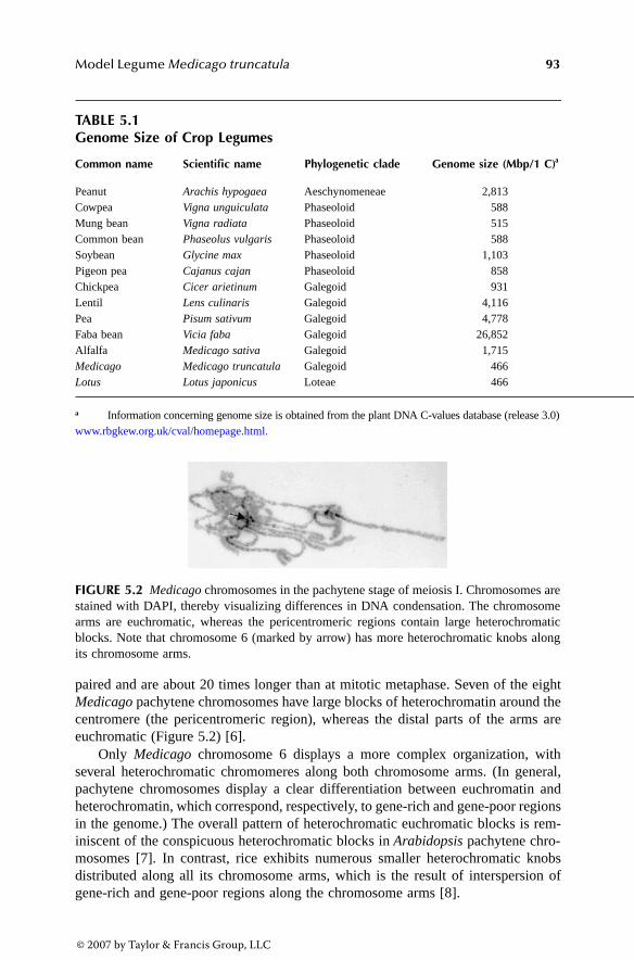
Model Legume Medicago truncatula 93
paired and are about 20 times longer than at mitotic metaphase. Seven of the eightMedicago pachytene chromosomes have large blocks of heterochromatin around thecentromere (the pericentromeric region), whereas the distal parts of the arms areeuchromatic (Figure 5.2) [6].
Only Medicago chromosome 6 displays a more complex organization, withseveral heterochromatic chromomeres along both chromosome arms. (In general,pachytene chromosomes display a clear differentiation between euchromatin andheterochromatin, which correspond, respectively, to gene-rich and gene-poor regionsin the genome.) The overall pattern of heterochromatic euchromatic blocks is rem-iniscent of the conspicuous heterochromatic blocks in Arabidopsis pachytene chro-mosomes [7]. In contrast, rice exhibits numerous smaller heterochromatic knobsdistributed along all its chromosome arms, which is the result of interspersion ofgene-rich and gene-poor regions along the chromosome arms [8].
TABLE 5.1Genome Size of Crop Legumes
Common name Scientific name Phylogenetic clade Genome size (Mbp/1 C)a
Peanut Arachis hypogaea Aeschynomeneae 2,813Cowpea Vigna unguiculata Phaseoloid 588Mung bean Vigna radiata Phaseoloid 515Common bean Phaseolus vulgaris Phaseoloid 588Soybean Glycine max Phaseoloid 1,103Pigeon pea Cajanus cajan Phaseoloid 858Chickpea Cicer arietinum Galegoid 931Lentil Lens culinaris Galegoid 4,116Pea Pisum sativum Galegoid 4,778Faba bean Vicia faba Galegoid 26,852Alfalfa Medicago sativa Galegoid 1,715Medicago Medicago truncatula Galegoid 466Lotus Lotus japonicus Loteae 466
a Information concerning genome size is obtained from the plant DNA C-values database (release 3.0)www.rbgkew.org.uk/cval/homepage.html.
FIGURE 5.2 Medicago chromosomes in the pachytene stage of meiosis I. Chromosomes arestained with DAPI, thereby visualizing differences in DNA condensation. The chromosomearms are euchromatic, whereas the pericentromeric regions contain large heterochromaticblocks. Note that chromosome 6 (marked by arrow) has more heterochromatic knobs alongits chromosome arms.
3063_book.fm Page 93 Thursday, August 24, 2006 1:23 PM
© 2007 by Taylor & Francis Group, LLC

94 Model Plants and Crop Improvement
Medicago has a relatively low gene density, based on the number of gene-basedmolecular markers that can be generated within a particular linkage group [9,10].To estimate the size of the gene-rich portion of the Medicago genome, two methodshave been used:
1. By determining the condensation degree at different positions of euchromatin,the amount of DNA in a euchromatic conformation can be extrapolated[6,11,12]. This has resulted in a euchromatin size estimation of 100 to 200 Mb.
2. In the second approach, the difference in genome size between the twoaccessions Jemalong A17 and R108-1 has been used. To this end the lengthof pachytene chromosomes and distribution of the hetero/euchromatinwere determined. The two genotypes do not differ in the length of euchro-matin present, but do in the size of the heterochromatic pericentromericblocks. Thus, the difference in genome size between both accessions iscontributed by the heterochromatic fraction. The size of the heterochro-matic blocks of Jemalong A17 has been estimated to be 320 Mb, leavingthe remaining 180 to 230 Mb as euchromatin [13].
Although both calculations are rather indirect, they support the hypothesis thatthe major part of the Medicago genome is composed of repeats within heterochro-matic regions. The current Medicago sequencing projects are focusing specificallyon the euchromatic part of the genome. The sequence of this region will be deter-mined in a BAC-by-BAC approach, enabling a full integration of the physical,genetic, and cytogenetic maps [14].
Functional genomic initiatives have resulted in the generation of >226,000expressed sequence tags (ESTs) originating from 35 different libraries. These ESTshave been assembled into 18,600 tentative consensus sequences (TCs) of more than1 EST and 18,200 singleton sequences (Release 8.0 of the TIGR Medicago GeneIndex, January 2005) [15–17]. Assuming that Medicago has a similar number ofgenes as Arabidopsis does, the vast majority of these are represented by at least oneEST (see also the following links: http://www.medicago.org/MtDB2, http://www.tigr.org/tigr-scripts/tgi/T_reports.cgi?species=medicago, and http://medicago.toulouse.inra.fr/Mt/EST). This data set has been used to construct cDNA-based microarrays[18], as well as oligonucleotide-based chips [19]. In addition, the EST collectionhas been mined to identify legume-specific genes.
To this end, the Medicago ESTs plus similar sets from soybean and Lotus werecompared to sequence data generated for non-legume plant species, resulting in theidentification of 2525 legume-specific EST contigs [20]. Among these are genesspecifically induced during Rhizobium symbiosis (so-called nodulin genes) [21] andgenes encoding for legume-specific seed storage proteins. However, for the vastmajority of genes, the function of the encoded proteins remains largely unknownand probably will need to be elucidated by other methods.
To unravel gene function, forward and reverse genetic tools have been appliedto Medicago. Reverse genetic approaches became possible with the development ofstable transgenic Medicago lines, although transformation is significantly less effi-cient and more time consuming than, for example, for Arabidopsis. Of several
3063_book.fm Page 94 Thursday, August 24, 2006 1:23 PM
© 2007 by Taylor & Francis Group, LLC
Model Legume Medicago truncatula 95
protocols that have been developed, protocols based on regeneration of transgeniccallus have been shown to be the most effective [22–26]. Lines with increasedregeneration efficiency have been selected. However, crosses between the best linein this respect—Medicago R108-1—and lines most widely used for genetic studies(selections from the cultivar Jemalong, e.g., A17 and J5) result in severe segregationdistortion, most likely due to genomic incompatibility [13]. These disadvantages ofMedicago R108-1 mean that it can be used only in applications that do not requireforward genetics.
Genome wide T-DNA and transposon tagging approaches have been initiatedfor Medicago. To effect the latter, the tobacco (Nicotiana tobaccum) retrotransposonelement Tnt1 was introduced into Medicago. The Tnt1 transposition is only activatedduring tissue culture, during which its copy number remains relatively low. Insertionsseem to occur preferentially in the gene-rich portion of the genome, generating genedisruptions. Currently, a collection of 8000 independent lines representing over150,000 Tnt1 insertions is being created by an international consortium.
In addition to stable transformation, efficient protocols have been developedbased on Agrobacterium rhizogenes-mediated root transformation [27,28]. In con-trast to stable lines, compound plants are generated in which a nontransformed shootcarries transgenic roots. The advantage of this system is mainly through a reductionin the time required for acquiring transgenic material; therefore, it is an attractivemethod to study gene function in roots. Compound plants can be obtained within 4to 6 weeks and can be nodulated by Rhizobium as well as be infected by othersymbionts (e.g., mycorrhizal fungi) or root pathogens. Roots generated with thissystem can be propagated in culture independently of the shoot due to the presenceof the root inducing locus (rol) genes of A. rhizogenes. The drawbacks of an A.rhizogenes-mediated root system are:
1. The introduced rol genes interfere with plant hormone balance (particu-larly in overproduction of cytokinin), making this system unsuitable forstudies of plant growth regulators.
2. The transformation remains transient because no transgenic offspring aregenerated.
3. Because the roots are primary transformed tissue, significant variation isobserved in expression levels of the introduced transgenes.
In addition, roots can be chimerical because root formation occurs from a group ratherthan from a single cell. Careful selection is therefore required, and methods basedon antibiotic resistance and/or fluorescent markers have been developed [27,28].
RNA interference (RNAi) has proven to be a powerful tool to unravel genefunction. RNAi can be triggered by generating transgenic lines that express RNAscapable of forming a double-stranded hairpin [29]. For Medicago (and legumes ingeneral), the generation of transgenic lines is time consuming, so RNAi has beenapplied in A. rhizogenes-mediated root transformation and shown to be functional[28,30,31]. However, systemic spreading of the silencing signal is limited in com-pound plants. It is transmitted very inefficiently from the transgenic root system tothe nontransgenic shoot, and transport from transgenic roots to nontransgenic roots
3063_book.fm Page 95 Thursday, August 24, 2006 1:23 PM
© 2007 by Taylor & Francis Group, LLC

96 Model Plants and Crop Improvement
does not occur. Furthermore, because the roots are primary transformed tissue,variation in silencing efficiency tends to occur.
In addition to reverse genetics methods that require transformation, TILLING(“targeted induced local lesions in genomes”) has been applied in Medicago and inLotus [32; D. Cook, personal communication]. This technique combines ethyl meth-anesulfonate (EMS)-induced mutagenesis with the ability to detect base pair changesby heteroduplex analysis by PAGE of Cel1 digests [33,34] and generates a range ofmutant alleles. The identification of mutant alleles obtained by TILLING is expectedin the near future.
5.3 THE MEDICAGO GENOME AS REFERENCEFOR LEGUME CROP SPECIES
To implement knowledge generated from model species for legume crop improve-ment, comparative genetic maps between model species and economical importantcrop species need to be constructed. To develop cross-species genetic markers, anintron-targeted marker strategy has been shown to represent a powerful approach[35]. For this purpose, PCR primer pairs are designed to anneal in exon regionsconserved between Medicago and Lotus, soybean, or Arabidopsis, designed toamplify across introns. Because introns are significantly more frequently polymor-phic than coding regions, these markers are often informative and have been usedto integrate the genetic maps of various legume species [10,35].
Comparative genetic maps have been created between Medicago and the galegoidcrop species alfalfa (Medicago sativa) and pea, between model species Medicagoand Lotus, and between Medicago and the phaseoloid crop species soybean, cowpea,and common bean [10,35]. A comparison of Medicago and alfalfa based on 68sequence-characterized genetic markers indicates that the two Medicago genomesare highly similar [10]. Pea is more differentiated from Medicago than is alfalfa. Ithas a significantly larger genome and contains one chromosome less than bothMedicago species (seven vs. eight). Despite these differences, a high degree ofsynteny exists between pea and the Medicago species, and only two major chromo-somal translocations have been identified.
Medicago linkage group two is distributed over linkage groups III and VI of pea(Figure 5.3) [35,36]. Similar comparisons have been conducted between Medicagoand Lotus. The Lotus genome is about the same size as that of Medicago and isdivided over six chromosomes [14]. Both species show a significant level of mac-rosynteny, but several chromosome arm translocations have occurred during evolu-tion. This synteny is also reflected on the microsynteny scale because the order andorientation of genes have been shown to be conserved significantly [35]. Loteae isa sister group of the Galegoideae, and therefore Lotus is more closely related toMedicago than to the phaseoloid species.
Alignment of the genetic maps of phaseoloid species and Medicago showssignificant distortions due to translocations, duplications, and loss of synteny. How-ever, the gene repertoire in orthologous regions in Medicago and soybean stilldisplays a degree of conserved gene order [35,37]. Based on these studies, it has
3063_book.fm Page 96 Thursday, August 24, 2006 1:23 PM
© 2007 by Taylor & Francis Group, LLC

Model Legume Medicago truncatula 97
been concluded that the Medicago genome can be effectively used as a referencefor the galegoid species. For the phaseoloid species, Medicago and Lotus can beused, but both show a reduced level of macrosynteny because of translocations.
5.3.1 RHIZOBIUM–LEGUME SYMBIOSIS
The interaction between Rhizobium and legumes has been the subject of many studiesover the last century. These have culminated in a detailed description of the stepsinvolved, including infection and primordium formation; a good understanding ofthe physiology of nitrogen fixation, including the requirement for low oxygen ten-sion; the mechanism of nitrogen assimilation; and the identification of plant genesthat are regulated during the interaction [38,39].
When rhizobia have colonized the root surface of their legume host, they inducemorphological changes in root hairs, a phenomenon referred to as root hair deformation.
FIGURE 5.3 A consensus map of Medicago, alfalfa (Medicago sativa), and pea (Pisumsativum). The three species are highly syntenic. Note that Medicago (and alfalfa) linkagegroup two is represented by pea linkage groups III and VI. (The figure is based on Choi, H.K.et al., Genetics, 166, 1463, 2004; Choi, H.K. et al., Proc. Natl. Acad. Sci. USA, 101, 15289,2004; and Kalo, P. et al., Mol. Genet. Genomics, 272, 235, 2004.)
Nod factor Mycorrhization
LysM-RKs Myc-receptor
DMI1 DMI2
Ca2+ influx Ca2+ spiking
DMI3
NSP1 NSP2
Gene expression
3063_book.fm Page 97 Thursday, August 24, 2006 1:23 PM
© 2007 by Taylor & Francis Group, LLC

98 Model Plants and Crop Improvement
In some root hairs, rhizobia induce curling as a result of which the bacteria becomeentrapped in the pocket of the curl. At this point, the plant cell wall is locallymodified, the plasma membrane invaginates, and new plant material is deposited. Inthis way, a tube-like structure, the infection thread, is formed containing the bacteria.The infection thread grows toward the base of the root hair cell and subsequentlyto the nodule primordium that has simultaneously been formed in the cortex of theroot. There, the infection thread ramifies and bacteria are released into primordialcells. The released bacteria—now called bacteroids—remain surrounded by a mem-brane of plant origin in a similar fashion as in mitochondria and chloroplasts.Subsequently, bacteroids differentiate and begin to fix nitrogen. Likewise, the noduleprimordium differentiates into a mature nodule.
Strikingly, genetic analyses have shown that part of the Rhizobium symbiosishas evolved from the much older mycorrhizal symbiosis [40–42]. In contrast toRhizobium symbiosis, which is restricted mainly to legume species, the majority ofhigher plants have the ability to interact with arbuscular endomycorrhiza, producinga symbiotic association between the plant root and fungi belonging to the order ofGlomales. These fungi grow toward the inner cortical cells of the root, where theydifferentiate into highly branched structures, the arbuscules. Because the fungusretains hyphae outside the plant it provides the host better access to nutrients suchas phosphate.
Genetic approaches in Rhizobium have been very successful in identifying thebacterial genes crucial to establishing a proper symbiosis. Many of these genesencode proteins required for production and secretion of a bacterial signaling mol-ecule, the Nod factors. Nod factors for which the structure has been elucidated allshare a β-1,4-linked N-acyl-D-glucosamine backbone of three to six subunits. Thenonreducing end of this glucosamine backbone is substituted with a fatty acid ofvariable structure. Furthermore, at both ends of the backbone, substitutions may bepresent that include acetyl, sulfuryl, fucosyl, mannosyl, or arabinosyl groups (forreview, see, for example, Spaink [43]).
Purified Nod factors applied in the nano- to picomolar range are able to inducedevelopmental processes needed for root nodule formation. These responses areprovoked at spatially separated sites—specifically, the epidermis, cortical cells, andpericycle. In some species (e.g., alfalfa), Nod factors can even trigger the formationof a complete nodule (lacking bacteria). Just as genetic approaches have led toidentification of genes crucial for signaling organogenesis in Arabidopsis, mutantsimpaired in nodulation have proven to be instrumental in identification of plant genesessential for Rhizobium–legume symbiosis and to a better understanding of theunderlying mechanisms.
Although several mutants that fail to establish proper symbiosis have beenidentified for some years in pea and soybean, the identity of the mutated genes hasonly become possible following establishment of mutagenesis programs in Medicagoand Lotus. Emerging mutants can be grouped roughly into three classes: class I,non-nodulators (nod-); class II, hypernodulators (nod+++); and class III, nodulatorswith impaired fixation (nod+, fix-) (Table 5.2). Here, we will focus on the currentknowledge obtained after identification of the genes corresponding to class I and IImutants in Medicago and Lotus.
3063_book.fm Page 98 Thursday, August 24, 2006 1:23 PM
© 2007 by Taylor & Francis Group, LLC

Model Legume Medicago truncatula 99
5.3.1.1 Non-Nodulators Enclose Nod Factor Signaling Genes
Various genetic approaches have been used to unravel the Nod factor-signalingcascade. Selection has been made for Lotus and Medicago mutants that are impaired
TABLE 5.2Medicago Symbiotic Loci Identified by Forward Genetics
Symbol Gene name Protein Ref.
Class IBIT Branching infection threads Not cloned 72DMI1 Does not make infections 1 Putative cation channel 54DMI2 Does not make infections 2 LRR-RK 53DMI3 Does not make infections 3 Ca2+ calmodulin kinase 55HCL Hair curling Not cloned 73LIN Lumpy infections Not cloned 74NIP Numerous infections with polyphenolics Not cloned 75NFP Nod factor perception Likely ortholog of Lotus NFR5
encoding a LysM-RK44
NSP1 Nodulation signaling pathway 1 Transcription factor 82NSP2 Nodulation signaling pathway 2 Transcription factor 83PDL Poodle Not cloned 76, 77RIT Root hairs infection threads trichomes Not cloned 72SYM1 Symbiosis 1 Not cloned 78SYM16 Symbiosis 16 Not cloned 79
Class II SKL Sickle Not cloned 80SUNN Supernumeric nodules Likely homolog of Lotus HAR
encoding an LRR-RK12
Class IIIDNF1 Defective in nitrogen fixation 1 Not cloned 72DNF2 Defective in nitrogen fixation 2 Not cloned 72DNF3 Defective in nitrogen fixation 3 Not cloned 72DNF4 Defective in nitrogen fixation 4 Not cloned 72DNF5 Defective in nitrogen fixation 5 Not cloned 72DNF6 Defective in nitrogen fixation 6 Not cloned 72DNF7 Defective in nitrogen fixation 7 Not cloned 72SYM6 Symbiosis 6 Not cloned 81SYM17 Symbiosis 17 Not cloned 79SYM18 Symbiosis 18 Not cloned 79SYM19 Symbiosis 19 Not cloned 79SYM20 Symbiosis 20 Not cloned 79SYM21 Symbiosis 21 Not cloned 79
Notes: Mutants are classified into three groups: non-nodulators (class I), supernodulators (class II), andnodulators with impaired fixation (class III). Detailed information concerning phenotypes and referencescan be obtained from the Nodulation Mutant Database (NodMutDB) http://nodmutdb.vbi.vt.edu. Allelismstudies between dnf and sym class III mutants (fix-) have not been reported.
3063_book.fm Page 99 Thursday, August 24, 2006 1:23 PM
© 2007 by Taylor & Francis Group, LLC

100 Model Plants and Crop Improvement
in the first visible Nod factor-provoked responses (Figure 5.4). In a second approach,naturally occurring variation within legumes has been exploited and characterizedat the molecular level using Medicago in a synteny-based approach. Both methodswill be discussed.
Among the non-nodulating mutants involving six loci in Medicago, only a feware disturbed in most of the Nod factor-induced responses. Cloning these genes inMedicago and Lotus has shown that a similar set of genes was mutated, suggestingthat mutation screens for impairment in Nod factor signaling are close to saturation.
A knock-out mutation in a Nod factor receptor is expected to be impaired in allNod factor-induced responses. In Medicago, only one mutant displaying such aphenotype has been identified—namely, nfp; in Lotus, two such loci have been found:NFR1 and NFR5 [44–46]. The other mutants disturbed in Nod factor signalingidentified in either species show at least some Nod factor-induced responses in roothair morphology. In Medicago there are three dmi and two nsp mutants (Table 5.2).Strikingly, all these genes (including NFP) are essential for Nod factor-inducedchanges in gene expression, as demonstrated by microarray analysis [47].
NFR1 and NFR5 of Lotus have been cloned and encode distinct LysM domain-containing receptor kinases (LysM-RK) that, based on their sequence, are localizedin the plasma membrane [45,46]. The putative extracellular regions of both proteinscontain LysM domains, which previously have been found in proteins binding pep-tidoglycans [48]. Thus, these LysM-RKs are good candidates as Nod factor bindersbecause they contain an N-acetyl glucosamine backbone. Because the extracellular
FIGURE 5.4 (Color Figure 5.4 follows p. 144.) Nod factor signaling cascade as identifiedby forward genetics. Nod factors are perceived by LysM receptor kinases. These activate atleast two downstream signaling pathways: one depending on the DMI proteins and a DMIindependent pathway for which no specific genes have yet been identified. Both pathwayscan be discriminated based on distinct Ca2+ signals, external Ca2+ influx, and perinuclearCa2+ spiking. The DMI pathway is shared mycorrhizal-secreted signal perceived by a hypo-thetical plant Myc receptor. For mycorrhizal-based signaling, a DMI independent pathwayis predicted.
8
81
1
2 2
3
34
4
5
5
66
7
7
I
II
III
IV
V
VI
VII
pea
alfalfa Medicago
3063_book.fm Page 100 Thursday, August 24, 2006 1:23 PM
© 2007 by Taylor & Francis Group, LLC
Model Legume Medicago truncatula 101
domains of NFR1 and NFR5 are markedly different from one another, it seemsunlikely that the two receptors function independently and recognize the same Nodfactor structure.
More probably, a heterodimer involving both receptors is needed for Nod factorperception, a model consistent with the loss of Nod factor responses in both mutants[45,46]. This is further supported by the atypical serine/threonine kinase in NFR5,which lacks an activation loop that generally regulates kinase activity. Therefore,activation of NFR5-type kinases probably occurs upon phosphorylation by an inter-acting kinase. Medicago NFP is the most likely ortholog of Lotus LjNFR5.
The DMI and NSP genes are positioned downstream of LysM-RK(s) becausegrowth responses in root hairs can be triggered upon Nod factor application (Figure5.4) [49,50]. In this respect, the dmi mutant root hairs mainly show root hair swellingand only very limited tip growth upon Nod factor perception; nsp1 and nsp2 mutantsshow root hair responses more similar to that of the wild-type. The functioning ofthe three DMI proteins can be dissected based on Nod factor-induced oscillation ofCa2+ concentration. This Ca2+ spiking occurs in the perinuclear region of epidermalcells and is induced within a few minutes [51]. Of the dmi mutants, only dmi3 showsthis response (Figure 5.4) [52].
Although the function of this intracellular Ca2+ signaling is not yet well under-stood, pharmacological studies have shown that that it is essential for Nod factor-induced gene expression. DMI1 has a low global similarity to ligand-gated cationchannels, whereas DMI2 is a receptor kinase in which the putative extracellularregion contains three LRR domains [53,54]. DMI3 encodes a Ca2+ calmodulin-dependent protein kinase (CCaMK) and is assumed to respond to this Ca2+ signal[55,56]. Genes orthologous to Medicago DMI1 and DMI2 have been identified inLotus [57,58]. Downstream of the DMI-module, NSP1 and NSP2 are functional(Figure 5.4) [49,50].
Because all these genes are essential for Nod factor-induced gene expression, itis probable that either of the NSP genes encodes a transcription factor that is activatedupon Nod factor signaling. Indeed, cloning of NSP1 and NSP2 shows that thesegenes encode transcription factors belonging to the GRAS family of plant-specifictranscription factors [82,83].
The three DMI genes are essential not only for Rhizobium-induced nodulation,but also for mycorrhizal symbiosis, whereas the putative Nod factor receptors arenot [49,50]. Because the mycorrhizal and Rhizobium symbioses only in part triggerexpression of a common set of genes [59,60], signaling cascades in addition to theDMI module must exist; these (together with the DMI genes) will be essential totrigger mycorrhizal or Rhizobium Nod factor specific transcriptional changes. In thecase of Nod factor signaling, the existence of a second such pathway is supportedby the different Ca2+ response. Apart from the intracellular Ca2+ spiking that occursupon Nod factor perception in a DMI1- and DMI2-dependent fashion, an influx ofextracellular Ca2+ occurs in a DMI-independent manner [61].
Ca2+ influx is one of the first responses in the root epidermis upon Nod factorsignaling and is essential for at least some induced transcriptional changes [62].Similarly to the Ca2+ influx following Nod factor signaling, it is possible that myc-orrhizal fungi also trigger an alternative DMI-independent signaling cascade that,
3063_book.fm Page 101 Thursday, August 24, 2006 1:23 PM
© 2007 by Taylor & Francis Group, LLC

102 Model Plants and Crop Improvement
together with the DMI module, is required for mycorrhizal specific transcriptionalchanges (Figure 5.4).
As mentioned earlier, a second strategy based on naturally occurring variationwas used to clone a putative Nod factor receptor specifically involved in bacterialinfection. In pea accessions originating from the Middle East, the SYM2 locus wasidentified as specifically involved in controlling infection thread formation in relationto Nod factor structure. In the pea accession Afghanistan, this locus inhibits infectionby Rhizobium leguminosarum bv. viciae strains that are unable to add an additionalacetate at the reducing end of the sugar backbone of the Nod factor [63,64]. Thus,the activity of SYM2 depends on the structure of Nod factors secreted by the infectingrhizobia and is part of the mechanism that controls bacterial entry.
Likewise, structure–function relationship studies derived from bacterial geneticshave shown that, in Medicago, bacterial infection is more dependent on Nod factorstructure than are other responses (e.g., nodule primordium formation) [65]. To clonea Nod factor receptor essential for Rhizobium infection, a synteny-based approachwas used to characterize the pea SYM2 orthologous region in Medicago [31,66].This region contains several genes encoding LysM-RKs that have been named LYK.Knockdown of LYK3 and LYK4 by means of A. rhizogenes-mediated RNAi hasshown that both genes are essential for Rhizobium infection in a Nod factor structure-dependent manner [31]. Medicago LYK3 and LYK4 are homologous to Lotus NFR1,although their loss-of-function phenotypes are strikingly different.
In addition to LYK3 and LYK4 in Medicago, the gene NODULE INCEPTION(NIN) is also essential for infection initiation. NIN encodes a protein with homologyto transcription factors and was originally cloned in Lotus by insertion of an ACtransposable element. The insertion mutant showed excessive root hair deformationand curling, but no infection [67]. In contrast to the genes described previously, NINis induced upon Nod factor perception and therefore cannot be primarily involvedin the Nod factor signaling pathway.
5.3.1.2 Supernodulators
In the 1980s, soybean and pea mutants were identified that formed nodules inde-pendent of the nitrogen status of the soil. Apart from this characteristic, these mutantplants formed more nodules than did wild-type plants grown in the absence ofnitrogen. As a result, these mutants have been termed supernodulators. The discoveryof supernodulators supports the notions that nodule formation is suppressed by thepresence of nitrogen and that legumes have an autoregulatory mechanism that con-trols the number of nodules formed. Strikingly, supernodulation does not lead to anyincrease in plant biomass, indicating that nodule formation and nitrogen fixation ofthe hosted Rhizobium are established at the expense of the plant.
The recent cloning of the orthologous genes HAR1 in Lotus, SYM29 in pea, andNARK in soybean has allowed for the characterization of an important key regulatorof the autoregulatory mechanism [68–70]. The signature of the protein encodedpredicts that it functions as a receptor kinase because of the presence of extracellularleucine-rich repeats (LRRs) and an intracellular serine/threonine kinase domain.Based on its homology to Arabidopsis CLAVATA1 and the observation that the
3063_book.fm Page 102 Thursday, August 24, 2006 1:23 PM
© 2007 by Taylor & Francis Group, LLC

Model Legume Medicago truncatula 103
number of lateral roots in the Lotus har1 mutant is affected after inoculation withRhizobium, it has been suggested that this protein has a role in mediating controlover root organ formation, including lateral roots as well as nodules.
5.4 PERSPECTIVE
The capacity to form root nodules in which bacteria convert nitrogen into ammoniaallows the seed of a legume crop to accumulate high protein content. This is a uniquefeature among plants. To keep pace with the growing world demand for protein-richfood, Rhizobium symbiosis must be exploited to its limits. This need has led todevelopment of the legume model species Lotus and Medicago, of which the latteris presented in this review in more detail. In addition, nonsymbiotic traits, liketolerance to biotic and abiotic stress, seed quality, plant architecture, and floweringbehavior, are important for legume crop performance. Therefore, these traits areimportant targets for legume breeders.
Series of gene-based genetic markers that can be used across legume specieshave been developed and used to integrate genetic linkage maps. The possibility toexploit the synteny between model and crop legumes will certainly be instrumentalin future legume breeding. This has been recognized; several well-funded programscovering many aspects of legume biology have been implemented worldwide. Thiswill ultimately lead to full integration of the Medicago and Lotus genome sequencewith high-density genetic maps of crop legumes.
Development of model systems has speeded up identification of genes encodingkey players in Rhizobium symbiosis. Initially, these studies have been focused ongenes involved in Nod factor signaling because perception of this bacterial signalmolecule by the plant forms the main trigger for root nodule development. Strikingly,the number of genes essential for Nod factor signaling that can be identified genet-ically is low and is conserved among all legume species studied so far. Furthermore,mutations in these genes mainly affect symbiosis, suggesting that they do not playimportant roles in other plant processes. It can be expected that in the near futurethe link between the Nod factor signaling network and common cellular processeswill be elucidated and thereby will make available knowledge of how plants rewireprocesses for organ formation.
Homologs of genes affected in non-nodulating mutants such as Nod factor recep-tor LysM-RKs or DMI genes are present also in non-legumes. This indicates that theprocesses needed for nodule formation could be, in part, already present in non-legume species and suggests that Rhizobium has recruited genes involved in generalplant development for nodule formation. The observation that dmi mutants are alsoimpaired in the interaction with arbuscular mycorrhiza has led to the hypothesis thatnodule formation evolved from this more widespread symbiosis [40–42]. This sug-gests that non-legumes may lack a spectrum of the genes that enable the establishmentof a symbiotic relationship with rhizobia. The longstanding dream of nodulated rice—or other important non-legume crop species—might be feasible.
However, transferring the capacity of nitrogen fixation to non-legume specieswill be a difficult task that will depend largely on how much processes needed fornodule development are present in non-legumes and can be geared to each other
3063_book.fm Page 103 Thursday, August 24, 2006 1:23 PM
© 2007 by Taylor & Francis Group, LLC

104 Model Plants and Crop Improvement
in the way in which the process occurs in legumes. This implies that we need toknow how many additional components must be transferred to non-legumes [71].An in-depth analysis of the 2500+ legume-specific genes identified could provideclues in this direction [20]. A major challenge is to uncover the extent to whichnon-legume homologs of genes such as LysM-RKs and DMI are able to complementcorresponding legume mutants. Such studies would give insight into the extent towhich the functioning of Nod factor signaling genes are unique to legumes. Theavailability of model species will definitely prove their value in finding answers tothese exciting questions.
REFERENCES
1. Graham, P.H. and Vance, C.P., Legumes: importance and constraints to greater use,Plant Physiol., 131, 872, 2003.
2. Dixon, R.A. and Sumner, L.W., Legume natural products: understanding and manip-ulating complex pathways for human and animal health, Plant Physiol., 131, 878, 2003.
3. Desbrosses, G.G., Kopka, J., and Udvardi, M.K., Lotus japonicus metabolic profiling.Development of gas chromatography-mass spectrometry resources for the study ofplant–microbe interactions, Plant Physiol., 137, 1302, 2005.
4. Lodwig, E.M. et al., Amino-acid cycling drives nitrogen fixation in the legume–Rhizo-bium symbiosis, Nature, 422, 722, 2003.
5. Bataillon, T., Ronfort, J., and Prosperi, J.M., Natural genetic variation in Medicagotrunctula: understanding the structure of genetic variation for the marker assistedbuilding of a core collection of inbred lines, in 1st International Conference on LegumeGenetics and Genomics: Translation to Crop Improvement (St. Paul, MN), 2002.
6. Kulikova, O. et al., Integration of the FISH pachytene and genetic maps of Medicagotruncatula, Plant J., 27, 49, 2001.
7. Koornneef, M., Fransz, P., and de Jong, H., Cytogenetic tools for Arabidopsisthaliana, Chromosome Res., 11, 183, 2003.
8. Cheng, Z. et al., Resolution of fluorescence in situ hybridization mapping on ricemitotic prometaphase chromosomes, meiotic pachytene chromosomes and extendedDNA fibers, Chromosome Res., 10, 379, 2002.
9. Zhu, H. et al., Phylogeny and genomic organization of the TIR and non-TIR NBS-LRR resistance gene family in Medicago truncatula, Mol. Plant Microbe Interact.,15, 529, 2002.
10. Choi, H.K. et al., A sequence-based genetic map of Medicago truncatula and com-parison of marker collinearity with M. sativa, Genetics, 166, 1463, 2004.
11. Ane, J.M. et al., Genetic and cytogenetic mapping of DMI1, DMI2, and DMI3 genesof Medicago truncatula involved in Nod factor transduction, nodulation, and mycor-rhization, Mol. Plant Microbe Interact., 15, 1108, 2002.
12. Schnabel, E. et al., An integrated physical, genetic and cytogenetic map around thesunn locus of Medicago truncatula, Genome, 46, 665, 2003.
13. Kulikova, O. et al., Satellite repeats in the functional centromere and pericentromericheterochromatin of Medicago truncatula, Chromosoma, 113, 276, 2004.
14. Young, N.D. et al., Sequencing the genespaces of Medicago truncatula and Lotusjaponicus, Plant Physiol., 137, 1174, 2005.
3063_book.fm Page 104 Thursday, August 24, 2006 1:23 PM
© 2007 by Taylor & Francis Group, LLC

Model Legume Medicago truncatula 105
15. Quackenbush, J. et al., The TIGR gene indices: analysis of gene transcript sequencesin highly sampled eukaryotic species, Nucleic Acids Res., 29, 159, 2001.
16. Journet, E.P. et al., Exploring root symbiotic programs in the model legume Medicagotruncatula using EST analysis, Nucleic Acids Res., 30, 5579, 2002.
17. Lamblin, A.F. et al., MtDB: a database for personalized data mining of the modellegume Medicago truncatula transcriptome, Nucleic Acids Res., 31, 196, 2003.
18. Kuster, H. et al., Construction and validation of cDNA-based Mt6k-RIT macro- andmicroarrays to explore root endosymbioses in the model legume Medicago truncatula,J. Biotechnol., 108, 95, 2004.
19. Barnett, M.J. et al., A dual-genome symbiosis chip for coordinate study of signalexchange and development in a prokaryote–host interaction, Proc. Natl. Acad. Sci.USA, 101, 16636, 2004.
20. Graham, M.A. et al., Computational identification and characterization of novel genesfrom legumes, Plant Physiol., 35, 1179, 2004.
21. Van Kammen, A., Suggested nomenclature for plant genes involved in nodulationand symbiosis, Plant Mol. Biol. Rep., 2, 43, 1984.
22. Trieu, A.T. et al., Transformation of Medicago truncatula via infiltration of seedlingsor flowering plants with Agrobacterium, Plant J., 22, 531, 2000.
23. Hoffmann, B. et al., A new Medicago truncatula line with superior in vitro regener-ation, transformation, and symbiotic properties isolated through cell culture selection,Mol. Plant Microbe Interact., 10, 307, 1997.
24. Kamaté, K. et al., Transformation of floral organs with GFP in Medicago truncatula,Plant Cell Rep., 19, 647, 2000.
25. Chabaud, M., De Carvalho-Niebel, F., and Barker, D.G., Efficient transformation ofMedicago truncatula cv. Jemalong using the hypervirulent Agrobacterium tumefa-ciens strain AGL1, Plant Cell Rep., 22, 46, 2003.
26. Zhou, X., Chandrasekharan, M.B., and Hall, T.C., High rooting frequency and func-tional analysis of GUS and GFP expression in transgenic Medicago truncatula A17,New Phytol., 162, 813, 2004.
27. Boisson-Dernier, A. et al., Agrobacterium rhizogenes-transformed roots of Medicagotruncatula for the study of nitrogen-fixing and endomycorrhizal symbiotic associa-tions, Mol. Plant Microbe Interact., 6, 695, 2001.
28. Limpens, E. et al., RNA interference in Agrobacterium rhizogenes-transformed rootsof Arabidopsis and Medicago truncatula, J. Exp. Bot., 55, 983, 2004.
29. Chuang, C. and Meyerowitz, M., Specific and heritable genetic interference by double-stranded RNA in Arabidopsis thaliana, Proc. Natl. Acad. Sci. USA, 97, 4985, 2000.
30. Kumagai, H. and Kouchi, H., Gene silencing by expression of hairpin RNA in Lotusjaponicus roots and root nodules, Mol. Plant Microbe Interact., 16, 663, 2003.
31. Limpens, E. et al., LysM domain receptor kinases regulating rhizobial Nod factor-induced infection, Science, 302, 630, 2003.
32. Perry, J.A. et al., A TILLING reverse genetics tool and a Web-accessible collectionof mutants of the legume Lotus japonicus, Plant Physiol., 131, 866, 2003.
33. McCallum, C.M. et al., Targeted screening for induced mutations, Nat. Biotechnol.,18, 455, 2000.
34. Till, B.J. et al., Mismatch cleavage by single-strand specific nucleases, Nucleic AcidsRes., 11, 2632, 2004.
35. Choi, H.K. et al., Estimating genome conservation between crop and model legumespecies, Proc. Natl. Acad. Sci. USA, 101, 15289, 2004.
3063_book.fm Page 105 Thursday, August 24, 2006 1:23 PM
© 2007 by Taylor & Francis Group, LLC

106 Model Plants and Crop Improvement
36. Kalo, P. et al., Comparative mapping between Medicago sativa and Pisum sativum,Mol. Genet. Genomics, 272, 235, 2004.
37. Yan, H.H. et al., Comparative physical mapping reveals features of microsyntenybetween Glycine max, Medicago truncatula, and Arabidopsis thaliana, Genome, 47,141, 2004.
38. Stougaard, J., Regulators and regulation of legume root nodule development, PlantPhysiol., 124, 532, 2000.
39. Hirsch, A.M., Lum, M.R., and Downie, J.A., What makes the rhizobia–legume sym-biosis so special? Plant Physiol., 127, 1484, 2001.
40. Gianinazzi-Pearson, V., Plant cell responses to arbuscular mycorrhizal fungi: gettingto the roots of the symbiosis, Plant Cell, 8, 1871, 1996.
41. Albrecht, C., Geurts, R., and Bisseling, T., Legume nodulation and mycorrhizaeformation; two extremes in host specificity meet, EMBO J., 18, 281, 1999.
42. Gualtieri, G. and Bisseling, T., The evolution of nodulation, Plant Mol. Biol., 42,181, 2000.
43. Spaink, H.P., Regulation of plant morphogenesis by lipo-chitin oligosaccharides, Crit.Rev. Plant Sci., 15, 559, 1996.
44. Ben Amor, B. et al., The NFP locus of Medicago truncatula controls an early stepof Nod factor signal transduction upstream of a rapid calcium flux and root hairdeformation, Plant J., 34, 495, 2003.
45. Madsen, E.B. et al., A receptor kinase gene of the LysM type is involved in legumeperception of rhizobial signals, Nature, 425, 637, 2003.
46. Radutoiu, S. et al., Plant recognition of symbiotic bacteria requires two LysM recep-tor-like kinases, Nature, 425, 585, 2003.
47. Mitra, R.M., Shaw, S.L., and Long, S.R., Six non-nodulating plant mutants defectivefor Nod factor-induced transcriptional changes associated with the legume–rhizobiasymbiosis, Proc. Natl. Acad. Sci. USA, 101, 10217, 2004.
48. Bateman, A. and Bycroft, M., The structure of a LysM domain from E. coli membrane-bound lytic murein transglycosylase D (MltD), J. Mol. Biol., 299, 1113, 2000.
49. Catoira, R. et al., Four genes of Medicago truncatula controlling components of aNod factor transduction pathway, Plant Cell, 12, 1647, 2000.
50. Oldroyd, G.E. and Long, S.R., Identification and characterization of nodulation-signaling pathway 2, a gene of Medicago truncatula involved in Nod actor signaling,Plant Physiol., 131, 1027, 2003.
51. Ehrhardt, D.W., Wais, R., and Long, S.R., Calcium spiking in plant root hairs respond-ing to Rhizobium nodulation signals, Cell, 85, 673, 1996.
52. Wais, R.J. et al., Genetic analysis of calcium spiking responses in nodulation mutantsof Medicago truncatula, Proc. Natl. Acad. Sci. USA, 97, 13407, 2000.
53. Endre, G. et al., A receptor kinase gene regulating symbiotic nodule development,Nature, 417, 962, 2002.
54. Ane, J.M. et al., Medicago truncatula DMI1 required for bacterial and fungal sym-bioses in legumes, Science, 303, 1364, 2004.
55. Levy, J. et al., A putative Ca2+ and calmodulin-dependent protein kinase required forbacterial and fungal symbioses, Science, 303, 1361, 2004.
56. Mitra, R.M. et al., A Ca2+/calmodulin-dependent protein kinase required for symbioticnodule development: gene identification by transcript-based cloning, Proc. Natl.Acad. Sci. USA, 101, 4701, 2004.
57. Stracke, S. et al., A plant receptor-like kinase required for both bacterial and fungalsymbiosis, Nature, 417, 959, 2002.
3063_book.fm Page 106 Thursday, August 24, 2006 1:23 PM
© 2007 by Taylor & Francis Group, LLC

Model Legume Medicago truncatula 107
58. Imaizumi-Anraku, H. et al., Plastid proteins crucial for symbiotic fungal and bacterialentry into plant roots, Nature, 433, 527, 2005.
59. Manthey, K. et al., Transcriptome profiling in root nodules and arbuscular mycorrhizaidentifies a collection of novel genes induced during Medicago truncatula root endo-symbioses, Mol. Plant Microbe Interact., 17, 1063, 2004.
60. Weidmann, S. et al., Fungal elicitation of signal transduction-related plant genesprecedes mycorrhiza establishment and requires the dmi3 gene in Medicago trunca-tula, Mol. Plant Microbe Interact., 17, 1385, 2004.
61. Shaw, S.L. and Long, S.R., Nod factor elicits two separable calcium responses inMedicago truncatula root hair cells, Plant Physiol., 131, 976, 2003.
62. Pingret, J.L., Journet, E.P., and Barker, D.G., Rhizobium nod factor signaling. Evi-dence for a G protein-mediated transduction mechanism, Plant Cell, 10, 659, 2003.
63. Firmin, J.L. et al., Resistance to nodulation of cv. Afghanistan peas is overcome bynodX, which mediates an O-acetylation of the Rhizobium leguminosarum lipo-oli-gosaccharide nodulation factor, Mol. Microbiol., 10, 351, 1993.
64. Geurts, R. et al., Sym2 of pea is involved in a nodulation factor-perception mechanismthat controls the infection process in the epidermis, Plant Physiol., 115, 351, 1997.
65. Ardourel, M. et al., Rhizobium meliloti lipo-oligosaccharide nodulation factors: dif-ferent structural requirements for bacterial entry into target root hair cells and induc-tion of plant symbiotic developmental responses, Plant Cell, 6, 1357, 1994.
66. Gualtieri, G. et al., Microsynteny between pea and Medicago truncatula in the SYM2region, Plant Mol. Biol., 50, 225, 2002.
67. Schauser, L. et al., A plant regulator controlling development of symbiotic rootnodules, Nature, 402, 191, 1999.
68. Krusell, L. et al., Shoot control of root development and nodulation is mediated bya receptor-like kinase, Nature, 420, 422, 2002.
69. Nishimura, R. et al., HAR1 mediates systemic regulation of symbiotic organ devel-opment, Nature, 420, 426, 2002.
70. Searle, I.R. et al., Long-distance signaling in nodulation directed by a CLAVATA1-like receptor kinase, Science, 299, 109, 2003.
71. Parniske, M., Intracellular accommodation of microbes by plants: a common devel-opmental program for symbiosis and disease? Curr. Opin. Plant Biol., 3, 320, 2000.
72. Mitra, R.M. and Long, S.R., Plant and bacterial symbiotic mutants define threetranscriptionally distinct stages in the development of the Medicago truncat-ula/Sinorhizobium meliloti symbiosis, Plant Physiol., 134, 595, 2004.
73. Catoira, R. et al., The HCL gene of Medicago truncatula controls Rhizobium-inducedroot hair curling, Development, 128, 1507, 2001.
74. Kuppusamy, K.T. et al., LIN, a Medicago truncatula gene required for nodule differ-entiation and persistence of rhizobial infections, Plant Physiol., 136, 3682, 2004.
75. Veereshlingam, H. et al., Nip, a symbiotic Medicago truncatula mutant that formsroot nodules with aberrant infection threads and plant defense-like response, PlantPhysiol., 136, 3692, 2004.
76. Cohn, J.R. et al., Differential regulation of a family of apyrase genes from Medicagotruncatula, Plant Physiol., 125, 2104, 2001.
77. Navarro-Gochicoa, M.T. et al., Characterization of four lectin-like receptor kinasesexpressed in roots of Medicago truncatula. Structure, location, regulation of expres-sion, and potential role in the symbiosis with Sinorhizobium meliloti, Plant Physiol.,133, 1893, 2003.
78. Benaben, V. et al., TE7, An inefficient symbiotic mutant of Medicago truncatulaGaertn. cv. Jemalong, Plant Physiol., 107, 53, 1995.
3063_book.fm Page 107 Thursday, August 24, 2006 1:23 PM
© 2007 by Taylor & Francis Group, LLC

108 Model Plants and Crop Improvement
79. Morandi, D. et al., Characterisation of new symbiotic Medicago truncatula (Gaertn.)mutants, and phenotypic or genotypic complementary information on previouslydescribed mutants, Mycorrhiza, in press.
80. Penmetsa, R.V. and Cook, D.R., A legume ethylene-insensitive mutant hyperinfectedby its rhizobial symbiont, Science, 275, 527, 1997.
81. Tirichine, L., de Billy, F., and Huguet, T., Mtsym6, a gene conditioning Sinorhizobiumstrain-specific nitrogen fixation in Medicago truncatula, Plant Physiol., 123, 845, 2000.
82. Kalo, P. et al., Nodulation signaling in legumes requires NSP2, a member of theGRAS family of transcriptional regulators, Science, 308, 1786, 2005.
83. Smit, P. et al., NSP1 of the GRAS protein family is essential for rhizobial Nod factor-induced transcription, Science, 308, 1789, 2005.
3063_book.fm Page 108 Thursday, August 24, 2006 1:23 PM
© 2007 by Taylor & Francis Group, LLC

109
6 Brachypodium distachyon: A New Model Systemfor Structural and Functional Analysisof Grass Genomes
David F. Garvin
CONTENTS
6.1 Introduction .................................................................................................. 1096.2 Evolutionary Relationships between Brachypodium distachyon
and Other Grass Crops................................................................................. 1106.3 Structural Features of Brachypodium distachyon Genome......................... 1126.4 Genome Composition .................................................................................. 1136.5 Gene Content and Organization versus Those of Other Grasses ............... 1146.6 Functional Genomics ................................................................................... 1156.7 Transformation and Mutagenesis................................................................. 1166.8 Genetic Stocks and Genetic Variation ......................................................... 1186.9 Growth Characteristics................................................................................. 1196.10 Conclusions .................................................................................................. 121References.............................................................................................................. 121
6.1 INTRODUCTION
The releases of the genome sequences of the dicot model plant Arabidopsis thaliana(Arabidopsis) and the monocot crop rice represent a revolutionary advance in plantbiology. These sequences have provided a potent source of information for genetics,evolution, development, and all other fields of plant biology at a resolution that waspreviously unavailable. The choice of these species as templates for genomicsequencing was based on their small genomes. Furthermore, Arabidopsis has longserved as a tool for exploration of basic plant processes, and rice is a member ofthe most important plant family (Poaceae) from the standpoint of human subsistence.
3063_book.fm Page 109 Thursday, August 24, 2006 1:23 PM
© 2007 by Taylor & Francis Group, LLC

110 Model Plants and Crop Improvement
The success of the Arabidopsis and rice genome sequencing projects, coupled withadvances in strategies and technologies, has led to efforts to sequence all or partsof the genomes of many other plant species representing diverse plant families.
Given the rapid progress in genome sequencing of crops, one question thatemerges is whether future investment in new model species to serve as surrogatesfor crops is necessary. The answer to this question is largely dependent upon thecrop or crops that would be represented by such a new model species. In this regard,a cogent argument in favor of a model crop species can be made for the cool-seasongrass crops. Although it is clear that the rice genome sequence has been profitablyexploited by researchers studying wheat, barley, and rye, it is equally clear that therice genome sequence has limitations as a template for use in isolating genes fromthe cool-season cereals. This is largely due to the nearly 50 million years ofevolution that separate rice from the cool-season grasses [1], which is reflected inprofound differences in phenology, morphology, physiology, and biotic and abioticstress susceptibility and tolerance. These differences impose limitations on the useof rice as a model for exploring the gene structure–function interface in the cool-season grasses.
An alternative model species that, like rice, is diploid, possesses a small genome,and is transformable, but more closely related to the cool-season grasses, can beexpected to find a place in the laboratories of scientists interested in cool-seasongrass crop improvement. The species Brachypodium distachyon possesses all ofthese characteristics and is emerging as a new model species. The intent of thisreview is to provide an introduction to evolutionary, genetic, genomic, and mor-phological attributes of B. distachyon that make it such an attractive new modelplant system.
6.2 EVOLUTIONARY RELATIONSHIPS BETWEEN BRACHYPODIUM DISTACHYON AND OTHER GRASS CROPS
The grass family Poaceae (Gramineae) comprises approximately 10,000 species,including most of the world’s most important crops such as wheat (Triticum spp.),rice (Oryza sativa), maize (Zea mays), barley (Hordeum vulgare), sorghum (Sorghumbicolor), and many forage and turf species [2]. The members of this large plantfamily are distributed around the world in highly diverse environments. Resultsdeveloped from the joint efforts of a consortium of grass systematicists support thepresence of eight subfamilies within the Poaceae [3]. The largest of these subfamiliesis the Pooideae which, depending upon the authority, includes about 10 tribes andover 3000 species. This one subfamily includes most of the important cool-seasongrain crops, forage grasses, and turfgrasses [4]. The genus Brachypodium belongsto the subfamily Pooideae as well and is considered distinct enough from othermembers of the Pooideae that it has been placed within its own tribe, the Brachy-poideae [2].
Presumably because of the economic importance of the subfamily, many studieshave been undertaken to resolve evolutionary relationships between the tribes in the
3063_book.fm Page 110 Thursday, August 24, 2006 1:23 PM
© 2007 by Taylor & Francis Group, LLC

Brachypodium distachyon 111
Pooideae, with some including Brachypodium in analyses [5]. A recent analysisbased on chloroplast gene (ndhF) sequence data indicated that the tribe Brachy-poideae is sister to the evolutionary lineage that subsequently gave rise to the tribesTriticeae, Aveneae, Bromeae, and Poeae [6]. As such, of the thousands of speciescomprising the entire family Poaceae (Gramineae), those of the genus Brachypodiumare considered to be the closest ancestors to these four “core pooid” tribes [3,7] thatencompass most of the major cool-season grasses. Thus, simply based on evolution-ary considerations, we can predict that members of the genus Brachypodium willpossess a genome structure and composition more similar to the “core pooids” thanother genera in the entire grass family. This is one feature that makes Brachypodiumattractive as a genus from which to select a possible model species for the cool-season grasses.
The systematics of Brachypodium has been examined a number of times in anattempt to resolve the evolutionary relationships among the species in this genus.The genus has not been unambiguously delineated, but it is likely to include 12 ormore species [8]. Several members of the genus are endemic to Europe, and one ormore different species are found in other parts of the world including regions ofAfrica, Central and South America, and Asia [2]. Two unusual characteristics of thegenus are that the different species of Brachypodium exhibit significant variation inbase chromosome number, including 5, 7, 8, or 9, and that polyploid series areencountered within species in the genus, so ecotypes in a given Brachypodiumspecies may range from diploid (2n = 2x) to octaploid (2n = 8x) in some cases [9].
The species within the genus Brachypodium are perennial in nature with oneexception: the annual species B. distachyon, often referred to as purple false brome.This species appears to have diverged from the other perennial Brachypodium speciesearly in the evolution of the genus [10]. The evolutionary divergence of B. distachyonfrom other Brachypodium species is bolstered by results of experimental hybridiza-tions between B. distachyon and other Brachypodium species. Although these crossescan produce F1 progeny, they are sterile and exhibit abnormal meiosis. This contrastswith results from crosses among perennial European Brachypodium species, whichgenerally produce fertile F1 hybrids [11].
With one exception, B. distachyon is the only species in the genus with a basechromosome number of 5. The observed ploidy levels reported in B. distachyonecotypes include diploidy (2n = 2x = 10), tetraploidy (2n = 4x = 20), and hexaploidy(2n = 6x = 30) [9]. This species is considered to be endemic to regions surroundingthe Mediterranean Sea; however, it has spread to other regions of the world, whereit can be a weed. In terms of its breeding system, B. distachyon is characterized ashighly self-compatible with a floral morphology that encourages inbreeding [11].
An annual habit, self-compatibility, and availability of diploid ecotypes make B.distachyon the most desirable of the Brachypodium species from the standpoint ofbroad potential as a model species for functional and structural genomics research.The first publication that touted B. distachyon as a possible model species was thatof Bablak et al. [12], in which it was noted that various parallels between B.distachyon and Arabidopsis might support research into the former species as a newmodel system.
3063_book.fm Page 111 Thursday, August 24, 2006 1:23 PM
© 2007 by Taylor & Francis Group, LLC

112 Model Plants and Crop Improvement
6.3 STRUCTURAL FEATURES OF BRACHYPODIUM DISTACHYON GENOME
In diploid ecotypes of B. distachyon, the 5 chromosomes, while small, can bedistinguished morphologically, and the major ribosomal gene tandem repeat loci (5Sand 45S) have been localized to their respective chromosomes by in situ hybridiza-tion [13]. Several publications have reported size estimates of the B. distachyongenome. Using flow cytometry, Draper et al. [13] measured the genome size ofdiploid ecotypes of the species and reported a c-value of 0.21 pg that they consideredequal to 172 megabase pairs—a size approximately the same as what they obtainedfor Arabidopsis. However, this value is in conflict with other reports.
Shi et al. [14] report that diploid B. distachyon has a c-value of 0.3 pg, whichis similar to recent results of Bennett and Leitch [15], who reported a c-value of0.36 pg. These latter values are similar to the average c-value of 0.39 pg obtainedfrom three separate analyses by flow cytometry for a reference inbred diploid lineof B. distachyon that we have developed from the accession PI 185134 and isdeposited in the United States Department of Agriculture’s National Plant Germ-plasm System (NPGS) [16]. It is unclear why the estimates of Draper et al. [13] arenot congruent with other reports because they used various plant species as calibra-tion standards in that report. At this time, it is recommended that the c-value estimateof 0.36 provided by Bennett and Leitch [15] be accepted as the best estimate for thegenome size of diploid B. distachyon. This indicates that the genome of diploidecotypes of this species is approximately 6.5% the size of barley and approximately2% the size of the hexaploid bread wheat genome.
Thus, the entire genome of diploid B. distachyon is slightly smaller than an“average” chromosome arm in barley or wheat, is smaller than the genome of rice,and is only approximately twice as large as the genome of Arabidopsis. Theextremely small genome of diploid B. distachyon is another important feature desiredin a model plant species.
The derivation of polyploidy in B. distachyon is an interesting topic worthdiscussing. Given that the base chromosome number of the diploid ecotypes is 5and that this doubled or tripled in the tetraploid and hexaploid ecotypes, respectively,one might assume that this is the signature of autopolyploidy rather than allopoly-ploidy. This assumption would be supported by the fact that no other Europeanspecies of Brachypodium with a base chromosome number of 5 are known to exist.Thus, this rules out the likelihood that the polyploids are in fact allopolyploidsderived from interspecific hybridization between diploids with a base number of 5,followed by chromosome doubling in a manner analogous to the evolution of tetra-ploid durum wheat (T. turgidum) and hexaploid bread wheat [17].
However, the observation that 15 bivalents form during meiosis in hexaploidecotypes of B. distachyon [18] is contrary to expectations in an autopolyploid, inwhich multivalent pairing for some of the chromosomes is expected between someproportion of the chromosomes. Furthermore, our recent observations that a largeset of putative hexaploid ecotypes of B. distachyon from the NPGS appear to exhibitc-values approximately two and not three times the size of diploid ecotypes [16] arenot concordant with an autopolyploid origin of the polyploid ecotypes.
3063_book.fm Page 112 Thursday, August 24, 2006 1:23 PM
© 2007 by Taylor & Francis Group, LLC

Brachypodium distachyon 113
A recent publication [18] reported on similarities and differences in chromosomekaryotypes, location, and abundance of ribosomal DNA loci, and results of genomicin situ hybridization between ecotypes representing the three ploidy levels. Fromthese results, the authors postulated that the tetraploid ecotypes may actually repre-sent a distinct diploid subtype (perhaps even a different species) with a base chro-mosome number of 10 and with chromosomes more similar to those of the relatedperennial species B. sylvaticum (slender false brome). They also proposed that thehexaploid ecotypes may represent the product of hybridization and chromosomedoubling between diploid B. distachyon and a plant similar to the putative tetraploidthat they proposed as a distinct subtype. As such, the polyploid ecotypes of B.distachyon may well have value as a system for exploring the impact of polyploidyon genome organization—a topic of great interest to wheat researchers because ofthe polyploid derivation of that crop.
6.4 GENOME COMPOSITION
The emergence of B. distachyon as a new model grass species is quite recent andthus information on the genome composition of the species is scarce. Nonetheless,data gleaned from available studies help to shed some light on this topic. In anearly molecular phylogenetic analysis of the genus Brachypodium, Catalan et al.[8] produced a HindIII library of genomic DNA from a diploid B. distachyonecotype to obtain probes for RFLP analysis. Hybridization of labeled genomic DNAagainst a series of randomly selected clones from this library revealed that less than12% of them gave a strong hybridization signal indicative of the presence of highcopy genomic sequences. Hybridization of several of these high copy clones toBrachypodium species DNA and DNA from other diverse grass species revealedthat they hybridized exclusively to Brachypodium DNA. Furthermore, the hybrid-ization patterns obtained from the different high copy probes suggested the presenceof tandemly repeated DNA as well as interspersed repetitive DNA [8]. In contrast,a set of “low copy” B. distachyon clones hybridized to DNA of most other grassspecies included in the hybridizations, suggesting conservation of such sequencesacross the Poaceae.
These results are consistent with those obtained in B. sylvaticum (slender falsebrome). Because of its small genome size, Moore et al. [19] proposed that thismember of the genus Brachypodium may be a useful model genome for studyingthe organization of grass genomes in general. Moore et al. [20] found that 100% ofthe short (<2 kb) clones from B. sylvaticum HpaII and HindIII genomic librariesthat were tested cross-hybridized to wheat genomic DNA, and 60% of these clonesgave simple hybridization patterns on wheat genomic DNA. In contrast, a set ofcomparably sized single copy rice PstI genomic clones hybridized to wheat just 64%of the time, with less than 60% of the hybridizing sequences revealing simplehybridization patterns on wheat genomic DNA.
Furthermore, their results indicated that the genome of B. sylvaticum is lessmethylated than those of wheat and barley. Also, a presumed centromere repeatsequence (CCS1) isolated from B. sylvaticum was found to hybridize to differentgrass species with different intensities; hybridization was strongest in wheat and rye,
3063_book.fm Page 113 Thursday, August 24, 2006 1:23 PM
© 2007 by Taylor & Francis Group, LLC

114 Model Plants and Crop Improvement
while maize and rice showed hybridization intensities less than 10% of that seen inthe former species [21].
Taken as a whole, the results obtained from these analyses of the genomes of B.distachyon and its close relative B. sylvaticum support the notion that the smallgenome of B. distachyon harbors significantly less repetitive DNA than large genomegrasses such as wheat and that the genomes of rice and B. distachyon are similar incomplexity. However, the B. distachyon genome exhibits higher sequence similarityto the Triticeae than does rice for low-copy clones and at least one repetitive sequence.
6.5 GENE CONTENT AND ORGANIZATION VERSUS THOSE OF OTHER GRASSES
One of the principal interests in B. distachyon is its potential as a compact grassgenome model that can be exploited to isolate genes from larger grass crop genomesbecause of conservation of genome organization and gene order. Various researchgroups are exploring this topic, though results have yet to be published.
In one study, the structure and abundance of C-repeat binding factor genes(CBFs), which encode transcription factors implicated in conditioning cold toler-ance, were examined in B. distachyon with the purpose of comparison to theorganization of the CBF gene family on chromosome 5H of barley. In barley,approximately ten CBF genes are present in a tightly linked gene cluster on chro-mosome 5H coincident with QTLs for cold tolerance, and they are estimated to bepresent at a density of less than one gene per 15 kb [22]. In contrast, seven CBFgenes in a diploid B. distachyon line derived from NPGS accession PI 185134,which include presumed orthologs and paralogs of many sequenced barley 5H CBFgenes based on phylogenetic analysis, have been localized to just two lambda phageclones spanning 33 kb.
Thus, the CBF gene density in the region of the B. distachyon genome presumedto be syntenic to the CBF cluster on barley chromosome 5H is at least three timeshigher [16]. These results also are in agreement with the expectation that significantcompression of coding sequences takes place in the small genome of B. distachyonbecause of a reduced amount of repetitive DNA, but not a large scale loss of codingsequences relative to its large genome cool-season relatives.
The phylogenetic analysis of barley and B. distachyon CBF genes mentionedearlier not only provides interesting insights into the evolution of the CBF genefamily in the grasses, but also offers evidence that orthologs of genes of agronomicinterest in small grain cereal crops such as barley are present in the B. distachyongenome. This contention is further bolstered by the observation that a presumedortholog of the Q gene, which played an important role in wheat domesticationbecause it confers the free-threshing character, was identified in B. sylvaticum [23].Finally, the genes A1, X1, X2, and Sh2 have been used to examine the evolutionarydynamics of genome size and organization in many grass crops spanning differentsubfamilies [24–26]. To date, the presence of orthologs of the first three of these hasbeen identified in a diploid inbred B. distachyon line derived from the NPGS acces-sion PI 185134 (unpublished data). Efforts are currently under way to identify the
3063_book.fm Page 114 Thursday, August 24, 2006 1:23 PM
© 2007 by Taylor & Francis Group, LLC

Brachypodium distachyon 115
B. distachyon ortholog of Sh2 so that the organization of these genes can be comparedto that in other grasses.
The limited information available on comparative gene content and genomeorganization between B. distachyon and other grasses is supplemented with resultsobtained from its sister species B. sylvaticum. Foote et al. [27] reported constructionand characterization of a bacterial artificial chromosome (BAC) library of B. sylvat-icum and then used this library to explore synteny relationships among rice, wheat,and B. sylvaticum. They screened the BAC library with 48 probes derived fromdifferent grass species, and 36 were found to hybridize to one or more BAC clones.Of these, 33 were postulated to exhibit the same gene order found in rice chromosome9 and/or the Triticeae group 5 chromosomes. The largest B. sylvaticum BAC contigspanned 367 kb, and 9 of 11 genes/probes examined on this contig were present asexpected in the syntenic region of Triticeae group 5 chromosomes. Furthermore,they sequenced a 163-kb B. sylvaticum BAC clone from this library that revealed aminimum of 17 hypothetical genes. This corresponds to a gene density exceedingone gene per 10 kb of genome sequence.
Thus, results from these studies suggest that we should have confidence thatorthologs of genes of broad interest to crop productivity will be present in B.distachyon. Furthermore, if we accept that the genome of B. distachyon will behighly similar in gene content and order to that of B. sylvaticum—aside from changesassociated with major structural rearrangements, we can also be confident that B.distachyon has a genome amenable to service as a surrogate genome to accelerategene discovery efforts in wheat, barley, and other large genome grasses.
6.6 FUNCTIONAL GENOMICS
The utility of B. distachyon to serve as a model grass genome is not limited toserving simply as a static “nucleotide roadmap” to help researchers navigate to genesof interest in grass genomes less tractable to physical analysis. Because B. distachyonis expected to have essentially the same ensemble of genes found in crops withinthe subfamily Pooideae, it may also be a potent resource for accelerating investiga-tions of the role of particular genes in biological processes—for instance, diversedevelopmental or biochemical pathways and responses to biotic or abiotic stresses—through functional genomics methods.
Initial steps have been taken in this direction. Comparative functional analysisof molecular responses of B. distachyon to Magnaporthe grisea, the fungal pathogencausing the disease rice blast, has been reported [28]. Microscopic analyses of hostresponses in the resistant ecotype strongly resembled those of rice. The responsesobserved included localized cell death likely due to oxidative stress and the rapidinduction of pathogenesis-related (PR) gene expression. Interestingly, ecotypes ofdiploid B. distachyon were found to exhibit differential resistance to the pathogen;this will permit the use of these ecotypes for comparative functional analysis ofresistance and susceptibility to this pathogen.
Although this example illustrates that B. distachyon can serve as a model forfunctional genomic analysis of this host–pathogen system in rice, it would be desir-able to extend similar opportunities to pathogens of relevance in the cool-season
3063_book.fm Page 115 Thursday, August 24, 2006 1:23 PM
© 2007 by Taylor & Francis Group, LLC

116 Model Plants and Crop Improvement
grass crops. For instance, the pathogens of greatest impact worldwide in wheat arethe rusts belonging to the genus Puccinia, including leaf rust (P. triticina), stem rust(P. graminis), and stripe or yellow rust (P. striiformis). Draper et al. [13] inoculatedB. distachyon with the leaf and stripe rust pathogens to determine its response tothese pathogens. In both instances, host responses ranged from flecking (presumablyindicative of a resistance response) to no visible response. In no case was an ecotypefound to exhibit a typical susceptible response, though stripe rust uredinia wereobserved within heavily necrotic regions in two ecotypes, indicating successfulcolonization by the pathogen. In the same publication, reaction of B. distachyonecotypes to Blumeria graminis, which causes powdery mildew, was investigated andparticular host resistance responses, including the formation of papillae, were found.
These initial studies suggest that B. distachyon holds promise for functionalgenomic analysis of disease resistance in cool-season crops. However, beyond thesestudies, there is a dearth of information reporting B. distachyon as a functionalgenomics tool. Nonetheless, as this species becomes adopted as a model system,this is expected to change rapidly.
6.7 TRANSFORMATION AND MUTAGENESIS
The ability to transform a plant is an important attribute for a model plant species.One important aspect of transformation competency is the regeneration of plantsfrom tissue culture. Bablak et al. [12] reported the first successful regeneration ofB. distachyon from tissue culture. Three diploid ecotypes were used in this study.A high frequency of callus induction was observed from cultured seeds, and signif-icant differences in callus induction were present between the ecotypes. The per-centage of embryogenic callus obtained with the appropriate culture medium wasas high as 60% in some experiments, and the number of regenerants per gram ofcallus was as high as 26. Subsequently, Draper et al. [13] confirmed the high levelof embryogenic callus that could be obtained and the high level of plant regenerationfor one of the same diploid ecotypes studied by Bablak et al. [12].
More recently, Christiansen et al. [29] found that it was possible to obtainembryogenic callus from immature embryos of diploid and tetraploid B. distachyonecotypes, but the quality of this callus varied considerably. Nonetheless, they foundthat the percentage of cultured embryos that produced embryogenic callus for thetwo diploid ecotypes they examined was approximately 50%, similar to the resultsof Draper et al. [13]. The frequency of plant regeneration from the callus of theirdiploids approached 100%, but was dependent on the age of the callus. Thus, itappears that B. distachyon is very amenable to regeneration from tissue culture.
Draper et al. [13] and Christiansen et al. [29] also undertook biolistic transfor-mation experiments on B. distachyon. Results of transformation for a single hexap-loid ecotype were reported by Draper et al. [13]. Callus of this hexaploid ecotypewas subjected to microprojectile bombardment with a plasmid carrying the hygro-mycin resistance gene and a glucuronidase (GUS) marker, resulting in recovery ofan average of seven independent hygromycin-resistant calli per gram of bombardedcallus. The authors reported a success rate in regenerating plants from the resistant
3063_book.fm Page 116 Thursday, August 24, 2006 1:23 PM
© 2007 by Taylor & Francis Group, LLC

Brachypodium distachyon 117
calli of approximately 70%. These plants were subsequently found to exhibit GUSactivity. However, transformation of diploid ecotypes was not reported.
Similarly, Christiansen et al. [29] undertook biolistic transformation of diploidand tetraploid ecotypes. In this study, dramatic differences in transformation effi-ciency between the two diploid ecotypes included were observed. Staining with GUSto examine transient transformation revealed that one diploid ecotype exhibited noapparent GUS-stained sectors in calli after microprojectile bombardment, and theother diploid exhibited an average of nearly 2000 GUS-positive sectors per bom-bardment experiment. This latter result was roughly similar to those obtained forthe two tetraploids included in the experiments. Plant regeneration efficiencies (per-cent of bombarded calli that produced a transgenic plant) from these experimentswere estimated at 5% for the diploid ecotype that exhibited GUS staining of calliand approximately 4% for the two tetraploids. However, individual experimentsyielded efficiencies of between 9 and 14% depending upon the ecotype.
An alternative method for plant transformation is through the use of Agrobac-terium tumafaciens, which has the advantage of producing lower copy and morestable genome integration events than particle bombardment [30]. Vogel et al. [31]studied Agrobacterium-mediated transformation efficiency of B. distachyon linesderived from accessions deposited in the NPGS and found a wide range of regen-eration and transformation efficiencies among lines, which included diploid andpolyploid ecotypes. For the three diploids evaluated (derived from the NPGS acces-sions PI 185133, PI 185134, and PI 254867), the regeneration efficiency ranged from4 to nearly 11%. However, as reported for microjectile-mediated transformation byChristiansen et al. [29], significant differences in transformation efficiency betweendiploid lines using Agrobacterium were found.
For two of the diploid B. distachyon lines, no transformed plants were generated,while the other diploid line derived from PI 254867 exhibited an average plantregeneration frequency of over 3% across different experiments. In contrast, theregeneration frequency for polyploid lines ranged from 0 to 15%, though it shouldbe noted that significantly more polyploids were examined in the experiments. Thus,it is clear that B. distachyon is transformable by two different and common methodsand that certain diploid ecotypes can be transformed by one or the other method.Presumably, in the future B. distachyon transformation can be optimized by buildingupon existing information provided by these earlier studies.
Mutagenesis provides a strategy to introduce new molecular variation into agenome. In doing so, it generates novel variants of genes that can be used fordissecting the molecular basis of traits of interest. As such, mutagenesis is animportant technique in the repertoire of a model species. In Brachypodium, littlemutagenesis research has been conducted to date. Draper et al. [13] have subjectedB. distachyon to gamma-irradiation, but no mutation frequencies were reported andno descriptions of the mutants obtained were provided.
The only other report of mutagenesis in B. distachyon is that of Engvild [32].In this report, three diploid accessions of B. distachyon were used to assess therelative efficacy of different common mutagens and mutagen concentrations toinduce mutations in B. distachyon. The mutagens included sodium azide and ethylmethanesulfonate (EMS). The sodium azide treatment appeared to be far more
3063_book.fm Page 117 Thursday, August 24, 2006 1:23 PM
© 2007 by Taylor & Francis Group, LLC

118 Model Plants and Crop Improvement
effective in inducing mutant phenotypes (based on the frequency of chlorophyllmutations) in the M1 generation than the EMS treatments. Furthermore, the relativeefficacy of the mutagen treatments in inducing mutations appeared to vary dependingupon the diploid accession used. Additional attempts to establish conditions thatpermit EMS or alternative mutagens to be used as effective mutagens in B. distachyonare strongly warranted so that mutant pools can be developed to identify mutationsof interest based on phenotype or to identify lesions in genes of interest usingmethods such as TILLING [33].
6.8 GENETIC STOCKS AND GENETIC VARIATION
For any model plant species it is desirable to have a large and diverse collection ofecotypes exhibiting variation in phenotype and variation at the molecular level. Broadvariation is particularly useful for successful development of segregating populationsto serve as the basis for genetic map construction and for positional cloning endeav-ors. Furthermore, a diverse collection of ecotypes may be useful for studying thegenetic and molecular basis of important crop traits by studying the same traitswithin the model system.
The NPGS has in its collection approximately 30 accessions of B. distachyoncollected from various regions of the world over the course of the last several decades.The collection list can be viewed at the NPGS Website (http://www.ars-grin.gov/npgs/ as of June 2005). We used flow cytometry to discriminate betweendifferent ploidy levels in lines derived from 27 NPGS accessions. Five of these lines(from PI 170218, PI 185133, PI 185134, PI 245730, and PI 254867) were identifiedas diploids [16,31]. These were collected in Turkey and Iraq. In addition, a collectionof over 50 accessions of B. distachyon is maintained by a company (BrachyomicsLtd.) established by the University of Wales [18]. The Brachyomics collectionincludes many lines collected in Europe and Asia, and elsewhere in the world.
It will be important to cross-reference collections of B. distachyon maintainedby different entities to provide the scientific community clear information on geo-graphic origin and other passport information associated with each ecotype. Todate, the number of unique diploid ecotypes of B. distachyon is limited to perhapsten. Therefore, it would be highly desirable to expand this number further byexamining B. distachyon germplasm collections in other countries for additionalnovel diploid ecotypes, as well making collecting trips to regions to which thisspecies is indigenous.
Although the number of diploid B. distachyon accessions/ecotypes is limited, itis promising to note that even among the five putative diploid accessions of B.distachyon deposited in the NPGS, significant phenotypic variation is evident. Forinstance, a cursory evaluation revealed clear differences in vernalization requirement,flowering date, plant height, pubescence, shattering, and seed size among theseaccessions (PI 170218, PI 185133, PI 185134, PI 245730, and PI 254867) [16,31].These morphological differences are likely a reflection of molecular variationthrough the genome of B. distachyon. Diploid B. distachyon held by Brachyomicscan be expected to contribute even more genetic diversity than that available in thesefew NPGS diploids.
3063_book.fm Page 118 Thursday, August 24, 2006 1:23 PM
© 2007 by Taylor & Francis Group, LLC

Brachypodium distachyon 119
Additionally, although little emphasis has been placed on the issue, it may beexpected that molecular variation within each of these accessions exists because theyvery likely are composed of seeds collected from multiple plants in a wild population.In fact, support for this prediction is found in the publication of Christiansen et al.[29], which alludes to evidence for molecular heterogeneity between plants fromthe same accession as revealed by AFLP analysis.
Because diploid B. distachyon is highly self-compatible and rarely exerts anthers,it is likely that individual plants are inbred and thus homozygous for most loci.However, this cannot be assumed for certain; genetic homogeneity with originalaccessions also cannot be assumed. Thus, reference inbred genetic stocks of B.distachyon will be a valuable resource to the Brachypodium research community.During the past 3 years, we have developed single seed descent-derived inbred linesfrom 27 different B. distachyon accessions obtained from the NPGS, including fiveputative diploids (PI 170218, PI 185133, PI 185134, PI 245730, and PI 254867)[16,31]. In most cases, we have developed two independent inbred sister lines fromeach accession to capture additional molecular variation that may reside in eachaccession, and in some of these inbred sister lines we have observed phenotypicdifferences, suggesting some success toward this end.
We are making these inbred lines freely available to interested parties. Theseinbred genetic stocks will be of benefit for various reasons, particularly at this earlystage of research with B. distachyon. For instance, the use of common inbred geneticstocks for research will reduce incongruous results that may be due to geneticvariation in source materials studied. Also, the use of common inbred lines willimprove the process of streamlining and integrating information emerging fromresearch undertaken by different laboratories.
The use of inbred lines of B. distachyon to develop segregating populations willresult in yet another highly useful resource. Despite the small size of the florets,populations appropriate for genetic mapping purposes will be developed unlessunanticipated incompatibilities exist between the inbred lines. The only known reportof a segregating population developed in B. distachyon is that of Routledge et al.[28], who developed F2 populations from crosses between two diploid ecotypes andused them to assess the inheritance of resistance to M. grisea.
Similarly, we have been working to develop segregating populations derivedfrom crosses among multiple diverse inbred lines. Crosses among all pairwise com-binations of four of our inbred diploid B. distachyon lines have been completed.Interestingly, we found that diploids appear to possess two anthers per floret, incontrast to reports in authoritative references that indicate that the species has threeanthers [2]. This character, therefore, could differentiate all diploids from polyploids(unpublished data). The putative hybrids we now have will serve as the basis ofrecombinant inbred line development, resulting in a diverse set of segregating pop-ulations that can be used for a range of purposes.
6.9 GROWTH CHARACTERISTICS
During our program to develop inbred B. distachyon lines, we noted that diploid B.distachyon plants are more petite than the polyploids during early vegetative growth.
3063_book.fm Page 119 Thursday, August 24, 2006 1:23 PM
© 2007 by Taylor & Francis Group, LLC

120 Model Plants and Crop Improvement
We initially relied upon vernalization ranging between 4 and 10 weeks to induceflowering in the diploid lines because the lines would not flower without such atreatment or took so long to flower that it was impractical to maintain the plants.These observations are similar to observations of Draper et al. [13], who found thatthe flowering of all but two of their diploid ecotypes could be synchronized with 6weeks of vernalization.
Because a vernalization requirement is not desirable if one wants to advancegenerations rapidly, we were interested in identifying conditions that would obviatethe need to vernalize diploids. We subsequently discovered that three of our inbreddiploid lines (derived from NPGS accessions PI 185133, PI 185134, and PI 254867)can be induced to flower rapidly without any vernalization simply by growing themunder long (>20 h) day-lengths [16,31]. Under such conditions, inflorescence emer-gence of the most rapidly maturing line (from PI 254867) begins approximately 3weeks after planting (Figure 6.1), and seeds will be mature 2 months after planting.The two other responsive lines flower approximately 1 and 2 weeks later than thisline, respectively. Our last two inbred diploids (from NPGS accessions PI 170218
FIGURE 6.1 (Color Figure 6.1 follows p. 144.) A diploid Brachypodium distachyon inbredline grown under long day-lengths can, depending upon the genotype, begin the transition toflowering within 3 weeks of planting. In this figure, a diploid genotype was grown under 20-h day-length restrictive conditions, as shown in the left panel, or under nutrient repleteconditions, as shown in the right panel. The plant in the left panel is 4 weeks old, and theplant on the right is 8 weeks old, with seeds maturing. Alternatively, by growing this inbreddiploid line under short-day conditions, it can be maintained in vegetative phase for longperiods of time. (Source: David Garvin, USDA-Agricultural Research Service.)
3063_book.fm Page 120 Thursday, August 24, 2006 1:23 PM
© 2007 by Taylor & Francis Group, LLC

Brachypodium distachyon 121
and PI 245730) appear to be unresponsive to long days and require significantvernalization periods to flower in a synchronized fashion.
The capacity to cycle B. distachyon rapidly is highly desired in a model species.At the same time, the capacity to retain these same genotypes in a vegetative statesimply by growing them under short days is a useful attribute if one is interested inusing the species as a model for forage or turf species. Furthermore, growing plantsunder conditions that encourage vegetative proliferation, followed by conditions thatinduce flowering, permits the recovery of large numbers of seeds from single plants.In our experience, we have been able to recover over 500 seeds from plants in thismanner. Seed size varies between lines, but some of the inbred diploids we havedeveloped produce seeds that have a mass approximately 15% that of wheat seeds,thus indicating a rather large endosperm that may also be beneficial for studiesfocusing on seed development.
6.10 CONCLUSIONS
The rice genome sequence is a resource that has great value for structural genomicsresearch in the grasses. However, B. distachyon is poised to become a potent modelsystem that complements rice because it also possesses attributes desired in a modelsystem for functional genomics, including a small physical size, a growth habit thatcan be regulated to control generation times, and, perhaps most importantly, a closeevolutionary affinity to the cool-season grain, turf, and forage species in the grasssubfamily Pooideae.
There is much to be learned about B. distachyon before it can be fully exploitedfor research purposes. If we use Arabidopsis as a template to help determine whereenergies need to be directed to establish B. distachyon firmly as a new model species,this would include assembling genetic resources (genetic stocks, segregating popu-lations), molecular resources (traditional genomic and cDNA libraries, as well aslarge insert libraries), obtaining sequence information (ESTs, genomic sequencedata), optimizing transformation systems, and developing mutagenesis protocols andmutant pools.
Although this review reveals deficiencies in this list that need to be addressed,more importantly it also highlights the significant strides forward that have beenmade in these areas in the short period of time that has passed since B. distachyonwas proposed as a potential model grass species. This review also highlights the factthat a significant amount of research now is under way in B. distachyon. Suchresearch efforts are a tacit acknowledgment by the grass research community that anew model species for the cool-season grasses is indeed desirable and thatB. distachyon possesses the attributes to serve in this capacity.
REFERENCES
1. Gaut, B., Evolutionary dynamics of grass genomes, New Phytol., 154, 15, 2002.2. Watson, L. and Dallwitz, M.J., The Grass Genera of the World, CAB International,
Wallingford, CT, 1992, chap. 1.
3063_book.fm Page 121 Thursday, August 24, 2006 1:23 PM
© 2007 by Taylor & Francis Group, LLC
122 Model Plants and Crop Improvement
3. Kellogg, E.A., Evolutionary history of the grasses, Plant Physiol., 125, 1198, 2001.4. Moser, L.E. and Hoveland, C.S., Cool-season grass overview, in Cool-Season Forage
Grasses, Moser, L.E., Buxton, D.R., and Casler, M.D., Eds., Agronomy Society ofAmerica, Madison, WI, 1996, chap. 1.
5. Kellogg, E.A., Relationships of cereal crops and other grasses, Proc. Natl. Acad. Sci.USA, 95, 2005, 1998.
6. Catalán, P., Kellogg, E.A., and Olmstead, R.G., Phylogeny of Poaceae subfamilyPooideae based on chloroplast ndhF gene sequences, Molec. Phylogenet. Evol., 8,150, 1997.
7. Davis, J.I. and Soreng, R.J., Phylogenetic structure in the grass family (Poaceae) asinferred from chloroplast DNA restriction site variation, Am. J. Bot., 80, 1444, 1993.
8. Catalán, P. et al., Molecular phylogeny of the grass genus Brachypodium P. Beauv.based on RFLP and RAPD analysis, Bot. J. Linnean Soc., 117, 263, 1995.
9. Robertson, I.H., Chromosome numbers in Brachypodium Beauv. (Gramineae), Genet-ica, 56, 55, 1981.
10. Catalán, P. and Olmstead, R.G., Phylogenetic reconstruction of the genus Brachypo-dium P. Beauv. (Poaceae) from combined sequences of chloroplast ndhF gene andnuclear ITS, Plant Syst. Evol., 220, 1, 2000.
11. Khan, M.A. and Stace, C.A., Breeding relationships in the genus Brachypodium(Poaceae: Pooideae), Nord. J. Bot., 19, 257, 1999.
12. Bablak, P. et al., Plant regeneration and micropropagation of Brachypodium dis-tachyon, Plant Cell, Tissue Organ Cult., 42, 97, 1995.
13. Draper, J. et al., Brachypodium distachyon. A new model system for functionalgenomics in grasses, Plant Physiol., 127, 1539, 2001.
14. Shi, Y., Draper, J., and Stace, C., Ribosomal DNA variation and its phylogeneticimplication in the genus Brachypodium (Poaceae), Plant Syst. Evol., 188, 125, 1993.
15. Bennett, M.D. and Leitch, I.J., Nuclear DNA amounts in angiosperms: progress,problems, and prospects, Ann. Bot., 95, 45, 2005.
16. Garvin, D.F. and Stockinger, E.J., Development of a U.S. Brachypodium distachyoncollection and the evaluation of microsynteny with cool-season grass crops, presentedat the Plant and Animal Genome XIII Conference, San Diego, http://www.intl-pag.org/13/abstracts/, Jan. 15–19, 2005, P373.
17. Feldman, M., Origin of cultivated wheat, in The World Wheat Book, Bonjean, A.P.and Angus, W.J., Eds., Lavoisier, Paris, 2001, chap. 1.
18. Hasterok, R., Draper, J., and Jenkins, G., Laying the cytotaxonomic foundations ofa new model grass, Brachypodium distachyon (L.) Beauv., Chromosome Res., 12,397, 2004.
19. Moore, G. et al., Molecular analysis of small grain cereal genomes: current statusand prospects, Bio/Technology, 11, 584, 1993.
20. Moore, G. et al., Key features of cereal genome organization as revealed by the useof cytosine methylation-sensitive restriction endonucleases, Genomics, 15, 472, 1993.
21. Aragon-Alcaide, L. et al., A cereal centromeric sequence, Chromosoma, 105, 261,1996.
22. Stockinger, E.J., Cheng, H., and Skinner, J.S., Structural organization of barley CBFgenes conicident with a QTL for cold hardiness, in Cold Hardiness in Plants, Chen,T.H.H, Uemura, M., and Fujikawa, S., Eds., CABI Publishing, Wallingford, CT,2006, 53.
23. Faris, J., personal communication, 2005.24. Chen, M. et al., Microcolinearity in sh2-homologous regions of the maize, rice, and
sorghum genomes, Proc. Natl. Acad. Sci. USA, 94, 3431, 1997.
3063_book.fm Page 122 Thursday, August 24, 2006 1:23 PM
© 2007 by Taylor & Francis Group, LLC

Brachypodium distachyon 123
25. Chen, M., SanMiguel, P., and Bennetzen, J.L., Sequence organization and conserva-tion in sh2/a1-homologous regions of sorghum and rice, Genetics, 148, 435, 1998.
26. Li, W. and Gill, B.S., The collinearity of the Sh2/A1 orthologous region in rice,sorghum and maize is interrupted and accompanied by genome expansion in theTriticeae, Genetics, 160, 1153, 2002.
27. Foote, T.N. et al., Construction and analysis of a BAC library in the grass Brachy-podium sylvaticum: its use as a tool to bridge the gap between rice and wheat inelucidating gene content, Funct. Integr. Genomics, 4, 26, 2004.
28. Routledge, A.P.M. et al., Magnaporthe grisea interactions with the model grassBrachypodium distachyon closely resemble those with rice (Oryza sativa), Molec.Plant Pathol., 5, 253, 2004.
29. Christiansen, P. et al., A rapid and efficient transformation protocol for the grassBrachypodium distachyon, Plant Cell Rep., 23, 751, 2005.
30. Dai, S. et al., Comparative analysis of transgenic rice plants obtained by Agrobacte-rium-mediated transformation and particle bombardment, Mol. Breeding, 7, 25, 2001.
31. Vogel, J. et al., Agrobacterium-mediated transformation and inbred line developmentin the model grass Brachypodium distachyon, Plant Cell Tissue Organ Cult., 84, 199,2006.
32. Engvild, K.C., Mutagenesis of the model grass Brachypodium distachyon withsodium azide, Risø-R Report 1510(EN), Risø National Laboratory, Roskilde, Den-mark, 2005, 8 pp.
33. McCallum, C.M. et al. Targeted screening for induced mutations, Nat. Biotech., 18,455, 2000.
3063_book.fm Page 123 Thursday, August 24, 2006 1:23 PM
© 2007 by Taylor & Francis Group, LLC

125
7 The Green Alga Chlamydomonas as a Tool to Study the Nitrate Assimilation Pathwayin Plants
Aurora Galván, Vicente Mariscal,David González-Ballester,and Emilio Fernández
CONTENTS
7.1 Chlamydomonas as a Model Organism....................................................... 1267.2 The Nitrate Assimilation Pathway and Its Key Points................................ 1267.3 The Usefulness of Chlorate-Resistant Mutants to Study
of Nitrate Assimilation................................................................................. 1317.4 Nitrate and Nitrite Reduction and Its Regulation ....................................... 1317.5 Nitrate and Nitrite Transporters................................................................... 133
7.5.1 Transport Systems............................................................................ 1347.5.2 Functionality of NRT1 Transporters................................................ 1357.5.3 Functionality of NRT2 Transporters................................................ 1362.5.4 NAR1 Transporters .......................................................................... 138
7.6 Ammonium Assimilation ............................................................................. 1397.7 Ammonium Transport Genes....................................................................... 1407.8 Nitrate Assimilation and Light/Circadian Rhythm...................................... 1437.9 Nitrate Assimilation and Redox Regulation................................................ 1437.10 Regulatory Genes in Nitrate Assimilation................................................... 1467.11 Conclusion.................................................................................................... 148Acknowledgments.................................................................................................. 149References.............................................................................................................. 149
3063_book.fm Page 125 Thursday, August 24, 2006 1:23 PM
© 2007 by Taylor & Francis Group, LLC
126 Model Plants and Crop Improvement
7.1 CHLAMYDOMONAS AS A MODEL ORGANISM
Model species are generally first selected because they allow novel insights intoprocesses that are poorly understood; they are then maintained and accepted becauseof their experimental amenability and usefulness in understanding further processes.Plants represent an extraordinarily diverse group of living beings at the molecular,genetic, biochemical, and physiological levels, and their great biodiversity reflectsthe evolution of complex genetic and biochemical networks [1].
Unicellular organisms lack the complexity and sophistication of higher plantsystems. However they remain useful as a tool to understand aspects of fundamentalplant biology at the cell level that have not yet been elucidated. In particular,conditions for efficient uptake, metabolism, and regulation of nutrient acquisition,as well as for accumulation of assimilate-derived products are relevant for determin-ing the efficiency of growth at whole plant and thus at crop levels. For this reason,many research groups have focused their attention on understanding key processesin model plant systems.
It is becoming clear that present model systems are not as representative as hadbeen hoped, so, for a proper understanding, each system needs to be studied in itself.At the same time, it seems that, to dissect the major processes at the molecular level,it is necessary to work in a system simple enough to control the influence ofextraneous factors. Interestingly, the molecular dissection of metabolic steps (e.g.,nitrate assimilation) has shown that the differences between unicellular and landplants can be smaller than feared at the level of complexity of gene and proteinfamilies and with respect to cellular strategies adopted to achieve particular endresults. An ideal model system needs a set of basic physiological, biochemical, andmolecular techniques, along with developed resources in genetics, genomics, andtransgenesis. These will allow for the ready genetic dissection of mutant phenotype,together with a straightforward correlation of phenotype to genotype. The Chlamy-domonas system fulfils most of these requirements, as summarized in Table 7.1.
The use of Chlamydomonas as an amenable biological system was first describedin Harris’s book [3]. The advantages of Chlamydomonas as a model unicellularsystem were further presented by Rochaix et al. [9] and were recently compared tothose of Arabidopsis [10]. Decoding the nuclear and chloroplast genomes of Chlamy-domonas and developing molecular tools (see Table 7.1) have strengthened theposition of this organism for study of important plant cell processes such as photo-synthesis, chloroplast inheritance and biology, mitochondrial genetics, nutrient defi-ciency, carbon metabolism, and nitrate assimilation.
7.2 THE NITRATE ASSIMILATION PATHWAY AND ITS KEY POINTS
Ammonium and nitrate are the primary inorganic nitrogen sources for plant growth.Though many species use ammonium in preference to nitrate, the majority of plants,algae, and microorganisms is able to use nitrate efficiently because ammonium isabout 10 to 1000 times less abundant than nitrate in natural soils, except in a fewecosystems such as coniferous forests [11].
3063_book.fm Page 126 Thursday, August 24, 2006 1:23 PM
© 2007 by Taylor & Francis Group, LLC

Chlamydomonas as a Tool to Study Nitrate Assimilation Pathways 127
In addition, nitrate provides an efficient signal for modulating metabolic pro-cesses and plant architecture [12]. The assimilation of ammonium ion has a lowerenergy cost than that of nitrate [13] and many genes involved in nitrate assimilationare strongly repressed in the presence of ammonium [14,15]. However, many plants,especially herbaceous crop plants, utilize nitrate or a combination of both ions asthe preferred nitrogen form for growth [16,17]. Thus, about 75% of the nitrogen inproteins consumed by man is ultimately derived from nitrate assimilated by plants.
The fundamental role of the nitrate assimilation pathway in plant nutrition hasbeen the object of intensive studies for many years and has been reviewed by manyauthors [12,18–31]. This chapter represents an update of information about nitrateassimilation in Chlamydomonas as a source of information for the equivalent processin higher plants.
The basic steps for nitrate utilization in a single photosynthetic eukaryotic cellare as follow (Figure 7.1):
1. The entry of nitrate into the cell by means of specific transport systems2. A first reduction step from nitrate to nitrite, which occurs in the cytosol
and is catalyzed by the nitrate reductase (NR) enzyme3. Nitrite transport to the chloroplast4. A second reduction step that occurs in the chloroplast, where nitrite
reductase (NiR) catalyses nitrite reduction to ammonium5. Finally, ammonium incorporation into carbon skeletons by the glutamine
synthetase/glutamate synthase cycle (GS/GOGAT)
Chlamydomonas does not use intracellular compartments to store nitrate orammonium. Thus, under conditions where the cell assimilation capability is exceeded,extrusion systems come into play (Figure 7.1), thereby avoiding any toxic effects ofexcessive intracellular ammonium or nitrite ions [32–34].
TABLE 7.1Advantages of the Chlamydomonas System in Molecular Plant Biology
Property Description Ref.
Genome Haploid organism with a 108-bp genome similar to Arabidopsis
2
Genome sequencing project Mostly sequenced; third assembly released http://www.chlamy.org/EST and microarrays Above 200,000 http://www.chlamy.org/Genetics Excellent classical genetics, with standard
tetrad and complementation analysis3
Transformation Nuclear, chloroplast, and mitochondrial genomes transformed
4
Markers Array of selectable markers including antibiotic resistance available
5
Interfering gene expression Antisense and RNAi methodologies 6, 7Mutant library A 22,000 mutant library with mostly single
insertions8
3063_book.fm Page 127 Thursday, August 24, 2006 1:23 PM
© 2007 by Taylor & Francis Group, LLC

128 Model Plants and Crop Improvement
The nitrate assimilation pathway is governed by specific regulatory genes medi-ating positive and negative responses against nitrate and ammonium, respectively.In addition, control of this pathway is connected to a network of stimuli needed tocoordinate nitrate assimilation with that of carbon, sulfur, and other nutrients (phos-phorous, potassium, etc.). Light is also essential for inorganic nutrient assimilationin photosynthetic organisms [24,26]. A complex network underlying the process ofnitrate assimilation has been proposed on the basis of transcriptome analyses inresponse to nitrate, potassium, etc. [35,36].
The Chlamydomonas genes involved in nitrate assimilation are shown in Table7.2. Some of the direct gene products have been studied and characterized; othershave been taken from the recently released genome sequence database(http://www.chlamy.org/) and will require further study. As shown, structural genesresponsible for nitrate and nitrite reduction are single copy, while those for ammo-nium incorporation correspond to plastidial (GS2) and cytosolic (GS1) forms. Thelarge number of genes involved in the transport of nitrate, nitrite, and ammonium isparticularly notable.
On Chlamydomonas chromosome IX, two gene clusters contain most of the nitrateassimilation genes (Figure 7.2), the majority of which are under control of the regu-latory gene Nit2 (Table 7.2). One occurs in a region of about 36 kb, where the structuralgenes Nia1 (encoding NR), Nii1(NiR), Nar1.1 (a plastidial nitrite transporter), Nar2(a component of some nitrate transporters), and Nrt2.1 and Nrt2.2 (nitrate transportercomponents) are found. In this cluster, a plastidial malate dehydrogenase gene, NMdh,not regulated by nitrate is also present and has been proposed to play an importantrole in the supply of reducing power for nitrate reduction [38,41,42,50,51].
FIGURE 7.1 (Color Figure 7.1 follows p. 144.) The nitrate assimilation pathway in Chlamy-domonas. NniUS = nitrate/nitrite uptake influx systems; NniES = nitrate/nitrite efflux systems;AUS = ammonium uptake influx systems; AES = ammonium efflux systems. Other detailsare indicated in the text.
NNHH44++
NNOO22––
NNOO33––
NNnniiUUSS AAEESS
NNRR
NNiiRR
NNnniiEESSNNOO33
––
NNHH44++
AAUUSSNNOO22––
NNOO22––
NNHH44++
GGOOGGAATT
Regulatory signalsGGSS
Nitrogen source NNHH44++
LightCarbonOther nutrients GGSSLL--GGllnn
LL--GGlluu
3063_book.fm Page 128 Thursday, August 24, 2006 1:23 PM
© 2007 by Taylor & Francis Group, LLC

Chlamydomonas as a Tool to Study Nitrate Assimilation Pathways 129
TABLE 7.2Major Elements for Nitrate and Ammonium Assimilation in Chlamydomonas
Gene product Gene/name
Scaffold/linkagegroup
Protein(aa residues)
Possible and subcellularlocalizationpredictions Ref.
Nitrate reductase Nia1 (also Nit1)(C_520041)
52/IX ? Cyt 37
Nitrite reductase Nii1 (C-520008) 52/IX 589 Chl 38Glutamine synthetase
GS1 (C-20337) 2/II 382 Cyt 39
GS2 (also GLN2)(C_380043)
38/XII/XIII 380 Chl 39
GS3 (C_380117) 38/XII/XIII ChlGlutamate synthase
NADH-GOGAT(C_1440026)
144
Fd-GOGAT(C-160008)
16/XII–XIII 847 Chl 40
Nitrate transporter NRT1 family
Nrt1.1 (C_40176) 4/IV
Nitrate transporter NRT2 family
Nrt2.1 (also Nar3)(C_520006)
52/IX 547 Pm 41, 42
Nrt2.2 (also Nar4)(C_520007)
52/IX Pm 41, 42
Nrt2.3 (C_330081) 33/IX 572 PmNrt2.4-5(C_1590030-1)
159/III Pm
Nrt2.6 (C-20370) 2/II PmNitrate transporter component NAR2
Nar2 (C_520042) 52/IX Pm 41, 42
Nitrite transporter NAR1 family
Nar1.1 (C_520040) 52/IX Chl 34
Nar1.2 (also LciA)(C_90197)
9/VI 336 Chl 43, 44
Nar1.3 (C_8440001) 844,100 43Nar1.4 (C_720018) 72/VII 43Nar1.5 (C_70011) 7/XII-XIII Chl 43Nar1.6 (C280009) 28/I
Ammonium transporter AMT1 family
Amt1.1 (C_110147) 11 / III 539 Pm 45
Amt1.2(C_4560001)
456 542 Chl 45
Amt1.3 (Amt3) (C_2680003)
268 579 Pm 45, 46
continued
3063_book.fm Page 129 Thursday, August 24, 2006 1:23 PM
© 2007 by Taylor & Francis Group, LLC

130 Model Plants and Crop Improvement
TABLE 7.2 (continued)Major Elements for Nitrate and Ammonium Assimilation in Chlamydomonas
Gene product Gene/name
Scaffold/linkagegroup
Protein(aa residues)
Possible and subcellularlocalizationpredictions Ref.
Amt1.4 (Amt4) (C_930017)
93 498 Chl 45
Amt1.5 (Amt5) (C_220054)
22/IX 610 Pm 45
CrAmt1.6 (Amt6) (C_980024)
98 Er,Prx 45
Amt1.7 (Amt7) (C_20186)
2/ II a: 411b: 487
a: Mitb: Pm
45
Amt1.8 (Amt8) (C_380121)
38/ XII–XIII 481 Pm, Chl 45
Positive regulator NIT2
Nit2 (C_860001) 86/III Nuc 47
MoCo carrier protein
Mcp1 (C_700030) 70 165 48
Alternative oxydase
Aox1 (C_330029) 33/IX 360 Mit 49
NADP+-malate dehydrogenase
NADP-Mdh (C_520009)
52/IX 415 Chl 50
Notes: Gene/name represents the usual denomination of a gene and the specific name annotation in theChlamydomonas Genome Project v. 2. Protein amino acid residues, where indicated, are based on knownfull-length cDNA information. For Amt1.7, two different mature mRNA are known (a and b). The mostprobable predicted subcellular localizations are indicated as follows: chloroplast (Chl), mitochondria(Mit), plasma membrane (Pm), endoplasmic reticulum (Er), peroxisome (Prx), and nuclear (Nuc). Ref-erences are indicated where appropriate; otherwise, it refers to information in the ChlamydomonasGenome or GenBank database.
FIGURE 7.2 Nitrate assimilation genes clustered in chromosome IX. Plus and minus symbolsrefer to positive and negative acting effectors, respectively, on the gene shown above.
Nii1Nii1
(NR)(NR)
Nar1Nar1
((NiTNiT))
Nar2Nar2
(SI, SII)(SI, SII)
Nrt2.1Nrt2.1
(SI)(SI)
Nrt2.2Nrt2.2
(SII)(SII)
Nii1Nii1
((NiRNiR))
MdhMdh
(MDH)(MDH)
Aox1Aox1
(AOX1)(AOX1)
Nrt2.3Nrt2.3
(SIII)(SIII)
NONO33--
NHNH44++
++ ++ ++ ++ ++ ++ ++ ++-– -– -– -– -– -– -– -–
Nit2Nit2 ++ ++ ++ ++ ++ ++ ++
ChromosomeChromosome IXIX
3063_book.fm Page 130 Thursday, August 24, 2006 1:23 PM
© 2007 by Taylor & Francis Group, LLC

Chlamydomonas as a Tool to Study Nitrate Assimilation Pathways 131
The second cluster (about 10 kb in size) contains two nitrate-regulated genes,Nrt2.3 (a nitrite transporter) and Aox1 (a mitochondrial alternative oxidase) [51,52].Clustering of nitrate assimilation genes has also been reported in Aspergillus nid-ulans [53] and in Hansenula polymorpha [54], so this may well represent a cellularstrategy to optimize the regulation of this pathway. In Aspergillus, the intergenicregion between the divergently transcribed niiA (NiR) and niaD (NR) genes containsmultiple NirA (pathway-specific positive regulator)-binding sites, which act bidi-rectionally [55].
Interestingly, genes encoding GS2, GS3, fd-GOGAT, and AMT1.8 are locatedin chromosome XII and chromosome XIII. None of these genes are regulated bynitrate, but they are related to ammonium assimilation. GS2 and GS3 are bidirectionalclustered genes (Table 7.2).
7.3 THE USEFULNESS OF CHLORATE-RESISTANT MUTANTS TO STUDY OF NITRATE ASSIMILATION
The isolation and characterization of mutants deficient in the nitrate assimilationpathway, mostly through chlorate resistance, have been powerful tools in definingstructural and regulatory elements of nitrate metabolism, as well as in understandingthe function of each component [12,20,41,47,56–58]. As an analog of nitrate, chlo-rate can be reduced by NR to produce chlorite, which is cell toxic [59]. Nevertheless,chlorate becomes toxic and causes mutagenesis in the cells by a process dependenton its transport [58]. Thus, mutants incapable of taking up chlorate and those deficientin NR activity could be selected in chlorate media. This strategy has been widelyused in fungi, algae, and plants; the mutants generated in this way have been usedto study the nitrate assimilation pathway [20,29,60].
The characterization of Chlamydomonas chlorate-resistant mutants has led toidentification of several loci involved in nitrate assimilation. The function of genesencoding nitrate transporters, nitrate reductase, nitrite reductase, the plastidial nitritetransporter NAR1, and the regulatory gene Nit2, was demonstrated by characterizationof chlorate-resistant mutants [33,34,38,41,42,47,52,61]. A chlorate-resistant mutantrelated to light regulation of nitrate assimilation has also been characterized, thoughthe locus affected has yet to be identified [62]. This mutant shares several character-istics with the Arabidopsis CR88 mutant that carries a lesion in a gene encoding achloroplast targeted Hsp90 protein and shows a pleiotropic phenotype [63].
7.4 NITRATE AND NITRITE REDUCTION AND ITS REGULATION
Nitrate reductases from photosynthetic eukaryotes are homodimeric proteins thatuse pyridine nucleotides as electron donors. Each monomer is a 100- to 120-kDapolypeptide containing three prosthetic groups—flavin adenin dinucleotide (FAD),heme b557, and molybdenum cofactor (Moco)—that are present in three functionaldomains spaced by two short hinge regions [64,65]. NR sequences from different
3063_book.fm Page 131 Thursday, August 24, 2006 1:23 PM
© 2007 by Taylor & Francis Group, LLC

132 Model Plants and Crop Improvement
eukaryotic organisms show a high level of conservation; however, they differ fromthe cyanobacterial NR, which uses ferredoxin as an electron donor, as do a numberof other prokaryotic NRs [25,66].
One or two genes can be identified for NR in different organisms. Barley andArabidopsis contain two structural loci (Nia1 and Nia2), one of which (Nia1) encodesthe most abundant isoform (NADH-dependent). In other organisms such as Nicotianaplumbaginifolia, Lotus japonicus, or Chlamydomonas reinhardtii, only one gene isresponsible for NR activity [25,37].
Molybdenum is a micronutrient essential for nitrate reduction. Its presence ina molybdopterin cofactor (Moco) was first genetically identified in Aspergillusnidulans as a cofactor common to NR and xanthine dehydrogenase. Afterwards,Moco was also found to be associated with aldehyde oxidase and sulfite oxidase.Thus, Moco is essential in key metabolic processes: nitrate assimilation, purinecatabolism, biosynthesis of phytohormone abscisic acid, and detoxification of sulfite[67,68]. Moco consists of the molybdopterin (MPT), an incompletely alkylatedaromatic pterin complexing one Mo atom via a dithiolene group to its four-carbonside chain. The pathway of Moco biosynthesis has been dissected in plants and itssteps determined from GTP to Moco [69]. For this purpose, the isolation andcharacterization of Moco mutants using chlorate resistance was particularly impor-tant. The Moco mutants have been classified into six different complementationgroups (CnxA-CnxF) [20,69].
Biochemical and genetic characterization of Chlamydomonas NR-deficientmutants has allowed for identification of seven loci (Nit3 to Nit7, Nit10, and Nit11)related to Moco biosynthesis [62,70,71]. Single mutants defective at the Nit5 or Nit6genes show a wild phenotype, whereas the double mutant Nit5–Nit6 lacks Moco andmolybdate uptake activity [70,72]. Molybdate uptake by Chlamydomonas cells isthought to be mediated by specific transporters: a high-capacity system related tothe Nit5 gene function and another system with less capacity [72].
Moco-carrier protein (CP) activity was first identified in Chlamydomonas [71]and its presence could also be demonstrated in Vicia faba seeds [73]. The corre-sponding gene, Mcp1, was isolated and functionally characterized [48,74]. Both pureMoco-CP from Chlamydomonas and the recombinant protein were able to protectMoco from inactivation by oxygen very efficiently. Moco-CP is proposed to partic-ipate directly in transfer of the prosthetic group Moco to the apoNR (Figure 7.3).Orthologs of Mcp1 have been found in prokaryotic organisms, but identification ineukaryotes is not easily deduced from sequence homology data [48].
NR activity is tightly regulated by environmental factors such as light, nitrogen,and carbon availability. In plants, NR [75–77] and GS [78] have been reported tobind 14-3-3 proteins. The mechanism of plant NR reversible inactivation dependson phosphorylation and binding of 14-3-3 [79,80]. The interaction of 14-3-3 proteinswith a number of metabolic enzymes is sequence specific and suggests involvementin regulation of complicated daily rhythms in sugar metabolism in coordination withphotosynthesis, ATP production, and nitrate reduction [80,81]. Chlamydomonas NRis not affected by regulatory interactions with 14-3-3. Nevertheless, GS1 is phos-phorylated and binds 14-3-3 [82]. The function of the 14-3-3 regulatory mechanismis not clear and has been related to protein turnover in the cells [83].
3063_book.fm Page 132 Thursday, August 24, 2006 1:23 PM
© 2007 by Taylor & Francis Group, LLC

Chlamydomonas as a Tool to Study Nitrate Assimilation Pathways 133
The Chlamydomonas NR is subject to a redox interconversion regulatory mech-anism. Thus, in the absence of nitrate, NR becomes over-reduced and inactivated[60,84]. This mechanism has been shown to be operative in vivo and in vitro; it canbe reversed in vivo by resupply of nitrate and in vitro by ferricyanide oxidation.Inactivation of NR results in a decrease in the enzyme’s half-life [85].
Nitrite reduction is a six-electron step catalyzed by NiR, which uses reducedferredoxin (fd) as electron donor. In photosynthetic eukaryotic organisms, NiR islocated at the chloroplast stroma and is also present in the plastids of nonphotosyn-thetic tissues. The holoenzyme is encoded by a nuclear gene. The 63-kDa proteincontains two redox centers: a siroheme and a [4Fe-4S] cluster. The N- and C-terminalparts of the protein are proposed to bind ferredoxin and the [4Fe-4S] redox center,respectively [38,64,86]. NiR is encoded by one gene in Chlamydomonas [38] as inArabidopsis, but other plants such as tobacco may contain as many as four genes [87].
The Chlamydomonas NiR shows a similar regulation pattern as NR and requiresabsence of ammonium and presence of light and nitrate for maximum expression[38,88]. However, no regulation at the activity level has been shown, so any changesin enzyme amounts appear to be due to transcriptional regulation. Post-transcrip-tional regulation has been demonstrated in Nicotiana and Arabidopsis [89].
7.5 NITRATE AND NITRITE TRANSPORTERS
Much attention has been paid over a long period to the reduction of nitrate becausethis is considered to be the key step in control of the pathway [18–20,29,84].However, the first transport step through the plasma membrane, together with trans-port at the chloroplast envelope membrane, has become the focus of present interestbecause it appears that these steps play an important role in regulation of the overallefficiency of the pathway [27]. As expected for this key role, the transporters aresubject to fine regulatory control [12,28,30,31].
FIGURE 7.3 Model for molybdate transport and Moco-carrier protein function in Chlamy-domonas. MPT = molybdopterin.
NR
MoCoMoCoMoOMoO44==
MPTMPT
MoCoMoCo-CP-CP
NIT5 ?NIT5 ?
NIT6 ?NIT6 ?
Plasma M. ApoApo-NR-NR
NR
MoCoMoCo-CP-CP
Active NRActive NR
MoOMoO44==
NR NR
3063_book.fm Page 133 Thursday, August 24, 2006 1:23 PM
© 2007 by Taylor & Francis Group, LLC

134 Model Plants and Crop Improvement
7.5.1 TRANSPORT SYSTEMS
As indicated in Figure 7.1, nitrate transport systems should work in influx as wellas efflux processes to provide sufficient nitrate under different nutritional and envi-ronmental conditions to satisfy the total demand of nitrogen. These transportersshould be operative at the level of the plasma and the plastidial membranes.
Nitrate/nitrite transporters have been classified on the basis of their substrateaffinity, specificity and requirements for induction into: constitutive high-affinitynitrate transport systems (cHANTS), inducible high-affinity nitrate transport systems(iHANTS), constitutive low-affinity nitrate transport systems (cLANTS), and induc-ible low-affinity nitrate transport systems (iLANTS) [26,27].
Studies on nitrate transport suggest that in plants, algae, and fungi, nitrate uptakeis electrogenic and driven by proton cotransport [90–94]. The proton gradient ismaintained by a H+-ATPase. Thus, the use of H+-ATPase inhibitors and alkalizationof external medium inhibit nitrate uptake [24,94–97].
Other nitrate transport mechanisms have also been proposed. A Na+/NO3– sym-
port system was suggested in the cyanobacteria Anacystis nidulans R2 [98] and anactive ATP-dependent transport system (ABC transporter) in Synechoccocus sp.PCC7942 [99]. In Escherichia coli, the nitrate transporters NarK and NarU mightbe able to catalyze nitrate uptake or nitrate–nitrite antiport [100,101]. Voltage-dependent chloride channels might also participate in nitrate transport. In Arabidop-sis, AtCLC-a is induced by nitrate and its involvement in the control of the intrac-ellular nitrate status has been suggested [102].
cHANTS have been described in higher plants such as Nicotiana [103], barley[104], and Arabidopsis [105] and are characterized by low values of Km and Vmax(typically 6 to 20 µM and 0.3 to 0.8 µmol g–1.h–1, respectively). The cHATS provideshigh-affinity, low-capacity activity for NO3
– entry in uninduced plants. Nevertheless,cHATS activity is upregulated (approximately threefold) by exposure to NO3
– [24].The iHANTS have been identified and well characterized in higher plants (Arabi-dopsis, Nicotiana, barley, etc.), algae (Chlamydomonas, Chlorella), yeasts(Hansenula polymorpha), and fungi (Aspergillus, Neurospora) [27,106–108]. TheiHANTSs provide high affinity and capacity (Km 20 to 100 µM and Vmax 3–8 µmolg–1.h–1) [24]. These transporters require NO3
– or NO2– to be induced and are subject
to repression by nitrogen metabolites such as ammonium and glutamine [27,109].The cLANTS and iLANTSs have been identified in higher plants and can signifi-cantly contribute to nitrate uptake at millimolar nitrate concentrations.
The specificity for the nitrate ion has also been used to name the transporter. InChlamydomonas, physiological studies with mutant strains carrying particular trans-porters have demonstrated their ability to distinguish between NO3
– and NO2–. Thus,
the nitrate and nitrite transporters in this alga can be classified into nitrate specific,nitrite specific, and nitrate/nitrite bispecific [27]. In addition, some plant nitratetransporters have been shown to transport amino acids [26,110].
The biochemical characteristics for nitrate transport activities seem to be ascomplex as the picture for nitrate transporter genes. This gene complexity isobserved from comparison of Arabidopsis and Chlamydomonas genomes. Threefamilies of nitrate/nitrite transporters, Nrt1, Nrt2, and Nar1 [12,27,43,109], operate
3063_book.fm Page 134 Thursday, August 24, 2006 1:23 PM
© 2007 by Taylor & Francis Group, LLC

Chlamydomonas as a Tool to Study Nitrate Assimilation Pathways 135
in photosynthetic eukaryotic organisms. For these families, there exist 51 Nrt1, 7Nrt2, and no Nar1-like genes in Arabidopsis, whereas in Chlamydomonas there are1 Nrt1, 5 Nrt2, and 6 Nar1 genes. The precise role of each transporter in terms ofsubstrate specificity, capacity, localization, and participation in nitrate assimilationand its efficiency is a challenge that is now starting to be addressed.
7.5.2 FUNCTIONALITY OF NRT1 TRANSPORTERS
The recently released Chlamydomonas genome sequence (http://genome.jgi-psf.org/chlre3/chlre3.home.html) suggests the existence of a putative NRT1 transporter(Table 7.2). However, a functional characterization of this system is needed. Thefollowing section refers only to data from plants.
The NRT1 transporters (also named PTR transporters) belong to the POT family,which includes numbers of H+-dependent oligopeptide transporters from mammals,plants, fungi, and bacteria [111,112]. The Arabidopsis AtNrt1.1 (CHL1) gene wasthe first member of the NRT1 family identified and was cloned on the basis of itsfunction. Mutants in the AtNrt1.1 gene (chl1 mutants) were originally isolated inthe early 1970s from screens based on ClO3 resistance [56] and later shown to bedefective in ClO3
– and NO3– uptake [113]. The AtNrt1.1 gene was cloned by T-DNA
tagging and found to encode a hydrophobic 65-kDa protein with the characteristicfeatures of a typical membrane transporter [91].
NRT1 proteins are predicted to have 12 transmembrane domains, with a longloop containing many charged residues separating the first six transmembranedomains from the second six, and short N- and C-terminal ends [91,114]. The N-and C-terminal domains are quite short (18 and 28 residues, respectively).
The Arabidopsis chl1 mutant was the primary source of information about thefunction of the AtNRT1.1 transporter. CHL1 was initially described as a NO3
–-inducible low-affinity transporter [91,115,116].
The present picture concerning NRT1.1 is complex and shows how a singletransporter is involved in regulating multiple functions:
NRT1.1 is now considered as a dual affinity transporter, both HATS and LATS[117,118]. The phosphorylation of NRT1.1, triggered by limited externalNO3
– availability, is responsible for the shift from low to high affinity, thusadapting the functional properties of the transporter to the resource levelin the root environment [119].
NRT1.1 is strongly expressed in nascent organs of root and shoot (root tips,emerging lateral roots, and nascent leaves) and plays a crucial role in earlyphases of development of these young organs [120]. In particular, NRT1.1mutants display altered root architecture in some conditions, with reducedgrowth of primary and secondary roots, even in the absence of added NO3
–
in the external medium. This suggests an alternative function for NRT1.1,independent of NO3
– transport [120]. It has been reported recently that the mutation of NRT1.1 also leads to lower
sensitivity to drought, related to a reduced stomatal opening because ofimpaired NO3
– transport in stomata guard cells [121b].
3063_book.fm Page 135 Thursday, August 24, 2006 1:23 PM
© 2007 by Taylor & Francis Group, LLC
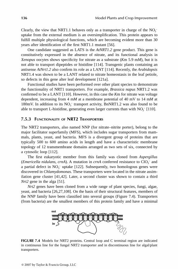
136 Model Plants and Crop Improvement
Clearly, the view that NRT1.1 behaves only as a transporter in charge of the NO3–
uptake from the external medium is an oversimplification. This protein appears tofulfill multiple physiological functions, which are becoming evident more than 30years after identification of the first NRT1.1 mutant [56].
One candidate suggested as LATS is the AtNRT1.2 gene product. This gene isconstitutively expressed in the absence of nitrate, and its functional analysis inXenopus oocytes shows specificity for nitrate as a substrate (Km 5.9 mM), but it isnot able to transport dipeptides or histidine [114]. Transgenic plants containing anantisense AtNrt1.2 also confirm its role as a LANT [114]. Recently, the ArabidopsisNRT1.4 was shown to be a LANT related to nitrate homeostasis in the leaf petiole,so defects in this gene alter leaf development [121a].
Functional studies have been performed over other plant species to demonstratethe functionality of NRT1 transporters. For example, Brassica napus NRT1.2 wasconfirmed to be a LANT [110]. However, in this case the Km for nitrate was voltagedependent, increasing from 4 mM at a membrane potential of 40 mV to 14 mM at180mV. In addition to its NO3
– transport activity, BnNRT1.2 was also found to beable to transport L-histidine, generating even larger currents than with NO3
– [110].
7.5.3 FUNCTIONALITY OF NRT2 TRANSPORTERS
The NRT2 transporters, also named NNP (for nitrate–nitrite porter), belong to themajor facilitator superfamily (MFS), which includes sugar transporters from mam-mals, plants, yeast, and bacteria. MFS is a divergent group of proteins that aretypically 500 to 600 amino acids in length and have a characteristic membranetopology of 12 transmembrane domains arranged as two sets of six, connected bya cytosolic loop [112].
The first eukaryotic member from this family was cloned from Aspergillus(Emericella nidulans, crnA). A mutation in crnA conferred resistance to ClO3
– anda partial defect in NO3
– uptake [122]. Subsequently, two homologous genes werediscovered in Chlamydomonas. These transporters were located in the nitrate assim-ilation gene cluster [41,42]. Later, a second cluster was shown to contain a thirdNrt2 gene in the alga [51].
Nrt2 genes have been cloned from a wide range of plant species, fungi, algae,yeast, and bacteria [26,27,108]. On the basis of their structural features, members ofthe NNP family have been classified into several groups (Figure 7.4). Transporters(from bacteria) are the smallest members of this protein family and have a minimal
FIGURE 7.4 Models for NRT2 proteins. Central loop and C terminal region are indicatedin continuous line for the fungal NRT2 transporter and in discontinuous line for algal/planttransporters.
inin
outout
3063_book.fm Page 136 Thursday, August 24, 2006 1:23 PM
© 2007 by Taylor & Francis Group, LLC

Chlamydomonas as a Tool to Study Nitrate Assimilation Pathways 137
amount of sequence outside the 12 transmembrane domains. The fungal members ofthe family have a large hydrophilic central loop of 90 amino acids located betweentransmembrane domains 6 and 7. The algal and higher plant members of the familyhave an extended C-terminal domain of ~70 amino acids that can include an N-terminal sequence extension of ~20 amino acids. This N-terminal domain is highlyconserved among the NRT2 family but is absent in the algal and barley sequences [26].
In silico structural analysis has been made to identify signature motifs for theNRT2 family and possible substrate recognition [23,112]. Recently, it has beenshown experimentally in Aspergillus crnA that many conserved glycine residuesthroughout the protein sequence have a structural role, while certain conservedcharged or polar residues within transmembrane domains (for example, arginineresidues conserved within transmembrane domains 2 and 8) are involved in a nitrate-binding function [123].
NRT2 transporters seem to involve one or two components (Figure 7.5). InChlamydomonas, a combination of mutant analysis and oocyte expression experi-ments indicates that some NRT2 members (NRT2.1 and NRT2.2) require an addi-tional protein (NAR2) for their functionality [42,92], and are thus two-componentsystems. Other NRT2 proteins, such as NRT2.3 from Chlamydomonas and CrnAfrom Aspergillus, are single-component systems [52,97]. Until now, NRT2 proteinsfrom plants have not been shown to be functional as single proteins, and they mightrequire a protein homolog to Chlamydomonas NAR2, as recently discovered in plants[124]. Nine Nar2-type genes have been identified in Hordeum vulgare and two inArabidopsis [124]. NAR2 proteins appear to have a single transmembrane domainand may interact with NRT2.1 to modify its function. The amino acid sequenceKX2KX2LCYX2SX3RXWRX3DX4DK between amino acids 140 and 180 seems tobe characteristic of the higher plant family of NAR2 proteins.
The use of the Xenopus oocytes expression system has shown that an NAR2 mRNA(HvNAR2.3) was able to reconstitute high-affinity NO3
– transport activity when co-injected with mRNA for the otherwise inactive HvNrt2.1 [124]. This result providesstrong evidence for the utility of the Chlamydomonas model for higher plants.
Chlamydomonas mutant strains defective in several of the nitrate gene clustershave allowed identification of four high-affinity nitrate/nitrite transporters (Figure
FIGURE 7.5 Single- and two-component nitrate transport systems in Chlamydomonas.
inin
outout
NONO33-– NONO22
-– NONO33-– NONO22
-– NONO33-– NONO22
-–NONO33-–
1.61.6 1.81.8 1111 55 4040 3333
HANTHANTHANiTHANiT
HANTHANT LANTLANTHANiTHANiT
HANTHANTHANiTHANiT
SystemSystem II SystemSystem IIII SystemSystem IIIIII SystemSystem IVIV
KKmm ((µM)
3063_book.fm Page 137 Thursday, August 24, 2006 1:23 PM
© 2007 by Taylor & Francis Group, LLC

138 Model Plants and Crop Improvement
7.5). System I corresponds to a bispecific HANT/HANiT encoded by Nrt2.1/Nar2;system II to a monospecific HANT encoded by Nrt2.2/Nar2; system III to a bispecificHANiT and LANT, probably encoded by Nrt2.3; and system IV to a bispecificHANT/HANiT for which the gene responsible has yet to be identified [42,52,61,62].The pH dependence of the nitrate-elicited currents by NRT2.1–NAR2 expressed inoocytes is consistent with an H+-cotransport mechanism [92].
These transport systems are differentially regulated by the carbon and nitrogensource. Systems I, II, and III are optimally expressed at high CO2, and their activityis blocked by ammonium; system IV is expressed optimally under limiting CO2 andits activity is not inhibited by ammonium. In contrast to systems I, II, and III, systemIV is inhibited by CO2 [52,61]. Concerning the function for each of these systems,mutants deleted in systems I and II and carrying functional systems III and IV areunable to grow efficiently in nitrate media [42]. Thus, under sufficient CO2, systemsI and II have a primary function in the provision of nitrate for growth and systemsI and III in nitrite entry.
7.5.4 NAR1 TRANSPORTERS
The nitrite transport step into the chloroplast is not well documented in plants,probably because of the lack of any molecular evidence and of the long-standingassumption that nitrite can diffuse freely as nitrous acid into the chloroplast [125].However, nitrite uptake into intact pea chloroplasts shows saturation kinetics, pref-erence for alkaline pH, and sensitivity to protein modifiers; this favors the existenceof a nitrite-mediated channel or transporter vs. the permeation of nitrous acid[126,127].
Nitrite transport into chloroplast inner envelope vesicles from pea has beenevaluated by Shingles et al. [128]. These authors propose that nitrite rapidly diffusesacross the plastid membrane depending on a proton gradient, so the proton-linkedNO2
– transport should be bidirectional. Nitrite concentrations change significantlyin roots of barley seedlings, depending on the nitrate availability in the environment[104], and in spinach leaves during the light–dark transitions [129]. Thus, the needfor a plastidial nitrite transporter is important for two reasons: (1) to avoid cellulartoxicity of nitrite; and (2) to increase efficiency of the nitrite reduction step.
Studies with Chlamydomonas Nar1.1 have provided the first molecular evidencethat nitrite transport to the chloroplast is a regulated process mediated by specifictransporters rather than the result of diffusion [34]. In spite of identification of sixmembers of the NAR1 family in Chlamydomonas (Table 7.2), further studies will berequired to know the role of each NAR1 protein. Available data show that the Nar1gene family may be closely associated with carbon and nitrogen metabolism becauseNar1.1 is nitrate upregulated and under control of the nitrate-pathway-specific regu-latory gene Nit2, whereas Nar1.2 (LciA) is upregulated by low CO2 and under controlof the carbon-pathway-specific regulatory gene ccm1 [44]. In plants, NAR1 proteinscannot be identified on the basis of sequence homology, but the role of NAR1.1 isso fundamental in Chlamydomonas that its function needs to be carried out by anotherprotein family in plants. The precise function for Nar1.1, as a chloroplast nitrite
3063_book.fm Page 138 Thursday, August 24, 2006 1:23 PM
© 2007 by Taylor & Francis Group, LLC

Chlamydomonas as a Tool to Study Nitrate Assimilation Pathways 139
transporter allowing for efficient nitrate utilization, has been deduced on the basisof the following data:
Nar1.1 is located in the nitrate cluster and coregulated with the other clusteredgenes (Figure 7.2; [41]). Thus, Nar1.1 is expressed in nitrate but not inammonium media.
NAR1.1 corresponds to an integral membrane protein predicted to have sixspanning-membrane domains, a plastidial localization, and significant iden-tity to formate and nitrite transporters from bacteria (Figure 7.6) [34].
The nitrite uptake activity by intact chloroplasts isolated from Nar1;1+ andNar1;1– strains supports the notion that NAR1;1 is a plastidial nitrite trans-porter with an apparent Ks for nitrite of about 5 µM [34].
Nar1.1 allows nitrate utilization when this nutrient is limiting for the cells.This limitation of nitrate takes place in strains lacking HANT systems Iand II at nitrate millimolar concentrations, or in strains having HANT withnitrate micromolar concentrations in the medium [34].
Nar1.1 improves nitrate use efficiency for growth under light/dark cycles andlow CO2 environments. Under such conditions, strains lacking Nar1.1uncouple nitrate reduction from the cells’ capability to assimilate the ammo-nium produced because of a significant deregulation in expression ofenzymes and transporters for nitrate assimilation including GS1 [130].
7.6 AMMONIUM ASSIMILATION
Incorporation of ammonium is a basic process shared by nitrate assimilation andother alternative nitrogen source pathways [37]. The GS/GOGAT cycle is the majorstep for ammonium incorporation into carbon skeletons by photosynthetic organisms.GS catalyses the formation of glutamine from ammonium and glutamate in an ATP-dependent reaction. In plants, GS isozymes are encoded by multigene families andsome of their members show cytosolic localization, while others have a plastidialor nodular localization [131]. The Chlamydomonas genome sequence shows threeGS genes (Table 7.2); GS1 encodes a cytosolic form and GS2 and GS3 are proposedto encode plastidial isozymes. Although a GS3 expression pattern is not documented,
FIGURE 7.6 (Color Figure 7.6 follows p. 144.) Model for the NAR1.1 protein showing thesix transmembrane domains. Residues mostly conserved among FNT proteins are shown inred. (Scheme modified from Galván, A. et al., J. Exp. Bot., 53, 845, 2002.)
Y
NN
CC
OutOut
InIn
3063_book.fm Page 139 Thursday, August 24, 2006 1:23 PM
© 2007 by Taylor & Francis Group, LLC

140 Model Plants and Crop Improvement
the major transcript level corresponds to that of GS2, which is constitutivelyexpressed with respect to nitrogen supply [39]. GS1 transcripts increase in cellsgrown in nitrate and decrease in cells grown in ammonium [39]. GS1 is proposedto play an active role in nitrate assimilation [132].
GOGAT catalyses the transfer of the amide group from glutamine to 2-oxoglu-tarate in a reaction that requires reducing equivalents. Two molecules of glutamate,the substrate of GS, are produced. Two GOGAT isoenzymes—one specific forreduced ferredoxin as electron donor and another specific for NADH—have beencharacterized in plants. Fd-GOGAT is a 130- to 150-kDa monomer, with a [3Fe-4S]center located in plastids and roots; NADH-GOGAT is a 158- to 240-kDa monomerwith the same prosthetic group, located in the plastids [133]. GOGAT gene numberper genome differs between species [134]. In Chlamydomonas, single genes encod-ing the NADH- and the ferredoxin-GOGAT are present (Table 7.2). These enzymeshave been characterized in detail [135,136].
7.7 AMMONIUM TRANSPORT GENES
As schematized in Figure 7.1, ammonium transporters are expected to operate indifferent cell localizations (plasma membrane, chloroplast, and mitochondria) medi-ating influx and efflux. Because ammonium is a strong negative signal of nitrateassimilation, it is important to know the different transporters that could mediate itseffects. Knowledge of the mechanisms regulating ammonium transport systems isalso essential for better understanding of nitrogen metabolism and plant growth.
At the physiological level, biphasic kinetics for ammonium uptake in severalspecies have been assigned to two different systems of transport: low-affinity trans-port systems (LATS), which are related to passive K+ channels [12,137,138], andhigh-affinity transport systems (HATS) that mediate active transport by couplingNH+
4 influx to a H+ gradient [139,140]. Similarly, at the physiological level, inChlamydomonas cells, two transport systems subject to circadian rhythm [141] arethought to participate in ammonium uptake: a constitutively expressed, low-affinityversion and a high-affinity version negatively regulated by ammonium [142].
Although LATSs have not yet been described at the molecular level, HATSshave been widely described in diverse organisms like plants, yeasts, bacteria, fungi,and animals, and form the AMT/MEP family [12,140,143]. This family has alsobeen related by homology with the human rhesus (Rh) blood proteins. Chlamydomo-nas is one of the few organisms that have genes from both families: Amt and Rh. Apossible role of the Chlamydomonas Rh proteins is as a bidirectional channel forammonia and CO2 [144,145].
In plants, most AMTs have been included in a large family designated AMT1[12,140], with the exception of some AMTs identified in Arabidopsis and Lotusjaponicus that are included in AMT2, a new family of transporters whose sequenceidentity is closer to yeast and bacteria than to plants [146–148]. Five members ofthe AMT1 family have been described in Arabidopsis [140,149,150] and three intomato [151]; its existence is also known in other species such as rice [152], Brassicanapus [153], and Lotus japonicus [154].
3063_book.fm Page 140 Thursday, August 24, 2006 1:23 PM
© 2007 by Taylor & Francis Group, LLC

Chlamydomonas as a Tool to Study Nitrate Assimilation Pathways 141
Eight members of the Amt1 gene family (Amt1.(1 to 8) genes) have been iden-tified in Chlamydomonas (Table 7.2) [45], representing the largest family thus fardescribed in any organism. On the other hand, no Amt2 genes have been found inChlamydomonas, suggesting that the proposed role of AMT2 transporters in plantscould be undertaken by some of the AMT1 proteins in the alga. In Figure 7.7, thephylogenetic relationships among these transporters and those from other organismsare shown. AMT1.1 to 6 are closely related to the plant AMT proteins, whereasAMT7 and 8 are more distantly related to plants.
The Chlamydomonas Amt1 genes show interesting evolutionary relationshipsbecause a particular feature present in all is the existence of some very small exons(less than 40 bp) and conservation of some intron positions that may be related toits transcriptional regulation. Alternative splicing has been found in the 5′ regionsof Amt1.5 and Amt1.7. This complex regulation might even result in different proteinlocalization, as suggested for the two isoforms deduced for AMT1.7 (Table 7.2) [45].
That a single cell such as Chlamydomonas has such a wide set of putativetransport systems for ammonium and other nutrients such as nitrate may reflect the
FIGURE 7.7 Phylogenetic tree of the AMT1 proteins. Alignment was performed with Clust-alW and the tree with the DNAStar package. Chlamydomonas AMT1 are included in a box.Other details are given in the text.
OsAMT1;1OsAMT1;3OsAMT1;2AtAMT1;2LeAMT1;2AtAMT1;1AtAMT1;3AtAMT1;4LeAMT1;1LeAMT1;3CrAMT1;2CrAMT1;6CrAMT1;4CrAMT1;1CrAMT1;3CrAMT1;5CrAMT1;8CrAMT1;7bAMT2 A. fulgidusNostoc spNostoc spAMT1 A. fulgidusAMT3 A. fulgidusMEP1MEP3MEP2AMTB E.coliX. axonopodisAtAMT2;1OsAMT2;1
020406080100
119.39
3063_book.fm Page 141 Thursday, August 24, 2006 1:23 PM
© 2007 by Taylor & Francis Group, LLC

142 Model Plants and Crop Improvement
need for complementary affinities and activities to obtain different substrates effi-ciently under changing environmental conditions. However, in Chlamydomonas, thenumber of Amt1 genes is higher than that of the Nrt2 genes (Table 7.2), in contrastto plants where the opposite pertains [12,15]. This is surprising if Chlamydomonasis considered as a unicellular green alga and thus no tissue specialization exists.
In any case, ammonium, in contrast to nitrate, requires a regulated intracellularflux because it can be assimilated in the cytoplasm or the chloroplast, accumulatedin the vacuole, or excreted to the medium, and ammonium produced during photo-respiration needs to be exported from the mitochondria [15,139]. The localizationsof the AMT1 proteins remain unknown, although several predictions suggest a clearchloroplast localization for AMT1.2 and especially for AMT1.4, whose hypotheticalsignal peptide has substantial similarity with that of the Chlamydomonas chloroplastprotein NiR (Table 7.2).
Putative ammonium transporters AMT1 from Chlamydomonas show particularexpression patterns in media containing different nitrogen sources (Figure 7.8): (1)Amt1.4 and Amt1.7 have the highest expression levels; and (2) by comparing differentnitrogen conditions, putative ammonium transporters 1, 2, 4, and 5 have maximumexpression in an N-free medium; the ammonium transporters 3 and 8 show maximumexpression in nitrate medium, whereas 6 and 7 are enhanced in ammonium medium[45]. The expression of the Chlamydomonas Amt1;1 has a complex regulatorymechanism responding to repression by ammonium, ammonium derivatives, andnitrate in a process mediated by the regulatory gene Nit2, so NIT2 provides a signalfor the presence of a usable nitrogen form—nitrate—connecting the pathway ofammonium uptake with that of nitrate assimilation. This means that NIT2 has a dualrole in gene expression: the well-known positive one on nitrate assimilation and anovel negative one on Amt1;1 expression [45,60]. Chlamydomonas Amt1;1 has anexpression pattern similar to that of the Arabidopsis gene Amt1;1, which encodesthe major system responsible for ammonium uptake [150].
FIGURE 7.8 Expression of Amt1 transcripts in different nitrogenous media. The relativeabundance is referred to that of ubiquitin ligase transcript used as a control. (From González-Ballester, D. et al., Plant Mol. Biol., 56, 863, 2004.)
0.01
0.1
1
10
100
1000
10000
Amt1.1
Amt1.2
Amt1.3
Amt1.4
Amt1.5
Amt1.6
Amt1.7
Amt1.8
Rel
ativ
etr
ansc
ripta
bund
ance
ammoniumnitrateN free
3063_book.fm Page 142 Thursday, August 24, 2006 1:23 PM
© 2007 by Taylor & Francis Group, LLC

Chlamydomonas as a Tool to Study Nitrate Assimilation Pathways 143
7.8 NITRATE ASSIMILATION AND LIGHT/CIRCADIAN RHYTHM
Light positively regulates most of the nitrate assimilation pathway genes and activ-ities. It is known that NR activity and mRNA levels of plants grown in a day/nightcycle fluctuate between a maximal level at the beginning of the day period and analmost undetectable level at the end of the day [22]. Further evidence of this lightregulation are the diurnal changes of nitrate and nitrite uptake that generally peakduring the light period and reach a minimum in the dark [26,155].
An endogenous clock regulates the temporal expression of genes/mRNAsinvolved in the circadian pathway. In Chlamydomonas, a recently identified, clock-controlled RNA-binding protein (Chlamy 1) represents an analog of the circadiantrans-acting factor, CCTR, from the phylogenetically diverse algal species Gon-yaulax polyedra [156]. Chlamy1 protein could act as a translational repressor, pre-venting translation of UG-repeats-bearing mRNAs from the end of the light untilthe end of the dark phase. The strength of the interaction between Chlamy1 andtranscripts may be influenced by the number of UG units in a region. Chlamy1 hasbeen cloned and is composed of two subunits: C1 is involved in protein–proteininteraction and C3 bears three RNA recognition motif domains [157].
Chlamy1 was reported to bind mRNAs whose products are key components ofnitrogen and CO2 metabolism [158]. Some of the genes under Chlamy1 control areinvolved in uptake of nitrite (Nrt2.3), its reduction to ammonium (Nii1), fixation ofammonium as glutamine (GS2), and arginine biosynthesis (ARG7). Other proteinsencoded by these RNAs are related to photosynthesis and the CO2 shuttle into thechloroplast (LIP36-G1), and CO2 fixation as ribulose-1,5-bisphosphate carboxylase(RBCS1, the small subunit of RUBISCO). Another is the light-dependent NADPH-protochlorophyllide-oxidoreductase (L-POR), one of two PORs that catalyze thereduction of protochlorophyllide to chlorophyllide, a regulatory step in chlorophyllbiosynthesis [159]. Finally, YPTC4 is a G-protein [160] whose function is not yetfully understood [158]. These genes, plus others containing UG units putativelycontrolled by Chlamy1, are shown in Figure 7.9.
The binding activity of Chlamy1 is controlled by the circadian clock. Thus, theregulatory properties of this factor are limited to a certain time window, which beginsat the end of the day phase and ends in the middle of the night phase. During thisperiod, binding activity of Chlamy1 is high, and at other time points it is low [161].Clock-controlled RNA-binding proteins have also been identified in other species.In Arabidopsis, a glycine-rich RNA-binding protein, GRP7, was characterized. Bothtranscripts and the protein are components of a negative feedback circuit capable ofgenerating a stable oscillation [162].
7.9 NITRATE ASSIMILATION AND REDOX REGULATION
It has been recently shown in Chlamydomonas that the redox state of the plasto-quinone (PQ) pool plays a key role in Nia1 gene expression [163]. A reduced PQpool is needed as a positive signal to allow Nia1 expression so that chemical inhibitors
3063_book.fm Page 143 Thursday, August 24, 2006 1:23 PM
© 2007 by Taylor & Francis Group, LLC
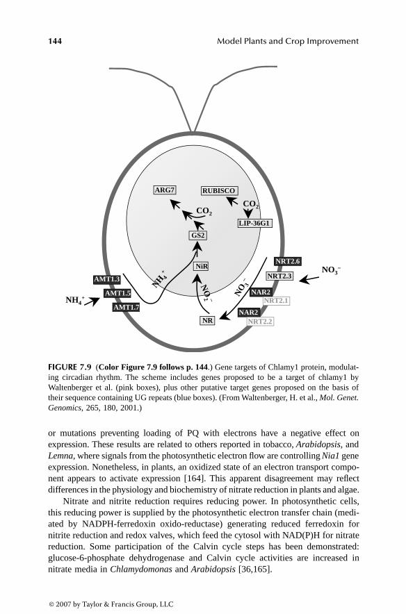
144 Model Plants and Crop Improvement
or mutations preventing loading of PQ with electrons have a negative effect onexpression. These results are related to others reported in tobacco, Arabidopsis, andLemna, where signals from the photosynthetic electron flow are controlling Nia1 geneexpression. Nonetheless, in plants, an oxidized state of an electron transport compo-nent appears to activate expression [164]. This apparent disagreement may reflectdifferences in the physiology and biochemistry of nitrate reduction in plants and algae.
Nitrate and nitrite reduction requires reducing power. In photosynthetic cells,this reducing power is supplied by the photosynthetic electron transfer chain (medi-ated by NADPH-ferredoxin oxido-reductase) generating reduced ferredoxin fornitrite reduction and redox valves, which feed the cytosol with NAD(P)H for nitratereduction. Some participation of the Calvin cycle steps has been demonstrated:glucose-6-phosphate dehydrogenase and Calvin cycle activities are increased innitrate media in Chlamydomonas and Arabidopsis [36,165].
FIGURE 7.9 (Color Figure 7.9 follows p. 144.) Gene targets of Chlamy1 protein, modulat-ing circadian rhythm. The scheme includes genes proposed to be a target of chlamy1 byWaltenberger et al. (pink boxes), plus other putative target genes proposed on the basis oftheir sequence containing UG repeats (blue boxes). (From Waltenberger, H. et al., Mol. Genet.Genomics, 265, 180, 2001.)
NRT2.3NRT2.3
NAR2
NAR2
NRT2.1NRT2.1
NRT2.2NRT2.2
NRT2.6
NRNR
NiRNiR
GS2GS2
RUBISCORUBISCO
LIP-36G1LIP-36G1
AMT1.3
AMT1.5
AMT1.7
ARG7ARG7
NONO33-–
NO
NO 33
-–
NO
NO
22 ––
NHNH 44++
NHNH44++
COCO22COCO22
3063_book.fm Page 144 Thursday, August 24, 2006 1:23 PM
© 2007 by Taylor & Francis Group, LLC

Chlamydomonas as a Tool to Study Nitrate Assimilation Pathways 145
In Chlamydomonas, NMDH and AOX1, which are closely related to their coun-terparts in plants, are involved in processes that control the fine-tuning of redoxmetabolism in chloroplast and mitochondrion [166] (Figure 7.10). NMDH partici-pates in the export of reducing equivalents from the chloroplast to the cytosol bythe malate–oxalacetate shuttle. Activity of this “malate valve” is regulated by theredox switch of NMDH, which is turned on and off by light signaling mediated bythe thioredoxin–ferredoxin system [50,167].
AOX is a mitochondrial terminal oxidase that funnels electrons to oxygen fromreduced ubiquinone. It bypasses the respiratory pathway through complexes III andIV of the mitochondrial electron transfer chain and the concomitant generation ofelectrochemical potential; thus, synthesis of the amount of ATP per electron pair isdiminished. AOX could play a role as a redox valve to balance carbon metabolismand mitochondrial electron transport [168].
Clustering of Nmdh1 and Aox1 genes with nitrate assimilation genes in Chlamy-domonas provides a direct molecular link between NADH and ATP pool regulationand nitrate assimilation (Figure 7.2 and Figure 7.10). NMDH provides reducingpower for cytosolic nitrate reduction, and its activity level is compromised bycompetition for reduced ferredoxin, the electron donor for nitrite reductase [166].
Specific induction of Aox1 by nitrate and not by ammonium might indicate therequirement to adapt mitochondrial electron flow and thus ATP synthesis duringassimilation of these nitrogen sources. This adaptation is especially critical under
FIGURE 7.10 (Color Figure 7.10 follows p. 144.) The redox valves and the nitrate assim-ilation pathway.
N/N/niTniT
NRNR
NONO33-– NONO22
-–
NONO22-– NiTNiTNONO33
-–
NONO22-–
NONO22-–
NADHNADH NADNAD++
NHNH44++
NiRNiR
FdFd--oxoxFdFd-red-red
NADPHNADPH NADPNADP++
OAAOAA MalMal
DATDAT
OAAOAA MalMal
MDHMDH
MDHMDH
NADHNADH NADNAD++
NADNAD++
CICICIVCIV UQUQee-–
ee-–HH22OO
OO22
DATDAT
ChloroplastChloroplast
MitochondriaMitochondria
AOX1AOX1
NONO22-–NO?NO?
CIIICIII
3063_book.fm Page 145 Thursday, August 24, 2006 1:23 PM
© 2007 by Taylor & Francis Group, LLC

146 Model Plants and Crop Improvement
stress conditions, such as high concentrations of nitrite in the cytosol that might actas substrate of NR and be converted to nitric oxide [169,170] or in the mitochondriawhere NO has been shown to be produced by the alternative oxidase in Chlorella[171]. NO in turn inhibits the mitochondrial electron transport chain, so alternativeoxidase would be essential for ATP production under NO presence. Whether or notNO production is the result of stress or a metabolic condition that triggers a signalingcascade will require the identification of intermediates in this cascade and the finaltarget of action for the cell response.
7.10 REGULATORY GENES IN NITRATE ASSIMILATION
Membrane proteins, such as pumps, ion channels, transporters, and receptors, areresponsible for controlling the flow of nutrients and other solutes across the plasmamembrane and cellular organelles. They also provide the appropriate signalingmetabolites for modulating the cell response to changing needs according to nutrientavailability and developmental or physiological status.
Nitrate is sensed, and this up- or down-regulates the expression of a large numberof genes, some of which are specific for nitrate assimilation. Other genes link thisroute to different metabolic pathways. Studies with Arabidopsis have found that byusing transcriptome analysis in short-term treatments with low levels of nitrate, morethan 1000 genes are significantly induced or repressed within 20 min of treatment[172]. In roots, 270 genes were differentially expressed in media containing nitrateas opposed to ammonium nitrate [36,172,173].
In Chlamydomonas, nitrate signaling occurs intracellularly and directly dependson the activity of the nitrate transport systems [174,175]. Thus, in Chlamydomonasthe presence of particular high-affinity nitrate transport systems has an importantregulatory role in expression of nitrate assimilation genes. According to their affinity,specificity, and capacity for nitrate, these systems are responsible for differentialnitrate signaling by regulating intracellular concentrations of nitrate.
Recently, serial analysis of gene expression (SAGE) transcriptome analysis inthe nitrate transporter Chl1 mutant of Arabidopsis has revealed how a single trans-porter affects the expression of hundreds of genes. Because of the marked deregu-lation of Nrt2.1 in the mutant, it was suggested that Nrt1.1 plays a direct signalingrole in regulating other nitrate transporters [176].
Ammonium transporters have been suggested to participate in the sensing ofammonium [177]. The yeast ammonium transporter MEP2, dispensable for cellgrowth, has been implicated in the sensing of ammonium [178,179]. In Chlamy-domonas, the ammonium-uptake defective mutant strain 2170 is also relieved fromthe negative effect of ammonium or methylammonium on the nitrate pathway [180].
The regulatory genes for nitrate assimilation are well defined in fungi and yeast[30,181]. Two positively acting regulatory genes are required for expression of nitratetransporter and nitrate reduction genes in fungi [29,181]. NirA/Nit4/YNA1 genesfrom Aspergillus, Neurospora, and Hansenula, respectively, are pathway-specificgenes involved in nitrate induction and correspond to GAL4-like Cys6/Zn2 binuclearzinc cluster [181]. AREA/NIT2 from Aspergillus and Neurospora are major regula-tory proteins mediating nitrogen repression from readily usable nitrogen sources
3063_book.fm Page 146 Thursday, August 24, 2006 1:23 PM
© 2007 by Taylor & Francis Group, LLC

Chlamydomonas as a Tool to Study Nitrate Assimilation Pathways 147
such as ammonium and glutamine and correspond to GATA-binding transcriptionfactors [182,183]. The mechanism proposed for the NIT2 negative function in Neu-rospora is that NMR1, a negatively acting protein, interacts with NIT2 in the presenceof catabolic repressors and prevents the binding of NIT2 to DNA for transcriptionof target genes.
Various attempts have been made to identify regulatory genes from plants[22,184]. However, the relationship of the isolated genes to regulation of the nitratepathway has not been shown, even though a GATA motif was found in the promoterof the spinach NiR gene [185] and in silico analysis revealed a large number ofGATA-family transcription factors in Arabidopsis and rice [186].
The regulatory fungal model does not seem to fit in photosynthetic eukaryotesand clues for regulatory genes may therefore come from systems such as Chlamy-domonas. It has been hypothesized that several genes act to mediate the positiveeffects of nitrate and the negative ones of ammonium. Their deficiency may lead topartial phenotypes that can explain the difficulties experienced in the genetic dis-section of regulation in algae and plants [27].
In Chlamydomonas, the expression of nitrate assimilation genes is co-regulated(Figure 7.2). These genes are subject to repression by ammonium, induction by nitrate,and the control of the positive-acting regulatory gene Nit2 [42,60]. This gene wascloned by transposon tagging from chlorate-resistant mutants [47]. Although struc-tural analysis of the deduced NIT2 protein has not been performed, the Chlamydomo-nas genome database reveals that NIT2 is a transcription factor containing anRWPXRK box present in plant proteins involved in N control and the Chlamydomonasmid protein involved in minus mating type dominance [36,187,188]. Nit2 is itselfsubject to ammonium repression, which implies an additional level of control [47].
A functional genomics approach to identify regulatory genes for nitrate assim-ilation has been recently performed in Chlamydomonas. By taking advantage ofdeletion events that occur during integration of a heterologous marker in Chlamy-domonas transformation, along with the Chlamydomonas genome sequence, anordered mutant library of 22,000 strains has recently been obtained [8]. Assumingthat Chlamydomonas contains about 17,000 genes and deletions affect wide genomicregions, it has been proposed that such a number of mutants will be sufficient tocover most of the Chlamydomonas genome.
The arylsulfatase reporter gene under the control of the Nia1 gene promoter hasserved as a sensor for identifying regulatory mutants (Figure 7.11). A forwardscreening of the library allowed for the selection of 145 mutants defective at putativegenes related to the positive signal of nitrate or the negative signal of ammonium.The ammonium-insensitive mutants were found to be defective at genomic regionsbearing putative new genes related to regulatory functions such as guanylate cyclase,protein kinase, peptidyl-prolyl isomerase, or DNA binding [8], although no directevidence correlating these genes with ammonium repression exists. In addition, someinsertions in ammonium-insensitive mutants map to unknown regions or genes inthe genome. From these and previous results [189–191], it is becoming evident thata complex network of signaling proteins mediates the effects of ammonium and itsderivatives on the nitrate assimilation pathway.
3063_book.fm Page 147 Thursday, August 24, 2006 1:23 PM
© 2007 by Taylor & Francis Group, LLC

148 Model Plants and Crop Improvement
Now, it will be possible to explore additional regulatory pathways by direct orreverse genetics—for example, those involving TOR or PII proteins reported in otherorganisms and considered to be integrators of nutrient availability (amino acids andenergy) and as key players of nutrient-mediated signal transduction [182,192–196].
7.11 CONCLUSION
As the most important pathway for nitrogen acquisition by crop plants, nitrateassimilation needs to be understood in its most basic molecular aspects. In spite ofits simplicity, this pathway has many intriguing questions still to solve. Modelsystems can provide invaluable information in the search for key genes and functionsin plants and, finally, to the optimization of nitrogen use efficiency. In the age ofgenomics, comparative biology and mutant collections, the work carried out inChlamydomonas should progress further in this direction.
FIGURE 7.11 The isolation of nitrate assimilation mutants defective in positive and negativesignaling. ARS = arylsulfatase activity. Other details are in the text.
3063_book.fm Page 148 Thursday, August 24, 2006 1:23 PM
© 2007 by Taylor & Francis Group, LLC

Chlamydomonas as a Tool to Study Nitrate Assimilation Pathways 149
ACKNOWLEDGMENTS
This work was supported by Ministerio de Ciencia y Tecnología, Spain (MCYTgrant no. BFU2005-07521), European Commission (RTN grant HPRN-CT-2002-00247), and Junta de Andalucía, Spain (PAI, CVI-0128).
REFERENCES
1. Mandoli, D.F. and Olmstead, R., The importance of emerging model systems in plantbiology, J. Plant Growth Reg., 19, 249, 2000.
2. Silflow, C.D., Organization of the nuclear genome, in The Molecular Biology ofChloroplasts and Mitochondria in Chlamydomonas, Rochaix, J.D., Goldschmidt-Clermont, M., and Merchant, S., Eds., Kluwer Academic Publishers, Dordrecht, theNetherlands, 1998, 25–40.
3. Harris, E., The Chlamydomonas Sourcebook, Academic Press, New York, 1989.4. Kindle, K.L., Nuclear transformation: technology and applications, in The Molecular
Biology of Chloroplasts and Mitochondria in Chlamydomonas, Rochaix, J.D., Gold-schmidt-Clermont, M., and Merchant, S., Eds., Kluwer Academic Publishers, Dor-drecht, the Netherlands, 1998, 41–61.
5. León-Bañares, R. et al., Transgenic microalgae as green cell-factories, Trends Bio-technol., 22, 45, 2004.
6. Fuhrmann, M. et al., The abundant retinal protein of the Chlamydomonas eye is notthe photoreceptor for phototaxis and photophobic responses, J. Cell Sci., 114, 3857,2001.
7. Rohr, J. et al., Tandem inverted repeat system for selection of effective transgenicRNAi strains in Chlamydomonas, Plant J., 40, 611, 2004.
8. González-Ballester, D. et al., Functional genomics of the regulation of the nitrateassimilation pathway in Chlamydomonas, Plant Physiol., 137, 522, 2005.
9. Rochaix, J.D., Goldschmidt-Clermont, M., and Merchant, S., The Molecular Biologyof Chloroplasts and Mitochondria in Chlamydomonas, Kluwer Academic Publishers,Dordrecht, the Netherlands, 1989.
10. Gutman, B.L. and Nigoyi, K.K., Chlamydomonas and Arabidopsis. A dynamic duo,Plant Physiol., 135, 607, 2004.
11. Hart, S.C. and Stark, J.M., Nitrogen limitation of the microbial biomass in an oldgrowth forest, Ecoscience, 4, 91, 1997.
12. Crawford, N.M., and Forde, B., Molecular and developmental biology of inorganicnitrogen nutrition, in The Arabidopsis Book, Somerville, C. and Meyerowitz, E., Eds.,American Society of Plant Physiologists, Rockville, MD, 2002, doi/10.1199/tab.0011,http://www.aspb.org/publications/arabidopsis/.
13. Bloom, A.J., Sukrapanna, S.S., and Warner, R.L., Root respiration associated withammonium and nitrate absorption and assimilation by barley, Plant Physiol., 99,1294, 1992.
14. Fernández, E. and Cárdenas, J., Regulation of the nitrate-reducing system enzymesin wild type and mutant strains of Chlamydomonas reinhardtii, Mol. Genet. Genomics,186, 164, 1982.
3063_book.fm Page 149 Thursday, August 24, 2006 1:23 PM
© 2007 by Taylor & Francis Group, LLC

150 Model Plants and Crop Improvement
15. Glass, A.D.M., Nitrogen use efficiency of crop plants: physiological constraints uponnitrogen absorption, Crit. Rev. Plant. Sci., 22, 453, 2003.
16. Hirsch, R.E. and Sussman, M.R., Improving nutrient capture from soil by the geneticmanipulation of crop plants, Trends Biotechnol., 17, 356, 1999.
17. Britto, D.T. et al., Futile transmembrane NH4(+) cycling: a cellular hypothesis toexplain ammonium toxicity in plants, Proc. Natl. Acad. Sci. USA, 98, 4255, 2001.
18. Wray, J.L. and Kinghorn, J.R., Molecular and Genetic Aspects of Nitrate Assimilation,Oxford Science Publications, Oxford, 1989.
19. Caboche, M. and Rouzé, P., Nitrate reductase a target for molecular and cellularstudies in higher plants, Trends Genet., 6, 187, 1990.
20. Hoff, T., Truong, H.N., and Caboche, M., The use of mutants and transgenic plantsto study nitrate assimilation, Plant Cell Environ., 17, 489, 1994.
21. Crawford, N.M., Nitrate: nutrient and signal for plant growth, Plant Cell, 7, 859, 1995.22. Daniel-Vedele, F. and Caboche, M., A tobacco cDNA clone encoding a GATA-1 zinc
finger protein homologous to regulators of nitrogen metabolism in fungi, Mol. Gen.Genet., 240, 365, 1993.
23. Trueman, L.J., Onyeocha, I., and Forde, B.G., Recent advances in the molecularbiology of a family of eukaryotic high affinity nitrate transporters, Plant Physiol.Biochem., 34, 621, 1996.
24. Crawford, N.M. and Glass, A.D.M., Molecular and physiological aspects of nitrateuptake in plants, Trends Plant Sci., 3, 389, 1998.
25. Campbell, W., Nitrate reductase structure, function and regulation: binding the gapbetween biochemistry and physiology, Annu. Rev. Plant. Physiol. Plant Mol. Biol.,50, 277, 1999.
26. Forde, B., Nitrate transporters in plants: structure, function and regulation. Biochim.Biophys. Acta Biomembranes, 1465, 219, 2000.
27. Galván, A. and Fernández, E., Eukaryotic nitrate and nitrite transporters, Cell Mol.Life Sci., 58, 225, 2001.
28. Williams, L.E. and Miller, A., Transporters responsible for the uptake and partitioningof nitrogen solutes, Annu. Rev. Plant Physiol. Plant Mol. Biol., 52, 659, 2001.
29. Crawford, N.M. and Arst, H.N., The molecular genetics of nitrate assimilation infungi and plants, Annu. Rev. Genet., 27, 115, 1993.
30. Siverio, J.M., Assimilation of nitrate by yeasts, FEMS Microbiol. Rev., 26, 277, 2002.31. Forde, B. and Cole, J., Nitrate finds a place in the sun, Plant Physiol., 131, 395, 2003.32. Azuara, M.P. and Aparicio, P., In vivo blue-light activation of Chlamydomonas rein-
hardtii nitrate reductase, Plant Physiol., 71, 286, 1983.33. Navarro, M.T. et al., Nitrite reductase mutants as an approach to understanding nitrate
assimilation in Chlamydomonas reinhardtii, Plant Physiol., 122, 283, 2000.34. Rexach, J., Fernández, E., and Galván, A., The Chlamydomonas reinhardtii Nar1
gene encodes a chloroplast membrane protein involved in nitrite transport, Plant Cell,12, 1441, 2000.
35. Armengaud, P., Breitling, R., and Amtmann, A., The potassium-dependent transcrip-tome of Arabidopsis reveals a prominent role of jasmonic acid in nutrient signaling,Plant Physiol., 136, 2556, 2004.
36. Scheible, W.R. et al., Genome-wide reprogramming of primary and secondary metab-olism, protein synthesis, cellular growth processes, and the regulatory infrastructureof Arabidopsis in response to nitrogen, Plant Physiol., 136, 2483, 2004.
37. Fernández, E. et al., Isolation and characterization of the nitrate reductase structuralgene in Chlamydomonas reinhardtii, Proc. Natl. Acad. Sci. USA, 86, 6449, 1989.
3063_book.fm Page 150 Thursday, August 24, 2006 1:23 PM
© 2007 by Taylor & Francis Group, LLC

Chlamydomonas as a Tool to Study Nitrate Assimilation Pathways 151
38. Quesada, A., Gómez, I., and Fernández, E., Clustering of the nitrite reductase geneand a light-regulated gene with nitrate assimilation loci in Chlamydomonas rein-hardtii, Planta, 206, 259 1998.
39. Chen, Q. and Silflow, C.D., Isolation and characterization of glutamine synthetasegenes in Chlamydomonas reinhardtii, Plant Physiol., 112, 987, 1996.
40. García-Sanchez, M.I. et al., Homology predicted structure and functional interactionof ferredoxin from the eukaryotic alga Chlamydomonas reinhardtii with nitrite reduc-tase and glutamate synthase, J. Biol. Inorg. Chem., 5, 713, 2000.
41. Quesada, A. et al., Five nitrate assimilation related loci are clustered in Chlamydomo-nas reinhardtii, Mol. Genet. Genomics, 240, 387, 1993.
42. Quesada, A., Galván, A., and Fernández, E., Identification of nitrate transporters inChlamydomonas reinhardtii, Plant J., 5, 407, 1994.
43. Galván, A. et al., Nitrite transport to the chloroplast in Chlamydomonas: molecularevidences for a regulated process, J. Exp. Bot., 53, 845, 2002.
44. Miura, K. et al., Expression profiling-based identification of CO2-responsive genesregulated by CCM1 controlling a carbon-concentrating mechanism in Chlamydomo-nas reinhardtii, Plant Physiol., 135, 1595, 2004.
45. González-Ballester, D., Camargo, A., and Fernández, E., Ammonium transportergenes in Chlamydomonas: the nitrate-specific regulatory gene Nit2 is involved inAmt11 expression, Plant Mol. Biol., 56, 863, 2004.
46. Kim, K.S. et al., Spontaneous mutations in the ammonium transport gene AMT4 ofChlamydomonas reinhardtii, Genetics, 170, 631, 2005.
47. Schnell, R.A. and Lefebvre, P.A., Isolation of the Chlamydomonas regulatory genenit2 by transposon tagging, Genetics, 134, 737, 1993.
48. Ataya, F.S. et al., Mcp1 encodes the molybdenum cofactor carrier protein in Chlamy-domonas reinhardtii and participates in protection, binding, and storage functions ofthe cofactor, J. Biol. Chem., 278, 10885, 2003.
49. Dinant, M. et al., Characterization of two genes encoding the mitochondrial alternativeoxidase in Chlamydomonas reinhardtii, Curr. Genet., 39, 101, 2001.
50. Gómez, I. et al., NADP-malate dehydrogenase from Chlamydomonas: prediction ofnew structural determinants for redox regulation by homology modeling, Plant Mol.Biol., 48, 211, 2002.
51. Quesada, A., Hidalgo, J., and Fernández, E., Three Ntr2 genes are differentiallyregulated in Chlamydomonas reinhardtii, Mol. Genet. Genomics, 258, 373, 1998.
52. Rexach, J. et al., Differential regulation of the high affinity nitrite transport systemsIII and IV in Chlamydomonas reinhardtii, J. Biol. Chem., 274, 27801, 1999.
53. Johnston, I.L. et al., Isolation and characterization of the crnA-niiA-niaD gene clusterfor nitrate assimilation in Aspergillus nidulans, Gene, 90, 181, 1990.
54. Perez, M.D. et al., The YNT1 gene encoding the nitrate transporter in the yeastHansenula polymorpha is clustered with genes YNI1 and YNR1 encoding nitritereductase and nitrate reductase, and its disruption causes inability to grow in nitrate,Biochem. J., 321, 397, 1997.
55. Punt, P.J. et al., The intergenic region between the divergently transcribed niiA andniaD genes of Aspergillus nidulans contains multiple NirA binding sites which actbidirectionally, Mol. Cell. Biol., 15, 5688, 1995.
56. Oostindiër-Braaksma, F.J. and Feenstra, W.J., Isolation and characterization of chlo-rate-resistant mutants of Arabidopsis thaliana, Mutation Res., 19, 175, 1973.
57. Cove, D.J., Chlorate toxicity in Aspergillus nidulans. Studies of mutants altered innitrate assimilation, Mol. Genet. Genomics, 146, 147, 1976.
3063_book.fm Page 151 Thursday, August 24, 2006 1:23 PM
© 2007 by Taylor & Francis Group, LLC
152 Model Plants and Crop Improvement
58. Prieto, R. and Fernández, E., Toxicity and mutagenesis by chlorate are independentof nitrate reductase activity in Chlamydomonas reinhardtii, Mol. Genet. Genomics,237, 429, 1993.
59. Aberg, B., On the mechanism of the toxic action of chlorate and some relatedsubstances upon young wheat plants, Annu. Rev. Agric. Coll. Sweden, 15, 37, 1947.
60. Fernández, E., Galván, A., and Quesada, A., Nitrogen assimilation and its regulation,in Molecular Biology of Chlamydomonas: Chloroplast and Mitochondria, Rochaix,J.D., Goldschmidt-Clermont, M., and Merchant, S., Eds., Kluwer Academic Publish-ers, Dordrecht, the Netherlands, 1998, 637.
61. Galván, A., Quesada, A., and Fernández, E., Nitrate and nitrite are transported bydifferent specific transport systems and by a bispecific transporter in Chlamydomonasreinhardtii, J. Biol. Chem., 271, 2088, 1996.
62. Navarro, M.T. et al., Chlamydomonas reinhardtii strains expressing nitrate reductaseunder control of the cabII-1 promoter: isolation of chlorate resistant mutants andidentification of new loci for nitrate assimilation, Photosynth. Res., 83, 151, 2005.
63. Cao, D. et al., The chlorate-resistant and photomorphogenesis-defective mutant cr88encodes a chloroplast-targeted HSP90, Plant J., 33, 107, 2003.
64. Campbell, W.H. and Kinghorn, J.R., Functional domains of assimilatory nitrate reduc-tases and nitrite reductases, Trends. Biochem. Sci., 15, 315, 1990.
65. Moura, J.J. et al., Mo and W bis-MGD enzymes: nitrate reductases and formatedehydrogenases, J. Biol. Inorg. Chem., 9, 791, 2004.
66. Moreno-Vivian, C. et al., Prokaryotic nitrate reduction: molecular properties andfunctional distinction among bacterial nitrate reductases, J. Bacteriol., 181, 6573, 1999.
67. Eilers, T. et al., Identification and biochemical characterization of Arabidopsisthaliana sulfite oxidase. A new player in plant sulfur metabolism, J. Biol. Chem., 14,46989, 2001.
68. Mendel, R.R. and Hansch, R., Molybdoenzymes and molybdenum cofactor in plants,J. Exp. Bot., 53, 1689, 2002.
69. Mendel, R. and Schwarz, G., Molybdoenzymes and molybdenum cofactor in plants,Crit. Rev. Plant Sci., 18, 33, 1999.
70. Fernández, E. and Matagne, R.F., Genetic analysis of nitrate reductase-deficientmutants in Chlamydomonas reinhardtii, Curr. Genet., 8, 635, 1984.
71. Aguilar, M. et al., Direct transfer of molybdopterin cofactor to aponitrate reductasefrom a carrier protein in Chlamydomonas reinhardtii, FEBS Lett., 307, 162, 1992.
72. Llamas, A., Kalakoutskii, K.L., and Fernández, E., Molybdenum cofactor amountsin Chlamydomonas reinhardtii depend on the Nit5 gene function related to molybdatetransport, Plant Cell Environ., 23, 1247, 2000.
73. Kalakoutskii, K. and Fernández, E., Chlamydomonas reinhardtii nitrate reductasecomplex has 105-kDa subunits in the wild-type strain and a structural mutant, PlantSci., 105, 195, 1995.
74. Witte, C.P. et al., The Chlamydomonas reinhardtii MoCo carrier protein is multimericand stabilizes molybdopterin cofactor in a molybdate charged form, FEBS Lett., 431,205, 1998.
75. Bachmann, M. et al., 14-3-3 proteins associate with the regulatory phosphorylationsite of spinach leaf nitrate reductase in an isoform-specific manner and reduce dephos-phorylation of Ser-543 by endogenous protein phosphatases, FEBS Lett., 398, 26,1996.
76. Moorhead, G. et al., Phosphorylated nitrate reductase from spinach leaves is inhibitedby 14-3-3 proteins and activated by fusicoccin, Curr. Biol., 6, 1104, 1996.
3063_book.fm Page 152 Thursday, August 24, 2006 1:23 PM
© 2007 by Taylor & Francis Group, LLC

Chlamydomonas as a Tool to Study Nitrate Assimilation Pathways 153
77. Kaiser, W.M. et al., Modulation of nitrate reductase: some new insights, an unusualcase and a potentially important side reaction, J. Exp. Bot., 53, 875, 2002.
78. Moorhead, G. et al., Phosphorylation-dependent interactions between enzymes ofplant metabolism and 14-3-3 proteins, Plant J., 18, 1, 1999.
79. Lillo, C. et al., Characterization of nitrate reductase from light- and dark-exposedleaves. Comparison of different species and effects of 14-3-3 inhibitor proteins, PlantPhysiol., 114, 1377, 1997.
80. Mackintosh, C., Dynamic interactions between 14-3-3 proteins and phosphoproteinsregulate diverse cellular processes, Biochem. J., 381, 329, 2004.
81. Comparot, S. et al., Function and specificity of 14-3-3 proteins in the regulation ofcarbohydrate and nitrogen metabolism, J. Exp. Bot., 54, 595, 2003.
82. Pozuelo, M. et al., Cytosolic glutamine synthetase and not nitrate reductase from thegreen alga Chlamydomonas reinhardtii is phosphorylated and binds 14-3-3 proteins,Planta, 212, 264, 2001.
83. Huber, S.C., MacKintosh, C., and Kaiser, W.M., Metabolic enzymes as targets for14-3-3 proteins, Plant Mol. Biol., 50, 1053, 2002.
84. Guerrero, M.G., Vega, J.M., and Losada, M. The assimilatory nitrate-reducing systemand its regulation, Annu. Rev. Plant Physiol., 32, 169, 1981.
85. Franco, A.R., Cárdenas, J., and Fernández, E., Involvement of reversible inactivationin the regulation of nitrate reductase enzyme levels in Chlamydomonas reinhardtii,Plant Physiol., 84, 665, 1987.
86. Vega, J.M. and Kamin, H., Spinach nitrite reductase. Purification and properties ofa siroheme-containing iron-sulfur enzyme, J. Biol. Chem., 252, 896, 1977.
87. Kato, C. et al., Differential expression of the nitrite reductase gene family in tobaccoas revealed by quantitative competitive RT-PCR, J. Exp. Bot., 55, 1761, 2004.
88. Martínez-Rivas, J.M., Vega, J.M., and Márquez, A., Differential regulation of thenitrate-reducing and ammonium-assimilatory systems in synchronous cultures ofChlamydomonas reinhardtii, FEMS Microbiol. Lett., 78, 85, 1991.
89. Crete, P., Caboche, M., and Meyer, C., Nitrite reductase expression is regulated atthe post-transcriptional level by the nitrogen source in Nicotiana plumbaginifolia andArabidopsis thaliana, Plant J., 11, 625, 1997.
90. Glass, A.D.M., Shaff, J.E., and Kochian, L.V., Studies of the uptake of nitrate inbarley. IV. Electrophysiology, Plant Physiol., 99, 456, 1992.
91. Tsay, Y.F. et al., An herbicide sensitive gene CHL1 of Arabidopsis encodes a nitrate-inducible nitrate transporter, Cell, 72, 705, 1993.
92. Zhou, J.J. et al., A high affinity nitrate transport system from Chlamydomonas requirestwo gene products, FEBS Lett., 28, 225, 2000.
93. Miller, A.J. et al., The use of microelectrodes to investigate compartmentation andthe transport of metabolized inorganic ions in plants, J. Exp. Bot., 52, 541, 2001.
94. Koltermann, M. et al., Cloning, functional expression and expression studies of thenitrate transporter gene from Chlorella sorokiniana (strain 211-8k), Plant Mol. Biol.,54, 855, 2003.
95. McClure, P.R. et al., Evidence for cotransport of nitrate and proton in maize roots.I. Effect of nitrate on the membrane potential, Plant Physiol., 93, 281, 1990.
96. McClure, P.R. et al., Evidence for cotransport of nitrate and protons in maize roots.II. Measurement of NO3- and H+ fluxes with ion-selective microelectrodes, PlantPhysiol., 93, 281, 1990.
97. Zhou, J.J. et al., A high affinity fungal nitrate carrier with two transport mechanisms,J. Biol. Chem., 275, 39894, 2000.
3063_book.fm Page 153 Thursday, August 24, 2006 1:23 PM
© 2007 by Taylor & Francis Group, LLC
154 Model Plants and Crop Improvement
98. Rodríguez, R., Lara, C., and Guerrero, M.G., Nitrate transport in the cyanobacteriumAnacystis nidulans R2. Kinetic and energetic aspects, Biochem. J., 15, 639, 1992.
99. Omata, T., Structure, function and regulation of the nitrate transport system of thecyanobacterium Synechococcus sp. PCC7942, Plant Cell Physiol., 36, 207, 1995.
100. DeMoss, J.A. and Hsu, P.Y., NarK enhances nitrate uptake and nitrite excretion inEscherichia coli, J. Bacteriol., 173, 3303, 1991.
101. Clegg, S. et al., The roles of the polytopic membrane proteins NarK, NarU and NirCin Escherichia coli K-12: two nitrate and three nitrite transporters, Mol. Microbiol.,44, 143, 2002.
102. Geelen, D. et al., Disruption of putative anion channel gene AtCLC-a in Arabidopsissuggests a role in the regulation of nitrate content, Plant J., 21, 259, 2000.
103. Fraisier, V. et al., Constitutive expression of a putative high-affinity nitrate transporterin Nicotiana plumbaginifolia: evidence for post-transcriptional regulation by areduced nitrogen source, Plant J., 23, 489, 2000.
104. Aslam, M., Travis, R.L., and Huffaker, R.C., Comparative induction inhibition ofnitrate and nitrite uptake and reduction systems by ambient nitrate and nitrite in intactroots of barley seedlings, Plant Physiol., 102, 811, 1993.
105. Wang, R. and Crawford, N.M., Genetic identification of a gene involved in constitutivehigh-affinity nitrate transport in higher plants, Proc. Natl. Acad. Sci. USA, 93, 9297,1996.
106. Quesada, A. et al., PCR-identification of a Nicotiana plumbaginifolia cDNA homol-ogous to the high-affinity nitrate transporters of the crnA family, Plant Mol. Biol.,34, 265, 1997.
107. Machín, F. et al., The role of Ynt1 in nitrate and nitrite transport in the yeastHansenula polymorpha, Yeast, 21, 265, 2004.
108. Orsel, M., Krapp, A., and Daniel-Vedele, F., Analysis of the NRT2 nitrate transporterfamily in Arabidopsis. Structure and gene expression, Plant Physiol., 129, 886, 2002.
109. Okamoto, M., Vidmar, J.J., and Glass, A.D., Regulation of NRT1 and NRT2 genefamilies of Arabidopsis thaliana: responses to nitrate provision, Plant Cell Physiol.,44, 304, 2003.
110. Zhou, J.J. et al., Cloning and functional characterization of a Brassica napus trans-porter that is able to transport nitrate and histidine, J. Biol. Chem., 273, 12017, 1998.
111. Paulsen, I.T. and Skurray, R.A., The POT family of transport proteins, Trends Bio-chem. Sci., 19, 404, 1994.
112. Pao, S.S., Paulsen, I.T., and Saier, M.H., The major facilitator superfamily, Microbiol.Mol. Biol. Rev., 62, 1, 1998.
113. Doddema, H. and Telkamp, G.P., Uptake of nitrate by mutants of Arabidopsisthaliana, disturbed in uptake or reduction of nitrate: II. Kinetics, Physiol. Plant., 45,332, 1979.
114. Huang, N.C., Cloning and functional characterization of an Arabidopsis nitrate trans-porter gene that encodes a constitutive component of low-affinity uptake, Plant Cell,11, 1381, 1999.
115. Huang, N.C. et al., CHL1 encodes a component of the low-affinity nitrate uptakesystem in Arabidopsis and shows cell type-specific expression in roots, Plant Cell,8, 2183, 1996.
116. Touraine, B. and Glass, A.D., NO3– and ClO3
– fluxes in the chl1-5 mutant of Arabi-dopsis thaliana, Does the CHL!-5 gene encode a low-affinity NO3
– transporter? PlantPhysiol., 114, 137, 1997.
117. Wang, R., Liu, D., and Crawford, N.M., The Arabidopsis CHL1 protein plays a majorrole in high-affinity nitrate uptake, Proc. Natl. Acad. Sci. USA, 95, 15134, 1998.
3063_book.fm Page 154 Thursday, August 24, 2006 1:23 PM
© 2007 by Taylor & Francis Group, LLC

Chlamydomonas as a Tool to Study Nitrate Assimilation Pathways 155
118. Liu, K.H., Huang, C.Y., and Tsay, Y.F., CHL1 is a dual-affinity nitrate transporter ofArabidopsis involved in multiple phases of nitrate uptake, Plant Cell, 11, 865, 1999.
119. Liu, K.H. and Tsay, Y.F., Switching between the two action modes of the dual-affinitynitrate transporter CHL1 by phosphorylation, EMBO J., 22, 1005, 2003.
120. Guo, F.Q. et al., The Arabidopsis dual-affinity nitrate transporter gene AtNRT1.1(CHL1) is activated and functions in nascent organ development during vegetativeand reproductive growth, Plant Cell, 13, 1761, 2001.
121a. Chiu, C.C. et al., Mutation of a nitrate transporter, AtNRT1:4, results in a reducedpetiole nitrate content and altered leaf development, Plant Cell. Physiol., 45, 1139,2004.
121b. Guo, F.Q., Young, J., and Crawford, N.M., The nitrate transporter AtNRT1.1 (CHL1)functions in stomatal opening and contributes to drought susceptibility in Arabidop-sis, Plant Cell, 15, 107, 2003.
122. Unkles, S.E. et al., Crna encodes a nitrate transporter in Aspergillus nidulans, Proc.Natl. Acad. Sci. USA, 88, 204, 1991.
123. Unkles, S.E. et al., Two perfectly conserved arginine residues are required for sub-strate binding in a high-affinity nitrate transporter, Proc. Natl. Acad. Sci. USA, 101,17549, 2004.
124. Tong, Y. et al., A two-component high affinity nitrate uptake system in barley, PlantJ., 41, 442, 2005.
125. Enser, U. and Heber, U, Metabolic regulation by pH gradients. Inhibition of photo-synthesis by indirect proton transfer across the chloroplast envelope, Biochim. Bio-phys. Acta, 159, 577, 1980.
126. Brunswick, P. and Cresswell, C.F., Nitrite uptake into intact pea chloroplasts. I.Kinetics and relationship with nitrite assimilation, Plant Physiol., 86, 378, 1988.
127. Brunswick, P. and Cresswell, C.F., Nitrite uptake into intact pea chloroplasts. II.Influence of electron transport regulators, uncouplers, ATPase and anion uptakeinhibitors and protein binding reagents, Plant Physiol., 86, 384, 1988.
128. Shingles, R., Roh, M.H., and McCarty, R.E., Nitrite transport in chloroplast innerenvelope vesicles. I. Direct measurement of proton-linked transport, Plant Physiol.,112, 1375, 1996.
129. Riens, B. and Heldt, W., Decrease of nitrate reductase activity in spinach leaves duringa light–dark transition, Plant Physiol., 98, 573, 1992.
130. Mariscal, V. et al., The plastidic nitrite transporter NAR1;1 improves nitrate useefficiency for growth in Chlamydomonas, Plant Cell Environ., 27, 1321, 2004.
131. Miflin, J. and Habash, D.J. The role of glutamine synthetase and glutamate dehydro-genase in nitrogen assimilation and possibilities for improvement in the nitrogenutilization of crops, J. Exp. Bot., 370, 979, 2002.
132. Cullimore, J. and Sims, P., Glutamine synthetase of Chlamydomonas: its role in thecontrol of nitrate assimilation, Planta, 153, 1824, 1981.
133. Tobin, A.K. and Yamaya, T., Cellular compartmentation of ammonium assimilationin rice and barley, J. Exp. Bot., 52:591, 2001.
134. Temple, S.J., Vance, C.P., and Gantt, J.S., Glutamate synthase and nitrogen assimi-lation, Trends Plant Sci., 3, 51, 1998.
135. Márquez, A.J., Galván, F., and Vega, J.M., Purification and characterization of theNADH-glutamate synthase from Chlamydomonas reinhardtii, Plant Sci. Lett., 34,305, 1984.
136. Márquez, A.J. et al., Ferredoxin–glutamate synthase from Chlamydomonas rein-hardtii. Prosthetic groups and preliminary studies of mechanism, Int. J. Biochem.,18, 531, 1986.
3063_book.fm Page 155 Thursday, August 24, 2006 1:23 PM
© 2007 by Taylor & Francis Group, LLC

156 Model Plants and Crop Improvement
137. Ullrich, W.R. et al., Ammonium uptake in Lemna gibba G1, related membranepotential change and inhibition of anion uptake, Physiol. Plant., 61, 369, 1984.
138. Wang, M.Y. et al., Ammonium uptake by rice roots (I. Fluxes and subcellular distri-bution of 13NH4
+), Plant Physiol., 103, 1249, 1993.139. Howitt, S.M. and Udvardi, M.K., Structure, function and regulation of ammonium
transporters in plants, Biochim. Biophys. Acta, 1465, 152, 2000.140. von Wirén, N. et al., The molecular physiology of ammonium uptake and retrieval,
Curr. Opin. Plant Biol., 3, 254, 2000.141. Byrne, T.E., Wells, M.R., and Johnson, C.H., Circadian rhythms of chemotaxis to
ammonium and of methylammonium uptake in Chlamydomonas, Plant Physiol., 93,879, 1992.
142. Franco, A.R., Cárdenas, J., and Fernández, E., Two different carriers transport bothammonium and methylammonium in Chlamydomonas reinhardti, J. Biol. Chem.,263, 14039, 1988.
143. Marini, A.M. et al., A family of ammonium transporters in Saccharomyces cerevisiae,Mol. Cell Biol., 17, 4282, 1997.
144. Soupene, E. et al., Rhesus expression in a green alga is regulated by CO2, Proc. Natl.Acad. Sci. USA, 99, 7769, 2002.
145. Soupene, E., Inwood, W., and Kustu, S., Lack of the rhesus protein Rh1 impairsgrowth of the green alga Chlamydomonas reinhardtii at high CO2, Proc. Natl. Acad.Sci. USA, 101, 7787, 2004.
146. Sohlenkamp, C. et al., Characterization of Arabidopsis AtAMT2, a novel ammoniumtransporter in plants, FEBS Lett., 467, 271, 2000.
147. Sohlenkamp, C. et al., Characterization of Arabidopsis AtAMT2, a high-affinityammonium transporter of the plasma membrane, Plant Physiol., 130, 1, 2002.
148. Simon-Rosin, U., Wood, C., Udvardi, M.K., Molecular and cellular characterizationof LjAMT2;1, an ammonium transporter form the model legume Lotus japonicus,Plant Mol. Biol., 51, 99, 2003.
149. Ninnemann, O., Jauniaux, J.C., and Frommer, W.B., Identification of a high affinityammonium transporter from plants, EMBO J., 13, 3464, 1994.
150. Gazzarrini, S. et al., Three functional transporters for constitutive, diurnally regulatedand starvation-induced uptake of ammonium into Arabidopsis roots, Plant Cell, 11,937, 1999.
151. von Wirén, N. et al., Differential regulation of three functional ammonium transportersgenes by nitrogen in root hairs and by light in leaves of tomato, Plant J., 21, 167, 2000.
152. von Wirén, N. et al., OsAMT1-1: a high-affinity ammonium transporter from rice(Oryza sativa cv. Nipponbare), Plant Mol. Biol., 35, 681, 1997.
153. Pearson, J.N., Finnemann, J., and Schjoerring, J.K., Regulation of the high-affinityammonium transporter (BnAMT1;2) in the leaves of Brassica napus by nitrogenstatus, Plant Mol. Biol., 49, 483, 2002.
154. Salvemini, F. et al., Functional characterization of an ammonium transporter genefrom Lotus japonicus, Gene, 270, 237, 2001.
155. Pajuelo, E. et al., Regulation of the expression of ferredoxin-nitrite reductase insynchronous cultures of Chlamydomonas reinhardtii, Biochim. Biophys. Acta, 1249,72, 1995.
156. Mittag, M. and Waltenberger, H., In vitro mutagenesis of binding site elements forthe clock-controlled proteins CCTR and Chlamy 1, J. Biol. Chem., 378, 1167, 2001.
157. Zhao, L. et al., Overexpression of LSH1, a member of an uncharacterized gene family,causes enhanced light regulation of seedling development, Plant J., 37, 694, 2004.
3063_book.fm Page 156 Thursday, August 24, 2006 1:23 PM
© 2007 by Taylor & Francis Group, LLC

Chlamydomonas as a Tool to Study Nitrate Assimilation Pathways 157
158. Waltenberger, H. et al., Identification of target mRNAs for the clock-controlled RNA-binding protein Chlamy 1 from Chlamydomonas reinhardtii, Mol. Genet. Genomics,265, 180, 2001.
159. Li, J. and Timko, M.P., The pc-1 phenotype of Chlamydomonas reinhardtii resultsfrom a deletion mutation in the nuclear gene for NADPH:protochlorophyllide oxi-doreductase, Plant Mol. Biol., 30,15, 1996.
160. Dietmaier, W. et al., Analysis of a family of ypt genes and their products fromChlamydomonas reinhardtii, Gene, 158, 41, 1995.
161. Mittag, M., Conserved circadian elements in phylogenetically diverse algae, Proc.Natl. Acad. Sci. USA, 93, 14401, 1996.
162. Heintzen, C. et al., AtGRP7, a nuclear RNA-binding protein as a component of acircadian-regulated negative feedback loop in Arabidopsis thaliana, Proc. Natl. Acad.Sci. USA, 94, 8515, 1997.
163. Giordano, M. et al., Regulation of nitrate reductase in Chlamydomonas reinhardtiiby the redox state of the plastoquinone pool, Eur. J. Phycol., 40, 345, 2005.
164. Sherameti, I. et al., Photosynthetic electron transport determines nitrate reductasegene expression and activity in higher plants, J. Biol. Chem., 277, 46594, 2002.
165. Huppe, H.C. and Turpin, D.H., Appearance of novel glucose-6-phosphate dehydro-genase isoforms in Chlamydomonas reinhardtii during growth on nitrate, Plant Phys-iol., 110, 1431, 1996.
166. Quesada, A., Gómez-García, L.A., and Fernández, E., Involvement of chloroplast andmitochondria redox valves in nitrate assimilation, Trends Plant Sci., 5, 463, 2000.
167. Lemaire, S.D. et al., NADP-malate dehydrogenase from unicellular green algaChlamydomonas reinhardtii, a first step towards redox regulation? Plant Physiol.,137, 514, 2005.
168. Vanlerberghe, G.C. and McIntosh, L., Alternative oxidase: from gene to function,Annu. Rev. Plant Physiol. Plant Mol. Biol., 48, 703, 1997.
169. Morot-Gaudry-Talarmain, Y. et al., Nitrite accumulation and nitric oxide emission inrelation to cellular signaling in nitrite reductase antisense tobacco, Planta, 215, 708,2002.
170. Sakihama, Y., Nakamura, S., and Yamasaki, H., Nitric oxide production mediated bynitrate reductase in the green alga Chlamydomonas reinhardtii: an alternative NOproduction pathway in photosynthetic organisms, Plant Cell Physiol., 43, 290, 2002.
171. Tischner, R., Planchet, E., and Kaiser, W.M., Mitochondrial electron transport as asource for nitric oxide in the unicellular green alga Chlorella sorokiniana, FEBS Lett.,576, 151, 2004.
172. Wang, R. et al., Genomic analysis of a nutrient response in Arabidopsis reveals diverseexpression patterns and novel metabolic and potential regulatory genes induced bynitrate, Plant Cell, 12, 1491, 2000.
173. Fizames, C. et al., The Arabidopsis root transcriptome by serial analysis of gene expres-sion. Gene identification using the genome sequence, Plant Physiol., 134, 67, 2004.
174. Llamas, A. et al., Nitrate signaling on the nitrate reductase gene promoter dependsdirectly on the activity of the nitrate transport systems in Chlamydomonas, Plant J.,30, 261, 2002.
175. Rexach, J. et al., The activity of the high-affinity nitrate transport system I (NRT2;1,NAR2) is responsible for the efficient signaling of nitrate assimilation genes inChlamydomonas reinhardtii, Planta, 215, 606, 2002.
176. Munos, S. et al., Transcript profiling in the chl1-5 mutant of Arabidopsis reveals arole of the nitrate transporter NRT1.1 in the regulation of another nitrate transporter,NRT2.1, Plant Cell, 16, 2433, 2004.
3063_book.fm Page 157 Thursday, August 24, 2006 1:23 PM
© 2007 by Taylor & Francis Group, LLC

158 Model Plants and Crop Improvement
177. Kronzucker, H.J. et al., Inhibition of nitrate uptake by ammonium in barley: analysisof component fluxes, Plant Physiol., 120, 283, 1999.
178. Lorenz, M.C. and Heitman, J., Regulators of pseudohyphal differentiation in Saccha-romyces cerevisiae identified through multicopy suppressor analysis in ammoniumpermease mutant strains, Genetics, 150, 1443, 1998.
179. Marini, A.-M. and André, B., In vivo N-glycosylation of the Mep2 high-affinityammonium transporter of Saccharomyces cerevisiae reveals an extracytosolic N-terminus, Mol. Microbiol., 38, 552, 2000.
180. Franco, A.R., Cárdenas, J., and Fernández, E., A mutant of Chlamydomonas rein-hardtii altered in the transport of ammonium and methylammonium, Mol. Gen.Genet., 206, 414, 1987.
181. Marzluf, G.A., Genetic regulation of nitrogen metabolism in the fungi, Microbiol.Mol. Biol. Rev., 61, 17, 1997.
182. Cooper, T.G., Transmitting the signal of excess nitrogen in Saccharomyces cerevisiaefrom Tor proteins to the Saccharomyces cerevisiae GATA-factors: connecting thedots, FEMS Microbiol. Rev., 26, 223, 2002.
183. Scazzocchio, C., The fungal GATA factors, Curr. Opin. Microbiol., 3, 126, 2000.184. Troung, H.N., Caboche, M., and Daniel-Vedele, F., Sequence and characterization of
two Arabidopsis thaliana cDNAs isolated by functional complementation of yeastgln3 gdh1 mutant, FEBS Lett., 410, 213, 1997.
185. Rastogi, R. et al., Footprinting of the spinach nitrite reductase gene promotor revealsthe preservation of nitrate regulatory elements between fungi and higher plants, PlantMol. Biol., 34, 465, 1997.
186. Reyes, J.C., Muro-Pastor, M.I., and Florencio, F.J., The GATA family of transcriptionfactors in Arabidopsis and rice, Plant Physiol., 134, 1718, 2004.
187. Ferris, P.J. and Goodenough, U.W., Mating type in Chlamydomonas is specified bymid, the minus-dominance gene, Genetics, 146, 859, 1997.
188. Schauser, L. et al., A plant regulator controlling development of symbiotic rootnodules, Nature, 402, 191, 1999.
189. Prieto, R. et al., Isolation and characterization of two regulatory mutants for nitrateassimilation in Chlamydomonas reinhardtii, Mol. Genet. Genomics, 251, 461, 1996.
190. Zhang, D. and Lefebvre, P.A., FAR1, a new negative regulatory locus required forthe repression of the nitrate reductase gene in Chlamydomonas reinhardtii, Genetics,146, 121, 1997.
191. Pérez-Alegre, M., Dubus, A., and Fernández, E., REM1, a new type of lng terminalrepeat retrotransposon in Chlamydomonas, Mol. Cell Biol., 25,10628, 2005.
192. Lee, H.M. et al., Phosphorylation of the signal transducer PII protein and an additionaleffector are required for the PII-mediated regulation of nitrate and nitrite uptake inthe Cyanobacterium synechococcus sp. PCC 7942, Eur. J. Biochem., 267, 591, 2000.
193. Arcondeguy, T., Jack, R., and Merrick, M., P(II) signal transduction proteins, pivotalplayers in microbial nitrogen control, Microbiol. Mol. Biol. Rev., 65, 80, 2001.
194. Bertram, P.G. et al., Convergence of TOR-nitrogen and Snf1-glucose signaling path-ways onto Gln3, Mol. Cell Biol., 22, 1246, 2002.
195. Moorhead, G.B. and Smith, C.S., Interpreting the plastid carbon, nitrogen, and energystatus. A role for PII? Plant Physiol., 133, 492, 2003.
196. Herrero, A., New targets of the PII signal transduction protein identified in cyano-bacteria, Mol. Microbiol., 52, 1225, 2004.
3063_book.fm Page 158 Thursday, August 24, 2006 1:23 PM
© 2007 by Taylor & Francis Group, LLC

159
8 Transcription Factors Regulating Plant Defense Responses
Pierre R. Fobert
CONTENTS
8.1 Introduction .................................................................................................. 1608.1.1 Transcriptional Regulation of Plant Defense Responses ................ 1608.1.2 Inducible Plant Defense Systems .................................................... 1618.1.3 Model Systems................................................................................. 162
8.2 Sequence-Specific Transcription Factors..................................................... 1638.2.1 Isolation............................................................................................ 163
8.2.1.1 DNA Binding.................................................................... 1648.2.1.2 Genetic Approaches.......................................................... 1648.2.1.3 Protein–Protein Interaction............................................... 1658.2.1.4 Gene Expression ............................................................... 1658.2.1.5 Sequence Similarity.......................................................... 1658.2.1.6 Genomic Approaches........................................................ 165
8.2.2 Structure and Evolution of Transcription Factor FamiliesImplicated in Mediating Defense Responses .................................. 1668.2.2.1 The ERF Family ............................................................... 1668.2.2.2 The WRKY Family .......................................................... 1678.2.2.3 The Whirly Family ........................................................... 1678.2.2.4 The TGA Factor Family................................................... 1688.2.2.5 The R2R3MYB Family .................................................... 1688.2.2.6 Zinc Finger-Related Proteins............................................ 1688.2.2.7 Others................................................................................ 169
8.2.3 Functional Characterization ............................................................. 1698.2.3.1 DNA Binding Preferences................................................ 1698.2.3.2 Transeffector Properties.................................................... 1708.2.3.3 Subcellular Localization ................................................... 1718.2.3.4 Gene Expression ............................................................... 172
8.2.4 Functional Analysis of Mutant and Transgenic Plantswith Altered Levels of Transcription Factor Genes ........................ 1738.2.4.1 Definitions and Limitations .............................................. 1738.2.4.2 ERFs.................................................................................. 174
3063_book.fm Page 159 Thursday, August 24, 2006 1:23 PM
© 2007 by Taylor & Francis Group, LLC

160 Model Plants and Crop Improvement
8.2.4.3 WKRY Factors.................................................................. 1778.2.4.4 Whirly Factors .................................................................. 1788.2.4.5 TGA Factors ..................................................................... 1788.2.4.6 R2R3 MYB Proteins ........................................................ 1808.2.4.7 Zinc Finger-Related Proteins............................................ 1808.2.4.8 MYC Proteins ................................................................... 181
8.2.5 Post-Translational Regulation.......................................................... 1818.2.5.1 Post-Translational Modifications...................................... 181
8.2.5.1.1 Phosphorylation .............................................. 1828.2.5.1.2 Oxidoreduction ............................................... 1838.2.5.1.3. Protein Turnover/Proteolysis .......................... 184
8.2.5.2 Protein–Protein Interactions ............................................. 1848.2.6 Transcriptional Targets and Networks ............................................. 185
8.3 NPR1 ............................................................................................................ 1878.3.1 Genetic Analysis of NPR1............................................................... 187
8.3.1.1 Isolation of npr1 Mutants and Cloning of NPR1 Gene..... 1878.3.1.2 npr1 Suppressors .............................................................. 1898.3.1.3 Other Genetic Interactions................................................ 189
8.3.1.3.1 SA-Dependent Signaling................................ 1898.3.1.3.2 JA/ET-Dependent Signaling ........................... 190
8.3.2 Biochemical Function of NPR1....................................................... 1918.3.3 Post-Translational Regulation of NPR1 .......................................... 1928.3.4 Analysis of Transgenic Plants Overexpressing NPR1 .................... 1948.3.5 NPR1-Related Genes ....................................................................... 194
8.4 . . . To Crop Improvement............................................................................. 195References.............................................................................................................. 196
8.1 INTRODUCTION
8.1.1 TRANSCRIPTIONAL REGULATION OF PLANT DEFENSE RESPONSES
Exposure to microbial pathogens results in massive transcriptional reprogrammingof the plant genome [1]. Historically, genes and proteins induced in response topathogen challenge were labeled as pathogenesis-related (PR). At least 14 classesof PR proteins are recognized, several of which have the potential for direct antimi-crobial activity, including chitinases, glucanases, and cationic peptides [2]. In gen-eral, ectopic (over) expression of individual or pairs of PR genes in transgenic plantsdoes not substantially augment disease resistance [3,4], and there has been noreported characterization of PR gene mutants. Accordingly, the specific contributionof individual PR proteins to disease resistance remains elusive.
The magnitude, complexity, and dynamics of pathogen challenge on plant geneexpression are currently being revealed by genome-wide transcript profiling studies[1,4]. In addition to classical PR genes, several hundred, if not thousands of, genesencoding products implicated in almost every aspect of plant physiology have been
3063_book.fm Page 160 Thursday, August 24, 2006 1:23 PM
© 2007 by Taylor & Francis Group, LLC

Transcription Factors Regulating Plant Defense Responses 161
shown to be affected. Such changes in gene expression likely represent a combinationof plant defense and disease susceptibility responses [4]. Although the precise func-tion of most genes modulated by pathogen challenge remains unknown, it is clearthat the timely, coordinated transcriptional control of large sets of genes is crucialfor plant disease resistance [5–7].
Transcription of a gene is ultimately determined by the combination of the cis-acting transcriptional regulatory elements that it possesses and the repertoire of activetrans-acting transcription factors (TFs) present in the cell. Whereas a gene’s com-plement of cis-acting elements is “hardwired” in the genome, the abundance andactivity of numerous TFs are modulated by signal transduction events initiatedfollowing pathogen recognition by the plant cell. Such TFs will include key regu-lators of the plant’s inducible defense responses against pathogens and are the subjectof this chapter. I will review how these TFs were identified and are functionallyanalyzed and regulated following pathogen challenge. To place the discussion intoproper context, a brief overview of different plant defense systems and the impor-tance of model systems in the study of TFs mediating plant defense responses arefirst provided.
Three broad types of trans-acting TFs can be distinguished: general (or basal)TFs, sequence-specific TFs, and cofactors [8,9]. With the exception of the large bodyof literature on the cofactor NONEXPRESSOR OF PATHOGENESIS-RELATEDGENES1 (NPR1) [10,11], most studies have focused on the sequence-specific DNA-binding TFs. Accordingly and unless otherwise stated, these will be referred to heresimply as TFs. Whenever possible, I reference recent reviews or representativeoriginal publications in an effort to minimize the number of references.
8.1.2 INDUCIBLE PLANT DEFENSE SYSTEMS
Plant defense against pathogen attack involves recognition of pathogen-derived mol-ecules called elicitors. One of the best characterized defense systems, race-specificresistance, is governed by genetic interactions between genes encoding a class ofelicitors called avirulence factors and plant resistance (R) genes (see Chapter 9 inthis volume for review of R-genes). Avirulence factors are polymorphic amongisolates of a single pathogen species and trigger a very rapid and specific responsein plants expressing the corresponding R gene product [12]. In these cases,host–pathogen interaction is said to be incompatible, the pathogen is avirulent, andthe plant host is resistant. Absence of specific genetic recognition results in acompatible interaction, in which the pathogen is said to be virulent and the hostsusceptible, and disease ensues.
Under these circumstances, plants rely on a basal defense response triggeredby the recognition of pathogen-associated molecular patterns (PAMPs) that areconserved among several microbial species [12]. Basal defense responses are notas specific or rapid as those mediated by R-gene–avirulence gene recognition. Theydo not prevent disease but restrict pathogen spread. Accordingly, mutations incomponents of the basal defense system result in hypersusceptibility to virulentpathogens [13].
3063_book.fm Page 161 Thursday, August 24, 2006 1:23 PM
© 2007 by Taylor & Francis Group, LLC

162 Model Plants and Crop Improvement
Regardless of whether pathogens are detected through avirulence determinantsor PAMPS, the signaling events that are triggered rapidly converge into a limitednumber of interacting pathways, or networks, that rely on small molecules, includingsalicylic acid (SA), jasmonic acid (JA), and ethylene (ET), as secondary messengers[13,14]. SA-dependent signaling is required for resistance to certain pathogens thatderive energy from living host cells (biotrophs; see Oliver and Ipcho [15] for defi-nitions) and for mediating a type of broad-spectrum, inducible disease resistanceknown as systemic acquired resistance (SAR) [16]. Pathogen activation of the PRgenes PR-1, PR-2, and PR-5 depends on SA, and these genes serve as marker genesfor SA-dependent signaling events. JA and ET signaling are generally required forresistance to necrotrophic pathogens (i.e., derive energy from killed cells) and foran SA-independent form of induced systemic resistance called ISR [13,14].
Expression of PDF1.2, which codes for a cationic antimicrobial peptide, is oftenused as a marker for JA signaling in Arabidopsis, while expressions of PR-3 andPR-4 are popular markers for ET-mediated signaling in tobacco. The SA- and JA/ET-dependent signaling pathways appear to interact in a complex fashion; the primarymode of interaction is mutual antagonism [17]. However, examples of positiveinteractions have also been reported. Interpathway communication has been specu-lated to help plants fine-tune and prioritize defense responses upon encounteringmultiple signals. As is discussed later, it is common for TFs to be regulated bymultiple signaling molecules, suggesting that they play an important role in inte-grating different signaling pathways.
8.1.3 MODEL SYSTEMS
Early model systems for studying TFs mediating defense responses included tobacco,potato, and cell suspension cultures from parsley and soybean. The rapid and efficientgenetic transformation systems in tobacco were ideal for functional testing of can-didate TF genes. Protoplasts and cell cultures were also convenient systems forfunctional assays based on transient or stable expression of foreign genes. Further-more, they provided large amounts of relatively uniform starting material well suitedfor studying physiological and biochemical responses induced by the addition ofdefined chemical elicitors of defense responses. The availability of a well-definedchemical elicitor was a contributing factor in stimulating research on TFs in potatotubers. These early model systems also provided ample starting material for geneisolation and biochemical analysis of TFs.
Well-defined host–pathogen interactions were also critical for the establishmentof model systems. The characterization, in the 1980s and early 1990s, of severalhost–pathogen systems involving Arabidopsis thaliana permitted application of thispopular model species to the study of molecular plant pathology [18]. The geneticand genomic resources available in Arabidopsis, including the full genomesequence, are particularly relevant to studies of TFs. Because TFs regulate geneexpression on a genome-wide basis, Arabidopsis resources maximize the potentialfor identifying and characterizing target genes [19]. Many TFs are encoded by largemultigene families (see Table 8.1). Accordingly, the availability of annotated full-genome sequence information greatly facilitates identification of closely related
3063_book.fm Page 162 Thursday, August 24, 2006 1:23 PM
© 2007 by Taylor & Francis Group, LLC

Transcription Factors Regulating Plant Defense Responses 163
family members with potentially redundant functionalities [20], while resources forreverse genetics allow identification and analysis of mutations in such genes [21].The large collection of Arabidopsis mutants affected in defense responses alsorepresents a formidable analytical tool [13].
8.2 SEQUENCE-SPECIFIC TRANSCRIPTION FACTORS
8.2.1 ISOLATION
The major classes of TFs implicated in mediating plant defense gene expression arelisted in Table 8.1. This section summarizes some of the approaches exploited toisolate them. Detailed information on structure and evolution of the TF families isprovided in the following section.
TABLE 8.1Transcription Factors Implicated in Mediating Plant Defense Responses
Family nameFamily size
(Arabidopsis) Cognate cis-element Comments
ERF 56 GCCGCC (CGG-box) Part of AP2/ERF superfamily; contains single ~60 a.a. DNA-binding domain consisting of three-stranded β-sheet with parallel running α-helix; binds DNA as monomers
WRKY 74 (T)GACC/T (W-box) Contains one or two ~60 a.a. DNA binding domain consisting of four-stranded β-sheet and novel zinc binding pocket; binds DNA as monomers
Whirly 3 GTCAAAAA/T (elicitor response element [ERE])
Highly conserved β-sheet surface within family members (Whirly domain) involved in DNA binding; acts as tetramers; bind single-stranded DNA
TGA 10 TGACGTa (as-1 element) Part of bZIP superfamily; extended α-helix contains basic DNA-binding domain and leucine-zipper dimerization motif; act as homo- and heterodimers
R2R3MYB 125 Various Largest group of plant MYB superfamily; contains two ~52 a.a. domains with helix–turn–helix structure
a This motif appears as a direct repeat in the as-1 element. TGA factors are also capable of bindingcis-elements containing only one copy of the motif. (Modified from Eulgem, T., Trends Plant Sci., 10,71, 2005.)
3063_book.fm Page 163 Thursday, August 24, 2006 1:23 PM
© 2007 by Taylor & Francis Group, LLC

164 Model Plants and Crop Improvement
Each of the methods described here has specific advantages and disadvantages.Those based on DNA-binding, protein–protein interactions, and mutant phenotypesdo not require a priori knowledge of gene or protein sequence and do not rely ondifferential gene expression. With the exception of genetic approaches, none of themethods listed relies on the recovery of recognizable mutant phenotypes. However,none of the methods, by itself, provides as convincing evidence for implicating aTF in mediating defense responses as does the genetic approach. Several of themethods can be applied to model species as well as crops. Genomic approaches andthose based on sequence similarity are well suited for identifying from crop speciesputative homologs of TFs initially characterized in model systems.
8.2.1.1 DNA Binding
The ability to bind short stretches of DNA of defined sequence with high affinity isa characteristic feature of TFs [9] and has been exploited extensively as a means ofisolating these proteins and their corresponding genes. The founding members ofall the families of TFs listed in Table 8.1, with the exception of MYB proteins, wereisolated based on their ability to bind to cis-acting elements required for geneexpression in response to microbial pathogens or signaling molecules such as SA,JA, or ET.
Southwestern hybridization, which involves screening expression libraries withradiolabeled oligonucleotide probes containing consensus cis-acting DNA sequences,was exploited to isolate the ethylene response factors (ERFs) [22], WRKY proteins[23], and different classes of basic leucine zipper (bZIP) proteins, including TGAfactors [24] and G/HBF-1 [25], as well as homeobox proteins [26]. Biochemicalpurification of proteins capable of binding elicitor responsive or silencing elementsfound in the potato PR-10 promoter was used to isolate a Whirly protein [27] andthe novel silencing element binding factor (SEBF) [28], respectively. Partial peptidesequencing of purified proteins was subsequently used to clone the correspondinggenes by degenerate PCR [27,28]. The yeast one-hybrid system has also beenexploited to isolate TFs, including MYB [29] and MYC [30] type proteins, based ontheir ability to bind specific cis-acting elements.
8.2.1.2 Genetic Approaches
The isolation of mutants compromised in their response to pathogens or the growthregulators SA, JA, and ET represents the most productive strategy for identifyinggenes that regulate plant defense responses [13]. However, only a small fraction ofthe genes recovered using this approach encodes TFs. Examples include the Ara-bidopsis LESION SIMULATING DISEASE1 (LSD1), which encodes a novel zincfinger protein [31]; BOTRYTIS SUSCEPTIBLE1 (BOS1), which encodes anR2R3MYB protein [32]; and JASMONATE INSENSITIVE1 (JIN1), which encodesan MYC protein [33].
Many plant TFs belong to large multigene families [20,34]. Consequently, it isvery probable that functional redundancy between related members of a family limitsthe applicability of conventional genetic screens as a means of isolating TF genes
3063_book.fm Page 164 Thursday, August 24, 2006 1:23 PM
© 2007 by Taylor & Francis Group, LLC
Transcription Factors Regulating Plant Defense Responses 165
[35]. This is well illustrated by a recent study of Arabidopsis TGA TFs, whichrevealed that knockout of three related genes (TGA2, TGA5, and TGA6) was requiredbefore differences in PR gene expression and disease resistance were apparent [36].Alternatively, loss of TF function may be lethal to the plant in certain instances[37,38]. Mutagenic strategies based on ectopic gene expression, such as activationtagging [39], may overcome the limitations of conventional, loss-of-function geneticscreens for identifying TF genes mediating plant defense responses.
8.2.1.3 Protein–Protein Interaction
Protein–protein interaction screens, such as the yeast two-hybrid system, usingcomponents of signal transduction pathways mediating plant defense responses havealso led to identification of TFs. In many cases, the TFs recovered (or closely relatedfactors) had been previously identified by other means. Nevertheless, their physicalinteraction with proteins implicated in mediating defense responses provided valu-able functional information. Examples include TGA factors found to interact withNPR1 [40] and the ERF proteins Pti4/5/6 recovered on the virtue of their ability tointeract with the tomato R protein Pto [41].
8.2.1.4 Gene Expression
Methods used to identify genes based on their differential expression following patho-gen infection or elicitation, including differential screening [42], suppressive subtrac-tive hybridization [43], differential-display reverse transcription-PCR [44], and cDNAamplified fragment length polymorphism (cDNA-AFLP) [45], have also led to iden-tification of TF genes. Furthermore, large-scale transcript profiling studies, includingthose making use of microarrays, serial analysis of gene expression (SAGE) or mas-sively parallel signature sequencing (MPSS) [46], are revealing that large numbers ofTF genes are differentially expressed under these conditions (e.g., Chen et al. [47],reviewed in Wan et al. [4]). Data from many of these studies are available in publicdatabases (e.g., several links can be found at www.arabidopsis.org/info/expres-sion/index.jsp) and tools facilitating the search of these databases are emerging [48].In addition to the TFs listed in Table 8.1, results from large-scale transcript profilingare implicating many other classes of TFs in plant defense responses.
8.2.1.5 Sequence Similarity
The isolation of relatively few TF genes using the preceding methods enabled rapidcloning of numerous genes encoding related factors on the basis of sequence simi-larity. Methods exploited for these purposes include screening libraries at reducedstringency with DNA probes [49] or with degenerate oligonucleotides [50], as wellas PCR with degenerate oligonucleotides [51].
8.2.1.6 Genomic Approaches
Large-scale expressed sequence tag (EST) projects under way in most major cropsare now identifying large numbers of TF genes expressed following infection with
3063_book.fm Page 165 Thursday, August 24, 2006 1:23 PM
© 2007 by Taylor & Francis Group, LLC

166 Model Plants and Crop Improvement
several pathogens of economic importance (e.g., www.plantgdb.org). Furthermore,whole genome sequencing efforts in Arabidopsis, rice, and, more recently, poplar,are revealing the entire repertoire of TF genes in these organisms [20,52]. Althoughthe rice genome is close to three times the size of the Arabidopsis genome, bothencode similar numbers of TFs (1300 to 1500). Both species also contain largefamilies of genes encoding classes of TFs implicated in mediating defense responses,such as ERF, WRKY, and R2R3 MYB proteins (Table 8.1).
The sizes of other gene families, such as those encoding Whirly and TGA factors,are considerably smaller. It is noteworthy that, even in Arabidopsis, only a smallfraction of the total number of TF genes has been functionally characterized. Somefamilies of TFs, such as the R2R3MYB proteins, contain members with very diversefunctions [53]; only a fraction of genes within these families may be involved inmediating defense gene expression. Conversely, some classes of TFs may be spe-cialized for modulating gene expression in response to pathogen challenge. Thismay be the case for the WRKY family, in which 49 of 72 WRKY genes tested werefound to be regulated in response to pathogen infection or SA treatment [54].
8.2.2 STRUCTURE AND EVOLUTION OF TRANSCRIPTION FACTOR FAMILIES IMPLICATED IN MEDIATING DEFENSE RESPONSES
Transcription factors are modular, consisting of one or more separate DNA-bindingand effector (i.e., transcriptional activation or repression) domains [9]. DNA-bindingdomains are by far the most conserved portion of TFs and are commonly used asthe basis for classifying these proteins [20]. Outside the DNA binding domains,family members may have little or no sequence similarity.
8.2.2.1 The ERF Family
ERF proteins contain a novel DNA-binding domain of about 60 amino acids previ-ously identified in the product of the floral homeotic gene APETALLA 2 (AP2) [22].Accordingly, AP2 and ERF TFs are commonly grouped into a single family(AP2/ERF family) that includes three other subfamilies: the dehydration-responsiveelement-binding (DREB) factors, related to ABI3/VP1 (RAV), and others [20,55,56].To date, only members of the ERF subfamily have been implicated in plant defenseresponses [56]. Based on sequence similarity, the ERF family can be further dividedinto a number of subclasses [56].
The structure of the AP2/ERF domain from the Arabidopsis ERF1 protein incomplex with its target DNA was solved by NMR [57]. It consists of a three-strandedantiparallel β-sheet and one α-helix running almost parallel to the β-sheet. DNAcontact is achieved through arginine and tryptophan residues in the β-sheet of theERF1 monomer. Whereas amino acid residues making contact with the DNA arehighly conserved among ERF proteins, they are not present in AP2 proteins [49],reinforcing the view that AP2 and ERF proteins are distinct. The three-dimensionalstructure of the ERF domain is related to those of the Tn916 and λ integrases andthe human methyl-CpG binding domain MBD, even though these proteins do notappear to share any amino acid sequence similarity [58].
3063_book.fm Page 166 Thursday, August 24, 2006 1:23 PM
© 2007 by Taylor & Francis Group, LLC

Transcription Factors Regulating Plant Defense Responses 167
The ERF/AP2 domain was initially thought to be plant specific [20]. However, genescapable of encoding this domain have recently been identified in the ciliate Tetrahymena[58,59], the cyanobacteria Trichodesmium erythraeum, and two bacteriophages [58].None of the nonplant AP2/ERF domain proteins are TFs. Instead, they are predictedHNH homing endonucleases, a class of proteins with catalytic and DNA-bindingactivities responsible for the lateral transfer of intervening sequences from genesinto cognate alleles lacking them. It has been proposed that AP2/ERF TFs originatedthrough lateral transfer of an HNH-AP2/ERF homing endonuclease gene from bac-teria or bacteriophages into plants [58].
8.2.2.2 The WRKY Family
WRKY proteins contain one or two conserved domains of approximately 60 aminoacids harboring the conserved sequence WRKYGQK at its N-terminal end and a novelzinc finger-like motif (reviewed in Eulgem et al. [60] and Zhang and Wang [61]; seeSection 8.2.2.6 for information on zinc fingers). This family of TFs was originallyclassified into three groups [60]; however, reclassification into five groups has recentlybeen proposed based on comparison of a more extensive set of proteins [61].
As revealed by NMR, the solution structure of the C-terminal WRKY domainof the Arabidopsis WRKY4 protein consists of a four-stranded antiparallel β-sheetwith a novel zinc-binding pocket at one end [62]. Proper folding of the domaindepends on the presence of zinc ions, which had previously been shown to berequired for the DNA-binding activity of WRKY proteins in vitro. The hallmarkWRKYGQK motif is localized within the N-terminal-most β-strand and has beenproposed to be involved directly in DNA binding. Based on partial structuralsimilarity, an evolutionary relationship was proposed to exist between WRKYdomains and the drosophila GCM TFs [62].
Similar to ERF proteins, WRKY proteins were originally thought to be plantspecific, but have recently been identified in other eukaryotes. More specifically,WRKY-like domains have been identified in the slime mold Dictyostelium discoi-deum and the unicellular protist Giardia lamblia [62,63]. They have also beenreported in ferns, mosses, and green algae. No functional information is availableon WRKY genes from nonplant and lower plant sources. Thus, it appears that thestructural framework of DNA-binding domains from so-called plant-specific TFswere in fact established before divergence of the plant kingdom [62].
8.2.2.3 The Whirly Family
The potato Whirly factor StWhy1 is the only sequence-specific TF from plant forwhich the crystal structure has been solved [64] (reviewed in Desveaux et al. [65]).The active TF consists of four protomers packed perpendicularly against each other.Each protomer is made up of two antiparallel β-sheets and a helix–loop–helix motif.Interaction of protomers occurs through this motif, with the β-strands protrudingoutwards and resulting in the whirligig appearance that inspired the family name.
Sequence comparison between Whirly-like proteins from different plant speciesindicates that the region forming the β-sheet surface of StWhy1 is the most highly
3063_book.fm Page 167 Thursday, August 24, 2006 1:23 PM
© 2007 by Taylor & Francis Group, LLC

168 Model Plants and Crop Improvement
conserved and has been named the Whirly domain [65]. This region is thought tobe the major surface involved in binding DNA. Interestingly, the StWhy1 tetramerbinds to single-stranded DNA [27] and StWhy1 shares limited sequence similaritywith single-stranded binding proteins from a number of sources [65]. However, genesencoding Whirly proteins have only been found in higher plants (angiosperms andgymnosperms) and the unicellular green algae Chlamydomonas reinhardtii [65].
8.2.2.4 The TGA Factor Family
TGA factors are a class of bZIP TFs [66] originally isolated based on their abilityto bind to the SA-, JA-, and auxin-inducible activating sequence-1 (as-1) elementfound in the cauliflower mosaic virus 35S promoter or the related ocs element inthe octopine synthase promoter. As such, this class of factors is occasionally referredto as ocs-element binding factors (OBFs). Although no proteins closely related toTGA factors are found outside the plant kingdom, the bZIP domain is found in TFsfrom all eukaryotic kingdoms.
bZIP proteins typically function as homodimers and/or heterodimers. When boundto DNA, the bZIP domain of each monomer exists as a contiguous α-helix. The N-terminal basic region consists of approximately 16 amino acids that bind in the majorgroove of double-stranded DNA. The C-terminal consists of heptad repeats of leucinesor other bulky hydrophobic amino acids and is amphipathic. This region mediatesdimerization, forming a parallel coiled coil called the leucine zipper. TGA factors alsocontain an additional, novel domain important for dimerization [67]. Three groups ofTGA factors (I, II, III) can be distinguished based on sequence similarity.
8.2.2.5 The R2R3MYB Family
The MYB domain is a conserved region of about 52 amino acids that displays ahelix–turn–helix structure capable of intercalating into the major groove of DNA[68]. One to three copies of this domain (R1, R2, R3) are typically present in MYBproteins. Compared to other eukaryotes, plant genomes encode a large number ofMYB proteins; most contain two MYB repeats (R2R3) [34].
8.2.2.6 Zinc Finger-Related Proteins
Zinc fingers are protein domains that use conserved cysteine and/or histidine resi-dues to coordinate a zinc iron, yielding a compact “finger”-like structure [69]. Thespecific arrangements of cysteines and histidines define different types of zinc fingerdomains, some of which have the potential to bind DNA and others that mediateprotein–protein interactions. Plant TFs implicated in mediating defense responsesappear to contain novel zinc finger domains. The pepper CAZFP1 contains twonovel C2H2-type zinc fingers [70], while LSD1 defines a novel type of plant-specificC2C2 zinc finger [31,38]. Neither LSD1 nor the related protein LSD One-Like1(LOL1) has been shown to bind DNA, and it is possible that they function asscaffolds instead of TFs [38].
3063_book.fm Page 168 Thursday, August 24, 2006 1:23 PM
© 2007 by Taylor & Francis Group, LLC

Transcription Factors Regulating Plant Defense Responses 169
8.2.2.7 Others
The structures of TF domains conserved across eukaryotic kingdoms, such as thehomeobox and MYC basic helix–loop–helix (bHLH), have been summarized else-where [9,69]. Some homeobox TFs implicated in regulating defense response genesalso contain a leucine zipper [26]. Interestingly, the plant-specific SEBF sharessequence similarity with nuclear-encoded chloroplast RNA-binding proteins, sug-gesting that it may also be involved in RNA processing [28].
8.2.3 FUNCTIONAL CHARACTERIZATION
Following their isolation, additional analyses are typically required to further eluci-date the role of TFs in mediating plant defense responses. This section brieflysummarizes information obtained on characterizing their DNA-binding sequences,transactivation and transrepression properties, gene expression patterns, and subcel-lular localization. Research aimed at identifying target genes, studying post-transla-tional regulation, and assessing the role of TFs for disease resistance by genetic ortransgenic approaches is discussed in subsequent sections.
8.2.3.1 DNA Binding Preferences
In general, the DNA-binding targets of TFs are initially determined in vitro, usingapproaches such as the electrophoretic mobility shift assay (EMSA) or PCR-basedoligo selection (see Carey and Smale [71] for a detailed account of methods usedto study TFs). Transcription factors to be tested are typically produced in vitro orhighly purified from plant tissues. It is also possible to make use of cell extracts incombination with antibodies against specific TFs to “supershift” the DNA-bindingactivity (see, for example, Lam and Lam [72] and Niggeweg [73]). At a minimum,studies should demonstrate specificity of DNA binding. This is typically achievedusing DNA probes with mutations at key residues or by addition of excess nonlabeledcompetitor DNA.
All ERF, WRKY, Whirly, and TGA factor proteins tested to date have the ability,in vitro, to bind to their cognate cis-elements listed in Table 8.1. Of note, two ERFproteins, Tsi1 from tobacco and CaPF1 from pepper, were shown to have dualspecificity in binding to the GCC-box and the DRE-box [44,74]. Binding of Tsi1and CaPF1 to the DRE-box appears to be biologically relevant because overexpres-sion of these TFs results in the constitutive expression of DRE-box-containing genesand enhanced tolerance to abiotic stress (osmotic [74] and freezing [44], respec-tively). Most ERFs have not been tested for binding to the DRE-box and, accordingly,it is not known how common this dual specificity may be. However, Tsi1 and CaFP1are not closely related family members [44], suggesting that other ERF proteins mayshare this property.
As a group, MYB proteins appear to have broader DNA-binding specificities [68].The tobacco Myb1 protein was shown to bind MBSII (GTTTGGT)- and MBSI(TAACTG)-related elements in the promoter of the PR-1a gene [75]; NtMYB2 was
3063_book.fm Page 169 Thursday, August 24, 2006 1:23 PM
© 2007 by Taylor & Francis Group, LLC

170 Model Plants and Crop Improvement
found to bind to a wounding and elicitor-responsive L-box element (TCTCACCTACC)present in the promoters of genes involved in phenylpropanoid biosynthesis [29].
The above groups of TFs bind to double-stranded DNA. In contrast, the Whirlyproteins StWhy1 [27] and AtWhy1 [37] (and probably all family members [65]),and SEBF [28] preferentially bind to single-stranded DNA. Unwinding of DNA togenerate single strands may be a means of relieving the torsional stress associatedwith gene transcription [27]. It has been proposed that the Whirly proteins and SEBFmay stabilize unstable regions of melted DNA in vivo [27].
Relatively few studies have attempted to determine the preferred bindingsequences of TFs implicated in mediating plant defense responses (see, for example,Desveaux et al. [27] and Krawczyk et al. [76]). When compared, the binding spec-ificity of individual members of a TF family displays slight differences [49,72],which are likely of regulatory significance in determining target site selection andgene expression parameters in vivo.
8.2.3.2 Transeffector Properties
A key function of TFs is to recruit the transcriptional machinery to specific genepromoters [9]. This is achieved through direct interactions with one or more generalTFs or indirectly through cofactors (coactivators) that do not bind DNA. Severalclasses of coactivator have been described [77,78]. Many comprise large multiproteincomplexes that possess chromatin remodeling and/or modifying activities. Theseactivities facilitate access of TFs and the basal transcriptional machinery to specificgene promoters. Conversely, transrepression domains may interact with co-repressorspossessing chromatin remodeling and/or modifying activities that impede access ofthe basal transcriptional machinery.
Transcription factor interfaces responsible for recruiting the preceding classesof proteins are called effector (transactivation or transrepression) domains. Thespecific amino acid sequences and structural features required to create effectordomains are not as well defined as those responsible for DNA binding. Manytransactivation domains are rich in acidic amino acids; others are rich in glutamineor isoleucine [9]. Repression domains have been characterized as being charged,rich in alanine or in alanine and proline [79]. Proline-rich domains have beenimplicated in transcriptional activation and repression. It is also possible for indi-vidual TFs to display transactivation and repression properties. The cell-specificconcentration of a TF as well as the presence and concentration of interacting proteinsare factors that may determine its ability to transactivate vs. transrepress [80].
Sequence analysis of TFs implicated in plant defense responses reveals that mostcontain features characteristic of effector domains (see, for example, references 56,60, 65, 81, and 82). Several of these proteins have been experimentally shown topossess transactivation or transrepression properties. To facilitate analysis, tests areusually performed in yeast cells [82] or transient plant-based assays [49], althoughstably transformed plant tissues have also been used [83]. It is also common in thesetests to fuse the TF to heterologous DNA-binding domains, such as the one fromthe yeast GAL4 protein, which have well characterized DNA-binding elements.
3063_book.fm Page 170 Thursday, August 24, 2006 1:23 PM
© 2007 by Taylor & Francis Group, LLC

Transcription Factors Regulating Plant Defense Responses 171
Each of the ERF, WRKY, and TGA families contain members that transactivateand others that transrepress [7,56,80]. The potato StWhy1 has been shown to trans-activate [37]. However, several Whirly factors lack obvious effector domains, afeature that was considered as a main source of divergence within the family [65].SEBF possesses transrepression activity [28]. The intact CaZFP1 protein did notdisplay transactivation potential in yeast [70]. Otherwise, the transactivation prop-erties of R2R3MYB and zinc finger proteins implicated in mediating defenseresponses have not been assessed.
A number of studies have attempted to localize protein regions important forthe transactivation or transrepression activity of TFs implicated in plant defenseresponses. In some cases, sequences commonly associated with effector domainswere found to be important (see, for example, Desveaux et al. [37]). In other cases,mutational analyses were not sufficiently detailed to resolve individual amino acidsrequired or indicated a role for multiple parts of the proteins [84,85]. Comparisonsof effector domains between family members suggest distinct modes of transactiva-tion within individual TF families.
The transrepression properties of ERF proteins have been studied in detail. Inaddition to repressing basal transcription in transient assays, NtERF3, AtERF3, andAtERF4 also repress transactivation of other TFs [49,85,86]. Repression occurredin a dose-dependent fashion and was effective against ERF and non-ERF transcrip-tional activators [49]. Repression also required DNA binding of the repressorprotein, but did not rely on competition for DNA binding sites with the transcrip-tional activator [49]. The transrepression activity of ERF proteins was localized toa conserved amphiphilic motif L/FDLNL/F(X)P (the EAR motif) with the capacityto suppress transactivation when fused to heterologous DNA-binding domains[86,87]. Several ERF proteins containing the EAR motif cluster into the samesubfamily [56].
8.2.3.3 Subcellular Localization
As would be expected for proteins with a role in regulating gene expression, TFsimplicated in mediating plant defense responses that have been analyzed to datelocalize to the nucleus (see, for example, References 70, 85, 88, and 89). Functionalnuclear localizing signals have been identified in WRKY [88] and TGA factors [90].SEBF, which shares sequence similarity to chloroplast RNA-binding proteins, wasfound to be localized to the nucleus and chloroplasts [28]. The SEBF cDNA has thecapacity to encode a putative transit peptide. Interestingly, the N-terminus of severalWhirly proteins is also predicted to be chloroplast transit peptides [65]. These resultssuggest that SEBF and Whirly factors could play a role in coordinating defenseresponses in both compartments [28,65].
Control of subcellular protein localization, in particular nuclear import andexclusion of TFs, can be of considerable regulatory importance [91]. However, therehave been no reports indicating that nuclear localization of TFs is regulated inresponse to pathogen challenge.
3063_book.fm Page 171 Thursday, August 24, 2006 1:23 PM
© 2007 by Taylor & Francis Group, LLC

172 Model Plants and Crop Improvement
8.2.3.4 Gene Expression
Many, if not most, of the TF genes implicated in regulating gene expression duringdefense responses are differentially regulated following pathogen challenge[47,56,60]. Collectively, and even within individual TF gene families, the range ofexpression patterns observed is complex, showing differences in directionality (up-or downregulated), kinetics (immediate early, early, late expression), amplitude(strong, weak) and duration (transient, sustained). For any given TF gene, theseparameters may be influenced as a function of the infecting pathogen and the planthost’s ability to resist the pathogen (see, for example, References 44, 70, 92, and 93).
In general, there does not appear to be any correlation between the directionalityof gene expression and the transactivation properties of the encoded protein (i.e.,genes encoding transactivators are not necessarily upregulated and those encodingtransrepressors are not all downregulated; see, for example, Fujimoto et al. [49]).Regulation of TF gene expression may occur at the site of infection [84] as well asin systemic, noninfected tissues [94]. Gene expression may also be affected by abioticstresses [44,63,70,92] and can show developmental regulation in the absence ofbiotic or abiotic stress [60,70,92].
It is also common for expression of these TF genes to be modulated by one ormore of the defense-related growth regulators, with simultaneous exposure to com-binations of growth regulators having synergistic effects on gene expression. Forexample, many ERF genes are regulated by ET, with ET and JA having synergisticeffects [56]. However, SA has also been shown to induce the expression of someERF genes and SA/ET synergism has been reported [92]. Transcription of certainERF genes [74,95] as well as the CAZFP1 gene encoding a novel zinc finger [70]is induced by SA, JA, and ET. Many WRKY genes are also regulated by SA or JA.Two MYC genes implicated in JA signaling were found to be induced by this growthregulator [30], while the tobacco Myb1 gene associated with tobacco mosaic virus(TMV) infection is induced by SA [96]. Some genes encoding TFs implicated inmediating defense responses do not appear to be modulated in response to pathogenchallenge or defense-related growth regulators (see, for example, References 49,92, 97, and 98).
To further elucidate the signaling pathways mediating TF gene expression,several groups have exploited mutants compromised in defense-related signaling[93,99,100]. Of note, the expression of several WRKY genes depends on NPR1[43,101], and an MYB gene (AtMYB30) associated with the hypersensitive responseis constitutively expressed in several lsd mutants that spontaneously form disease-like lesions [102].
Little is known about the mechanism regulating expression of TF genes impli-cated in defense responses. Several immediate-early WRKY genes contain W-boxesin their promoters, and it was recently shown using chromatin immunoprecipitation(ChIP; see Section 8.2.6) that the parsley WRKY3 and WRKY1 genes are in vivotargets of WRKY1 [103]. This suggests possible autoregulatory control of WRKYgene expression.
Transcriptional regulation of the Arabidopsis ERF1 gene has been studied insome detail. Expression of ERF1 requires the ETHYLENE INSENSITIVE 3 (EIN3)
3063_book.fm Page 172 Thursday, August 24, 2006 1:23 PM
© 2007 by Taylor & Francis Group, LLC

Transcription Factors Regulating Plant Defense Responses 173
TF, which specifically binds a primary ethylene response element (PERE) distinctfrom the GCC-box, in the ERF1 promoter in vitro [104]. In the absence of ET, EIN3is continuously degraded through the action of two closely related F-box proteins,EBF1 and EBF2 (EIN3-binding F box protein 1 and 2) [105–107]. Exposure to ETor mutation of EBF1 and EBF2 leads to increased levels of EIN3 [105–107] andincreased levels of ERF1 [106].
It is not known how ET modifies EIN3 or EBF proteins to prevent EIN3degradation, but it has been speculated that ubiquitination and degradation of EIN3may be triggered by its phosphorylation status [105]. In support of this hypothesis,a mitogen-activated protein kinase (MAPK) signaling cascade proposed to operateupstream of EIN3 has recently been identified. Constitutive activation of the MAPKkinase involved (SIMKK) leads to constitutive expression of ERF1 [108]. Theconsequences on disease resistance of elevated ERF1 expression in the EBF1/EBF2mutant and SIMKK plants have yet to be determined.
Treatment of tobacco calli with inhibitors of protein kinases and phosphatasessubstantially inhibited the levels of ERF2-4 transcripts [51], suggesting that proteinphosphorylation is also important for expression of these genes. Curiously, expres-sion of these genes [51] as well as several Arabidopsis ERFs [49] was induced bythe protein synthesis inhibitor cycloheximide (CHX). It was proposed that CHX maybe preventing the de novo synthesis of a labile transcriptional repressor or mRNAdegrading enzymes [49]. It will be interesting to determine whether the putativelabile repressor is associated with EIN3 degradation.
8.2.4 FUNCTIONAL ANALYSIS OF MUTANT AND TRANSGENIC PLANTS WITH ALTERED LEVELS OF TRANSCRIPTION FACTOR GENES
8.2.4.1 Definitions and Limitations
Convincing proof of a TF’s participation in mediating defense responses usuallyrequires the characterization of plants containing altered levels of it (see Chandlerand Werr [109] for a recent review on different approaches possible). The goldstandard for such analyses is the study of loss-of-function mutations. The moretraditional, forward genetic approach involves screening populations of mutagenizedplants for a given phenotype (e.g., altered PR gene expression or disease resistance).The alternative, reverse genetics, involves identifying mutations in genes of knownsequence and subsequently determining the resulting phenotypes.
Such strategies have become increasingly popular with onset of the genomicera. Reverse genetics offer the researcher more control in targeting genes of interestand facilitate creation of plants harboring mutations in multiple (related) genes.However, there are no guarantees that alleles recovered by reverse genetics will yieldvisible phenotypes. In fact, data from Arabidopsis and nonplant model systemssuggest that few do [35]. It is also important to recognize that unrelated, second-site mutations may be generated by mutagenic treatments [110].
Several transgenic strategies have also been exploited to reduce levels of TFgene expression. These include antisense and RNA interference (RNAi) technology.Compared to antisense, RNAi is usually more effective at reducing levels of target
3063_book.fm Page 173 Thursday, August 24, 2006 1:23 PM
© 2007 by Taylor & Francis Group, LLC

174 Model Plants and Crop Improvement
gene transcripts. The creation of dominant-negative TFs has been a popular meansof interfering with TGA factor function. Basically, transgenes are designed that willproduce proteins lacking DNA binding activity but retain the ability to dimerize withbiologically active, endogenous factors. Expression of the dominant-negative TFsequesters the endogenous dimerization partners, preventing them from bindingDNA and regulating gene expression. One advantage of the dominant-negativeapproach is that it may overcome issues of functional redundancy in cases whererelated TFs have the potential to dimerize.
Transgenic ectopic (over) expression has represented the most popular meansof functionally testing plant TFs [35]. It is not affected by functional redundancyand can be applied to crop species in which tools for reverse genetics are notavailable. Even when loss-of-function phenotypes are available, ectopic expressionhas been shown to yield valuable new information [35]. A twist to the ectopicexpression approach involves use of TF fused to strong, constitutive transactivationdomains, such as the one from viral particle 16 (VP16). This strategy may beparticularly informative in cases in which the transactivation potential of a TF isweak or tightly regulated.
It is noteworthy that overexpression of TFs can trigger artifacts, so results needto be interpreted with caution. One potential problem is that of squelching [35]. Asdiscussed in Section 8.2.3.2, effector domains represent interfaces for protein–proteininteractions. Accordingly, increasing the quantity of a TF through transgenic ectopicexpression can lead to sequestering of interacting proteins, such as cofactors, whichmay be limiting and required for the function of unrelated TFs. Expression of TFsat unphysiologically high levels may also result in binding of low-affinity DNAelements not normally bound by the TFs and the subsequent nonspecific activationor repression of gene sets. As a result of nonspecific binding or squelching, cellularhomeostasis may be disturbed, yielding pleiotropic effects. Dominant-negativeapproaches described earlier are also prone to yielding pleiotropic effects.
Transient gene expression methods have also emerged as important tools forfunctionally testing TFs. Of note, virus-induced gene silencing (VIGS) offers thepotential to test large numbers of candidate genes rapidly, without the need togenerate stable transgenic lines [111].
8.2.4.2 ERFs
Individual ERF genes have been overexpressed in a variety of transgenic plantsincluding Arabidopsis, tobacco, tomato, and hot pepper (Table 8.2). In mostinstances, these modifications resulted in enhanced levels of disease resistance to aselected range of pathogens, including Botrytis cinerea, Fusarium oxysporum, andPlectosphaerella cucumerina, which elicit the JA/ET-defense pathways in theinfected host [14,93,112]. Enhanced resistance was not compromised by mutantsblocking early steps of ET signaling, indicating that the downstream events in thispathway were constitutively activated [93].
Testing of pathogens in which resistance is not mediated primarily by the JA/ETsignaling pathways yielded variable results. For example, overexpression of tomatoPti4 in Arabidopsis enhanced tolerance to the biotrophic bacterial pathogen
3063_book.fm Page 174 Thursday, August 24, 2006 1:23 PM
© 2007 by Taylor & Francis Group, LLC
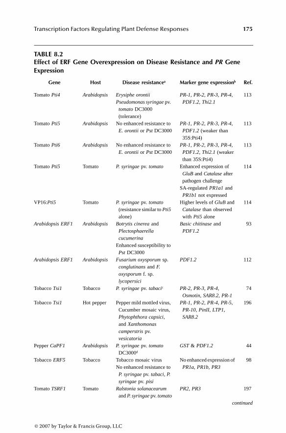
Transcription Factors Regulating Plant Defense Responses 175
TABLE 8.2Effect of ERF Gene Overexpression on Disease Resistance and PR Gene Expression
Gene Host Disease resistancea Marker gene expressionb Ref.
Tomato Pti4 Arabidopsis Erysiphe orontiiPseudomonas syringae pv.
tomato DC3000 (tolerance)
PR-1, PR-2, PR-3, PR-4, PDF1.2, Thi2.1
113
Tomato Pti5 Arabidopsis No enhanced resistance to E. orontii or Pst DC3000
PR-1, PR-2, PR-3, PR-4, PDF1.2 (weaker than 35S:Pti4)
113
Tomato Pti6 Arabidopsis No enhanced resistance to E. orontii or Pst DC3000
PR-1, PR-2, PR-3, PR-4, PDF1.2, Thi2.1 (weaker than 35S:Pti4)
113
Tomato Pti5 Tomato P. syringae pv. tomato Enhanced expression of GluB and Catalase after pathogen challenge
SA-regulated PR1a1 and PR1b1 not expressed
114
VP16:Pti5 Tomato P. syringae pv. tomato (resistance similar to Pti5 alone)
Higher levels of GluB and Catalase than observed with Pti5 alone
114
Arabidopsis ERF1 Arabidopsis Botrytis cinerea and Plectosphaerella cucumerina
Enhanced susceptibility to Pst DC3000
Basic chitinase and PDF1.2
93
Arabidopsis ERF1 Arabidopsis Fusarium oxysporum sp. conglutinans and F. oxysporum f. sp. lycopersici
PDF1.2 112
Tobacco Tsi1 Tobacco P. syringae pv. tabacic PR-2, PR-3, PR-4, Osmotin, SAR8.2, PR-1
74
Tobacco Tsi1 Hot pepper Pepper mild mottled virus, Cucumber mosaic virus, Phytophthora capsici, and Xanthomonas camperstris pv. vesicatoria
PR-1, PR-2, PR-4, PR-5, PR-10, PinII, LTP1, SAR8.2
196
Pepper CaPF1 Arabidopsis P. syringae pv. tomato DC3000d
GST & PDF1.2 44
Tobacco ERF5 Tobacco Tobacco mosaic virusNo enhanced resistance to P. syringae pv. tabaci, P. syringae pv. pisi
No enhanced expression of PR1a, PR1b, PR3
98
Tomato TSRF1 Tomato Ralstonia solanacearum and P. syringae pv. tomato
PR2, PR3 197
continued
3063_book.fm Page 175 Thursday, August 24, 2006 1:23 PM
© 2007 by Taylor & Francis Group, LLC

176 Model Plants and Crop Improvement
Pseudomonas syringae [113], but overexpression of Arabidopsis ERF1 increasedsusceptibility to this pathogen [93]. Such results may reflect examples of negativeor positive cross-talk, respectively, between the JA/ET and SA signaling pathwaysmediated by transgenic expression of the ERF protein. It is also notable that over-expression of tomato Pti5 in Arabidopsis failed to increase resistance to P. syringaeor Erysiphe orontii [113], but this TF was effective against the former pathogenwhen overexpressed in tomato [114]. These apparently conflicting results may beattributed to distinct functionalities between proteins (in the case of ERF1 and Pti4)or of the proteins in different host plants (Arabidopsis vs. tomato), as well as tovariations in experimental designs between studies.
Enhanced resistance to pathogens conferred by overexpression of ERF geneswas usually correlated with enhanced levels of marker PR gene expression followingpathogen challenge or with their constitutive expression [93,113,114]. Expressionof a tomato Pti5:VP16 fusion protein was found to elicit higher constitutive levelsof marker PR genes than the native Pti5, but did not substantially increase diseaseresistance compared to native Pti5 [114]. Lack of enhanced disease resistance inArabidopsis plants overexpressing Pti5 or Pti6 was correlated with weak to noconstitutive expression of marker genes [113].
Most marker genes analyzed in the preceding studies contained GCC-boxes intheir promoters and were known to be responsive to ET or JA. In some cases, onlya subset of GCC-box-containing marker genes tested was activated in disease-resistant transgenic plants [114]. SA-inducible genes were also tested and found tobe activated in some cases [113], but not in others [98]. As discussed later (Section8.2.6), transcript profiling of transgenic plants overexpressing ERF genes revealedthat these TFs potentially regulate large sets of genes [100,115,116].
Overexpression of Pti4 and ERF1 in Arabidopsis was correlated with develop-mental abnormalities. The most common phenotype observed is reminiscent of the
TABLE 8.2 (continued)Effect of ERF Gene Overexpression on Disease Resistance and PR Gene Expression
Gene Host Disease resistancea Marker gene expressionb Ref.
Tomato TSRF1 Tobacco R. solanacearum PR1, PR2 & PR3 197Tomato OPBP1 Tobacco Phytophthora parasitica
var. nicotianae and P. syringae pv. tabacie
PR-5d, PR-1a 95
Pepper CaERFLP1 Tobacco P. syringae pv. tabacie β-glucanase, osmotin, HMG-CoA reductase, cysteine protease
198
a Unless otherwise noted, overexpression of ERF gene resulted in enhanced disease resistance.b Unless otherwise noted, overexpression of ERF gene resulted in constitutive expression of the genes listed.c Plants also display enhanced tolerance to osmotic stress.d Plant also display enhanced freezing tolerance.e Plants also display enhanced salt tolerance.
3063_book.fm Page 176 Thursday, August 24, 2006 1:23 PM
© 2007 by Taylor & Francis Group, LLC

Transcription Factors Regulating Plant Defense Responses 177
so-called ethylene triple response, referring to the inhibition of hypocotyl and rootelongation and an exaggerated curvature of the apical hook observed in wild-typeseedlings exposed to ET. Seedlings of Arabidopsis plants overexpressing Pti4 orERF1 display the inhibition of hypocotyl elongation in the absence of ET[104,113,115]; those overexpressing ERF1 also display inhibition of seedling rootelongation [104]. In no case was the curvature of the apical hook found to be affected.Interestingly, HOOKLESS1 (HLS1), a gene regulating the apical hook curvature,contains a GCC-box in its promoter. Although ERF proteins can bind in vitro to apromoter containing a multimer of the HLS1 GCC-box [49], HLS1 expression is notactivated in Pti4 or ERF1 overexpressing plants [104,113,115].
Plants overexpressing Pti4 and ERF1 also display phenotypes associated withET exposure at the adult stage, including smaller size, greener leaves, and, in thecase of ERF1 overexpressors, inhibition of cell enlargement, wilting, and deathbefore bolting [104]. Together, these results indicate that subsets of ET responsesare constitutively activated by overexpression of these ERF genes. Overexpressionof the other ERF genes tested to date has not been reported to induce developmentalabnormalities.
The phenotypic consequences associated with loss of ERF gene function haveyet to be determined. Analysis of the SIGnAL T-DNA database (http://signal.salk.edu)revealed the presence of insertions in or near many members of the ArabidopsisAP2/ERF family [117]. However, there have been no published reports of theircharacterization. Similarly, antisense expression of the tomato Pti5 was reported tohave no effect on race-specific resistance [114], although no data were shown.
8.2.4.3 WKRY Factors
Similar to the ERF family, there are few reports describing the phenotypic conse-quences of mutations in WRKY genes. Mutations in more than 40 Arabidopsis WRKYgenes have been identified by reverse genetics, but most do not appear to displayvisible mutant phenotypes [63]. The consequences of altering levels of WRKYfactors on disease resistance have been reported for only five Arabidopsis genes.VIGS has also been applied to study the involvement of WRKY genes from solan-aceous plants in R-gene mediated resistance pathways.
Overexpression of WRKY70 enhanced resistance to virulent strains of the bac-terial pathogens Erwinia carotovora and P. syringae, but antisense-mediated reduc-tion of WKRY70 resulted in enhanced susceptibility to these pathogens [43]. Over-expression was correlated with increased expression of SA-inducible PR genes anddecreased expression of JA/ET-regulated defense-related genes. Conversely, anti-sense suppression of WRKY70 resulted in constitutive expression of JA/ET-regulatedmarker genes. Together, these results indicate that WRKY70 acts as a positiveregulator of disease resistance and it was proposed to represent a convergence pointfor the SA and JA signaling pathways [43].
Overexpression of WRKY18 also resulted in enhanced resistance to P. syringaeand increased expression of SA-inducible PR genes [118]. However, these phenotypeswere only observed in older plants (e.g., 5 weeks old). Interestingly, mutations at theNPR1 locus had little effect on PR gene expression in the WKRY70 overexpressors
3063_book.fm Page 177 Thursday, August 24, 2006 1:23 PM
© 2007 by Taylor & Francis Group, LLC

178 Model Plants and Crop Improvement
[43]; however, they abolished potentiation of PR genes and enhanced disease resis-tance observed in older plants overexpressing WKRY18 [118].
Despite increasing levels of PR genes, overexpression of WRKY6 had no appre-ciable effect on resistance to virulent or avirulent strains of P. syringae [80]. Transpo-son-induced mutation of WRKY6 also induced changes in gene expression consistentwith a possible role in defense responses, but was not associated with any visiblemutant phenotype [80]. Thus, although results implicate WKRY6 and WRKY18 inmediating the expression of defense genes, they also indicate that neither gain nor loss(in the case of WRKY6) of these TFs by itself is sufficient to affect disease resistance.
In all cases reported, stable transgenic overexpression of WRKY genes resultedin developmental abnormalities, including stunted growth, altered leaf morphology,and changes in flowering time [43,80,118]. Overexpression of WRKY6 was alsoassociated with development of necrotic areas on leaves and loss of apical dominance[80]. Reduction of WRKY70 levels resulted in larger plants and early flowering(overexpression of WRKY70 delays flowering) [43], but reduction or loss of WRKY6did not result in any developmental abnormalities [80]. Expression of WRKY6 andWRKY18 transgenes also decreased levels of corresponding endogenous genes[80,118], suggesting that these TFs are involved in regulating expression of theirgenes and, possibly, other members of the family. Transcript profiling of plants withaltered levels of WRKY6 and WRKY70 also supports the notion that these TFs canactivate and suppress target gene expression [43, 80].
Transient overexpression of AtWRKY29 in Arabidopsis leaves reduced diseasesymptoms caused by P. syringae and B. cinerea [119]. However, the effects ofaltering levels of this WRKY gene have not been tested in stable transgenic or mutantplants. VIGS has also implicated the tobacco WRKY1, WRK2, and WRKY3 genes asbeing required for full N gene-mediated resistance to TMV [120].
8.2.4.4 Whirly Factors
AtWhy1 is the only Whirly factor functionally characterized to date [37]. Twomissense mutant alleles in this gene were recovered by targeting induced local lesionsin genomes (TILLING). Based on the crystal structure of StWhy1, one mutationmapped to the single-stranded DNA-binding domain, and the other was located inthe region implicated in tetramerization. Mutant plants exhibited reduced AtWhy1DNA-binding activity and PR-1 expression following SA treatment. Both mutantbackgrounds were hypersusceptible to a virulent strain of the biotrophic oomycetePeronospora parasitica. The atwhy1.2 mutant also showed intermediate susceptibil-ity to an avirulent strain of P. parasitica and was unable to mount an effective SARresponse following treatment with SA [37]. Thus, AtWhy1 constitutes a positiveregulator of SA-dependent disease resistance. However, SA-induced Whirly DNA-binding activity does not require functional NPR1, suggesting that Whirly activationoccurs through a distinct, NPR1-independent pathway.
8.2.4.5 TGA Factors
The most compelling evidence implicating TGA factors as mediators of defenseresponses comes from the analysis of the triple tga2tga5tga6 loss-of-function
3063_book.fm Page 178 Thursday, August 24, 2006 1:23 PM
© 2007 by Taylor & Francis Group, LLC

Transcription Factors Regulating Plant Defense Responses 179
Arabidopsis mutant [36]. This mutant is compromised in SAR against virulent strainsof P. syringae and P. parasitica. Interestingly, basal resistance to these pathogenswas not compromised. The mutant also failed to express PR-1 in response to SA,but displayed higher basal levels of PR-1 in the absence of SA elicitation. Althoughno developmental abnormalities were reported, seedlings of the triple mutant werehypersensitive to the toxic effects of SA, a phenotype also observed in npr1 mutants(see Section 8.3.1.1).
Attempts to study the role for TGA factors in mediating disease and PR geneexpression using dominant-negative versions of TGA factors have yielded conflictingresults. Expression of a dominant-negative Arabidopsis TGA2 gene in Arabidopsiscompromised basal resistance against P. syringae pv. maculicola [83]; expression ofa similar dominant-negative version of the same Arabidopsis gene in tobaccoenhanced SAR against P. syringae pv. tabaci [121]. In these studies and others[73,122], the transgenic plants were also monitored for expression of marker genescontaining as-1 elements. Two types of genes were considered, based on the timingof their expression following SA treatment: early genes and late genes.
PR-1 is considered a late gene, and was the only maker gene evaluated in onestudy [83]. Loss of basal resistance observed by transgenic expression of dominant-negative TGA2 was correlated with reduced levels of PR-1 in Arabidopsis [83], whileenhanced SAR in tobacco was associated with increased levels of PR-1a but reducedlevels of early gene transcripts [121]. In contrast, a dominant-negative tobaccoTGA2.2 resulted in reduced levels of early and late genes, while (over)expressionof wild-type TGA2.2 increased levels of early genes but had no affect on PR-1a [73].Finally, plants expressing wild-type or dominant-negative versions of the closelyrelated tobacco TGA2.1 gene displayed enhanced and reduced levels of early genes,respectively, but showed no changes in PR-1a levels [122].
Based on the observation that dominant-negative TGA2 has opposite effects onearly and late gene expression, it was proposed that TGA factors have both positiveand negative roles in mediating plant defense responses [121]. Additional evidencein support of a dual role for TGA factors comes from RNAi analysis, which revealeda negative role for TGA4 and a positive role for TGA5 in regulating a reporter geneunder the control of a multimerized cis-element related to as-1 [123]. The conse-quences of (over)expressing wild-type or dominant-negative TGA2.1 and TGA2.2,or the TGA4 and TGA5 RNAi transgenes on disease resistance have yet to bedetermined.
In the only study that appears to have observed altered disease resistance as aconsequence of (over)expressing TGA factor genes, Kim and Delaney [124] foundthat transgenic Arabidopsis plants overexpressing TGA5 displayed enhanced resis-tance to a virulent strain of P. parasitica, but reduced levels of PR genes. In contrast,neither sense nor antisense overexpression of TGA2 affected resistance to thisoomycete [124].
Overall, most analyses to date have focused on group II TGA factors, whichinclude TGA2, TGA5, and TG6. The only study demonstrating a role for group ITGA factors in mediating disease resistance comes from VIGS of tomato TGA1homologs, which compromised Pto-mediated resistance to P. syringae pv. tomatoharboring avrPto [125].
3063_book.fm Page 179 Thursday, August 24, 2006 1:23 PM
© 2007 by Taylor & Francis Group, LLC

180 Model Plants and Crop Improvement
8.2.4.6 R2R3 MYB Proteins
The R2R3MYB gene BOS1 was recovered in a genetic screen aimed at identifyingArabidopsis genes required for resistance to B. cinerea [32]. Loss of BOS1 functionresulted in enhanced susceptibility to necrotrophic fungi, including B. cinerea andAlternaria brassicicola. The bos1 mutant displayed more severe disease symptomsin response to infection by the biotrophic pathogens P. parasitica and P. syringae;however, no detectable increase in growth of these pathogens was observed. Themutant accumulated more reactive oxygen species (ROS) in response to B. cinereainfection and was also more sensitive to a number of abiotic stresses [32]. Expressionof marker genes for the SA and JA signaling pathways were not altered in the bos1mutant; however, BOS1 transcripts were found to accumulate following infectionwith B. cinerea. Increased BOS1 expression was blocked in the coi1 mutant thatmediates JA signaling, implicating BOS1 as a mediator of this defense pathway.
The Arabidopsis MYB30 gene was initially identified based on its differentialexpression during a type of programmed cell death called the hypersensitive response(HR) associated with incompatible interactions [102]. Constitutive overexpressionof MYB30 in Arabidopsis and tobacco resulted in elevated expression of HR markergenes, accelerated HR against avirulent pathogens, and development of HR-likelesions upon infection with virulent pathogens [42]. Importantly, it increased resis-tance to virulent and avirulent biotrophic pathogens. Conversely, antisense suppres-sion of MYB30 suppressed HR marker gene expression, delayed the HR againstavirulent pathogens, and decreased resistance against virulent and avirulent patho-gens. Constitutive expression of MYB30 did not result in spontaneous lesion forma-tion, indicating that factors other than MYB30 are required to initiate the HR [42].
The tobacco MYB1 gene was also shown to be required for N-mediated resistanceto TMV using VIGS technology [120].
8.2.4.7 Zinc Finger-Related Proteins
The Arabidopsis LSD1 gene was isolated in a screen aimed at identifying mutantsthat misregulate cell death responses [31]. Loss-of-function lsd1 mutants initiallydisplay a normal HR in response to infection with virulent pathogens, but cannotlimit the extent of the cell death, leading to a phenotype known as runaway celldeath. Runaway cell death in the lsd1 mutant is also observed following treatmentwith SA and SA analogs and depends on production of superoxide. It requires NPR1as well as the positive regulators of disease resistance ENHANCED DISEASESUSCEPTIBILITY 1 (EDS1) and PHYTOALEXIN DEFICIENT 4 (PAD4), bothof which are putative lipases [126]. lsd1 mutants are more resistant virulent strainsof P. parasitica, but neither SA nor NPR1 is required for this phenotype [38,126].These results indicate that LSD1 is a negative regulator of basal defense responses,somehow interpreting ROS-dependent signals triggered by the HR [126].
LOL1 was identified based on its similarity to LSD1 and the two proteins appearto have opposite roles in mediating cell death and disease resistance [38]. Reductionof LOL1 expression in the lsd1 mutant suppresses runaway cell death, while condi-tional high-level expression of LOL1 is sufficient to trigger cell death in the absence
3063_book.fm Page 180 Thursday, August 24, 2006 1:23 PM
© 2007 by Taylor & Francis Group, LLC

Transcription Factors Regulating Plant Defense Responses 181
of pathogen infection or SA treatment. Furthermore, stable moderate overexpressionof LOL1 enhanced resistance to virulent strains of P. parasitica, while reduction ofLOL1 levels enhanced susceptibility to this pathogen [38].
Overexpression of the pepper CaZFP1 gene in transgenic Arabidopsis resultedin enhanced resistance to a virulent strain of P. syringae and enhanced tolerance ofdrought [70]. Overexpressing lines also displayed developmental abnormalities.Expression of SA-regulated PR genes or the JA-regulated PDF1.2 gene was notaltered by CaZFP1 overexpression, suggesting that the observed enhanced resistancewas mediated through the activation of other pathways.
8.2.4.8 MYC Proteins
The Arabidopsis jin1 mutants were identified in an ET-insensitive genetic back-ground as being insensitive to JA and represent alleles of AtMYC2 [33]. Mutantplants display enhanced resistance to necrotrophic pathogens, including B. cinereaand P. cucumerina. All jin1 mutant alleles are semidominant and retain the capacityto produce the N-terminal end of the protein. It has been speculated that these couldinterfere with interacting proteins.
8.2.5 POST-TRANSLATIONAL REGULATION
In addition to regulation at the transcriptional level (reviewed in Section 8.2.3.4),there is overwhelming evidence for the post-translational regulation of TFs inresponse to pathogen challenge. Most studies have focused on reversible phospho-rylation/dephosphorylation, which is a prevalent means of modulating the activityof eukaryotic TFs [127]. Reversible oxidoreduction of key cysteine residues isemerging as an important mechanism for regulating mammalian and microbial TFs,and recent studies have implicated this type of control in the regulation of plant TFsmediating defense responses [128]. Physical interactions between TFs and otherproteins are also likely to be of regulatory significance.
8.2.5.1 Post-Translational Modifications
In one of the few studies aimed at monitoring changes in the nuclear pools of TFsfollowing defense response elicitation, Turck et al. [103] visualized changes in thepattern of WRKY proteins by two-dimensional gel electrophoresis. Several proteinsmigrated as adjacent pearl strings, suggesting different post-translationally modifiedforms of the same protein. The number of proteins and the complexity of modifica-tions rapidly increased following treatment with an elicitor. However, the nature ofthe putative post-translational modifications has not been determined.
As detailed later, the property of TFs most frequently shown to be influencedby post-translational modification is their ability to bind DNA. In a few cases,protein–protein interactions were shown to be affected. Few studies have directlylinked post-translational modification to changes in transactivation potential. How-ever, several have shown that transactivation is regulated in response to treatmentwith elicitors or growth regulators implicated in mediating defense responses; this
3063_book.fm Page 181 Thursday, August 24, 2006 1:23 PM
© 2007 by Taylor & Francis Group, LLC
182 Model Plants and Crop Improvement
suggests possible post-translational control. In many cases, the significance of post-translational modification for disease resistance is unknown.
8.2.5.1.1 PhosphorylationThe ability of TFs present in plant extracts to bind to several cis-acting elementsimplicated in mediating defense gene expression (e.g., W-box, as-1, ERE; see Table8.1) is dramatically altered by treatment with phosphatase or protein kinase inhibitors[65,129–131]. In addition, constitutive activation of a MAPK signaling cascade intobacco resulted in substantially more binding activity to the W-box [129].
Consistent with this result, two WRKY factors (WKRY22 and WRKY29) wereidentified as downstream components of an MAPK signaling cascade conferringbasal resistance to bacterial and fungal pathogens in Arabidopsis [119]. Transcriptionfactors and components of MAPK signaling cascades were also identified as beingrequired for N-mediated resistance to TMV in tobacco [120] and Pto-mediatedresistance to P. syringae (avrPto) in tomato [125]. However, in none of these studieswas it demonstrated that the TFs were directly phosphorylated by MAPKs. Onestudy suggested that the MAPK cascade may lead to phosphorylation and inactiva-tion of a specific inhibitor of WRKY proteins [119]; results from another wereconsistent with regulation occurring at the point of WRKY transcription [129].
The rice ERF OsEREBP1 is the only plant TF implicated in mediating defensegene expression that has been shown to be phosphorylated by a MAPK [132].Activity of this MAPK, BWMK1, is rapidly induced by a pathogen-derived elicitor,ET, SA, and JA. Overexpression of BWMK1 in transgenic tobacco results in con-stitutive expression of PR genes and enhanced resistance to P. syringae and theoomycete pathogen Phytophthora parasitica [132]. BWMK1 phosphorylatesOsEREBP1 in vitro, resulting in enhanced binding to the GCC-box. Furthermore,transient coexpression of BWMK1 and OsEREBP1 in Arabidopsis protoplastsinduced expression of a reporter gene under the control of the GCC-box to higherlevels than either gene alone [132]. Therefore, it is likely that OsEREBP1 is regulatedin vivo by a pathogen-activated MAPK signaling cascade.
The tomato ERF Pti4 is also regulated by phosphorylation. Pti4 physically interactswith the Pto R-gene product, which encodes a serine-threonine protein kinase [41].Pto kinase activity is required for interaction with Pti4 as well as for race-specificresistance against P. syringae pv. tomato harboring avrPto [92]. Phosphorylation ofPti4 by Pto enhances its DNA binding to the GCC-box element in vitro. This effectis highly specific because two protein kinases related to Pto (Fen and Pti1) do notphosphorylate Pti4, and Fen does not stimulate Pti4 DNA-binding activity [92]. Pti4contains ten threonine residues: two each in the putative DNA-binding and transacti-vation domains and one near the nuclear localization signal [41]. Pto phosphorylatesat least four of these residues (but no serines) [92]. At this time it is not known whichresidues are phosphorylated in vitro or in vivo. It is also not known whether Pti4 maybe regulated by other protein kinases during compatible plant–pathogen interactions.As discussed in Section 8.2.4.2, overexpression of Pti4 confers enhanced basal resis-tance to pathogens independently of the Pto–avrPto interaction.
Phosphorylation has also been implicated in the regulation of bZIP proteins.SARP (salicylic acid response protein) is a cellular factor immunologically related
3063_book.fm Page 182 Thursday, August 24, 2006 1:23 PM
© 2007 by Taylor & Francis Group, LLC

Transcription Factors Regulating Plant Defense Responses 183
to TGA factors implicated in mediating the rapid induction of as-1 binding activityfollowing treatment with SA [130]. In the absence of SA, it was proposed that SARPis sequestered by an inhibitory protein called SAI (SA-inhibitor) and that release ofSARP from SAI is triggered by SA-induced phosphorylation of either or bothproteins. Inhibitors of casein kinase II (CK2) suppress as-1 binding, suggesting thatCK2 may be responsible for activation of SARP [131].
More recently, the Arabidopsis TGA2 was shown to be phosphorylated in vivoby a CK2-like activity induced by SA [131]. Phosphorylation of TGA2 appeared tosuppress its ability to bind to the as-1 element. This is in contrast to previouslypublished studies showing that phosphorylation by CK2-like kinases enhanced as-1 binding in vitro. Importantly, no difference in PR-1 expression was observed intransgenic plants overexpressing TGA2 compared to those expressing site-directedmutant with putative CK2 phosphorylation sites removed [131]. Thus, the regulatorysignificance of TGA2 phosphorylation by CK2-like kinases remains elusive.
G/HBF-1 is a non-TGA bZIP TF that binds to an SA and elicitor-responsiveelement in the promoter of the chalcone synthase15 gene from soybean [25]. Elicitortreatment does not alter levels of G/HBF-1 transcript or protein; however, it rapidlyinduces activation of a protein kinase capable of phosphorylating G/HBF-1. Further-more, phosphorylation of G/HBF-1 in vitro increases its DNA-binding activity. Theidentity of the G/HBF-1 kinase is not known.
DNA-binding activity of Whirly proteins is also regulated by phosphorylation.In particular, a kinase related to mammalian protein kinase C has been implicated[65]. Even though Whirly factors are constitutively present in the nucleus, theirassociated DNA-binding activity is not detectable until after pathogen challenge ortreatment with SA or an elicitor [65]. Chromatographic purification of StWhy1 fromuninduced nuclei activates its DNA-binding activity, possibly by removing an uni-dentified inhibitor protein. The possible role of phosphorylation in mediating theinteraction of Whirly proteins with inhibitor proteins is unknown.
8.2.5.1.2 OxidoreductionBinding of nuclear factors to the as-1 element is regulated by redox conditions(reviewed by Fobert and Després [128]). A recent study has started to unravel thepossible mechanism involved.
The interaction between Arabidopsis TGA1 and NPR1 in plant cells is positivelyinfluenced by treatment with SA [133]. Two conserved cysteines located in the C-terminal region of TGA1 (C260 and C266) play a key role in regulating this inter-action. Although wild-type TGA1 and NPR1 do not interact in the yeast two-hybridsystem, mutation of these residues permits interaction with NPR1 in yeast and inArabidopsis cells regardless of SA induction. Using a novel labeling strategy thatdistinguishes between protein sulfhydryls and disulfides, it was demonstrated thatthe redox status of cysteines in TGA1 and/or the closely related TGA4 shiftedconsiderably following SA treatment to become predominantly reduced [133]. Thus,strong interaction with NPR1 is correlated with the reduced state of TGA1 and/orTGA4 cysteines.
Changing redox conditions altered the mobility of in vitro produced TGA1 innonreducing gel electrophoresis, consistent with the formation of an intramolecular
3063_book.fm Page 183 Thursday, August 24, 2006 1:23 PM
© 2007 by Taylor & Francis Group, LLC
184 Model Plants and Crop Improvement
disulfide bridge under oxidizing conditions [133]; this could impede interaction withNPR1. Changing redox conditions has been shown to influence the DNA-bindingactivity of several TFs (see Fobert and Després [128]); however, the ability of TGA1to bind the as-1 element in vitro was unaltered by vast molar excess of redox-regulatingcompounds. Instead, redox regulation of TGA1 DNA binding required the redox-regulated recruitment of NPR1, which was proposed to act as a cofactor in stimulatingTGA1 DNA-binding activity [133] (see also Section 8.3.2 and Section 8.3.3).
8.2.5.1.3. Protein Turnover/ProteolysisTGA factors were shown to have different stabilities during tobacco development;TGA1 and TGA3, but not TGA2, were rapidly degraded in mature leaves [89].Degradation of TGA3 appeared to be mediated via the proteosome. However, theregulatory significance, if any, of TGA factor degradation in response to SA orpathogen challenge was not assessed.
The role of targeted protein degradation in the regulation of ERF1 transcriptionwas discussed in Section 8.2.3.4. Some preliminary evidence also suggests regulationof defense-related TFs by proteolysis. The tobacco ERF3 was shown to interact witha ubiquitin-conjugating enzyme (NtUBC2) in the yeast two-hybrid system [134].However, NtUBC2 did not affect the transrepression capacity of ERF3 in transientassays. The Arabidopsis R2R3MYB protein BOS1 contains a consensus sumolyationmotif, suggesting that it may be regulated by SUMO, a ubiquitin-like modifierassociated with protein stabilization [32]. Finally, mutations of the Arabidopsis F-box protein CORONATINE INSENSITIVE1 (COI1) block JA-mediated defenseresponses, resulting in increased susceptibility to several necrotrophic pathogens[135]. Although the targets of COI1 are unknown, it has been proposed that it maymediate removal of TFs tagged by JA-dependent phosphorylation.
8.2.5.2 Protein–Protein Interactions
Transcription of eukaryotic genes is achieved by synergistic interactions betweencombinations of TFs [9]. This combinatorial control relies on precise juxtaposition-ing of specific TFs through DNA–protein and protein–protein interactions usingfunctional groupings of cis-regulatory elements called enhancers (or silencers).Enhancers are responsible for a subset of the total gene expression pattern.
Accordingly, a typical gene may contain several enhancer elements. The stablenucleoprotein complex containing enhancer DNA with associated bound TFs andinteracting proteins is sometimes referred to as an enhanceosome [136]. Little isknown about the organization of cis-acting modules required to create pathogen-responsive enhancer elements in plants. However, individual cis-acting elementsimplicated in defense gene regulation frequently cluster, and synergistic interactionsbetween closely spaced elements have been reported (e.g., W-boxes), suggestingcooperation between TFs [60].
Several examples of protein–protein interactions have already been discussed inthis chapter, including Pti4–Pto and TGA factors–NPR1. TGA3 has also been shownto interact with calmodulin, suggesting a possible role for calcium signaling inregulating TGA factors [137]. BZI-1, a bZIP protein not part of the TGA factor
3063_book.fm Page 184 Thursday, August 24, 2006 1:23 PM
© 2007 by Taylor & Francis Group, LLC

Transcription Factors Regulating Plant Defense Responses 185
family, interacts with a novel protein (ANK1) containing ankyrin repeats [138]. Asrevealed by analysis of dominant-negative transgenes, BZI-1 is required for resis-tance to TMV. ANK1 does not appear to act as a cofactor of BZI-1 and was speculatedto function in the cytosol rather than the nucleus.
Although the combinatorial model of gene expression stresses the importanceof interactions between TFs, few have been reported between factors implicated inmediating plant defense responses. TGA4 was shown to interact with the ERF proteinAtERF [139] and with members of the Dof family of TFs, which encode proteinswith a single zinc finger [140]. The Dof proteins (OBP1-3) are capable of bindingto a cis-element in the CaMV35S promoter and enhance the DNA-binding activityof TGA4 in vitro. The genes encoding OBP1-3 are inducible by SA. Overexpressionof OBP3 results in numerous developmental phenotypes, but its effects on defensegene expression and disease resistance were not assessed. PRHA, a homeoboxprotein capable of binding to an elicitor responsive element of a PR-10 promoter,also interacted with two putative cofactors [141].
The limited number of reported protein interactions may be attributed in part tolimitations of the yeast two-hybrid system for studying TFs. First, the natural auton-omous transactivation or transrepression properties of many TFs complicate theiranalysis in systems that rely on reconstitution of an active TF to detect reporter geneexpression. Second, yeast cells may not be competent to carry out post-translationalmodifications required for interactions. Finally, the yeast two-hybrid system monitorsbinary interactions between proteins. As indicated at the beginning of this section,TFs are likely to function as part of larger protein complexes requiring multipleprotein–protein and protein–DNA interactions. Approaches aimed at recovery andcharacterization of protein complexes in vivo, such as tandem affinity purification(TAP) strategies, coupled with mass spectroscopy, promise to be useful tools inidentifying proteins interacting with plant TFs [142].
8.2.6 TRANSCRIPTIONAL TARGETS AND NETWORKS
Even for the best characterized plant TFs, the identity of very few target genes isknown. Nevertheless, such information is very important in assigning function toindividual TFs. Large-scale transcript profiling studies have identified motifs relatedto W-box, GCC-box, and ERE as enriched in genomic DNA upstream of genesfound to be differentially expressed following exposure to pathogens, elicitors, ordefense-related growth regulators, the W-box being the most prevalent [7,143]. Thesegenes can be viewed as putative targets for WRKY, ERF, and Whirly factors, respec-tively. There have been no reports of enrichment for MYB binding sites, and as-1like elements appear to be under-represented in promoters of these genes. Severalnovel motifs are also enriched in promoters of these genes [7,116]; however, theidentity of the TFs binding to these sites, if any, is unknown.
As discussed earlier in this chapter, TFs bind to short stretches of DNA (5 to10 bp) that can accommodate limited sequence variability. Sequences outside theconsensus cis-element may also influence binding—in particular, the presence andspacing of neighboring TF recognition sites. Furthermore, only a fraction of TFswithin a family is likely to bind to any given promoter containing cis-elements that
3063_book.fm Page 185 Thursday, August 24, 2006 1:23 PM
© 2007 by Taylor & Francis Group, LLC
186 Model Plants and Crop Improvement
conform to the consensus binding sequence for that family. For example, a numberof genes containing GCC-boxes in their promoters are not differentially regulatedin plants overexpressing ERF factors [104,113,115]. Accordingly, it is not possibleto identify target genes accurately based uniquely on DNA sequence information.
Genome-wide transcript profiling of plants containing altered levels of a TFoffers a means of identifying putative targets for that specific factor. However, dueto the existence of transcriptional cascades (see later discussion), these approachescannot distinguish direct and indirect targets. Nevertheless, they have confirmed thatmany genes differentially regulated in plants with altered levels of WRKY6 containmultiple W-boxes in their promoters [80]; those ectopically expressing Pti4 areenriched for GCC-boxes [115,116]. One study also reported an enrichment of MYB-binding sites in Pti4 overexpressors [116].
Many of the genes found to be differentially regulated in plants expressing alteredlevels of WRKY or ERF TFs genes encode proteins with functions potentially relevantto disease resistance [43,80,100,115]. These include classical PR proteins, proteinsimplicated in oxidative stress responses (e.g., P450s, glutathione-S-transferase), cal-cium signaling, and cell wall modification. Numerous genes identified in these plants,including many without known functions, are also differentially regulated in wild-type plants following treatment with pathogen, elicitors, or defense-related growthregulators, suggesting a role in mediating disease resistance.
Comparison of overall gene expression profiles between plants with alteredWRKY70 levels and signaling mutants suggests that this TF controls expression ofa substantial number of genes regulated by the JA- and SA-dependent pathways(~60 to 40%, respectively); a positive correlation was observed between WRKY70-and JA-dependent expression, but the inverse was true for SA-dependent expression[43]. Similarly, more than one third of the genes induced by treatment with ET andJA were constitutively expressed in ERF1 overexpressors [100]. The overlapincreased to 80% if only genes classified as “defense related” were considered.
Another class of target genes frequently recognized in plants with altered levelsof WRKY and ERF genes includes those that encode TFs. Thus, transcriptionalrepression or, alternatively, activation and subsequent translation, of such genes couldlead to modulation of additional sets of target genes, some of which may againencode TFs. The result is a transcriptional cascade in which TFs at the top of thehierarchy have the potential to regulate entire developmental or metabolic programs[144,145]. Such proteins are commonly referred to as “master switches” or “con-trollers.” Well-known examples include homeotic genes in drosophila and plants;mutation of these genes leads to the dramatic development of organs at inappropriatepositions. Closer to the bottom of the hierarchy would be TFs controlling morefocused aspects of specific programs. Research in well-characterized systems, suchas yeast, has revealed the existence of a number of regulatory loops, or networks,in which the activity of one or more TFs influences that of another [146].
Results obtained with WRKY70 and ERF1 suggest that these TFs could bemaster controllers of defense-related pathways involving JA/SA and JA/ET signaling,respectively. Neither TF controls the expression of all genes known to be regulatedby any given signaling molecule; instead, they appear to control branches of thesepathways implicated in defense responses [43,100]. ERF1 has been proposed to
3063_book.fm Page 186 Thursday, August 24, 2006 1:23 PM
© 2007 by Taylor & Francis Group, LLC

Transcription Factors Regulating Plant Defense Responses 187
integrate JA and ET defense pathways and WRKY70 to mediate cross-talk betweenSA and JA pathways. Based on expression of marker genes, the Arabidopsis MYC2was recently proposed to discriminate between different JA-mediated defenseresponses [33]. It will be interesting to see if this claim is substantiated by genome-wide transcript profiling.
Chromatin immunoprecipitation (ChIP) permits identification of direct targetsfor TFs in real time and space [147]. Using this approach, StWhy1 was shown tobind to the potato PR-10 promoter in response to wounding and elicitor treatment[37], while the recruitment of Arabidopsis TGA2 and TGA3 to the PR-1 promoterwas shown to depend on SA and NPR1 [148]. ChIP analysis of parsley WRKY1revealed it was transiently recruited to regions containing W-boxes in the promoterof its own gene, as well as those of the immediate-early genes WRKY3 and PR1-1following elicitor treatment [103]. Occupancy by WRKY1 was associated withreduced expression of WRKY1 but enhanced expression of PR1-1.
When not occupied by WRKY1, these promoters are otherwise constitutivelyoccupied by different WRKY factors. It was proposed that activation of WRKY1 andPR1-1 involves elicitor-dependent post-translational modification of WRKY factorsalready occupying W-boxes in these promoters; the newly produced WRKY1 wouldbe subsequently recruited to autoregulate expression of its own gene and activatelate gene expression. Post-translational modification of these WRKY factors mayrely on MAPK signaling cascades as described in Section 8.2.5.1.1 [103].
ChIP analysis of a subset of Arabidopsis genes differentially expressed inresponse to Pti4 overexpression revealed that about 60% (11 of 18) were directtargets of this ERF [116]. Three promoters that were not immunoprecipitated withthe Pti4 antibody contained GCC-boxes, further emphasizing the notion that thepresence of cognate cis-elements in a gene promoter does not necessarily make it atarget in vivo. Interestingly, seven of the promoters immunoprecipitated lacked GCC-boxes within 1 kb upstream of the coding region. It was suggested that Pti4 may becapable of binding to a novel cis-element other than the GCC-box or be recruitedto promoters indirectly through other DNA-binding proteins [116].
New strategies such as ChIP chip [147] and sequence tag of genomic enrichment(STAGE) [149] now permit identification of direct targets of a TF on a genome-wide basis. Although neither method has yet to be applied for the characterizationof plant TFs implicated in mediating defense responses, a whole-genome Arabidopsistiling array is available that would be suitable for ChIP chip [19].
8.3 NPR1
8.3.1 GENETIC ANALYSIS OF NPR1
8.3.1.1 Isolation of npr1 Mutants and Cloning of NPR1 Gene
The npr1-1 mutation was found to block SA-inducible PR gene expression followingtreatment with the SA analog 2,6-dichloroisonicotinic acid (INA) [150]. Additionalnpr1 alleles were subsequently recovered in different genetic screens aimed atidentifying genes required for expression of PR genes in response to SA (salicylic
3063_book.fm Page 187 Thursday, August 24, 2006 1:23 PM
© 2007 by Taylor & Francis Group, LLC

188 Model Plants and Crop Improvement
acid insensitive1, renamed npr1-5 [151]), basal resistance against virulent P. syringae(enhanced disease susceptibility5 and 53, renamed npr1-2 and npr1-3, respectively[152]), and INA-induced SAR against P. parasitica (non-inducible immunity1; nim1[153]). The nim1 mutants have not been redesignated as npr1 alleles and continueto be referred to by their original names. All npr1/nim mutants, with the exceptionof nim1-5, are recessive; however, the dominant phenotype observed in nim1-5 plantsis likely attributed to a second site mutation [154]. Because the npr1 phenotypecannot be rescued by exogenous SA, it was proposed that NPR1 functions down-stream of this metabolite in the signaling pathway.
Mutations in NPR1 compromise basal resistance against biotrophic pathogenssuch as P. syringae, P. parasitica, and E. cichoracearum. They cannot mount effectiveSAR against P. syringae or P. parasitica or ISR against P. syringae [155]. Also,different npr1 mutants have been found to be more susceptible to some incompatibleraces of P. parasitica [153,156,157], P. syringae [151], or E. cichoracearum [158].Loss of NPR1 function does not appear to affect age-related resistance against P.syringae [159] or basal resistance against necrotrophic pathogens, including A.brassicicola and B. cinerea [160]. However, unlike the wild-type, npr1 mutants donot show enhanced resistance to B. cinerea following treatment with SA [161].
Although npr1 mutants fail to express well-accepted marker genes for the SA-signaling pathway (PR-1, PR-2, PR-5) in response to treatment with SA or SAanalogs, transcripts for these genes continue to be expressed in response to pathogenchallenge (albeit at reduced levels [152,157]) or in combination with mutations atother loci (see, for example, Dong [162]). These observations indicate that one ormore SA-dependent, NPR1-independent defense pathways exist in Arabidopsis.
Despite being unresponsive to exogenous SA, npr1 mutants accumulate hightiters of this metabolite following pathogen challenge [153] and npr1 seedlingsgrown in the presence of SA bleach and die after developing cotyledons [163]. Ithas been proposed that NPR1 may be involved in feedback regulation of SAaccumulation.
Cloning of the NPR1 gene revealed that it encodes a protein with two identifiableprotein–protein interaction motifs: a BTB/POZ (broad-complex, tramtrack, and bric-a-brac/pox virus and zinc finger) and an ankyrin repeat domain (ARD) [154,163].Several npr1 alleles affect conserved amino acids within the ARD, suggesting thatthis domain is important for NPR1 function. Although one mutation (npr1-2) mapsto the BTB/POZ, it is within a nonconserved region of the domain (unpublishedobservation) and thus should not be used as evidence that the NPR1 POZ/BTB isrequired for disease resistance. NPR1 contains no well-recognized DNA-bindingmotifs and cannot bind to the as-1 element in vitro [40]. This suggests that NPR1does not function as a sequence-specific TF; however, its ability to bind DNA hasnot been rigorously assessed.
Sequences encoding NPR1 or NPR1-related proteins can be identified in manyhigher plants, suggesting that the NPR1 function is well conserved. In support ofthis notion, VIGS analysis has demonstrated that a tobacco homolog of NPR1 isrequired for resistance to TMV [164] and a tomato homolog is required for resistanceto P. syringae (avrPto) [125].
3063_book.fm Page 188 Thursday, August 24, 2006 1:23 PM
© 2007 by Taylor & Francis Group, LLC

Transcription Factors Regulating Plant Defense Responses 189
8.3.1.2 npr1 Suppressors
In efforts to identify additional genes implicated in mediating defense responses,several groups have screened for mutations that restore PR gene expression or diseaseresistance in npr1 backgrounds (i.e., genetic suppressors). Several suppressors appearto have activated defense responses constitutively; even in the absence of pathogenchallenge, they contain elevated levels of SA, express PR genes, and displayenhanced basal resistance to pathogens, typically P. syringae and P. parasitica[165–168]. Many of these suppressors also display dwarfism and spontaneous dis-ease-like lesions. Several other mutants displaying these properties were also recov-ered in unrelated genetic screens. The observed phenotypes were subsequently shownto be largely independent of NPR1, prompting some researchers to classify them asnpr1 suppressors (see, for example, Kim and Delaney [169]).
Conversely, others have argued that any mutant displaying elevated levels of SAshould not be considered as a true suppressor of npr1 [162]. In fact, two suppressorsof this class contain mutations in putative R-genes, and the phenotypes likely resultfrom the constitutive activation of R-gene signaling [167,168].
A recessive mutation that restores SA-inducible PR gene expression and diseaseresistance in the npr1-1 background is suppressor of npr1-1 inducible1 (sni1) [170].Although plants are dwarfed, they contain wild-type levels of SA, express only lowconstitutive levels of PR genes, and do not produce any spontaneous disease-likelesions. Neither sni1 npr1-1 nor sni1 NPR1 plants are more resistant than the wild-type in the absence of INA. SNI1 encodes a novel leucine-rich nuclear protein thatlikely acts as a negative regulator of SAR. It was suggested that the probable roleof NPR1 in SAR is to remove SNI1 repression. Currently, no evidence suggests thatNPR1 and SNI1 physically interact [170].
Increased resistance to virulent P. syringae and P. parasitica in the suppressorof nim1 (son1) nim1-1 double mutant is independent of SA and not correlated withincreased levels of PR genes. Accordingly, it has been proposed to define a noveltype of SAR-independent resistance (SIR) [169]. Several PR genes are constitutivelyactivated in the son1 NIM1 (i.e., NPR1) background, indicating that SON1 alsoparticipates in SAR, possibly upstream of or at NPR1 [169]. SON1 encodes an F-box protein. Given that the son1 mutation is recessive, SON1 is likely a negativeregulator of SIR and SAR. It has been proposed that SON1 may target specificpositive regulators of defense responses for degradation by the ubiquitin/proteosomepathway [169].
8.3.1.3 Other Genetic Interactions
Genetic interactions between NPR1 and genes encoding TFs were described inSection 8.2. The current section summarizes findings that link NPR1 to other regu-lators of plant defense responses.
8.3.1.3.1 SA-Dependent SignalingThe recessive enhanced disease resistance1 (edr1) mutation leads to increased resis-tance against P. syringae and E. cichoracearum without constitutive activation ofPR genes. EDR1 codes for a MAP kinase kinase kinase [171]. This enhanced
3063_book.fm Page 189 Thursday, August 24, 2006 1:23 PM
© 2007 by Taylor & Francis Group, LLC

190 Model Plants and Crop Improvement
resistance is lost in the edr1 npr1 double mutant, suggesting that EDR1 is part of aMAPK cascade operating upstream of NPR1 that negatively regulates plant defenseresponses [171]. MAPK4 is a MAPK that also acts as a negative regulator of SA-dependent defense responses; however, the mapk4 mutant phenotype is not attenu-ated in the npr1 background, suggesting that it is part of a MAPK cascade that actsindependently or downstream of NPR1 [172].
Overexpression of two soybean calmodulin isoforms, GmCaM4-5, in Arabidop-sis results in constitutive expression of SA-inducible PR genes, spontaneous micro-HR, and enhanced resistance to virulent P. syringae [173]. Constitutive expressionof PR genes is lost when GmCaM4-5 are introduced into an npr1 mutant background,suggesting that it is mediated by NPR1. NPR1 is also partly required for the enhancedresistance to E. cichoracearum observed in the powdery mildew resistant4 (pmr4)mutant [174].
Pathogen-induced expression of SA-INDUCTION DEFICIENT2 (SID2), whichencodes ISOCHORISMATE SYNTHASE1, a key enzyme in SA synthesis, is ele-vated in the npr1 mutant [175]. In contrast, SA-dependent expression of PAD4 iscompromised in the npr1 mutant, although P. syringae-induced expression of thisgene is not affected [176]. These provide further evidence that NPR1 acts down-stream of SA and indicate that its role as a negative regulator of SA metabolismmay be achieved, at least in part, at the level of SID2 expression or through a signalamplification loop involving PAD4 [175,176].
The double mutant between npr1 and the gain-of-function mutation acceleratedcell death6 (acd6) displayed dramatic alterations in cell enlargement and divisionresulting in production of abnormal growths [177]. Subsequent detailed analysis ofthe npr1 single mutant revealed reduced cell numbers with high ploidy levels. Thus,in addition to a role in mediating SA defense responses, NPR1 is also required forSA-dependent control of cell growth. Such effects were not apparent upon casualobservation of the npr1 mutants.
The requirement of NPR1 for runaway cell death in the lsd1 mutant has alreadybeen discussed (Section 8.2.7.4). HR-associated cell death is also increased in thenpr1 single mutant in response to infection with avirulent races of P. syringae[177,178]. In contrast, no cell death or hydrogen peroxide accumulation is detectablein the nonrace-specific disease resistance1 (ndr1) mutant following challenge withP. syringae avrRpt2 [178]. Genetic analyses demonstrated that ndr1 is epistatic tonpr1 with respect to both these parameters. However, NDR1 and NPR1 have anadditive effect on SAR and PR-1 expression [179].
8.3.1.3.2 JA/ET-Dependent SignalingEpistasis analysis has revealed links between NPR1 and the ET signaling pathways.Resistance to B. cinerea is not compromised in the npr1-1 mutant and is moderatelyreduced in ein2, a mutant affected in the ET pathway; however, the npr1-1 ein2double mutant is considerably more susceptible to this necrotroph [161]. Further-more, breakdown of R-gene-mediated resistance to P. syringae (avrRpt2) is moresevere in the double mutant than in either single mutant alone [180].
Simultaneous application of SA with JA can inhibit JA-dependent responses inwild-type plants. This inhibition is alleviated in the npr1 mutant, suggesting that
3063_book.fm Page 190 Thursday, August 24, 2006 1:23 PM
© 2007 by Taylor & Francis Group, LLC

Transcription Factors Regulating Plant Defense Responses 191
NPR1 acts as a negative regulator of JA-dependent signaling. Interestingly, thisfunction of NPR1, unlike its role in SA-mediated signaling, does not require local-ization to the nucleus [181].
8.3.2 BIOCHEMICAL FUNCTION OF NPR1
The protein sequence of NPR1 provided few clues as to its function, other than itlikely interacts with protein partners through its POZ/BTB or ARD. Using the yeasttwo-hybrid system, several groups established that NPR1 interacts with various TGAfactors, including the Arabidopsis TGA2, TGA3, TGA5, TGA6, and TGA7[40,124,182]. As described in Section 8.2.5.1.2, TGA1 and TGA4 are capable ofinteracting with NPR1 only after reduction or mutation of key cysteines. The Ara-bidopsis NPR1 was also used as bait to screen a rice cDNA library and found tointeract with three TGA factors related to TGA2 and a putative homolog of the maizeliguleless2 [183]. Screening of a tomato library with a putative ortholog of NPR1from this species identified a group II TGA factor named NIF1 (NPR1 interactingfactor1) [184]. The tobacco group II factors TGA2.1 and TGA2.2 were also shownto interact with Arabidopsis NPR1 in directed two-hybrid tests [82].
With the exception of nim1-4 (Chern et al. [183] and our unpublished observa-tions) all NPR1 mutant alleles that compromise disease resistance encode proteinsthat fail to interact with TGA factors in the yeast two-hybrid system. Where tested,these mutants also fail to interact with TGA factors in vitro. This suggests that theability to interact with TGA factors in vivo is important for NPR1 function andestablishes TGA factors as downstream components of the NPR1 signaling pathway.
The NPR1–TGA2 interaction observed in yeast and in vitro has been confirmedin plant cells. Importantly, these studies have revealed that the interaction is stimu-lated by SA [83,185]. Visualization of the interaction in protoplasts using a protein-complementation assay revealed weak diffuse interaction throughout the cell underuninduced conditions, but intense nuclear interaction following SA treatment [185].This pattern is consistent with reports that TGA2 and NPR1 are predominantlylocalized in the nucleus following SA treatment [89,186]. In fact, nuclear localizationof NPR1 was shown to be required for PR-1 expression [186].
Several lines of evidence indicate that interaction with NPR1 is important forTGA factor function in vivo:
NPR1 stimulates the DNA-binding properties of interacting TGA factors invitro, including the reduced form of TGA1 [40,133].
Protein extracts prepared from wild-type transgenic plants expressing a chi-meric TGA2:GAL4 DNA-binding domain (DB) protein bound a probecontaining GAL4 binding sites substantially better than similar extractsprepared from npr1 mutant plants expressing the chimeric protein [83].
Activation of a reporter gene under the control of a promoter containingGAL4-binding sites was only detected in wild-type transgenic plantsexpressing the TGA2:GAL4 DB fusion; no expression was detected in thenpr1 background [83]. The DNA-binding enhancement and transactivation
3063_book.fm Page 191 Thursday, August 24, 2006 1:23 PM
© 2007 by Taylor & Francis Group, LLC

192 Model Plants and Crop Improvement
properties of the TGA2:GAL4 DB chimeric protein observed in the wild-type plants depended on SA.
The ability of TGA2 and TGA3 to bind to the PR-1 promoter, as measuredby ChIP, depended on functional NPR1 and SA [148].
Although NPR1 clearly enhances the binding of interacting TGA factors byEMSA, it does not alter the migration of the protein–DNA complex [40,83,133].Furthermore, supershift experiments using antibodies against NPR1 antibodies indi-cate that NPR1 is not present in these complexes. This suggests that NPR1 stimulatesthe DNA-binding activity of TGA factors without binding stably to the TGA–DNAcomplex. It is possible that the NPR1–TGA interaction is insufficiently robust towithstand the polyacrylamide gel electrophoresis or that, upon binding DNA, theTGA factors release NPR1.
Although there are no reports of direct interactions between NPR1 and WRKYfactors, several lines of evidence suggest a role for this group of TFs as regulatorsof NPR1 expression and potential mediators of the NPR1-signaling pathway. First,many genes differentially regulated in the npr1 mutant contain W-boxes in theirpromoters [7,143]. Second, the expression of numerous WRKY genes depends onNPR1 function [101]. Finally, WRKY factors bind to W-boxes in the promoter ofthe NPR1 gene, and mutation of these elements compromises NPR1 expression [101].
Thus, NPR1 appears to act as a novel cofactor to regulate PR-gene expressionby modulating the activity of TGA factors and possibly other groups of TFs inresponse to pathogen challenge. However, the observation that cross-talk betweenthe SA and JA signaling pathways does not require nuclear localization of NPR1[181] suggests that this versatile regulator may have additional modes of action.
8.3.3 POST-TRANSLATIONAL REGULATION OF NPR1
Yeast two-hybrid screens also identified a novel group of four Arabidopsis proteinscalled NIMINs (NIM1 INTERACTORs) [187]. The interaction of NPR1 withNIMIN1 has been confirmed in plants. Overall, NIMIN proteins share limitedsequence identity (14 to 44%); however, they contain short stretches of high simi-larity. For example, NIMIN1 and NIMIN3 possess stretches of acidic amino acidsthat may serve as effector domains, and NIMIN1 and NIMIN2 each contain a clusterof basic amino acids that may represent a nuclear localization signal. All NIMINproteins contain a motif similar to the EAR domain necessary for transrepressionof ERF proteins (see Section 8.2.3.2) and are nuclear localized. NIMIN genes aretransiently expressed following treatment with SA.
Overexpression of NIMIN1 in Arabidopsis reduced PR gene expression inresponse to SA and avirulent P. syringae and compromised SAR against virulent P.syringae [187]. These phenotypes are reminiscent of the npr1 mutation; however,unlike that mutant, NIMIN1 overexpressing plants were also compromised in R-gene-mediated resistance against P. syringae (avrRpt2). Overexpression of a NIMIN1mutant protein unable to interact with NPR1 had no effect on PR gene expression ordisease resistance, indicating that NIMIN1 function is mediated through NPR1 [187].
3063_book.fm Page 192 Thursday, August 24, 2006 1:23 PM
© 2007 by Taylor & Francis Group, LLC
Transcription Factors Regulating Plant Defense Responses 193
Mutation and RNAi suppression of NIMIN1 resulted in increased expression of PRgenes but had no measurable effects on resistance to P. syringae.
It was proposed that NIMIN1 may regulate only a subset of NPR1-dependentdefense genes and that derepression of this specific gene set in the nimin1 mutantand RNAi lines is insufficient to confer disease resistance. It is also possible thatthe closest relative of NIMIN1 (NIMIN1b) may partly compensate for loss ofNIMIN1 function under some conditions [187]. Furthermore, one cannot discountthe possibility that the phenotypes observed in the plants overexpressing NIMIN1may be related to squelching (see Section 8.2.4.1), with the vast excesses of NIMIN1protein binding all available NPR1 at the expense of other NIMIN proteins orinteracting partners. Overall, functional analysis of NINIM1 indicates that it is anegative regulator of NPR1, possibly involved in modulating the amplitude of certaindefense responses that may be relevant to plant fitness [187].
The fact that NPR1 is constitutively expressed but changes subcellular localiza-tion following pathogen infection or SA treatment suggests that it is regulated at thepost-translational level. NPR1 shares limited sequence similarity with the mamma-lian transcriptional regulator Iκ-B, including conserved N-terminal lysines andserines potentially involved in ubiquitination and phosphorylation events, respec-tively [154]. However, there is no evidence to support the notion that NPR1 isregulated by either of these mechanisms.
In contrast, some evidence does support post-translational regulation by redoxchanges of conserved cysteines [188]. Using nonreducing SDS-PAGE to monitorthe presence of disulfide bonds, the mobility of a protein fusion between NPR1 andthe green fluorescent protein (GFP) was found to change dramatically in responseto INA. In uninduced samples, a large cytosolic complex was detected, consistentwith an NPR1 oligomer held together by intermolecular disulfide bridges. FollowingINA treatment, a band corresponding to the monomeric size of NPR1-GFP graduallyaccumulated; the fusion protein was detected in the nucleus and PR-1 expressionactivated. Accordingly, it was proposed that INA triggered the reduction of NPR1disulfide bridges, yielding biologically active monomers [188].
To identify residues affected by changes in redox status, NPR1-GFP fusionscontaining site-directed mutations at each of ten conserved NPR1 cysteines wereexpressed in transgenic plants. Mutation of two cysteines (C82 and C216) resultedin the constitutive expression of monomeric, nuclear NPR1-GFP as well as PR-1expression in the absence of INA. Because altered residues would no longer becapable of forming disulfide bridges, they were predicted to mimic the reduced stateof the cysteine residues.
Together these results suggest that reduction of C82 and C216 is critical forNPR1 activation. Consistent with this notion, monitoring the levels and ratio ofreduced to oxidized glutathione following INA treatment revealed that after an initialoxidative phase, the cellular redox status became more reducing [188]. Togetherwith those obtained with TGA1 ([133]; see Section 8.2.5.1.2), these results indicatethat properties of proteins mediating SA-dependent defense responses are regulatedby SA-mediated reduction of key cysteine residues. Currently, the enzymes respon-sible for modulating the redox changes of TGA1 and NPR1 cysteines or how thesechanges specifically affect protein structure and function is not known.
3063_book.fm Page 193 Thursday, August 24, 2006 1:23 PM
© 2007 by Taylor & Francis Group, LLC

194 Model Plants and Crop Improvement
8.3.4 ANALYSIS OF TRANSGENIC PLANTS OVEREXPRESSING NPR1
Arabidopsis plants expressing higher levels of NPR1 protein have increased basalresistance to P. syringae, P. parasitica [189,190], and E. cichoracearum [189]. Inone study, transgenic plants also displayed enhanced SAR following treatment withlow levels of a chemical activator (benzothiadiazole; BTH) that did not elicit aresponse in untransformed plants [189]. Most transgenic lines did not constitutivelyexpress SA-inducible PR genes. In one study, increased resistance was correlatedwith stronger, rather than faster, PR gene expression [190]; in the other study,resistance was correlated with the speed, rather that the level, of PR gene expression[189]. A direct correlation between levels of NPR1 and disease resistance wasreported in only one of the two studies [190].
The Arabidopsis NPR1 gene has also been overexpressed in two heterologoushosts. Expression in tomato resulted in substantial resistance against virulent strainsof P. syringae and F. oxysporum; moderate resistance to Xanthamonas campestris,Ralstonia solanacearum, and Stemphylium solani; and no enhanced resistance to P.infestans, cucumber mosaic virus, and tomato yellow leaf curl virus [191]. Levelsof resistance to P. syringae and F. oxysporum were reported to be comparable, butnot as complete, as those conferred by R-genes. In general, levels of NPR1 werecorrelated with the effectiveness of disease resistance; however, several exceptionswere noted, leading the authors to propose that resistance may require a thresholdlevel of NPR1 expression. There appeared to be no correlation between the levelsof six PR genes tested with levels of NPR1 or disease resistance [191].
Expression of Arabidopsis NPR1 in rice resulted in increased resistance tovirulent Xanthamonas oryzae [183]. These results suggest that NPR1 function andsignaling are conserved among dicots, monocots, and, in particular, rice, which hasvery high constitutive levels of endogenous SA. Resistance to X. oryzae conferredby NPR1 was not as effective as R-gene resistance; however, substantial reductionof pathogen growth was observed in the leaf central vein, which limited bacterialspread and enhanced survival.
As suggested in tomato, it appeared that a threshold level of NPR1 was requiredto confer resistance in rice [183]. Closer examination of the rice transgenics underdifferent growth conditions revealed that overexpression of NPR1 can trigger thedevelopment of spontaneous disease-like lesions [192]. This phenotype was corre-lated with the accumulation of hydrogen peroxide and could be potentiated byexposure to SA or BTH. The transgenic plants were found to contain lower levelsof SA—once again providing a link between NPR1 and regulation of SA metabolism.Neither the production of lesions nor changes in SA levels were observed in dicot-yledonous plants overexpressing NPR1 [189–191].
8.3.5 NPR1-RELATED GENES
Completion of genome sequence revealed that Arabidopsis encodes six NPR1-relatedproteins that can be separated into three discrete groups [157]. All of these proteinscontain the POZ/BTB and ARD domains implicated in mediating protein–proteininteractions. Every amino acid affected by known npr1 mutations that compromise
3063_book.fm Page 194 Thursday, August 24, 2006 1:23 PM
© 2007 by Taylor & Francis Group, LLC
Transcription Factors Regulating Plant Defense Responses 195
PR gene expression and disease resistance is conserved within the family. The genesencoding the three family members most closely related to NPR1 (At4g26120,At5g45110, At4g19660) also contain introns at the same positions as NPR1.
Three Arabidopsis NPR1-related genes have recently been functionally analyzed.Of these, the product of NPR4 (A4g19660) is the most closely related to NPR1,sharing 36% identity. Similar to NPR1, NPR4 is localized in the nucleus and caninteract with the same spectrum of TGA factors. NPR4 transcript is induced by SAand pathogen challenge and is rapidly repressed by JA [157]. Mutation of NPR4results in a modest increase in susceptibility to virulent P. syringae and E. cichora-cearum but not P. parasitica. However, partial breakdown in R-gene resistanceagainst some races of P. parasitica was reported. PR gene expression was onlymarginally reduced in the npr4 mutant. Although these results implicate NPR4 inregulation of PR genes and resistance against biotrophic pathogens, they also suggestthat its role is not as prominent as that of NPR1. It was suggested that NPR1 andNPR4 may have distinct roles in modulating cross-talk between SA- and JA-depen-dent defense signaling.
BLADE ON PETIOLE (BOP) 1 and 2 are the two most distantly related membersof the NPR1-gene family. These genes have been implicated in regulation of plantdevelopment [193,194] and their contribution to defense responses is unclear.
8.4 . . . TO CROP IMPROVEMENT
Thanks in large part to use of model systems, our knowledge of TFs involved inregulating plant defense responses has increased very rapidly in recent years. It isclear that numerous TFs, encoded by different gene families, are required to ensurethe highly coordinated expression of defense genes in response to pathogen chal-lenge. Many TF genes are highly regulated at the transcription level and the productsthey encode can act as positive or as negative regulators of gene expression. Thus,in addition to the activation of positive factors, the elimination of negative regula-tors—at the transcriptional or post-translational level—is required for timely activa-tion of plant defense responses.
Highly related TFs can be recognized within gene families. The roles of theseproteins appear to overlap substantially, making assignment of function difficult.Overall, differences in DNA-binding preferences, gene expression patterns, transac-tivation properties, and, probably, post-translational regulation indicate that membersof a TF family have diversified to fulfill unique roles. Although interactions betweenmembers of a family and between different families of TFs are likely critical forcontrolling defense gene expression, they remain poorly understood. The same istrue of the post-translational modifications that alter the functionality of TFs follow-ing pathogen challenge.
Even though our understanding of how plant TFs regulate defense responses isfar from complete, several studies have revealed that changing expression levels ofindividual TFs or cofactors can have a profound effect on disease resistance. Giventhat plant defense signaling pathways are broadly conserved across plant species[7], TFs found to be effective at enhancing resistance against specific pathogens inArabidopsis, or related proteins from crop plants, may also function against the same
3063_book.fm Page 195 Thursday, August 24, 2006 1:23 PM
© 2007 by Taylor & Francis Group, LLC

196 Model Plants and Crop Improvement
or similar microbes in the crop species. It is noteworthy that the vast majority ofstudies in Arabidopsis were performed under controlled laboratory conditions. Itwill be important to determine whether overexpression of TFs in crop plants willconfer commercially relevant levels of disease resistance under field conditions. Arecent study indicated that Arabidopsis mutants constitutively expressing defenseresponses had reduced fitness, although NPR1 overexpressors were unaffected [195].
As discussed in Section 8.2.4, many plants overexpressing TFs implicated inmediating defense responses also display developmental abnormalities. Obviously,these undesirable side effects must be minimized or eliminated before any commer-cialization can be considered. A better understanding of where the TF lies within atranscription cascade may allow targeting of proteins that have minimal impact ondevelopment. Manipulation of interactions with other proteins or of post-translationalmodifications may also alleviate negative side effects, as would use of promotersthat offer better control of transgene expression.
REFERENCES
1. Katagiri, F., A global view of defense gene expression regulation—a highly intercon-nected signaling network, Curr. Opin. Plant Biol., 7, 506, 2004.
2. Van Loon, L.C. and van Strien, E.A., The families of pathogenesis-related proteins,their activities, and comparative analysis of PR-1 type proteins, Physiol. Mol. PlantPathol., 55, 85, 1999.
3. Salmeron, J.M. and Vernooij, B., Transgenic approaches to microbial disease resis-tance in crop plants, Curr. Opin. Plant Biol., 1, 347, 1998.
4. Wan, J., Dunning, F.M., and Bent, A.F., Probing plant–pathogen interactions anddownstream defense signaling using DNA microarrays, Funct. Integr. Genomics, 2,259, 2002.
5. Rushton, P.J. and Somssich, I.E., Transcriptional control of plant genes responsiveto pathogens, Curr. Opin. Plant Biol., 1, 311, 1998.
6. Dangl, J.L. and Jones, J.D., Plant pathogens and integrated defense responses toinfection, Nature, 411, 826, 2001.
7. Eulgem, T., Regulation of the Arabidopsis defense transcriptome, Trends Plant Sci.,10, 71, 2005.
8. Biggin, M.D. and Tjian, R., Transcriptional regulation in Drosophila: the post-genomechallenge, Funct. Integr. Genomics, 1, 223, 2001.
9. White, R.J., Gene Transcription: Mechanisms and Control, Blackwell Science,Oxford, 2001, 273.
10. Dong, X., NPR1, all things considered, Curr. Opin. Plant Biol., 7, 547, 2004.11. Pieterse, C.M. and Van Loon, L., NPR1: the spider in the web of induced resistance
signaling pathways, Curr. Opin. Plant Biol., 7, 456, 2004.12. Gomez-Gomez, L., Plant perception systems for pathogen recognition and defense,
Mol. Immunol., 41, 1055, 2004.13. Hammond-Kosack, K.E. and Parker, J.E., Deciphering plant–pathogen communica-
tion: fresh perspectives for molecular resistance breeding, Curr. Opin. Biotechnol.,14, 177, 2003.
14. Thomma, B.P. et al., The complexity of disease signaling in Arabidopsis, Curr. Opin.Immunol., 13, 63, 2001.
3063_book.fm Page 196 Thursday, August 24, 2006 1:23 PM
© 2007 by Taylor & Francis Group, LLC

Transcription Factors Regulating Plant Defense Responses 197
15. Oliver, R.P. and Ipcho, S.V.S., Arabidopsis pathology breathes new life into thenecrotrophs-vs.-biotrophs classification of fungal pathogens, Mol. Plant Pathol., 5,347, 2004.
16. Durrant, W.E. and Dong, X., Systemic acquired resistance, Annu. Rev. Phytopathol.,42, 185, 2004.
17. Feys, B.J. and Parker, J.E., Interplay of signaling pathways in plant disease resistance,Trends Genet., 16, 449, 2000.
18. Davis, K.R. and Hammerschmidt, R., Arabidopsis thaliana as a Model forPlant–Pathogen Interactions, APS Press, Paul, MN, 1993, 132.
19. Mockler, T.C. and Ecker, J.R., Applications of DNA tiling arrays for whole-genomeanalysis, Genomics, 85, 1, 2005.
20. Riechmann, J.L. et al., Arabidopsis transcription factors: genome-wide comparativeanalysis among eukaryotes, Science, 290, 2105, 2000.
21. Ostergaard, L. and Yanofsky, M.F., Establishing gene function by mutagenesis inArabidopsis thaliana, Plant J., 39, 682, 2004.
22. Ohme-Takagi, M. and Shinshi, H., Ethylene-inducible DNA-binding proteins thatinteract with an ethylene-responsive element, Plant Cell, 7, 173, 1995.
23. Rushton, P.J. et al., Interaction of elicitor-induced DNA-binding proteins with elicitorresponse elements in the promoters of parsley PR1 genes, EMBO J., 15, 5690, 1996.
24. Katagiri, F., Lam, E., and Chua, N.H., Two tobacco DNA-binding proteins withhomology to the nuclear factor CREB, Nature, 340, 727, 1989.
25. Droge-Laser, W. et al., Rapid stimulation of a soybean protein–serine kinase thatphosphorylates a novel bZIP DNA-binding protein, G/HBF-1, during the inductionof early transcription-dependent defenses, EMBO J., 16, 726, 1997.
26. Korfhage, U. et al., Plant homeodomain protein involved in transcriptional regulationof a pathogen defense-related gene, Plant Cell, 6, 695, 1994.
27. Desveaux, D. et al., PBF-2 is a novel single-stranded DNA-binding factor implicatedin PR-10a gene activation in potato, Plant Cell, 12, 1477, 2000.
28. Boyle, B. and Brisson, N., Repression of the defense gene PR-10a by the single-stranded DNA binding protein SEBF, Plant Cell, 13, 2525, 2001.
29. Sugimoto, K., Takeda, S., and Hirochika, H., MYB-related transcription factorNtMYB2 induced by wounding and elicitors is a regulator of the tobacco retrotrans-poson Tto1 and defense-related genes, Plant Cell, 12, 2511, 2000.
30. Boter, M. et al., Conserved MYC transcription factors play a key role in jasmonatesignaling both in tomato and Arabidopsis, Genes Dev., 18, 1577, 2004.
31. Dietrich, R.A. et al., A novel zinc finger protein is encoded by the Arabidopsis LSD1gene and functions as a negative regulator of plant cell death, Cell, 88, 685, 1997.
32. Mengiste, T. et al., The BOTRYTIS SUSCEPTIBLE1 gene encodes an R2R3MYBtranscription factor protein that is required for biotic and abiotic stress responses inArabidopsis, Plant Cell, 15, 2551, 2003.
33. Lorenzo, O. et al., JASMONATE-INSENSITIVE1 encodes a MYC transcription factoressential to discriminate between different jasmonate-regulated defense responses inArabidopsis, Plant Cell, 16, 1938, 2004.
34. Riechmann, J.L. and Ratcliffe, O.J., A genomic perspective on plant transcriptionfactors, Curr. Opin. Plant Biol., 3, 423, 2000.
35. Zhang, J.Z., Overexpression analysis of plant transcription factors, Curr. Opin. PlantBiol., 6, 430, 2003.
36. Zhang, Y. et al., Knockout analysis of Arabidopsis transcription factors TGA2, TGA5,and TGA6 reveals their redundant and essential roles in systemic acquired resistance,Plant Cell, 15, 2647, 2003.
3063_book.fm Page 197 Thursday, August 24, 2006 1:23 PM
© 2007 by Taylor & Francis Group, LLC

198 Model Plants and Crop Improvement
37. Desveaux, D. et al., A “Whirly” transcription factor is required for salicylic acid-dependent disease resistance in Arabidopsis, Dev. Cell, 6, 229, 2004.
38. Epple, P. et al., Antagonistic control of oxidative stress-induced cell death in Arabi-dopsis by two related, plant-specific zinc finger proteins, Proc. Natl. Acad. Sci. USA,100, 6831, 2003.
39. Tani, H. et al., Activation tagging in plants: a tool for gene discovery, Funct. Integr.Genomics, 4, 258, 2004.
40. Despres, C. et al., The Arabidopsis NPR1/NIM1 protein enhances the DNA-bindingactivity of a subgroup of the TGA family of bZIP transcription factors, Plant Cell,12, 279, 2000.
41. Zhou, J., Tang, X., and Martin, G.B., The Pto kinase conferring resistance to tomatobacterial speck disease interacts with proteins that bind a cis-element of pathogenesis-related genes, EMBO J., 16, 3207, 1997.
42. Vailleau, F. et al., A R2R3-MYB gene, AtMYB30, acts as a positive regulator of thehypersensitive cell death program in plants in response to pathogen attack, Proc. Natl.Acad. Sci. USA, 99, 10179, 2002.
43. Li, J., Brader, G., and Palva, E.T., The WRKY70 transcription factor: a node ofconvergence for jasmonate-mediated and salicylate-mediated signals in plant defense,Plant Cell, 16, 319, 2004.
44. Yi, S.Y. et al., The pepper transcription factor CaPF1 confers pathogen and freezingtolerance in Arabidopsis, Plant Physiol., 136, 2862, 2004.
45. Durrant, W.E. et al., cDNA-AFLP reveals a striking overlap in race-specific resistanceand wound response gene expression profiles, Plant Cell, 12, 963, 2000.
46. Meyers, B.C. et al., Methods for transcriptional profiling in plants. Be fruitful andreplicate, Plant Physiol., 135, 637, 2004.
47. Chen, W. et al., Expression profile matrix of Arabidopsis transcription factor genessuggests their putative functions in response to environmental stresses, Plant Cell,14, 559, 2002.
48. Zimmermann, P. et al., GENEVESTIGATOR. Arabidopsis microarray database andanalysis toolbox, Plant Physiol., 136, 2621, 2004.
49. Fujimoto, S.Y. et al., Arabidopsis ethylene-responsive element binding factors act astranscriptional activators or repressors of GCC box-mediated gene expression, PlantCell, 12, 393, 2000.
50. Zhang, B., Foley, R.C., and Singh, K.B., Isolation and characterization of two relatedArabidopsis ocs-element bZIP binding proteins, Plant J., 4, 711, 1993.
51. Yamamoto, S., Suzuki, K., and Shinshi, H., Elicitor-responsive, ethylene-independentactivation of GCC box-mediated transcription that is regulated by both protein phos-phorylation and dephosphorylation in cultured tobacco cells, Plant J., 20, 571, 1999.
52. Goff, S.A. et al., A draft sequence of the rice genome (Oryza sativa L. ssp. japonica),Science, 296, 92, 2002.
53. Meissner, R.C. et al., Function search in a large transcription factor gene family inArabidopsis: assessing the potential of reverse genetics to identify insertional muta-tions in R2R3 MYB genes, Plant Cell, 11, 1827, 1999.
54. Dong, J., Chen, C., and Chen, Z., Expression profiles of the Arabidopsis WRKY genesuperfamily during plant defense response, Plant Mol. Biol., 51, 21, 2003.
55. Sakuma, Y. et al., DNA-binding specificity of the ERF/AP2 domain of ArabidopsisDREBs, transcription factors involved in dehydration- and cold-inducible geneexpression, Biochem. Biophys. Res. Commun., 290, 998, 2002.
56. Gutterson, N. and Reuber, T.L., Regulation of disease resistance pathways byAP2/ERF transcription factors, Curr. Opin. Plant Biol., 7, 465, 2004.
3063_book.fm Page 198 Thursday, August 24, 2006 1:23 PM
© 2007 by Taylor & Francis Group, LLC

Transcription Factors Regulating Plant Defense Responses 199
57. Allen, M.D. et al., A novel mode of DNA recognition by a β-sheet revealed by thesolution structure of the GCC-box binding domain in complex with DNA, EMBO J.,17, 5484, 1998.
58. Magnani, E., Sjolander, K., and Hake, S., From endonucleases to transcription factors:evolution of the AP2 DNA binding domain in plants, Plant Cell, 16, 2265, 2004.
59. Wuitschick, J.D. et al., Homing endonucleases encoded by germ line-limited genesin Tetrahymena thermophila have APETELA2 DNA binding domains, Eukaryot. Cell,3, 685, 2004.
60. Eulgem, T. et al., The WRKY superfamily of plant transcription factors, Trends PlantSci., 5, 199, 2000.
61. Zhang, Y. and Wang, L., The WRKY transcription factor superfamily: its origin ineukaryotes and expansion in plants, BMC Evol. Biol., 5, 1, 2005.
62. Yamasaki, K. et al., Solution structure of an Arabidopsis WRKY DNA-bindingdomain, Plant Cell, 17, 944, 2005.
63. Ülker, B. and Somssich, I.E., WRKY transcription factors: from DNA bindingtowards biological function, Curr. Opin. Plant Biol., 7, 491, 2004.
64. Desveaux, D. et al., A new family of plant transcription factors displays a novelssDNA-binding surface, Nat. Struct. Biol., 9, 512, 2002.
65. Desveaux, D., Marechal, A., and Brisson, N., Whirly transcription factors: defensegene regulation and beyond, Trends Plant Sci., 10, 95, 2005.
66. Jakoby, M. et al., bZIP transcription factors in Arabidopsis, Trends Plant Sci., 7, 106,2002.
67. Katagiri, F., Seipel, K., and Chua, N.H., Identification of a novel dimer stabilizationregion in a plant bZIP transcription activator, Mol. Cell Biol., 12, 4809, 1992.
68. Martin, C. and Paz-Ares, J., MYB transcription factors in plants, Trends Genet., 13,67, 1997.
69. Luscombe, N.M. et al., An overview of the structures of protein–DNA complexes,Genome Biol., 1, REVIEWS001, 2000.
70. Kim, S.H. et al., CAZFP1, Cys2/His2-type zinc-finger transcription factor gene func-tions as a pathogen-induced early-defense gene in Capsicum annuum, Plant Mol.Biol., 55, 883, 2004.
71. Carey, M.F. and Smale, S.T., Transcriptional Regulation in Eukaryotes: Concepts,Strategies, and Techniques, Cold Spring Harbor Press, Cold Spring Harbor, NY,2000, 640.
72. Lam, E. and Lam, Y.K., Binding site requirements and differential representation ofTGF factors in nuclear ASF-1 activity, Nucleic Acids Res., 23, 3778, 1995.
73. Niggeweg, R. et al., Tobacco transcription factor TGA2.2 is the main component ofas-1-binding factor ASF-1 and is involved in salicylic, J. Biol. Chem., 275, 19897,2000.
74. Park, J.M. et al., Overexpression of the tobacco Tsi1 gene encoding an EREBP/AP2-type transcription factor enhances resistance against pathogen attack and osmoticstress in tobacco, Plant Cell, 13, 1035, 2001.
75. Yang, Y. and Klessig, D.F., Isolation and characterization of a tobacco mosaic virus-inducible myb oncogene homologue from tobacco, Proc. Natl. Acad. Sci. USA, 93,14972, 1996.
76. Krawczyk, S. et al., Analysis of the spacing between the two palindromes of activationsequence-1 with respect to binding to different TGA factors and transcriptionalactivation potential, Nucleic Acids Res., 30, 775, 2002.
77. Taatjes, D.J., Marr, M.T., and Tjian, R., Regulatory diversity among metazoan co-activator complexes, Nat. Rev. Mol. Cell Biol., 5, 403, 2004.
3063_book.fm Page 199 Thursday, August 24, 2006 1:23 PM
© 2007 by Taylor & Francis Group, LLC

200 Model Plants and Crop Improvement
78. Naar, A.M., Lemon, B.D., and Tjian, R., Transcriptional coactivator complexes, Annu.Rev. Biochem., 70, 475, 2001.
79. Hanna-Rose, W. and Hansen, U., Active repression mechanisms of eukaryotic tran-scription repressors, Trends Genet., 12, 229, 1996.
80. Robatzek, S. and Somssich, I.E., Targets of AtWRKY6 regulation during plant senes-cence and pathogen defense, Genes Dev., 16, 1139, 2002.
81. Jin, H. and Martin, C., Multifunctionality and diversity within the plant MYB-genefamily, Plant Mol. Biol., 41, 577, 1999.
82. Niggeweg, R. et al., Tobacco TGA factors differ with respect to interaction withNPR1, activation potential and DNA-binding properties, Plant Mol. Biol., 42, 775,2000.
83. Fan, W. and Dong, X., In vivo interaction between NPR1 and transcription factorTGA2 leads to salicylic acid-mediated gene activation in Arabidopsis, Plant Cell, 14,1377, 2002.
84. Eulgem, T. et al., Early nuclear events in plant defense signaling: rapid gene activationby WRKY transcription factors, EMBO J., 18, 4689, 1999.
85. Ohta, M., Ohme-Takagi, M., and Shinshi, H., Three ethylene-responsive transcriptionfactors in tobacco with distinct transactivation functions, Plant J., 22, 29, 2000.
86. Ohta, M. et al., Repression domains of class II ERF transcriptional repressors sharean essential motif for active repression, Plant Cell, 13, 1959, 2001.
87. Hiratsu, K. et al., Dominant repression of target genes by chimeric repressors thatinclude the EAR motif, a repression domain, in Arabidopsis, Plant J., 34, 733, 2003.
88. Robatzek, S. and Somssich, I.E., A new member of the Arabidopsis WRKY tran-scription factor family, AtWRKY6, is associated with both senescence- and defense-related processes, Plant J., 28, 123, 2001.
89. Pontier, D. et al., Differential regulation of TGA transcription factors by post-tran-scriptional control, Plant J., 32, 641, 2002.
90. van der Krol, A.R. and Chua, N.H., The basic domain of plant B-ZIP proteinsfacilitates import of a reporter protein into plant nuclei, Plant Cell, 3, 667, 1991.
91. Vandromme, M. et al., Regulation of transcription factor localization: fine-tuning ofgene expression, Trends Biochem. Sci., 21, 59, 1996.
92. Gu, Y.Q. et al., Pti4 is induced by ethylene and salicylic acid, and its product isphosphorylated by the Pto kinase, Plant Cell, 12, 771, 2000.
93. Berrocal-Lobo, M., Molina, A., and Solano, R., Constitutive expression of ETHYL-ENE-RESPONSE-FACTOR1 in Arabidopsis confers resistance to several necrotrophicfungi, Plant J., 29, 23, 2002.
94. Brown, R.L. et al., A role for the GCC-box in jasmonate-mediated activation of thePDF1.2 gene of Arabidopsis, Plant Physiol., 132, 1020, 2003.
95. Guo, Z.J. et al., Overexpression of the AP2/EREBP transcription factor OPBP1 enhancesdisease resistance and salt tolerance in tobacco, Plant Mol. Biol., 55, 607, 2004.
96. Broun, P., Transcription factors as tools for metabolic engineering in plants, Curr.Opin. Plant Biol., 7, 202, 2004.
97. Amador, V. et al., Gibberellins signal nuclear import of PHOR1, a photoperiod-responsive protein with homology to Drosophila armadillo, Cell, 106, 343, 2001.
98. Fischer, U. and Droge-Laser, W., Overexpression of NtERF5, a new member of thetobacco ethylene response transcription factor family enhances resistance to tobaccomosaic virus, Mol. Plant Microbe Interact., 17, 1162, 2004.
99. Onate-Sanchez, L. and Singh, K.B., Identification of Arabidopsis ethylene-responsiveelement binding factors with distinct induction kinetics after pathogen infection, PlantPhysiol., 128, 1313, 2002.
3063_book.fm Page 200 Thursday, August 24, 2006 1:23 PM
© 2007 by Taylor & Francis Group, LLC
Transcription Factors Regulating Plant Defense Responses 201
100. Lorenzo, O. et al., ETHYLENE RESPONSE FACTOR1 integrates signals fromethylene and jasmonate pathways in plant defense, Plant Cell, 15, 165, 2003.
101. Yu, D., Chen, C., and Chen, Z., Evidence for an important role of WRKY DNA-binding proteins in the regulation of NPR1 gene expression, Plant Cell, 13, 1527,2001.
102. Daniel, X. et al., A novel myb oncogene homologue in Arabidopsis thaliana relatedto hypersensitive cell death, Plant J., 20, 57, 1999.
103. Turck, F., Zhou, A., and Somssich, I.E., Stimulus-dependent, promoter-specific bind-ing of transcription factor WRKY1 to its native promoter and the defense-relatedgene PcPR1-1 in parsley, Plant Cell, 16, 2573, 2004.
104. Solano, R. et al., Nuclear events in ethylene signaling: a transcriptional cascademediated by ETHYLENE-INSENSITIVE3 and ETHYLENE-RESPONSE-FACTOR1,Genes Dev., 12, 3703, 1998.
105. Guo, H. and Ecker, J.R., Plant responses to ethylene gas are mediated by SCFEBF1/EBF2-dependent proteolysis of EIN3 transcription factor, Cell, 115, 667, 2003.
106. Potuschak, T. et al., EIN3-dependent regulation of plant ethylene hormone signalingby two Arabidopsis F box proteins: EBF1 and EBF2, Cell, 115, 679, 2003.
107. Gagne, J.M. et al., Arabidopsis EIN3-binding F-box 1 and 2 form ubiquitin-proteinligases that repress ethylene action and promote growth by directing EIN3 degrada-tion, Proc. Natl. Acad. Sci. USA, 101, 6803, 2004.
108. Ouaked, F. et al., A MAPK pathway mediates ethylene signaling in plants, EMBOJ., 22, 1282, 2003.
109. Chandler, J.W. and Werr, W., When negative is positive in functional genomics, TrendsPlant Sci., 8, 279, 2003.
110. McElver, J. et al., Insertional mutagenesis of genes required for seed development inArabidopsis thaliana, Genetics, 159, 1751, 2001.
111. Burch-Smith, T.M. et al., Applications and advantages of virus-induced gene silencingfor gene function studies in plants, Plant J., 39, 734, 2004.
112. Berrocal-Lobo, M. and Molina, A., Ethylene response factor 1 mediates Arabidopsisresistance to the soil-borne fungus Fusarium oxysporum, Mol. Plant Microbe Inter-act., 17, 763, 2004.
113. Gu, Y.Q. et al., Tomato transcription factors Pti4, Pti5, and Pti6 activate defenseresponses when expressed in Arabidopsis, Plant Cell, 14, 817, 2002.
114. He, P. et al., Overexpression of Pti5 in tomato potentiates pathogen-induced defensegene expression and enhances disease resistance to Pseudomonas syringae pv. tomato,Mol. Plant Microbe Interact., 14, 1453, 2001.
115. Wu, K. et al., Functional analysis of tomato Pti4 in Arabidopsis, Plant Physiol., 128,30, 2002.
116. Chakravarthy, S. et al., The tomato transcription factor Pti4 regulates defense-related geneexpression via GCC box and non-GCC box cis elements, Plant Cell, 15, 3033, 2003.
117. Alonso, J.M. et al., Genome-wide insertional mutagenesis of Arabidopsis thaliana,Science, 301, 653, 2003.
118. Chen, C. and Chen, Z., Potentiation of developmentally regulated plant defenseresponse by AtWRKY18, a pathogen-induced Arabidopsis transcription factor, PlantPhysiol., 129, 706, 2002.
119. Asai, T. et al., MAP kinase signalling cascade in Arabidopsis innate immunity, Nature,415, 977, 2002.
120. Liu, Y., Schiff, M., and Dinesh-Kumar, S.P., Involvement of MEK1 MAPKK, NTF6MAPK, WRKY/MYB transcription factors, COI1 and CTR1 in N-mediated resistanceto tobacco mosaic virus, Plant J., 38, 800, 2004.
3063_book.fm Page 201 Thursday, August 24, 2006 1:23 PM
© 2007 by Taylor & Francis Group, LLC

202 Model Plants and Crop Improvement
121. Pontier, D., Miao, Z.H., and Lam, E., Trans-dominant suppression of plant TGAfactors reveals their negative and positive roles in plant defense responses, Plant J.,27, 529, 2001.
122. Kegler, C. et al., Functional characterization of tobacco transcription factor TGA2.1,Plant Mol. Biol., 55, 153, 2004.
123. Foley, R.C. and Singh, K.B., TGA5 acts as a positive and TGA4 acts as a negativeregulator of ocs element activity in Arabidopsis roots in response to defense signals,FEBS Lett., 563, 141, 2004.
124. Kim, H.S. and Delaney, T.P., Overexpression of TGA5, which encodes a bZIP tran-scription factor that interacts with NIM1/NPR1, confers SAR-independent resistancein Arabidopsis thaliana to Peronospora parasitica, Plant J., 32, 151, 2002.
125. Ekengren, S.K. et al., Two MAPK cascades, NPR1, and TGA transcription factorsplay a role in Pto-mediated disease resistance in tomato, Plant J., 36, 905, 2003.
126. Aviv, D.H. et al., Runaway cell death, but not basal disease resistance, in lsd1 is SA-and NIM1/NPR1-dependent, Plant J., 29, 381, 2002.
127. Whitmarsh, A.J. and Davis, R.J., Regulation of transcription factor function by phos-phorylation, Cell Mol. Life Sci., 57, 1172, 2000.
128. Fobert, P.R. and Després, C., Redox control of systemic acquired resistance, Curr.Opin. Plant Biol., 8, 378, 2005.
129. Kim, C.Y. and Zhang, S., Activation of a mitogen-activated protein kinase cascadeinduces WRKY family of transcription factors and defense genes in tobacco, PlantJ., 38, 142, 2004.
130. Jupin, I. and Chua, N.H., Activation of the CaMV as-1 cis-element by salicylic acid:differential DNA-binding of a factor related to TGA1a, EMBO J., 15, 5679, 1996.
131. Kang, H.G. and Klessig, D.F., Salicylic acid-inducible Arabidopsis CK2-like activityphosphorylates TGA2, Plant Mol. Biol., 57, 541, 2005.
132. Cheong, Y.H. et al., BWMK1, a rice mitogen-activated protein kinase, locates in thenucleus and mediates pathogenesis-related gene expression by activation of a tran-scription factor, Plant Physiol., 132, 1961, 2003.
133. Després, C. et al., The Arabidopsis NPR1 disease resistance protein is a novel cofactorthat confers redox regulation of DNA-binding activity to the basic domain/leucinezipper transcription factor TGA1, Plant Cell, 15, 2181, 2003.
134. Koyama, T. et al., Isolation of tobacco ubiquitin-conjugating enzyme cDNA in a yeasttwo-hybrid system with tobacco ERF3 as bait and its characterization of specificinteraction, J. Exp. Bot., 54, 1175, 2003.
135. Turner, J.G., Ellis, C., and Devoto, A., The jasmonate signal pathway, Plant Cell, 14Suppl., S153, 2002.
136. Merika, M. and Thanos, D., Enhanceosomes, Curr. Opin. Genet. Dev., 11, 205, 2001.137. Szymanski, D.B., Liao, B., and Zielinski, R.E., Calmodulin isoforms differentially
enhance the binding of cauliflower nuclear proteins and recombinant TGA3 to aregion derived from the Arabidopsis Cam-3 promoter, Plant Cell, 8, 1069, 1996.
138. Kuhlmann, M. et al., The α-helical D1 domain of the tobacco bZIP transcriptionfactor BZI-1 interacts with the ankyrin-repeat protein ANK1 and is important forBZI-1 function, both in auxin signaling and pathogen response, J. Biol. Chem., 278,8786, 2003.
139. Buttner, M. and Singh, K.B., Arabidopsis thaliana ethylene-responsive element bind-ing protein (AtEBP), an ethylene-inducible, GCC box DNA-binding protein interactswith an ocs element binding protein, Proc. Natl. Acad. Sci. USA, 94, 5961, 1997.
3063_book.fm Page 202 Thursday, August 24, 2006 1:23 PM
© 2007 by Taylor & Francis Group, LLC

Transcription Factors Regulating Plant Defense Responses 203
140. Kang, H.G. and Singh, K.B., Characterization of salicylic acid-responsive, Arabidop-sis Dof domain proteins: overexpression of OBP3 leads to growth defects, Plant J.,21, 329, 2000.
141. Cormack, R.S., Hahlbrock, K., and Somssich, I.E., Isolation of putative plant tran-scriptional coactivators using a modified two-hybrid system incorporating a GFPreporter gene, Plant J., 14, 685, 1998.
142. Rubio, V. et al., An alternative tandem affinity purification strategy applied to Ara-bidopsis protein complex isolation, Plant J., 41, 767, 2005.
143. Pan, Y. et al., Discovery of functional genes for systemic acquired resistance inArabidopsis thaliana through integrated data mining, J. Bioinform. Comput. Biol., 2,639, 2004.
144. Coen, E.S., The Art of Genes: How Organisms Make Themselves, Oxford UniversityPress, Oxford, 1999, 386.
145. Gehring, W., Master Control Genes in Development and Evolution: The HomeoboxStory, Yale University Press, New Haven, CT, 1999, 254.
146. Lee, T.I. et al., Transcriptional regulatory networks in Saccharomyces cerevisiae,Science, 298, 799, 2002.
147. Buck, M.J. and Lieb, J.D., ChIP-chip: considerations for the design, analysis, andapplication of genome-wide chromatin immunoprecipitation experiments, Genomics,83, 349, 2004.
148. Johnson, C., Boden, E., and Arias, J., Salicylic acid and NPR1 induce the recruitmentof trans-activating TGA factors to a defense gene promoter in Arabidopsis, PlantCell, 15, 1846, 2003.
149. Kim, J. et al., Mapping DNA–protein interactions in large genomes by sequence taganalysis of genomic enrichment, Nat. Methods, 2, 47, 2005.
150. Cao, H. et al., Characterizaton of an Arabidopsis mutant that is nonresponsive toinducers of systemic acquired resistance, Plant Cell, 6, 1583, 1994.
151. Shah, J., Tsui, F., and Klessig, D.F., Characterization of a salicylic acid-insensitivemutant (sai1) of Arabidopsis thaliana, identified in a selective screen utilizing theSA-inducible expression of the tms2 gene, Mol. Plant Microbe Interact., 10, 69, 1997.
152. Glazebrook, J., Rogers, E.E., and Ausubel, F.M., Isolation of Arabidopsis mutantswith enhanced disease susceptibility by direct screening, Genetics, 143, 973, 1996.
153. Delaney, T.P., Friedrich, L., and Ryals, J.A., Arabidopsis signal transduction mutantdefective in chemically and biologically induced disease resistance, Proc. Natl. Acad.Sci. USA, 92, 6602, 1995.
154. Ryals, J. et al., The Arabidopsis NIM1 protein shows homology to the mammaliantranscription factor inhibitor IκB, Plant Cell, 9, 425, 1997.
155. Pieterse, C.M. et al., A novel signaling pathway controlling induced systemic resis-tance in Arabidopsis, Plant Cell, 10, 1571, 1998.
156. McDowell, J.M. et al., Downy mildew (Peronospora parasitica) resistance genes inArabidopsis vary in functional requirements for NDR1, EDS1, NPR1 and salicylicacid accumulation, Plant J., 22, 523, 2000.
157. Liu, G. et al., An Arabidopsis NPR1-like gene, NPR4, is required for disease resis-tance, Plant J., 41, 304, 2005.
158. Xiao, S. et al., The atypical resistance gene, RPW8, recruits components of basaldefense for powdery mildew resistance in Arabidopsis, Plant J., 42, 95, 2005.
159. Kus, J.V. et al., Age-related resistance in Arabidopsis is a developmentally regulateddefense response to Pseudomonas syringae, Plant Cell, 14, 479, 2002.
3063_book.fm Page 203 Thursday, August 24, 2006 1:23 PM
© 2007 by Taylor & Francis Group, LLC

204 Model Plants and Crop Improvement
160. Thomma, B.P.H.J. et al., Separate jasmonate-dependent and salicylate-dependentdefense-response pathways in Arabidopsis are essential for resistance to distinctmicrobial pathogens, Proc. Natl. Acad. Sci. USA, 95, 15107, 1998.
161. Ferrari, S. et al., Arabidopsis local resistance to Botrytis cinerea involves salicylicacid and camalexin and requires EDS4 and PAD2, but not SID2, EDS5 or PAD4,Plant J., 35, 193, 2003.
162. Dong, X., Genetic dissection of systemic acquired resistance, Curr. Opin. Plant Biol.,4, 309, 2001.
163. Cao, H. et al., The Arabidopsis NPR1 gene that controls systemic acquired resistanceencodes a novel protein containing ankyrin repeats, Cell, 88, 57, 1997.
164. Liu, Y. et al., Tobacco Rar1, EDS1 and NPR1/NIM1 like genes are required for N-mediated resistance to tobacco mosaic virus, Plant J., 30, 415, 2002.
165. Nandi, A., Welti, R., and Shah, J., The Arabidopsis thaliana dihydroxyacetone phos-phate reductase gene SUPPRESSSOR OF FATTY ACID DESATURASEDEFICIENCY1 is required for glycerolipid metabolism and for the activation ofsystemic acquired resistance, Plant Cell, 16, 465, 2004.
166. Shah, J., Kachroo, P., and Klessig, D.F., The Arabidopsis ssi1 mutation restorespathogenesis-related gene expression in npr1 plants and renders defensin gene expres-sion salicylic acid dependent, Plant Cell, 11, 191, 1999.
167. Zhang, Y. et al., A gain-of-function mutation in a plant disease resistance gene leadsto constitutive activation of downstream signal transduction pathways in suppressorof npr1-1, constitutive 1, Plant Cell, 15, 2636, 2003.
168. Shirano, Y. et al., A gain-of-function mutation in an Arabidopsis Toll Interleukin1receptor-nucleotide binding site-leucine-rich repeat type R gene triggers defenseresponses and results in enhanced disease resistance, Plant Cell, 14, 3149, 2002.
169. Kim, H.S. and Delaney, T.P., Arabidopsis SON1 is an F-box protein that regulates anovel induced defense response independent of both salicylic acid and systemicacquired resistance, Plant Cell, 14, 1469, 2002.
170. Li, X. et al., Identification and cloning of a negative regulator of systemic acquiredresistance, SNI1, through a screen for suppressors of npr1-1, Cell, 98, 329, 1999.
171. Frye, C.A., Tang, D., and Innes, R.W., Negative regulation of defense responses inplants by a conserved MAPKK kinase, Proc. Natl. Acad. Sci. USA, 98, 373, 2001.
172. Petersen, M. et al., Arabidopsis MAP kinase 4 negatively regulates systemic acquiredresistance, Cell, 103, 1111, 2000.
173. Park, C.Y. et al., Pathogenesis-related gene expression by specific calmodulin iso-forms is dependent on NIM1, a key regulator of systemic acquired resistance, Mol.Cells, 18, 207, 2004.
174. Nishimura, M.T. et al., Loss of a callose synthase results in salicylic acid-dependentdisease resistance, Science, 301, 969, 2003.
175. Wildermuth, M.C. et al., Isochorismate synthase is required to synthesize salicylicacid for plant defence, Nature, 414, 562, 2001.
176. Jirage, D. et al., Arabidopsis thaliana PAD4 encodes a lipase-like gene that is impor-tant for salicylic acid signaling, Proc. Natl. Acad. Sci. USA, 96, 13583, 1999.
177. Vanacker, H. et al., A role for salicylic acid and NPR1 in regulating cell growth inArabidopsis, Plant J., 28, 209, 2001.
178. Zhang, C., Gutsche, A.T., and Shapiro, A.D., Feedback control of the Arabidopsishypersensitive response, Mol. Plant Microbe Interact., 17, 357, 2004.
179. Zhang, C. and Shapiro, A.D., Two pathways act in an additive rather than obligatorilysynergistic fashion to induce systemic acquired resistance and PR gene expression,BMC Plant Biol., 2, 9, 2002.
3063_book.fm Page 204 Thursday, August 24, 2006 1:23 PM
© 2007 by Taylor & Francis Group, LLC

Transcription Factors Regulating Plant Defense Responses 205
180. Clarke, J.D. et al., Roles of salicylic acid, jasmonic acid, and ethylene in cpr-inducedresistance in Arabidopsis, Plant Cell, 12, 2175, 2000.
181. Spoel, S.H. et al., NPR1 modulates cross-talk between salicylate- and jasmonate-dependent defense pathways through a novel function in the cytosol, Plant Cell, 15,760, 2003.
182. Zhou, J.M. et al., NPR1 differentially interacts with members of the TGA/OBF familyof transcription factors that bind an element of the PR-1 gene required for inductionby salicylic acid, Mol. Plant Microbe Interact., 13, 191, 2000.
183. Chern, M.S. et al., Evidence for a disease-resistance pathway in rice similar to theNPR1-mediated signaling pathway in Arabidopsis, Plant J., 27, 101, 2001.
184. Zhang, Y. et al., Interaction of NPR1 with basic leucine zipper protein transcriptionfactors that bind sequences required for salicylic acid induction of the PR-1 gene,Proc. Natl. Acad. Sci. USA, 96, 6523, 1999.
185. Subramaniam, R. et al., Direct visualization of protein interactions in plant cells, Nat.Biotechnol., 19, 769, 2001.
186. Kinkema, M., Fan, W., and Dong, X., Nuclear localization of NPR1 is required foractivation of PR gene expression, Plant Cell, 12, 2339, 2000.
187. Weigel, R.R., Pfitzner, U.M., and Gatz, C., Interaction of NIMIN1 with NPR1 mod-ulates PR gene expression in Arabidopsis, Plant Cell, 17, 1279, 2005.
188. Mou, Z., Fan, W., and Dong, X., Inducers of plant systemic acquired resistanceregulate NPR1 function through redox changes, Cell, 113, 935, 2003.
189. Friedrich, L. et al., NIM1 overexpression in Arabidopsis potentiates plant diseaseresistance and results in enhanced effectiveness of fungicides, Mol. Plant MicrobeInteract., 14, 1114, 2001.
190. Cao, H., Li, X., and Dong, X., Generation of broad-spectrum disease resistance byoverexpression of an essential regulatory gene in systemic acquired resistance, Proc.Natl. Acad. Sci. USA, 95, 6531, 1998.
191. Lin, W.C. et al., Transgenic tomato plants expressing the Arabidopsis NPR1 genedisplay enhanced resistance to a spectrum of fungal and bacterial diseases, TransgenicRes., 13, 567, 2004.
192. Fitzgerald, H.A. et al., Overexpression of (At)NPR1 in rice leads to a BTH- andenvironment-induced lesion-mimic/cell death phenotype, Mol. Plant Microbe Inter-act., 17, 140, 2004.
193. Ha, C.M. et al., BLADE-ON-PETIOLE1 encodes a BTB/POZ domain protein requiredfor leaf morphogenesis in Arabidopsis thaliana, Plant Cell Physiol., 45, 1361, 2004.
194. Hepworth, S.R. et al., BLADE-ON-PETIOLE-dependent signaling controls leaf andfloral patterning in Arabidopsis, Plant Cell, 17, 1434, 2005.
195. Heidel, A.J. et al., Fitness costs of mutations affecting the systemic acquired resistancepathway in Arabidopsis thaliana, Genetics, 168, 2197, 2004.
196. Shin, R. et al., Ectopic expression of Tsi1 in transgenic hot pepper plants enhanceshost resistance to viral, bacterial, and oomycete pathogens, Mol. Plant MicrobeInteract., 15, 983, 2002.
197. Zhang, H. et al., Tomato stress-responsive factor TSRF1 interacts with ethyleneresponsive element GCC box and regulates pathogen resistance to Ralstonia solan-acearum, Plant Mol. Biol., 55, 825, 2004.
198. Lee, J.H., et al., The ethylene-responsive factor like protein 1 (CaERFLP1) of hotpepper (Capsicum annuum L.) interacts in vitro with both GCC and DRE/CRTsequences with different binding affinities: possible biological roles of CaERFLP1in response to pathogen infection and high salinity conditions in transgenic tobaccoplants, Plant Mol. Biol., 55, 61, 2004.
3063_book.fm Page 205 Thursday, August 24, 2006 1:23 PM
© 2007 by Taylor & Francis Group, LLC

207
9 Defense Signalingand Pathway Interactions Involved in Rice Disease Resistance
Xiangjun Zhou, Tameka Bailey, and Yinong Yang
CONTENTS
9.1 Introduction .................................................................................................. 2079.2 Disease-Resistance Genes and Early Signal Perception ............................. 2089.3 Major Signaling Pathways Involved in Rice Defense Response ................ 210
9.3.1 Salicylic Acid Signaling Pathway.................................................... 2109.3.2 Jasmonate Signaling Pathway.......................................................... 2119.3.3 Ethylene Signaling Pathway ............................................................ 2139.3.4 Abscisic Acid Signaling Pathway.................................................... 2139.3.5 Brassinosteroid Signaling Pathway ................................................. 214
9.4 Defense-Related Map Kinases and Transcription Factors .......................... 2149.4.1 Mitogen-Activated Protein Kinase Cascade in Rice ....................... 2149.4.2 Transcription Factors and Defense Gene Activation....................... 216
9.5 Interactions among Various Defense Signal Pathways ............................... 2189.5.1 Cross-Talk between SA and JA Signaling Pathways...................... 2189.5.2 Cross-Talk between ET and ABA Signaling Pathways .................. 2189.5.3 Cross-Talk between JA and ABA Pathways.................................... 2189.5.4 Cross-Talk between MAP Kinase and Other
Signaling Pathways .......................................................................... 2199.6 Conclusions and Prospects .......................................................................... 219References.............................................................................................................. 220
9.1 INTRODUCTION
Plants are constantly faced with a variety of biotic and abiotic challenges duringtheir lifetime. As sessile organisms, plants have evolved elaborate mechanisms toperceive environmental cues and adjust their metabolism in response to microbialinfection or abiotic stresses. The host perception of pathogen infection, which is
3063_book.fm Page 207 Thursday, August 24, 2006 1:23 PM
© 2007 by Taylor & Francis Group, LLC

208 Model Plants and Crop Improvement
often mediated by disease resistance (R) genes, triggers a cascade of signal trans-duction that involves protein phosphorylation, ion fluxes, reactive oxygen species,and other signaling events [1]. Subsequent transcriptional and/or post-translationalactivation of transcription factors leads to induction of a diverse array of plant defensegenes such as those encoding pathogenesis-related (PR) proteins and phytoalexinbiosynthetic enzymes.
Following early defense signaling, primary signals are typically amplified in theplant through the generation of secondary signal molecules such as salicylic acid(SA), jasmonic acid (JA), ethylene (ET), abscisic acid (ABA), hydrogen peroxide(H2O2), and nitric oxide (NO). Increasing evidence has shown that the defensepathways mediated by these endogenous signal molecules often cross-talk (i.e.,interact with each other) and form a complex network of signal transduction thateventually leads to induced resistance such as hypersensitive response (HR) andsystemic acquired resistance (SAR).
Much of what is known about the molecular mechanism of plant defenseresponse has been derived from studies on dicotyledons, mainly Arabidopsis andtobacco. However, studies are increasingly conducted in rice and other economicallyimportant cereal crops. In addition to its importance as a staple food, rice has arisenas a pivotal model for cereals because of its small genome size, extensive geneticmapping data, complete genome sequences, and relative ease of transformation.Furthermore, physical mapping and genome sequencing have revealed extensivesynteny and collinearity among rice, maize, wheat, and other cereals [2,3]. Thisconservation of gene order may allow for information gained from studies of therice defense mechanism to allow predictions based on gene orthology to be madewith respect to other cereal crop species.
In this chapter, we attempt to provide an overview of current knowledge onsignal perception and transduction of the rice defense response. Emphasis will begiven to signaling pathways (JA, SA, ABA, ET, brassinosteriods, and the mitogen-activated protein kinase cascade) in rice and the emerging theme of complicatedcross-talk among these signaling pathways.
9.2 DISEASE-RESISTANCE GENES AND EARLY SIGNAL PERCEPTION
To date, seven rice R genes have been isolated by map-based cloning, specificallyXa21, Xa1, Xa26, Xa27, xa5, Pi-ta, and Pib. These genes encode extracellular/trans-membrane receptors or cytoplasmic/nuclear factors that may recognize avirulence(avr) gene products through direct or indirect interaction [4–10]. Of these, Xa21,Xa1, Xa26, Xa27, and xa5 confer resistance to particular races of Xanthomonasoryzae pv. oryzae, the causal agent of rice bacterial blight, which affects productionin irrigated and rain-fed lowland ecosystems throughout Asia, north Australia, main-land Africa, and Latin America [4,6] and can cause yield losses of up to 50% underspecific conditions [11].
Xa21 is probably the best characterized rice R gene. It is a member of a smallmultigene family of about seven members, most of which are clustered in a complex
3063_book.fm Page 208 Thursday, August 24, 2006 1:23 PM
© 2007 by Taylor & Francis Group, LLC

Rice Disease Resistance 209
locus [12]. The Xa21 locus confers resistance to all known races of X. oryzae pv.oryzae in the Philippines and India [13], and the Xa21 gene specifies resistance to29 isolates from eight countries [12]. The resistance activity of Xa21 is develop-mentally controlled; its contribution to resistance increases progressively from weakor no resistance at the juvenile stage to full resistance at the adult stage [9].
Because Xa21 encodes a transmembrane protein with an intracellular kinasedomain and a leucine-rich repeat (LRR) domain exposed to the extracellular envi-ronment, it has been postulated that Xa21 may recognize the pathogen secretedAvrXa21 product at the surface of the host cell. Upon interaction between AvrXa21and the extracellular LRR domain of Xa21, it has been proposed that the receptorkinase domain becomes phosphorylated at specific serine and threonine residues,leading to a conformational change of the protein and subsequent activation of thedisease resistance response [5].
The Xa1 gene confers resistance to Japanese race 1 of X. oryzae pv. oryzae,which is one of the dominant races present in Japanese inoculum populations. It isa single-copy gene in rice that belongs to the NBS-LRR class of plant R genes [6].Xa1 is induced by wounding or infection of virulent and avirulent X. oryzae pv.oryzae strains. It is possible that induced accumulation of Xa1 may lead to highlyefficient interaction with AvrXa1.
Xa26 is constitutively expressed and confers the bacterial blight resistance at bothseedling and adult stages. Xa21 and Xa26 belong to a common LRR-transmembrane-kinase class of plant R proteins with 23 and 26 imperfect LRRs respectively, butmediate resistance to different spectra of pathogen races [9]. Resistant lines carryingXa26 displayed significant differences in lesion length after inoculation with X. oryzaepv. oryzae strains. Moreover, transgenic plants expressing Xa26 showed enhancedresistance compared with the donor line of the gene. Therefore, expression of Xa26-mediated resistance is probably significantly influenced by genetic background [9].
Most recently, Xa27 and its corresponding avrXa27 were isolated from rice andX. oryzae pv. oryzae, respectively [10]. Xa27 is an intronless gene and encodes aprotein of 113 amino acids with no discernible sequence similarity to proteins fromother plant species. Although resistant and susceptible alleles of Xa27 encode iden-tical proteins, induction of Xa27 expression only occurs in resistant lines infectedwith the bacterial blight pathogen carrying avrXa27. The avrXa27 is a member ofthe AvrBs3/PthA family of type III effectors and contains a highly conserved car-boxy-terminal region with three nuclear localization signal motifs and a transcriptionactivation domain [14]. Following bacterial infection, avrXa27 likely enters the ricecell nucleus and specifically triggers Xa27 expression and disease resistance.
In contrast with Xa21, Xa1, Xa26, and Xa27, which are dominant R genes, xa5is a recessive gene conferring resistance to races containing avrxa5, another memberof the avrBs3/PthA gene family that encodes nuclear localization signals and tran-scriptional activation domains. Interestingly, like xa5, Xa27 does not conform to thetypical R gene classes and uniquely encodes the gamma subunit of general eukaryotictranscription factor IIA [8].
Rice blast disease is a major constraint for rice production worldwide. The causalagent, M. grisea, is a hemibiotrophic filamentous ascomycete that infects manygrasses, including cereal crops such as rice, wheat, barley, and millet [15]. To date,
3063_book.fm Page 209 Thursday, August 24, 2006 1:23 PM
© 2007 by Taylor & Francis Group, LLC

210 Model Plants and Crop Improvement
at least two blast resistance genes (Pib and Pita) have been isolated from rice. Pibconfers resistance to most Japanese blast races [16] and belongs to a small genefamily with additional members PibH8, HPibH8-1, and HPibH8-2. Members of thePib gene family appear to be transcriptionally regulated by environmental conditionssuch as high humidity and darkness that favor pathogen infection. They are alsoinduced by defense signal molecules such as JA, SA, and ET [17].
Pita confers resistance to M. grisea strains containing the avrPita gene and isconstitutively expressed at a low level in resistant as well as susceptible rice cultivars.It encodes a predicted 928-amino acid cytoplasmic protein with an NBS motif anda leucine-rich domain at the C-terminus [18]. Transient expression of Pita togetherwith avrPita in cells of susceptible rice lines induces a resistance response. Further-more, interaction between avrPita and Pita proteins was observed in the yeast two-hybrid system and in an in vitro binding assay [7]. Single amino acid mutations inthe Pita leucine-rich domain or in the AvrPita protease motif disrupt physical inter-action and loss of the resistance response. Therefore, the leucine-rich domain of Pitaprotein is required for the protein–protein interaction, which is important for initi-ating the Pita-mediated blast resistance response.
9.3 MAJOR SIGNALING PATHWAYS INVOLVEDIN RICE DEFENSE RESPONSE
9.3.1 SALICYLIC ACID SIGNALING PATHWAY
The role of SA as a key defense signal molecule has been well established in dicots,based on the correlation of SA accumulation, PR gene expression, and induction oflocal and systemic acquired resistance [19–22]. However, relatively little is knownabout the potential role of SA in mediating defense signaling in monocots.
Rice plants contain a high level of free endogenous SA. The basal levels of SAfor various rice cultivars range from 5 to 30 µg/g fresh weight, in comparison toless than 0.1 µg/g fresh weight for tobacco and Arabidopsis [23]. Despite the highbasal level of SA, rice plants have been shown to respond to exogenous SA treatmentin some cases. For example, SA is capable of inducing the SA glucosyl transferase,an enzyme that conjugates free SA [24]. Exogenous application of SA can promoteH2O2 accumulation in the veins and interveinal regions of rice leaves, suggestingthat SA may induce oxidative stress through production of H2O2 and active oxygenspecies [25].
The existence of the SA-mediated signaling pathway was implicated by analysisof transgenic rice expressing NPR1, a key regulator of the SA signaling pathwayand SAR in Arabidopsis. Transgenic rice overexpressing AtNPR1 is more resistantto X. oryzae pv. oryzae and displays a lesion-mimic phenotype [26], which correlateswith the expression of rice defense genes and accumulation of H2O2 [27]. Further-more, AtNPR1 can interact with the rice TGA family of transcription factors [26].Therefore, the molecular mechanism of NPR1-dependent SA signaling may besimilar in rice and Arabidopsis.
On the other hand, SA levels in rice do not change significantly after infectionwith compatible or incompatible pathogens. Our recent study suggests that SA is
3063_book.fm Page 210 Thursday, August 24, 2006 1:23 PM
© 2007 by Taylor & Francis Group, LLC

Rice Disease Resistance 211
not an effective signal molecule for defense gene expression because depletion ofendogenous SA in transgenic rice overexpressing bacterial NahG gene (whichencodes a salicylate hydroxylase that degrades SA) does not measurably affect PRgene expression [23]. Interestingly, SA-deficient transgenic rice contains elevatedlevels of superoxide and H2O2 and exhibits spontaneous lesion formation in an age-and light-dependent manner. When infected with M. grisea, SA-deficient rice exhib-its increased susceptibility to oxidative bursts elicited by avirulent blast isolates.Furthermore, the same transgenic rice is hyper-responsive to oxidative damagecaused by paraquat treatment. These results strongly suggest that the high-levelendogenous SA may play an important role in modulating redox balance and pro-tecting rice plants from oxidative stress.
9.3.2 JASMONATE SIGNALING PATHWAY
Jasmonates (JAs), including jasmonic acid (JA) and methyl jasmonate (MeJA), aresignal molecules important for initiating and/or maintaining developmental processesand defense responses in various plants. In rice, the level of endogenous JA differsamong various tissue and cell types and increases in response to diverse environ-mental stimuli such as pathogen attack and wounding. Jasmonates are rapidly bio-synthesized in chloroplasts from α-linolenic acid via an inducible octadecanoidpathway [28]. Key enzymes of the Arabidopsis octadecanoid pathway are lipase,lipoxygenase (LOX), allene oxide synthase (AOS), allene oxide cyclase (AOC), and12-oxo-phytodienoic acid reductase (OPR). The rice homologs of genes encodingfor enzymes of the octadecanoid pathway have recently been isolated, partiallycharacterized, and reviewed [29-34].
The biosynthesis of JA starts from α-linolenic acid, a C18 unsaturated fatty acid.Phospholipases catalyze the release of linoleic acid from plant cell membranes. Fivephospholipase D isoforms (RPLD1-5) were recently isolated from rice cultivarIRBB10 [35]. Of the five RPLD genes, RPLD1 shares the highest sequence similarityto AtPLDα1, which is involved in wound-induced JA accumulation and the activationof JA-responsive genes in Arabidopsis [36]. In rice, RPLD1 and RPLD2 are inducedby wounding and X. oryzae pv. oryzae infection, suggesting their potential role inrice defense responses [35].
Lipoxygenase is the second enzyme of the octadecanoid pathway, which syn-thesizes 13-hydroperoxy-octadecanoic acid from linolenic acid. Homolog searchreveals that at least 16 LOX genes are present in the rice genome. However, littlewas known about their involvement and regulation in the rice octadecanoid pathwayuntil a full-length rice, LOX cDNA (RCI-1), was cloned [37]. The transcript of RCI-1 accumulates in response to treatment of SAR inducers such as benzo (1,2,3)thiadiazole-7-carbothioic acid, S-methyl ester (BTH), 2,6-dichloroisonicotinic acid(INA), probenazol (PBZ), and JA. However, inoculation with compatible and incom-patible races of M. grisea and the nonhost pathogen Pseudomonas syringae pv.Syringae failed to induce RCI-1 expression.
Recombinant RCI-1 protein exhibits lipoxygenase activity that converts linoleicor linolenic acid to 13-hydroperoxy-octadecanoic acid. The RCI-1 protein alsocontains a putative chloroplast transit peptide, further indicating its involvement in
3063_book.fm Page 211 Thursday, August 24, 2006 1:23 PM
© 2007 by Taylor & Francis Group, LLC

212 Model Plants and Crop Improvement
the octadecanoid pathway. In addition to RCI-1, four other OsLOX genes were foundto share high levels of sequence similarity with LOX genes involved in the octade-canoid pathway of Arabidopsis and other plants [38–41].
AOS enzymes convert 13-hydroperoxy-9,11,15-octadecatrienoic acid (13-HPOT) to 12,13-epoxy-9,11,15-octadecatrienoic acid (12,13-EOT), which is the firstspecific reaction in the biosynthesis of jasmonate. In contrast to the single-copy AOSgene in Arabidopsis [42], at least four AOS genes are in the rice genome [29,30,34].These chloroplast-localized enzymes are members of the cytochrome P450 CYP74Asubfamily. Recombinant AOS protein expressed in Escherichia coli was able toconvert 13-hydroperoxylinoenic acid to allene oxide [30]. Expression of OsAOS2(designated as OsAOS previously) is weak in response to wounding as well as SA,ET, H2O2, or ABA treatment; JA, protein phosphatase inhibitors, and M. griseainfection induced strong expression of OsAOS2. In addition, expression of OsAOS1and OsAOS4 was enhanced in response to red and far red light, suggesting thatphytochrome mediates the transcriptional expression of these two genes. This upreg-ulation of JA may in turn inhibit growth of rice coleoptiles [34].
The 12,13-EOT product synthesized by AOS is unstable and is converted imme-diately to 12-oxo-phytodienoic acid (OPDA) by allene oxide cyclase (AOC). TwoOsAOC genes, OsAOC1 and OsAOC2, have been isolated from rice and encode foralmost identical proteins differing by a single amino acid. Comparative analysis ofthe N-terminal amino acid sequence of OsAOC1 and OsAOC2 with their homologsfrom tomato and Arabidopsis shows that both proteins contain a chloroplast transitpeptide [43]. OsAOC1 expression is differentially regulated by wounding and JA inaddition to other signaling molecules, including SA, ABA, ET, H2O2, and fungalelicitors. Wounding has the most profound effect on OsAOC1 expression, suggestingthat OsAOC may play a role in rice defense response [31].
Reduction of OPDA to 10,11-dihydro-12-oxo-phytodienoic acid, which is thelast committed enzymatic step of the octadecanoid pathway leading to JA biosyn-thesis, is catalyzed by 12-oxo-phytodienoic acid reductase (OPR). The rice genomeencodes for at least 13 OPR genes; however, only OsOPR1 has been characterizedat transcriptional and translational levels [33]. Encoding a 380-amino acid peptidewith OPR activity [44], OsOPR1 is thought to be a regulatory signal for JA biosyn-thesis because application or coapplication of JA, SA, ET, ABA, H2O2, and de novoprotein synthesis inhibitors results in a massive transient expression of OsOPR1. Inaddition, other genes putatively involved in the rice octadecanoid pathway, includingRPLD1, RPLD2, OsAOS, and OsOPR, may also regulate JA biosynthesis during ricedefense responses. Pathogen attack may enhance expression of these genes and resultin rapid JA accumulation.
Currently, the mechanism of JA signal perception in rice is unclear. In Arabi-dopsis, the mode of jasmonate action was elucidated with the aid of several JA signalmutants, such as coi1, which exhibits JA insensitivity, pollen sterility, and enhancedresistance to a necrotrophic pathogen [45]. COI1 contains 16 imperfect LRRs anda degenerate F-box, both of which may be involved in protein–protein interactions.By searching the rice genome sequences [46,47], an ortholog of COI1 was identifiedand used to generate rice coi1 suppression lines via double-stranded RNA interfer-ence (RNAi) [48].
3063_book.fm Page 212 Thursday, August 24, 2006 1:23 PM
© 2007 by Taylor & Francis Group, LLC

Rice Disease Resistance 213
The rice coi1 RNAi lines exhibit reduced sensitivity to JA and increased sensi-tivity to gibberellic acid. As a result, they have much longer neck internodes due tocell elongation and are significantly taller (35%) than untransformed control plants.Preliminary data indicate that disruption of JA signaling in the coi1 RNAi lines alsoalters defense gene expression and disease resistance in rice.
9.3.3 ETHYLENE SIGNALING PATHWAY
Rice growers and researchers have long observed the phenomenon that drought stressand upland culture conditions increase lesion number, lesion size, and severity ofrice blast [49,50], whereas flood conditions reduce the number of blast lesions andrate of lesion expansion as well as flatten disease gradients [51]. However, untilrecently, the mechanism for this phenomenon remained unknown.
Biotic and abiotic stresses, including pathogen infection and anaerobic condi-tions caused by water submergence, induce the biosynthesis of the simple gaseoushormone, ethylene. For instance, submergence leads to activation of Os-ACS1 andOS-ACS5, two rice ethylene biosynthetic genes, thereby causing rapid ethyleneaccumulation in deepwater rice [52,53]. In addition, application of ethephon (2-chloroethylphosphonic acid), an ethylene-releasing chemical, significantly increasesblast resistance in disease-susceptible rice cultivars [54], indicating a circumstantialassociation between ethylene accumulation and disease resistance.
During the past decade, genes involved in ET signaling have been successfullyidentified and characterized in Arabidopsis [55] and tomato [56] using various genet-ics approaches. Orthologs have also been found in other plant species, includingrice. Furthermore, a dominant negative mutant of the Arabidopsis ET receptor gene(ETR1) confers ethylene insensitivity in heterologous plants, including tobacco [57],tomato, and petunia [58], suggesting the universal existence of the ethylene signalingpathway throughout the plant kingdom [59].
In addition to the ETR1 receptor gene, EIN2 encodes an integral membraneprotein that is a key component of ethylene signaling. Loss-of-function mutationsin Arabidopsis EIN2 gene block ethylene responses completely [60]. Based pri-marily on sequence similarity, two rice EIN2 orthologs, which share, respectively,57 and 31.7% sequence identity with AtEIN2, were isolated from rice [61,62].OsEIN2 antisense lines and OsEIN2-2 RNAi lines exhibit ethylene insensitivity,which is a reduced shoot elongation and a decreased expression of ethylene-responsive genes, thus suggesting that both genes are important for ethylenesignaling in rice. Because suppression of OsEIN2-2 results in increased suscepti-bility of the RNAi lines to M. grisea and Burkholderia glumae, ethylene signalingwas demonstrated to play an important role in mediating broad-spectrum resistanceagainst rice pathogens [62].
9.3.4 ABSCISIC ACID SIGNALING PATHWAY
The role of abscisic acid (ABA) in seed development and dormancy, stomatal guardcell regulation, root geotropism, bud dormancy, and environmental stress responsessuch as high salinity, drought, and low temperature has been well documented. In
3063_book.fm Page 213 Thursday, August 24, 2006 1:23 PM
© 2007 by Taylor & Francis Group, LLC

214 Model Plants and Crop Improvement
addition, several studies in tomato, Arabidopsis, and potato indicated that ABA hasa negative effect on plant defense responses. An ABA-deficient tomato mutant, sitens,exhibited enhanced resistance to the necrotrophic fungus Botrytis cinerea in com-parison to wild-type tomato [63]. In Arabidopsis, ABA deficiency or disruption ofABA signaling leads to increased resistance to the necrotrophic fungal pathogenFusarium oxysporum, upregulation of JA/ethylene responsive defense genes [64],and reduced anthocyanin accumulation during symptom development of Arabidop-sis–Verticillium dahliae interactions [65].
In contrast, exogenous application of ABA enhances potato and tomato diseasesusceptibility to Phytophthora infestans, Cladosporium cucumerinum, and B.cinerea [63,66]. Similarly, pretreatment of rice plants with ABA leads to increasedsusceptibility to M. grisea [67,68]. RNAi suppression of OsMAPK5, an ABA-inducible MAP kinase in rice, results in enhancement of broad-spectrum diseaseresistance and constitutive PR gene expression [69]. Treatment of the OsMAPK5RNAi lines with ABA leads to reversion from blast resistance to a blast susceptiblephenotype [68]. These data suggest that ABA is probably a negative regulator inrice defense responses.
9.3.5 BRASSINOSTEROID SIGNALING PATHWAY
BRs are a class of plant steroids that, until recently, were not regarded as planthormones. However, they have been shown to have important regulatory roles inmany developmental and physiological processes, including seed germination, stemelongation, leaf expansion, xylem differentiation, stress tolerance, and disease resis-tance. In rice, the most well-known physiological response to BRs is the bendingof the lamina joint [70].
The OsBRI1 gene, an ortholog of Arabidopsis BRI1, encodes a putative BRreceptor kinase. Complementary and antisense experiments in rice show that itcontrols internode elongation, bending of the lamina joint, and skotomorphogenesis[71]. Recently, BRs were shown to induce rice resistance to M. grisea and X. oryzaepv. oryzae, but failed to induce expression of PBZ1, suggesting that BRs may regulaterice disease resistance through an independent pathway [72].
9.4 DEFENSE-RELATED MAP KINASES AND TRANSCRIPTION FACTORS
9.4.1 MITOGEN-ACTIVATED PROTEIN KINASE CASCADE IN RICE
Mitogen-activated protein kinases (MAPKs) mediate signal transduction and areinvolved in a plethora of biological processes, including plant growth and develop-ment as well as biotic and abiotic stress responses. As a component of an integratedsignaling network, MAP kinase cascades transduce the perception of environmentalcues to the intracellular milieu. The MAP kinase cascade is minimally composed ofthree kinase modules: MAP kinase (MAPK), MAP kinase kinase (MAPKK), andMAP kinase kinase kinase (MAPKKK).
3063_book.fm Page 214 Thursday, August 24, 2006 1:23 PM
© 2007 by Taylor & Francis Group, LLC
Rice Disease Resistance 215
MAPK cascades are linked in various ways to upstream receptors and down-stream targets [73]. To date, about ten rice MAPK cascade components have beenisolated and partially characterized. These include one MAPKKK (OsEDR1) [74],two MAPKKs (OsMEK1) [75,76], and six MAPKs (OsBWMK1, OsMAPK3,OsMSRMK3, OsMAPK4, OsMAPK5, and OsWJUMK1) [69,77–81]. Most are acti-vated by defense signal molecules and/or pathogen infection and are implicated inthe rice defense response.
OsEDR1 is an ortholog of AtEDR1, which is a MAPKKK that negativelyregulates SA-inducible defense responses in Arabidopsis [74]. Loss of function ofAtEDR1 (enhanced disease resistance) in Arabidopsis confers resistance to fungalpowdery mildew disease [82]. OsEDR1 is constitutively expressed in the leaves ofrice seedlings and is drastically upregulated by key defense signal molecules, includ-ing JA, SA, ethephon, ABA, and H2O2, as well as fungal elicitors and temperaturechanges [74].
OsMEK1, which encodes an MAPKK, was initially isolated from rice suspen-sion-culture cells treated with M. grisea-derived elicitor in an mRNA differentialdisplay experiment. The transcript of OsMEK1 is specifically induced by elicitorsderived from an avirulent race of M. grisea and thus may function in an R-gene-dependent manner [75]. The delayed induction of OsMEK1 in response to rice blastelicitors suggests that this gene may be involved in late defense signaling events.However, another rice MAPKK (also named OsMEK1) was shown to be involvedprimarily in abiotic stress signaling [76].
OsBWMK1 (blast-and wound-induced MAP kinase) was the first MAPK iso-lated in rice. OsBWMK1 is induced by M. grisea infection and mechanical wounding[77]. Recently, OsBWMK1 was shown to target and phosphorylate OsEREBP1, anethylene responsive element binding protein transcription factor. Phosphorylation ofOsEREBP1 by BWMK1 enhanced in vitro DNA-binding activity of the factor to aGCC box cis-element (AGCCGCC) present in the promoters of several basic PRgenes. Furthermore, transgenic tobacco plants overexpressing OsBWMK1 enhancePR gene expression and disease resistance. These findings suggest that BWMK1mediates defense signal transduction by phosphorylating one or more transcriptionfactors [83].
OsWJUMK1 encodes a wound and JA-uninducible MAP kinase of approxi-mately 65 kDa. Its expression is constitutive in seedling leaves and is enhanced bycold stress, heavy metals, and H2O2. Its expression also increases with plant maturity,particularly in the panicles, indicating its involvement in rice stress signaling anddevelopment [80].
OsMAPK4 and OsMSRMK3 (multiple stress responsive MAP kinase), whoseprotein sequences are 99% identical with one another [80], are developmentallyregulated. OsMAPK4 is expressed strongly in mature leaves and weakly in youngleaves and panicles [78]. OsMSRMK3 is constitutively expressed in seedling leaves,and expression levels increase with the plant’s maturity. OsMSRMK3 expression isinduced by many biotic and abiotic factors, including wounding, JA, SA, ET, ABA,H2O2, fungal elicitors, high salinity, and heavy metals. Similarly, OsMAPK4 expres-sion is enhanced by high salinity as well as cold and sugar starvation [78]. These
3063_book.fm Page 215 Thursday, August 24, 2006 1:23 PM
© 2007 by Taylor & Francis Group, LLC

216 Model Plants and Crop Improvement
observations suggest that OsMSRMK3 and OsMAPK4 are involved in rice stresssignaling and developmental processes.
OsMAPK5 (variously named as OsMSRMK2, OsMAPK2, OsMAP1, orOsBIMK1) was identified by at least five laboratories and shown to be induced atthe mRNA level by multiple biotic and abiotic stresses [69,76,79,81,84]. OsMAPK5is a single-copy gene but can generate at least two differentially spliced transcripts.The OsMAPK5 gene, its protein, and kinase activity are inducible by pathogeninfection, ABA, salt, drought, and low temperature. Suppression of OsMAPK5expression and its kinase activity resulted in constitutive expression of several PRgenes, including PR1 and PR10, as well as enhanced rice resistance to rice patho-gens such as M. grisea and B. glumae. These results strongly suggest that OsMAPK5can negatively modulate PR gene expression and broad-spectrum disease resistancein rice [69].
9.4.2 TRANSCRIPTION FACTORS AND DEFENSE GENE ACTIVATION
In plants, many of the biological processes are regulated at the transcriptional level.Transcriptional activation and repression of defense genes during pathogen infectionultimately determine the outcome of disease during plant–pathogen interactions[85,86]. Regulation of defense genes is mediated by change in the levels and/oractivities of sequence-specific DNA-binding factors that interact with their promoters[86]. Transcription factors are divided into families based upon characteristics oftheir respective DNA-binding domains. In rice, at least four major families oftranscription factors, including AP2/EREBP, WRKY, Myb, and bZIP, are implicatedin host defense response.
AP2/EREBP genes form a large gene family in rice. The rice genome is estimatedto contain approximately 143 members of the AP2/ERF/RAV family [46].AP2/EREBP transcription factors have been shown to play a variety of roles through-out the life cycle of a plant, including growth and development as well as biotic andabiotic stress responses. AP2/EREBP transcription factors are classified accordingto the number of conserved AP2 (APETALA2) domains.
AP2s contain two AP2 domains and EREBPs contain one AP2 domain. Membersof the EREBP subgroup are commonly induced by low temperature, water deficit,salinity, pathogen infection, or other environmental stimuli [87–91]. OsEREBP1belongs to the EREBP subgroup of the AP2/EREBP family and was isolated bydifferential display from rice suspension-cultured cells treated with M. grisea-derivedelicitor [75]. It has recently been shown that phosphorylation of OsEREBP1 byBWMK1 enhances its DNA-binding activity to a GCC cis-element in vitro, indicatingits involvement in regulation of the rice defense response [83].
Until recently, WRKY genes were thought to be restricted to plants. However,it is now known that they occur in slime moulds and protists. The members of therice WRKY family contain either no zinc finger domain (group IV) or one (groupII and III) or two (group I) WRKY domains in addition to the normal features oftranscription factors, including nuclear localization signals and transcriptional acti-vation domains [92–94]. Each WRKY domain is a 60-amino acid region composedof an N-terminal WRKY motif, with the most common being WRKYGQK,
3063_book.fm Page 216 Thursday, August 24, 2006 1:23 PM
© 2007 by Taylor & Francis Group, LLC

Rice Disease Resistance 217
WRKYGKK, or WRKYGEK in addition to a C-terminal zinc finger-like motif[92,94–96]. The sequences outside the WRKY domains are highly divergent evenfor closely related WRKY transcription factors. This difference may reflect theprotein’s ability to function in various biological processes [92,97]. The sequenceof the zinc fingers (DNA-binding motif) in the rice WRKY domain(s) of groups Iand II WRKY proteins are C-H4–5-C-X22–23-H-X-H and C-H4–5-C-X22–24-H-X1–2-H,respectively. These motifs are novel in comparison to the C2H2 zinc fingers (C-H2–4-C-X12-H-X3–5-H) found in other transcription factors. Members of rice WRKY groupIII have a C2-HC motif, C-X6-7-C-X23–33-H-X1-C [92,94,95,98]. In addition to theDNA-binding function, zinc fingers of WRKY transcription factors may also playa vital role in stabilization of protein–protein interactions between WRKY transcrip-tion factors and other general transcription machinery for optimal DNA binding andtranscriptional activation [99].
Many studies in Arabidopsis, parsley, potato, and rice have shown that WRKYgenes are involved in wounding, defense response, senescence, trichrome develop-ment, and regulation of secondary metabolism. WRKY proteins bind the w-box cis-element is commonly found in the promoter of defense-related genes, suggesting animportant role of WRKY proteins in regulating defense gene expression. The ricegenome contains approximately 93 members of the WRKY transcription factor family[95]. At least two WRKY genes were shown to be associated with rice defenseresponse. OsWRKY1 was isolated from rice (cv. Milyang 117) suspension-culturecells and shown to be induced rapidly by a rice blast elicitor [75]. Another rice WRKYgene (OsiWRKY) is induced by pathogen attack and mechanical wounding [100].When challenged with X. oryzae pv. oryzae, OsiWRKY was induced more drasticallyin IR-26, a resistant rice cultivar in comparison to the susceptible cultivar Jingang 30.
The myb gene was originally identified as an oncogene carried by the avianmyeloblastosis virus (AMV). Animal myb genes encode transcription factors andplay an important role in animal pathogenesis and the immune response [101,102].Similarly, plant myb orthologs have been implicated in plant pathogenesis and stressresponses [103–107]. Myb transcription factors comprise the largest family of tran-scription factors in rice [46]. Among these, JAmyb is JA- and pathogen-inducibleand associated with fungal infection and host cell death [108].
The JAmyb transcript can be induced within one day of fungal infection inresistant and susceptible interactions. Its induction by the blast fungus is higher inthe susceptible interaction that is accompanied by large lesions and extensive tissuedamage. Significant induction of JAmyb has also been observed during cell deathand lesion formation in certain lesion mimic mutants. Interestingly, JAmyb can bespecifically and rapidly activated by JA or wounding, but not by other endogenoussignal molecules.
In contrast to the preceding transcription factors, which regulate host geneexpression, RF2a and RF2b, two bZIP proteins isolated from rice, have been foundto bind Box II, an essential cis element in the phloem-specific promoter of ricetungro bacilliform virus (RTBV). Transgenic rice plants with reduced levels of RF2bexhibit a disease-like phenotype, demonstrating that RF2a and RF2b or other hostfactors regulate expression of the RTBV promoter and potentially control RTBVreplication and development of tungro disease [109].
3063_book.fm Page 217 Thursday, August 24, 2006 1:23 PM
© 2007 by Taylor & Francis Group, LLC

218 Model Plants and Crop Improvement
9.5 INTERACTIONS AMONG VARIOUS DEFENSE SIGNAL PATHWAYS
Genetic and molecular analyses have so far identified many important componentsinvolved in different defense signaling pathways. Increasingly, it has been shownthat defense signaling is not merely mediated by parallel, linear pathways. Rather,different signal pathways often cross-talk to one another through positive or negativeinteractions [110]. As a result, defense signaling involves a network of interactingdefense pathways that eventually determines disease resistance and susceptibility.
9.5.1 CROSS-TALK BETWEEN SA AND JA SIGNALING PATHWAYS
In most cases, JA signaling regulates plant resistance to wounding and necrotrophicpathogens, whereas SA signaling affects resistance to biotrophic pathogens. Com-plicated and mostly antagonistic interactions between JA and SA have been observedin Arabidopsis and tobacco pathosystems. In Arabidopsis, NPR1 and WRKY70 wereidentified as the nodes that mediate cross-talk between two pathways [111,112]. Inrice, crosstalk between JA and SA pathways was inferred from expression analysisof some defense genes and changes in JA and SA levels in response to wounding.For example, transcript levels of salT and OsIRL genes are upregulated followingblast infection, JA, or fungal elicitor treatment, but exogenous SA strongly inhibitstheir activation by JA or fungal elicitors [113,114]. During the early response towounding, an inverse kinetic pattern was observed in terms of accumulation ofendogenous JA and SA, suggesting a potential for negative interaction between SAand JA signaling pathways in rice stress response [115].
9.5.2 CROSS-TALK BETWEEN ET AND ABA SIGNALING PATHWAYS
ABA and ET appear to function antagonistically during the rice defense response.Exogenous application of ET enhances the level of resistance against M. grisea [54].Treatment of rice plants with ABA leads to decreased endogenous ethylene contentand increased disease susceptibility to M. grisea [68]. Suppression of an ABA-inducible MAP kinase (OsMAPK5) results in an increase of endogenous ET, con-stitutive expression of PR genes, and enhanced disease resistance. In contrast, thesame RNAi lines show reduction in tolerance to drought, salt, and cold, which usuallyis mediated by ABA [69]. On the other hand, suppression of OsEIN2-2 results inreduced sensitivity to ET, but hypersensitivity to ABA [62]. In comparison withwild-type plants, the OsEIN2-2 suppression lines exhibited increased susceptibilityto M. grisea and B. glumae. Interestingly, the same transgenic lines were moretolerant of cold, drought, and salt stress. Thus, OsEIN2-2 and OsMAPK5 maymediate antagonistic interaction between the ET and ABA pathways and inverselyregulate disease resistance and abiotic stress tolerance in rice.
9.5.3 CROSS-TALK BETWEEN JA AND ABA PATHWAYS
Recent studies suggest an antagonistic interaction between the ABA and JA/ETsignaling pathways regarding defense gene expression and disease resistance in
3063_book.fm Page 218 Thursday, August 24, 2006 1:23 PM
© 2007 by Taylor & Francis Group, LLC
Rice Disease Resistance 219
Arabidopsis. Exogenous ABA suppresses activation of PDF1.2, CHI, HEL, andLEC by JA and ethylene, whereas ABA deficiency upregulates their expression[116]. In addition, jin1/myc2 and aba2-1 mutants, which have reduced ABA sensi-tivity and ABA biosynthesis, respectively, show increased resistance to thenecrotrophic fungal pathogen F. oxysporum [64]. In rice, endogenous ABA andmethyl jasmonate levels in roots show a differential increase with the dose andduration of salt stress [117]. ABA and JA also regulate different sets of rice genes.ABA failed to induce a cationic peroxidase, acidic PR-1, PR-10, and salT that aremarkedly induced by JA and salt stress; JA did not induce an ABA-responsiveOsLEA3 protein. ABA and JA inversely affected the transcript level of salT andOsLEA3. Together, the implication is that ABA and jasmonates antagonisticallyregulate responses to abiotic or biotic stresses [117].
9.5.4 CROSS-TALK BETWEEN MAP KINASE AND OTHER SIGNALING PATHWAYS
Many signals, including phytohormones and environmental cues, activate MAPkinase cascades. MAP kinases in turn affect, via phosphorylation, the function oftarget proteins (particularly with respect to activity, specificity, and protein–proteininteractions) and ultimately regulate cellular responses. In rice, only OsMAPK5 hasbeen shown to mediate cross-talk between the ABA and ET signal pathways [68].
However, increasing evidence from other plant systems indicates that the MAPKcascade plays an important role in mediating cross-talk among various defensesignaling pathways—for example, SIPK, an SA-inducible MAP kinase in tobacco,that, when activated by NtMEK2, results in a dramatic increase in ethylene pro-duction [118]. MPK6, an Arabidopsis ortholog of tobacco SIPK, phosphorylatesACC synthases (ACS2 and ACS6), and phosphorylation of ACS2 and ACS6 leadsto the stable accumulation of ACS proteins, elevated ACS activity, and ethyleneproduction [119]. Because tobacco mosaic virus (TMV) is known to activate theNtMEK–SIPK cascade and increase ethylene biosynthesis in tobacco, theNtMEK2–SIPK/WIPK cascade is probably mediating tobacco defense response viaan ET-dependent pathway [120].
9.6 CONCLUSIONS AND PROSPECTS
Rice diseases, as well as drought, salt, and cold stresses, are major constraints forrice production worldwide. Although significant progress has been made in cloningrice disease resistance genes and functional genomics in general, relatively little isknown about the defense signaling and pathway interactions involved in determiningdisease resistance and abiotic stress tolerance. Using a combination of genetic,genomic, molecular, biochemical, physiological, and pathological approaches,future studies should not only focus on analyzing individual signaling componentsand specific defense pathways, but also emphasize elucidation of the complicatedinteractions among various defense pathways. Better understanding of defense sig-naling in rice should facilitate development of better breeding strategies and culturalpractices for protecting rice and other cereal crops from biotic and abiotic stresses.
3063_book.fm Page 219 Thursday, August 24, 2006 1:23 PM
© 2007 by Taylor & Francis Group, LLC

220 Model Plants and Crop Improvement
REFERENCES
1. Yang, Y., Shah, J., and Klessig, D.F., Signal perception and transduction in plantdefense responses, Genes Dev., 11, 1621, 1997.
2. Ahn, S. et al., Homologous relationships of rice, wheat and maize chromosomes,Mol. Gen. Genet., 241, 483, 1993.
3. Gale, M.D. and Devos, K.M., Comparative genetics in the grasses, Proc. Natl. Acad.Sci. USA, 95, 1971, 1998.
4. Song, W.-Y. et al., A receptor kinase-like protein encoded by the rice disease resistancegene, Xa21, Science, 270, 1804, 1995.
5. Ronald, P.C., The molecular basis of disease resistance in rice, Plant Mol. Biol., 35,179, 1997.
6. Yoshimura, S. et al., Expression of Xa1, a bacterial blight-resistance gene in rice, isinduced by bacterial inoculation, Proc. Natl. Acad. Sci. USA, 95, 1663, 1998.
7. Jia, Y. et al., Direct interaction of resistance gene and avirulence gene products confersrice blast resistance, EMBO J., 19, 4004, 2000.
8. Iyer, A.S. and McCouch, S.R., The rice bacterial blight resistance gene xa5 encodesa novel form of disease resistance, Mol. Plant Microbe Interact., 17, 1348, 2004.
9. Sun, X. et al., Xa26, a gene conferring resistance to Xanthomonas oryzae pv. oryzaein rice, encodes an LRR receptor kinase-like protein, Plant J., 37, 517, 2004.
10. Gu, K. et al., R gene expression induced by a type-III effector triggers diseaseresistance in rice, Nature, 435, 1122, 2005.
11. Shen, Y. and Ronald, P., Molecular determinants of disease and resistance in inter-actions of Xoo and rice, Microbes Infect., 4, 1361, 2002.
12. Wang, G.-L. et al., The cloned gene, Xa21, confers resistance to multiple Xanthomo-nas oryzae pv. oryzae isolates in transgenic plants, Mol. Plant Microbe Interact., 9,850, 1996.
13. Khush, G.S., Bacalangco, E., and Ogawa, T., A new gene for resistance to bacterialblight from O. longistaminata, Rice Genet. Newsl., 7, 121, 1990.
14. White, F.F., Yang, B., and Johnson, L.B., Prospects for understanding avirulence genefunction, Curr. Opin. Plant Biol., 3, 291, 2000.
15. Howard, R.J. and Valent, B., Breaking and entering: host penetration by the fungalrice blast pathogen Magnaporthe grisea, Annu. Rev. Microbiol., 50, 491, 1996.
16. Wang, Z.X. et al., The Pib gene for rice blast resistance belongs to the nucleotide-binding and leucine-rich repeat class of plant disease resistance genes, Plant J., 19,55, 1999.
17. Wang, Z.X. et al., Expression of the Pib rice-blast-resistance gene family is upregu-lated by environmental conditions favoring infection and by chemical signals thattrigger secondary plant defenses, Plant Mol. Biol., 47, 653, 2001.
18. Bryan, G.T. et al., A single amino acid difference distinguishes resistant and suscep-tible alleles of the rice blast resistance gene Pi-ta, Plant Cell, 12, 2033, 2000.
19. Delaney, T.P. et al., A central role of salicylic acid in plant disease resistance, Science,266, 1247, 1994.
20. Friedrich, L. et al., Characterization of tobacco plants expressing a bacterial salicylatehydroxylase gene, Plant Mol. Biol., 29, 959, 1995.
21. Meuwly, P. et al., Local and systemic biosynthesis of salicylic acid in infectedcucumber plants, Plant Physiol., 109, 1107, 1995.
22. Shah, J. and Klessig D.F., Identification of a salicylic acid-responsive element in thepromoter of the tobacco pathogenesis-related beta-1,3-glucanase gene, PR-2d, PlantJ., 10, 1089, 1996.
3063_book.fm Page 220 Thursday, August 24, 2006 1:23 PM
© 2007 by Taylor & Francis Group, LLC

Rice Disease Resistance 221
23. Yang, Y., Qi, M., and Mei, C., Endogenous salicylic acid protects rice plants fromoxidative damage caused by aging as well as biotic and abiotic stress, Plant J., 40,909, 2004.
24. Silverman, P. et al., Salicylic acid in rice (biosynthesis, conjugation, and possiblerole), Plant Physiol., 108, 633, 1995.
25. Ganesan, V. and Thomas, G., Salicylic acid response in rice: influence of salicylicacid on H2O2 accumulation and oxidative stress, Plant Sci., 160, 1095, 2001.
26. Chern, M.S. et al., Evidence for a disease-resistance pathway in rice similar to theNPR1-mediated signaling pathway in Arabidopsis, Plant J., 27, 101, 2001.
27. Fitzgerald, H.A. et al., Overexpression of (At)NPR1 in rice leads to a BTH- andenvironment-induced lesion-mimic/cell death phenotype, Mol. Plant Microbe Inter-act., 17, 140, 2004.
28. Liechti, R. and Farmer, E.E., The jasmonate pathway, Science, 296, 1649, 2002.29. Agrawal, G.K. et al., Molecular cloning and mRNA expression analysis of the first
rice jasmonate biosynthetic pathway gene allene oxide synthase, Plant Physiol. Bio-chem., 40, 771, 2002.
30. Ha, S.B. et al., Molecular characterization of the gene encoding rice allene oxidesynthase and its expression, Biosci. Biotechnol. Biochem., 66, 2719, 2002.
31. Agrawal, G.K. et al., Cloning of novel rice allene oxide cyclase (OsAOC): mRNAexpression and comparative analysis with allene oxide synthase (OsAOS) gene pro-vides insight into the transcriptional regulation of octadecanoid pathway biosyntheticgenes in rice, Plant Sci., 164, 979, 2003.
32. Agrawal, G.K. et al., Diverse environmental cues transiently regulate OsOPR1 of the“octadecanoid pathway” revealing its importance in rice defense/stress and develop-ment, Bioche., Biophys. Res. Commun., 310, 1073, 2003.
33. Agrawal, G.K. et al., Rice octadecanoid pathway, Biochem. Biophys. Res. Commun.,317, 1, 2004.
34. Haga, K. and Iino, M., Phytochrome-mediated transcriptional upregulation ofALLENE OXIDE SYNTHASE in rice seedlings, Plant Cell Physiol., 45, 119, 2004.
35. McGee, J.D. et al., Rice phospholipase D isoforms show differential cellular locationand gene induction, Plant Cell Physiol., 44, 1013, 2003.
36. Wang, C. et al., Involvement of phospholipase D in wound-induced accumulation ofjasmonic acid in Arabidopsis, Plant Cell, 12, 2237, 2000.
37. Schaffrath, U., Zabbai, F., and Dudler, R., Characterization of RCI-1, a chloroplasticrice lipoxygenase whose synthesis is induced by chemical plant resistance activators,Eur. J. Biochem., 267, 5935, 2000.
38. Bell, E., Creelman, R.A., and Mullet, J.E., A chloroplast lipoxygenase is required forwound-induced jasmonic acid accumulation in Arabidopsis, Proc. Natl. Acad. Sci.USA, 92, 8675, 1995.
39. Royo, J. et al., Characterization of three potato lipoxygenases with distinct enzymaticactivities and different organ-specific and wound-regulated expression patterns, J.Biol. Chem., 271, 21012, 1996.
40. Heitz, T., Bergey, D.R., Ryan, C.A., A gene encoding a chloroplast-targeted lipoxy-genase in tomato leaves is transiently induced by wounding, systemin, and methyljasmonate, Plant Physiol., 114, 1085, 1997.
41. Voros, K. et al., Characterization of a methyljasmonate-inducible lipoxygenase frombarley (Hordeum vulgare cv. Salome) leaves, Eur. J. Biochem., 251, 36, 1998.
42. Laudert, D. and Weiler, E.W., Allene oxide synthase: a major control point in Ara-bidopsis thaliana octadecanoid signaling, Plant J., 15, 675, 1998.
3063_book.fm Page 221 Thursday, August 24, 2006 1:23 PM
© 2007 by Taylor & Francis Group, LLC

222 Model Plants and Crop Improvement
43. Ziegler, J. et al., Molecular cloning of allene oxide cyclase. The enzyme establishingthe stereochemistry of octadecanoids and jasmonates, J. Biol. Chem., 275, 19132, 2000.
44. Sobajima, H. et al., Cloning and characterization of a jasmonic acid-responsive geneencoding 12-oxophytodienoic acid reductase in suspension-cultured rice cells, Planta,216, 692, 2003.
45. Xie, D.X. et al., COI1: an Arabidopsis gene required for jasmonate-regulated defenseand fertility, Science, 280, 1091, 1998.
46. Goff, S.A. et al., A draft sequence of the rice genome (Oryza sativa L. ssp. japonica),Science, 296, 92, 2002.
47. Yu, J. et al., A draft sequence of the rice genome (Oryza sativa L. ssp. indica),Science, 296, 79, 2002.
48. Mei, C., Zhou, X., and Yang, Y., Rice COI1 gene mediates hormone signal interactionsand negatively regulates cell elongation and plant height, presented at Annual Meetingof American Society of Plant Biologists, Orlando, July 24–29, 2004.
49. Bonman, J.M., Sanchez, L.M., and Machill, A.O., Effects of water deficit on riceblast. II. Disease—development in the field, J. Plant Prot., 5, 67, 1988.
50. Gill, M.A. and Bonman, J.M., Effects of water deficit on rice blast. I. Influence ofwater deficit on components of resistance, J. Plant Prot., 5, 61, 1988.
51. Kim, C.H., Rush, M.C., and Mackenzie, D.R., Epidemiology of rice blast underdifferent water management practices, Annual progress report—Louisiana Agricul-tural Experiment Station, 77, 263, 1985.
52. Zarembinski, T.I. and Theologis, A., Expression characteristics of OS-ACS1 and OS-ACS2, two members of the 1-aminocyclopropane-1-carboxylate synthase gene familyin rice (Oryza sativa L. cv. Habiganj Aman II) during partial submergence, PlantMol. Biol., 33, 71, 1997.
53. Van Der Straeten, D. et al., A comparative molecular-physiological study of submer-gence response in lowland and deepwater rice, Plant Physiol., 125, 955, 2001.
54. Singh, M.P. et al., Mediation of partial resistance to rice blast through anaerobicinduction of ethylene, Phytopathology, 94, 819, 2004.
55. Wang, K.L., Li, H., and Ecker, J.R., Ethylene biosynthesis and signaling networks,Plant Cell, 14, S131, 2002.
56. Wilkinson, J.Q. et al., An ethylene-inducible component of signal transductionencoded by never-ripe, Science, 270, 1807, 1995.
57. Knoester, M. et al., Ethylene-insensitive tobacco lacks nonhost resistance against soil-borne fungi, Proc. Natl. Acad. Sci. USA, 95, 1933, 1998.
58. Wilkinson, J.Q. et al., A dominant mutant receptor from Arabidopsis confers ethyleneinsensitivity in heterologous plants, Nat. Biotechnol., 15, 444, 1997.
59. Bleecker, A.B., Ethylene perception and signaling: an evolutionary perspective,Trends Plant Sci., 4, 269, 1999.
60. Alonso, J.M. et al., EIN2, a bifunctional transducer of ethylene and stress responsesin Arabidopsis, Science, 284, 2148, 1999.
61. Jun, S.H. et al., OsEIN2 is a positive component in ethylene signaling in rice, PlantCell Physiol., 45, 281, 2004.
62. Zhou, X., Mei, C., and Yang, Y., A rice EIN2-like gene mediates ethylene and abscisicacid crosstalk and inversely regulates disease resistance and abiotic stress tolerance,presented at Second International Symposium on Rice Functional Genomics, Tucson,AZ, November 15–17, 2004.
63. Audenaert, K., De Meyer, G.B., and Hofte, M.M., Abscisic acid determines basalsusceptibility of tomato to Botrytis cinerea and suppresses salicylic acid-dependentsignaling mechanisms, Plant Physiol., 128, 491, 2002.
3063_book.fm Page 222 Thursday, August 24, 2006 1:23 PM
© 2007 by Taylor & Francis Group, LLC

Rice Disease Resistance 223
64. Anderson, J.P. et al., Antagonistic interaction between abscisic acid and jasmonate-ethylene signaling pathways modulates defense gene expression and disease resis-tance in Arabidopsis, Plant Cell, 16, 3460, 2004.
65. Veronese, P. et al., Identification of a locus controlling Verticillium disease symptomresponse in Arabidopsis thaliana, Plant J., 35, 574, 2003.
66. Henfling, J.W.D.M., Bostock, R., and Kuc, J., Effect of abscisic acid on rishitin andlubimin accumulation and resistance to Phytophthora infestans and Cladosporiumcucumerinum in potato tuber tissue slices, Phytopathology, 70, 1074, 1980.
67. Koga, H., Dohi, K., and Mori, M. Abscisic acid and low temperatures suppress thewhole plant-specific resistance reaction of rice plants to the infection of Magnaporthegrisea, Physiol. Mol. Plant Pathol., 65, 3, 2004.
68. Bailey, T. and Yang, Y., A stress responsive MAP kinase mediates abscisic acid andethylene signal interaction in rice plants, presented at Second International Sympo-sium on Rice Functional Genomics, Tucson, AZ, November 15–17, 2004.
69. Xiong, L. and Yang, Y., Disease resistance and abiotic stress tolerance in rice areinversely modulated by an abscisic acid-inducible mitogen-activated protein kinase,Plant Cell, 15, 745, 2003.
70. Cao, H. and Chen, S., Brassinosteroid-induced rice lamina joint inclination and itsrelation to indole-3 acetic acid and ethylene, Plant Growth Regul., 16, 189, 1995.
71. Yamamuro, C. et al., Loss of function of a rice brassinosteroid insensitive 1 homologprevents internode elongation and bending of the lamina joint, Plant Cell, 12, 1591,2000.
72. Nakashita, H. et al., Brassinosteroid functions in a broad range of disease resistancein tobacco and rice, Plant J., 33, 887, 2003.
73. Jonak, C. et al., Complexity, cross talk and integration of plant MAP kinase signaling,Curr. Opin. Plant Biol., 5, 415, 2002.
74. Kim, J.A. et al., Molecular cloning and mRNA expression analysis of a novel rice(Oryza sativa L.) MAPK kinase kinase, OsEDR1, an ortholog of ArabidopsisAtEDR1, reveal its role in defense/stress signaling pathways and development, Bio-chem. Biophys. Res. Commun., 300, 868, 2003.
75. Kim, C.Y. et al., Identification of rice blast fungal elicitor-responsive genes by dif-ferential display analysis, Mol. Plant Microbe Interact., 13, 470, 2000.
76. Wen, J.Q., Oono, K., and Imai, R., Two novel mitogen-activated protein signalingcomponents, OsMEK1 and OsMAP1, are involved in a moderate low-temperaturesignaling pathway in rice, Plant Physiol., 129, 1880, 2002.
77. He, C. et al., BWMK1, a novel MAP kinase induced by fungal infection and mechan-ical wounding in rice, Mol. Plant Microbe Interact., 12, 1064, 1999.
78. Fu, S.F. et al., Transcriptional regulation of a rice mitogen-activated protein kinasegene, OsMAPK4, in response to environmental stresses, Plant Cell Physiol., 43, 958,2002.
79. Song, F. and Goodman, R.M., OsBIMK1, a rice MAP kinase gene involved in diseaseresistance responses, Planta, 215, 997, 2002.
80. Agrawal, G.K. et al., Novel rice MAP kinases OsMSRMK3 and OsWJUMK1involved in encountering diverse environmental stresses and developmental regula-tion, Biochem. Biophys. Res. Commun., 300, 775, 2003.
81. Agrawal, G.K., Rakwal, R., and Iwahashi, H., Isolation of novel rice (Oryza sativaL.) multiple stress responsive MAP kinase gene, OsMSRMK2, whose mRNA accu-mulates rapidly in response to environmental cues, Biochem. Biophys. Res. Commun.,294, 1009, 2002.
3063_book.fm Page 223 Thursday, August 24, 2006 1:23 PM
© 2007 by Taylor & Francis Group, LLC

224 Model Plants and Crop Improvement
82. Frye, C.A., Tang, D., and Innes, R.W., Negative regulation of defense responses inplants by a conserved MAPKK kinase, Proc. Natl. Acad. Sci. USA, 98, 373, 2001.
83. Cheong, Y.H. et al., BWMK1, a rice mitogen-activated protein kinase, locates in thenucleus and mediates pathogenesis-related gene expression by activation of a tran-scription factor, Plant Physiol., 132, 1961, 2003.
84. Huang, H.J. et al., Expression of Oryza sativa MAP kinase gene is developmentallyregulated and stress-responsive, Physiol. Plant., 114, 572, 2002.
85. Nimchuk, Z. et al., Recognition and response in the plant immune system, Annu. Rev.Genet., 37, 579, 2003.
86. Kim, C.Y. and Zhang, S. Activation of a mitogen-activated protein kinase cascadeinduces WRKY family of transcription factors and defense genes in tobacco, PlantJ., 38, 142, 2004.
87. Yamaguchi–Shinozaki, K. and Shinozaki, K., A novel cis-acting element in an Ara-bidopsis gene is involved in responsiveness to drought, low-temperature, or high-saltstress, Plant Cell, 6, 251, 1994.
88. Ohme-Takagi, M. and Shinshi, H., Ethylene-inducible DNA-binding proteins thatinteract with an ethylene-responsive element, Plant Cell, 7, 173, 1995.
89. Stockinger, E.J., Gilmour, S.J., and Thomashow, M.F., Arabidopsis thaliana CBF1encodes an AP2 domain-containing transcriptional activator that binds to the C-repeat/DRE, a cis-acting DNA regulatory element that stimulates transcription inresponse to low temperature and water deficit, Proc. Natl. Acad. Sci. USA, 94, 1035,1997.
90. Zhou, J., Tang, X., and Martin, G.B., The Pto kinase conferring resistance to tomatobacterial speck disease interacts with proteins that bind a cis-element of pathogenesis-related genes, EMBO J., 16, 3207, 1997.
91. Sakuma, Y. et al., DNA-binding specificity of the ERF/AP2 domain of ArabidopsisDREBs, transcription factors involved in dehydration- and cold-inducible geneexpression, Biochem. Biophys. Res. Commun., 290, 998, 2002.
92. Eulgem, T. et al., The WRKY superfamily of plant transcription factors, Trends PlantSci., 5, 199, 2000.
93. Robatzek, S. and Somssich, I.E., Targets of AtWRKY6 regulation during plant senes-cence and pathogen defense, Genes Dev., 16, 1139, 2002.
94. Xie, Z. et al., Annotations and functional analyses of the rice WRKY gene superfamilyreveal positive and negative regulators of abscisic acid signaling in aleurone cells,Plant Physiol., 137, 176, 2005.
95. Maeo, K. et al., Role of conserved residues of the WRKY domain in the DNA-bindingof tobacco WRKY family proteins, Biosci. Biotechnol. Biochem., 65, 2428, 2001.
96. Dong, J., Chen, C., and Chen, Z., Expression profiles of the Arabidopsis WRKY genesuperfamily during plant defense response, Plant Mol. Biol., 51, 21, 2003.
97. Rushton, P.J. et al., Interaction of elicitor-induced DNA-binding proteins with elicitorresponse elements in the promoters of parsley PR1 genes, EMBO J., 15, 5690, 1996.
98. Kalde, M. et al., Members of the Arabidopsis WRKY group III transcription factorsare part of different plant defense signaling pathways, Mol. Plant Microbe Interact.,16, 295, 2003.
99. de Pater, S. et al., Characterization of a zinc-dependent transcriptional activator fromArabidopsis, Nucleic Acids Res., 24, 4624, 1996.
100. Guo, Z.J. et al., Characterization of a rice WRKY gene whose expression is inducedupon pathogen attack and mechanical wounding, Acta Bot. Sin., 46, 955, 2004.
101. Graf, T., Myb: a transcriptional activator linking proliferation and differentiation inhematopoietic cells, Curr. Opin. Genet. Dev., 2, 249, 1992.
3063_book.fm Page 224 Thursday, August 24, 2006 1:23 PM
© 2007 by Taylor & Francis Group, LLC

Rice Disease Resistance 225
102. Thompson, M.A. and Ramsay, R.G., Myb: an old oncoprotein with new roles, BioEssays,17, 341, 1995.
103. Urao, T. et al., An Arabidopsis myb homolog is induced by dehydration stress andits gene product binds to the conserved MYB recognition sequence, Plant Cell, 5,1529, 1993.
104. Yang, Y. and Klessig, D.F., Isolation and characterization of a tobacco mosaic virus-inducible myb oncogene homolog from tobacco, Proc. Natl. Acad. Sci. USA, 93,14972, 1996.
105. Vailleau, F. et al., A R2R3-MYB gene, AtMYB30, acts as a positive regulator of thehypersensitive cell death program in plants in response to pathogen attack, Proc. Natl.Acad. Sci. USA, 99, 10179, 2002.
106. Mengiste, T. et al., The BOTRYTIS SUSCEPTIBLE1 gene encodes an R2R3MYBtranscription factor protein that is required for biotic and abiotic stress responses inArabidopsis, Plant Cell, 15, 2551, 2003.
107. Liu, Y., Schiff, M., and Dinesh–Kumar, S.P., Involvement of MEK1 MAPKK, NTF6MAPK, WRKY/MYB transcription factors, COI1 and CTR1 in N-mediated resistanceto tobacco mosaic virus, Plant J., 38, 800, 2004.
108. Lee, M.W., Qi, M., and Yang, Y., A novel jasmonic acid-inducible rice myb geneassociates with fungal infection and host cell death, Mol. Plant Microbe Interact.,14, 527, 2001.
109. Dai, S. et al., RF2b, a rice bZIP transcription activator, interacts with RF2a and isinvolved in symptom development of rice tungro disease, Proc. Natl. Acad. Sci. USA,101, 687, 2004.
110. Kunkel, B.N. and Brooks, D.M., Cross-talk between signaling pathways in pathogendefense, Curr. Opin. Plant Biol., 5, 325, 2002.
111. Spoel, S.H. et al., NPR1 modulates cross-talk between salicylate- and jasmonate-dependent defense pathways through a novel function in the cytosol, Plant Cell, 15,760, 2003.
112. Li, J., Brader, G., and Palva, E.T., The WRKY70 transcription factor: a node ofconvergence for jasmonate-mediated and salicylate-mediated signals in plant defense,Plant Cell, 16, 319, 2004.
113. Kim, S.T. et al., A rice isoflavone reductase-like gene, OsIRL, is induced by rice blastfungal elicitor, Mol. Cells., 16, 224, 2003.
114. Kim, S.T. et al., Expression of a salt-induced protein (SALT) in suspension-culturedcells and leaves of rice following exposure to fungal elicitor and phytohormones,Plant Cell Rep., 23, 256, 2004.
115. Lee, A. et al., Inverse correlation between jasmonic acid and salicylic acid duringearly wound response in rice, Biochem. Biophys. Res. Commun., 318, 734, 2004.
116. Abe, H. et al., Arabidopsis AtMYC2 (bHLH) and AtMYB2 (MYB) function astranscriptional activators in abscisic acid signaling, Plant Cell, 15, 63, 2003.
117. Moons, A. et al., Antagonistic effects of abscisic acid and jasmonates on salt stress-inducible transcripts in rice roots, Plant Cell, 9, 2243, 1997.
118. Zhang, S. and Liu, Y., Activation of salicylic acid-induced protein kinase, a mitogen-activated protein kinase, induces multiple defense responses in tobacco, Plant Cell,13, 1877, 2001.
119. Liu, Y. and Zhang, S., Phosphorylation of 1-aminocyclopropane-1-carboxylic Acidsynthase by MPK6, a stress-responsive mitogen-activated protein kinase, inducesethylene biosynthesis in Arabidopsis, Plant Cell, 16, 3386, 2004.
120. Kim, C.Y. et al., Activation of a stress-responsive mitogen-activated protein kinasecascade induces the biosynthesis of ethylene in plants, Plant Cell, 15, 2707, 2003.
3063_book.fm Page 225 Thursday, August 24, 2006 1:23 PM
© 2007 by Taylor & Francis Group, LLC

227
10
Identificationof Heat-Shock Factor Regulated Genesand Pathways
Friedrich Schöffl, Wolfgang Busch,Mukesh Kumar, and Tressa J. Panikulangara
CONTENTS
10.1 Introduction................................................................................................. 22810.2 Transgenic HSF Loss-of-Function Mutants of
Arabidopsis
...................... 22910.2.1 Loss of Function by Overexpression of Heat-Shock Factor
Constructs ...................................................................................... 22910.2.2 Functional Analysis of EN-Hsf Lines in
Arabidopsis
.................. 22910.3
Arabidopsis
Heat-Shock Factor Knockout Mutants .................................. 23110.3.1 Gene Knockout Mutants in Early Active Heat-Shock Factors..... 23110.3.2 Heat Shock and Heat-Shock Factor-Dependent Transcriptome
of
Arabidopsis
................................................................................ 23310.4 Identification of Heat-Shock Factor Target Genes..................................... 234
10.4.1 AtHsfA1a/1b-Dependent Genes .................................................... 23410.4.2 AtHsfA1a/1b-Regulated Expression of Other Transcription
Factors during Heat Stress ............................................................ 23510.4.3 AtHsfA1a/1b-Controlled Pathway for Raffinose Family
Oligosaccharides............................................................................ 23610.5 General Aspects of Expression Profiling for Identification
of Target Genes........................................................................................... 23710.5.1 Experimental Design Affecting Detected Target Gene
Expression...................................................................................... 23710.5.2 AtHsfA1a/1b Target Genes Exhibit Two Different
Expression Profiles ........................................................................ 24010.6 General Conclusions ................................................................................... 240References.............................................................................................................. 242
3063_C010.fm Page 227 Thursday, August 24, 2006 1:57 PM
© 2007 by Taylor & Francis Group, LLC

228
Model Plants and Crop Improvement
10.1 INTRODUCTION
The heat-shock response is a conserved cellular defense mechanism activated by avariety of cytotoxic stimuli, such as elevated temperature and a number of chemicalstressors. It is characterized by a rapid reprogramming of gene expression pattern,leading to a transient accumulation of heat-shock proteins (HSP), most of which actas molecular chaperones [1,2]. The central regulator of HSP expression is the heatshock transcription factor (HSF). HSF specifically binds to heat-shock promoterelements (HSE) and activates the transcription of heat-shock genes [3,4]. HSFsdisplay a modular structure with a highly conserved N-terminal DNA-bindingdomain (DBD), which is characterized by a central helix–turn–helix motif, and anadjacent bipartite oligomerization domain (HR-A/B) composed of hydrophobic hep-tad repeats.
HSF trimerization via formation of a triple-stranded
α
-helical coiled coil is aprerequisite for high-affinity DNA binding and subsequently for transcriptional acti-vation of heat-shock genes [5]. Other functional domains of HSF include clustersof basic amino acid residues (NLS) essential for nuclear import, leucine-rich exportsequences in the HR-C region (NES) and a less conserved C-terminal activatordomain (CTAD) rich in aromatic, hydrophobic, and acidic amino acids—the so-called AHA motifs [6].
In
Arabidopsis thaliana
, 21 different HSF genes have been identified by screen-ing cDNA libraries [7] and genomic databases [8]. Based on amino acid homology,plant HSFs are assigned to three classes: A, B, and C [6,8]. In contrast to class-Band nonplant HSFs, class-A and -C HSFs show an insertion of 21 or 7 amino acids,respectively, separating hydrophobic regions HR-A and -B. A striking peculiarity ofclass-B HSFs is their CTAD’s lack of AHA motifs, which are crucial for activatorfunction of class-A HSFs [9]. In contrast to class-A HSFs, transient overexpressionof several class-B HSFs was not sufficient for activation of heat-shock promoters intobacco protoplasts [10,11], and class-B HSFs failed to rescue the yeast
Hsf1
muta-tion [12]. These results together with the structural differences led to the hypothesisthat class-B HSFs represent inert or repressor HSFs [10,11].
An exclusive feature of the plant HSF family is the finding that expression ofseveral members is heat induced, suggesting a multistep mechanism of HSF involve-ment in the heat-shock response [6]. The high number of class-A HSFs (15 membersin
Arabidopsis
) suggests that functional diversification and/or genetic redundancyhave evolved in plants. In
Arabidopsis
certain heat-susceptible mutants [13,14] werenot caused by mutation in an
Hsf
gene. Only one mutation occurs naturally in plants:the rice spotted leaf gene
Spl7
known in an
Hsf
gene; however, the mutant allelecarrying a point mutation is dispensable for heat-shock gene expression [15].
The role of
Arabidopsis
class A-HSFs as positive regulators of heat-shock geneexpression was implicated by transgenic overexpression of
AtHsfA1a
(previouslydesignated
Hsf1
in
Wunderlich et al. [24] and Lohmann et al. [28]) or
AtHsfA1b
(previously designated
Hsf3
in Lohmann et al. [28], Prändl et al. [17], Panchuk etal. [26], and Panikulangara et al. [40]) constructs in
Arabidopsis
, which resulted ina constitutive expression of HSP and enhanced levels of thermotolerance [16,17]).
3063_C010.fm Page 228 Thursday, August 24, 2006 1:57 PM
© 2007 by Taylor & Francis Group, LLC

Identification of Heat-Shock Factor Regulated Genes and Pathways
229
Identification of target genes for central transcriptional regulators is crucial tounderstanding the stress response network in plants.
Arabidopsis
as a model systemprovides the best available access to genetic and molecular analysis of regulationand function of the stress response. Some evidence suggests that the knowledgegained in this model plant can be exploited, at least to some extent, for generationof stress tolerance in agronomically important plants.
10.2 TRANSGENIC HSF LOSS-OF-FUNCTION MUTANTS OF
ARABIDOPSIS
10.2.1 L
OSS
OF
F
UNCTION
BY
O
VEREXPRESSION
OF
H
EAT
-S
HOCK
F
ACTOR
C
ONSTRUCTS
The perplexing multiplicity of HSF genes in
Arabidopsis
renders it difficult toidentify mutant phenotypes. One approach to generating transdominant negativemutants of HSFs was the transgenic expression of a protein fusion construct, EN-AtHSFA1a, consisting of the
Drosophila
engrailed inhibitor domain (EN) and
Ara-bidopsis
AtHsfA1a.
Drosophila
engrailed has been successfully used as an inhibitorof gene expression in different vertebrate cells [18–20]. The
en
gene encodes ahomeodomain-type transcription factor that is required for cell fate specificationthroughout
Drosophila
development. The EN protein is an active repressor of tran-scription capable of repressing basal transcription and transcription enhanced by avariety of activators in a manner that requires EN-binding sites in target genes, butdoes not require competition for activator-binding sites [21,22].
In addition to its use in
Drosophila
and vertebrate systems, chimeric constructsbetween the EN repressor domain and plant transcription factors have been successfullyused to generate phenocopies of loss-of-function mutants or to identify biologicalfunctions
[23] in transgenic
Arabidopsis
plants. One possibility is that the chimeric EN-AtHsfA1a molecules act as true transcriptional repressors. Expressed from the strong
CaMV 35S
promoter, the vast excess may displace the native
AtHsfA1a
gene productsfrom target gene promoters, thus explaining transdominance over the native HSF.
In contrast to active repression, however, competitive modes can also be envi-sioned. Nonfunctional chimeric EN-AtHsfA1a proteins included in multimeric pro-tein complexes may displace functional native complexes from target genes orexcessive EN-AtHSFA1a proteins may interfere with assembly or function of HSFcomplexes. The latter phenomenon is generally considered
squelching
. Indepen-dently of whether the dominant-negative EN-AtHsfA1a version acts via transcrip-tional repression of target gene promoters or is based on squelching, the underlyingmechanism must explain how individual target genes are affected differently byincreasing levels of EN-AtHsfA1a protein.
10.2.2 F
UNCTIONAL
A
NALYSIS
OF
EN-H
SF
L
INES
IN
A
RABIDOPSIS
The strategy of turning a plant transcriptional activator (AtHsfA1a) into a repressorby adding the
Drosophila
EN repressor domain was successful considering thefollowing criteria [24]:
3063_C010.fm Page 229 Thursday, August 24, 2006 1:57 PM
© 2007 by Taylor & Francis Group, LLC

230
Model Plants and Crop Improvement
Transgenic plants expressing EN-AtHsfA1a at different levels were partiallyor severely impaired in their ability to induce high levels of transcripts ofHSF-dependent heat-shock genes, indicating a transdominant negativeeffect on HSF activity.
The reduced level of heat-induced HSP correlated with a lower level ofthermotolerance.
The reduction of heat-inducible heat-shock gene expression correlated withexpression levels of the
En
-
AtHsfA1a
transgene.
Most strongly affected was expression of
Hsp18.2
; less pronounced was expres-sion of other heat-shock genes. The least reduction was observed for
Hsp101
. Thedifferences in target gene response indicate that heat-shock promoters respondedindividually to excess of EN-AtHsfA1a. EN-AtHsfA1a overexpression also had anegative effect on the heat-inducible transcript levels of
AtHsfB1
(previously desig-nated HSF4 in Prändl et al. [17], Wunderlich et al. [24], and Lohmann et al. [28]),which were largely reduced in different lines. This was the first evidence for HSF-dependent regulation of
AtHsfB1
expression.AtHsfB1 is a member of the class-B HSFs, which all lack certain structural
features of the class-A activator HSFs [8]; for example, the heat-dependent expres-sion of
AtHsfB1
suggests that its function is not required for induction, but possiblyfor delayed effects or shut-off of the heat-shock response. If AtHsfB1 is a negativeregulator of the heat-shock response, its activity may require heat-dependent acti-vation because transgenic overexpression of AtHsfB1 and AtHsfB1-GUS fusionproteins had no detectable effect on expression of heat-shock genes [17].
Interestingly, it was not possible to shut down expression of HSP completely inEN-HSF transgenic plants. Two possible reasons need to be considered:
A complete knockout of HSF-dependent target gene expression may belethal; some evidence indicates that HSF is essential in certain stages ofdevelopment in
Drosophila
[25] and yeast [4]. HSF may also be requiredfor embryo development and seed maturation in plants (for an overview,see Schöffl et al. [2]).
The EN-AtHsfA1a fusion protein may not be completely devoid of residualactivator function. However, owing to strong effects in compromising heat-shock gene expression, it was concluded that the negative effect dominatesthe molecular function of EN: AtHsfA1a.
Although a dominant mutant phenotype of HSF has been achieved as shown bythe effects on heat-shock gene expression, there was relatively little impact onthermotolerance. Basal and acquired thermotolerance was impaired, but the lethaltemperature was only 2
°
C lower than that of WT plants. This may be explained bythe fact that, despite a large reduction of mRNA levels, expression of HSP is muchless impaired; this suggests that increased translational efficiency may partiallycompensate the reduction of HSP mRNA during heat stress in mutant lines.
Another example of dominant negative effects on the heat-shock response wasreported for
LpHSF-A1
cosuppression in tomato [27]. The effects are reminiscent
3063_C010.fm Page 230 Thursday, August 24, 2006 1:57 PM
© 2007 by Taylor & Francis Group, LLC

Identification of Heat-Shock Factor Regulated Genes and Pathways
231
of EN-AtHsfA1a repression in
Arabidopsis
, but are referred to as gene silencing of
LpHSF-A1
, which was caused by inverse tandem duplication at the insertion site inthe tomato genome. In this study, it was suggested that
LpHSF-A1
is the majorregulator of the heat-shock response in tomato.
However, it seems possible that cosuppression was not only specific for
LpHSF-A1
because it has not been tested whether other constitutive HSFs are still expressedand active in the cosuppression line. At present no evidence suggests any majordominant HSF in
Arabidopis
. Transgenic overexpression of AtHsfA1b [17] orAtHsfA1a fusion proteins [16] resulted in cases in gain of function, a derepressionof the heat-shock response, which indicates functional redundancy. The analysis ofindividual gene knockout mutations is required to identify and discriminate clearlyfunctional roles of different HSFs in plants.
10.3
ARABIDOPSIS
HEAT-SHOCK FACTOR KNOCKOUT MUTANTS
10.3.1 G
ENE
K
NOCKOUT
M
UTANTS
IN
E
ARLY
A
CTIVE
H
EAT
-S
HOCK
F
ACTORS
Considering the high complexity of HSF genes exemplified by the 21 HSF genes in
Arabidopsis
and a relatively large number of HSF genes in other plant species,isolation of HSF knockout mutants is crucial for determining their functional rolesand biological importance in plants. At present, genetic analysis is focused on loss-of-function mutants in
Arabidopsis
. T-DNA insertions in individual class-A HSFgenes
AtHsfA1a
or
AtHsfA1b
had no detectable effects on the heat-shock responseor on any physiological or morphological characteristics of mutant plants. However,in both mutant lines there is a complete but selective loss of DNA-binding capacityof the respective HSF [28].
Evidence suggests that AtHsfA1a and AtHsfA1b are the major heat-activatedDNA-binding factors that recognize conserved HSE sequences during early stagesof the heat-shock response. The deficiency of AtHsfA1a/AtHsfA1b resulted in inabil-ity to form the major heat-induced HSE–HSF-binding complex in the
hsfA1a/1b
double knockout plants. The role of AtHsfA1a as a heat stress-activated promoter-binding factor has been confirmed
in vivo
by UV-laser cross-linking to
Hsp18.2
and
Hsp70
promoters in
Arabidopsis
cells [29]. The HSF mutant analysis suggests that genetic redundancy of class-A HSF in
Arabidopsis
compensates for deficiency of a single HSF. Although the double knock-out line
A
t
hsfA1a/1b
was little affected in the thermotolerance phenotype, it exertsclear negative effects in the immediate induction of expression of HSF targets, HSP,and heat-inducible HSF genes. There is also a strong negative effect on inductionof
Apx2
gene expression that has been identified as a novel HSF-controlled heat-shock gene [26], suggesting that expression of all potential HSF-target genes maybe downregulated in mutant plants.
The effect on HSF–DNA-binding complex formation was only detectable in thevery early phase (10 to 60 min) of heat shock, in which mRNA accumulates tomaximum levels in WT but not in
AthsfA1a/1b
knockout plants. After 2 h heat shock,
3063_C010.fm Page 231 Thursday, August 24, 2006 1:57 PM
© 2007 by Taylor & Francis Group, LLC

232
Model Plants and Crop Improvement
there is no difference in mRNA levels of HSF target genes between WT and
AthsfA1a/1b
double-mutant plants. The kinetics of mRNA accumulations indicatethat loss of AtHSFA1a/1b is compensated in mutant plants by a slow but steadyincrease that, after 2 h heat shock, reaches the same levels as WT, where mRNAlevels have already declined considerably at this time.
This indicates that AtHSFA1a or AtHSFA1b is necessary and sufficient for earlyonset of heat-shock gene expression at the transcriptional level, but they are not thesole regulators of the heat-shock response. In the HSF double-mutant plants, otherHSFs must still be active or, alternatively, the function of AtHSFA1a/AtHSFA1b isreplaced by activity of other “late-acting HSFs; this may cause slow induction ofheat-shock gene transcription. In addition to the low level of transcription in
AthsfA1a/1b plants, translational compensation seems to take place, as indicated bya large discrepancy between strongly impaired mRNA accumulation and substantialamounts of sHSP accumulating after 2 h heat shock. This may explain why acquisitionof thermotolerance is only marginally affected in mutant plants. Impaired thermotol-erance could be detected only by electrolyte leakage assay after a severe heat pulse.
At present, there is no evidence that a single HSF dominates regulation of theheat-shock response in Arabidopsis. At least two genes, AtHsfA1a and AtHsfA1b,previously identified as positive transcriptional regulators of heat-shock gene expres-sion (e.g., by a gain of function upon transgenic overexpression [16,17]) seem to benecessary for immediate early regulation of the heat-shock response. In contrast tovertebrate cells, which clearly show deficiencies in the heat-shock response whenHsf1 or Hsf3 genes have been disrupted [30–32], plants may have a stronger back-up and more complex control of the stress response system.
In AthsfA1a/1b plants, differences in heat-induced mRNA levels evident fordifferent heat-shock genes further suggest that not all heat-shock genes have thesame requirements of HSF and/or synergistic effects between them. Except forHsp18.2, transient accumulation of mRNA indicates that during sustained stress thetranscription of heat-shock genes is negatively regulated in WT, probably via negativeregulation of HSF activity. It is not known whether this negative regulation is stillimplemented in AthsfA1a/1b double mutants, but there is no indication for a transientexpression. Potential negative regulators of HSF activity are HSP70 and HSP90,which are associated in a complex with HSF in its inactive form in vertebrate cells[33–36]. The interactions between HSP70 and AtHSFA1a and HSF–DNA-bindingcomplexes suggest that HSP70 proteins may be involved in feedback regulation inplants [37].
The late DNA-binding-complexes appearing after 1 h heat shock [28] cannotresult from a plain association of a negative regulator to the larger early activatorcomplex. The lower molecular weight and strong HSE-binding activity of latecomplexes suggest an altered composition of participating HSF and/or other proteins.At present, no direct evidence suggests involvement of class-B HSF in formation ofthese complexes.
Heat-dependent expression of class-B HSFs (AtHsfB1 and AtHsfB2b), which iscontrolled by class-A HSFs, suggests that their function is required for delayedeffects of the heat-shock response. Members of class-B HSF, which lack certainstructural features of the class-A activator HSF [8], are implicated as transcriptional
3063_C010.fm Page 232 Thursday, August 24, 2006 1:57 PM
© 2007 by Taylor & Francis Group, LLC

Identification of Heat-Shock Factor Regulated Genes and Pathways 233
repressors or attenuators of the heat-shock response [10,11]. However, as indicated fortomato, certain class-B HSFs may serve as coactivators of gene expression duringprolonged heat stress or during recovery [38]. Isolation and analysis of further HSFknockout mutants is necessary to investigate functional roles of class-B HSFs in plants.
In conclusion, loss-of-function mutant analysis provided evidence that two tran-scription factors, AtHsfA1a and AtHsfA1b, are fast response regulators, which areimportant for coordination of stress gene expression and generation of stress toleranceunder rapidly changing environmental conditions in nature. HSF functions seem tobe required for fast induction of stress genes (e.g., HSP, HSF) and consequently alsofor timing of negative feedback regulation causing transient expression in wild-type.
10.3.2 HEAT SHOCK AND HEAT-SHOCK FACTOR-DEPENDENT TRANSCRIPTOME OF ARABIDOPSIS
AtHsfA1a and AtHsfA1b are considered to be key regulators for the fast and strongbut also transient heat-shock-induced transcription. Loss-of-function mutants ofindividual HSF genes—for example, in AtHsfA1a or AtHsfA1b genes—had no detect-able effects on the heat-shock response. Only AthsfA1a/AthsfA1b double mutants(previously designated hsf1/3) were significantly impaired in early (during the firsthour of heat shock) transient mRNA accumulation of a number of Hsp genes [28].
Identification of the complete set of AtHsfA1a/1b target genes was crucial fordetermining the functional roles and biological importance of HSFs and for under-standing the molecular mechanism involved in generation of stress tolerance inArabidopsis. Using the Affymetrix ATH1 microarrays to conduct whole transcrip-tome expression profiling, it was possible to discriminate between directly recog-nized AtHsfA1a/1b target genes and heat-shock-induced genes that may be regulatedby other factors or secondary effects of heat shock at a very early time point (1 hheat stress) in the heat-shock response.
Analysis of the transcriptomes from heat-stressed leaves of Arabidopsis WT andAthsfA1a/1b double-knockout plants revealed differential expression of a large num-ber of genes for the immediate early heat-shock response, but few of them weredirect targets for the transcription factors AtHsfA1a/1b [39]. From the total numbersof differentially expressed genes (control vs. heat shock) in WT (2567 genes) andAthsfA1a/1b knockout plants (3056 genes), 112 genes were identified as potentialAtHsfA1a/1b targets. Comparison of observed expression levels between microarrayand qRT-PCR quantifications confirmed that all heat-shock genes tested showedclearly heat-inducible transcript levels by both methods.
In addition to several of the known HSP genes, a number of novel genes havefunctions in different pathways. The highest ranking unconventional heat-shock genewas GolS1; its mRNA is 122-fold induced by heat shock in WT and represents thehighest score as an unconventional AtHsfA1a/1b-dependent heat-shock gene. TheGolS1 mRNA levels in WT and AthsfA1a/1b knockout plants were confirmed byNorthern hybridizations and qRT-PCR [40]. General comparison of microarray andqRT-PCR quantification data indicates a higher probability for genes to score as adifferentially expressed gene in qRT-PCR analysis than in chip hybridizations. Thenumbers of differentially expressed genes detected by chip hybridizations represent
3063_C010.fm Page 233 Thursday, August 24, 2006 1:57 PM
© 2007 by Taylor & Francis Group, LLC

234 Model Plants and Crop Improvement
a minimal but reliable set of genes regulated by heat shock and HSF, respectively,due to the stringent screening parameters applied.
Microarray expression analysis of long-term heat-shock treatment (6 h, 37°C)of Arabidopsis WT plants resulted in approximately tenfold lower numbers (262upregulated, 279 downregulated) of differentially expressed genes [41]. When thesame cut-off (1.5-fold log2) was applied in experiments after 1 h heat stress [39],the number of differentially expressed genes was still two- to fourfold higher (576upregulated, 1116 downregulated). These differences reflect the fact that expressionof many heat-shock-upregulated genes is transient, with a maximum after 1 to 2 hheat shock followed by strong decline [28]. The larger number of downregulatedgenes is probably the result of transient transcriptional repression of many non-heat-shock genes [42]. The long-term heat shock may lead to adjustment of thesteady-state levels of mRNAs to control levels for a large number of transientlyup- or downregulated genes. It should be noted that, in addition to the duration ofheat shock, several other experimental parameters may have profound effects ongene expression.
10.4 IDENTIFICATION OF HEAT-SHOCK FACTOR TARGET GENES
10.4.1 ATHSFA1A/1B-DEPENDENT GENES
The criterion for AtHsfA1a/1b-dependent expression of genes was a significantquantitative difference of heat-induced mRNAs between WT and AthsfA1a/1b mutantplants. The 112 AtHsfA1a/1b-dependent genes represent only a small fraction ofheat-shock-regulated WT genes [39]. The majority (105 genes) is downregulated inthe mutant and thus activated by AtHsfA1a/1b. This confirmed classification of theseHSFs as transcriptional activators [16,17,28]. Both belong to the class-A HSF, whichcontains members for initiation of the heat-shock response.
The majority of AtHsfA1a/1b-upregulated target genes belong to the group ofthe most strongly induced heat-shock genes in WT. However, there is no correlationwith the presence of HSE sequences in the promoter regions of those genes [39].Whereas 47% of the AtHsfA1a/1b-regulated genes contain HSE sequences, nopreferential representation is found among highly ranked heat-shock- or HSF-regu-lated genes. The other HSF-regulated genes contain variant HSEs, which are knownto bind HSF in vitro [8] and may also function as AtHSFA1a/1b binding sites in vivo.
The presence of HSE is not a sufficient criterion for predicting heat-shock- orHSF-regulated expression in plants. Perfect and slightly altered HSE sequences havebeen identified in the promoter regions of all 21 HSF genes of Arabidopsis [8], butonly the expression of 6 of them is regulated by heat shock and/or HSF [39]. Agenome-wide analysis of the mammalian heat-shock response has also shown nostrict correlation among the presence of HSE, HSF1-binding, and heat-inducedtranscription. However, they are independent post-transcriptional mechanisms thatregulate accumulation of a significant number of heat-shock-elevated transcripts [43].
In the analysis of Busch et al. [39], many of the highly ranking heat-shock-induced genes have been annotated as heat-stress-related genes. A total of 111 genes
3063_C010.fm Page 234 Thursday, August 24, 2006 1:57 PM
© 2007 by Taylor & Francis Group, LLC

Identification of Heat-Shock Factor Regulated Genes and Pathways 235
represented on the chip is related to heat-shock/HSP functions; expression of 44 ofthose genes was induced by heat shock and 10 of them appeared to depend onAtHsfA1a/1b. Within the family of sHSP, which comprises 18 members, inductionlevels of six of the heat-shock-inducible sHSP genes (37.5%) clearly depended onAtHsfA1a/1b. Expression of nine other sHSP genes was also lower, but not signif-icantly impaired in the AthsfA1a/1b mutant. Two sHSP genes, Hsp15.4-CI(r) andHsp14.2-P(r), which are only distantly related to the other sHSP genes [44], werenot activated by heat shock; Hsp15.4-CI(r) was downregulated. This may indicatethat these two genes/proteins are related sHSP isoforms, which are not requiredunder heat-shock conditions in leaves.
Other HSF-dependent genes include genes involved in protein biosynthesis/deg-radation, membrane transport, oxidative stress response, and signaling [39]. Involve-ment of ABA, SA, ethylene, and oxidative burst signaling in acquisition of thermo-tolerance has been implicated by studies using the respective signaling mutants[45,46]. The biological function of most of these genes during heat shock is notknown; however, some have been connected with environmental stress responses.Another striking connection exists to N-myristoylation, which has been describedas required for SOS3 function in plant salt tolerance [47]. The involvement of theseprocesses in plant protection is not unexpected; however, these are not only pathwaysand mechanisms dealing with protein synthesis and stability but also direct and earlytargets of HSF in the heat-shock response.
The strong but not strict correlation between high induction levels andAtHsfA1a/1b-dependent expression suggests that other factors and mechanisms thatcause strong induction of gene expression must be activated upon heat shock. Thiscan also be predicted for expression of other heat-shock-activated, but notAtHsfA1a/1b-dependent, genes in WT.
Different HSFs may cooperate in immediate early expression of different HSFtarget genes. Expressions of AtHsfA1a/1b-dependent genes rely exclusively on theseHSFs during early stages of the heat-shock response. Genes that scored asAtHsfA1a/1b independent in expression profiling use other as yet unknown HSFsfor induction of transcription. AtHsfA1a and/or AtHsfA1b may also participate inexpression of some of these genes. The importance of AtHsfA1a/1b for initiation ofHSP expression is supported by the gain of function effects of HSF overexpressionin transgenic plants, which resulted in constitutive expression of a number of heat-shock genes tested [16,17].
Only 11 genes showed HSF-dependent expression at normal temperature [39].Surprisingly, basal levels but not heat-induced expression of these genes was nega-tively affected in the AthsfA1a/1b plants. This indicates that HSFs are involved inbasal expression for a small number of heat-shock genes but may become replacedby other HSFs upon heat stress.
10.4.2 ATHSFA1A/1B-REGULATED EXPRESSION OF OTHER TRANSCRIPTION FACTORS DURING HEAT STRESS
AtHsfA1a/1b seems to regulate expression of a small number of transcription factorgenes and hence seems also to be responsible for secondary changes in gene expression
3063_C010.fm Page 235 Thursday, August 24, 2006 1:57 PM
© 2007 by Taylor & Francis Group, LLC

236 Model Plants and Crop Improvement
after heat shock. Most striking is the AtHsfA1a/1b-dependent expression of otherHSFs, in particular AtHsfB1, -B2a, and -A7a, which represent three out of six heat-inducible HSF genes. qRT-PCR experiments offer evidence for AtHsfA1a/1b-depen-dent expression of AtHsfB1 and AtHsfB2b (HSF7), which are heat shock induced inWT but to lower levels in AthsfA1a/1b plants [28]. Both HSFs are members of theclass-B factors, which lack HSF-typical transcription activator domains.
It is interesting to note that AtHsfB1 mRNA induction has been identified byexpression profiling analyzing the wounding response in Arabidopsis [48]. Identifi-cation of AtHsfA7a (class A) as an HSF-dependent gene indicates that not only class-B factors are involved in controlling delayed functions in the heat-shock response.After long-term heat shock, the steady-state levels of HSF mRNAs [41] differsignificantly from the profile after short-term treatment [39]. Of the early heat-shock-induced HSFs, only the mRNAs of AtHsfB1, AtHsfA2, and AtHsfC are present atenhanced levels after long-term heat shock. Alterations in the expression profiles ofHSF indicate that AtHsfA1a/1b-dependent regulation of HSF expression is transientand that other HSFs and/or regulatory mechanisms are involved in the sustainedexpression of certain HSFs during continuous heat shock.
In addition to HSF genes, other transcription factors previously connected to stressresponses are controlled by AtHsfA1a/1b. WRKY7 is connected to disease resistance[49]; scarecrow factors are involved in developmental processes but are also affectedin expression by salt stress in Arabidopsis [50] and osmotic stress in white spruce[51]. Dof zinc finger proteins have been associated with stress response [52,53] andwith control of seed germination [54]. Members of the AP2/EREBP factor family playa variety of roles throughout the plant life cycle; they are implicated in regulation ofdevelopmental processes and environmental stress responses [55]. All DRE-bindingproteins identified to date belong to the AP2/EREBP family; the factors DREB1 andDREB2 are involved in regulation of drought-, salt-, and cold-inducible genes [56].
Other examples are IAA2, a transcription factor involved in auxin signaling [57],and AtERF4, an active repressor of transcription shown to be induced by wounding,cold, high salinity, and drought stress [58]. Interestingly, both genes and two othersignaling related transcription factors are upregulated in AthsfA1a/1b knockout plants.
It is striking that expression of relatively few environmental stress-related tran-scription factor genes of large multigene families appears to be affected by heatshock and/or AtHsfA1a/1b. The majority of the heat-shock-regulated genes of thesefamilies are downregulated upon heat shock and only one gene of each family appearsto be regulated by HSF. This demonstrates that alterations in gene expression in theheat-shock response are dominated by HSF. Secondary effects, based on HSF-dependent expression of other transcription factors, seem to play a minor role. The21 AtHsf family members represent a very small fraction of the more than 1500potential transcription factor genes of Arabidopsis [59].
10.4.3 ATHSFA1A/1B-CONTROLLED PATHWAY FOR RAFFINOSE FAMILY OLIGOSACCHARIDES
A major category of AtHsfA1a/1b-regulated genes concerns enzymes involved incarbohydrate metabolism. All enzymes required for synthesis of RFO precursors
3063_C010.fm Page 236 Thursday, August 24, 2006 1:57 PM
© 2007 by Taylor & Francis Group, LLC
Identification of Heat-Shock Factor Regulated Genes and Pathways 237
leading to galactinol and RFO synthesis are members of gene families. Expressionof at least one member of each family was found to be positively affected by heatshock, several of them in an AtHsfA1a/1b-dependent fashion.
Two enzymes of this pathway were previously linked to stress responses. Myo-inositol-1-phosphate-synthase was shown to be induced by cold, drought, and saltstress in Arabidopsis [60] and involved in salt stress response and osmoprotectionin the halophyte Mesembryanthemum crystallinum [61]. Two of seven galactinolsynthase genes, GolS1 and GolS2, were heat inducible, but only GolS1 was activatedvia AtHsfA1a/1b. Taji et al. [62] had shown that both genes are induced by droughtand high salinity stress, but the expression was obviously not regulated by thetranscription factor DREB.
It will be interesting to see whether HSFs are involved in induction by droughtand salt stress, which would suggest that HSFs integrate different signaling pathwaysthat lead to expression of GolS1. HSF-dependent expression of GolS1 is correlatedwith an increase in the level of RFO and knockout mutations of GolS1 are unableto accumulate stress-induced galactinol and raffinose levels in leaves [40]. It hasbeen shown that enhanced levels of galactinol and raffinose generated in leaves oftransgenic plants by overexpression of GolS2 caused improved drought tolerance ofArabidopsis [62].
This is the first example of a highly adjustable pathway in carbohydrate metab-olism that can be alternatively driven by a set of stress-responsive genes. Theexpression/function of these genes overlaps between different environmentalresponses that require protection by osmoprotective solutes. In contrast to the path-way leading to RFO, in many cases, heat-shock-/HSF-dependent expression affectsonly single genes of complex biochemical pathways.
10.5 GENERAL ASPECTS OF EXPRESSION PROFILING FOR IDENTIFICATION OF TARGET GENES
10.5.1 EXPERIMENTAL DESIGN AFFECTING DETECTED TARGET GENE EXPRESSION
Microarray expression profiling is frequently used as a tool in identifying stress-responsive genes. A number of analyses have identified sets of heat-shock-activatedor -downregulated genes in Arabidopsis. In all analyses, heat-shock-upregulatedgenes include HSP and HSF genes, but representation of these genes is inconsistent.These differences are demonstrated by the analysis of available data sets. Figure10.1 and Figure 10.2 show comparisons of expression profiles of sHSP (Figure 10.1)and HSF (Figure 10.2) genes of Arabidopsis after one 1 h heat stress in two differentsets of experiments. The expression data were analyzed by comparing experimentsconducted by Busch et al. [39] on cut leaves of wild-type Arabidopsis thaliana(Wassilewskija) after applying heat shock in buffer and data available about heatshock applied to the whole shoot of Arabidopsis thaliana (Columbia-0) in AtGen-Express (http://www.arabidopsis.org/servlets/Tair Object).
The profiles determined for sHSP and HSF are completely different. The heat-shock induction of sHSP is clearly obvious for 15 out of 18 genes in Wassilewskija
3063_C010.fm Page 237 Thursday, August 24, 2006 1:57 PM
© 2007 by Taylor & Francis Group, LLC

238 Model Plants and Crop Improvement
leaves, but very few of these genes reach high expression levels in Columbia-0 shoots.However, heat-induced expression of 18 sHSP genes and heat-stress-dependent down-regulation of sHsp15.4-C are common to both analyses. Comparison of HSF profilesshows that 6 out of 21 HSF genes are heat shock induced in Wassilewskija, whereasin Columbia-0 shoots, almost no induction of AtHsfB1 and AtHsfA4 and a stronginduction of AtHsfA7B are observed. Despite these quantitative differences, the heat
FIGURE 10.1 Expression estimates of Arabidopsis small heat-shock proteins (sHSP). Theexpression levels are measured in relative units representing the normalized signals of chiphybridization analysis. The data were retrieved from chip hybridizations obtained with mRNAprobes from Arabdiopsis ecotypes. (A) leaves of Wassilewskija (reprinted from Busch, W. etal., Plant J., 41, 1, 2005, with permission from Blackwell Publishing); shoot tissue Columbia-0 (AtGenExpress, http://www.arabidopsis.org/servlets/Tair Object), which had been (B) incu-bated under control conditions (C) or subjected to 1 h heat stress (HS).
3063_C010.fm Page 238 Thursday, August 24, 2006 1:57 PM
© 2007 by Taylor & Francis Group, LLC

Identification of Heat-Shock Factor Regulated Genes and Pathways 239
stress induction of at least four (AthsfA2, -B2a, -B2b, -A7a) out of a total of sevenupregulated genes is evident in both analyses. In Wassilewskija, only three out ofsix heat-shock-induced HSF genes are controlled by AtHsfA1a/1b [39]. Thus far,no data are available for target genes of these HSFs in Columbia-0.
These discrepancies, exemplified for two multigene families, demonstrate thatdifferences in experimental conditions, developmental stage, plant tissue, and, pos-sibly, genotype have an enormous impact on expression of genes during heat shock.Discrepancies in expression profiles of heat-shock-regulated genes are also evidentfor other genes and gene families. It should be noted that differences in processingand filtering of expression data sets may also influence the significance of detection,
FIGURE 10.2 Expression estimates of Arabidopsis heat-shock factors (HSF). The expressionlevels are measured in relative units representing the normalized signals of chip hybridizationanalysis. The data were retrieved from chip hybridizations obtained with mRNA probes fromArabdiopsis ecotypes. (A) leaves of Wassilewskija (reprinted from Busch, W. et al., Plant J.,41, 1, 2005, with permission from Blackwell Publishing) and shoot tissue of Columbia-0(AtGenExpress, http://www.arabidopsis.org/servlets/Tair Object) (B) incubated under controlconditions (C) or subjected to 1 h heat stress (HS).
3063_C010.fm Page 239 Thursday, August 24, 2006 1:57 PM
© 2007 by Taylor & Francis Group, LLC

240 Model Plants and Crop Improvement
especially, if data sets are compared on a fold-change basis only. In our comparison,we used two data sets normalized by RMA and we emphasize only major differencesin the expression levels of genes.
The conclusion can be drawn that the expression profiles determined for heatshock in Arabidopsis are very specific and strongly dependent on the experimentaldesign. Such data cannot be extrapolated to other conditions and ecotypes. Evenwithin a given ecotype and with strictly defined experimental conditions, it is notpossible to identify primary target genes for transcription factors such as HSF if dataare not validated by expression profiling of HSF knockout mutants.
10.5.2 ATHSFA1A/1B TARGET GENES EXHIBIT TWO DIFFERENT EXPRESSION PROFILES
The availability of microarray mRNA expression profiling data of Arabidopsisthaliana, ecotype Col 0 (AtGeneExpress), allows analysis of expression profiles forheat stress and recovery of AtHsfA1a/1b target genes in this ecotype. The analysis(Figure 10.3) is based on 105 genes upregulated upon heat shock in wild-typecompared to AthsfA1a/1b knockout plants [39]. It was possible to identify twodifferent types of profiles within this group. The subgroup A profile shows a stronginduction up to 3 h heat stress with slowly declining expression levels duringsubsequent recovery. Only a few genes are represented in this group, includingAtHsp22.0-ER, AtHsp18.1-Cl, and AtHsp25.3-P. The subgroup B profile comprisesthe majority of genes that exhibit a biphasic profile.
Expression during heat stress is rapidly induced, too, followed by a fast declineduring recovery but with a second peak after 12 h recovery. Representatives of theprofile-B genes are: AtHsp 26.5-P(r), AtHsp23.6-M, AtHsp15.7-CL(r), AtHsfA7A,and AtHsfB1. Further analysis revealed a number of other genes with similar profilesA and B that are not AtHsfA1a/1b-dependent genes in Arabidopsis thaliana, ecotypeWassilewskija. Among such genes are other sHSP genes (profile A) and AtHsfA2and Apx2 (profile B).
Provided that AtHsfA1a/1b–dependent target genes are the same in bothecotypes, the two different kinetics (A, B) of HSF-dependent genes and the existenceof similar profiles of genes that appear to be independent AtHsfA1a/1b in heat shock-induced expression have two major implications. First, similarity in expressionprofiles cannot be taken as a criterion for HSF control of target gene expression.Second, differences in expression profiles are not a strict criterion for excluding agiven gene from the group of HSF-regulated genes.
This analysis underscores the strict requirement of analyzing expression profilesof gene knockout mutations in respective transcription factors (e.g., HSF) for reliableidentification of stress-related target genes and pathways.
10.6 GENERAL CONCLUSIONS
The analysis of HSF gain- and loss-of-function mutants has shown that the heat-shock response is a complex regulatory system, with primary and secondary targetgenes. Overexpression of activator HSF results in a general increase of heat-shock
3063_C010.fm Page 240 Thursday, August 24, 2006 1:57 PM
© 2007 by Taylor & Francis Group, LLC
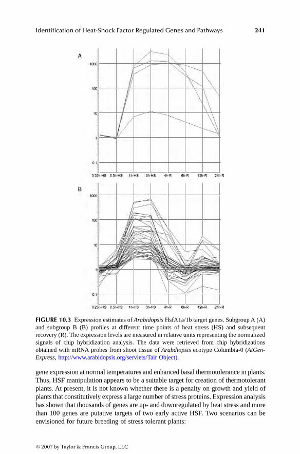
Identification of Heat-Shock Factor Regulated Genes and Pathways 241
gene expression at normal temperatures and enhanced basal thermotolerance in plants.Thus, HSF manipulation appears to be a suitable target for creation of thermotolerantplants. At present, it is not known whether there is a penalty on growth and yield ofplants that constitutively express a large number of stress proteins. Expression analysishas shown that thousands of genes are up- and downregulated by heat stress and morethan 100 genes are putative targets of two early active HSF. Two scenarios can beenvisioned for future breeding of stress tolerant plants:
FIGURE 10.3 Expression estimates of Arabidopsis HsfA1a/1b target genes. Subgroup A (A)and subgroup B (B) profiles at different time points of heat stress (HS) and subsequentrecovery (R). The expression levels are measured in relative units representing the normalizedsignals of chip hybridization analysis. The data were retrieved from chip hybridizationsobtained with mRNA probes from shoot tissue of Arabdiopsis ecotype Columbia-0 (AtGen-Express, http://www.arabidopsis.org/servlets/Tair Object).
3063_C010.fm Page 241 Thursday, August 24, 2006 1:57 PM
© 2007 by Taylor & Francis Group, LLC

242 Model Plants and Crop Improvement
HSF-manipulation that causes more stable performance and predictable yieldof agricultural plants under adverse environmental conditions such asincreased global climate warming
Selective manipulation of primary or secondary HSF target genes for specialstress-dependent pathways and functions, e.g. certain chaperones, osmolytes,carbohydrate transport, ROS scavenging, or pathogen resistance genes.
REFERENCES
1. Morimoto, R.I., Tissieres, A., and Georgopoulos, C. Progress and perspectives on thebiology of heat shock proteins and molecular chaperones, in The Biology of HeatShock Proteins and Molecular Chaperones. Morimoto, R.I., Tissieres, A., and Geor-gopoulos, C. (Eds.). Cold Spring Harbor Laboratory Press, Cold Spring Harbor, NewYork, 1, 1994.
2. Schöffl, F., Prändl, R., and Reindl, A. Regulation of the heat shock response. PlantPhysiol., 117, 1135, 1998.
3. Wiederrecht, G. et al. The Saccharomyces and Drosophila heat shock transcriptionfactors are identical in size and DNA-binding properties. Cell, 48, 507, 1987.
4. Sorger, P.K. and Pelham, H.R.B. Yeast heat shock factor is an essential DNA-bindingprotein that exhibits temperature-dependent phosphorylation. Cell, 54, 855, 1988.
5. Wu, C. Heat shock transcription factors: structure and regulation. Annu. Rev. CellDev. Biol., 11, 441, 1995.
6. Nover, L. et al. The HSF world: classification and properties of plant heat stresstranscription factors. Cell Stress Chaperones, 1, 215, 1996.
7. Schöffl, F. and Prändl, R. Derepression of the heat shock protein synthesis in trans-genic plants, in Plant Responses to Environmental Stress. Smallwood, M., Calvert,C., and Bowles, D. (Eds.). BIOS Scientific Publishers Limited, Oxford, 65, 1999.
8. Nover, L. et al. Arabidopsis and the heat stress transcription factor world: how manyheat stress transcription factors do we need? Cell Stress Chaperones, 6, 177, 2001.
9. Doring, P. et al. The role of AHA motifs in the activator function of tomato heatstress transcription factors HsfA1 and HsfA2. Plant Cell, 12, 265, 2000.
10. Czarnecka-Verner, E. et al. Plant heat shock transcription factors: positive and neg-ative aspects of regulation. Acta Physiol. Plant, 19, 529, 1997.
11. Czarnecka-Verner, E. et al. Plants contain a novel multimember class of heat shockfactors without transcriptional activator potential. Plant Mol. Biol., 43, 459, 2000.
12. Boscheinen, O. et al. Heat stress transcription factors from tomato can functionallyreplace HSF1 in the yeast Saccharomyces cerevisiae. Mol. Gen. Genet., 255, 322,1997.
13. Burke, J.J., O’Mahony, P.J., and Oliver, M.J. Isolation of Arabidopsis mutants lackingcomponents of acquired thermotolerance. Plant Physiol., 123, 575, 2000.
14. Hong, S.W. and Vierling, E. Mutants of Arabidopsis thaliana defective in the acqui-sition of tolerance to high temperature stress. Proc. Natl. Acad. Sci. USA, 97, 4392,2000.
15. Yamanouchi, U. et al. A rice spotted leaf gene, Spl7, encodes a heat stress transcriptionfactor protein. Proc. Natl. Acad. Sci. USA, 99, 7530, 2002.
16. Lee, J.H., Hübel, A., and Schöffl, F. Derepression of the activity of geneticallyengineered heat shock factor causes constitutive synthesis of heat shock proteins andincreased thermotolerance in transgenic Arabidopsis. Plant J., 8, 603, 1995.
3063_C010.fm Page 242 Thursday, August 24, 2006 1:57 PM
© 2007 by Taylor & Francis Group, LLC

Identification of Heat-Shock Factor Regulated Genes and Pathways 243
17. Prändl, R. et al. HSF3, a new heat shock factor from Arabidopsis thaliana, derepressesthe heat shock response and confers thermotolerance when overexpressed in trans-genic plants. Mol. Gen. Genet., 258, 269, 1998.
18. John, A., Smith, S.T., and Jaynes, J.B. Inserting the Ftz homeodomain into theengrailed creates a dominant transcriptional repressor that specifically turns off Ftztarget genes in vivo. Development, 121, 1801, 1995.
19. Conlon, S.G. et al. Inhibition of Xbra transcription activation causes defects inmesodermal patterning and reveals autoregulation of Xbra in dorsal mesoderm. Devel-opment, 122, 2427, 1996.
20. Lyon, J.J. and Watson, R.J. Interference of Myb transactivation activity by a condi-tional dominant negative protein: functional interference in a cytotoxic T-cell lineresults in G1 arrest. Gene, 182, 123, 1996.
21. Jaynes, J.B. and O’Farrel, P.H. Active repression of transcription by the engrailedhomeodomain protein. EMBO J., 10, 1427, 1991.
22. Han, K. and Manley, J.L. Functional domains of the Drosophila engrailed protein.EMBO J., 12, 2723, 1993.
23. Markel, H., Chandler, J., and Werr, W. Translational fusions with the engrailedrepressor domain efficiently convert plant transcription factors into dominant negativefunctions. Nucleic Acids Res., 30, 4709, 2002.
24. Wunderlich, M., Werr, W., and Schöffl, F. Generation of dominant negative effectson the heat shock response in Arabidopsis thaliana by transgenic expression of achimeric HSF1 protein fusion construct. Plant J., 35, 442, 2003.
25. Jedlicka, P., Mortin, M.A., and Wu, C. Multiple functions of Drosophila heat shockfactor in vivo. EMBO J., 16, 2452, 1997.
26. Panchuck, I., Volkov, R., and Schöffl, F. Heat stress and HSF-dependent expressionof ascorbate peroxidase in Arabidopsis. Plant Physiol., 129, 838, 2002.
27. Mishra, S.K. et al. In the complex family of heat stress transcription factors, HsfA1has a unique role as master regulator of thermotolerance in tomato. Genes Dev., 16,1555, 2002.
28. Lohmann, C. et al. Two different heat shock transcription factors regulate immediateearly expression of stress genes in Arabidopsis. Mol. Gen. Genom., 271, 11, 2004.
29. Zhang, L. et al. Heat stress-dependent DNA binding of Arabidopsis heat shocktranscription factor HSF1 to heat shock gene promoters in Arabidopsis suspensionculture cells in vivo. Biol. Chem., 384, 959, 2003.
30. McMillan, D.R. et al. Targeted disruption of heat shock transcription factor 1 abolishesthermotolerance and protection against heat-inducible apoptosis. J. Biol. Chem., 273,7523, 1998.
31. Tanabe, M. et al. Disruption of the HSF3 gene results in the severe reduction of heatshock gene expression and loss of thermotolerance. EMBO J., 17, 1750, 1998.
32. Xiao, X. et al. HSF1 is required for extra-embryonic development, postnatal growthand protection during inflammatory responses in mice. EMBO J., 18, 5943, 1999.
33. Baler, R., Zou, J., and Voellmy, R. Evidence for a role of Hsp70 in the regulation ofthe heat shock response in mammalian cells. Cell Stress Chaperones, 1, 33, 1996.
34. Mosser, D.D., Duchaine, J., and Massie, B. The DNA-binding activity of the humanheat shock transcription factor is regulated in vivo by hsp70. Mol. Cell Biol., 13,5427, 1993.
35. Satyal, S.H. et al. Negative regulation of the heat shock transcriptional response byHSBP1. Genes Dev., 12, 1962, 1998.
36. Zou, J. et al. Repression of heat shock transcription factor HSF1 activation by HSP90(HSP90 complex) that forms a stress-sensitive complex with HSF1. Cell, 94, 471, 1998.
3063_C010.fm Page 243 Thursday, August 24, 2006 1:57 PM
© 2007 by Taylor & Francis Group, LLC

244 Model Plants and Crop Improvement
37. Kim, B.H. and Schöffl, F. Interaction between Arabidopsis heat shock transcriptionfactor 1- and 70 k-Da heat shock proteins. J. Exp. Bot., 53371, 2002.
38. Bharti, K. et al. Tomato heat stress transcription factor HsfB1 represents a novel typeof general transcription co-activator with a histone-like motif interacting with theplant CREB binding protein ortholog HAC1. Plant Cell, 16, 1521, 2004.
39. Busch, W., Wunderlich, M., and Schöffl, F. Identification of novel heat shock factordependent genes and biochemical pathways in Arabidopsis thaliana. Plant J., 41, 1,2005.
40. Panikulangara, T.J. et al. Galactinol synthase 1, a novel heat-inducible and HSF-target gene responsible for heat-induced synthesis of raffinose family oligosaccharidesin Arabidopsis. Plant Physiol., 136, 3148, 2004.
41. Rizhsky, L. et al. When defense pathways collide. The response of Arabidopsis to acombination of drought and heat stress. Plant Physiol., 134, 1683, 2004.
42. Schöffl, F., Rossol, I., and Angermüller, S. Regulation of the transcription of heatshock genes in nuclei from soybean (Glycine max) seedlings. Plant Cell Environ.,10, 113, 1987.
43. Trinklein, N.D. et al. The role of heat shock transcription factor 1 in the genome-wideregulation of the mammalian heat shock response. Mol. Biol. Cell., 15, 1254, 2004.
44. Scharf, K.D., Siddique, M., and Vierling, E. The expanding family of Arabidopsisthaliana small heat stress proteins and a new family of proteins containing a-crystallindomains (Acd proteins). Cell Stress Chaperones, 6, 225, 2001.
45. Clarke, S.M. et al. Salicylic acid dependent signaling promotes basal thermotolerancebut is not essential for acquired thermotolerance in Arabidopsis thaliana. Plant J.,38, 432, 2004.
46. Larkindale, J. et al. Heat stress phenotypes of Arabidopsis mutants implicate multiplesignaling pathways in the acquisition of thermotolerance. Plant Physiol., 138, 882,2005.
47. Ishitani, M. et al. SOS3 function in plant salt tolerance requires N-myristoylation andcalcium binding. Plant Cell, 12, 1667, 2000.
48. Cheong, Y.H. et al. Transcriptional profiling reveals novel interactions betweenwounding, pathogen, abiotic stress, and hormonal responses in Arabidopsis. PlantPhysiol., 129, 661, 2002.
49. Maleck, K. et al. The transcriptome of Arabidopsis thaliana during systemic acquiredresistance. Nat. Genetic., 26, 403, 2000.
50. Ueda, A. et al. Analysis of salt-inducible genes in barley roots by differential display.J. Plant Res., 115, 119, 2002.
51. Stasolla, C. et al. The effects of polyethylene glycol on gene expression of developingwhite spruce somatic embryos. Plant Physiol., 131, 49, 2003.
52. Chen, W., Chao, G., and Singh, K. The promoter of a H2O2-inducible, Arabidopsisglutathione S-transferase gene contains closely linked OBF- and OBP-binding sites.Plant J., 10, 955, 1996.
53. Kang, H.G.W. and Singh, K. Characterization of salicylic acid-responsive, Arabidop-sis Dof domain proteins: overexpression of OBP3 leads to growth defects. Plant J.,21, 329, 2000.
54. Papi, M. et al. Identification and disruption of an Arabidopsis zinc finger genecontrolling seed germination. Genes Dev., 14, 28, 2000.
55. Riechmann, J.L. and Meyerowitz, E.M. The AP2/EREBP family of plant transcriptionfactors. Biol. Chem., 379, 633, 1998.
56. Kizis, D., Lumbreras, V., and Pagès, M. Role of AP2/EREBP transcription factors ingene regulation during abiotic stress. FEBS Lett., 498, 187, 2001.
3063_C010.fm Page 244 Thursday, August 24, 2006 1:57 PM
© 2007 by Taylor & Francis Group, LLC

Identification of Heat-Shock Factor Regulated Genes and Pathways 245
57. Reed, J.W. Roles and activities of Aux/IAA proteins in Arabidopsis. Trends Plant.Sci., 6, 420, 2001.
58. Fujimoto, S.Y. et al. Arabidopsis ethylene-responsive element binding factors act astranscriptional activators or repressors of GCC box-mediated gene expression. PlantCell, 12, 393, 2000.
59. Riechmann, J.L. et al. Arabidopsis transcription factors: genome-wide comparativeanalysis among eukaryotes. Science, 290, 2105, 2000.
60. Kreps, J.A. et al. Transcriptome changes for Arabidopsis in response to salt, osmotic,and cold stress. Plant Physiol., 130, 2129, 2002.
61. Ishitani, M. et al. Coordinate transcriptional induction of myo-inositol metabolismduring environmental stress. Plant J., 9, 537, 1996.
62. Taji, T. et al. Important roles of drought- and cold-inducible genes for galactinolsynthase in stress tolerance in Arabidopsis thaliana. Plant J., 29, 417, 2002.
3063_C010.fm Page 245 Thursday, August 24, 2006 1:57 PM
© 2007 by Taylor & Francis Group, LLC

247
11
Improving Low-Temperature Tolerance in Plants
Markku K. Aalto, Pekka Heino, and E. Tapio Palva
CONTENTS
11.1 Introduction................................................................................................. 24811.1.1 Freezing Stress............................................................................... 24811.1.2 Cold Acclimation........................................................................... 249
11.2 Cold Signaling in Plants ............................................................................. 25011.2.1 Signal Transduction at Low Temperature ..................................... 25011.2.2 Cold-Regulated Gene Expression ................................................. 25111.2.3 The CBF/DREB1 Regulon............................................................ 25211.2.4 ABA-Mediated Gene Expression .................................................. 253
11.3 Engineering Tolerance ................................................................................ 25411.3.1 Target Genes .................................................................................. 25411.3.2 LEA Proteins ................................................................................. 25511.3.3 LEA Proteins and Yeast................................................................. 25611.3.4 Functional Divergence of LEA Proteins ....................................... 25611.3.5 Membrane-Binding Proteins.......................................................... 25711.3.6 Antifreeze Proteins ........................................................................ 25811.3.7 Antioxidants and Detoxification.................................................... 25811.3.8 Membranes..................................................................................... 25911.3.9 Enzyme Engineering...................................................................... 260
11.4 Metabolic Engineering................................................................................ 26011.4.1 Betaines.......................................................................................... 26111.4.2 Amino Acids .................................................................................. 26511.4.3 Sugars............................................................................................. 26511.4.4 Other Metabolites .......................................................................... 267
11.5 Regulon Engineering .................................................................................. 26711.5.1 Stress Signaling ............................................................................. 26711.5.2 Cold-Tolerant Mutations................................................................ 268
11.6 Conclusions................................................................................................. 26811.7 Perspective .................................................................................................. 269Acknowledgments.................................................................................................. 270References.............................................................................................................. 270
3063_C011.fm Page 247 Thursday, August 24, 2006 2:39 PM
© 2007 by Taylor & Francis Group, LLC

248
Model Plants and Crop Improvement
11.1 INTRODUCTION
Throughout their growth period, plants are exposed to abiotic and biotic stresses.This has led to plants’ development of adaptive strategies to survive these stresses.Low temperature is one of the most important abiotic factors limiting growth,productivity, and distribution of plants [1,2]. Low temperature decreases biosyntheticactivity of plants, inhibits normal function of physiological processes, and may causepermanent injuries leading to death.
Plants vary widely in their ability to tolerate low temperature [3]. Chilling-sensitive tropical species can be severely damaged even at temperatures significantlyhigher than freezing [4]. Chilling-tolerant but freezing-sensitive plants are able tosurvive temperatures somewhat below 0
°
, but are damaged upon ice formation inthe tissues. On the other hand, freezing-tolerant plants are able to survive variablelevels of freezing temperatures; the degree of tolerance depends on species, devel-opmental stage, and duration of stress [3,4].
Chilling and freezing are stresses that affect the germination and seedling stagesof the plants during early summer, as well as flowering, fruiting, and maturing ofthe crop during the growth period. Furthermore, because the successful minimizationof damage during overwintering is the prerequisite for efficient growth the nextseason, low-temperature stress tolerance during winter is important for perennials.
Increasing low-temperature tolerance is not only important for securing cropsagainst sudden periods of colder climate but also provides a possibility of extendingthe present geographical limits of cultivated varieties, and even species, to colderclimates. Another very important application is the increased cold tolerance extend-ing the storage period of fruits and vegetables [5].
Increased cold tolerance appears to be antagonistic to optimal growth, so it isimportant to analyze cost and benefits in crop plant breeding and find the perfectcombination of preferred traits [6]. This can only be achieved by directly testingcandidate genes. It also underlines the fact that molecular biology and especiallytransformation offer powerful tools for breeding programs [7].
This chapter focuses on current knowledge obtained largely from transgenicexperiments studying adaptive strategies used by plants to tolerate freezing stressand the genetic engineering applications that can be used to increase the tolerance.
11.1.1 F
REEZING
S
TRESS
Plants from temperate regions are commonly exposed to freezing temperatures sea-sonally and during their growth season. Plants encountering freezing temperatures havetwo general strategies to survive freezing stress: avoidance or tolerance of freezing[2]. The former is mainly achieved by supercooling tissue water. However, this mech-anism has limited value because it mainly occurs in special organs such as seeds,overwintering buds, or parenchymal cells of temperate trees [8,9]. Therefore, toleranceof freezing is the dominant mechanism by which plants survive freezing stress.
Exposure of plants to subzero temperatures leads to freezing of tissue water. Dueto the higher freezing point and presence of more active ice nucleators in apoplasticsolution compared to the cytoplasm, freezing invariably occurs extracellularly. Ice
3063_C011.fm Page 248 Thursday, August 24, 2006 2:39 PM
© 2007 by Taylor & Francis Group, LLC

Improving Low-Temperature Tolerance in Plants
249
formation outside the cells reduces water potential of the apoplastic solution, whichleads to withdrawal of water from the cells and subsequent cellular dehydration.Therefore, on a cellular level, freezing stress also leads to dehydration stress; conse-quently, tolerance of freezing is correlated to tolerance of dehydration [2].
Cellular membranes appear to be the main target for freezing injuries [10]. Freeze-induced dehydration can cause different types of structural perturbations on mem-branes. These include membrane fusions and lamellar to hexagonal II phase transitions[11]. In addition to dehydration, other factors also contribute to the damage. Growingice crystals can cause mechanical damage and low temperature per se can have directeffects on cellular processes due to denaturation of proteins and disruption of mac-romolecular complexes. In addition, low temperature, especially in combination withhigh light intensities, generally leads to increased production of reactive oxygenspecies (ROS), which can damage different macromolecules in the cells [12,13].
Reactive oxygen species (ROS) such as O
2
, H
2
O
2
, and OH– are mainly formedin plastids, microsomes, and mitochondria and they damage macromolecules andmembranes [14,15]. Plants have developed an elaborate repertoire of defense mech-anisms to scavenge these and other radical compounds. These include specificenzymes such as catalase, superoxide dismutase (SOD), ascorbate peroxidase (APX),and glutathione reductase, as well as nonenzyme molecules such as ascorbate,glutathione, carotenoids, and anthocyanins [16,17].
Osmolytes [18,19], proteins like dehydrin [20], phenolic metabolites, and iso-prenoids also function as ROS scavengers [21–24]. Membranes are protected by
α
-tocopherol and specific glutathione peroxidases [25–27]. Also, many sugars areactive ROS scavengers; for example, mannitol is active specifically against hydroxylradicals, the most dangerous form of activated oxygen species [28].
However, a complex pattern of antioxidant signaling and redox homeostasisinvolved in cell death and acclimation responses is only beginning to be understood[29]. For example, the nature of ROS formation may be different under low tem-peratures compared to other stress types because the gene network (regulon) containsdifferent genes or they are differentially expressed [16].
11.1.2 C
OLD
A
CCLIMATION
Plants species native to temperate regions need to adjust to daily and seasonalfluctuations in temperature; the seasonal acclimation is important for overwinteringherbaceous and woody plants. Several plant species have the ability to increase theirlevel of freezing tolerance in response to low nonfreezing temperatures by a processknown as cold acclimation [2,3]. These species utilize environmental signals totrigger processes that lead to increased freezing tolerance.
In overwintering woody species, acclimation is a two-step process in which thefirst phase is triggered by shortening of the photoperiod below a critical length. Thisleads to growth cessation and dormancy development and is accompanied by amoderate increase in freezing tolerance. The second stage of acclimation, which isalso essential for obtaining full winter hardiness, is induced by subsequent exposureto low temperatures.
3063_C011.fm Page 249 Thursday, August 24, 2006 2:39 PM
© 2007 by Taylor & Francis Group, LLC

250
Model Plants and Crop Improvement
In annual and in most overwintering herbaceous plants, freezing tolerance is notaffected by the photoperiod but acclimation is triggered solely by low nonfreezingtemperatures. In these species, cold acclimation is a rather rapid process; for example,in
Arabidopsis
a substantial increase in freezing tolerance can be achieved after 1-dayexposure to low temperature, although full acclimation requires more than a week. Inaddition to low nonfreezing temperatures, cold acclimation can be triggered by exposingplants to moderate water stress or by exogenously applied abscisic acid (ABA) [30–32].
The level of freezing tolerance obtained through cold acclimation is not static,but is rapidly lost upon return to warm nonacclimating temperature. Cold acclimationis associated with several physiological and biochemical alterations in plants. Thebest characterized changes include changes in hormone levels; increases in solublesugars, amino acids, and organic acids; and accumulation of osmoprotectants andprotective proteins, as well as modification of membrane lipid composition [33–35](see also other reviews listed later). Most of these changes are derived from alteredgene expression and, although the causal relationship of many of these changes toincreased freezing tolerance is still unclear, some of the genes and regulons involvedin cold acclimation are being unraveled [35–38].
11.2 COLD SIGNALING IN PLANTS
11.2.1 S
IGNAL
T
RANSDUCTION
AT
L
OW
T
EMPERATURE
To respond to low temperature, plants must perceive the stress, transduce the signalto the nucleus, and activate expression of specific genes required for cold acclimationand metabolic adjustments needed for growth at low temperatures. The exact mech-anisms by which this occurs are not clear, even though an increasing number ofcomponents involved in the signal processes have been characterized. However, thenature of the cold sensor still needs to be established.
Calcium is a ubiquitous second messenger in plants and also mediates abioticstress signaling [39]. The specificity of signaling is derived from specific Ca
2+
-signatures evoked by different types of stresses and generated by a combined actionof Ca
2+
-channels, -pumps, and -transporters as reviewed [40]. Transient increase incytosolic Ca
2+
-concentration in the early stages of cold acclimation has been shownto be necessary for development of freezing tolerance [41,42].
The source of Ca
2+
- and the specific channels for Ca
2+
-influx are currentlyunknown; however, by using pharmacological approaches, it has been demonstratedthat rigidification of membranes and cytoskeletal rearrangements are needed for theCa
2+
-influx in alfalfa and
Brassica napus
, indicating involvement of mechanosensi-tive channels in this process [43,44].
Downstream signaling from the Ca
2+
-signature is generally mediated by diversetypes of Ca
2+
-binding proteins, like calmodulin (CaM), calcineurin B-like proteins(CBLs), and Ca
2+
-dependent protein kinases (CDPKs). The proteins are activatedby Ca
2+
-binding and, in activated forms, they regulate activity of proteins involvedin generation of signal-specific responses [40,45]. The involvement of CaM andCDPKs in cold acclimation was initially demonstrated by Monroy et al. [41], whoshowed that treatment of alfalfa suspension cultures with CaM or CDPK antagonists
3063_C011.fm Page 250 Thursday, August 24, 2006 2:39 PM
© 2007 by Taylor & Francis Group, LLC

Improving Low-Temperature Tolerance in Plants
251
resulted in inhibition of low-temperature responsive gene expression and develop-ment of freezing tolerance.
Genes encoding CaM and CaM-related proteins as well as genes encodingCDPKs have also been shown to be low temperature responsive, indicating a positiverole in regulation of low-temperature tolerance [42,46,47]. However, Townley andKnight have shown that overexpression of the CaM gene in
Arabidopsis
leads toinhibition of low-temperature responsive gene expression, indicating that CaM canact as a negative regulator of cold acclimation [48]. CDPKs have also been shownto be involved in acquisition of chilling tolerance. Saijo et al. have demonstratedthat overexpression of the
OsCDPK
gene leads to increased low-temperature toler-ance in rice [47].
CBLs are a family of Ca
2+
-binding proteins that mediate the activation of proteinkinases called CIPKs (CBL-interacting protein kinases). A gene encoding a memberof the CBL family,
CBL1
, has been shown to be responsive to abiotic stresses,including low temperature in
Arabidopsis
[49]. However,
cbl1
null mutant exhibitsenhanced freezing tolerance and overexpression of
CBL1
leads to reduced level oftolerance, indicating that CBL1 acts as a negative regulator of cold responses [50].A CIPK, CIPK3, has been demonstrated to be involved in activation of low-temper-ature responsive genes in
Arabidopsis
[51].
cipk3
mutant plants exhibited delayedinduction of gene expression but no differences in the maximum level of induction,indicating that CIPK3 acts at the early stages of cold acclimation [51].
Mitogen-activated protein kinases (MAPKs) are a family of proteins involvedin transduction of diverse signals in eukaryotic cells. Activation of MAPKs is medi-ated through a phosphorylation cascade where activated MAPKKKs phosphorylateMAPKKs, which then phosphorylate MAPKs. Activated MAPKs typically phospho-rylate and modulate the activity of specific transcription factors, participating ingeneration of signal-specific transcription patterns.
Arabidopsis
harbors about 60 putative MAPKKKs, 10 MAPKKs, and 20 MAPKs,which suggests that MAPKKs form a cross-talk node between different signalsmediated through the cascade. However, very little information exists concerningthe components involved in transmission of specific signals [52].
11.2.2 C
OLD
-R
EGULATED
G
ENE
E
XPRESSION
Cold acclimation is associated with marked alterations in gene expression; severalof the alterations are likely to be significant for development of freezing tolerance.The cold-responsive genes are often also induced by other environmental stimuli,like drought and salt, indicating common protective mechanisms in these stresses.Several, but not all, of the low-temperature responsive genes are also induced byexogenous ABA, suggesting ABA involvement in generation of stress responses[35,53,54].
The effects of low temperature on
Arabidopsis
transcriptome have been analyzedin several different studies and the results indicate that up to 25% of the genes areresponsive to cold [55–57]. The temporal patterns of low-temperature responsive geneexpression are complex and the genes involved clearly belong to different regulons.
3063_C011.fm Page 251 Thursday, August 24, 2006 2:39 PM
© 2007 by Taylor & Francis Group, LLC

252
Model Plants and Crop Improvement
11.2.3 T
HE
CBF/DREB1 R
EGULON
The emerging insight in cold acclimation is that this mechanism is complex,consisting of several distinct signal pathways that appear to interact with eachother [35,36,53,58–61]. Moreover, it is becoming evident that the low-temperaturesignal pathways are converging and interacting with other stress-induced path-ways, such as those mediating dehydration and abscisic acid responses [54,62,63](Figure 11.1).
Our earlier work demonstrated the presence of ABA-independent and ABA-medi-ated pathways in low-temperature signaling [64,65]. Interestingly, recent data indicatethat there is cross-talk even between these two pathways of cold signaling [66].
Work done mainly by the Thomashow and Shinozaki laboratories has establishedthe central role of the CBF or DREB1 family of transcription factors (TF) in con-trolling ABA-independent responses to low temperature [58,61,67,68]. Identificationof these AP2/EREBP-types of DNA-binding proteins was initiated by recognizing acommon binding site in many promoters of the low-temperature responsive genes.
We originally suggested the identity of such a low-temperature responsive ele-ment (LTRE) in the
LTI78/RD29A
promoter in
Arabidopsis
[65]. This 9-bp element,TACCGACAT, with a core sequence of CCGAC, was subsequently demonstratedby deletion analysis to confer responsiveness to low temperature, drought, and highsalinity, but not to ABA [69]. This dehydration-responsive element designated asDRE also occurs in several other cold-responsive promoters and has been referredto as the C-repeat (CRT) [70]. The TF binding to the DRE/CRT/LTRE element and
FIGURE 11.1
Simplified scheme of
Arabidopsis thaliana
signal transduction cascadesinvolved in cold acclimation. Regulation and cross-talk are omitted for simplicity. (Picturebased on Heino, P. and Palva, E.T., in
Plant Responses to Abiotic Stress
, vol. 4, Hirt, H. andShinozaki, K., Eds., Springer-Verlag, Berlin, 2003, 151–186; and Shinozaki, K. et al.,
Curr.Opin. Plant Biol.
, 6, 410–417, 2003.)
Drought
[ABA]
Low temperature
SENSOR
Ca2+
SENSOR
Phosphorylation cascade
ABRE
ICE1MYC CBF
DRE/LTRE/CRT LTI/COR etc.
ACCLIMATION
ABFs/AREBsCBF
Ca2+
Abiotic stress
Signal perception
Signal transduction
Signal molecules, hormones
Gene expression
Responses
ABA-independent pathway ABA-dependent pathway(s)
Phosphorylation cascade
3063_C011.fm Page 252 Thursday, August 24, 2006 2:39 PM
© 2007 by Taylor & Francis Group, LLC

Improving Low-Temperature Tolerance in Plants
253
activating cold-induced gene expression was first identified by Stockinger et al. [71]and designated CBF1 (C-repeat binding factor 1).
Additional genes were subsequently isolated and shown to encode a small familyof related TFs called CBF1, CBF2, and CBF3 or DREB1B, DREB1C, and DREB1A(DRE-binding protein) in
Arabidopsis
[67,68,71,72]. An additional member of thisTF family, CBF4, was isolated [73] but may be more related to drought response.
Interestingly, the
CBF1-3/DREB1A-C
genes are transiently regulated by lowtemperature [67,68,74]. Rapid activation of
CBF
expression (within 15 to 30 min)is followed by cold-regulated target genes that define the CBF/DREB1 regulon. Anefficient mutant screen by the J.-K. Zhu laboratory led to isolation of the first
ICE
(inducer of CBF) gene encoding an MYC-type bHLH transcription factor [75]. Asexpected, expression of
ICE1
appears constitutive.Overexpression of
CBF
/
DREB1
leads to the constitutive expression of a numberof target genes with promoters containing the DRE/CRT/LTRE element and toimproved freezing, drought, and salt tolerance of nonacclimated plants [72,76]. Inaddition, overexpression of CBF3 leads to elevated levels of proline and sugars thatare normally associated with cold acclimation [77]. In several recent studies, tran-scriptome analysis has been used to identify genes of the CBF/DREB1 regulon in
Arabidopsis
[38,55,56,78,79].Of the hundreds of cold-regulated genes identified, 85 were upregulated by CBF2
and, of the 25 most highly cold-induced genes, the majority were under CBF2 control[38]. These studies demonstrate that the CBF/DREB1-controlled genes constitute alow-temperature responsive regulon central to the plant cold acclimation responseand development of plant freezing tolerance and could be employed for engineeringplant cold tolerance [80].
The conserved nature of the CBF/DREB1 regulon in cold-tolerant plants furthersupports the notion that this regulon has an important role in the cold acclimationprocess. For example, in wheat, the differences in Cbf expression were associatedwith variation in frost tolerance [81]. Orthologs of CBF genes have been found ina number of herbaceous species, including
Brassica napus
[82], barley [83], wheat[84], rice [85], tomato [86], sour cherry and strawberry [87], sweet cherry [88], andeven woody species such as silver birch [89; Welling et. al.,
in preparation].
11.2.4 ABA-M
EDIATED
G
ENE
E
XPRESSION
ABA is a phytohormone that regulates diverse aspects of plant development andgrowth, including stress responses. Genes’ responsiveness to abiotic stresses is partlymediated by pathways that are ABA dependent [53,63,90,91]. ABA-deficient mutantsof
Arabidopsis
are not able to increase their freezing tolerance to wild-type levelsduring cold acclimation [92,93] and ABA deficiency also decreases cold-responsivegene expression [93,94]. However, the growth rate at low temperatures is not regu-lated by ABA but by gibberellic acid (GA) and salicylic acid (SA) [95,96].
Recently, Zhu et al. [97] identified a gene encoding HOS10, a MYB-type tran-scription factor needed for development of freezing tolerance in
Arabidopsis
. HOS10appears to be required for stress induction of
NCED
, which encodes the rate-limiting
3063_C011.fm Page 253 Thursday, August 24, 2006 2:39 PM
© 2007 by Taylor & Francis Group, LLC

254
Model Plants and Crop Improvement
enzyme in ABA biosynthesis. This indicates a role for ABA in generating protectionagainst freeze-induced dehydration [97].
ABA-responsive gene expression has been shown to be mediated byABFs/AREBPs, basic leucine zipper transcription factors binding to ABA responseelements (ABREs) present in the promoter regions of ABA responsive genes [98,99].Characteristic of ABREs is that more than one copy of the element is necessary forgene activation. Alternatively, a coupling element replaces one of the ABREs [100].
A low-temperature and ABA-responsive gene encoding a C
2
H
2
-type zinc fingerprotein in soybean has been characterized [101]. The encoded protein, SCOF1,regulates low-temperature responsive gene expression and development of freezingtolerance. SCOF1 was found to enhance binding of the bZIP transcription factorSGBF1 to ABREs in stress-responsive genes, suggesting that SCOF1 regulates ABA-mediated gene expression in low temperatures [101].
11.3 ENGINEERING TOLERANCE
There are many reports about genes that, when overproduced, give plants enhancedprotection against low-temperature stress. However, definition of their roles as partof temperature stress tolerance mechanisms and possible overlapping functions needsto be elucidated. Indeed, an obvious explanation for the wide range of differentproteins in low-temperature stress is manifested in the many different forms of stress,each with its own specific protection mechanisms evolved. This may also be themost important motivation for studying low-temperature stress for plant cropimprovement purposes. In yeast, for example, the best selection strategy to obtainhighly improved multiple-stress-resistant strains was found to be batch selection forfreezing–thawing stress [102].
11.3.1 T
ARGET
G
ENES
By 1985 it was established that altered gene expression at low, nonfreezing temper-atures correlated with cold acclimation [103]. Subsequently, a great number of cold-induced genes has been isolated and characterized from a variety of plant species[33,34,104]. These genes code for a number of different proteins, including enzymesinvolved in metabolic pathways, proteins with a protective role, and proteins affectingsignaling pathways. Among these are fatty acid desaturases, chaperones, lipid trans-fer proteins, enzymes in osmoprotectant biosynthesis, antifreeze proteins, transcrip-tion factors, kinases, and phosphatases [35,54]. Furthermore, the functions of manycold-induced proteins are not clearly defined. However, in many cases, correspondinggenes have been found in a number of different plant species, indicating that manyproteins induced by low temperature are conserved.
In addition to specifically induced target genes, a number of regulation mecha-nisms are only beginning to be unraveled. These include signaling cascades leadingto specific transcription factors, as well as more global regulation of whole genomes.Thus, in addition to well-characterized regulatory cascades, including CBF/DREBgenes that may have a function at the level of histone acetylation [105,106], chro-mosome structure is regulated following low-temperature stress [107–110].
3063_C011.fm Page 254 Thursday, August 24, 2006 2:39 PM
© 2007 by Taylor & Francis Group, LLC

Improving Low-Temperature Tolerance in Plants
255
11.3.2 LEA P
ROTEINS
One of the most common categories of target genes includes a number of looselyrelated groups of genes called LEA (
l
ate
e
mbryogenesis
a
bundant) proteins[111,112]. Many of these genes are induced in response to any environmentalinfluence that has a dehydration component (such as drought, low temperature, orsalinity) by ABA or during the late stages of embryogenesis. These proteins arethought to be involved in protecting cellular structures from effects of water loss—for example, by retaining water together with sugars [113] or directly protectingother proteins or membranes including renaturation of unfolded proteins and seques-tration of ions in a wide range of higher plants [111,114–116]. Table 11.1 showsthe transgenic LEA proteins expressed in plants.
LEA proteins have been identified primarily from plants and comprise at leastsix different subgroups [112]. These proteins do not seem to have a common origin;rather, they have adopted the same strategy for protection of cells from a commonproblem [117].
Group 1 family members are unstructured in solution. Their sequence isconserved especially within the hydrophobic internal 20-amino acid motif,which may be repeated. Group 1 proteins are induced by osmotic stress orABA and they have been postulated to function as general water stressprotectants in nonembryonic tissues [118].
Group 2 (also called dehydrins) is the largest group [112]. Purified maizeDHN1 proteins from this group have been shown to bind with phospholipids
in vitro
[119]. The purified recombinant GmDHN1 exhibits a highlyextended conformation at low temperatures, which could constitute thebasis of the functional role in prevention of freezing, desiccation, ionic, orosmotic stress-related damage to macromolecular structures ([120]; see alsoa review by Allagulova et al. [121]).
TABLE 11.1Expressed LEA Proteins Increasing Freezing Tolerance
LEA group Origin Model organism Ref.
Group 2 CoCOR19—citrus In tobacco, leads to a slight degrease in ion leakage during chilling and freezing stress
126
Group 2 CAP85—spinach Small improvement in freezing tolerance of tobacco in time-course experiment
296
Group 2 Wcor410—wheat Acidic dehydrin improves strawberry freezing tolerance
297
Group 2 COR15a—
A. thaliana
Increases freezing tolerance in
Arabidopsis
139Group 3 HiC6—
C. vulgaris
Significant suppression of chilling injury in tobacco 298Group 3 WCS19—wheat Group 3 chloroplast targeted protein increased freezing
tolerance in
Arabidopsis
299
3063_C011.fm Page 255 Thursday, August 24, 2006 2:39 PM
© 2007 by Taylor & Francis Group, LLC

256
Model Plants and Crop Improvement
A member of group 3 has been shown to have
in vitro
antiaggregation activitydue to water stress, which was synergistic with trehalose. This group alsocontains members outside plants, as referred in Goyal et al. [122].
Group 4 has also been proposed to form amphiphilic
α
-helices that mayinteract with ions or membrane [111,123].
Group 5 proteins are embryogenesis specific. Group 6 contains atypical hydrophopic LEA proteins [124].
Although LEA proteins have been known for quite some time (as reviewed inCuming [115]), the functions of these proteins have remained somewhat unanswered.Despite some obvious enhancement in prediction tools [125], overexpression ofgenes in plants has not been conducted systematically. Among the published reports,Hara et al. [126] have recently shown that overproduction of a citrus dehydrin(CoCOR19) in tobacco leads to a slight decrease in ion leakage during chilling andfreezing stress. Interestingly, the hydroxyl and peroxyl radical scavenging activityof CoCOR19 was found
in vitro
to be equal to serum albumin, a known antioxidantprotein in mammals [20]. However, a hot-pepper protein, CaLEA6, tested in tobaccoshowed enhanced tolerance to dehydration and NaCl but not to chilling [124].Furthermore, a much-studied Group 3 barley HVA1 has not been reported to betested for cold tolerance [127–129], although it is naturally cold inducible [130].
11.3.3 LEA P
ROTEINS
AND
Y
EAST
The baker’s yeast
Saccharomyces cerevisiae
has been a popular model organism instudying functions of many LEA group proteins, although yeast also has LEA-likeproteins [117,131]. Table 11.2 depicts the LEA proteins expressed in yeast. BarleyHVA1 (group 3) displayed a shortened lag period after transfer to high-NaCl or KClmedia, whereas tomato le4 (group 2) increased tolerance only to KCl, maybe bystabilizing KCl-sensitive structures. However, both increased freezing tolerance,supporting the idea of specialized functions of different LEA proteins during low-temperature stress [128]. Thus, different LEA proteins appear to have separatefunctions and protect cells against different stresses, even in yeast.
11.3.4 FUNCTIONAL DIVERGENCE OF LEA PROTEINS
Many dehydrins are hydrophilic, containing random coil or α-helices [120], andremain soluble after boiling [132,133]. However, some atypical LEA proteins are
TABLE 11.2Plant LEA Genes Expressed in Yeast
Group 1 Em—wheat Enhanced osmotic tolerance 118Group 2 le4—tomato Enhanced freezing and KCl tolerance 128Group 3 HVA1—barley Enhanced freezing and salt tolerance 128Group 3 hiC6—Chlorella vulgaris Enhanced freezing tolerance 300Group 4 le25—tomato Increased salt and freezing tolerance 123
3063_C011.fm Page 256 Thursday, August 24, 2006 2:39 PM
© 2007 by Taylor & Francis Group, LLC

Improving Low-Temperature Tolerance in Plants 257
hydrophobic [112,124], suggesting a different function despite involvement in dehy-dration response. Even though the exact function of these proteins has not beenestablished, their biochemical properties and accumulation patterns suggest thatdehydrins could work in stabilizing cellular structures during dehydration stress[120], molecular chaperones [122], sequesters of iron [116,134] and calcium [135],and scavenging oxygen radicals [20].
The genome of Arabidopsis [136] has at least ten dehydrin genes, suggesting thateach of the family members has specified functions. This specialization might be onereason why overexpression of an LEA protein does not increase tolerance to aparticular type of stress—for example, chilling tolerance [124] or when expressionof three different LEA proteins did not increase tobacco’s drought tolerance [137].
Only a few comparative studies have been made between different plant protec-tive assets. One study compared dehydration, salt, and heat tolerance of bacterialotsA, a trehalose-6-phosphate synthase, the first protein in making of a nonreducingdisaccharide of two glucose units called trehalose, and an atypical LEA protein,CaLEA [138]. In this case, the LEA protein was found to be slightly better.
Several transgenic plant lines overexpressing different low-temperature respon-sive genes have been created, but only a marginal effect on freezing tolerance hasbeen demonstrated [139,140]. Similar results have been obtained in studies of waterstress tolerance. Therefore, this type of target gene engineering has thus far met withlimited success. The approach is most likely hampered by redundancy of the system,where an individual gene product makes a minor contribution to overall tolerance.Thus, this approach would require pyramiding a number of target genes providingadditive effects on tolerance.
This is further emphasized by the fact that several LEA proteins are activatedtogether by their common transcription factors and correlate with stress tolerance intransgenic plants [72,76,82] Recent results from our group in expressing pairwisecombinations of four different endogenous dehydrin genes in Arabidopsis [136] wasan attempt to test this. Thus, RAB18, which accumulates in response to ABA,drought, and low temperatures [133,141], was put together with COR47, whichaccumulates primarily in response to low temperature, but also to ABA and saltstress [133].
On the other hand, LTI29, responding primarily to low temperature but also toABA and salt stress, was put together with LTI30, which accumulates mainly undercold stress [133,142]. Using both constructs, we were able to show enhanced freezingtolerance that we did not see with RAB18 expressed alone [143] (data unpublished)and without any detectable deleterious effect to the plants [136]. These proteins havediffering functions inside the cell, as witnessed by partitioning of LTI29 from beingmainly cytoplasmic to mainly membrane localized following cold acclimation;LTI30 was not detected at the cytoplasm. Thus, pyramiding protective proteins withdiffering functions is clearly one way of proceeding with research.
11.3.5 MEMBRANE-BINDING PROTEINS
In general, plant survival during freezing may be more limited by stability of cellularmembranes rather than by soluble proteins [144–146]. Indirect evidence for interactions
3063_C011.fm Page 257 Thursday, August 24, 2006 2:39 PM
© 2007 by Taylor & Francis Group, LLC

258 Model Plants and Crop Improvement
with membranes or partially denatured proteins has been shown for the chloroplast-located hydrophilic COR15a protein, which is not a dehydrin but is predicted to belargely composed of amphipathic α-helices.
Interaction between the protein and the plastidic inner envelope, which is thoughtto be mediated through amphipathic α-helices, is correlated with decrease in mem-brane leakage [147]. Formation of the inverted hexagonal phase membrane structurecan be prevented by the cold acclimation process and, in part, by overexpressingCor15a [147,148].
Moreover, two other reports about membrane binding proteins have beenreported. Osmotin-like protein from bittersweet nightshade (Solanum dulcamara)stabilizes kale (Brassica oleracea) protoplasts during freeze–thaw cycles [149].Cryoprotectin, a lipid transfer protein homolog, shows cryoprotective activity stabi-lizing isolated thylakoids from freeze–thaw cycles [150]. The protective function ofthe protein lies in its capability to bind and immobilize thylakoid lipids [151]. Also,maize DHN1 binds to lipid vesicles, pointing to the possibility that the mode ofaction of some dehydrins is to support membranes [119]
11.3.6 ANTIFREEZE PROTEINS
Antifreeze proteins (AFPs) were first identified from fish and found later from otherspecies, including plants, as reviewed in Griffith and Yaish [152]. Their function isto bind to the growing surface of ice crystals and prevent them from growing.Roughly, animal and insect AFPs exhibit a substantial thermal hysteresis activitythat is a noncolligative depression of the freezing point of an ice-containing solutionbelow its melting point—the freeze avoidance strategy. AFP-producing plants andbacteria that cannot avoid freezing rely on another strategy by showing ice recrys-tallization inhibition.
Thus, controlling growth of an ice crystal during freeze–thaw cycles instead ofpreventing their formation is the freeze-tolerant strategy [153]. Transgenic attemptshave been made with limited success in plant, synthetic, insect, and fish genes toexpress AFPs in frost-susceptible crops to increase their freezing tolerance [152]. Inplants, the additional effect of these proteins is to function as pathogenesis-relatedproteins. Thus, plants are acquired with systemic, nonspecific, pre-emptive defenseagainst psychrophilic pathogens that would otherwise prosper under snow cover atsubzero temperatures [152,154].
11.3.7 ANTIOXIDANTS AND DETOXIFICATION
The importance of reactive oxygen species (ROS) scavenging proteins is that theyhave been shown to increase success rates not only in controlled experiments but alsoin overwintering plants in field trials, like the expression of superoxide dismutasewithin chloroplast, which did not affect freezing tolerance but rather enhanced recov-ery from stress [155,156]. Reactive oxygen species control many different processesin plants [16], so transgenic approaches may not always produce clear improvement,although it might just reflect a problem in expression or localization [157,158]. Forexample, using SODs creates enhanced hydrogen peroxide production, which maybe alleviated in plants with upregulation of H2O2 scavenging enzymes [159,160].
3063_C011.fm Page 258 Thursday, August 24, 2006 2:39 PM
© 2007 by Taylor & Francis Group, LLC

Improving Low-Temperature Tolerance in Plants 259
ROS scavenging pathways include the water–water cycle, ascorbate–glutathionecycle, glutathione peroxidase cycle, and catalase, as reviewed in Mittler et al. [16]and Mittler [161]. The ascorbate–glutathione cycle plays an important role in regu-lating cellular ROS levels [162]. In this cycle, ascorbate peroxidase (APX) reduceH2O2 using ascorbic acid (AsA) as an electron donor generating monodehydroascor-bate (MDA), which can be reduced back to AsA by MDA reductase using NAD(P)H.MDA can also spontaneously produce dehydroascorbate (DHA), which is reducedback to AsA by DHA reductase with the help of glutathione, GSH, oxidized toGSSG (the oxidized form of glutathione). The cycle closes with glutathione reductaseconverting GSSG back to GSH with NAD(P)H.
Plants contain a family of ascorbate peroxidases and overexpression of thyla-koidal APX (tAPX) in tobacco gives chilling tolerance. Antisense lines were notobtained, suggesting an essential function [163]. However, tAPX overexpression inArabidopsis did not increase resistance to low temperatures but rather to paraquat-induced oxidative stress and nitric oxide [164]. Furthermore, antisense constructswith 50% tAPX activity left showed no symptoms under normal conditions.
Expression of cytosolic cAPX has been reported to confer tolerance to chilling intomato [165]. However, in soybean, the cAPX deficiency is associated with chillingtolerance in cultivated species [166]. Also, downregulating cAPX activity in tobacco,BY-2 cell lines showed increased tolerance to heat and salt but cells grew more slowly.The APX activity in BY-2 cells is much higher than in Arabidopsis, the residual activityresulting as marginal increase in cellular ROS levels triggering tolerance mechanismswas discussed as an explanation for increased tolerance [167]. It has been suggestedthat excessive levels of cAPX scavenge ROS too actively, thus hindering expressionof defense genes, which need a certain level of stress for signaling [166].
11.3.8 MEMBRANES
Membrane fluidity depends on composition of lipid molecular species, degree oflipid saturation where increase in the level of unsaturation increases fluidity, andtemperature environments [168–170]. Thus, temperature-induced changes in mem-brane fluidity represent a potential site for cold perception [43,171], as also discussedin Sung et al. [172]. The proportion of unsaturated fatty acids in the lipid acyl chainsis particularly high in chloroplast membranes [173]. Several genetic loci involvedin fatty acid desaturation of lipids in chloroplast or microsomal membranes inArabidopsis have been identified [174] and the genetic engineering of plant mem-brane lipids has been recently reviewed in Iba [175].
It seems that increase of the content of unsaturated lipids gives better chillingtolerance in tobacco [176,177]. A decrease in synthesis of unsaturated trienoic fattyacid increases high-temperature tolerance [178]. Glycerol-3-phosphate acyltrans-ferase (GPAT) from Arabidopsis does not change proportions of individual lipidclasses but increases the level of unsaturation, leading to increased chilling tolerancein tobacco and rice [179,180], whereas overexpression of squash GPAT in tobaccoand rice leads to increase of saturated species of phosphatidylglycerol, which resultsin plants more sensitive to chilling [179,181]. Thus, it is possible to engineer toler-ance to low or high temperature, but not both at the same time [175]. In addition to
3063_C011.fm Page 259 Thursday, August 24, 2006 2:39 PM
© 2007 by Taylor & Francis Group, LLC
260 Model Plants and Crop Improvement
membrane unsaturation, it appears that lipid asymmetry in the membrane also con-tributes to low-temperature tolerance. Internalization of phosphatidylserine from theouter leaflet of plasma membrane by an Arabidopsis ALA1, a putative aminophos-pholipid translocase, in a yeast dsr1 mutant was tightly linked to rescue in cold [182].
11.3.9 ENZYME ENGINEERING
A completely different approach for achieving low-temperature tolerance is to engi-neer protein structure instead of engineering expression pattern, the main topic ofthis chapter. Pyruvate orthophosphate dikinase, a key enzyme in C4 pathway inmaize, loses its activity under 12°C due to dissociation. Expressing engineered genesfrom F. brownii in maize that does not dissociate as readily gives a small improve-ment, which may be enhanced if the endogenous gene can be downregulated orengineered [183]. Nevertheless, it represents a potentially very interesting approachin trying to enhance the photosynthetic rate at low temperatures.
11.4 METABOLIC ENGINEERING
Compared to engineering expression of protective proteins, metabolic engineeringof osmoprotectants is another way of improving plant stress tolerance. A group oftarget genes that have been successfully modified for increasing low-temperaturetolerance in plants are those participating in producing certain small organic mole-cules called compatible and counteracting solutes, compensatory solutes, chemicalchaperones, or osmoprotectants [184,185].
Induction of osmoprotectant biosynthesis is part of the plant response to drought,salinity, and low temperatures [186]. Compatible solutes occur in all organisms fromarchaebacteria to higher plants and animals [187]. These are highly soluble com-pounds that carry no net charge at physiological pH and do not perturb macromol-ecules such as proteins [188]. Some of these molecules are already present inunstressed tissues. Synthesis is often only increased or degradation decreased duringstress [189,190]. The primary function of these compatible solutes is understood tomaintain cell turgor and thus the driving gradient for water uptake [191,192]. Forthis purpose, by definition they should be able to exist at high concentrations in cellswithout any deleterious effects.
It seems that compatible solutes also function as free-radical scavengers orchemical chaperones and directly stabilize membranes and/or proteins [28,193,194].Furthermore, the biosynthetic flux of the compatible solute may help to maintainredox balance and affected sugar levels may provide a signal for adaptive regulationthroughout the plant [186]. Compatible solutes fall into several major groups: aminoacids (e.g. proline), quaternary amines (e.g., betaines), tertiary sulfoniums (e.g.,dimethylsulfoniopropionate), and polyol/sugars (e.g., mannitol, trehalose, sucrose,etc.) [195]. However, out of many potentially interesting compounds [28,187,196],relatively few have ever been tested by genetic engineering.
Of the few compounds that have been tested, in a number of publications theincrease in cold tolerance was obtained by introducing simple metabolic traits fromother organisms into plants that are not natural accumulators. However, some of the
3063_C011.fm Page 260 Thursday, August 24, 2006 2:39 PM
© 2007 by Taylor & Francis Group, LLC
Improving Low-Temperature Tolerance in Plants 261
compounds may not be compatible any more in species that do not naturally accu-mulate them, as discussed later. The other way is to try to increase and maintain ahigher concentration level of the metabolite in species where it has earlier beenshown to elevate naturally following stress response [184,197]. The way to achievethis in the latter case is to establish a new pathway or to increase the synthesis rateof the metabolite by increasing the gene dosage (and gene expression) of the rate-limiting enzyme [198–200] or by decreasing the degradation rate of the metaboliteby downregulating the degrading enzyme-encoding gene [201]. Table 11.3 liststransgenic experiments giving enhanced low-temperature tolerance.
11.4.1 BETAINES
Betaines are amino acid derivatives in which the nitrogen atom is fully methylated.In plants, a representative member of this group is glycine betaine (GB) [189],which is a well studied subject in salt stress; more than 15 articles on transgenicplants have been published [202]. In GB-accumulating plants, it is synthesized inthe chloroplast through an oxidation reaction from choline to glycine betaine viabetaine aldehyde intermediate. In higher plants, two enzymes—choline monoxy-genase (CMO; in mammals and some bacteria, CDH) and betaine aldehyde dehy-drogenase (BADH)—are responsible for these reactions. In certain bacteria, bothreactions are catalyzed by one enzyme, choline oxidase (COD), as reviewed inSakamoto and Murata [203].
Transgenic plants expressing betaines are more resistant to high salt concentra-tions, osmotic stress, and cold and warm temperatures [204,205]. Improved low-temperature tolerance has been reported when expressing the Arthrobacter globifor-mis codA (coding for choline oxidase, which synthesizes glycine betaine in one step)gene in Brassica juncea [206], tobacco [207], and tomato [208], as well as rice andArabidopsis [203]. However, in the case of codA, one must consider the role of theby-product of the GB synthesis, H2O2, which is a signaling component inducing theROS-response and thus gives chilling tolerance [208].
Expressing CDH alone or together with BADH in tobacco leaf discs alsoimproved tolerance to photoinhibition under low temperature [209]. However, themain theme in all reported cases has been that the levels of GB produced are verymodest compared to plants that accumulate it naturally. It is thought that this occursbecause of limited supplies of precursors in nonaccumulators [199,210,211]. How-ever, this problem seems to be possible to circumvent by using extensive engineeringfor precursor production.
Moreover, a novel pathway from extreme halophile bacteria for producing GBby direct methylation of glycine also gives improved osmotolerance in E. coli[212,213]. Using closely related enzymes in transgenic Arabidopsis gave muchhigher (10×) concentration of GB in Arabidopsis and Synechococcus than with codA.In this case, the precursor is not choline but glycine, the levels of which are onlysomewhat limiting [214].
Thus, it seems that the protective effect of GB is independent of species ormode of synthesis in natural accumulators. However, there are also reports aboutGB in non-natural accumulators giving only modest [215] or even deleterious
3063_C011.fm Page 261 Thursday, August 24, 2006 2:39 PM
© 2007 by Taylor & Francis Group, LLC

262M
od
el Plants an
d C
rop
Imp
rovem
ent
TABLE 11.3Transgenic Plants Showing Enhanced Low-Temperature Tolerance
Protein and origin Model plant Remarks Ref.
Chilling stress
desC—S. vulcanus Tobacco Thermophilic bacterium acyl-lipid ∆9-desaturase giving chilling tolerance 177acyl-lipid ∆9-desaturase—A. nidulans Tobacco Broad specificity ∆9 desaturase “fluidizing” membrane bound lipids 305CvFAD2 and 3—C. vulgaris Tobacco Two microsomal desaturases increasing and decreasing freezing tolerance 306FAD7—A. thaliana Tobacco Chloroplast ω3 desaturase increases trienoic fatty acid (TA) amount and confers
chilling tolerance176
GPAT—A. thaliana, spinach Tobacco, rice Increases proportion but not amount of unsaturated phosphatidylglycerol 179, 180Glycerol-3-P-acyl transferase (GPAT)—squash and A. thaliana
Tobacco, rice Causes changes in the unsaturation of fatty acids and chilling tolerance levels 181, 307
ALA1—A. thaliana A. thaliana Aminophospholipid translocase involved in generating membrane lipid asymmetry 182Glutamine synthetase—rice Rice Faster recovery from chilling stress due to increase in glutamine levels; increase in
photorespiration capacity224
Glutathione peroxidase—Chlamydomonas
Tobacco Cytosolic and chloroplast versions increased chilling tolerance 308
MnSOD and Fe-SOD—tobaccoand A. thaliana
Tobacco Fe-SOD binds to membranes and protects PSII; stromal Mn-SOD less effective 157
Cu/Zn-SOD—pea Tobacco Chloroplast targeted SOD 309Nt107—tobacco Tobacco Glutathione S-transferase/glutathione peroxidase combination 310DHAR—human Tobacco Dehydroascorbate dehydrogenase reduces DHA to ascorbate 311APX—P. sativum Tomato Cytosolic expression of ascorbate oxidase enhancing chilling tolerance 165Ipt—A. tumefaciens F. arundinacea Isopentenyl transferase resulted in increased cold tolerance 312ProDH—A. thaliana A. thaliana Antisense suppression of proline degradation improves tolerance to freezing 201Invertase—S. cerevisiae Potato Apoplast-localized invertase inhibits export of sugars and thus leaves retain higher
sugar content313
3063_C011.fm
Page 262 Thursday, A
ugust 24, 2006 2:39 PM
© 2007 by Taylor &
Francis Group, LLC

Imp
rovin
g Low
-Temp
erature To
lerance in
Plants
263
otsA-otsB—E. coli Rice Fusion of two E. coli trehalose biosynthetic genes allowing enhanced protection and recovery without stunting growth
252, 314
TpS1 and TpS2—S. cerevisiae A. thaliana Trehalose synthases TPS1 alone or together with TPS2 gives pleiotrophic effects and enhanced stress tolerance
253
mtlD—E. coli Eggplant Mannitol production enhancing chilling tolerance in a crop plant 315SacB—B. subtilis Tobacco Bacterial levansucrase for fructan biosynthesis gives low temperature tolerance 207OsCDPK 7 and 13—rice Rice Signaling; calcium-dependent protein kinase confers tolerance; number 7 has separate
pathways for cold and salt47, 267
OSISAP1—rice Tobacco Signaling; zinc finger protein confers tolerance at germination stage 316Bcl-xL—human and Ced-9—C. elegans Tobacco Plants transgenic to animal cell-death suppressors germinate at lower temperatures
than wild-type317
Ppdk—F. bidentis/F. brownii Maize Chimeric enzyme dissociates at lower temperature than native 183
Freezing stress
Fe-SOD—A. thaliana Alfalfa Enhanced recovery from stress after resuming growth 156Mn-SOD—tobacco Alfalfa Increased winter survival 318AAPT1—canola A. thaliana Phospholipid synthesis; increased resistance to damage at low growth temperatures 319Phospholipase D—Arabidopsis A. thaliana PLDδ overexpression and PLDα downregulation increases freezing tolerance; involved
in hydrolyzation of phospholipids320, 321
Spermidine synthase (SPDS)—C. ficifolia
A. thaliana Increase in polyamine content gives abiotic stress tolerance and rise in DREB levels 322
Galactosidase—L. esculentum Petunia Downregulation of hydrolytic enzyme increases raffinose concentration and freezing tolerance
249
SEX1—A. thaliana A. thaliana Involved in regulation of starch hydrolysis in early cold response 257Wft1, Wft2—T. aestivum L. perenne Fructan (sugar polyol) biosynthesis genes increase fructan levels likely in the vacuole 247P5C—Arabidopsis, Vigna Tobacco Proline, fructan, and betaine biosynthesis give freezing tolerance to tobacco 207codA—A. globiformis Tomato Betaine production protects flowers and seeds (reported) 208codA—A. globiformis A. thaliana, rice,
Brassica, tomatoBiosynthesis of glycine betaine enhances germination and cold tolerance of plants 206, 208, 323,
324continued
3063_C011.fm
Page 263 Thursday, A
ugust 24, 2006 2:39 PM
© 2007 by Taylor &
Francis Group, LLC

264M
od
el Plants an
d C
rop
Imp
rovem
ent
TABLE 11.3 (continued)Transgenic Plants Showing Enhanced Low-Temperature Tolerance
Protein and origin Model plant Remarks Ref.
Cox—A. pascens A. thaliana Choline oxidase giving modest freezing tolerance, but not to canola or tobacco 215betA, betB—E. coli Tobacco Improved tolerance to photoinhibition under low temperature in leaf discs 209GSMT, DMT—A. halophytica Cyanobacterium,
A. thalianaGermination yield retained high despite cold imbibition-treatment of seeds 214
Antifreeze protein—carrot Tobacco Increased freezing tolerance 325MKK2—A. thaliana A. thaliana Signaling; MAPKK especially activated by cold 266Osmyb4—rice A. thaliana Rice transcription factor showing significantly increased freezing tolerance in Arabidopsis 326OsMAPK5—rice Rice Signaling; affects adversely between abiotic and pathogen tolerance 327SCOF-1—soybean A. thaliana,
tobaccoZinc finger protein improves freezing and chilling tolerance 101
Cryophyte/Los4—A. thaliana A. thaliana RNA helicase involved in mRNA export affects CBF and ABA signaling 272Fiery1/Hos2—A. thaliana A. thaliana IP3 turnover; mutation gives cold tolerance due to increased inositol concentration 328NPK1—tobacco Maize Signaling; MAPKKK enhances freezing resistance in maize 263ABF3—A. thaliana A. thaliana, rice ABA-signaling component gives increased low temperature tolerance 329, 330ABI3—A. thaliana A. thaliana Signaling; transcription factor in ABA mediated processes 331CaPF1— C. annuum A .thaliana Overexpression of ERF/AP2-type transcription factor allowed slightly better protection 332ICE1—A. thaliana A. thaliana CBF master regulator can be overexpressed without pleiotrophic effects, giving better
freezing tolerance75
CBF/DREB—A. thaliana, P. avium A. thaliana, B. napus, maize, tomato
Constitutive overexpression of AP2-type confers low temperature resistance, according to several papers, but causes pleiotropic side effects
68, 72, 76, 77, 82, 88, 333, 334
CBF/DREB—A. thaliana Tomato, tobacco Use of regulatable promoter confers stress tolerance without affecting yield 277, 335CBF/DREB—A. thaliana Rice Increased cold tolerance without detrimental side effects 330
Note: Lists of transgenic plants against other abiotic stresses can be found in References 28, 80, 172, 195, 198, 200, and 301 through 304.
3063_C011.fm
Page 264 Thursday, A
ugust 24, 2006 2:39 PM
© 2007 by Taylor &
Francis Group, LLC
Improving Low-Temperature Tolerance in Plants 265
[216–218] effects. Furthermore, even in natural accumulators, glycine betaine isnot degraded once the stress is over [211,219]. Thus, GB cannot be utilized oncegrowth is resumed [192].
BADH and S-adenosyl-L-methionine-dependent N-methyltransferase (NMTase)are enzymes able to synthesize another betaine, β-ala-betaine, which reportedlyaccumulates to high levels in transgenic plants; however, low-temperature tolerancewas not tested [205,220]. The levels of several other betaines (proline betaine,hydroxyproline betaine, sulfonium betaine DMSP, and choline-o-sulfate) are knownto respond to cold [221,222] but have not been assayed for engineering coldtolerance.
11.4.2 AMINO ACIDS
Two amino acids are strongly upregulated following cold in Arabidopsis: prolineand glutamine [37,223]. Glutamine is an amide form of glutamate synthesized bythe glutamine synthetase GS1. Overexpression in rice allows faster recovery fromchilling stress, which may be due to increase in photorespiration capacity [224].
Proline is synthesized from glutamate via glutamic-γ-semialdehyde (GSA) and∆1-pyrroline-5-carboxylate (P5C). P5C synthase (P5CS) catalyzes the conversion ofglutamate to P5C, followed by P5C reductase (P5CR), which reduces P5C to proline.In the reverse reaction, proline is metabolized to glutamate in a feedback mannervia P5C and GSA with the aid of proline dehydrogenase (ProDH) followed by P5Cdehydrogenase (P5CDH) [225].
It has been proposed that proline would not function as a compatible solute perse, but rather by regulating NADP+/NADH ratios by cycling between proline andits precursors. The flux would be an important homeostatic mechanism and potentiateactivity of the oxidative pentose phosphate pathway. Following relief from the stress,oxidation of proline would also provide an important energy source for ADP phos-phorylation [193]. Concentration of proline in Arabidopsis is actively regulated byosmotic stress and drought. Cold regulates proline levels timewise much more slowly[226,227]. In rice, the proline amount is cold inducible, and the proline synthesisknockout mutant is sensitive to cold [228].
Proline application to low-temperature-stressed chickpea improved floral reten-tion and pod set and the concentration of proline was higher in retained vs. abortedflowers [229]. Moreover, when proline catabolism was depressed in the Arabidopsisantisense line, it improved tolerance to freezing [201]. However, further addition ofexternal proline caused hypersensitivity [230,231].
11.4.3 SUGARS
Intracellular concentration of soluble sugars has been shown to increase in severalfrost-hardy plant species upon exposure to low temperatures. These include fructans,fructose, galactinol, glucose, raffinose, stachyose, sucrose, and “sugar alcohols”(mannitol, trehalose, myo-inositol, and sorbitol) [77,223,227,232–237]. Many sugarslike mannitol, sorbitol, D-ononitol, and D-pinitol are also active ROS scavengers[28,198,238]. Many soluble sugars need to be synthesized upon exposure to cold.
3063_C011.fm Page 265 Thursday, August 24, 2006 2:39 PM
© 2007 by Taylor & Francis Group, LLC

266 Model Plants and Crop Improvement
However, the simplest way to achieve soluble sugar (mannitol, sucrose, raffinose,and stachyose) accumulation inside the cell following cold sensing is to degradestarch. This was noted a long time ago [3] and also shown with populus [239] andrecently with moss Physcomitrella patens [240] and joint Arabidopsis and peaexperiments [241].
Sugar metabolism seems to be involved in a delicate balance affecting manydifferent signaling pathways in plants; pleiotropic effects when engineering sugarsynthesis have been reported [186,242]. Undesirable phenotypes have been associ-ated with trehalose, as discussed in Bae [243] and Avonce [244], and accumulationof sorbitol in transgenic tobacco transformed with stpd1 (a cDNA encoding sorbitol-6-phosphate dehydrogenase) from apple caused necrotic lesions assumed to haveresulted from disturbance in carbohydrate transport and allocation [245]. Also, bac-terial SacB encoding for fructan synthesis gene exhibited symptoms like stunting,necrosis, reduction in starch accumulation, and chloroplast agglutination in manyplant species, although no Arabidopsis experiment is reported [246,247]. Lists ofexamples can be found in Cairns [246] and Nuccio et al. [248].
Furthermore, there may be species-specific differences in the importance ofdifferent sugars. The transgenic approach and mutant analyses in Arabidopsis clearlyproved that raffinose is not essential for basic freezing tolerance [237]. However, inpetunia, there seems to be a correlation with freezing tolerance and raffinose [249].It was discussed that α-galactosidase engineered in petunia might have some otherrole that might explain some of the discrepancy [237], but it may also argue for thepossible explanation that the amount of a single sugar is not important if the overallconcentration of sugars remains the same.
However, there are also examples of improved low-temperature tolerance usinggenetic engineering: transgenic fructan production showed increased tolerance tofreezing in ryegrass using wheat fructosyltransferase directed to vacuole. Localiza-tion was suggested to explain why there were no negative effects noticed, unlikewith fructan synthesis using bacterial SacB [246,247].
Trehalose, disaccharide of glucose, can protect membranes and transgenic rice,tobacco, and Arabidopsis plants from cold and dehydration [250–253]; smallamounts of trehalose accumulate in Arabidopsis following cold signaling [37]. Thephosphorylated precursor, trehalose-6-phosphate, is considered to have an importantrole in sugar signaling and the photosynthesis rate in Arabidopsis [254]. However,pathogens also secrete trehalose in trying to redirect plant carbohydrate metabolism.Thus, it seems that trehalose induces detoxification and stress response proteinsinvolved in pathogen attack and oxidative stress in Arabidopsis and further inducesdegradation of trehalose [243,255].
Mutations in the sugar metabolism genes have also provided fruitful information.Arabidopsis gly1 (coding for glycerol kinase) mutant, which is unable to utilizeglycerol and thus transiently accumulates glycerol, exhibits enhanced freezing tol-erance [256].
Furthermore, a mutation at a starch-related α-glucan/water dikinase encoded bySTARCH EXCESS 1 (SEX1) in Arabidopsis (hypothesized to regulate starch degra-dation in plastids by phosphorylating starch to ensure better accessibility for thedegrading enzymes) is unable to accumulate malto-oligosaccharides, glucose, and
3063_C011.fm Page 266 Thursday, August 24, 2006 2:39 PM
© 2007 by Taylor & Francis Group, LLC

Improving Low-Temperature Tolerance in Plants 267
fructose during the first day of cold acclimation and shows impaired freezing toler-ance. However, when low-temperature treatment is continued, the situation is nor-malized. Even though the cold-induced starch degradation activity was not linkedto this enzyme, it enabled faster mobilization [257]. Thus, starch is important duringthe very early phase of cold acclimation [240,257]. Sugars seem to be importantduring the first week of acclimation according to an Arabidopsis growth roomexperiment, after which other metabolites, among them proline, accumulate andsugar concentration starts to decline [258].
11.4.4 OTHER METABOLITES
Analyses of low-temperature induced metabolites have only begun [259]. Thus,analysis of polar metabolites from Arabidopsis identified 114 metabolites, out of434 monitored, where the pool sizes were substantially (>5×) increased in responseto low temperature. Identity of roughly 30 was determined; among them, trehalose,putrescine, and ascorbate were reported from Arabidopsis for the first time [37] (seealso Lange and Ghassemian [260]). However, the structure of most of the cold-induced metabolites remains to be determined. This is in accordance with resultsobtained in birch, where we have been able to identify only a few of the cold-inducedmetabolites of the sugar fraction (G. Brader, A. Welling, I. Tsitko, and T. Palva,personal communication). Thus, there is a good possibility for obtaining new andeven better suited metabolites for freezing protection than the few tested thus far.Many of them were originally identified when a stress type other than low temper-ature [28,187,196] was studied (sucrose is a notable exception) [261].
11.5 REGULON ENGINEERING
11.5.1 STRESS SIGNALING
Engineering signaling cascades opens a possibility for recruiting many more targetgenes under the control of one signaling pathway. However, a delicate adjustment forproper expression pattern is needed to balance minimizing the negative effects. Nic-otiana protein kinase (NPK1) is the uppermost signaling component in a MAP-kinasecascade [262]. Low level of expression was found to be enough for lowering freezingtolerance points by 2°C in maize without deleterious effects [263]; higher productioncaused detrimental effects [264]. NPK1 induced a heat-shock protein (HSP) and aGST in Arabidopsis and maize [262]. HSPs are known to be involved in chillingresistance of fruits [265] and GSTs are involved in oxidative stress protection.
An MAPK cascade consisting of MEKK (MAPKKK), MKK2 (MAPKK), andMPK4/MPK6 (MAPKs) has been recently shown to mediate cold responses inArabidopsis [266].Overexpression of MKK2, the second kinase in the MAP kinasecascade, increases cold and salt tolerance. Overexpression resulted in increase in thetranscript levels of an MAP kinase cascade, up- or downregulating 152 genes at leastthreefold [266].
Increased cold tolerance is achieved with overexpressing calcium-dependentprotein kinases 7 and 13 of rice [47,267]. The potential functions of these proteins
3063_C011.fm Page 267 Thursday, August 24, 2006 2:39 PM
© 2007 by Taylor & Francis Group, LLC

268 Model Plants and Crop Improvement
are to enable cross-talk between multiple signaling pathways [45]. The first gene wasshown to cause induction of genes encoding salt/drought specific LEA proteins [47].
OsWRKY72 and 77 are ABA-inducible rice transcription factors; their transientexpression leads to induction of HVA22 reporter construct in rice and, together withABA, they synergistically induce HVA22 promoter. However, no data on low tem-perature tolerance are yet reported [268].
Early response to dehydration 15 (ERD15) [269] is a signaling componentencoded by a gene induced within 30 min from onset of biotic or abiotic stress.ERD15 presumably affects the cross-talk between ABA signaling and pathogensignaling. Overexpression of ERD15 gives insensitivity to ABA signaling and RNAisilencing leads to improved freezing tolerance of transgenic Arabidopsis (Kariola etal., manuscript in preparation).
11.5.2 COLD-TOLERANT MUTATIONS
In some cases, silencing a gene or a mutation in a gene leads to increased low-temperature tolerance instead of overexpression. Thus, T-DNA insertion mutationof a vacuolar Ca2+/H+ antiporter, CAX1, leads to improved cold acclimation. How-ever, long-term freezing tolerance is normal. Genes encoding CBFs have been shownto be upregulated in a CAX1 mutant that normally may play a role in reducingcytosolic Ca2+ concentration to resting levels. CBF transcription was also found tobe dependent on Ca2+ [270]. CRYOPHYTE/LOS4 is a DEAD box RNA helicaseregulating mRNA export from the nucleus and affecting the CBF pathway. Mutationsin the gene render plants more sensitive (los4-1) or resistant (los4-2) to freezing andchilling [271,272].
Constitutive overaccumulation of proline amino acid is caused by a mutation inESKIMO1, esk1, which conferred freezing tolerance to nonacclimated Arabidopsis.Because soluble sugars and genes tested did not show a clear difference with wild-type plants, it was suggested that proline accumulation alone could be responsiblefor a large proportion of Arabidopsis freezing tolerance. However, only a smallincrease in total freezing tolerance of the acclimated plants was reported [273].Antisense downregulation of phosphatase PP2CA of A. thaliana, also involved insignaling, showed accelerated development of freezing tolerance but no increase inbasal tolerance [274].
11.6 CONCLUSIONS
Resistance to abiotic stress such as low temperature is a very complex phenomenonin plants. In addition to the actual protective mechanisms, there is a distinct role inthe repair mechanisms after the stress. Furthermore, maintenance of energy metab-olism and pH stability promoting these two mechanisms is a factor contributing toboth types of resistance ability [5,275]. However, increasing knowledge of thosepathways is offering insights as to how and where to direct research. These, forexample, include targeting detoxification pathways for obtaining plants that can besustained under true field conditions [276] or engineering CBF/DREB and ZAT12regulons [38]. Modification of a single gene, like CBF/DREB, resulted in significant
3063_C011.fm Page 268 Thursday, August 24, 2006 2:39 PM
© 2007 by Taylor & Francis Group, LLC

Improving Low-Temperature Tolerance in Plants 269
improvements in stress tolerance in several cases [277,278]. However, changes moreupstream in the pathway often lead to activation of a larger set of genes, includingother than stress related [79,279].
Because resistance to abiotic stress is a polygenic trait, engineering signalingpathways offers a potent way to hit the relevant genetic target by engineering a singlegene or a small number of genes. It is important to remember that, during stress,protection mechanisms are induced and growth-related genes are repressed. Thus,growth and stress resistance are inversely correlated. Possibly, stress mechanismsare expensive to build and maintain and growth-related metabolism may be sensitiveto stress [6,280].
The solution for the problem would be careful regulation of the engineered trait.However, the constitutive overexpression of the CBF master regulator, ICE1, intransgenic plants did not exhibit obvious growth or developmental abnormalities [75].
Useful for all interested in genetic engineering is the article about fructan bio-synthesis and the most usual problems encountered in expression of foreign genesin plants [246] and a recent list of chloroplast targeting articles [281]. Also, pyra-miding several protective genes into the same organism is the next step in researchnow that several interesting candidates have emerged [28,282].
As important as transforming individual genes is to exposing their functions, itis also at the same time very labor intensive and time consuming (as well asexpensive). New methods have recently emerged that offer tools for speeding up theresearch. Thus, a method called tilling consists of making up point mutations in thegenome with chemicals, combined with PCR-based screening of the region ofinterest. Amplified region harboring mutation is hybridized with respective wild-type regions and heteroduplex is cleaved by CEL I, after which the products areresolved using electrophoresis. The method enables one to form allelic series thatprovide a range of phenotypic severities, which is important if the gene is essential,for example [283]. Another very interesting method is transforming large chromo-somal fragments instead of individual genes. These can be based on Agrobacterium[284] or particle bombardment [285]. Importantly, these methods can be used evenif the whole genome is not sequenced.
11.7 PERSPECTIVE
It is clear that some of the acclimation mechanisms needed for acquired low-temperature tolerance are different between annuals and perennials, and activegrowth phase and overwintering phase. For example, shortening day length under acritical photoperiod (so-called short-day [SD] signal) in birch tree Betula pendulapotentiates low temperature-induced dehydrine expression up to five times higherthat that of cold alone; in Arabidopsis only cold induces the same birch gene [89].However, in many cases the molecular basis for cold acclimation is remarkablyconserved, allowing us to appreciate different variations of the theme and to learnmore about how the acclimation process is done and how the seasonal low temper-ature causes characteristic changes in the induction pattern of transcripts and metab-olites in many plant species. The recurring theme in seasonal acclimation is the
3063_C011.fm Page 269 Thursday, August 24, 2006 2:39 PM
© 2007 by Taylor & Francis Group, LLC

270 Model Plants and Crop Improvement
constant change at levels of metabolites and “division of labor” among them[286–290].
Differences in low-temperature-resistance mechanisms lie within the plant also.For example, leaves accumulate sugars different from those of roots followingacclimation in Arabidopsis, thus signifying that, when a decision about the transgenicapproach must be made, choosing the correct promoter may have a bigger effect inthe phenotype than traditionally has been thought [291,292]. Choosing the rightcompartment for targeting the gene, whether it is to be cytoplasmic or organellar, isanother important factor to consider in an attempt to achieve a useful addition inthe resistance mechanisms [28,247].
One key problem to address in crop improvement is lack of well-defined bio-chemical indicators for tolerance that would enhance breeding programs, with orwithout a transgenic component. An attempt to solve this for salinity stress has beenreported [293]; this would also be applicable to low temperatures. However, aspointed out by the authors, the plant physiology is so complex that, in addition tovariation among species, in many cases physiological responses to stress vary fromcultivar to cultivar within a single species. This fact stresses that biochemical indi-cators would need to be specified for individual species rather than generalized for all.
Finally, it is worth remembering that many of the genetically modified stress-tolerant plants generated to date are nonagronomic plants. The stress resistance,however useful for the plant, is important only if it results in higher crop yield. Thisis exemplified in the case of glycine betaine accumulators, which are more prone tofungal diseases in field trials, thus negating the effect of drought tolerance [28]. Theeffects of engineered traits on overwintering, flowering, or seed production are onlysometimes reported [208,294,295], although flowers are the parts most susceptibleto abiotic stress [208,229]. Securing the crop is, after all, one of the most usedarguments for motivating plant stress research in general.
ACKNOWLEDGMENTS
The authors wish to thank all the past and present members of the group. ClaireDuhazé is gratefully acknowledged for comments and suggestions about this manu-script. This work was supported by the Academy of Finland (Finnish Center ofExcellence Program 2000–2005).
REFERENCES
1. Boyer, J.S., Plant productivity and environment, Science, 218, 443–448, 1982.2. Sakai, A. and Larher, W., Frost Survival of Plants: Responses and Adaptation to
Freezing Stress, Springer-Verlag, Berlin, 1987.3. Levitt, J., Responses of Plants to Environmental Stresses: Chilling, Freezing and High
Temperature Stresses, Academic Press, New York, 1980.4. Graham, D. and Patterson, B.D., Responses of plants to low, non-freezing tempera-
tures—proteins, metabolism, and acclimation, Annu. Rev. Plant Physiol. Plant Mol.Biol., 33, 347–372, 1982.
3063_C011.fm Page 270 Thursday, August 24, 2006 2:39 PM
© 2007 by Taylor & Francis Group, LLC
Improving Low-Temperature Tolerance in Plants 271
5. Maldonado, R., Sanchez-Ballesta, M.T., Alique, R., Escribano, M.I., and Merodio,C., Malate metabolism and adaptation to chilling temperature storage by pretreat-ment with high CO2 levels in Annona cherimola fruit, J. Agric. Food Chem., 52,4758–4763, 2004.
6. Jackson, M.W., Stinchcombe, J.R., Korves, T.M., and Schmitt, J., Costs and benefitsof cold tolerance in transgenic Arabidopsis thaliana, Mol. Ecol., 13, 3609–3615, 2004.
7. Edmeades, G.O., McMaster, G.S., White, J.W., and Campos, H., Genomics and thephysiologist: bridging the gap between genes and crop response, Field Crops Res.,90, 5–18, 2004.
8. Burke, M.J., Gusta, L.V., Quamme, H.A., Weiser, C.J., and Li, P.H., Freezing andinjury in plants, Annu. Rev. Plant Phys., 27, 507–528, 1976.
9. George, M.F. and Burke, M.J., Supercooling of tissue water to extreme low-temper-ature in overwintering plants, Trends Biochem. Sci., 9, 211–214, 1984.
10. Steponkus, P.L., Uemura, M., and Webb, M.S., Membrane destabilization duringfreeze-induced dehydration, Curr. Topics Plant Physiol., 10, 37–47, 1993.
11. Gordon-Kamm, W.J. and Steponkus, P.L., Lamellar-to-hexagonal II phase transitionsin the plasma membrane of isolated protoplasts after freeze-induced dehydration,Proc. Natl. Acad. Sci. USA, 81, 6373–6377, 1984.
12. McKersie, B.D. and Bowley, S.R., Active oxygen and freezing tolerance in transgenicplants, in Plant Cold Hardiness, Li, P.H. and Chen, T.H.H., Eds., Plenum Press, NewYork, 1998, 203–212.
13. Pastori, G.M. and Foyer, C.H., Common components, networks, and pathways ofcross-tolerance to stress. The central role of “redox” and abscisic acid-mediatedcontrols, Plant Physiol., 129, 460–468, 2002.
14. Halliwell, B. and Gutteridge, J.M.C., Free Radicals in Biology and Medicine, Clar-endon Press, Oxford, 1989.
15. Edreva, A., Generation and scavenging of reactive oxygen species in chloroplasts: asubmolecular approach, Agric. Ecosyst. Environ., 106, 119–133, 2005.
16. Mittler, R., Vanderauwera, S., Gollery, M., and Van Breusegem, F., Reactive oxygengene network of plants, Trends Plant Sci., 9, 490–498, 2004.
17. Apel, K. and Hirt, H., Reactive oxygen species: metabolism, oxidative stress, andsignal transduction, Annu. Rev. Plant. Biol., 55, 373–399, 2004.
18. Chen, W.P., Li, P.H., and Chen, T.H.H., Glycinebetaine increases chilling toleranceand reduces chilling-induced lipid peroxidation in Zea mays L., Plant Cell Environ.,23, 609–618, 2000.
19. Parvanova, D., Ivanov, S., Konstantinova, T., Karanov, E., Atanassov, A., Tsvetkov,T., Alexieva, V., and Djilianov, D., Transgenic tobacco plants accumulating osmolytesshow reduced oxidative damage under freezing stress, Plant Physiol. Biochem.:PPB/Soc. Francaise Physiol. Vegetale, 42, 57–63, 2004.
20. Hara, M., Fujinaga, M., and Kuboi, T., Radical scavenging activity and oxidativemodification of citrus dehydrin, Plant Physiol. Biochem., 42, 657–662, 2004.
21. Grace, S.C. and Logan, B.A., Energy dissipation and radical scavenging by the plantphenylpropanoid pathway, Philosophical Trans. R. Soc. London Series B-Biol. Sci.,355, 1499–1510, 2000.
22. Sakihama, Y., Cohen, M.F., Grace, S.C., and Yamasaki, H., Plant phenolic antioxidantand prooxidant activities: phenolics-induced oxidative damage mediated by metalsin plants, Toxicology, 177, 67–80, 2002.
23. Munne-Bosch, S., Linking tocopherols with cellular signaling in plants, New Phytol.,166, 363–366, 2005.
3063_C011.fm Page 271 Thursday, August 24, 2006 2:39 PM
© 2007 by Taylor & Francis Group, LLC

272 Model Plants and Crop Improvement
24. Penuelas, J. and Munne-Bosch, S., Isoprenoids: an evolutionary pool for photopro-tection, Trends Plant Sci., 10, 166–169, 2005.
25. Porfirova, S., Bergmuller, E., Tropf, S., Lemke, R., and Dormann, P., Isolation of anArabidopsis mutant lacking vitamin E and identification of a cyclase essential for alltocopherol biosynthesis, Proc. Natl. Acad. Sci. USA, 99, 12495–12500, 2002.
26. Muller-Moule, P., Havaux, M., and Niyogi, K.K., Zeaxanthin deficiency enhancesthe high light sensitivity of an ascorbate-deficient mutant of Arabidopsis, PlantPhysiol., 133, 748–760, 2003.
27. Rodriguez Milla, M.A., Maurer, A., Rodriguez Huete, A., and Gustafson, J.P., Glu-tathione peroxidase genes in Arabidopsis are ubiquitous and regulated by abioticstresses through diverse signaling pathways, Plant J., 36, 602–615, 2003.
28. Bohnert, H.J. and Shen, B., Transformation and compatible solutes, Sci. Horticul.,78, 237–260, 1999.
29. Foyer, C.H. and Noctor, G., Redox homeostasis and antioxidant signaling: a metabolicinterface between stress perception and physiological responses, Plant Cell, 17,1866–1875, 2005.
30. Lang, V., Heino, P., and Palva, E.T., Low-temperature acclimation and treatment withexogenous abscisic acid induce common polypeptides in Arabidopsis thaliana (L.)Heynh, Theor. Appl. Genet., 77, 729–734, 1989.
31. Mantyla, E., Lang, V., and Palva, E.T., Role of abscisic acid in drought-inducedfreezing tolerance, cold acclimation, and accumulation of LT178 and RAB18 proteinsin Arabidopsis thaliana, Plant Physiol., 107, 141–148, 1995.
32. Guy, C.L., Freezing tolerance of plants: current understanding and selected emergingconcepts, Can. J. Bot. Rev. Can. Bot., 81, 1216–1223, 2003.
33. Palva, E.T., Welin, B., Vahala, T., Olson, A., Nordin–Henriksson, K., Mantyla, E.,and Lang, V., Regulation of low temperature-induced genes during cold acclimationof Arabidopsis thaliana, NATO ASI Ser. Ser. H, Cell Biol., 86, 527–542, 1994.
34. Hughes, M.A. and Dunn, M.A., The molecular biology of plant acclimation to lowtemperature, J. Exp. Bot., 47, 291–305, 1996.
35. Thomashow, M.F., Plant cold acclimation: freezing tolerance genes and regulatorymechanisms, Annu. Rev. Plant Physiol. Plant Mol. Biol., 50, 571–599, 1999.
36. Nuotio, S., Heino, P., and Palva, E.T., Signal transduction under low-temperaturestress, in Crop Responses and Adaptations to Temperature Stress, Basra, A.B., Ed.,Food Products Press, Binghamton, NY, 2001, 151–176.
37. Cook, D., Fowler, S., Fiehn, O., and Thomashow, M.F., A prominent role for the CBFcold response pathway in configuring the low-temperature metabolome of Arabidop-sis, Proc. Natl. Acad. Sci. USA, 101, 15243–15248, 2004.
38. Vogel, J.T., Zarka, D.G., Van Buskirk, H.A., Fowler, S.G., and Thomashow, M.F.,Roles of the CBF2 and ZAT12 transcription factors in configuring the low temperaturetranscriptome of Arabidopsis, Plant J., 41, 195–211, 2005.
39. Knight, H., Calcium signaling during abiotic stress in plants, Int. Rev. Cytol.—SurveyCell Biol., 195, 269–324, 2000.
40. Reddy, V.S. and Reddy, A.S.N., Proteomics of calcium-signaling components inplants, Phytochemistry, 65, 1745–1776, 2004.
41. Monroy, A.F., Sarhan, F., and Dhindsa, R.S., Cold-induced changes in freezingtolerance, protein phosphorylation, and gene expression (evidence for a role of cal-cium), Plant Physiol., 102, 1227–1235, 1993.
42. Monroy, A.F. and Dhindsa, R.S., Low-temperature signal transduction: inductionof cold acclimation-specific genes of alfalfa by calcium at 25°C, Plant Cell, 7,321–331, 1995.
3063_C011.fm Page 272 Thursday, August 24, 2006 2:39 PM
© 2007 by Taylor & Francis Group, LLC

Improving Low-Temperature Tolerance in Plants 273
43. Orvar, B.L., Sangwan, V., Omann, F., and Dhindsa, R.S., Early steps in cold sensingby plant cells: the role of actin cytoskeleton and membrane fluidity, Plant J., 23,785–794, 2000.
44. Sangwan, V., Foulds, I., Singh, J., and Dhindsa, R.S., Cold-activation of Brassicanapus BN115 promoter is mediated by structural changes in membranes and cytosk-eleton, and requires Ca2+ influx, Plant J., 27, 1–12, 2001.
45. Harper, J.E., Breton, G., and Harmon, A., Decoding Ca2+ signals through plant proteinkinases, Annu. Rev. Plant Biol., 55, 263–288, 2004.
46. Polisensky, D.H. and Braam, J., Cold-shock regulation of the Arabidopsis TCH genesand the effects of modulating intracellular calcium levels, Plant Physiol., 111,1271–1279, 1996.
47. Saijo, Y., Hata, S., Kyozuka, J., Shimamoto, K., and Izui, K., Overexpression of asingle Ca2+-dependent protein kinase confers both cold and salt/drought tolerance onrice plants, Plant J., 23, 319–327, 2000.
48. Townley, H.E. and Knight, M.R., Calmodulin as a potential negative regulator ofArabidopsis COR gene expression, Plant Physiol., 128, 1169–1172, 2002.
49. Kudla, J., Xu, Q., Harter, K., Gruissem, W., and Luan, S., Genes for calcineurin B-like proteins in Arabidopsis are differentially regulated by stress signals, Proc. Natl.Acad. Sci. USA, 96, 4718–4723, 1999.
50. Cheong, Y.H., Kim, K.N., Pandey, G.K., Gupta, R., Grant, J.J., and Luan, S., CBL1,a calcium sensor that differentially regulates salt, drought, and cold responses inArabidopsis, Plant Cell, 15, 1833–1845, 2003.
51. Kim, K.N., Cheong, Y.H., Grant, J.J., Pandey, G.K., and Luan, S., CIPK3, a calciumsensor-associated protein kinase that regulates abscisic acid and cold signal transduc-tion in Arabidopsis, Plant Cell, 15, 411–423, 2003.
52. Nakagami, H., Pitzschke, A., and Hirt, H., Emerging MAP kinase pathways in plantstress signaling, Trends Plant Sci., 10, 339–346, 2005.
53. Shinozaki, K. and Yamaguchi-Shinozaki, K., Molecular responses to dehydration andlow temperature: differences and cross-talk between two stress signaling pathways,Curr. Opin. Plant Biol., 3, 217–223, 2000.
54. Heino, P. and Palva, E.T., Signal transduction in plant cold acclimation, in PlantResponses to Abiotic Stress, Vol. 4, Hirt, H. and Shinozaki, K., Eds., Springer-Verlag,Berlin, 2003, 151–186.
55. Seki, M., Narusaka, M., Abe, H., Kasuga, M., Yamaguchi-Shinozaki, K., Carninci,P., Hayashizaki, Y., and Shinozaki, K., Monitoring the expression pattern of 1300Arabidopsis genes under drought and cold stresses by using a full-length cDNAmicroarray, Plant Cell, 13, 61–72, 2001.
56. Fowler, S. and Thomashow, M.F., Arabidopsis transcriptome profiling indicates thatmultiple regulatory pathways are activated during cold acclimation in addition to theCBF cold response pathway, Plant Cell, 14, 1675–1690, 2002.
57. Kreps, J.A., Wu, Y., Chang, H.S., Zhu, T., Wang, X. and Harper, J.F., Transcriptomechanges for Arabidopsis in response to salt, osmotic, and cold stress, Plant Physiol.,130, 2129–2141, 2002.
58. Thomashow, M.F., So what’s new in the field of plant cold acclimation? Lots, PlantPhysiol., 125, 89–93, 2001.
59. Zhu, J.K., Cell signaling under salt, water and cold stresses, Curr. Opin. Plant Biol.,4, 401–406, 2001.
60. Viswanathan, C. and Zhu, J.K., Molecular genetic analysis of cold-regulated genetranscription, Philos. Trans. R. Soc. Lond. B. Biol. Sci., 357, 877–886, 2002.
3063_C011.fm Page 273 Thursday, August 24, 2006 2:39 PM
© 2007 by Taylor & Francis Group, LLC

274 Model Plants and Crop Improvement
61. Shinozaki, K., Yamaguchi-Shinozaki, K., and Seki, M., Regulatory network of geneexpression in the drought and cold stress responses, Curr. Opin. Plant Biol., 6,410–417, 2003.
62. Ishitani, M., Xiong, L., Stevenson, B., and Zhu, J.K., Genetic analysis of osmoticand cold stress signal transduction in Arabidopsis: interactions and convergence ofabscisic acid-dependent and abscisic acid-independent pathways, Plant Cell, 9,1935–1949, 1997.
63. Chinnusamy, V., Schumaker, K., and Zhu, J.K., Molecular genetic perspectives oncross-talk and specificity in abiotic stress signalling in plants, J. Exp. Bot., 55,225–236, 2004.
64. Nordin, K., Heino, P., and Palva, E.T., Separate signal pathways regulate the expres-sion of a low-temperature-induced gene in Arabidopsis thaliana (L.) Heynh, PlantMol. Biol., 16, 1061–1071, 1991.
65. Nordin, K., Vahala, T., and Palva, E.T., Differential expression of two related, low-temperature-induced genes in Arabidopsis thaliana (L.) Heynh, Plant Mol. Biol., 21,641–653, 1993.
66. Knight, H., Zarka, D.G., Okamoto, H., Thomashow, M.F., and Knight, M.R., Abscisicacid induces CBF gene transcription and subsequent induction of cold-regulated genesvia the CRT promoter element, Plant Physiol., 135, 1710–1717, 2004.
67. Gilmour, S.J., Zarka, D.G., Stockinger, E.J., Salazar, M.P., Houghton, J.M., andThomashow, M.F., Low temperature regulation of the Arabidopsis CBF family ofAP2 transcriptional activators as an early step in cold-induced COR gene expression,Plant J., 16, 433–442, 1998.
68. Liu, Q., Kasuga, M., Sakuma, Y., Abe, H., Miura, S., Yamaguchi-Shinozaki, K., andShinozaki, K., Two transcription factors, DREB1 and DREB2, with an EREBP/AP2DNA binding domain separate two cellular signal transduction pathways in drought-and low-temperature-responsive gene expression, respectively, in Arabidopsis, PlantCell, 10, 1391–1406, 1998.
69. Yamaguchi-Shinozaki, K. and Shinozaki, K., A novel cis-acting element in an Ara-bidopsis gene is involved in responsiveness to drought, low-temperature, or high-saltstress, Plant Cell, 6, 251–264, 1994.
70. Baker, S.S., Wilhelm, K.S., and Thomashow, M.F., The 5′-region of Arabidopsisthaliana cor15a has cis-acting elements that confer cold-, drought- and ABA-regu-lated gene expression, Plant Mol. Biol., 24, 701–713, 1994.
71. Stockinger, E.J., Gilmour, S.J., and Thomashow, M.F., Arabidopsis thaliana CBF1encodes an AP2 domain-containing transcriptional activator that binds to the C-repeat/DRE, a cis-acting DNA regulatory element that stimulates transcription inresponse to low temperature and water deficit, Proc. Natl. Acad. Sci. USA, 94,1035–1040, 1997.
72. Kasuga, M., Liu, Q., Miura, S., Yamaguchi-Shinozaki, K., and Shinozak, K., Improv-ing plant drought, salt, and freezing tolerance by gene transfer of a single stress-inducible transcription factor, Nat. Biotechnol., 17, 287–291, 1999.
73. Haake, V., Cook, D., Riechmann, J.L., Pineda, O., Thomashow, M.F., and Zhang,J.Z., Transcription factor CBF4 is a regulator of drought adaptation in Arabidopsis,Plant Physiol., 130, 639–648, 2002.
74. Medina, J., Bargues, M., Terol, J., Perez-Alonso, M., and Salinas, J., The ArabidopsisCBF gene family is composed of three genes encoding AP2 domain-containingproteins whose expression is regulated by low temperature but not by abscisic acidor dehydration, Plant Physiol., 119, 463–469, 1999.
3063_C011.fm Page 274 Thursday, August 24, 2006 2:39 PM
© 2007 by Taylor & Francis Group, LLC

Improving Low-Temperature Tolerance in Plants 275
75. Chinnusamy, V., Ohta, M., Kanrar, S., Lee, B.H., Hong, X., Agarwal, M., and Zhu,J.K., ICE1: a regulator of cold-induced transcriptome and freezing tolerance in Ara-bidopsis, Genes Dev., 17, 1043–1054, 2003.
76. Jaglo-Ottosen, K.R., Gilmour, S.J., Zarka, D.G., Schabenberger, O., and Thomashow,M.F., Arabidopsis CBF1 overexpression induces COR genes and enhances freezingtolerance, Science, 280, 104–106, 1998.
77. Gilmour, S.J., Sebolt, A.M., Salazar, M.P., Everard, J.D., and Thomashow, M.F., Over-expression of the Arabidopsis CBF3 transcriptional activator mimics multiple biochem-ical changes associated with cold acclimation, Plant Physiol., 124, 1854–1865, 2000.
78. Seki, M., Narusaka, M., Ishida, J., Nanjo, T., Fujita, M., Oono, Y., Kamiya, A.,Nakajima, M., Enju, A., Sakurai, T., Satou, M., Akiyama, K., Taji, T., Yamaguchi-Shinozaki, K., Carninci, P., Kawai, J., Hayashizaki, Y., and Shinozaki, K., Monitoringthe expression profiles of 7000 Arabidopsis genes under drought, cold and high-salinity stresses using a full-length cDNA microarray, Plant J., 31, 279–292, 2002.
79. Maruyama, K., Sakuma, Y., Kasuga, M., Ito, Y., Seki, M., Goda, H., Shimada, Y.,Yoshida, S., Shinozaki, K., and Yamaguchi-Shinozaki, K., Identification of cold-inducible downstream genes of the Arabidopsis DREB1A/CBF3 transcriptional factorusing two microarray systems, Plant J., 38, 982–993, 2004.
80. Zhang, J.Z., Creelman, R.A., and Zhu, J.K., From laboratory to field. Using infor-mation from Arabidopsis to engineer salt, cold, and drought tolerance in crops, PlantPhysiol., 135, 615–621, 2004.
81. Vagujfalvi, A., Aprile, A., Miller, A., Dubcovsky, J., Delugu, G., Galiba, G., andCattivelli, L., The expression of several Cbf genes at the Fr-A2 locus is linked tofrost resistance in wheat, Mol. Genet. Genomics, 1–9, 2005.
82. Jaglo, K.R., Kleff, S., Amundsen, K.L., Zhang, X., Haake, V., Zhang, J.Z., Deits, T.,and Thomashow, M.F., Components of the Arabidopsis C-repeat/dehydration-respon-sive element binding factor cold-response pathway are conserved in Brassica napusand other plant species, Plant Physiol., 127, 910–917, 2001.
83. Choi, D.W., Rodriguez, E.M., and Close, T.J., Barley Cbf3 gene identification, expres-sion pattern, and map location, Plant Physiol., 129, 1781–1787, 2002.
84. Vagujfalvi, A., Galiba, G., Cattivelli, L., and Dubcovsky, J., The cold-regulatedtranscriptional activator Cbf3 is linked to the frost-tolerance locus Fr-A2 on wheatchromosome 5A, Mol. Genet. Genomics, 269, 60–67, 2003.
85. Xue, G.P., The DNA-binding activity of an AP2 transcriptional activator HvCBF2involved in regulation of low-temperature responsive genes in barley is modulatedby temperature, Plant J., 33, 373–383, 2003.
86. Zhang, X., Fowler, S.G., Cheng, H., Lou, Y., Rhee, S.Y., Stockinger, E.J., and Tho-mashow, M.F., Freezing-sensitive tomato has a functional CBF cold response path-way, but a CBF regulon that differs from that of freezing-tolerant Arabidopsis, PlantJ., 39, 905–919, 2004.
87. Owens, C.L., Thomashow, M.F., Hancock, J.F., and Iezzoni, A.F., CBF1 orthologsin sour cherry and strawberry and the heterologous expression of CBF1 in strawberry,J. Am. Soc. Hort. Sci., 127, 489–494, 2002.
88. Kitashiba, H., Ishizaka, T., Isuzugawa, K., Nishimura, K., and Suzuki, T., Expressionof a sweet cherry DREB1/CBF ortholog in Arabidopsis confers salt and freezingtolerance, J. Plant Physiol., 161, 1171–1176, 2004.
89. Puhakainen, T., Li, C., Boije-Malm, M., Kangasjarvi, J., Heino, P. and Palva, E.T.,Short-day potentiation of low temperature-induced gene expression of a C-repeat-binding factor-controlled gene during cold acclimation in silver birch, Plant Physiol.,136, 4299–4307, 2004.
3063_C011.fm Page 275 Thursday, August 24, 2006 2:39 PM
© 2007 by Taylor & Francis Group, LLC

276 Model Plants and Crop Improvement
90. Zhu, J.K., Salt and drought stress signal transduction in plants, Annu. Rev. Plant Biol.,53, 247–273, 2002.
91. Verslues, P.E. and Zhu, J.K., Before and beyond ABA: upstream sensing and internalsignals that determine ABA accumulation and response under abiotic stress, Biochem.Soc. Trans., 33, 375–379, 2005.
92. Heino, P., Sandman, G., Lang, V., Nordin, K., and Palva, E.T., Abscisic-acid deficiencyprevents development of freezing tolerance in Arabidopsis thaliana (L.) Heynh, Theor.Appl. Genet., 79, 801–806, 1990.
93. Xiong, L., Ishitani, M., Lee, H., and Zhu, J.K., The Arabidopsis LOS5/ABA3 locusencodes a molybdenum cofactor sulfurase and modulates cold stress- and osmoticstress-responsive gene expression, Plant Cell, 13, 2063–2083, 2001.
94. Xiong, L., Schumaker, K.S., and Zhu, J.K., Cell signaling during cold, drought, andsalt stress, Plant Cell, 14, S165–S183, 2002.
95. Rapacz, M., Waligorski, P., and Janowiak, F., ABA and gibberellin-like substancesduring prehardening, cold acclimation, de- and reacclimation of oilseed rape, ActaPhysiol. Plantarum, 25, 151–161, 2003.
96. Scott, I.M., Clarke, S.M., Wood, J.E., and Mur, L.A., Salicylate accumulation inhibitsgrowth at chilling temperature in Arabidopsis, Plant Physiol., 135, 1040–1049, 2004.
97. Zhu, J., Verslues, P.E., Zheng, X., Lee, B.H., Zhan, X., Manabe, Y., Sokolchik, I.,Zhu, Y., Dong, C.H., Zhu, J.K., Hasegawa, P.M., and Bressan, R.A., HOS10 encodesan R2R3-type MYB transcription factor essential for cold acclimation in plants, Proc.Natl. Acad. Sci. USA, 102, 9966–9971, 2005.
98. Choi, H., Hong, J., Ha, J., Kang, J., and Kim, S.Y., ABFs, a family of ABA-responsiveelement-binding factors, J. Biol. Chem., 275, 1723–1730, 2000.
99. Uno, Y., Furihata, T., Abe, H., Yoshida, R., Shinozaki, K., and Yamaguchi-Shinozaki,K., Arabidopsis basic leucine zipper transcription factors involved in an abscisic acid-dependent signal transduction pathway under drought and high-salinity conditions,Proc. Natl. Acad. Sci. USA, 97, 11632–11637, 2000.
100. Yamaguchi-Shinozaki, K. and Shinozaki, K., Organization of cis-acting regulatoryelements in osmotic- and cold-stress-responsive promoters, Trends Plant Sci., 10,88–94, 2005.
101. Kim, J.C., Lee, S.H., Cheong, Y.H., Yoo, C.M., Lee, S.I., Chun, H.J., Yun, D.J., Hong,J.C., Lee, S.Y., Lim, C.O., and Cho, M.J., A novel cold-inducible zinc finger proteinfrom soybean, SCOF-1, enhances cold tolerance in transgenic plants, Plant J., 25,247–259, 2001.
102. Cakar, Z.P., Seker, U.O., Tamerler, C., Sonderegger, M., and Sauer, U., Evolutionaryengineering of multiple-stress resistant Saccharomyces cerevisiae, FEMS Yeast Res.,5, 569–578, 2005.
103. Guy, C.L., Niemi, K.J., and Brambl, R., Altered gene expression during cold accli-mation of spinach, Proc. Natl. Acad. Sci. USA, 82, 3673–3677, 1985.
104. Thomashow, M.F., Role of cold-responsive genes in plant freezing tolerance, PlantPhysiol., 118, 1–8, 1998.
105. Stockinger, E.J., Mao, Y., Regier, M.K., Triezenberg, S.J., and Thomashow, M.F.,Transcriptional adaptor and histone acetyltransferase proteins in Arabidopsis and theirinteractions with CBF1, a transcriptional activator involved in cold-regulated geneexpression, Nucleic Acids Res., 29, 1524–1533, 2001.
106. Kim, H.J., Hyun, Y., Park, J.Y., Park, M.J., Park, M.K., Kim, M.D., Kim, H.J., Lee,M.H., Moon, J., Lee, I., and Kim, J., A genetic link between cold responses andflowering time through FVE in Arabidopsis thaliana, Nat. Genet., 36, 167–171, 2004.
3063_C011.fm Page 276 Thursday, August 24, 2006 2:39 PM
© 2007 by Taylor & Francis Group, LLC

Improving Low-Temperature Tolerance in Plants 277
107. Chiatante, D., Scippa, G.S., Maiuro, L., Onelli, E., and Patrignani, G., Modificationof chromatin organization at low water potential in cultured cells of Solarium tubero-sum: possible involvement of dehydrins, Plant Biosyst., 136, 35–47, 2002.
108. Cremonini, R., Castiglione, M.R., Grif, V.G., Kotseruba, V.V., Punina, E.O., Rodi-onov, A.V., Muravenko, O.V., Popov, K.V., Samatadze, T.E., and Zelenin, A.V.,Chromosome banding and DNA methylation patterns, chromatin organisation andnuclear DNA content in Zingeria biebersteiniana, Biol. Plant., 46, 543–550, 2003.
109. Los, D.A., The effect of low-temperature-induced DNA supercoiling on the expres-sion of the desaturase genes in synechocystis, Cell. Mol. Biol., 50, 605–612, 2004.
110. Scippa, G.S., Di Michele, M., Onelli, E., Patrignani, G., Chiatante, D., and Bray,E.A., The histone-like protein H1-S and the response of tomato leaves to water deficit,J. Exp. Bot., 55, 99–109, 2004.
111. Bray, E.A., Molecular responses to water deficit, Plant Physiol., 103, 1035–1040, 1993.112. Wise, M.J., LEAping to conclusions: a computational reanalysis of late embryogen-
esis abundant proteins and their possible roles, BMC Bioinformatics, 4, 52, 2003.113. Wolkers, W.F., McCready, S., Brandt, W.F., Lindsey, G.G., and Hoekstra, F.A., Iso-
lation and characterization of a D-7 LEA protein from pollen that stabilizes glassesin vitro, Biochim. Biophys. Acta, 1544, 196–206, 2001.
114. Ingram, J. and Bartels, D., The molecular basis for dehydration tolerance in plants,Annu. Rev. Plant Physiol. Plant Mol. Biol., 47, 377–403, 1996.
115. Cuming, A.C., LEA proteins, in Seed Proteins, Shewry, P.R. and Casey, R., Eds.,Kluwer Academic Publishers, The Netherlands, 1999, 753–780.
116. Palva, E.T. and Heino, P., Molecular mechanisms of plant cold acclimation andfreezing tolerance, in Plant Cold Hardiness: Molecular Biology, Biochemistry andPhysiology, Li, P.H. and Chen, T.H.H., Eds., Plenum Press, New York, 1998, 1–14.
117. Garay-Arroyo, A., Colmenero-Flores, J.M., Garciarrubio, A., and Covarrubias, A.A.,Highly hydrophilic proteins in prokaryotes and eukaryotes are common during con-ditions of water deficit J. Biol. Chem., 275, 5668–5674, 2000.
118. Swire-Clark, G.A. and Marcotte, W.R., Jr., The wheat LEA protein Em functions asan osmoprotective molecule in Saccharomyces cerevisiae, Plant Mol. Biol., 39,117–128, 1999.
119. Koag, M.C., Fenton, R.D., Wilkens, S., and Close, T.J., The binding of maize DHN1to lipid vesicles. Gain of structure and lipid specificity, Plant Physiol., 131, 309–316,2003.
120. Soulages, J.L., Kim, K., Arrese, E.L., Walters, C., and Cushman, J.C., Conformationof a group 2 late embryogenesis abundant protein from soybean. Evidence of poly(L-proline)-type II structure, Plant Physiol., 131, 963–975, 2003.
121. Allagulova, C., Gimalov, F.R., Shakirova, F.M., and Vakhitov, V.A., The plant dehy-drins: structure and putative functions, Biochemistry (Moscow), 68, 945–951, 2003.
122. Goyal, K., Walton, L.J., and Tunnacliffe, A., LEA proteins prevent protein aggregationdue to water stress, Biochem. J., 388, 151–157, 2005.
123. Imai, R., Chang, L., Ohta, A., Bray, E.A., and Takagi, M., An LEA class gene oftomato confers salt and freezing tolerance when expressed in Saccharomyces cere-visiae, Gene, 170, 243–248, 1996.
124. Kim, H.S., Lee, J.H., Kim, J.J., Kim, C.H., Jun, S.S., and Hong, Y.N., Molecular andfunctional characterization of CaLEA6, the gene for a hydrophobic LEA protein fromCapsicum annuum, Gene, 344, 115–123, 2005.
125. Wise, M.J. and Tunnacliffe, A., POPP the question: what do LEA proteins do? TrendsPlant Sci., 9, 13–17, 2004.
3063_C011.fm Page 277 Thursday, August 24, 2006 2:39 PM
© 2007 by Taylor & Francis Group, LLC

278 Model Plants and Crop Improvement
126. Hara, M., Terashima, S., Fukaya, T., and Kuboi, T., Enhancement of cold toleranceand inhibition of lipid peroxidation by citrus dehydrin in transgenic tobacco, Planta,217, 290–298, 2003.
127. Xu, D., Duan, X., Wang, B., Hong, B., Ho, T., and Wu, R., Expression of a lateembryogenesis abundant protein gene, HVA1, from barley confers tolerance to waterdeficit and salt stress in transgenic rice, Plant Physiol., 110, 249–257, 1996.
128. Zhang, L., Ohta, A., Takagi, M., and Imai, R., Expression of plant group 2 and group3 lea genes in Saccharomyces cerevisiae revealed functional divergence among LEAproteins, J. Biochem. (Tokyo), 127, 611–616, 2000.
129. Babu, R.C., Zhang, J., Blum, A., Ho, T.-.D., Wu, R., and Nguyen, H.T., HVA1, aLEA gene from barley confers dehydration tolerance in transgenic rice (Oryza sativaL.) via cell membrane protection, Plant Sci., 166, 855–862, 2004.
130. Sutton, F., Ding, X., and Kenefick, D.G., Group 3 LEA gene HVA1 regulation bycold acclimation and deacclimation in two barley cultivars with varying freeze resis-tance, Plant Physiol., 99, 338–340, 1992.
131. Sales, K., Brandt, W., Rumbak, E., and Lindsey, G., The LEA-like protein HSP 12in Saccharomyces cerevisiae has a plasma membrane location and protects mem-branes against desiccation and ethanol-induced stress, Biochim. Biophys. Acta, 1463,267–278, 2000.
132. Close, T.J., Dehydrins: emergence of a biochemical role of a family of plant dehy-dration proteins, Physiol. Plantarum, 97, 795–803, 1996.
133. Nylander, M., Svensson, J., Palva, E.T., and Welin, B.V., Stress-induced accumulationand tissue-specific localization of dehydrins in Arabidopsis thaliana, Plant MolecularBiol., 45, 263–279, 2001.
134. Kruger, C., Berkowitz, O., Stephan, U.W., and Hell, R., A metal-binding member ofthe late embryogenesis abundant protein family transports iron in the phloem ofRicinus communis L., J. Biol. Chem., 277, 25062–25069, 2002.
135. Heyen, B.J., Alsheikh, M.K., Smith, E.A., Torvik, C.F., Seals, D.F., and Randall,S.K., The calcium-binding activity of a vacuole-associated, dehydrin-like protein isregulated by phosphorylation, Plant Physiol., 130, 675–687, 2002.
136. Puhakainen, T., Hess, M.W., Makela, P., Svensson, J., Heino, P., and Palva, E.T.,Overexpression of multiple dehydrin genes enhances tolerance to freezing stress inArabidopsis, Plant Molecular Biol., 54, 743–753, 2004.
137. Iturriaga, G., Schneider, K., Salamini, F., and Bartels, D., Expression of desiccation-related proteins from the resurrection plant Craterostigma plantagineum in transgenictobacco, Plant Molecular Biol., 20, 555–558, 1992.
138. Park, S.H., Jun, S.S., An, G.H., Hong, Y.N., and Park, M.C., A comparative study onthe protective role of trehalose and LEA proteins against abiotic stresses in transgenicChinese cabbage (Brassica campestris) overexpressing CaLEA or otsA, J. Plant Biol.,46, 277–286, 2003.
139. Artus, N.N., Uemura, M., Steponkus, P.L., Gilmour, S.J., Lin, C., and Thomashow,M.F., Constitutive expression of the cold-regulated Arabidopsis thaliana COR15agene affects both chloroplast and protoplast freezing tolerance, Proc. Natl. Acad. Sci.USA, 93, 13404–13409, 1996.
140. McKersie, B.D., Murnaghan, J., and Bowley, S.R., Manipulating freezing tolerancein transgenic plants, Acta Physiol. Plantarum, 19, 485–495, 1997.
141. Lang, V. and Palva, E.T., The expression of a rab-related gene, rab18, is induced byabscisic acid during the cold acclimation process of Arabidopsis thaliana (L.) Heynh,Plant Molecular Biol., 20, 951–962, 1992.
3063_C011.fm Page 278 Thursday, August 24, 2006 2:39 PM
© 2007 by Taylor & Francis Group, LLC

Improving Low-Temperature Tolerance in Plants 279
142. Welin, B.V., Olson, A., Nylander, M., and Palva, E.T., Characterization and differ-ential expression of dhn/lea/rab-like genes during cold acclimation and drought stressin Arabidopsis thaliana, Plant Molecular Biol., 26, 131–144, 1994.
143. Lang, V. The role of ABA and ABA-induced gene expression in cold acclimation ofArabidopsis thaliana, Dissertation, Swedish University of Agricultural Sciences,Uppsala Genetic Center, Uppsala, Sweden, 1993.
144. Steponkus, P.L., Role of the plasma membrane in freezing injury and cold acclimation,Annu. Rev. Plant Physiol., 35, 543–584, 1984.
145. Xin, Z. and Browse, J., Cold comfort farm: the acclimation of plants to freezingtemperatures, Plant Cell Environ., 23, 893–902, 2000.
146. Kawamura, Y. and Uemura, M., Mass spectrometric approach for identifying putativeplasma membrane proteins of Arabidopsis leaves associated with cold acclimation,Plant J., 36, 141–154, 2003.
147. Steponkus, P.L., Uemura, M., Joseph, R.A., Gilmour, S.J., and Thomashow, M.F.,Mode of action of the COR15a gene on the freezing tolerance of Arabidopsis thaliana,Proc. Natl. Acad. Sci. USA, 95, 14570–14575, 1998.
148. Uemura, M., Joseph, R.A., and Steponkus, P.L., Cold acclimation of Arabidopsisthaliana: effect on plasma membrane lipid composition and freeze-induced lesions,Plant Physiol., 109, 15–30, 1995.
149. Newton, S.S. and Duman, J.G., An osmotin-like cryoprotective protein from the bitter-sweet nightshade Solanum dulcamara, Plant Molecular Biol., 44, 581–589, 2000.
150. Hincha, D.K., Neukamm, B., Sror, H.A., Sieg, F., Weckwarth, W., Ruckels, M.,Lullien-Pellerin, V., Schroder, W., and Schmitt, J.M., Cabbage cryoprotectin is amember of the nonspecific plant lipid transfer protein gene family, Plant Physiol.,125, 835–846, 2001.
151. Sror, H.A.M., Tischendorf, G., Sieg, F., Schmitt, J.M., and Hincha, D.K., Cryopro-tectin protects thylakoids during a freeze–thaw cycle by a mechanism involving stablemembrane binding, Cryobiology, 47, 191–203, 2003.
152. Griffith, M. and Yaish, M.W., Antifreeze proteins in overwintering plants: a tale oftwo activities, Trends Plant Sci., 9, 399–405, 2004.
153. Gilbert, J.A., Davies, P.L., and Laybourn-Parry, J., A hyperactive, Ca2+-dependentantifreeze protein in an Antarctic bacterium, FEMS Microbiol. Lett., 245, 67–72, 2005.
154. Bertrand, A. and Castonguay, Y., Plant adaptations to overwintering stresses andimplications of climate change, Can. J. Bot.—Rev. Can. Bot., 81, 1145–1152, 2003.
155. McKersie, B.D., Bowley, S.R., Harjanto, E., and Leprince, O., Water-deficit toleranceand field performance of transgenic alfalfa overexpressing superoxide dismutase,Plant Physiol., 111, 1177–1181, 1996.
156. McKersie, B.D., Murnaghan, J., Jones, K.S., and Bowley, S.R., Iron-superoxidedismutase expression in transgenic alfalfa increases winter survival without a detect-able increase in photosynthetic oxidative stress tolerance, Plant Physiol., 122,1427–1438, 2000.
157. van Camp, W., Capiau, K., Montagu, M.V., Inze, D., and Slooten, L., Enhancementof oxidative stress tolerance in transgenic tobacco plants overproducing Fe superoxidedismutase in chloroplasts, Plant Physiol., 112, 1703–1714, 1996.
158. Samis, K., Bowley, S., and McKersie, B., Pyramiding Mn-superoxide dismutasetransgenes to improve persistence and biomass production in alfalfa, J. Exp. Bot., 53,1343–1350, 2002.
159. Gupta, A.S., Webb, R.P., Holaday, A.S., and Allen, R.D., Overexpression of superoxidedismutase protects plants from oxidative stress: induction of ascorbate peroxidase insuperoxide dismutase-overexpressing plants, Plant Physiol., 103, 1067–1073, 1993.
3063_C011.fm Page 279 Thursday, August 24, 2006 2:39 PM
© 2007 by Taylor & Francis Group, LLC

280 Model Plants and Crop Improvement
160. Kingston-Smith, A.H. and Foyer, C.H., Overexpression of Mn-superoxide dismutasein maize leaves leads to increased monodehydroascorbate reductase, dehydroascor-bate reductase and glutathione reductase activities, J. Exp. Bot., 51, 1867–1877, 2000.
161. Mittler, R., Oxidative stress, antioxidants and stress tolerance, Trends Plant Sci., 7,405–410, 2002.
162. Shigeoka, S., Ishikawa, T., Tamoi, M., Miyagawa, Y., Takeda, T., Yabuta, Y., andYoshimura, K., Regulation and function of ascorbate peroxidase isoenzymes, J. Exp.Bot., 53, 1305–1319, 2002.
163. Yabuta, Y., Motoki, T., Yoshimura, K., Takeda, T., Ishikawa, T., and Shigeoka, S.,Thylakoid membrane-bound ascorbate peroxidase is a limiting factor of antioxidativesystems under photo-oxidative stress, Plant J., 32, 915–925, 2002.
164. Murgia, I., Tarantino, D., Vannini, C., Bracale, M., Carravieri, S., and Soave, C.,Arabidopsis thaliana plants overexpressing thylakoidal ascorbate peroxidase showincreased resistance to paraquat-induced photo-oxidative stress and to nitric oxide-induced cell death, Plant J., 38, 940–953, 2004.
165. Wang, Y.J., Wisniewski, M., Meilan, R., Cui, M.G., Webb, R., and Fuchigami, L.,Overexpression of cytosolic ascorbate peroxidase in tomato confers tolerance tochilling and salt stress, J. Am. Soc. Hort. Sci., 130, 167–173, 2005.
166. Funatsuki, H., Kurosaki, H., Murakami, T., Matsuba, S., Kawaguchi, K., Yumoto, S.,and Sato, Y., Deficiency of a cytosolic ascorbate peroxidase associated with chillingtolerance in soybean, Theor. Appl. Genet., 106, 494–502, 2003.
167. Ishikawa, T., Morimoto, Y., Madhusudhan, R., Sawa, Y., Shibata, H., Yabuta, Y.,Nishizawa, A., and Shigeoka, S., Acclimation to diverse environmental stresses causedby a suppression of cytosolic ascorbate peroxidase in tobacco BY-2 cells, Plant CellPhysiol., 46, 1264–1271, 2005.
168. Murata, N. and Los, D.A., Membrane fluidity and temperature perception, PlantPhysiol., 115, 875–879, 1997.
169. Mikami, K. and Murata, N., Membrane fluidity and the perception of environmentalsignals in cyanobacteria and plants, Prog. Lipid Res., 42, 527–543, 2003.
170. Los, D.A. and Murata, N., Membrane fluidity and its roles in the perception ofenvironmental signals, Biochim. Biophys. Acta, 1666, 142–157, 2004.
171. Inaba, M., Suzuki, I., Szalontai, B., Kanesaki, Y., Los, D.A., Hayashi, H., and Murata,N., Gene-engineered rigidification of membrane lipids enhances the cold inducibilityof gene expression in synechocystis, J. Biol. Chem., 278, 12191–12198, 2003.
172. Sung, D.Y., Kaplan, F., Lee, K.J., and Guy, C.L., Acquired tolerance to temperatureextremes, Trends Plant Sci., 8, 179–187, 2003.
173. Harwood, J.L., Fatty-acid metabolism, Annu. Rev. Plant Physiol. Plant MolecularBiol., 39, 101–138, 1988.
174. Wallis, J.G. and Browse, J., Mutants of Arabidopsis reveal many roles for membranelipids, Prog. Lipid Res., 41, 254–278, 2002.
175. Iba, K., Acclimative response to temperature stress in higher plants: approaches of geneengineering for temperature tolerance, Annu. Rev. Plant. Biol., 53, 225–245, 2002.
176. Kodama, H., Hamada, T., Horiguchi, G., Nishimura, M., and Iba, K., Genetic enhance-ment of cold tolerance by expression of a gene for chloroplast [omega]-3 fatty aciddesaturase in transgenic tobacco, Plant Physiol., 105, 601–605, 1994.
177. Orlova, I.V., Serebriiskaya, T.S., Popov, V., Merkulova, N., Nosov, A.M., Trunova,T.I., Tsydendambaev, V.D., and Los, D.A., Transformation of tobacco with a gene
3063_C011.fm Page 280 Thursday, August 24, 2006 2:39 PM
© 2007 by Taylor & Francis Group, LLC

Improving Low-Temperature Tolerance in Plants 281
for the thermophilic acyl-lipid desaturase enhances the chilling tolerance of plants,Plant Cell Physiol., 44, 447–450, 2003.
178. Murakami, Y., Tsuyama, M., Kobayashi, Y., Kodama, H., and Iba, K., Trienoic fattyacids and plant tolerance of high temperature, Science, 287, 476–479, 2000.
179. Murata, N., Ishizakinishizawa, Q., Higashi, S., Hayashi, H., Tasaka, Y., and Nishida,I., Genetically engineered alteration in the chilling sensitivity of plants, Nature, 356,710–713, 1992.
180. Ariizumi, T., Kishitani, S., Inatsugi, R., Nishida, I., Murata, N., and Toriyama, K.,An increase in unsaturation of fatty acids in phosphatidylglycerol from leavesimproves the rates of photosynthesis and growth at low temperatures in transgenicrice seedlings, Plant Cell Physiol., 43, 751–758, 2002.
181. Sakamoto, A., Sulpice, R., Hou, C.X., Kinoshita, M., Higashi, S.I., Kanaseki, T.,Nonaka, H., Moon, B.Y., and Murata, N., Genetic modification of the fatty acidunsaturation of phosphatidylglycerol in chloroplasts alters the sensitivity of tobaccoplants to cold stress, Plant Cell Environ., 27, 99–105, 2004.
182. Gomes, E., Jakobsen, M.K., Axelsen, K.B., Geisler, M., and Palmgren, M.G., Chillingtolerance in Arabidopsis involves ALA1, a member of a new family of putativeaminophospholipid translocases, Plant Cell, 12, 2441–2454, 2000.
183. Ohta, S., Ishida, Y., and Usami, S., Expression of cold-tolerant pyruvate, orthophos-phate dikinase cDNA, and heterotetramer formation in transgenic maize plants, Trans-genic Res., 13, 475–485, 2004.
184. Yancey, P.H., Clark, M.E., Hand, S.C., Bowlus, R.D., and Somero, G.N., Living withwater stress: evolution of osmolyte systems, Science, 217, 1214–1222, 1982.
185. Yancey, P.H., Compatible and counteracting solutes: protecting cells from the DeadSea to the deep sea, Sci. Prog., 87, 1–24, 2004.
186. Hare, P.D., Cress, W.A., and Van Staden, J., Dissecting the roles of osmolyte accu-mulation during stress, Plant Cell Environ., 21, 535–553, 1998.
187. Yancey, P.H., Compatible and counteracting solutes, in Cellular and Molecular Phys-iology of Cell Volume Regulation, Strange, K., Ed., CRC Press, Boca Raton, FL,1994, 81–109.
188. Ahmad, I., Larher, F., and Stewart, G.R., Sorbitol, a compatible osmotic solute inPlantago maritima, New Phytol., 82, 671–678, 1979.
189. Rhodes, D. and Hanson, A.D., Quaternary ammonium and tertiary sulfonium com-pounds in higher plants, Annu. Rev. Plant Physiol. Plant Mol. Biol., 44, 357–384, 1993.
190. Bohnert, H.J., Nelson, D.E., and Jensen, R.G., Adaptations to environmental stresses,Plant Cell, 7, 1099–1111, 1995.
191. Cayley, S. and Record, M.T., Jr., Roles of cytoplasmic osmolytes, water, and crowdingin the response of Escherichia coli to osmotic stress: biophysical basis of osmopro-tection by glycine betaine, Biochemistry, 42, 12596–12609, 2003.
192. Larher, F.R., Aziz, A., Gibon, Y., Trotel-Aziz, P., Sulpice, R., and Bouchereau, A., Anassessment of the physiological properties of the so-called compatible solutes usingin vitro experiments with leaf discs, Plant Physiol. Biochem., 41, 657–666, 2003.
193. Hare, P.D. and Cress, W.A., Metabolic implications of stress-induced proline accu-mulation in plants, Plant Growth Regul., 21, 79–102, 1997.
194. Diamant, S., Rosenthal, D., Azem, A., Eliahu, N., Ben-Zvi, A.P., and Goloubinoff,P., Dicarboxylic amino acids and glycine-betaine regulate chaperone-mediated pro-tein-disaggregation under stress, Mol. Microbiol., 49, 401–410, 2003.
3063_C011.fm Page 281 Thursday, August 24, 2006 2:39 PM
© 2007 by Taylor & Francis Group, LLC

282 Model Plants and Crop Improvement
195. Wang, W., Vinocur, B., and Altman, A., Plant responses to drought, salinity andextreme temperatures: towards genetic engineering for stress tolerance, Planta, 218,1–14, 2003.
196. Hanson, A.D., Rathinasabapathi, B., Rivoal, J., Burnet, M., Dillon, M.O., and Gage,D.A., Osmoprotective compounds in the Plumbaginaceae: a natural experiment in met-abolic engineering of stress tolerance, Proc. Natl. Acad. Sci. USA, 91, 306–310, 1994.
197. McCue, K.F. and Hanson, A.D., Drought and salt tolerance: towards understandingand application, Trends Biotechnol., 8, 358–362, 1990.
198. Rathinasabapathi, B., Metabolic engineering for stress tolerance: installing osmopro-tectant synthesis pathways, Ann. Bot., 86, 709–716, 2000.
199. McNeil, S.D., Nuccio, M.L., Ziemak, M.J., and Hanson, A.D., Enhanced synthesis ofcholine and glycine betaine in transgenic tobacco plants that overexpress phosphoeth-anolamine N-methyltransferase, Proc. Natl. Acad. Sci. USA, 98, 10001–10005, 2001.
200. Chen, T.H.H. and Murata, N., Enhancement of tolerance of abiotic stress by metabolicengineering of betaines and other compatible solutes, Curr. Opin. Plant Biol., 5,250–257, 2002.
201. Nanjo, T., Kobayashi, M., Yoshiba, Y., Kakubari, Y., Yamaguchi-Shinozaki, K., andShinozaki, K., Antisense suppression of proline degradation improves tolerance tofreezing and salinity in Arabidopsis thaliana, FEBS Lett., 461, 205–210, 1999.
202. Flowers, T.J., Improving crop salt tolerance, J. Exp. Bot., 55, 307–319, 2004.203. Sakamoto, A. and Murata, N., The role of glycine betaine in the protection of plants
from stress: clues from transgenic plants, Plant Cell Environ., 25, 163–171, 2002.204. Sakamoto, A. and Murata, N., The use of bacterial choline oxidase, a glycinebetaine-
synthesizing enzyme, to create stress-resistant transgenic plants, Plant Physiol., 125,180–188, 2001.
205. Kumar, S., Dhingra, A., and Daniell, H., Plastid-expressed betaine aldehyde dehy-drogenase gene in carrot cultured cells, roots, and leaves confers enhanced salttolerance, Plant Physiol., 136, 2843–2854, 2004.
206. Prasad, K.V.S.K. and Saradhi, P.P., Enhanced tolerance to photoinhibition in trans-genic plants through targeting of glycinebetaine biosynthesis into the chloroplasts,Plant Sci., 166, 1197–1212, 2004.
207. Konstantinova, T., Parvanova, D., Atanassov, A., and Djilianov, D., Freezing toleranttobacco, transformed to accumulate osmoprotectants, Plant Sci., 163, 157–164, 2002.
208. Park, E.J., Jeknic, Z., Sakamoto, A., DeNoma, J., Yuwansiri, R., Murata, N., andChen, T.H., Genetic engineering of glycinebetaine synthesis in tomato protects seeds,plants, and flowers from chilling damage, Plant J., 40, 474–487, 2004.
209. Holmstrom, K.O., Somersalo, S., Mandal, A., Palva, E.T., and Welin, B., Improvedtolerance to salinity and low temperature in transgenic tobacco producing glycinebetaine, J. Exp. Bot., 51, 177–185, 2000.
210. Rontein, D., Rhodes, D., and Hanson, A.D., Evidence from engineering that decar-boxylation of free serine is the major source of ethanolamine moieties in plants, PlantCell Physiol., 44, 1185–1191, 2003.
211. Tabuchi, T., Kawaguchi, Y., Azuma, T., Nanmori, T., and Yasuda, T., Similar regulationpatterns of choline monooxygenase, phosphoethanolamine N-methyltransferase andS-adenosyl-L-methionine synthetase in leaves of the halophyte Atriplex nummulariaL., Plant Cell Physiol., 46, 505–513, 2005.
212. Nyyssola, A., Kerovuo, J., Kaukinen, P., von Weymarn, N., and Reinikainen, T.,Extreme halophiles synthesize betaine from glycine by methylation, J. Biol. Chem.,275, 22196–22201, 2000.
3063_C011.fm Page 282 Thursday, August 24, 2006 2:39 PM
© 2007 by Taylor & Francis Group, LLC
Improving Low-Temperature Tolerance in Plants 283
213. von Weymarn, N., Nyyssola, A., Reinikainen, T., Leisola, M., and Ojamo, H.,Improved osmotolerance of recombinant Escherichia coli by de novo glycine betainebiosynthesis, Appl. Microbiol. Biotechnol., 55, 214–218, 2001.
214. Waditee, R., Bhuiyan, M.N.H., Rai, V., Aoki, K., Tanaka, Y., Hibino, T., Suzuki, S.,Takano, J., Jagendorf, A.T., Takabe, T., and Takabe, T., Genes for direct methylationof glycine provide high levels of glycinebetaine and abiotic-stress tolerance in Syn-echococcus and Arabidopsis, Proc. Natl. Acad. Sci. USA, 102, 1318–1323, 2005.
215. Huang, J., Hirji, R., Adam, L., Rozwadowski, K.L., Hammerlindl, J.K., Keller, W.A.,and Selvaraj, G., Genetic engineering of glycinebetaine production toward enhancingstress tolerance in plants: metabolic limitations, Plant Physiol., 122, 747–756, 2000.
216. Gibon, Y., Bessieres, M.A., and Larher, F., Is glycine betaine a noncompatible solutein higher plants that do not accumulate it? Plant Cell Environ., 20, 329–340, 1997.
217. Sulpice, R., Gibon, Y., Bouchereau, A., and Larher, F., Exogenously supplied glycinebetaine in spinach and rapeseed leaf discs: compatibility or noncompatibility? PlantCell Environ., 21, 1285–1292, 1998.
218. Sulpice, R., Gibon, Y., Cornic, G., and Larher, F.R., Interaction between exogenousglycine betaine and the photorespiratory pathway in canola leaf discs, Physiol. Plan-tarum, 116, 460–467, 2002.
219. Hanson, A.D. and Wyse, R., Biosynthesis, translocation, and accumulation of betaine insugar-beet and its progenitors in relation to salinity, Plant Physiol., 70, 1191–1198, 1982.
220. Raman, S.B. and Rathinasabapathi, B., Beta-alanine N-methyltransferase of Limo-nium latifolium. cDNA cloning and functional expression of a novel N-methyltrans-ferase implicated in the synthesis of the osmoprotectant β-alanine betaine, PlantPhysiol., 132, 1642–1651, 2003.
221. Nolte, K.D., Hanson, A.D., and Gage, D.A., Proline accumulation and methylationto proline betaine in citrus: implications for genetic engineering of stress resistance,J. Am. Soc. Hortic. Sci., 122, 8–13, 1997.
222. McNeil, S.D., Nuccio, M.L., and Hanson, A.D., Betaines and related osmopro-tectants, targets for metabolic engineering of stress resistance, Plant Physiol., 120,945–949, 1999.
223. Klotke, J., Kopka, J., Gatzke, N., and Heyer, A.G., Impact of soluble sugar concen-trations on the acquisition of freezing tolerance in accessions of Arabidopsis thalianawith contrasting cold adaptation—evidence for a role of raffinose in cold acclimation,Plant Cell Environ., 27, 1395–1404, 2004.
224. Hoshida, H., Tanaka, Y., Hibino, T., Hayashi, Y., Tanaka, A., Takabe, T., and Takabe,T., Enhanced tolerance to salt stress in transgenic rice that overexpresses chloroplastglutamine synthetase, Plant Mol. Biol., 43, 103–111, 2000.
225. Kavi Kishor, P.B., Sangam, S., Amrutha, R.N., Sri Laxmi, P., Naidu, K.R., Rao,K.R.S.S., Sreenath Rao, Reddy, K.J., Theriappan, P., and Sreenivasulu, N., Regulationof proline biosynthesis, degradation, uptake and transport in higher plants: its impli-cations in plant growth and abiotic stress tolerance, Curr. Sci., 88, 424–438, 2005.
226. Yoshiba, Y., Kiyosue, T., Katagiri, T., Ueda, H., Mizoguchi, T., Yamaguchi-Shinozaki,K., Wada, K., Harada, Y., and Shinozaki, K., Correlation between the induction of agene for delta 1-pyrroline-5-carboxylate synthetase and the accumulation of prolinein Arabidopsis thaliana under osmotic stress, Plant J., 7, 751–760, 1995.
227. Wanner, L.A. and Junttila, O., Cold-induced freezing tolerance in Arabidopsis, PlantPhysiol., 120, 391–400, 1999.
228. Hur, J., Jung, K.H., Lee, C.H., and An, G.H., Stress-inducible OsP5CS2 gene isessential for salt and cold tolerance in rice, Plant Sci., 167, 417–426, 2004.
3063_C011.fm Page 283 Thursday, August 24, 2006 2:39 PM
© 2007 by Taylor & Francis Group, LLC

284 Model Plants and Crop Improvement
229. Nayyar, H., Bains, T., and Kumar, S., Low temperature induced floral abortion inchickpea: relationship to abscisic acid and cryoprotectants in reproductive organs,Environ. Exp. Bot., 53, 39–47, 2005.
230. Mani, S., Van De Cotte, B., Van Montagu, M., and Verbruggen, N., Altered levels ofproline dehydrogenase cause hypersensitivity to proline and its analogs in Arabidop-sis, Plant Physiol., 128, 73–83, 2002.
231. Nanjo, T., Fujita, M., Seki, M., Kato, T., Tabata, S., and Shinozaki, K., Toxicity offree proline revealed in an Arabidopsis T-DNA-tagged mutant deficient in prolinedehydrogenase, Plant Cell Physiol., 44, 541–548, 2003.
232. Hendry, G.A.F., Evolutionary origin and natural functions of fructans—a climatolog-ical, biogeographic and mechanistic appraisal, New Phytol., 123, 3–14, 1993.
233. McKown, R., Kuroki, G., and Warren, G., Cold responses of Arabidopsis mutantsimpaired in freezing tolerance, J. Exp. Bot., 47, 1919–1925, 1996.
234. Castonguay, Y. and Nadeau, P., Enzymatic control of soluble carbohydrate accumu-lation in cold-acclimated crowns of alfalfa, Crop Sci., 38, 1183–1189, 1998.
235. Taji, T., Ohsumi, C., Iuchi, S., Seki, M., Kasuga, M., Kobayashi, M., Yamaguchi-Shinozaki, K., and Shinozaki, K., Important roles of drought- and cold-induciblegenes for galactinol synthase in stress tolerance in Arabidopsis thaliana, Plant J., 29,417–426, 2002.
236. Rohde, P., Hincha, D.K., and Heyer, A.G., Heterosis in the freezing tolerance ofcrosses between two Arabidopsis thaliana accessions (Columbia-0 and C24) thatshow differences in nonacclimated and acclimated freezing tolerance, Plant J., 38,790–799, 2004.
237. Zuther, E., Buchel, K., Hundertmark, M., Stitt, M., Hincha, D.K., and Heyer, A.G.,The role of raffinose in the cold acclimation response of Arabidopsis thaliana, FEBSLett., 576, 169–173, 2004.
238. Abebe, T., Guenzi, A.C., Martin, B., and Cushman, J.C., Tolerance of mannitol-accumulating transgenic wheat to water stress and salinity, Plant Physiol., 131,1748–1755, 2003.
239. Sauter, J.J. and van Cleve, B., Biochemical and ultrastructural results during starch-sugar-conversion in ray parenchyma cells of populus during cold adaptation, J. PlantPhysiol., 139, 19–26, 1991.
240. Nagao, M., Minami, A., Arakawa, K., Fujikawa, S., and Takezawa, D., Rapid degra-dation of starch in chloroplasts and concomitant accumulation of soluble sugarsassociated with ABA-induced freezing tolerance in the moss Physcomitrefla patens,J. Plant Physiol., 162, 169–180, 2005.
241. Kaplan, F. and Guy, C.L., β-Amylase induction and the protective role of maltoseduring temperature shock, Plant Physiol., 135, 1674–1684, 2004.
242. Bohnert, H.J. and Sheveleva, E., Plant stress adaptations—making metabolism move,Curr. Opin. Plant Biol., 1, 267–274, 1998.
243. Bae, H., Herman, E., and Sicher, R., Exogenous trehalose promotes nonstructuralcarbohydrate accumulation and induces chemical detoxification and stress responseproteins in Arabidopsis thaliana grown in liquid culture, Plant Sci., 168,1293–1301, 2005.
244. Avonce, N., Leyman, B., Thevelein, J., and Iturriaga, G., Trehalose metabolism andglucose sensing in plants, Biochem. Soc. Trans., 33, 276–279, 2005.
245. Sheveleva, E.V., Marquez, S., Chmara, W., Zegeer, A., Jensen, R.G., and Bohnert,H.J., Sorbitol-6-phosphate dehydrogenase expression in transgenic tobacco. Highamounts of sorbitol lead to necrotic lesions, Plant Physiol., 117, 831–839, 1998.
246. Cairns, A.J., Fructan biosynthesis in transgenic plants, J. Exp. Bot., 54, 549–567, 2003.
3063_C011.fm Page 284 Thursday, August 24, 2006 2:39 PM
© 2007 by Taylor & Francis Group, LLC

Improving Low-Temperature Tolerance in Plants 285
247. Hisano, H., Kanazawa, A., Kawakami, A., Yoshida, M., Shimamoto, Y., and Yamada,T., Transgenic perennial ryegrass plants expressing wheat fructosyltransferase genesaccumulate increased amounts of fructan and acquire increased tolerance on a cellularlevel to freezing, Plant Sci., 167, 861–868, 2004.
248. Nuccio, M.L., Rhodes, D., McNeil, S.D., and Hanson, A.D., Metabolic engineeringof plants for osmotic stress resistance, Curr. Opin. Plant Biol., 2, 128–134, 1999.
249. Pennycooke, J.C., Jones, M.L., and Stushnoff, C., Down-regulating alpha-galactosidaseenhances freezing tolerance in transgenic petunia, Plant Physiol., 133, 901–909, 2003.
250. Rudolph, A.S. and Crowe, J.H., Membrane stabilization during freezing: the role oftwo natural cryoprotectants, trehalose and proline, Cryobiology, 22, 367–377, 1985.
251. Holmstrom, K.O., Mantyla, E., Welin, B., Mandal, A., and Palva, E.T., Droughttolerance in tobacco, Nature, 379, 683–684, 1996.
252. Garg, A.K., Kim, J.K., Owens, T.G., Ranwala, A.P., Choi, Y.D., Kochian, L.V., andWu, R.J., Trehalose accumulation in rice plants confers high tolerance levels todifferent abiotic stresses, Proc. Natl. Acad. Sci. USA, 99, 15898–15903, 2002.
253. Tamminen, I., Puhakainen, T., Makela, P., Holmstrom, K., Mueller, J., Heino, P., andPalva, E.T., Engineering trehalose biosynthesis improves stress tolerance in Arabidop-sis, in Plant Cold Hardiness. Gene Regulation and Genetic Engineering, Li, P.H. andPalva, E.T., Eds., Kluwer Academic/Plenum Publishers, New York, 2002, 249–257.
254. Eastmond, P.J. and Graham, I.A., Trehalose metabolism: a regulatory role for treha-lose-6-phosphate? Curr. Opin. Plant Biol., 6, 231–235, 2003.
255. Brodmann, D., Schuller, A., Ludwig-Muller, J., Aeschbacher, R.A., Wiemken, A.,Boller, T., and Wingler, A., Induction of trehalase in Arabidopsis plants infected withthe trehalose-producing pathogen Plasmodiophora brassicae, Mol. Plant–MicrobeInteract., 15, 693–700, 2002.
256. Eastmond, P.J., Glycerol-insensitive Arabidopsis mutants: gli1 seedlings lack glycerolkinase, accumulate glycerol and are more resistant to abiotic stress, Plant J., 37,617–625, 2004.
257. Yano, R., Nakamura, M., Yoneyama, T., and Nishida, I., Starch-related α-glucan/waterdikinase is involved in the cold-induced development of freezing tolerance in Arabi-dopsis, Plant Physiol., 138, 837–846, 2005.
258. Takagi, T., Nakamura, M., Hayashi, H., Inatsugi, R., Yano, R., and Nishida, I., Theleaf-order-dependent enhancement of freezing tolerance in cold-acclimated Arabi-dopsis rosettes is not correlated with the transcript levels of the cold-inducible tran-scription factors of CBF/DREB1, Plant Cell Physiol., 44, 922–931, 2003.
259. Browse, J. and Lange, B.M., Counting the cost of a cold-blooded life: metabolomicsof cold acclimation, Proc. Natl. Acad. Sci. USA, 101, 14996–14997, 2004.
260. Lange, B.M. and Ghassemian, M., Comprehensive post-genomic data analysisapproaches integrating biochemical pathway maps, Phytochemistry, 66, 413–451, 2005.
261. Steponkus, P.L., Cold acclimation of Hedera helix. Evidence for a two-phase process,Plant Phys., 47, 175–180, 1971.
262. Kovtun, Y., Chiu, W.L., Tena, G., and Sheen, J., Functional analysis of oxidativestress-activated mitogen-activated protein kinase cascade in plants, Proc. Natl. Acad.Sci. USA, 97, 2940–2945, 2000.
263. Shou, H., Bordallo, P., Fan, J.B., Yeakley, J.M., Bibikova, M., Sheen, J., and Wang,K., Expression of an active tobacco mitogen-activated protein kinase kinase kinaseenhances freezing tolerance in transgenic maize, Proc. Natl. Acad. Sci. USA, 101,3298–3303, 2004.
264. Kovtun, Y., Chiu, W.L., Zeng, W.K., and Sheen, J., Suppression of auxin signaltransduction by a MAPK cascade in higher plants, Nature, 395, 716–720, 1998.
3063_C011.fm Page 285 Thursday, August 24, 2006 2:39 PM
© 2007 by Taylor & Francis Group, LLC

286 Model Plants and Crop Improvement
265. Wang, C.Y., Bowen, J.H., Weir, I.E., Allan, A.C., and Ferguson, I.B., Heat-inducedprotection against death of suspension-cultured apple fruit cells exposed to lowtemperature, Plant Cell Environ., 24, 1199–1207, 2001.
266. Teige, M., Scheikl, E., Eulgem, T., Doczi, R., Ichimura, K., Shinozaki, K., Dangl,J.L., and Hirt, H., The MKK2 pathway mediates cold and salt stress signaling inArabidopsis, Mol. Cell, 15, 141–152, 2004.
267. Abbasi, F., Onodera, H., Toki, S., Tanaka, H., and Komatsu, S., OsCDPKI3, a calcium-dependent protein kinase gene from rice, is induced by cold and gibberellin in riceleaf sheath, Plant Mol. Biol., 55, 541–552, 2004.
268. Xie, Z., Zhang, Z.L., Zou, X., Huang, J., Ruas, P., Thompson, D., and Shen, Q.J.,Annotations and functional analyses of the rice WRKY gene superfamily revealpositive and negative regulators of abscisic acid signaling in aleurone cells, PlantPhysiol., 137, 176–189, 2005.
269. Kiyosue, T., Yamaguchi-Shinozaki, K., and Shinozaki, K., ERD15, a cDNA for adehydration-induced gene from Arabidopsis thaliana, Plant Physiol., 106, 1707, 1994.
270. Catala, R., Santos, E., Alonso, J.M., Ecker, J.R., Martinez-Zapater, J.M., and Salinas,J., Mutations in the Ca2+/H+ transporter CAX1 increase CBF/DREB1 expression andthe cold-acclimation response in Arabidopsis, Plant Cell, 15, 2940–2951, 2003.
271. Gong, Z., Lee, H., Xiong, L., Jagendorf, A., Stevenson, B., and Zhu, J.K., RNAhelicase-like protein as an early regulator of transcription factors for plant chillingand freezing tolerance, Proc. Natl. Acad. Sci. USA, 99, 11507–11512, 2002.
272. Gong, Z., Dong, C.H., Lee, H., Zhu, J., Xiong, L., Gong, D., Stevenson, B., and Zhu,J.K., A DEAD box RNA helicase is essential for mRNA export and important fordevelopment and stress responses in Arabidopsis, Plant Cell, 17, 256–267, 2005.
273. Xin, Z. and Browse, J., Eskimo1 mutants of Arabidopsis are constitutively freezingtolerant, Proc. Natl. Acad. Sci. USA, 95, 7799–7804, 1998.
274. Tahtiharju, S. and Palva, T., Antisense inhibition of protein phosphatase 2C acceleratescold acclimation in Arabidopsis thaliana, Plant J., 26, 461–470, 2001.
275. Naidu, S.L. and Long, S.P., Potential mechanisms of low-temperature tolerance ofC4 photosynthesis in Miscanthus × giganteus: an in vivo analysis, Planta, 220,145–155, 2004.
276. Bartels, D., Targeting detoxification pathways: an efficient approach to obtain plantswith multiple stress tolerance? Trends Plant Sci., 6, 284–286, 2001.
277. Kasuga, M., Miura, S., Shinozaki, K., and Yamaguchi-Shinozaki, K., A combinationof the Arabidopsis DREB1A gene and stress-inducible rd29A promoter improveddrought- and low-temperature stress tolerance in tobacco by gene transfer, Plant CellPhysiol., 45, 346–350, 2004.
278. Pellegrineschi, A., Reynolds, M., Pacheco, M., Brito, R.M., Almeraya, R., Yamaguchi-Shinozaki, K., and Hoisington, D., Stress-induced expression in wheat of the Arabi-dopsis thaliana DREB1A gene delays water stress symptoms under greenhouseconditions, Genome, 47, 493–500, 2004.
279. Seki, M., Satou, M., Sakurai, T., Akiyama, K., Iida, K., Ishida, J., Nakajima, M., Enju,A., Narusaka, M., Fujita, M., Oono, Y., Kamei, A., Yamaguchi-Shinozaki, K., andShinozaki, K., RIKEN Arabidopsis full-length (RAFL) cDNA and its applications forexpression profiling under abiotic stress conditions, J. Exp. Bot., 55, 213–223, 2004.
280. Sakamoto, H., Maruyama, K., Sakuma, Y., Meshi, T., Iwabuchi, M., Shinozaki, K.,and Yamaguchi-Shinozaki, K., Arabidopsis Cys2/His2-type zinc-finger proteins func-tion as transcription repressors under drought, cold, and high-salinity stress condi-tions, Plant Physiol., 136, 2734–2746, 2004.
3063_C011.fm Page 286 Thursday, August 24, 2006 2:39 PM
© 2007 by Taylor & Francis Group, LLC

Improving Low-Temperature Tolerance in Plants 287
281. Daniell, H., Kumar, S., and Dufourmantel, N., Breakthrough in chloroplast geneticengineering of agronomically important crops, Trends Biotechnol., 23, 238–245,2005.
282. Yamamoto, A., Bhuiyan, M.N., Waditee, R., Tanaka, Y., Esaka, M., Oba, K., Jagen-dorf, A.T., and Takabe, T., Suppressed expression of the apoplastic ascorbate oxidasegene increases salt tolerance in tobacco and Arabidopsis plants, J. Exp. Bot., 2005.
283. Till, B.J., Reynolds, S.H., Greene, E.A., Codomo, C.A., Enns, L.C., Johnson, J.E.,Burtner, C., Odden, A.R., Young, K., Taylor, N.E., Henikoff, J.G., Comai, L., andHenikoff, S., Large-scale discovery of induced point mutations with high-throughputTILLING, Genome Res., 13, 524–530, 2003.
284. Takken, F.L.W., van Wijk, R., Michielse, C.B., Houterman, P.M., Ram, A.F.J., andCornelissen, B.J.C., A one-step method to convert vectors into binary vectors suitedfor Agrobacterium-mediated transformation, Curr. Genet., 45, 242–248, 2004.
285. Altpeter, F., Baisakh, N., Beachy, R., Bock, R., Capell, T., Christou, P., Daniell, H.,Datta, K., Datta, S., Dix, P.J., Fauquet, C., Huang, N., Kohli, A., Mooibroek, H.,Nicholson, L., Nguyen, T.T., Nugent, G., Raemakers, K., Romano, A., Somers, D.A.,Stoger, E., Taylor, N., and Visser, R., Particle bombardment and the genetic enhance-ment of crops: myths and realities, Mol. Breed., 15, 305–327, 2005.
286. Murakeozy, E.P., Nagy, Z., Duhaze, C., Bouchereau, A., and Tuba, Z., Seasonalchanges in the levels of compatible osmolytes in three halophytic species of inlandsaline vegetation in Hungary, J. Plant Physiol., 160, 395–401, 2003.
287. Han, Q.M., Katahata, S., Kakubari, Y., and Mukai, Y., Seasonal changes in thexanthophyll cycle and antioxidants in sun-exposed and shaded parts of the crown ofCryptomeria japonica in relation to rhodoxanthin accumulation during cold acclima-tion, Tree Physiol., 24, 609–616, 2004.
288. Marian, C.O., Eris, A., Krebs, S.L., and Arora, R., Environmental regulation of a 25-kDa dehydrin in relation to Rhododendron cold acclimation, J. Am. Soc. Hort. Sci.,129, 354–359, 2004.
289. Welling, A., Rinne, P., Vihera-Aarnio, A., Kontunen-Soppela, S., Heino, P., and Palva,E.T., Photoperiod and temperature differentially regulate the expression of twodehydrin genes during overwintering of birch (Betula pubescens Ehrh.), J. Exp. Bot.,55, 507–516, 2004.
290. Verhoeven, A.S., Swanberg, A., Thao, M., and Whiteman, J., Seasonal changes inleaf antioxidant systems and xanthophyll cycle characteristics in Taxus × mediagrowing in sun and shade environments, Physiol. Plantarum, 123, 428–434, 2005.
291. Amiard, V., Morvan-Bertrand, A., Billard, J.P., Huault, C., Keller, F., and Prud’homme,M.P., Fructans, but not the sucrosyl-galactosides, raffinose and loliose, are affected bydrought stress in perennial ryegrass, Plant Physiol., 132, 2218–2229, 2003.
292. Tapernoux-Luthi, E.M., Bohm, A., and Keller, F., Cloning, functional expression, andcharacterization of the raffinose oligosaccharide chain elongation enzyme, galac-tan:galactan galactosyltransferase, from common bugle leaves, Plant Physiol., 134,1377–1387, 2004.
293. Ashraf, M. and Harris, P.J.C., Potential biochemical indicators of salinity tolerancein plants, Plant Sci., 166, 3–16, 2004.
294. Sulpice, R., Tsukaya, H., Nonaka, H., Mustardy, L., Chen, T.H.H., and Murata, N.,Enhanced formation of flowers in salt-stressed Arabidopsis after genetic engineeringof the synthesis of glycine betaine, Plant J., 36, 165–176, 2003.
295. Jing, Z.P., Gallardo, F., Pascual, M.B., Sampalo, R., Romero, J., de Navarra, A.T.,and Canovas, F.M., Improved growth in a field trial of transgenic hybrid poplaroverexpressing glutamine synthetase, New Phytol., 164, 137–145, 2004.
3063_C011.fm Page 287 Thursday, August 24, 2006 2:39 PM
© 2007 by Taylor & Francis Group, LLC

288 Model Plants and Crop Improvement
296. Kaye, C., Neven, L., Hofig, A., Li, Q.B., Haskell, D., and Guy, C., Characterizationof a gene for spinach CAP160 and expression of two spinach cold-acclimationproteins in tobacco, Plant Physiol., 116, 1367–1377, 1998.
297. Houde, M., Dallaire, S., N’Dong, D., and Sarhan, F., Overexpression of the acidicdehydrin WCOR410 improves freezing tolerance in transgenic strawberry leaves,Plant Biotechnol. J., 2, 381–387, 2004.
298. Honjoh, K., Shimizu, H., Nagaishi, N., Matsumoto, H., Suga, K., Miyamoto, T., Iio,M., and Hatano, S., Improvement of freezing tolerance in transgenic tobacco leavesby expressing the hiC6 gene, Biosci. Biotechnol. Biochem., 65, 1796–1804, 2001.
299. NDong, C., Danyluk, J., Wilson, K.E., Pocock, T., Huner, N.P., and Sarhan, F., Cold-regulated cereal chloroplast late embryogenesis abundant-like proteins. Molecularcharacterization and functional analyses, Plant Physiol., 129, 1368–1381, 2002.
300. Honjoh, K.I., Oda, Y., Takata, R., Miyamoto, T., and Hatano, S., Introduction of thehiC6 gene, which encodes a homologue of a late embryogenesis abundant (LEA)protein, enhances freezing tolerance of yeast, J. Plant Physiol., 155, 509–512, 1999.
301. Grover, A., Aggarwal, P.K., Kapoor, A., Katiyar-Agarwal, S., Agarwal, M., andChandramouli, A., Addressing abiotic stresses in agriculture through transgenic tech-nology, Curr. Sci., 84, 355–367, 2003.
302. Bartels, D. and Sunkar, R., Drought and salt tolerance in plants, Crit. Rev. Plant Sci.,24, 23–58, 2005.
303. Chinnusamy, V., Jagendorf, A., and Zhu, J.K., Understanding and improving salttolerance in plants, Crop Sci., 45, 437–448, 2005.
304. Vinocur, B. and Altman, A., Recent advances in engineering plant tolerance to abioticstress: achievements and limitations, Curr. Opin. Biotechnol., 16, 123–132, 2005.
305. Ishizaki-Nishizawa, O., Fujii, T., Azuma, M., Sekiguchi, K., Murata, N., Ohtani, T.,and Toguri, T., Low-temperature resistance of higher plants is significantly enhancedby a nonspecific cyanobacterial desaturase, Nat. Biotechnol., 14, 1003–1006, 1996.
306. Shimizu, H., Furuya, N., Suga, K., Honjoh, K., Miyamoto, T., Hatano, S., andMasayoshi, I., Freezing tolerance of transgenic tobacco with increased content ofunsaturated fatty acid by expressing the CvFad2 or CvFad3 gene, J. Fac. Agric.Kyushu Univ., 47, 307–317, 2003.
307. Yokoi, S., Higashi, S., Kishitani, S., Murata, N., and Toriyama, K., Introduction ofthe cDNA for Arabidopsis glycerol-3-phosphate acyltransferase (GPAT) confersunsaturation of fatty acids and chilling tolerance of photosynthesis on rice, Mol.Breed., 4, 269–275, 1998.
308. Yoshimura, K., Miyao, K., Gaber, A., Takeda, T., Kanaboshi, H., Miyasaka, H., andShigeoka, S., Enhancement of stress tolerance in transgenic tobacco plants overex-pressing Chlamydomonas glutathione peroxidase in chloroplasts or cytosol, Plant J.,37, 21–33, 2004.
309. Gupta, A.S., Heinen, J.L., Holaday, A.S., Burke, J.J., and Allen, R.D., Increasedresistance to oxidation stress in transgenic plants that overexpress chloroplastic Cu/Znsuperoxide dismutase, Proc. Natl. Acad. Sci. USA, 90, 1629–1633, 1993.
310. Roxas, V.P., Smith, R.K., Jr., Allen, E.R., and Allen, R.D., Overexpression of glu-tathione S-transferase/glutathione peroxidase enhances the growth of transgenictobacco seedlings during stress, Nat. Biotechnol., 15, 988–991, 1997.
311. Kwon, S.Y., Choi, S.M., Ahn, Y.O., Lee, H.S., Lee, H.B., Park, Y.M., and Kwak, S.S.,Enhanced stress tolerance of transgenic tobacco plants expressing a human dehy-droascorbate reductase gene, J. Plant Physiol., 160, 347–353, 2003.
3063_C011.fm Page 288 Thursday, August 24, 2006 2:39 PM
© 2007 by Taylor & Francis Group, LLC

Improving Low-Temperature Tolerance in Plants 289
312. Hu, Y., Jia, W., Wang, J., Zhang, Y., Yang, L., and Lin, Z., Transgenic tall fescuecontaining the Agrobacterium tumefaciens ipt gene shows enhanced cold tolerance,Plant Cell Rep., 2004.
313. Deryabin, A.N., Dubinina, I.M., Burakhanova, E.A., Astakhova, N.V., Sabel’nikova, E.P.,and Trunova, T.I., Influence of yeast-derived invertase gene expression in potato plantson membrane lipid peroxidation at low temperature, J. Therm. Biol., 30, 73–77, 2005.
314. Jang, I.C., Oh, S.J., Seo, J.S., Choi, W.B., Song, S.I., Kim, C.H., Kim, Y.S., Seo,H.S., Choi, Y.D., Nahm, B.H., and Kim, J.K., Expression of a bifunctional fusion ofthe Escherichia coli genes for trehalose-6-phosphate synthase and trehalose-6-phos-phate phosphatase in transgenic rice plants increases trehalose accumulation andabiotic stress tolerance without stunting growth, Plant Physiol., 131, 516–524, 2003.
315. Prabhavathi, V., Yadav, J.S., Kumar, P.A., and Rajam, M.V., Abiotic stress tolerancein transgenic eggplant (Solanum melongena L.) by introduction of bacterial mannitolphosphodehydrogenase gene, Mol. Breed., 9, 137–147, 2002.
316. Mukhopadhyay, A., Vij, S., and Tyagi, A.K., Overexpression of a zinc-finger proteingene from rice confers tolerance to cold, dehydration, and salt stress in transgenictobacco, Proc. Natl. Acad. Sci. USA, 101, 6309–6314, 2004.
317. Qiao, J., Mitsuhara, I., Yazaki, Y., Sakano, K., Gotoh, Y., Miura, M., and Ohashi, Y.,Enhanced resistance to salt, cold and wound stresses by overproduction of animalcell death suppressors Bcl-xL and Ced-9 in tobacco cells—their possible contributionthrough improved function of organella, Plant Cell Physiol., 43, 992–1005, 2002.
318. McKersie, B.D., Bowley, S.R., and Jones, K.S., Winter survival of transgenic alfalfaoverexpressing superoxide dismutase, Plant Physiol., 119, 839–848, 1999.
319. Qi, Q.G., Huang, Y.F., Cutler, A.J., Abrams, S.R., and Taylor, D.C., Molecular andbiochemical characterization of an aminoalcoholphosphotransferase (AAPT1) fromBrassica napus: effects of low temperature and abscisic acid treatments on AAPTexpression in Arabidopsis plants and effects of over-expression of BnAAPT1 intransgenic Arabidopsis, Planta, 217, 547–558, 2003.
320. Li, W.Q., Li, M.Y., Zhang, W.H., Welti, R., and Wang, X.M., The plasma membrane-bound phospholipase D delta enhances freezing tolerance in Arabidopsis thaliana,Nat. Biotechnol., 22, 427–433, 2004.
321. Welti, R., Li, W.Q., Li, M.Y., Sang, Y.M., Biesiada, H., Zhou, H.E., Rajashekar, C.B.,Williams, T.D., and Wang, X.M., Profiling membrane lipids in plant stressresponses—role of phospholipase D alpha in freezing-induced lipid changes in Ara-bidopsis, J. Biol. Chem., 277, 31994–32002, 2002.
322. Kasukabe, Y., He, L., Nada, K., Misawa, S., Ihara, I., and Tachibana, S., Overexpres-sion of spermidine synthase enhances tolerance to multiple environmental stressesand upregulates the expression of various stress-regulated genes in transgenic Ara-bidopsis thaliana, Plant Cell Physiol., 45, 712–722, 2004.
323. Alia, Hayashi, H., Chen, T.H.H., and Murata, N., Transformation with a gene forcholine oxidase enhances the cold tolerance of Arabidopsis during germination andearly growth, Plant, Cell Environ., 21, 232–239, 1998.
324. Sakamoto, A., Alia, and Murata, N., Metabolic engineering of rice leading to bio-synthesis of glycinebetaine and tolerance to salt and cold. [Erratum: May 1999, vol.40(1), p. 195], Plant Mol. Biol., 38, 1011–1019, 1998.
325. Fan, Y., Liu, B., Wang, H., Wang, S., and Wang, J., Cloning of an antifreeze proteingene from carrot and its influence on cold tolerance in transgenic tobacco plants,Plant Cell Rep., 21, 296–301, 2002.
3063_C011.fm Page 289 Thursday, August 24, 2006 2:39 PM
© 2007 by Taylor & Francis Group, LLC

290 Model Plants and Crop Improvement
326. Vannini, C., Locatelli, F., Bracale, M., Magnani, E., Marsoni, M., Osnato, M., Mat-tana, M., Baldoni, E., and Coraggio, I., Overexpression of the rice Osmyb4 geneincreases chilling and freezing tolerance of Arabidopsis thaliana plants, Plant J., 37,115–127, 2004.
327. Xiong, L. and Yang, Y., Disease resistance and abiotic stress tolerance in rice areinversely modulated by an abscisic acid-inducible mitogen-activated protein kinase,Plant Cell, 15, 745–759, 2003.
328. Xiong, L., Lee, H., Huang, R., and Zhu, J.K., A single amino acid substitution in theArabidopsis FIERY1/HOS2 protein confers cold signaling specificity and lithiumtolerance, Plant J., 40, 536–545, 2004.
329. Kim, J.B., Kang, J.Y., and Kim, S.Y., Overexpression of a transcription factor regu-lating ABA-responsive gene expression confers multiple stress tolerance, Plant Bio-technol. J., 2, 459–466, 2004.
330. Oh, S.J., Song, S.I., Kim, Y.S., Jang, H.J., Kim, S.Y., Kim, M., Kim, Y.K., Nahm,B.H., and Kim, J.K., Arabidopsis CBF3/DREB1A and ABF3 in transgenic riceincreased tolerance to abiotic stress without stunting growth, Plant Physiol., 138,341–351, 2005.
331. Tamminen, I., Makela, P., Heino, P., and Palva, E.T., Ectopic expression of ABI3gene enhances freezing tolerance in response to abscisic acid and low temperaturein Arabidopsis thaliana, Plant J., 25, 1–8, 2001.
332. Yi, S.Y., Kim, J.H., Joung, Y.H., Lee, S., Kim, W.T., Yu, S.H., and Choi, D., Thepepper transcription factor CaPF1 confers pathogen and freezing tolerance in Arabi-dopsis, Plant Physiol., 136, 2862–2874, 2004.
333. Qin, F., Sakuma, Y., Li, J., Liu, Q., Li, Y.Q., Shinozaki, K., and Yamaguchi–Shinozaki,K., Cloning and functional analysis of a novel DREB1/CBF transcription factorinvolved in cold-responsive gene expression in Zea mays L., Plant Cell Physiol., 45,1042–1052, 2004.
334. Hsieh, T.H., Lee, J.T., Yang, P.T., Chiu, L.H., Charng, Y.Y., Wang, Y.C., and Chan,M.T., Heterology expression of the Arabidopsis C-repeat/dehydration response ele-ment binding factor 1 gene confers elevated tolerance to chilling and oxidative stressesin transgenic tomato, Plant. Physiol., 129, 1086–1094, 2002.
335. Lee, J.T., Prasad, V., Yang, P.T., Wu, J.F., Ho, T.H.D., Charng, Y.Y., and Chan, M.T.,Expression of Arabidopsis CBF1 regulated by an ABA/stress inducible promoter intransgenic tomato confers stress tolerance without affecting yield, Plant, Cell Envi-ron., 26, 1181–1190, 2003.
3063_C011.fm Page 290 Thursday, August 24, 2006 2:39 PM
© 2007 by Taylor & Francis Group, LLC
Related Documents