Welcome message from author
This document is posted to help you gain knowledge. Please leave a comment to let me know what you think about it! Share it to your friends and learn new things together.
Transcript

ASSOCIATE EDITOR
1. S.K. Dhattarwal (Professor)Forensic Medicine, PGIMS, Rohtak, Haryana
2. Dr. Adarsh Kumar (Additional Professor)Forensic Medicine, AIIMS, New Delhi
3. Dr. Vijaynath V (Associate Professor)Forensic Medicine, Vinayaka Mission Medical college, Tamil Nadu
4. Ms. Roma Khan, Forensic Sciences, INSAAF Mumbai
5. Dr. Imran Sabri (Assistant Professor)Department of Bio-Medical Sciences.College of Medicine, KingFaisal University,Saudi Arabia
INTERNATIONAL EDITORIAL ADVISORY BOARD
1. B. N. Yadav (Professor)Forensic Medicine, BP Koirala Institute of Medical Sciences, Nepal
2. Dr. Vasudeva Murthy Challakere Ramaswam (Senor Lecturer)Department of Pathology, International Medical University, BukitJalil, Kuala Lumpur. Malaysia
3. Babak Mostafazadeh (Associate Professor)Department of Forensic Medicine & Toxicology, Shahid BeheshtiUniversity of Medical Sciences, Tehran-Iran
4. Dr. Sarathchandra Kodikara (Lecturer)Forensic Medicine Department of Forensic Medicine, Faculty ofMedicine, University of Peradeniya, Sri Lanka
NATIONAL EDITORIAL ADVISORY BOARD
1. Prof. N.K. Agarwal (Professor) Forensic Medicine, UCMS, Delhi
2. P.K. Chattopadhyay, (Professor)Forensic Sciences, Amity University, Noida
3. Dalbir Singh (Professor) Forensic Medicine, PGIMER, Chandigarh
4. Dr. Harish Pathak, Mumbai
5. J. Gargi (Professor) GGS Medical College, Faridkot
6. P.C. Dikshit (Professor)Forensic Medicine, Jamia Hamdard Medical College, New Delhi
7. Anil Mittal (Professor)Forensic Medicine, Vardhman Mahavir Medical college, New Delhi
8. Balbir Kaur (Professor)Forensic Medicine, MM institute of Medical Sciences, Ambala
9. Mukesh Yadav (Professor) Forensic Medicine, School of Medical Sciences and research,Greater Noida
10. T.K.K. Naidu (Professor) Forensic Medicine, Prathima Instituteof Medical Sciences Andhra Pradesh
11. S. Das (Professor) Forensic Medicine, Himalayan Institute ofMedical Sciences Dehradun
12. Col Ravi Rautji, Forensic Medicine, Armed Forces Medical College, Pune
13. Dr. Manish Nigam (Professor and Head)Department of Forensic Medicine & Toxicology Sri AurobindoInstitute of Medical Sciences, INDORE (M.P.)
14. Dr. Shailesh Kudva (Principal)Rajasthan Dental College and Hospital Jaipur-302026
15. Usmanganishah Makandar (Associate Professor)Anatomy, AIMS, Bhatinda
16. Dr. Pratik Patel (Professor and Head) Forensic Medicine, SmtNHL Municipal Medical College Ahmedabad
17. Basappa S. Hugar (Associate Professor)Forensic Medicine, Ramaiah Medical College, Bangalore
NATIONAL EDITORIAL ADVISORY BOARD
18. Dr. Vandana Mudda (Awati) (Associate Prof)Dept of FMT, M.R. Medical College, Gulbarga, Karnataka, India
19. Dr. HarishKumar. N. (AssociateProfessor)Dept.of ForensicMedicine, Sri Siddhartha MedicalCollege, Tumkur
20. Dr. Gowri Shankar (Associate Professor)Forensic Medicine, SNMC, Bagalkot
21. Dr. Manjunath Badni (Reader) Dept of Oral pathology MaharanaPratap college of Dentistry and Research Centre, Gwalior
22. Dr. L.Ananda Kumar (Associate Professor) Forensic Medicine,Rajiv Gandhi Institute of Medical Sciences, (RIMS), Kadapa
23. Dr. Ramesh Nanaji Wasnik (Associate Professor and Head)Forensic Medicine Late B.R.K.M. Govt. Medical college, Jagdalpur
24. Dr. Sachin Sinha (Reader), Dept. of Oral Pathology & Microbiology Daswani Dental College & Research Centre, Rajasthan
25. Dr.Sasi Kanth, Asst. Professor, A.C.S.R Government Medical College,
Nellore, Andhra Pradesh.
Medico-Legal UpdateEditor-in Chief
Prof. (Dr) R K SharmaFormer Head, Department of Forensic Medicine & Toxicology
All-India Institute of Medical Sciences, New Delhi-110029E-mail: [email protected]
EditorDr. R.K. Sharma
Institute of Medico-legal Publications501, Manisha Building, 75-76, Nehru Place,
New Delhi-110019 Printed, published and owned by
Dr. R.K. SharmaInstitute of Medico-legal Publications501, Manisha Building, 75-76, Nehru Place,
New Delhi-110019 Published at
Institute of Medico-legal Publications501, Manisha Building, 75-76, Nehru Place,
New Delhi-110019
Medico Legal Update is a scientifi c journal which brings latest knowledge
regarding changing medico legal scenario to its readers. The journal caters
to specialties of Forensic Medicine, Forensic Science, DNA fi ngerprinting,
Toxicology, Environmental hazards, Sexual Medicine etc. The journal has
been assigned international standard serial number (ISSN) 0971-720X. The
journal is registered with Registrar of Newspaper for India vide registration
numbers 63757/96 under Press and Registration of Books act, 1867. The
journal is also covered by EMBASE (Excerpta Medica Database) from
1997 and by INDEX COPERNICUS, POLAND. Medico legal update is
a half yearly peer reviewed journal. The journal has also been assigned
E-ISSN 0973-1283 (Electronic version). The fi rst issue of the journal was
published in 1996.
Website: www.medicolegalupdate.org© All Rights reserved The views and opinions expressed are of the authors
and not of the Medico Legal Update. The Medico Legal Update does not
guarantee directly or indirectly the quality or effi cacy of any products or
service featured in the advertisement in the journal, which are purely
commercial.
Volume 17, Number 2 July-December 2017
I
CONTENTS
Medico-Legal Update
1. Epidemiology of Fatal Drowning Cases in B G Nagara, Mandya District- A Retrospective Study ................. 01Kumar U, Vijay Kumar A G, Satish NT, M G Shivaramu, Rudresh YC
2. Scenario in Attending Emergencies .................................................................................................................. 04Rajesh Sangram
3. Recent Trends in Sudden Deaths with Special Reference to Cardiac Causes: Autopsy based Study ............... 08from Western MaharashtraTaware Ajay A, Bandgar Abhijit L, Punpale Satyanarayan B, Jadhao Vijay T, Tatiya Harish S
4. Psychological Autopsy of Complete Suicide Cases in Bhopal Region of Central India: ................................. 12A Retrospective StudySingh Sandeep, Juglan Sarthak, Benzal Rajeev Kumar, Yadav Jayanthi
5. A Prospective Study to Ascertain the Profile of Unnatural Deaths at Basaveshwara ....................................... 17 Teaching and General Hospital Mortuary, Gulbarga (Kalaburagi) – A Research PaperAkshay Kumar Ramtake, Vandana Mudda, Santosh S Garampalli, Umesh S R
6. Study of Pattern of Suicides among Adolescent and Youth among Autopsies Conducted ............................... 22at KIMS Hospital, HubballiGajanan H Nayak, Ravindra Kumar C N, Sunilkumar S Biradar, Madhu Sudhan S
7. Socio-demographic Profile of Snake Bite Cases Admitted in Tertiary Hospital in Bengaluru ......................... 27Yogesh C, Chandrakant M Kokatanur, Satish K V
8. Profile of Road Traffic Accidents in Southern Rajasthan - A Retrospective Hospital based ............................ 31Cross Sectional StudySanjeev Kumar, G L Dad, Rajkumar Patil
9. Trend of Poisoning in Females at a Tertiary Care Hospital of Haryana ............................................................ 36S S Dalal, S K Dhattarwal, Sunil Gambhir, Dhruva Chaudhary, Kunal Khanna
10. Homicide by Smothering: A Case Report ......................................................................................................... 41Avinash Kumar, Jitender Jakhar, Jai Prakash Soni, S K Dhattarwal, S S Dalal
11. Trends of Smear Positive Pulmonary Tuberculosis at District Tuberculosis Centre, Bhiwani, ....................... 44Haryana: A Two Year Retrospective StudyAnil Kumar Chahal, Jitender Kumar Jakhar, Jasminder Singh, Randeep Singh Poonia
12. An Epidemiological Study of Dowry Deaths in Special Reference to Burns in Indian Scenario: .................... 50A Medicolegal StudyPramod Kumar Kistigari Sathyanarayana, Suraj Sundaragiri
13. Pattern of Craniocerebral Injuries among Homicidal Deaths in Hubballi ........................................................ 56Dharwad Region- One Year Retrospective StudyGajanan H Nayak, Muthamizh Selvan P
14. Suicide by Ingestion of Hydrochloric Acid: A Case Report .............................................................................. 60Luigi T Marsella , Giuseppe Calvisi, Alessandro Feola, Franco Serri, Mauro Arcangeli
15. Estimation of Stature from the Inter-acromial Length of Adults Belonging to the Soliga, ............................... 64a Genetically Isolated Tribe from Southern IndiaVinay R Hallikeri, Chandrakant M Kokatanur, K H Manjulabai
16. A Retrospective Study of Exhumation Cases Done at Sri Devaraj Urs Medical College, ................................ 70Kolar over a Period of 3 YearsMurali Mohan MC, Yadukul S, Kiran J
17. Identification and Interpretation of Artefacts Encountered during Medico-legal Autopsies ............................ 74Anand Kumar Vasudevan, Stephen Cordner, Kumar M P, Vinay J
18. Pattern of Fingerprint Pattern and Ridge Density in Unidentified Bodies – An Autopsy Study ....................... 80C N Sumangala, Ginto Antony Gilbert, Thomas John, Bheemappa Havanur
19. Teaching Forensic Medicine for Undergraduate Medicine Student at College of Medicine, ........................... 86 King Faisal University, Al-Ahsa, Saudi ArabiaImran Sabri
20. Pattern of Skull Fractures and Intracranial Hemorrhages in Fatal Road Traffic .............................................. 88Accidents - A Prospective StudyMahesh C, Suresh V
21. Estimation of Stature from Percutaneous Measurement of Upper Limb Length by ......................................... 93 Linear Regression EquationRattan Singh, Jyoti Barwa, Roohi Nanda, Sakshi Mamgain, Saksham Sabharwal, Sanchit Chadha, Sahil Kataria
22. Gender Prediction – Anthropometric Study of Orbital Parameters ................................................................... 97K Srinivasulu, K K Bairagi, Sowmiya KR
23. Study of Blast Injuries in Tribal Region of Bastar: A Five Year Study ........................................................... 101Pawan Tekade, Prachi Parakh, Jaideo Ughade, S J Chahankar, C R Tekade, P B Kardile
24. A Medicolegal Examination of Drowning Deaths- A Retrospective Study .................................................... 104Gajanan H Nayak, MahalaxmiKarlawad, Sunil Kumar S Biradar
25. Pattern of Skull Bones Involvement in Cases of Road Traffic Accidents- A Prospective Study .................... 109Dileep Kumar KB, Puneeth Kumar BP, Bheemappa Havanur
26. Legal, Social & Ethical Issues During Performing Peri-natal Autopsy in ...................................................... 114India - A Practical ExperienceRajashree Pradhan, Sajeeb Mondal, Subrata Pal, Arindam Banerjee, Debosmita Bhattacharyay
27. Estimation of Time Since Death from Algor Mortis – Still a Golden Method in the Modern Era ................. 118Satin K Meshram, Sushim A Waghmare, Santosh B Bhoi, Rizwan A Kamle, S S Gupta
II

28. A Cross Sectional Study on Pattern of Organs Fatally Damaged in Road Traffic Accidents ......................... 124in Mangalore During the Year 2015: Autopsy-Based StudyGedion Hailemariam Abebe, Pavanchand Shetty H, B Suresh Kumar Shetty, Prateek Rastogi, Jagadish Rao Padubidri, Animesh Jain, Enyew Debash
29. Pattern of Malaria Infection in District Bhiwani, Haryana-A Two Year Study ............................................... 130Anil Kumar Chahal, Jitender Kumar Jakhar, Jasminder Singh, Randeep Singh Poonia
30. Pact Suicide Two Sister end their Life by Hanging - A Case Report .............................................................. 136Shamim Raza, M K Pathak, S K Tripathi, S K Pandey
31. Study of Fingerprint Pattern and Gender Distribution of Fingerprints ........................................................... 139 Singh PM, Singh H, Aggarwal OP, Kaur B
32. A Medicolegal Study of Unnatural Female Deaths- A Retrospective Study ................................................... 142Gajanan H Nayak, Sunilkumar Biradar, Mahalaxmi Karlawad
33. A Research Study on Female Suicides Due to Infertility in Chittoor, Andhra Pradesh .................................. 146from 2010 to 2016V Jayashankar, Chaitanya R
34. Profile of Sudden Deaths Due to Lung Pathologies ........................................................................................ 150Enyew Debash, Jagadish Rao Padubidri, Pavanchand Shetty, B Suresh Kumar Shetty, Prateek Rastogi, Sesen Tsegaye
35. Determination of Sex from Mandibular Canine Index in Delhi Population ................................................... 156Harsimran Sekhon, Rattan Singh, Jyoti Barwa
36. Post Mortem Study of Fatal Blunt Trauma to Chest ....................................................................................... 160Manoj Kumar Jena, Soumya Ranjan Nayak
37. A Study of Pattern of Unnatural Deaths among the Adolescents in Mumbai Region ..................................... 165Vikas P Meshram, Girish V Tasgaonkar, Harish M Pathak, Abhijeet Hosmani, Harshwardhan K Khartade, Manoj B Parchake
38. Profile of Cases of Fatal Road Traffic Accident with Respect to Intra Intracranial Injury ............................. 169and Cause of Death in a Centre at Rural India – Autopsy based StudyB H Tirpude, Deepak Bhagwat, P N Murkey, Sharjeel Khan, I L Khandekar, Pravin Zopate, Ranjan Ambedkar, Rahul Ramteke
39. Study of Unidentified Dead Bodies in Central Mumbai Region ..................................................................... 174Mahendra Namdeo Wankhede, Abhijit Hosmani JR, Harish Pathak, Manoj Bhausaheb Parchake
40. Data-based Profiling of Internet Child Pornography Offenders: A Study of the Causal Link ........................179between use of Internet Child Pornography and the Paedophilic Offending BehaviourAnand Kumar Vasudevan, S Ross, L Eccleston
41. An Autopsy Study of Hypertensive Heart Disease-Retrospective Study ........................................................ 187N S Kamakeri, Sunilkumar S Biradar, Smitha M, Mallikarjun K Biradar, Lohit Kumar
42. Visit to a Scene of Crime to Determine Manner of Death .............................................................................. 192Ajit G Pathak, Kapileshwar M Chaudhari, Ramesh K Gadhari, Nilesh A Devraj, Ajit G Pathak, Kapileshwar M Chaudhari, Ramesh K Gadhari, Nilesh A Devraj, Ajit G Pathak, Kapileshwar M Chaudhari, Ramesh K Gadhari, Nilesh A Devraj Vijay T Jadhao,Piyush S Gavale
III
43. Profile of Medico-legal Autopsies Done at Sri Devaraj Urs Medical College, Kolar for a ............................ 197Period of 3 YearsMurali Mohan MC, Yadukul S, Kiran J
44. Profile of Cases of Fatal Road Traffic Accident Autopsied with Respect to Type (Status) of ........................ 201Victim, Type of Vehicle used and Cause of Death in Rural IndiaDeepak Bhagwat, B H Tirpude, Sharjeel Khan, P N Murkey, I L Khandekar, Pravin Zopate, Ranjan Ambedkar, Rahul Ramteke
45. Prevalence of Coronary Atherosclerosis in Different Age Groups on Autopsy .............................................. 206Tejpal R, Kaur B, Chand N
46. Profile of Cause of Death in Unknown Dead Bodies Autopsied in a Tertiary Care Centre - ......................... 210A One Year StudySravani Yandava, B Alekhya, K Vijaya Durga, Md Abdul Mujeeb Siddiqui, K Ravi Muni, V Jayasurya Prasad Babu
47. Scenario of Present Doctor – Patient Relationship .......................................................................................... 214Phuspendra Singh, B H Tirpude, P N Murkey, Sharjeel Khan, Ninad Nagrale, Swapnil Patond
48. Contribution of Toxicology Laboratory in Establishing Exact Clinical Diagnosis ......................................... 217in Cases of PoisoningPhuspendra Singh, B.H Tirpude, P N Murkey, Sharjeel Khan, Ninad Nagrale, Swapnil Patond
49. Study of Unnatural Deaths in Females in Garhwal Region of Uttrakhand ..................................................... 221Pradeep Kumar Agarwal
50. Profile of Dowry Deaths in Garhwal Region of Uttrakhand ........................................................................... 224Pradeep Kumar Agarwal
51. Demographic Pattern of Two Wheeler Accidental Deaths in and around Guntur City, Andhra Pradesh ........ 227Ravi Muni K, Chandra SekharaRao P
52. A Study of Bony and Cartilaginous Neck Structures in Deaths Due to Suicidal Hanging at New Delhi ....... 233Jegadheeshwararaj J, Thejaswi H T, Atul Murari
53. A Review on Violent Behavior among Patients Admitted in Psychiatric Setting ........................................... 237Binil V, Christopher Sudhakar, Supriya Hegde, Ravishankar N
54. Effectiveness of Indian Classical Music on Developmental Responses of Preterm Infants - ......................... 243A Randomized Controlled Trial ProtocolSonia R B D’Souza, Leslie E.S Lewis, Vijay Kumar, Ramesh Bhat Y, Jayashree Purkayastha, Asha Kamath
55. Effectiveness of Early Initiation of Breastfeeding on Sleeping Pattern of Newborns – An ........................... 248Evaluative StudyGeena Louis D’Souza, Pratibha Kamath, Sonia R B D’Souza
IV

INTRODUCTION
Drowning is a form of Asphyxia due to the process of experiencing respiratory impairment from submersion/immersion in liquid. The WHO estimates the annual worldwide incidence of death by drowning to about 400,000. The global mortality rate is 6.8 per 100,000 persons-years. These places drowning as the second leading cause of death from unintentional injuries, after Road traffic injuries. Over half of global mortality occurs among children < 15 years. 97% of all drowning deaths occur in low and middle income countries1.
Drowning is the third leading cause of accidental deaths worldwide. 545,000 drowning deaths were reported in 1990 and in 2013; it was estimated to have resulted in 368,000 deaths2. 7% of the total number of accidental deaths was attributed to drowning, with majority of these deaths seen in low and middle-income countries.3
As per the data of National Crime Records Bureau, drowning deaths is estimated to be in 2010-28001, 2011-29,708, 2012-27,558, 2013-30,041, 2014-29,708 and 2015-29,205.4
Autopsy examination of drowning deaths more often poses a challenge to the Forensic pathologist as the manner of death cannot be concluded or interpreted from autopsy alone and should include history, toxicology and crime scene examination to arrive at a conclusion.
In 2014, 1276 people committed suicide by drowning in Maharashtra. Drowning accounts for more accidental deaths among children and adolescents than all other causes except motor vehicle accidents. More
Epidemiology of Fatal Drowning Cases in B G Nagara, Mandya District- A Retrospective Study
Kumar U1, Vijay Kumar A G1, Satish NT1, M G Shivaramu2, Rudresh YC3
1Associate Professor, 2Professor, 3Tutor, Department of Forensic Medicine & Toxicology, Adichunchanagiri Institute of Medical Sciences, Mandya, Karnataka State, India
ABSTRACT
Introduction: Drowning is a form of asphyxia due to the process of experiencing respiratory impairment from submersion/immersion in liquid.
Materials and Method: In this retrospective study, all the fatal drowning cases that were subjected to postmortem from the period January 1st 2012 to December 31st 2012 to December 31st st 2015 were analyzed at the Department st 2015 were analyzed at the Department st
of Forensic Medicine and Toxicology, Adichunchanagiri Institute of Medical Sciences, B G Nagara, and Karnataka.
Results and Discussion: In this current study, maximum number of fatal drowning was seen among 20 to 40 years age group and male to female ratio is 2.1:1. Accidental drowning was reported in 10 cases and suicidal drowning was seen in 29 cases of fatal drowning. Manner of death could not be found out in 10 cases of drowning due to lack of identity of the body and advanced decomposition features at the time of autopsy.
Keywords: Accidental, Drowning, Suicide, Asphyxia, WHO.
Corresponding author:Kumar UAssociate Professor, Department of Forensic Medicine & Toxicology, Adichunchanagiri Institute of Medical Sciences, Mandya, Karnataka State, IndiaE mail- [email protected] number-9742220084
DOI Number: 10.5958/0974-1283.2017.00056.1
Medico-legal Update, July-December 2017, Vol.17, No. 2 2 3 -legal Update, July-December 2017, Vol.17, No. 2
than the accidental drowning deaths, it is the increasing number of suicidal drowning deaths that are more common in India. Homicidal drowning is rare and is seen mostly in infants and children. In cases of drowning, sometimes heavy stone slabs or other heavy objects may be seen tied to the body which is depictive of suicidal or homicidal drowning pattern of death. However if such a circumstance is seen in cases of drowning in children, then homicidal drowning is usually suspected.
MATERIALS AND METHOD
The present study conducted was a retrospective study where all the fatal drowning cases that were subjected to postmortem during the period January 1st 2012 to December 31st 2015 were analyzed at the Department of Forensic Medicine and Toxicology, B G Nagara, Mandya district, Karnataka.
Inclusion criteria: All the fatal drowning cases which were subjected to postmortem irrespective of the manner of death.
Exclusion criteria: Cases which have a doubtful history and postmortem submersion cases.
RESULTS
Increased number of fatal drowning cases observed among 11-50 yrs age group (Table 1).
Drowning deaths were seen more in the males than females in the ratio Male:Female- 2.1:1 (Table 2).
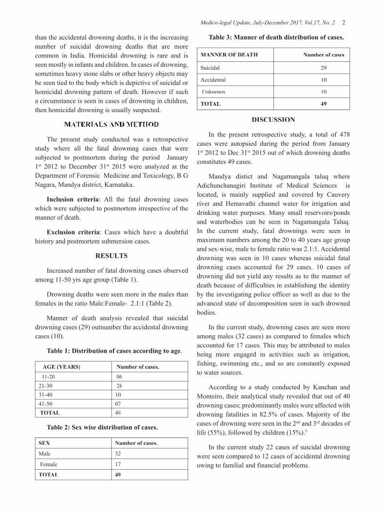
Manner of death analysis revealed that suicidal drowning cases (29) outnumber the accidental drowning cases (10).
Table 1: Distribution of cases according to age.
AGE (YEARS) Number of cases.
11-20 0621-30 2631-40 1041-50 07 TOTAL 49
Table 2: Sex wise distribution of cases.
SEX Number of cases.
Male 32
Female 17
TOTAL 49
Table 3: Manner of death distribution of cases.
MANNER OF DEATH Number of cases
Suicidal 29
Accidental 10
Unknown 10
TOTAL 49
DISCUSSION
In the present retrospective study, a total of 478 cases were autopsied during the period from January 1st 2012 to Dec 31st 2015 out of which drowning deaths constitutes 49 cases.
Mandya distict and Nagamangala taluq where Adichunchanagiri Institute of Medical Sciences is located, is mainly supplied and covered by Cauvery river and Hemavathi channel water for irrigation and drinking water purposes. Many small reservoirs/ponds and waterbodies can be seen in Nagamangala Taluq. In the current study, fatal drownings were seen in maximum numbers among the 20 to 40 years age group and sex-wise, male to female ratio was 2.1:1. Accidental drowning was seen in 10 cases whereas suicidal fatal drowning cases accounted for 29 cases. 10 cases of drowning did not yield any results as to the manner of death because of difficulties in establishing the identity by the investigating police officer as well as due to the advanced state of decomposition seen in such drowned bodies.
In the current study, drowning cases are seen more among males (32 cases) as compared to females which accounted for 17 cases. This may be attributed to males being more engaged in activities such as irrigation, fishing, swimming etc., and so are constantly exposed to water sources.
According to a study conducted by Kanchan and Monteiro, their analytical study revealed that out of 40 drowning cases; predominantly males were affected with drowning fatalities in 82.5% of cases. Majority of the cases of drowning were seen in the 2nd and 3rd decades of life (55%), followed by children (15%).5
In the current study 22 cases of suicidal drowning were seen compared to 12 cases of accidental drowning owing to familial and financial problems.

-legal Update, July-December 2017, Vol.17, No. 2 2 3 Medico-legal Update, July-December 2017, Vol.17, No. 2
The current study also highlights the fact that accidental drowning is more common among children and adolescents. In this study, fatal drowning was observed in 06 cases of children and adolescent age groups. Accidental drowning is more common among females, children and adolescents who reside near open water sources, such as irrigation canals, ponds, lakes or pools.
There are various factors which may be attributed to increased risk of drowning incidences such as: Middle and lower economic status, children left unsupervised around water bodies, lack of higher education, swimming or entering water under the influence of alcohol, diseases like epilepsy, hydrophobia etc., and sometimes visiting and venturing out into tourist places with water bodies without supervision of guides.
In Australia, drowning was reported to be the most common cause of accidental deaths among children. In Western Australia, there were 81 cases of drowning among children between 1987 and 1996, out of which 54% of these cases occurred in backyard swimming pools. An average of 13.5 children had drowned annually between 1975 and 1986 in Western Australia. Drowning fatalities associated with swimming pools in hotels and backyards increased as a percentage total of all drowning from 38% in 1986 to 56% in 1996.6
According to a study done by Ahmed M K, a data relating to deaths of children from drowning was derived from a longitudinal, population-based surveillance system that was in operation in a rural area of Bangladesh from 1983 to 1995. 10% to 25% of child deaths were attributed to drowning during 1983-1995.7
CONCLUSION
From the current study, it can be concluded that Males are more affected in drowning fatalities more so among the middle aged persons and also the manner of death is suicide in majority of males whereas accidental drowning deaths are more common in females and children. Standard guidelines and protocols should be installed near water bodies to prevent accidental drowning deaths.
World health organization (WHO) has prescribed number of preventive measures to avoid drowning deaths
such as installation of barriers/fencing at river side’s, setting up and enforcing safe boating practices, shipping and Ferry regulations. The children and adolescents should be adequately supervised at the river and pool sides. The school-age children should be thought about basic swimming skills, water safety measures and safe rescue skills. The Government should also work towards developing a national water safety strategy to raise awareness about safety around water.
Ethical Clearance: Taken from Institutional Ethics committee.
Source of Funding: Self
Conflict of Interest: Nil
REFERENCES
1. Anil A. Essentials of Forensic Medicine amd Toxicology. 1st edition, Avichal publishing co., Inc. 2014, pg 301.
2. GBD 2013 Mortality and Causes of Death, Collaborators. Global, regional, and national age-sex specific all-cause and cause-specific mortality for 240 causes of death, 1990-2013; A systematic analysis for the Global burden of disease study 2013. Lancet 2014;385;117-171.
3. “Drowning”. World Health Organization. Available from: http://www.who.int/mediacentre/factsheets/fs347/en/. [Last cited on 2010 Dec 30].
4. Accidental Deaths in India. Available from: http//www.ncrb.nic.in/CD-ADSI-2012/accidental- deaths-11.pdf. [Last cited 2013 Oct 21].
5. Kanchan T, Monteiro FN. An analysis of accidental drowning fatalities in Manipal, South India. Inj Prev 2012;18:A 132.
6. Linda W. Child accident prevention foundation of Australia preventing preschool aged drowning. WA Child Inj Prev 1989;1:14-20.
7. Ahmed MK, Rahman M, VAN Ginneken J. Epidemiology of child deaths due to drowningin Matlab, Bangladesh. Int J Epidemiol 1999;28;306-11.
5 -legal Update, July-December 2017, Vol.17, No. 2
Scenario in Attending Emergencies
Rajesh SangramProfessor and Head, Dept. of Forensic Medicine and Toxicology,
E S I Medical College, Kalaburagi, Karnataka
ABSTRACT
It is the duty of every human being to help others in case of emergency. This responsibility is accentuated in cases of medical profession and every attempt should be made to provide the patient emergency care required for his well being. No person shall be denied first aid and immediate management, once he walks into a clinic to the extent possible in that particular setup, irrespective of ability or inability to pay.
In an emergency or a critical case, it is the implicit duty of a noble profession to treat the injured person without waiting either for consent or for fees. The refusal to give treatment would even be violative of the provisions of the code of medical ethics and would constitute a deficiency in service.
Keywords Negligence, Triage, Head injuries, Medicolegal.
Address for Correspondence:Dr. Rajesh Sangram MD., LL.B(spl) Professor and Head, Dept. Of Forensic Medicine and Toxicology, E S I Medical College, Kalaburagi, Karnataka. Ph No: - 09480161314E mail: - [email protected]
INTRODUCTION
Obligations in Emergency
Medico-legal problems in the practice of medicine have become most common but are still relatively infrequent. Today we should accept medico-legal problems as the part and parcel of medical practice and not get distressed up by it.
Many times, a patient accompanied by either parents or relatives or friends enter the casualty room of a physician breaking all the barriers uttering the words emergency care and to be attended at first leaving all the waiting patient in queue. This disturbing and unconvincing situation will be faced by all the practicing doctors at least once in their lifetime.
The word “Emergency” means a sudden unexpected happening or sudden unforeseen occurrence or condition where there is a question of life and death.
Neither Indian law nor the orders of the Supreme Court and various high courts of India have defined
medical emergency. Therefore the definition of medical emergency is still largely left to the discretion of medical professionals. It is accepted that in injured and critically ill patients the priority of the doctors is to save life. However, often there is reluctance on the part of doctors to attend to the emergency needs of patients who, in medical jargon are “Medico-legal cases”. This unwillingness is largely due to medical professionals with the instinct to evade the inconvenience associated with subsequent legal proceedings.
Many patients come to a doctor believing him to be “god”. This attitude must change. As of now people’s expectations are sky high, and they expect nothing sort of a miracle. When the doctor is obviously unable to work this miracle, their god is found to have feet of clay and is thus abused.
If doctor’s indicate on their hospital or nursing home board “24 hours emergency services available” make sure this is really the case. Otherwise it may amount to misrepresentation and make them liable if someone is not attended and suffers damage.
In case doctors cannot always provide round the clock service always though this may be possible most of the days. It is better to avoid announcing 24 hours services etc1.
DOI Number: 10.5958/0974-1283.2017.00057.3
-legal Update, July-December 2017, Vol.17, No. 2 4 5 Medico-legal Update, July-December 2017, Vol.17, No. 2
There are certain important ethical and legal aspects of emergency medical care that medical professionals’ needs to be aware of and these are discussed below2.
• The legal and ethical obligations of a medical practitioner to attend to the emergency medical needs of a patient are total, absolute and paramount.
• Every doctor, either in a government hospital or in private practice, is duty bound to immediately attend to and protect lives of injured victims brought before him.
• It is the constitutional obligation of the state to provide adequate medical services to the people.
• The Indian Medical Council (professional conduct, etiquette and ethics) Regulations, 2002 unambiguously states that a medical professional should attend to a patient in an emergency.
Necessary aid:
In case of head injuries which are very common in the roadside accidents, earlier the doctors who was first approached would start giving first aid and apply stitches to stop the bleeding. However, now what is often seen in that doctors act of fear of facing legal proceeding do not give first aid to the patient, and instead tell him to proceed to the hospital by which time the patient may develop other complications.
In cases of accident, injury and emergency cases, after providing necessary first-aid, the patient is referred to the higher centre, but the patient dies during transport would not be the liability of the doctor. Rather, delay in referral by the doctor could constitute negligence. Remember, not to forget to inform the police if it is a medico-legal case3.
Doctor in the court
Medical professionals harbour apprehensions about being witnesses facing police interrogation and having to repeatedly visit police stations and losing their valuable earning hours. Specially the private practitioners are under the wrong impression that emergencies which are mostly medicolegal cases are dealt with or are to be dealt with only by government doctors. For the government doctors there is no option but they are obliged to attend on MLC.The private doctors usually refuses and refer such a case to a government hospital as there is no authority who can compel any doctor to attend on any
particular case unless there is a military regime.
It is the duty of every human being to help others in case of emergency. This responsibility is accentuated in cases of medical profession and every attempt should be made to provide the patient emergency care required for his well being. No person shall be denied first aid and immediate management, once he walks into a clinic to the extent possible in that particular setup, irrespective of ability or inability to pay.
The doctors are also reluctant to be a witness in a court of law as they may be required to attend the proceedings on multiple occasions, wait for a long time and sometimes face long and unnecessary cross examination. There prevent a medical professionals from doing the needful when a person requires emergency treatment.
To allay these apprehensions the Supreme Court held in Paramanand Katara. V. Union Of India that “The police, the members of the legal profession, law court and everyone concerned will also keep in mind that a man in the medical profession should not be unnecessarily harassed for purposes of interrogation or for any other formalities and should not be dragged during investigation at the police station. Our law cases will not summon a medical professional to give evidence unless the evidence is necessary and even if he is summoned, attempt should be made to see that the men in this profession are not made to wait and waste time unnecessarily. It is also expected that where the facts are so clear it is expected that unnecessary harassment of the members of the medical profession either by way of requests for adjournments or by cross examination should be avoided4.
Correct observation made by the Supreme Court are not only gratifying but also make sense. The public needs to be educated about the fact driven by the court that no sensible professional would intentionally comment an act of omission which would request in loss or injury to the patient as the professional reputation is at stake. A single failure may cost the doctor dear in his career; medical practitioner faced with emergency situation ordinarily tries his best to redeem the patient out of suffering5.
In an emergency or a critical case, it is the implicit duty of a noble profession to treat the injured person without waiting either for consent or for fees. The
Medico-legal Update, July-December 2017, Vol.17, No. 2 6 7 -legal Update, July-December 2017, Vol.17, No. 2
refusal to give treatment would even be violative of the provisions of the code of medical ethics and would constitute a deficiency in service.
In a concurring judgment it is said, ‘when a man in a miserable state hanging between life and death reaches the medical practitioner either in a hospital run or managed by the state, public authority or a private person or a medical professional doing only private practice he is always called upon to rush to help such an injured person and to do all that is within power to save life. It is a duty coupled with human instinct which needs neither decision nor any code of ethics nor any rule or law’.
Triage and Emergency
Stedman’s medical dictionary defines ‘TRIAGE’ as the medical screening of patients to determine their relative priority for treatment; the separation of a large number of casualties, in military or civilian disaster medical care, into three groups6.
1. Those who cannot be expected to survive even with treatment.
2. Those who will recover without treatment; and
3. Those who need treatment to survive.
The doctor has the absolute right to decide which patient he would examine first and even out of turn, depending on the condition of the patient.
Triage means allocation of injured patients into certain categories, a common scheme being as follows:
1. Critical: within seconds
2. Immediate: within minutes
3. Urgent: within the “golden hour”
4. Deferred: as soon as practical.
What the IPC says:
Sections 80 and 88 of the Indian Penal Code contain defences for doctors accused of criminal liability. Under Section 80 (accident in doing a lawful act) nothing is an offence that is done by accident or misfortune and without any criminal intention or knowledge in the doing of a lawful act in a lawful manner by lawful means and with proper care and caution. According to section
88, a person cannot be accused of an offence if she/he performs an act in good faith for the other’s benefit, does not intend to cause harm even if there is a risk, and the patient has explicitly or implicitly given consent.
Section 92 of the Indian Penal Code offers legal immunity for a registered medical practitioner to proceed with appropriate treatment even without the consent of the patient in an emergency, when the victim is incapable of understanding the nature of the treatment, or when there are no legal heirs to sign the consent.
If the patient is conscious and refuses treatment without which the person might endanger his/her life, then the surgeon can inform the judicial magistrate and get the sovereign power of guardianship over persons under disability.
In New India Assurance Co. Ltd. V Dr. Kritkumar S Shera. It was held that there is a difference in the degree of care, caution and skill in normal times and in the care of an emergency, nobody can expect the same degree and amount of care, caution and skill. The amount of care, skill and caution expected of a reasonable and prudent medical practitioner may not be the same during an emergency.
In Amid Ali Shakir V St John’s Medical College Hospital Bangalore, it was held that reasonable delay in shifting the accident victims to the operation theatres because of the necessity to correct the shock is not negligent7.
RECOMMENDATIONS
The three member commission, headed by justice Mr. Jagannadha Rao, drafted a bill pertaining to the private hospitals and practitioner on accident victims and emergency patients; if implemented the following guidelines to be followed by the doctors.
a) The Hospital can’t refuse the accident victim even on the ground that it was a medico-legal case.
b) The bill also stipulates punishment for refusing to admit, treat or transfer a patient after emergency treatment to another hospital.
c) The commission lays down the punishment of six months imprisonment along with fine of Rs. 10,000/- to the doctor or persons running the hospital if an emergency treatment is denied.
-legal Update, July-December 2017, Vol.17, No. 2 6 7 Medico-legal Update, July-December 2017, Vol.17, No. 2
d) The commission says doctor would ensure sufficient medical support is provided enroute for an unharmed transit of patient from one hospital to another.
e) In case ambulance is not available, then doctor will seek the help of police to transfer the patient.
Conflict of Interest Statement
“The undersigned author / authors hereby declare that the article is original, neither the article nor a part of it is under consideration for publication anywhere else and has not been previously published anywhere. We have declared all vested interests. We have meticulously followed the instructions. The article, if published, shall be the property of the Journal and we surrender all rights to the Editors. We agree to provide the latest follow up of cases prior to the publication of case reports when requested”.
Source of Funding- Self
Ethical Clearance – Issued Ethical clearance from Ethical Clearance Committee, ESIC, Medical College,
Kalaburagi, Karnataka.
REFERENCES
1) K. K. Agarwal, General Principals relating to medical negligence, Medilaw, IJCP 2009, P 23-24.
2) Suganthi G Iyer, Legal Aspects Of Emergency Medicine, The Journal of General Medicine, April-June 2001, Vol 13, No. 1, P 36-39.
3) Doctor and law, Jan 2005 Vol 1 No.1, P-8
4) Paramanand Katara. V. Union of Indian (1989) 4 SSC 286; (AIR 1989 SC 2039).
5) V. Parameshvara, Supreme Court Judgment - A much needed relief Medilaw IJCP’s Medinews, November (1-15) 2005, P 6
6) Karunakaran Mathiharan, Emergency Medical Care; Its ethical and legal aspects. The National Medical Journals of India, 2004 No. 1, vol 17, P 31-35.
7) Karnataka SCDRC 1996 (1) CPJ 169; 1995 (3) CPR 174.
9 -legal Update, July-December 2017, Vol.17, No. 2
Recent Trends in Sudden Deaths with Special Reference to Cardiac Causes: Autopsy based Study
from Western Maharashtra
Taware Ajay A1, Bandgar Abhijit L2, Punpale Satyanarayan B3, Jadhao Vijay T4, Tatiya Harish S4
1Associate Professor, Department of Forensic Medicine and Toxicology, BJGMC, Pune, Maharashtra, India, 1Associate Professor, Department of Forensic Medicine and Toxicology, BJGMC, Pune, Maharashtra, India, 1
2Assistant Professor, Department of forensic medicine and Toxicology, GMCH, Aurangabad, Maharashtra, India, 2Assistant Professor, Department of forensic medicine and Toxicology, GMCH, Aurangabad, Maharashtra, India, 2
3Professor and Head, 4Assistant Professor, Department of Forensic Medicine and Toxicology, 4Assistant Professor, Department of Forensic Medicine and Toxicology, 4
BJGMC, Pune, Maharashtra, India
ABSTRACT
WHO defines sudden death as, “Death is said to be sudden or unexpected when a person not known to have been suffering from any dangerous disease, injury or poisoning is found dead or dies within 24 hours after the onset of signs and symptoms.” In sudden death, the immediate cause is almost always to be found in the cardiovascular system, even though topographically the lesion is not in the heart or great vessels. The present autopsy based study was conducted at mortuary of department of forensic medicine of B.J. Medical College and Sassoon General hospital, Pune region, western part of Maharashtra state, India, from October 2013 to May 2015. During the study period total 9497 cases were brought for medico-legal autopsies out of that 807 cases of sudden deaths constituted the study population. Further out of those 807 cases of sudden deaths, 354 cases had cardiovascular system involment which were further analyzed in detail. In the present study, sudden deaths most common in age group of 31 to 60 years with male population being more affected. Cardiovascular diseases constitutes the most common causes of sudden deaths. This analysis will provide a better understanding of epidemiology and burden of sudden natural deaths in society and will help to formulate comprehensive programmes and strategies to prevent same.
Keywords: Sudden Deaths, Cardiac Causes, Autopsy based Study, Western Maharashtra.
Corresponding author: Bandgar Abhijit LAssociate Professor, Department of Forensic Medicine and Toxicology, BJGMC, Pune, Maharashtra, India, Mobile no +91 9860469512.Email: [email protected]
INTRODUCTION
As noted American cardiologist Paul Dudley White said in 1951 in his classic book “Heart Disease”, “The ideal goal towards which mankind should strive is to control cardiovascular threats so that, one lives a healthy, happy and useful youth and middle age life till an advanced age- a point at which even sudden death from cardiovascular disease itself wouldn’t have to be regretted1.”
WHO defines sudden death as, “Death is said to be sudden or unexpected when a person not known to have
been suffering from any dangerous disease, injury or poisoning is found dead or dies within 24 hours after the onset of signs and symptoms”2
The majority of these are natural deaths, but very often, natural deaths form the basis of medico-legal investigations, if they have occurred suddenly and unexpectedly in apparently healthy persons and under the suspicious conditions. In such cases, it is usually not possible to certify the cause of death only on external examination of body; in all such cases, an autopsy is imperative to obviate the possibility of unnatural death.3
In sudden death, the immediate cause is almost always to be found in the cardiovascular system, even though topographically the lesion is not in the heart or great vessels.4
As stressed by WHO scientific group the early recognition of Ischemic Heart Disease is very important
DOI Number: 10.5958/0974-1283.2017.00058.5

9 Medico-legal Update, July-December 2017, Vol.17, No. 2
aspect of prevention, as it would lead us to the development of methods for prevention and control.5
The present autopsy based study is an attempt to do the same by analyzing the cardiac causes of sudden deaths.
MATERIAL AND METHOD
The present autopsy based study was conducted at mortuary of department of forensic medicine of B.J. Medical college and Sassoon General hospital, Pune region, western part of Maharashtra state, India, from October 2013 to May 2015; Cases of sudden deaths brought for medico-legal autopsies died within 24 hours of onset of symptoms, who is not known to have been suffering from any dangerous disease, injury or poisoning were selected as study population. Aims and objectives of the study were to study prevalence of sudden deaths in medicolegal autopsies with special analysis of cardiac causes of sudden death cases, died during the study period.
RESULTS AND DISCUSSION
During the study period total 9497 cases were brought for medico-legal autopsies out of that 807 cases of sudden deaths constituted the study population. Further out of those 807 cases of sudden deaths, 354 cases had cardiovascular system involment which were further analyzed in detail.
Prevalence of Sudden natural Deaths in Medicolegal autopsies
Table 01: Prevalence of Sudden death Cases
Total medico-legal autopsies
Cases of sudden deaths
Percentage ( n=9497)
9497 807 8.49%
In the present study, the prevalence of sudden death was 807 cases amongst 9497 medico- legal autopsies conducted during the study period i.e. 8.49%. These findings are somewhat consistent as mentioned by Dr Narayan Reddy2 (10%) and with the studies conducted by Zanjad et al6 (8.92%) and Sarkojia T. et al 7 (5%) but are inconsistent with that reported by A. Meina Singh et al8 (3%) and Ivar Nordrum et al9 (27.8%). The inconsistency is mainly due to differences in selection of cases due to lack of proper definition of sudden death.
Age and Sex Wise Affection of Sudden Death Cases
Table 02: Age Distribution of Sudden death Cases
Age Group In Years Total Number Of Cases Percentage
( n=807)30 year or below 174 21.55%
31 – 60 463 57.41%
Above 60 170 21.07%
Total 807 100%
In the present study out of 807 cases of sudden deaths, most of the cases belonged to age group 31-60 years i.e. 57.41%. This finding matches with the study conducted by Zanjad et al6, A. Meina Singh et al8 and Kagne R.N. et al10.
Table 03: Sex Distribution of Sudden death Cases
Sex of Deceased Number Of Cases Percentage( n=64)
Male 602 74.60%
Female 205 25.40%
Total 807 100%
In the present study, among the 807 cases of Sudden death, Male to Female ratio was almost 3 indicating, males (74.60%) are more commonly affected than females (25.40%).These findings matches with study conducted by study of T. Sarkojia et al7, Anthony Thomas et al11 and Ivar Nordrum et al9, all found very high male prevalence in sudden deaths.
System-Wise Affection of Sudden Death Cases
Table 04: System-Wise Affection of Sudden Death Cases
System affected Total death
Percentage ( n=807)
Cardiovascular disease 354 43.86%Respiratory system 239 29.62%GIT 87 10.78%CNS 75 9.29%Genitourinary 20 2.48%Multiple system involvement 32 3.97%Total 807 100%
In the study population, cardiovascular system was the most common system affected with 43.86% of the cases; respiratory system was affected in 29.62% of cases, GIT system in 10.78% of cases, CNS system in
Medico-legal Update, July-December 2017, Vol.17, No. 2 10 11 -legal Update, July-December 2017, Vol.17, No. 2
09.29% of cases and genitourinary system was affected in 02.48% of cases. While 03.97% of cases showed multiple system involvement.
Likewise Dr Narayan Reddy2 stated the same that, most of the sudden deaths were due to cardiovascular causes i.e. about 45 %. Similar findings were seen in
Cardiovascular System Affection in Sudden Deaths Cases
Table 5: Cardiac causes of sudden deaths in Present Study
Cardiac Pathology Total Death Percentage (n=354)Coronary atherosclerosis 186 52.54%Myocardial infarction 64 18.07%Rheumatic heart disease 20 5.65%Aortic aneurysm 07 1.98%Aortic dissection 09 2.54%Pericarditis 12 3.39%Acute myocarditis 21 5.93%Congenital heart disease 13 3.68%Cardio myopathies 22 6.22%Total 354 100
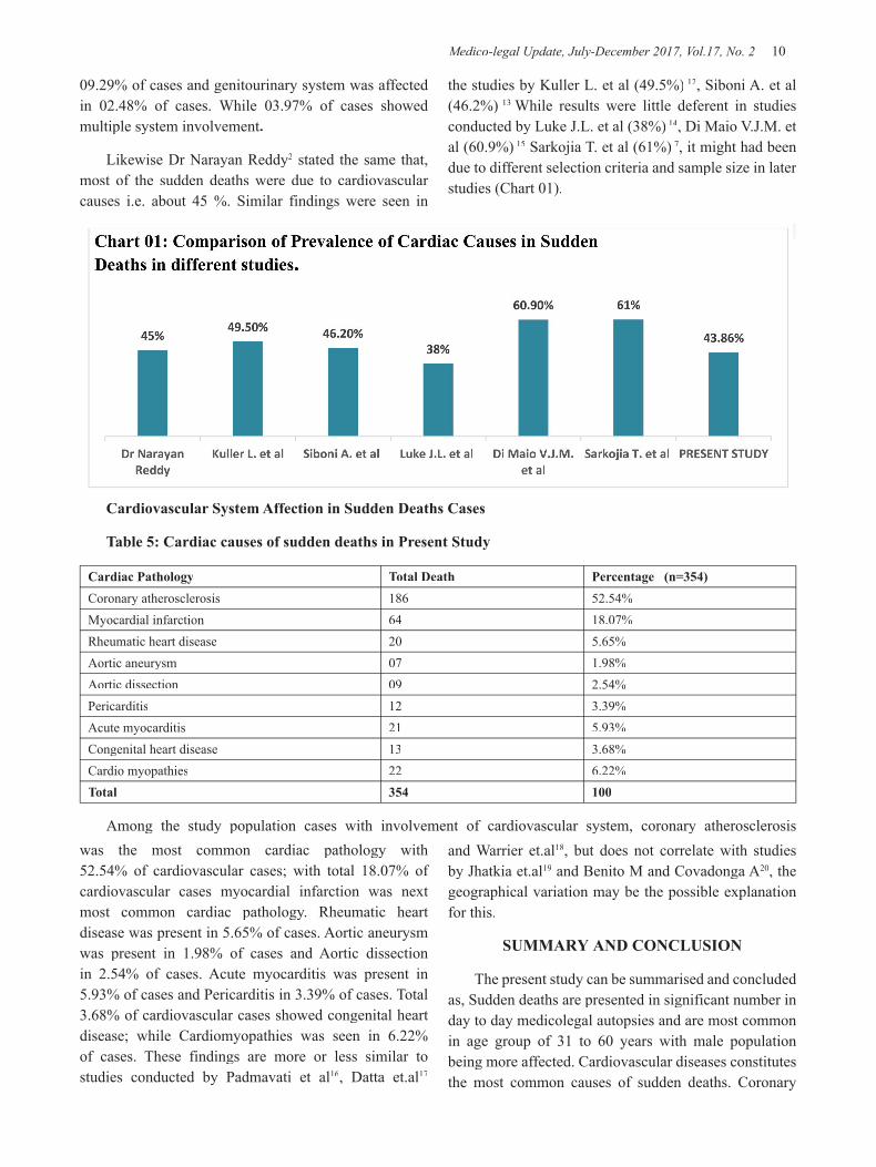
Among the study population cases with involvement of cardiovascular system, coronary atherosclerosis
the studies by Kuller L. et al (49.5%) 12, Siboni A. et al (46.2%) 13 While results were little deferent in studies conducted by Luke J.L. et al (38%) 14, Di Maio V.J.M. et al (60.9%) 15 Sarkojia T. et al (61%) 7, it might had been due to different selection criteria and sample size in later studies (Chart 01).
was the most common cardiac pathology with 52.54% of cardiovascular cases; with total 18.07% of cardiovascular cases myocardial infarction was next most common cardiac pathology. Rheumatic heart disease was present in 5.65% of cases. Aortic aneurysm was present in 1.98% of cases and Aortic dissection in 2.54% of cases. Acute myocarditis was present in 5.93% of cases and Pericarditis in 3.39% of cases. Total 3.68% of cardiovascular cases showed congenital heart disease; while Cardiomyopathies was seen in 6.22% of cases. These findings are more or less similar to studies conducted by Padmavati et al16, Datta et.al17
and Warrier et.al18, but does not correlate with studies by Jhatkia et.al19 and Benito M and Covadonga A20, the geographical variation may be the possible explanation for this.
SUMMARY AND CONCLUSION
The present study can be summarised and concluded as, Sudden deaths are presented in significant number in day to day medicolegal autopsies and are most common in age group of 31 to 60 years with male population being more affected. Cardiovascular diseases constitutes the most common causes of sudden deaths. Coronary

-legal Update, July-December 2017, Vol.17, No. 2 10 11 Medico-legal Update, July-December 2017, Vol.17, No. 2
atherosclerosis and myocardial infarction are the most common causes of sudden cardiac death. This analysis will provide a better understanding of epidemiology and burden of sudden natural deaths in society and will help to formulate comprehensive programmes and strategies to prevent same.
Source of Funding: None
Ethical Clearance: Nil.
Conflict of Interest: Nil
REFERENCES
1) White Paul Dudley. Heart Disease. The Classics of Medicine Library; Special edition; 1991; 12: 07-08.
2) Reddy Narayan KS, Murty O.P. The Essentials of Forensic Medicine and Toxicology. Medical Book Co. Hyderabad 32nd edition.2013; 145.
3) Strong JP, McGill HC: The Natural History of Coronary Atherosclerosis. Am J Pathol.1962; 40: 37-49.
4) Knight B, Saukko P. Knight’s forensic pathology, 3rd edition. London: Edward Arnold Publishers Ltd; 2004; 492-500.
5) WHO Scientific group: The pathological diagnosis of acute ischemic heart disease: WHO Technical Rep.Ser: 1970; 441:5-17.
6) Zanjad NP, Nanadkar SD: Study of Sudden Unexpected Deaths In Medico-Legal Autopsies. JIAFM; 2006; 28 (1): 0971-0973
7) Sarkojia T, Hirvonen J. Causes of sudden unexpected deaths in young and middle aged persons. Forensic Sci In: 1984; 24:247-61.
8) Meina Singh A., Subadani Devi S, Nabachandra H., Fimate L., Sudden Deaths in Manipur - A Preliminary Study. Journal of Forensic medicine & Toxicology, Vol. 19, July-Dec. 2002, 26-28.
9) Nordrum I., Edid T.J., Jorgensen L., Unexplained and explained Natural Deaths Among Persons
above 1 year of Age in a Series of Medico-legal Autopsies. Forensic Science International, 93, 1998, PP 89-98.
10) Kagne R.N., Kamble S.R., Godbole H.V., Borde B.S., Study of Sudden Natural Death, JFMT, Vol. 16,No.1, Jan to June 1999, PP 31-33.
11) Thomas A.C., Knapman P.A., Krikler D.M. & Davis M.J., Community Study of Causes of Natural Sudden Death, British Medical Journal, Vol.297, Dec. 1988, PP 1453-1456.
12) Kuller L, Lilienfeld A, Fisher R: Sudden and unexpected deaths in young adults. JAMA 1966: 198(3): 248-52.
13) Siboni A, Simonsen J. Sudden unexpected natural death in young persons. Forensic Sci Int 1986; 31:159-66.
14) Luke JL, Helpern M. Sudden unexpected death from natural causes in young adults. Arch Pathology 1968; 85:10-16.
15) Di Maio Vincent JM, Di Maio Dominick JM. Natural death as viewed by the medical examiner-A Review of 1000 consecutive autopsies of individuals dying of natural disease. J Forensic Sci 1991; 36(1): 17-24.
16) Padmavati S. Heart Disease In Delhi: Indian H.J. 1958: Volume 33:10.
17) Datta B.N., B. Bhushnurmath, H.N. Khatri, Wahi. Myocardial infarction at autopsy- morphological observations on 272 cases. Indian H.J. 1985: Volume 37:6. 353- 359.
18) Warrier C.B.C. and Venugopal N.S. Incidence and pattern of cardiovascular disease in Kerala. J.Asso. Phy.India. 1967: 29: 229.
19) Jhatkia K.U.: Incidence and etiology of coronary artery disease. J.A.P.I.: 1966:14: 283.
20) Benito M, Covadonga A. Population-Based Study of Out-of-Hospital Sudden Cardiovascular Death: Incidence and Causes of Death in Middle-Aged Adults. Rev Esp Cardiol. 2011; 64(1):28-34.
13 -legal Update, July-December 2017, Vol.17, No. 2
Psychological Autopsy of Complete Suicide Cases in Bhopal Region of Central India: A Retrospective Study
Singh Sandeep1, Juglan Sarthak2, Benzal Rajeev Kumar3, Yadav Jayanthi4 1Associate Professor, Department of Forensic Medicine and Toxicology, L.N. Medical College, Kolar Road Bhopal
(M.P.), 2Assistant Professor, Department of Forensic Medicine and Toxicology, Gajra Raja Medical College, Gwalior (M.P.), 3Associate Professor, Department of Forensic Medicine and Toxicology, Bundelkhand Medical
College, Sagar (M.P.), 4Professor and Head, Department of Forensic Medicine and Toxicology, Gandhi Medical College, Bhopal (M.P.),
ABSTRACT
Background: Psychological autopsy is the reconstruction of events leading to death. There are few studies on psychological autopsy.
Aims: To understand the profile of suicide completers and compare patients who attempted suicide by hanging with those who attempt suicide by consuming poison on demographic and phenomenological variables and assess the socio-demographic characteristics, psychosocial factors, and psychiatric and physical co-morbidity associated with completed suicide.
Materials and Method: Two hundred complete suicide cases were analyzed. Using a semi-structured, self-designed questionnaire, the family, friends and relatives of the deceased were interviewed.
Results: The presence of some type of stressful life events and family history of suicide are two important factors for committing suicide.
Conclusions: It could be concluded that psychological autopsy just like physical autopsy can be useful tool to investigate the antecedent of death and reveals the deceased contribution to their own death.
Keywords: Psychological autopsy, suicide, hanging, poisonous substances.
Corresponding author: Dr. Sarthak Juglan, Assistant Professor, Department of Forensic Medicine and Toxicology, Gajra Raja Medical College, Gwalior (M.P.), E-mail. – [email protected]
INTRODUCTION
A psychological autopsy is the reconstruction of events leading to death; ascertainment of the circumstances of the death, including suicidal intent; and an in-depth exploration of other significant risk factors for suicide.1–7
In depth study of the history of suicide prior to the suicidal act is known as psychological autopsy. [8] This is the most informative means of studying the nature and causes of suicide. [9] This method is commonly used in
various studies to assess and manage suicidal patients. [1
0],[11] Findings from such studies offer clues for planning suicide prevention strategies. Suicide rate in India is approximately 114 per million in males and 80 per million in females. [12] India and China are responsible for 30% of all cases of suicide worldwide. [12]
In an Indian study, it was reported that predominant suicidal victims were unemployed males, middle-aged and high school-educated subjects; and they were mostly from a rural background. [13] Even people with low suicidal intention may end up in completed suicides as a result of using more lethal methods and inadequate treatment. [14] In India, it is the comparatively younger people who are suicide victims. [15],[16] This phenomenon of successful completion of suicide is a dangerous trend in India. While the population increase in the last decade was 25%, the suicide rate increased by 60%. [17]
DOI Number: 10.5958/0974-1283.2017.00059.7
13 Medico-legal Update, July-December 2017, Vol.17, No. 2
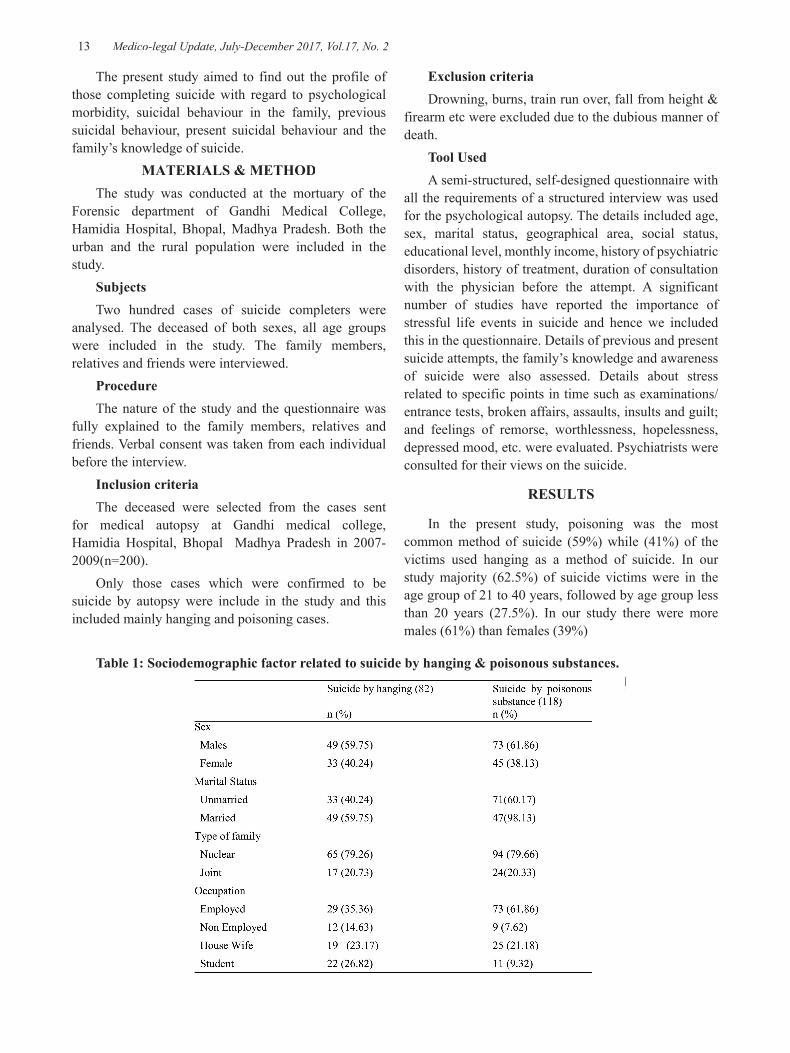
Table 1: Sociodemographic factor related to suicide by hanging & poisonous substances.
The present study aimed to find out the profile of those completing suicide with regard to psychological morbidity, suicidal behaviour in the family, previous suicidal behaviour, present suicidal behaviour and the family’s knowledge of suicide.
MATERIALS & METHODThe study was conducted at the mortuary of the
Forensic department of Gandhi Medical College, Hamidia Hospital, Bhopal, Madhya Pradesh. Both the urban and the rural population were included in the study.
SubjectsTwo hundred cases of suicide completers were
analysed. The deceased of both sexes, all age groups were included in the study. The family members, relatives and friends were interviewed.
ProcedureThe nature of the study and the questionnaire was
fully explained to the family members, relatives and friends. Verbal consent was taken from each individual before the interview.
Inclusion criteriaThe deceased were selected from the cases sent
for medical autopsy at Gandhi medical college, Hamidia Hospital, Bhopal Madhya Pradesh in 2007- 2009(n=200).
Only those cases which were confirmed to be suicide by autopsy were include in the study and this included mainly hanging and poisoning cases.
Exclusion criteriaDrowning, burns, train run over, fall from height &
firearm etc were excluded due to the dubious manner of death.
Tool UsedA semi-structured, self-designed questionnaire with
all the requirements of a structured interview was used for the psychological autopsy. The details included age, sex, marital status, geographical area, social status, educational level, monthly income, history of psychiatric disorders, history of treatment, duration of consultation with the physician before the attempt. A significant number of studies have reported the importance of stressful life events in suicide and hence we included this in the questionnaire. Details of previous and present suicide attempts, the family’s knowledge and awareness of suicide were also assessed. Details about stress related to specific points in time such as examinations/ entrance tests, broken affairs, assaults, insults and guilt; and feelings of remorse, worthlessness, hopelessness, depressed mood, etc. were evaluated. Psychiatrists were consulted for their views on the suicide.
RESULTS
In the present study, poisoning was the most common method of suicide (59%) while (41%) of the victims used hanging as a method of suicide. In our study majority (62.5%) of suicide victims were in the age group of 21 to 40 years, followed by age group less than 20 years (27.5%). In our study there were more males (61%) than females (39%)

Medico-legal Update, July-December 2017, Vol.17, No. 2 14 15 -legal Update, July-December 2017, Vol.17, No. 2
Table 1 shows that a large number of subjects who committed suicide by poisonous substance 47(39.83%) were married whereas 71(60.17%) were unmarried but in suicide by hanging 49(59.75%) were unmarried. Majority of the subjects in suicide by poisonous substance were employed 73 (61.86%) were as in suicide by hanging 29 (35.36%) were employed; and
among the student category, suicide by hanging was seen in 22 (26.82%) and suicide by poisonous substance in 11(9.32%).19(23.17%) victims of suicide by hanging and 25(21.18%) of the subjects of suicide by poisonous substance were found to be housewives. No difference was seen between suicides by hanging or poisonous substance among subjects belonging to nuclear or joint family.
Table 2: Psychosocial factor related to suicide by hanging & poisonous substances.
Table 2 shows psycho-social stressors of subtype interpersonal conflicts were found in 69(58.47%) cases of suicide by poisonous substance and 41 (50%) in suicide by hanging; while subjects debt/ financial loss were the cause of suicide by hanging in 20(24.39%) and 21(17.79%) the cause was suicide by poisonous substance; and 25.60% cases of suicide by hanging and 23.72% victims of suicide by poisonous substance had financial stressors. Major depressive disorder was the most common probable psychiatric diagnosis in 36 (43.90%) victims of suicide by hanging & 43 (36.44%) victims of suicides by poisonous substance. Family history of suicide was present in victims of suicide by hanging.
DISCUSSION
There is a steep rise in the number of suicides all over the world. Currently, it is becoming a matter of concern for mental-health professionals on account of its increasing incidence. Our study reports on 200 victims
who committed suicide in 2007-2009, conducted at the mortuary of the Forensic department of Gandhi medical college, Hamidia Hospital, Bhopal Madhya Pradesh. We interviewed a key informant of the deceased, and thus the information available was reliable. Psychological autopsy approach was used in this study to elucidate the nature and causes of completed suicide. Similar approach has been employed in earlier Indian and international studies. [9],[11]
The present study was showing suicide by poisonous substances as common but BS Chavan et al18 shows hanging was the most common method (72.2%), while 15.8% of the victims used poisoning as a method of suicide. Some hospital-based Indian studies had revealed that insecticide poisoning was the commonly employed method in committing suicide. [19],[20],[21]
Some study observed that majority (59.4%) of suicide victims were in the age group of 20 to 29 years, followed by age group 30 to 39 years (14.8%) and males

-legal Update, July-December 2017, Vol.17, No. 2 14 15 Medico-legal Update, July-December 2017, Vol.17, No. 2
(57.4%) slightly outnumbered females (42.57%) but in our study 62.5% of suicide victims were in the age group of 21 to 40 years, followed by age group less than 20 years (27.5%) and males (61%) more than females (39%).
Our study findings showed that unmarried persons, employed persons committed suicide but other studies show unmarried and unemployed persons. [18],[22],[23],[24]
In BS Chavan et al18 study psycho-social stressors were found in 61 (60.3%) suicide victims; while 47.5% of the subjects believed interpersonal stressors were the cause of suicide, and 8.9% of suicide victims had financial stressors, Occupational stressors were found in 3.9% of the suicide victims, the finding are almost similar in our study . Interpersonal stress appears to be the common cause of suicide in this study. Similar observations have been made by several other researchers. [11],[14],[15] Psychiatric illness was found in 33.6% ( n = 34) of the subjects in this study. This figure was higher than the reported rate (23%) in a previous Indian study. [15] We found that 11.9% (n = 12) of the victims had depressive episode and 23.7% had alcohol/substance abuse. As many as 57.4% of the suicide victims had shown change in behavior before committing the act. In our study, majority of victim of suicide are suffering from major depression, followed by border line personality disorder and than alcohol abuse. In many international studies, psychiatric illness has been thought to be the most important cause of suicide. In these studies, the proportion of suicide victims with psychiatric illness ranged from 73% to 100%. [9],[10]
CONCLUSION
We aimed to study the profile of those who had completed suicide on sociodemographic status, psychiatric morbidity, stressful life events responsible for suicide and the family’s awareness of the suicidal behaviour. We conclude that psychological autopsy is a very important tool for assessing the causes and precipitants of suicide. It is very difficult to assess the exact reason or pinpoint the cause of suicide. More studies in this field are required with a larger sample size for the evaluation of suicide.
Ethical Clearance - Taken from College Ethical Committee (Gandhi Medical College, Bhopal)
Source of Funding - Nil (Self)
Conflict of Interest - Nil
REFERENCES
1. Beskow J, Runeson B, Asgard U. Ethical aspects of psychological autopsy. Acta Psychiatr Scand.1991;84:482–7.
2. Brent DA, Perper JA, Goldstein CE, et al. Risk factors for adolescent suicide: A comparison of adolescent suicide victims with suicidal inpatients. Arch Gen Psychiatry. 1988;45:581–8.
3. Brent DA, Perper JA, Moritz G, et al. Psychiatric risk factors for adolescent suicide: A case–control study. J Am Acad Child Adolesc Psychiatry. 1993;32:521–9.
4. Cooper J. Ethical issues and their practical application in psychological autopsy study of suicide. J Clin Nursing. 1999;8:467–75.
5. Hawton K, Simkin S, Fagg J, et al. Suicide in Oxford University students, 1976–1990. Br J Psychiatry. 1995;166:44–50.
6. Kelly TM, Mann JJ. Validity of DSM-III-R diagnosis by psychological autopsy: A comparison with clinician ante-mortem diagnosis. Acta Psychiatr Scand. 1996;94:337–43.
7. Velting DM, Shaffer D, Gould MS, et al. Parent–victim agreement in adolescent suicide research. J Am Acad Child Adolesc Psychiatry. 1998;37:1161–6.
8. Unni KE, Vyas JN, Ahuja N, editors. Human self destructive behaviour. In : Postgaduate psychiatry. Jaypee Brothers Medical Publishers: New Delhi 1999;p.526-56.
9. Hawton K, Fagg J. Suicide and other causes of death, following attempted suicide. Br J Psychiatry 1988;152:359-66.
10. Morris R, Gask L, Battersby L, Francheschini A, Robson M. Teaching front-line health and voluntary workers to assess and manage suicidal patients. J Affect Disord 1999;52:77-83.
11. Khan FA, Anand B, Gowridevi M. Psychological autopsy of suicide: A cross-sectional study. Indian J Psychiatry 2005;47:73-8.
12. World Health Organization. Figures and Facts about Suicide (Doc.WHO/MNH/MBD/99.1). WHO: Geneva;1999.
13. Kumar PN. An analysis of suicide attempters versus

Medico-legal Update, July-December 2017, Vol.17, No. 2 16
completers in Kerala. Indian J Psychiatry 2004;46:144-9.
14. Roy A, Segal N, Sarchiapone M. Attempted suicide among living cotwins of twin suicide victims. Am J Psychiatry 1995;152:1075-6.
15. Hegde RS. Suicide in a rural community of North Karnataka. Indian J Psychiatry 1980;22:368-70.
16. Shukla GD, Verma BL, Mishra DN. Suicide in Jhansi city. Indian J Psychiatry 1990;32:44-51.
17. Murthy RS. Suicide prevention: Policies and priorities; Community mental health perspective. Indian J Soc Psychiatry 1993;9:47-52.
18. BS Chavan, Gurvinder Pal Singh, Jaspreet Kaur, Reshma Kochar : Psychological autopsy of 101 suicide cases from northwest region of India. Indian psy J;50(1);34-38.
19. Gargi J, Rai H, Chanana A. Current trends of poisoning: A hospital profile. J Indian Med Assoc
2006;104:72-3.
20. Khosla SN, Nand N, Kumar P, Trehan V. Muscle involvement in aluminium phosphide poisoning. J Assoc Physicians India 1988;36:289-90.
21. Gargi J, Chanana A. Epidemiological aspects of suicide due to poisoning in North-West Punjab: A changing trend. Rom J Legal Med 1997;5:78-9.
12. Platt S. Suicidal behaviour and unemployment: A literature review: Health policy implication of unemployment. World Health Organization: Geneva; 1985. p. 87-132.
23. Heikkinen ME, Isometsa ET, Marttunen MJ, Aro HM, L φnnqvist JK. Social factors in suicide. Br J Psychiatry 1995;167:747-53.
24. Kreitman N. Suicide and parasuicide. In : Kendell RE, Zeally AK, editors. Companion to psychiatric studies. Churchill Livingstone: London; 1988. p. 459-75.
-legal Update, July-December 2017, Vol.17, No. 2 16
A Prospective Study to Ascertain the Profile of Unnatural Deaths at Basaveshwara Teaching and General Hospital Mortuary, Gulbarga (Kalaburagi) – A Research Paper
Akshay Kumar Ramtake1, Vandana Mudda2, Santosh S Garampalli3, Umesh S R4
1Assistant Professor, Department of Forensic Medicine and Toxicology, Government Medical College, Ambikapur, 1Assistant Professor, Department of Forensic Medicine and Toxicology, Government Medical College, Ambikapur, 1
Sarguja, Chhattisgarh, 2Associate Professor, 2Associate Professor, 2 3Assistant Professor, Department of Forensic Medicine and 3Assistant Professor, Department of Forensic Medicine and 3
Toxicology, M. R. Medical College, Kalaburagi, Karnataka, 4Professor & H.O.D., Department of Forensic 4Professor & H.O.D., Department of Forensic 4
Medicine and Toxicology, Gulabarga Institute of Medical Sciences, Kalaburagi
ABSTRACT
The aim of our study was to characterize the cases of unnatural deaths admitted to the Basaveshwara Teaching & General Hospital (BTGH), M. R. Medical College, Kalaburagi. This is prospective study conducted at mortuary of Basaveshwara Teaching and General Hospital, Kalaburagi, Karnataka between November 2013 to August 2015, which included 151 cases of unnatural deaths. Burns (49.01%) constituted the maximum number of cases, followed by RTA (31.79%), poisoning (16.56%), fall from height (1.32%) and bites and stings (1.32%). Out of 151 cases of unnatural deaths, 81 (53.64%) cases were males and 70 (46.36%) cases were females. The largest number of victims was in the age group 21-30 years (29.80%). Maximum numbers of the victims (76.82%) were married. Majority of the victims (31.12%) were house wife, followed by (23.52%) were employed in service. Most of the victims (80.13%) were having low level education. Majority of cases (67.55%) were from rural area. Maximum numbers of victims were Hindu (89.40%). Most of the cases (42.38%) were belongs to class V socioeconomic status followed by (26.49%) were belongs to class IV. Maximum number of cases (36.42%) were reported in summer season followed by (34.44%) in winter seasons. Majority of incidents were occurred in the afternoon hours (35.10%).Maximum numbers of victims (33.77%) were survived for less than 24 hours, followed by (29.14%) victims for 3 days to 1 week. Most of the cases (80.79%) were accidental in nature.
Keywords: Unnatural deaths, Religion, Education, Rural.
Corresponding author: Dr. Akshay Kumar RamtakeAssistant Professor, Department of Forensic Medicine and Toxicology, Government Medical College, Ambikapur, Sarguja, Chhattisgarh-497001
INTRODUCTION
Unnatural deaths claim a substantial number of lives in developing countries like India. The unnatural causes of death are many with accidents being the most common amongst others. Over the decades there has been a steady increase in the transportation deaths and injuries, on road, rail and in the air. Accidental burns, poisoning, drowning, electrocution and fall from height are also on the rise. Suicidal and homicidal fatalities are common among both the urban and rural population.1
MATERIALS AND METHOD
This is prospective study conducted at mortuary of Basaveshwara Teaching and General Hospital, Kalaburagi, Karnataka between November 2013 to August 2015, which included 151 cases of unnatural deaths. The data is collected from Case papers, Inquest reports, post-mortem reports and from interviewing relatives and friends of deceased. The data thus obtained was analyzed and the study was done with respect to: types of unnatural deaths, age & sex wise distribution, domicile pattern, religion of victim, occupational status, marital status, socio-economic status, educational status, diurnal variation, period of survival, manner of poisoning, and season wise distribution.
DOI Number: 10.5958/0974-1283.2017.00060.3

Medico-legal Update, July-December 2017, Vol.17, No. 2 18 19 -legal Update, July-December 2017, Vol.17, No. 2
RESULTS
Distribution of cases according to types of Unnatural Deaths:
In the present study, burns 74 (49.01%) constituted the maximum number of cases, followed by RTA 48 (31.79%), poisoning 25 (16.56%), fall from height 02 (1.32%) and bites and stings 02 (1.32%).
Table 1: Distribution of cases according to types of Unnatural Deaths
Type Total No. of cases Total Percentage (%)Burns 74 49.01RTA 48 31.79Poisoning 25 16.56Fall from Height 02 1.32Bites and Stings 02 1.32Assault 00 00Mechanical Asphyxia 00 00Total 151 100%
Distribution of cases of Unnatural Deaths according to Age and Sex:
Out of 151 cases of unnatural deaths, 81 (53.64%) cases were males and 70 (46.36%) cases were females, thus indicating that majority of victims were males. In the present study, maximum number of victims 45 (29.80%) were in the age group of 21-30 years, followed by 31 (20.53%) cases and 27 (17.88%) cases were in the age group of 31-40 years and 11-20 years. Minimum numbers of victims were in the age group of more than 80 years 3 (1.99%).
Table 2: Distribution of cases of Unnatural Deaths according to Age and Sex
Age (in Years) Male Female Total No. of cases Total Percentage (%)<10 04 05 09 5.9611-20 7 20 27 17.8821-30 22 23 45 29.8031-40 22 09 31 20.5341-50 10 04 14 9.2751-60 09 05 14 9.2761-70 05 03 08 5.3071-80 00 00 00 00>80 02 01 03 1.99Total 81 (53.64%) 70 (46.36%) 151 100%
Distribution of cases of Unnatural Deaths according to Marital Status:
In the present study, maximum number of the victims 116 (76.82%) were married, followed by unmarried victims 35 (23.18%).
Distribution of cases of Unnatural Deaths according to Occupation of the victims:
Out of 151 cases of unnatural deaths, maximum number of victims 47 (31.12%) were house wife,
followed by 34 (23.52%) were employed in service and least number of cases 04 (2.65%) were children of less than 5 years of age.
Distribution of cases of Unnatural Deaths according to Educational status of the victims:
In the present study, maximum number of victims 121 (80.13%) were having low level education (including pre primary, primary level education, secondary level education, matriculation, higher secondary level education and graduation), followed by 19 (12.58%)
-legal Update, July-December 2017, Vol.17, No. 2 18 19 Medico-legal Update, July-December 2017, Vol.17, No. 2
were illiterates and least number of victims 11 (7.29%) were having high level education (postgraduates and post doctoral degree holders).
Distribution of cases of Unnatural Deaths according to Domicile Pattern
Out of 151 cases of unnatural deaths, 102 (67.55%) cases were from rural area followed by 49 (32.45%) were belongs to urban area.
Distribution of cases of Unnatural Deaths according to Religion
In the present study, maximum numbers of victims were Hindu 135 (89.40%), followed by Muslim 15 (9.94%) and 01 (0.66%) was Christian.
Distribution of cases of Unnatural Deaths according to Socioeconomic status of victim
Out of 151 cases of unnatural deaths, 64 (42.38%) cases were belongs to class V socioeconomic status followed by 40 (26.49%) were belongs to class IV and only 09 (5.97%) cases were belongs to class I.
Distribution of cases of Unnatural Deaths according to Seasonal variation
In the present study, maximum number of cases 55 (36.42%) were reported in summer season, followed by 52 (34.44%) and 44 (29.14%) cases in winter and rainy seasons.
Distribution of cases of Unnatural Deaths according to Diurnal variation:
In the present study, among over all cases of unnatural deaths maximum number of incidents occurred in the afternoon hours 53 (35.10%) and minimum 23 (15.23%) in the night. 39 (25.83%) and 36 (23.84%) cases occurred during morning and evening hours.
Distribution of cases of Unnatural Deaths according to Period of survival following incident:
Out of 151 cases of unnatural deaths, maximum number of victims 51 (33.77%) were survived for less than 24 hours, followed by 44 (29.14%) victims for 3 days to 1 week and 27 (17.88%) for 24 hours to 3days, and least number of cases 05 (3.31%) were survived for more than 4 weeks.
Distribution of cases of Unnatural Deaths according
to Manner of death:
In the present study, 122 (80.79%) cases were accidental in nature followed by 24 (15.89%) were suicidal and 05 (3.32%) were homicidal.
DISCUSSION
Unnatural death is an indicator of social and mental health of a country. Unnatural deaths is on the rise this is due to socioeconomic problems, increase in stress in day to day life, domestic violence, tremendous growth in road transport sector, technology, self destruction by poisoning, etc. The unnatural deaths affect not only the individual but also his family, and also to the country either directly or indirectly.
In the present study, burns 74 (49.01%) constituted the maximum number of cases, followed by RTA 48 (31.79%), poisoning 25 (16.56%), fall from height 02 (1.32%) and bites and stings 02 (1.32%).
Our result was similar to the observations made in the study conducted by Parashar R et al (2015)2 in which the commonest cause of unnatural death was burn 71(46.71%). The second most commonest cause of unnatural death was RTA 50 (32.89%) cases, , followed by poisoning 17 (11.18%), fall from height 12 (7.89%), etc; by Pradeep kumar MP et al (2015)3 where burns 20.80% account for majority of cases among unnatural deaths followed by RTA 16.79%, poisoning 11.24%, etc.
Out of 151 cases of unnatural deaths, 81 (53.64%) cases were males and 70 (46.36%) cases were females, thus indicating that majority of victims were males. Male to female ratio was 1.2:1. In the present study, maximum number of victims 45 (29.80%) were in the age group of 21-30 years, followed by 31 (20.53%) cases and 27 (17.88%) cases were in the age group of 31-40 years and 11-20 years. Minimum numbers of victims were in the age group of more than 80 years 3 (1.99%).
Our result was similar to the observations made in the study conducted by Sharma BR et al (2006)4 in which males were 69.3% and females were 30.7%. Maximum numbers of victims 49% were in the age group of 21-30 years; by Rahim M & Das TC (2009)5 in which males were 77.27% and females were 22.72%. Maximum number of victims 33.72% were in the age group of 21-30 years

Medico-legal Update, July-December 2017, Vol.17, No. 2 20 21 -legal Update, July-December 2017, Vol.17, No. 2
Maximum number of the victims 116 (76.82%) were married, followed by unmarried victims 35 (23.18%)
Our results for overall cases of unnatural deaths were similar to the observations made in study conducted by Sharma BR et al (2006)4 where 63.04% persons were married and 36.96% were unmarried; by Bansude ME et al (2012)6 in which 80.06% victims were married followed by 19.94% unmarried victims.
Out of 151 cases of unnatural deaths, maximum number of victims 47 (31.12%) were house wife and least number of cases 04 (2.65%) were children of less than 5 years of age.
Our results for overall cases of unnatural deaths were similar to the observations made in study conducted by Biswas DK et al (2013)7 where maximum victims 19% with known occupation were house wife. Housewives are more commonly subjected to harassment and torture by their husbands or their family members and thus they live a stressful life.
In the present study, maximum number of victims 121 (80.13%) were having low level education and least number of victims 11 (7.29%) were having high level education.
Our results for overall cases of unnatural deaths were similar to the observations made in study conducted by Sharma BR et al (2006)4 in which maximum numbers of cases were of those who did not complete matric level education (34%), followed by those with education between matric and higher secondary (26%) and least number of cases was seen in post doctoral degree holders victims (0.5%) cases; by Kumar S et al (2013)8 where majority of the victims (91.55%) were either illiterate, under metric or education up to matriculation level or graduation level and only (8.45%) victims had got higher education.
Out of 151 cases of unnatural deaths, 102 (67.55%) cases were from rural area followed by 49 (32.45%) cases were belongs to urban area.
Our results for overall cases of unnatural deaths were similar to the observations made in study conducted by Kumar S et al (2013)8 where maximum number of victims were from rural area (51.48%) followed by urban area (48.51%); by Sarkar AP et al (2013)9 in which majority of cases were from rural area (64.5%).
In the present study, maximum numbers of victims were from Hindu religion 135 (89.40%) cases, followed by Muslim 15 (9.94%) cases and 01 (0.66%) was Christian.
Our results for overall cases of unnatural deaths were similar to the observations made in study conducted by Bansude ME et al (2012)6 in which Hindus were more commonly involved (87.26%) than Muslims (12.74%); by Sarkar AP et al (2013)9 where majority of victims belongs to Hindu religion (78.2%); by Biswas DK et al (2013)10 in which 82 % of cases were Hindu and 9% were Muslim by religion.
Out of 151 cases of unnatural deaths, 64 (42.38%) cases were belongs to class V socio-economic status followed by 40 (26.49%) cases were belongs to class IV and only 09 (5.97%) cases were belongs to class I.
Our results for overall cases of unnatural deaths were similar to the observations made in study conducted by Sharma BR et al (2006)4 in which majority of deaths (63%) were seen in low socio-economic group.
In the present study, maximum number of cases 55 (36.42%) were reported in summer season, followed by 52 (34.44%) cases and 44 (29.14%) cases in winter and rainy seasons.
Our results for overall cases of unnatural deaths were similar to the observations made in study conducted by Yousfani GM & Memon MU (2010)11 in which unnatural deaths were occur more in summer season (63%). During summer season holidays are declared in various schools and colleges, person loose temperament easily and this is also the season for marriages.
In the present study, among over all cases of unnatural deaths maximum number of incidents occurred in the afternoon hours 53 (35.10%) and minimum 23 (15.23%) in the night. 39 (25.83%) and 36 (23.84%) cases were occurred during morning and evening hours.
Our results for overall cases of unnatural deaths were not correlating with the observations made in study conducted by Kumar S et al (2013)8 in which cases were most commonly occurring during night (36%) and evening hours (31.93%).
In the present study, out of 151 cases of unnatural deaths, maximum number of victims 51 (33.77%) were survived for less than 24 hours, followed by 44 (29.14%)

-legal Update, July-December 2017, Vol.17, No. 2 20 21 Medico-legal Update, July-December 2017, Vol.17, No. 2
victims for 3 days to 1 week and 27 (17.88%) for 24 hours to 3days, and least number of victims 05 (3.31%) were survived for more than 4 weeks.
Our results for overall cases of unnatural deaths were not correlating with the observations made in study conducted by Parashar R et al (2015)2 in which survival period of the victim of unnatural deaths was on average 13 days in case of burns followed by 7 days in RTA, etc. This is due to delay in hospitalization of the victims in our study.
In the present study, the manner of death in 122 (80.79%) cases were accidental, followed by 24 (15.89%) were suicidal and 05 (3.32%) were homicidal.
Our results for overall cases of unnatural deaths were similar to the observations made in study conducted by Kumar S et al (2013)8 in which (64.46%) cases were due to accident followed by suicide (22.75%) cases and homicide cases were (12.77%).
CONCLUSION
In our study we conclude that, Burns (49.01%) constituted the maximum number of cases, followed by RTA (31.79%), poisoning (16.56%), fall from height (1.32%) and bites and stings (1.32%). Out of 151 cases of unnatural deaths, 81 (53.64%) cases were males and 70 (46.36%) cases were females. The largest number of victims was in the age group 21-30 years (29.80%). Maximum numbers of the victims (76.82%) were married. Majority of the victims (31.12%) were house wife, followed by (23.52%) were employed in service. Most of the victims (80.13%) were having low level education. Majority of cases (67.55%) were from rural area. Maximum numbers of victims were Hindu (89.40%). Most of the cases (42.38%) were belongs to class V socioeconomic status followed by (26.49%) were belongs to class IV.Maximum number of cases (36.42%) were reported in summer season followed by (34.44%) in winter seasons. Majority of incidents were occurred in the afternoon hours (35.10%). Maximum numbers of victims (33.77%) were survived for less than 24 hours, followed by (29.14%) victims for 3 days to 1 week. Most of the cases (80.79%) were accidental in nature.
Conflict of Interests: The author declares that there is no conflict of interests.
Source of Funding: None
Ethical Clearance: Yes
REFERENCES
1. Santhosh CS, Vishwanathan KG, Satish Babu BS. Pattern of Unnatural Deaths- A Cross Sectional Study of Autopsies at Mortuary of KLES’S Hospital and MRC, Belgaum. J Indian Acad Forensic Med. 2011 Jan-Mar; 33(1):18-20.
2. Parashar R, Asawa S, Parashar H. Study of Un-Natural Deaths of the patients brought to People’s Hospital, Bhopal (a Retrospective Study). Indian Journal of Forensic Medicine & Toxicology. 2015 Jan-June; 9(1): 172-5.
3. Pradeep Kumar MP, Devadass PK, Bheemappa L Havanur. A Retrspective study of Pattern of Un-natural Deaths: an Autopsy Study at Victoria Hospital Mortuary, Bangalore. Medico Legal Update. 2015 Jan-June; 15(1): 86-9.
4. Sharma BR, Harish D, Sharma A, Sharma S, Singh H. Unnatural Deaths in Northern India: An Overview of Social Etiology. J Indian Acad Forensic Med. 2006; 28 (4): 148-54.
5. Rahim M, Das TC. Mortuary Profile for Unnatural Deaths at Forensic Medicine Department of Dhaka Medical College. Bangladesh Med J. 2009 July; 38(2): 44-7.
6. Bansude ME, Kachare RV, Dode CR, Kumre VM.Trends of Unnatural Deaths in Latur District of Maharashtra. Journal of Medico legal Association of Maharashtra. 2012 Jul-Dec; 21(2): 17-24.
7. Biswas DK, Biswas A, Das DK, Bhunia R, Ghosh D. Socio-demographic Profiles of Post Mortem Cases in a Sub-district, West Bengal, India. Medico Legal Update. 2013 Jul-Dec; 13(2): 32-7.
8. Kumar S, Verma AK, Ahmad I, Ali W, Singh US. Profile of Unnatural Deaths – A Study of Autopsies at Mortuary of King George’s Medical University, Lucknow, India. Medico Legal Update. 2013 Jul-Dec; 13(2): 113-8.
9. Sarkar AP, Mandal R, Ghorai S, Sengupta P. A Study on Socio- Demographic Profile of Post Mortem Cases in the Morgue of a District Hospital in West Bengal. Medico Legal Update. 2013 Jul-Dec; 13(2): 1-4.
10. Biswas DK, Biswas A, Das DK, Bhunia R, Ghosh D. Socio-demographic Profiles of Post Mortem Cases in a Sub-district, West Bengal, India. Medico Legal Update. 2013 Jul-Dec; 13(2): 32-7.
11. Yousfani GM, Memon MU. Spectrum of Unnatural Deaths in Hyderabad: An Autopsy Based Study. JDUHS. 2010; 4(2): 54-7.
23 -legal Update, July-December 2017, Vol.17, No. 2
Study of Pattern of Suicides among Adolescent and Youth among Autopsies Conducted at KIMS Hospital, Hubballi
Gajanan H Nayak1, Ravindra Kumar C N2, Sunilkumar S Biradar3, Madhu Sudhan S4
1Prof & Head, Department of Forensic Medicine & Toxicology, KIMS, Hubballi, 2Tutor, Department of Forensic Medicine & Toxicology, East Point College of Medical Sciences & Research Centre, Bengaluru, 3Associate 3Associate 3
Prof, Department of Forensic Medicine & Toxicology, KIMS, Hubballi, 4Assistant Prof, Department of Forensic 4Assistant Prof, Department of Forensic 4
Medicine & Toxicology, Shridevi Institute of Medical Sciences & Research Hospital, Tumakuru
ABSTRACT
The present study evaluated suicides among adolescents and youth for a period of one year, from January 2014 - December 2014 in the Department of Forensic Medicine and Toxicology at Karnataka Institute of Medical Sciences, Hubballi, Karnataka.
There were 71cases with females (64.8%) and males (35.2%), in between ages 14 and 24 years. The highest incidence (73.2%) was seen in youth aged between 19 and 24 years. 63.4 % were from rural origin and most of the victims were literates and completed secondary education (46.5%). Majority of victims (54.9%) belonged to upper lower (Class IV) socioeconomic class and majority of the victims were students (35.2%), followed by home-makers (29.6%). 45 (63.4%) victims committed suicide during day time (6 am - 6 pm) and most of the victims (90.1%) committed suicides in their homes. Most of the victims (39.4%) died within six hours of occurrence of incident and were brought dead to hospital. Family conflicts and disturbed family relations was the main reason for committing suicide in majority (59.2%) of victims. Majority of victims committed suicide by consuming poison (52.1%), which was followed by hanging (29.6%).
Keywords: Patterns of Suicide, Suicidal Behaviour, Adolescent, Youth.
INTRODUCTION
Suicide is defined as an act of intentionally terminating one’s own life.1 Suicide is a complex phenomenon which is associated with psychological, biological and social factors involving by and large every corner of the world. It is distinctively a human affair and continues to be a major public health issue. It has always attracted the attention of not only the medical fraternity but also of the philosophers and theologians.2,3,4
Suicides occur all over the world and can take place at almost any age. Globally, suicide rates are highest
among people aged 70 years and over. In some countries, the highest rates are found among the young. Suicide is the second leading cause of death in 15-29 year-olds globally. In low and middle-income countries, young adults and elderly women have higher rates of suicide than their counterparts in high-income countries. Every 40 seconds a person dies by suicide somewhere in the world. Over 800 000 people die by suicide every year. Ingestion of pesticide, hanging and firearms are among the most common methods of suicide globally.5,6
According to National Crime Records Bureau, in India 15 Suicides took place every one hour during the year 2013. The number of suicides were 1, 34,799 and rate of suicide rate was 11.0 during the year 2013. Around 46,368 (34.4%) suicide victims were youth in the age group of 15-29 years and 289 (12.1%) were children up to 14 years and constitute about 28% of population in India.7
Corresponding author:Dr. Ravindra Kumar C NDepartment of Forensic Medicine & Toxicology,East Point College of Medical Sciences & Research Centre, Bidarahalli, Virgonagar Post, Bengaluru – 560049. Mob: +91 9538432275, e-mail: [email protected].
DOI Number: 10.5958/0974-1283.2017.00061.5
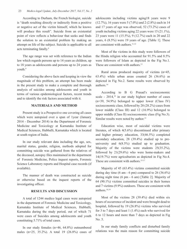
23 Medico-legal Update, July-December 2017, Vol.17, No. 2
According to Durham, the French biologist, suicide is “death resulting directly or indirectly from a positive or negative act of the victim himself, which he knows will produce this result”. Suicide from an existential point of view reflects a behaviour that seeks and finds the solution to an existential problem by making an attempt on life of the subject. Suicide is applicable to all acts terminating fatally.8
The age range was set with reference to the Indian law which regards persons up to 14 years as children, up to 18 years as adolescents and persons up to 24 years as youth.9
Considering the above facts and keeping in view the magnitude of this problem, an attempt has been made in the present study to make a complete and thorough analysis of suicides among adolescents and youth in terms of various epidemiological factors, recent trends and to identify the risk factors associated with it.
MATERIALS AND METHOD
Present study is a Prospective one covering all cases which were autopsied over a span of 1year (January 2014 - December 2014) in the Department of Forensic Medicine and Toxicology at Karnataka Institute of Medical Sciences, Hubballi, Karnataka which is located at south region of India.
In our study relevant data including the age, sex, marital status, gender, religion, methods adopted for committing suicide was gathered from the relatives of the deceased, autopsy files maintained in the department of Forensic Medicine, Police inquest reports, Forensic Science Laboratory reports and Hospital case records (if available).
The manner of death was constructed as suicide or otherwise based on the inquest reports of the investigating officer.
RESULTS AND DISCUSSION
A total of 1244 medico legal cases were autopsied in the department of Forensic Medicine and Toxicology, Karnataka Institute of Medical Sciences, Hubballi, Karnataka during the study period, out of which 71 were cases of Suicides among adolescents and youth constituting 5.71% of total cases.
In our study females (n=46, 64.8%) outnumbered males (n=25, 35.2%). A total 19 (26.8%) cases of
adolescents including victims aging18 years were 9 (12.7%), 16 years were 5 (7.0%) and 2 (2.8%) each in 14 and 17 years of age was observed, 52 (73.2%) cases of youth including victims aging 22 years were 15 (21.1%), 23 years were 11 (15.5%), 9 (12.7%) each in 20 and 22 years, 6 (8.5%) were 19 years of age. [Table 1]. These are consistent with authors.11,12
Most of the victims in this study were followers of the Hindu religion who accounted for 91.5% and 8.5% were followers of Islam as depicted in the Fig No.1. These are consistent with authors.13
Rural areas produced majority of victims (n=45, 63.4%) while urban areas counted 26 (36.6%) as depicted in Fig No.2. Same results were observed by authors.14
According to B G Prasad’s socioeconomic scale - 2014,10 in our study highest number of cases (n=39, 54.9%) belonged to upper lower (Class IV) socioeconomic class, followed by 20 (28.2%) cases from lower middle (Class III) and 12 (16.9%) belonged to upper middle (Class II) socioeconomic class (Fig No.3). Similar results were noted by author.15
Education wise, most of suicidal victims were literates, of which 4(5.6%) discontinued after primary and higher primary education, 33(46.5%) completed secondary education, 28 (39.4%) studied up to pre-university and 6(8.5%) studied up to graduation. Majority of the victims were students 25(35.2%), followed by 21(29.6%) who were home-makers and 14(19.7%) were agriculturists as depicted in Fig No.4. These are consistent with authors.15-17
Majority of 45 (63.4%) victims committed suicide during day time (6 am - 6 pm) compared to 26 (36.6%) during night time (6 pm - 6 am) [Table 2]. Majority of 64 (90.1%) victims committed suicides in their homes and 7 victims (9.9%) outdoors. These are consistent with authors.18,19
Most of the victims 28 (39.4%) died within six hours of occurrence of incident and were brought dead to hospital, followed by 19 (26.8%) victims who survived for 3 to 7 days and least 1 (1.4%) each who survived for 6 to 12 hours and more than 7 days as depicted in Fig No. 5.
In our study family conflicts and disturbed family relations was the main reason for committing suicide
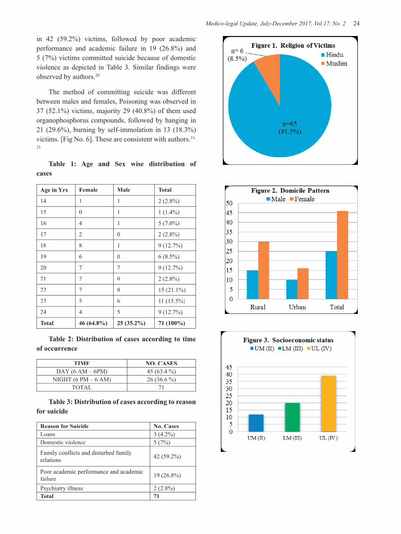
Medico-legal Update, July-December 2017, Vol.17, No. 2 24 25 -legal Update, July-December 2017, Vol.17, No. 2
in 42 (59.2%) victims, followed by poor academic performance and academic failure in 19 (26.8%) and 5 (7%) victims committed suicide because of domestic violence as depicted in Table 3. Similar findings were observed by authors.20
The method of committing suicide was different between males and females, Poisoning was observed in 37 (52.1%) victims, majority 29 (40.8%) of them used organophosphorus compounds, followed by hanging in 21 (29.6%), burning by self-immolation in 13 (18.3%) victims. [Fig No. 6]. These are consistent with authors.21-
23
Table 1: Age and Sex wise distribution of cases
Age in Yrs Female Male Total
14 1 1 2 (2.8%)
15 0 1 1 (1.4%)
16 4 1 5 (7.0%)
17 2 0 2 (2.8%)
18 8 1 9 (12.7%)
19 6 0 6 (8.5%)
20 7 2 9 (12.7%)
21 2 0 2 (2.8%)
22 7 8 15 (21.1%)
23 5 6 11 (15.5%)
24 4 5 9 (12.7%)
Total 46 (64.8%) 25 (35.2%) 71 (100%)
Table 2: Distribution of cases according to time of occurrence
TIME NO. CASESDAY (6 AM – 6PM) 45 (63.4 %)
NIGHT (6 PM – 6 AM) 26 (36.6 %)TOTAL 71
Table 3: Distribution of cases according to reason for suicide
Reason for Suicide No. CasesLoans 3 (4.2%)Domestic violence 5 (7%)
Family conflicts and disturbed family relations 42 (59.2%)
Poor academic performance and academic failure 19 (26.8%)
Psychiatry illness 2 (2.8%)Total 71
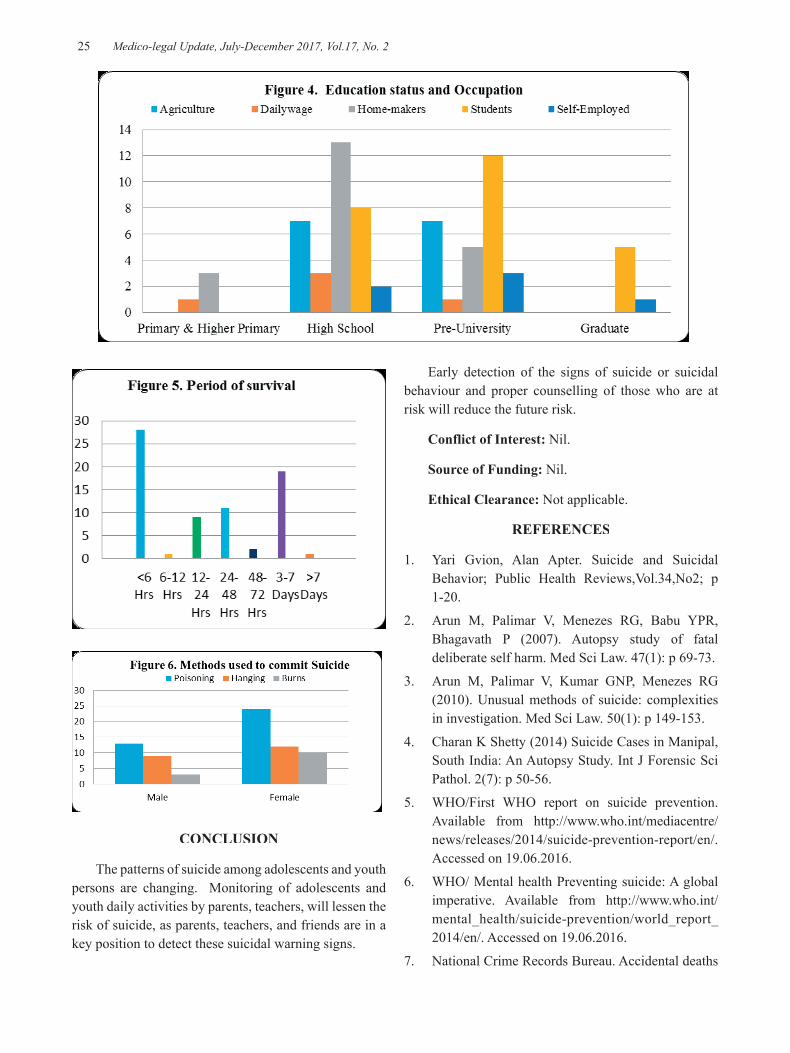
-legal Update, July-December 2017, Vol.17, No. 2 24 25 Medico-legal Update, July-December 2017, Vol.17, No. 2
CONCLUSION
The patterns of suicide among adolescents and youth persons are changing. Monitoring of adolescents and youth daily activities by parents, teachers, will lessen the risk of suicide, as parents, teachers, and friends are in a key position to detect these suicidal warning signs.
Early detection of the signs of suicide or suicidal behaviour and proper counselling of those who are at risk will reduce the future risk.
Conflict of Interest: Nil.
Source of Funding: Nil.
Ethical Clearance: Not applicable.
REFERENCES
1. Yari Gvion, Alan Apter. Suicide and Suicidal Behavior; Public Health Reviews,Vol.34,No2; p 1-20.
2. Arun M, Palimar V, Menezes RG, Babu YPR, Bhagavath P (2007). Autopsy study of fatal deliberate self harm. Med Sci Law. 47(1): p 69-73.
3. Arun M, Palimar V, Kumar GNP, Menezes RG (2010). Unusual methods of suicide: complexities in investigation. Med Sci Law. 50(1): p 149-153.
4. Charan K Shetty (2014) Suicide Cases in Manipal, South India: An Autopsy Study. Int J Forensic Sci Pathol. 2(7): p 50-56.
5. WHO/First WHO report on suicide prevention. Available from http://www.who.int/mediacentre/news/releases/2014/suicide-prevention-report/en/. Accessed on 19.06.2016.
6. WHO/ Mental health Preventing suicide: A global imperative. Available from http://www.who.int/mental_health/suicide-prevention/world_report_2014/en/. Accessed on 19.06.2016.
7. National Crime Records Bureau. Accidental deaths

Medico-legal Update, July-December 2017, Vol.17, No. 2 26
and suicides in India. ADSI 2014. Available from http://ncrb.govin/accdeaths.htm. Accessed on 10.07.2016.
8. Shetty CK, Palimar V, Bhagavath P, Kumar GP. Fatal deliberate self harm in Manipal, India: Autopsy Study. Sri Lanka Journal of Forensic Medicine, Science & Law. May 2014; 5 (1). p 17-27.
9. R. N. Karmakar, editor. Personal Identification:J B Mukherjee‘s Forensic Medicine and Toxicology. 4thed.Culcutta: Academic Publishers. Combined Vol. 2011: p 139.
10. Dudala S R, Reddy A K A, Prabhu G R. Prasad’s socio-economic status classification- An update for 2014. IJRHS. Jul–Sept 2014; 2(3): 875-878.
11. OP Ghai, Vinod K Paul, Arvind Bagga. Adolescent Health and development. In: Essential Pediatrics. 7th ed. New Delhi: CBS Publishers & Distributors. 2013; p 42.
12. Arun M, Palimar V, Menezes RG, Babu YPR, Bhagavath P. Autopsy study of fatal deliberate self-harm. Med Sci Law. 2007; 47(1): p 69-73.
13. Arun M, Palimar V, Kumar GNP, Menezes RG. Unusual methods suicide complexities in investigation. Med Sci Law. 2010; 50(1): p 149-153.
14. Gupta BD, Singh OG.A unique trend of murder-suicide in the Jamnagar region of Gujarat. J Forensic Leg Med. 2008; 15: p 250-255.
15. Reddy MS. Suicide Incidence and Epidemiology. Ind J Psychol Med. 2010; 32(7): p 77-82.
16. Shaw D, Fernandes JR, Rao C. Suicides in Children and Adolescents A 10 –Year Retrospective Review. Am J Forensic Med Pathology. 2005; 26: p 309-315.
17. Reddy MS. Suicide Incidence and Epidemiology. Ind J Psychol Med. 2010; 32(7): p 77-82.
18. Rastogi Pooja, Kocher SR. Suicide in Youth: Shifting Paradigm J Indian Acad Forensic Med. 2010; 32(1): p 45-47.
19. S.K. Panneer Selvam, R Dishina Murthy. Suicide among Adolescents. Indian Streams Research Journal. 2012 April; 2(11): p 1-4.
20. Basvaraj Patil, Santhosh Garampalli, Syed H. Uzair, Nagesh Kuppast, Ragavendra K.M. Suicidal trends in Children and Adolescents. Indian J Forensic Med. 2011 Jan-June; 5(1): p 23-26.
21. George C. Patton, Carolyn Coffey, Susan M Sawywer, Russell M Viner, Dagmar M Haller, Krishna Bose, Theo Vos, Jane Ferguson, Colin D Mathers. Global Patterns of mortality in young people: a systematic analysis of population health data. www. The lancet.com September 12, 2009; 374: p 881-892.
22. Praveen Athani, S. Harish, B.S. Hugar, Girish Chandra Y P. A Prospective Study of Pattern of Suicidal Deaths among Children in Bangalore during the Period 2007-2009. Indian J FMT. Jan-June, 2013; 7(1): p 144-146.
23. Tahereh Seghatoleslam, Esmaeil Farzaneh, Omidavar Rezaee, Fatemeh Sajadfar, Omid Mehrpour.Factors Related to Suicide Attempts by Poisoning in Iranian Children. IJFMT, Jan-June 2013; 7(1): p 129-132.
-legal Update, July-December 2017, Vol.17, No. 2 26
Socio-demographic Profile of Snake Bite Cases Admitted in Tertiary Hospital in Bengaluru
Yogesh C1, Chandrakant M Kokatanur2, Satish K V3
1Assistant Professor, Department of Forensic Medicine, PSG Institute of Medical Sciences & Research, 1Assistant Professor, Department of Forensic Medicine, PSG Institute of Medical Sciences & Research, 1
Coimbatore, 2Associate Professor, Department of Forensic Medicine, Krishna Institute of Medical Sciences Karad, 2Associate Professor, Department of Forensic Medicine, Krishna Institute of Medical Sciences Karad, 2
Maharashtra, 3Associate Professor, Department of Forensic Medicine, Bengaluru Medical College and Research 3Associate Professor, Department of Forensic Medicine, Bengaluru Medical College and Research 3
Institute,Bengaluru
ABSTRACT
Introduction: Snake-bite is a known cause of increased mortality and morbidity in our country. Materials and method: A cross sectional study of a total of 443 cases of snake bite admitted in Victoria hospital, Bengaluru, was conducted over a period of 18 months from November 2011 to May 2013. Results: A total of 443 cases of snake bite were analysed. Out of 443 cases, 62.75% (n=278) were from rural area and 37.25% (n=165) from urban area. Most common age group of cases of snake bite was 20-29 years (28.89%). Males (64.3%) were commonly affected than females (35.7%). The highest number of cases were seen in the month of June (11.5%, n=34) followed by October and November (11.2%, n=33). In the study period, majority of cases of snake bite were improved (95%) where as death occurred in 5% cases. Conclusion:Snakebite still remains a major occupational hazard affecting productive age group predominantly males in rural area involved in agriculture.
Keywords: Snakebite, morbidity, mortality, sociodemography.
INTRODUCTION
Snake bite is a public health problem distributed mainly in the tropical and sub-tropical countries1.Snake bite results in approximately 2500,000 venomous bites each year and 125,000 deaths worldwide2. Envenomation in India is estimated to be at 81,000/year, which is highest in the world3.
People in countries like India prefer traditional healers rather than trained doctors, mainly because of ignorance and monetary issues as a result of which 77% of the snakebite victims in rural areas die outside the health care set up3. Reliable data for morbidity and mortality are not available since there is no proper reporting system4. Very few epidemiological surveys had been done on the snake bite problem in India. To know the problem and to get some way out to tackle the
Corresponding author: Chandrakant M KokatanurAssociate Professor, Department of Forensic Medicine, Krishna Institute of Medical Sciences Karad, Maharashtra. Email: [email protected].
problem, knowledge regarding epidemiological profile of snake bite is essential5,6,7.
The study of the pattern of snakebite is essential to provide pertinent information to the concerned authorities in order to manage snakebite appropriately8.The study was conducted to know the socio-demographic profile of snake bites cases admitted in Victoria hospital, Bengaluru.
MATERIALS AND METHOD
A cross sectional study of a total of 443 cases of snake bite admitted in Victoria hospital, Bengaluru, was conducted over a period of 18 months from November 2011 to May 2013. The data regarding age, sex, occupation, education, residence, seasonal variation, hospital stay and outcome was collected. All the patients coming to casualty with the history of snakebite during the study period were considered. Data was collected and analysed with Microsoft excel and presented as descriptive statistics.
DOI Number: 10.5958/0974-1283.2017.00062.7
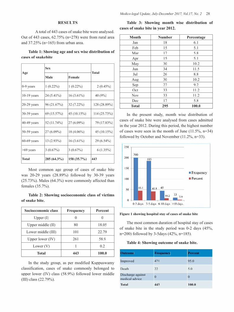
Medico-legal Update, July-December 2017, Vol.17, No. 2 28 29 -legal Update, July-December 2017, Vol.17, No. 2
RESULTS
A total of 443 cases of snake bite were analysed. Out of 443 cases, 62.75% (n=278) were from rural area and 37.25% (n=165) from urban area.
Table 1: Showing age and sex wise distribution of cases of snakebite
AgeSex
TotalMale Female
0-9 years 1 (0.22%) 1 (0.22%) 2 (0.45%)
10-19 years 24 (5.41%) 16 (3.61%) 40 (9%)
20-29 years 96 (21.67%) 32 (7.22%) 128 (28.89%)
30-39 years 69 (15.57%) 45 (10.15%) 114 (25.73%)
40-49 years 52 (11.74%) 27 (6.09%) 79 (17.83%)
50-59 years 27 (6.09%) 18 (4.06%) 45 (10.15%)
60-69 years 13 (2.93%) 16 (3.61%) 29 (6.54%)
>69 years 3 (0.67%) 3 (0.67%) 6 (1.35%)
Total 285 (64.3%) 158 (35.7%) 443
Most common age group of cases of snake bite was 20-29 years (28.89%) followed by 30-39 years (25.73%). Males (64.3%) were commonly affected than females (35.7%).
Table 2: Showing socioeconomic class of victims of snake bite.
Socioeconomic class Frequency Percent
Upper (I) 0 0
Upper middle (II) 80 18.05
Lower middle (III) 101 22.79
Upper lower (IV) 261 58.9
Lower (V) 1 0.2
Total 443 100.0
In the study group, as per modified Kuppuswamy classification, cases of snake commonly belonged to upper lower (IV) class (58.9%) followed lower middle (III) class (22.79%).
Table 3: Showing month wise distribution of cases of snake bite in year 2012.
Month Number PercentageJan 18 6.1Feb 15 5.1Mar 17 5.8Apr 15 5.1May 30 10.2Jun 34 11.5Jul 26 8.8
Aug 30 10.2Sep 27 9.2Oct 33 11.2Nov 33 11.2Dec 17 5.8
Total 295 100.0
In the present study, month wise distribution of cases of snake bite were analysed from cases admitted in the year 2012. During this period, the highest number of cases were seen in the month of June (11.5%, n=34) followed by October and November (11.2%, n=33).
Figure 1 showing hospital stay of cases of snake bite
The most common duration of hospital stay of cases of snake bite in the study period was 0-2 days (45%, n=200) followed by 3-5days (42%, n=185).
Table 4: Showing outcome of snake bite.
Outcome Frequency Percent
Improved 421 95.0
Death 22 5.0Discharge against medical advice 0 0
Total 443 100.0

-legal Update, July-December 2017, Vol.17, No. 2 28 29 Medico-legal Update, July-December 2017, Vol.17, No. 2
In the study period, majority of cases of snake bite were improved (95%, n=421) where as death occurred in 5% (n=22) cases.
DISCUSSION
A total of 443 cases of snake bite were analysed. The most common age group of cases of snake bite cases was 20-29 years (28.89%) followed by 30-39 years (25.73%) as shown in Table 1. The higher incidence of snake bite in young age of 20-39 years could be due to active involvement of these age groups in outdoor occupation. This finding is similar to studies by by Thapar R3 , Singh A8, Siddique A9, Adiga U10 and Arun KS11.
The incidence of snake bite was more common in males (64.3%) as compared to females (35.7%) with male to female ratio 1.8:1. This could be due to more active involvement by males in outdoor agriculture related activities than females. This finding is similar to observations made by Thapar R3, Kalyani P12, Rao CP13, Mondal TK14.
In the present study, 62.75% cases of snake bite were from rural area and 37.25% from urban area. Similar observations are found in studies by Joshi15 , Anjum A16. This could be due to snake envenoming affects largely the poor people depending on agriculture as source of livelihood in rural area16. Many houses in rural India are made of mud and have many crevices where rodents flourish and snakes are attracted to such houses in search of prey17.
In our study, majority of victims of snake bite were housewives (32.7%) followed by farmers (28.2%) and labourers (19%) .This finding is similar to observations made by Rao CP13, Nagaraju K18, Mondal R19 Monteiro FN20. Farmers are more prone to accidental contact with snakes while working in the field barefooted, which is a phenomenon, is observed in India20.
In the present study, majority of cases of snakes had education at primary level (43.3%) followed by secondary level (37%) . The low literacy level also leads to lack of knowledge regarding precautions to be taken to avoid snake bite. Similar observations were made in studies by Singh A8, Adiga U10, Anjum A16.
In the study group, as per modified Kuppuswamy classification, cases of snake commonly belonged to upper lower (IV) class(58.9%) followed lower middle(III) class (22.79%) as shown in table 2. This
could be due to low literacy rate and poor financial conditions.
In the present study, month wise distribution of cases of snake bite were analysed from cases admitted in the year 2012. During this period, the highest number of cases were seen in the month of June (11.5%) followed by October and November (11.2%) as shown in table 3. Similar observations were found in studies by Inamdar IF4, Singh A8, Joshi SC15. In contrast to this, maximum incidence of snakebites was reported during the post-monsoon months of September to December by Thapar R3. During the rainy season, rainwater floods their burrows and snakes then try to take shelter near human dwellings, which increases the chances of snakes feeling threatened or startled or provoked by human beings, and biting them.
The most common duration of hospital stay of cases of snake bite in the study period was 0-2 days (45%) followed by 3-5days (42%) as shown in figure 1.Similar observation of average hospital stay was found by Sam K21.
In the study period, majority of cases of snake bite were improved (95%) where as death occurred in 5% cases as shown in table 4.Similar observation were found in studies by Inamdar IF4, Singh A8, Joshi SC15, Nagaraju K18.This could be due to early proper management of cases of snake bite in tertiary hospital.
CONCLUSION
Snakebite still remains a major occupational hazard of agriculture affecting productive age group predominantly males in rural area. People must be advised to use protective measures like long pants and gumboots while working outdoors and good sanitation around their houses. People must be educated regarding early visit to hospitals for treatment in cases of snake bite.
Conflict of Interest: No.
Source of Funding: Nil.
Ethical Clearance: The study was approved by Institutional Ethical Committee of Bangalore Medical College and Research Institute, Bengaluru.
REFERENCES1. Bawaskar HS, Bawaskar PH, Punde DP, Inamdar
Medico-legal Update, July-December 2017, Vol.17, No. 2 30
MK, Dongare RB, Bhoite RR. Profile of snakebite envenoming in rural Maharashtra, India. J Assoc Physicians India 2008;56:88-95.
2. Amin RU, Hussain S, Iqbal M, Hussain W. Toxicity patterns and outcome of snake bite cases at Nishtar hospital Multan. P J M H S 2015 Jan;9(1):113.
3. Thapar R, Darshan BB, Unnikrishnan B, Mithra P, Kumar N, Kulkarni V et al. Clinico-epidemiological profile of snakebite cases admitted in a tertiary care centre in South India: A 5years study. Toxicol Int 2015;22:66-70.
4. Inamdar IF, Aswar NR, Ubaidulla M, Dalvi SD.Snakebite: admissions at a tertiary health care centre in Maharashtra, India. S Afr Med J 2010;100:456-8.
5. Pandey DP. Epidemiology of snake bites based on hospital survey in Chitwan and Nawalparasi districts, Nepal. J Nepal Health Res Counc 2006;4:121-9.
6. Hati AK, Mandal M, De MK, Mukherjee H, Hati RN. Epidemiology of snake bite in the district of Burdwan, West Bengal. J Indian Med Assoc 1992;90:145-7.
7. Majumder D, Sinha A, Bhattacharya SK, Ram R, Dasgupta U, Ram A. Epidemiological profi le of snake bite in South 24 Parganas district of West Bengal with focus on underreporting of snake bite deaths. Indian J Public Health 2014;58:17-21.
8. Singh A, Goel S, Singh AA, Goel AK, Chhoker VK, Goel S et al. An epidemiological study of snakebites from rural Haryana. Int J Adv Med Health Res 2015;2:39-43.
9. Siddique A, Kishore G, Ahmed EM, Khader HA. Epidemiological profile of snake bite at a Tertiary Care Hospital Bengaluru, India. Arch Pharma Pract 2015;6:58-63.
10. Adiga U, Adiga S. An epidemiological study on snakebite in Karwar. J Int Med Dent 2014; 1 (2): 70-74.
11. Arun KS, Reddy A, Balaraman R. Epidemiological study of fatal snake bite cases in rural Pondicherry. Medico-legal update 2016 Jan;16(1):88-91.
12. Kalyani P, Felix AJ, Arulmani A, Ambayiram AV. Clinico-epidemiological profile of snake bite reported in a tertiary care hospital in Tamilnadu.Asian J Pharm Res 2015;5(2): 70-4.
13. Rao CP, Parameshwar S, Mothi VR. Fatal snake bites – sociodemography, latency pattern of injuries. J Occupational Med Toxicol 2013;8(7):1-5.
14. Mondal TK, Das M, Sarkar AP, Dudhe AP, Ghosh S. A clinico-epidemiological study on snake bite in a tertiary care hospital of West Bengal. Int J Med Sci Public Health 2017;6(2):1-4.
15. Joshi SC, Prakash C, Joshi A, Joshi G, Nigam P. Profile of snakebite cases admitted at a tertiary care centre. J Indian Acad Forensic Med 2012 Jul;34(3):217-9.
16. Anjum A, Husain M, Hanif SA, Ali SM, Beg M, Sarda M. Epidemiological profile of snake bite at tertiary care hospital, North India. J Forensic Res 2012; 3(4):146.
17. Pius PS, Vinoj J, Indhuja, Premkumar, Akila D. A retrospective review of snake bite cases admitted in a tertiary care hospital. J Evid Based Med Healthc 2016; 3(67), 3636-9.
18. Nagaraju K, Kannappan N, Gopinath K. Survey on pattern of snake bite cases admitted in south Indian tertiary care hospitals. IJPSR 2015;6(10): 4362-7.
19. Mondal R, Rani M, Nurmohammad, Islam MM, Islam MS, Saha AC et al. Pattern of poisonous snake bite in Rangpur medical college. RCMC 2011 Jul;1(1)14-8.
20. Monteiro FN, Kanchan T, Bhagavath P, Kumar G, Menezes RG, Yoganarasimha K. Clinico-epidemiological features of viper bite envenomation: a study from Manipal, South India.Singapore Med J 2012;53(3):203-7.
21. Sam K, Khan M, Peerally S, Kumar P, Rao P. Snake-bite envenomation: a comprehensive evaluation of severity, treatment and outcome in a tertiary care South Indian hospital. Internet J Emergency Med 2008;5(1):1-9.

-legal Update, July-December 2017, Vol.17, No. 2 30
Profile of Road Traffic Accidents in Southern Rajasthan - A Retrospective Hospital based Cross Sectional Study
Sanjeev Kumar1, G L Dad2, Rajkumar Patil3
1Associate Professor, Department of Forensic Medicine, Geetanjali Medical College & Hospital, Udaipur, Rajasthan (India), 2Professor & Head, Department of Forensic Medicine, Geetanjali Medical College & Hospital, Udaipur, Rajasthan (India), 3Professor, Department of Community Medicine, Mahatma Gandhi Medical College
and Research Institute, Sri Balaji Vidyapeeth Deemed University, Pondicherry (India)
ABSTRACT
Background:Road traffic accident (RTA) is one of the major causes of death in world. If no action is taken, road traffic crashes are predicted to become seventh leading cause of death by year 2030. RTAs underreporting is a serious and global problem. Further, the scientific investigations behind these accidents are lacking due to which the real causes and consequences are not revealed. Road traffic accidents and related deaths can be prevented if related factors are identified.
Objective: To identify the profile of road traffic accident (RTA) cases admitted in Geetanjali Medical College and Hospital, Udaipur, Rajasthan.
Materials and Method: The present retrospective study was conducted in department of Forensic Medicine and Toxicology of Geetanjali Medical College and Hospital (GMCH), Udaipur, Rajasthan. Data of road traffic accident cases admitted during year 2015 were analyzed. A proforma was used to gather the information like age, gender, time of injury, type of injury etc. from medical records department of the hospital. Data were entered in MS-excel and analyzed.
Results: Total 449 cases of RTAs were analyzed. Of these 76.8% were male. Most common (30.7%) age group involved was 21-30 years. Highest number of cases were recorded in May month followed by April. Nearly half of the accidents (49.4%) happened between 6 pm and 12 midnight. Most commonly (40.1%) pedestrians were the victims. Four wheeler was the offending vehicle in nearly half (50.3%) of the victims. Head injury was most common (59%) injury.
Conclusion: In present study, RTAs in Udaipur and nearby areas were more common in the adults, mostly in 21-30 years group males, most of the accidents happened during evening and night. There is a need to create awareness about traffic safety rules and most importantly change in behavior.
Keywords: Accident, injury, road traffic accident, (RTA) head injury.
INTRODUCTION
Road traffic accidents (RTA) are a human tragedy. It is any event that occurs on a way or street open to public traffic; resulting in one or more persons being injured or killed, where at least one moving vehicle is involved.1 Every year 12 lakhs people die as a result of road traffic injury (RTI). It is the leading cause of death specially among young people in 15–29 years. About 90% of the world’s fatalities on the roads occur in low and middle income countries. If no action is taken, road
traffic crashes are predicted to become seventh leading cause of death by 2030.2 In India, about 2.3 lakh people were died in road traffic crashes in year 2013 and deaths due to RTAs are projected to increase by 150% in India by the year 2020.3,4
RTAs underreporting is a serious and global problem.5 Of the estimated 14 lakh severe road accidents occurring every year in India, only about 29% are recorded. Further, the scientific investigations behind these accidents are lacking due to which the real causes
DOI Number: 10.5958/0974-1283.2017.00063.9

Medico-legal Update, July-December 2017, Vol.17, No. 2 32 33 -legal Update, July-December 2017, Vol.17, No. 2
and consequences are not revealed.6
In India, there is mixed traffic on road with fast and slow moving vehicles, there is movement of animals and human beings which is often unpredictable and sudden, condition of Indian roads and disobey of traffic rules along with poor maintenance of vehicles are some of the important factors related to road traffic accidents. Over speeding, overtaking and driving under the influence of alcohol are the main factors usually in RTAs. Road traffic accidents can be prevented if the above mentioned factors are addressed adequately. There is also difficulty in getting timely emergency care of the victim which leads to fatality. Research about various factors causing accidents and establishment of more emergency and trauma centers can help in reducing the deaths.
There is limited information about the pattern of RTAs in India, as trauma registries and the hospital-based research is underdeveloped in India.5 There are only few studies related to RTAs which are conducted in Rajasthan. Present study was planned and conducted with the objective to study the profile of road traffic accidents in Udaipur,Rajasthan.
MATERIALS AND METHOD
Present study was conducted at Geetanjali Medical College and Hospital, Udaipur, Rajasthan. A cross sectional retrospective study was planned. Information about all the RTA victims who were admitted in hospital during one year period from January 2015 to December 2015 were collected. Ethical clearance from institutional ethical committee was obtained before conducting the study. Proforma was developed which included the information like socio-demographic details, body area injured, time of accident and vehicle involved etc. Data entry and analysis is done with the help of MS Excel software. Proportions were calculated wherever required.
RESULTS
There were total 449 cases of road traffic accidents admitted in year 2015.Of these 345(76.8%) were male and 104(23.2%) were female.Most common (30.7%)age group involved was 21-30 years and least common(3.8%) was children less than 10 years.(Table 1)
Table 1. Age and sex wise distribution of RTA cases
Age group (yr)
Male(%)
Female(%)
Total(%)
0-10 12(3.5) 5(4.8) 17(3.8)
11-20 44(12.8) 13(12.5) 57(12.7)
21-30 107(31) 31(29.8) 138(30.7)
31-40 78(22.6) 25(24) 103(22.9)
41-50 51(14.8) 19(18.3) 70(15.6)
51-60 33(9.6) 10(9.6) 43(9.6)
>60 20(5.8) 1(0.9) 21(4.7)
Total 345 104 449
Highest number of cases were recorded in May followed by April, while least number of cases were seen in September month. (Table 2)
Table 2. Month wise distribution of RTA cases (n=449)
Month Nnmber (%)
January 39(8.7)
February 33(7.4)
March 29(6.5)
April 47(10.5)
May 57(12.7)
Jun 35(7.8)
July 29(6.5)
August 26(5.8)
September 25(5.6)
October 43(9.6)
November 42(9.4)
December 44(9.8)
Nearly half of the accidents (49.4%) happened between 6 pm and 12 midnight while least number were seen during morning 6 o’clock in morning to 12 noon. (Table 3)
Table 3.Time wise distribution of RTA cases (n=449)
Time of accident Number (%)12 midnight - 6 AM 70(15.6)6 AM -12 Noon 22(4.9)12 Noon- 6 PM 135(30.1)6 PM - 12 midnight 222(49.4)Total 449
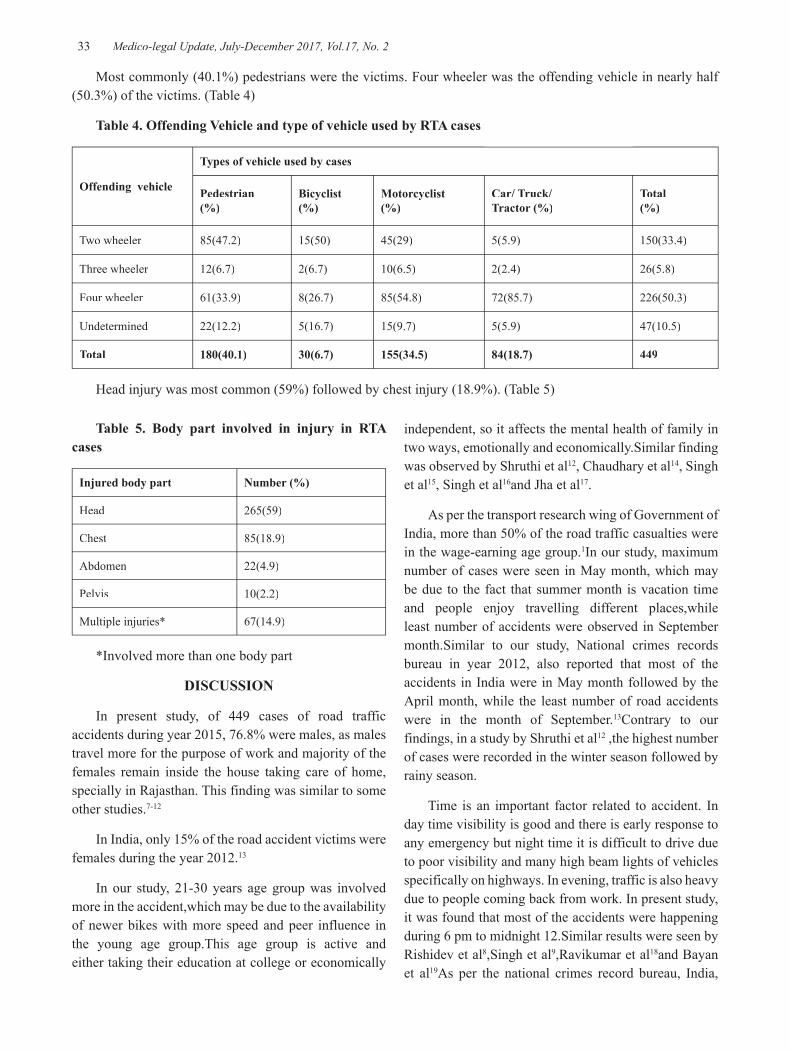
-legal Update, July-December 2017, Vol.17, No. 2 32 33 Medico-legal Update, July-December 2017, Vol.17, No. 2
Most commonly (40.1%) pedestrians were the victims. Four wheeler was the offending vehicle in nearly half (50.3%) of the victims. (Table 4)
Table 4. Offending Vehicle and type of vehicle used by RTA cases
Offending vehicle
Types of vehicle used by cases
Pedestrian(%)
Bicyclist(%)
Motorcyclist(%)
Car/ Truck/ Tractor (%)
Total(%)
Two wheeler 85(47.2) 15(50) 45(29) 5(5.9) 150(33.4)
Three wheeler 12(6.7) 2(6.7) 10(6.5) 2(2.4) 26(5.8)
Four wheeler 61(33.9) 8(26.7) 85(54.8) 72(85.7) 226(50.3)
Undetermined 22(12.2) 5(16.7) 15(9.7) 5(5.9) 47(10.5)
Total 180(40.1) 30(6.7) 155(34.5) 84(18.7) 449
Head injury was most common (59%) followed by chest injury (18.9%). (Table 5)
Table 5. Body part involved in injury in RTA cases
Injured body part Number (%)
Head 265(59)
Chest 85(18.9)
Abdomen 22(4.9)
Pelvis 10(2.2)
Multiple injuries* 67(14.9)
*Involved more than one body part
DISCUSSION
In present study, of 449 cases of road traffic accidents during year 2015, 76.8% were males, as males travel more for the purpose of work and majority of the females remain inside the house taking care of home, specially in Rajasthan. This finding was similar to some other studies.7-12
In India, only 15% of the road accident victims were females during the year 2012.13
In our study, 21-30 years age group was involved more in the accident,which may be due to the availability of newer bikes with more speed and peer influence in the young age group.This age group is active and either taking their education at college or economically
independent, so it affects the mental health of family in two ways, emotionally and economically.Similar finding was observed by Shruthi et al12, Chaudhary et al14, Singh et al15, Singh et al16and Jha et al17.
As per the transport research wing of Government of India, more than 50% of the road traffic casualties were in the wage-earning age group.1In our study, maximum number of cases were seen in May month, which may be due to the fact that summer month is vacation time and people enjoy travelling different places,while least number of accidents were observed in September month.Similar to our study, National crimes records bureau in year 2012, also reported that most of the accidents in India were in May month followed by the April month, while the least number of road accidents were in the month of September.13Contrary to our findings, in a study by Shruthi et al12 ,the highest number of cases were recorded in the winter season followed by rainy season.
Time is an important factor related to accident. In day time visibility is good and there is early response to any emergency but night time it is difficult to drive due to poor visibility and many high beam lights of vehicles specifically on highways. In evening, traffic is also heavy due to people coming back from work. In present study, it was found that most of the accidents were happening during 6 pm to midnight 12.Similar results were seen by Rishidev et al8,Singh et al9,Ravikumar et al18and Bayan et al19As per the national crimes record bureau, India,
Medico-legal Update, July-December 2017, Vol.17, No. 2 34 35 -legal Update, July-December 2017, Vol.17, No. 2
in year 2012, about one third of the accidents happened during 3 pm to 9 pm.13
In India, pedestrians and two wheeler riders are at high risk for road traffic crashes and these are the major road users.5 Most of the places there is no separate lane for pedestrians and bicyclists, further crossing the road seems to be a nightmare nowadays. In our study, pedestrians were the main victims followed by motorcyclists. Pedestrians are the ones for whom it is not applicable to wear any protective device and most of motorcyclists in India don’t use helmets, further four wheeler driver as well as passengers usually don’t use the seat belt.
In our study, four wheeler was the main offending vehicle. Similarly Chaudhary et al14, Shruthi et al12 and Khajuria et al20 also observed that pedestrians were the main victims in road traffic accidents. During year 2012 in India, majority of the victims of road accidents were occupants of ‘two wheelers’ followed by truck or lorry.13
Similar to our study, Shruthi et al12 also reported that four wheeler was the most common offending vehicle for the accident.
Head is the most common site of injury in road traffic accidents. Traffic rules and legislations are not followed by most of the road users in India and this is one of the main reason for head injury. In our study, more than half of the accident victims had head injury, this finding is similar to the findings of the studies by Rishidev et al8, Shruthi et al12, Jha N et al.17and Singh et al.16 There is a need to create awareness about traffic safety rules and most importantly change in behavior e.g. use of helmet and speed limit is required.
CONCLUSION
In present study, RTAs in Udaipur and nearby areas were more common in the adults, mostly in 21-30 years group males, most of the accidents happened during evening and night. Pedestrians were mostly affected and head injuries were most common. The results of our study may help to reduce RTAs by highlighting the important risk factors in study area.
Source of Funding: Self
Conflict of Interest: None
REFERENCES
1. Transport Research Wing, Ministry of Road Transport and Highways. Road Accidents in India 2011. New Delhi: Ministry of Road Transport and Highways, Government of India; 2012.
2. World health organization. Road traffic injuries-Fact sheet.[Online].2016 [cited 2016 Nov 17]. Available from:URL:http://www.who.int/mediacentre/factsheets/fs358/en/
3. World health organization.Violence and injury prevention. Road safety in India. [Online].2016 [cited 2016 Nov 17]. Available from: URL:http://www.who.int/violence_injury_prevention/road_traffic/countrywork/ind/en/
4. Kopits E, Cropper M. The World Bank. Washington DC: World Bank Policy Research Working Paper No 3035.Traffic Fatalities and Economic Growth. [Online].2003[cited 2016 Nov 18]. Available from: URL:http://www.ntl.bts.gov/lib/24000/24400/24490/25935_wps3035.pdf
5. Gururaj G. Road traffic deaths, injuries and disabilities in India: Current scenario. National Medical Journal of India 2008;21:14-20.
6. Institute of Road Traffic Education.Crash Investigation.[Online].2016[cited 2016 Nov 18]. Available from: URL:http://www.irte.com/crash-investigation.html
7. Kaul A, Sinha US, Pathak YK, Singh A, Kapoor AK, Sharma S et al. Fatal road Traffic Accidents, Study of Distribution, Nature and Type of Injury. Journal of Indian Academy of Forensic Medicine 2005;27(2):71-6.
8. Rishidev A, Gupta SD, Musa R, Bhavsar N, Thakor A, Patel P. Profile of Nonfatal Injuries in Road Traffic Accidents Cases Treated at a Tertiary Level Trauma Centre. International Journal of Scientific study 2016;4(6):36-40.
9. Singh A, Bhardwaj A, Pathak R, Ahluwalia SK. An epidemiological study of road traffic accident cases at a tertiary care hospital in rural Haryana. Indian Journal of Community Health 2011;23(2):53-5.
10. Sharma BR, Dasari H, Sharma V, Vij K. Road Traffic Accidents-A Demographic and Topographic analysis. Medicine, Science and Law 2001, 41 (3):266-74.
-legal Update, July-December 2017, Vol.17, No. 2 34 35 Medico-legal Update, July-December 2017, Vol.17, No. 2
11. Singh H, Dhattarwal SK. Pattern and Distribution of Injuries in Fatal Road Traffic Accidents in Rohtak,Haryana. Journal of Indian Academy of Forensic Medicine 2004;26(1):20-3.
12. Shruthi P, Venkatesh VT, Viswakanth B, Ramesh C, Sujatha PL, Dominic IR. Analysis of Fatal Road Traffic Accidents in a Metropolitan City of South India. Journal of Indian Academy of Forensic Medicine.2013:35(4); 317-20.
13. National Crimes Records Bureau. Accidental Deaths and Suicides in India 2012. New Delhi: Ministry of Home Affairs, Government of India; 2013.
14. Chaudhary BL, Singh D, Tirpude BH, Sharma RK, Veena M. Profile of Road Traffic Accident Cases in Kasturba Hospital of MGIMS, Sevagram, Wardha, Maharashtra 2005;5(4).127-33.
15. Singh A, Goel A, Shekhar. Epidemiological Study of non-fatal road traffic accidents in Rohilkhand Region. Medico-Legal Update 2011;11(1):5-9.
16. Singh R, Singh HK, Gupta SK, Kumar Y. Pattern, Severity and Circumstances of Injuries
Sustained in Road Traffic Accidents: A Tertiary Care Hospital-Based Study. Indian Journalof Community Medicine 2014;39(1):30-4.
17. Jha N, Srinivasa DK, Roy G, Jagdish S. Injury Pattern Among Road Traffic Accident Cases : A Study From South India. Indian Journal of Community Medicine 2003. 28(2).20–5.
18. Ravikumar R. Patterns of head injuries in road traffic accidents involving two wheelers: An autopsy study. Journal of Indian Academy of Forensic Medicine 2013;35(4):349-52.
19. Bayan P, Bhawalkar JS, Jadhav SL, Banerjee A. Profile of non-fatal injuries due to road traffic accidents from a industrial town in India. International Journal of Critical Illness and Injury Science 2013;3(1):8-11.
20. Khajuria B, Sharma R, Verma A. A Profile of the autopsies of road traffic accident victims in Jammu. Journal of Clinical and Diagnostic Research 2008;2(1):639-42.
37 -legal Update, July-December 2017, Vol.17, No. 2
Trend of Poisoning in Females at a Tertiary Care Hospital of Haryana
S S Dalal1, S K Dhattarwal2, Sunil Gambhir3, Dhruva Chaudhary4, Kunal Khanna5
1Associate Professor, Deptt. of Forensic Medicine, N.C. Medical College, Israna, Panipat, 1Associate Professor, Deptt. of Forensic Medicine, N.C. Medical College, Israna, Panipat, 1 2Sr. Professor & Head, Deptt. of Forensic Medicine PGIMS Rohtak, 3HCMS Haryana, 4Sr. Professor & Head, Deptt. of Pulmonary & Critical Care Medicine, PGIMS Rohtak, 5Assistant Professor, Deptt. of Forensic Medicine, KCGMC Karnal5Assistant Professor, Deptt. of Forensic Medicine, KCGMC Karnal5
ABSTRACT
Poisoning is one of the leading causes of morbidity and mortality in developing countries affecting all ages and both genders. Though a lot of research has been conducted on poisoning cases, data regarding the epidemiology of poisoning in female gender remains deficient. Keeping the above scarcity of information in mind, a prospective study was undertaken in a leading tertiary care hospital of North India that not only addresses the health needs of the local population but also attracts a lot of patients from the adjacent districts and neighbouring states. All the female cases that reported in the Emergency department of the institute from 1st March 2008 to 28 February 2009 with a suspected history of poisoning were included in the study st March 2008 to 28 February 2009 with a suspected history of poisoning were included in the study st
and analyzed for demographic and social factors regarding age, marital status, residential area, season, and manner of poisoning, survival period and type of poison consumed. Out of total 436 cases analyzed, majorities were in 21-30 year age group and married. Most of the victims belonged to rural areas. Most of the poisoning cases were reported in the morning hours and the average duration of stay in the hospital was less than 6 hours. Suicidal attempts were reported in 58.7% cases. Death was reported in 49.8% of the victims. Aluminium phosphide was most frequently involved.
Keywords: Poisoning, morbidity, female, India.
INTRODUCTION
Globally more than 3 million cases of pesticide poisoning are reported every year with more than 250,000 deaths.1 The problem is particularly significant in South East Asia. In India about 1 to 1.5 million cases of poisoning are reported every year out of which nearly 50,000 die.2 A poison is a lethal tool in the hands and has been found to be one of the most common methods of ending a life due to easy availability and painless death.3 In the Indian context, women in particular are expected to make more compromises and adjustments in life as compared to males that sometimes leads to a wide range of emotions such as anxiety, grief, fear and anger. When these emotions cross the acceptable limits, they may result in catastrophic consequences like in ending one’s or someone else’s life. According to a WHO report, poisoning is the second leading cause of death in women next only to road traffic injuries among the unintentional injuries.4 This startling finding necessitates more research into the epidemiology of poisoning in the female gender.
MATERIAL AND METHOD
The present prospective study under taken in the Department of Forensic Medicine and Medicine including ICU (Accident and Emergency) in Pt. B.D. Sharma Post Graduate Institute of Medical Sciences (PGIMS), Rohtak Haryana from 1st March 2008 to 28 st March 2008 to 28 st
February 2009. This study included a total of 436 cases that were reported to the Emergency Department during the study period. The data required was collected from the statements of the victim, relatives/friends accompanying the deceased and toxicological examination. The cases where chemical examination reports showed ‘No poison detected’ have not been included in the study. The data thus collected was then compared with various national as well as international studies.
OBSERVATIONS
The present study found that the most commonly affected age was 21-30 years (39.7%) followed closely by 31-40 years. The least commonly affected were the
DOI Number: 10.5958/0974-1283.2017.00064.0

37 Medico-legal Update, July-December 2017, Vol.17, No. 2
victims belonging to extremes of ages i.e. (less than 10 years) and (>60 years) (Table 1)
Table 1: Age wise distribution of cases
Age Group Number of Cases %
Less than 10 years 4 0.9
11-20 years 100 22.9
21-30 years 173 39.7
31-40 years 78 17.9
41-50 years 48 11
51-60 years 29 6.7
Above 60 years 4 0.9
Total 436 100
It was observed that more married females died as compared to unmarried with a married: unmarried ratio of 7:3 (Table 2)
Table 2: Distribution of cases on basis of marital status
Marital Status Number of cases %
Married 305 70
Unmarried 131 30
More number of rural people died as compared to urban with a ratio of almost 7:3 (Table 3)
Table 3: Distribution of cases on the basis of area of residence
Area of Residence Number of cases %
Rural 302 69.3
Urban 134 30.7
Maximum numbers of cases were reported during the morning time (32.8%) while least was reported in the evening (Figure 1)
Graph- 1: Case percentage on the basis of time of day when incident occurred
The study observed that most of the victims attempted suicide (58.7%) while accidental ingestion was reported in 32.8% cases.
Graph- 2: Case percentage on the basis of Manner of Death
As far as duration of the stay in the hospital is concerned, maximum number of victims remained in the hospital for less than 6 hours (50.5%) while least number stayed for more than 48 hours (1.8%) before the final outcome of poisoning (Table 4)
Table 4: Case distribution on the basis of duration of stay in hospital
Duration of hospital stay Number of cases Percentage
Less than 6 hours 220 50.56-12 hours 72 16.612-24 hours 76 17.424-36 hours 38 8.736-48 hours 22 5More than 48 hours 8 1.8Total 436 100
It was observed that the most frequently affected poison involved in female poisoning cases was Aluminium phosphide followed by Organo-phosphorus poisoning. Rest of the cases fell under miscellaneous group that included drugs, plant poisons and all other types of poisons reported by the chemical examination reports (Table 5)
Table 5: Case distribution on the basis of poison used
Type of Poisoning Number of Number of CasesCases %
Aluminium phosphide 266 61.1
Organophosphorus compounds 161 36.9
Miscellaneous 9 2
Total 436 100
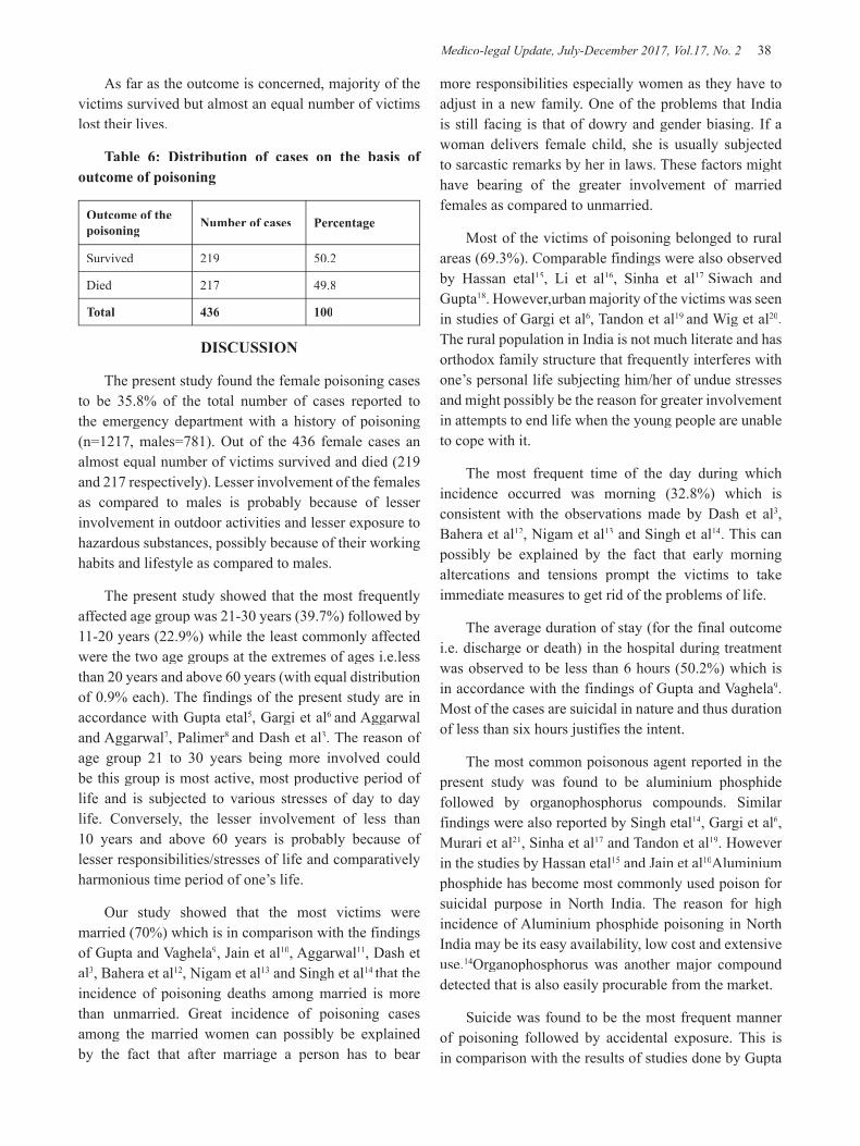
Medico-legal Update, July-December 2017, Vol.17, No. 2 38 39 -legal Update, July-December 2017, Vol.17, No. 2
As far as the outcome is concerned, majority of the victims survived but almost an equal number of victims lost their lives.
Table 6: Distribution of cases on the basis of outcome of poisoning
Outcome of the poisoning Number of cases Percentage
Survived 219 50.2
Died 217 49.8
Total 436 100
DISCUSSION
The present study found the female poisoning cases to be 35.8% of the total number of cases reported to the emergency department with a history of poisoning (n=1217, males=781). Out of the 436 female cases an almost equal number of victims survived and died (219 and 217 respectively). Lesser involvement of the females as compared to males is probably because of lesser involvement in outdoor activities and lesser exposure to hazardous substances, possibly because of their working habits and lifestyle as compared to males.
The present study showed that the most frequently affected age group was 21-30 years (39.7%) followed by 11-20 years (22.9%) while the least commonly affected were the two age groups at the extremes of ages i.e.less than 20 years and above 60 years (with equal distribution of 0.9% each). The findings of the present study are in accordance with Gupta etal5, Gargi et al6 and Aggarwal and Aggarwal7, Palimer8 and Dash et al3. The reason of age group 21 to 30 years being more involved could be this group is most active, most productive period of life and is subjected to various stresses of day to day life. Conversely, the lesser involvement of less than 10 years and above 60 years is probably because of lesser responsibilities/stresses of life and comparatively harmonious time period of one’s life.
Our study showed that the most victims were married (70%) which is in comparison with the findings of Gupta and Vaghela9, Jain et al10, Aggarwal11, Dash et al3, Bahera et al12, Nigam et al13 and Singh et al14 that the incidence of poisoning deaths among married is more than unmarried. Great incidence of poisoning cases among the married women can possibly be explained by the fact that after marriage a person has to bear
more responsibilities especially women as they have to adjust in a new family. One of the problems that India is still facing is that of dowry and gender biasing. If a woman delivers female child, she is usually subjected to sarcastic remarks by her in laws. These factors might have bearing of the greater involvement of married females as compared to unmarried.
Most of the victims of poisoning belonged to rural areas (69.3%). Comparable findings were also observed by Hassan etal15, Li et al16, Sinha et al17 Siwach and Gupta18. However,urban majority of the victims was seen in studies of Gargi et al6, Tandon et al19 and Wig et al20. The rural population in India is not much literate and has orthodox family structure that frequently interferes with one’s personal life subjecting him/her of undue stresses and might possibly be the reason for greater involvement in attempts to end life when the young people are unable to cope with it.
The most frequent time of the day during which incidence occurred was morning (32.8%) which is consistent with the observations made by Dash et al3, Bahera et al12, Nigam et al13 and Singh et al14. This can possibly be explained by the fact that early morning altercations and tensions prompt the victims to take immediate measures to get rid of the problems of life.
The average duration of stay (for the final outcome i.e. discharge or death) in the hospital during treatment was observed to be less than 6 hours (50.2%) which is in accordance with the findings of Gupta and Vaghela9. Most of the cases are suicidal in nature and thus duration of less than six hours justifies the intent.
The most common poisonous agent reported in the present study was found to be aluminium phosphide followed by organophosphorus compounds. Similar findings were also reported by Singh etal14, Gargi et al6, Murari et al21, Sinha et al17 and Tandon et al19. However in the studies by Hassan etal15 and Jain et al10Aluminium phosphide has become most commonly used poison for suicidal purpose in North India. The reason for high incidence of Aluminium phosphide poisoning in North India may be its easy availability, low cost and extensive use.14Organophosphorus was another major compound detected that is also easily procurable from the market.
Suicide was found to be the most frequent manner of poisoning followed by accidental exposure. This is in comparison with the results of studies done by Gupta
-legal Update, July-December 2017, Vol.17, No. 2 38 39 Medico-legal Update, July-December 2017, Vol.17, No. 2
etal5, Gupta and Vaghela9, Escoffery et al22 in 2004, Singh et al14 in 2003 and Elif et al23 in 2003. India is an agriculture based country with majority of people involved in agricultural activities. Many of the farmers are poor and under debt. Inability to pay debt sometimes leads the men to put pressure on their wives to get money from her parents who usually are also poor. This might be one of the main reasons for females to end their lives as they get embarrassed to ask for money from their parents again and again.
CONCLUSION
The problem of poisoning in females is a major health issue that should be considered seriously from all aspects. The incidence of suicidal attempts by females with poison has highlighted this issue again. Adding further to the problem is the easy access of poisons like aluminium phosphide that are not just easily available in the market but also lack a specific antidote leading to treatment problems and poor prognosis. Sincere efforts should be made to educate the rural population and address their problems so as to bring harmony in their lives and prevent the loss of lives.
Conflict of Interest: Nil
Source of Funding: Self
Ethical Clearance: It is taken from the Ethical Committee, Pt. B.D. Sharma PGIMS, Rohtak at the time of study.
REFERENCES
1. http://www.who.int/mental_health/prevention/suicide/en/PesticidesHealth2.pdf
2. Aggarwal P, Handa R, Wali J.P. Common Poisoning in India. Proceedings of National Workshop on Practical and Emergency Toxicology; 1998; (1): 25-31.
3. Dash SK, Raju AS, Mohanty MK, Patnaik KK, Mohanty S. Socio-demographic profile of poisoning cases. J Indian Acad For Med 2005; 27(3):133-8.
4. Global Burden of Diseases 2004. Projected Deaths 2030, Baseline scenario. World Health Organization, 2008.
5. Gupta BD, Hapani JH, Shah VH. Current trend of poisoning in Jamnagar: An experience of tertiary care teaching hospital. J Indian Acad For Med 2006; 28(3):90-92.
6. Gargi J, Rai H, Chanana A, Rai G, Sharma G, Bagga IJS. Current trends in poisoning - A Hospital profile. J Punjab Acad For Med Toxicol 2003; 3:41-5.
7. Aggarwal NK, Aggarwal BBL. Trends of poisoning in Delhi. J Indian Acad For Med. 1998; 20(2):32.
8. Palimar V, Arun M, Saralaya KM, Singh B. Spectrum of organophosphorus poisoning in Manipal. Med Leg Update 2005; 5(2):55-7.
9. Gupta BD, Vaghela PC. Profile of fatal poisoning in and around Jamnagar. J Indian Acad For Med 2005; 27(3):145-8.
10. Jain R, Asawa S, Ruia S. Status of poisoning in a rural hospital in Maharashtra. J For Med Toxicol 2001; 18(1):8-11.
11. Aggarwal SB. A clinical, biochemical, neurobehavioural and socio-psychological study of 190 patients admitted to hospital as a result of acute Organophosphorus poisoning. Environ Res. 1993; 62(1):63-70.
12. Bahera A, Balabantray JK, Nayak SR. Review of suicidal cases- A retrospective study. J Indian Acad For Med 2005; 27(2):100-2.
13. Nigam M, Jain AK, Dubey BP, Sharma VK. Trends of Organo-Phosphorus poisoning in Bhopal region: An autopsy based study. J Indian Acad For Med. 2004; 26(2):62-5.
14. Singh K, Oberoi SS, Bhullar DS. Poisoning trends in the Malwa region of Punjab. J Punjab Acad For Med Toxicol 2003; 3:26-9.
15. Hassan G, Qureshi W, Kadri SM, Khan GQ, Kundal DC, Qureshi KA, Kak M, Ahmad M, Arshid H, Maajid, Khan NA. Increasing incidence of suicidal poisoning in the turmoil affected Kashmir Valley- a threatening situation. Middle East J Fam Med 2009; 7(5):26-9.
16. Li ZJ, Chen SY, Zhou J, Wu YQ. The study of poisoning-suicide-attempted patients in emergency departments of 25 hospitals in China. Zhonghua Liu Xing Bing Xue Za Zhi. 2004; 25(4):285-7.
17. Sinha US, Kapoor AK, Agnihotri AK, Srivastava PC. A profile of poisoning cases admitted in SRN Hospital, Allahabad with special reference to aluminium phosphide. J For Med Toxicol 1999; 16(1):40.
18. Siwach SB, Gupta A. The profile of acute poisoning

Medico-legal Update, July-December 2017, Vol.17, No. 2 40
in Haryana-Rohtak study. J Assoc Phys Ind 1995; 43(11):756-9.
19. Tandon SK, Qureshi GU, Pandey DN, Aggarwal A. A profile of poisoning cases admitted in S.N. Medical College and Hospital Agra. J For Med Toxicol. 1996; 13(1-2):10-2.
20. Wig N, Biswas A. Aluminium phosphide poisoning: prognostic indicators, a study of 61 cases. J For Med Toxicol. 1995; 12(3-4):9-10.
21. Murari A, Sharma GK. A comparative study of
poisoning cases autopsied in LHMC, New Delhi and JIPMER, Puducherry. J For Med Toxicol 2002; 19(1):18-20.
22. Escoffery CT, Shirley SE. Fatal poisoning in Jamaica: a coroner’s autopsy study from the University Hospital of the West Indies. Med Sci Law 2004; 44(2):116-20.
23. Elif D, Akgur SA, Ozturk P, Sen F. Fatal poisonings in the Aegean region of Turkey. Vet Hum Toxicol 2003; 45(2):106-8.
-legal Update, July-December 2017, Vol.17, No. 2 40
Homicide by Smothering: A Case Report
Avinash Kumar1, Jitender Jakhar2, Jai Prakash Soni1, S K Dhattarwal3, S S Dalal4
1Resident, 2Assoc. Professor, 2Assoc. Professor, 2 3Sr. Professor & Head, Department of Forensic Medicine, Pt. B. D. Sharma PGIMS, Rohtak, 4Assoc. Professor, Department of Forensic Medicine, N.C.Medical College Panipat4Assoc. Professor, Department of Forensic Medicine, N.C.Medical College Panipat4
ABSTRACT
Smothering is a form of asphyxia which is caused by closing the external respiratory orifices either by the hand or by other means, or blocking up the cavities of the nose and mouth by the introduction of a foreign substance. It can be Homicidal, Accidental or Suicidal. As such, homicide by smothering is possible when victim is incapacitated from drinks or drugs, very weak, child or old person, in ill health and when the victim is stunned by blow.
We encountered a case of smothering in a 39 years old male who was found dead in mysterious circumstances at under-constructing temple near water pool after attending a party with his friends. The body was referred from district Hospital to Pt. B.D. Sharma PGIMS, Rohtak for postmortem examination by experts. On postmortem examination diffuse contusions were noticed over face along with teeth marks and laceration over inner aspect of lower lip. Deceased was also having head injury accompanied by multiple contusions over limbs and back. The cause of death was opined as smothering coupled with head injury.
Keywords: Smothering, Asphyxia, Teeth mark, Homicidal, Contusion, laceration.
Corresponding author: Dr. Jitender Kumar Jakhar, Assoc. Professor, House No 5/8 J, Medical Campus, PGIMS, Rohtak, Haryana
INTRODUCTION
Asphyxia is one of the common modes of death encountered in Medico-Legal practice. Asphyxial death by hanging and drowning are most common with varying percentage of other causes such as ligature strangulation, throttling etc. Asphyxial death by smothering is less frequent. Suffocation is a general term to indicate that form of asphyxia, where entry of air to the lungs is prevented by any means other than pressure on the neck or drowning.
Smothering is a form of asphyxia which is caused by closing the external respiratory orifices either by the hand or by other means, or blocking up the cavities of the nose and mouth by the introduction of a foreign substance such as mud, paper, cloth etc1. It may be brought by any circumstance that prevents breathing by obstruction of the nose and mouth. Smothering agent is usually fabric, pillow or hands. Sometimes sand, grain,
mud, flour, thick grass or vegetations may be responsible for blocking the air passage. Death in such cases may occur either by the occluding substance pressing over the nose and mouth or by the passive weight of the head pressing the nose and mouth into the occlusion. It can be suicidal, accidental or homicidal2.
CASE REPORT
Dead body of a 39 years male individual was referred from a district hospital to PGIMS Rohtak mortuary for autopsy because the doctors of the district hospital were unable to find the cause of death. As per Police Inquest report the apparent cause of death was “Gala Dabakar wa Chot Pahunchaker” During investigation it came to the notice that On 24th August, 2015 at around 10:00 PM, the deceased went to attend a party with his friends. His nephew noticed that the deceased was drinking alcohol and talking with his friend at under construction temple near water pool. He called him to come back to home, but he denied. At late night there was conflict and quarrel among them and the deceased was found dead in mysterious circumstances in the morning hours by the police. The body was in prone position without shirt. The body was taken into custody by the police at 9:00 AM
DOI Number: 10.5958/0974-1283.2017.00065.2

Medico-legal Update, July-December 2017, Vol.17, No. 2 42 43 -legal Update, July-December 2017, Vol.17, No. 2
on 25.08.15 with suspicion of murder. The body was brought to district hospital from where that was referred to PGIMS Rohtak,
During autopsy it was observed that the body was of normal built male individual. Length of body was 173 cm. It was wearing black coloured lowers and grey underwear which was smudged with dried mud. Blood was oozing out through nostril.
Following injuries were noticed over the body:
• Diffuse contusion was present over bilateral frontal and temporal areas of scalp.
• Diffuse contusion was present over occipital region of scalp. On opening the cranial cavity subarachnoid haemorrhage was present over left fronto-temporal region of brain.
• Diffuse contusion was present over both the eyelids, periorbital region, both cheeks, tip of nose, upper and lower lips, and chin.
Figure 1. Contusions over head, both eyes and face.
• Teeth marks along with laceration of size 0.3x0.3 cm was present over inner aspect of left side of lower lip.
Figure 2.Teeth marks, laceration and contusion over inner aspect of lower lip.
• A contusion of size 6x5 cm was present over right shoulder at acromian process region.
• A contusion of size 9x6 cm was present over anterolateral aspect of right arm. It was situated 7 cm below the tip of right shoulder.
• A reddish contusion of size 10x3 cm was transversely present over right back situated 7cm below the tip of scapulas.
On internal examination, hyoid was found to be intact. Brain, both lungs, liver, spleen and both kidneys were found to be congested. Mucosa of stomach was found to be congested. After post mortem examination it was opined that the cause of death was smothering coupled with head injury.
DISCUSSION
Homicide by smothering is possible when victim is incapacitated from drink or drugs, very weak, child or old person in ill-health and when the victim is stunned by a blow. Usually, the mouth and nose are closed by hand or cloth, or the face may be pressed into a pillow3.
The present case is a homicide by smothering by closing the mouth and nose with hands or cloths by more than one assailants when there was great physical disparity between assailant and victim, incapacitated by virtue of drink.
Most common manner of smothering is accidental. It can occur by (i) Accidental fall in large quantity of semisolid or finally divided material e.g ashes, coal dust, grains, mud, sand etc. (ii) Auto erotic asphyxia. (iii) children while playing with plastic bags over their face. (iv) Epileptic or intoxicated person. (v) Infants covered with heavy bedding, blanket or sheets. (vi) Glue sniffing. Suicidal smothering by hands is impossible4.
The present case showed contusion over face, nose, teeth marks and lacerations over inner aspect of lower lip, blood oozing through nostrils, head injury and signs of other physical assault over other parts of body. Considering the described findings we can very well say that the manner of death was none other than homicide.
Seon Jung jang et al reported a case of homicidal smothering in 21 years old man with hot steam towel by his mother by thinking that the person was possessed by demon5.
-legal Update, July-December 2017, Vol.17, No. 2 42 43 Medico-legal Update, July-December 2017, Vol.17, No. 2
A recent study shows that a higher variability of manner of death was revealed between cases, but homicides are occupied 20.7% in total smothering cases6.
Conflict-of-Interest Statement: There is no conflict of interest as it was a retrospective study based on the already available record.
Statement of Informed consent: This study is based on the available record so there is no matter of consent.
Statement of Human and Animal Rights: No human right and animal right is violated in this case.
No ethical violation is done.
CONCLUSION
In this case a meticulous autopsy and careful history helped in determination of cause and manner of death. The autopsy reveals that he was hit over his head and other parts of body by hard and blunt object and then smothered by closing the nose and mouth. This clearly indicates that this is a case of homicide involving more than one person.
Source of Funding: Self.
REFERENCES
1. Reddy KSN. The essentials of Forensic Medicine and Toxicology. 33rd ed. New Delhi: Jaypee Brothers Medical Publishers (P) Ltd.; 2014.p.360
2. Vij K, Textbook of Forensic medicine & Toxicology. 6th ed. New Delhi: Reed Elsevier Pvt. Ltd; 2014.p.110
3. Reddy KSN. The essentials of Forensic Medicine and Toxicology. 33rd ed. New Delhi: Jaypee Brothers Medical Publishers (P) Ltd.; 2014.p.361
4. Aggrawal A, Textbook of Forensic medicine & Toxicology. 1st ed. New Delhi: Avichal Publication Company; 2014.p.387.
5. J. Seon, Hyeok J, Joo Y, Ham S, Young N, Ha H. Korean J Leg Med. 2013 May; 37(2): 90-92.
6. Boghossian E, Tambuscio S, Sauvageau A. nonchemical suffocation deaths in forensic settings: a 6-years retrospective study of environmental suffocation, smothering, choking and traumatic/positional asphyxia. J Forensic Sci 2010; 55: 646-651.
45 -legal Update, July-December 2017, Vol.17, No. 2
Trends of Smear Positive Pulmonary Tuberculosis at District Tuberculosis Centre, Bhiwani, Haryana: A Two
Year Retrospective Study
Anil Kumar Chahal1, Jitender Kumar Jakhar2, Jasminder Singh3, Randeep Singh Poonia4
1Medical Microbiologist, General Hospital Bhiwani, 2Associate Professor, 2Associate Professor, 2 3Associate Professor, Department of 3Associate Professor, Department of 3
Medicine, Pt. B.D. Sharma Post Graduate Institute of Medical Sciences, Rohtak, 4Civil Surgeon, District Bhiwani, Haryana
ABSTRACT
Tuberculosis caused by Mycobacterium tuberculosis continues to be one of the most important causes of morbidity and mortality worldwide. Tuberculosis (TB) is a major public health problem in India and contributes to approximately 2.3 million new cases annually; thus making India the country with highest load of TB cases in the world. In 2010, India had estimated one-quarter (26%) of all TB cases worldwide. India has more TB cases annually than any other country globally, with estimated prevalence, incidence and death rate of 256, 185 and 26 per l00,000 population respectively. The prevalence of TB is an important epidemiological index to measure the burden in a community. A retrospective study was conducted from first January 2014 to thirty first December 2015 at district tuberculosis centre Bhiwani in Haryana. Data of Clinically suspected TB cases during this period was collected from TB laboratory registration log book using data abstraction sheet. The data were compiled and analyzed.
The study indicates the trends of occurrence of smear positive pulmonary TB cases in the study area from January 2014 to December 2015. The Study showed higher occurrence of TB in males i.e 1218 (74.04%) compared to females i.e. 437 (26.56%). Maximum affected age group was between 41 and 50 years. The lowest number of pulmonary TB was observed in the age group < 15 year.
Keywords: Tuberculosis, Mycobacterium tuberculosis, Smear microscopy.
Corresponding author:Dr. Jitender Kumar JakharAssociate Professor, 5/8J Medical Campus, PGIMS, Rohtak.
INTRODUCTION
Tuberculosis (TB) continues to be one of the most important causes of morbidity and mortality worldwide, and is caused mainly by Mycobacterium tuberculosis and occasionally by other species of M. tuberculosis complex such as Mycobacterium bovis, Mycobacterium africanum and Mycobacterium canneti1. TB typically affects the lungs (Pulmonary TB), but can affect any other part of the body as well (extra pulmonary TB)2. The route of infection is inhalation of infectious aerosols which are able to reach the terminal airways3. Globally around 8.8 million people develop TB and 1.5 million people die every year due to TB4.
Prior to the twentieth century, tuberculosis as a disease was considered to be of little importance to the general population in India. Much more importance was attached to the description of disease in European immigrants as compared to its occurrence in native Indians: a scenario much different from the situation today. In his notes on the prevalence of tuberculosis in India, Lt Col Wilkinson noted with interest in 1914 that the mention of tuberculosis in Indian literature was scanty due to its low prevalence5.
Today Tuberculosis (TB) is a major public health problem in India, which, with an estimated 2.3 million new TB cases annually is the country with highest burden of TB cases in the world. In 2010, India alone accounted for an estimated one-quarter (26%) of all TB cases worldwide6.
Currently under Revised National TB Control
DOI Number: 10.5958/0974-1283.2017.00066.4

45 Medico-legal Update, July-December 2017, Vol.17, No. 2
Programme (RNTCP), any person presenting with a cough of more than 2 weeks is screened for pulmonary TB (PTB) by two sputum smear examinations, one spot and one overnight sample. The samples are collected and examined at designated microscopic centres. The treatment of TB patients is based on the internationally recommended directly observed treatment short course strategy7.
Different studies across the country have estimated a prevalence of smear-positive PTB cases between 60 to 760 per 100,000 population, culture positive TB cases between 170 to 980 per 100,000 population, and culture and/or smear-positive TB cases between 180 to 1270 per 100,000 population8.
The prevalence of TB is an important epidemiological index to measure the burden of disease in a community and if it is measured periodically, will enable trends in disease prevalence to be observed over time. Epidemiological information on TB is also vital for the planning of control strategies and service delivery systems6.
In view of low health service coverage, a developing diagnostic network, and a weak disease notification system, it is not only difficult to determine the magnitude of TB from case notification alone but it is also impossible to monitor the effectiveness of control measures. Therefore, the aim of this study was to assess the trends in occurrence of smear positive pulmonary TB in Bhiwani district in Haryana.
MATERIALS AND METHOD
The study was conducted at district tuberculosis centre (DTC) Bhiwani in Haryana. Bhiwani is the largest district of Haryana, located in its centre andhaving a population of 1,634,445; out of which 866,672 are males and 767,773 females respectively. The DTC provides outpatient department (OPD) consultation at the TB clinic, diagnostic facilities through designated laboratory which uses acid fast-bacilli (AFB) smear microscopy technique and Directly Observed Treatment, Short-course clinic (DOTS) through DOTS centres. The study was conducted from first January 2014 to thirty first December 2015.
A retrospective study was conducted from smear positive pulmonary TB suspected patients that are diagnosed and recorded their results at district tuberculosis centre, Bhiwani, Haryana registration book from first January 2014 to thirty first December 2015. Spot-morning sputum samples was taken from the study participants and all patient samples were diagnosed by conventional procedure of Ziehl-Neelsen staining and examined microscopically for acid fast bacilli (AFB). Subject entries containing full information regarding age, sex and AFB results recorded in tuberculosis laboratory AFB registration book were included in our study whereas subject entries containing incomplete information were excluded.
The data was collected and analyzed statistically to observe the trends of smear positive pulmonary tuberculosis and variability over demographic variables (age and sex).
OBSERVATIONS
Table 1. Sex wise distribution of suspected cases of tuberculosis which were subjected to sputum examination.
Years
Suspected cases of tuberculosis which were subjected to sputum examination Smear positive cases
Male Female Total Male Female Total
2014 2807(74.1%) 979(25.9%) 3786 512 248 750(19.80%)
2015 3991(81%) 937(19%) 4928 706 189 895(18.16%)
Total 6798(78.01%) 1916(21.98%) 8714 1218(74.04%) 437(26.56%) 1645
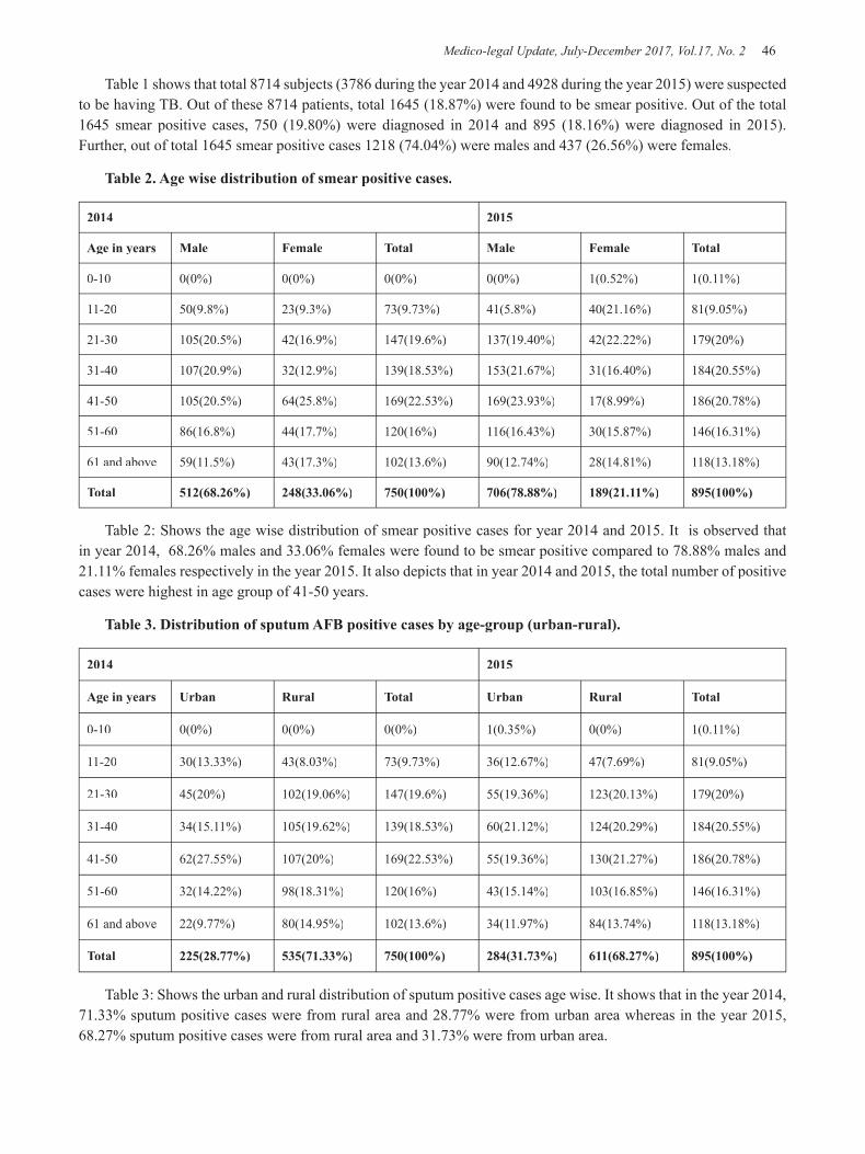
Medico-legal Update, July-December 2017, Vol.17, No. 2 46 47 -legal Update, July-December 2017, Vol.17, No. 2
Table 1 shows that total 8714 subjects (3786 during the year 2014 and 4928 during the year 2015) were suspected to be having TB. Out of these 8714 patients, total 1645 (18.87%) were found to be smear positive. Out of the total 1645 smear positive cases, 750 (19.80%) were diagnosed in 2014 and 895 (18.16%) were diagnosed in 2015). Further, out of total 1645 smear positive cases 1218 (74.04%) were males and 437 (26.56%) were females.
Table 2. Age wise distribution of smear positive cases.
2014 2015
Age in years Male Female Total Male Female Total
0-10 0(0%) 0(0%) 0(0%) 0(0%) 1(0.52%) 1(0.11%)
11-20 50(9.8%) 23(9.3%) 73(9.73%) 41(5.8%) 40(21.16%) 81(9.05%)
21-30 105(20.5%) 42(16.9%) 147(19.6%) 137(19.40%) 42(22.22%) 179(20%)
31-40 107(20.9%) 32(12.9%) 139(18.53%) 153(21.67%) 31(16.40%) 184(20.55%)
41-50 105(20.5%) 64(25.8%) 169(22.53%) 169(23.93%) 17(8.99%) 186(20.78%)
51-60 86(16.8%) 44(17.7%) 120(16%) 116(16.43%) 30(15.87%) 146(16.31%)
61 and above 59(11.5%) 43(17.3%) 102(13.6%) 90(12.74%) 28(14.81%) 118(13.18%)
Total 512(68.26%) 248(33.06%) 750(100%) 706(78.88%) 189(21.11%) 895(100%)
Table 2: Shows the age wise distribution of smear positive cases for year 2014 and 2015. It is observed that in year 2014, 68.26% males and 33.06% females were found to be smear positive compared to 78.88% males and 21.11% females respectively in the year 2015. It also depicts that in year 2014 and 2015, the total number of positive cases were highest in age group of 41-50 years.
Table 3. Distribution of sputum AFB positive cases by age-group (urban-rural).
2014 2015
Age in years Urban Rural Total Urban Rural Total
0-10 0(0%) 0(0%) 0(0%) 1(0.35%) 0(0%) 1(0.11%)
11-20 30(13.33%) 43(8.03%) 73(9.73%) 36(12.67%) 47(7.69%) 81(9.05%)
21-30 45(20%) 102(19.06%) 147(19.6%) 55(19.36%) 123(20.13%) 179(20%)
31-40 34(15.11%) 105(19.62%) 139(18.53%) 60(21.12%) 124(20.29%) 184(20.55%)
41-50 62(27.55%) 107(20%) 169(22.53%) 55(19.36%) 130(21.27%) 186(20.78%)
51-60 32(14.22%) 98(18.31%) 120(16%) 43(15.14%) 103(16.85%) 146(16.31%)
61 and above 22(9.77%) 80(14.95%) 102(13.6%) 34(11.97%) 84(13.74%) 118(13.18%)
Total 225(28.77%) 535(71.33%) 750(100%) 284(31.73%) 611(68.27%) 895(100%)
Table 3: Shows the urban and rural distribution of sputum positive cases age wise. It shows that in the year 2014, 71.33% sputum positive cases were from rural area and 28.77% were from urban area whereas in the year 2015, 68.27% sputum positive cases were from rural area and 31.73% were from urban area.
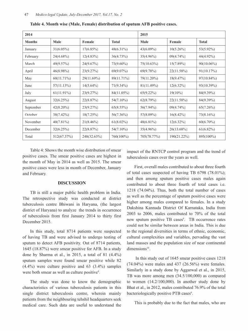
-legal Update, July-December 2017, Vol.17, No. 2 46 47 Medico-legal Update, July-December 2017, Vol.17, No. 2
Table 4. Month wise (Male, Female) distribution of sputum AFB positive cases.
2014 2015
Months Male Female Total Male Female Total
January 31(6.05%) 17(6.85%) 48(6.31%) 43(6.09%) 10(5.26%) 53(5.92%)
February 24(4.68%) 12(4.83%) 36(4.73%) 35(4.96%) 09(4.74%) 44(4.92%)
March 49(9.57%) 24(9.67%) 73(9.60%) 75(10.63%) 15(7.89%) 90(10.06%)
April 46(8.98%) 23(9.27%) 69(9.07%) 69(9.78%) 22(11.58%) 91(10.17%)
May 60(11.71%) 29(11.69%) 89(11.71%) 79(11.20%) 18(9.47%) 97(10.84%)
June 57(11.13%) 14(5.64%) 71(9.34%) 81(11.49%) 12(6.32%) 93(10.39%)
July 61(11.91%) 23(9.27%) 84(11.05%) 65(9.22%) 19(10%) 84(9.39%)
August 32(6.25%) 22(8.87%) 54(7.10%) 62(8.79%) 22(11.58%) 84(9.39%)
September 42(8.20%) 23(9.27%) 65(8.55%) 56(7.94%) 09(4.74%) 65(7.26%)
October 38(7.42%) 18(7.25%) 56(7.36%) 57(8.09%) 16(8.42%) 73(8.16%)
November 40(7.81%) 21(8.46%) 61(8.02%) 48(6.81%) 12(6.32%) 60(6.70%)
December 32(6.25%) 22(8.87%) 54(7.10%) 35(4.96%) 26(13.68%) 61(6.82%)
Total 512(67.37%) 248(32.63%) 760(100%) 705(78.77%) 190(21.22%) 895(100%)
Table 4: Shows the month wise distribution of smear positive cases. The smear positive cases are highest in the month of May in 2014 as well as 2015. The smear positive cases were less in month of December, January and February.
DISCUSSION
TB is still a major public health problem in India. The retrospective study was conducted at district tuberculosis centre Bhiwani in Haryana, (the largest district of Haryana) to analyze the trends in occurrence of tuberculosis from first January 2014 to thirty first December 2015.
In this study, total 8714 patients were suspected of having TB and were advised to undergo testing of sputum to detect AFB positivity. Out of 8714 patients, 1645 (18.87%) were smear positive for AFB. In a study done by Sharma et al., in 2015, a total of 81 (4.4%) sputum samples were found smear positive while 82 (4.4%) were culture positive and 63 (3.4%) samples were both smear as well as culture positive8.
The study was done to know the demographic characteristics of various tuberculosis patients in this single district tuberculosis centre, wherein mainly patients from the neighbouring tehshil headquarters seek medical care. Such data are useful to understand the
impact of the RNTCP control program and the trend of tuberculosis cases over the years as well.
First, overall males contributed to about three fourth of total cases suspected of having TB 6798 (78.01%); and then among sputum positive cases males again contributed to about three fourth of total cases i.e. 1218 (74.04%). Thus, both the total number of cases as well as the percentage of sputum positive cases were higher among males compared to females. In a study Dakshina Kannada District Of Karnataka, India from 2003 to 2006, males contributed to 70% of the total new sputum positive TB cases9. TB occurrence rates could not be similar between areas in India. This is due to the regional diversities in terms of ethnic, economic, cultural complexities and variables, pervading the vast land masses and the population size of near continental dimensions10.
In this study out of 1645 smear positive cases 1218 (74.04%) were males and 437 (26.56%) were females. Similarly in a study done by Aggarwal et al., in 2015, TB was more among men (34.5/100,000) as compared to women (14.2/100,000). In another study done by Bhat et al., in 2012, males contributed 76.9% of the total bacteriologically positive PTB cases6.
This is probably due to the fact that males, who are

Medico-legal Update, July-December 2017, Vol.17, No. 2 48 49 -legal Update, July-December 2017, Vol.17, No. 2
usually working are being commonly exposed to the TB bacillus more than females. This may also be due to increased access of males to diagnostic facilities (such as chest radiography, culture facilities etc) and treatment services compared to females. There also may be other independent factors like smoking, lifestyle, diabetes, occupation related health hazards etc. which may be associated with increased susceptibility to TB in an individual.
In general, the disease occurrence rates are about similar in both sexes, till the puberty in females. This is followed by a continuing widening of the gap between the sexes in favour of the females, the differences acutely accentuated past the 35-40 years age mark10.
In this study out of smear positive cases of year 2014, 68.26% were male and 33.06% were female and out of smear positive cases of year 2015, 78.88% were male and 21.11% were female. The study also conclude that in year 2014 and 2015 the positive cases were highest in age group of 41-50 years,169 (22.53%), 186 (20.78%) respectively.
In a study done by Dhanaraj et al., in 2015, while the maximum prevalence of bacteriologically positive PTB (1241/100,000) was found in the age group of 55-64 years7. In a study done by Bhat et al., in 2012, the highest proportion of positive PTB cases (25.8%) was seen in the 35-44 years age group6. In a study done by Rao et al., in 2010, the prevalence increased with age, being 546/100,000 in the 15-24 years age group, increasing to 3086/100,000 in the 55+ years age group12. In a study done by Bhat et al., 2009, the prevalence increased with age being 174/100,000 in the 15-24 years age group to 990/100,000 in the 55+ years age group13.
It is also observed in this study that the age-wise proportion of positive cases was substantially higher in age group of 21-30 years onwards, reaching the peak for the age group of 41- 50 years. The occurrence of smear positive TB rises with age, in both sexes (Table 2 and table 3).
The smear positive cases are highest in the month of May and are less in the months of December, January and February.
CONCLUSIONS
The study indicates the trends of occurrence of smear positive pulmonary TB cases in the study area
from January 2014 to December 2015. The Study showed higher occurrence of TB in males i.e 1218 (74.04%) compared to females i.e. 437 (26.56%). Maximum affected age group was between 41 and 50 years. The lowest number of pulmonary TB was observed in the age group < 15 year. Therefore, planning and enforcement agencies and health authorities should pay more attention to and design special preventive strategies to prevent TB transmission, especially in the older age groups, especially males.
Acknowledgement: Nil
Source of Funding: Self
Conflict-of-Interest Statement: There is no conflict of interest as it was a retrospective study based on the already available record.
Statement of Informed consent: This study is based on the available record so there is no matter of consent.
Statement of Human and Animal Rights: No human right and animal right is violated in this case.
No ethical violation is done.
REFERENCES
1. Jenkins A. In: Davies PDO clinical tuberculosis. Second edition. Chapman and hall, London; 1998, 69-79.
2. Admasu K, Kebede A. Tuberculosis prevalence survey. First edition. Addis Ababa; 2011, 13-15.
3. Ashi Thompson’s. Medical Microbiology. Seventh edition. Edenberg; 1030-1032.
4. World health organization. Global tuberculosis control. WHO; 2011.
5. Wilkinson E. Notes on the prevalence of tuberculosis in India. Proc R Soc Med. 1914;7:195–226.
6. Rao VG, Bhat J, Yadav R, Gopalan GP, Nagamiah S, Bhondeley MK, et al. Prevalence of pulmonary tuberculosis - A baseline survey in central India. PLoS One 2012;7:e43225.
7. Dhanaraj B, Papanna MK, Adinarayanan S, Vedachalam C, Sundaram V, Shanmugam S, et al. Prevalence and risk factors for adult pulmonary tuberculosis in a metropolitan city of South India.
-legal Update, July-December 2017, Vol.17, No. 2 48 49 Medico-legal Update, July-December 2017, Vol.17, No. 2
PLoS One 2015;10:e0124260.
8. Sharma SK, Goel A, Gupta SK, Mohan K, Sreenivas V, Rai SK, et al. Prevalence of tuberculosis in Faridabad district, Haryana State, India. Indian J Med Res 2015;141:228-35.
9. Ganesh KS, Harsha Kumar HN, Ramakrishna R, Jayarama S, Kotian MS. Trend of Tuberculosis Cases under DOTS Strategy in Dakshina Kannada District of Karnataka, India: Issues and Challenges. Iranian J Publ Health 2009;72-6.
10. Chakraborty AK. Epidemiology of tuberculosis: Current status in India. Indian J Med Res 2004;120:248-76.
11. Aggarwal AN, Gupta D, Agarwal R, Sethi S, Thakur JS, Anjinappa SM, et al. Prevalence of pulmonary tuberculosis among adults in a north Indian district. PLoS One 2015;10:e0117363.
12. Rao VG, Gopi PG, Bhat J. Pulmonary tuberculosis: A public health problem amongst the Saharia, a primitive tribe of Madhya Pradesh, Central India. Int J Infect Dis 2010;14:e713-6.
13. Bhat J, Rao VG, Gopi PG, Yadav R, Selvakumar N, Tiwari B, et al. Prevalence of pulmonary tuberculosis amongst the tribal population of Madhya Pradesh, central India. Int J Epidemiol 2009;38:1026-32.

51 -legal Update, July-December 2017, Vol.17, No. 2
An Epidemiological Study of Dowry Deaths in Special Reference to Burns in Indian Scenario: A Medicolegal Study
Pramod Kumar Kistigari Sathyanarayana1, Suraj Sundaragiri2
1Assistant Professor, Forensic Medicine and Toxicology, MediCiti Institute of Medical Sciences, Hyderabad, 1Assistant Professor, Forensic Medicine and Toxicology, MediCiti Institute of Medical Sciences, Hyderabad, 1
Telangana, India, 2Senior Resident, Forensic Medicine and Toxicology, Jawaharlal Institute of Postgraduate Medical Education & Research, Puducherry, India
ABSTRACT
Background: Dowry death is a burning problem of the society in India owing to prevailing traditional cultural practices, socioeconomic and psychosocial taboos and life style.
Objectives: This paper appraises the patterns, socioeconomic and demographic factors of dowry deaths cases due to burns.
Materials and Method: The present prospective study comprised of 212 dowry death cases pertaining to burns brought for medico-legal autopsy at tertiary teaching hospital in Telangana, India for 2 years. Data was collected and analyzed in relation to different factors from police inquest report, first information report and autopsy examination proper.
Results: Deaths due to burn (n=212) constituted commonest causative factor for dowry deaths. Majority (n=141; 64.09%) were found to be homicide. Majority (n=63; 29.72%) were recorded in the age group of 23 to 26 years. 91 cases (42.92%) had more than 80% of total body surface area involved in burns. Majority of deaths (n=77; 36.32%) occurred during night (9 pm to 6 am). Husband alone was found to be most common offender (n=71; 33.49%). Majority of the victims were housewives (n=173; 81.61%). Majority of the dowry deaths were drawn from joint family (n=174; 82.08%). Incidence were more in arranged marriages (n=173; 81.60%). First and the foremost location of incidence was in-law’s house (n=160; 75.47%).
Conclusions: This social malicious evil needs to be targeted by a multifaceted and united approach of women welfare organizations, police and judiciary towards enhancement of socioeconomic and educational profile along with provision of strict laws and speedy justice.
Keywords: Dowry deaths, Burns, homicide, suicide, Bride burning.
INTRODUCTION
In recent times, issues related to women have been raised and discussed at various forums. Among these crimes against women, dowry occupied the center stage. As many as 22 dowry deaths occur in India every day1.
Corresponding author:Dr. Suraj Sundaragiri, H.No: 4-9-848/ 2, Lecturers Colony, Hayathnagar, Hyderabad, Telangana. India. 501505, Contact No.: 091-9908267327, Email: [email protected]
Dowry is an age-old traditional social evil which is present in our society since time immemorial. In spite Indian laws against dowry being in effect since times, they have been to a great extent criticized as being ineffective. It may be emphasized that a girl by act of marriage is introduced into an unfamiliar insecure world entirely rely on the mercy of husband and his family members who might or might not look after her. Majority gets suppressed and suffers without revealing the actual fact. Only in rare circumstances, women prefer to take recourse to police and judiciary.
Dowry deaths accounts to a woman’s homicide or suicide committed by her husband and his family due to
DOI Number: 10.5958/0974-1283.2017.00067.6

51 Medico-legal Update, July-December 2017, Vol.17, No. 2
their dissatisfaction with the dowry2.According to Indian Evidence act 1872, section 113 A denotes presumption as to abatement of suicide by a married woman and section 113 B presumption as to ‘dowry death’ if that death is caused within seven years of marriage3. When she is unable to bear torture and harassment, she commits suicide by burning or hanging herself or consuming poison. On other hand, there are other forms like poisoning, strangulation, etc., by which women are murdered. Bride burning, where brides are set ablaze by her husband or his family is the most common form of dowry deaths as there being inadequate evidence of crime and less chance of survival4.
Even though there are the long list of legislations protecting the rights of women, most importantly the prohibition of giving and taking dowry - the Dowry Prohibition Act, 1961, is the one which is most commonly defied since its inception all over the country5. Precisely to deal these crimes, section 304 B (Dowry deaths) and 498 A (Cruelty by husband or in-laws) were incorporated in the Indian Penal Code, 18602. The Protection of Women from Domestic Violence Act, 2005 was passed in order to provide civil law remedy for the protection of women from domestic violence6. This present study makes an effort to appraise the patterns, demographic and socioeconomic factors of dowry deaths with special reference to burns.
MATERIALS AND METHOD
The present prospective and descriptive study comprised of cases brought for medico legal autopsy at tertiary teaching hospital in Telangana, India for the period of 2 years, during 2012 and 2013. Section 304B of Indian Penal Code, 1860 and section 113 of Indian Evidence Act, 1872 were considered as criteria to involve these cases as dowry deaths. This work was confined to dowry deaths as a result of burns only. Deaths due to accidental burns were excluded. Data was collected from first information report, police inquest reports and autopsy examination proper, and the manner of death confirmed before study. These cases were examined regarding certain criteria, and data was then compiled, analyzed and discussed.
RESULTS
During the study period, out of total 342 dowry deaths, majority (n=220; 64.32%) were homicide and remaining (122; 35.68%) suicide. A probe regarding
cause of death like burns, poison, asphyxia and other concerned causes showed that out of total dowry deaths, deaths due to burn i.e. study group constituted commonest causative factor for dowry death (n=212; 61.99%), followed by mechanical asphyxia (n=77; 22.51%) (Table 1). With regard to manner of death in study group (n=212 cases), majority (n=141; 64.09%) were found to be homicide and rest (n=71; 33.49 %) suicide. The age of victims ranged from 18 to 38 years, among which majority (n=63; 29.72%) were recorded in the age group of 23 to 26 years, followed 18 to 22 years (n=59; 27.83%), 27 to 30 years (48; 22.64%), and beyond age of 30 years, only 42 cases (19.81 %) were noted.
It was observed that about 91 cases (42.92%) had more than 80% of total body surface area(TBSA) involved in burns, followed by 60–80% TBSA (n=79; 37.26%) and 40-60% TBSA (n= 39; 18.4%). An attempt has also been made to find out relation with peak time of occurrence. Majority of deaths (n=77; 36.32%) occurred during night (9 pm to 6 am) followed by evening (4 pm to 9 pm) (n=74; 34.90%), morning (6 am to 12 noon) (n=38; 17.84%) and afternoon (12 noon to 4 pm) (n=23; 10.85%). The breakup of culprits involved has also been worked up to make an in-depth study. Husband and his family/or his relatives were found to be responsible for most deaths (n=196; 92.45%). They together either killed or forced the victims to end their life. Husband alone was found to be most common offender responsible (n=71; 33.49%), followed by husband and his family (n=70; 33.02%), husband’s family and relatives (n=55; 25.94%) and other persons including neighbors, passersby etc. accounted for meager dowry deaths (n=16; 7.54%).
A link with educational qualification of victims revealed that most deaths was seen in matriculates (n=102; 48.12%), followed by non-matriculates (n=55; 25.94%) and graduates (n=52; 24.52%). The break up amongst homicidal victims was matriculates (n=66; 47.48%), followed by graduates (n=41; 29.50%) and non-matric (n=32; 23.02%). In educated females, suicide was most common. Precisely in postgraduates, almost all the cases were suicidal and no homicidal case was reported (Table 2). A major chunk (n=107; 50.58%) belonged to the low socioeconomic group, followed by lower middle class (n=67; 31.58%) and very low socioeconomic class (n=21; 9.65%). Only 17 (8.19%) victims came from higher middle class. Majority i.e. 111 of 139 homicidal deaths belonged to low and lower

Medico-legal Update, July-December 2017, Vol.17, No. 2 52 53 -legal Update, July-December 2017, Vol.17, No. 2
middle class socioeconomic status (Table 3).
A great majority of the victims were housewives (n=173; 81.61%) with high incidence of homicide (n=115; 82.73%). Among the employed, 9 cases (4.24%) were recorded who died by suicidal manner. In student category, out of 21 cases, 15(10.79%) died by homicide and remaining suicide (n=6; 8.22%) (Table 4). Majority of the dowry deaths were drawn from joint family (n=174; 82.08%), whereas 38 cases (17.92%) came from nuclear family. In joint families, both homicidal (n=118; 84.89%) and suicidal deaths were more common (n=56; 76.71%) than nuclear family where homicidal (n=21; 15.11%) and suicidal deaths (n=17; 23.29%) were less. Incidence were more in arranged marriages (n=173; 81.60%) whereas only 39(18.40%) deaths occurred amongst love marriages. Amongst the arranged marriages, both homicidal (n=116; 83.45%) and suicidal deaths (n=57; 78.08%) were most commonly seen as compared to love marriages with less suicidal (n=16; 21.92%) and homicidal deaths (n=23; 16.55%).
In more than half (n=160; 75.47%), first and the foremost location of incidence was the in-law’s house, followed by husband’s house (n=41; 19.34%). Dowry deaths in parental house were recorded in only 4 cases (1.89%) and all of them were suicide. Incidence of dowry deaths at the other locations like fields, well, pond or roadside were quite limited, and only 7 such cases were recorded (Table 5). In-law’s house constituted the commonest location for homicidal deaths as well.
DISCUSSION
Destruction of woman’s life as a result of dowry is not uncommon in this part of the country in spite of high awareness in this aspect and higher literacy/educational status, and also stringent laws in practice. Our study revealed that majority of deaths is caused by burn followed by asphyxia as reported in other studies7,
8, 9, 10. There was preponderance of homicidal cases
over suicidal which are in consistency with others10, 11. But, this in contrast to results of others7, 12. Majority of burnt victims had more than 80 % TBSA involvement as observed in other study13. However, analysis on age incidence reveals that highest was among the age group of 23-26 years followed by 18-22 years. Involvement of younger age group has also been supported by various studies7, 10, 14, 15. In the majority, either husband and/or his relatives are responsible for killing or forcing victim to end her life, and husband alone is found to be most common offender responsible9, 16. Educational background has relevance. It may be presumed that incidence is barely negligible in highly educated women who can opt for job in emergency. Majority of dowry deaths are reported in either illiterate and below matric or matriculate as noticed in other studies7, 8, 16. In educated females, suicide is more common. In contrast, victims who having education with matriculation, dowry death incidence was found to be more common.
Our study observed that majority of the cases is seen in low socioeconomic group. This finding corresponds well with other studies in the field8, 9. Most of deaths occurred during night followed by evening which in contrast to other studies where it was in the evening11,
14, 15. Most victims were house-wives, amongst which homicides comprised highest incidence as indicated in other studies7, 8, 11, 16. The number of victims from joint families is about 4 times compared to nuclear families which were in agreement with studies11, 16, 17. Majority of dowry deaths are reported in arranged marriages18. The study clearly points to those young housewives from joint families are the major target particularly if they belong to poor socioeconomic status. The first and foremost location of incidence is in-law’s house followed by husband’s house, which is in conformity with other studies9, 16. The probable explanation is she becomes an easy prey in in-law’s house as nobody is there supporting/ protecting the harassed victim.
Table 1: Causative factors and nature of dowry deaths
DOWRY DEATHSCausative Factors Homicidal Suicidal Total
No. % No. % No. %Burns 141 64.09 71 58.20 212 61.99Mechanical Asphyxia 42 19.1 35 28.69 77 22.51Poison 28 12.73 9 7.38 37 10.82Others 9 4.09 7 5.74 16 4.68Total 220 100 122 100 342 100
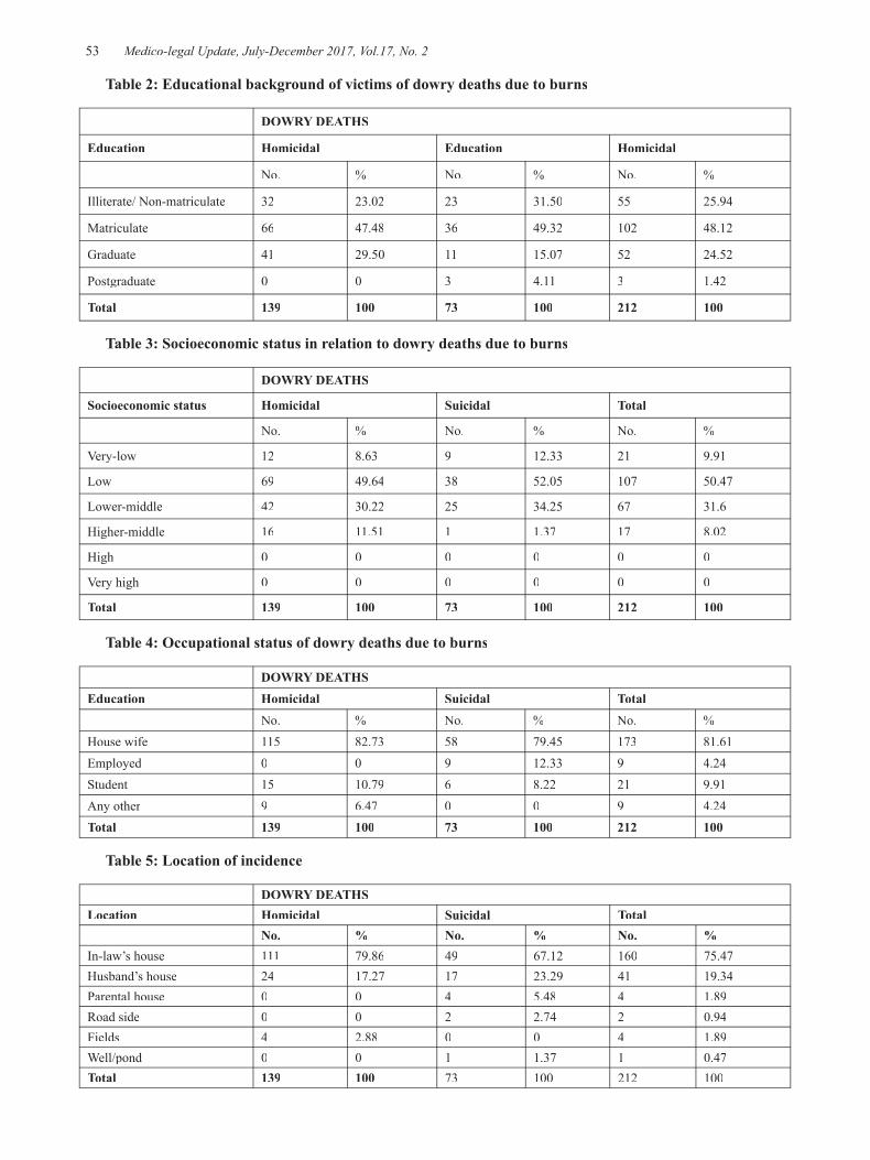
-legal Update, July-December 2017, Vol.17, No. 2 52 53 Medico-legal Update, July-December 2017, Vol.17, No. 2
Table 2: Educational background of victims of dowry deaths due to burns
DOWRY DEATHS
Education Homicidal Education Homicidal
No. % No. % No. %
Illiterate/ Non-matriculate 32 23.02 23 31.50 55 25.94
Matriculate 66 47.48 36 49.32 102 48.12
Graduate 41 29.50 11 15.07 52 24.52
Postgraduate 0 0 3 4.11 3 1.42
Total 139 100 73 100 212 100
Table 3: Socioeconomic status in relation to dowry deaths due to burns
DOWRY DEATHS
Socioeconomic status Homicidal Suicidal Total
No. % No. % No. %
Very-low 12 8.63 9 12.33 21 9.91
Low 69 49.64 38 52.05 107 50.47
Lower-middle 42 30.22 25 34.25 67 31.6
Higher-middle 16 11.51 1 1.37 17 8.02
High 0 0 0 0 0 0
Very high 0 0 0 0 0 0
Total 139 100 73 100 212 100
Table 4: Occupational status of dowry deaths due to burns
DOWRY DEATHSEducation Homicidal Suicidal Total
No. % No. % No. %House wife 115 82.73 58 79.45 173 81.61Employed 0 0 9 12.33 9 4.24Student 15 10.79 6 8.22 21 9.91Any other 9 6.47 0 0 9 4.24Total 139 100 73 100 212 100
Table 5: Location of incidence
DOWRY DEATHSLocation Homicidal Suicidal Total
No. % No. % No. %In-law’s house 111 79.86 49 67.12 160 75.47Husband’s house 24 17.27 17 23.29 41 19.34Parental house 0 0 4 5.48 4 1.89Road side 0 0 2 2.74 2 0.94Fields 4 2.88 0 0 4 1.89Well/pond 0 0 1 1.37 1 0.47Total 139 100 73 100 212 100
Medico-legal Update, July-December 2017, Vol.17, No. 2 54 55 -legal Update, July-December 2017, Vol.17, No. 2
CONCLUSION
The present study highlights the following features pertaining to the dowry deaths due to burns- incidents usually occur in women between 23 to 26 years age group and mostly in night, deaths were more common in arranged marriages and mostly occurred in joint families, majority of victims were housewives and belonged to the lower socioeconomic status and total body surface area burnt is usually more than 80%. “Bride burning” is a social scourge in today’s civilized society. The result clearly indicates that law alone will not warrant a favorable result against dowry. The social media and voluntary social organizations must play an important role by not only bringing social awareness but also condemning the evils of dowry. Additional, they must further raise at all possible forums, the issues of women harassment and torture, thereby forcing the offender husband to act with intimidation and give social dignity and respect to his wife at all levels. This social malicious evil also needs to be targeted by a multifaceted and united approach by women welfare organizations, police and judiciary towards enhancement of socioeconomic and educational profile with increased job potentials as well as a respect for women with better social status along with strict laws and speedy justice.
FUNDING
This research did not receive any specific grant from funding agencies in the public, commercial, or not-for-profit sectors.
Declaration of Conflicting Interests: The authors declare that there is no conflict of interest.
Ethical Clearance: Relative permission and necessary clearance was already taken during the autopsies.
REFERENCES
1. Crime in India 2014: Crimes against Women. National Crime Records Bureau, Ministry of Home Affairs, Government of India.2014. Available from: http://ncrb.nic.in/StatPublications/CII/CII2014/chapters/Chapter%205.pdf
2. Council of the Governor General of India. Indian Penal Code 1860. Government of India.1860. Available from: http://indiacode.nic.in/
3. Council of the Governor General of India. Indian
Evidence Act 1872. Government of India.1872. Available from: http://devgan.in/iea
4. Teays W. The burning bride: The dowry problem in India. Journal of Feminist Studies in Religion. 1991 Oct 1; 7(2):29-52.
5. The Dowry Prohibition Act 1961. Government of India.1961. Available from: http://ncw.nic.in/acts/THEDOWRYPROHIBITIONACT1961.pdf
6. The Protection of Women from Domestic Violence Act 2005. Government of India.2005. Retrieved from http://ncw.nic.in/acts/TheProtectionofWomenfromDomesticViolenceAct2005.pdf
7. Kulshrestha P, Sharma RK, Dogra TD. The study of sociological and demographical variables of unnatural deaths among young women in south Delhi within seven years of marriage. Hindu.2002; 103(88.03):88-03.
8. Karukutla N, Priya KD. An Autopsy Based Study of Socio-Etiological Aspects in Dowry Death Cases. Indian Journal of Forensic and Community Medicine.2015; 2(3):141-3.
9. Batra AK. Burn mortality: recent trends and sociocultural determinants in rural India. Burns.2003; 29(3):270-5.
10. Bhullar, D. S., Oberoi, S. S., Aggrawal, O. P., & Tuli, H. Profile of unnatural female deaths (between 18-30 years of age) in Government Medical College/Rajindra Hospital, Patiala. Journal of Forensic Medicine and Toxicology.1996; 8(3):5-8.
11. Sinha, U. S., Kapoor, A. K., Agnihotri, A. K., & Srivastava, P. C. The epidemiological study of dowry death cases with special reference to burn cases in Allahabad range (UP). International Journal of Medical Toxicology & Legal Medicine.1998; 1(1):65-71.
12. Nagesh Kumar G. Rao: Study of fatal female burns in Manipur. J ForensicMed Toxicology.1997; 14(2):57-59.
13. Meera T, Nandeibam P, Slong D, Nabachandra H. Burn Deaths: A Study on Female Victims in Manipur. Journal of Indian Academy of Forensic Medicine.2015; 37(4):358-60.
14. Kumar V. Burnt wives:a study of suicides. Burns.2003 Feb 28; 29(1):31-5.
15. Kumar V, Tripathi CB. Burnt wives: a study of

-legal Update, July-December 2017, Vol.17, No. 2 54 55 Medico-legal Update, July-December 2017, Vol.17, No. 2
homicides. Medicine, science and the law.2004; 44(1):55-60.
16. Mohanty S, Sen M, Sahu G. Analysis of risk factors of dowry death–a south Indian study. Journal of forensic and legal medicine.2013; 20(4):316-20.
17. Srivastava AK, Arora P. Suspicious deaths in newly married females–A medicolegal analysis.
Journal of Indian Academy of Forensic Medicine. 2007; 29(4):63-7.
18. Parameswaran R. Coverage of “Bride Burning” in the “Dallas Observer”: A Cultural Analysis of the” Other”. Frontiers: A Journal of Women Studies.1996; 16(2/3):69-100.
57 -legal Update, July-December 2017, Vol.17, No. 2
Pattern of Craniocerebral Injuries among Homicidal Deaths in Hubballi Dharwad Region- One Year Retrospective Study
Gajanan H Nayak1, Muthamizh Selvan P2
1Professor and Head, Karnataka Institute of Medical Sciences, Hubballi, 2Postgraduate Student, Department of 2Postgraduate Student, Department of 2
Forensic Medicine and Toxicology, Karnataka Institute of Medical Sciences, Hubballi
ABSTRACT
Inflicting injury to the head is one of the most effective methods of homicide. The aim of the present study was to evaluate the pattern of head injury among homicidal death victims. In present study, out of 45 homicidal deaths, 23 cases of homicide infliction over the head were studied in one year period from 1st January 2015 till 31st December 2015. Majority of cases were in 21-40 years of age comprising of 12 cases st December 2015. Majority of cases were in 21-40 years of age comprising of 12 cases st
(52.17%). Injuries were inflicted by blunt weapons in significant number of cases; 13 cases (56.5%) which were followed by injuries by sharp weapons in 9 cases (39.1%) and firearm in 1 case (04.34%). The skull was fractured in 21 cases (91.30%) of victims and 2 cases (08.69%) of deceased presented without skull fracture. Most common intracranial hemorrhage observed was combination of subdural hemorrhage and subarachnoid hemorrhage(73.91%).
Keywords: Head injury, Homicide, Blunt weapon, Skull fracture.
INTRODUCTION
One of the oldest crimes in human civilization is homicide which started from murder of Abel by hands of Cane. With the passage of time manner of homicide have been changing. To analyze the homicidal deaths in any civil society is the first step towards development to reduce its impact. [1]
Death resulting from blunt force trauma to head are some of the most common cases encountered by practicing forensic pathologist. Head is the most vulnerable part of the body to receive injuries. Injuries to the head are accidental, mostly vehicular or homicidal. Violence is a significant public health problem and homicide is the severest form of violence, depriving a human being of his fundamental right to live. Homicide is prevalent widely almost all over the world [2].
Head injury as defined by the National Advisory Neurological Diseases and Stroke Controlas been defined as, “a morbid state, resulting from gross or
subtle structural changes in the scalp, skull, and/or the contents of skull, produced by mechanical forces”. [3]
Blunt force trauma to the head could potentially result in death. When the victim is attacked on the head and neck with a blunt object, the presence of underlying damage to the brain or its covering is more likely to determine the outcome for the victim- alive or dead. It has been noted that death in this condition can occur without the presence of a skull fracture. However, this phenomenon is rarely seen and skull fracture can be regarded as an indirect indicator of severity of the resulting trauma. [4]
Investigation of a homicidal death can never be complete without a detailed postmortem examination. The detailed analysis and scientific interpretation of autopsy finding is imperative to reconstruct the crime scene.
The main objective of this analysis was to analyze the information to determine trends of head injury in homicide cases in Northern Karnataka region during the study period.
MATERIALS AND METHOD
A retrospective autopsy study of 23 cases of
DOI Number: 10.5958/0974-1283.2017.00068.8
Corresponding author: Dr. Muthamizh Selvan PPostgraduate Student, Karnataka Institute of Medical Sciences, Hubballi-580021, Karnataka, Ph.: 9916040474

57 Medico-legal Update, July-December 2017, Vol.17, No. 2
head injury resulting from assault was conducted at the Department of Forensic Medicine& Toxicology, Karnataka Institute of Medical Sciences, Hubballi, Karnataka over a period of one year. Out of 45 homicidal deaths, 23 cases (51.11%) were due to fatal head injury. Those who died by suicide or by accidental and natural causes were excluded. Information regarding the victims was collected from the inquest report, police personnel’s regarding the scene of occurrence and examination findings mentioned in the autopsy reports. All observations were then transferred to a Microsoft Excel sheet.
Descriptive statistics for qualitative type of data was summarized and tabulated using frequency and percentages with summarization of all findings.
OBSERVATION AND RESULTS
A total of 23 cases of homicidal fatal head injury deaths were included in this study, of which 21 cases(91.30%) were males and 2 cases (8.69%) were females succumbing to episodes of homicide. This is quite explainable by the pattern of Indian societies where males are the active members. (Table 1) In our study majority of the homicidal attacks were committed by blunt weapons 13 cases (56.5%). (Table 2) The use of blunt weapons for an episode of homicidal attack was reported significantly in this study. Sharp weapons and firearm weapons were put to use in 9 cases (39.1%) and 1 case (4.34%) respectively. (Table 2) In this study total head injury cases were 23, of which 21 cases (91.30%) were having fracture. Most common pattern observed was combination of SDH (Subdural Hemorrhage) & SAH (Sub arachnoid Hemorrhage) along with skull fracture in 14 cases (60.86%) followed by brain injury observed in 4 cases (17.39%) (Table 3). Isolated Subarachnoid hemorrhage was observed in 1case (4.34%). Intracerebral hemorrhage observed in 1 case(4.34%). Pontine hemorrhage was observed in 1 case (4.34%). Defense wounds were present in only 09 (39.13%) cases out of total 23 cases of homicide in this study. Abrasion was present in 07 cases (30.43%) followed by contusion in 02 cases (08.69%) and incised wound in 01 case (04.34%). (Table 5) Present study showed that in 91% cases of fatal head injuries there was associated fracture skull. The most common bone to be fractured was combination of temporal and occipital bone followed by combination of parietal and temporal bone. Isolated Occipital bone was fractured in 2 cases,
isolated temporal bone was fractured in 1 case and isolated parietal bone was fractured in 1 case. There was no isolated frontal bone fracture observed. No fracture of skull was present in 2 cases. (Table 4)
DISCUSSION
In our study, among the 23 victims included in study population, there were 21 males (91.30%) and 2 females (8.69%).This is line with the study conducted by R.K. Punia et al, male: female (M: F) ratio was 3.5:1. Among the 72 victims included in study population, there were 56 males (77.77%) and 16 females (22.23%) [17].
In our study, the most commonly affected age group was 21-40 years (52.17%) followed by 41-50 years (26.08%) and 51-60 years (8.69%). One case was in age group less than 10 years. This is line with the study conducted by B. C. Shivakumar et al, where the peak incidence of homicide was reported in age group 21-30 years (n=16, 40%) followed by 31-40 years (n=8, 20%). Majority of the victims in our study belonged to the most productive age groups between 21-40 years (52.17%). [5]
Present study reports the use of blunt weapons as a means of committing homicide in a significant number of cases (56.5%). Similar findings have been reported by other Indian studies. [6,7,8,9,10,11,12,13]. However, our results are contradictory to study conducted by shivakumar et al where sharp weapons were used as the most common weapon for commission of homicide. [5]
Fingerhut et al [14] reported firearms to be the most common murder weapon in the United States. Being light, handy and easy to use, they are far more effective than blunt objects or sharp weapons.
In single bone involvement, occipital was observed in two cases followed by parietal and temporal bone. No isolated fracture of frontal bone observed in our case. However our results are contradictory to study conducted by Yavuz et al reported linear fractures to be most common in the frontal and temporal region. [15] The occipital bone (2.78%) showed the least involvement in fracture, as it is the thickest among the cranial bones.
Homicide is the only major cause of childhood deaths that has increased over past three decades. In 1999, some 1,800 juveniles or 3 per 10,000 of U.S. juvenile population were homicide victims- a rate substantially higher than those of other developed countries. At the
Medico-legal Update, July-December 2017, Vol.17, No. 2 58 59 -legal Update, July-December 2017, Vol.17, No. 2
same time murders of juveniles are infrequent in many of our country. [16] Here in our study one case was in the age group below 10 years. Homicide is always tragic but our sympathies are heightened when it is a child.
DEFENSE WOUND
Defense wound present only in 09 cases out of 23 cases of homicidal infliction over the head. (Table 5) It is the instinctive behavior of the victim to raise his/her arm to ward off an attack and to protect vital organs such as brain. The presence of such injuries indicates an assault by some other person. However, the absence of defense wounds does not exclude homicide, since the victim may be incapable of effective defense for reasons such as surprise, being unconscious or under the influence of alcohol.
Table 1: Age and Sex wise Distribution of Victims of Homicidal Deaths.
Age groups Male Female Total victims
<10 01 - 01 [04.34%]
11-20 01 - 01 [04.34%]
21-30 06 - 06 [26.08%]
31-40 04 02 06 [26.08%]
41-50 05 - 05 [21.73%]
51-60 02 - 02 [08.69%]
61-70 01 - 01 [04.34%]
>70 01 - 01 [04.34%]
Total (%) 21 02 23 [100%]
Table 2: Distribution of victims according to weapon used to inflict the attack
Weapon Cases Total(%)
Blunt 13 56.5%
Sharp 09 39.1%
Firearm 01 4.34%
Total 23 100%
Table 3: Distribution of intracranial hemorrhage in victims of homicidal deaths
Intacranial hemorrhage Cases Percentage
EDH 00 00
SDH 01 [04.34%]
SAH 01 [04.34%]
EDH+SAH 00 00
SDH+SAH 14 [60.86%]
EDH+SDH+SAH 00 00
Intracerebral hemorrhage 01 [04.34%]
Brain injury 04 [17.39%]
Cerebellar hemorrhage 01 [04.34%]
Pontine hemorrhage 01 [04.34%]
Table 4: Distribution of skull fracture in victims of homicidal deaths
Bones fractured Total percentage
Frontal 00
Parietal 01 [04.34%]
Temporal 01 [04.34%]
Occipital 02 [08.69%]
Frontal+Parietal 02 [08.69%]
Frontal+Temporal 02 [06.69%]
Parietal+Temporal 03 [13.04%]
Temporal+Occipital 04 [17.39%]
Pairetal+Occipital 00
Frontal+Occipital 02 [08.69%]
Multiple skull bones 04 [17.39%]
No fractures 02 [08.69%]
Total 23 cases(100%)
Table 5: Types of defense wound
Type Cases Percentage
Abrasion 06 [26.08%]
Contusion 02 [08.69%]
Incised wound 01 [04.34%]
-legal Update, July-December 2017, Vol.17, No. 2 58 59 Medico-legal Update, July-December 2017, Vol.17, No. 2
CONCLUSION
Out of 23 cases of homicidal infliction over the head, 21-40 years of age group was mostly affected. Only two cases were the female victims. Infliction with blunt weapons was commonly observed than use of sharp weapons or firearms. Defense injuries present only in 9 cases (39.13%).Homicide is one of the worst forms ofcrime. State, society and legal officers should take firm steps to control this heinous crime.Continuous research in thisfield is the need of hour to frame strategies to counter unlawful human killings.
Conflict of Interest: Nil
Source of Funding: Self
Consent: Not taken as it was autopsy based retrospective study.
REFERENCES
1. Muhammad Humayun et al. Analysis of Homicidal Deaths in District Di Khan: An Autopsy Study. JAMC, 2009; 21(1): 155-7.
2. Prahlow J. Forensic pathology for forensic scientists, police, and death investigators. 2nd ed. nd ed. nd
New York, USA: Humana Press; 2009.
3. American College of. Surgeons Committee on trauma. Resources for the optimal care of· the injured patient, Chicago III. American College of Surgeons 1993.
4. Chattopadhyay S, Tripathi C. Skull fracture and hemorrhage pattern among fatal and non fatal head injury assaulat victims- a critical analysis, J Inj Violence Res. 2010; 2(2): 99-103.
5. Shivakumar BC, Vishwanath D, Srivastava PC. Trends of homicidal deaths at a tertiary care centre Bengaluru. JIAFM 2011; 33(2):120-124.
6. Buchade D, Mohite S. Pattern of Injuries in Homicidal Cases in Greater Mumbai- A Three Year Study. JIAFM 2011 Jan-March; 33(1):46-49.
7. Karthik SK, Balaji PA, Syed Sadat Ali, Jayaprakash G, Mohan Velu J. Analysis of Homicidal patterns
in Bangalore City, India. Indian Journal of Forensic Medicine and Toxicology. 2012 Dec; 6(2):64-67.
8. Mohanty MK, Kumar M, Mohanram A, Palimar V. Victims of homicidal deaths- An analysis of variables. J Clin, Forensic Med. 2005 Dec; 12(6):302-304.
9. Oberoi SS, Singh SP, Aggarwal KK, Bhullar DS, Aggarwal A, Walia DS, Thind AS. Profile of fatal assault cases in Patiala. J Punjab Acad Forensic Med Toxicol. 2011; 11(2): 87-89.
10. Dhaval J Patel Analysis of Homicidal Deaths in and Around Bastar Region of Chhattisgarh. J Indian Acad Forensic Med. April-June 2012,Vol. 34, No.2
11. Gupta S, Prajapati P. Homicide trends at Surat region of Gujarat, India. Journal of Forensic Medicine and Toxicology. 2009; 26(1):4548.
12. Strom C, Nordenram A, Johanson G. Injuries due to violent crimes. Med.Sci. Law. 1991; 31: 251-60
13. O. Gambhir Singh, B.D. Gupta. Trends of homicide in and around Jamnagar region of Gujarat, India- A retrospective study of five years, JFMT. 2007; 24(2): 6-11.
14. Hugar BS, Chandra G, Harish S. Pattern of homicidal deaths. JIAFM 2010 July; 32 (3):194-198
15. Fingerhut LA, Ingram DD, Feldman JJ. Firearm and non firearm homicides among persons 15 through 19 years of age. JAMA, 1992; 267(22):3048-53.
16. David Finkelhor andRichard Ormrod,Homicides of children and youth; U.S. Department of justice, office of justice program; office of juvenile justice and delinquency prevention; October 2001.
17. R.K Punia et al; Pattern of Head injuries in Homicidal deaths at JaipurDuring 2012-13, India:An Autopsy based Study; J Indian Acad Forensic Med. July-September 2014, Vol. 36, No. 3

61 -legal Update, July-December 2017, Vol.17, No. 2
Suicide by Ingestion of Hydrochloric Acid: A Case Report
Luigi T Marsella1 , Giuseppe Calvisi2, Alessandro Feola1, Franco Serri3, Mauro Arcangeli3
1Department of Biomedicine and Prevention, University of Rome “Tor Vergata”, Rome, Italy, 2 Anatomical Pathology Unit, “San Salvatore” Civil Hospital, L’Aquila, Italy, 3Department of Life, Health and Environmental
Sciences, University of L’Aquila, L’Aquila, Italy
ABSTRACT
Hydrochloric acid (HCl) is a common reagent for neutralization of alkaline materials, in the synthesis of dyes and chemicals, and in metal refining. Ingestion of hydrochloric acid causes lesions, mainly in the esophagus and stomach. The severity of these injuries will vary with the concentration of the acid. Ingestion of concentrated HCl can lead to perforation of the bowel and be fatal. Most cases involve accidental ingestion by children or psychiatric patients, but it is used for suicide in adults. We describe a case of suicide by ingestion of about 250 mL of 33%–35% hydrochloric acid. The patient died a few hours after ingestion, and before health professionals could implement appropriate surgical therapy. Anatomically and pathologically, this case was characterized by severe esophageal damage and gastric perforation.
Keywords: caustic ingestion, hydrochloric acid, suicide
Corresponding author: Dr. Alessandro FeolaDepartment of Biomedicine and Prevention, University of Rome “Tor Vergata”, via Montpellier 1, 00133 Rome, Italy. Ph. +39 06 64764208 E-mail: [email protected]
INTRODUCTION
Caustic substances can cause chemical inflammation of the surface of internal tissues, necrosis, and gastric perforation 1. These chemicals can be either acidic (e.g. sulfuric acid, oxalic acid, hydrochloric acid, phosphoric acid) or alkaline (e.g. sodium hydroxide, potassium hydroxide, sodium carbonate, and ammonia). Injuries from acids are more common because they are widely used in household applications as solvents, for cleaning metal, and as cleaners. Simple inhalation of these chemicals can also result in damage, with aerosols causing irritation of the upper respiratory tract, pulmonary edema, chemical pneumonia, and structural alterations that result in chronic lung disease and severe impairment of lung function 2. Inhalation or absorption through the skin can also lead to poisoning. Contact with the tissue can result in lesions. Because of these issues, exposure to caustic chemicals is an important clinical problem. In the USA, injuries from ingestion of caustic substances are uncommon and only
5000 cases are reported annually, with most acute care healthcare facilities admitting only a few cases annually 3. The frequency is bimodal, with peaks for children and adults. Children represent 80% of the ingestion injury population globally, primarily because of accidental ingestion, and rarely in cases of child abuse. By contrast, ingestion in adults is more often suicidal in intent, and is frequently life-threatening4,5. In Italy, according to data provided by the Ministry of Health, there were 1389 admissions for caustic ingestion in 20016. In the present study, we investigated a case of suicide by hydrochloric acid ingestion, and discuss the main clinical and forensic aspects.
CASE PRESENTATION
Before lunch in the presence of his wife, a 37-year-old man with a history of psychiatric illness and suicide threats poured into his glass about 250 mL of 33%–35% hydrochloric acid and drank it. His wife immediately alerted emergency services, and the man was transported to the local hospital. Physicians performed an initial series of blood tests that revealed severe metabolic acidosis (pH 7.21) with marked leukocytosis (21,000 c/mm3), and radiology showed signs of multiple perforations of the digestive tract. A tracheotomy was performed, and medical therapy was used to try and limit the harmful effects of the acid before an exploratory laparotomy.
DOI Number: 10.5958/0974-1283.2017.00069.X
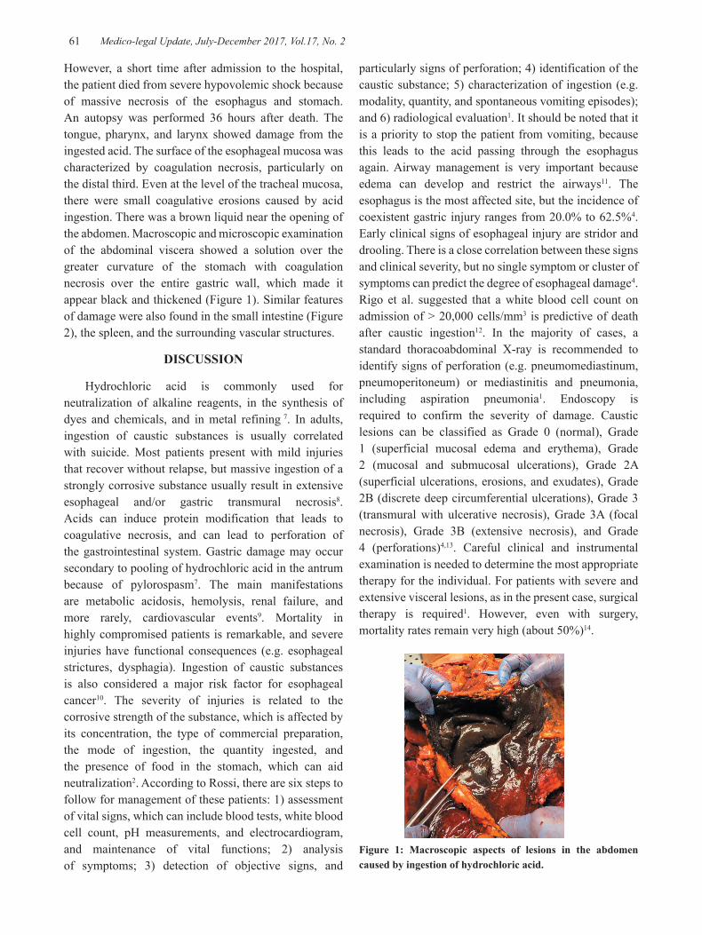
61 Medico-legal Update, July-December 2017, Vol.17, No. 2
However, a short time after admission to the hospital, the patient died from severe hypovolemic shock because of massive necrosis of the esophagus and stomach. An autopsy was performed 36 hours after death. The tongue, pharynx, and larynx showed damage from the ingested acid. The surface of the esophageal mucosa was characterized by coagulation necrosis, particularly on the distal third. Even at the level of the tracheal mucosa, there were small coagulative erosions caused by acid ingestion. There was a brown liquid near the opening of the abdomen. Macroscopic and microscopic examination of the abdominal viscera showed a solution over the greater curvature of the stomach with coagulation necrosis over the entire gastric wall, which made it appear black and thickened (Figure 1). Similar features of damage were also found in the small intestine (Figure 2), the spleen, and the surrounding vascular structures.
DISCUSSION
Hydrochloric acid is commonly used for neutralization of alkaline reagents, in the synthesis of dyes and chemicals, and in metal refining 7. In adults, ingestion of caustic substances is usually correlated with suicide. Most patients present with mild injuries that recover without relapse, but massive ingestion of a strongly corrosive substance usually result in extensive esophageal and/or gastric transmural necrosis8. Acids can induce protein modification that leads to coagulative necrosis, and can lead to perforation of the gastrointestinal system. Gastric damage may occur secondary to pooling of hydrochloric acid in the antrum because of pylorospasm7. The main manifestations are metabolic acidosis, hemolysis, renal failure, and more rarely, cardiovascular events9. Mortality in highly compromised patients is remarkable, and severe injuries have functional consequences (e.g. esophageal strictures, dysphagia). Ingestion of caustic substances is also considered a major risk factor for esophageal cancer10. The severity of injuries is related to the corrosive strength of the substance, which is affected by its concentration, the type of commercial preparation, the mode of ingestion, the quantity ingested, and the presence of food in the stomach, which can aid neutralization2. According to Rossi, there are six steps to follow for management of these patients: 1) assessment of vital signs, which can include blood tests, white blood cell count, pH measurements, and electrocardiogram, and maintenance of vital functions; 2) analysis of symptoms; 3) detection of objective signs, and
particularly signs of perforation; 4) identification of the caustic substance; 5) characterization of ingestion (e.g. modality, quantity, and spontaneous vomiting episodes); and 6) radiological evaluation1. It should be noted that it is a priority to stop the patient from vomiting, because this leads to the acid passing through the esophagus again. Airway management is very important because edema can develop and restrict the airways11. The esophagus is the most affected site, but the incidence of coexistent gastric injury ranges from 20.0% to 62.5%4. Early clinical signs of esophageal injury are stridor and drooling. There is a close correlation between these signs and clinical severity, but no single symptom or cluster of symptoms can predict the degree of esophageal damage4. Rigo et al. suggested that a white blood cell count on admission of > 20,000 cells/mm3 is predictive of death after caustic ingestion12. In the majority of cases, a standard thoracoabdominal X-ray is recommended to identify signs of perforation (e.g. pneumomediastinum, pneumoperitoneum) or mediastinitis and pneumonia, including aspiration pneumonia1. Endoscopy is required to confirm the severity of damage. Caustic lesions can be classified as Grade 0 (normal), Grade 1 (superficial mucosal edema and erythema), Grade 2 (mucosal and submucosal ulcerations), Grade 2A (superficial ulcerations, erosions, and exudates), Grade 2B (discrete deep circumferential ulcerations), Grade 3 (transmural with ulcerative necrosis), Grade 3A (focal necrosis), Grade 3B (extensive necrosis), and Grade 4 (perforations)4,13. Careful clinical and instrumental examination is needed to determine the most appropriate therapy for the individual. For patients with severe and extensive visceral lesions, as in the present case, surgical therapy is required1. However, even with surgery, mortality rates remain very high (about 50%)14.
Figure 1: Macroscopic aspects of lesions in the abdomen caused by ingestion of hydrochloric acid.
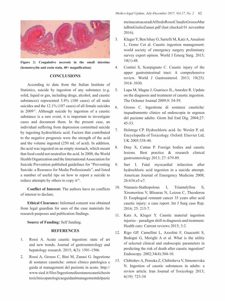
Medico-legal Update, July-December 2017, Vol.17, No. 2 62 63 -legal Update, July-December 2017, Vol.17, No. 2
Figure 2: Coagulative necrosis in the small intestine (hematoxylin and eosin stain, 40× magnification)
CONCLUSIONS
According to data from the Italian Institute of Statistics, suicide by ingestion of any substance (e.g. solid, liquid or gas, including drugs, alcohol, and caustic substances) represented 5.8% (180 cases) of all male suicides and the 12.1% (107 cases) of all female suicides in 200915. Although suicide by ingestion of a caustic substance is a rare event, it is important to investigate cases and document them. In the present case, an individual suffering from depression committed suicide by ingesting hydrochloric acid. Factors that contributed to the negative prognosis were the strength of the acid and the volume ingested (250 mL of acid). In addition, the acid was ingested on an empty stomach, which meant that food could not neutralize the acid. In 2008, the World Health Organization and the International Association for Suicide Prevention published guidelines for “Preventing Suicide: a Resource for Media Professionals”, and listed a number of useful tips on how to report a suicide to reduce attempts by others to copy it16.
Conflict of Interest: The authors have no conflicts of interest to declare.
Ethical Clearance: Informed consent was obtained from legal guardian for uses of the case materials for research purposes and publication findings.
Source of Funding: Self funding.
REFERENCES
1. Rossi A. Acute caustic ingestion: state of art and new trends. Journal of gastroenterology and hepatology research. 2015; 4(3): 1501-1506.
2. Rossi A, Grosso C, Bini M, Zanasi G. Ingestione di sostanze caustiche: sintesi clinico patologica e guida al management del paziente in acuto. http://www.sied.it/files/Ingestionedisostanzecaustichesintesiclinicopatologicaeguidaalmanagementdelpazie
nteinacutoacuradiAlfredoRossiClaudioGrossoMartaBiniGiulioZanasi.pdf (last checked 01 november 2016).
3. Kluger Y, Ben Ishay O, Sartelli M, Katz A, Ansaloni L, Gomz Cet al. Caustic ingestion management: world society of emergency surgery preliminary survey expert opinon. World J Emerg Surg. 2015; 10(1):48.
4. Contini S, Scarpignato C. Caustic injury of the upper gastrointestinal tract: A comprehensive review. World J Gastroenterol. 2013; 19(25): 3918–3930.
5. Lupa M, Magne J, Guarisco JL, Amedee R. Update on the diagnosis and treatment of caustic ingestion. The Ochsner Journal 2009;9: 54-59.
6. Grosso C. Ingestione di sostanze caustiche: inquadramento clinico ed endoscopia in urgenza del paziente adulto. Giorn Ital End Dig. 2004;27:45-53.
7. Holstege CP. Hydrochloric acid. In: Wexler P, ed. Encyclopedia of Toxicology. Oxford: Elsevier Ltd, UK 2005:538-40.
8. Dray X, Cattan P. Foreign bodies and caustic lesions. Best practice & research clinical gastroenterology 2013; 27: 679-89.
9. Sari I. Fatal myocardial infarction after hydrochloric acid ingestion in a suicide attempt. American Journal of Emergency Medicine 2008; 26:634.e5-e7.
10. Ntanasis-Stathopolous I, Triantafyllou S, Xiromeritou V, Bliouras N, Loizou C, Theodorou D. Esophageal remnant cancer 35 years after acid caustic injury: a case report. Int J Surg case Rep. 2016; 25: 215-7.
11. Katz A, Kluger Y. Caustic material ingestion injuries – paradigm shift in diagnosis and treatment. Health care: Current reviews 2015; 3:2.
12. Rigo GP, Camellini L, Azzolini F, Guazzetti S, Bedogni G, Merighi A et al. What is the utility of selected clinical and endoscopic parameters in predicting the risk of death after caustic ingestion? Endoscopy. 2002;34(4):304-10.
13. Chibishev A, Pereska Z, Chibisheva V, Simonovska N. Ingestion of caustic substances in adults: a review article. Iran Journal of Toxicology 2013; 6(19): 723-34

-legal Update, July-December 2017, Vol.17, No. 2 62 63 Medico-legal Update, July-December 2017, Vol.17, No. 2
14. Koschny R, Herceg M, Stremmel W, Eisenbach C. Fatal course of a suicidal intoxication with hydrochloric acid. Case reports in gastroenterology 2013; 7:89-96
15. Istat. I suicidi in Italia. http://www.istat.it/it/archivio/68812 (last checked 01 november 2016).
16. WHO. Preventing Suicide: a Resource for Media Professionals. http://apps.who.int/iris/bitstream/10665/43954/1/9789241597074_eng.pdf (last checked 01 november 2016).
65 -legal Update, July-December 2017, Vol.17, No. 2
Estimation of Stature from the Inter-acromial Length of Adults Belonging to the Soliga, a Genetically Isolated Tribe from
Southern India
Vinay R Hallikeri1, Chandrakant M Kokatanur2, K H Manjulabai3
1Assistant Professor, Anna Medical College, Sans Saunci road, Montangne Blanche, Mauritius. 1Assistant Professor, Anna Medical College, Sans Saunci road, Montangne Blanche, Mauritius. 1
2Associate Professor, Department of Forensic Medicine, Krishna Institute of Medical Sciences Karad, 2Associate Professor, Department of Forensic Medicine, Krishna Institute of Medical Sciences Karad, 2
Maharashtra, 3Professor cum Dean, K.D.Medical College, Hospital & Research Center, Mathura, Uttar Pradesh
BACKGROUND
Stature is an important primary character that defines an individual’s identity and has paramount importance when only a part of the body or a grossly mutilated body is available to the expert for forensic work up. Presently there exist no studies on populations which are ancient, unique, which are geographically, genetically and culturally isolated. Soliga tribal population of Biligiri Rangana Hills (Karnataka) in South India represents one such population. Aim of the study: The purpose of the study is to estimate of stature from the inter-acromial length of adults. Materials and method: Stature and inter-acromial length were measured among 98 Soliga volunteers above 25 years of age. A set of simple regression equations (combined and separate for males and females) of forensic significance were established to estimate the stature from the inter-acromial length Results: The average stature observed was 164.39 cm in males and 152.67 cm in females. The average inter-acromial length was 30.06 cm in males and 26.70 cm in females. Conclusion: - In the present study, regression analysis revealed positive correlation between the stature and the IAL.
Keywords: Forensic anthropology, physical anthropometry, stature, inter-acromial length, Soliga tribal population.
INTRODUCTION
Identification of an individual, living or dead is of paramount importance in forensic practice. Post-mortem identification is essential when dismembered body parts or skeletal remains are received for examination. Eventually one has to define identification features like race, sex, age, and stature. Two major methods exist for estimation of stature: the anatomical method, which requires the presence of a complete skeleton and is more useful in examination and interpretation of archeological remains and has limited or no forensic applicability when the bodies are dismembered or mutilated. Under such scenarios the mathematical method, which requires a complete or a partial long bone or a body part and
Corresponding author: Vinay R HallikeriAssistant Professor, Anna Medical college, Sans Saunci road, Montangne Blanche, Mauritius. Email: [email protected]
employs regression formulae or multiplication factors to estimate the stature based on the correlation of individual measurements to living statures fulfills the forensic need1, 2. Stature of an individual can be estimated if upper portion of the trunk is available without long bones3.
Various dimensions/measurements of the various body parts exhibit biological relationships of different degrees with the stature of an individual. Populations exhibit variations with regard to body proportions because of the various factors like ethnicity, geographical location, nutrition status, physical activity and environment4, 5.
Careful literature review revealed that presently there exist no studies on populations which are ancient, unique, which are geographically, genetically and culturally isolated. These populations are the best candidates for any anthropometric and anthropological study. The Soliga tribal population of Biligiri Rangana Hills in South India represents one such population 6. This paper represents a specific part of a wide
DOI Number: 10.5958/0974-1283.2017.00070.6
65 Medico-legal Update, July-December 2017, Vol.17, No. 2
study - “Anthropometric profile of the Soliga tribal population”. The present paper focuses mainly on relationship between the IAL and stature in the Soliga tribal population, wherein an attempt has been made to develop regression formulae to estimate the stature from this measurement. These findings would be a valuable tool for the anthropologists in general and forensic experts in specific for estimation of stature.
MATERIALS AND METHOD
A total of 98 normal, healthy Soliga tribal volunteers residing at Biligiri Rangana Hills in South India, comprising 46 males and 52 females above the age of 25 completed years were enrolled in this study. Subjects below the age of 25 years were not included since the epiphyseal fusion of the bones is not complete until 25 years (clavicle being the last) 7. The data collection was performed during a 2-week period between 8:00 am and 10:00 am to avoid the influence of diurnal variation 8. Subjects with a history of chronic illness, dwarfism, gigantism, steroidal therapy, trauma, physical deformity, or any surgical procedure that affected the stature or the IAL, were excluded from the study. Written informed consent was obtained for every subject and all female subjects were examined in the presence of another female attendant. The IAL of each subject was measured in centimeters to the nearest millimeter and the stature of each subject was measured in centimeters. All the measurements were performed by the same observer to exclude inter-observer errors.
The stature was measured using a Harpenden portable stadiometer (Holtain Ltd, UK) in centimeters (cm). The subjects were asked to stand barefooted on the stadiometer platform with their feet in close contact, the trunk braced along the vertical board, and the eyes gazing straight ahead. Each subject’s face was placed in the Frankfurt plane, and the measurement was taken by bringing the projecting horizontal sliding bar to the vertex 9.
The subjects were made to stand erect in an anatomical position to measure the IAL. They were then asked to relax their shoulders to the point of slumping forward. Standing behind the subject, the observer felt for the outside edge of the acromial process of the shoulder blade, which could be felt as a ridge just above the shoulder joint 10. This was done on both the sides and the landmarks were marked using a pen. The distance
between the right and the left acromial process of both scapulae, the IAL, was measured by a large pointed spreading caliper (GPM Ltd, Switzerland).
The data were analyzed using the Statistical Package for the Social Sciences (SPSS) version 14 (SPSS, Inc., USA). The intra-observer measurement error and reliability were estimated to be within acceptable standards for all measurements (R > 0.9; rTEM < 5%) 11. The absolute technical error of measurement (TEM) was estimated using the following equation:
TEM=√ED2 /√2N
where D is the difference between each two successive measurements, and N implies the number of individuals tested 11-13. The relative technical error of measurement (rTEM) was estimated from each two successive sets of measurements by dividing the TEM for a given variable by the grand mean of that variable and multiplying the result by 100 11,12. Along with these measurements, the coefficient of reliability (R) was also calculated 14. The means, standard deviations, ranges (minimum and maximum) and differences were estimated. An independent t-test was used to test for differences between mean measurements. The strength of correlation between the stature and the IAL was calculated using Pearson correlation coefficient. Sex-specific and combined simple regression equations for stature estimation were developed using the IAL. The simple linear regression equations for estimation of stature were derived in the form of, Y (stature) = a (constant) + b (regression coefficient of the independent variable) X (individual variable) +/- SEE (standard error of the estimate). A low SEE indicates a higher accuracy. A p value of less than 0.05 was considered statistically significant in the present study. The developed equations were then tested by substituting the minimum, the maximum and the means of the IAL in the respective regression equations to estimate the stature13, 15.
RESULTS
Descriptive statistics for the stature and the IAL of combined and both sexes are as represented in Tables 1 and 2 respectively. When the means of measurements for males and females were compared using a paired t test, all of the measurements were significantly larger for males than for females (p < 0.001).
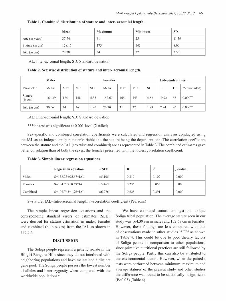
Medico-legal Update, July-December 2017, Vol.17, No. 2 66 67 -legal Update, July-December 2017, Vol.17, No. 2
Table 1. Combined distribution of stature and inter- acromial length.
Mean Maximum Minimum SD
Age (in years) 37.74 61 25 11.59
Stature (in cm) 158.17 175 143 8.00
IAL (in cm) 28.28 34 22 2.53
IAL: Inter-acromial length; SD: Standard deviation
Table 2. Sex wise distribution of stature and inter- acromial length.
Males Females Independent t test
Parameter Mean Max Min SD Mean Max Min SD T Df P (two tailed)
Stature(in cm) 164.39 175 150 5.33 152.67 165 143 5.57 9.92 45 0.000***
IAL (in cm) 30.06 34 26 1.96 26.70 31 22 1.89 7.84 45 0.000***
IAL: Inter-acromial length; SD: Standard deviation
***the test was significant at 0.001 level (2 tailed)
Sex-specific and combined correlation coefficients were calculated and regression analyses conducted using the IAL as an independent parameter/variable and the stature being the dependent one. The correlation coefficient between the stature and the IAL (sex wise and combined) are as represented in Table 3. The combined estimates gave better correlation than of both the sexes, the females presented with the lowest correlation coefficient.
Table 3. Simple linear regression equations
Regression equation ± SEE R r2 p-value
Males S=138.33+0.867*IAL ±5.105 0.319 0.102 0.000
Females S=134.237+0.69*IAL ±5.463 0.235 0.055 0.000
Combined S=102.763+1.96*IAL ±6.276 0.625 0.391 0.000
S=stature; IAL=Inter-acromial length; r=correlation coefficient (Pearsons)
The simple linear regression equations and the corresponding standard errors of estimates (SEE), were derived for stature estimation in males, females and combined (both sexes) from the IAL as shown in Table 3.
DISCUSSION
The Soliga people represent a genetic isolate in the Biligiri Rangana Hills since they do not interbreed with neighboring populations and have maintained a distinct gene pool. The Soliga people possess the lowest number of alleles and heterozygosity when compared with the worldwide populations 6.
We have estimated stature amongst this unique Soliga tribal population. The average stature seen in our study was 164.39 cm in males and 152.67 cm in females. However, these findings are less compared with that of observations made in other studies 10, 17-20 as shown in Table 4. This could be due to poor dietary factors of Soliga people in comparison to other populations, since primitive nutritional practices are still followed by the Soliga people. Partly this can also be attributed to the environmental factors. However, when the paired t tests were performed between minimum, maximum and average statures of the present study and other studies the difference was found to be statistically insignificant (P>0.05) (Table 4).
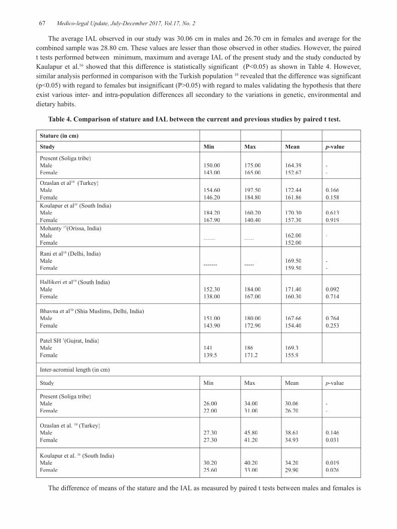
-legal Update, July-December 2017, Vol.17, No. 2 66 67 Medico-legal Update, July-December 2017, Vol.17, No. 2
The average IAL observed in our study was 30.06 cm in males and 26.70 cm in females and average for the combined sample was 28.80 cm. These values are lesser than those observed in other studies. However, the paired t tests performed between minimum, maximum and average IAL of the present study and the study conducted by Kaulapur et al.16 showed that this difference is statistically significant (P<0.05) as shown in Table 4. However, similar analysis performed in comparison with the Turkish population 10 revealed that the difference was significant (p<0.05) with regard to females but insignificant (P>0.05) with regard to males validating the hypothesis that there exist various inter- and intra-population differences all secondary to the variations in genetic, environmental and dietary habits.
Table 4. Comparison of stature and IAL between the current and previous studies by paired t test.
Stature (in cm)
Study Min Max Mean p-value
Present (Soliga tribe)Male Female
150.00143.00
175.00165.00
164.39152.67
--
Ozaslan et al10 (Turkey)Male Female
154.60 146.20
197.50184.80
172.44161.86
0.1660.158
Koulapur et al16 (South India)Male Female
184.20167.90
160.20 140.40
170.30 157.30
0.6130.919
Mohanty 17(Orissa, India)Male Female ------ ----- 162.00
152.00--
Rani et al18 (Delhi, India)Male Female ------- ----- 169.50
159.50--
Hallikeri et al19 (South India)Male Female
152.30138.00
184.00167.00
171.40160.30
0.0920.714
Bhavna et al20 (Shia Muslims, Delhi, India)Male Female
151.00 143.90
180.00172.90
167.66154.40
0.7640.253
Patel SH 3(Gujrat, India)MaleFemale
141139.5
186171.2
169.3155.9
Inter-acromial length (in cm)
Study Min Max Mean p-value
Present (Soliga tribe)Male Female
26.00 22.00
34.0031.00
30.0626.70
--
Ozaslan et al. 10 (Turkey)Male Female
27.3027.30
45.8041.20
38.6134.93
0.1460.031
Koulapur et al. 16 (South India)Male Female
30.2025.60
40.2033.00
34.2029.90
0.0190.026
The difference of means of the stature and the IAL as measured by paired t tests between males and females is

Medico-legal Update, July-December 2017, Vol.17, No. 2 68 69 -legal Update, July-December 2017, Vol.17, No. 2
significant (P<0.05), the values in males being higher. This observation is in accordance to observations made in similar studies 10, 16 and is attributable to direct relationship of body dimensions with sex of an individual, since the difference in the hormonal status results in early hormonal maturity in females in comparison to males, which results in early epiphyseal closure consequently reducing time involved in the linear growth. In the present study, regression analysis revealed positive correlation between the stature and the IAL. The correlation was stronger in males than in females and when combined together a higher correlation was reached and this is in accordance to other similar studies 10, 16. When compared, the magnitude of correlation is comparable to that performed by Ozaslan et al.10 on a Turkish population. However, the values are less when compared to the one conducted by Kaulapur et al. 16 on a South Indian population again supporting the hypothesis of genetic and nutritional factors contributing to the differences. The SEE of the present study and other similar studies are comparable with each other (Table.5)
Table 5. Comparison of the regression coefficient and Standard error of estimate (SEE) between present and previous studies.
Study SEE R
Present study (Soliga tribe)Combined Male Female
±6.276±5.105±5.463
0.6250.3190.235
Patel SH 3(Gujarat, India)CombinedMaleFemale
±7±6±5
0.590.310.23
Ozaslan et al. 10
(Turkey)Male Female
±6.201±6.307
0.4200.260
Koulapur et al. 16 Combined Male Female
±5.±6.±4.
0.7000.5000.600
A total of 3 simple regression equations were derived. However, the regression coefficient and the SEE with respect to the IAL is lower compared to many other body dimensions used in routine forensic practice, like percutaneous upper and lower limb measurements by regression analysis, 16-23 implying
relative inferiority of the IAL as a parameter to estimate stature in comparison to these dimensions. However, the IAL is the best candidate for assessment of stature when only the trunk is available with all the limbs being severed out. To assess the accuracy, the means of the minimum, maximum and average IAL were applied to the respective equations and when the means of the estimated values and original values were compared, statistically insignificant differences (P<0.05) were yielded implying that the equations can be applied to the Soliga tribal population in India within acceptable margins of error for routine forensic practice.
CONCLUSION
In the present study, regression analysis revealed positive correlation between the stature and the IAL. The correlation was stronger in males than in females and when combined together a higher correlation was reached. IAL is the best candidate for assessment of stature when only the trunk is available with all the limbs being severed out.
Conflict of Interest: None declared.
Source of Funding: None
Ethical Clearance: The study was approved by Ethical Committee.
REFERENCES
1. Krishan K, Sharma A. Estimation of stature from dimensions of hands and feet in a North Indian population. J Forensic Leg Med 2007;14:327-32.
2. Lundy JK. The mathematical versus anatomical methods of stature estimate from long bones. Am J Forensic Med Pathol 1985;6:73-6.
3. Patel SH, Bastia BK, Kumar L, Kumaran SM. Estimation of adult human stature from measurements of inter-acromial length in Gujarati population of India. J Indian Acad Forensic Med 2015;37(4):365-8.
4. Katzmarzyk PT, Leonard WR. Climatic influences on human body size and proportions: ecological adaptations and secular trends. Am J Phys Anthropol 1998;106:483-503.
5. Telkka A. On the prediction of human stature from the long bones, Acta Anat (Basel) 1950;9:103-17.
6. Morlote DM, Gayden T, Arvind P, Babu A, Herrera
-legal Update, July-December 2017, Vol.17, No. 2 68 69 Medico-legal Update, July-December 2017, Vol.17, No. 2
RJ. The Soliga, an isolated tribe from Southern India: genetic diversity and phylogenetic affinities. J Hum Genet 2011;56:258-69.
7. Saukko P, Knight B. Knight’s Forensic Pathology. 3rd ed. London: Edward Arnold; 2004.p. 118,121.
8. Krishan K, Kanchan T, Menezes RG, Ghosh A. Forensic anthropology casework: essential methodological considerations in stature estimation. J Forensic Nurs 2012;8:45-50.
9. Vallois HV. Anthropometric techniques. Curr Anthropol 1965;6:127-44.
10. Ozaslan A, Karadayi B, Kolusayin MO, Ahsen K. Stature estimation from bi-acromial and bi-iliocristal measurements. Rom J Leg Med 2011;19:171-6.
11. Ward RE, Jamison PL. Measurement precision and reliability in craniofacial anthropometry: implications and suggestions for clinical applications. J Craniofac Genet Dev Biol 1991;11:156-64.
12. Ulijaszek SJ, Kerr DA. Anthropometric measurement error and the assessment of nutritional status. Br J Nutr 1999;82:165-77.
13. Ahmed AA. Estimation of stature from the upper limb measurements of Sudanese adults. Forensic Sci Int 2013;228:178.e1-7.
14. Jamison PL, Ward RE. Brief communication: measurement size, precision, and reliability in craniofacial anthropometry: bigger is better. Am J Phys Anthrop 1993;90:495-500.
15. Krishan K, Kanchan T, Asha N. Estimation of stature from index and ring finger length in a North Indian adolescent population, J Forensic Leg Med 2012;19:285-90.
16. Koulapur VV, Agarwal SS, Mestri SC. Estimation of stature by anthropometric measurements of inter-acromial length. J Indian Acad Forensic Med 2010;32(2):101-3.
17. Mohanty NK. Prediction of height from percutaneous tibial length amongst Oriya population. Forensic Sci Int 1998;98:137-41.
18. Rani M, Tyagi AK, Verma SK, Kohli A. Estimation of stature from percutaneous measurements of legs. J Forensic Med Toxicol 2004;21:12-4.
19. Hallikeri VR. Estimation of stature by using percutaneous measurements of long bones of the leg and forearm in South Indian population: a cross sectional study. KLE University (Dissertation); 2012.
20. Bhavna S, Nath S, Use of lower limb measurements in reconstructing stature among Shia Muslims. The Internet Journal of Biological Anthropology.2008; 2 (2):1-13. Available from https://print.ispub.com/api/0/ispub-article/12024 [Accessed on 2017 February 5th].
21. Celbis O, Agritmis H. Estimation of stature and determination of sex from radial and ulnar bone lengths in a Turkish corpse sample. Forensic Sci Int 2006;158:135-9.
22. Yili Y. Estimation of stature from tibial length. J Forensic Med 1996;12:87-93.
23. Kate BR, Muzumdar RD. Stature estimation from femur and humerus by regression and autometry. Acta Anat 1976;94:311-20.
24. Bhavna S, Nath S. Estimation of stature on the basis of measurements of the lower limb. Anthropology Today: Trends, Scope and Applications 2007;3:219-22.
71 -legal Update, July-December 2017, Vol.17, No. 2
A Retrospective Study of Exhumation Cases Done at Sri Devaraj Urs Medical College, Kolar over a Period of 3 Years
Murali Mohan MC1, Yadukul S2, Kiran J3
1Assistant Professor, Department of Forensic Medicine & Toxicology, Sri Devaraj Urs Medical College, Kolar, 1Assistant Professor, Department of Forensic Medicine & Toxicology, Sri Devaraj Urs Medical College, Kolar, 1
Karnataka, 2Assistant Professor, Department of Forensic Medicine & Toxicology, Chamarajanagar Institute of 2Assistant Professor, Department of Forensic Medicine & Toxicology, Chamarajanagar Institute of 2
Medical Sciences, Chamarajanagar, Karnataka, 3Professor & Head of the Department, Department of Forensic Medicine & Toxicology, Sri DevarajUrs Medical College, Kolar, Karnataka
ABSTRACT
Background: The term exhumation is restricted to legally digging out the body from the grave after burial. The objectives of disinterment can vary in various cultures as well as countries and requirements vary from place to place. However, most cases of exhumation occur because there is a request from a magistrate to carry out an autopsy to gain essential forensic evidence.
Objective: To make a comprehensive analysis of the exhumation cases those have been done in Kolar & to find out the pitfalls, limitations, lacunae encountered which are subjective as well as objective in nature.
Results: A total of 11 exhumation cases have been done during the study period. Young males in the age group of 21yrs to 30yrs were the major population. Though 10 cases among 11 cases done had alleged that it was a homicide case, prior to exhumation; turned out to be on the other side after the exhumation results.
Conclusion: The study of these cases from registration to reburial has yielded a lot of information from medico legal point of view, as the analytical findings are ac tual proofs rather than analytical presumptions.
Keywords: Exhumation; Autopsy; Kolar.
Corresponding author: Dr. Yadukul S E-mail: [email protected]
INTRODUCTION
Exhumation, also called disinterment or disentombment, refers to the uncovering, digging up or removal of a body from a grave, although those terms can be loosely applied to describe the removal of a body from any tomb below or above ground1. Exhumations performed by forensic anthropologists/ pathologists occur in two general contexts: legally interred bodies and unlawful burials2, 3. Legally interred bodies are burials which have been approved by the authorities and which are buried in a cemetery. Unlawful burials on the other hand involve bodies not found buried in a cemetery and are the result of an individual or group trying to conceal or dispose of a body. Some authors further divide the exhumations of legally interred bodies in two categories:
civil cases and criminal cases3, 4. Exhumations for civil purposes are done to determine if the deceased medical diagnosis or treatment was faulty, if a pre-existing illness affected life expectancy, if conscious pain and suffering was present and for how long, if drugs or alcohol contributed to an accident, if an injury was caused by faulty appliance and so forth3,4. Criminal cases, on the other hand, are done to establish if the cause of death is related to a criminal act, often in the context of new information following the interment.
If an individual died under suspicious circumstances and buried, a legitimate investigating agency (police) may ex hume the body to determine the cause of death. However, Law of land also protects a body interred in ‘consecrated’ ground, and permission must also be sought from the competent authorities before any exhumation can be considered. All the exhumed cases are dealt with under section 176 CrPC. i.e., Executive Magistrate has to conduct inquest5. In India, amongst others, the burial practice and postmortem interval
DOI Number: 10.5958/0974-1283.2017.00071.8
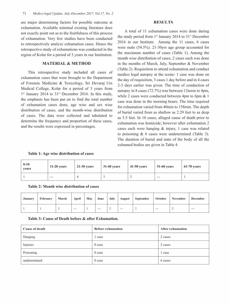
71 Medico-legal Update, July-December 2017, Vol.17, No. 2
are major determining factors for possible outcome at exhumation. Available minimal existing literature does not exactly point out as to the fruitfulness of this process of exhumation. Very few studies have been conducted to retrospectively analyze exhumation cases. Hence the retrospective study of exhumations was conducted in the region of Kolar for a period of 3 years in our Institution.
MATERIAL & METHOD
This retrospective study included all cases of exhumation cases that were brought to the Department of Forensic Medicine & Toxicology, Sri Devaraj Urs Medical College, Kolar for a period of 3 years from 1st January 2014 to 31st January 2014 to 31st st December 2016. In this study, st December 2016. In this study, st
the emphasis has been put on to find the total number of exhumation cases done, age wise and sex wise distribution of cases, and the month-wise distribution of cases. The data were collected and tabulated to determine the frequency and pro portion of these cases, and the results were expressed in percentages.
RESULTS
A total of 11 exhumation cases were done during the study period from 1st January 2014 to 31st January 2014 to 31st st December st December st
2016 in our Institute. Among the 11 cases, 6 cases were male (54.5%). 21-30yrs age group accounted for the maximum number of cases (Table 1). Among the month wise distribution of cases, 2 cases each was done in the months of March, July, September & November (Table 2). Requisition to attend exhumation and conduct medico legal autopsy at the scene- 1 case was done on the day of requisition, 3 cases 1 day before and in 4 cases 2-3 days earlier was given. The time of conduction of autopsy in 8 cases (72.7%) was between 12noon to 4pm, while 2 cases were conducted between 4pm to 6pm & 1 case was done in the morning hours. The time required for exhumation varied from 40min to 158min. The depth of burial varied from as shallow as 2.29 feet to as deep as 5.5 feet. In 10 cases, alleged cause of death prior to exhumation was homicide; however after exhumation 2 cases each were hanging & injury, 1 case was related to poisoning & 6 cases were undetermined (Table 3). The duration of burial and state of the body of all the exhumed bodies are given in Table 4.
Table 1: Age wise distribution of cases
0-10 years 11-20 years 21-30 years 31-40 years 41-50 years 51-60 years 61-70 years
1 --- 4 3 2 --- 1
Table 2: Month wise distribution of cases
January February March April May June July August September October November December
1 1 2 --- 1 --- 2 --- 2 --- 2 ---
Table 3: Cause of Death before & after Exhumation.
Cause of death Before exhumation After exhumation
Hanging 1 case 2 cases
Injuries 0 case 2 cases
Poisoning 0 case 1 case
undetermined 0 case 6 cases
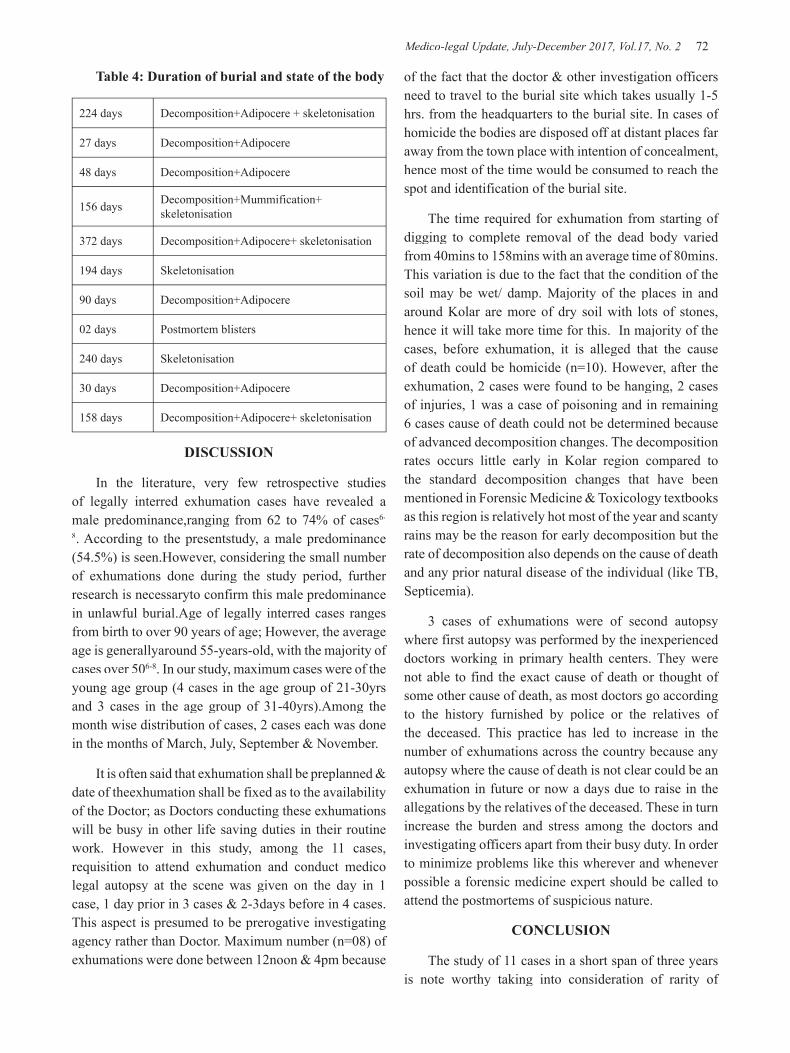
Medico-legal Update, July-December 2017, Vol.17, No. 2 72 73 -legal Update, July-December 2017, Vol.17, No. 2
Table 4: Duration of burial and state of the body
224 days Decomposition+Adipocere + skeletonisation
27 days Decomposition+Adipocere
48 days Decomposition+Adipocere
156 days Decomposition+Mummification+ skeletonisation
372 days Decomposition+Adipocere+ skeletonisation
194 days Skeletonisation
90 days Decomposition+Adipocere
02 days Postmortem blisters
240 days Skeletonisation
30 days Decomposition+Adipocere
158 days Decomposition+Adipocere+ skeletonisation
DISCUSSION
In the literature, very few retrospective studies of legally interred exhumation cases have revealed a male predominance,ranging from 62 to 74% of cases6-
8. According to the presentstudy, a male predominance (54.5%) is seen.However, considering the small number of exhumations done during the study period, further research is necessaryto confirm this male predominance in unlawful burial.Age of legally interred cases ranges from birth to over 90 years of age; However, the average age is generallyaround 55-years-old, with the majority of cases over 506-8. In our study, maximum cases were of the young age group (4 cases in the age group of 21-30yrs and 3 cases in the age group of 31-40yrs).Among the month wise distribution of cases, 2 cases each was done in the months of March, July, September & November.
It is often said that exhumation shall be preplanned & date of theexhumation shall be fixed as to the availability of the Doctor; as Doctors conducting these exhumations will be busy in other life saving duties in their routine work. However in this study, among the 11 cases, requisition to attend exhumation and conduct medico legal autopsy at the scene was given on the day in 1 case, 1 day prior in 3 cases & 2-3days before in 4 cases. This aspect is presumed to be prerogative investigating agency rather than Doctor. Maximum number (n=08) of exhumations were done between 12noon & 4pm because
of the fact that the doctor & other investigation officers need to travel to the burial site which takes usually 1-5 hrs. from the headquarters to the burial site. In cases of homicide the bodies are disposed off at distant places far away from the town place with intention of concealment, hence most of the time would be consumed to reach the spot and identification of the burial site.
The time required for exhumation from starting of digging to complete removal of the dead body varied from 40mins to 158mins with an average time of 80mins. This variation is due to the fact that the condition of the soil may be wet/ damp. Majority of the places in and around Kolar are more of dry soil with lots of stones, hence it will take more time for this. In majority of the cases, before exhumation, it is alleged that the cause of death could be homicide (n=10). However, after the exhumation, 2 cases were found to be hanging, 2 cases of injuries, 1 was a case of poisoning and in remaining 6 cases cause of death could not be determined because of advanced decomposition changes. The decomposition rates occurs little early in Kolar region compared to the standard decomposition changes that have been mentioned in Forensic Medicine & Toxicology textbooks as this region is relatively hot most of the year and scanty rains may be the reason for early decomposition but the rate of decomposition also depends on the cause of death and any prior natural disease of the individual (like TB, Septicemia).
3 cases of exhumations were of second autopsy where first autopsy was performed by the inexperienced doctors working in primary health centers. They were not able to find the exact cause of death or thought of some other cause of death, as most doctors go according to the history furnished by police or the relatives of the deceased. This practice has led to increase in the number of exhumations across the country because any autopsy where the cause of death is not clear could be an exhumation in future or now a days due to raise in the allegations by the relatives of the deceased. These in turn increase the burden and stress among the doctors and investigating officers apart from their busy duty. In order to minimize problems like this wherever and whenever possible a forensic medicine expert should be called to attend the postmortems of suspicious nature.
CONCLUSION
The study of 11 cases in a short span of three years is note worthy taking into consideration of rarity of
-legal Update, July-December 2017, Vol.17, No. 2 72 73 Medico-legal Update, July-December 2017, Vol.17, No. 2
these cases in regular medico legal work. The study of these cases from registration to reburial has yielded a lot of information from medico legal point of view, as the analytical findings are ac tual proofs rather than analytical presumptions.
Conflict of Interest: Nil
Source of Funding: Nil
Ethical Clearance: Taken from the Institute.
REFERENCES
1. Hanzlick, R. Embalming, body preparation, burial, and disinterment. An overview for forensic pathologists. Am. J. Forensic Med. Pathol., 1994, 15, 122-31.
2. Vanezis, P.; Sims, B.G.; Grant, J.H. Medical and scientific investigations of an exhumation in unhallowed ground. Med. Sci. Law, 1978, 18, 209-21.
3. Baden, M.M. Part 4. Exhumation – Time of death and changes after death. In: Spitz, W.U., ed. Spitz and Fisher’s medicolegal investigation of death: guidelines for the application of pathology to crime
investigation. 4th edition. Springfield, IL:Charles C Thomas Publisher LTD, 2006, 174-83.
4. Eckert, W.G.; Katchis, G.S.; James, S. Disinterments. Their value and associated problems. Am. J. Forensic Med. Pathol., 1990, 11, 9-16.
5. Sections 176 of the Code of Criminal Procedure, 1973. Basu’s Criminal Court Handbook containing Criminal Major Acts, 10th Ed 2007. Orient Publishing Company, New Delhi: pp. 134-36, 112-13.
6. Karger, B.; Lorin de la Grandmaison, G.; Bajanowski, T.; Brinkmann, B. Analysis of 155 consecutive forensic exhumations with emphasis on undetected homicides. Int. J. Legal Med., 2004, 118, 90-4.
7. Grellner, W.; Glenewinkel, F. Exhumations: synopsis of morphological and toxicological findings in relation to the postmortem interval. Survey on a 20- year period and review of the literature. Forensic Sci. Int., 1997, 90, 139-59.
8. Stachetzki, U.; Verhoff, M.A.; Ulm, K.; Müller, K.M. MorphologischeBefunde und versicherungsmedizinischeAspektebei 371 Exhumierungen. Pathologe2001, 22, 252-8.
75 -legal Update, July-December 2017, Vol.17, No. 2
Identification and Interpretation of Artefacts Encountered during Medico-legal Autopsies
Anand Kumar Vasudevan1, Stephen Cordner2, Kumar M P3, Vinay J4
1Associate Professor, Department of Forensic Medicine, Sri Muthukumaran Medical College Hospital and 1Associate Professor, Department of Forensic Medicine, Sri Muthukumaran Medical College Hospital and 1
Research Institute, Mangadu, Chennai, India, 2Professor, Forensic Medicine Monash University and Director 2Professor, Forensic Medicine Monash University and Director 2
of Victorian Insititute of Forensic Medicine, 57-83 Kavanagh Street, Southbank, Victoria, Australia, 3Professor and Head, Department of Forensic Medicine, Mysore Medical College, Irwin Road, Mysore, Karnataka, India,
4Assistant Professor, Department of Forensic Medicine, Sri Muthukumaran Medical College Hospital and Research 4Assistant Professor, Department of Forensic Medicine, Sri Muthukumaran Medical College Hospital and Research 4
Institute, Mangadu, Chennai, India
ABSTRACT
Artefacts exist in every branch, unless a professional man recognizes these. The forensic pathologist who conducts medico-legal autopsy will undoubtedly encounter one or the other situations where ones opinions are contrary to which he or she originally believed upon. Post-mortem study of “Identification and Interpretation of Artefacts encountered during Medico-legal Autopsies” was carried out in two places simultaneously, at the Department of Forensic Medicine, Dr.B.R.Ambedkar Medical College, Bangalore, from February 2003 to September 2004. Also, continued at The Victorian Institute of Forensic Medicine, Monash University, Melbourne, Australia by the author from May 20, 2004. A total of 128 (13.64%) artefacts were encountered from the 938 medico-legal autopsies conducted.
Artefacts due to various factors enumerated such as decomposition (31.3%) is the major contributor, followed by burns (15.6%), breaking of rigor due to handling of the bodies and agonal artefacts found respectively (14.8%), artefacts due to animal and insect activity (12.5%), faulty dissection technique led to artefacts (8.5%) and that due to resuscitative techniques resulted in (2.3%) respectively.
As the analysis indicates that the number of artefacts (13.64%) encountered itself speaks the importance of the study. In medico legal cases misinterpretations may turn to have disastrous effects, particularly in an instance where homicide is a possibility. However, these may make the difference between freedom and imprisonment or the life or death sentence for some one.
Keywords: Artefact, Medico-legal Autopsies, investigations
Corresponding author:Anand Kumar VasudevanAssociate Professor, Department of Forensic Medicine, Sri Muthukumaran Medical College Hospital and Research Institute, Mangadu, Chennai, India 600069.Email: [email protected]
INTRODUCTION
Artefacts exist in every branch of the biological sciences, unless the professional man recognizes these artefacts and is able to interpret his findings in proper perspective. Every forensic pathologist who conducts medico-legal autopsies is undoubtedly will have
encountered one or more situations where the findings may prove contrary to which he or she originally believed upon.
In relation to medico legal investigation artefacts may be encountered during the course of Viz. at the crime scene, on autopsy table, on gross and microscopic examination of tissues and during examination of specimen in forensic science laboratories. This is not as easy as it seems to be, for some artefacts can simulate a real pathological or physiological changes so closely that it might be extremely difficult for even the most experienced and skilled forensic pathologist and other forensic scientists to recognize the difference let alone
DOI Number: 10.5958/0974-1283.2017.00072.X
75 Medico-legal Update, July-December 2017, Vol.17, No. 2
the police or the rural medical officers with basic training.
AIMS AND OBJECTIVES OF THE STUDY
This study has been taken up for:
1) A clear appreciation of what constitutes an artefact, which would perhaps help to reduce the number of errors.
2) To expose a post mortem wound which resembles an Ante mortem injury’s features.
3) To suggest measures to prevent Post mortem artefacts that could be caused during autopsy procedure.
4) To throw more light on existing literature.
5) To know the recent advances in minimizing these mistakes.
MATERIALS AND METHOD
Source of Data
The materials utilized for this study were obtained from the cases that were brought to the Department of Forensic Medicine, Dr.B.R.Ambedkar Medical College, Bangalore for Post - mortem Examination from February 2003 to September 2004 for a period of 19 months. Also from cases observed at The Victorian Institute of Forensic Medicine, Australia under the guidance of the Director of Forensic Medicine, Prof. Dr. Stephen Cordner between the period of May 2004 and December 2004.
The bodies were sent by investigating officers from various police stations of Bangalore to know the cause and manner of death and to seek such information from forensic pathologist that can aid in the process of investigation
Method of collection of Data
The post-mortem examination were conducted carefully after collecting various details from requisition forms such as history, injuries, circumstances of death and where ever it was felt necessary, enquiries were made with the investigating officers, emphasizing on the time and date of death being reported to them, place of retrieval of the body Eg: water medium, open field etc., whether medical treatment was instituted prior to death. Also, noted the mode of transport, and whether body was preserved in cold storage or not.
In addition special care was taken in cases where various features / Injuries on the body were caused due to assault, accidents, burns, decomposition, and animal activity which were labelled either as Ante-mortem in nature or post-mortem, under various sections namely 174 (c) Cr.P.C, 201, 302, 376 I.PC. The post-mortem examination was conducted in accordance with the standard procedures and protocols followed in the respective countries.
The nature of the injuries or features whether ante-mortem or post-mortem was determined by examination with a Hand Lens, the characteristics of the injuries were studied carefully with reference to the presence or absence of vital reactions, wound edges, the site of the injuries, the overall state of the body and also by ascertaining the cause and mode of death that was determined after complete autopsy / chemical analysis report and by histopathological report.
Where ever necessary photographs of interesting cases were taken with keeping in mind the various factors as mentioned above.
LIST OF FACTORS:
a) Physiological - During the process of dying.
b) Resuscitative measures
c) Delay in subjecting the bodies for examination
d) Rough handling of the bodies
e) Environmental factors - Heat or cold
f) Animal and insect activity
g) Improper storage of the bodies
h) Faulty Dissection Technique.
OBSERVATIONS
This study consisted of 128 artefacts that were encountered from 938 medico-legal cases. Precisely, 865 medico-legal autopsies conducted in the Department of Forensic Medicine, Dr.B.R.Ambedkar Medical College, Bangalore, from February 2003 to September 2004 and 73 medico-legal cases conducted in The Victorian Institute of Forensic Medicine, Australia between May 2004 and December 2004.
As the analysis indicates that the number of artefacts constitutes 13.64% of total autopsies conducted in the above said period, which speaks of the importance of the study.
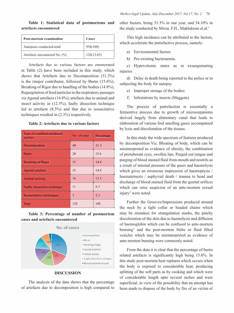
Medico-legal Update, July-December 2017, Vol.17, No. 2 76 77 -legal Update, July-December 2017, Vol.17, No. 2
Table 1: Statistical data of postmortems and artefacts encountered
Post-mortem examination Cases
Autopsies conducted-total 938(100)
Artefacts encountered No. (%) 128(13.65)
Artefacts due to various factors are enumerated in Table (2) have been included in this study, which shows that Artefacts due to Decomposition (31.3%) is the (major contributor, followed by Burns (15.6%), Breaking of Rigor due to handling of the bodies (14.8%), Regurgitation of food particles in the respiratory passages viz Agonal artefacts (14.8%), artefacts due to animal and insect activity in (12.5%), faulty dissection technique led to artefacts (8.5%) and that due to resuscitative techniques resulted in (2.3%) respectively.
Table 2: Artefacts due to various factors
Type of condition produced artefact No. of cases Percentage
Decomposition 40 31.3
Burns 20 15.6
Breaking of Rigor 19 14.8
Agonal artefacts 19 14.8
Animal activity 16 12.5
Faulty dissection technique 11 8.5
Resuscitative techniques 3 2.3
Total 128 100
Table 3: Percentage of number of postmortem cases and artefacts encountered
DISCUSSION
The analysis of the data shows that the percentage of artefacts due to decomposition is high compared to
other factors, being 31.3% in one year, and 34.10% in the study conducted by Mirza. F.H., Makhdoom et.al.1
This high incidence can be attributed to the factors, which accelerate the putrefactive process, namely:
a) Environmental factors
b) Pre-existing bacteraemia.
c) Hypovolemic states as in exsanguinating injuries
d) Delay in death being reported to the police or in subjecting the body for autopsy
e) Improper storage of the bodies
f) Infestations by insects (Maggots).
The process of putrefaction is essentially a fermentive process due to growth of microorganisms derived largely from alimentary canal that leads to elaboration of various foul smelling gases accompanied by lysis and discoloration of the tissues.
In this study the wide spectrum of features produced by decomposition Viz. Bloating of body, which can be misinterpreted as evidence of obesity, the combination of protuberant eyes, swollen lips. Purged out tongue and purging of blood stained fluid from mouth and nostrils as a result of internal pressure of the gases and haemolysis which gives an erroneous impression of haemoptysis / haematemesis / asphyxial death / trauma to head and discharge of blood stained fluid from the genital orifices which can raise suspicion of an ante-mortem sexual injury2 were noted.
Further the Grooves/Impressions produced around the neck by a tight collar or beaded chains which may be mistaken for strangulation marks, the patchy discoloration of the skin due to haemolysis and diffusion of haemoglobin which can be confused to ante-mortem bruising3 and the post-mortem blebs or fluid filled vesicles which may be misinterpreted as evidence of ante mortem burning were commonly noted.
From the data it is clear that the percentage of burns related artefacts is significantly high being 15.6%. In this study post-mortem heat ruptures which occurs when the body is exposed to considerable heat, producing splitting of the soft parts as by cooking and which were of considerable length upto several inches and were superficial, in view of the possibility that an attempt has been made to dispose of the body by fire of an victim of
-legal Update, July-December 2017, Vol.17, No. 2 76 77 Medico-legal Update, July-December 2017, Vol.17, No. 2
wounds special care was taken in the interpretation of these changes which simulate incised wounds/lacerated wounds.4
Heat ruptures were commonly noted over the shoulder blades, flexor aspects of both upper and lower limbs and over the front of thighs. Heat fractures and charring of the outer table of the skull were encountered in some cases. The combination of artefactual hemorrhage and fracture can pose a most difficult situation.
5some cases. The combination of artefactual hemorrhage
5some cases. The combination of artefactual hemorrhage
The contributing factors to the high percentage in Bangalore city, India were
a) Self-immolation being the common form of suicide.
b) Homicidal burns as in dowry deaths
c) Accidental burns sustained while cooking with Kerosene pump stoves.
Estimation of time since death is important from medico-legal point of view, which is determined during autopsy examination by the presence or absence of rigor mortis.
However due to:
a) Rough handling of the bodies
b) During removal of clothing from the body and
c) While shifting the bodies holding the upper limbs, the rigor mortis may be broken at the shoulder or elbow which may lead to a false estimation of time since death but the presence of rigor in the jaw and lower limbs helps in rectifying the mistake. The percentage being 14.8% and 16.15% in the study by Mirza. F.H., Makhdoom et.al.1 Notably, bodies preserved in cold storage were not included in this study.
Presence of food particles in the respiratory passages may lead to misinterpretation as death due to choking. However in this study and in the study conducted by Dr. Abdullah Fatteh3, coincidental presence of food particles in the respiratory passages were seen in deaths with a distinctive cause Viz - In Natural deaths-due to coronary artery disease, in lung infections like pneumonia and in un-natural deaths due to Automobile accidents, asphyxial deaths as in hanging and in cases subjected to external cardiac massage.
Absence of inflamed mucosa, tracheo-bronchial secretions and just the mere presence of food particles in the upper respiratory passage which could not be seen in terminal bronchioles clinched the diagnosis and the findings were attributed as an agonal artefact which accounted to 14.8% and were 23.5% in Dr. Abdullah Fatteh’s study.6
Contribution from animal and insect activity accounted for 12.5% as against 19.65% in Mirza. F.H., Makhdoom et.al study.1
Artefacts due to ant-bite marks which resemble abrasions were the most common feature encountered, resulting in distinctive patterns of injuries which were dry, brown with irregular margins seen commonly over the moist parts of the body viz-around the eyes, armpit, groin, along the lining of clothes and in areas of hypostasis as in cases of hanging the oozing of blood from the bitten or ruptured petechiae over the lower limbs led to vertical streaks of blood staining the skin and soles of feet, also artefactual ligature marks were produced around the neck.
The author Alan R. Moritz7opines that post-mortem
animal activity is more commonly associated with misjudgement and is particularly so with insect eating of skin which may be mistaken for human bite marks or ligature strangulation patterns.
In bodies retrieved from water or aquatic environments, loss of soft tissues due to the aquatic animal bites was seen around the eyes, lips, and ear lobes and over the hands and feet.
Rodent activity resulted in loss of tissue mainly over the face and in one case the whole of the right eye and part of the tissue over the nose were missing leaving characteristic teeth marks and also nibbling activity over the fingers and toes without any bleeding were seen.
An awareness of the existing fauna from where the body was retrieved viz-aquatic medium or open fields, presence of insects or maggots on the body and a knowledge of the distinctive characteristics produced by them with absence of extravasations of blood and a distinctive cause of death, helped in opining the features as post-mortem in nature due to animal or insect activity.
The author Hugland W.D.8 analysed human remains recovered from aquatic environments and observed that

Medico-legal Update, July-December 2017, Vol.17, No. 2 78 79 -legal Update, July-December 2017, Vol.17, No. 2
apart from the fish, crab, turtles which gnaw soft tissues around the eyes, ears, mouth, genitalia and arms, and also injuries caused by propellers, he concluded that post-mortem intervals based on the condition of the body at the time of recovery followed a particular sequence.
The initial disappearance of soft tissues, resulting in exposure of bones occurred in the area thinly overlain by soft tissues, exposing the bones of the hands and wrists, bones of the feet and ankles, the mandible and then the Cranium. The lower legs, fore arms and the arms were the next units to follow, and concluded that this knowledge of disarticulation sequences ranging from weeks to a few months helps in the interpretation of artefacts and the events produced by aquatic environment.
In a study conducted by A.Keith Mant9
states that after death, the blood in the vessels is in a stagnant condition and remains for a time in fluid state and if autopsy is carried out in the first few hours’ one important area where post-mortem artefactual bleeding may give rise to suspicion is in the neck.
During the process of dissection, extravasations of blood in the neck tissues resembling ante-mortem extravasations of blood, Subluxation of cornua of hyoid bone and injury to the thyroid cartilage during the removal of the neck structures and injuries to the visceral organs viz-lungs, heart and liver by the dissecting knife were commonly noted and accounted for 8.5%.
Multiple injection marks over the pericardial region, Defibrillators marks simulating ring like contusions, extravasations of blood in the soft tissues of chest due to external cardiac massage and in one case fracture of the left side ribs were noted.
A history of prior admission to hospital where resuscitative measures were taken up helped in opining them as artefactual. The percentage was 2.3% in our study however it was 3.50% in the Mirza. F.H., Makhdoom et.al study.1
CONCLUSION
From the various artefacts observed, enumerated and discussed in this study, the essence lies in the recognition of pit falls in the practice of forensic pathology and separation of significant ante mortem changes from the inconsequential post mortem artefacts.
In medico legal cases misinterpretations may turn
to have disastrous effects, particularly in an instance where homicide is a possibility one should be aware of the artefacts, for an autopsy finding is so important that it may make the difference in judgement between freedom and imprisonment or the life or death sentence for some one.
Hence it is the duty of the forensic pathologist to recognize and interpret the findings correctly or else misinterpretation or wrong conclusions may lead to;
a) A wrong cause of death.
b) A wrong manner of death.
c) Undue suspicions of criminal offence
d) Non-detection of a homicide.
e) Unnecessary spending of time and effort as a result of misleading findings.
In addition, it must be stressed that a mistake made by pathologist in the interpretation of an artefact may haunt him later in the court, for many a lawyer knows about the pit falls and may attempt to shake the testimony of the forensic pathologist.
Ethical Clearance- Obtained from ethical committee from Victorian Institute of Forensic Medicine, Monash University, Australia. [Dated May 20,2004]
Source of Funding- Self
Conflict of Interest – Nil
Acknowledgment: Nil
REFERENCES
1. Mirza. F.H., Makhdoom et.al - Importance of correct interpretation of post-mortem artefacts in medico-legal autopsies. Journal of Pakistan Medical Association, Page 49-50 (1998).
2. K. Kibayash, n. Tobata et. al - Immunohistochemical demonstration of bleeding in decomposed bodies by using anti-glycophorin - A, monoclonal antibody. JAPAN - Forensic Science International Page 1-8, Jan (1997).
3. Grellner. et. al, - Quantitative alternation of DNA content in strangulation marks is an artefact. Forensic Science International Page 15-20, Sep (1997).
4. Michael Bohnert et. al - Fractures of the base of skull in charred bodies- post mortem heat injuries

-legal Update, July-December 2017, Vol.17, No. 2 78 79 Medico-legal Update, July-December 2017, Vol.17, No. 2
or signs of mechanical traumatization - Forensic Science International Page 55-62 (1997).
5. J.K.Mason – Forensic Medicine for lawyers, 2nd
Edition, Page 50, 52, 141 – 142.
6. Dr. Abdullah Fatteh – Artefacts in Forensic Pathology Page. 42 – 71, [1966]
7. Alan. R. Moritz - Animal activity after death. Pathology of trauma. Edited by J.K. Mason chapter -18 Pages 227 - 231.
8. Huglund W. D. - Contribution of Rodents to Post-mortem artefacts of bone and soft tissues. King county medical examiner’s office. Seattle U.S.A. Journal of Forensic Science. Page 6, Nov (1992).
9. Prinsloo, Gordon. Keith Mant - Taylofs Principle and practice of medical jurisprudence 13th edition. Page 139.
81 -legal Update, July-December 2017, Vol.17, No. 2
Pattern of Fingerprint Pattern and Ridge Density in Unidentified Bodies – An Autopsy Study
C N Sumangala1, Ginto Antony Gilbert2, Thomas John2, Bheemappa Havanur3
1Associate Professor, 1Associate Professor, 1 2PostGraduate Student, 2PostGraduate Student, 2 3Professor & Head Department of Forensic Medicine, Bangalore Medical College & Research Institute, Bangalore
ABSTRACT
The present study was undertaken to determine the fingerprint pattern, and gender determination using finger print ridge density. The fingerprints were collected from 100 unidentified bodies (60 males and 40 females) autopsied at Victoria and Bowring and Lady Curzon hospitals in Bangalore by simple random technique in the age group of 18-60 years. After taking fingerprints, each fingerprint is analysed using a magnifying lens to determine its pattern and then the ridges were counted in the upper portion of the radial border of each print for all ten fingers and mean value is calculated. It represents the approximate number of ridges for that particular individual. The present study shows that loops were the most common pattern followed by whorls and arches in both hands in males and females. A fingerprint with a ridge count of <=14 ridges/25 mm2 is more likely to be of male origin and that of >=15 ridges/25 mm2 is likely to be of female origin. The results of the study thereby support the hypothesis that women have a statistically significant greater ridge density than men.
Keywords: unidentified bodies, fingerprint, ridge density
INTRODUCTION
In India, millions of people are homeless. As these individuals have no close acquaintances, in the event of death, their bodies remain unclaimed. These unclaimed corpses pose a major challenge for the local police to establish identity. Identification means determination of individuality and it is one of the main concerns in forensic investigation. Fingerprint patterns are genotypically determined and remain unchanged from birth till death1 and hence it is considered to be one of the best and cheapest proofs of identification. The present study was undertaken to determine the fingerprint pattern, and gender determination using finger print ridge density among unidentified bodies.
MATERIALS AND METHOD
The study was conducted on 100 unidentified dead bodies (60 males and 40 females) brought for autopsy
Corresponding author :Dr. Ginto Antony GilbertEmail – [email protected] Ph no- 9535167518
to the Department of Forensic Medicine, Bangalore Medical College and Research Institute (BMC&RI). In this study, the subjects (unidentified bodies) were chosen randomly in the approximate age group of 18-60 years from Bangalore, in the state of Karnataka. Both the hands of the study subjects were washed, cleaned with water and spirit and then dried to remove dirt or grease. If rigor mortis of the fingers is present, the rigor is broken by applying force. The fingerprints were collected using Glass slab-Inking Roller method.2 For collection of fingerprint, a plain glass plate is cleaned thoroughly, and smeared with a uniform layer of black printing ink using the roller. The subject’s finger bulbs are gently pressed on the smeared glass plate and transferred to the prepared fingerprint card, keeping in mind to minimise dimensional artefact. The prints are taken with the fingers applied with regular pressure on the proforma. The fingerprints of all ten fingers of all the subjects were prepared. Only plain prints were taken (no roll prints). After taking the fingerprints the pattern of fingerprints in each finger is carefully analysed using a magnifying lens.
The upper portion of the radial border of each print
DOI Number: 10.5958/0974-1283.2017.00073.1

81 Medico-legal Update, July-December 2017, Vol.17, No. 2
was chosen as an area for the data collection because all fingerprint pattern types showed a similar ridge flow in this region. In this selected area of the prints, epidermal ridges of both males and females were counted carefully within a square of 5mm x 5mm drawn on a transparent film fixed to the lens. Counting was started from one corner of the square to the diagonally opposite corner. Some specific criteria were observed during the counting procedure such as the dots, which were not counted, and the handle of the fork and a lake was counted as two ridges. Hence this value represented the number of ridges/25 mm square and would reflect the ridge density value. After the ridge counts were done for all the ten fingers, the mean value is calculated. This new value represented the approximate number of ridges for that particular individual. The significance of this value was determined.
Specific comparisons of means were made and calculations were performed using SPSS software on HP computer. The likelihood ratio (LR) was calculated to obtain the probability inferences of gender, based on ridge density values.
The likelihood ratio is based on Baye’s theorem3
- LR =
PROBABILITY OF GIVEN FINGERPRINT ORIGINATING FROM MALE CONTRIBUTOR (C)
PROBABILITY OF GIVEN FINGERPRINT ORIGINATING FROM FEMALE CONTRIBUTOR (C’)
The favoured odds for support of the most likely hypothesis for a given ridge density is also calculated.
RESULTS AND OBSERVATIONS
A total of 1000 fingerprint patterns were studied. Loops were the most common pattern (63.2%) followed by whorls (28.2%) and arches (8.6%) in both hands among the study population. Loops were more common in females(65%) than males(61.3%).While loops were the predominant patterns on the middle, ring and little fingers, predominance of whorls on thumb, and arches on index finger was noted in both the sexes. There was insignificant difference in overall distribution of fingerprint pattern in both hands among males and females.
Table - 1: Distribution of fingerprint pattern in right and left hands in males.
DIGIT N Whorls Loops Arches
Thumb
Right 60 29(48.3%) 30(50%) 1(1.7%)
Left 60 32(53.3%) 26(43.4%) 2(3.3%)
R+L 120 61(50.8%) 56(46.7%) 3(2.5%)
Index
Right 60 19(31.7%) 20(33.3%) 21(35%)
Left 60 17(28.3%) 21(35%) 22(36.7%)
R+L 120 36(30%) 41(34.2%) 43(35.8%)
Middle
Right 60 13(21.7%) 46(76.7%) 1(1.7%)
Left 60 14(23.3%) 45(75%) 1(1.7%)
R+L 120 27(22.5%) 91(75.8%) 2(1.7%)
Ring
Right 60 15(25%) 44(73.3%) 1(1.7%)
Left 60 16(26.7%) 43(71.7%) 1(1.7%)
R+L 120 31(25.8%) 87(72.5%) 2(1.7%)
Little
Right 60 12(20%) 47(78.3%) 1(1.7%)
Left 60 14(23.3%) 46(76.7%) 0(0%)
R+L 120 26(21.7%) 93(77.5%) 1(0.8%)
All fingers
Right 300 88(29.3%) 187(62.4%) 25(8.3%)
Left 300 93(31%) 181(60.3%) 26(8.7%)
R+L 600 181(30.2%) 368(61.3%) 51(8.5%)
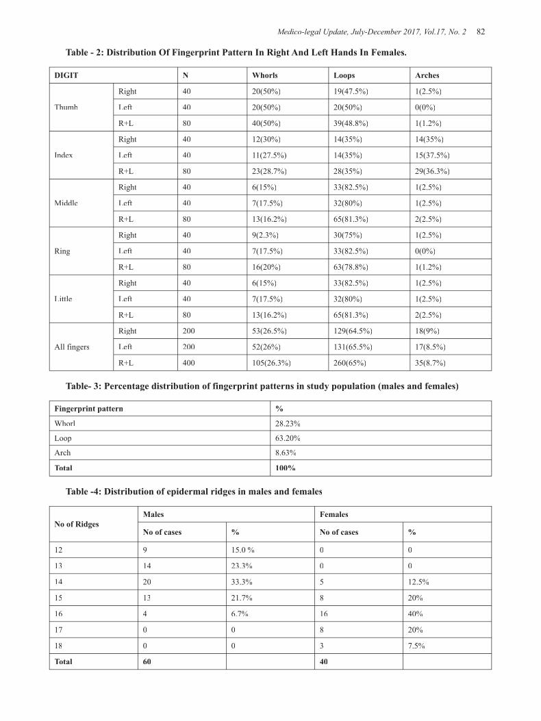
Medico-legal Update, July-December 2017, Vol.17, No. 2 82 83 -legal Update, July-December 2017, Vol.17, No. 2
Table - 2: Distribution Of Fingerprint Pattern In Right And Left Hands In Females.
DIGIT N Whorls Loops Arches
Thumb
Right 40 20(50%) 19(47.5%) 1(2.5%)
Left 40 20(50%) 20(50%) 0(0%)
R+L 80 40(50%) 39(48.8%) 1(1.2%)
Index
Right 40 12(30%) 14(35%) 14(35%)
Left 40 11(27.5%) 14(35%) 15(37.5%)
R+L 80 23(28.7%) 28(35%) 29(36.3%)
Middle
Right 40 6(15%) 33(82.5%) 1(2.5%)
Left 40 7(17.5%) 32(80%) 1(2.5%)
R+L 80 13(16.2%) 65(81.3%) 2(2.5%)
Ring
Right 40 9(2.3%) 30(75%) 1(2.5%)
Left 40 7(17.5%) 33(82.5%) 0(0%)
R+L 80 16(20%) 63(78.8%) 1(1.2%)
Little
Right 40 6(15%) 33(82.5%) 1(2.5%)
Left 40 7(17.5%) 32(80%) 1(2.5%)
R+L 80 13(16.2%) 65(81.3%) 2(2.5%)
All fingers
Right 200 53(26.5%) 129(64.5%) 18(9%)
Left 200 52(26%) 131(65.5%) 17(8.5%)
R+L 400 105(26.3%) 260(65%) 35(8.7%)
Table- 3: Percentage distribution of fingerprint patterns in study population (males and females)
Fingerprint pattern %
Whorl 28.23%
Loop 63.20%
Arch 8.63%
Total 100%
Table -4: Distribution of epidermal ridges in males and females
No of RidgesMales Females
No of cases % No of cases %
12 9 15.0 % 0 0
13 14 23.3% 0 0
14 20 33.3% 5 12.5%
15 13 21.7% 8 20%
16 4 6.7% 16 40%
17 0 0 8 20%
18 0 0 3 7.5%
Total 60 40

-legal Update, July-December 2017, Vol.17, No. 2 82 83 Medico-legal Update, July-December 2017, Vol.17, No. 2
Table 4 shows that males tend to have a lower ridge density as compared to females. It is clear from the above table that males with ridge count <=14 ridges/25mm2 constitute 71.7% of the total males. Females with ridge count >=15 ridges/25 mm2 constitute 87.5 % of the total female subjects.
No male subjects were found with ridge density more than 17 ridges/25 mm2 and no female subjects were found with ridge density less than 13 ridges/25 mm.2
Table 5 (below) shows the descriptive statistics of dermal ridges for males and females. From the table it is clear that ridge density ranges from 12-16 ridges/25 mm2
in males and 14-18 ridges/25 mm2 in females. The mean value for ridge density for male is 13.82 and the mean value for ridge count for females is 15.90. Statistical
analysis was done by using Student’s t-test and results show that males have significantly lesser ridge density than females with P<0.001 (Highly significant).
Table-5: Descriptive statistics and gender comparison of ridge density
MALE FEMALE
Mean Ridge density 13.82 15.90
Standard Deviation 1.14 1.10
Standard Error 0.15 0.17
Median 14 16
Sample Variance 1.30 1.22
P(T<=t) two-tail 0.0000000000000289
Table -6: Probability densities and likelihood ratios derieved from observed ridge count
Ridge CountProbability Density LR
(C/C’)LR(C’/C)
Favoured Odds
Males(C) Females(C’) Males Females
12 0.15 0.00 NA 0.00 1.00 0.00
13 0.23 0.00 NA 0.00 1.00 0.00
14 0.33 0.125 2.6 0.38 0.8 0.2
15 0.22 0.2 1.1 0.91 0.62 0.38
16 0.07 0.4 0.18 5.71 0.2 0.8
17 0.00 0.2 0.00 NA 0.00 1.00
18 0.00 0.075 0.00 NA 0.00 1.00
Table – 6 shows the Probability Density for males(C) and females (C’) and using these values Likelihood Ratio LR (C/C’) and (C’/C) were calculated.
Results show that fingerprints with Ridge density of <=14 ridges/mm2 is more likely to be of male. Similarly fingerprints with Ridge density of >=15 ridges/mm2 is more likely to be of female.
The LR value for males decreases as we move from ridge counts 12 to 18. It is very high for ridge counts 12 and 13 as only males were found to have this ridge count. It becomes 0 for ridge counts 17 and 18 as no males were found to have ridge densities 17 and 18. Similarly LR value for females increases as we move from ridge counts 12 to 18. It is 0 for ridge counts 12 and 13 as there are no females with ridge count 12 and 13. It is very high for ridge counts 17 and 18 as only females were found to have this ridge count.
Based on posterior probability and favoured odds, for example if a fingerprint with a ridge density of 14 is found at a crime scene, then the ratio for it to be male compared to female will be 0.8:0.2 i.e. roughly 4:1.
DISCUSSION
This study shows that the most common fingerprint pattern among the study population is loops followed by whorls and arches. It also shows that males have significantly lesser ridge density as compared to females. It shows similar trend as other studies conducted on Indians as well as other races.
The predominant fingerprint pattern in the present study was loops, followed by whorls and arches which is in accordance with the studies conducted by Gangadhar et al.4 among the Adikarnataka population of Mysore city of Karnataka State, Namouchi et al.5 among
Medico-legal Update, July-December 2017, Vol.17, No. 2 84 85 -legal Update, July-December 2017, Vol.17, No. 2
Tunisians, Quazi et al.6 among American Blacks, Nithin et al.7 among South Indians of Mysore, Wijerathne et al.8 among Sinhalese population, Jaja and Igbigbi9
among people of Ijaw ethnicity (Southern Nigerians), and Igbigbi and Msamati10 among Indigenous black Zimbabweans.
The results of the study are in contrast to the studies conducted by Ching Cho11among Samoans of New Zealand, Banik et al.12 among Rengma Nagas of Nagaland, Tiwari et al.13 among Tibetans, Ghosh et al.14
among the Sunni Muslims of West Bengal and Biswas15 among Dhimals of North Bengal where the researchers reported whorls to be the most common pattern, followed by loops and arches in both hands of male and females. These differences might be due to the geographic and racial variation.
The present study shows that males have a lower ridge density than females which is in accordance with the studies conducted by Mark Acree16 in Caucasian and African American males and females, Dr. Sudesh Gungadin17 in South Indian population from Karnataka, Gutiérrez et al.18 in Spanish Caucasian population, Suthiprabha in Thai population, Nayak V.C. et al.19 in Chinese and Malaysian population.
The results of the study are in contrast to the studies conducted by Chris C Plato et al.20 in American Caucasians, Florence S Steinberg et al21 in American Negros and Reddy22 among tribal population of Andhra Pradesh, India. These results could be due to some defect in counting method as no details of the counting method are mentioned.
The present study has proved that there is an increased ridge density in females compared to males because of less coarseness of ridges. The study of sex identification by ridge density is highly significant (P <0.001).
CONCLUSION
The present study has shown that frequency distribution of fingerprint patterns among unidentified bodies from Bangalore is similar in most aspects to other population groups. Frequency of loops is the highest followed by whorls and arches in both males and females and whorls are more common in thumb and arches are more common in index fingers. Distribution of fingerprint patterns was similar on both hands for males
and females. Gender-based differences in fingerprint pattern could not be established.
Women have more ridge density than men. No female was found to have ridge density <=13 ridges/25mm2 and no male was found to have ridge density >=17 ridges/25mm.2 A fingerprint ridge density <=14 ridges/25mm2is more likely to be of male origin and >=15 ridges/25mm2 is more likely to be of female origin. Similar studies in other population groups are desirable for better correlation
Conflict of Interest – None
Source of Funding - Self
Ethical Clearance – Not Applicable
REFERENCES
1. Vij K. Textbook of Forensic Medicine and Toxicology. 3rd ed. New Delhi: Elsevier; 2005. p. 89-91.
2. Nadar DS. Forensic Science in crime investigation. 3rd ed. Hyderabad: Asia Law House; 2002. p. 48-9.
3. Grieve MC, Dunlop J. A practical aspect of the Bayesian interpretation of fibre evidence. J Forensic Sci Soc 1992;32:169-75.
4. Gangadhar MR, Reddy R. Finger dermatoglyphics of Adikarnatakas: A scheduled caste population of Mysore city, Karnataka. Man in India 1983; 83(1&2):183-93.
5. Namouchi I. Anthropological significance of dermatoglyphic trait variation: an intra-Tunisian population analysis. Int J Mod Anthropol 2011;1:12–27.
6. Qazi QH, Mapa HC, Woods J. Dermatoglyphics of American blacks. Am J Phys Anthropol 1977;47:483–7.
7. Nithin MD, Balaraj BM, Manjunatha B, Mestri SC. Study of fingerprint classification and their gender distribution among South Indian population. J Forensic Leg Med 2009;16(8):460-3.
8. Wijerathne BTB, Rathnayake GK, Adikari SC, Amarasinghe S, Abhayarathna PL, Jayasena AS.Sexual dimorphism in digital dermatoglyphic traits among Sinhalese people in Sri Lanka. J Physiol Anthropol 2013;32(27):1–9.
-legal Update, July-December 2017, Vol.17, No. 2 84 85 Medico-legal Update, July-December 2017, Vol.17, No. 2
9. Jaja BN, Igbigbi PS. Digital and palmar dermatoglyphics of the Ijaw of Southern Nigeria. Afr J Med Med Sci 2008; 37(1):1–5.
10. Igbigbi PS, Msamati BC.Palmar and digital dermatoglyphics of indigenous black Zimbabweans. Med Sci Monit Int Med J Exp Clin Res 2002;8:CR757-61.
11. Ching Cho. A finger dermatoglyphics of the New ZealandSamoans. Korean J Biol Sci 1998;2:507–11.
12. Banik SD, Pal P, Mukherjee DP. Finger dermatoglyphic variations in Rengma Nagas of Nagaland India. Coll Antropol 2009; 33(1):31–5.
13. Tiwari SC, Chattopadhyay PK. Finger dermatoglyphics of the Tibetans. Am J Phys Anthropol 1967;26:289–96.
14. Ghosh JR, Chatterjee M, Wasim R, Bandyopadhyay A R. Study on finger and palmar dermatoglyphics among the Sunni muslims of West Bengal. Anthropologist 2011; 13(2):107-9.
15. Biswas S. Finger and palmar dermatoglyphic study among the Dhimals of North Bengal, India. Anthropologist 2011;13(3):235–8.
16. Acree MA. Is there a gender difference in fingerprint technology. Forensic Sci Int 1999;102(1):35-44.
17. Gungadin S.Sex Determination from fingerprint ridge density. IJMU 2007 Jul-Dec;2(2):4-7.
18. Gutiérrez-Redomero E, Alonso C, Romero E, Galera V. Variability of fingerprint ridge density in a sample of Spanish Caucasians and its application to sex determination. Forensic Sci Int 2008 Aug 25;180(1):17-22
19. Nayak VC, Rastogi P, Kanchan T, Yoganarasimha K, Kumar GP, Menezes RG. Sex differences from fingerprint ridge density in Chinese and Malaysian population. Forensic Sci Int 2010 Apr 15;197(1-3):67-9.
20. Plato CC, Cereghino JJ, Steinberg FS. The dermatoglyphics of American Caucasians. Am J Phys Anthrop 1975;42:194-210.
21. Steinberg FS, Cereghino JJ, Plato CC. The dermatographics of American Negros. AmJ Phy Antrhop1975;42:183-93.
22. Reddy CC. Finger dermatoglyphics of the Bagathas of Araku valleyIndia. Am J Phys Antrhopol 1975;2:225-8.
87 -legal Update, July-December 2017, Vol.17, No. 2
Teaching Forensic Medicine for Undergraduate Medicine Student at College of Medicine, King Faisal University,
Al-Ahsa, Saudi Arabia
Imran Sabri
Assistant Professor, Forensic Medicine Division,Bio-Medical Sciences, College of Medicine, King Faisal University, P.O. Box No. 400 Al-Ahsa-[31982]. KSA
ABSTRACT
In Saudi Arabia, there are more than 30 universities with medical colleges. About 60% teach forensic medicine to undergraduate medical students. The content of this article is based on traditional curriculum, which is the same as taught in other universities of the Kingdom. Forensic Medicine is a one credit hour course with 15 contact hours. Level of the course offered is 4th year. In this article we mentioned the Forensic Medicine teaching for traditional curriculum at College of Medicine, King Faisal University, Al-Ahsa, as per course specification prescribed by The National Commission for Academic Accreditation and Assessment [NCAAA] Educational Evaluation Commission for Higher Education Sector [EEC-HES]
Keywords: forensic medicine, Saudi, Undergraduate, Medical.
INTRODUCTION
In Saudi Arabia, there are more than 30 universities with medical colleges. About 60 % teach forensic medicine to undergraduate medical students. There is variation in teaching methods, years where the course is taught and length of the course1. It is not possible to define clearly what is the actual status of teaching forensic medicine. The College of Medicine, King Faisal University has adopted new integrated curriculum in 2012, The content of this article is based on traditional curriculum, which is the same as taught in other universities of the Kingdom. The forensic medicine undergraduate course is important for medical students. We recommended that it be taught at the undergraduate level because it is the only academic source for forensic medicine that physicians may encounter in their careers2.
Forensic Medicine at College Of Medicine, King Faisal University, Al-Ahsa, as per course specification prescribed by The National Commission for Academic Accreditation and Assessment [NCAAA]/Educational Evaluation Commission for Higher Education Sector [EEC-HES]3
Forensic Medicine is a one credit hour course with 15 contact hours. Level of the course offered is 4th year. The pre-requisite to take the course is pathology in previous year. Methods of assessment is by Student assignments, Continuous assessment, Final term exam (Essay and MCQ ).
Main learning outcomes for students enrolled in forensic medicine course.3
At the completion of the course, the student will be able to;
• Describe the medical matters that come in relation to the law statement of the living and dead
• Describe the mechanical and injuries of the weapons
• Describe the different types of the injuries
• Understand the forensic medical viewpoint of Crime Scene
• Describe the principle of Autopsy
• Understand some effect of poisons
• Understand the main principle of Toxicology
• Understand the principle of human live and deceased Identification
DOI Number: 10.5958/0974-1283.2017.00074.3

87 Medico-legal Update, July-December 2017, Vol.17, No. 2
• Be aware about the sexual offences
• Understanding what is the meaning of the malpractice
• Understand the meaning of somatic death
• Understand the signs of death and the postmortem changes
The main topics to be covered are Death and brain death, Post-mortem changes, Wound and the role of physician in dealing with Head injuries, Firearm injuries, Injuries Due to physical Agent ( Bump), Burn injuries, Death due to electrical Burn, Asphyxia (general aspect), Compression on the neck, Sexual offences, Criminal Abortion, Infanticide, Introduction to clinical toxicology, Identification, Child Abuse, Drowning. One contact hour is allotted to each topic.
Recommended Books are Simpson`s Forensic Medicine, Knight`s Forensic Medicine, DiMaio`s Forensic Pathology, Color Atlas of Forensic Pathology.
SUMMARY AND CONCLUSION
The article describes the forensic course contents and teaching methods in traditional curriculum at college of medicine with the intention to facilitate the medical students and teachers at other institute. It is recommended to apply undergraduate forensic medicine teaching on similar pattern where it is still not functional. It is concluded that these contents are necessary for medical students pursuing forensic medicine at undergraduate level.
Ethical approval: None/Not Applicable
Funding: None/Self-Funded
Conflicts of Interest: No conflicts of interest.
REFERENCES
1. Madadin M, Al-Saif DM, Khamis AH, Taha AZ, Kharoshah MA, Alsayyah A, Alfehaid S, Yaghmour K, Hakami AY, Bamousa MS, Menezes RG, Almadani OM. Undergraduate teaching of forensic medicine in Saudi Arabia. Med Sci Law. 2016 Jul;56(3):163-6. doi: 10.1177/0025802416653584.
2. Madadin MS.Assessment of knowledge about, attitudes toward, and awareness of a forensic medicine course among medical students at the University of Dammam.J Forensic Leg Med. 2013 Nov;20(8):1108-11. doi: 10.1016/j.jflm.2013.10.003. Epub 2013 Oct 21.
3. Course Specification, Forensic Medicine, May 2013, College of Medicine, King Faisal University, Al-Ahsa, Kingdom of Saudi Arabia.
4. Al Madani OM, Kharoshah MA, Zaki MK, . Origin and development of forensic medicine in the Kingdom of Saudi Arabia. Am J Forensic Med Pathol 2012; 33: 147–151.
5. Saudi Commission for Health Specialties. http://www.scfhs.org.sa/en/MESPS/TrainingProgs/TrainingProgsStatement/Pathologists/Pages/default.aspx# Last accessed on March 14, 2017.
6. Alsaif DM, Alfaraidy M, Alsowayigh K,. Forensic experience of Saudi nurses; an emerging need for forensic qualifications. J Forensic Leg Med 2014; 27: 13–16.

89 -legal Update, July-December 2017, Vol.17, No. 2
Pattern of Skull Fractures and Intracranial Hemorrhages in Fatal Road Traffic Accidents - A Prospective Study
Mahesh C1, Suresh V2
1Postgraduate Student, 2Assistant Professor, Department of Forensic Medicine and Toxicology. 2Assistant Professor, Department of Forensic Medicine and Toxicology. 2
Bangalore Medical College and Research Institute, Bengaluru. Karnataka
ABSTRACT
India accounts for about 10% of RTA fatalities worldwide. In more than half of RTAs the major cause of death is Head Injury. The present prospective study was conducted at Department of Forensic Medicine, BMC&RI, Bengaluru over a period of 18 months from March 2015 to August 2016. A total of 208 cases of fatal RTA victims with head injury were studied. Highest number of RTAs (43.27%) was seen in young adults in the age group between 20-40 years. Male preponderance with male: female ratio of 7.32:1 was observed. Highest number accidents (34.14%) occurred between 9AM – 5PM. Highest number of victims were two-wheeler drivers (42.31%) followed by pedestrians (36.54%). In the present study, 25% of cases died on the spot and 9.62 % of cases died on the way to hospital. More than half of the victims (55.77%) died within 24 hrs of accident. Alcohol consumption, using mobile phones and failure to wear a protective helmet were identified as important risk factors. 97.11% of cases had skull fractures. The dominant type of skull fracture found was linear fracture (49.04%).Temporal bone (51.44%) was the commonest bone to be involved. SDH is the most commonly found intracranial hemorrhage observed in this study, found in 83.14% of cases followed by SAH in 82.69% of cases.
Keywords: Road traffic accidents, Head injuries, Skull fracture, intracranial hemorrhage.
Corresponding author:Dr Mahesh C Postgraduate, Department of Forensic Medicine and Toxicology. Bangalore Medical College and Research Institute. Bengaluru. Karnataka.560002Email: [email protected] : 9880655902
INTRODUCTION
A Road Traffic Accident (RTA) is any vehicular accident occurring on the roadway i.e. originating on, terminating on, or involving a vehicle partially on the roadway1. According to WHO’s “Global status report on road safety 2015”, about 1.25 million people die each year as a result of road traffic crashes worldwide2. India accounts for about 10% of RTA fatalities worldwide. According to the latest report of National Crime Records Bureau (NCRB), 4,50,898 road accidents caused 1,41,526 deaths during 20143. According to, Transport Research Wing, Ministry of Road Transport and Highways, Government of India, the total number of
persons killed in road accidents increased by 4.6 percent from 1, 39,671 in 2014 to 1, 46,133 in 20154. The total number of deaths in road accidents in Karnataka was 10,856 in 2015, which was 7.4% of the total RTA deaths in India during the year5. In Bangalore 740 were killed out of 4828 RTAs during 20156.
Head is the most vulnerable part of the body to be fatally injured in RTAs. In more than half of RTAs the major cause of death is Head Injury. Head Injury is defined as “a morbid state, resulting from gross or subtle structural changes in the scalp, skull, and/or the contents of the skull, produced by mechanical forces”7.In this study sincere effort has been made to study the types of intracranial haemorrhage in association with skull fractures, socio-demographic factors, victims of different categories of road users, survival time and risk factors, from autopsies done on victims of fatal RTAs.
MATERIALS AND METHOD
The present study was undertaken on 208 victims of fatal road traffic accidents with head injuries autopsied
DOI Number: 10.5958/0974-1283.2017.00075.5
89 Medico-legal Update, July-December 2017, Vol.17, No. 2
at Victoria Hospital mortuary, attached to Bangalore Medical College and Research Institute for a period of 18 months from March 2015 to August 2016. The study was done with the permission of Institutional ethical committee. The information regarding age, sex, socioeconomic status, details of accident and date and time of death, treatment details were gathered from: Inquest papers, History taken from relatives or traffic police, Hospital records. Details regarding injury like pathological features, pattern of skull fractures, intracranial haemorrhages and their distribution were noted during postmortem examination.The data were collected in pre-formed proforma. At the end of the study, collected data were entered in a master chart. The results were then computed statistically and tabulated. Results were presented in the form of tables and charts.
RESULT
The age group between, 20-40 yr covers the maximum number of incidences (43.27%). Male victims outnumbered females with an overall male: female ratio of 7.32: 1. {Table.1} Majority of victims (52.88%) belonged to socioeconomic class 3 i.e. lower middle class. In the present study, most number of accidents occurred between 9AM – 5PM (34.14%), followed by 5PM-9PM (27.4%). Highest number of victims were Two-wheeler drivers (42.31%) followed by pedestrians (36.54%) {Table.2}. More than half of the victims i.e. 116 victims out of 208 cases (55.77%) died within 24 hrs of accident.{CHART 1} In a total of 113 two wheeler accidents, 95 cases (84.08%) were not wearing any protective helmets at the time of incident. Consumption of alcohol (20.2%) and using electronic gadgets (23.55%) were important risk factors. Commonest injury found on the scalp of victims was contusion (44.71%), followed by laceration (32.21%). Out of 208 cases, only 6 cases (2.89%) didn’t show any skull fracture.{chart,2}.The dominant type of skull fracture found was the linear (fissured) fracture.{chart.3} Temporal bone was the commonest bone to be involved.{Table.3} The most common cranial fossa to be involved was middle cranial fossa.{chart.4} Intracranial hemorrhages were noted in all 208 cases. In a majority of cases a combination of more than one type of intracranial hemorrhage was present. Subdural hemorrhage was the most common type of intracranial hemorrhage. {table.4} Contusion of brain was the most common brain injury seen in 43.27% of cases. Shock and hemorrhage was the most common mechanism of death seen in 47.6% of cases.
DISCUSSION
In the present study, the age group of 20-40 year covered the maximum number of incidences. Males were more prone to death by RTAs as compared to females with a male: female ratio of 7.32: 1.This result is in concurrence with other studies. In the present study maximum number accidents occurred between 9AM – 5PM (Office hours). According to NCRB3 statistics, most of the road accidents were reported during 3PM - 6PM (17.3%) followed by 6PM - 9PM (16.9%). Tandle RM et al8 in their study observed that most of the incidents (37.43%) occurred between 12PM – 6PM. This finding clearly suggests that human errors are responsible for more number of accidents than environmental factors. In the present study, maximum number of victims were Two-wheeler riders (54.33%) followed by Pedestrians (36.54%). Similar observations were made by Tandle RM et al8 and NCRB3. But, Chavali et al9 and Ahamad et al10 found pedestrians as the largest group of casualty. It might be because of different sample size. Two wheelers being economical and convenient are preferred by middle class working population and their numbers have increased significantly in past 5 years. In the present study, 25% of cases died on the spot and more than half of the victims died within 24 hrs of accident. Only 36 cases (17.3%) survived up to more than 7 days. Similar observations were made by Pathak A et al11. A vast majority of cases dying within 24 hrs clearly shows that the present available health facilities and paramedic ambulances transfer service are not sufficient to address the needs.
About 84.08% of 2-wheeler riders were not wearing any protective helmets at the time of incidence, while in only 15.92% of cases, the victims died due to fatal head trauma even when they were wearing protective helmets. This observation is in concurrence with study by Shivakumar BC et al12 and Jha S et al13. This clearly suggests that use of helmet, can be a lifesaving measure during an accident. According to WHO, wearing a helmet correctly can reduce the risk of death by almost 40% and the risk of severe injury by over 70%.2 Karnataka Motor Vehicles [Amendment] Rule 2015 which came into force from 31/12/2015 makes the use of helmet compulsory for both riders and pillion riders. History of consumption of alcohol at the time of accident or presence of smell of alcohol in stomach content during postmortem examination was found in 20.2% of cases. Similar finding was observed by Surender J et al14.
Medico-legal Update, July-December 2017, Vol.17, No. 2 90 91 -legal Update, July-December 2017, Vol.17, No. 2
These figures are only showing the tip of the ice-berg. Observations lower than the actual level is due to concealment of history of alcohol consumption by relatives and large number of hospital treated cases in the study group. Including pedestrians, 23.55% of victims were using electronic gadgets like mobile phone or music player at the time of accident. Drivers using mobile phones may have: slower reaction times, impaired ability to keep in correct lane, and shorter following distances. According to WHO, drivers using a mobile phone are approximately 4 times more likely to be involved in a crash. Hands free phones are not much safer than hand-held phone sets.2
In this study, in 38% of cases there were no injuries either over the face or on the scalp. So, the possibility of severe head injury cannot be ruled out based on absence of external injuries. Out of 208 cases, only 2.89% didn’t show any skull fracture and rest 97.11% had skull fractures. This is similar to studies conducted by JhaS et al13 and Tandle et al.8 The presence of skull fracture is an indication of the severity of the force applied. Fracture of the base of skull is associated with high fatality as it is likely to cause damage to vital centers.
In the present study, the dominant type of skull fracture found was the linear (fissured) fracture. Similar result was observed in the Ahmad M et al10 study, Tandle et al8 study. Linear fracture is the most common one because during RTA head strikes by forcible contact with broad resting surface like road. But, in their study, Kalougivaki et al15 observed comminuted fracture (46.2%) as the commonest type of skull fracture. Presence of a comminuted or depressed fracture indicates the application of a great amount of force as compared to the fissured fracture. In present study, Temporal bone was the commonest bone to be involved. This coincides with studies done by Ahmad M et al10 and Kalogivaki et al.15 But Tandle et al8 in his study observed the parietal bone (13.75%) to be the commonest fractured single bone of vault of skull.
In present study, the most common cranial fossa to be involved was middle cranial fossa (50.48%). This can be explained by the fact that, as most of the fractures in the vault were noted in the temporal and parietal bones, the force was transmitted along these bones to fracture the middle cranial fossa. This observation is consistent with studies by Kalouguvakiet al15 and Tandle RM et al.8
Intracranial hemorrhages were noted in all 208 cases of head injury in this study. In 97.11% of cases, intra cranial hemorrhages were associated with skull fractures. But the absence of skull fracture does not rule out intracranial hemorrhage. This shows that, there is a strong association between skull fracture and intracranial hemorrhage. In a majority of 75.48 % of cases a combination of more than one type of intracranial hemorrhage was present. A combination of subdural hemorrhage (SDH) with subarachnoid hemorrhage (SAH) was most commonly observed (59.2%). SDH was the most commonly found intracranial hemorrhage observed in this study. This finding coincides with Pathak A et al11 study with SDH in 83.33%, Shivakumar BC et al12 study with SDH (98%) and SAH (96%). But, contradictory to this finding, study by Ahmad M et al10,Kalougivaki et al15 observed SAH as the most common type of intracranial hemorrhage. In this study, almost all cases of EDH were found in the age group of more than 20 yrs., which shows that the EDH is less common in children and adolescent due to the greater adherence of dura to skull and absence of bony canal for the artery. EDH was always associated with fissured fracture in this study.
Table 1 Distribution of cases as per age and gender
AGE MALE FEMALE TOTAL %0-9 3 3 6 2.88%10-19 13 3 16 7.69%20-29 37 6 43 20.67%30-39 43 4 47 22.60%40-49 35 2 37 17.79%50-59 21 3 24 11.54%60-69 22 2 24 11.54%>70 9 2 11 5.29%TOTAL 183 25 208
Table 2: Distribution of cases according to road user category
Type of victim No. of cases %
Pedestrian 76 36.54%Two wheeler driver 88 42.31%Two wheeler pillion rider 25 12.02%Three wheeler driver/occupant 06 2.88%LMV- front seat occupant 09 4.33%LMV- rear seat occupant 01 0.48%HMV driver/occupant 03 1.44%

-legal Update, July-December 2017, Vol.17, No. 2 90 91 Medico-legal Update, July-December 2017, Vol.17, No. 2
Table 3: Localization of skull fractures
Vault bone involved No.cases %
Frontal (f) 14 6.73%
Temporal (t) 36 17.3%
Parietal (p) 04 1.92%
Occipital (o) 25 12.01%
F+P 08 3.85%
F+T 07 3.36%
P+T 20 9.61%
T+O 09 4.37%
P+ O 11 5.29%
F+ T + P 05 2.40%
T+ P + O 03 1.44%
F+ P + O 02 0.96%
F+ P + T + O 27 12.98%
Total Frontal 63 30.29%
Total Parietal 80 38.46%
Total Temporal 107 51.44%
Total Occipital 77 37.01%
TABLE 4: Distribution of intracranial hemorrhages
Type No. of cases %
Extradural (EDH) 4 1.9 %
Subdural (SDH) 30 14.41 %
Subarachnoid (SAH) 17 8.17 %
EDH + SDH 2 0.96 %
EDH + SAH 10 4.8 %
SDH + SAH 123 59.2 %
SAH + ICH 2 0.96 %
SDH+SAH+ICH 20 9.6 %
Total Extradural 16 7.69 %
Total Subdural 175 84.13 %
Total Subarachnoid 172 82.69 %
Total Intracerebral 22 10.58 %

Medico-legal Update, July-December 2017, Vol.17, No. 2 92
CONCLUSION
Most of the accidents are preventable. ‘Mobility should not get priority over human lives’. Strict enforcement of existing drunk and drive rule, speed limit regulations, promotion of safety measures like use of helmet and seat belts and banning the use of mobiles during driving can reduce RTAs. Safe pedestrian crossings should be created by constructing pavements, pedestrian bridges or underpass for pedestrians. Hospitals should establish ‘trauma teams’ to initiate rapid assessment and resuscitation of trauma victims in general, and head injury, in particular. Delay in diagnosis and subsequent intervention may be too late to save the life of the patients.
Conflict of Interest: Nil
Source of Funding: Self
Ethical Clearence: obtained from institution ethical committee.
REFERENCES
1. ICD-10 ‘International Statistical Classification of Diseases and Related Problem’. 10th revision. Volume 1. Geneva: WHO; 891-943.
2. Global status report on road safety:WHO; Available from: URL: http://www.who.int >road_safety_status>2015
3. National Crime Records Bureau. Accidental deaths & suicides in India 2014. New Delhi: Ministry of Home Affairs, GOI; 2015
4. Transport research wing. Road accidents in India – 2015. New Delhi: Government of India, Ministry of Road transport & Highways.
5. RTA in Karnataka [Online].2015 [cited 2016 Aug 15]; Available from: URL:http:/ /www.karnataka.gov.in/roadsafetycell / . . . /RTA%20in%20Karnataka.pdf
6. Bangalore City Traffic Police Statistics; Available from: URL: http://www.bangaloretrafficpolice.gov.in/index.phpimmittee
7. Resources for the optimal care of the injured patient. American college of surgeons Committee on trauma. Chicago: American College of Surgeons; 1993
8. Tandle RM,Keoliya AN. Patterns of head injuries in fatal road traffic accidents in a rural district of Maharashtra- Autopsy based study. J Indian Acad Forensic Med 2011;33(3):228 – 31
9. Chavali KH, Sharma BR, Harish, Sharma A. Head Injury: the principal killer in road traffic accidents.J Indian Acad Forensic Med2006;28(4):121-4.
10. Ahmad M, Rahman FN, Chowdhury MH, Islam AKMS, Hakim MA. Postmortem study of head injury in fatal road traffic accidents. J Armed Forces med col Bangladesh. 2009;5(2):24-8
11. Pathak A, Vyas PC, Guptha BM. Autopsy finding of pattern of skull fractures and intracranial hemorrhages in cases of head trauma: A Prospective Study. J Indian Acad Forensic Med 2006;28(4):187-90
12. Shivakumar BC, Srivastava PC, Shantakumar HP. Pattern of Head Injuries in mortality due to road traffic accidents involving two-Wheelers. J Indian Acad Forensic Med 2008;32(3):239 – 42
13. Jha S, YadavBN ,Karn A, Aggrawal A, Gautam AP.Epidemiological study of fatal head injury in road traffic accident cases: A study from BPKIHS, Dharan.Health Renaissance 2010 May-Aug;8(2);97-101
14. SurendarJ, Ranjan S D. A comprehensive study of fatal head injuries among motorcyclists: A one year prospective study.J Dent and Med Scie2013;10(4):98-101
15. Kalougivaki JJVP, Goundar RPS.Retrospective autopsy based study of fatal Road traffic accidents in Fiji. J Forensic Res2014;5(6):243-9.

-legal Update, July-December 2017, Vol.17, No. 2 92
Estimation of Stature from Percutaneous Measurement of Upper Limb Length by Linear Regression Equation
Rattan Singh1, Jyoti Barwa2, Roohi Nanda3, Sakshi Mamgain3, Saksham Sabharwal3, Sanchit Chadha3, Sahil Kataria3
1Assistant Professor, Forensic Medicine and Toxicology Department, Himalayan Institute of Medical Sciences, 1Assistant Professor, Forensic Medicine and Toxicology Department, Himalayan Institute of Medical Sciences, 1
Swami Rama Himalayan University, Dehradun, 2Assistant Professor, Department of Forensic Medicine & 2Assistant Professor, Department of Forensic Medicine & 2
Toxicology, GMCH, Sector -32, Chandigarh, 3MBBS Student, Himalayan Institute of Medical Sciences, Swami Rama Himalayan University, Dehradun
ABSTRACT
Introduction: Anthropometry refers to measurements of human individuals; aim of using anthropometry in Forensic Medicine is to help the law enforcement agencies in achieving personal identity in cases of unknown human remains. Stature prediction from skeletal remains is important for formulating biological profile during the process of personal identification, especially when mutilated and amputated limbs or body segments are found. This study was designed to elucidate norms for upper limb length and propose gender specific values for stature estimation from upper limb length. Material and Method: Study comprised of 100 (50 males and 50 females) healthy individuals studying in the Himalayan Institute of Medical Sciences, Uttarakhand aged between 19 to 22 years. Their stature and upper limb length were measured using standard techniques. Findings: Mean height of total subjects were found to be 166.00cm with a standard deviation(S.D) of 10.19 cm. Mean length of left upper limb of total subjects was 75.82cm with a S.D of 5.86cm. Mean length of right upper limb of total subjects was 76.38cm with S.D of 5.82cm. The mean height of females was 158.42 cm; mean length of female left and right upper limb was 71.59cm and 72.15cm respectively. The mean height of males was173.58 cm; mean length of male left and right upper limb was 80.05cm and 80.61cm respectively. In this study, no significant difference between boys and girls was found (p<0.001), but a significant strong positive correlation of stature and upper limb length was observed in both the sexes. Conclusion: Measurements of the right side were found to be greater than that of left side, but the difference was marginal and statistically insignificant. By applying the regression analysis, regression equation for stature estimations were obtained to both sexes for left and right upper limb length.
Keywords: anthropometry, stature, upper limb length
Corresponding author: Dr. Rattan SinghAssistant Professor, Forensic Medicine and Toxicology Department, Himalayan Institute of Medical Sciences, Swami Rama Himalayan University, Dehradun
INTRODUCTION
Anthropometry (Anthropos: man; metros: measure.) refers to measurement of human individuals to correlate physical with racial traits. Forensic anthropometry on the whole, comprise of Somatometry, Cephalometry, Craniometry, and Osteometry, which are used for the identification of human remains and to help the law
enforcement agencies in achieving personal identity specially in cases of unknown human remains.
The body segments exhibit consistent ratios among themselves as well as relative to the stature; these ratios being dependent upon age, sex, physical activity and race. Sometimes in cases of mass disaster such as plane/train crashes, wars, bomb blasts, assault cases, explosions and most of natural disasters only human limbs or other body segments are retrieved. Stature prediction from incomplete human limbs and skeletal remains thus, invariably becomes an important parameter for personal identification.
Some studies have used bones or bony parts, either
DOI Number: 10.5958/0974-1283.2017.00076.7
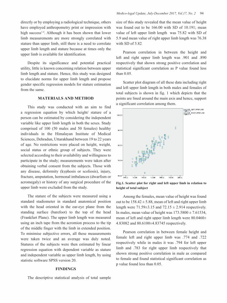
Medico-legal Update, July-December 2017, Vol.17, No. 2 94 95 -legal Update, July-December 2017, Vol.17, No. 2
directly or by employing a radiological technique, others have employed anthropometry print or impression with high success1-4. Although it has been shown that lower limb measurements are more strongly correlated with stature than upper limb, still there is a need to correlate upper limb length and stature because at times only the upper limb is available for identification.
Despite its significance and potential practical utility, little is known concerning relation between upper limb length and stature. Hence, this study was designed to elucidate norms for upper limb length and propose gender specific regression models for stature estimation from the same.
MATERIALS AND METHOD
This study was conducted with an aim to find a regression equation by which height/ stature of a person can be estimated by considering the independent variable like upper limb length in both the sexes. Study comprised of 100 (50 males and 50 females) healthy individuals in the Himalayan Institute of Medical Sciences, Dehradun, Uttarakhand between 19 to 22 years of age. No restrictions were placed on height, weight, social status or ethnic group of subjects. They were selected according to their availability and willingness to participate in the study; measurements were taken after obtaining verbal consent from the subjects. Those with any disease, deformity (kyphosis or scoliosis), injury, fracture, amputation, hormonal imbalances (dwarfism or acromegaly) or history of any surgical procedure of the upper limb were excluded from the study.
The stature of the subjects were measured using a standard stadiometer in standard anatomical position with the head oriented in the ear-eye plane from the standing surface (barefoot) to the top of the head (Frankfurt Plane). The upper limb length was measured using an inch tape from the acromion process to the tip of the middle finger with the limb in extended position. To minimise subjective errors, all these measurements were taken twice and an average was duly noted. Statures of the subjects were then estimated by linear regression equation with dependent variable as stature and independent variable as upper limb length, by using statistic software SPSS version 20.
FINDINGS
The descriptive statistical analysis of total sample
size of this study revealed that the mean value of height was found out to be 166.00 with SD of 10.191; mean value of left upper limb length was 75.82 with SD of 5.9 and mean value of right upper limb length was 76.38 with SD of 5.82.
Pearson correlation in between the height and left and right upper limb length was .901 and .890 respectively that shown strong positive correlation and statistical significant correlation as P value found less than 0.05.
Scatter plot diagram of all these data including right and left upper limb length in both males and females of total subjects is shown in fig. 1 which depicts that the points are lined around the main axis and hence, support a significant correlation among them.
Fig.1. Scatter plot for right and left upper limb in relation to height of total subject
Among the females, mean value of height was found out to be 158.42 ± 5.88, mean of left and right upper limb length were 71.59±3.15 and 72.15 ± 2.914 respectively. In males, mean value of height was 173.5800 ± 7.61534, mean of left and right upper limb length were 80.0460± 4.83082 and 80.6100±4.83745 respectively.
Pearson correlation in between female height and female left and right upper limb was .774 and .722 respectively while in males it was .794 for left upper limb and .783 for right upper limb respectively that shown strong positive correlation in male as compared to female and found statistical significant correlation as p value found less than 0.05.

-legal Update, July-December 2017, Vol.17, No. 2 94 95 Medico-legal Update, July-December 2017, Vol.17, No. 2
Fig.2: Scatter plot of height and upper limb length in female
Fig. 3: Scatter plot of height and upper limb length in males
As depicted in fig 2 and fig 3 above, in the scatter plot diagram for males all the points are gathered towards the main axis showing strong positive correlation in between them and in scatter plot diagram for females, all the points are scattered diffuse from the main axis showing less positive correlation in between the height and upper limb length as compared to male.
The regression equation obtained for estimation of height of male and females from the independent variable (upper limb length) are shown in table1.
Table 1: Regression equation for calculating expected height (dependable variable) from both upper limb length (independent variable) of male and female.
PARAMETER REGRESSION EQUATION
Female height by female left upper limb length
YGIRLS HEIGHT = 53.549 + 1.465 * X (X = female left upper limb length)
Female height by female right upper limb length
YGIRLS HEIGHT = 53.296 + 1.457 * X (X= female right upper limb length)
Male height by male left upper limb length
YBOYS HEIGHT = 73.387 + 1.252* X(X = male left upper limb length)
Male height by male right upper limb length
YBOYS HEIGHT = 74.241 + 1.232* X(X= male right upper limb length)
DISCUSSION
A significant correlation of stature and upper limb length has been observed in both the sexes in our study. Measurements of height from the right upper limb length were found to be greater than that of the left upper limb length in male by use of linear regression equation and measurements of height from the left upper limb length were found to be greater than that of the right upper limb length in female, but the difference was marginal and statistically insignificant. In study conducted by M. R. Shende et al5, they found mean height for male was 170.12 ± 6.99, mean height for female was 156.15 ± 11.10, and mean length of upper extremity in male was 79.11 ± 3.98 and mean length of upper extremity in female was 72.92 ± 3.96, a positive correlation between height and superior extremity was observed in both sexes and it was statistically significant and these findings was consistent with our study.
Similar study was conducted by A. A. Ahmed6, Akhalagi M et al7 and Ozaslan A et al8on assessment of relationship between upper limb dimensions and stature, in which significant and positive correlation was found.
The findings of our study are also consistent with those observed by Sunil et al9 and Chandra et al10 in which estimation of stature was done from hand anthropometry. They concluded that there was a significant correlation between the stature and upper limb length.
Ebeye Oladunni. A1. conducted a study for Stature Estimation from Upper Extremity Long Bones and found Regression formulae with statistical significance in estimation of the stature from the sum of the length of the upper extremity long bones were established. And regression coefficient was higher in males than in females similar to our study findings.
The regression equation obtained were used to estimate the height of the subjects, by considering the upper limb length as an independent variable. These formulae devised for determination of stature from upper limb length would be beneficial in unidentified fragmentary or mutilated remains of upper limb.
CONCLUSION
Stature estimation from limbs or dismembered body parts is of paramount importance for personal identification, especially when the utility of DNA analysis is limited due to economic issues or in war.

Medico-legal Update, July-December 2017, Vol.17, No. 2 96
According to the current study, there is a strong positive correlation between stature and upper limb length; the stature can be estimated by the linear regression equations thus calculated for males and females from each of the upper limbs individually.
Though our subjects represent a wide diversity of geographical areas across India, still there persists a lot of variation in estimating stature from upper limb measurements due to other factors. Thus, there is a need to conduct further studies among larger population, belonging to different race and ethnicity so that stature estimation becomes more reliable and identification of an individual is established effectively.
Ethical Clearance: Obtained from ethical committee, HIMS, Jolly Grant
Conflict of Interest: None
Source of Funding: Self
REFERENCES
1. Ebeye Oladunni. A; Stature Estimation From Upper Extremity Long Bones In A Southern Nigerian Population; Australian Journal of Basic and Applied Sciences, 7(7): 400-403, 2013
2. Dan U, Mukhopadhyay P P, Ghosh T K, Biswas S. Estimation of stature from fragment of long bone (tibia) in Indian Bengali population J. Anat Soc. India 2009; 58(2): 169-172
3. Bidmos A Mubarak. Stature Reconstruction Using Fragmentary Femora in South Africans of European Descent. Journal of forensic Sciences 2008; 53, (5,): 1044-1048. 6.
4. Soumeek Chowdhuri et al. Estimation of Stature
from Radiological Measurement of Sternal Length with Corroboration in Living Individuals A Study in Contemporary Bengali Adults; J Indian Acad Forensic Med. October-December 2015, Vol. 37, No. 4
5. Shende M. R, et al; Correlation of Superior Extremity Length with Stature In Central Indian Population; J Indian Acad Forensic Med. July-September 2013, Vol. 35, No.
6. Ahmed A, A; Estimation of stature from the upper limb measurements of Sudanese adults; Forensic Sci Int. 2013 May 10;228(1-3).
7. Akhalagi M, Hajibeygi M, Zamani N, Moradi B; A study on estimation of stature on upper limb anthropometry in Iranian population; Journal o:f Forensic and Legal Medicine- July 2012; 19(5): 280-4.
8. Ozaslan A, KoçS,OzaslanI, Tuðcu H; A study on estimation of stature from upper extremity in Istanbul population. Military Medicine- International Journal of AMSUS- 2006, April; 171(4):288-91.
9. Sunil, P.C. Dikshit, Anil Aggarwal, Mukta Rani; Estimation of stature from hand length; A study in students of various colleges on Delhi. Journal of Indian Academy of Forensic Medicine- 2005, 24(4) ISSN 0971-0973.
10. Almesh Chandra, Pankaj Chandna, SurinderDeswal, Rajesh Kumar Mishra, Ranjender Kumar; A study on stature prediction model based on hand anthropometry on industrial workers of Haryana state. International Journal of Medical Health, Bio-Medical and Pharmaceutical Engineering Vol. 9; Number 2 in 2015.
-legal Update, July-December 2017, Vol.17, No. 2 96
Gender Prediction – Anthropometric Study of Orbital Parameters
K Srinivasulu1, K K Bairagi2, Sowmiya KR3
1Associate Professor, Dept. of Forensic Medicine & Toxicology. Tagore Medical College & Hospital. Chennai, 1Associate Professor, Dept. of Forensic Medicine & Toxicology. Tagore Medical College & Hospital. Chennai, 1
2Prof & HOD, Dept. of Forensic Medicine & Toxicology, Sridev Suman Subharti Medical College & Hospital, 2Prof & HOD, Dept. of Forensic Medicine & Toxicology, Sridev Suman Subharti Medical College & Hospital, 2
Dehradun, 3Associate Professor, Dept. of Community Medicine. Tagore Medical College & Hospital, Chennai3Associate Professor, Dept. of Community Medicine. Tagore Medical College & Hospital, Chennai3
ABSTRACT
Objective: The aim of this study is to evaluate the use of orbital measurements as a tool for sex determination in unidentified skeleton.
Method: Seventy seven (45 male and 32 female) completely undamaged skulls of known sex in book record were used for the study. Five different measurements of both orbits of each skull were recorded. Measurements were made using vernier/ sliding callipers (0.01mm). Analysis was done using IBM – Statistical Package for Social sciences (SPSS) version 21. The means between the male and female samples were compared for significance using the Student t – test.
Results: Statistics revealed high significance in four measurements except Inter-orbital Breadth.
Conclusion: Reports on the use of orbit as a tool for sex determination in unidentified human skeleton has been reported in different populations. The present study supports this finding among South Indian population.
Keywords: Orbit, Sexual dimorphism, Moore- Jansen1, South Indian population.
Correspondance author:K SrinivasuluAssociate Professor, Dept. of Forensic Medicine & Toxicology. Tagore Medical College & Hospital. Rathinamangalam. Chennai, 600127.E-mail: [email protected]
INTRODUCTION
Sex determination of human skeletal remains is considered as an initial step in identification. In cases of fragmented or mutilated body, it is difficult to identify the body. Sex determination of such skeletal remains have immensely helped in identification of an individual. This skilful process is carried out by forensic and anatomy experts. Anthropometry which aids the understanding (Last, 1968)2 of anatomical structures, constitutes the technique of expressing quantitatively, the form of human body and skeleton. Anthropometric studies are an integral part of craniofacial surgery and syndromology (Novit, 2006)3. For these reasons,
standards based on ethnic or racial data are desirable because these standards reflect the potentially different patterns of craniofacial growth resulting from racial, ethnic and sexual differences (Evereklioglu et al., 2002)4. The orbital cavities, which form the subject of this work, are situated on either side of the sagittal plane of the skull between the cranium and the skeleton of the face (Patniak et al.,2001)6. Each orbital cavity is essentially intended as a socket for the eyeball and also contains associated muscles, nerves, vessels and in essence lodges the visual apparatus (Soames, 1999)5. Orbital index the proportion of orbital height to its breath X 100 (Igbigbi & Ebite 2010)8 is determined by the shape of face and varies with race, religion. Patniak et al. (2001) stated that in each orbital cavity, the width is usually greater than the height, the relation between the two is given by the orbital index, which varies in different races.
MATERIALS AND METHOD
Adult crania (age ranges between 20 and 70 years) of known sex were obtained from the archives of
DOI Number: 10.5958/0974-1283.2017.00077.9
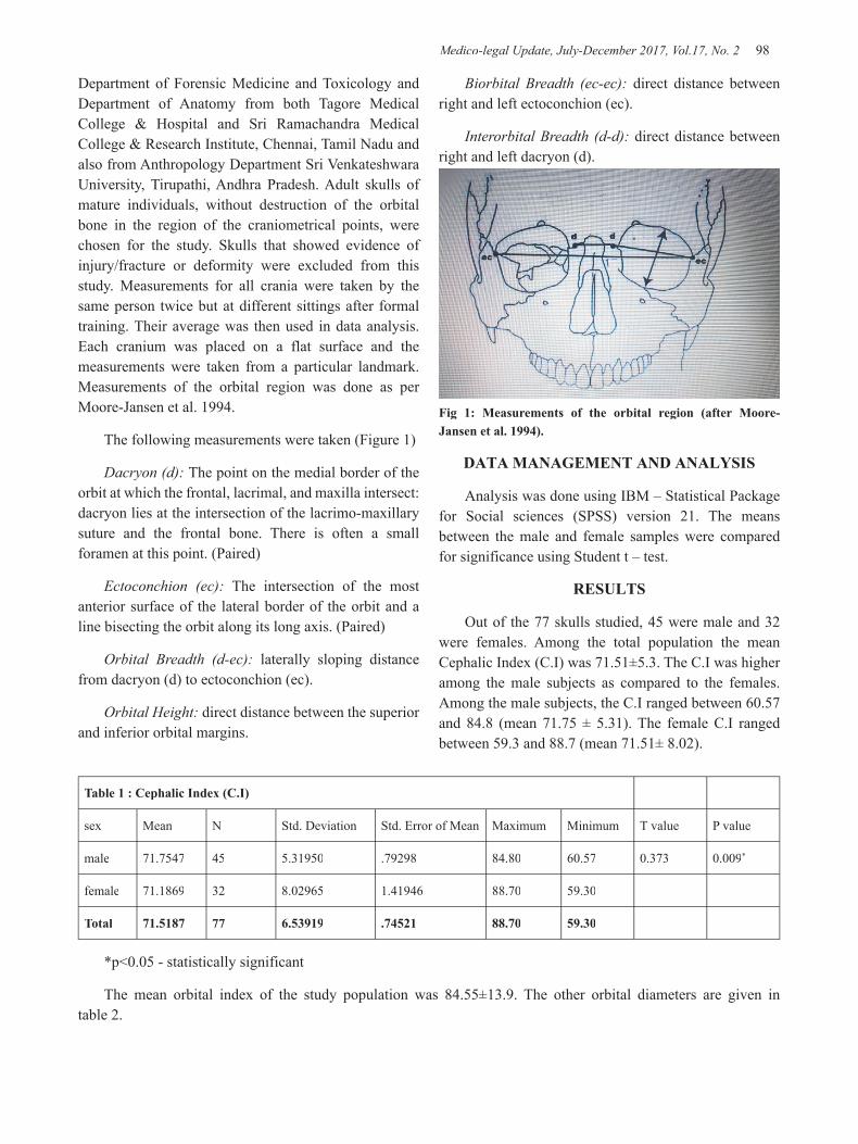
Medico-legal Update, July-December 2017, Vol.17, No. 2 98 99 -legal Update, July-December 2017, Vol.17, No. 2
Department of Forensic Medicine and Toxicology and Department of Anatomy from both Tagore Medical College & Hospital and Sri Ramachandra Medical College & Research Institute, Chennai, Tamil Nadu and also from Anthropology Department Sri Venkateshwara University, Tirupathi, Andhra Pradesh. Adult skulls of mature individuals, without destruction of the orbital bone in the region of the craniometrical points, were chosen for the study. Skulls that showed evidence of injury/fracture or deformity were excluded from this study. Measurements for all crania were taken by the same person twice but at different sittings after formal training. Their average was then used in data analysis. Each cranium was placed on a flat surface and the measurements were taken from a particular landmark. Measurements of the orbital region was done as per Moore-Jansen et al. 1994.
The following measurements were taken (Figure 1)
Dacryon (d): The point on the medial border of the orbit at which the frontal, lacrimal, and maxilla intersect: dacryon lies at the intersection of the lacrimo-maxillary suture and the frontal bone. There is often a small foramen at this point. (Paired)
Ectoconchion (ec): The intersection of the most anterior surface of the lateral border of the orbit and a line bisecting the orbit along its long axis. (Paired)
Orbital Breadth (d-ec): laterally sloping distance from dacryon (d) to ectoconchion (ec).
Orbital Height: direct distance between the superior and inferior orbital margins.
Biorbital Breadth (ec-ec): direct distance between right and left ectoconchion (ec).
Interorbital Breadth (d-d): direct distance between right and left dacryon (d).
Fig 1: Measurements of the orbital region (after Moore-Jansen et al. 1994).
DATA MANAGEMENT AND ANALYSIS
Analysis was done using IBM – Statistical Package for Social sciences (SPSS) version 21. The means between the male and female samples were compared for significance using Student t – test.
RESULTS
Out of the 77 skulls studied, 45 were male and 32 were females. Among the total population the mean Cephalic Index (C.I) was 71.51±5.3. The C.I was higher among the male subjects as compared to the females. Among the male subjects, the C.I ranged between 60.57 and 84.8 (mean 71.75 ± 5.31). The female C.I ranged between 59.3 and 88.7 (mean 71.51± 8.02).
Table 1 : Cephalic Index (C.I)
sex Mean N Std. Deviation Std. Error of Mean Maximum Minimum T value P value
male 71.7547 45 5.31950 .79298 84.80 60.57 0.373 0.009*
female 71.1869 32 8.02965 1.41946 88.70 59.30
Total 71.5187 77 6.53919 .74521 88.70 59.30
*p<0.05 - statistically significant
The mean orbital index of the study population was 84.55±13.9. The other orbital diameters are given in table 2.
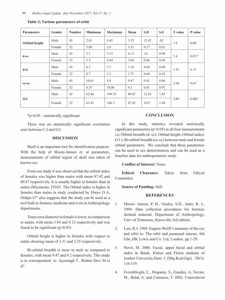
-legal Update, July-December 2017, Vol.17, No. 2 98 99 Medico-legal Update, July-December 2017, Vol.17, No. 2
Table 2: Various parameters of orbit
Parameters Gender Number Minimum Maximum Mean S.D S.E T value P value
Orbital heightMale 45 2.81 3.65 3.25 12.43 .02
1.9 0.04*
Female 32 3.00 3.8 3.31 0.17 0.03
d-ecMale 45 3.1 5.15 4.12 .62 0.09
2.4 0.017*
Female 32 3.2 4.84 3.84 0.46 0.08
d-dMale 45 0.2 2.5 1.54 0.64 0.09
1.55 0.12Female 32 0.7 2.5 1.77 0.60 0.10
ec-ecMale 45 10.61 8.8 9.47 0.45 0.06
2.64 0.01*
Female 32 8.55 10.00 9.2 0.41 0.07
O.IMale 45 62.44 109.70 80.87 12.43 1.85
2.89 0.005*
Female 32 63.43 106.3 87.42 10.9 1.94
*p<0.05 - statistically significant
There was no statistically significant correlation seen between C.I and O.I.
DISCUSSION
Skull is an important tool for identification purpose. With the help of Moore-Jansen et. al parameters, measurements of orbital region of skull was taken of known sex.
From our study it was observed that the orbital index of females was higher than males with mean 87.42 and 80.87 respectively. It is usually higher in females than in males (Miyamoto; 1924)9. The Orbital index is higher in females than males in study conducted by Ebeye O A, Otikpo O10 also suggests that the study can be used as a tool both in forensic medicine and even in Anthropology departments.
Transverse diameter in female is lower, in comparison to males, with mean 3.84 and 4.12 respectively and was found to be significant (p<0.05)
Orbital height is higher in females with respect to males showing mean of 3.31 and 3.25 respectively.
Bi-orbital breadth is more in male as compared to females, with mean 9.47 and 9.2 respectively. This study is in correspondent to Jayasingh T , Rohini Devi M et al7 .
CONCLUSION
In this study, statistics revealed statistically significant parameters (p<0.05) in all four measurements i.e; Orbital breadth (d- ec); Orbital height; Orbital index( O.I ); Bi-orbital breadth (ec-ec) between male and female orbital parameters. We conclude that these parameters can be used in sex determination and can be used as a baseline data for anthropometric study.
Conflict of Interest: None.
Ethical Clearance: Taken from Ethical Committee.
Source of Funding: Self.
REFERENCES
1. Moore- Jansen, P. H., Ousley, S.D., Jantz, R. J., 1994. Data collection procedures for forensic skeletal material; Department of Anthropology, Univ of Tennessee, Knoxville.3rd edition.
2. Last, R.J. 1968. Eugene Wolff`s anatomy of the eye and orbit in: The orbit and paranasal sinuses. 6th Edn.,HK Lewis and Co. Ltd, London. pp.1-29.
3. Novit, M. 2006. Facial, upper facial and orbital index in Batak, Klaten and Flores students of Jember University.Dent. J. (Maj.Ked.Gigi) . 39(3): 116-119.
4. Evereklioglu, C., Doganay, S., Gunduz, A, Tercan, M., Balat, A. and Cumurcu, T. 2002. Craniofacial

Medico-legal Update, July-December 2017, Vol.17, No. 2 100
anthropometry in a Turkish population. Cleft Palate Craniofacial J. 39(2): 208-218.
5. Soames, R.W. 1999. Skeletal systems: Williams P.L Bannister LH, Berry MM Collins P,Dyson Mary, Dussek J, Ferguson MW, ed. Gray`s Anatomy the anatomical basis of medicine and surgery. 38th Edn. p. 555.
6. Patniak, V.V.G., Bala Sanju and Singla Rajan, K. 2001. Anatomy of the bony orbitssome applied aspects. J.Anatomical Soc. Ind. 50(1): 59-67.
7. T. Jeyasingh, Rohini Devi M; Indian journal of forensic medicine & toxicology volume:10, number 2/ July-Dec 2016; page no:56.
8. Igbigbi, P.S., Ebite, L.E. Orbital Index of Adult Malawians. Anil Aggrawal's Internet Journal of Forensic Medicine and Toxicology [serial online], 2010; Vol. 11, No. 1 (January – June2010):[about21p]. (Accessed: December 05, 2016)
9. Miyamoto, H., 1924. Gendai nihonjin jinkotsu no jinruigaku-teki kenkyu, Dai-1-bu: Togaikotsu no kenkyu (An anthropological study on the skeletons of modern Japanese, Part 1: A study of skulls). Journal of the Anthropological Society of Nippon, 39: 307–451; Data 1–48. (In Japanese.)
10. Ebeye O A ; Otikpo O IOSR Journal of Dental and Medical Sciences (IOSR-JDMS) e-ISSN: 2279-0853, p-ISSN: 2279-0861. Volume 8, Issue 2 (Jul.- Aug. 2013), PP 51-53.
-legal Update, July-December 2017, Vol.17, No. 2 100
Study of Blast Injuries in Tribal Region of Bastar: A Five Year Study
Pawan Tekade1, Prachi Parakh2, Jaideo Ughade3, S J Chahankar4, C R Tekade5, P B Kardile6 1Associate Professor, Department of Forensic Medicine, 2Final Year M.B.B.S. Student, 3Associate Professor, Department of Anatomy, Late Lakhiram Agrawal Memorial Government Medical College, Raigarh (C.G.),
4Pathologist, NMDC Patholgy, Amravati, 5NMDC Patholgy, Amravati, 6Assistant Professor, Shri V.N. Government Medical College, Yavatmal
ABSTRACT
Introduction: Blast or explosive injuries are less studied areas in medical field. Injury pattern and morbidity and mortality needed discussion in detail, as the instances of blast are on rise all over the world.
Material and method: The present retrospective study included 74 cases of deaths, due to explosion, brought to mortuary of Late Baliram Kashyap Memorial Government Medical College, Jagdalpur (C.G.), over the period of five years.
Observations: The young males (80% in age group of 21 – 40 years) are the only victims of explosions. Multiple organs are usually involved in victims, which increases the risk of mortality. Most of the blasts are of homicidal intent. The commonest injuries were lacerations and fractures (79.73% and 71.62), followed by burns, abrasions, contusions, and puncture lacerations. Long bones were the most common (45.95% cases) internal organ damaged, followed by lungs, blood vessels, spine, liver and brain. Kidneys were the least injured internal organs. In most of the cases, type of blast injuries was either not mentioned or undetermined (82.43%). The primary injuries were reported in 14% cases.
Summary and conclusion- As most of the blasts are of homicidal intent, social awareness and monitoring might be useful tools to reduce morbidity and mortality, as the blast injuries are highly fatal.
Keywords- blast injuries, explosive injuries.
INTRODUCTION
Discovery of fire is considered as a big step of humanity towards civilization, but the discovery of explosives is a major setback to peaceful civilization. Now a day these are no more the weapons of war field. Blast injuries are possible at any place where chemicals and explosives are stored or used.
Blast injuries usually create an atmosphere of confusion in investigating agencies as well as in society. There is lot of questions to be answered, by investigating agencies. But for medical fraternity, what concerns us is prevention and management of morbidity and mortality.
The present study was undertaken to study the profile of deaths due to blast, both explosive and industrial.
MATERIAL AND METHOD
The present study was carried out in Late Baliram Kashyap Memorial Government Medical College, Jagdalpur, the only tertiary care center in tribal region of Bastar, over the period of five years, from January 2010 to December 2014. The cases were recorded from the autopsy reports of the institute. All the cases, where death was due to explosion, or where history of explosion was mentioned in requisition, were included in study, irrespective of presence or absence of any other associated injuries.
OBSERVATIONS
In this five year study, total 74 cases of death due to blast were recorded. Out of 74 recorded cases in the study, only two cases survived to receive treatment, but
DOI Number: 10.5958/0974-1283.2017.00078.0

Medico-legal Update, July-December 2017, Vol.17, No. 2 102 103 -legal Update, July-December 2017, Vol.17, No. 2
expired within 24 hours. Rest of the cases was reportedly dead on the spot.
Table: 1. Age wise distribution of cases.
Age in years No. of cases (n=74) Percentage
< 20 00 0
21-30 28 37.84
31-40 32 43.24
41-50 13 17.57
51- 60 01 1.35
61 & above 00 0
Most of the cases (almost 80%) clustered in age group of 21 – 40 years age group. The number of cases abruptly tapered towards the higher age group, ending before 60 years of age, in which single case was reported. Not a single case was reported from the terminal age groups.
Table: 2. Manner of death.
Manner Cases (n=74) Percentage
1 Accidental 01 1.35
2 Homicidal 73 98.65
Only one case of explosion of vehicular tyre inflating gas machine, was reported due to accidental manner. Rest of the cases were homicidal in nature. Most of the homicidal explosion deaths were those of improvised explosive devices (IED), country made bombs, and hand grenades, planted or launched.
Table: 3. Injuries over body.
Injuries Cases (n=74) Percentage
1 Laceration 59 79.73
2 Fracture 53 71.62
3 Burns 26 35.14
4 Abrasion 25 33.78
5 Contusion 22 29.73
6 Puncture 04 5.41
7 Chop 02 2.70
8 Stab 02 2.70
9 Incised 01 1.35
The commonest injuries were lacerations and fractures (79.73% and 71.62), followed by burns, abrasions, contusions, and puncture lacerations. Other associated injuries were chop, stab and incised wounds.
The later were suspected as non-associated with blast, and caused separately, as in all such cases causative object was not detected in situ. Out of 74 cases, 34 cases also had firearm injuries (included as lacerations and puncture), of which 08 cases were those of riffled firearm and in remaining 26 it was not recorded. One case had blast, riffled firearm and firearm injury of unknown weapon.
Table: 4. Organs involved.
Sr No. Organs No of case (n=74) Percentage
1 Brain 22 30.73
2 Neck 09 12.16
3 Lung 30 40.54
4 Heart 10 13.51
5 Liver 23 31.08
6 Spleen 09 12.16
7 Kidney 02 2.70
8 Intestine 11 14.86
9 Stomach 07 9.46
10 Vessels 28 37.84
11 long bones 34 45.95
12 Spine 26 35.13
13 Fragmentation 22 29.73
Long bones were the most common (45.95% cases) internal organ damaged, followed by lungs, blood vessels, spine, liver and brain. In 29.73% cases bodies were fragmented, with internal organs damage. Intestine, heart, spleen, neck structures and stomach were next in frequency of damage. Kidneys were the least frequently damaged tissues.
Table: 5. Type of blast injuries.
Sr. No. Type of blast injuries No. of cases
(n= 74) Percentage
1 Primary injuries 14 18.92
2 Secondary injuries 09 12.16
3 Tertiary injuries 01 1.35
4 Undetermined/ not mentioned 61 82.43
In most of the cases, type of blast injuries was either not mentioned or undetermined (82.43%). The primary injuries were reported in 14% cases, while tertiary injuries were reported in only one case.

-legal Update, July-December 2017, Vol.17, No. 2 102 103 Medico-legal Update, July-December 2017, Vol.17, No. 2
DISCUSSION
In the present study, all the cases were males1. The reason being, at present female troops are not deployed in lead front for control of terror activities, or restricted to enter such sensitive areas. Similar findings were, observed by Rasikh M2 et al.
All the cases but one were homicidal in nature, in the present sample. Rasikh M2 et al, Bapin Kumar M3 et al and Kobi P4 et al, reported the similar outcome.
In the present study, the commonest injuries were lacerations and fractures (79.73% and 71.62), followed by burns, abrasions, contusions, and puncture lacerations.5,6. Out of 74 cases, 34 cases also had firearm injuries. One case had blast, riffled firearm and firearm injury of unknown weapon. The lacerations and fractures represent the blast effect and so, are the commonest injuries recorded, as the blast wave acts like a hard and blunt object, in such cases. Turégano-Fuentes F7 et al, Kobi P4 et al, Burkle FM8 et al, reported the similar findings.
Collectively bones and long bones to be specific, were the most common (45.95% cases) internal organ damaged, followed by lungs, blood vessels, spine, liver and brain. In 29.73% cases bodies were fragmented. Close proximity with the explosive, particularly planted ones, might be the reason for damage to extremities and hence to long bones. Rasikh M2, Kobi P4 et al, Turégano-Fuentes F7 et al and Burkle FM8 et al, reported the similar findings.
In the present study, in most of the cases, type of blast injuries was either not mentioned or undetermined (82.43%). Next in frequency were primary injuries. In most of the autopsy reports, the type of blast injuries were not commented upon, hence included in group of not mentioned or undetermined.
LIMITATION OF STUDY
The study is retrospective one, so individual variation in recording of findings might have influenced the parameters. This can be a baseline reference study for explosive injuries. Further research particularly in type of injuries may prospectively be undertaken.
SUMMARY AND CONCLUSION
The present study concludes that the young males are the most common victims of explosions. Multiple
organs are usually involved in victims, which increases the risk of mortality. Most of the blasts are of homicidal intent. Social awareness and monitoring might be useful tools to reduce morbidity and mortality, as the blast injuries are highly fatal.
Conflict of Interest: None.
Source of Funding: None.
Ethical Clearance: Yes. No ethical issues involved.
REFERENCES
1. Verzeletti A, Astorri P, De Ferrari F.- Firearm-Related Deaths In Brescia (Northern Italy) Between 1994 And 2006: A Retrospective Study. J Forensic Leg Med. 2009 Aug; 16 (6): 325-31.
2. Rasikh M, Rasikh A, Tariq A, Irfan S.- Pattern Of Injuries Seen In Mass Casualties In Terrorist Attacks In Baluchistan, Pakistan- A Three Years Experience. J Ayub Med Coll Abbottabad- 2015; 27 (4); 858- 860.
3. Bapin Kumar M, Chakma P, Meera T. - Firearm Fatalities: A Postmortem Study In A Northeastern State Of India Medico-Legal Update, January- June 2016, Vol.16, No. 1; 196- 200.
4. Kobi P, Limor AD, Michael S, Moshe M, Et Al. – Gunshot And Explosion Injuries: Characteristics, Outcomes, And Implications For Care Of Terror Related Injuries In Israel. Annals Of Surgery: March 2004; Vol. 239; 3; 311-318.
5. Saukko P, Knight B.- Knight’s Forensic Pathology. Third edition. Edward Arnold Publishers. London (UK), 2004. 274- 280.
6. Bardale R. Principles of Forensic Medicine and Toxicology. First Edition. Jaypee Brothers Medical Publishers. New Delhi. 2011. 217-218.
7. Turégano- Fuentes F, Caba- Doussoux P, Jover- Navalón JM, Et Al.- Injury Patterns From Major Urban Terrorist Bombings In Trains: The Madrid Experience. World J Surg. 2008 Jun; 32 (6): 1168-75.
8. Burkle FM, Newland C, Meister SJ, Blood CG: Emergency Medicine In The Persian Gulf War- Part 3: Battlefield Casualties. Ann Emerg Med April 1994; 23: 755-760.

105 -legal Update, July-December 2017, Vol.17, No. 2
A Medicolegal Examination of Drowning Deaths- A Retrospective Study
Gajanan H Nayak1, MahalaxmiKarlawad2, Sunil Kumar S Biradar3
1Professor and Head, 2Postgraduate, 2Postgraduate, 2 3Associate Professor, Karnataka Institute of Medical Sciences, Hubballi3Associate Professor, Karnataka Institute of Medical Sciences, Hubballi3
India is a vast country having plenty of water bodies’ i.e. Rivers, ponds, wells and an extensive sea coast. So drowning is one of the most common causes of death among all manners of death in this country. This was a 2 year retrospective autopsy study of deaths due to drowning cases analyzed and tabulated. Study performed in department of Forensic Medicine & Toxicology at KIMS, Hubballi from January 2014 to December 2015. We studied total 23 cases in two years,the maximum drowning cases were of male sex -14(60.86%), with commonest age group being 11-20 years - 06 cases(26.08%). Most of the drowning cases were accidental in nature 13(56.52%) and occurred commonly in lakes and rivers 15(62.51%). The most significant postmortem findings noted on autopsy were bleaching of skin 19(82.60%) and presence of water in stomach in 13 cases(56.52%).
Keywords: Drowning, age group, postmortem findings.
Corresponding author:Dr. MahalaxmiKarlawadPostgraduate Student, Dept. Forensic Medicne and Toxicology, Karnataka Institute of Medical Sciences, Hubballi. E-mail- [email protected]
INTRODUCTION
India is a vast country having plenty of water bodies’ i.e. Rivers, ponds, wells and an extensive seacoast. In such a conducive environment, it is but natural that death due to drowning is a frequent event, where medico legal expertise is called upon for investigations. A recent global burden of disease (GBD) study reveals global mortality from drowning to be 7% of all injury related deaths (WHO, 2010). The world Health Organization estimates the annual global incidence of death by drowning to be 400,000.[1]According to data from the Global Burden of Disease, during 2000 around 449,000 individuals died of drowning worldwide, while drowning also cost 1.3 million years of lost life and disability.[2]
Accidental drowning occurs often in India, nearly 40, 000 Indians die annually from drowning. It occurs occasionally among swimmers due to their rashness in swimming, but it occurs mostly in non-swimmers who venture to go beyond their depth in the sea, rivers, canals and lakes. Many lives are lost during floods, which are
so frequent. It also occurs among persons at bathing places while bathing in deep water. Females may fall accidentally into a well while drawing water from it. Children may also accidentally fall into ponds or lakes while playing near their banks. They may even fall accidentally into domestic vessels of water, such as water tanks, bathtubs and buckets. [3] Accidental drowning in shallow water is very rare, except when the individual happens to be intoxicated, insane or epileptic.[4]The autopsy diagnosis of death by drowning can be one of the most difficult problems in forensic pathology because in our Indian setup the time required to complete the inquest formalities and transport of the body to the mortuary is enough fordecomposition to set in, which masks most of the postmortem findingsrequired to estimate the cause of death. And most of the bodies recovered from water are in various stages of decomposition.[5]
One of the most classical definition of drowning as provided by Roll as ‘death by drowning is the result of hampering of the respiration by obstruction of respiratory tract by afluid medium (usually water)’.[6]
MATERIAL AND METHOD
This was a 2 years retrospective autopsy study of deaths due to drowning cases analyzed and tabulated. Study was performed in department of Forensic Medicine & Toxicology at KIMS, Hubballi from January 2014 to December 2015.
DOI Number: 10.5958/0974-1283.2017.00079.2

105 Medico-legal Update, July-December 2017, Vol.17, No. 2
OBJECTIVES OF STUDY
1. To study the manner of death and categories whether the form of drowning deaths were accidental, suicidal or homicidal in nature.
2. To confirm whether the drowning was antemortem in nature
3. To study the specific findings of drowning deaths in postmortem examination.
OBSERVATIONS AND RESULTS
The present study has been performed on 23 cases of drowning deaths which were brought to Karnataka Institute of Medical Sciences, Hubballi, for routine medico legal autopsy.
Table No. 1 showing sex-wise distribution, out of total of 23 cases that were studied, 14 (60.86 %) were males & 09 (39.13%) were females.
Thus it is obvious that the majority of cases reported for postmortem examination were males.
Table No. 2 showing age wise distribution, out of 23 cases studied, the incidence of drowning was found in all age groups.
The maximum number of cases studied belonged to the age group of 11 – 20 yrs and the minimum number of the cases was reported in age group of 71 – 90 yrs.
Table No. 3 showing manner of death, Out of the total of 23 cases of drowning, it was found that 13cases (56.52%) were accidental, 10 cases (43.47%) were suicidal.
Significantly we did not find any case of homicidal drowning.
Table No. 4 showing the postmortem findings drowning cases. Significantly, we did not find any case of dry drowning.
Table No: 5 showing place of occurrences, lakes and rivers being the commonest sources, 15(65.21%)cases, 03(13.04%) cases in wells,04(17.39%) cases in house tanks and 01 (4.34%)case in swimming pool were observed.
Table no:1 Sex wise distribution
MALE FEMALE TOTAL
14(60.86%) 09(39.13%) 23
Table no:2 Age wise distribution
Age group in years Total cases
0-10 02 (08.69%)
11-20 06 (26.08%)
21-30 03 (13.04%)
31-40 02 (08.69%)
41-50 03 (13.04%)
51-60 02(08.69%)
61-70 03 (13.04%)
71-80 01 (04.34%)
81-90 01(04.34%)
Table no:3 Manner of death
Suicidal Accidental Homicidal Total
10(43.47%) 13(56.52%) 00 23
Table no:4 Postmortem findings
Sl.No PM findings Total cases
01 Cyanosis 05(21.73%)
02 Froth from mouth and nostrils 08(34.78%)
03 Washer woman’s hands and feet 05(21.73%)
04 Bleaching of skin 19(82.60%)
05 Internal petechial hemorrhages 04(17.39%)
06
Presence of heavy, voluminous, edematous and congested lungs with c/s showing copious frothy fluid
12(52.17%)
07 Presence of water in stomach 13(56.52%)
08 Animal bites and Decomposition 04(17.39%)
Table no: 5 Place of occurrence
01 Lakes/Rivers 15(65.21%)
02 Wells 03(13.04%)
03 House Tanks 04(17.39%)
04 Swimming Pools 01(4.34%)

Medico-legal Update, July-December 2017, Vol.17, No. 2 106 107 -legal Update, July-December 2017, Vol.17, No. 2
DISCUSSION
Drowning is defined as the impairment of tissue oxygenation consequent to submersion in a fluid medium.[3]
According to Mason JK[7], the precise number of drowning fatalities on a worldwide basis is uncertain but is probably around some 140, 000 people per year. The trend is increasing worldwide due to increase in water transport, and adventure water sports.
According to Modi[8], Accidental drowning occurs often in India, nearly 40, 000 Indians die annually from drowning. It occurs occasionally among swimmers due to rashness in swimming, but it occurs mostly in non-swimmers who venture to go beyond their depth in sea, rivers, canals, and lakes etc.
Modi[8] observed that in India drowning occupies the first position of all models of committing suicide. In his study of 231 cases of suicide, 90 were due to drowning. Indian females, even on the least provocation, commit suicide by jumping into a well or a tank in the neighborhood.
In our study of total 23 cases of drowning, 14(60.86%)were males &09(39.13%)were females. In all age groups males predominated. Male dominance in drowning deaths were shown in many other studies by Kanchan T et al, Chowdhury B L et al, Byard R W et al, Davoudi-Kiakalayeh A et al .[9,10,11,12]ArdeshirSheikhazadi et al (2009) found in their study in Iran that male - female ratio was 6.5:1 among drowning deaths [13].The percentage of drowning deaths where the deceased was a male in various studies -Sayed ZAT[14] (75.68%), Chormunge et al[15] (73.53%), Amandeep Singh et al[16] (67.56%), Salachin et al[17](75.6%), Gross VA et al[18] (90%), Momanchand et al[19] (80.3%), and Srinivasa Reddy P[20] (59.14%).
In our study, we found most common incidence of drowning occurred in the age group of 11 – 20 yrs i.e. 06 cases (26.08%), followed by the age groups 21-30 yrs, 41-50 yrs and 61 to 70 yrs,with 03cases each(13.04%).Inthe age group of 0-10 yrs, 31-40 yrs and 51-60 yrsthere were 02 cases each (08.69%) and the least common age groupwas 81 – 90 yrsi.e. 1 case (1.43%). Thus the maximum number of cases in our study belonged to the age group of 11 – 20 yrs. and the minimum numbers of the cases were reported in age group of 81–90 yrs.
Considering other references in same field, Prabir et al in their study in 2015 had maximum cases(13cases) of drowning between 11-20 years of age,[21] Manjunath S (2010) in their study at Manipal found that the age group commonly involved in drowning was 11-20 years[22], Davoudi-Kiakalayeh A et al (2008) in their study in Iran also found that more than one third of the victims were less than 20 years of age[12]. Results of these studies corroborated with our findings.
In present study, it was also found that 13 cases (56.52%) were accidental and 10 cases (43.47%) were suicidal in nature. Similar observations were made in study done by Mukherjee AA et al,wherein 26 cases (37.14) were accidental and 19 cases (27.14%) were suicidal and in 25 cases (35.72%) police did not ascertain the manner of death [3]. The probable explanation to the above may be that as drowning deaths are mostly suicidal and accidental in nature, the age group 11- 50 yrs. are more vulnerable as this age is more prone to accidents in water during adventure sports, travels and swimming. The challenges and struggle for livelihood in adolescents, frustrations due to failure of high ambitions and in love affairs makes them more prone for suicide.
As far as the postmortem findings seen in drowning cases, out of the total of 23 cases, cyanosis was observed in 05(21.73%),oozing of whitish to pinkish froth, copious in amount from mouth and nostrils was found in 08(34.78%) cases,bleached skin was observed in 19(82.60%) cases and washer woman’s hands & feet were observed in 05(21.73%)cases. Internal petechial hemorrhages over pleura, pericardium, under scalp, over the lungs and in the interlobar areas were found in 04(17.39%) cases. In 12(52.17%)cases we observed the presence of heavy, voluminous, oedematous and congested lungs, with weight of either lung above 500 gm and with presence of copious amount of whitish, fine, leathery froth oozing out freely on cut section. Significantly, we did not find any case of dry drowning. The presence of water in stomach and first part of small intestine was observed in 13(56.52%)of cases. Animal bites and decomposition changes were noted in 04cases(17.39%)
In our study, in 15 cases(65.21%) drowning occured in lakes/rivers, followed by 04 cases(17.39%) in house tanks, 03cases(13.04%) in wells and 01(04.34%) case in swimming pool. Similar observation were made in study done by Chidanand C et al,wherethe commonest
-legal Update, July-December 2017, Vol.17, No. 2 106 107 Medico-legal Update, July-December 2017, Vol.17, No. 2
place of submersion was lakes (37.6%) followed by wells (17.8%).[23] Study done by Prabir et al, showed maximum cases drowned in ponds 21(35%), followed by rivers 17(23%) and lakes 13(22%).[21]
CONCLUSION
This study reveals that the maximum drowning cases were of male sex 14(60.86%), with commonest affected age group being 11-20 years, i.e. 06 cases(26.08%). Most of the drowning cases were accidental in nature 13(56.52%) and occurred commonly in lakes and rivers15(62.51%). The most significant findings noted on autopsy were bleaching of skin – 19 cases (82.60%) and presence of water in stomach - 13 cases(56.52%).
At the end it may be said that the study carried out is neither easy nor noncontroversial but it is likely to open the doors for carrying on constant reconsideration, Government legislation, community participation related to proper swimming lessons, aquatic safety measures and evaluation of the problem for its improvement and throw light on the present state.
Some preventive measures that can be implemented to avoid drowning such as unattended underground tanks should not be kept open. Proper engineering and meshing/closing of the construction tanks and drainages should be made compulsory. Fencing and meshing of the lakes with prominent warning signals may reduce the rate of accidental submersions. Introduction of resuscitation education and swimming lessons to the curriculum of the children can do better. Parents and or caregivers should be advised not to leave the children alone/unattended.
In our study the number of drowning cases that presented to Karnataka Institute of Medical Sciences, Hubballi are less because, rapid urbanization has resulted in loss of natural water reservoirs and most of the cases are taken to local primary health care centers for autopsy.
Conflict of Interest: Nil
Source of Funding: Self
Consent: Not taken as it was autopsy based retrospective study
REFERNCES
1. Brundtland GH. From WHO Reducing risks to
health, promoting healthy life. JAMA 2002; 288(16):1974.
2. Peden MM, Mc Gee K. The epidemiology of drowning world wide. Inj Control SafPromot 2003; 10(4):195-9.
3. Mukherjee AA, DhawaneSG, Dhoble SV. Medicolegal study of drowning deaths: a forensic perspective. Journal of Research in Forensic Medicine and Toxicology; Vol 2, Issue 1, Jan-June 2016; Page 1-4.
4. Mathiharan K, Patnaik AK. Modi’s Medical Jurisprudence and Toxicology, 23rded. New Delhi: Lexis Nexis 2006:610-11.
5. Ranga Rao et.al, Sch. J.App.Med.Sci.,2014;2(4D):1397-1401.
6. H.F.Roll, Leerboek der GerechtelijkeGeeneeskundevoor de scholen tot opleiding van Ind.Arsten, ‘S- Gravenhage, Martinus Nijhoff,1918.
7. Mason JK, The pathology of trauma 2nd ed. Edward Arnold 1993:204-26.
8. Modi JP. Textbook of Medical Jurisprudence and Toxicology, 21st ed. N.M.Tripathi Private Ltd 1996:188-220.
9. Dr.Tanuj Kanchan, Dr.Prateekrastogi, Dr.Manoj Kumar Mohanty, Profile of near drowning victims in coastal region of Karnataka, JIAFM,2007;29(4).
10. Chaudhary B.L, Singh Deepak, MeelVeena, Tirpude B.H, Study of drowning cases in Kasturba Hospital Sevagram, Wardha, Journal of Forensic Medicine and Toxicology 2005:22(2):29-30.
11. Roger W.Byard,M.D.GillianHouldsworth, Ross A.James and John D.Gilbert, Characteristic Featuresof Suicidal Drowning A 20year study, The American Journal of Forensic Medicine and Pathology 2001;22(2):134-138.
12. Davoudi-Kiakalayeh A, Mohammadi R, Stark-Ekman D, Yousefzade-Chabok S, Behboudi F, Jamson B. Estimating drowning deaths in Northern Iran using capture-recapture method. Health Policy 2011:100(2-3):290-296.
13. Ardeshir S, Mohammad H G, Epidemiology of Drowning in Isfahan Province, cener of Iran. J.Res Med Sci.2009, March-April;13(2):79-87.
14. Sayed ZAT, Farhat HM, Hamid AP. Medicolegal investigation of violent asphyxial deaths–an
Medico-legal Update, July-December 2017, Vol.17, No. 2 108
autopsy based study. Journal of the Dow University of Health Sciences, Karachi 2012;6(3):86-90.
15. Vijay C, Prashant B, Ajay S. Violent asphyxial deaths in rural area of Maharashtra. Indian Journal of Forensic Medicine and Pathology 2009;2(4):161-4.
16. Singh A, Goria RK. A study of demographic variables of violent asphyxial death. JPAFMAT 2003;3:22-5.
17. Salachin S. An analysis of the medicolegal autopsies performed in adana, Turkey, in 1983- 1988. American Journal of Forensic Med and Pathology 1991;12(3):191-93.
18. Gross VA, Weiss MG, Ring M, et al. Method of suicide: international suicide patterns derived from WHO mortality database. Available at: www.unece.org/trans/conventn/distsigns.pdf.
19. Momamchand A. Violent asphyxial deaths in imphal. Journal of Forensic Medicine & Toxicology 1998;15(1):60-4.
20. Reddy SP, Kumar R, Rudramurthy. Asphyxial deaths at district hospital, Tumkur a retrospective study. J Indian Acad Forensic Med 2012;34(2):146-147.
21. Prabir et al, Histopathological changes in lungs and spleen in cases of drowning with demographic variability, European Journal of Biomedical and Pharmaceutical Sciences, 2015 2(2):479-485.
22. Palimar V, Manjunath S. drwoning deaths in Manipal. Int. J Med Toxicol Legal Med.2010 Apr-Jun; xii(4): ISSN:0972-0448.
23. Chidanand C, Satish K.V. Study of incidence of middle ear hemorrhage in drowning. Journal of evidnce based Medicine an dHealth care: 2(6) Feb 09,2015;621-628.

-legal Update, July-December 2017, Vol.17, No. 2 108
Pattern of Skull Bones Involvement in Cases of Road Traffic Accidents- A Prospective Study
Dileep Kumar KB1, Puneeth Kumar BP2, Bheemappa Havanur3
1Assistant Professor, 1Assistant Professor, 1 2 Postgraduate Student, 3 Professor and Head, Department of Forensic Medicine and Toxicology, Bangalore Medical College and Research Institute, Bengaluru, Karnataka
ABSTRACT
Head injuries are one of the devastating injuries that can happen in one’s life in a case of road traffic accident.
Objective- To study various pattern of skull bone injuries in fatal cases of road traffic accidents.
Study design- Cross sectional autopsy based study.
Materials and Method- Autopsy done during the period of July 2015 to December 2015.
Results- In the present study, a total of 121 cases of fatal RTA victims with head injury were studied. Highest number of RTAs was seen in young adults in the age group between 21-30 years. Male preponderance with male: female ratio of 7.07:1 was observed. Highest numbers of victims were two-wheeler riders (38.84%) followed by pedestrians (35.54%). 22.31% of cases died on the spot and 66.94% of cases died within 24 hrs. 87.23% of two wheeler riders were not wearing any protective helmets at the time of accident. Alcohol consumption was found in 28.93% of cases and 26.45% of victims were using electronic gadget at the time of accident. The dominant type of skull fracture found was linear fracture (57.02%) Temporal bone (69 cases) was the commonest bone to be involved. Middle cranial fossa was the dominant cranial fossa to be involved.
Keywords: road traffic accidents, head injuries, occipital bone injuries, skull fractures, autopsy.
INTRODUCTION
A Road Traffic Accident (RTA) can be defined as, ‘An event that occurs on a way or street open to public traffic; resulting in one or more persons being injured or killed, where at least one moving vehicle is involved. Thus RTA is a collision between vehicles; between vehicles and pedestrians; between vehicles and animals; or between vehicles and geographical or architectural obstacles.’ Road traffic accidents are a human tragedy. They involve high human suffering and socioeconomic costs in terms of premature deaths, injuries, loss of
Corresponding author: Dr Dileep Kumar KBAssistant Professor, Dept of Forensic Medicine & Toxicology, Bangalore Medical College & Research Institute, Bangalore 560002, Mb.no:+919591780922, Email: [email protected]
productivity, and so on.1
India accounts for about 10% of road crash fatalities worldwide. India has the highest absolute number of recorded road deaths (105,725), followed by China (96,611), the US (42,642) and Russia (35,972). In comparison, the UK has 3,298 recorded road deaths when you look at recorded road deaths proportional to a country’s population, the Cook Islands comes out on top (45.0 road deaths per 100,000 people), followed by Libya (34.7), South Africa (33.2) and Iran (32.2).2
The total number of deaths in road accidents in Karnataka was 10,444 in 2014.3 In Bangalore 737 were killed out of 5004 RTAs in 2014.4
In more than half of RTAs the major cause of death is Head Injury. Head Injury as defined by the National Advisory Neurological Diseases and Stroke Council is
Original Article DOI Number: 10.5958/0974-1283.2017.00080.9
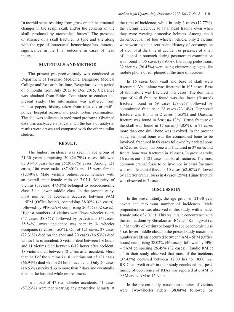
Medico-legal Update, July-December 2017, Vol.17, No. 2 110 111 -legal Update, July-December 2017, Vol.17, No. 2
“a morbid state, resulting from gross or subtle structural changes in the scalp, skull, and/or the contents of the skull, produced by mechanical forces”. The presence or absence of a skull fracture, its type and site along with the type of intracranial hemorrhage has immense significance in the final outcome in cases of head injury.
MATERIALS AND METHOD
The present prospective study was conducted at Department of Forensic Medicine, Bangalore Medical College and Research Institute, Bengaluru over a period of 6 months from July 2015 to Dec 2015. Clearance was obtained from Ethics Committee to conduct the present study. The information was gathered from inquest papers, history taken from relatives or traffic police, hospital records and post-mortem examination. The data was collected in preformed proforma. Obtained data was analyzed statistically. On the basis of analysis, results were drawn and compared with the other similar studies.
RESULT.
The highest incidence was seen in age group of 21-30 years comprising 30 (24.79%) cases, followed by 31-40 years having 25(20.66%) cases. Among 121 cases, 106 were males (87.60%) and 15 were female (12.40%). Male victims outnumbered females with an overall male:female ratio of 7.07:1. Majority of victims (58cases, 47.93%) belonged to socioeconomic class 3 i.e. lower middle class. In the present study, most number of accidents occurred between 9AM – 5PM (Office hours), comprising 38.02% (46 cases), followed by 9PM-5AM comprising 26.45% (32 cases). Highest numbers of victims were Two- wheeler riders (47 cases, 38.84%) followed by pedestrians (43cases, 35.54%).Lowest incidence was seen in 3- wheeler occupants (2 cases, 1.65%). Out of 121 cases, 27 cases (22.31%) died on the spot and 20 cases (16.53%) died within 1 hr of accident. 5 victims died between 1-6 hours and 11 victims died between 6-12 hours after accident. 18 victims died between 12-24hrs after accident. More than half of the victims i.e. 81 victims out of 121 cases (66.94%) died within 24 hrs of accident. Only 20 cases (16.53%) survived up to more than 7 days and eventually died in the hospital while on treatment.
In a total of 47 two wheeler accidents, 41 cases (87.23%) were not wearing any protective helmets at
the time of incidence, while in only 6 cases (12.77%), the victims died due to fatal head trauma even when they were wearing protective helmets. Among the 6 driver/occupant of four wheeler vehicle, only 2 victims were wearing their seat belts. History of consumption of alcohol at the time of accident or presence of smell of alcohol in stomach during postmortem examination was found in 35 cases (28.93%). Including pedestrians, 32 victims (26.45%) were using electronic gadgets like mobile phone or ear phones at the time of accident.
In 16 cases both vault and base of skull were fractured. Vault alone was fractured in 105 cases. Base of skull alone was fractured in 5 cases. The dominant type of skull fracture found was the linear (fissured) fracture, found in 69 cases (57.02%) followed by comminuted fracture in 28 cases (23.14%). Depressed fracture was found in 2 cases (1.65%) and Diastatic fracture was found in 5cases(4.13%). Crush fracture of the skull was found in 17 cases (14.05%). In 77 cases more than one skull bone was involved. In the present study, temporal bone was the commonest bone to be involved, fractured in 69 cases followed by parietal bone in 52 cases. Occipital bone was fractured in 37 cases and frontal bone was fractured in 35 cases. In present study, 16 cases out of 121 cases had basal fractures. The most common cranial fossa to be involved in basal fractures was middle cranial fossa, in 10 cases (62.50%) followed by anterior cranial fossa in 4 cases (25%). Hinge fracture was observed in 7 cases.
DISCUSSIOIN
In the present study, the age group of 21-30 year covers the maximum number of incidences. Male preponderance was observed in this study, with a male: female ratio of 7.07 : 1. This result is in concurrence with the studies done by Shivakumar BC et al,5 Kalougivaki et al.6 Majority of victims belonged to socioeconomic class 3 i.e. lower middle class. In the present study maximum number accidents occurred between 9AM – 5PM (Office hours) comprising 38.02% (46 cases), followed by 9PM – 5AM comprising 26.45% (32 cases).. Tandle RM et al7 in their study observed that most of the incidents (37.43%) occurred between 12:00 hrs to 18:00 hrs. RK Chaturvedi et al8 in their study concluded that peak timing of occurrence of RTAs was reported at 6 AM to 9AM and 9 AM to 12 Noon.
In the present study, maximum number of victims were Two-wheeler riders (38.84%) followed by
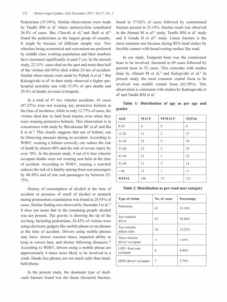
-legal Update, July-December 2017, Vol.17, No. 2 110 111 Medico-legal Update, July-December 2017, Vol.17, No. 2
Pedestrians (35.54%). Similar observations were made by Tandle RM et al7 where motorcyclists constituted 36.9% of cases. But, Chavali et al,9 and JhaS et al10
found the pedestrians as the largest group of casualty. It might be because of different sample size. Two wheelers being economical and convenient are preferred by middle class working population and their numbers have increased significantly in past 5 yrs. In the present study, 22.31% cases died on the spot and more than half of the victims (66.94%) died within 24 hrs of accident. Similar observations were made by Pathak A et al.11 But Kalougivaki et al6 in their study observed a higher pre-hospital mortality rate with 51.9% of spot deaths and 29.4% of deaths en route to hospital.
In a total of 47 two wheeler accidents, 41 cases (87.23%) were not wearing any protective helmets at the time of incidence, while in only 12.77% of cases, the victims died due to fatal head trauma even when they were wearing protective helmets. This observation is in concurrence with study by Shivakumar BC et al5 and Jha S et al.10 This clearly suggests that use of helmet, can be lifesaving measure during an accident. According to WHO2, wearing a helmet correctly can reduce the risk of death by almost 40% and the risk of severe injury by over 70%. In the present study, 4 out of 6 four wheeler occupant deaths were not wearing seat belts at the time of accident. According to WHO3, wearing a seat-belt reduces the risk of a fatality among front seat passengers by 40-50% and of rear seat passengers by between 25-75%.
History of consumption of alcohol at the time of accident or presence of smell of alcohol in stomach during postmortem examination was found in 28.93% of cases. Similar finding was observed by Surender J et al.12
It does not mean that in the remaining people alcohol was not present. The gravity is showing the tip of the ice-berg. Including pedestrians, 26.45% of victims were using electronic gadgets like mobile phone or ear phones at the time of accident. Drivers using mobile phones may have: slower reaction times, impaired ability to keep in correct lane, and shorter following distances.15 According to WHO3, drivers using a mobile phone are approximately 4 times more likely to be involved in a crash. Hands free phones are not much safer than hand-held phone.
In the present study, the dominant type of skull-vault fracture found was the linear (fissured) fracture,
found in 57.02% of cases followed by comminuted fracture present in 23.14%. Similar result was observed in the Ahmad M et al13 study, Tandle RM et al7 study and S Gouda H et al14 study. Linear fracture is the most common one because during RTA head strikes by forcible contact with broad resting surface like road.
In our study, Temporal bone was the commonest bone to be involved, fractured in 69 cases followed by parietal bone in 52 cases. This coincides with studies done by Ahmad M et al,13 and Kalogivaki et al.6 In present study, the most common cranial fossa to be involved was middle cranial fossa (62.50%). This observation is consistent with studies by Kalouguvaki et al6 and Tandle RM et al.7
Table 1: Distribution of age as per age and gender
AGE MALE FEMALE TOTAL
0-10 4 0 4
11-20 11 2 13
21-30 25 5 30
31-40 22 3 25
41-50 21 1 22
51-60 11 3 14
> 60 12 1 13
TOTAL 106 15 121
Table 2. Distribution as per road user category
Type of victim No. of cases Percentage
Pedestrian 43 35.54%
Two wheelerdriver 47 38.84%
Two wheelerpillion rider 16 13.22%
Three wheelerdriver/ occupant 2 1.65%
LMV- front seatoccupant 6 4.96%
HMV-driver/ occupant 7 5.79%
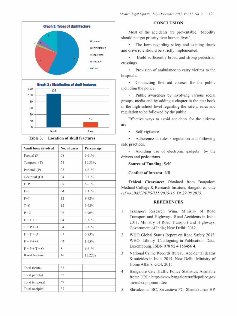
Medico-legal Update, July-December 2017, Vol.17, No. 2 112 113 -legal Update, July-December 2017, Vol.17, No. 2
Table 3. Location of skull fractures
Vault bone involved No. of cases Percentage
Frontal (F) 08 6.61%
Temporal (T) 24 19.83%
Parietal (P) 08 6.61%
Occipital (O) 04 3.31%
F+P 08 6.61%
F+T 04 3.31%
P+T 12 9.92%
T+O 12 9.92%
P+ O 06 4.96%
F + T + P 04 3.31%
T + P + O 04 3.31%
F + T + O 01 0.83%
F + P + O 02 1.65%
F + P + T + O 8 6.61%
Basal fracture 16 13.22%
Total frontal 35
Total parietal 52
Total temporal 69
Total occipital 37
CONCLUSION
Most of the accidents are preventable. ‘Mobility should not get priority over human lives’.
• The laws regarding safety and existing drunk and drive rule should be strictly implemented.
• Build sufficiently broad and strong pedestrian crossings.
• Provision of ambulance to carry victims to the hospitals.
• Conducting first aid courses for the public including the police.
• Public awareness by involving various social groups, media and by adding a chapter in the text book in the high school level regarding the safety, rules and regulation to be followed by the public.
Effective ways to avoid accidents for the citizens are:
• Self-vigilance
• Adherence to rules / regulation and following safe practices.
• Avoiding use of electronic gadgets by the drivers and pedestrians.
Source of Funding: Self
Conflict of Interest: Nil
Ethical Clearance: Obtained from Bangalore Medical College & Research Institute, Bangalore. vide ref.no.:BMCRI/PS/155/2015-16. Dt:29.08.2015.
REFERENCES
1 Transport Research Wing. Ministry of Road Transport and Highways. Road Accidents in India 2011. Ministry of Road Transport and Highways, Government of India; New Delhi: 2012.
2 WHO Global Status Report on Road Safety 2013, WHO Library Cataloguing-in-Publication Data; Luxembourg. ISBN 978 92 4 156456 4.
3 National Crime Records Bureau. Accidental deaths & suicides in India 2014. New Delhi: Ministry of Home Affairs, GOI; 2015
4 Bangalore City Traffic Police Statistics; Available from: URL: http://www.bangaloretrafficpolice.gov.in/index.phpimmittee
5 Shivakumar BC, Srivastava PC, Shantakumar HP.
-legal Update, July-December 2017, Vol.17, No. 2 112 113 Medico-legal Update, July-December 2017, Vol.17, No. 2
Pattern of Head Injuries in mortality due to road traffic accidents involving two-Wheelers. J Indian Acad Forensic Med 2008;32(3):239 – 42
6 Kalougivaki JJVP, Goundar RPS.Retrospective autopsy based study of fatal Road traffic accidents in Fiji. J Forensic Res2014;5(6):243-9
7 Tandle RM,Keoliya AN. Patterns of head injuries in fatal road traffic accidents in a rural district of Maharashtra- Autopsy based study. J Indian Acad Forensic Med 2011;33(3):228 – 31
8 Chaturvedi RK, Mishra A, Chaturvedi P. Pattern of head injuries in fatal road traffic accidents in Indore region, MP. J Evol Med and Dent Scie 2014;3(21):5645-51
9 Chavali KH, Sharma BR, Harish, Sharma A. Head Injury: the principal killer in road traffic accidents.J Indian Acad Forensic Med2006;28(4):121-4.
10 Jha S, YadavBN ,Karn A, Aggrawal A, Gautam AP.Epidemiological study of fatal head injury in road traffic accident cases: A study from BPKIHS, Dharan.Health Renaissance 2010 May-Aug;8(2);97-101
11 Pathak A, Vyas PC, Guptha BM. Autopsy finding of pattern of skull fractures and intracranial hemorrhages in cases of head trauma: A Prospective Study. J Indian Acad Forensic Med 2006;28(4):187-90
12 Surendar J, Ranjan S D. A comprehensive study of fatal head injuries among motorcyclists: A one year prospective study.J Dent and Med Scie2013;10(4):98-101
13 Ahmad M, Rahman FN, Chowdhury MH, Islam AKMS, Hakim MA. Postmortem study of head injury in fatal road traffic accidents. J Armed Forces med col Bangladesh. 2009;5(2):24-8
14 Gouda SH, Meghana PR, Prabha B. Cranio-cerebral injuries in victims of fatal road traffic accident: a 5 year post-mortem study. International J Med Toxicology and Forensic Med2014;4(3):77-82.
15 Mobile phone use: a growing problem of driver distraction. Geneva, Switzerland;WHO :2011;Available from http://www.who.int/violence_injury_prevention/publications/road_tra€c/en/index.html
115 -legal Update, July-December 2017, Vol.17, No. 2
Legal, Social & Ethical Issues During Performing Peri-natal Autopsy in India - A Practical Experience
Rajashree Pradhan1, Sajeeb Mondal2, Subrata Pal3, Arindam Banerjee4, Debosmita Bhattacharyay5 1Assistant Professor, 2Faculty, 3Demonstrator. Dept of Pathology, 4Faculty, Dept of Pediatrics, College of Medicine
and Sagore Dutta Hospital, 5MD (PGT). Post Graduate Trainee, Department of Pathology, RG Kar Medical College and Hospital
ABSTRACT
Background: Obtaining consent from bereaved parents is difficult job. Difficulties are multifaceted. Researchers need to sail through social, ethical, legal, administrative and other barriers in obtaining consent for perinatal autopsy.
Aims: To explore the different problems for getting consent from parents to perform perinatal autopsy and identification the root causes of those constrains and the possible way outs in Indian scenario.
Material & Method: A prospective study was done in India between Jan 2011 to Dec 2015 after taking written consent from parents where 110 perinatal autopsy were performed; though 180 parents were approached for consent but 70 parents refused to do so. Data was collected about consent for autopsy, causes of refusal, additional findings and change of clinical cause of death.
Results: Perinatal autopsy were done in 110 cases (61%) and in 70 cases parents (39%) to give consent for autopsy. We obtained additional information in 42 cases (38.18%) and diagnosis was changed in 9 cases (8.18%). Most common cause of refusal to provide consent was socio-cultural (39 cases, 55.71%).
Conclusion: Social factor emerged as the chief problem to obtain consent. Even today at the era of molecular pathology perinatal autopsy is still the gold standard to know the cause of death. Regarding its revival (as it is a dying and declining art), we need all round effort from clinician, pathologist, social worker and all medical care givers.
Keywords: Perinatal autopsy, medical ethics, consent.
Corresponding author: Dr Subrata PalDemonstrator. Dept of Pathology, College of Medicine and Sagore Dutta Hospital, Phone number: 9851773224E-mail address: [email protected]
INTRODUCTION
Addressing ethical, legal, social religious and administrative issue related to use of human biological samples in research has become a vital element of a successful research project.
Autopsy literally means to see for one self – is the direction and examination of a dead body to determine the cause of death and establish the evolution of a
disease. [1] Perinatal autopsy remains the gold standard in investigating perinatal deaths. [2] The provision of perinatal and pediatric pathology services is a sign of enlightened society. [3]
Addressing ethical, legal, social religious and administrative issue related to use of human biological samples in research has become a vital element of a successful research project. Autopsy in perinatal deaths provides an etiology and summary of contributing factors in most of the cases and allow improved management in future pregnancies [4]. Even though autopsy is the best method to detect the cause for perinatal death, there has been a decline in the autopsy rate recently [5].
Getting consent from bereaved parents (or from their relatives who might have more say than the parents!) is
DOI Number: 10.5958/0974-1283.2017.00081.0

115 Medico-legal Update, July-December 2017, Vol.17, No. 2
a hard to crack job as it has social ethical and religious constrains that need cater with utmost delicacy and need involvement of all, neonatologists obstetricians, pathologists and other medical caregivers. Reluctance to request for autopsy by the clinicians sometimes may be due to potential of litigation if a clinically important misdiagnosis has been identified. But no doubt that failure to get parental consent is the major limiting factor. [6] The constraining issues for getting consent from bereaved parents may not be readily resolved but public debate and adhomineum decisions on each parental loss may help to alleviate the problems.
MATERIAL AND METHOD
This was a prospective cross sectional study involving the all the foetal death in our institute at obstretics department during the study period of four years (Jan 2011 to Dec 2014). We have approached to get consent from the parents of the dead baby. First approach was done either by clinician, health stuffs or pathologist. Those parents were willing to provide consent, written consent was obtained from parents before performing autopsy. Autopsy was performed in 110 cases. Clinical data of the perinatal death was obtained from the register and questionare. Data was collected about consent for autopsy, causes of refusal, additional findings and change of clinical cause of death.The unwilling parents were asked about some questionare regarding the causes of unwillingness. After autopsy we tried to confirm the cause of death of the newborn. Statistical analysis was done by SPSS version 22 (window 8). Statistical data was expressed by bar diagram and tables.
RESULTS
During the study period, we found 214 cases of perinatal death in our institute. Perinatal autopsy was approached in 180 cases (84.11%). In 110 cases (61.1%), patient party were agreed for perinatal autopsy and in another 70 cases (38.89%), patient party was not agreed for perinatal autopsy. Among the 110 cases, parents of 62 cases (56.37%) communicated for report of autopsy, whereas parents of 30 cases (27.27%) never communicated for report. In 14 cases parents communicated at the assigned date of delivery of report but did not gate it as reports were not ready but latter communicated and got the reports. In 4 cases parents did not communicate for second time, but reports were sent to them by institutional endeavour. Bodies of the
babies were taken back by parents in 88 cases (39.09%) with an approximately equal sex distribution. In 22 cases (male-12, female- 8, intersex - 2), parents were not interested to take back the bodies. We obtained additional information in 42 cases (38.18%) and prenatal diagnosis was changed in 9 cases (8.18%) after autopsy. We approached perinatal autopsy in 8 cases of recurrent perinatal death, who were not approached for perinatal autopsy after the death of 1st baby. Among these 8 cases six cases provided consent. Success rate of acquiring consent is higher in double approach by two doctors (clinician and pathologist in 48 cases, 43.64%) than the single approach (only clinician in 31 cases, 28.18% and only pathologist in 26 cases, 23.64%). Success rate of other health care personal to obtain consent is very poor (5 cases, 4.55%). Most common cause of refusal to give consent was socio-cultural (39 cases, 55.71%). Others causes of refusal to provide consent were religious (22 cases, 31.43%), psychological (5 cases, 7.14%) and non specific etc. Refusal to provide consent were predominantly from both parents (22 cases, 31.43%), father (20.57%) and other family members (20 cases, 28.57%).
DISCUSSION
Out of 180 families, 70 parents (38.89%) refused permission for autopsy. This was similar to the study of H.E. Mcttaffie. [7] Socio cultural factors emerged as single most important factor for declining the request of perinatal autopsy. This is contrary to the western study of Mcttaffie et al, where 38% declined the request of autopsy considering it unnecessary.[7] Contrary to our common belief, it is the father and other family members (57.14%) refused maximum to give consent, not the mother of the baby.
The encouraging fact was 80 percent (72.73%) showed their desire to get final autopsy diagnosis. It reflects the attitude, desire and competence of medical care givers to build up proper perception of the parents about the importance of perinatal autopsy. 79.99% of the parents took back babies body once the perinatal autopsy is over. In 46.36% of cases, in our study perinatal autopsy either changed the diagnosis or additional information obtained after autopsy. This was similar to study of Saller et al. [8] Chance of getting consent is more in case of recurrent perinatal death (75% of all perinatal death cases in our study). It can be argued that perinatal autopsies carry special benefits for parents in that they
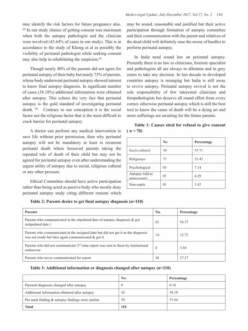
Medico-legal Update, July-December 2017, Vol.17, No. 2 116 117 -legal Update, July-December 2017, Vol.17, No. 2
may identify the risk factors for future pregnancy also. [8] In our study chance of getting consent was maximum when both the autopsy pathologist and the clinician were involved (43.64% of cases in our study). This is in accordance to the study of Khong et al as possibly the visibility of perinatal pathologist while seeking consent may also help in rehabilating the suspicion.[6]
Though nearly 40% of the parents did not agree for perinatal autopsy of their baby but nearly 75% of parents, whose body underwent perinatal autopsy showed interest to know final autopsy diagnosis. In significant number of cases (38.18%) additional information were obtained after autopsy. This echoes the very fact that perinatal autopsy is the gold standard of investigating perinatal death. [8] Contrary to our conception it is the social factor not the religious factor that is the most difficult to crack barrier for perinatal autopsy.
A doctor can perform any medical intervention to save life without prior permission, then why perinatal autopsy will not be mandatory at least in recurrent perinatal death where bereaved parents taking the repeated tole of death of their child but may not be agreed for perinatal autopsy even after understanding the urgent utility of autopsy due to social, religious cultural or any other pressure.
Ethical Committee should have active participation rather than being acted as passive body who mostly deny perinatal autopsy study citing different reasons which
may be sound, reasonable and justified but their active participation through formation of autopsy committee and their communication with the parent and relatives of the dead child will definitely ease the noose of hurdles to perform perinatal autopsy.
In India need sound law on perinatal autopsy. Presently there is no law so clinicians, forensic specialist and pathologists all are always in dilemma and in grey zones to take any decision. In last decade in developed countries autopsy is resurging but India is still away to revive autopsy. Perinatal autopsy revival is not the sole responsibility of few interested clinicians and fetopathologists but deserve all round effort from every corner, otherwise perinatal autopsy which is still the best tool to know the cause of death will be a dying art and more sufferings are awaiting for the future parents.
Table 1: Causes sited for refusal to give consent ( n = 70)
No Percentage
Socio cultural 39 55.71
Religiouys 22 31.43
Psychological 05 7.14
Autopsy told as unnecessaryunnecessary 03 4.29
Non-septic 01 1.43
Table 2: Parents desire to get final autopsy diagnosis (n=110)
Parents No. Percentage
Parents who communicated at the stipulated date of autopsy diagnosis & got itstipulated date i 62 56.37
Parents who communicated at the assigned date but did not get it as the diagnosis was not ready but later again communicated & got it 14 12.72
Parents who did not communicate 2nd time report was sent to them by institutional nd time report was sent to them by institutional nd
endeavour 4 3.64
Parents who never communicated for report 30 27.27
Table 3: Additional information or diagnosis changed after autopsy (n=110)
No. Percentage
Parental diagnosis changed after autopsy 9 8.18
Additional information obtained after autopsy 42 38.18
Pre natal finding & autopsy findings were similar 59 53.64
Total 110
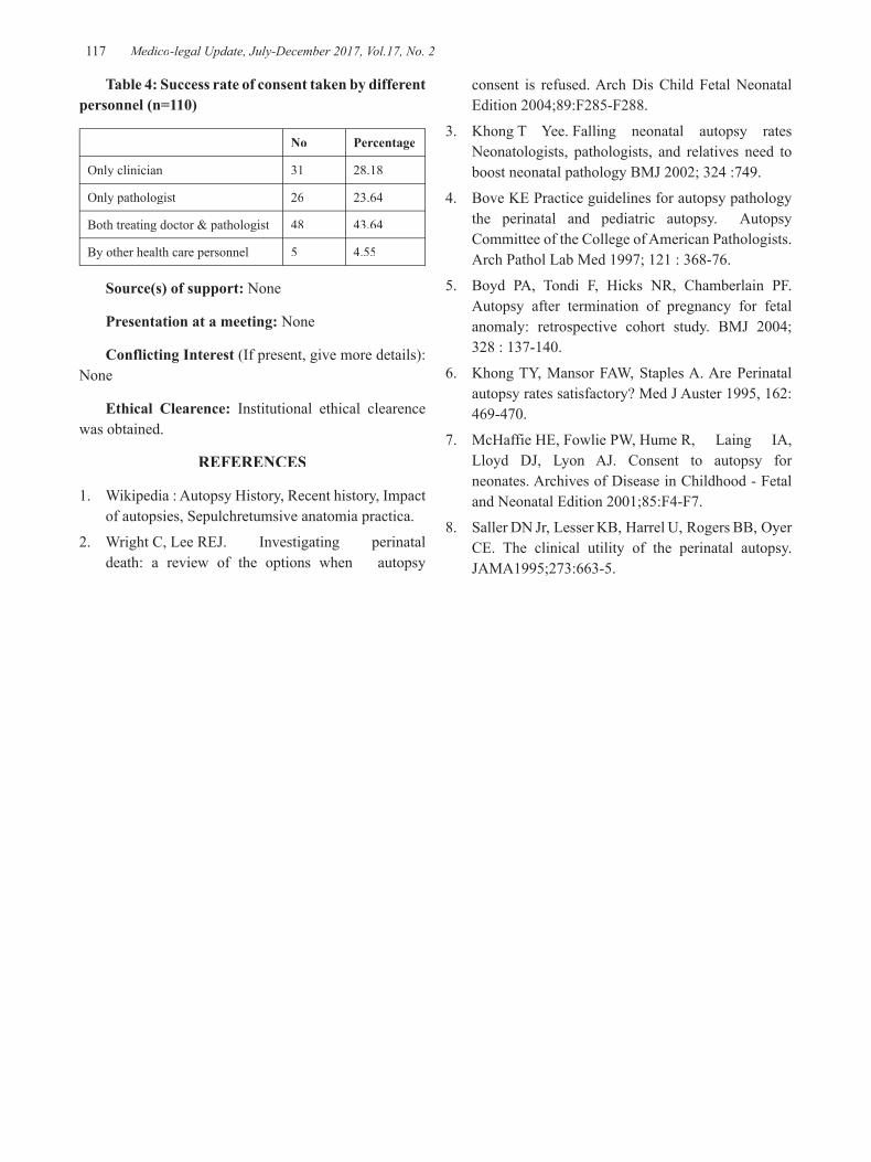
-legal Update, July-December 2017, Vol.17, No. 2 116 117 Medico-legal Update, July-December 2017, Vol.17, No. 2
Table 4: Success rate of consent taken by different personnel (n=110)
No Percentage
Only clinician 31 28.18
Only pathologist 26 23.64
Both treating doctor & pathologist 48 43.64
By other health care personnel 5 4.55
Source(s) of support: None
Presentation at a meeting: None
Conflicting Interest (If present, give more details): None
Ethical Clearence: Institutional ethical clearence was obtained.
REFERENCES
1. Wikipedia : Autopsy History, Recent history, Impact of autopsies, Sepulchretumsive anatomia practica.
2. Wright C, Lee REJ. Investigating perinatal death: a review of the options when autopsy
consent is refused. Arch Dis Child Fetal Neonatal Edition 2004;89:F285-F288.
3. Khong T Yee. Falling neonatal autopsy rates Neonatologists, pathologists, and relatives need to boost neonatal pathology BMJ 2002; 324 :749.
4. Bove KE Practice guidelines for autopsy pathology the perinatal and pediatric autopsy. Autopsy Committee of the College of American Pathologists. Arch Pathol Lab Med 1997; 121 : 368-76.
5. Boyd PA, Tondi F, Hicks NR, Chamberlain PF. Autopsy after termination of pregnancy for fetal anomaly: retrospective cohort study. BMJ 2004; 328 : 137-140.
6. Khong TY, Mansor FAW, Staples A. Are Perinatal autopsy rates satisfactory? Med J Auster 1995, 162:469-470.
7. McHaffie HE, Fowlie PW, Hume R, Laing IA, Lloyd DJ, Lyon AJ. Consent to autopsy for neonates. Archives of Disease in Childhood - Fetal and Neonatal Edition 2001;85:F4-F7.
8. Saller DN Jr, Lesser KB, Harrel U, Rogers BB, Oyer CE. The clinical utility of the perinatal autopsy. JAMA1995;273:663-5.
119 -legal Update, July-December 2017, Vol.17, No. 2
Estimation of Time Since Death from Algor Mortis – Still a Golden Method in the Modern Era
Satin K Meshram1, Sushim A Waghmare2, Santosh B Bhoi3, Rizwan A Kamle4, S S Gupta5
1Professorand Head, 2Associate Professor, 2Associate Professor, 2 3Associate Professor, 3Associate Professor, 3 4Assistant Professor, Dept. of Forensic Medicine, 4Assistant Professor, Dept. of Forensic Medicine, 4
Dr. V. M. Govt. Medical College and Shree. C.S.M.G. Hospital, Solapur, 5Dean, S.B.H.GMC, Dhule and former 5Dean, S.B.H.GMC, Dhule and former 5
Professor and Head, Forensic Medicine, IGGMC, Nagpur
ABSTRACT
For any death investigation estimation of time since death is of crucial importance. Rigor mortis, Algor mortis and Livor mortis were still considered to be useful as they are easy to assess and less sophisticate instruments with much low cost were required to assess the same. Out of these three classical triads Algor mortis has a benefit that it would be able to estimate the time since death with more accuracy and less time range. The present study focuses on the various factors affecting and influencing the rate of cooling after death so that it should be measured with more accuracy whenever the situation demands.
Keywords- Algor Mortis, time since death, cooling of dead body.
Corresponding author:Dr Sushim WaghmareAssociate Professor, Dept. of Forensic Medicine & Toxicology, Dr. V. M. Govt. Medical College and Shree. C.S.M.G. Hospital, Solapur, Maharashtra 413003, Email: [email protected]: 8888824415
INTRODUCTION
The most popular topic for research in forensic medicine is the attempt to increase the accuracy of ‘estimating the time since death” for obvious reasons. This is an integral and vital part of any death investigations. Such estimations are of prime importance in the investigations of criminal and suspicious deaths.
Except where the environmental (ambient) temperature remains at or even above 370 C, the human body will cool after death. A uniform, homogenous laboratory body will cool according to Newton’s Law of cooling, which states that “the rate of loss of heat at any instance is directly proportional to the temperature difference between the object and its surroundings”.
The fall of temperature of the cadaver occurs due to the facts that, after death there is no heat generation, due to loss of all physical, chemical and metabolic functions
of the body. Also the heat transfer within the body through the circulation ceases after death. Metabolic heat production, occurring mainly in the muscles and liver, does not cease uniformly and some heat generation continuous for a variable time. But there is constant loss of the body heat until it comes to the level of environmental temperature, as the heat regulating center is inactive. Loss of the body heat occurs by way of conduction, radiation and evaporation (a form of convection) when the body is in the atmospheric environment and by ways of conduction and convection when the body is in the water media.
After death the surface (outer core) temperature falls rather rapidly for some time. But during this period, the loss of heat from the depth (inner core) of the body is rather negligible. This is due to the thickness of the skin and the subcutaneous tissue which are very good insulator of heat. However, some hours after death, by the time there has been reasonable fall of the surface temperature, a constant rate of flow of heat from inside the body establishes. It is then, that the fall of temperature at the inner core of the body, achieves a regular and constant pattern. Thus, if we take into consideration the rate of fall of the inner core temperature of the body, then, we do not get actually a simple (exponential) curve for the fall of the temperature, instead in such a case we
DOI Number: 10.5958/0974-1283.2017.00082.2
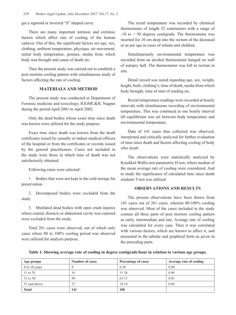
119 Medico-legal Update, July-December 2017, Vol.17, No. 2
get a sigmoid or inverted “S” shaped curve.
There are many important intrinsic and extrinsic factors which affect rate of cooling of the human cadaver. Out of this, the significant factors are age, sex, clothing, ambient temperature, physique, air movement, initial body temperature, posture, media from which body was brought and cause of death etc.
Thus the present study was carried out to establish a post mortem cooling pattern with simultaneous study of factors affecting the rate of cooling.
MATERIALS AND METHOD
The present study was conducted at Department of Forensic medicine and toxicology, IGGMC&H, Nagpur during the period April 2001 to April 2002.
Only the dead bodies whose exact time since death was known were utilized for the study purpose.
Exact time since death was known from the death certificates issued by casualty or indoor medical officers of the hospital or from the certificates or records issued by the general practitioners. Cases not included in the study were those in which time of death was not satisfactorily obtained.
Following cases were selected:
1. Bodies that were not kept in the cold storage for preservation.
2. Decomposed bodies were excluded from the study.
3. Mutilated dead bodies with open crush injuries where cranial, thoracic or abdominal cavity was exposed were excluded from the study.
Total 261 cases were observed, out of which only cases where 80 to 100% cooling period was observed were utilized for analysis purpose.
The rectal temperature was recorded by chemical thermometer of length 32 centimeters with a range of -10 to + 50 degrees centigrade. The thermometer was inserted for 10 cm deep into the rectum of the deceased or as per age in cases of infants and children.
Simultaneously environmental temperature was recorded from an alcohol thermometer hanged on wall of autopsy hall. The thermometer was left in rectum in situ.
Detail record was noted regarding age, sex, weight, height, built, clothing’s, time of death, media from which body brought, time of start of reading etc.
Rectal temperature readings were recorded at hourly intervals with simultaneous recording of environmental temperature. This was continued at one hourly interval till equilibrium was set between body temperature and environmental temperature.
Data of 141 cases thus collected was observed, interpreted and critically analyzed for further evaluation of time since death and factors affecting cooling of body after death.
The observations were statistically analyzed by Krushkal-Wallis non parametric H test, where median of the mean average rate of cooling were considered. And to study the significance of calculated time since death students T-test was utilized.
OBSERVATIONS AND RESULTS
The present observations have been drawn from 141 cases out of 261 cases, wherein 80-100% cooling was observed. Most of the cases included in the study contain all three parts of post mortem cooling pattern as early, intermediate and late. Average rate of cooling was calculated for every case. Then it was correlated with various factors, which are known to affect it, and presented in the tabular and graphical form as given in the preceding parts.
Table 1. Showing average rate of cooling in degree centigrade/hour in relation to various age groups.
Age groups Number of cases Percentage of cases Average rate of cooling0 to 10 years 9 6.38 0.8911 to 20 16 11.34 0.8621 to 50 89 63.12 0.8151 and above 27 19.14 0.89Total 141 100

Medico-legal Update, July-December 2017, Vol.17, No. 2 120 121 -legal Update, July-December 2017, Vol.17, No. 2
It is seen that average rate of cooling was more in the extremes of ages.
Table 2: Showing percent wise distribution and average rate of cooling in degree centigrade per hour as regard to sex.
Sex Number of cases
Percentage of cases
Average rate of cooling
Male 97 69 0.84
Female 44 31 0.86
Total 141 100
It is seen that average rate of cooling was slightly more in males as compared to females.
Table 3: Showing average rate of cooling in degree centigrade per hour in relation to the built.
Built Number of cases
Percentage of cases
Average rate of cooling
Thin Built <45 kg 27 19.14 0.93
Moderate Built 45-60 kg
81 57.44 0.83
Well built > 60 kg 33 23.40 0.78
Total 141 100
Average rate of cooling was decreases as the built increases, thus showing inverse relationship between built and cooling of the corpse.
By Krushkal Walli’s Non Parametric H test, H obtained was 10.48 and H critical was 5.99; d.f=2, thus p < 0.05 hence significant.
Table 4: Showing average rate of cooling in degree centigrade per hour in relation to clothing’s.
Clothing’s Number of cases
Percentage of cases
Average rate of cooling
Light clothed 31 22 0.88
Well clothed 110 78 0.84
Total 141 100
Average rate of cooling was more with minimum clothed or naked as compared to the individuals who are well clothed.
Statistical H obtained = 3.6 and H critical = 5.99, d.f=2 p<0.05, hence significant.
Table 5: Showing average rate of cooling in degree centigrade per hour in relation to various causes of death
Cause of Death
Number of cases
Percentage of cases
Average rate of cooling
Regional Injuries 13 9.21 0.68
Poisoning 24 17.02 0.76
Acute and chronic diseases
13 9.21 0.81
Head injury 22 15.60 0.85
Asphyxial deaths 11 7.80 0.86
Infectious deaths 36 25.53 0.86
Electrocution 5 3.54 0.93
Burns 10 7.09 0.98
Drowning 7 4.96 1.03
Total 141 100
Average rate of cooling was seen to be maximum in cases died due to drowning and lowest in those died due to regional injuries.
Table 6: Showing average rate of cooling in degrees centigrade per hour in relation to the bodies brought from different media’s
Media Number of cases
Percentage of cases
Average rate of coolingcooling
Water 9 6.38 1.08
Air 132 93.61 0.82
Total 141 100

-legal Update, July-December 2017, Vol.17, No. 2 120 121 Medico-legal Update, July-December 2017, Vol.17, No. 2
Average rate of cooling in bodies brought from water media is higher than that of bodies from air media.
Statistically by Krushkal Walli’s non parametric H test H obtained = 10.60, H critical = 5.99, p <0.05, hence significant.
Table 7: Showing average rate of cooling in degree centigrade per hour at different time interval after death.
Time interval
Number of cases
Percentage of cases (out of 141)
Average rate of cooling
0-3 hours 36 25.51 0.7
3-6 hours 133 94.32 1.05
6-12 hours 108 76.59 0.88
12 hours onwards 88 62.41 0.57
The cooling was slow during first 3 hours after death, then it was maximum in next 6 hours and again slow in the preceding 6 hours unless it reaches the equilibrium with that of surroundings.
Table 8: Showing average rate of cooling in degree centigrade per hour in relation to various average atmospheric temperatures ranges in degree centigrade.
Average atmospheric temperatures ranges
Number of cases
Percentage of cases
Average rate of cooling
15 – 20 29 20.56 0.97
21 – 25 94 66.66 0.84
26 – 30 8 6.38 0.71
30 – 35 10 7.09 0.41
Total 141 100
As the atmospheric temperature increases rate of fall decreases.
DISCUSSION
To study the relationship between age and cooling of body after death, the cases were divided according to age groups and their average rate of cooling was calculated. Robertson W H Atichison1 stated that the rapid rate of cooling was seen in the extremes of ages, bodies of
infants and old peoples soon part with their heat as their mass was small.
Gradwhol’s2 stated that bodies of infants and old aged persons cool much more rapidly than the bodies of adults.
In present study average rate of cooling (ARC) was more at the extremes of age groups i.e. 0.89 in 0 -10 years age group and 0.89 in 51 and above age group in comparison with middle age group i.e. 21 – 50 years (ARC was 0.81).
Mukkherjee J B3 stated that young women with more subcutaneous fat cool slowly than young average built males.
Karl Sellier4as quoted by Green M A and Wright J C made an interesting assertion that the retardation of cooing in fat bodies was not due to insulating effects on the fatty pads, but to the fact that the access adipose tissue increased the radius of the cylinder.
In the present study, we studied 44 females having ARC 0.86 and 97 males having ARC 0.84, which is less as compared to females. The reason may be that as most of the females examined were of thin built having less amount of subcutaneous fat.
Bernard Knight6 stated that a thin person cools more quickly because of both the mass, surface area ratio and the lack of fatty insulation.
In present study it was found that cooling is markedly retarded in those who are well built and or obese than thin built persons.
Out of 141 cases studied in present study, maximum cases i.e. 25.53% were of infectious diseases with ARC to be 0.86. Minimum cases were of the electrocution i.e. 3.54% with ARC to be 0.93. 4.96% of cases are of drowning and the ARC was highest (1.03) in these cases.
Out of 9.21% cases of regional injuries, the ARC was 0.68, which was low as compared to ARC other than regional injuries. This seems to be contradicting the fact that bodies with regional injuries cool rapidly. The justifiable reason for this may be due to the fact that during initial phase i.e. 0 to 2 hours after death the rate of cooling in these cases was very rapid and then the process of cooling was retarded. That means that most of
Medico-legal Update, July-December 2017, Vol.17, No. 2 122 123 -legal Update, July-December 2017, Vol.17, No. 2
the time the temperature may be low at the time of death. Therefore the average rate was seemed to be low.
The ARC was found to be comparatively low in deaths due to poisoning and asphyxia, as dead bodies keep warm longer in these cases due to elevated core temperature at the time of death.
In 7.09% of cases with death due to shock as a result of burns have ARC 0.98 and was comparatively higher than others. This comparative elevation was due to initial rise of surface temperature at the time of death. But after death gradient between surface and core temperature is established very quickly and the body temperature falls rapidly resulting into high rate of cooling.
Thus most of our findings regarding cause of death and cooling of body after death were in accordance with the standard studies available.
Taylor7stated that clothing’s on a body substantially retarded the rate of cooling. Gardon & Shapirio5mentioned that the presence of clothing’s or some coverings by insulating the body from air materially affects the flow of heat from a body.
In present study the ARC in clothed bodies was 0.84 and was comparatively less than the ARC of naked or minimum clothed bodies which was 0.88.
We found the average rate of cooling of bodies from water medium (1.08) was found to be significantly higher than those brought from air (0.82) as shown in table 7. Thus it was concluded that water hastens cooling by far better conduction than air. Also when such bodies are drawn out of water, cooling was fastened due to faster evaporation from wet bodies. Thus our findings are in accordance with findings as mentioned by Bernard Knight6,8, and Gardon & Shapiro5.
Marshall T K7, Gardon & Shapiro5, Mukkherjee J.B3 have stated that rate of cooling was directly proportional to the temperature difference in the body and surroundings. Greater the difference higher would be the rate of cooling.
In present study we found inverse relation between atmospheric temperature and the rate of cooling. Average rate of cooling was highest (0.97) in average atmospheric range 15-20 degree Celsius and it was lowest (0.41) when average atmospheric temperature ranges 31-35 degree Celsius. Hence our findings are in
accordance with the previous studies in this arena.
The average range of cooling in the present study is 0.9 to 0.4 degree centigrade per hour with the average rate of cooling 0.72 centigrade per hour.
From the foregoing it should be obvious that post mortem cooling rate is not merely a quantitative phenomenon, but it is a result of interaction of number of diverse factors like environmental temperature, age, sex, clothing’s, media from which body is recovered, surface area, built, cause of death etc.
While coming to any conclusion regarding post mortem interval estimation from rate of cooling, due consideration must be paid for the prevailing local conditions, environmental factors and details of the specific individuals characteristics of the deceased under study.
The basic scientific information enclosed in this present work will positively contribute towards establishment of postmortem interval when a situation during a death investigation demands it.
Conflict of Interest: None
Source of Funding – Self
Ethical Clearance- The said research was carried out for thesis of M.D purpose, hence ethical clearance was taken as per university norms.
REFERENCES
1. Aitchison W G. Robertson, Cooling of the body, Manual of Medical jurisprudence and toxicology, 1925, 5th edition, 63-64.
2. Francis E camps, Changes after death, Gradwohl’s legal Medicine, 1976,3rd Edition,86-88.rd Edition,86-88.rd
3. M ukkherjee J B, Death and its medicolegal aspects, Forensic medicine and toxicology, volume 1, 1994, 2nd edition, 213-218.nd edition, 213-218.nd
4. Green M A and Wright J C, The theoretical aspects of the time dependant Z equation as a mean of postmortem interval estimation using body temperature data only, Forensic Science international, Vol 28, 1985,35-46.
5. Gordon I, Shapiro H A, Diagnosis and the early signs of death, Text book of Foresic Medicine, 1988, 2nd edition, 12-18.nd edition, 12-18.nd

-legal Update, July-December 2017, Vol.17, No. 2 122 123 Medico-legal Update, July-December 2017, Vol.17, No. 2
6. Knight Bernard, The evolution of methods for estimating the time of death from body temperature, Forensic science International, 1988, Vol 36, 47-55.
7. Knight Bernard Henssege Claus, Krompecher Thomos, Madea Barkhard, Nokes Leonard, Edited by Bernard Knight, The estimation of time since
death in early postmortem period, Edward Arnold Publication, London, 1995, 4-35.
8. Knight Bernard, The pathophysiology of death, Forensic pathology, 2nd edition, 1996, 80-89.nd edition, 1996, 80-89.nd
9. Marshall T K, Temperature methods of estimating the time of death, Medicine science and Law, 1965, Vol 5, 224-232.

125 -legal Update, July-December 2017, Vol.17, No. 2
A Cross Sectional Study on Pattern of Organs Fatally Damaged in Road Traffic Accidents in Mangalore During the
Year 2015: Autopsy-Based Study
Gedion Hailemariam Abebe1, Pavanchand Shetty H3, B Suresh Kumar Shetty4, Prateek Rastogi3, Jagadish Rao Padubidri3, Animesh Jain5, Enyew Debash2
1Forensic Medicine Resident, St.Pauls Hospital Millenium Medical College, Ethiopia, 2Forensic Medicine 2Forensic Medicine 2
Resident, Mekelle University, Ethiopia, 3Associate Professor, 3Associate Professor, 3 4Professor and Head, Department of Forensic 4Professor and Head, Department of Forensic 4
Medicine, 5Professor and Head, Department of Community Medicine, 5Professor and Head, Department of Community Medicine, 5 Kasturba Medical College, Mangaluru, Manipal University
ABSTRACT
Introduction: The statistical profile speculates worldwide forecast of 5.1 million deaths in the year 2000 which accounted for 10% of deaths due to all causes. Amongst these, a quarter of injuries related deaths happened in the South-East Asian regions.[1]
Road traffic accidents are known to cause human loss as well as socioeconomic burden to the society at large.
The cause and effect relations in road traffic accidents need meticulous care to sort out offending factors and deliver proper solutions.
Research Questions
Objectives:
1) To assess the pattern of organs fatally damaged during road traffic accidents in Mangalore during the year 2015
2) To identify socio demographic pattern of those exposed to fatal injuries.
3) To forward practicable recommendation for policy makers and service providers.
Study Design: Descriptive cross-sectional autopsy-based retrospective study.
Study Setting: The study was performed in Department of Forensic Medicine, Kasturba Medical College, Mangalore.
Participants: 191 Victims of road traffic accidents in Mangalore in 2015
Results: Share of road traffic accident among the post mortem done in the year 2015 was 21%. Male victims constitute 86% and female 14%. Age group of 21 to 30 exposed to RTA accounts for 22% followed by age group of 41 to 50 accounting for 20%. Among vehicular share of inflicting road traffic accidents, motorized two wheelers constitute 45% followed by 4-wheeler and truck 43%. Among victims exposed to road traffic accident, non-pedestrian accounts to 59% and pedestrian 41%.Death on arrival and/or after hospitalization accounts for 88%. Opinion deduced out of all the cases cranio cerebral injury accounted for 70% of cause of death in road traffic accident.
Keywords: Socio demographic pattern, Victim, External and Internal findings.
INTRODUCTION
The statistical profile speculates worldwide forecast of 5.1 million deaths in the year 2000 which was as a result of injuries that accounted for 10% of deaths due to all causes. Amongst these, a quarter of injuries –related deaths happened in the south-east asian regions. (1) Road
Corresponding author Dr Pavanchand Shetty H Associate Professor, Department of Forensic Medicine Kasturba Medical College, Mangaluru, Manipal University, Email id: [email protected] [email protected], Ph.: 9591702214
DOI Number: 10.5958/0974-1283.2017.00083.4
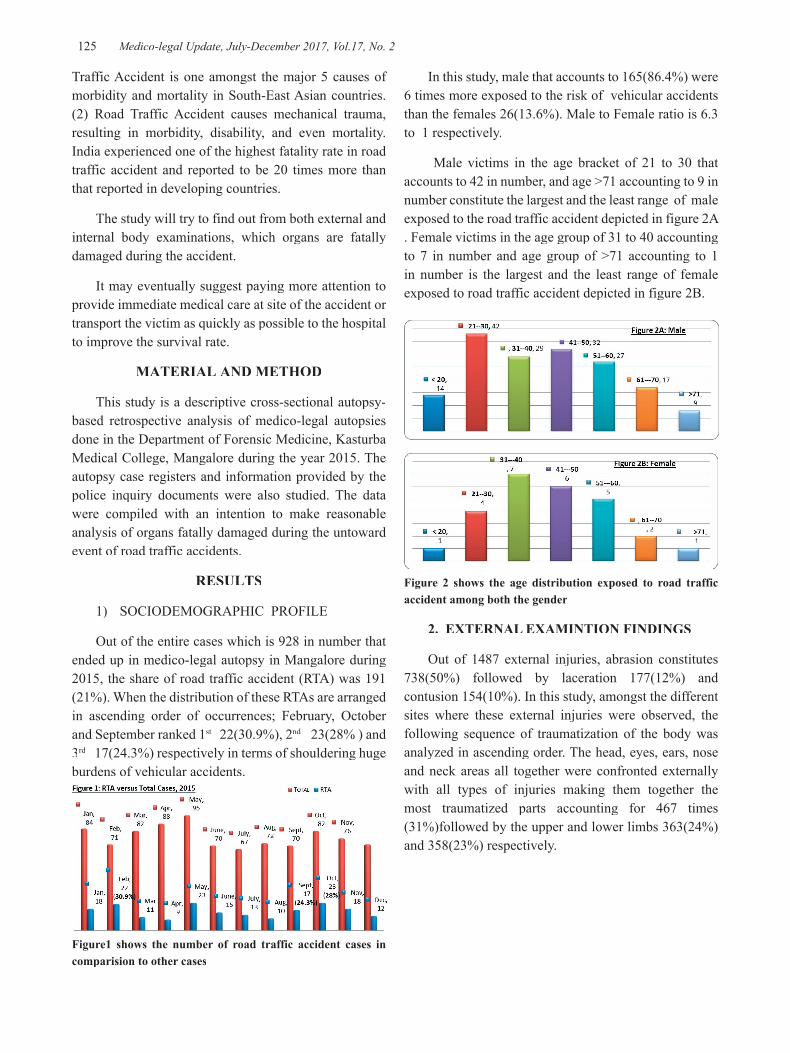
125 Medico-legal Update, July-December 2017, Vol.17, No. 2
Traffic Accident is one amongst the major 5 causes of morbidity and mortality in South-East Asian countries. (2) Road Traffic Accident causes mechanical trauma, resulting in morbidity, disability, and even mortality. India experienced one of the highest fatality rate in road traffic accident and reported to be 20 times more than that reported in developing countries.
The study will try to find out from both external and internal body examinations, which organs are fatally damaged during the accident.
It may eventually suggest paying more attention to provide immediate medical care at site of the accident or transport the victim as quickly as possible to the hospital to improve the survival rate.
MATERIAL AND METHOD
This study is a descriptive cross-sectional autopsy-based retrospective analysis of medico-legal autopsies done in the Department of Forensic Medicine, Kasturba Medical College, Mangalore during the year 2015. The autopsy case registers and information provided by the police inquiry documents were also studied. The data were compiled with an intention to make reasonable analysis of organs fatally damaged during the untoward event of road traffic accidents.
RESULTS
1) SOCIODEMOGRAPHIC PROFILE
Out of the entire cases which is 928 in number that ended up in medico-legal autopsy in Mangalore during 2015, the share of road traffic accident (RTA) was 191 (21%). When the distribution of these RTAs are arranged in ascending order of occurrences; February, October and September ranked 1st 22(30.9%), 2nd 23(28% ) and 3rd 17(24.3%) respectively in terms of shouldering huge rd 17(24.3%) respectively in terms of shouldering huge rd
burdens of vehicular accidents.
Figure1 shows the number of road traffic accident cases in comparision to other cases
In this study, male that accounts to 165(86.4%) were 6 times more exposed to the risk of vehicular accidents than the females 26(13.6%). Male to Female ratio is 6.3 to 1 respectively.
Male victims in the age bracket of 21 to 30 that accounts to 42 in number, and age >71 accounting to 9 in number constitute the largest and the least range of male exposed to the road traffic accident depicted in figure 2A . Female victims in the age group of 31 to 40 accounting to 7 in number and age group of >71 accounting to 1 in number is the largest and the least range of female exposed to road traffic accident depicted in figure 2B.
Figure 2 shows the age distribution exposed to road traffic accident among both the gender
2. EXTERNAL EXAMINTION FINDINGS
Out of 1487 external injuries, abrasion constitutes 738(50%) followed by laceration 177(12%) and contusion 154(10%). In this study, amongst the different sites where these external injuries were observed, the following sequence of traumatization of the body was analyzed in ascending order. The head, eyes, ears, nose and neck areas all together were confronted externally with all types of injuries making them together the most traumatized parts accounting for 467 times (31%)followed by the upper and lower limbs 363(24%) and 358(23%) respectively.

Medico-legal Update, July-December 2017, Vol.17, No. 2 126 127 -legal Update, July-December 2017, Vol.17, No. 2
3.2 INTRACRANIAL COMPARTMENT
The study shows 258 internal examination findings in the intracranial compartment. Out of which subarachnoid hemorrhage constitutes 116(45%) followed by subdural hemorrhage 85(33%) and intraventricular hemorrhage 24(9%).
3.3 FINDINGS OF EXTERNAL/INTERNAL THORACIC and HEART
This study finds out altogether 135 injuries seen sustained on the external and/or internal thoracic areas and including mediastinum. Out of which rib fracture took the highest share 64(47%) followed by pleural hemorrhage 33(24%) and subendocardial hemorrhage 14(10%).
3.4 FINDINGS OF INTERNAL EXAMINATION OF THE ABDOMEN
In the gastrointestinal system examination the findings were distributed taking into account major visceral organs like Liver laceration ,Liver rupture, Spleen rupture, Peritoneal hemorrhage, Diaphragm tear, Omental contusion, Mesentery tear and laceration, Jejunum tear, Intestinal rupture and Evisceration.
Figure 3: Shows the distribution of injuries
Figure 4: Shows the body areas involved during road traffic accident
3. INTERNAL EXAMINATION FINDINGS
3.1 SCALP AND SKULL
Examination was performed cranial to caudal. Internal injuries observed on the scalp and skull was totally 258 in numbers. Out of which contusion and /or hematoma on the scalp were 129(50%). Fractures sustained on the skull were classified as Flat impact (Linear, non-displaced fractures) and High Velocity Impact (Penetrating or depressed fractures).
Both linear fractures and basal linear fractures took the greatest toll and constitute 53(42%) and 56(44%) respectively. Comminuted and depressed fractures constituted 11(9%) in number were not seen much as compared to the former once. The distribution of these numbers of trauma across the months show that the highest were registered on May accounting for 41(16%) followed by November accounting for 32 (12%) and February accounting for 30(11%).
Figure 3: Shows the distribution of injuries

-legal Update, July-December 2017, Vol.17, No. 2 126 127 Medico-legal Update, July-December 2017, Vol.17, No. 2
Total 54 findings were seen in the abdomen during medico-legal examination. Out of which Peritoneal hemorrhage constitute 23(43%) followed by Liver laceration and rupture 18(33%), and Mesenteric laceration and rupture 5(8%).
4. TIME DISTRIBUTION OF ACCIDENT
The following findings were observed 73(38%) was seen during day time followed by 9(36%) at night and 49(26%) during morning.
5. TYPE OF VEHICLES CAUSING THE ACCIDENT
The type of vehicles involved during RTA in 2015 at Mangalore were generally known to have been undertaken by Bicycles, Motorized 2- ,3-,4-Wheeled and Trucks. The hit and run cases that are not witnessed account for 10 in number were unfortunately assumed intuitively to have been dealt with or inflicted by motorized 4-wheeled cars.
The share of motorized 2-wheeled and 4-wheeled vehicles were intimately close to cause the RTA events that victimized 84 and 83 persons with a percentage of 44% and 43% respectively.
6. VICTIMS EXPOSED FOR ROAD TRAFFIC ACCIDENT
The victims of RTAs were grossly classified as pedestrian and non-pedestrian (Vehicular occupants that include Driver and/or Passenger).
The non- pedestrians were the once fatally exposed with 112 persons (59%) as compared to the pedestrian 79 persons (41%).
7. CIRCUMSTANCE OF ROAD TRAFFIC ACCIDENT
The circumstance of death has been classified into Death at the site of the accident and Death on arrival
and/or after hospitalization. Death on arrival and/or after hospitalization were studied taking into account the following considerations on the body during conducting autopsy [Freshness of the body, Non-fixation of post mortem lividity, Presentation of the dead body at morgue covered with white hospital cloth, Presence of Surgically stapled or sutured wounds or Tracheostomy or POP slab or Metallic Nail, or Bed sore or partially Healed Wound on the body.]
Out of the total RTA victims 169(88%) had their autopsy done after death on arrival and/or after hospitalization while only 22(12%) were known to pass away at the site of the accident.
IMPRESSION AS THE CAUSE OF DEATH
This study categorized the causes that inflicted death to the victim into two major groups. The predominant cause being death as a complication of head injury has been considered as one class representing cranio cerebral injury while the other class of non-craniocerebral injury comprises of different causes of death as complications of injuries of crush, poly trauma, thoraco-abdominal, vertebral, major vessels and organs, etc. The study found out that cranio cerebral injury accounts for 134(70%) and the non-craniocerebral injuries accounts for 57(30%).
DISCUSSION
Out of the entire cases (928) in number that ended up in medico-legal autopsy in Mangalore during 2015, the share of road traffic accident (RTA) was 191 (21%).
In this study, male that accounts to 165(86.4%) were 6 times more exposed to the risk of vehicular accidents than the females 26(13.6%) .Similar studies done at Chandigarh and Bhopal showed males accounted for 84% and 82.5% respectively. 2,384% and 82.5% respectively. 2,384% and 82.5% respectively
The data revealed that the aggregate sum of all types of external injuries observed in this study amount to 1485 in number whereby abrasion comprises 738 (50%) followed by laceration 177(12%) and contusion 154 (10%).
Amongst the different sites where the injuries took place the data show the following sequence of traumatization of the body in ascending order. The Head, Eye, Ear, Nose & Neck parts summed up together were confronted externally and found to be the most traumatized parts accounting to 467 times ( 31%)
Medico-legal Update, July-December 2017, Vol.17, No. 2 128 129 -legal Update, July-December 2017, Vol.17, No. 2
followed by the upper & lower limbs 363 (24%) and 358 (23%) times respectively. Similarly other studies conducted in India showed abrasion, contusion and laceration to account for 100% followed by upper and lower limb fractures accounting for 29.8% and other fast-moving injuries 12.2% are almost consistent with the finding of the study in Fiji 4
Both linear fractures and basal linear fractures took the greatest toll and constitute 53(42%) and 56(44%) respectively. Comminuted and depressed fractures 11(9%) in number were not seen much as compared to the former once. The distribution of these numbers of trauma across the months show that the highest were registered on May accounting for 41(16%) followed by November accounting for 32 (12%) and February accounting for 30(11%). Similarly, other study done at Manipal showed similar findings. (10) Similarly, other autopsy- based study done in rural district of Maharashtra showed that injuries on the scalp happened to be the commonest once seen in 129 of the entire 138 traumatic head injury cases. skull that sustained linear fracture with the associated basal skull fracture was found to be the commonest finding seen in 23 cases (24.21%) followed by linear fracture only (16.84) 5,6,7
There were totally 258 findings in the intracranial compartment. Out of which subarachnoid hemorrhage constitute 116(45%) followed by subdural hemorrhage 85(33%) and intra ventricular hemorrhage 24(9%). Similar trends were seen by other studies whereby subdural hemorrhage constitutes (77%) followed by subarachnoid hemorrhage (55%). 5
Injuries sustained around the thorax and inside the mediastinum were totally 135 in number. Out of which rib fracture constitutes 64(47%) followed by pleural hemorrhage 33(24%) and subendocardial hemorrhage 14(10%). Likewise, other study shows similar findings that injuries sustained over the external surface of the thorax were seen in 136 victims while injures over the internal aspect of the thorax were reported in 80 cases. 8
Total 54 findings registered in the abdomen. Out of which Peritoneal hemorrhage constitute 23(43%) followed by Liver laceration 13(24%), Liver rupture 5(9%) and Mesenteric laceration 3(6%). Similarly other study found out that liver laceration was more common than contusions 8
The share of motorized 2-wheeled and 4-wheeled
vehicles were intimately close to cause the RTA events that victimized 84 and 83 persons with a percentage of 44% and 43% respectively. Likewise other study done at Gujarat had shown that, amongst 236 drivers or riders ,motorized 2 wheeler riders accounted for 75% accompanied by 4 –wheeler drivers (10.59%) and those riding bicycles (10.59%) 9,10,11
The non- pedestrians were the once fatally exposed with 112 persons (59%) as compared to the pedestrian 79 persons (41%).There is similarity in this study finding with other study done in Fiji where vehicular occupants took the largest share 67.5% to be fatally exposed to RTA as compared to pedestrian (Fiji) 4
The study found out that craniocerebral injury accounts for 134(70%) and the non-craniocerebral injuries 57(30%). Likewise, autopsy-based study in a rural district of Maharashtra showed similar result that the craniocerebral injury was the major cause of death accounting for 39.57% 7
CONCLUSIONS
The energetic and the younger male sex seem to be at the forefront to be victimized from RTAs. Apart from the huge death toll, the economic burden influenced by these untoward events needs meticulous consideration on how to curb the accident.
1) The very promising result deduced from this quantitative cross sectional study witnesses the efficiency of the pre-hospital care along with trauma centers to extend the survival rate of victims who had the opportunity of hospitalization. This achievement must be further augmented so that it remains stand fast in constant improvement to attain Excellency. In fact this is one of the options left to increase the survival rate after the incidence of RTA if we are not presently in a position to make our roads at least 80-90% safe for both pedestrians and non-pedestrians.
2) In almost all brief descriptions narrated by traffic polices who had custody to the body of the victim during the RTAs; there is often one word “negligence” used to refer to the contribution of the driver to the accident. This “negligence “could mean reckless and speedy driving. So the easy solution may be to think of limiting the driving speed on the road.
3) Sensitization of the public on how to take safe precautions while crossing and using roads obviously
-legal Update, July-December 2017, Vol.17, No. 2 128 129 Medico-legal Update, July-December 2017, Vol.17, No. 2
can have life-saving effect on pedestrians.
4) Over bridges and under passes may have paramount importance to pedestrians who often try to cross the roads struggling against the traffic that may expose them to accidents. These techniques can produce win-win effects both for pedestrians who will be protected from road traffic accident and drivers who want to maintain their momentum of speed on the road.
5) If a certain portion of the road for vehicle transport is partitioned to be dedicated for motorized 2-and 3-wheeled vehicles only, this may have some bilateral impact to decrease vehicle collisions on one hand and reduce the possible exposure of pedestrians to fatal accidents.
Acknowledgement:- The authors are indebted to all the teaching and non-Teaching faculty of Department of Forensic Medicine, KMC, Mangalore for the source and time during the article preparation. The research is ostensibly supported by the Manipal University, India and Mekelle University, Ethiopia as a part of MoU programme. The authors credits Professor Harish Kumar , Ayder Research Hospital , Mekelle Hospital for all the support in facilitating the residents to do collaborative work .
Ethical Clearance- Taken from Institutional Ethical committee-IEC KMC MLR 11-16/313 (KMC, Mangalore)
Source of Funding- Nil
Conflict of Intrest- Nil
REFERRENCES
1) Paden M, McGee K,Krug E.Geneva, Switzerland: World Health Organization. 2000,2002. Injury: The global burden of disease.
2) Khare N,Gupta SK,Varashney A,Athavale AV.
Epidemological study of road traffic accident cases attending tertiary care hospital, in Bhopal Madhya Pradesh. Nat J Community Med2012,3(3):395
3) Shama D,Singh US,Mukherjee S.A study on road traffic accidents in Anand Gujarat. Healthline 2011 2(2) : 12
4) James JVP Kalgugivaki,Ramaswamy PS Goundar. Retrospective Autopsy study of Fatal road Traffic Accident in Fiji. J Forensic Res 2014, 5:6
5) Anand Menon, Nagesh KR. Pattern of fatal head injuries due to vehicular accidents in Manipal. JIAFM, 2005:27(1),19-22
6) J Chandra, TD Dogra, P C Dixit. Pattern of cranio-intracranial injuries in fatal vehicular accidents in Delhi, 1966-76 Med. Sci. Law (1979) Vol.19, No. 3,186-94
7) Ranjit M. Tandle,A.M. Kediya: patterns of head injuries and fatal road traffic accidents in a rural district of Maharashtra. Autopsy- based study. JIAFM. July-September 2011, Vol.33, No.3
8) Suresh Kumar SB, Tanuj K,Ritesh GM, Shankar MB, Vinod CN,Yoganaras imha K. Victim profile and Pattern of Thoraco-Abdominal Injuries sustained in Fatal Road Traffic Accidents.J Indian Acad Forensic Med. 2012;34:17–20.
9) Animesh Jain,Ritesh G.Menezes,Tanuj Kanchan,S.Gagan,Rashmi Jain. Two Wheeler accidents on Indian roads a study from mangalore,India. Journal of Forensic and Legal Medicine.
10) Singh A,Bhardwaj A,Pathak R, Ahluwalia SK. An epidemiological study of road traffic accident cases at a tertiary care hospital in rural Haryana. Indian J of community health 2011; 23(2): 53
11) Gururaj G,Kolluri SVR, Chandramouli BA, Subbakrishna DK, Krus JF. Traumatic Brain Injury. Publication No.61. Bangaluru 360029,India: National Institute of Mental Health and Neurosciences;2005:17
-

131 -legal Update, July-December 2017, Vol.17, No. 2
Pattern of Malaria Infection in District Bhiwani, Haryana-A Two Year Study
Anil Kumar Chahal1, Jitender Kumar Jakhar2, Jasminder Singh3, Randeep Singh Poonia4
1Medical Microbiologist, General Hospital Bhiwani, 2Associate Professor, Department of Forensic Medicine, Pt. 2Associate Professor, Department of Forensic Medicine, Pt. 2
B.D. Sharma Post Graduate, Institute of Medical Sciences, Rohtak, 3Associate Professor, Department of Medicine, 3Associate Professor, Department of Medicine, 3
Pt. B.D. Sharma Post Graduate Institute of Medical Sciences, Rohtak, 4Civil Surgeon, District Bhiwani, Haryana
ABSTRACT
Malaria is a well-known mosquito-borne illness and it continues to be a major public health problem at the start of new millennium. The problem is persistent in both rural and urban area.We studied the problem and certain epidemiological factors associated with malaria, pattern and seasonal trend of malaria infection in Bhiwani District Haryana.
The data of peripheral blood films prepared from 1st January 2015 to 20th December 2016 were collected from the district malaria office Bhiwani. Data were analysed and observed that 446113 slides of blood smear taken from patients with symptom suggestive of malaria , out of which 355(0.07%) were found smear positive for malaria parasite. Plasmodium vivax found in 100% cases and Plasmodium falciparum found in zero percent cases. Mixed infection was also not observed.
Malaria was reported in all age groups and both sexes, but the 11–20 year age group were affected more (26.42%) and 52.13% patients were 11 to 30 years. Males were more affected than females. Majority of cases reported in August and September months. The present study reported the prevalence of Plasmodium vivax more than Plasmodium falciparum in our region. Adult males were more prone to infect with malaria.
Keyword: Malaria, Plasmodium vivax, Plasmodium falciparum.
Corresponding author:Dr. Jitender Kumar JakharAssociate Professor, 5/8J Medical Campus PGIMS, Rohtak.
INTRODUCTION
Malaria is common of mosquito-borne illnesses in our country. It is a protozoan disease caused by the parasites of the genus plasmodium. Common species in India are Plasmodium vivax, Plasmodium malariae, Plasmodium falciparum, and Plasmodium ovale. Sporozoite of Plasmodium is transmitted by the bite of female Anopheles mosquitoes commonly from dusk to dawn1. A typical attack comprises of three stages such as cold stage, hot stage and sweating stage and showing symptoms such as headache, fever, shivering, vomiting, haemolytic anaemia, jaundice, haemoglobin in urine, retinal damage and convulsions2. Plasmodium vivax and Plasmodium falciparum are commonly seen
in the tropical countries like Africa, India, Pakistan, Afghanistan, Shri-Lanka, Middle East and South East Asia, Plasmodium Malariae and Plasmodium Ovale are less common cause of disease and generally do not cause severe illness3.
Approximately, 5% of the world’s population is infected by malaria, which includes African regions with vast number of cases (81%), followed by the South-East Asia (13%), and the Eastern Mediterranean region (5%) with over 1 million deaths each year in the world. In India, malaria is mainly caused by Plasmodium vivax and Plasmodium falciparum. Despite significant national and international efforts to control malaria, the disease remains one of the top public health problems in the country. In Haryana, most of the malaria cases (80- 90%) are caused by P. vivax and the remainder by P. falciparum4.
In the far north of India, Haryana is a key agricultural state, dependent on the monsoon rains
DOI Number: 10.5958/0974-1283.2017.00084.6
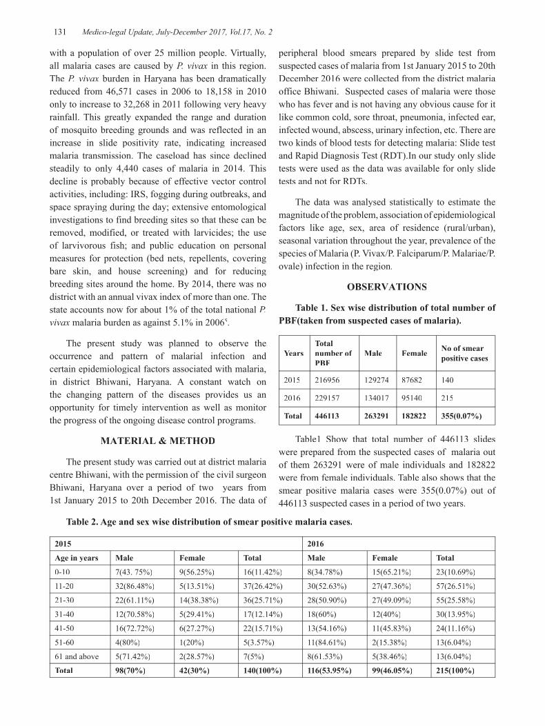
131 Medico-legal Update, July-December 2017, Vol.17, No. 2
with a population of over 25 million people. Virtually, all malaria cases are caused by P. vivax in this region. The P. vivax burden in Haryana has been dramatically reduced from 46,571 cases in 2006 to 18,158 in 2010 only to increase to 32,268 in 2011 following very heavy rainfall. This greatly expanded the range and duration of mosquito breeding grounds and was reflected in an increase in slide positivity rate, indicating increased malaria transmission. The caseload has since declined steadily to only 4,440 cases of malaria in 2014. This decline is probably because of effective vector control activities, including: IRS, fogging during outbreaks, and space spraying during the day; extensive entomological investigations to find breeding sites so that these can be removed, modified, or treated with larvicides; the use of larvivorous fish; and public education on personal measures for protection (bed nets, repellents, covering bare skin, and house screening) and for reducing breeding sites around the home. By 2014, there was no district with an annual vivax index of more than one. The state accounts now for about 1% of the total national P. vivax malaria burden as against 5.1% in 20065.
The present study was planned to observe the occurrence and pattern of malarial infection and certain epidemiological factors associated with malaria, in district Bhiwani, Haryana. A constant watch on the changing pattern of the diseases provides us an opportunity for timely intervention as well as monitor the progress of the ongoing disease control programs.
MATERIAL & METHOD
The present study was carried out at district malaria centre Bhiwani, with the permission of the civil surgeon Bhiwani, Haryana over a period of two years from 1st January 2015 to 20th December 2016. The data of
peripheral blood smears prepared by slide test from suspected cases of malaria from 1st January 2015 to 20th December 2016 were collected from the district malaria office Bhiwani. Suspected cases of malaria were those who has fever and is not having any obvious cause for it like common cold, sore throat, pneumonia, infected ear, infected wound, abscess, urinary infection, etc. There are two kinds of blood tests for detecting malaria: Slide test and Rapid Diagnosis Test (RDT).In our study only slide tests were used as the data was available for only slide tests and not for RDTs.
The data was analysed statistically to estimate the magnitude of the problem, association of epidemiological factors like age, sex, area of residence (rural/urban), seasonal variation throughout the year, prevalence of the species of Malaria (P. Vivax/P. Falciparum/P. Malariae/P. ovale) infection in the region.
OBSERVATIONS
Table 1. Sex wise distribution of total number of PBF(taken from suspected cases of malaria).
Years Total number of PBF
Male Female No of smear positive cases
2015 216956 129274 87682 140
2016 229157 134017 95140 215
Total 446113 263291 182822 355(0.07%)
Table1 Show that total number of 446113 slides were prepared from the suspected cases of malaria out of them 263291 were of male individuals and 182822 were from female individuals. Table also shows that the smear positive malaria cases were 355(0.07%) out of 446113 suspected cases in a period of two years.
Table 2. Age and sex wise distribution of smear positive malaria cases.
2015 2016
Age in years Male Female Total Male Female Total
0-10 7(43. 75%) 9(56.25%) 16(11.42%) 8(34.78%) 15(65.21%) 23(10.69%)
11-20 32(86.48%) 5(13.51%) 37(26.42%) 30(52.63%) 27(47.36%) 57(26.51%)
21-30 22(61.11%) 14(38.38%) 36(25.71%) 28(50.90%) 27(49.09%) 55(25.58%)
31-40 12(70.58%) 5(29.41%) 17(12.14%) 18(60%) 12(40%) 30(13.95%)
41-50 16(72.72%) 6(27.27%) 22(15.71%) 13(54.16%) 11(45.83%) 24(11.16%)
51-60 4(80%) 1(20%) 5(3.57%) 11(84.61%) 2(15.38%) 13(6.04%)
61 and above 5(71.42%) 2(28.57%) 7(5%) 8(61.53%) 5(38.46%) 13(6.04%)
Total 98(70%) 42(30%) 140(100%) 116(53.95%) 99(46.05%) 215(100%)

Medico-legal Update, July-December 2017, Vol.17, No. 2 132 133 -legal Update, July-December 2017, Vol.17, No. 2
Table 2. Shows age and sex wise distribution of malaria positive cases. It also shows that males are affected more than the female. In year 2015, 70% males were affected and in 2016, 54% males were affected. This table also shows that the 11-20 years age group is highest(26.42%) in affected individuals and the age group from 11to 30 make the major contribution (52.13%) to the smear positive cases.
Table 3. Age wise and residential area wise (rural, urban) distribution of smear positive malaria cases.
2015 2016
Age in years Urban Rural Total Urban Rural Total
0-10 4 12 16(11.42%) 3 20 23(10.69%)
11-20 4 33 37(26.42%) 10 47 57(26.51%)
21-30 11 25 36(25.71%) 10 45 55(25.58%)
31-40 3 14 17(12.14%) 3 27 30(13.95%)
41-50 6 16 22(15.71%) 11 13 24(11.16%)
51-60 1 4 5(3.57%) 4 9 13(6.04%)
61 and above 0 7 7(5%) 0 13 13(6.04%)
Total 29(20%) 111(80%) 140(100%) 41(19.06%) 174(80.93%) 215(100%)
Table 3 Shows the age wise urban, rural distribution of malaria positive cases. The table shows that the malaria positive cases were more in rural areas than in urban areas. 80% positive cases were from rural areas.
Table 4. Month wise distribution(Male, Female) of smear positive malaria cases.
2015 2016
Months Male Female Total Male Female Total
January 1 1 2(1.42%) 2 0 2(0.93%)
February 2 1 3(2.14%) 0 0 0(0%)
March 3 0 3(2.14%) 1 0 1(0.46%)
April 1 1 2(1.42%) 1 1 2(0.93%)
May 3 1 4(2.85%) 6 1 7(3.25%)
June 2 1 3(2.14%) 7 1 8(3.72%)
July 19 7 26(18.57%) 14 15 29(13.48%)
August 8 5 13(9.28%) 39 37 76(35.34%)
September 48 20 68(48.57%) 40 34 74(34.41%)
October 8 4 12(8.57) 5 10 15(6.97%)
November 0 4 4(2.85%) 1 0 1(0.46%)
December 0 0 0(0%) 0 0 0(0%)
Total 95(67.85%) 45(32.14%) 140(100%) 116(53.95%) 99(46.04%) 215(100%)
Table 4 Shows month wise distribution of smear positive malaria cases. It is observed from the table that highest smear positive cases occurred in month of August and September. In month of December the smear positive cases were zero.
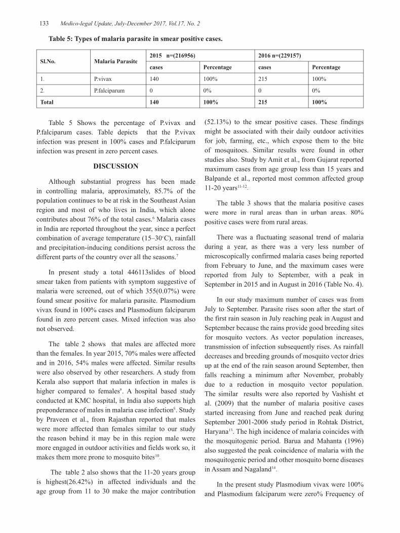
-legal Update, July-December 2017, Vol.17, No. 2 132 133 Medico-legal Update, July-December 2017, Vol.17, No. 2
Table 5: Types of malaria parasite in smear positive cases.
Sl.No. Malaria Parasite2015 n=(216956) 2016 n=(229157)
cases Percentage cases Percentage
1. P.vivax 140 100% 215 100%
2. P.falciparum 0 0% 0 0%
Total 140 100% 215 100%
Table 5 Shows the percentage of P.vivax and P.falciparum cases. Table depicts that the P.vivax infection was present in 100% cases and P.falciparum infection was present in zero percent cases.
DISCUSSION
Although substantial progress has been made in controlling malaria, approximately, 85.7% of the population continues to be at risk in the Southeast Asian region and most of who lives in India, which alone contributes about 76% of the total cases.6 Malaria cases in India are reported throughout the year, since a perfect combination of average temperature (15–30oC), rainfall and precipitation-inducing conditions persist across the different parts of the country over all the seasons.7
In present study a total 446113slides of blood smear taken from patients with symptom suggestive of malaria were screened, out of which 355(0.07%) were found smear positive for malaria parasite. Plasmodium vivax found in 100% cases and Plasmodium falciparum found in zero percent cases. Mixed infection was also not observed.
The table 2 shows that males are affected more than the females. In year 2015, 70% males were affected and in 2016, 54% males were affected. Similar results were also observed by other researchers. A study from Kerala also support that malaria infection in males is higher compared to females8. A hospital based study conducted at KMC hospital, in India also supports high preponderance of males in malaria case infection9. Study by Praveen et al., from Rajasthan reported that males were more affected than females similar to our study the reason behind it may be in this region male were more engaged in outdoor activities and fields work so, it makes them more prone to mosquito bites10.
The table 2 also shows that the 11-20 years group is highest(26.42%) in affected individuals and the age group from 11 to 30 make the major contribution
(52.13%) to the smear positive cases. These findings might be associated with their daily outdoor activities for job, farming, etc., which expose them to the bite of mosquitoes. Similar results were found in other studies also. Study by Amit et al., from Gujarat reported maximum cases from age group less than 15 years and Balpande et al., reported most common affected group 11-20 years11-12.
The table 3 shows that the malaria positive cases were more in rural areas than in urban areas. 80% positive cases were from rural areas.
There was a fluctuating seasonal trend of malaria during a year, as there was a very less number of microscopically confirmed malaria cases being reported from February to June, and the maximum cases were reported from July to September, with a peak in September in 2015 and in August in 2016 (Table No. 4).
In our study maximum number of cases was from July to September. Parasite rises soon after the start of the first rain season in July reaching peak in August and September because the rains provide good breeding sites for mosquito vectors. As vector population increases, transmission of infection subsequently rises. As rainfall decreases and breeding grounds of mosquito vector dries up at the end of the rain season around September, then falls reaching a minimum after November, probably due to a reduction in mosquito vector population. The similar results were also reported by Vashisht et al. (2009) that the number of malaria positive cases started increasing from June and reached peak during September 2001-2006 study period in Rohtak District, Haryana13. The high incidence of malaria coincides with the mosquitogenic period. Barua and Mahanta (1996) also suggested the peak coincidence of malaria with the mosquitogenic period and other mosquito borne diseases in Assam and Nagaland14.
In the present study Plasmodium vivax were 100% and Plasmodium falciparum were zero% Frequency of

Medico-legal Update, July-December 2017, Vol.17, No. 2 134 135 -legal Update, July-December 2017, Vol.17, No. 2
P. Vivax (100%) cases was remarkably high with, while the study conducted by Swetha et al., they found 95.83% cases were Plasmodium vivax 4.13% were Plasmodium falciparum15. It is quite probable that few cases would have been missed due to non reporting in our study.
From the findings of the study, it has been observed that P. vivax is the most common malaria infection in District Bhiwani. Similar observations were also made in neighbouring Haryana16. Similarly in Sri Lanka, the majority of the malaria cases were caused by P. vivax17. However in the Purulia district of West Ben-gal, a tribal dominated area, P. falciparum infection was the most common (20%) followed by P. vivax (6.4%)18. It is observed during the study that in two year of study period no death due to malaria was reported.
Malaria mortality and morbidity may be reduced significantly by improving diagnostic tests, and management of the cases to prevent further unwanted complications. A sustainable community based system from diagnoses and treatment through community health workers, school teachers, influential people, religious leaders etc. would be more effective. There is an urgent need to increase the awareness regarding climate change and the possibility of increased incidence of vector borne diseases and their prevention. Community participation through information, education and communication activities is the one of the keys of the success.
CONCLUSION
The findings of this study confirmed that malaria endemicity is very high in the study area and the major outbreak of malaria was caused by P. vivax rather than P. falciparum. Most of the patients seen were in the age group of 11-20 years old. There is a seasonal variation in the incidence, the peak being in the month of August and September. An integrated approach is needed which includes mass survey or active surveillance, appropriate drug therapy, spraying of insecticide and environmental sanitation before July. Special attention should be given to the children and females.
Acknowledgement: Nil
Source of Funding: Self
Conflict-of-Interest Statement: There is no conflict of interest as it was a study based on the already available record.
Statement of Informed consent: This study is based on the available record so there is no matter of consent.
Statement of Human and Animal Rights: No human right and animal right is violated in this case.
No ethical violation is done.
REFERENCE
1. Sweta M Murawala, Vyoma Chudasama, Mahendra Vegad Prevalence of mosquito-borne Plasmodium infection in patients attending fever clinic in a tertiary-care teaching hospital, western India International Journal of Medical Science and Public Health 2015;4:11
2. Chatterjee KD. Parasitology (Protozoology and Helminthology), 13th edn. New Delhi: CBS, 2012. pp. 90–127.
3. Park K., Text book of Preventive and Social Medicine,2002 XV111 edn; 193
4. World Malaria Report 2011, (World Health Organization, Geneva, 2012).
5. Director of Health Services (Malaria), 2011. Annual Report on Implementation of National Vector Borne Disease Control Programme (NVBDCP) in Haryana State.
6. Kondrachine AV. Malaria in WHO Southeast Asia Region. Indian J Malariol 1992; 29: 129–160.
7. Das A, Anvikar AR, Cator LJ, Dhiman RC, Eaten A, Mishra N. Malaria in India: The Center for the Study of Complex Malaria in India. Acta Trop 2012; 121(3): 267–273.
8. Chandrashekar UK, Sudharshan Murthy KA and Sunila. A study on clinical profile and outcome in falciparum malaria, Calicut Medical Journal 2011; 9:3
9. Chowta M.N. and Chowta K.N., Study of clinical profile of malaria at KMC, Hospital Attavar, India, Journal of clinical and diagnostic research, 2007;3:110-115
10. Kumar A Praveen, Ramchandran R. Malaria associated factors in Thar Desert of Rajasthan, India: A case-control study. Journal of Rural Tropical Public Health 2010; 9: 6166.
11. Amit H. Agravat, Gauravi A. Dhruva , Khyati Babaria, Kalpesh Rathod Clinico-Hematological
-legal Update, July-December 2017, Vol.17, No. 2 134 135 Medico-legal Update, July-December 2017, Vol.17, No. 2
pattern of Malaria in Rajkot city, Gujrat, India. International Journal of Biomedical and Advance Research 2012; 03:11
12. Balpande L, Gupta SK, Agarwal SS Epidemiological trends of malaria cases in rural health and training centre of Madhya Pradesh National Journal of Community Medicine 2014;5(2):227–9.
13. Vashisht BM, Kalhan M, Seema, Jyothi. Situation Analysis of Malaria in District Rohtak, Haryana. J Comm Dis 2009; 41(2): 137-138.
14. Barua, HC, Mahanta J. Serological evidence of Den-2 activity in Assam and Nagaland. J Comm Dis 1996; 28: 56-58.
15. Sweta M Murawala, Vyoma Chudasama, Mahendra Vegad Prevalence of mosquito-borne Plasmodium infection in patients attending fever clinic in a
tertiary-care teaching hospital, western India International Journal of Medical Science and Public Health 2015;4:11
16. Srivastava A, Nagpal BN, Saxena R, Wadhwa TC, Mohan S, Siroha GP, Prasad J, Subbarao SK. Malaria epidemicity of Mewat region, District Gurgaon, Haryana, India: a GIS-based study. Curr Sci 2004;86:1297-1303.
17. Konradsen F, Steele P, Perera D, Hock W V, Amarasinghe F P. Cost of Malaria Control in Sri Lanka. Bull World Health Organ 1997;77:301-9.
18. Mukhopadhyay AK, Hati AK, Dey P. Malario-genic situation in areas of Aiodhya Hills of Dis-trict Purulia West Bengal and its present status. Indian J Public Health 2001;45:31-32.

137 -legal Update, July-December 2017, Vol.17, No. 2
Pact Suicide Two Sister end their Life by Hanging - A Case Report
Shamim Raza1, M K Pathak2, S K Tripathi3, S K Pandey4
1Jr-III, 2Associate Professor, 3Professor, 4Assistant Professor, Department of Forensic Medicine, Institute of Medical Sciences, Banaras Hindu University, Varanasi, U.P
ABSTRACT
A suicide pact means “the suicides of two or more individuals in an agreed-upon plan”. Plan may be to die together / separately & closely timed. The individuals involved in pact suicides have been usually ‘two adults’ occasionally reports says ‘triple pact suicide’. Pact suicides have been most commonly reported from Japan. The usual profile of persons involved in pact suicides includes those females, older, married and of high social class. We discussed a case of suicide pact where. Two sisters end their life by hanging, in present case the history of emotional stress found due to the death of their mother & father resulting in severe degree of depression and finely financial constraint.
Keywords: Pact suicide, mass suicide, depression, dupattas.
Correspondence: Dr. Shamim Raza Email – [email protected]
CASE REPORT
Elder sister 45 years graduate and younger sister 40 years old with law graduate, both were married and very intimate to their parents.
After demise of their mother they left their in-laws and started residing with their father. Unfortunately after 4 years father also died, this sudden event in their lives put them “in severe grade of depression” resulting in ‘Apathy’.
They withdrawn themselves from routine life activities with lost their jobs resulting in extreme degree of poverty; also they restricted their lives and closed themselves in a room of their house for about 18 years. Somehow the neighbors were kind enough to provide them food with great difficulty.
Sudden one morning, on 22nd Nov’16 the neighbors found “foul smell emitting out from the house of the victims, rooms found bolted from inside, police were informed. After opening the door a very dirty house found with full of foul smell”. Both sisters were found hanged with their “dupattas”. One with ceiling fan and another with the iron bars of the ventilator of the bathroom.
After inquest both deceased were sent for medico legal postmortem examination in the department of forensic medicine, Institute Of Medical Sciences, BHU on dated 24 Nov.2016.
PACT SUICIDE -INTRODUCTION
A suicide pact means “the suicides of two or more individuals in an agreed-upon plan”. Plan may be to die together / separately & closely timed.
The individuals involved in pact suicides have been usually ‘two adults’ occasionally reports says ‘triple pact suicide’.1 Pact suicides have been most commonly reported from Japan.2 The usual profile of persons involved in pact suicides includes those females, older, married and of high social class.3 In general, suicide pact victims choose non- violent suicide methods: commonly used methods are “poisoning by a car exhaust or over- dosing with medicine”.4 A rare case of a suicide pact has been reported where two young men were found dead near their severely damaged car with gags in their mouths and a rope tightened around their necks and fixed to the car.5 In India, pact suicides generally involve “issueless couples, disappointed lovers, unmarried sisters, frustrated individuals or a woman ending her own life with her children due to love affection not to leave alone them in difficulties”.6
When many people commit suicide together, it is known
DOI Number: 10.5958/0974-1283.2017.00085.8
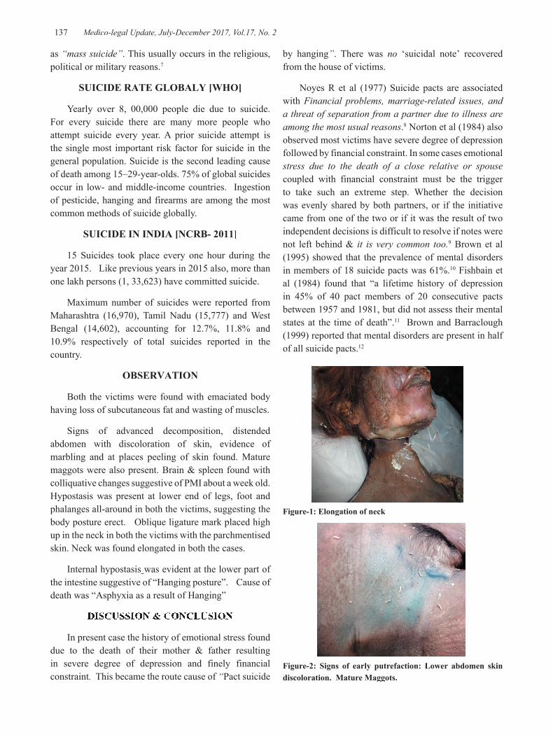
137 Medico-legal Update, July-December 2017, Vol.17, No. 2
as “mass suicide”. This usually occurs in the religious, political or military reasons.7
SUICIDE RATE GLOBALY [WHO]
Yearly over 8, 00,000 people die due to suicide. For every suicide there are many more people who attempt suicide every year. A prior suicide attempt is the single most important risk factor for suicide in the general population. Suicide is the second leading cause of death among 15–29-year-olds. 75% of global suicides occur in low- and middle-income countries. Ingestion of pesticide, hanging and firearms are among the most common methods of suicide globally.
SUICIDE IN INDIA [NCRB- 2011]
15 Suicides took place every one hour during the year 2015. Like previous years in 2015 also, more than one lakh persons (1, 33,623) have committed suicide.
Maximum number of suicides were reported from Maharashtra (16,970), Tamil Nadu (15,777) and West Bengal (14,602), accounting for 12.7%, 11.8% and 10.9% respectively of total suicides reported in the country.
OBSERVATION
Both the victims were found with emaciated body having loss of subcutaneous fat and wasting of muscles.
Signs of advanced decomposition, distended abdomen with discoloration of skin, evidence of marbling and at places peeling of skin found. Mature maggots were also present. Brain & spleen found with colliquative changes suggestive of PMI about a week old. Hypostasis was present at lower end of legs, foot and phalanges all-around in both the victims, suggesting the body posture erect. Oblique ligature mark placed high up in the neck in both the victims with the parchmentised skin. Neck was found elongated in both the cases.
Internal hypostasis was evident at the lower part of the intestine suggestive of “Hanging posture”. Cause of death was “Asphyxia as a result of Hanging”
DISCUSSION & CONCLUSION
In present case the history of emotional stress found due to the death of their mother & father resulting in severe degree of depression and finely financial constraint. This became the route cause of “Pact suicide
by hanging”. There was no ‘suicidal note’ recovered from the house of victims.
Noyes R et al (1977) Suicide pacts are associated with Financial problems, marriage-related issues, and a threat of separation from a partner due to illness are among the most usual reasons.8 Norton et al (1984) also observed most victims have severe degree of depression followed by financial constraint. In some cases emotionalstress due to the death of a close relative or spousecoupled with financial constraint must be the trigger to take such an extreme step. Whether the decision was evenly shared by both partners, or if the initiative came from one of the two or if it was the result of two independent decisions is difficult to resolve if notes were not left behind & it is very common too.9 Brown et al (1995) showed that the prevalence of mental disorders in members of 18 suicide pacts was 61%.10 Fishbain et al (1984) found that “a lifetime history of depression in 45% of 40 pact members of 20 consecutive pacts between 1957 and 1981, but did not assess their mental states at the time of death”.11 Brown and Barraclough (1999) reported that mental disorders are present in half of all suicide pacts.12
Figure-1: Elongation of neck
Figure-2: Signs of early putrefaction: Lower abdomen skin discoloration. Mature Maggots.
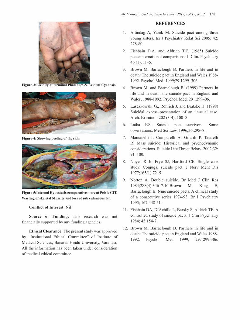
Medico-legal Update, July-December 2017, Vol.17, No. 2 138
Figure-3:Lividity at terminal Phalanges & Evident Cyanosis.
Figure-4: Showing peeling of the skin
Figure-5:Internal Hypostasis comparative more at Pelvic GIT. Wasting of skeletal Muscles and loss of sub cutaneous fat.
Conflict of Interest: Nil
Source of Funding: This research was not financially supported by any funding agencies.
Ethical Clearance: The present study was approved by “Institutional Ethical Committee” of Institute of Medical Sciences, Banaras Hindu University, Varanasi. All the information has been taken under consideration of medical ethical committee.
REFERENCES
1. Altindag A, Yanik M. Suicide pact among three young sisters. Isr J Psychiatry Relat Sci 2005; 42:278-80
2. Fishbain D.A. and Aldrich T.E. (1985) Suicide pacts:international comparisons. J. Clin. Psychiatry 46 (1), 11–5.
3. Brown M, Barraclough B. Partners in life and in death: The suicide pact in England and Wales 1988-1992. Psychol Med. 1999;29:1299–306
4. Brown M. and Barraclough B. (1999) Partners in life and in death: the suicide pact in England and Wales, 1988-1992. Psychol. Med. 29 1299–06.
5. Lasczkowski G., Röhrich J. and Bratzke H. (1998) Suicidal excess–presentation of an unusual case. Arch. Kriminol. 202 (3-4), 100–8
6. Latha KS. Suicide pact survivors: Some observations. Med Sci Law. 1996;36:295–8.
7. Mancinelli I, Comparelli A, Girardi P, Tatarelli R. Mass suicide: Historical and psychodynamic considerations. Suicide Life Threat Behav. 2002;32:91–100.
8. Noyes R Jr, Frye SJ, Hartford CE. Single case study. Conjugal suicide pact. J Nerv Ment Dis 1977;165(1):72–5
9. Norton A. Double suicide. Br Med J Clin Res 1984;288(4):346–7.10.Brown M, King E, Barraclough B. Nine suicide pacts. A clinical study of a consecutive series 1974-93. Br J Psychiatry 1995; 167:448-51.
11. Fishbain DA, D’Achille L, Barsky S, Aldrich TE. A controlled study of suicide pacts. J Clin Psychiatry 1984; 45:154-7.
12. Brown M, Barraclough B. Partners in life and in death: The suicide pact in England and Wales 1988-1992. Psychol Med 1999; 29:1299-306.

-legal Update, July-December 2017, Vol.17, No. 2 138
Study of Fingerprint Pattern and Gender Distribution of Fingerprints
Singh PM1, Singh H2, Aggarwal OP3, Kaur B4
1Junior Resident, 2Professor, 2Professor, 2 3Professor, 4Professor and Head, 4Professor and Head, 4
Department of Forensic Medicine, MMIMSR, Mullana, Ambala, Haryana, India
ABSTRACT
Identification means determination of individuality of a person. This work was undertaken to study the fingerprint pattern and Gender distribution of fingerprints. The present study was conducted at department of Forensic Medicine MMIMSR, Mullana from Nov 2014 to July 2016. Total number of subjects included were 250 Males and 250 Females. Amongst male age group ranged from 18-64 years mean age was 31.88+9.17 and amongst female age group ranged from 18-64 years with the mean age of 33.5+10.12. The results showed that the most frequent pattern among both Males and Females was Ulnar loop (37.7%) in Males and (45.2%) in Females. It was followed by Plain whorl (24.4%) in Males and (22.0%) in Females. Least noted pattern among both Males (0.7%) and Females (0.3%) was Exceptional Arch. Mean ridge count in Males and Females was 12.58 and 12.03 respectively. The ridge count in Males was slightly more than Females.
Keywords: Fingerprint, ridge count, Identification, Individuality, Arches, Loops, Whorls
Corresponding author Dr. Preet Mohinder SinghPhone: +91- 7404660667, Email: [email protected]
INTRODUCTION
Establishing individuality of a person is called identification. Fingerprint is based on the principle that skin of the balls of the fingers and thumbs is covered with characteristics ridges, the arrangement and distribution of which remains constant and persists throughout life and that the patterns of no two hands resembles each other. Even the fingerprints of the twins are different. Probability of two individuals having similar fingerprints is one in sixty-four thousand million.1 Finger prints appear in the embryonic period of 12th to 16th week of embryonic development, these appear at first at the palms, fingers, toes and soles. The formation of fingerprints is completed in the 14th week. These ridges formed remain unchanged during the life of an individual till they get destroyed due to decomposition changes after death.2 Identification of a person with the help of dactylography is used since long time and its contribution to law enforcement has been greatest. It has
also been very helpful where identification of a person is of utmost importance.3 The medico-legal application of fingerprints is as identification of criminals whose fingerprints where found at scene, fugitive through fingerprint comparison, exchange of criminal identifying information, unknown deceased person, in disaster work, accidental exchange of new born infants, licensing procedure for automobile, firearm, aircrafts, problems of mistaken identity and detection of bank forgeries.4 The aim of present study was to determine the fingerprint pattern in males and females and to determine the male and female sex from fingerprint ridge count.
MATERIAL AND METHOD
The present study was conducted at department of Forensic Medicine MMIMSR,Mullana, from November 2014 to July 2016. Total number of subjects included in this study were 250 Males and 250 Females respectively. Students and Staff members from various departments of MMIMSR, Mullana, population from in and around Mullana were included in the study. Subjects with normal hands and healthy were included in the study. Subjects with any mark of injury of fingertips that could lead to transition in the fingerprint pattern were excluded.
DOI Number: 10.5958/0974-1283.2017.00086.X

Medico-legal Update, July-December 2017, Vol.17, No. 2 140 141 -legal Update, July-December 2017, Vol.17, No. 2
Procedure explained properly and Informed consent was obtained. The Glass slab –Inking Roller method was used for this study. The materials used are Glass Plate, Printer Black Ink-Kores quick drying duplicating ink, A foldable magnifying lens, Ink roller, Measuring scale, Pencil & Proforma.
Dirt and grease was removed from the hands. The bulbs of the fingers were rolled on the glass slab, the thumbs were rolled towards the subject’s body and the fingers were rolled away from the body. Complete print of ten fingers were prepared for each subject. The Chi square (X
2of ten fingers were prepared for each subject. The Chi 2of ten fingers were prepared for each subject. The Chi ) was applied to the test. Mean of each finger
both in Male and Female was used to calculate the ridge count. Unpaired ‘t’ test was applied and ‘p’ value was noted.
RESULTS AND OBSERVATIONS
In this study Male age group ranged from 18-64 years with mean age of 31.88 ±9.17 where as in Female
age group was between 18-64 years and mean age was 33.5 ± 10.12. The most frequent pattern in both Males and Females was Ulnar loop with (37.7%) in Males and (45.2%) in Females respectively (p<0.05 significant). It was followed by Plain whorl with (24.4%) in Males and (22.0%) in Females. Followed by Central pocket loop in Males (15.1%) and in Females (13.6%) respectively. The least frequent pattern among both Males (0.7%) and Females (0.3%) was Exceptional Arch(Table 1). The ridge count was maximum in Left thumb (16.20) in Males and Right thumb (14.11) in Females. Followed by Right thumb (15.76) in Males and left thumb (13.68) in Females. Least noted ridge count was seen in Right little finger (9.75) in Males and Right index finger (10.63) in Females. The ridge count of Ulnar loop was highest and Radial loop was least(Table 2). Mean ridge count in Males and Females was 12.58 and 12.03 respectively. The ridge count in Male was slightly more than Female(Table 3).
Table-1: Showing frequency and percentage of different patterns in Male and Female
MALE FEMALEFrequency Percent Frequency Percent
Tented arch 70 2.8% 75 3.0%Central pocket loop whorl 377 15.1% 341 13.6%Exceptional arch 17 0.7% 7 0.3%Plain whorl 609 24.4% 549 22.0%Radial loop 19 0.8% 31 1.2%Simple arch 190 7.6% 126 5.0%Twinned loop 181 7.2% 158 6.3%Ulnar loop 943 37.7% 1129 45.2%Accidental 94 3.8% 84 3.4%
2500 100.0% 2500 100.0%
Table-2: Showing Mean ridge count of Ulnar and Radial loops of all ten fingers in both Male and in Female
RIDGE COUNT MALE FEMALEt P-VALUE
MEAN SD MEAN SD
LEFT
LITTLE FINGER 12.04 4.72 11.04 4.82 1.724 0.086RING FINGER 13.67 5.56 12.10 3.56 1.972 0.051MIDDLE FINGER 11.50 5.49 11.68 4.41 -0.319 0.750INDEX FINGER 12.80 4.79 11.24 5.64 1.904 0.059THUMB 16.20 5.55 13.68 5.92 3.179 0.002
RIGHT
LITTLE FINGER 9.75 4.47 11.23 4.79 -2.689 0.008RING FINGER 12.54 6.11 11.41 5.18 1.063 0.290MIDDLE FINGER 11.54 5.42 12.13 4.36 -0.955 0.341INDEX FINGER 12.72 5.78 10.63 5.11 2.507 0.013THUMB 15.76 5.10 14.11 5.66 2.057 0.041

-legal Update, July-December 2017, Vol.17, No. 2 140 141 Medico-legal Update, July-December 2017, Vol.17, No. 2
Table 3: Showing Mean ridge count in Males and Females.
MALE FEMALEt P-VALUE
RIDGE COUNT
MEAN SD MEAN SD
12.58 3.85 12.03 3.33 1.630 0.104
DISCUSSION
Gangadhar and Reddy found that the loops were most common, followed by whorls and arches.5 In a tribal study by Purkait on Mundas and Lodhas, a ethnic group of Midnapur area in state of West Bengal where Mundas have greater frequency of whorl and loop designs while loops are predominant among Lodhas.6 These findings are almost in consistent with the present study findings, loops followed by whorls. Igbigbi and Msamati observed in indigenous black Zimbabweans and found that ulnar loops were the most predominant digital pattern type in both males and females, followed by whorls in males and arches in females.7 Similar findings were noticed in the present study except arches in females as stated in the above study. In contrast to this it is found in the present study that the frequency of arches is more in Males.
Reddy reported mean ridge count for males as 13.41 and that of female as 12.04.8 In comparison with this study our findings differ for Males but almost same for Females. A study was done on males and females of American Negroes and Caucasian American by Plato et al and they found the mean ridge count in Male is more than Female.9 In the present study also the mean ridge count being more in Males than in Females. Jantz studied sex and race differences in finger ridge count. He noticed in the parsis of Indian Males showed significant higher average correlations than females.10 Gungadin showed that a mean ridge count of 13 ridges is more likely to be males and 14 ridges is likely to be females.11
Nayak reported that significant gender differences occur in the finger ridge count of 12 ridges more likely to be of males and more than 13 ridges is more likely to be of female origin in Chinese subjects. In Malaysian male 11 ridges or less and in female more than 13 ridges is observed.12
Conflict of Interest: None Declared
Ethics Clearance: It has been cleared by Institutional Ethics Committee, MMIMSR, Mullana, Ambala
Acknowledgement : Nil
Source of Funding: Self
REFERENCES1. Vij K.Text Book of Forensic Medicine, Principles
and Practice. 6th Ed. New Delhi:Elsevier 2014. p.34-70.
2. Nath S. Fingerprints Identification, Geeta Press, C-7 Bhajanpuira, Delhi.1988. p.1-15
3. Nath S. Fingerprint Identification. 1st Ed New Delhi: C.G.O. Pvt. Ltd 1991. p.7-8.
4. Biswas G. Review of Forensic Medicine & Toxicology. 2nd Ed. New Delhi: Jaypee.2012. p.73.
5. Gangadhar M, Reddy RK.Finger Dermatoglyphics of Adikarnatakas: A Scheduled Caste Population of Mysore City, Karnataka, Man in India 1983(1&2):183-193.
6. Purkait R. Fingerprint Classification: A comparative study among Mundas and Lodhas. JMFT 2003 Vol.XIV No.one:31-32.
7. Igbigbi PS, Msamati BC.Palmar and Digital dermatoglyphics of Indigenous black Zimbabweans: Med Sci Monit.2002 Nov; 8(II):757-761.
8. Reddy CC. Finger Dermatoglyphics of the Bagathas of Araku valley, India. Am.J.Phy Anthrop 1975; 42:225-8.
9. Plato CC, Cereglino JJ, Steinberg FS. The Dermatoglyphics of American Caucasian. Am.J.Phy Anthrop 1975; 42:192-210.
10. Jantz RL.Sex and Race differences in finger ridge-count correlations. Am J Phys Anthropol 1975 Mar; 42(2):297-80.
11. Gungadin S. Sex Determination from Fingerprint ridge density. Internet Journal of Medical Update 2007 Jul-Dec; 2(2).
12. Vinod C, Nayak V, Rastogi P, Kanchan T, Yoganarasimha K, Kumar GP. Sex differences from fingerprint ridge density in Chinese and Malaysian Population. Forensic.Sci Int.2010; 197(1):67-69.

143 -legal Update, July-December 2017, Vol.17, No. 2
A Medicolegal Study of Unnatural Female Deaths- A Retrospective Study
Gajanan H Nayak1, Sunilkumar Biradar2, Mahalaxmi Karlawad3
1Professor and Head, 2 Associate Professor, 3Postgraduate, Department of Forensic Medicine and Toxicology, Karnataka Institute of Medical Sciences, Hubballi
ABSTRACT
Discrimination against women in our society is deeply embedded within the family context of the women. The women are often subjected to violence from their husbands and from relatives in their natal as well as marital homes. Even in present era of women liberalization, empowerment and education, there is increase in crimes against women, which is a matter of great social concern. The present study was undertaken in the Department of Forensic Medicine and Toxicology, Karnataka Institute of Medical Sciences, Hubballi, to determine the nature of death in unnatural female deaths brought for autopsy during the period of January 2016 to December 2016. Out of 1262 postmortem done in the year 2016, 311cases (24.64%) belonged to female sex with majority deaths in age group between 21-30 years, 98 cases (31.51%) and the most common cause of death was burns 116 cases (37.29%). The most common manner of death was suicidal, 194 cases (62.37%) followed by accidental deaths in 86 cases(27.65%).
Keywords: Unnatural, female deaths, burns, suicidal, accidental.
INTRODUCTION
Unnatural deaths are not uncommon in our century. It may be accidental, suicidal, homicidal or undetermined. Among all unnatural deaths, female deaths are also common from the known human history. From time immemorial crimes against women existed, only pattern varied with time and place. Women always have been at receiving end of male dominated society. Types and trends of crime however, kept changing with change in mind set and techniques. Besides, there also developed situational and institutional violence against women along with the new changing trends of time, where they have to step out of the confines of their homes to earn a living. In recent times there has been a phenomenal spurt in violence and crime against females and this has not only been the concern of the contemporary society but also is present since time immemorial. Working women were most commonly affected and they have
Corresponding author: Dr.Sunilkumar S BiradarAssociate Professor, Dept. Forensic Medicine and Toxicology, Karnataka Institute of Medical Sciences, Hubballi. [email protected].
to go through various vital events such as marriage, change of social environment, job responsibility, bearing and rearing children, for which she has to face mental, physical psychological and social stress. But since last few decades time has changed drastically specifically in last two decades in which female liberalization achieved significantly and females are equally treated as male in most of the countries globally. Now a days in the era of social media we are talking about women empowerment liberalization but on the other hand we should not forget that crime against women has also increased in society. So crimes against women have become a phenomenal concern of contemporary society, law enforcing agencies and judiciary of this country also. In present study an attempt has been made to study the incidence of unnatural deaths of females so that it will be beneficial to make preventive strategies to reduce their incidence in future.
MATERIAL AND METHOD
This was a one year retrospective observational autopsy study of unnatural female deaths analyzed and tabulated. Study was performed in department of Forensic Medicine & Toxicology at KIMS, Hubballi from January 2016 to December 2016.
DOI Number: 10.5958/0974-1283.2017.00087.1

143 Medico-legal Update, July-December 2017, Vol.17, No. 2
OBJECTIVES OF STUDY
1. To gather the epidemiologic information on the unnatural deaths in females.
2. To analyze the common mode and manner of death among female population.
OBSERVATIONS AND RESULTS
TABLE 1: Showing age wise distribution of the cases
Sl.no Age group in years Total cases Percentage
01 Less than 01 year 01 0.32%
02 01-10 years 09 2.89%
03 11-20 years 46 14.79%
04 21-30 years 98 31.51%
05 31-40 years 64 20.57%
06 41-50 years 40 12.86%
07 51-60 years 24 7.71%
08 61-70 years 19 6.10%
09 71-80 years 08 2.57%
10 81-90 years 02 0.64%
TABLE 2: Showing cause of death
Sl.no Cause of death Total cases Percentage
01 Burns 116 37.29%
02 Poisoning 57 18.32%
03 Hanging 21 6.75%
04 Drowning 07 2.25%
05 Vehicular accidents 66 21.22%
06 Accidental fall and wall collapse 13 4.18%
07 Assault 05 1.60%
08 Snake bite 13 4.18%
09 Postoperative complications 13 4.18%
TABLE 3: Showing manner of death
Sl.no Manner of death Total cases Percentage
01 SUICIDAL 194 62.37%
02 ACCIDENTAL 86 27.65%
03 HOMICIDAL 05 1.60%
04OTHERS- snakebite -postop complications
1313
4.18%4.18%
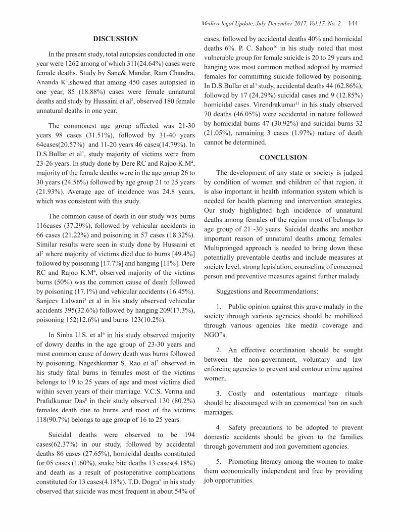
Medico-legal Update, July-December 2017, Vol.17, No. 2 144 145 -legal Update, July-December 2017, Vol.17, No. 2
DISCUSSION
In the present study, total autopsies conducted in one year were 1262 among of which 311(24.64%) cases were female deaths. Study by Sane& Mandar, Ram Chandra, Ananda K1,showed that among 450 cases autopsied in one year, 85 (18.88%) cases were female unnatural deaths and study by Hussaini et al2, observed 180 female unnatural deaths in one year.
The commonest age group affected was 21-30 years 98 cases (31.51%), followed by 31-40 years 64cases(20.57%) and 11-20 years 46 cases(14.79%). In D.S.Bullar et al3, study majority of victims were from 23-26 years. In study done by Dere RC and Rajoo K.M4, majority of the female deaths were in the age group 26 to 30 years (24.56%) followed by age group 21 to 25 years (21.93%). Average age of incidence was 24.8 years, which was consistent with this study.
The common cause of death in our study was burns 116cases (37.29%), followed by vehicular accidents in 66 cases (21.22%) and poisoning in 57 cases (18.32%). Similar results were seen in study done by Hussaini et al2 where majority of victims died due to burns [49.4%] followed by poisoning [17.7%] and hanging [11%]. Dere RC and Rajoo K.M4, observed majority of the victims burns (50%) was the common cause of death followed by poisoning (17.1%) and vehicular accidents (16.45%). Sanjeev Lalwani5 et al in his study observed vehicular accidents 395(32.6%) followed by hanging 209(17.3%), poisoning 152(12.6%) and burns 123(10.2%).
In Sinha U.S. et al6 in his study observed majority of dowry deaths in the age group of 23-30 years and most common cause of dowry death was burns followed by poisoning. Nageshkumar S. Rao et al7 observed in his study fatal burns in females most of the victims belongs to 19 to 25 years of age and most victims died within seven years of their marriage. V.C.S. Verma and Prafulkumar Das8 in their study observed 130 (80.2%) females death due to burns and most of the victims 118(90.7%) belongs to age group of 16 to 25 years.
Suicidal deaths were observed to be 194 cases(62.37%) in our study, followed by accidental deaths 86 cases (27.65%), homicidal deaths constituted for 05 cases (1.60%), snake bite deaths 13 cases(4.18%) and death as a result of postoperative complications constituted for 13 cases(4.18%). T.D. Dogra9 in his study observed that suicide was most frequent in about 54% of
cases, followed by accidental deaths 40% and homicidal deaths 6%. P. C. Sahoo10 in his study noted that most vulnerable group for female suicide is 20 to 29 years and hanging was most common method adopted by married females for committing suicide followed by poisoning. In D.S.Bullar et al3 study, accidental deaths 44 (62.86%), followed by 17 (24.29%) suicidal cases and 9 (12.85%) homicidal cases. Virendrakumar11 in his study observed 70 deaths (46.05%) were accidental in nature followed by homicidal burns 47 (30.92%) and suicidal burns 32 (21.05%), remaining 3 cases (1.97%) nature of death cannot be determined.
CONCLUSION
The development of any state or society is judged by condition of women and children of that region, it is also important in health information system which is needed for health planning and intervention strategies. Our study highlighted high incidence of unnatural deaths among females of the region most of belongs to age group of 21 -30 years. Suicidal deaths are another important reason of unnatural deaths among females. Multipronged approach is needed to bring down these potentially preventable deaths and include measures at society level, strong legislation, counseling of concerned person and preventive measures against further malady.
Suggestions and Recommendations:
1. Public opinion against this grave malady in the society through various agencies should be mobilized through various agencies like media coverage and NGO‟s.
2. An effective coordination should be sought between the non-government, voluntary and law enforcing agencies to prevent and contour crime against women.
3. Costly and ostentatious marriage rituals should be discouraged with an economical ban on such marriages.
4. Safety precautions to be adopted to prevent domestic accidents should be given to the families through government and non government agencies.
5. Promoting literacy among the women to make them economically independent and free by providing job opportunities.

-legal Update, July-December 2017, Vol.17, No. 2 144 145 Medico-legal Update, July-December 2017, Vol.17, No. 2
6. Enact more stringent laws taking care of the torture of housewives by their husbands and In-laws.
7. Strengthening of Mahila Courts.
8. Women protection cell working in the State should be given to deal with deaths resulting from burns with adequate and sufficient facilities.
9. There should be transparency and accountability in the judicial system.
10. Early marriage should be discouraged and punitive measures to be strictly implemented in case of default
11. Centers shall be developed to provide free counseling to the families and newlywed couple about their expected problems and their solutions in initial years of marriage.
12. Risk factors of suicides in women should be identified and attended.
13. A change in attitude and mindset of society, judiciary, and the most importantly of a male person (husband/father) who is supposed to be guardian of a woman should be sensitized to make home/workplace a safer and happier place for a woman.
Conflict of Interest: Nil
Source of Funding: Self
Consent: Not taken as it was autopsy based retrospective study
REFERNCES
1. Sane Mandar, Ram Chandra, Ananda K. Unnatural deaths of adult females in south Banglore, an
autopsy study. Journal of Indian academy of forensic medicine. 2014;36(2):130-132.
2. Hussaini Numan , Padole Trishul , Batra Anil , Pinge Anil , Hussaini SK Profile of Unnatural Death of Adult Females in and Around Akola – A Medico-Legal Study. J Cont Med A Dent January-April 2015 Volume 3 Issue 1:58-6.
3. D.S. Bhullar: Profile of unnatural female deaths between 18-30 years of age, JFMT, Vol. XIII, No-3 & 4 1997, 5-8.
4. Dere RC and Rajoo KM. Study of Unnatural Deaths in Females, A Medico-legal Study at Rural Medical College, Loni. J Indian Acad Forensic Med. 2011; 33(3): 211-213.
5. Sanjeev Lalwani: Profile of unnatural deaths of females at AIIMS- A retrospective review, FORENSICON 2001, 49.
6. Sinha U. S.: Crime against women with special reference to dowry deaths, FORENSICON 1999, 10.
7. Nageshkumar Rao: Study of fatal female burns in Manipal, JFMT, Vol. XIV No.2, Dec 1997, 57-59.
8. V C S Verma and Prafulkumar Das: Burns and its medico-legal aspect, IAFM, Jan 1990, 7-9.
9. T.D. Dogra: Suicide in south Delhi 1974-1993- The choice of method, Indian Medical Gazette, November 1996, 364-368.
10. P C Sahoo: Trends in suicide-A study in M.K.C.G. Medical College PM center, JFMT, Vol.16 No.1, Jan-June 1999, 34-35.
11. Virendrakumar: Burnt wives- a sociology study, International Journal of Medical Toxicology and Legal Medicine, Vol.2 No.1, July-Dec 1999, 27-33.
147 -legal Update, July-December 2017, Vol.17, No. 2
A Research Study on Female Suicides Due to Infertility in Chittoor, Andhra Pradesh from 2010 to 2016
V Jayashankar1, Chaitanya R2
1Associate Professor, Department of Forensic Medicine, The Apollo Medical College (AIMSR), Murakambattu, Chittoor, Andhra Pradesh, 2Associate Professor, Dept. of Forensic Medicine & Toxicology,
VIMS, Ballari, Karnataka
ABSTRACT
According to the World Health Organization (WHO), infertility can be described as the inability to become pregnant, maintain a pregnancy, or carry a pregnancy to live birth. Infertility affects about 10% of all couples. About 30% of infertility is because of female infertility, 30% of infertility is because of male infertility and in the rest of the cases infertility affects both of them or the reason is unclear. Each menstrual cycle is regulated by complex increase and changes of different reproductive hormones which work together to prepare the woman’s body ready for conception and pregnancy. Conception occurs only when egg is fertilised by sperm. Female infertility affects an estimated 48 million women in the world. Infertility in woman is caused by many causes, including nutrition, diseases, and other malformations of the uterus. Although factors of female infertility can be classified as acquired or genetic, female infertility is usually more or less a combination of nature and nurture. When a woman cannot bear children, it tremendously affects her identity, but the pain can extend far beyond her to impact personal relationships. Women often begin to imagine themselves as mothers long before actually trying to have children, and this is certainly influenced by implicit cultural and society messages that idealize motherhood. When this imagined self of a mother, however tentative, is withdrawn, it may result in feeling a loss of control, threaten her imagined future, cause her to doubt her womanhood, and feel like an assault on her ability leading to depression and suicidal thoughts, in some cases landing up in suicidal deaths.
Keywords: Infertility, menstrual cycle, sex hormones, PID, egg, sperm, ovulation period, fallopian tube, uterus etc
Corresponding author:Dr. Chaitanya. R., M.DAssociate Professor, Dept. of Forensic Medicine & Toxicology, VIMS, Ballari, Karnataka - 583104Affiliated to Rajiv Gandhi University of Health Sciences, Karnataka, Mobile - 09008735628Email – [email protected]
INTRODUCTION
Infertility is defined as a failure of a woman to conceive within one or more years of regular unprotected coitus. Primary infertility denotes those patients who have never conceived. Secondary infertility indicates previous pregnancy but failure to conceive subsequently. Regarding incidence, the male is directly responsible in about 30 – 40% of infertility cases, female
in about 40 – 45% cases, both are responsible in 10% cases and remaining is unexplained inspite of thorough investigations.1
The causes of female infertility is attributed to 1. Dyspareunia and vaginal causes 2. Congenital defects in the genital tract 3. Infections of genital tract 4. Cervical Factors 5. Uterine causes 6. Tubal factors 7. Ovarian factors 8. Peritoneal causes 9. Hormonal factors. Dyspareunia is loosely used for difficult as well as painful coitus. The important organic causes are fixed retroversion with prolapsed ovaries, inflamed adnexal disease and pelvic endometriosis. These conditions are associated with blocked tubes. Congenital defects of the genital tract includes absent or septatae vagina, hypoplasia and absent uterus which are obvious causes leading to sterility.2
DOI Number: 10.5958/0974-1283.2017.00088.3
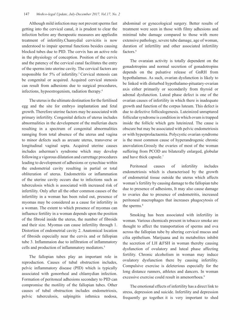
147 Medico-legal Update, July-December 2017, Vol.17, No. 2
Although mild infection may not prevent sperms fast getting into the cervical canal, it is prudent to clear the infection before any therapeutic measures are appliedin treatment of infertility.Chamydial cervicitis is now understood to impair spermal functions besides causing blocked tubes due to PID. The cervix has an active role in the physiology of conception. Position of the cervix and the patency of the cervical canal facilitates the entry of the sperms into uterine cavity. The cervical factors are responsible for 5% of infertility.3 Cervical stenosis can be congenital or acquired. Acquired cervical stenosis can result from adhesions due to surgical procedures, infections, hypoestrogenism, radiation therapy.4
The uterus is the ultimate destination for the fertilised egg and the site for embryo implantation and fetal growth. Therefore uterine factors may be associated with primary infertility. Congenital defects of uterus includes abnormalities in the development of the mullerian ducts resulting in a spectrum of congenital abnormalities rannging from total absence of the uterus and vagina to minor defects such as arcuate uterus, transverse or longitudinal vaginal septa. Acquired uterine causes includes asherman’s syndrome which may develop following a vigorous dilatation and currettage procedures leading to development of adhesions or synechiae within the endometrial cavity resulting in partial or total obliteration of uterus. Endometritis or inflammation of the uterine cavity occurs due to infections such as tuberculosis which is associated with increased risk of infertility. Only after all the other common causes of the infertility in a woman has been ruled out, presence of myomas may be considered as a cause for infertility in a woman. The extent to which presence of myomas can influence fertility in a woman depends upon the position of the fibroid inside the uterus, the number of fibroids and their size. Myomas can cause infertility through 1. Distortion of endometrial cavity 2. Anatomical location of fibroids especially near the cervix and or fallopian tube 3. Inflammaion due to infiltration of inflammatory cells and production of inflammatory mediators.5
The fallopian tubes play an important role in reproduction. Causes of tubal obstruction includes, pelvic inflammatory disease (PID) which is typically associated with gonorrheal and chlamydian infection. Formation of peritoneal adhesions secondary to PID can compromise the motility of the fallopian tubes. Other causes of tubal obstruction includes endometriosis, pelvic tuberculosis, salpingitis isthmica nodosa,
abdominal or gynecological surgery. Better results of treatment were seen in those with filmy adhesions and minimal tube damage compared to those with more extensive pathology, severe tube damage, age of woman, duration of infertility and other associated infertility factors.6
The ovaraian activity is totally dependent on the gonadotropins and normal secretion of gonadotropins depends on the pulsative release of GnRH from hypothalamus. As such, ovarian dysfunction is likely to be linked with disturbed hypothalamo-pituatary-ovarian axis either primarily or secondarily from thyroid or adrenal dysfunction. Luteal phase defect is one of the ovarian causes of infertility in which there is inadequate growth and function of the corpus luteum. This defect is due to defective folliculogenesis. Luteinized unruptured follicular syndrome is condition in which ovum is trapped inside the follicle which gets luteinized. The cause is obscure but may be associated with pelvic endometriosis or with hyperprolactaemia. Polycystic ovarian syndrome is the most common cause of hyperandrogenic chronic anovulation.Grossly the ovaries of most of the woman suffering from PCOD are bilaterally enlarged, globular and have thick capsule.7
Peritoneal causes of infertility includes endometriosis which is characterised by the growth of endometrial tissue outside the uterus which affects woman’s fertility by causing damage to the fallopian tube due to presence of adhesions, It may also cause damage to ovaries due to presence of endometritis, increased peritoneal macrophages that increases phagocytosis of the sperms.8
Smoking has been associated with infertility in woman. Various chemicals present in tobacco smoke are thought to affect the transportation of sperms and ova across the fallopian tube by altering cervical mucus and cilia epithelium. Marijuana and its metabolites inhibit the secretion of LH &FSH in woman thereby causing dysfunction of ovulatory and luteal phase affecting fertility. Chronic alcoholism in woman may induce ovulatory dysfunction there by causing infertility. Compulsive exercise is deleterious especially for the long distance runners, athletes and dancers. In woman excessive exercise could result in amenorrhoea.9
The emotional effects of infertility has a direct link to stress, depression and suicide. Infertility and depression frequently go together. it is very important to shed
Medico-legal Update, July-December 2017, Vol.17, No. 2 148 149 -legal Update, July-December 2017, Vol.17, No. 2
Table No.3: Method of committing suicide
CONCLUSION
Diagnosis of infertility begins with a proper medical history and thorough physical examination with necessary full diagnostic investigations. Patient should be assured that many causes of infertility can be well treated. Once a woman is diagnosed with infertility, the overall likelihood for successful treatment is 50%. Whether a treatment is successful or not depends on 1. Underlying cause of the problem 2. Woman’s age 3. History of previous pregnancies 4. How long she has had infertility issues. Fertility treatments are most likely to benefit women whose infertility is due to problems with ovulation. Treatment is least likely to benefit infertility caused by damage to the fallopian tubes or severe endometriosis. The first step of treating infertility in many cases is to treat the underlying cause of infertility. For example, in cases where thyroid disease causes hormone imbalances, medication for thyroid disease may be able to restore fertility.
One of the most important thing is that the woman who is undergoing treatment for her infertility issues should receive full support from her family members, friends and members of society. The woman has be educated about many available assisted reproductory techniques like IUI, IVF, surrogacy for getting a child for which her in-laws especially and importantly her husband should support strongly. If all the assisted reproductory techniques fail, then adoption of child should be encouraged. At last the woman should be psychologically motivated to understand that life is more important than parenthood.
Female infertility may be prevented by maintaining a healthy lifestyle. Excessive exercise, consumption of caffeine and alcohol, and smoking must be avoided as these have all been associated with decreased fertility. Eating a well-balanced, nutritious diet, with plenty of
some much needed light on how serious the emotional ramifications of infertility can be. Depression is a major risk factor for thoughts of suicide, and actual suicide attempts. 13% of women had suicidal thoughts following a failed IVF attempt. Approximately half of the women reported infertility as the most stressful experience of their life. Research has shown that the psychological stress experienced by women with infertility is similar to that of women coping with illnesses like cancer, HIV and chronic pain. Infertility is not an easy disease to cope with, and the traumatic effects don’t “go away” after the last IVF attempt. There are many reports of women committing suicide over infertility. These tragic events once again show that infertility is a terrible disease to suffer from, with real and frightening consequences.10
MATERIALS
Postmortem reports, gynaecology case sheets, hospital statistics
FINDINGS
Total number of suicidal deaths due to infertility - 46
Table No.1: Infertility causes and its incidence in suicidal deaths due to infertility
Table No.2: Distribution of woman’s age among suicidal deaths due to infertility
-legal Update, July-December 2017, Vol.17, No. 2 148 149 Medico-legal Update, July-December 2017, Vol.17, No. 2
fresh fruits and vegetables, and maintaining a normal weight, on the other hand, have been associated with better fertility prospects.
Treating or preventing existing diseases especially genital tract infections is very useful. Identifying and controlling chronic diseases such as diabetes and hypothyroidism increases fertility prospects. Lifelong practice of safer sex reduces the likelihood that sexually transmitted diseases will impair fertility. Obtaining prompt treatment for sexually transmitted diseases reduces the likelihood that such infections will do significant damage. Regular physical examinations (including pap smears) help detect early signs of infections or abnormalities.
Fertility does not ultimately cease before menopause, but it starts declining after the age of 27 years and drops at a somewhat greater rate after age of 35 years
Conflict of Interest: Nil
Source of Funding : Self
Ethical Clearance: Obstetrics & Gynecology Dept (AIMSR), General Hospital, Chittoor, Andhra Pradesh.
REFERENCE
1. D.C Dutta’s textbook of gynecology including conception, Jaypee publishers, 6th edition, Pg. 227-228
2. Shaw’s textbook of gynecology, 16th edition, Elsevier publishers, Pg.249-250
3. Shaw’s textbook of gynecology, 16th edition, Elsevier publishers, Pg.249-250
4. Richa Saxena’s bedside obstetrics & gynecology, 2nd edition, Jaypee publishers, Pg.1019
5. Richa Saxena’s bedside obstetrics & gynecology, 2nd edition, Jaypee publishers, Pg.1019-1021
6. Richa Saxena’s bedside obstetrics & gynecology, 2nd edition, Jaypee publishers, Pg.1025
7. D.C Dutta’s textbook of gynecology including conception, Jaypee publishers, 6th edition, Pg. 229-230
8. Richa Saxena’s bedside obstetrics & gynecology, 2nd edition, Jaypee publishers, Pg.1026
9. Shaw’s textbook of gynecology, 16th edition, Elsevier publishers, Pg.250
10. http://www.conceiveivf.com/slow-swimmers-and-friedeggs/infertility suicide-whats-link.

151 -legal Update, July-December 2017, Vol.17, No. 2
Profile of Sudden Deaths Due to Lung Pathologies
Enyew Debash1, Jagadish Rao Padubidri2, Pavanchand Shetty2, B Suresh Kumar Shetty3, Prateek Rastogi2, Sesen Tsegaye1
1Post-graduate Scholar, College of Health Sciences, Forensic Medicine, Mekelle University, Ethopia, 2Associate 2Associate 2
Professor and District Medicolegal Consultant, Forensic Medicine, Kasturba Medical College, Mangalore, Manipal University, 3Professor and Head, State Medicolegal consultant, Forensic Medicine, Kasturba Medical
College, Mangalore, Manipal University
ABSTRACT
Introduction: Sudden unexpected death due to respiratory disease contributes to 15 – 23% of all sudden death cases. Tuberculosis (TB) is a major respiratory cause of morbidity and mortality worldwide and its prevalence is high in the Indian subcontinent, and cases may not be diagnosed until after an autopsy is performed.
Aim of this study: The aim of this autopsy based retrospective study was to identify common causes of sudden death due to lung pathologies, to study the prevalence of mycobacterium tuberculosis in autopsied lungs and to identify cases of active pulmonary tuberculosis.
Material and Method: Confirmed cases of sudden deaths due to lung pathologies based on gross and histopathological examination from 2014- 16 were included in the study.
Results: Out of 210 cases of sudden unexpected deaths. 23.81% were due to lung pathologies, of which 25 (50%) of cases were due to pulmonary tuberculosis. 20% were found to be positive for acid fast bacilli (AFB) by Ziehl- Neelsen stain, 28% of cases were due to pneumonia, 6% of cases were due to pulmonary thromboembolism and 4% cases were due to acute respiratory distress syndrome.
Conclusion: The risk of exposure to unrecognized active pulmonary tuberculosis cases is very high in this particular region of South India, so adequate precautions should be taken for the autopsy surgeons, lab technicians, mortuary workers and investigating officers while handling unknown cases of sudden deaths.
Keywords: Sudden Death; Tuberculosis; Acid fast bacilli, Pneumonia; Pulmonary thromboembolism
Corresponding author:Dr Jagadish Rao PadubidriAssociate Professor and District Medicolegal Consultant Forensic Medicine, Kasturba Medical College, Mangalore, Manipal UniversityContact No: 9900405085Email id: [email protected]
INTRODUCTION
Although the major component of forensic pathology is the ability to identify and interpret injury and determine its role in causing death, the majority of cases investigated by medical examiners or coroners
are sudden deaths caused by natural disease.1 WHO has defined sudden death as death occurring within 24 hours from the onset of symptoms. 2,3 In India, a similar system is followed and the very purpose of medico legal autopsy in such deaths is to determine whether poisoning or violence has been in any way responsible for the death. 4
Majority of the sudden deaths reportedly involve the cardiovascular system (45 to 50%), respiratory system (15 to 23%), the nervous system (10 to 18%), the alimentary system (6 to 8%), the genito-urinary system (3 to 5) and other causes contribute to remaining 5 to 10% of sudden deaths. 5 The major cause of sudden death within the respiratory organs is vascular. Pulmonary
DOI Number: 10.5958/0974-1283.2017.00089.5

151 Medico-legal Update, July-December 2017, Vol.17, No. 2
embolism is very common and is the most clinically under-diagnosed cause of death. Other causes of sudden death in the respiratory system include acute epiglottitis, fulminant tracheobronchitis, pneumonia, bronchial asthma, spontaneous pneumothorax and massive hemoptysis from cavitating pulmonary tuberculosis or from an invasive tumour. 6
Tuberculosis (TB) remains a major respiratory cause of morbidity and mortality worldwide and has been identified as a ‘global emergency’ by the WHO. 7 The most common form is pulmonary tuberculosis, characterized pathologically by necrotizing granulomas, associated pneumonia, and a great propensity for fibrosis and dystrophic calcification. 8 The prevalence of tuberculosis is high in the Indian subcontinent, and cases may not be diagnosed until after an autopsy is performed. 9 India is the highest TB burden country in the world, accounting for one-fifth of the global incidence with an estimated 1.96 million cases annually. 10
The aim of this study is to identify common causes of sudden death due to lung pathologies in autopsy cases and to study the prevalence of mycobacterium tuberculosis in autopsied lungs as well as to identify cases of active tuberculosis.
MATERIAL AND METHOD
This autopsy based retrospective research was conducted in the Department of Forensic Medicine, Kasturba Medical College, Mangalore from the cases autopsied at Government Wenlock District Hospital, Mangalore from 1st September 2014 to 31st of August 2016. Confirmed cases of sudden unexpected deaths due to lung pathologies based on gross and histopathological examinations within the study period were included in the study. Significant pathology in other systems was ruled out by the autopsy surgeons based on gross and microscopic features, before assigning lung pathologies as the cause of death. Hematoxylin and eosin stain along with Ziehl-Neelsen (ZN) stain for identification of tuberculous bacilli was used during the histological examination. Data was analyzed using SPSS (Statistical package for social sciences) program, version 17.0 and a detailed epidemiologic profile was made.
RESULTS
A total of 1833 postmortem examinations were performed at the mortuary of Government Wenlock
District Hospital from 1st of September 2014 to 31st of August 2016 out of which 210 (11.46%) cases were sudden unexpected deaths. 50 (23.81%) out of all sudden death cases were due to lung pathologies, of which 25 (50%) of cases were due to pulmonary tuberculosis. Out of 22 pulmonary tuberculosis cases for which Ziehl-Neelsen (ZN) stain was done 10 cases (20%) were found to be positive for acid fast bacilli (AFB), 14 (28%) of cases were due to pneumonia, 3 (6%) of case were due to pulmonary thromboembolism and 2 (4%) cases were due to acute respiratory distress syndrome ( Table 1). Males were predominantly affected 46 (92%) cases. Age groups 30 - 49 years and those 50 years and above were equally affected, each consisting 21 (42%) of cases (Fig: 5). 32 (64%) cases were unknown and found dead by the side of the road and 17 (34%) cases were admitted and died in the hospital.
Table 1: - Causes of Sudden Unexpected Death
Cause OF Death Frequency Percent
Pulmonary tuberculosis 25 50%
Pneumonia 14 28%
Acute respiratory distress syndrome 2 4%
Adenocarcinoma of the lung 1 2%
Squamous cell carcinoma of the lung 1 2%
Lung abscess 1 2%
Aspergillosis 1 2%
Extrinsic allergic alveolitis 1 2%
Foreign body aspiration 1 2%
Pulmonary thromboembolism 3 6%
Total 50 100%
Figure 1:- Gross appearance of cavitary lesions in pulmonary tuberculosis
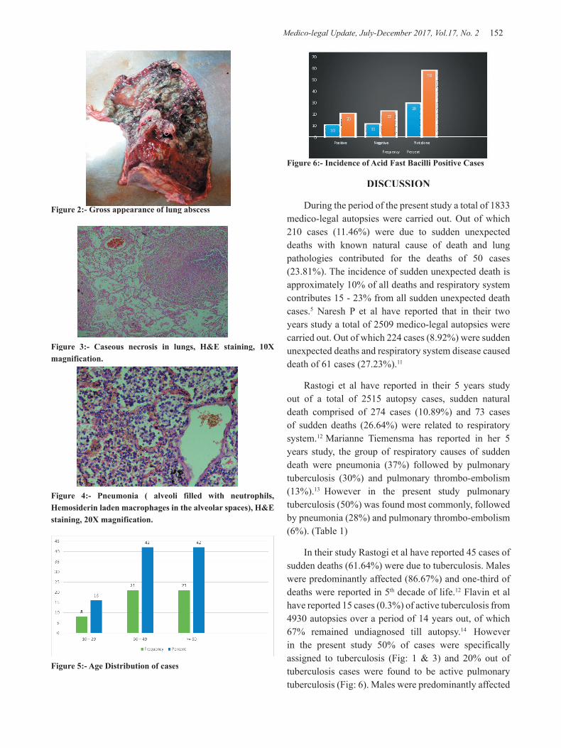
Medico-legal Update, July-December 2017, Vol.17, No. 2 152 153 -legal Update, July-December 2017, Vol.17, No. 2
Figure 2:- Gross appearance of lung abscess
Figure 3:- Caseous necrosis in lungs, H&E staining, 10X magnification.
Figure 4:- Pneumonia ( alveoli filled with neutrophils, Hemosiderin laden macrophages in the alveolar spaces), H&E staining, 20X magnification.
Figure 5:- Age Distribution of cases
Figure 6:- Incidence of Acid Fast Bacilli Positive Cases Figure 6:- Incidence of Acid Fast Bacilli Positive Cases
DISCUSSION
During the period of the present study a total of 1833 medico-legal autopsies were carried out. Out of which 210 cases (11.46%) were due to sudden unexpected deaths with known natural cause of death and lung pathologies contributed for the deaths of 50 cases (23.81%). The incidence of sudden unexpected death is approximately 10% of all deaths and respiratory system contributes 15 - 23% from all sudden unexpected death cases.5 Naresh P et al have reported that in their two years study a total of 2509 medico-legal autopsies were carried out. Out of which 224 cases (8.92%) were sudden unexpected deaths and respiratory system disease caused death of 61 cases (27.23%).11
Rastogi et al have reported in their 5 years study out of a total of 2515 autopsy cases, sudden natural death comprised of 274 cases (10.89%) and 73 cases of sudden deaths (26.64%) were related to respiratory system.12 Marianne Tiemensma has reported in her 5 years study, the group of respiratory causes of sudden death were pneumonia (37%) followed by pulmonary tuberculosis (30%) and pulmonary thrombo-embolism (13%).13 However in the present study pulmonary tuberculosis (50%) was found most commonly, followed by pneumonia (28%) and pulmonary thrombo-embolism (6%). (Table 1)
In their study Rastogi et al have reported 45 cases of sudden deaths (61.64%) were due to tuberculosis. Males were predominantly affected (86.67%) and one-third of deaths were reported in 5th decade of life.12 Flavin et al have reported 15 cases (0.3%) of active tuberculosis from 4930 autopsies over a period of 14 years out, of which 67% remained undiagnosed till autopsy.14 However in the present study 50% of cases were specifically assigned to tuberculosis (Fig: 1 & 3) and 20% out of tuberculosis cases were found to be active pulmonary tuberculosis (Fig: 6). Males were predominantly affected

-legal Update, July-December 2017, Vol.17, No. 2 152 153 Medico-legal Update, July-December 2017, Vol.17, No. 2
(92%) and 84% of cases were above 30 years of age. Most of the cases (64%) were unknown and found dead by the side of the road and all of them were from poor socio-economic background.
Correia et al have demonstrated that MTB can remain viable after death for up to 36 days and the bodies were kept in cold chambers.15 It is estimated that staff of laboratories and autopsy rooms are between 100 and 200 times more likely than the general public to develop tuberculosis. 16 A national study of tuberculosis in the United States of America found that funeral home directors had higher tuberculosis morbidity and mortality.17 Exposure may occur from gurgling at the nose and mouth of the cadaver due to fluid buildup in the chest cavity and putrefaction of tissues and organs. Also residual air in the deceased’s lungs may be exhaled when the body is moved. 18 On the other hand, storage of many cadavers together in temporary mortuaries may present an increased risk of infection; once aerosolized, the tubercle bacilli may remain viable for extended periods of time. 19
Tahir et al have reported in their study that pneumonia was present in 32 out of 810 cases (4%). Majority (75%) cases were below the age of 50 years and there was no sex difference. 20 However in the present study it is the second common causes of sudden death (28%) (Fig: 4) and it is more common in males (85.71%). Majority of cases were below the age of 50 years and most of the cases were those who were found dead by the side of the road. Pneumonia, which is most commonly caused by Streptococcus pneumoniae, is usually diagnosed clinically. 21
Data from literatures indicate that pulmonary thromboembolism (PTE) is the commonest cause of sudden death from respiratory disease and it is the most clinically under-diagnosed cause of death. Each year in the United States , approximately 600,000 individuals suffer an acute PTE event, and 10% of them die of PTE. 22-24 Due to the non-specific symptoms and imaging features, a timely and accurate diagnosis of PTE is still challenging, and the majority of cases are either missed or misdiagnosed, 25-28 while only approximately 10-30% of cases could have been diagnosed before death. 29 It is estimated that about half of the sudden, unexpected deaths of PTE occur within one hour of the onset of symptoms, and the diagnosis of fatal PTE was always made after autopsy. 30 In the present study only
3 cases (6%) of sudden deaths were due to pulmonary thromboembolism. All of them were males and they were admitted to hospital secondary to road traffic accident for 7-10 days and died inside hospital. Trauma and immobilization are the major risk factors in these cases to develop pulmonary thromboembolism.
In the remaining cases of sudden unexpected deaths in the present study, there were findings of ARDS (4%), adenocarcinoma of the lung (2%) , squamous cell carcinoma of the lung (2%), lung abscess (2%) (Fig: 2), extrinsic allergic alveolitis (2%), foreign body aspiration (2%) and lung aspergillosis (2%) which were the causes of death. In all cases disease was not known during life, hence the person died undiagnosed. The cause of death was found only after autopsy.
CONCLUSION
The study revealed that the commonest cause of sudden unexpected death from lung pathologies was pulmonary tuberculosis contributing for the death of 25 cases (50%) out of which 10 cases (20%) were active pulmonary tuberculosis. The risk of unrecognized tuberculosis not only extends to health professionals but also to public. Measures to minimize the risk to mortuary staff should be taken which includes the use of proper respiratory masks and performing the autopsy in the infection suit. Precautions should be made while handling dead bodies especially for the funeral workers, investigating officers and relatives of the deceased. These cases from autopsy studies, probably representing the tip of the iceberg and indicating seriousness of the problem of tuberculosis. Histopathology remains one of the most important methods for diagnosing tuberculosis. Tuberculosis should be considered in the differential diagnosis of all respiratory diseases because of its varied clinical presentations and manifestations. Knowing common lung pathologies causing sudden unexpected death will also help to keep them in top differential diagnosis list while performing the autopsy procedure.
Acknowledgement: The authors are indebted to all the teaching and non-Teaching faculty of Department of Forensic Medicine, KMC, Mangalore for the source and time during the article preparation. The research is ostensibly supported by the Manipal University and Mekelle University, Ethopia as a part of MoU programme. The authors credits Professor Harish Kumar, Ayder Research Hospital, Mekelle Hospital, Ethopia for all the support in facilitating the residents to
Medico-legal Update, July-December 2017, Vol.17, No. 2 154 155 -legal Update, July-December 2017, Vol.17, No. 2
do collaborative work.
Research Funding: None
Conflict of Interest: None declared
Ethical Clearance: This study was approved by Institutional ethics committee, Kasturba Medical College, Mangalore, Manipal University, IEC KMC MLR 11-16/311, dated 16/9/2016.
REFERENCES
1. David Dolinak, Evan Matches, Emma Lew, Forensic Pathology Principle and Practice, UK, Elsevier, 2005
2. Bernard Knight, Knight’s Forensic Pathology, UK, Hodder Arnold, 2004, 3rd edition
3. Craig CH, Principles and practice of medical jurisprudence, UK, Churchill Livingstone, 1984
4. Krishan Vij, Textbook of Forensic Medicine and Toxicology Principles and Practice, India, Elsevier, 2011, 5th edition.
5. Narayan Reddy, The essentials of forensic medicine and toxicology, India, Jaypee Brothers, 2014
6. Jason Payne James, Richard Jones, Simpson’s Forensic Medicine, UK, Hodder Arnold, 2011, 13th edition.
7. World Health Organization World Health Organization (WHO). Tuberculosis Fact sheet N°104 - Global and regional incidence. March 2006, Retrieved on 6 October 2006.
8. J.-G. Im, H. Itoh, K. S. Lee, and M. C. Han, “CT-pathology correlation of pulmonary tuberculosis,” Critical Reviews in Diagnostic Imaging, vol. 36, no. 3, pp. 227–285, 1995.
9. V. M. Vashishtha, “Current status of tuberculosis and acute respiratory infections in India: much more needs to be done!,” Indian Pediatrics, vol. 47, no. 1, pp. 88–89, 2010.
10. M. Garg, A. D. Aggarwal, S. Singh, and S. P. Kataria, “Tuberculous lesions at autopsy,” Journal of Indian Academy of Forensic Medicine, vol. 33, no. 2, pp. 116–119, 2011.
11. Naresh P.et al, Study of Sudden Unexpected Deaths In Medico-Legal Autopsies, Journal of Indian Academy of Forensic Medicine,Vol 28, no 1,PP 27-30, 2006
12. Prateek R., Tanuj K., Ritesh G. Sudden unexpected death due to lung tuberculosis. Journal Of Forensic Medicine, 2011; 28(2): 81
13. Marianne T. sudden and unexpected deaths in adults: an investigation of cases reported to tygerberg forensic pathology services from january 2001 – december 2005, 2010,
14. Flavin RJ, Gibbons N, O’Briain DS. Mycobacterium tuberculosis at autopsy – exposure and protection: an old adversary revisited. J Clin Pathol.2007; 60:487-
15. J.A. Correia. Assesing the survival of Mycobacterium Tuberculosisin unembalmed and enbalmed human remains, South Africa, Clinical Anatomy 27:304–307, 2014
16. Jason Payne James, Roger Byard, Encyclopedia of Forensic Medicine and Legal Medicine, USA, Academic Press, 2005.
17. United States, Centers for Disease Control and Prevention. Proportionate mortality from pulmonary tuberculosis associated with occupations— 28 states, 1979–1990. MMWR Morb Mortal Wkly Rep. 1995;44(1):14–9.
18. Gershon RR, Vlahov D, Escamilla Cejudo JA, Badawi M, McDiarmid M, Karkashian C, et al. Tuberculosis risk in funeral home employees. J Occup Environ Med. 1998;40(5):497–503
19. United States, Centers for Disease Control and Prevention. Guidelines for preventing the transmission of Mycobacterium tuberculosis in health-care facilities. MMWR Recomm Rep. 1994;43 (RR-13):1–132.
20. Tariq M., Fakeha R., Sadia A. Patterns of pulmonary morphological lesions seen at autopsy. Biomedica, 2013; 29: 64
21. J. DiMaio, D. DiMaio, Forensic Pathology, USA, CRC Press LLC, 2001, 2nd edition
22. Dentali F, Ageno W, Becattini C, Galli L, Gianni M, Riva N, Imberti D, Squizzato A, Venco A, Agnelli G. Prevalence and clinical history of incidental, asymptomatic pulmonary embolism: a metaanalysis. Thromb Res 2010; 125: 518-522
23. Garcia D, Ageno W, Libby E. Update on the diagnosis and management of pulmonary embolism. Br J Haematol 2005; 131: 301-312
-legal Update, July-December 2017, Vol.17, No. 2 154 155 Medico-legal Update, July-December 2017, Vol.17, No. 2
24. Goldhaber SZ, Elliott CG. Acute pulmonary embolism: part I: epidemiology, pathophysiology, and diagnosis. Circulation 2003; 108: 2726- 2729.
25. Nathan SS, Simmons KA, Lin PP, Hann LE, Morris CD, Athanasian EA, Boland PJ, Healey JH. Proximal deep vein thrombosis after hip replacement for oncologic indications. J Bone Joint Surg Am 2006; 88: 1066-1070.
26. Tapson VF. Acute pulmonary embolism. N Engl J Med 2008; 358: 1037-1052
27. Laporte S, Mismetti P, Decousus H, Uresandi F, Otero R, Lobo JL, Monreal M; Clinical predictors
for fatal pulmonary embolism in 15,520 patients with venous thromboembolism: findings from the Registro Informatizado de la Enfermedad TromboEmbolica venosa (RIETE) Registry. Circulation 2008; 117: 1711-1716.
28. Kearon C. Diagnosis of pulmonary embolism. CMAJ 2003; 168: 183-194.
29. Kyrle PA, Eichinger S. Deep vein thrombosis. Lancet 2005; 365: 1163-1174.
30. Steiner I. Pulmonary embolism–temporal changes. Cardiovasc Pathol 2007; 16: 248-251.
157 -legal Update, July-December 2017, Vol.17, No. 2
Determination of Sex from Mandibular Canine Index in Delhi Population
Harsimran Sekhon1, Rattan Singh2, Jyoti Barwa3
1Junior House Officer, Townsville Hospital and Health Services, Queensland, Australia, 2Assistant Professor, Department of Forensic Medicine and Toxicology, Himalayan Institute of Medical Sciences, Swami Rama Himalayan University, Dehradun, 3Assistant Professor, Department of Forensic Medicine and Toxicology,
Government Medical College & Hospital, Chandigarh
ABSTRACT
Introduction: The identity of an individual can be established by various parameters such as age, sex, stature, anthropometric measurements, teeth, DNA and so on; sex determination being one of the primary characteristics of identity of an individual. Teeth provide excellent models for the study of relationship between ontogeny and phylogeny. Out of all the teeth, mandibular canines are considered to be most stable as they resist decay due to caries or other diseases of teeth.Materials and Method: A total of 100 candidates, 50 males and 50 females; between 18 to 25 years of age were chosen for the study in the Department of Forensic Medicine & Toxicology, Maulana Azad Medical College, New Delhi. Mandibular Canine Index (MCI) was calculated for all these subjects by preparing a dental cast for the lower jaw, then mandibular canine crown width and inter-canine arch width were measured from these casts by using a vernier caliper of 0.02 mm calibration. Observations & result: The mean right mandibular canine index (RMCI) was found out to be 0.26311 ± 0.016826 and 0.25703± 0.017761 for males and females respectively. The mean left mandibular canine index (LMCI) was 0.26240 ± 0.017955 and 0.25792 ± 0.016559 for males and females respectively. The mean mandibular canine index (MCI) for both sides for males was 0.26275 ± 0.016697 and for females was observed to be 0.25748 ± 0.016501.Thus, by using standard MCI, we were able to correctly classify 62% males and 56% females. Overall predictability was calculated to be 59 %. Independent t-test was done and it was fond that the mean of Right canine width (RCW), left canine width (LCW) and Inter canine width (ICW) has significant mean difference. However, the mean of RMCI, LMCI and mean MCI are not significant. Conclusion: It can be concluded that though RCW, LCW and ICW can be considered as independent factors for sex determination, MCI as such is not that significant. MCI has a limited individual value and thus may be used only as an adjunct in sex determination in combination with skeletal measurements.
Keywords: Mandibular Canine Index (MCI), Identity, Sex, Dental Cast.
INTRODUCTION
An individual can be identified by various parameters such as age, sex, stature, anthropometric
Corresponding author: Dr Rattan Singh, Assistant Professor, Department of Forensic Medicine and Toxicology, Himalayan Institute of Medical Sciences, Swami Rama Himalayan University, Dehradun, Uttarakhand-248016 E mail: [email protected]: 9968444816, 8958461235
measurements, teeth, DNA and so on. Sex is one of the primary characteristics of identity of an individual. Forensic pathologists are often requested to opine regarding sex of an individual in civil matters like heir-ship, marriage, divorce, legitimacy, impotence, disputed sex, etc. as well as in criminal cases where a person is accused of impersonation or in cases of missing persons, etc.1 It is considered that accuracy for determination of sex can be 100 % by using the whole skeleton, 95% by pelvis alone, 92 % by skull alone, 98 % by pelvis plus skull, 80 % by long bones alone and 98 % by long bones and pelvis.2
DOI Number: 10.5958/0974-1283.2017.00090.1
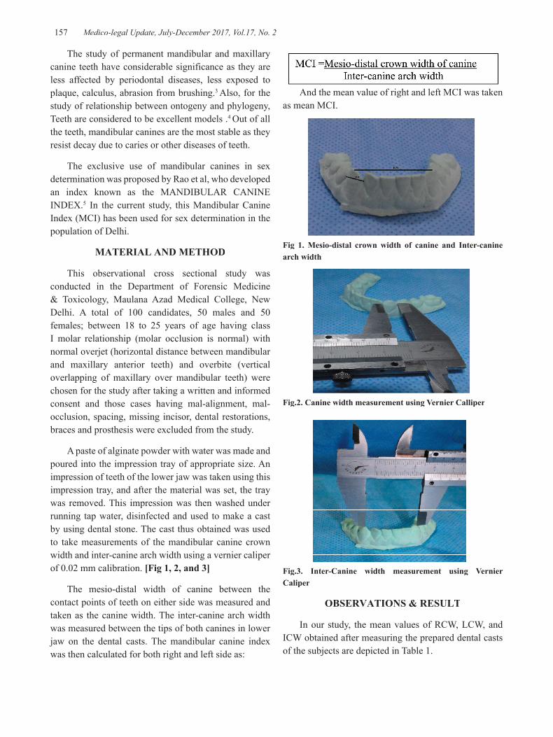
157 Medico-legal Update, July-December 2017, Vol.17, No. 2
The study of permanent mandibular and maxillary canine teeth have considerable significance as they are less affected by periodontal diseases, less exposed to plaque, calculus, abrasion from brushing.3 Also, for the study of relationship between ontogeny and phylogeny, Teeth are considered to be excellent models .4 Out of all the teeth, mandibular canines are the most stable as they resist decay due to caries or other diseases of teeth.
The exclusive use of mandibular canines in sex determination was proposed by Rao et al, who developed an index known as the MANDIBULAR CANINE INDEX.5 In the current study, this Mandibular Canine Index (MCI) has been used for sex determination in the population of Delhi.
MATERIAL AND METHOD
This observational cross sectional study was conducted in the Department of Forensic Medicine & Toxicology, Maulana Azad Medical College, New Delhi. A total of 100 candidates, 50 males and 50 females; between 18 to 25 years of age having class I molar relationship (molar occlusion is normal) with normal overjet (horizontal distance between mandibular and maxillary anterior teeth) and overbite (vertical overlapping of maxillary over mandibular teeth) were chosen for the study after taking a written and informed consent and those cases having mal-alignment, mal-occlusion, spacing, missing incisor, dental restorations, braces and prosthesis were excluded from the study.
A paste of alginate powder with water was made and poured into the impression tray of appropriate size. An impression of teeth of the lower jaw was taken using this impression tray, and after the material was set, the tray was removed. This impression was then washed under running tap water, disinfected and used to make a cast by using dental stone. The cast thus obtained was used to take measurements of the mandibular canine crown width and inter-canine arch width using a vernier caliper of 0.02 mm calibration. [Fig 1, 2, and 3]
The mesio-distal width of canine between the contact points of teeth on either side was measured and taken as the canine width. The inter-canine arch width was measured between the tips of both canines in lower jaw on the dental casts. The mandibular canine index was then calculated for both right and left side as:
And the mean value of right and left MCI was taken as mean MCI.
Fig 1. Mesio-distal crown width of canine and Inter-canine arch width
Fig.2. Canine width measurement using Vernier Calliper
Fig.3. Inter-Canine width measurement using Vernier Caliper
OBSERVATIONS & RESULT
In our study, the mean values of RCW, LCW, and ICW obtained after measuring the prepared dental casts of the subjects are depicted in Table 1.

Medico-legal Update, July-December 2017, Vol.17, No. 2 158 159 -legal Update, July-December 2017, Vol.17, No. 2
Table 1: Mean values of canine width
Gender Mean right canine width (RCW) in mm
Mean left canine width (LCW) in mm
Mean inter canine width(ICW) in mm
Males 6.85 +/- 0.49 6.83 +/- 0.52 26.0800+/-1.74952
Females 6.3760 +/- 0.43 6.40+/- 0.43 24.8540+/-1.52037
Table 2 shows the mean values of mandibular canine index (MCI) which were obtained, thereafter by measuring the Mesio-distal crown width of canine and Inter-canine arch width of dental casts of the subjects.
Table 2: Mean values of Mandibular canine index
Gender Mean right Mandibular canine index (RMCI) in mm
Mean left Mandibular canine index (LMCI) in mm
Mean Mandibular canine index(MCI) in mm
Males 0.26311+/- 0.016826 0.26240+/-0.017955 0.26275+/- 0.016697
Females 0.25703+/- 0.017761 0.25792+/- 0.016559 0.25748+/- 0.016501
DISCUSSION
CANINE WIDTH MEASUREMENTS:
In the present study, we found that the canine width is significantly greater in males as compared to females. These values obtained were found to be similar to the findings of AB Acharya et al6 and PC Srivastava7. Rao et al5 did not mention the values of canine width and early studies like Muller at al8 and Shishir Yadav et al9 have used the mean of right and left canine width for calculating the Mandibular Canine index. However, despite using the mean, the difference between the canine width in males and females was found out to be significant.
INTER-CANINE WIDTH
The values of Inter canine width (ICW), in our study was found to be greater in males than in females. On applying the t-test, the p-value was <0.001, hence highly significant. This was also similar to Rao NG et al5, Acharya AB et al6, Srivastava PC et al7, Muller M et al8, Yadav S et al9 but contradictory to the findings of N Vishwakarma et al.10
MANDIBULAR CANINE INDEX
A) Right Mandibular Canine Index (RMCI):
The value of RMCI for female was comparable to that of PC Srivastava7 and N Vishwakarma et al10. Rao et
al5, Muller et al8, Shishir Yadav et al9, and AB Acharya et al6 did not calculate the right and left mandibular index separately. However, in contrast to our study, Kaushal et al11obtained a higher value for females i.e >0.26. On applying the t-test, the p value was found to be 0.082, hence not significant for differentiating the gender.
B) Left Mandibular Canine Index:
This value of LMCI, for the females was comparable to that of PC Srivastava7. However, Vishwakarma N. etal10, Kaushal S etal11 obtained a higher value for the females. On applying the t-test, the p value was found to be 0.198, therefore insignificant for differentiating the gender.
C) Mean Mandibular Canine Index:
The mean mandibular canine index as calculated in our study was found to be 0.263+/-0.016 in males and 0.257+/-0.016 in females. This value for females was similar to that observed by Rao et al5. Kaushal et al11, PC Srivastava7 and N Vishwakarma et al10 did not calculate the mean mandibular canine index. On applying the t-test, the p value was found to be 0.115, therefore, not significant. This is in contrast to the studies conducted by Rao et al5, Muller et al8and Shishir Yadav et al9
who observed a significant difference between male and female mandibular canine index. However, PC Srivastava7 and AB acharya et al6 did not observe any significant difference, as with our study.
-legal Update, July-December 2017, Vol.17, No. 2 158 159 Medico-legal Update, July-December 2017, Vol.17, No. 2
STANDARD MANDIBULAR CANINE INDEX
The standard MCI calculated was 0.260 which is same as that calculated by AB Acharya et al6. Using standard MCI, in our study, the predictability was found to be 62% for males and 56% for females (overall: 59%). This was in stark contrast to the predictability obtained by other authors like Rao et al5 (85.9%), Shishir Yadav et al9 (M: 83.3%; F: 81%), and IA Mughal et al12 (75.97%). Lower predictability has been previously reported by Muller et al8 (M: 62.55%: F: 63.81%) and AB Acharya et al6 (M: 57.14%; F: 44.44%). Muller et al8 concluded that this variability could be due to different geographical location and the alignment of lower anterior teeth. However, other authors have not commented anything on the same.
CONCLUSION
In our study male predominance was observed for the right and left canine width as well as for inter canine width. The Mandibular Canine Index, though higher in males was not found to be a significant variable for correctly predicting the gender of a person.
On applying the t-test, both the right and left MCI were found to be insignificant and the overall predictability was also found to be very low. The mean MCI could only classify 59% of the subjects correctly (M: 62%; F: 56%). Similarly, the standard MCI could also classify only 59% of the subjects correctly.
Hence, we conclude that though RCW, LCW and ICW can be considered as independent factors for sex determination, MCI as such is not that significant. This could be attributed to the fact that unlike RCW, LCW and ICW which are absolute measurements; MCI is a ratio. Since, this index has been found out to have limited individual value, thus it may be used only as an adjunct in sex determination along with the skeletal measurements.
Ethical Clearance: Obtained from ethical committee, MAMC, Delhi
Conflict of Interest: None
Source of Funding: Self
REFERENCES
1. Knight B. The establishment of identity of human remains. In Saukko P, Knight B, editors. Knight’s Forensic Pathology. 3rd ed. London: Edward Arnold Publishers; 2004. p.104.
2. Krogmann WM. The human skeleton in Forensic medicine. In: sexing skeletal remains.
3. Anderson DL, Thompson GW. Inter-relationship and sex differences of dental and skeletal measurements. J Dent Res 1973; 52; 431-38
4. Eimerl S and DeVore L. Physical anthropology and primatology: The Primates. American anthropologist 1967; 69 (2); 258-60
5. Rao NG, Rao NN, Pai ML, Kotian MS. Mandibular canine index- A clue for establishing sex identity. Forensic Sci Int.1989; 42: 249-54
6. Acharya AB, Mainali S. Limitations of the mandibular canine index in sex assessment. J Forensic Leg Med 2009; 16(2):67-9
7. Srivastava PC. Correlation of odontometric measures in sex determination. J Indian Acad Forensic Med. 2010; 32(1); 56-61\
8. Muller M, Lupipegurier L, Quatrehomme G, Bolla M. Odontometrical method useful in determining gender and dental alignment. Forensic Sci Int 2001; 1211:194-7z
9. Yadav S, Nagabhushan D, Rao BB, Mamatha GP. Mandibular canine index in establishing sex identity. Indian J Dent Res 2002; 13: 143-6.
10. Vishwakarma N., Guha R. A study of sexual dimorphism in permanent mandibular canines and its implications in forensic investigations. Nepal Med Coll J. 2011; 13(2): 96-99
11. Kaushal S, Patnaik VVG, Agnihotri G. Mandibular canines in sex determination. J Anat Soc India 2003; 52(2):119-24
12. Mughal IA, Saqib AS, Manzur F. Mandibular canine index: Its role in determining gender. Professional Med J 2010; 17(3):459-63

161 -legal Update, July-December 2017, Vol.17, No. 2
Post Mortem Study of Fatal Blunt Trauma to Chest
Manoj Kumar Jena1, Soumya Ranjan Nayak2
1Professor & HOD, Deptt. of FMT, S.C.B Medical College, Cuttack, Odisha, 2Associate Professor, Deptt. of FMT, V.S.S Medical College, Burla, Odisha
ABSTRACT
Blunt thoracic trauma is of major significance in absence of injury to the head and stands most common cause of death after head injury. Trauma to anterior chest wall considered to be lethal as these trauma can lead to internal injuries. The commonest cause of blunt chest trauma in modern world is motor vehicle accident which accounts for up to 80% of such injuries. This study was undertaken on 128 cases of death as a result of blunt chest injuries which were brought for autopsy at S.C.B Medical College, Cuttack with the objective to study the epidemiological parameters and to assess the extent of chest injuries incompatible with life. The study group comprises of 118 (92.19%) male victims and 10 (7.81%) female victims. Road traffic accidents accounting for the majority 113 (88.28%) cases of death due to blunt chest trauma .Out of the total 128 victims, sternum and ribs are found fractured in 24 (18.75%) and 117 (91.41%) cases respectively. Lungs are seen injured in 123 (96.09%) cases and right ventricle of heart is found to be the most common anatomical site of heart affected by blunt thoracic trauma.
Keywords : Blunt trauma, Thoracic, Road traffic accident.
Corresponding author:Dr. Soumya Ranjan Nayak Associate Professor, Deptt. of FM&T, V.S.S Medical College, Burla, Odisha, 768017. Mob.: 09437158325Email ID : [email protected]
INTRODUCTION
Injuries on chest are important because of presence of vital organs like heart, lungs, big blood vessels within the chest cage which are compressible and deformable. The thoraco-abdominal region because of its dimension and anatomical position is a major site of impact in road accidents, land slide, stampede, assault, collapse of buildings, etc. Blunt trauma to chest and its contents contributing to a fatal result goes on increasing day by day in direct proportion to the ever increasing trend of traffic accidents throughout the world. Injuries of the chest are mostly accidental, occasionally homicidal and rarely suicidal as stated by Modi.1 According to Reddy, injuries of the chest may be (1) Non penetrating or closed i.e, they do not open up any part of the thoracic cavity, and (2) Penetrating or open. Most closed chest injuries are caused by blunt force.2
AIMS AND OBJECTIVES
This study was undertaken to analyse the epidemiological parameters and to relate the pattern and distribution of blunt thoracic injuries in accordance to survival period and mortality and to suggest how best the morbidity and mortality can be reduced.
MATERIAL AND METHOD
This study was conducted on 128 cases of death due to blunt chest trauma brought for medico legal autopsy at S.C.B Medical College, Cuttack during September 2013 to August 2015. Different data in reference to the incidents collected from the police, friends and relatives accompanying the dead bodies for detail analysis. All cases of death showing fatal thoracic injuries caused by blunt force with or without external injury sustained in road traffic accidents, fall from height, industrial accident, stampede, collapse of buildings and assault have been considered for this study. The factors like age, sex, type of vehicle involved in the accident, extent of injuries sustained, the site of impact have been tabulated, analysed and inference were drawn in the department. Decomposed and skeletonised dead bodies were not included in this study. All cases with penetrating injuries to the chest and cases with only superficial external chest
DOI Number: 10.5958/0974-1283.2017.00091.3
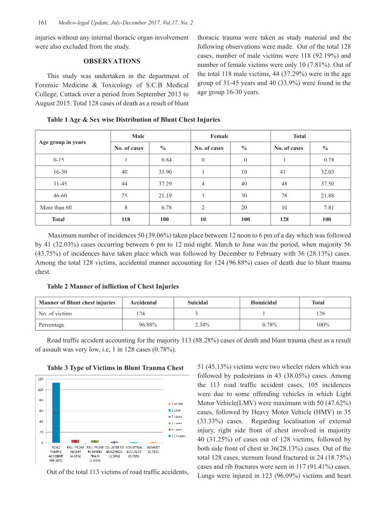
161 Medico-legal Update, July-December 2017, Vol.17, No. 2
Table 3 Type of Victims in Blunt Trauma Chest
Out of the total 113 victims of road traffic accidents,
Table 1 Age & Sex wise Distribution of Blunt Chest Injuries
Age group in years Male Female Total
No. of cases % No. of cases % No. of cases %
0-15 1 0.84 0 0 1 0.78
16-30 40 33.90 1 10 41 32.03
31-45 44 37.29 4 40 48 37.50
46-60 25 21.19 3 30 28 21.88
More than 60 8 6.78 2 20 10 7.81
Total 118 100 10 100 128 100
Maximum number of incidences 50 (39.06%) taken place between 12 noon to 6 pm of a day which was followed by 41 (32.03%) cases occurring between 6 pm to 12 mid night. March to June was the period, when majority 56 (43.75%) of incidences have taken place which was followed by December to February with 36 (28.13%) cases. Among the total 128 victims, accidental manner accounting for 124 (96.88%) cases of death due to blunt trauma chest.
Table 2 Manner of infliction of Chest Injuries
Manner of Blunt chest injuries Accidental Suicidal Homicidal Total
No. of victims 124 3 1 128
Percentage 96.88% 2.34% 0.78% 100%
Road traffic accident accounting for the majority 113 (88.28%) cases of death and blunt trauma chest as a result of assault was very low, i.e, 1 in 128 cases (0.78%).
injuries without any internal thoracic organ involvement were also excluded from the study.
OBSERVATIONS
This study was undertaken in the department of Forensic Medicine & Toxicology of S.C.B Medical College, Cuttack over a period from September 2013 to August 2015. Total 128 cases of death as a result of blunt
thoracic trauma were taken as study material and the following observations were made. Out of the total 128 cases, number of male victims were 118 (92.19%) and number of female victims were only 10 (7.81%). Out of the total 118 male victims, 44 (37.29%) were in the age group of 31-45 years and 40 (33.9%) were found in the age group 16-30 years.
51 (45.13%) victims were two wheeler riders which was followed by pedestrians in 43 (38.05%) cases. Among the 113 road traffic accident cases, 105 incidences were due to some offending vehicles in which Light Motor Vehicle(LMV) were maximum with 50 (47.62%) cases, followed by Heavy Motor Vehicle (HMV) in 35 (33.33%) cases. Regarding localisation of external injury, right side front of chest involved in majority 40 (31.25%) of cases out of 128 victims, followed by both side front of chest in 36(28.13%) cases. Out of the total 128 cases, sternum found fractured in 24 (18.75%) cases and rib fractures were seen in 117 (91.41%) cases. Lungs were injured in 123 (96.09%) victims and heart

Medico-legal Update, July-December 2017, Vol.17, No. 2 162 163 -legal Update, July-December 2017, Vol.17, No. 2
was found injured in 36 (28.13%) cases and in 32 (25%) number of incidences, both lungs and heart were affected simultaneously.
Table 4 Distribution of Thoracic Injuries
Thoracic Injuries No. of cases Percentage (%)
Sternum fracture 24 18.75
Rib fracture 117 91.41 Pleura 123 96.09 Lungs 123 96.09 Pericardium 37 28.91 Heart 36 28.13 Airways 8 6.25 Oesophagus 4 3.13
Large vessels 6 4.69
Out of the total 36 cases of injury of the heart, right ventricle was involved in 19 (52.78%) cases, followed by left ventricle in 7 (19.44%) cases and large vessels were involved in 6 (16.67%) victims.
Table 5 Incidence of Involvement of Anatomical Site of Heart
Part of the Heart involved No. of cases Percentage (%)
Right ventricle 19 52.77
Left ventricle 7 19.44
Left atrium 0 0
Right atrium 2 5.56
Large vessel 6 16.67
Whole heart 2 5.56
Total 36 100
It is observed that, out of 128 victims, cardiac rupture seen in only 2 (1.56%) cases and pulmonary laceration seen in 1 (0.78%) case in absence of any fracture of thoracic cage. Haemothorax was present in 119 (92.96%) cases out of total 128 incidences. Among these hemothorax, 114 (95.79%) cases were associated with rib fracture and rest 5 (4.21%) cases were not associated with any rib fracture. Out of the total 128 incidences, 111 (86.72%) victims died within 24 hours of sustaining blunt trauma to chest, followed by 13 (10.15%) victims dying within 24 hours to 7 days and 4 (3.13%) victims beyond 7 days. Shock and haemorrhage found to be the most common cause of death.
DISCUSSION
The present study shows that number of male victims (118) clearly outnumbered female victims(10) and male to female ratio is 11.8:1 which is similar to the
observation made by Punia RK.3 The male predominance is attributed to the fact that males are more exposed to the outer world than females and their outdoor activities are coupled with relative lack of self restraint while using roads and entering conflicts and more risk taking approach. It is observed that 71.19% male victims and 50% female victims are in the age group of 16-45 years which is almost similar to the findings of Shetty B Suresh Kumar (2012).4 High mortality in the age group of 16-45 years in both sexes is observed because this age group being the most active period of life. People in this age group are most frequent users of road, vehicles and other modes of transportation and they are more involved in outdoor activities. The present study shows that out of 128 victims, accidental manner accounting for 124 (96.88%) cases of death due to blunt trauma chest out of which 113 (88.28%) death are due to vehicular accidents and this corresponds with the study done by Dougall AM.5 This type of trend in our study can be
-legal Update, July-December 2017, Vol.17, No. 2 162 163 Medico-legal Update, July-December 2017, Vol.17, No. 2
explained by existence of over crowded population with large number of vehicles on roads, poor road condition, people not following traffic rules properly, not using seat belts and lack of safety measures in the vehicles. Our study shows that, maximum number of incidences 50 (39.03%) taken place between 12 noon to 6 pm of a day which is followed by 41 (32.03%) incidences occurring between 6 pm to 12 mid night. This closely resembles with the findings of Gillbert and McCarthy.6 This may be due to the fact that during these hours of a day, most of the victims are returning from their work place and are mentally and physically tired and not able to concentrate fully. Our study reflects maximum incidences taken place in summer season i.e, from March to June with 56 (43.75%) cases which is followed by 36 (28.13%) cases in December to February ie, in winter. Our results are comparable to the observations made by Gillbert and McCarthy in their study.6 This could be due to extreme hot weather in which physically and mentally people are more exhausted particularly in afternoon period and in winter, due to cold weather and fog cause accidents due to less visibility. This study reveals 51 (45.13%) victims are two wheelers riders and 43 (38.05%) are pedestrian which closely resembles the studies done by Shetty BSK.4 This can be explained as due to ill road conditions and abundance of stray animals on road makes the motor cyclist first in the list of vulnerability to such incidence along with drunkenness and intoxication by the drivers and followed by absence of inhibition for speed in bike riding. In our study, Light Motor Vehicles (LMV) are offending vehicles in majority 50 (47.62%) cases, while Heavy Motor Vehicle (HMV) are the offending vehicle in 35 (33.33%) cases which are in agreement with the study done by Singh H et al in 2007 .7 This might be due to Light Motor Vehicles are more than Heavy Motor Vehicles in crowded traffic areas and inside city. Our study shows that right side front of chest is affected locally in maximum 40 (31.25%) victims, followed by both side front of chest in 36 (28.13%) victims. Left side front of chest stands third with 27 (21.09%) cases and these are nearly similar to the observations made by Kumar Amit.8 High prevalence of external injuries on right side front of chest is due to head on collision with vehicles coming from the opposite side and towards right side of victims as according to Indian traffic rules to drive staying at left side of the road. This study shows that out of 128 cases, lungs are injured in 123 (96.09%) cases and ribs are fractured in 117 (91.41%) victims while heart injured in 36 (28.13%) victims. This is corroborating with the
study done by researcher like Shorr Robert M (1987).9
This may be due to lungs occupying most of the thoracic cavity and are most vulnerable to injury in comparison to heart. In our study, right ventricle of heart is the most common anatomical site of heart injured due to blunt trauma in 19 (52.78%) cases which is followed by left ventricle in 7 (19.44%) cases. These findings are in agreement with the observations of Meera TH (2005).10
This may be due to the fact that right ventricle exposes its widest areas on the front of chest. Our study depicts among 128 cases, cardiac rupture seen in 2 (1.56%) cases and pulmonary laceration found in only 1 (0.78%) case in absence of bony fracture of thoracic cage and this is in accordance with Reddy who says contusions and lacerations of the heart and lung may or may not be associated with external injury or fracture of the ribs or sternum.2 It is observed in our study that 111 (86.72%) victims died within 24 hours of sustaining blunt trauma to chest, followed by 13 (10.16%) victims dying within 24 hours to 7 days and rest died beyond 7 days which closely resembles the study done by Shorr RM in 1987.9
This can be explained by the fact that thoracic cavity contains vital organs to sustain life and injury to those organs leads to early death in most of the cases.
CONCLUSION
• Majority of the victims of blunt thoracic injury were young males between 16-45 years of age.
• Most of the fatalities as a result of blunt thoracic trauma were due to road traffic accident. Motor cyclists and pedestrian were the targeted group and they were hit from the front side in major number of cases.
• Majority of the victims died within first 24 hours of the incidence.
• Right ventricle was the most common anatomical site of heart affected by blunt chest injury.
As road traffic accident was the major cause of blunt thoracic trauma, the risk stratification in the susceptible population and study of nature of the offending agent in RTA can help the concerned authorities in propagating safety measures and better availability of heath care on roads. The authorities need to take appropriate steps for better enforcement of traffic rules for safety. Though protection of head prevents fatalities to certain extent blunt trauma to chest must be evaluated as at times these unseen trivial injuries cost the life.
Medico-legal Update, July-December 2017, Vol.17, No. 2 164
Ethical Clearance – This is not applicable in this study as this study was conducted on dead bodies brought for medico legal autopsy and no organs or tissues of the body was taken out for the study purpose. This study was completely based upon the routine post mortem findings and demographic profiles of the victims of blunt chest trauma which were ascertained from the verbal information provided by the close relatives of the deceased and from the data provided in the Inquest report and Dead body challan.
Source of Funding – Self
Conflict of Interest - Nil
REFERENCES
1. Modi J.P, A text book of Medical Jurisprudence and Toxicology,24th Ed., Nagpur, Lexis Nexis Butterworths; 2012, Chapter no. 28, Regional Injuries, p 596-599.
2. Reddy K.S.N & Murty O.P, The Essentials of Forensic Medicine and Toxicology, 33rd Ed., New Delhi, Jaypee Brothers Medical Publishers; 2014, chapter no.9, Regional Injuries, p 267-270.
3. Punia R.K., & Meena D.S., Missed Injuries in Fatal Blunt Thoraco Abdominal Region, JIAFM, 2013; 35(3) : 230-232.
4. Shetty B.S.K., Kanchan T., Menezes R.G., Bakkannavar S.M., Nayak V.C., & Yogonarasimha K., Victim profile and Pattern of ThoracoAbdominal Injuries sustained in Fatal Road Traffic Accidents, JIAFM, 2012; 34(1) : 16-19.
5. Dougall A.M., Chest Trauma – Current morbidity and mortality, Journal of Trauma,1997; 17:7.
6. Gilbert K., & Mc Carthy M., Death of Cyclists in London 1985-92 : The hazards of road traffic, BMJ, 1994 June 11; 308(6943) : 1534-37.
7. Singh H., Dhattarwal S.K., Mittal S., Aggarwal A., Sharma G., & Chawla R., A review of pedestrian traffic fatalities, JIAFM, 2007; 29(4) : 55-58.
8. Kumar A., Singh M., Verma A.K., & Rastogi A.K., Blunt injuries of Chest : A Medicolegal Analysis, JIAFM, 2012; 34(2) : 108-110.
9. Shorr R.M., Crittenden M., Indeck M., Hartunian S.L., & Rodriguez A., Blunt thoracic Trauma : Analysis of 515 patients, Ann. Surg, 1987; 206(2) : 200-205.
10. Meera T.H., & Nabachandra H., A postmortem study of Blunt Cardiac Injuries, JIAFM, 2005; 27(2) : 82-84.
-legal Update, July-December 2017, Vol.17, No. 2 164
A Study of Pattern of Unnatural Deaths among the Adolescents in Mumbai Region
Vikas P Meshram1, Girish V Tasgaonkar1, Harish M Pathak2, Abhijeet Hosmani3, Harshwardhan K Khartade4, Manoj B Parchake4
1Assistant Professor, 1Assistant Professor, 1 2Professor and Head, 2Professor and Head, 2 3Resident Doctor, 4Assistant Professor, Department of Forensic 4Assistant Professor, Department of Forensic 4
Medicine, Seth G S Medical College & KEM Hospital Mumbai
ABSTRACT
An adolescent is a person between the ages of 10 to 19 years as defined by WHO. They constitute about 1.2 billion population in the world. With changing time, the mortality among adolescents is also on rise. This observational study was conducted in Department of Forensic Medicine and Toxicology, Seth G S medical college, Mumbai on 217 postmortem cases from adolescent age group during period of January 2013 to December 2015. It was aimed to study the pattern of unnatural deaths among adolescents and various correlating factors. Male preponderance was observed in the study. Majority of the cases were belonging to urban region and were hindu. Trauma was the leading cause of death in 51.51% cases followed by poisoning in 25.80% cases. 71.88% deaths were accidental.
Keywords: Adolescent, Unnatural deaths, Trauma, Accidental.
Corresponding author:Dr. V.P. MeshramAssistant Professor, Department of Forensic Medicine and Toxicology, Seth G.S.Medical College and KEM Hospital, Parel, Mumbai-400012,Mobile No. 8888856992,Email [email protected]
INTRODUCTION
As per World Health Organisation (WHO) an adolescent is a person between the ages of 10 and 19 years. Adolescents constitute about 1.2 billion population in the world1. In India, 21.4 % of population is of adolescent which is 243 millions2.
With changing society and dynamics of relationships, adolescent age group is subjected to critical physiological as well as psychological changes in their life3. They are also exposed to peer pressure, academic demands, conflicts, dynamics of love relationship, expectations from family and impending dreams related to carrier in this critical phase of life. Indulgence in substance abuse is seen among around 43 per cent adolescents4. The overall psychiatric morbidity consisting of depression, panic disorder, social anxiety etc among adolescents varied
from 12 to 16.5 per cent making them prone to suicide5,
6. In India, nearly 1, 31,666 persons have committed suicide as per official reports in 2014 of NCRB out of which Maharashtra constitutes 12.4 %. About 42.44% per cent of suicides in India are committed by persons below the age of 30years7.
Adolescents are often found indulging in risky life style which could be attributed to their growing curiosity and enthusiasm and also inexperience. The leading cause of unintentional mortality in India is road traffic accident which is about 29% cases8. Worldwide, an estimated 200000 homicides occur among youth and young adults aged 10-29 years9.In accordance with studies from India, 19 to 42.8 per cent of adolescent females were subjected to domestic violence10, 11. Adolescents are building blocks in development of nation hence it is essential to introspect and investigate into the various factors and causes attributing to raising mortality rates among adolescents. The present study is contemplated to review various aspects of the unnatural deaths among adolescents.
OBJECTIVE
• To study the pattern of unnatural deaths among the adolescents in Mumbai region.
DOI Number: 10.5958/0974-1283.2017.00092.5
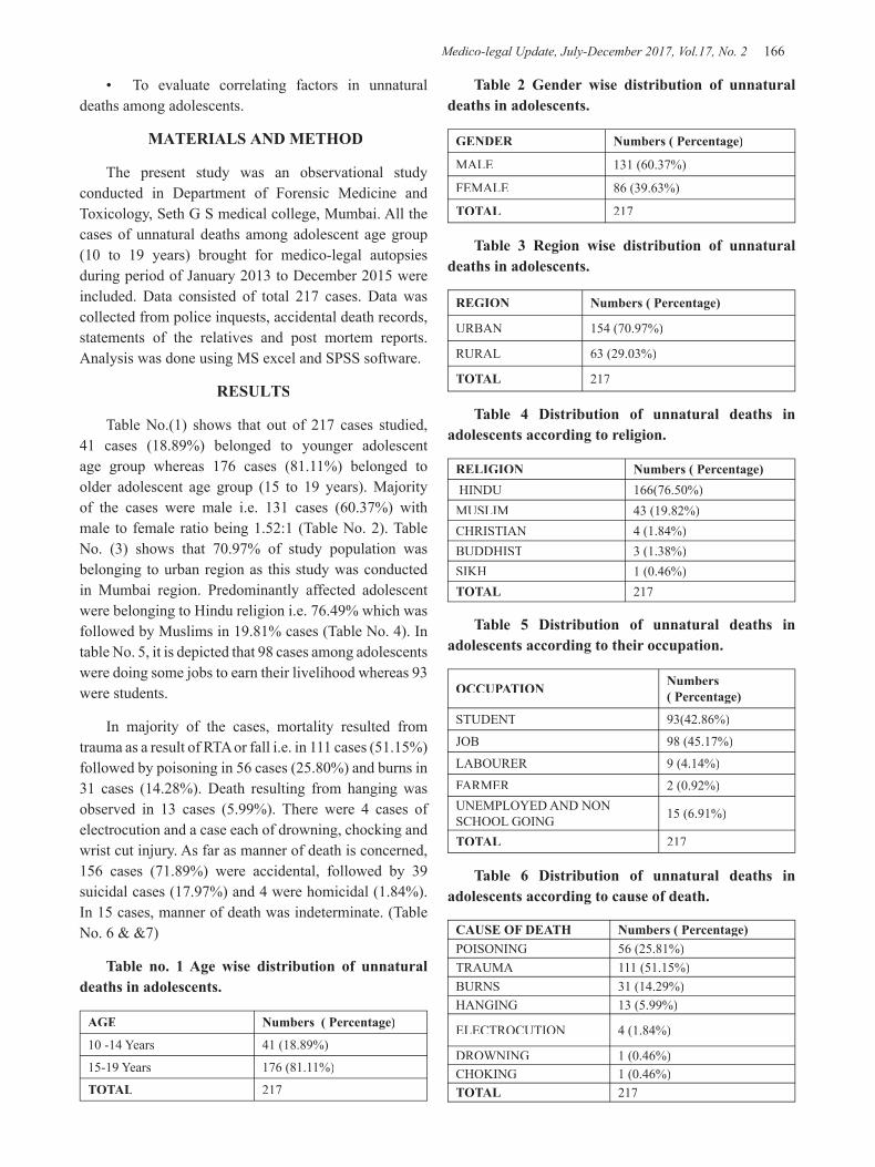
Medico-legal Update, July-December 2017, Vol.17, No. 2 166 167 -legal Update, July-December 2017, Vol.17, No. 2
• To evaluate correlating factors in unnatural deaths among adolescents.
MATERIALS AND METHOD
The present study was an observational study conducted in Department of Forensic Medicine and Toxicology, Seth G S medical college, Mumbai. All the cases of unnatural deaths among adolescent age group (10 to 19 years) brought for medico-legal autopsies during period of January 2013 to December 2015 were included. Data consisted of total 217 cases. Data was collected from police inquests, accidental death records, statements of the relatives and post mortem reports. Analysis was done using MS excel and SPSS software.
RESULTS
Table No.(1) shows that out of 217 cases studied, 41 cases (18.89%) belonged to younger adolescent age group whereas 176 cases (81.11%) belonged to older adolescent age group (15 to 19 years). Majority of the cases were male i.e. 131 cases (60.37%) with male to female ratio being 1.52:1 (Table No. 2). Table No. (3) shows that 70.97% of study population was belonging to urban region as this study was conducted in Mumbai region. Predominantly affected adolescent were belonging to Hindu religion i.e. 76.49% which was followed by Muslims in 19.81% cases (Table No. 4). In table No. 5, it is depicted that 98 cases among adolescents were doing some jobs to earn their livelihood whereas 93 were students.
In majority of the cases, mortality resulted from trauma as a result of RTA or fall i.e. in 111 cases (51.15%) followed by poisoning in 56 cases (25.80%) and burns in 31 cases (14.28%). Death resulting from hanging was observed in 13 cases (5.99%). There were 4 cases of electrocution and a case each of drowning, chocking and wrist cut injury. As far as manner of death is concerned, 156 cases (71.89%) were accidental, followed by 39 suicidal cases (17.97%) and 4 were homicidal (1.84%). In 15 cases, manner of death was indeterminate. (Table No. 6 & &7)
Table no. 1 Age wise distribution of unnatural deaths in adolescents.
AGE Numbers ( Percentage)
10 -14 Years 41 (18.89%)
15-19 Years 176 (81.11%)
TOTAL 217
Table 2 Gender wise distribution of unnatural deaths in adolescents.
GENDER Numbers ( Percentage)
MALE 131 (60.37%)
FEMALE 86 (39.63%)
TOTAL 217
Table 3 Region wise distribution of unnatural deaths in adolescents.
REGION Numbers ( Percentage)
URBAN 154 (70.97%)
RURAL 63 (29.03%)
TOTAL 217
Table 4 Distribution of unnatural deaths in adolescents according to religion.
RELIGION Numbers ( Percentage) HINDU 166(76.50%)MUSLIM 43 (19.82%)CHRISTIAN 4 (1.84%)BUDDHIST 3 (1.38%)SIKH 1 (0.46%)TOTAL 217
Table 5 Distribution of unnatural deaths in adolescents according to their occupation.
OCCUPATION Numbers ( Percentage)
STUDENT 93(42.86%)
JOB 98 (45.17%)
LABOURER 9 (4.14%)
FARMER 2 (0.92%)UNEMPLOYED AND NON SCHOOL GOING 15 (6.91%)
TOTAL 217
Table 6 Distribution of unnatural deaths in adolescents according to cause of death.
CAUSE OF DEATH Numbers ( Percentage)POISONING 56 (25.81%)TRAUMA 111 (51.15%)BURNS 31 (14.29%)HANGING 13 (5.99%)
ELECTROCUTION 4 (1.84%)
DROWNING 1 (0.46%)CHOKING 1 (0.46%)TOTAL 217
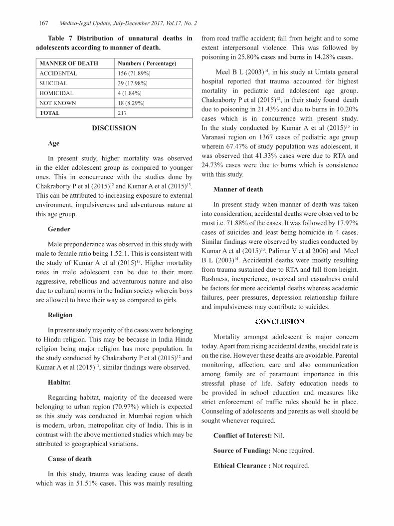
-legal Update, July-December 2017, Vol.17, No. 2 166 167 Medico-legal Update, July-December 2017, Vol.17, No. 2
Table 7 Distribution of unnatural deaths in adolescents according to manner of death.
MANNER OF DEATH Numbers ( Percentage)
ACCIDENTAL 156 (71.89%)
SUICIDAL 39 (17.98%)
HOMICIDAL 4 (1.84%)
NOT KNOWN 18 (8.29%)TOTAL 217
DISCUSSION
Age
In present study, higher mortality was observed in the elder adolescent group as compared to younger ones. This in concurrence with the studies done by Chakraborty P et al (2015)12 and Kumar A et al (2015)13. This can be attributed to increasing exposure to external environment, impulsiveness and adventurous nature at this age group.
Gender
Male preponderance was observed in this study with male to female ratio being 1.52:1. This is consistent with the study of Kumar A et al (2015)13. Higher mortality rates in male adolescent can be due to their more aggressive, rebellious and adventurous nature and also due to cultural norms in the Indian society wherein boys are allowed to have their way as compared to girls.
Religion
In present study majority of the cases were belonging to Hindu religion. This may be because in India Hindu religion being major religion has more population. In the study conducted by Chakraborty P et al (2015)12 and Kumar A et al (2015)13, similar findings were observed.
Habitat
Regarding habitat, majority of the deceased were belonging to urban region (70.97%) which is expected as this study was conducted in Mumbai region which is modern, urban, metropolitan city of India. This is in contrast with the above mentioned studies which may be attributed to geographical variations.
Cause of death
In this study, trauma was leading cause of death which was in 51.51% cases. This was mainly resulting
from road traffic accident; fall from height and to some extent interpersonal violence. This was followed by poisoning in 25.80% cases and burns in 14.28% cases.
Meel B L (2003)14, in his study at Umtata general hospital reported that trauma accounted for highest mortality in pediatric and adolescent age group. Chakraborty P et al (2015)12, in their study found death due to poisoning in 21.43% and due to burns in 10.20% cases which is in concurrence with present study. In the study conducted by Kumar A et al (2015)13 in Varanasi region on 1367 cases of pediatric age group wherein 67.47% of study population was adolescent, it was observed that 41.33% cases were due to RTA and 24.73% cases were due to burns which is consistence with this study.
Manner of death
In present study when manner of death was taken into consideration, accidental deaths were observed to be most i.e. 71.88% of the cases. It was followed by 17.97% cases of suicides and least being homicide in 4 cases. Similar findings were observed by studies conducted by Kumar A et al (2015)13, Palimar V et al 2006) and Meel B L (2003)14. Accidental deaths were mostly resulting from trauma sustained due to RTA and fall from height. Rashness, inexperience, overzeal and casualness could be factors for more accidental deaths whereas academic failures, peer pressures, depression relationship failure and impulsiveness may contribute to suicides.
CONCLUSION
Mortality amongst adolescent is major concern today. Apart from rising accidental deaths, suicidal rate is on the rise. However these deaths are avoidable. Parental monitoring, affection, care and also communication among family are of paramount importance in this stressful phase of life. Safety education needs to be provided in school education and measures like strict enforcement of traffic rules should be in place. Counseling of adolescents and parents as well should be sought whenever required.
Conflict of Interest: Nil.
Source of Funding: None required.
Ethical Clearance : Not required.
Medico-legal Update, July-December 2017, Vol.17, No. 2 168
REFERENCES
(1) Adolescents: Health risk and solutions, Fact sheet N°345 Updated May 2014, WHO, Geneva. Availed at: http://www.who.int/mediacentre/factsheets/fs345/en/. Cited on: 9/4/2015.
(2) Orientation programmes for medical officers to provide Adolescent–friendly Reproductive and Sexual health services, facilitator’s guide 11-18. Availed at: http://www.pbnrhm.org/dOCS/facilitator_guide.pdf; Cited on: 9/4/2015.
(3) Sunitha S, Gururaj G. Health behaviours & problems among young people in India: Cause for concern & call for action. Indian J Med Res 2014; 140:185-208.
(4) Sarangi L, Acharya H P, Panigrahi O P. Substance abuse among adolescents in urban slums of Sambalpur. Indian J Community Med 2008; 33: 265-7.
(5) Srinath S, Girimaji SC, Gururaj G, Seshadri S, Subbakrishna DK, Bhola P, et al. Epidemiological study of child & adolescent psychiatric disorders in urban & rural areas of Bangalore, India. Indian J Med Res 2005; 122: 67-79.
(6) Anita S, Gaur DR, Vohra AK, Subash S, Khurana H. Prevalence of psychiatric morbidity among 6 to 14 years old children. Indian J Community Med 2003; 28: 133-7.
(7) Suicides in India 2014 New Delhi,, NCRB. Ministry of Home Affairs 2014, Page 192. Available from: http:/ncrb.nic.in/state publications/ADSI 2014/Chapter 21.20 Suicides.pdf. Assesed on 09/01/2017.
(8) Jagnoor J, Suraweera W, Keay L, Ivers R Q, Thakur JS, Jha P. Million Death Study Collaborators. Unintentional injury mortality in India, 2005: Nationally representative mortality survey of 1.1 million homes. BMC Public Health 2012; 12: 487.
(9) Violence and injury prevention. World health organization. Available from: http://www.who.in/violence_inury_prevention/viloence/youth/enc. Assessed on 09/01/2017.
(10) Sarkar M. A study on domestic violence against adult and adolescent females in a rural area of West Bengal. Indian J Community Med 2010; 35: 311-5.
(11) Ackerson L K, Subramanian S V. Domestic violence and chronic malnutrition among women and children in India. Am J Epidemiol 2008; 167: 1188-96.
(12) Chakrabarty P, Saren A, Tudu N, Dwari A, Haldar D, Mitra S. Spectrum of unnatural deaths among the adolescents: An autopsy based study. Journal of Dental and Medical Sciences 2015;14(4):18-24.
(13) Kumar A, Pandey SK, Singh TB. Epidemiological Study of Unnatural Death among Children’s in Varanasi Area (India). International Journal of Science and Research (IJSR) 2014; 3:1438-41.
(14) Meel B L; Mortality of children in the Transkei region of South Africa; The American Journal of Forensic Medicine and Pathology; 2003; 24(2): 141-7.
(15) Palimar V, Arun M. Prashantha B. Paediatric fatalities due to trauma. Medico Legal Update; 2006; 6 (4): 10 –12.
-legal Update, July-December 2017, Vol.17, No. 2 168
Profile of Cases of Fatal Road Traffic Accident with Respect to Intra Intracranial Injury and Cause of Death in a Centre at
Rural India – Autopsy based Study
B H Tirpude1, Deepak Bhagwat3, P N Murkey1, Sharjeel Khan3, I L Khandekar1, Pravin Zopate2, Ranjan Ambedkar3, Rahul Ramteke3
1Professor, 2Assistant Professor, 2Assistant Professor, 2 3Post Graduate Student, Dpt. of Forensic Medicine and Toxicology, Mahatma Gandhi Institute of Medical Sciences, Sevagram, Wardha
ABSTRACT
Road traffic accident is a complex phenomenon. Constant rise in the number of motor vehicles, rampant encroachment of road, easy to avail the vehicle because of loan facility, nasty tendency of violating traffic rules and anarchic traffic systems have greatly contributed to rapid increase in RTAs. Population explosion is a catalyzing factor for a number of accidents. The rise of road traffic accidents has become a major public health problem. RTAs cost a lot not only to the individuals affected and their families but also to the nation. The injuries, disability and fatality resulting from unexpected RTAs put a significant drain on the economy of the nation.
The present study was a cross-sectional study conducted in central India during 2 year period from 1st June st June st
2014 to 31st May 2016. During the study period, a total of 757 medicolegal autopsies were conducted in this st May 2016. During the study period, a total of 757 medicolegal autopsies were conducted in this st
institute, out of which 109 cases of fatal road traffic accidents (died on spot or within 24 hours of accident) were studied. The purpose of the study was to know the age and sex wise distribution , diurnal variation and to correlate the above parameters with the cause of death.
Keywords : Fatal road traffic accident, Head Injuries, Intracranial haemorrhage, death.
INTRODUCTION
Accidents are world’s most serious health problem. The motorized transportation media like vehicles, trains, aeroplane, with fast moving vehicular traffic, vast urbanization and changing social patterns, have contributed to increase in the incidence of trauma to human body. Road Traffic Accident (RTA) is any vehicular accident occurring on the roadway (i.e. originating on, or involving a vehicle partially on the roadway)10. This includes collision of an automobile with a pedestrian, another automobile or with a non-automobile on the roadway or fall from a moving vehicle causing injuries or death of involved individuals.
In recent years, death due to RTAs are increasing at an alarming rate throughout the world. Thereby it poses itself as a major epidemiological as well as medico legal problem. This is due to the tremendous increase in the number of vehicle, high speed technology along
with other contributing factors like congestion and poor condition of roads, intoxicating influence of alcohol or drugs, inexperienced drivers without proper driving license, ignorance or intentional violation of traffic rules etc.
In 2013, global rate of death from Road Traffic Injuries was 16.6 per 1,00,000 population, 1.25 million people died globally from road traffic injuries in year 201311. In India, more than 4.8 lakh accidents and more than 1.3 lakh deaths were reported during 20133. According to an expert study group appointed by Government of India “RTAs have come to be considered as the third deadly killer, next to heart disease and cancer.” Every year the World Health Organization (WHO) hosts an event, usually on 7th April, to celebrate the anniversary of its founding in 1946. Each year the event focuses on one health issue. In response to a growing concern about RTAs the WHO Director-General, Dr. Lee Jong Wook has for the first time in history of WHO devoted6 7th
DOI Number: 10.5958/0974-1283.2017.00093.7
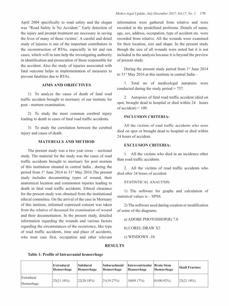
Medico-legal Update, July-December 2017, Vol.17, No. 2 170 171 -legal Update, July-December 2017, Vol.17, No. 2
April 2004 specifically to road safety and the slogan was “Road Safety Is No Accident.” Early detection of the injury and prompt treatment are necessary in saving the lives of many of these victims7. A careful and detail study of injuries is one of the important contributors in the reconstruction of RTAs, especially in hit and run cases, which will in turn help the investigating authority in identification and prosecution of those responsible for the accident. Also the study of injuries associated with fatal outcome helps in implementation of measures to prevent fatalities due to RTAs.
AIMS AND OBJECTIVES
1) To analyze the cause of death of fatal road traffic accident brought to mortuary of our institute for post - mortem examination.
2) To study the most common cerebral injury leading to death in cases of fatal road traffic accidents.
3) To study the correlation between the cerebral injury and cause of death.
MATERIALS AND METHOD
The present study was a two year cross – sectional study. The material for the study was the cases of road traffic accidents brought to mortuary for post mortem of this institution situated in central India , during the period from 1st June 2014 to 31st June 2014 to 31st st May 2016.The present st May 2016.The present st
study includes documenting types of wound, their anatomical location and commonest injuries leading to death in fatal road traffic accidents. Ethical clearance for the present study was obtained from the institutional ethical committee. On the arrival of the case in Mortuary of this institute, informed expressed consent was taken from the relative of deceased for examination of wound and their documentation. In the present study, detailed information regarding the wounds and various factors regarding the circumstances of the occurrence, like type of road traffic accidents, time and place of accidents, who treat case first, occupation and other relevant
information were gathered from relative and were recorded in the predefined proforma. Details of name, age, sex, address, occupation, type of accident etc. were recorded from relative. All the wounds were examined for their location, size and shape. In the present study though the size of all wounds were noted but it is not included in the analysis because it is beyond the preview of present study.
During the present study period from 1st June 2014 st June 2014 st
to 31st May 2016 at this institute in central India – st May 2016 at this institute in central India – st
1. Total no of medicolegal autopsies were conducted during the study period = 757.
2. Autopsies of fatal road traffic accident (died on spot, brought dead to hospital or died within 24 hours of accident) = 109.
INCLUSION CRITERIA:
All the victims of road traffic accidents who were died on spot or brought dead to hospital or died within 24 hours of accident.
EXCLUSION CRITERIA:
1. All the victims who died in an incidence other than road traffic accidents.
2. All the victims of road traffic accidents who died after 24 hours of accident.
STATISTICAL ANALYSIS:
1) The software for graphs and calculation of statistical values is – SPSS.
2) The software used during creation or modification of some of the diagrams.
a) ADOBE PHOTOSHOP(R) 7.0
b) COREL DRAW X3
c) WINDOWS -10.
RESULTS
Table 1: Profile of Intracranial hemorrhage
Extradural Hemorrhage
Subdural Hemorrhage
Subarachnoid Hemorrhage
Intraventricular Hemorrhage
Brain Stem Hemorrhage Skull Fracture
Extradural
Hemorrhage23(21.10%) 22(20.18%) 21(19.27%) 10(09.17%) 01(00.92%) 23(21.10%)
-legal Update, July-December 2017, Vol.17, No. 2 170 171 Medico-legal Update, July-December 2017, Vol.17, No. 2
Subdural
Hemorrhage22(20.18%) 87(79.82%) 86(78.90%) 14(12.84%) 02(01.83%) 84(77.06%)
Subarachnoid
Hemorrhage21(19.27%) 86(78.90%) 86(78.90%) 13(11.93%) 02(01.83%) 83(76.15%)
Intraventricular
Hemorrhage10(09.17%) 14(12.84%) 13(11.93%) 15(13.76%) 01(00.92%) 13(11.93%)
Brain Stem
Hemorrhage01(0.92%) 02(01.83%) 02(01.83%) 01(00.92%) 03(02.75%) 03(02.75%)
Cont... Table 1: Profile of Intracranial hemorrhage
Profile of Intracranial hemorrhage:
In our present study intracranial hemorrhages were classified as extradural hemorrhage, subdural hemorrhage, subarachnoid hemorrhage, intraventricular hemorrhage and brain stem hemorrhage. Subdural hemorrhage was most commonly seen (87 cases, 79.82%) followed by subarachnoid hemorrhage (86 cases, 78.90%), extradural hemorrhage (23 cases, 21.10%) and intraventricular hemorrhage (15 cases, 13.76%). Least seen was brain stem hemorrhage (03 cases, 02.75%).
- Subdural hemorrhage with skull fracture was seen in 84 cases (77.06%).
- Subarachnoid hemorrhage with skull fracture was seen in 83 cases (76.15%).
- Extradural hemorrhage with skull fracture was seen in all 23 cases (21.10%).
- Intraventricular hemorrhage with skull fracture was seen in 13 cases (11.93%).
- Brain stem hemorrhage with skull fracture was seen in all 03 cases (02.75%).
Table 2: Profile of cause of death
Cause of death No. of cases Percentage
Intracranial injuries (intracranial hemorrhage/brain injury) 86 78.90%
Hemorrhagic shock 17 15.60%
Traumatic asphyxia 04 03.67%
Spinal cord injury 02 01.83%
Total 109 100%
Profile of cause of death:
In our present study, cause of death of maximum cases was Intracranial injuries (intracranial hemorrhage/brain injury) 86 (78.90%) followed by hemorrhagic shock 17 (15.60%), traumatic asphyxia 04 (03.67%) and spinal cord injury 02 (01.83%).
DISCUSSION
Profile of Intracranial hemorrhage (Table 1):
In the present study, brain hemorrhages are classified as extradural hemorrhage, subdural hemorrhage, subarachnoid hemorrhage, intraventricular hemorrhage and brain stem hemorrhage. Subdural hemorrhage was most commonly seen in 87cases (79.82%) followed by subarachnoid hemorrhage 86 cases (78.90%) extradural hemorrhage and intraventricular hemorrhage was seen in 23 cases (21.10%) and 15 cases (13.76%) respectively. Least seen was brain stem hemorrhage 03 cases (02.75%). Subdural hemorrhage associated with subarachnoid hemorrhage in 86 cases (78.90%). Subdural hemorrhage was associated with intraventricular hemorrhage in 14 cases (12.84%). Subdural hemorrhage was associated with extradural hemorrhage in 22 cases (20.18%). Subdural hemorrhage associated with brain stem hemorrhage in 02 cases (01.83%).
All the 23 cases (21.10%) of extradural hemorrhage had fracture of skull. Among the 87 cases (79.82%) of subdural hemorrhage 84 cases (77.06%) had fracture of skull. Among the 86 cases (78.90%) of subarachnoid hemorrhage 83 cases (76.15%) had fracture of skull. Among the 15 cases (13.76%) of intraventricular hemorrhage 13 cases (11.93%) had skull fracture. Among the 03 cases (02.75%) of brain stem hemorrhage 03 cases (02.75%) had skull fracture. Significant association was

Medico-legal Update, July-December 2017, Vol.17, No. 2 172 173 -legal Update, July-December 2017, Vol.17, No. 2
seen between subdural and subarachnoid hemorrhage in our study. According to various study subdural hemorrhage is the most common hemorrhage which is similar to our study.15, 4, 12, 14, 13
This shows that intracranial injuries are most common fatal injuries in road traffic accidents in this region. This could be due to fact that, the intracranial injuries cannot be treated successfully even in tertiary level hospital. This may be because of their complicated physiological and anatomical configuration.
Profile of cause of death (Table 2):
In our study cause of death was intracranial injuries 86 (78.90%) in maximum number of cases. Next to intracranial injuries was hemorrhagic shock seen in 17 cases (15.60%), traumatic asphyxia in 4 cases (03.67%) and spinal cord injury in 2 cases (01.83%). The finding of present study are similar to studies4, 2, 1, 5, 8. In the study conducted in Finland involving RTA during the period 1972 to 1982,9 in which an injury to cervical spine was the main cause of death. Increasing age seems to increase the risk of fatal cervical spinal injuries. Patients between 16 to 25 years of age had the lowest risk and the patient over the age of 60 years had the highest risk of sustaining a fatal cervical spinal injury 16. Accordingly in our study most of the cervical spinal cord injury cases were above 40 years.
Several human and environmental risk factors such as age, alcoholism, without driving license, type of vehicle etc. were found associated in occurrence of road traffic injuries. If we control these factors appropriately mortality and morbidity can be prevented.
CONCLUSION
Road traffic accident is a complex phenomenon. Constant rise in the number of motor vehicles, rampant encroachment of road, easy to avail the vehicle because of loan facility, nasty tendency of violating traffic rules and anarchic traffic systems have greatly contributed to rapid increase in RTAs. Population explosion is a catalyzing factor for a number of accidents. The rise of road traffic accidents has become a major public health problem. RTAs cost a lot not only to the individuals affected and their families but also to the nation. The injuries, disability and fatality resulting from unexpected RTAs put a significant drain on the economy of the nation. The deaths due to RTAs accounted for 14.40%
of total medico legal autopsies conducted (i.e. died on spot, brought dead to hospital or died within 24 hours of accident). All the victims of fatal RTAs had injuries of one or other system. Intracranial injuries were seen in 80.73% of the cases. In majority of victims, intracranial injuries contributed either directly or indirectly to death. Intracranial injuries cause alone was responsible for death in 78.90% of cases, followed by hemorrhagic shock 15.60%, spinal cord injury 01.83% and traumatic shock 03.67%. This shows that intracranial injuries are most common fatal injuries in road traffic accidents in this region. This could be due to the fact that, the intracranial injuries cannot be treated successfully, even in tertiary level hospitals. This may be because of their physiological and anatomical configuration. Hence, fatalities due to injuries of road traffic accidents can be reduced by preventing the occurrence of such injuries. Therefore, the old saying, “Prevention is better than cure” holds true even here. Injuries and fatalities due to RTA can be prevented or at least can be reduced by preventing the accidents/crashes, in turn reducing fatal injuries and fatalities.
The present study was cross-sectional study conducted in central India during 2 year period from 1st
June 2014 to 31st May 2016. During the study period, st May 2016. During the study period, st
a total of 757 medicolegal autopsies were conducted in this institute, out of which 109 cases of fatal road traffic accidents (died on spot or within 24 hours of accident) were studied.
The predesigned and pretested proforma was used to collect the required data and following were the findings.
1) Out of 109 victims of fatal RTA, among the intracranial hemorrhage, subdural hemorrhage was seen in 79.82% of the cases followed by subarachnoid hemorrhage in 78.90% of the cases.
2) Out of 109 victims of fatal RTA an intracranial injury alone was responsible for death in 86 cases (78.90%).
Source of Funding : Self
Conflict of Interest : Nil
REFERENCES
1. Aggarwal KK, Oberoi SS, Rakesh Kumar, Sharma M. Pattern and distribution of injuries in fatal road

-legal Update, July-December 2017, Vol.17, No. 2 172 173 Medico-legal Update, July-December 2017, Vol.17, No. 2
traffic accident cases. JPAFMAT 2009; 9(2): 71-75.
2. Arvind Kumar, Lalwani S, Agarwal D, Rautji R, Dogra TD. Fatal road traffic accidents and their relationship with head injuries: An epidemiological survey of five years. IJNT 2008; 5(2): 63-67.
3. Chandra J, Dogra TD, Dixit PC. Pattern of cranio-intracranial injuries in fatal vehicular accidents in Delhi, 1966-76. Med Sci Law. 1979 Jul; 19(3):186-94.
4. Chaudhary BL, Singh D, Tirpude BH, Sharma RK, Meel V. Profile of road traffic accident cases in Kasturba Hospital of M.G.I.M.S., Sevagram, Wardha, Maharashtra; Medico-legal updates 2005: 5(4).
5. Conroy C, Schwartz A, Holt DB, Brent Eastman A, Pacyna S, Hlobrook TL et al. Upper extremity fracture patterns following motor vehicle crashes differ for drivers and passengers. Accid Anal Prev 2007 Mar; 38(3):350-7.
6. District Statistician. A view on statistics of Belgaum district 2003-04. Belgaum: Zilla Panchayat; 2005.
7. Dorland’s illustrated medical dictionary. 30th ed. Philadelphia: Saunders; 2003.
8. Ganveer GB, Tiwari RR. Injury pattern among non-fatal road traffic accident cases: A cross-
sectional study in Central India. Indian Journal of Medical Sciences 2005; 59(1): 9-12.
9. Ghosh PK. Epidemiological study of victims of vehicular accidents in Delhi. J. Indian Med Assoc 1992 Dec; 90(12): 309-312.
10. Ghosh PK. Post-mortem study of pattern of injury involving pedestrian victims. J For Med Tox. 1991 Jul-Dec;8(3-4):1-8.
11. Gissane W, Bull J. Injuries from road accidents. The practioner 1962 April; 188: 489-497.
12. Gupta S, Roychowdhury UB, Deb PK, Motira R, Chhetri D. Demographic study of fatal cranio-cerebral road traffic injuries in north Bengal region. JIAFM 2007; 29(1).
13. Kuchewar SV, Meshram RD, Gadge SJ. Demographic study and medico-legal aspects of fatal road traffic accidents in Aurangabad. J life sci 2012; 4(1): 7-10.
14. Sharma BR, Dasari H, Singh G, Vij K. Patterns of fatal head injury in road traffic accidents. Bahrain Medical Bulletin, March 2003; 25(1): 31-33.
15. Singh YN, Bairagi K, Das KC. An epidemiological study of road traffic accident victims in medicolegal autopsies. JIAFM 2005; 27(3): 169-172.
175 -legal Update, July-December 2017, Vol.17, No. 2
Study of Unidentified Dead Bodies in Central Mumbai Region
Mahendra Namdeo Wankhede1, Abhijit Hosmani JR2, Harish Pathak3, Manoj Bhausaheb Parchake1 1Assistant Professor, 1Assistant Professor, 1 2Resident Doctor, Department of Forensic Medicine, 2Resident Doctor, Department of Forensic Medicine, 2 3Professor and Head of Department of
Forensic Medicine, Seth Gordhandas Sunderdas Medical College & King Edward Memorial Hospital Parel Mumbai
ABSTRACT
Unidentified persons are people who have died and whose bodies have not been identified during autopsy. Mumbai tops the national charts in disposing of unclaimed bodies. Unidentified bodies impose great challenge to autopsy surgeon. In recent years there is increased in number of unidentified dead bodies in central Mumbai region. Aim of our study is to find out most common age group, sex, place where the body recovered, time since identification after autopsy and cause of death in unidentified dead bodies. A total number of 196 cases were studied during period 2013-2015. Most common age group in unidentified dead bodies was middle age group 61(31.12%) (41 to 50 year). Males (89.71%) preponderance observed in our study. One day after autopsy 9 (23.07%) was most common time since identification after autopsy. Most of the dead bodies were recovered from footpath 107(54.66%). Most common cause of death was related to natural deaths 109(55.61%).
Keywords- Unidentified, Mumbai, middle age, identification, footpath
Corresponding author:Dr Mahendra Namdeo WankhedeAssistant Professor, Department of Forensic MedicineSGSMC & KEM Hospital Parel MumbaiContact no.091-9322650861, Email –[email protected]
INTRODUCTION
Identity is the determination of the individuality of a person. According to article 6 of the Universal Declaration of Human Rights states, that everyone has the right to recognition everywhere as a person before the law1. Unidentified persons are individuals who have died and whose bodies have not been recognized during autopsy. As per national crime record bureau of India a total of 34,592 unidentified dead bodies were recovered at all India level and required inquests as per the law were conducted by the investigating authority. Thus, the police had conducted inquest for around 95 such cases everyday on an average at all India level. Certain States indicated higher recovery of un-identified dead bodies, these State were Maharashtra (6,187 victims), Tamil Nadu (3,739 victims), Karnataka (3,533 victims) Uttar Pradesh (3,409 victims), West Bengal (3,086 victims) and Delhi UT (3,063 victims).2
Mumbai ranks first on national charts in disposing of unidentified dead bodies. Disposal of unidentified bodies are done as per the provision laid under section 5 of the Bombay Anatomy Act of 1949 3. As Mumbai is the economic capital of India, having every facility to live dignified life. Nevertheless Mumbai have also large number of unidentified dead bodies. There is rise in number of unidentified dead bodies progressively in Mumbai. This increasing number of unknown dead body is also described as silent mass disaster. Forensic experts, medical and non-medical persons play an important role in identification process. This is done by scientific methods. But identification becomes difficult in highly decomposed, mutilated bodies and in mass disaster. The identification of cadavers is a key issue in autopsy and equally important for ethical, criminal and civil reasons. Hence we conducted this study to emphasis on problems of unidentified bodies.
MATERIALS AND METHOD
The present prospective study of postmortem examination in unidentified dead bodies brought to the mortuary of the department of Forensic Medicine & Toxicology, Seth G S Medical College & K E M Hospital, Parel, Mumbai; during the period from
DOI Number: 10.5958/0974-1283.2017.00094.9

175 Medico-legal Update, July-December 2017, Vol.17, No. 2
January 2013 to December 2015 comprised the material for the study. During this period the total 196 cases of unidentified dead bodies were studied in central Mumbai region. Data regarding these cases was compiled from the postmortem reports, inquest papers; Hospital records, detailed history taken from investigating police officials at the time of postmortem examination, etc. All data related to these cases were compiled, analyzed, tabulated and paying special attention to demographics and other medico-legal perspectives like Gender, Age, Date of death, Date of post mortem examination; police jurisdiction where the body was found, the cause of death and the special identification marks noted down.
RESULTS AND OBSERVATION
As per figure no.1 109(55.61) cases were belonging to natural deaths while 72(36.74%) cases were unnatural deaths in unidentified dead bodies and in 15 (7.65%) cases manner was not known. Table no.1 shows most common age group involved in unidentified dead bodies were 40 to 50 years 61(31.12%) followed by 20 to 30 years 43(21.93%), 50 to 60 years 35(17.85%) and no deaths observed in 0 to 10 years age group. So almost 50% deaths were observed in 40 to 60 years age group. Males (89.79%) outnumbered the females (10.21%) in our study. Maximum number of cases were observed in cold season 82(41.84%) followed by summer 62(31.63%) and least in rainy season 52 (26.53%). Table no.3 shows footpath 107(54.66%) was most common place where unidentified dead body found followed by parel railway station 56 (28.57%) and KEM hospital campus 17 (8.67%) and least cases were found at wadala sky bridge walk 01(0.51%) and sea shore 02(1.02%). Out of total 196 unidentified dead bodies 39 were identified after autopsy, among them most of the unidentified cases were identified on first day after autopsy 9 (23.07%) followed by second day after autopsy 8 (20.51%)and least was six to seven day after autopsy 1 (2.56%) Most common cause of death in unidentified dead bodies were natural 109(55.61%) followed by Head injury 43 (21.93%) and Multiple injury 35(17.97%) and least common cause of deaths were drowning01 (0.51%), burns01 (0.51%), blunt trauma to abdomen 01 (0.51%). According to manner of death natural death 109(55.61%) was most common manner of death followed by accidental 65(33.16%) and least common was homicidal 03 (1.53%). Most of the cases were identified 1 day after the autopsy 9(23.07%).
DISCUSSION
Unidentified dead bodies have imposed big challenge to autopsy surgeon and investigating officer. These cases are silent mass disaster in society, which are ignored by society, police and hospitals. Special reference should be given to these cases.
In present study most common age group in unidentified cases were 40 to 50 years 61(31.12%) followed by 20 to 30 years 43(21.93%), 50 to 60 years 35(17.85%). So almost 50% deaths were observed in 40 to 60 years age group, which is middle age group. The reason might be due to the fact that the huge movement of people for search of job and livelihood. Also many intra-state migrations happen due to poverty and drought situations in Maharashtra. Majority of the people engaged as daily wage laborers or as beggars. Similar to present study, Chaudhary BL et al 4 stated that the most of the cases (27.36%) were in the age group of 40-49 years. As per study by Altun G et al 5 (27.95%) in 40 to 49 age group. While in Study of Kumar S. 6, the maximum numbers 47.24%) were in age group 41-60 years. Also as per study by Kumar A et al7 most common age group in unknown cases were 41 to 50 years (35%). This might be due to the fact that the most mobile age group for various reasons, both economic and social. Almost similar to our study, Andrev et al 8 reported 9.13% of all deaths in men aged 25–54 years were unidentified. According to Cheung AM et al9 study In Toronto the mortality rate among home-less women aged 18–44 years was 515 per 100,000 persons per year.
Males (89.79%) outnumbered the females (10.21%) in our study this might be due to the fact that males usually migrating for jobs in Mumbai city. Similar to our study, Chaudhary BL 4 et al stated that in the deaths of homeless unknown persons (HUPs) with the predominance of male sex (90.25%), this might be due to the fact that most of the these cases were medicolegal hence male predominance. Another study in Mumbai by Wagmare et al [8] found that out of 51 cases 34 were identified as males. Likewise study of Kumar A et al. 7
(87.75%) and study of Kumar S et al. 6 (69.99%), carried out in Delhi and North India respectively found male predominance. The results were also in accordance to global studies carried out by Altun G., et al 5 and Buyuk et al. 9 that presented the preponderance of male cases (91.27%)

Medico-legal Update, July-December 2017, Vol.17, No. 2 176 177 -legal Update, July-December 2017, Vol.17, No. 2
Maximum number of cases were observed in cold season 82(41.84%) followed by summer 62(31.63%) and least in rainy season 52 (26.53%). In accordance with our study, Chaudhary BL et al 4 found that increase in the number (14.55%) of deaths in September month. Similarly Altun G., et al 5 stated that maximum deaths (59.38%) were during winter season. Contrast to our study, findings in study of Philadelphia 10 shows that 53% of deaths were happened during summer months and additional Buyuk et al. 9 reported that high percentages of the cases living in the open are every danger both environmental and criminal. On the other hand, in study of Kumar A et al. 7 and Kumar S., et al. 6
were highest number (38.64%) & (69.70%) respectively reported in rainy season (i.e. to be July to September in India). Kumar A et al 11 found that maximum number of cases observed in October month 12.2%. Reason might be a seasonal pattern of tuberculosis with a mostly predominant peak is seen during the spring and summer seasons in all of the countries.
Footpath 107(54.66%) was most common place where unidentified dead body found followed by parel railway station 56 (28.57%) and KEM hospital campus 17 (8.67%). In accordance to our study, Chhattopadhya et al 13 reported that 275 were found the city roads or adjacent footpaths. Reason might be that a good number of homeless people reside on the streets and are below the poverty line. Kumar A 7 an also found almost similar findings from his study in South Delhi that about 32.2% were recovered from the river or its banks. Altun 5
reported that out of the 126 cases of deaths of homeless persons, 94 were found outdoors.
Out of total 196 unidentified dead bodies 39 were identified after autopsy, among them most of the unidentified cases were identified on first day after autopsy 9 (23.07) followed by second day after autopsy 8 (20.51%). The reasons for so many unclaimed and unidentified dead bodies are “Metros like Mumbai, witness enormous migration of people for search of job and livelihood. Also many intra-state movements happen due to poverty and drought situations in Maharashtra. Majority of the people engaged as daily wage laborers or as beggars, etc. “Also most of the time, even if the deceased are identified, the claimants are not keen to come forward and claim the body, as they do not have money for carrying out the last rites, and hence do not turn up.
Similar to our study, Chattopadhyay et al13 reported that among the unidentified persons, the identity of 109 deceased could be confirmed and 38.5% (n =109) of them were identified on the same day after the autopsy. Consequently, the rate of identity decreased and none were identified from the fourth day after the autopsy. As the bodies get older, decomposition sets in and that hinders the identification process. In such circumstances, it is difficult for the family members to confirm the identity simply by viewing the body. Moreover, the family members make a frantic effort to search for the missing ones within the few days after they’ve gone missing. A large number of persons remained unidentified in this study because many were lonely street dwellers without any family and 32.2% of the bodies were recovered from the river, which might have come floating from distant places.
Most common cause of death in unidentified dead bodies were natural 109(55.61%) followed by Head injury 43 (21.93%) and Multiple injury 35(17.97%). Similar to our study, Chattopadhyay et al 13 found that natural deaths due to disease or pathological conditions were the leading causes of death (48.3%). This was mostly seen among the street dwellers that were of very poor health condition. Due to lack of food, shelter and proper medical care. They were the victims of various infections and other diseases leading to fatality. Similar to our study, Chaudhary et al 4 found that pulmonary pathology was leading cause of death and contributed 67.95% cases of total 71.56% of natural deaths. These findings are in concurrence with study of Kumar A et al. 7 and Buyuk Y et al 9 where natural events were the main cause of death in 61.36% & 60.26% cases respectively.
Figure no.1 showing distribution of unidentified dead bodies as per nature of death
-legal Update, July-December 2017, Vol.17, No. 2 176 177 Medico-legal Update, July-December 2017, Vol.17, No. 2
Table no.1 showing distribution of unidentified cases as their age group.
Age (years) Total no. of cases. (%)
0 to 10 0(0%)
11 to 20 4 (2.04%)
21 to 30 43(21.93)
31 to 40 28(14.28%)
41 to 50 61(31.12%)
51 to 60 35 (17.85%)
61 to 70 13 (6.65%)
More than 70 12(6.12%)Total 196 (100%)
Figure no.2 showing distribution of unidentified cases as per sex
Table no.2 showing distribution of unidentified cases deaths as per environmental season
Season Total no. of cases (%)
Cold season 82 (41.84%)
Summer 62 (31.63%)
Rainy 52(26.53%)
Total 196 (100%)
Table no.3 Showing distribution of unidentified cases as per place where they found.
Place of found Total no. of cases.Footpath 107(54.66%)Garden 02(1.02%)KEM Hospital campus 17(8.67%)MGM Hospital 02(1.02%)Parel railway station 56(28.57%)Sewree railway station 03(1.53%)Wadala sky walk bridge 01(0.51%)Sea Shore 02(1.02%)Not Known 6(3.06)Total 196(100%)
Table no.4 showing time since identification after autopsy in unidentified dead bodies.
Time since Identification after autopsy Total no. of cases(%)
Same day of the autopsy 04 (10.25%)
1 day after autopsy 09 (23.07%)
2 day after autopsy 08 ( 20.51% )
3 day after autopsy 02 ( 5.12% )
4 day after autopsy 03 (7.69% )
5 day after autopsy 03 (7.69% )
6-7 day after autopsy 01 (2.56% )
More than 1 week 09 (23.07%)
Total no. of cases 39 ( 100% )
Table no.5 showing distribution cause of death in unidentified cases
Cause of death Total no. of cases (%)
Natural death 109 (55.61%)
Drowning 01(0.51%)
Blunt trauma to chest 03(1.53%)
Blunt trauma to abdomen 01(0.51%)
Burns 01(0.51%)
Multiple injuries 35(17.97%)
Shock and haemorrhage due to stab wound to abdomen 01(0.51%)
Hanging 02(1.02%)
Head injury 43(21.93)
Total 196 (100%)
Table no.6 showing distribution of manner of death in unidentified dead bodies.
Manner of Death Total no of cases (%)
Accidental 65 (33.16)
Suicidal 04(2.04%)
Homicidal 03(1.53%)
Natural 109 (55.61%)
Not Known 15 (7.65%)
Total 196 (100%)
CONCLUSION
Sincere efforts from the government are required to deliver food and shelter to its citizens to decrease the

Medico-legal Update, July-December 2017, Vol.17, No. 2 178
burden of such deaths. Rapid and better identification methods along with coordinated efforts of the law enforcing agencies would help to establish the identity of such individuals. It would not only be important for statistical purposes, but may also provide vital information related to various criminal acts involving homicidal cases.
Even in 21 century still we are using basic technology for identification (photographs and fingerprinting). Used of modern technologies is a need of an hour. The center for identification should be established at various medical colleges with co-ordination of ministry of home affairs and medical education.
Proper measures should be made for coordination between the police and autopsy surgeon to identify the unidentified persons.
Conflict of Interest: None
Source of Funding: None
Ethical Clearance: Necessary ethical clearance taken.
REFERENCE
1. United Nations. International Covenant on Civil and Political Rights. Article 6 on the right to life [document on Internet] 1966 [cited 4 January 2017]. Available from http://www2.ohchr.org/english/law/ccpr.htm
2. Crime in India 2015. New Delhi: Government of India, Minister of Home Affairs, National Crime Record Bureau Report 63th Edition; 2016 July. 66 p.
3. Bombay act no.xi 1949. Government of Maharashtra, law and judiciary department; Jan 2014: 1-6.
4. Chaudhary BL, Kumar VR, Arvind K et al. An investigation in to the deaths of homeless unknown persons at New Delhi District. EC Pulmonology and Respiratory Medicine2.3(2016):90-97
5. Altun G., et al. “Deaths among homeless people in Istanbul”. Forensic Science International 99.2 (1999): 143-147.
6. Kumar S., et al. “Homeless and unclaimed person’s death in North India (Jan 2008 - Nov 2012)”. Medicine, Science and the Law 55.1 (2015): 11-15.
7. Kumar A., et al. “Deaths of homeless unclaimed persons in South Delhi (2001-2005): a retrospective review”. Medicine Science and The Law 331.5 (2009): 304-309.
8. Andrev EM, Pridemore WA, Shkolnikov VM, Antonova OI. An investigation into the growing number of deaths of unidentified people in Russia. European J Public Health 2008;18(3):252–7.
9. Cheung AM, Hwang SW. Risk of death among homeless women: a cohort study and review of literature. CMAJ 2004;170(8):1243–7.
10. Wagmare PB, Chikhalkar BG, Nanandkar SD. Establishing identity and cause of death in mutilated and un identifiable corpes: A challenging task for medico legal expert. Journal of forensic biomed 2015 :6(1): 1-6.
11. Buyuk Y., et al. “Homeless deaths in Istanbul, Turkey”. Journal of Forensic and Legal Medicine 15.5 (2008): 318-321.
12. Hibbs JR., et al. “Mortality in a Cohort of Homeless Adults in Philadelphia”. New England Journal of Medicine 331.4 (1994): 304-309.
13. Chattopadyay S, Shee B, Sukul B.Unidentified bodies in autopsy-A disaster in disguise.Egyptian journal of Forensic sciences 2013 vol 3: 112-113.
-legal Update, July-December 2017, Vol.17, No. 2 178
Data-based Profiling of Internet Child Pornography Offenders: A Study of the Causal Link between use of Internet Child Pornography and the Paedophilic Offending Behaviour
Anand Kumar Vasudevan1, S Ross2, L Eccleston3
1Associate Professor, Department of Forensic Medicine, Sri Muthukumaran Medical College Hospital and 1Associate Professor, Department of Forensic Medicine, Sri Muthukumaran Medical College Hospital and 1
Research Institute, Mangadu, Chennai, India, 2Director and Senior Researcher of the Melbourne Criminological 2Director and Senior Researcher of the Melbourne Criminological 2
Research and Evaluation, 3Ex-Program Coordinator (Forensic Psychology), Department of Criminology, University of Melboure, Carlton Victoria 3010, Australia
ABSTRACT
This current topic was of interest of study by the researchers after the controvertial arrest of child pornography traffickers and abusers in Victoria on October 2004. However, this was a startling revelation to the extent of propogation of child pornography in Australia and Internationally. Thereafter, the issue of child pornography has become a major area of investigation for law enforcement authorities, as it parallels increasing concerns for the children in our society. However, this research study is to focus only on Interent sex offenders knowingly possessing child pornography. As it will be interesting to know, from the findings of this research study, the association between the usage of the Internet medium and its contribution to the development of sexually abusive behaviours towards minors. In addition, to investigate whether there were any other associated risk factors involved in this offending processes. One of the major challenges to a researcher in this study was to examine the causal links or associations between use of Internet child pornography and paedophilic offending behaviours with respect to their sex offending risk levels of two risk-categories such as the non-contact Internet sex offenders and contact Internet sex offenders. The findings of this research were startling that the low-risk and high-risk group of Internet sex offenders had significant differences in their risk factors that existed among the two-risk groups, assisted by exploring the demographic and offending-related risk factor variables in understanding the paedophilic offending behaviours. By studying their risk factors it is more likely to play a vital role in determining the causal link for these Internet sex offenders to progress from just viewing or collecting these images to potentially cause sexual violence against children.
Keywords: Internet Child Pornography, risk factors, Non-contact Internet sex offender, Contact Internet Sex offender.
Corresponding author:Anand Kumar VasudevanAssociate Professor, Department of Forensic Medicine, Sri Muthukumaran Medical College Hospital and Research Institute, Mangadu, Chennai, India 600069.Email: [email protected]
INTRODUCTION
Addressing the critical issues in this research study
In the recent years there has been a rapid development and expansive growth in the use of computer applications such as the Internet1. Child pornography procured by
this rapidly improving technology is of worrying issue to whether the viewers of such materials will become an active abuser of children2. In the recent times, majority of the convictions on Internet sex crimes are related to child pornography3. It has been argued by Taylor and Quayle4 that there is no clear evidence that exposure to child pornography leads to a person to commit a contact sexual offence with a child. Henceforth, researchers in this area of study began to think, “What role does this ICP play in the development of paedophilic offending behaviour?” Therefore, researchers investigating in this area of study needs to focus on the issues of risk and dangerousness of these Internet sex offenders5&6. Clear
DOI Number: 10.5958/0974-1283.2017.00095.0
Medico-legal Update, July-December 2017, Vol.17, No. 2 180 181 -legal Update, July-December 2017, Vol.17, No. 2
efforts to control child pornography proliferation through the Internet must focus on the offender4. Although, this research will focus more specifically on the risk issues of how the non-contact and contact Internet sex offenders differ from each other. Considering, this important risk issue about the dangerousness of these offenders, attempted the researchers to initiate the research aim. To examine in-depth, the causal link or association between use of Internet child pornography and the paedophilic offending behaviour of these Internet sex offenders according to their level of risk in their offending processes. The risk factors to be explored in terms of determining the causal links to child pornography usage and paedophilic offending behaviours were, significant differences seen in demographic-related factors, offending-related factors, risk-related factors such as childhood abuse factors, sexual-deviance factors
MATERIAL AND METHOD
Design of this study: Since, in this research these Internet child pornography offenders as considered are of two distinct risk groups. Firstly, the non-contact Internet sex offender, or low-risk group as individuals with paedophilic fantasies of possession of child pornography without any active sexual intervention with minors. Secondly, the contact Internet sex offenders or the high-risk group, who not only tend to possess these abusive materials but also are predatory to sexually molest child victims. “High” risk-category included offenders’ convicted of charges for possession of Internet child pornography and involved in sexual contact/grooming/stalking of children and or convicted of prior sexual assault with minors. Whereas, “Low” risk category-included offenders’ convicted of charges for possession of Internet child pornography without any direct sexual abuse of children. Therefore, this differentiation between “High” and “Low” risk levels were evaluated based on the offender’s conviction or index offence related to Internet child pornography (fig.1).
Source of data: a) Ethical approval was obtained prior to collection of data
• Ethical approval was granted from The Univesity of Melbourne by Human Research Ethics Committee on October 10, 2005.
• Ethics approval granted by Department of Justice, to undergo research at Sex Offenders Program, Corrections Victoria on July 3, 2006.
b) Data collection: Researchers intended to use de-identified information about these Internet sex offenders were expected to be obtained from clinical reports (Tier Two specialist assessment and management plan) from Sex Offenders Program, Corrections Victoria, Melbourne, Australia. The Sex Offender Program (SOP) is a community-based and government-funded organization, which provides with specialist assessment and intervention plan for rehabilitation and treatment of these sex offenders (10) either in prisons or in community corrections within the State of Victoria. These clinical reports reflected clinical opinion and recommendations based on the interview conducted by highly professional and qualified forensic psychologist Moreover, this confidential report on each sex offender comprised of the following data; purpose of report, source of information, offence summary, self report of offence, previous offence history, social/developmental history, educational history, employment history, sexual development history, substance abuse history, medical/psychiatric history, intellectual functioning history, risk assessment which included Static Risk Factors, risk factors, and protective factors with Intervention plan.
c) Statistical data collection
• Subjects: The sample comprised of de-identified case files or clinical reports of offenders convicted of charges of Internet child pornography offences and were enrolled in the Sex Offender Program database
• Offence type(s):
o Offenders charged only on conviction of knowingly possess child pornography.
o Offenders charged of knowingly possess child pornography and convicted of produce/distribute child pornography.
o Offenders charged and convicted of knowingly possess child pornography and sexual penetration/indecent act/indecent exposure of a child under the age of 16.
• Number of subjects: The subjects in the current study included 133 clinical reports of Internet sex offenders.
• Age range: It is proposed that this study involved offenders’ over the age of 18 years. In this

-legal Update, July-December 2017, Vol.17, No. 2 180 181 Medico-legal Update, July-December 2017, Vol.17, No. 2
study, all child victims were defined to be under the age of 16
• Sex: It is proposed that this study involved only male Internet sex offenders
Henceforth, the research methods needed to incorporate five tools in this study namely:
1) Sex Offender Program, clinical report (Tier Two specialist assessment and management plan)
2) INCOP (Internet Child Pornography Offenders Profile) case file/pro-forma
3) SPSS 16.0 Data Editor for Mac (Software program for analysis of data)
4) SPSS Code book
5) SPSS Data Set (Spread sheet with data and variables)
d) Data Entry: These statistical data comprising of de-identified clinical reports of Internet sex offenders are expected to be a large sample size of qualitative data foreseen by forensic clinicians during clinical-based practice with sex offenders. Hence, this study attempts to analyse this qualitative data into a quantitative data required a database INCOP (Internet Child Pornography Offenders Profile). This database INCOP is in a checklist format that reflects predominantly more static and systematically collected and recorded criminological information of these convicted Internet child pornography offenders from the Sex Offender program database. The INCOP database, that comprised information about 133 case files of Internet sex offenders convicted of knowingly possessing child pornography and its related offences arranged either as categorical or numerical variables relevant to the current study (see Table.1). Systematically, these variables were uploaded into SPSS 16.0 data sheet on Mac to enable analysis of SPSS data entry as these variables were then transferred into a SPSS data codebook for this current research. Hence, the qualitative data from the clinical reports (SOP case file) were then converted to quantitative data by encoding the variables in the INCOP case file.
INCOP case file was structured into three sections, Section A, B and C.
Section A – Demographic information
Section B – Offending information
Section C – Risk-factors related information
This study identifies two broad behaviour patterns by assessing these Internet sex offenders in two level of risk in offending behaviour as “High” and “Low”.
OBSERVATION AND RESULTS
Statistical data analysis was performed from the sample size of 133 subjects, which enabled researcher to examine the differences between characteristics of the two levels of risk in paedophilic offending behaviours’ of Internet child pornography offenders. However, in this current study a statistical analysis was performed on 245 variables from the sample size (n = 133). Henceforth, 133 subjects were classified into two risk categories, the “High” risk category consisted of 68 subjects (Internet contact sex offenders), and the “Low” risk groups included 65 subjects (Non-contact Internet offenders). As mentioned earlier, the variables were analysed statistically in a three-fold method (see Table. 1). Since, most of the variables described were categorical or nominal measures. The nominal measures were statistically analysed by arranging them according to the related variables or information to determine the differences between the two risk groups in this current research. However, Chi-square analysis were conducted to detemine the significant difference between the two levels of risk in offending behaviour.
Moreover, the scale measures were analysed by an independent samples t test in estimating the significant difference between the two groups of low and high-risk participant. The significance was tested to be <0.001 in this current study. However, the study examined whether there was a relationship between categorical independent variables (e.g., Risk-related factors, Offence-related factors, and Demographic-related factors) and categorical dependent variables (low-risk and high-risk offender status).
When summarising the first-fold of data variables from the demographic information it was noticed. The demographic variables measured were background, education, occupation, family background, social activities, and recreational activities between low and high-risk groups of Internet sex offenders. However, it was reported that there was no significant differences between the two risk groups. However, low-risk Internet

Medico-legal Update, July-December 2017, Vol.17, No. 2 182 183 -legal Update, July-December 2017, Vol.17, No. 2
sex offenders showed higher marks for educational achievement and level of education. Moreover, similar significant differences were also observed in demographic variables representing occupation. The low-risk group of Internet sex offenders were employed, as white-collar workers in their occupational skill, with a stable employment and as of were more financially independent. Whereas, the high-risk group were more commonly blue-collar workers, and were reliant on unemployment benefits most of the time (see Table. 2).
The second-fold of data variables for analysis were from the offending information. The offending variables measured were: criminal histories (sexual and non-sexual); possession, production, and distribution of Internet child pornography; details of contact sexual crimes. According to the findings of the described offending variables, it was significant that the high-risk participants were more likely to have prior criminal histories or convictions of both non-sexual and sexual offences (see Table.3). High-risk group of Internet sex offenders were more likely to be engaged in all types of sexual offences and interventions, such as indecent assault, indecent exposure and sexual contact with child victim(s).
The risk factors-related variables were the third-fold of data variables for statistical analysis that measured childhood abuse, relationship factors, negative emotional factors both in childhood and adulthood, alcohol/substance abuse, psychological factors, and illness (medical and psychiatric). Remarkably, according to the findings in the current study, the high-risk group of Internet sex offenders were more likely to be abused in childhood, by all means emotional, physical and sexual. Especially, with childhood sexual abuse, it was estimated that almost seventy five percent of the high-risk participants were being sexually abused (see Table. 4). Also, majority of the high-risk groups were sexually abused below 10 years of age, usually a single event by an unknown male subject (see Table. 5). The other risk-related factors were not of much significance.
DISCUSSION
When summarizing, some of the risk factors-related variables, which were analysed. It is more likely expected to demonstrate the causal links for the paedophilic offending behaviour exhibited by these users of Internet child pornography. According to the
findings, some of risk factors encountered by high-risk groups in demographic-related factors were lower level of education and academic achievements. Moreover, these groups of Internet sex offenders were mostly having a blue-collar job skill and were often with unstable employment or even unemployed. In contrary, the low-risk groups were more financially independent, and maintained higher levels in both education and occupation. This in fact, restricted this group of offenders not to engage in any sort of hand-on sexual offences with child victim(s) and to face drastic consequences. However, these risk factors-related variables did play a substantial role in balancing the subjects to their crime or offending behaviours. When discussing other risk-related factors, non-secure parental attachment observed in high-risk groups was a significant finding suggesting a dysfunctional family environment and were more likely to have poor relationship with their father.
High-risk groups were more likely to involve in negative social activities that demonstrated antisocial behaviours. In contrast, the low-risk group was at the risk of spending more time in accessing the Internet for their recreational activities. Therefore, this finding is suggestive of restricting their paedophilic behaviours towards online activities. Some to the offending-related risk factors that were examined predominantly suggested the high-risk groups to be taking active participation in child molestation and usage of child pornography. Some of the disturbing events noticed among this high-risk group of Internet sex offenders were, that most of them had prior criminal convictions of both non-sexual and sexual crimes. However, their prior sexual crimes more commonly involved sexual and digital penetration of minors. Henceforth, the high-risk group also engaged in grooming and sexual contact with potential child victim(s). This group of offenders also had re-offending paedophilic behaviours. Also, the high-risk participants were more likely involved in the production of child pornography with direct engagement with children and also participated in distribution of such abusive materials to minors and paedophile network. These two issues also add a significant risk factor in propagation of child pornography to serve the sexual appetite of the remaining paedophiles in the cyber community.
When analysing some of the risk factor-related variables, it was seen that childhood sexual abuse was portrayed as an important risk factor in the paedophilic offending behaviours. Since, seventy-five percent of
-legal Update, July-December 2017, Vol.17, No. 2 182 183 Medico-legal Update, July-December 2017, Vol.17, No. 2
the high-risk populations were subjected to sexual abuse during their childhood. This probably causes a psychological scar in the life of individual child victim. Who as an adult seeks redemption by active molestation of a child victims and also photographing them to recollect the memories of pain and suffering dealt in the past. Although, there could be possibility that the sexual abuse during childhood could have desensitised an individual to such as extent. That his distorted cognitive thinking admits his rational behaviour as normal and further escalates to commit such heinous crime thereby leading to re-offending sexual behaviours. This theoretical concept needs further evaluation by researcher investigating this area in the mere future. Lastly, in deviance-oriented risk factors it was found that high-risk Internet sex offenders preferred grooming
and sexual contact with potential child victims in their sexual fantasies, while low-risk Internet sex offenders preferred solely to masturbation. However, this finding further demonstrate the contact versus non-contact nature of sexual deviance between the two groups, but also provided possibly useful information as to the association between Internet child pornography use and paedophilic behaviours.
Table 1: Three-fold of variables to be examined in this current study
Variables
Demographic variables
Offending variables
Risk factors-related variables
Table 2: Demographic-related risk factors according to their paedophilic offending behaviours
Demographic-related factors Low-Risk High-Risk
Background Living alone or living with Living alone or living with wife or parents partner with children
Education Higher academic achievement Lower academic achievement
Higher education levels Lower education levels
Mostly trained in Mostly trained in mixed
Information Technology field programs
Occupation White-collar worker Blue-collar worker
More stable employment Unstable employment or unemployed
Financially independent Reliant on unemployment benefits
Family background Secure attachment to Non-secure attachment to
parental figure parental figure
Social and community Non-involvement in negative Active involvement in negative
interaction social activities social activities
Recreational activities Active interest on accessing Less likely interested on the Internet the internet accessing the Internet
Table 3: Offending-related risk factors according to their paedophilic offending behaviours
Offending-related factors Low-risk High-risk
Prior criminal convictions Not/Less likely to have criminal More likely to have extensive
records criminal records
Non-sexual criminal history Not/Less likely involved in More likely involved in
non-sexual crimes non-sexual crimes
Sexual criminal history Non-involvement in sexual crimes Active involvement in sexual crimes
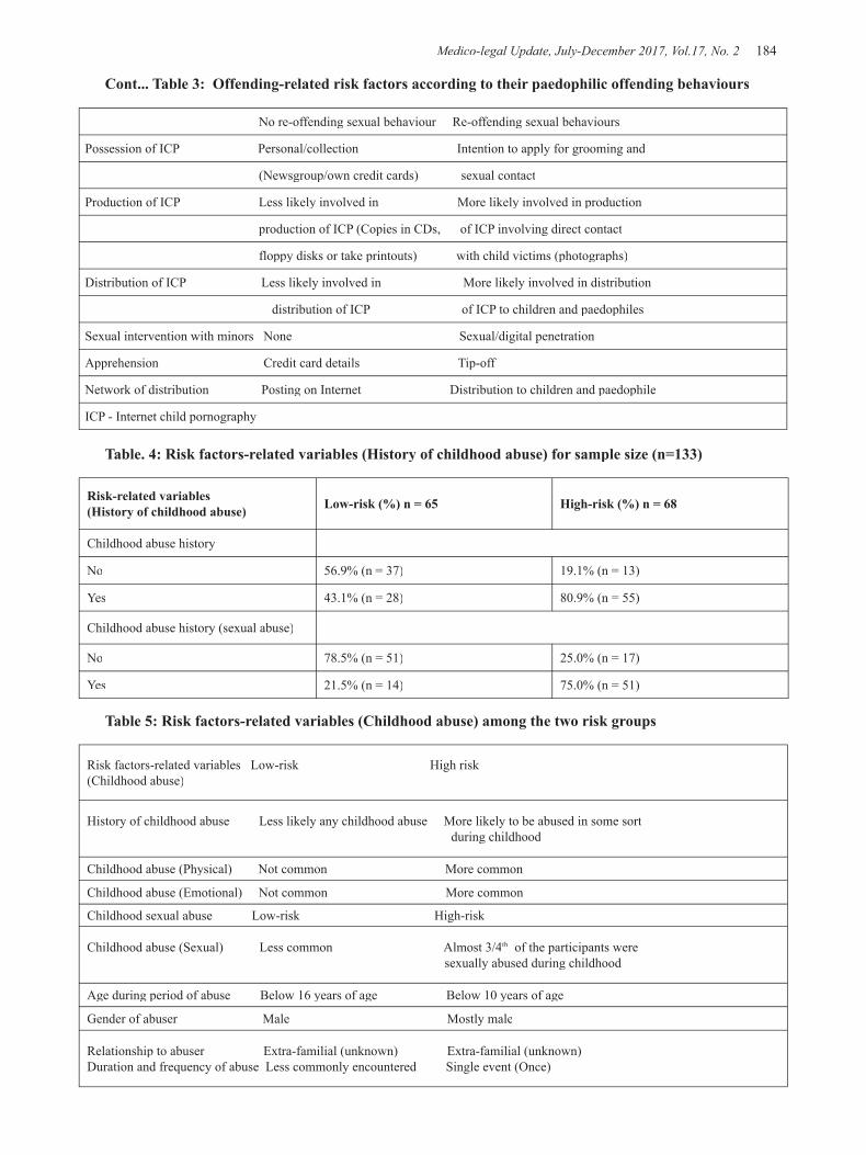
Medico-legal Update, July-December 2017, Vol.17, No. 2 184 185 -legal Update, July-December 2017, Vol.17, No. 2
No re-offending sexual behaviour Re-offending sexual behaviours
Possession of ICP Personal/collection Intention to apply for grooming and
(Newsgroup/own credit cards) sexual contact
Production of ICP Less likely involved in More likely involved in production
production of ICP (Copies in CDs, of ICP involving direct contact
floppy disks or take printouts) with child victims (photographs)
Distribution of ICP Less likely involved in More likely involved in distribution
distribution of ICP of ICP to children and paedophiles
Sexual intervention with minors None Sexual/digital penetration
Apprehension Credit card details Tip-off
Network of distribution Posting on Internet Distribution to children and paedophile
ICP - Internet child pornography
Table. 4: Risk factors-related variables (History of childhood abuse) for sample size (n=133)
Risk-related variables (History of childhood abuse) Low-risk (%) n = 65 High-risk (%) n = 68
Childhood abuse history
No 56.9% (n = 37) 19.1% (n = 13)
Yes 43.1% (n = 28) 80.9% (n = 55)
Childhood abuse history (sexual abuse)
No 78.5% (n = 51) 25.0% (n = 17)
Yes 21.5% (n = 14) 75.0% (n = 51)
Table 5: Risk factors-related variables (Childhood abuse) among the two risk groups
Risk factors-related variables Low-risk High risk(Childhood abuse)
History of childhood abuse Less likely any childhood abuse More likely to be abused in some sort during childhood
Childhood abuse (Physical) Not common More common
Childhood abuse (Emotional) Not common More common
Childhood sexual abuse Low-risk High-risk
Childhood abuse (Sexual) Less common Almost 3/4th of the participants were sexually abused during childhood
Age during period of abuse Below 16 years of age Below 10 years of age
Gender of abuser Male Mostly male
Relationship to abuser Extra-familial (unknown) Extra-familial (unknown) Duration and frequency of abuse Less commonly encountered Single event (Once)
Cont... Table 3: Offending-related risk factors according to their paedophilic offending behaviours
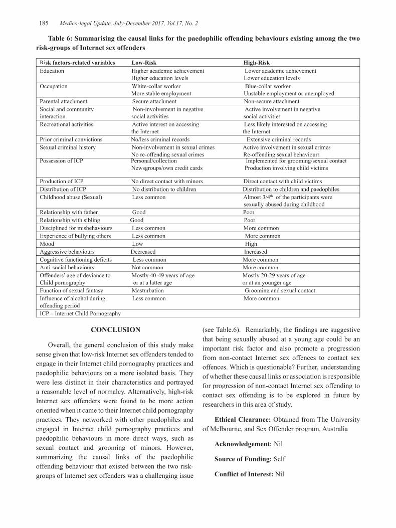
-legal Update, July-December 2017, Vol.17, No. 2 184 185 Medico-legal Update, July-December 2017, Vol.17, No. 2
Table 6: Summarising the causal links for the paedophilic offending behaviours existing among the two risk-groups of Internet sex offenders
Risk factors-related variables Low-Risk High-RiskEducation Higher academic achievement Lower academic achievement Higher education levels Lower education levels Occupation White-collar worker Blue-collar worker More stable employment Unstable employment or unemployedParental attachment Secure attachment Non-secure attachment Social and community Non-involvement in negative Active involvement in negativeinteraction social activities social activitiesRecreational activities Active interest on accessing Less likely interested on accessing the Internet the InternetPrior criminal convictions No/less criminal records Extensive criminal recordsSexual criminal history Non-involvement in sexual crimes Active involvement in sexual crimes No re-offending sexual crimes Re-offending sexual behavioursPossession of ICP Personal/collection Implemented for grooming/sexual contact Possession of ICP Personal/collection Implemented for grooming/sexual contact Newsgroups/own credit cards Production involving child victims Production of ICP No direct contact with minors Direct contact with child victimsDistribution of ICP No distribution to children Distribution to children and paedophiles Childhood abuse (Sexual) Less common Almost 3/4th of the participants were sexually abused during childhoodRelationship with father Good PoorRelationship with sibling Good PoorDisciplined for misbehaviours Less common More commonExperience of bullying others Less common More commonMood Low HighAggressive behaviours Decreased IncreasedCognitive functioning deficits Less common More commonAnti-social behaviours Not common More common Offenders’ age of deviance to Mostly 40-49 years of age Mostly 20-29 years of ageChild pornography or at a latter age or at an younger ageFunction of sexual fantasy Masturbation Grooming and sexual contact Influence of alcohol during Less common More commonoffending periodICP – Internet Child Pornography
CONCLUSION
Overall, the general conclusion of this study make sense given that low-risk Internet sex offenders tended to engage in their Internet child pornography practices and paedophilic behaviours on a more isolated basis. They were less distinct in their characteristics and portrayed a reasonable level of normalcy. Alternatively, high-risk Internet sex offenders were found to be more action oriented when it came to their Internet child pornography practices. They networked with other paedophiles and engaged in Internet child pornography practices and paedophilic behaviours in more direct ways, such as sexual contact and grooming of minors. However, summarizing the causal links of the paedophilic offending behaviour that existed between the two risk-groups of Internet sex offenders was a challenging issue
(see Table.6). Remarkably, the findings are suggestive that being sexually abused at a young age could be an important risk factor and also promote a progression from non-contact Internet sex offences to contact sex offences. Which is questionable? Further, understanding of whether these causal links or association is responsible for progression of non-contact Internet sex offending to contact sex offending is to be explored in future by researchers in this area of study.
Ethical Clearance: Obtained from The University of Melbourne, and Sex Offender program, Australia
Acknowledgement: Nil
Source of Funding: Self
Conflict of Interest: Nil

Medico-legal Update, July-December 2017, Vol.17, No. 2 186
REFERENCES
1. Stanley, J (2001), ‘Child abuse and the Internet’, Child Abuse Prevention, Issue. 15, viewed 12 July 2005
2. Aster, AZ (2007), ‘New study examines risk factors for child sexual offences’, The Journal of Addiction and Mental Health, viewed 15 December 2006 <http://www.camh.net/Publications/cross_currents/Autumn_2005/sexualoffence_crcucautumn05.html>.
3. Sheldon, K & Howitt, D (2008). ‘Sexual fantasy in paedophile offenders: Does any model satisfactorily explain the role of fantasy in offending?’ Legal and
Criminological Psychology, Vol. 13, Issue 1, pp. 137-158.
4. Taylor, M & Quayle, E (Eds.). (2003), Child pornography: an Internet crime. Hove, East Sussex; New York, NY: Brunner-Routledge.
5. Burke, A Sowerbutts, S Blundell, B & Sherry, M (2002). ‘Child Pornography and the Internet: Policing and Treatment Issues.’ Psychiatry, Psychology and Law, vol. 9, no. 1, pp. 79-84.
6. Durkin, K (1997). ‘Misuse of the Internet by pedophiles: implication for law enforcement and probation practice.’ Federal Probation, vol. 61, pp. 14-18.
-legal Update, July-December 2017, Vol.17, No. 2 186
An Autopsy Study of Hypertensive Heart Disease-Retrospective Study
N S Kamakeri1, Sunilkumar S Biradar2, Smitha M3, Mallikarjun K Biradar,4 Lohit Kumar5
1Associate Professor, Department of Pathology, 1Associate Professor, Department of Pathology, 1 2Associate Professor, 2Associate Professor, 2 Department of Forensic Medicine & Toxicology, 3Tutor, Department of Anatomy, Karnataka Institute of Medical Sciences, Hubballi, 4Associate 4Associate 4
Professor, Department of Community Medicine, Koppala Institute of Medical Sciences, Koppala, 5Assistant 5Assistant 5
Professor, Department of Forensic Medicine & Toxicology, Shivamogga Institute of Medical Sciences, Shivamogga
ABSTRACT
Introduction: Hypertension is a chronic condition of concern due to its role in the causation of coronary heart disease, stroke and other vascular complications. It is the commonest cardiovascular disorder posing a major public health challenge to population.
Aims and objectives: To know the cause of sudden deaths in hospitalized and brought dead cases.
Materials and method: A total of 1500 hearts were studied over a period of 20 years from 1997 to 2016.
Result: Ischemic heart disease were reported in 255 (17%) cases (Fig.1, 2&4) and left ventricular hypertrophy in 145(9.60%) cases (Fig.3.) Altogether hypertensive heart diseases contributed about 26% of total sudden deaths.
Conclusion: Most common cause of hypertensive heart diseases was atherosclerosis
Keywords: Hypertension; Ischemia; Heart
Corresponding author:Dr. Sunilkumar S BiradarAssociate Professor, Department of Forensic Medicine & Toxicology, Karnataka Institute of Medical Sciences, Hubballi, Email id: [email protected]: 09880027727.
INTRODUCTION
In modern scientific days because of urbanization, stressful life, food habits, usage of machineries in routine life, there is no physical activities. Hence obesity, hyperlipedemia, hypertension and diabetes and stressful life are becoming very common health problems. Because of this cardiac diseases and complications are becoming common causes of sudden death. Most often in sudden deaths the heart does not show any evidence of ischemic changes histopathologically. Sudden unexpected (natural) death can be defined as death occurring instantaneously or within an estimated 24 hours of the onset of symptoms or signs1.
MATERIALS AND METHOD
The present study was conducted at the department
of pathology, KIMS, Hubballi, a tertiary teaching hospital located in north Karnataka. All the sudden deaths which happened in hospital from 1997 to 2016 were included in the study. In 20 years (1997-2016) nearly 1500 hearts were studied for sudden deaths in our institute. All the hearts were dissected according to line of blood flow and sections were stained with Hemotoxyline and Eosin. In the past 20 years hearts of sudden death were examined and majority of hearts were unremarkable both grossly and microscopically, but some of the hearts were showing left ventricular hypertrophy and some of them revealed frank ischemic changes. The cardiac arrhythmias and coronary spasms were attributed for sudden deaths in absence of pathological evidence of ischemic changes.
RESULTS
A total of 1500 hearts were studied over a period of 20 years from 1997 to 2016. 37.6% were in the age group of 40-49 years, followed by 26.8% in 30-39 years age group. Only 0.5% of subjects were observed in 10-19 years. Male subjects constituted about 59.2%, while female 40.2% (Table-1). Age ranged from 20-
DOI Number: 10.5958/0974-1283.2017.00096.2
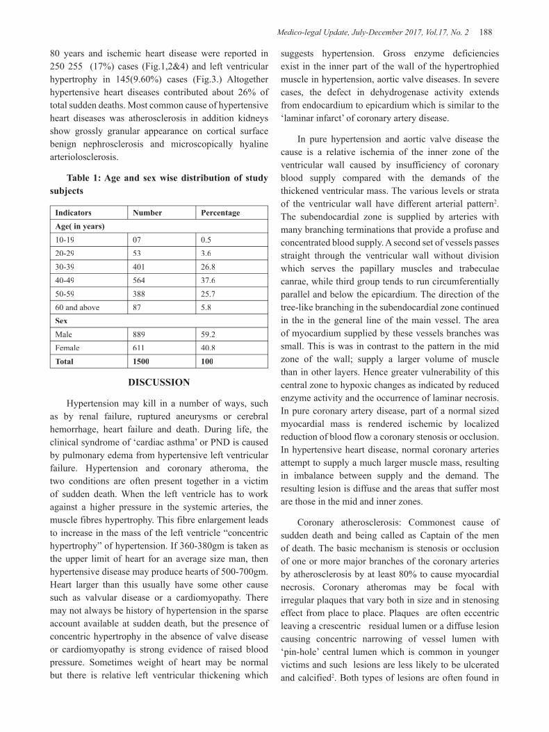
Medico-legal Update, July-December 2017, Vol.17, No. 2 188 189 -legal Update, July-December 2017, Vol.17, No. 2
80 years and ischemic heart disease were reported in 250 255 (17%) cases (Fig.1,2&4) and left ventricular hypertrophy in 145(9.60%) cases (Fig.3.) Altogether hypertensive heart diseases contributed about 26% of total sudden deaths. Most common cause of hypertensive heart diseases was atherosclerosis in addition kidneys show grossly granular appearance on cortical surface benign nephrosclerosis and microscopically hyaline arteriolosclerosis.
Table 1: Age and sex wise distribution of study subjects
Indicators Number PercentageAge( in years)10-19 07 0.520-29 53 3.630-39 401 26.840-49 564 37.650-59 388 25.760 and above 87 5.8SexMale 889 59.2Female 611 40.8Total 1500 100
DISCUSSION
Hypertension may kill in a number of ways, such as by renal failure, ruptured aneurysms or cerebral hemorrhage, heart failure and death. During life, the clinical syndrome of ‘cardiac asthma’ or PND is caused by pulmonary edema from hypertensive left ventricular failure. Hypertension and coronary atheroma, the two conditions are often present together in a victim of sudden death. When the left ventricle has to work against a higher pressure in the systemic arteries, the muscle fibres hypertrophy. This fibre enlargement leads to increase in the mass of the left ventricle “concentric hypertrophy” of hypertension. If 360-380gm is taken as the upper limit of heart for an average size man, then hypertensive disease may produce hearts of 500-700gm. Heart larger than this usually have some other cause such as valvular disease or a cardiomyopathy. There may not always be history of hypertension in the sparse account available at sudden death, but the presence of concentric hypertrophy in the absence of valve disease or cardiomyopathy is strong evidence of raised blood pressure. Sometimes weight of heart may be normal but there is relative left ventricular thickening which
suggests hypertension. Gross enzyme deficiencies exist in the inner part of the wall of the hypertrophied muscle in hypertension, aortic valve diseases. In severe cases, the defect in dehydrogenase activity extends from endocardium to epicardium which is similar to the ‘laminar infarct’ of coronary artery disease.
In pure hypertension and aortic valve disease the cause is a relative ischemia of the inner zone of the ventricular wall caused by insufficiency of coronary blood supply compared with the demands of the thickened ventricular mass. The various levels or strata of the ventricular wall have different arterial pattern2. The subendocardial zone is supplied by arteries with many branching terminations that provide a profuse and concentrated blood supply. A second set of vessels passes straight through the ventricular wall without division which serves the papillary muscles and trabeculae canrae, while third group tends to run circumferentially parallel and below the epicardium. The direction of the tree-like branching in the subendocardial zone continued in the in the general line of the main vessel. The area of myocardium supplied by these vessels branches was small. This is was in contrast to the pattern in the mid zone of the wall; supply a larger volume of muscle than in other layers. Hence greater vulnerability of this central zone to hypoxic changes as indicated by reduced enzyme activity and the occurrence of laminar necrosis. In pure coronary artery disease, part of a normal sized myocardial mass is rendered ischemic by localized reduction of blood flow a coronary stenosis or occlusion. In hypertensive heart disease, normal coronary arteries attempt to supply a much larger muscle mass, resulting in imbalance between supply and the demand. The resulting lesion is diffuse and the areas that suffer most are those in the mid and inner zones.
Coronary atherosclerosis: Commonest cause of sudden death and being called as Captain of the men of death. The basic mechanism is stenosis or occlusion of one or more major branches of the coronary arteries by atherosclerosis by at least 80% to cause myocardial necrosis. Coronary atheromas may be focal with irregular plaques that vary both in size and in stenosing effect from place to place. Plaques are often eccentric leaving a crescentric residual lumen or a diffuse lesion causing concentric narrowing of vessel lumen with ‘pin-hole’ central lumen which is common in younger victims and such lesions are less likely to be ulcerated and calcified2. Both types of lesions are often found in
-legal Update, July-December 2017, Vol.17, No. 2 188 189 Medico-legal Update, July-December 2017, Vol.17, No. 2
the same patient without difference in pathogenesis. Complications in coronary atheromatous lesions include ulcerations, hemorrhage and coronary thrombosis.3
MYOCARDIAL INFARCTION: Clinically signs of chest pain, shock mean an infarct, a much higher proportion of patients who reach hospital beds do have myocardial infarcts. In the pathology of sudden death, overt infarcts are exception. Almost all myocardial infarcts are caused by atheromatous lesions and their complications. A few are the result of other types of coronary obstruction such as polyarteritis other vasculitides, embolismof various types, ostial occlusion by syphilis, severe aortic stenosis, some congenital anomalies of the coronary artery or great vessels, dissecting aneurysms at the aortic rootand tumor or sarcoidosis affecting the coronary vessels. Most infarcts are caused by super added coronary thrombi. Muscle necrosis can follow severe narrowing of the supplying vessel by subintimal hemorrhage, a ruptured plaque or simple severe stenosis from atheroma. Original lumen must be reduced to 20% or less before the ischemia in the distribution zone is sufficient to cause myocardial necrosis. Most sudden death autopsies reveal undoubted infarction in absence of 80% stenosis or normal coronary arteries. The contrary is much more common: finding of complete thrombosis of a major vessel with no sign of infarction because of collateral circulation.
TYPES OF INFARCT:
a) Laminar Infarct: Subendocrdial region of left ventricle is involved. This is due to a reduction in perfusion pressure to the inner zone as all the coronary supply comes from the epicardial surfaces. This type of infarct are the result of generalized stenosis in the major branches of the coronary vessels.
b) Regional /focal infarct: Common in pure coronary artery disease and caused by localized occlusion/severe stenosis in a coronary artery. Almost all infarcts are in the left ventricle,6-9% in right ventricle,7% in atria3. The relative immunity of the right ventricle and atria is presumably because of the relatively thin walls which do not require so much blood,do not have the perfusion gradient of the thick left ventricle. The presence of conus artery, small branches to foci such as the sinus node, and the presence of anstomotic channels between left circumflex and coronary artery. Many infarcts involve only certain layers of muscle which
cannot be supplied by a particular vessel4.
The autopsy diagnosis of myocardial infarction:The macroscopic appearances of myocardial infarction vary with duration of symptoms and signs and in most of the cases, age of an infarct is notoriously difficult to establish in human .Gross pathologic findings in humans, the necrotic myocardium can be detected within 2-3 hours after infarct by immersion of the fresh heart slices in a solution of TTC( 2,3,5 Triphenyl tetrazolium chloride) (Red) or NBT( Nitroblue tetrazolium)(blue). TTC staining ha a diagnostic sensitivity of 77% and specificity of 93% 5.
a) 12-24 hrs: No definite naked eye changes are visible, but edema in the affected area of muscles, pallor seen.
b)1-3 days: The infracted area becomes better demarcated and turns yellow, with breakdown of the myocytes, streaks of red appear, being both dilated vascular channels and areas of ‘inter-fibre hemorrhage .This gives a tigroid appearance suggestive of tiger stripes across the area.
c) After a few days (weeks), the infarct becomes softer and more friable, justifying the old name ‘Myomalacia cordis’. It is at this stage from 2nd -3rd day onwards that rupture occurs into the pericardial sac.
d) From 3rd week onwards centre of the infarct becomes gelatinous, the colour fading to translucent grey. During the next month or two, depending upon the size of infarct, fibrosis replaces the dead muscle to scar.
Infarcts may be transmural extending from epicardium to endocardium. The papillary muscles are usually involved being particularly vulnerable to ischemia as they are at the end of the line of coronary supply. The central part of muscle may necrose and rupture. Infarction usually spares the immediate subendocardial zone, 3-4 most superficial layers of fibres surviving, even though they may show ischemic damage.
Microscopic appearances of myocardial infarction:
a) Early changes:(i) The infracted area becomes swollen, so that the interfibre clefts become obliterated and the overall appearance is thicker and more solid in routine sections.(ii) The cytoplasm becomes granular

Medico-legal Update, July-December 2017, Vol.17, No. 2 190 191 -legal Update, July-December 2017, Vol.17, No. 2
(‘clowdy swelling’). Eosinophilia increases uniformly or in patches. Only hypereosinophilic cells autofluoresce6, and myocardium that had normal eosinophilic staining also fluoresce yellow in ultraviolet light7,8..These early changes are not usually visible in the first 8-12 hrs after the onset of infarction apart from the autofluroscence. Other methods, such as dehydrogenase enzyme histochemistry and acridine orange fluorescence can detect changes as early as 4-5hrs.
b) Later changes: The later stages of infarction follows the 18-24hrs and now macroscopic changes will be apparent. The histology is merely a confirmatory procedure.
There is progressive degeneration of the fibres, eosinophilia becomes stronger. The cellular edema subsides, intrefibre edema takes over, separating the myofibres. The fibres often become wavy and irregular, though uniform ‘sine waves’ in parallel bundles, fibres are pinched or constricted at irregular intervals and hyperchromatic. Fragmentation of muscle fibres at intercalated discs best seen in PTAH stain. The PTAH does not reveal ischaemic damage at an earlier dstage. Large areas can be scanned under low power of the microscope, the striations become distorted and break up, forming either general ‘sandy appearance’ or the ‘contraction bands’ where fragmentation occurs in transverse lines across the fibre. The contractile material aggregates into thicker, darker masses “chenese writing” at first within an intact sarcolemma, but later the cell membrane ruptures and the basophilic contents scatter extrcellularly
Contraction bands can occur in circumstances other than infarction. They may be seen after cardio-pulmonary resuscitation, cardiac massage, noradrenaline injections and in conditions causing cardiac stress.
Loss of nuclei is not an early sign, 2-4 day the nuclei becomes hollow, shadowy and eventually ghost-like, though nuclear remnants can sometimes seen in necrotic infarcts. Cellular infiltration is variable and sometimes does not occur at all. In general, there is neutrophilic infiltration in most infarcts during the first few days which subsides gradually and is replaced by a mononuclear response within a week. These are mainly macrophages clearing up the debries and fibroblasts in to lay down new collagen during repair.
By the end of first week, depending on the total size
of the infarct, early fibrosis is evident. It is difficult to date such an infarct even within week and different parts of the same infarct may show different appearances.
Ultrastructural studies: Ultrastructural studies on ischemic muscle, although of great interest. Early changes include swelling and crystolysis of mitochondria, swelling of the tubular system and clumping of nuclear chromatin. Such changes may prove reversible if oxygenation is restored quickly but otherwise they rapidly progress to irreversible distintegration of the cell organelles9.
Fig1. Specimen of hypertensive heart with ischemic scar over the surface.
Fig 2. Specimen of cut section of heart with ischemic white scars heart disease with scar over the surface
Fig.3.Specimen of cut section of heart showing left ventricular muscle hypertrophy with calcified mitral leaflets
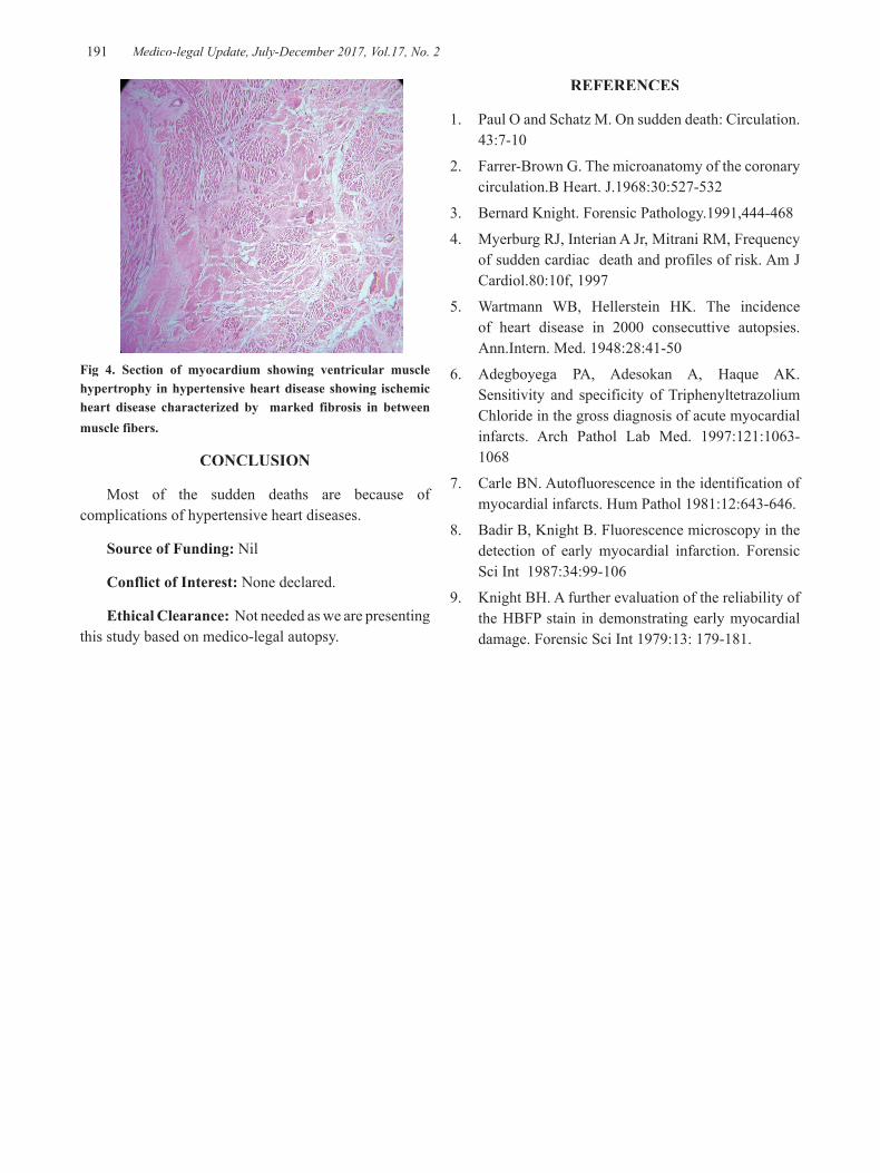
-legal Update, July-December 2017, Vol.17, No. 2 190 191 Medico-legal Update, July-December 2017, Vol.17, No. 2
Fig 4. Section of myocardium showing ventricular muscle hypertrophy in hypertensive heart disease showing ischemic heart disease characterized by marked fibrosis in between muscle fibers.
CONCLUSION
Most of the sudden deaths are because of complications of hypertensive heart diseases.
Source of Funding: Nil
Conflict of Interest: None declared.
Ethical Clearance: Not needed as we are presenting this study based on medico-legal autopsy.
REFERENCES
1. Paul O and Schatz M. On sudden death: Circulation. 43:7-10
2. Farrer-Brown G. The microanatomy of the coronary circulation.B Heart. J.1968:30:527-532
3. Bernard Knight. Forensic Pathology.1991,444-468
4. Myerburg RJ, Interian A Jr, Mitrani RM, Frequency of sudden cardiac death and profiles of risk. Am J Cardiol.80:10f, 1997
5. Wartmann WB, Hellerstein HK. The incidence of heart disease in 2000 consecuttive autopsies. Ann.Intern. Med. 1948:28:41-50
6. Adegboyega PA, Adesokan A, Haque AK. Sensitivity and specificity of Triphenyltetrazolium Chloride in the gross diagnosis of acute myocardial infarcts. Arch Pathol Lab Med. 1997:121:1063-1068
7. Carle BN. Autofluorescence in the identification of myocardial infarcts. Hum Pathol 1981:12:643-646.
8. Badir B, Knight B. Fluorescence microscopy in the detection of early myocardial infarction. Forensic Sci Int 1987:34:99-106
9. Knight BH. A further evaluation of the reliability of the HBFP stain in demonstrating early myocardial damage. Forensic Sci Int 1979:13: 179-181.
193 -legal Update, July-December 2017, Vol.17, No. 2
Visit to a Scene of Crime to Determine Manner of Death
Ajit G Pathak1, Kapileshwar M Chaudhari2, Ramesh K Gadhari3, Nilesh A Devraj3,
Vijay T Jadhao3, Piyush S Gavale4
1Professor & Head, 2Associate Professor, 2Associate Professor, 2 3Assistant Professor, 3Assistant Professor, 3 4Tutor, Department of Forensic Medicine & Toxicology, S.B.H. Government Medical College, Dhule, Maharashtra, India
ABSTRACT
Visiting the scene of crime, by the Forensic Medicine Expert who has conducted the autopsy is of immense value to determine manner of death and thus to solve mystery regarding the death. We are hereby presenting four cases in which we visited scene of crime regarding manner of death. In all the four cases, which we mentioned, highlights visit to the scene of crime by the Forensic Medicine expert after thorough medico-legal autopsy, to opine about manner of death so that proper justice to the case would be delivered.
Keywords : Crime Scene, Visit, Forensic Expert, Manner of death, Autopsy.
INTRODUCTION
When a crime occurs, investigating officers often visit a scene of crime, prepare a sketch, makes spot panchnama, and sends the body for medico-legal post mortem examination. The doctor performs post mortem examination and gives his opinion about cause of death. His opinion regarding manner of death is mainly based on documentary and/ or photographic evidence, without visiting the scene of crime. Visiting the scene of crime, by the Forensic Medicine Expert who has conducted the autopsy is of immense value to determine manner of death and thus to solve mystery regarding the death.
Here we are presenting four cases in which post mortem examination was conducted at our institute and we visited the scene of crime to determine the manner of death.
MATERIAL AND METHOD
We selected post mortem reports, inquest panchnama, spot panchnama, viscera analysis reports of five dead bodies in which we were requested by the
Corresponding author:Dr. A G PathakProfessor & Head of Department, Department of Forensic Medicine &Toxicology, SBH Government Medical College & Hospital, Dhule, Maharashtra, India.- 424001, Email id: [email protected]
police authorities to visit the scene of crime, to present four case reports. Post mortem examinations was carried out in these cases between 1st January 2014 to 31st
December 2016 by the panel of doctors(authors of this article) who also visited the scene of crime.
CASE NO. 1
A 47 years old male prisoner was found in a dead condition inside 500 liters water tank situated above the toilet block in the prison premises. Ligature material was present around the neck with two turns of a jute rope present with a fixed knot over front of neck in midline. During post mortem examination the dead body was in a state of decomposition and a single grooved transverse ligature mark over neck was noted corresponding to the ligature material. It was completely encircling the neck and situated below the level of thyroid cartilage. On dissection of neck, subcutaneous tissues, muscles of neck, tracheal rings, laryngeal cartilages and hyoid bone were intact. Tongue was clinched in between the teeth and was cyanosed.
The cause of death in this case was hanging and the authorities requested us to visit the scene of crime to determine manner of death. The scene of crime was in the prison premises, above the toilet block. The height of the toilet block was 3.87 meters, its width was 3 meters X 2.16 meters. 500 liters water tank was situated above it. The deceased used a pipe of water to reach the water tank (Photograph no 1). We climbed up to the water
DOI Number: 10.5958/0974-1283.2017.00097.4
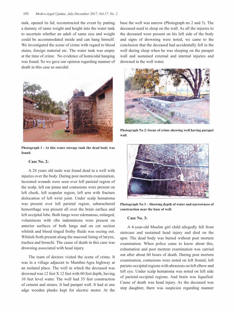
193 Medico-legal Update, July-December 2017, Vol.17, No. 2
base the well was narrow (Photograph no 2 and 3). The deceased used to sleep on the wall. As all the injuries to the deceased were present on his left side of the body and signs of drowning were noted, we came to the conclusion that the deceased had accidentally fell in the well during sleep when he was sleeping on the parapet wall and sustained external and internal injuries and drowned in the well water.
Photograph No 2–Scene of crime showing well having parapet wall.
Photograph No 3 – Showing depth of water and narrowness of construction near the base of well.
Case No. 3:
A 4-year-old Muslim girl child allegedly fell from staircase and sustained head injury and died on the spot. The dead body was buried without post mortem examination. When police came to know about this, exhumation and post mortem examination was carried out after about 60 hours of death. During post mortem examination, contusions were noted on left frontal, left parieto-occipital regions with abrasions on left elbow and left eye. Under scalp hematoma was noted on left side of parietal-occipital regions. And brain was liquefied. Cause of death was head injury. As the deceased was step daughter, there was suspicion regarding manner
tank, opened its lid, reconstructed the event by putting a dummy of same weight and height into the water tank to ascertain whether an adult of same size and weight could be accommodated inside and can hang himself. We investigated the scene of crime with regard to blood stains, foreign material etc. The water tank was empty at the time of crime. No evidence of homicidal hanging was found. So we gave our opinion regarding manner of death in this case as suicidal.
Photograph 1 - At this water storage tank the dead body was found.
Case No. 2:
A 24 years old male was found dead in a well with injuries over the body. During post mortem examination, lacerated wounds were seen over left parietal region of the scalp, left ear pinna and contusions were present on left cheek, left scapular region, left arm with fracture dislocation of left wrist joint. Under scalp hematoma was present over left parietal region, subarachnoid hemorrhage was present all over the brain surface and left occipital lobe. Both lungs were edematous, enlarged, voluminous with ribs indentations were present on anterior surfaces of both lungs and on cut section whitish and blood tinged frothy fluids was oozing out. Whitish froth present along the mucosal lining of larynx, trachea and bronchi. The cause of death in this case was drowning associated with head injury.
The team of doctors visited the scene of crime. It was in a village adjacent to Mumbai-Agra highway at an isolated place. The well in which the deceased was drowned was 12 feet X 12 feet with 60 feet depth, having 10 feet level water. The well had 35 feet construction of cement and stones. It had parapet wall. It had at one edge wooden planks kept for electric motor. At the
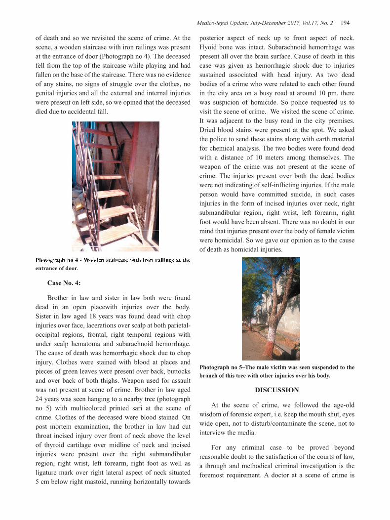
Medico-legal Update, July-December 2017, Vol.17, No. 2 194 195 -legal Update, July-December 2017, Vol.17, No. 2
of death and so we revisited the scene of crime. At the scene, a wooden staircase with iron railings was present at the entrance of door (Photograph no 4). The deceased fell from the top of the staircase while playing and had fallen on the base of the staircase. There was no evidence of any stains, no signs of struggle over the clothes, no genital injuries and all the external and internal injuries were present on left side, so we opined that the deceased died due to accidental fall.
Photograph no 4 - Wooden staircase with iron railings at the entrance of door.
Case No. 4:
Brother in law and sister in law both were found dead in an open placewith injuries over the body. Sister in law aged 18 years was found dead with chop injuries over face, lacerations over scalp at both parietal-occipital regions, frontal, right temporal regions with under scalp hematoma and subarachnoid hemorrhage. The cause of death was hemorrhagic shock due to chop injury. Clothes were stained with blood at places and pieces of green leaves were present over back, buttocks and over back of both thighs. Weapon used for assault was not present at scene of crime. Brother in law aged 24 years was seen hanging to a nearby tree (photograph no 5) with multicolored printed sari at the scene of crime. Clothes of the deceased were blood stained. On post mortem examination, the brother in law had cut throat incised injury over front of neck above the level of thyroid cartilage over midline of neck and incised injuries were present over the right submandibular region, right wrist, left forearm, right foot as well as ligature mark over right lateral aspect of neck situated 5 cm below right mastoid, running horizontally towards
posterior aspect of neck up to front aspect of neck. Hyoid bone was intact. Subarachnoid hemorrhage was present all over the brain surface. Cause of death in this case was given as hemorrhagic shock due to injuries sustained associated with head injury. As two dead bodies of a crime who were related to each other found in the city area on a busy road at around 10 pm, there was suspicion of homicide. So police requested us to visit the scene of crime. We visited the scene of crime. It was adjacent to the busy road in the city premises. Dried blood stains were present at the spot. We asked the police to send these stains along with earth material for chemical analysis. The two bodies were found dead with a distance of 10 meters among themselves. The weapon of the crime was not present at the scene of crime. The injuries present over both the dead bodies were not indicating of self-inflicting injuries. If the male person would have committed suicide, in such cases injuries in the form of incised injuries over neck, right submandibular region, right wrist, left forearm, right foot would have been absent. There was no doubt in our mind that injuries present over the body of female victim were homicidal. So we gave our opinion as to the cause of death as homicidal injuries.
Photograph no 5–The male victim was seen suspended to the branch of this tree with other injuries over his body.
DISCUSSION
At the scene of crime, we followed the age-old wisdom of forensic expert, i.e. keep the mouth shut, eyes wide open, not to disturb/contaminate the scene, not to interview the media.
For any criminal case to be proved beyond reasonable doubt to the satisfaction of the courts of law, a through and methodical criminal investigation is the foremost requirement. A doctor at a scene of crime is
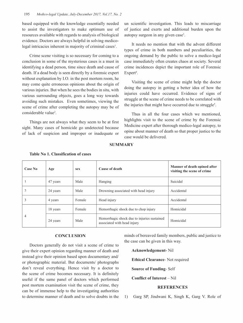
-legal Update, July-December 2017, Vol.17, No. 2 194 195 Medico-legal Update, July-December 2017, Vol.17, No. 2
based equipped with the knowledge essentially needed to assist the investigators to make optimum use of resources available with regards to analysis of biological evidence. Doctors are always helpful in solving medico-legal intricacies inherent in majority of criminal cases1.
Crime scene visiting is so necessary for coming to a conclusion in some of the mysterious cases is a must in identifying a dead person, time since death and cause of death. If a dead body is seen directly by a forensic expert without explanation by I.O. in the post mortem room, he may come quite erroneous opinions about the origin of various injuries. But when he sees the bodies in situ, with various surrounding objects, goes a long way towards avoiding such mistakes. Even sometimes, viewing the scene of crime after completing the autopsy may be of considerable value2.
Things are not always what they seem to be at first sight. Many cases of homicide go undetected because of lack of suspicion and improper or inadequate or
un scientific investigation. This leads to miscarriage of justice and exerts and additional burden upon the autopsy surgeon in any given case3.
It needs no mention that with the advent different types of crime in both numbers and peculiarities, the ongoing demand by the public to solve a medico-legal case immediately often creates chaos at society. Several crime incidences depict the important role of Forensic Expert4.
Visiting the scene of crime might help the doctor doing the autopsy in getting a better idea of how the injuries could have occurred. Evidence of signs of struggle at the scene of crime needs to be correlated with the injuries that might have occurred due to struggle5.
Thus in all the four cases which we mentioned, highlights visit to the scene of crime by the Forensic Medicine expert after thorough medico-legal autopsy, to opine about manner of death so that proper justice to the case would be delivered.
SUMMARY
Table No 1. Classification of cases
Case No Age sex Cause of death Manner of death opined after visiting the scene of crime
1 47 years Male Hanging Suicidal
2 24 years Male Drowning associated with head injury Accidental
3 4 years Female Head injury Accidental
4
18 years Female Hemorrhagic shock due to chop injury Homicidal
24 years Male Hemorrhagic shock due to injuries sustained associated with head injury Homicidal
CONCLUSION
Doctors generally do not visit a scene of crime to give their expert opinion regarding manner of death and instead give their opinion based upon documentary and/ or photographic material. But documents/ photographs don’t reveal everything. Hence visit by a doctor to the scene of crime becomes necessary. It is definitely useful if the same panel of doctors which performed post mortem examination visit the scene of crime, they can be of immense help to the investigating authorities to determine manner of death and to solve doubts in the
minds of bereaved family members, public and justice to the case can be given in this way.
Acknowledgement- Nil
Ethical Clearance- Not required
Source of Funding- Self
Conflict of Interest – Nil
REFERENCES
1) Garg SP, Jindwani K, Singh K, Garg V. Role of

Medico-legal Update, July-December 2017, Vol.17, No. 2 196
medical doctor at scene of crime, J Indian Acad Forensic Med. Jan-March 2013, Vol. 35, No. 1, pg 65-70.
2) Kant ZS, Lal BR. Importance of crime scene visit: a case study. J Indian Acad Forensic Med. October-December 2011, Vol. 33, No. 4, pg 372-74.
3) Shilekh M, Sonia M, Singh MM. A visit of Forensic Pathologist to the scene of crime - need of an hour. Medico-Legal Update, 2008, Volume 8, Issue 2, pg 19-20.
4) Tirpude BH, Murkey PN, Zopate P, Mazumder A, Bhowate S, Pawar VG, et al. Importance of autopsy and crime scene visit by Forensic Expert. J Indian Acad Forensic Med, 31(4), pg 402-404.
5) Mathiharan K, Kannan K. Legal procedures in criminal courts. A textbook of Medical Jurisprudence and Toxicology, 24thed, LexisNexis Butterworths Wadhawa, Nagpur, pg 24.
-legal Update, July-December 2017, Vol.17, No. 2 196
Profile of Medico-legal Autopsies Done at Sri Devaraj Urs Medical College, Kolar for a Period of 3 Years
Murali Mohan MC1, Yadukul S2, Kiran J3
1Assistant Professor, Department of Forensic Medicine & Toxicology, Sri Devaraj Urs Medical College, Kolar,1Assistant Professor, Department of Forensic Medicine & Toxicology, Sri Devaraj Urs Medical College, Kolar,1
2Assistant Professor, Department of Forensic Medicine & Toxicology, Chamarajanagar Institute of Medical 2Assistant Professor, Department of Forensic Medicine & Toxicology, Chamarajanagar Institute of Medical 2
Sciences, Chamarajanagar, 3Professor & Head of the Department, Department of Forensic Medicine & Toxicology, Sri Devaraj Urs Medical College, Kolar
ABSTRACT
Background: Medico-legal autopsy otherwise known as coroner’s autopsy dates back to 4000 BC. It was performed to protect the future what is known as horuspicy. All deaths due to unnatural causes and deaths that are believed to be due to natural causes but the cause of death is not certain or known are subject to inquest. The profiling of medicolegal cases is necessary in order to prevent the preventable casualties in future and to study the genuine crime rate in the area.
Objective: The study was undertaken to determine the pattern of medico-legal autopsy done in our setup and to examine implication to medical education.
Results: A total number of 898 medico-legal autopsies were performed during the study period of 3 years i.e., from 1st Jan 2014 to 31st Jan 2014 to 31st st Dec 2016 in our institute. In this study period, male subjects constituted 76.3% st Dec 2016 in our institute. In this study period, male subjects constituted 76.3% st
(n=685) of the cases making it Male: Female ratio of 3.26: 1. Middle aged males constituted the major number of cases with Road traffic injuries as the major cause followed by poisoning. September month recorded the highest number of cases in our study.
Conclusion: This study helps to interpret the types of medico-legal autopsy cases presenting at the Tertiary care hospital attached to Department of Forensic Medicine & Toxicology in Sri Devaraj Urs Medical College, Kolar. This will provide an insight to the policy makers, law custodians, and the community to look into the specific aspects of the cases and, then, take measures, accordingly, for the benefit of the community and people at large of this area.
Keywords: Medico-legal Autopsy; Road traffic injuries; Kolar.
Corresponding author: Dr. Yadukul.S E-mail: [email protected]
INTRODUCTION
The term ‘Autopsy’ originates from ancient ‘Autopsia’ which is derived from ‘Autos’ i.e. ‘Oneself’ and ‘Opsis’ i.e. ‘to see for oneself’. An autopsy is frequently done in cases of sudden death where a doctor is not able to give a death certificate or when death is believed due to an unnatural cause1. Section 174 and 176 Code of Criminal Procedure (CrPC) mentions the concept of a medico-legal autopsy during
the investigations of a sudden, suspicious & unnatural death2. The objective of medicolegal post-mortem examination is to establish the identity of a body, when not known; to ascertain the time since death and the cause of death; and whether the death was natural or unnatural and if unnatural, whether it was homicidal, suicidal or accidental. In case of new born infants, the question of live birth and viability assume importance and should be determined3. The term “postmortem examination” is often used as a simile for “autopsy.” Basically, it is not4. A postmortem examination means only what it says that the body was examined after death. It can mean and often does mean that the physician merely looked at the body, fully clothed, or that he “viewed” the body
DOI Number: 10.5958/0974-1283.2017.00098.6

Medico-legal Update, July-December 2017, Vol.17, No. 2 198 199 -legal Update, July-December 2017, Vol.17, No. 2
at a funeral home or in a morgue. A complete autopsy involves opening all body cavities and all organs of the trunk, chest, and head5. In all cases, a complete and not a partial examination are more necessary in this country on account of the imperfectness of the preliminary evidence as to the possible cause of death6.
MATERIAL AND METHOD
This retrospective study included all cases of deaths brought for autopsy to the Department of Forensic Medicine & Toxicology, Sri Devaraj Urs Medical College, Kolar for a period of 3 years from 1st January st January st
2014 to 31st December 2016. In this study, the emphasis st December 2016. In this study, the emphasis st
has been put on to find the total number of death cases, age wise and sex wise distribution of cases, pattern of medico-legal autopsies done and the month-wise distribution of cases. The data were collected and tabulated to determine the frequency and pro portion of these fatalities, and the results were expressed in percentages.
RESULTS
A total number of 898 medico-legal autopsies were performed during the study period of 3 years i.e., from 1st Jan 2014 to 31st Jan 2014 to 31st st Dec 2016 in our institute. In this study st Dec 2016 in our institute. In this study st
period, male subjects constituted 76.3% (n=685) of the cases making it Male: Female ratio of 3.26: 1. Among the age wise distribution of cases (Table 1), maximum cases were recorded in the age group of 21-30yrs (28.5%) followed closely by 31-40yrs (20.1%). The year wise pattern of deaths that occurred during our study period has been given below (Table 2), in which 36.6% (n=329) of cases was done in 2016, followed by 32% (n=287) done in 2015. In month wise distribution of cases (Table 2) in our study, highest number of cases was performed during the month of September (11.4%) followed by the month of March (9.02%) & October (8.7%). Among the different variety of cases (Table 3) that was done, Road traffic injuries accounted for the highest number of cases (46.3%) followed by poisoning cases (17.9%).
Table 1: Age and sex wise distribution of cases.
Male Female Total Percentage < 1Year 1 3 4 0.4%1-10 yrs 20 11 31 3.4%11-20 yrs 73 23 96 10.7%21-30yrs 197 59 256 28.5%31-40yrs 165 33 198 20.1%41-50yrs 109 36 145 16.1%51-60yrs 65 26 91 10.1%61-70yrs 47 18 65 6.2%71-80yrs 7 4 11 1.2%81-90yrs 1 0 1 0.1%>91yrs 0 0 0 0%
685 213 898 100%
Table 2: Year & month wise distribution of cases.
Month/Year 2014 2015 2016 Total no. of CasesJanuary 16 17 22 55February 26 26 25 77March 30 32 19 81April 16 20 22 58May 21 31 22 74June 15 19 30 64July 17 30 28 75August 25 22 28 75September 46 26 30 102October 24 28 26 78November 25 16 26 67December 21 18 36 75Total 282 (31.4%) 287 (32%) 329 (36.6%) 898 (100%)

-legal Update, July-December 2017, Vol.17, No. 2 198 199 Medico-legal Update, July-December 2017, Vol.17, No. 2
Table 3: Pattern of Medico-legal cases
Sl. No Type of case 2014 2015 2016
1 Road Traffic Accident 147 124 145
2 Poisoning 44 51 66
3 Hanging 15 25 38
4 Fall from Height 13 09 07
5 Burns 04 03 03
6 Assault 13 21 23
7 Natural Death 17 27 19
8 Snake Bite 11 05 05
9 Drowning 10 08 11
10 electrocution 02 06 01
11 Railway Accidents 03 01 00
12 Exhumation 02 04 05
13 Wild animal attack 00 03 01
14 Industrial accidents 00 00 02
15 Bee sting 00 00 02
16 Firearm injuries 00 00 01
TOTAL 282 287 329
Grand total 898
DISCUSSION
Of the 898 medico-legal autopsies that were performed during the study period, male subjects constituted 76.3% (n=685) of the cases making it Male: Female ratio of 3.26: 1. These results were similar to the studies done by Radhakrishna et al7, Sharma et al8, Wasnik et al9, Shrivastava et al10, Zine et al11, Qasim et al12, Afandi et al13 & Jigneshkumar BP et al14. The male case predominance may be explained by the fact that male subjects experience more exposure to the outside environment, as the socioeconomic structure of our commu nity is where the man is the only partner who earns and has freedom out-of-doors, while women usually stay indoors.
The most common age group involved in all types of cases was 21–30 years and of both sex group. This finding is also consistent with the studies by Radhakrishna et al7, Wasnik et al9, Zine et al11, Qasim et al12 and Afandi13. In the stud ies by Sharma et al8, maximum cases was in the age group of 21–25 years, followed by 26–30 years. The maximum numbers of victims were young adults. In our study, maximum cases were recorded during the month of September (11.4%) followed by the month of March
(9.02%) & October (8.7%). This is similar to the study by Zine et al11, which says 12.34% deaths occurred in the month of October and, particularly, on Fridays.
Among the different variety of cases that was done, Road traffic injuries accounted for the highest number of cases (46.3%) followed by poisoning cases (17.9%). This distribution is almost similar to the studies by Radhakrishna et al7, Sharma et al8, Wasnik9, Shrivastava et al10 and Zine et al11. The reason is owing to the availability of faster mode of transportation leading to traffic accidents, more traveling, and the chances of being more exposed to industrial and occupational hazards. Road traffic injuries are a preventable feature, and it is in fact sad to see that maximum number of deaths occur owing to this reason. The traffic rules and traffic sense needs to be taught right from the junior level, and laws should be strictly implemented.
CONCLUSION
This study helps to interpret the types of medico-legal autopsy cases presenting at the Tertiary care hospital attached to Department of Forensic Medicine & Toxicology in Sri Devaraj Urs Medical College,
Medico-legal Update, July-December 2017, Vol.17, No. 2 200
Kolar. This will provide an insight to the policy makers, law custodians, and the community to look into the specific aspects of the cases and, then, take measures, accordingly, for the benefit of the community and people at large of this area. Health awareness about stress regulation and control and education creating awareness of the traffic rules and motor vehicle driving legislations must have to be strength ened and reinforced on time and again to bring down the numbers of death due to road traffic injuries. Improvement of road surface infra structure, strict compliance with road safety rules by drivers and pedestrians, rapid emergency services, and the estab lishment of trauma care centers are the major factors that can reduce this hazard.
Conflict of Interest: Nil
Source of Funding: Nil
Ethical Clearence: Taken from the Institute.
REFERENCE
1. Vij K. Textbook of Forensic Medicine and Toxicology, 4th Ed. India: Elsevier, 2009. p. 24-6.
2. Sections 174 & 176 of the Code of Criminal Procedure, 1973. Basu’s Criminal Court Handbook containing Criminal Major Acts, 10th Ed 2007. Orient Publishing Company, New Delhi: pp. 134-36, 112-13.
3. Kannan K, Mathiharan K. Modi- A textbook of Medical Jurisprudence and Toxicology. 24th Ed 2012. LexisNexis Butterworth’s Nagpur: p. 293-7.
4. Curran WJ. The Medico-legal autopsy and Medico-legal investigation. Bull N Y Acad Med July 1971; 47(7): 766-75.
5. The Punjab Medical Manual, 2nd Ed. 1933. nd Ed. 1933. nd
Superintendent, Government Printing, Punjab,
Lahore; App. XXXVII: CII, Chapter X, p. 154, Para 595
6. World Health Organization; Suicide and attempted suicide in young people, Geneva: WHO; 1974.
7. Radhakrishna KV, Makhani CS, Sisodiya CN, Chourasia S, Sarala M, Khan RN. Profile of medicolegal autopsies at tertiary medicolegal centre in southwestern India. Int J Healthcare Biomed Res 2015;3(2):70–5.
8. Sharma BR, Singh VP, Sharma R, Sumedha S. Unnatural deaths in northern India a profile. J Indian Acad Forensic Med 2004;26(4):140–6.
9. Wasnik RN. Trends of unnatural deaths in Nagpur, India. Medico-Legal Update 2011;11(2):114–7.
10. Shrivastava P, Som D, Nandy S, Saha I, Pal PB, Ray TG, et al. Profile of postmortem cases conducted at a morgue of a tertiary care hospital in Kolkata. J Indian Med Assoc 2010;108(11):730–3.
11. Zine KU, Wakde SD, Tandle RM, Varma NM, Jambure MP, Tasgaonker GV. Study of deaths in industrial areas around Aurangabad city. J Indian Acad Forensic Med 2012;34(2):111–3.
12. Qasim AP, Tariq SA, Naeem M. Profile of unnatural deaths; in Faisalabad. Med Forum 2014;25(5):51–4. Available at: http:// www.medforum.pk/index.php/articledatabase/9articles/9profile ofunnaturaldeaths. (last accessed on February 26, 2015).
13. Afandi D. Profile of medicolegal autopsies in Pekanbaru, Indonesia 2007–2011. Malays J Pathol 2012;34(2):123–6.
14. Jigneshkumar BP, Paresh VC, Umang PP, Shivnath NP, Ganesh G. Profile of autopsy cases at New Civil Hospital, Surat: a retrospective study. Int J Med Sci Public Health 2016 : 5 (1): p 10-3.

-legal Update, July-December 2017, Vol.17, No. 2 200
Profile of Cases of Fatal Road Traffic Accident Autopsied with Respect to Type (Status) of Victim, Type of Vehicle used and
Cause of Death in Rural India
Deepak Bhagwat1, B H Tirpude2, Sharjeel Khan1, P N Murkey2, I L Khandekar2, Pravin Zopate3, Ranjan Ambedkar1, Rahul Ramteke1
1Post Graduate Student, 2Professor, 3Assistant Professor, Dpt. of Forensic Medicine and Toxicology, Mahatma Gandhi Institute of Medical Sciences, Sevagram, Wardha
ABSTRACT
Road traffic accident is a complex phenomenon. Constant rise in the number of motor vehicles, rampant encroachment of road, easy to avail the vehicle because of loan facility, nasty tendency of violating traffic rules and anarchic traffic systems have greatly contributed to rapid increase in RTAs. Population explosion is a catalyzing factor for a number of accidents. The rise of road traffic accidents has become a major public health problem. RTAs cost a lot not only to the individuals affected and their families but also to the nation.
The present study was a cross-sectional study conducted in central India during 2 year period from 1st June st June st
2014 to 31st May 2016. During the study period, a total of 757 medicolegal autopsies were conducted in this st May 2016. During the study period, a total of 757 medicolegal autopsies were conducted in this st
institute, out of which 109 cases of fatal road traffic accidents (died on spot or within 24 hours of accident) were studied. The purpose of the study was to know the age and sex wise distribution, diurnal variation and to correlate the above parameters with the cause of death.
Keywords : Fatal road traffic accident, Intracranial Hemorrhage, Motorvehicle, death.
INTRODUCTION
Accidents are world’s most serious health problem. The motorized transportation media like vehicles, trains, aeroplane, with fast moving vehicular traffic, vast urbanization and changing social patterns, have contributed to increase in the incidence of trauma to human body.
Road Traffic Accident (RTA) is any vehicular accident occurring on the roadway (i.e. originating on, or involving a vehicle partially on the roadway)11. This includes collision of an automobile with a pedestrian, another automobile or with a non-automobile on the roadway or fall from a moving vehicle causing injuries or death of involved individuals. In recent years, death due to RTAs are increasing at an alarming rate throughout the world. Thereby it poses itself as a major epidemiological as well as medico legal problem.
In 2013, global rate of death from Road Traffic Injuries was 16.6 per 1,00,000 population, 1.25 million
people died globally from road traffic injuries in year 201312. In India, more than 4.8 lakh accidents and more than 1.3 lakh deaths were reported during 20134. According to an expert study group appointed by Government of India “RTAs have come to be considered as the third deadly killer, next to heart disease and cancer.” Every year the World Health Organization (WHO) hosts an event, usually on 7th April, to celebrate the anniversary of its founding in 1946. Each year the event focuses on one health issue. In response to a growing concern about RTAs the WHO Director-General, Dr. Lee Jong Wook has for the first time in history of WHO devoted7 7th
April 2004 specifically to road safety and the slogan was “Road Safety Is No Accident.” Early detection of the injury and prompt treatment are necessary in saving the lives of many of these victims8.
AIMS AND OBJECTIVES
1) To analyze the cause of death of fatal road traffic accident brought to mortuary of our institute for post - mortem examination.
DOI Number: 10.5958/0974-1283.2017.00099.8
Medico-legal Update, July-December 2017, Vol.17, No. 2 202 203 -legal Update, July-December 2017, Vol.17, No. 2
2) To study the type of victim ( Status of victim) of fatal road traffic accidents.
3) To study the type of vehicle involved in fatal road traffic accidents.
MATERIALS AND METHOD
The present study was a two year cross – sectional study. The material for the study was the cases of road traffic accidents brought to mortuary for post mortem of this institution situated in central India, during the period from 1st June 2014 to 31st May 2016.The present study includes documenting types of wound, their anatomical location and commonest injuries leading to death in fatal road traffic accidents. Ethical clearance for the present study was obtained from the institutional ethical committee. On the arrival of the case in Mortuary of this institute, informed expressed consent was taken from the relative of deceased for examination of wound and their documentation. In the present study, detailed information regarding the wounds and various factors regarding the circumstances of the occurrence, like type of road traffic accidents, time and place of accidents, who treat case first, occupation and other relevant information were gathered from relative and were recorded in the predefined proforma. Details of name, age, sex, address, occupation, type of accident etc. were recorded from relative. All the wounds were examined for their location, size and shape. In the present study though the size of all wounds were noted but it is not included in the analysis because it is beyond the preview of present study.
During the present study period from 1st June 2014 to 31st May 2016 at this institute in central India –
1. Total no of medicolegal autopsies were conducted during the study period = 757.
2. Autopsies of fatal road traffic accident (died on spot, brought dead to hospital or died within 24 hours of accident) = 109.
INCLUSION CRITERIA:
All the victims of road traffic accidents who were died on spot or brought dead to hospital or died within 24 hours of accident.
EXCLUSION CRITERIA:
1. All the victims who died in an incidence other
than road traffic accidents.
2. All the victims of road traffic accidents who died after 24 hours of accident.
STATISTICAL ANALYSIS:
1) The software for graphs and calculation of statistical values is – SPSS.
2) The software used during creation or modification of some of the diagrams.
a) ADOBE PHOTOSHOP(R) 7.0
b) COREL DRAW X3
c) WINDOWS -10.
RESULTS
Table 1: Profile of medicolegal autopsies conducted during the study period
Type of cases No. of cases Percentage
Fatal road traffic accidents 109 14.40%
Other 648 85.60%
Total 757 100%
Table 2: Distribution of cases of fatal Road Traffic Accidents as per type of victim (Status of victim)
Type of victim (Status of victim) No. of cases Percentage
Pedestrian 17 15.60%
Driver of vehicle 60 55.05%
Passenger 19 17.43%
Hawker 02 01.83%
Pillion - driver 08 07.34%
other 03 02.75%
Total 109 100%
Table 3: Distribution of cases of fatal Road Traffic Accidents as per type of offending vehicle
Type of offending vehicle No. of cases Percentage
Motor cycle 23 21.10%Car 36 33.02%Auto 04 03.70%Truck 28 25.68%Tractor 03 02.75% Bus 07 06.42%Other 08 07.33%Total 109 100%
-legal Update, July-December 2017, Vol.17, No. 2 202 203 Medico-legal Update, July-December 2017, Vol.17, No. 2
Table 4: Profile of cause of death
Cause of death No. of cases Percentage
Intracranial injuries (intracranial hemorrhage/brain injury) 86 78.90%
Hemorrhagic shock 17 15.60%
Traumatic asphyxia 04 03.67%
Spinal cord injury 02 01.83%
Total 109 100%
DISCUSSION
Profile of medicolegal autopsies conducted during the study period (Table 1):
In the present study a total of 757 medico-legal autopsies were conducted out of which 109 cases were of road traffic accidents, died on spot or within 24 hours of accidents. Death due to fatal road traffic accidents accounted for 14.40% of the total medico-legal autopsies conducted. This result is significantly less when compared with result of studies conducted at Medical College Belgaum, Karnataka (56.81%),20 Government Medical College Chandigarh (42.18%),21 AIIMS, New Delhi (35.50%),3 PGIMS, Rohtak (29.80%),22 Office of Judicial Medical Officer, Colombo (22.60%),17 MLN Medical College, Allahabad (42.29%),14 KMC, Manipal (36.50%)18 and RM College, Loni (24.10%).13 Which could be due to study area (rural) and other reason could be the study cases evaluated are died within 24 hours of accidents. The number of RTA related death observed in different study can be explained by the fact that RTA depends upon various epidemiological factors like geographical area, conditions prevailing in that region, category of road users, condition of road etc. The low percentage of RTA related death in our study might have been due to the study is conducted in rural area where number of vehicles and high speeding vehicles are less on road. Another reason may be that our hospital is in vicinity with one more tertiary hospital and Civil Hospital in this area where road traffic accident cases are distributed for emergency treatment to cash the golden hours.
Profile of distribution of cases of fatal Road Traffic Accidents as per type of victim (Status of victim) (Table 2):
In the present study out of 109 cases maximum fatalities occur in drivers 60 cases(55.05%) followed by 19 cases (17.43%) in passenger sitting in vehicle, pedestrian 17 cases (15.60%), pillion-driver 08 cases (07.34%). Other researchers NB Kumar, PS Ghormade et al at IGMC Nagpur15 observed more fatalities amongst driver of the vehicle that too in motorcyclist (47.76%), which is slightly less than our study. In our study 15.60% fatalities occurred amongst pedestrian which is slightly less with the observation done by NB Kumar, PS Ghormade et al IGMC Nagpur,15 where they observed 20.53% fatalities amongst pedestrian. Other researchers D. Rao, Mukherjee et al MIMER Medical college Talegaon, Pune,11 observed that motorcyclist involved in 41.73%, passenger in 34.25% and pedestrian in 04.72% cases, which differs from the observations made in our study. The reason for fatalities observed more amongst motorcyclist can be due to among all vehicles motorcycles are prone for accidents as they are unstable and tumble even on slight impact. Motorcyclist are prone to injuries as they are not enclosed, leaving the riders (pillions) vulnerable to contact with road surface as well as not using protective devices like helmets and sit belts while driving the vehicle.
In our study we observed more fatalities in motorcyclist may be because of the study area is situated in rural area where heavy vehicles run less on road than motorcycles and motorcycle is the best and affordable mode of transport in rural area.
Profile of distribution of cases of fatal Road Traffic Accidents as per type of offending vehicle (Table 3):
In our study 36cases (33.02%) of fatal road traffic accidents, the offending vehicle was light motor vehicle (car) followed by truck with 28 cases (25.68%) and motorcycle 23 cases (21.10%). In this study the offending vehicle was either heavy or light motor vehicle which has collided on motorcycle and the victims of fatal RTAs was motorcyclist. The observations of our study differs from the observation done by Pradeep Kumar Singh et al from RIMS Imphal,23 who observed that heavy motor vehicle was the offending vehicle in 57.57% cases. Other researchers Khajuria B, Sharma A et al from Jammu16 also observed that heavy motor vehicle was the commonest offending vehicle in 48% cases which also differs from observations noted in our study. Impact with heavy vehicle usually has fatal

Medico-legal Update, July-December 2017, Vol.17, No. 2 204 205 -legal Update, July-December 2017, Vol.17, No. 2
outcome due to their great mass and velocity. The lack of discipline and traffic sense due to low educational level in the rural area amongst such drivers is responsible for their rash driving.
Profile of cause of death (Table 4):
In our study cause of death was intracranial injuries 86 (78.90%) in maximum number of cases. Next to intracranial injuries was hemorrhagic shock seen in 17 cases (15.60%), traumatic asphyxia in 4 cases (03.67%) and spinal cord injury in 2 cases (01.83%). The finding of present study are similar to studies5, 2, 1, 6, 9. In the study conducted in Finland involving RTA during the period 1972 to 1982,10 in which an injury to cervical spine was the main cause of death. Increasing age seems to increase the risk of fatal cervical spinal injuries. Patients between 16 to 25 years of age had the lowest risk and the patient over the age of 60 years had the highest risk of sustaining a fatal cervical spinal injury 16. Accordingly in our study most of the cervical spinal cord injury cases were above 40 years.
CONCLUSION
Constant rise in the number of motor vehicles, rampant encroachment of road, easy to avail the vehicle because of loan facility, nasty tendency of violating traffic rules and anarchic traffic systems have greatly contributed to rapid increase in RTAs. Population explosion is a catalyzing factor for a number of accidents. The rise of road traffic accidents has become a major public health problem. The injuries, disability and fatality resulting from unexpected RTAs put a significant drain on the economy of the nation. The deaths due to RTAs accounted for 14.40% of total medico legal autopsies conducted (i.e. died on spot, brought dead to hospital or died within 24 hours of accident). All the victims of fatal RTAs had injuries of one or other system. Intracranial injuries were seen in 80.73% of the cases. In majority of victims, intracranial injuries contributed either directly or indirectly to death. Intracranial injuries cause alone was responsible for death in 78.90% of cases, followed by hemorrhagic shock 15.60%, spinal cord injury 01.83% and traumatic shock 03.67%. This shows that intracranial injuries are most common fatal injuries in road traffic accidents in this region. This could be due to the fact that, the intracranial injuries cannot be treated successfully, even in tertiary level hospitals. This may be because of their physiological and anatomical
configuration. Hence, fatalities due to injuries of road traffic accidents can be reduced by preventing the occurrence of such injuries. Therefore, the old saying, “Prevention is better than cure” holds true even here. Injuries and fatalities due to RTA can be prevented or at least can be reduced by preventing the accidents/crashes, in turn reducing fatal injuries and fatalities.
The present study was cross-sectional study conducted in central India during 2 year period from 1st
June 2014 to 31st May 2016. During the study period, a total of 757 medicolegal autopsies were conducted in this institute, out of which 109 cases of fatal road traffic accidents (died on spot or within 24 hours of accident) were studied.
The predesigned and pretested proforma was used to collect the required data and following were the findings.
1) Out of 109 victims of fatal RTA, 60 victims (55.07%) were driver itself.
2) Out of 109 cases, 36cases (33.02%) of fatal road traffic accidents, the offending vehicle was light motor vehicle (car).
3) An intracranial injury alone was responsible for death in 86 cases (78.90%).
Source of Funding : Self
Conflict of Interest : Nil
REFERENCES
1. Arvind Kumar, Lalwani S, Agarwal D, Rautji R, Dogra TD. Fatal road traffic accidents and their relationship with head injuries: An epidemiological survey of five years. IJNT 2008; 5(2): 63-67.
2. Biswas G, Verma SK, Sharma JJ, Aggarwal NK. Pattern of road traffic accidents in north east Delhi. Journal of Forensic Medicine and Toxicology 2003 Jan-Jun; 20(1):27-32.
3. Chandra J, Dogra TD, Dixit PC. Pattern of cranio-intracranial injuries in fatal vehicular accidents in Delhi, 1966-76. Med Sci Law. 1979 Jul; 19(3):186-94.
4. Chaudhary BL, Singh D, Tirpude BH, Sharma RK, Meel V. Profile of road traffic accident cases in Kasturba Hospital of M.G.I.M.S., Sevagram, Wardha, Maharashtra; Medico-legal updates 2005:

-legal Update, July-December 2017, Vol.17, No. 2 204 205 Medico-legal Update, July-December 2017, Vol.17, No. 2
5(4).
5. Conroy C, Schwartz A, Holt DB, Brent Eastman A, Pacyna S, Hlobrook TL et al. Upper extremity fracture patterns following motor vehicle crashes differ for drivers and passengers. Accid Anal Prev 2007 Mar; 38(3):350-7.
6. Dorland’s illustrated medical dictionary. 30th ed. Philadelphia: Saunders; 2003.
7. Ganveer GB, Tiwari RR. Injury pattern among non-fatal road traffic accident cases: A cross-sectional study in Central India. Indian Journal of Medical Sciences 2005; 59(1): 9-12.
8. Ghosh PK. Post-mortem study of pattern of injury involving pedestrian victims. J For Med Tox. 1991 Jul-Dec;8(3-4):1-8.
9. Honnungar RS, Aramani SC, Vijay Kumar AG, Ajay Kumar TS, Jirli PS. An epidemiological survey of fatal road traffic accidents and their relationship with head injuries. J Indian Acad Forensic Med. 2011 April-June;33(2):135-137.
10. India tops in road accident deaths. Deccan Herald. 2005 October 1;15.
11. Jakkam S. Pattern of injuries in fatal road traffic accidents in Warangal area. JIAFM 2013 Jan-March; 35(1): 55-59.
12. Jha N, Srinivasan DK, Roy G, Jagdish S. Injury pattern among road traffic accident cases: a study from South India. Indian Journal of Community Medicine 2003; April-June; 28(2): 85-89.
13. Kachre RV, Kachre VH, Asawa SS. Pattern of vehicular accidents in Pravara region: a rural region of Ahmednagar district of Maharashtra. Journal of Forensic Medicine and Toxicology 2003 Jul-Dec; 20(2):29-32.
14. Kaul A, Sinha US, Pathak YK, Singh A, Kapoor AK, Sharma S, Singh S. Fatal road traffic accidents, study of distribution, nature and type of
injuries. JIAFM 2005 April; 27(2): 71-76.
15. Keoliya AN, Kumar NB, Ghormade PS, Tingne PS. Trends of fatal road traffic accidents in central India. Journal of Forensic Medicine, Science and Law 2013 Jul-Dec; 22(2).
16. Khajuria B, Sharma R, Verma A. A profile of the autopsies of road traffic accident victims in Jammu. Journal of clinical and diagnostic research 2008 February; 2: 639-642.
17. Lal S, Kohli A, Aggarwal NK, Pattern of road traffic accidents in north east Delhi with special emphasis on head injury. Medico Legal Update 2004 Jul-Sept; 4(3): 75-93.
18. Palimar V, Arun M, Singh B, Mohanty MK. Victimologic study of road traffic fatalities. Medico Legal Updates 2004 Jul-Sept; 4(1): 91-93.
19. Rao D, Mukerjee S. A study of pattern of injuries in road traffic collisions. JPAFMAT 2010; 10(1): 14-16.
20. Satish Babu, Manjula BS, Bai KH, Santosh CS. Interpretation of injuries and causes of death among victims of fatal road traffic accidents at KLE’S DR Prabhakar Kore Hospital and Medical Research Centre, Belgaum-a cross sectional study. Medico-legal Update 2010 July-Dec; 10(2): 90-94.
21. Sharma BR, Dasari H, Sharma V, Vij K. Dynamics of road traffic fatalities in Chandigarh – a surprise. Journal of Forensic Medicine and Toxicology 2002 Jan-Jun; 19(1):25-30.
22. Singh H, Dhattarwal SK. Pattern and distribution of injuries in fatal road traffic accidents in Rohtak (Haryana). JIAFM 2004; 26(1).
23. Singh P, Slong D, Meera T. Pattern of road traffic accidents in Imphal. JIAFM 2012 Oct-Dec; 34(4): 301-303.
207 -legal Update, July-December 2017, Vol.17, No. 2
Prevalence of Coronary Atherosclerosis in Different Age Groups on Autopsy
Tejpal R1, Kaur B2, Chand N3
1Junior Resident, 2Professor & Head, Department of Forensic Medicine, MMIMSR, Mullana, 2Professor & Head, Department of Forensic Medicine, MMIMSR, Mullana, 2
Ambala, Haryana, India, 3Professor, Department of Pathology, MMIMSR Mullana, Ambala Haryana
ABSTRACT
The heart is a structurally equipped and well functioning muscle which sustains life .Most of the myocardium depend on nutrients and oxygen delivered by the coronary arteries. Coronary artery disease are the leading cause of death worldwide. This study on pathological findings in coronary arteries, in post-mortem cases was done in the department of pathology and forensic medicine, MMIMSR, Mullana, Ambala. Specimens of heart were received in the department in medico-legal cases which were available for study. The gross and microscopic study of 100 cases provided the prevalence of atherosclerosis, which is the major and most prominent cause of morbidity and mortality
Keywords: Heart, Atherosclerosis, Coronary artery, Autopsy.
Corresponding author Dr. Ravi TejpalPhone: +91- 8295822801Email: [email protected]
INTRODUCTION
Coronary artery disease due to atherosclerosis is an epidemic in India. The incidence of coronary heart disease has markedly increased in India over the past few years. It will soon emerge as the single largest disease accounting for nearly one-third of all deaths in India. Atherosclerosis, among others, is a common causative risk factor for Coronary Heart disease.1
Atherosclerosis is a chronic degenerative condition of arteries responsible for significant cardiovascular morbidity and mortality worldwide. In Indian subcontinent it is responsible for more than 25% deaths.2 Coronary atherosclerosis is amongst the most common diseases seen affecting the elderly. But off late the incidence is rising in young people especially in developing countries. In a study conducted by Kumar et al 3.
Deaths from this group of diseases are likely to amount to be a staggering 34 lakhs. An estimated 1.3 million Indians died from this in 2000. The projected
deaths from coronary artery disease is 2.95 million, of which 14%will be less than 30 years, 31 % will be less than 40 years.4Many advances in medical ,interventional and surgical therapy, together with effective secondary prevention, has resulted in extended life expectancy and an improvement in the quality of life of most patients with clinical coronary artery disease. Despite these achievements, the prevalence of coronary artery disease seems to remain high. However. the exact data on the prevalence of coronary atherosclerosis or clinical coronary artery disease are extremely diverse.5
Unfortunately in India there are no statistics giving the national incidence of this disorder. In a study conducted at All India Institute of Medical Sciences, New Delhi, with the help of autopsy studies and taking atherogenic index as an indicator, the incidence of coronary heart disease is given as 35.5% in males and 14% in females.6
In a similar study conducted by Arnel et al7, vulnerable plaques were found to be more in number in the sudden death category (87%) compared to the random cases (55%). There was a higher grade of inflammation in sudden death (66%) cases compared to random cases (24.5%).
MATERIAL AND METHOD
The present study was conducted among 100 autopsy cases chosen by set inclusion and exclusion criteria
DOI Number: 10.5958/0974-1283.2017.00100.1
207 Medico-legal Update, July-December 2017, Vol.17, No. 2
during the period from November 2014 to April 2016 in the Department of Forensic Medicine and Toxicology along with the Department of Pathology MMIMSR, Mullana, Ambala.
OBSERVATIONS AND RESULTS
After a thorough statistical analysis, the results were obtained, which showed that the incidence of coronary artery atherosclerosis in males was 77.63% and in 70.83% in females while the incidence of severe atherosclerosis was 15.25% in males and 11.76% in females and the severe changes start in males at earlier age as in comparison to females.
Table 1 : Gross and Microscopic findings in cases of death due to myocardial infarction
Case no Age Sex
Weight of heart (in gms)
Thickness of left ventricle(in mms)
MuscleDegree of Atherosclerosis Approximate narrowing of lumen in
percent( on microscopic examination)
RightCoronary
AnteriorDescending Circumflex Right
Coronary
AnteriorDescending
Circumflex
1 70 M 370 18 - Severe Severe Moderate 70% 85% -
2 50 M 467 29Eosinophilia & Fatty infilteration
Moderate Severe Moderate 50% 90% -
3 60 M 538 30 Calcification Moderate Severe Moderate 75% 80% -
4 65 M 480 21 - Mild Mild - - 90% -
5 30 F 342 19 - Moderate Severe Mild 60% 85% -
6 37 M 330 16 - Mild Moderate - 55% 65% 50%
Table 2 Comparison of Weight with Ventricular Thickness (in males)
Age groupWEIGHT THICKNESS OF RIGHT VENTRICLE THICKNESS OF LEFT VENTRICLE
Mean SD Minimum Maximum Mean SD Minimum Maximum Mean SD Minimum Maximum
10-20yrs 263.33 28.72 230 308 3.83 0.98 3.0 5.0 16.00 4.05 12.0 22.0
21-30yrs 263.91 49.29 170 376 4.82 1.26 3.0 7.0 14.95 2.38 12.0 19.0
31-40yrs 276.17 49.50 198 410 4.88 1.23 3.0 9.0 16.29 3.26 13.0 28.0
41-50yrs 345.17 89.17 153 467 6.08 2.31 3.0 12.0 18.83 4.78 14.0 29.0
51-60yrs 294.00 130.38 150 538 6.67 2.80 4.0 12.0 19.83 5.27 15.0 30.0
61-70yrs 334.40 95.65 249 480 5.60 1.67 3.0 7.0 18.20 3.11 13.0 21.0
71-80yrs 342.00 0.00 342 342 7.00 0.00 7.0 7.0 20.00 0.00 20.0 20.0
81-90yrs - - - - - - - - - - - -
Total 288.61 72.43 150 538 5.18 1.73 3.0 12.0 16.74 3.80 12.0 30.0
In 6 cases, where the cause of death was myocardial infarction, 5(83.33%) cases showed moderate to severe atherosclerotic changes . More than 2/3rd of the lumen was also obliterated in 5(83.3%) out of 6 cases. In one case rd of the lumen was also obliterated in 5(83.3%) out of 6 cases. In one case rd
although moderate to severe degree of atherosclerotic changes were present in vessels, lumen obliteration was about 50% only.
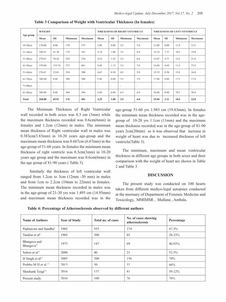
Medico-legal Update, July-December 2017, Vol.17, No. 2 208 209 -legal Update, July-December 2017, Vol.17, No. 2
Table 3 Comparison of Weight with Ventricular Thickness (In females)
Age groupWEIGHT THICKNESS OF RIGHT VENTRICLE THICKNESS OF LEFT VENTRICLE
Mean SD Minimum Maximum Mean SD Minimum Maximum Mean SD Minimum Maximum
10-20yrs 178.00 0.00 178 178 3.00 0.00 3.0 3.0 11.00 0.00 11.0 11.0
21-30yrs 240.91 41.30 176 342 4.18 1.08 3.0 6.0 14.18 2.32 10.0 19.0
31-40yrs 279.67 78.42 229 370 4.33 1.53 3.0 6.0 15.67 4.73 12.0 21.0
41-50yrs 339.00 119.74 222 481 4.00 1.15 3.0 5.0 16.00 4.69 11.0 22.0
51-60yrs 276.67 23.01 254 300 4.67 0.58 4.0 5.0 15.33 0.58 15.0 16.0
61-70yrs 300.00 0.00 300 300 5.00 0.00 5.0 5.0 17.00 0.00 17.0 17.0
71-80yrs - - - - - - - - - - - -
81-90yrs 300.00 0.00 300 300 6.00 0.00 6.0 6.0 20.00 0.00 20.0 20.0
Total 268.88 69.91 176 481 4.29 1.08 3.0 6.0 15.04 3.11 10.0 22.0
The Minimum Thickness of Right Ventricular wall recorded in both sexes was 0.3 cm (3mm) while the maximum thickness recorded was 0.6cm(6mm) in females and 1.2cm (12mm) in males. The minimum mean thickness of Right ventricular wall in males was 0.383cm(3.83mm) in 10-20 years age-group and the maximum mean thickness was 0.667cm (6.67mm) in the age-group of 51-60 years. In females the minimum mean thickness of right ventricle was 0.3cm(3mm) in 10-20 years age group and the maximum was 0.6cm(6mm) in the age group of 81-90 years ( Table 3).
Similarly the thickness of left ventricular wall ranged from 1.2cm to 3cm (12mm -30 mm) in males and from 1cm to 2.2cm (10mm to 22mm) in females. The minimum mean thickness recorded in males was in the age-group of 21-30 yrs was 1.495 cm (14.95mm) and maximum mean thickness recorded was in the
age-group 51-60 yrs 1.983 cm (19.83mm). In females the minimum mean thickness recorded was in the age-group of 10-20 yrs 1.1cm (11mm) and the maximum mean thickness recorded was in the age-group of 81-90 years 2cm(20mm) so it was observed that increase in weight of heart was due to increased thickness of left ventricle(Table 3).
The minimum, maximum and mean ventricular thickness in different age groups in both sexes and their comparison with the weight of heart are shown in Table 2 and Table 3.
DISCUSSION
The present study was conducted on 100 hearts taken from different medico-legal autopsies conducted at the mortuary of Department of Forensic Medicine and Toxicology, MMIMSR , Mullana , Ambala.
Table 4: Percentage of Atherosclerosis observed by different authors
Name of Authors Year of Study Total no. of cases No. of cases showing atherosclerosis Percentage
Padmavati and Sandhu9 1969 555 374 67.3%
Tandon et al8 1969 300 85 28.33%
Bhargava and Bhargava10 1975 147 69 46.93%
Sahoo et al11 2000 40 21 52.5%
H Singh et al12 2005 200 156 78%
Prabhu M H et al 13 2013 50 33 66%
Shashank Tyagi14 2016 137 81 59.12%
Present study 2016 100 76 76%
-legal Update, July-December 2017, Vol.17, No. 2 208 209 Medico-legal Update, July-December 2017, Vol.17, No. 2
CONCLUSION
The incidence of coronary atherosclerosis is very high in this part of the Haryana State. Though incidence of coronary artery disease is more in males as compared to females, but in both sexes it is alarming. Although the primary lesions start very early in life, but severe lesions are seen more commonly in elderly people. In early ages, single vessel involvement is more common and in elderly double vessel involvement and triple vessel in old age is more common respectively. Weight of heart also shows an increase with age up to a certain age, after which it starts declining. Although the cause of death was clear in majority of the cases, but still the occult role of ischemic heart disease or coronary atherosclerosis cannot be undermined as a contributing or aggravating factor in the death of the patient.
Conflict of Interest: None Declared
Ethics Clearance: It has been cleared by Institutional Ethics Committee, MMIMSR, Mullana, Ambala
Acknowledgment: Nil
Source of Funding: Self
REFERENCES
1. Garg M, Aggarwal AD, Kataria SP. Coronary Atherosclerosis and Myocardial Infarction: An Autopsy Study. J Indian Acad Forensic Med 2011; Vol 33,No.1.
2. Gupta R, Joshi P, Mohan V, Reddy KS, Yusuf S. Epidemiology and causation of coronary heart disease and stroke in India. Heart 2008; 94(1): 16-26.
3. Kumar. S, Verma. AK , Kumar. N, Verma. RK, Prevalence of coronary artery disease in different age groups: A Prospective study. Biomedical Research 2013;24(1): P139-141.
4. Indrayan . A , Forecasting vascular disease cases and associated mortality in India. NCMH Background Papers: Burden of Disease India. National Commission on Macroeconomics and
Health, Government of India.2005; P197-215.
5. Widimsky . P, Andel . M, Prevalence of coronary atherosclerosis in asymptomatic population. European Heart Journal 2000;21:P13-14.
6. Grundy . SM , Atherosclerosis. Pathology ,Pathogenesis and role of risk factors.(Disease-a-month, 1983);29:P3.
7. Arnel R, Kolodgie FD, Burke AP, Farb A, Schwartz SM. Lessons from sudden coronary death – A comprehensive morphological classification scheme for atherosclerotic lesions. Arteioscler Thromb Vasc Biol 2000; 20 : 1262-75
8. Tandon . Op, Aggarwal. VC, Katiyar. BC, Coronary and aortic atherosclerosis. Indian Heart Journal 1969;21:5-10.
9. Padmavati . S, Sandhu . I , Incidence of coronary artery disease in Delhi from medico-legal autopsies. Indian Journal of Medical Research 1969;57:465-475.
10. Bhargava. MK, Bhargava.SK , Coronary atherosclerosis in North Karnataka. Indian Journal Pathology and Microbiology 1975;18:65-77.
11. Sahoo. PC, Bahera. S, Acharya. S, Age factor in causation of atherosclerosis in the population of Southern Orissa. Journal of Indian Academy of Forensic Medicine 2000; 22(1): P6-9
12. Singh . H,Oberoi . SS, Gorea . RK, Bal . MS , Atherosclerosis in Coronaries in Malwa Region of Punjab. ( Journal of Indian Academic Forensic Medicine 2005);27(4):P32-5.
13. Prabhu . M.H, et al Atherosclerosis of Coronary Arteries-An Autopsy Study. (Global Journal of Medical Research Surgeries and Cardiovascular System 2013 , USA);Vol 13.(3):P19-24
14. Tyagi . S , Sukhadeve. BR , Pathak. M H , Autopsy findings in sudden cardiac deaths: Study in Medico-legal Autopsies,(Scholars Journal of Applied Medical Sciences,March2016);4(3C):P845-854.

211 -legal Update, July-December 2017, Vol.17, No. 2
Profile of Cause of Death in Unknown Dead Bodies Autopsied in a Tertiary Care Centre - A One Year Study
Sravani Yandava1, B Alekhya1, K Vijaya Durga2, Md Abdul Mujeeb Siddiqui3, K Ravi Muni4, V Jayasurya Prasad Babu5
1Assistant Professor, Physiology, Mallareddy Medical College for Women, Hyderabad, 2Assistant Professor of Forensic Medicine, Apollo Institute of Medical Sciences & Research Centre, Chittoor, 3Assistant Professor of
Forensic Medicine, Deccan Medical College, Hyderabad, 4Assistant Professor of Forensic Medicine, NRI Medical College, Chinakakani, Guntur, 5Senior Resident of Forensic Medicine, RIMS, Ongole
ABSTRACT
The term John Doe(man) or Jane Doe(woman) refers to corpse or hospital patient whose identity is unknown1. The identification of cadavers is a key concern in forensic investigation, but is equally important for ethical, criminal and civil reasons. All human beings have the right to bury and mourn their loved ones, and without identification of a dead body there are many civil procedures which cannot be completed2.At the finish of the post-mortem examination, the police with the aid of forensic experts are supposed to arrive at the identification of the body, so that a post-mortem certificate, in the name of the deceased, may be issued. This death certificate in turn facilitates the funeral arrangements, burial or cremation of the individual, life insurance benefits, disposition of instructions in the decedent’s last will and testament, and matters of inheritance.
Keywords: John doe, Jane doe, identification, cadaver, deceased, burial, cremation.
Corresponding author: Dr Y. Sravani, Assistant Professor of Forensic Medicine, Mallareddy Medical College for Women, Hyderabad;Mobile No: 7207710698, Email: [email protected]
INTRODUCTION
Human identity is a mainstay of civilization and the identification of unknown individuals has always been of paramount importance to the society.3 Article 6 of universal declaration of Human Rights states that “Everyone has the right to recognition everywhere, as a person, before the law”.4 Identification means the determination of the individuality or recognition of that person or dead body based on certain physical characters unique to that individual. It may be complete or partial. Complete identification means the absolute fixation of the individuality of a person, while partial identification means ascertainment of only some facts like race, sex, age and stature.5
William E. Gladstone, Prime Minister of England from 1892-94 rightly said:” Show me the manner in which a nation or community cares for its dead and I will measure, with mathematical exactness, the tender mercies of its people, their respect for the law of the land and their loyalties to high ideals.”
The fact that a large number of unclaimed bodies also cannot be positively identified in the population register suggests that corresponding identification records for these individuals do not exist with the Central Home Department. This implies that many destitute people in India are not registered as citizens. It may also suggest that the processes and techniques employed to verify identification through the population register may be flawed.6
Another dilemma that often surrounds unidentified deceased persons is that they may be in a geographical region where they are not known and therefore not identified. Information about unidentified decedents is often not communicated to anyone outside the district in which the death occurred, and such information may not reach relatives living outside of the district.6
DOI Number: 10.5958/0974-1283.2017.00101.3
211 Medico-legal Update, July-December 2017, Vol.17, No. 2
Also contributing to the number of unknown deceased individuals admitted to the Medico legal department are, the large number of persons who are reported missing on a daily basis in the country, some never to be found.6 At present no procedures in which missing persons’ profiles are matched with unclaimed bodies seem to exist in India.6 It is therefore not known whether any unidentified persons, and how many of them that are buried as paupers, may be amongst those reported missing. This cannot however be verified as statistics, or any other information regarding missing and unclaimed bodies, are not formally collected.6
It thus becomes increasingly important to conduct research into the number of unidentified bodies admitted at Mortuary, GGH, Guntur, in order to estimate the number of unidentified decedents on an annual basis. It is of paramount importance to make sincere and systematic efforts to trace out the missing persons and identify the unidentified dead bodies in railway accidents, destitute found dead in public places, etc and to list out the common problems encountered in unknown cases.
A thorough search of the literature did not yield much information regarding the identification of the unidentified dead in the Indian context. Mostly, they were devoted to individual body identification or identification of victims of mass disasters7-9. However, identifying an unknown deceased individual is often problematic. The reasons are many – inadequate/ no history, disinterested Investigating Officers unwilling to properly work out the case, partial/complete decomposition/ destruction/ mutilation of the body, burns etc10. As in many other countries, comparative data needed in order to make a positive identification is limited in India. At present no procedures in which missing persons‟ profiles are matched with unclaimed bodies seem to exist in India11. The present one year study was undertaken to find out the patterns of cause of death and their manner in unknown dead bodies.
AIMS & OBJECTIVES
1. To study the cause of death and the patterns of cause of death in unknown dead bodies
2. To evaluate the various efforts made on part of the investigating officer and the autopsy surgeon to establish the identity
MATERIAL AND METHOD
All unidentified, unclaimed, unknown destitute bodies brought for postmortem examination to the mortuary, department of Forensic Medicine & Toxicology, Guntur Medical College & Government General Hospital, Guntur during the year 2013, comprised the material for the study. A retrospective observational autopsy based study was done with the data compiled from the postmortem reports, inquest papers; detailed history elicited from the concerned police officials.
OBSERVATIONS
All unknown, unclaimed, destitute, abandoned, unidentified dead bodies admitted in morgue from 1 January to 31 December 2013 were taken for study which comprised 113(7.5%) of the total 1506 cases. Out of 113 cases, 100 cases were taken for study. Maximum number of cases belonged to the age group 31-40 years, 33%.Male to female ratio is 2.8:1. Males accounted for 74% while females were 26%. The manner of death in majority is accidental (54%), followed by suicidal (28%). Majority of the opinions regarding the cause of death were given as cranio-cerebral damage due to head injury (30%) followed by multiple injuries in 14%, combined cranio-cerebral damage & multiple other injuries in 10%, traumatic decapitation in 10%, poisoning in 10%, crush injuries in 6%, prematurity (foetal deaths) in 4%, drowning in 6%, Embolism/CAD in 4%, starvation in 2% and others in 4%. There is statistically significant relationship between deaths over railway track and cranio-cerebral damage being the cause of death. Viscera for chemical analysis were sent in 24%, histopathology in 8% and for both in 27% cases.
Regarding the efforts made by the investigating team to establish the identity, local inquiry was done in 100% cases, identification marks & injuries were noted in 100%, photographs were taken in 92%, advertisements in print & electronic media were done in 92% of cases, finger prints were taken in 18%, specimens for DNA analysis were taken in 6% and message to all the police stations in the state was done in 94% cases. Also local announcement in the neighbourhood, distribution of pamphlets in the vicinity, sticking the photographs in bus & railway stations were also done in few cases. The milieu of the unknown with natural deaths is destitute (62%), no attendants at the time of admission/discharge
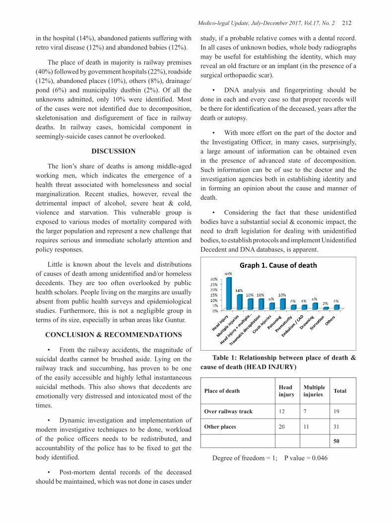
Medico-legal Update, July-December 2017, Vol.17, No. 2 212 213 -legal Update, July-December 2017, Vol.17, No. 2
in the hospital (14%), abandoned patients suffering with retro viral disease (12%) and abandoned babies (12%).
The place of death in majority is railway premises (40%) followed by government hospitals (22%), roadside (12%), abandoned places (10%), others (8%), drainage/pond (6%) and municipality dustbin (2%). Of all the unknowns admitted, only 10% were identified. Most of the cases were not identified due to decomposition, skeletonisation and disfigurement of face in railway deaths. In railway cases, homicidal component in seemingly-suicide cases cannot be overlooked.
DISCUSSION
The lion’s share of deaths is among middle-aged working men, which indicates the emergence of a health threat associated with homelessness and social marginalization. Recent studies, however, reveal the detrimental impact of alcohol, severe heat & cold, violence and starvation. This vulnerable group is exposed to various modes of mortality compared with the larger population and represent a new challenge that requires serious and immediate scholarly attention and policy responses.
Little is known about the levels and distributions of causes of death among unidentified and/or homeless decedents. They are too often overlooked by public health scholars. People living on the margins are usually absent from public health surveys and epidemiological studies. Furthermore, this is not a negligible group in terms of its size, especially in urban areas like Guntur.
CONCLUSION & RECOMMENDATIONS
• From the railway accidents, the magnitude of suicidal deaths cannot be brushed aside. Lying on the railway track and succumbing, has proven to be one of the easily accessible and highly lethal instantaneous suicidal methods. This also shows that decedents are emotionally very distressed and intoxicated most of the times.
• Dynamic investigation and implementation of modern investigative techniques to be done, workload of the police officers needs to be redistributed, and accountability of the police has to be fixed to get the body identified.
• Post-mortem dental records of the deceased should be maintained, which was not done in cases under
study, if a probable relative comes with a dental record. In all cases of unknown bodies, whole body radiographs may be useful for establishing the identity, which may reveal an old fracture or an implant (in the presence of a surgical orthopaedic scar).
• DNA analysis and fingerprinting should be done in each and every case so that proper records will be there for identification of the deceased, years after the death or autopsy.
• With more effort on the part of the doctor and the Investigating Officer, in many cases, surprisingly, a large amount of information can be obtained even in the presence of advanced state of decomposition. Such information can be of use to the doctor and the investigation agencies both in establishing identity and in forming an opinion about the cause and manner of death.
• Considering the fact that these unidentified bodies have a substantial social & economic impact, the need to draft legislation for dealing with unidentified bodies, to establish protocols and implement Unidentified Decedent and DNA databases, is apparent.
Table 1: Relationship between place of death & cause of death (HEAD INJURY)
Place of death Head injury
Multiple injuries Total
Over railway track 12 7 19
Other places 20 11 31
50
Degree of freedom = 1; P value = 0.046

-legal Update, July-December 2017, Vol.17, No. 2 212 213 Medico-legal Update, July-December 2017, Vol.17, No. 2
Table 2: Efforts made for identification Total number of cases n = 100
photographs taken Advertised in print & electronic media Finger prints taken Specimen for DNA analysis
takenMessage to all P.S
92 92 18 6 94
92% 92% 18% 6% 94%
Ethical Cearance – Obtained from Institute Ethical Committee
Source of Funding – Nil
Conflict of Interest - Nil
REFERENCES
1. Twitched indictment – Justice.gov
2. Cattaneo C, Porta D, De Anquelis D, Gibelli D, Poppa P, Grandi M. Unidentified bodies and human remains: an Italian glimpse through a European problem. Forensic Sci Int 2010; 195(1–3): 167c1-167c6.
3. Rothwell BR. Pr dental identification. Dent Clin North Am. 2001; 45(2):253-70.
4. Modi JP. Modi’s Medical Jurisprudence and Toxicology. Personal Identity.23rded. New Delhi: Lexis Nexis Butterworth’s; 2006:263-337.
5. Parikh CK. Parikh Text Book of Medical Jurisprudence and Toxicology. Identification. 6th ed; 2011:2.1-2.23.
6. Naidoo P. Frontal sinus comparison is a credible and cost effective method of establishing unique
human identity when applied to missing persons and unidentified bodies [dissertation]; University of Kwa-Zulu Natal, 2007.
7. Job C. Determination of cause of death in decomposed bodies – a regional study. JIAFM 2009; 31(1):11-17.
8. Kahana T, Hiss J. Personal identification based on radiographic vertebral features. Am J For Med Pathol 2002; 28(1):36-41.
9. Ludes B, Tracqui A, Pfitziner H, Kintz P, Levy P, Disteldrof M, et al. Medico-legal investigations of the Airbus- A 320 crash upon Mount Ste-Odile, France. JFS 1994; 39(5):1147-1152.
10. Ajay Kumar, Dasari Harish, Chavali KH, Amandeep Singh. Original Research Paper. Patterns of Cause of Death in Unknown Dead Bodies. A Three Year Study in a Tertiary Care Hospital. J Indian Acad Forensic Med. October-December 2012, Vol. 34, No. 4: pg304-8.
11. Naidoo P. Frontal sinus comparison is a credible and cost effective method of establishing unique human identity when applied to missing persons and unidentified bodies [dissertation]; University of Kwa-Zulu Natal, 2007.)
215 -legal Update, July-December 2017, Vol.17, No. 2
Scenario of Present Doctor – Patient Relationship
Pushpendra Singh1, B H Tirpude2, P N Murkey3, Sharjeel Khan4, Ninad Nagrale4, Swapnil Patond4
1Associate professor Forensic Medicine and Toxicology ,Veer Chandra Singh Garwahi Government Institute of 1Associate professor Forensic Medicine and Toxicology ,Veer Chandra Singh Garwahi Government Institute of 1
Medical Sciences and Research, Srinagar, Pauri Garwahal, 2Professor and HOD, 2Professor and HOD, 2 3Professor, 4Post Graduate Student, Department of Forensic Medicine and Toxicology, 4Post Graduate Student, Department of Forensic Medicine and Toxicology, 4
Mahatma Gandhi Institute of Medical Sciences, Sewagram
ABSTRACT
The relationship maintained between a doctor as a savior and a patient with a great need of sympathy, kindness and health guidance – is an age old faithful understanding. As on today, the society has been changed enormously. The attitude of doctors has been turned mostly towards business and the behavior of a patient has become maximally unfaithful. Today also the relation between the doctor and the patient exists but it is more like a relation between a consumer and a trader which is often devoid of any dedication and lovely bond that used to be seen long back as our forefathers remember. Though the problem has come up vigorously very recently, but the work on this relation was started in 1951. TALCOTT PARSONS was the first social scientist to theorize the doctor-patient relation.1
The landmark case IMA Vs V P Shanta brought the medical professionals within the ambit of “service” as defined in the CPA 1986.2
Keywords : Physicians, Commercialization, Consent, Media.
Corresponding author:Pushpendra SinghAssociate Professor Forensic Medicine and Toxicology, Veer Chandra Singh Garwahi Government Institute of Medical Sciences and Research, Srinagar, Pauri Garwahal.
INTRODUCTION
1) Family physicians- A family physician used to be a friend, philosopher and a guide to a family. It is told that they used to provide their valuable opinion even during marriage proceedings of a young lady whom he treated since her childhood. Naturally, a fantastic bond used to perished between a doctor and a family.
2) Total dedication – Doctors used to be in total dedication for the service of people for which they even used to spend money of their own.
3) Communication – Every successful physician used to be very good in conversation which used to give great relief to a sick man.
4) Law and Doctor – Very seldom lawful actions
used to taken against doctors, who in the mind of common people was placed above anything.
5) Private sector – Concept of private sector was not predominant and naturally idea of health business or health industry was far from the mind of a doctor or a patient.
6) Godly attitude – Common people used to consider a doctor as a God only- who also used to possess several characters which showed his honest mind and vast kindness like a sage.
MATERIALS AND METHOD
The study was conducted by taking into consideration of different case studies conducted by various authors.
FINDINGS
1) About one in four patient feels that the physician sometimes exposes them to unnecessary risk, according to a data from John Hopkin’s study3. The picture may not be so grave in our country, but the fact persists here also.
DOI Number: 10.5958/0974-1283.2017.00102.5
215 Medico-legal Update, July-December 2017, Vol.17, No. 2
2) Role of family physician is going to be extinct very soon. So, now very few doctor has detail idea of atleast health aspect of families of his own locality. Restoring the concept of family physician can strengthen the doctor-patient relationship and redeem the image of the medical Practioner which has been besmirched in recent years 4.
3) Lack of communication – It is often seen in nursing homes and clinics of private practitioners who underestimate common people and rarely explain present situation of a disease which is a common complaint of public.
4) Media – Mainly due to prompt broadcasting of sensational news often even of a small mistake of a medico, the faith on a doctor is broken which is a bad sign.
5) Quacks – Improperly trained persons have come up in large numbers mainly in rural and semi urban sectors. Their misadventures cause a lot of misunderstandings.
6) Politician and anti-socials – These two groups have come up as very much lethal for doctor- patient relation. For cheap popularity and vested interest, they spoil a smooth procedure.
7) Government and CPA – Often with changing political scenario, medicos are not in a position to get support from govt. Doctors nowadays remain all time apprehensive about the court or punishment which hinders devoted action on treatment of a sick man.
8) Service at Hospitals – Govt. hospitals often are worst in providing expected service to a patient. Even for private sector, inspite of spending a good amount of money, patients often do not get rational care which creates a sense of fury towards doctors.
9) Commercialization – Health Industry has come up today, but these are mainly located at urban sectors only. People are more conscious of health nowadays. With more expectations and less facilities available at the larger rural belts, misunderstandings with a medico is growing up which is often fracturing the bond between a doctor and the patient.
DISCUSSION
We know that the relation between a doctor and a
patient is often emotional based on trust. But we forget that behind that emotion there are few responsibilities from both sides- a doctor and a patient. Most of the complexities are created due to lack of this concept of legal aspect.
Both should remember: Whenever a patient comes to a doctor for treatment there is a contract established between the doctor and the patient.
Doctors should remember: 5
1) They have compulsory duties to report birth, death, infectious diseases, and to inform police all cases of crimes as located by them.
2) They should maintain good moral character, maximum skill, sufficient knowledge.
3) They should be aware of professional secrecy, proper service to patient.
4) Honesty should be maintained always .There should not be any commissioning business.
5) Patient or relatives should be communicated about all treatment procedures regularly.
Patient should remember:
1) Polite and sober behavior and neat and clean appearance is preliminary requirement.
2) They should give correct and full history and allow a doctor to examine.
3) They have to maintain all medical records and pay due fees to a doctor along with following the instructions on treatment .
4) He should keep faith on his treating doctor.
5) The patient has every right to terminate the relationship with his doctor at any time and seek the help of another .A reciprocal right rests with the doctor also.6
A MEDICO SHOULD BE AWARE OF 3 ‘C’S :
1. C- CONSENT – Every doctor should follow “informed consent “rule for any proceedings
2. C – CONSULTATION – Honest consultation and communication makes a good raport.
3) C- CROSS – The belief that red cross symbol can

Medico-legal Update, July-December 2017, Vol.17, No. 2 216
be used on vehicles and building is wrong except red cross society and army medical establishments – as per Geneva Convention Act, Section 12 and 13.
With the growing problem of tough relation between a doctor and a patient, the matter has already been considered in our country. In United States a course called Interviewing and the Doctor – Patient relationship has already started.7
CONCLUSION
The situation of misunderstandings will change if we think that Doctor-patient relation is a planned effort to be established. It is a way of developing reciprocal understanding and good will.8
Conflict of Interest: To improve doctor – Patient relationship.
Source of Support: All the Cases of Medical Negligience2.
Ethical Clearance: As the study was not conducted on any living subjects so ethical clearance was not required hence not taken.
REFERENCES
1. J Hughes Ph.d-Organisation and information at the bed side
2. Suprereme court and medical negligence necessary protection. www.legal service media .com/ 178 medical negligence.
3. Parker Pope on health July 29, 2008.
4. The Hindu – Monday Jan 17, 2005.
5. Singhals’s the doctor and law 1st edition 1999 page no138.
6. Sunil K Pandya- Doctor patient relationship- Indian Journal of Medical ethics, April- June 1995, Vol 3 (2)
7. N.p.r. august 29, 2008- a news update
8. A.V.shrinivasan- Managing a modern hospital 1st
edition 2000, page no 328.

-legal Update, July-December 2017, Vol.17, No. 2 216
Contribution of Toxicology Laboratory in Establishing Exact Clinical Diagnosis in Cases of Poisoning
Pushpendra Singh1, B.H Tirpude2, P N Murkey3, Sharjeel Khan4, Ninad Nagrale4, Swapnil Patond4
1Associate Professor, Forensic Medicine and Toxicology, Veer Chandra Singh Garwahi Government Institute of 1Associate Professor, Forensic Medicine and Toxicology, Veer Chandra Singh Garwahi Government Institute of 1
Medical Sciences and Research, Srinagar, Pauri Garwahal, 2Professor and HOD, 2Professor and HOD, 2 3Professor, 4Post Graduate Student, Department of Forensic Medicine and Toxicology,4Post Graduate Student, Department of Forensic Medicine and Toxicology,4
Mahatma Gandhi Institute of Medical Sciences, Sewagram
ABSTRACT
History by the patient or his attendant is never binding on the clinician. History can be variously presented and interpreted. But it does influence his medical judgement, considering actuality from history forwarded and the clinical presentation. For the diagnosis of a case of poisoning, judgement is based on history obtained and signs and symptoms prevailed by a particular consumed poison. But, where poisoning may not be suspected ordinarily, because symptoms and signs are not present, specifically and it may even resemble natural disease. Besides it, the history is not available most of the time or if it is available is not correct. Diagnosis of poisoning based on wrong information by exclusion technique has hazardous clinical and legal implications.
Keywords- poisoning, clinical toxicology, toxicology laboratory.
Corresponding author: Pushpendra SinghAssociate Professor Forensic Medicine and Toxicology, Veer Chandra Singh Garwahi Government Institute of Medical Sciences and Research, Srinagar, Pauri Garwahal.
INTRODUCTION OR BACK GROUND
Toxicology can be of great service to emergency department clinicians when used appropriately provide them with valuable information helpful in diagnosis and management. Forensic toxicology deals with medical & legal aspects of harmful effects of chemicals on human beings. Clinical toxicology deals with human diseases caused by or associated with abnormal exposure to chemical substances. In research work, toxicology laboratory can play a vital role in determining the epidemiological aspect of poisoning and to find out the incidences of various poisons. Such knowledge can be utilized by public health authorities in proper planning of preventive measures and public education
MATERIALS AND METHOD
This study was carried out at Kasturba Hospital in the Department of Forensic Medicine & Toxicology of MGIMS, Sewagram, dist. Wardha (Maharashtra), consisting of 375 cases of poisoning admitted which were analysed in the departmental toxicology laboratory during the period from January 2000 to December 2001.Each case was followed from the time of admission till the discharge or death and autopsy conducted at the mortuary of department of Forensic Medicine of this institute.
DOI Number: 10.5958/0974-1283.2017.00103.7
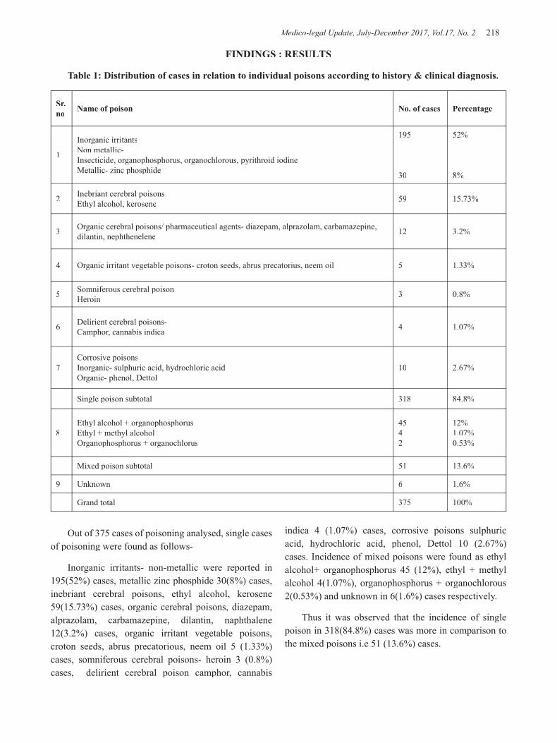
Medico-legal Update, July-December 2017, Vol.17, No. 2 218 219 -legal Update, July-December 2017, Vol.17, No. 2
FINDINGS : RESULTS
Table 1: Distribution of cases in relation to individual poisons according to history & clinical diagnosis.
Sr. no Name of poison No. of cases Percentage
1
Inorganic irritantsNon metallic-Insecticide, organophosphorus, organochlorous, pyrithroid iodineMetallic- zinc phosphide
195
30
52%
8%
2 Inebriant cerebral poisonsEthyl alcohol, kerosene 59 15.73%
3 Organic cerebral poisons/ pharmaceutical agents- diazepam, alprazolam, carbamazepine, dilantin, nephthenelene 12 3.2%
4 Organic irritant vegetable poisons- croton seeds, abrus precatorius, neem oil 5 1.33%
5 Somniferous cerebral poisonHeroin 3 0.8%
6 Delirient cerebral poisons-Camphor, cannabis indica 4 1.07%
7Corrosive poisonsInorganic- sulphuric acid, hydrochloric acidOrganic- phenol, Dettol
10 2.67%
Single poison subtotal 318 84.8%
8Ethyl alcohol + organophosphorusEthyl + methyl alcoholOrganophosphorus + organochlorus
4542
12%1.07%0.53%
Mixed poison subtotal 51 13.6%
9 Unknown 6 1.6%
Grand total 375 100%
Out of 375 cases of poisoning analysed, single cases of poisoning were found as follows-
Inorganic irritants- non-metallic were reported in 195(52%) cases, metallic zinc phosphide 30(8%) cases, inebriant cerebral poisons, ethyl alcohol, kerosene 59(15.73%) cases, organic cerebral poisons, diazepam, alprazolam, carbamazepine, dilantin, naphthalene 12(3.2%) cases, organic irritant vegetable poisons, croton seeds, abrus precatorious, neem oil 5 (1.33%) cases, somniferous cerebral poisons- heroin 3 (0.8%) cases, delirient cerebral poison camphor, cannabis
indica 4 (1.07%) cases, corrosive poisons sulphuric acid, hydrochloric acid, phenol, Dettol 10 (2.67%) cases. Incidence of mixed poisons were found as ethyl alcohol+ organophosphorus 45 (12%), ethyl + methyl alcohol 4(1.07%), organophosphorus + organochlorous 2(0.53%) and unknown in 6(1.6%) cases respectively.
Thus it was observed that the incidence of single poison in 318(84.8%) cases was more in comparison to the mixed poisons i.e 51 (13.6%) cases.
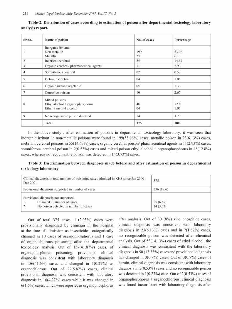
-legal Update, July-December 2017, Vol.17, No. 2 218 219 Medico-legal Update, July-December 2017, Vol.17, No. 2
Table-2: Distribution of cases according to estimation of poison after departmental toxicology laboratory analysis report-
Sr.no. Name of poison No. of cases Percentage
1Inorganic irritantsNon metallicMetallic
19923
53.066.13
2 Inebrient cerebral 55 14.673 Organic cerebral/ pharmaceutical agents 11 2.93
4 Somniferous cerebral 02 0.53
5 Delirient cerebral 04 1.06
6 Organic irritant vegetable 05 1.33
7 Corrosive poisons 10 2.67
8Mixed poisonsEthyl alcohol + organophosphorusEthyl + methyl alcohol
4804
12.81.06
9 No recognizable poison detected 14 3.73
Total 375 100
In the above study , after estimation of poisons in departmental toxicology laboratory, it was seen that inorganic irritant i.e non-metallic poisons were found in 199(53.06%) cases, metallic poison in 23(6.13%) cases, inebriant cerebral poisons in 55(14.67%) cases, organic cerebral poison/ pharmaceutical agents in 11(2.93%) cases, somniferous cerebral poison in 2(0.53%) cases and mixed poison ethyl alcohol + organophosphorus in 48(12.8%) cases, whereas no recognizable poison was detected in 14(3.73%) cases.
Table 3: Discrimination between diagnoses made before and after estimation of poison in departmental toxicology laboratory
Clinical diagnosis in total number of poisoning cases admitted in KHS since Jan 2000- Dec 2001 375
Provisional diagnosis supported in number of cases 336 (89.6)
Provisional diagnosis not supported1. Changed in number of cases2. No poison detected in number of cases
25 (6.67)14 (3.73)
Out of total 375 cases, 11(2.93%) cases were provisionally diagnosed by clinician in the hospital at the time of admission as insecticides, categorically changed as 10 cases of organophosphorus and 1 case of organochlorous poisoning after the departmental toxicology analysis. Out of 157(41.87%) cases, of organophosphorus poisoning, provisional clinical diagnosis was consistent with laboratory diagnosis in 156(41.6%) cases and changed in 1(0.27%) as organochlorous. Out of 22(5.87%) cases, clinical provisional diagnosis was consistent with laboratory diagnosis in 16(4.27%) cases while it was changed in 6(1.6%) cases, which were reported as organophosphorus
after analysis. Out of 30 (8%) zinc phosphide cases, clinical diagnosis was consistent with laboratory diagnosis in 23(6.13%) cases and in 7(1.87%) cases, no recognizable poison was detected after chemical analysis. Out of 53(14.13%) cases of ethyl alcohol, the clinical diagnosis was consisitent with the laboratory diagnosis in 50 (13.33%) cases and provisional diagnosis has changed in 3(0.8%) cases. Out of 3(0.8%) cases of heroin, clinical diagnosis was consistent with laboratory diagnosis in 2(0.53%) cases and no recognizable poison was detected in 1(0.27%) case. Out of 2(0.53%) cases of organophosphorus + organochlorous, clinical diagnosis was found inconsistent with laboratory diagnosis after
Medico-legal Update, July-December 2017, Vol.17, No. 2 220
poison estimation in the toxicology laboratory. Out of 6(1.6%) cases of unknown poison diagnosis has established in 1(0.27%) case and no recognisable poison was detected in 5(1.33%) cases.
DISCUSSION
In present study, history/ clinical diagnosis with laboratory results were studied from various information’s and were forwarded to treating physician. Consistency of laboratory results with history from hospital was determined and expressed in percentage. Overall accuracy of history with laboratory analysis was determined and found to be 89.7%.
In the present study, from the above tables it was observed that the provisional diagnosis was supported by departmental toxicology laboratory after analysis of samples in 336(89.6%) cases. Whereas in 25 (6.67%) cases, laboratory diagnosis changed from provisional clinical diagnosis made by clinician in the hospital at the time of admission. Whereas in 14(3.73%) cases, no recognizable poison was detected in departmental toxicology laboratory after analysis of samples.Thus it can be concluded that in the present study, the toxicology laboratory has played major role in the confirmation and specification of the clinical diagnosis.
CONCLUSION
1. History was consistent with laboratory findings in 336(89.7%) cases only.
2. In 25(6.67%) cases the provisional clinical diagnosis was changed after the samples were analysed in departmental toxicology laboratory, and in 14(3.73%) cases, no recognizable poison was detected.
Thus departmental toxicology laboratory was found to be contributing directly in establishing diagnosis of 39 (10.4%) cases of poisoning and in 25(6.67%) cases suggested suitable antidote for treatment and management in hospital.
Conflict of Interest: To establish the role and importance of Toxicology Laboratory in the Clinical Diagnosis of Cases of Poisoning.
Source of Support: All the cases of Poisoning admitted in Rural Hospital in central India.
Ethical Clearance: The thesis on the above mentioned had already been approved and admitted in RTM Nagpur University .
REFERENCES
1. Dewan A. (1997); “role and relevance of poison information centres in India”, ICMR bulletin; Vol. 27(5):P.43-47
2. Dogra TD, Singh B.(1996); “present status of poisoning in India”; Indian Medical Gazette; Nov. 1996, P.364-368
3. Fernando R. (1995), “pesticide poisoning in Asia- pacific region and role of a regional information network (1995); J. Toxicol. Clin. Toxicol 33; P. 6-11.
4. Jain R. Agarwal P.(1996); “role of Toxicology laboratory in acute poisoning and drug overdose”, medicolegal update Vol. No. 1, Jan-Jun, 1996, P. 14-17
5. Malley M. (1997); “clinical evaluation of pesticide exposure and poisoning”; the Lancet 1997; 349, P. 1161-66
6. Naik RS, Tirpude BH, Sarwey GN, Agarwal S, Bhati A. (1999); “role and relevance of Toxicology laboratory in department of Forensic Medicine of Medical college”, Journal of Forensic Medicine and Toxicology; 1999, Vol. 16, No.1; Jan-Jun 1999; P. 70-72
7. Parikh CK. (1990); “Parikh’s textbook of Medical jurisprudence and Toxicology”, 1990 5th edition, Toxicology section, P. 694.
-legal Update, July-December 2017, Vol.17, No. 2 220
Study of Unnatural Deaths in Females in Garhwal Region of Uttrakhand
Pradeep Kumar Agarwal , Assistant Professor, VCSGGMSRI, Srinagar, Uttrakhand
ABSTRACT
Death is unnatural when caused prematurely against the order of the nature by injury or other means of violence. There is a noticeable increase in unnatural deaths of females in recent times. The aim of this study was to find out demographical factors, cause and manner of death in unnatural female deaths. A total number of 100 cases of unnatural deaths in female were studied during study period. 68% of unnatural deaths were seen in 15-30 years age group. Married women were about 3.5 times more prone to unnatural deaths then unmarried. The ratio of accidental, suicidal and homicidal deaths among females was 11:6:3. The commonest cause of unnatural deaths was burns (49%) followed by accidents (17%), poisoning (14%), asphyxia (12%) and violence (7%) respectively. 44% of unnatural deaths occurred within 7 year of marriage out of which 21 were accidental while 23 were suicidal and homicidal deaths. These 23 cases were established cases of dowry deaths. This study reflects susceptibility of married women to unnatural deaths. So, there is a need to enhance the multi-pronged protective system to curb these potentially preventable deaths.
Keywords: Unnatural deaths, Adult Females, Married, Suicide, Burns
Corresponding author: Dr. P.K. Agarwal,Assistant Prof., Department of forensic Medicine,VCSGGMSRI, UttarakhandE-Mail Id:[email protected]
INTRODUCTION
Unnatural deaths are common among both sexes in varying proportion of different causative factors leading to it. Females, not only in this country but all over the world has been the victim of mans evils due to some reason or other. A total of 2,13,585 incidents of crime against women were reported in the country during 2010 as compared to 2,03,804 during 2009. The rate of crime has increased marginally from 17.4% during the year 2009 to 18% during 20101. These are mainly due to increase in number of dowry deaths, torture to women and sexual offences. Dowry might have started as an innocent custom, a symbol of love from parents to their daughters on the eve of marriage. But it has in recent years grown in to a social evil with many incidence of bride burning and suicides. These are symptoms of social corruption. The present study was conducted to evaluate factors related to unnatural deaths in females in Garhwal region of Uttrakhand.
MATERIAL & METHOD
Materials for the present study comprised 100 cases of unnatural female deaths received in the mortuary of the Department of Forensic Medicine , VCSGGMSRI, Srinagar, Uttrakhand, for medico-legal autopsy during the period 01.01.2010 to 31.12.2010. The various epidemiological aspects were collected from the perusal of police papers, medicolegal reports of the cases and through interrogation of the parents, relatives, friends, and neighbors as well as police officers accompanying the dead body. Detailed external and internal examination was done at postmortem.
RESULTS
In the present study 100 cases of female unnatural deaths were studied out of which 78 were married and 22 were unmarried (Table 1). The case were distributed from 0-75 years of age out of which 68 cases fell in age group of 15-30 years. The most common cause of unnatural death cause burns (49%) followed by accidents (17%), poisoning (14%), asphyxia (12%), and violence (7%) (Table 2). The manner of unnatural death was accidental in 55% cases followed by suicidal in 30% cases and homicidal in 15% cases respectively (Table 3).
DOI Number: 10.5958/0974-1283.2017.00104.9

Medico-legal Update, July-December 2017, Vol.17, No. 2 222 223 -legal Update, July-December 2017, Vol.17, No. 2
In 49 cases due to burns, kerosene oil stove has been the commonest method employed. Out of these, 35 cases were accidental, 8 were suicidal and 6 were homicidal in manner. 42 out of these 49 cases were married females (Table 4,5). 17 cases died due to various accidents out of which trucks took toll of 6 cases (Table 6).12 females died due to asphyxia out of which 8 died due to hanging (Table 8). Poisoning was cause of death in 14 cases. Organophosphorus compound was commonest poison used. 13 out of 14 cases were suicidal and 9 of them were married females (Table 7). 7 out of 100 cases died due to violence caused by blunt, sharp cutting and firearm weapons. All these victims were married females (Table 9)
Out of 78 married females 44 died within 7 year of marriage and 34 died after 7 years of marriage. In 44 cases the cause of death was observed to be burns (26), asphyxia (6), accidents (6), poisoning (5) and violence (1) (Table 10). From the above table, 21 out of 44 cases died accidently while the remaining 23 died due to suicide (13) and homicide (10). So, it is observed that there were 23 established cases of dowry deaths out of 44 cases who died with in 7 years of marriage.
Table 1 : Distribution of cases according to Marital status (n=100)
Status No. of cases Percentage
Married 78 78
Unmarried 22 22
Table 2: Distribution of cases according to Cause of death (n=100)
S.No. Cause of Death
No. of Cases (Married)
No. of Cases (Unmarried) Total
1 Burns 42 7 49
2 Accidental 9 8 17
3 Poisoning 10 4 14
4 Asphyxial 9 3 12
5 Violence 7 0 7
6 Miscellaneous 1 0 1
Table 3: Distribution of cases according to Manner of death (n=100)
S.No. Manner No. of Cases (Married)
No. of cases (Unmarried) Total
1 Accidental 40 15 55
2 Suicidal 23 7 30
3 Homicidal 15 0 15
Table 4: Burns: Cause of Burns (n=49)
S.No. Cause of Burns No. of Cases Percentage
1 Caught Fire from Kerosene Stove 29 59.2
2 Sprinkling of Kerosene Oil 12 24.5
3 Angithi 5 10.2
4 Cooking Gas 3 6.1
Table 5: Burns: Manner of Burns (n=49)
S.No. Manner of Burns No. of Cases Percentage
1 Accidental 35 71.4
2 Suicidal 8 16.3
3 Homicidal 6 12.3
Table 6: Causes of Accidental Deaths (n=17)
S.No. Causes of Accidents No. of Cases Percentage
1 Hit by Truck 6 35.4
2 Fall from height 4 23.5
3 Hit by Train 2 11.7
4 Firearm 2 11.7
5 Hit by Bus 1 5.9
6 Fall from Tonga 1 5.9
7 Cold Exposure 1 5.9
Table 7 : Causes of Poisoning Deaths (n=14)
S.No. Poison No. of Cases Percentage
1 Organophosphorus 8 57.2
2 Barbiturates 3 21.4
3 Copper Sulphate 2 14.2
4 Nitric Acid 1 7.2
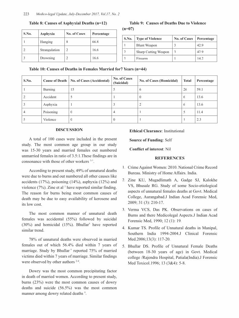
-legal Update, July-December 2017, Vol.17, No. 2 222 223 Medico-legal Update, July-December 2017, Vol.17, No. 2
Table 8: Causes of Asphyxial Deaths (n=12)
S.No. Asphyxia No. of Cases Percentage
1 Hanging 8 66.8
2 Strangulation 2 16.6
3 Drowning 2 16.6
Table 9: Causes of Deaths Due to Violence (n=07)
S.No. Type of Violence No. of Cases Percentage
1 Blunt Weapon 3 42.9
2 Sharp Cutting Weapon 3 42.9
3 Firearm 1 14.2
Table 10: Causes of Deaths in Females Married for7 Years (n=44)
S.No. Cause of Death No. of Cases (Accidental) No. of Cases (Suicidal) No. of Cases (Homicidal) Total Percentage
1 Burning 15 5 6 26 59.1
2 Accident 5 1 0 6 13.6
3 Asphyxia 1 3 2 6 13.6
4 Poisoning 0 4 1 5 11.4
5 Violence 0 0 1 1 2.3
DISCUSSION
A total of 100 cases were included in the present study. The most common age group in our study was 15-30 years and married females out numbered unmarried females in ratio of 3.5:1.These findings are in consonance with those of other workers 2-3.
According to present study, 49% of unnatural deaths were due to burns and out numbered all other causes like accidents (17%), poisoning (14%), asphyxia (12%) and violence (7%). Zine et al 2 have reported similar finding. The reason for burns being most common causes of death may be due to easy availability of kerosene and its low cost.
The most common manner of unnatural death females was accidental (55%) followed by suicidal (30%) and homicidal (15%). Bhullar5 have reported similar trend.
78% of unnatural deaths were observed in married females out of which 56.4% died within 7 years of marriage. Study by Bhullar 5marriage. Study by Bhullar 5marriage. Study by Bhullar reported 75% of married victims died within 7 years of marriage. Similar findings were observed by other authors 2-4.
Dowry was the most common precipitating factor in death of married women. According to present study, burns (23%) were the most common causes of dowry deaths and suicide (56.5%) was the most common manner among dowry related deaths 5.
Ethical Clearance: Institutional
Source of Funding: Self
Conflict of interest: Nil
REFERENCES
1. Crime Against Women: 2010. National Crime Record Bureau. Ministry of Home Affairs. India.
2. Zine KU, Mugadlimath A, Gadge SJ, Kalokhe VS, Bhusale RG. Study of some Socio-etiological aspects of unnatural females deaths at Govt. Medical College, Aurangabad.J Indian Acad Forensic Med, 2009; 31 (3): 210-17.
3. Verma VCS, Das PK. Observations on cases of Burns and there Medicolegal Aspects.J Indian Acad Forensic Med, 1990; 12 (1): 19.
4. Kumar TS. Profile of Unnatural deaths in Manipal, Southern India 1994-2004.J Clinical Forensic Med.2006;13(3): 117-20.
5. Bhullar DS. Profile of Unnatural Female Deaths (between 18-30 years of age) in Govt. Medical college /Rajendra Hospital, Patiala(India).J Forensic Med Toxicol.1996; 13 (3&4): 5-8.
225 -legal Update, July-December 2017, Vol.17, No. 2
Profile of Dowry Deaths in Garhwal Region of Uttrakhand
Pradeep Kumar Agarwal
Assistant Professor, VCSGGMSRI, Srinagar, Uttarakhand
ABSTRACT
One of the most heinous and shocking form of violence against women is dowry deaths. It is one among the various reasons for suicidal and homicidal deaths of women in India. The evil of dowry system is a matter of serious concern to everyone in view of its ever increasing and disturbing proportions. The present study was conducted to evaluate the magnitude and study various socio-etiological factors of dowry deaths. The most common age group to be affected was 21-30 years with 27 cases (61.4%) and first two years of marital life were more prone to dowry deaths with 10 cases (22.7%) followed by 6 years(20.5%). Burns was the cause of death in majority of cases (59.1%) followed by asphyxia (13.6%), accidents (13.6%), poisoning (11.4%) and violence (2.3%). 21 out of 44 cases died accidently while the remaining 23 died due to suicide (13) and homicide (10). Education of females, awareness about laws and strict and timely punishment of offenders will help in controlling dowry deaths.
Keywords: Dowry death, bride burning, and unnatural women deaths
Corresponding author: Dr. P.K. Agarwal,Assistant Prof., Department of forensic Medicine,VCSGGMSRI, UttarakhandE-Mail Id: [email protected]
INTRODUCTION
Dowry is a custom were money and property is brought by women to her husband when they marry. This customary practice is common in many Asian countries including Bangladesh, India, Pakistan and Sri Lanka. All though the trend is declining in other countries but the incidence of dowry is on rise in our country. According to section 304B IPC, dowry death is the death of young married women within 7years of marriage who are murdered or driven to suicide by continuous harassment and torture by husband and in laws in an effort to extort an increased dowry. The Indian National Crime Records Bureau (NCRB) reports that there were
8,391 dowry deaths reported in 2010, 1.4 deaths per 100,000 women1 .Most dowry deaths occur when young women, unable to bear the harassment and torture, commits suicide. Most of the suicides are by burns, hanging and poisoning2
. Sometimes, woman is killed by setting her self on fire , this is known as bride burning. To
combat this menance, the Government of India brought strict legislation and prohibited payment of dowry under the 1961 Dowry Prohibition Act in Indian civil law and subsequently by sections 304B and 498A of Indian Penal Code. Despite all these anti dowry laws in India, it is still rampant and incidence is on rise annually. Therefore keeping the above perspective in mind, this study was carried out to investigate into the cause of death, manner of death and other epidemiological factors responsible for dowry deaths and suggest remedial measures in order to prevent such untimely deaths.
MATERIAL & METHOD
Materials for the present study comprised 44 cases of unnatural female deaths occurring within 7 years of marriage brought in the mortuary of the Department of Forensic Medicine, VCSGGMSRI, Srinagar, Uttrakhand , for medico-legal autopsy during the period 01.01.2011 to 31.12.2011. The various epidemiological aspects were collected from the perusal of police papers, medicolegal reports of the cases and through interrogation of the parents, relatives, friends, and neighbors as well as police officers accompanying the dead body. Detailed external and internal examination was done at postmortem.
DOI Number: 10.5958/0974-1283.2017.00105.0
225 Medico-legal Update, July-December 2017, Vol.17, No. 2
RESULTS
In the present study 44 cases of female unnatural deaths were studied who died within 7 years of marriage. The most common age group involved was 21-30 years (61.4%) followed by 11-20 years (38.6%) (Table 1). With reference to duration of marital life (Table 2), the women married for two years were much more prone to dowry deaths (22.7%) followed by those married for six years (20.5). The most common cause of death was observed to be burns (59.1%) followed by asphyxia (13.6%), accidents (13.6%), poisoning (11.4%) and violence (2.3%) (Table 3).From the above table, 21 out of 44 cases died accidently while the remaining 23 died due to suicide (13) and homicide (10). So, it is observed that there were 23 established cases of dowry deaths out of 44 cases who died with in 7 years of marriage. Out of 23 cases of dowry deaths, 11 cases died due to burns, out of which 5 were suicidal and 6 were homicidal. poisioning was responsible for death in 5 cases, out of which 4 were suicides and 1 was homicide. Asphyxia was also responsible for 5 deaths out of which 3 were suicidal and 2 were homicidal. Final analysis of 23 dowry death cases is given in Table 4.
Table 1: Age – wise Distribution of cases (n=44)
S.No. Age Group No. of Cases Percentage
1 0-10 years 0 0
2 11-20 years 17 38.6
3 21-30 years 27 61.4
4 31-40 years 0 0
Table 2: Duration of Marriage (n=44)
S.No. Duration No. of Cases Percentage
1 >1year 01 2.3
2 1 year 05 11.4
3 2 years 10 22.7
4 3 years 05 11.4
5 4 years 07 15.9
6 5 years 04 9.0
7 6 years 09 20.5
8 7 years 03 6.8
Table 3: Causes of Deaths in Females Married for7 Years (n=44)
S.No. Cause of Death No. of Cases(Accidental)
No. of Cases (Suicidal)
No. of Cases (Homicidal) Total Percentage
1 Burning 15 5 6 26 59.12 Accident 5 1 0 6 13.63 Asphyxia 1 3 2 6 13.6
4 Poisoning 0 4 1 5 11.4
5 Violence 0 0 1 1 2.3
Table 4: Final Analysis of dowry Death cases (n=23)
S.No. Cause of Death No. of cases (Homicidal) No. of cases (Suicidal) Total No. of cases Total
1 Burns 11
Sprink kerosene oil 6 4 10
Cooking Gas 1 01
2 Accidents 1
Train 1 1
3 Poisoning 5
Organophosphorus 1 1 2
Barbiturates 2 2
Copper Sulphate 1 1

Medico-legal Update, July-December 2017, Vol.17, No. 2 226
4 Asphyxia 5
Hanging 3 3
Strangulation 2 2
5 Violence 1
Sharp Cutting Weapon 1 1
Total 10 13 23 23
DISCUSSION
Most dowry deaths occur when young women, unable to bear harassment and torture, commit suicide. Most of these suicides are by burns, hanging and poisoning. 44 cases of unnatural deaths in females married for 7 years were included in present study. The most common age group involved was 21-30 years followed by 11-20 years. Similar trends were observed by other authors 3-5. With reference of duration of marital life, maximum cases were seen in women married for 2 years followed by those married for 6 years. More number of victims in early marital life can be due to stress female faces during adaptation in the in laws house which gets aggravated by harassment for dowry 4-6. Out of 44 cases of deaths in married females studied, 21 cases were accidental, 13 suicidal and 10 homicidal 5,6. These 23 suicidal and homicidal deaths were the established cases of dowry deaths. Out of 13 cases of suicides in females, 5 were due to burns by sprinkling of kerosene and cooking gas, 4 cases were due to poisoning by organophosphorous compounds and barbiturates, 3 cases were due to hanging and 1 was due to railway accidents 3-5. Out of 10 cases of homicide, 6 were due to burns by kerosene, 2 were due to strangulation, and 1 each due to organophosphorous poisoning and injury by sharp weapon 5-7.
The different legislations to protect females from dowry deaths including Dowry Prohibition Act, 19611; Sec. 498 (A) IPC, Sec. 304 (B) IPC, Sec. 113 (A) and 113(B) of IEA9 etc have been passed by Government of India. Constitutionally, women were provided special protection under Article 21 and Article 1410. Supreme Court of India extended the ambit of Article 21 and held that mere existence is not the right to live, it is the right to live with dignity. The Government of India passed a Domestic Violence Bill, 2001, “To protect the rights of women who are victims of violence of any kind occurring within the family and to provide for matters connected therewith or incidental thereto”. In spite
of all these legislations, the incidence of dowry death is still alarming in our country. A female will be in a better position when she is educated, employed and well aware of her rights. Marriage registration and counseling should be mandatory. Speedy trial and strict punishment for perpetrators of the crime will help to reduce such deaths.
Ethical Clearance: Institutional
Source of Funding: Self
Conflict of Interest: Nil
REFERENCES
1. Crime Against Women: 2010. National Crime Record Bureau. Ministry of Home Affairs. India.
2. Kumar, Virendra (Feb 2003). “Burnt wives”. Burns 29(1): 31–36.
3. Kulshrestha P, Sharma R.K., Dogra T.D. The Study of Sociological and Demographical Variables of Unnatural Deaths among Young Women in South Delhi within Seven Years of Marriage. Journal of Punjab academy of forensic medicine and toxicology (online) 2002; Vol: 2.
4. Zine Kailash U, Mugadlimath A., Gadge S.J., Kalokhe V. S., Bhusale R. G. Study of some socio-etiological aspects of unnatural female deaths at government medical college, Aurangabad. JIAFM 2009; vol 31(3) P: 210 – 217.
5. Saha KK, Mohanty S. Alleged Dowry Death: A study of homicidal burns. Med.Sci.Law. 2006; 46(2):105
6. Mohanty S, Sahu G, Mohanty MK and Patnaik M. Suicide in India – A four year retrospective study. Journal of Forensic and Legal Medicine. 2007;14:185-189
7. Radhika.R.H, Ananda.K. An Autopsy Study of Socio-etiological Aspects in Dowry Death Cases. J Indian Acad Forensic Med. 2011; 33(3): 224-27.
Cont... Table 4: Final Analysis of dowry Death cases (n=23)

-legal Update, July-December 2017, Vol.17, No. 2 226
Demographic Pattern of Two Wheeler Accidental Deaths in and around Guntur City, Andhra Pradesh
Ravi Muni K1, Chandra SekharaRao P2
1Assistant Professor, Department of Forensic Medicine, NRI Medical College/Hospital, Chinakakani, Guntur, 1Assistant Professor, Department of Forensic Medicine, NRI Medical College/Hospital, Chinakakani, Guntur, 1
Andhra Pradesh, 2Associate Professor, Department of Forensic Medicine, Guntur Medical College/General 2Associate Professor, Department of Forensic Medicine, Guntur Medical College/General 2
Hospital, Guntur, Andhra Pradesh
ABSTRACT
Two wheeler road accidents are world’s most serious health problem. The present study of two wheeler accidental deaths were evaluated for a period of one year in the Department of Forensic Medicine and Toxicology, Guntur Medical College, Guntur covering various individual, social and demographic aspects 103 cases of two wheeler accidental deaths in and around Guntur city were taken to study from which the demographic criteria of age,sex of victim, occupation,diurnal variation, marital status, day or night accident,seasonal incidence, survival period, use of helmet and presence of alcohol, and cause of death. Road traffic accident continues to be a growing menace, incurring heavy loss of valuable human resources, along with wastage of potential economic growth. Interventions in RTA should include combined efforts from the community, governmental and non-governmental organizations. Thus while medical science had conquered the ravages of many diseases and epidemics, accidents have become a new epidemic of public health importance calling for equal efforts for control and prevention.
Keywords: Two wheeler accidental deaths,age, gender, occupation, marital status, seasonal variation, types of roads
Corresponding author: Dr. Ravi Muni KAssistant Professor, Department of Forensic Medicine, NRI Medical College/ Hospital, Chinakakani, Pincode: 522503, Guntur District. email ID [email protected], Mob: 9502852356
INTRODUCTION
Accident is an event, occurring suddenly, unexpectedly and inadvertently under unforeseen circumstances (1). An accident that takes place on the road involving a vehicle is termed as road traffic accident(2).The problem of road traffic accidents on a large scale has arisen for the first time from the beginning of the 20th century with rapid motorization and modernization. The problem of two wheeler traffic accidents is directly proportional to the development and motorizations of a country (2).
According District Crime Records Bureau (3)
(DCRB) Guntur, the total number of accidents occurred in and around Guntur in the year of 2011 (from 1st
January to 31st December) are 2093, of these 778 deaths st December) are 2093, of these 778 deaths st
happened. Among these 2093 total accidents, two wheeler vehicularaccidents are 367 and due that 183 deaths happened. This constitutes 35% of deaths from two wheeler accidental deaths that occurred in Guntur and surroundings. Presently about 5 million people are dying worldwide as a result of injury every year (4)dying worldwide as a result of injury every year (4)dying worldwide as a result of injury every year . It is estimated that by the year 2020, 8.4 million people will die every year from injury and injuries from road traffic accidents will be the third most common cause of disability worldwide and the second most common cause in the developing world(2).
A middle-aged male is more likely to die from injuries received in a traffic accident than from any other cause, and motor vehicle accidents are single leading cause of death(6). Thus while medical science had conquered the ravages of many diseases and epidemics, accidents have become a new epidemic of public health importance calling for equal efforts for control and prevention (4).
It is essential to study the problems of traffic
DOI Number: 10.5958/0974-1283.2017.00106.2

Medico-legal Update, July-December 2017, Vol.17, No. 2 228 229 -legal Update, July-December 2017, Vol.17, No. 2
accidents from time to time so that improved measures can be suggested to minimize the morbidity, mortality and subsequent economic loss and thereby to pave the way for the development of traffic medicine. Therefore unlike countries in which motorcycle use is limited to recreation and leisure travel, it is an important means of daily transportation for a significant number of people in India. For the group of people, motorcycle ownership and use is less expensive than car ownership and still gives them independence from public transport (5).gives them independence from public transport (5).gives them independence from public transport The victims belonged to the vulnerable group of society which is male and between age group of 21-42 years predominantly working people are affected (5)predominantly working people are affected (5)predominantly working people are affected .
MATERIALS AND METHOD
This study has been carried out, in the year 2011, after approval from the ethical committee of Guntur Medical College/General Hospital, 1365 post-mortem examinations were conducted in the department of forensic medicine, Guntur medical college Government general hospital modern mortuary, Guntur, out of which 380 (27.8%) cases were deaths due to motor vehicle accidents, and out of which, 183(13.45%) cases were deaths due to motorcycle accidents. This indicates that in all motor vehicle accidents deaths, 48.15% of deaths were involved in motorcycles.
All the autopsies reported were done in the modern mortuary, Guntur General Hospital, Guntur. The victims were said to have died of road accident during the period from 1st January 2011 to 31st January 2011 to 31st st December 2011. Among st December 2011. Among st
those cases, which were admitted into the hospital, the details regarding the duration of stay in the hospital, the period of survival were recorded. Based on these observations and history obtained from the police regarding the accidents an attempt has been made to give some recommendations to reduce the number of accidents, to increase the traffic awareness in public, to develop traffic emergency medicine in the field of public health. The materials used were inquest reports, inpatient case sheets, per usual of police papers, data from District Crime Records Bureau (DCRB), Guntur, Records from medical record section of Guntur General Hospital, postmortem reports of all cases, Information collected from the investigation officer, Relatives and friends of the deceased accompanying dead bodies and Various standard text books and Internet sources and journals. The factors taken to enumerate the study are age, sex, occupation, education, marital status, day / night
accident, time of accident in hours, monthly incidence, seasonal incidence, and motorcycle collided with other vehicles like two wheelers, light motor vehicle, heavy motor vehicle, and self-accident.
OBSERVATIONS AND RESULTS:
The total postmortems conducted from 01st January st January st
to 31st December 2011 in G.G.H. Mortuary, Guntur were st December 2011 in G.G.H. Mortuary, Guntur were st
1365. Among these 103 cases of two wheeler accidents were taken into study and the following observations were obtained.
• In this study, of the 103 motorcycle accidental deaths, maximum fatalities were seen in the age group of 31-40 years (30 deaths 28.12%) and 21-30 years age group, deaths were 30, 28.12%). And least involved were 1-10 years where only 1 death was reported.Of the 103 accidental deaths males amounted for maximum fatalities (95 deaths, 92.23%) and females in 8 cases (7.76%). Out of 95 male deaths, 29 (28.1%)deaths were in age group of 31-40. out of 8 female deaths 3 (2.9%) deaths were in age group of 61-75. (Figure 1)
• Out of all 103 cases in this study, 77 (74.7%) cases were male laborers and 4(3.88%) were female laborers. Least affected people were retired or housewives were only 2 cases (1.94) and others i.e. students, employs and business people include cases 20. ( Figure 2)
• In this study married males constituted 80 (77.76%) persons, unmarried males 13 (12.62%) persons, whereas divorced males constituted 2 (1.9%) cases and married females 8 (7.7%) cases only. (Figure 3)
• In this study, out of 103 motorcycle accidents deaths, majority of accidents were happened from 6AM to 6Pm. 32 (31%) accidents occurred in the daytime (6AM to 6 PM), 71 (68.93%) accidents occurred in the nighttime (6PM to 6AM). In this study out of 103 Motorcycle accident deaths, 48 (46.6%) deaths occurred in summer and 23 (22.3%) deaths in Rainy Season, 32 (31%) deaths occurred in winter season. (Table 1)
• In this study, out of 103 Motorcycle accident deaths, riders were involved in 67 cases (65%); pillion riders were involved in 11 (10.6%) cases. In riders, males were involved in 65 (63.1%) cases and females in 2 (1.94%) cases only. In pillion riders out of 11 (10.6%) cases males were involved in 7 (6.7%) cases and female

-legal Update, July-December 2017, Vol.17, No. 2 228 229 Medico-legal Update, July-December 2017, Vol.17, No. 2
cases were 4(3.88%) and others i.e, pedestrians and cyclists include 27 cases. (Table 2)
DISCUSSION
Age and sex:
21-30 age groups are 30(28.1%). In these cases, 29(28.1%) cases are male and 1(0.9%) is female. In the age group of 31-40 years 30 cases. In these 29(28.1%) are males and 1(0.9%) are female. 41-50 years age group 16 cases were there. Among these, 14(13.5%) were male, and 2 (1.9%) are female. At the age of 51-60 cases 13 cases were there. In these 12 are male and 1 is female. In 61-75 age groups, 11 cases were found. Among these 8 were male and 3 were female.
The age of victims ranged from 1-75 years and maximum cases were seen in the age group of 21-40 i.e. 60 (58.2%) were seen. This shows that maximum cases at the adolescent and adults which is the most vulnerable period of life. Among these males are more.Among these 103 cases, female are only 8(7.76%) and males are 95(92.2%). This shows that males are to be more careful while driving particularly during summer season, during get together. It is suggested that to avoid night drives, triple riding.
In this study, out of the total 103 motorcycle accident deaths, 30 deaths (28.12%) belong to 31- 40 years age group, Followed by 21-30 years age group involved in 30 deaths (28.12%). Least involved was 0-10 year’s age group only in one case. The findings in this study were consistent with the studies of D.M. Sosin, J.J. sacks, P.Holmgreenetal. (1979—1986), Julian Stella, Clive cook, peter spirvulis. (2002), Robertson, Angus,Giannoudis, Peter V.Branfoot, Toby; Barlow, Ian; Matthews, StuartJ et al (2002), Lateef F (2002), OluwadiyaK.S,Adegbehingbe O.O (2004), Jeffers, R. F, Tan, H. Boon, Nicolopoulos, C. Kamath. R. Giannoudis, P. V. et al (2004), K-Y Tham, E Seow, G LauEmerg (2004)(10).
In this study out of total 103 Motorcycle accidents deaths; males constituted 95 (92.23%) cases, and females were involved only in 8 cases (7.76%). Out of 95 male deaths, 29 (28.1%)deaths were in age group of 31-40. out of 8 female deaths 3 (2.9%) deaths were in age group of 61-75. The findings in this study were consistent with the studies of Gissane and Bull (1962), D. M. Sosin, J. J. Sacks and P. Holmgreenet al (1979-1986), Wick M, Ekkernkamp A, Muhr G, (1992), Bradbury A, Robertson
C (1993), E. O. Odelowo (1994), Julian Stella, Clive Cooke and Peter Sprivulis et al (2002), Robertson, Angus,Giannoudis, Peter V.Branfoot, Toby; Barlow, Ian; Matthews, StuartJ et al (2002)(7).
Occupation:
Out of103, laborers were 78 (75.2%) males and females were 3(2.91%). Next comes employs 6(5.8%) males female 1(0.9%), 6(5.8%) male business people and 2(1.94%) females. Students 5 males (4.8%) and no female student found. 2 (1.94%) housewives were found. In this study, case distribution among the occupation found that was daily wage male workers.Jha S et al [8] study showed that over 3/4th (65 i.e. 76.8%) victims were either illiterate or had education only up to schools level.
Marital Status:
In this study married males constituted 80 (77.76%) persons, unmarried males 13 (12.62%) persons, whereas divorced males constituted 2 (1.9%) cases and married females 8 (7.7%) cases only.
Out of all 103 cases in this study, 77 (74.7%) cases were male laborers and 4(3.88%) were female laborers. Least affected people were retired or housewives were only 2 cases (1.94) and others i.e. students, employs and business people include cases 20. The findings in this study were showed inLagarde E, Chastang JF, Gueguen A, Coeuret-Pellicer M, Chiron M, Lafont S. Emotional stress and traffic accidents: The impact of separation and divorce(11).
Seasonal variation
Of the total number of cases of alleged two wheeler accidental deaths, Department of forensic medicine, GGH, Guntur, maximum accidental cases occurred in 2011 when compared to previous 5 years. In our study, 32 cases (31%) happened in winter, summer 48 cases (48.6%), and in rainy 23 cases(22.3%). Summer is the vacation period and people will drive two wheeler more on these days for get together, picnics etc. It can be attributed to the fact that this is the season for marriages and also for harvesting and marketing of the grains. These are also the vacation months eventuated by increase movement of people from one place to another through the road transport. In this study out of 103 Motorcycle accident deaths, 48 (46.6%) deaths occurred in summer and 23 (22.3%) deaths in Rainy Season, 32
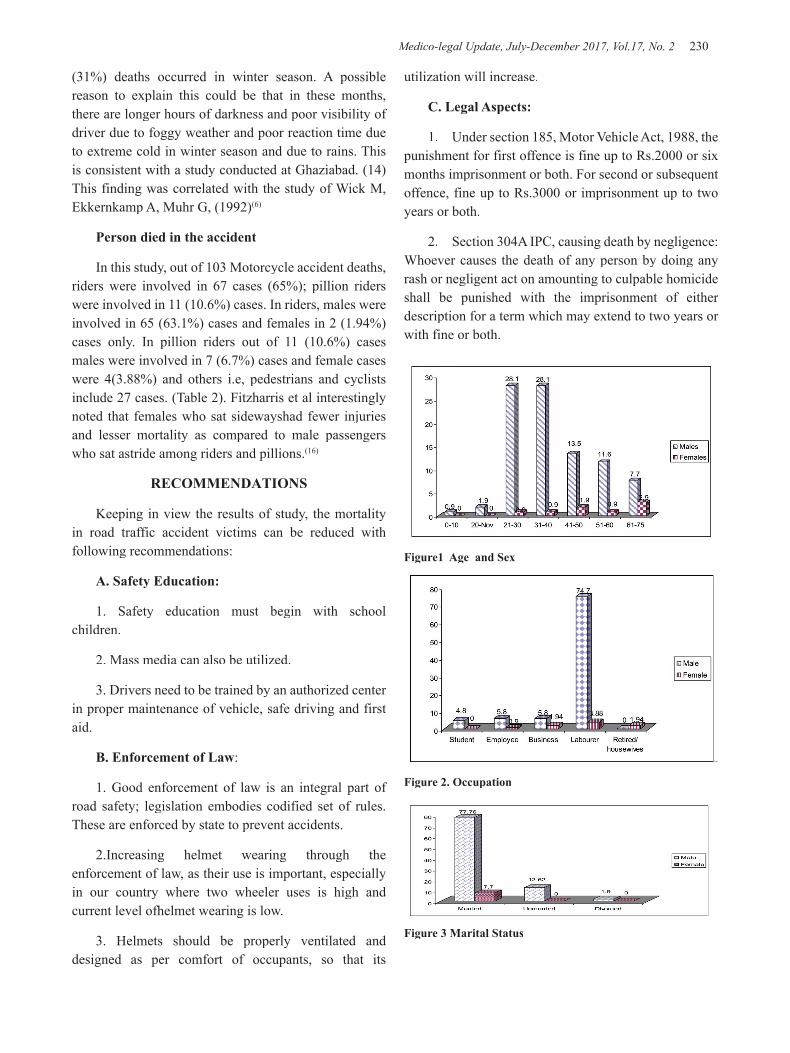
Medico-legal Update, July-December 2017, Vol.17, No. 2 230 231 -legal Update, July-December 2017, Vol.17, No. 2
(31%) deaths occurred in winter season. A possible reason to explain this could be that in these months, there are longer hours of darkness and poor visibility of driver due to foggy weather and poor reaction time due to extreme cold in winter season and due to rains. This is consistent with a study conducted at Ghaziabad. (14) This finding was correlated with the study of Wick M, Ekkernkamp A, Muhr G, (1992)(6)
Person died in the accident
In this study, out of 103 Motorcycle accident deaths, riders were involved in 67 cases (65%); pillion riders were involved in 11 (10.6%) cases. In riders, males were involved in 65 (63.1%) cases and females in 2 (1.94%) cases only. In pillion riders out of 11 (10.6%) cases males were involved in 7 (6.7%) cases and female cases were 4(3.88%) and others i.e, pedestrians and cyclists include 27 cases. (Table 2). Fitzharris et al interestingly noted that females who sat sidewayshad fewer injuries and lesser mortality as compared to male passengers who sat astride among riders and pillions.(16)
RECOMMENDATIONS
Keeping in view the results of study, the mortality in road traffic accident victims can be reduced with following recommendations:
A. Safety Education:
1. Safety education must begin with school children.
2. Mass media can also be utilized.
3. Drivers need to be trained by an authorized center in proper maintenance of vehicle, safe driving and first aid.
B. Enforcement of Law:
1. Good enforcement of law is an integral part of road safety; legislation embodies codified set of rules. These are enforced by state to prevent accidents.
2.Increasing helmet wearing through the enforcement of law, as their use is important, especially in our country where two wheeler uses is high and current level ofhelmet wearing is low.
3. Helmets should be properly ventilated and designed as per comfort of occupants, so that its
utilization will increase.
C. Legal Aspects:
1. Under section 185, Motor Vehicle Act, 1988, the punishment for first offence is fine up to Rs.2000 or six months imprisonment or both. For second or subsequent offence, fine up to Rs.3000 or imprisonment up to two years or both.
2. Section 304A IPC, causing death by negligence: Whoever causes the death of any person by doing any rash or negligent act on amounting to culpable homicide shall be punished with the imprisonment of either description for a term which may extend to two years or with fine or both.
Figure1 Age and Sex
Figure 2. Occupation
Figure 3 Marital Status
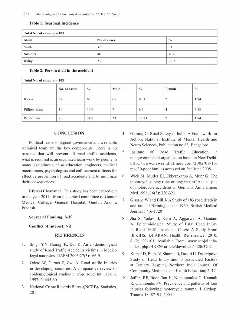
-legal Update, July-December 2017, Vol.17, No. 2 230 231 Medico-legal Update, July-December 2017, Vol.17, No. 2
Table 1: Seasonal Incidence
Total No. of cases n = 103
Month No. of cases %
Winter 32 31
Summer 48 46.6
Rainy 23 22.3
Table 2: Person died in the accident
Total No. of cases n = 103
No. of cases % Male % Female %
Riders 67 65 65 63.1 2 1.94
Pillion riders 11 10.6 7 6.7 4 3.88
Pedestrians 25 24.2 23 22.33 2 1.94
CONCLUSION
Political leadership,good governance and a reliable technical team are the key components. There is no panacea that will prevent all road traffic accidents, what is required is an organized team work by people in many disciplines such as education, engineers, medical practitioners, psychologists and enforcement officers for effective prevention of road accidents and to minimize their consequences.
Ethical Clearance: This study has been carried out in the year 2011, from the ethical committee of Guntur Medical College/ General Hospital, Guntur, Andhra Pradesh
Source of Funding: Self
Conflict of Interest: Nil
REFERENCES
1. Singh Y.N, Bairagi K, Das K. An epidemiological study of Road Traffic Accidents victims in Medico legal autopsies. JIAFM 2005:27(3):166-9.
2. Odero W, Garner P, Zwi A. Road traffic Injuries in developing countries: A comparative review of epidemiological studies : Trop. Med Int. Health. 1997; 2: 445-60
3. National Crime Records Bureau(NCRB)- Statistics, 2011
4. Gururaj G. Road Safety in India: A Framework for Action, National Institute of Mental Health and Neuro Sciences, Publication no 83, Bangalore
5. Institute of Road Traffic Education, a nongovernmental organization based in New Delhi. http://www.newsindiatimes.com/2002/09/13/med30-poor.html as accessed on 2nd June 2008.
6. Wick M, Muller EJ, Ekkernkamp A, Muhr G: The motorcyclist: easy rider or easy victim? An analysis of motorcycle accidents in Germany Am J Emerg Med 1998; 16(3): 320-321
7. Gissane W and Bill J- A Study of 183 road death in and around Birmingham in 1960, British Medical Journal 1716-1720
8. Jha S, Yadav B, Karn A, Aggarwal A, Gautam A. Epidemiological Study of Fatal Head Injury in Road Traffic Accident Cases: A Study From BPKIHS, DHARAN. Health Renaissance. 2010, 8 (2): 97-101. Available From: www.nepjol.info/ index. php /HREN/ article/download/4420/3702
9. Kumar D, Bains V, Sharma B, Dasari H. Descriptive Study of Head Injury and its associated Factors at Tertiary Hospital, Northern India Journal Of Community Medicine and Health Education; 2012
10. Jeffers RF, Boon Tan H, Nicolopoulos C, Kamath R, Giannoudis PV: Prevalence and patterns of foot injuries following motorcycle trauma. J. Orthop. Trauma 18: 87–91, 2004
Medico-legal Update, July-December 2017, Vol.17, No. 2 232
11. Lagarde E, Chastang JF, Gueguen A, Coeuret-Pellicer M, Chiron M, Lafont S. Emotional stress and traffic accidents: The impact of separation and divorce. Epidemiology. 2004;15:762–6
12. Subramanayam B.V (editor).Modi’s Medical Jurisprudence and Toxicology, 22 ed., edited by 2001; 393-396
13. Cawich SO, Harding HE, Evans NR, Crandon IW, Mitchell DIG, Martin AC et al. “ Strategies for secondary prevention of Motorcycle road traffic injuries” Time for policy reevaluation in Jamaica, The internet journal of emergency med, 2010; 12 (1)
14. Ranjana Singh, MBhatnager H K Singh, GP Singh, Yogesh Kumar. An epidemiological study of victims of road traffic accidents cases: A study from national capital region (Ghaziabad), India. Indian J. Prev. Soc. Med. 2011, Vol. 42 No.1:28-33
15. Global status report on road safety 2013-Supporting a decade of action. WHO 2013. http://www.who.int/violence_injury_prevention/road_safety_status/2013/en/index.html (accessed Feb 2013)
16. Fitzharris M, Dandona R, Kumar GA, Dandona L. Crash characteristics and patterns of injury among hospitalized motorised two-wheeled vehicle users in urban India.BMC Public Health . 2009; 9:11.
-legal Update, July-December 2017, Vol.17, No. 2 232
A Study of Bony and Cartilaginous Neck Structures in Deaths Due to Suicidal Hanging at New Delhi
Jegadheeshwararaj J1, Thejaswi H T2, Atul Murari3
1Assistant Professor, Department of Forensic Medicine and Toxicology, PSG Institute of Medical Sciences and 1Assistant Professor, Department of Forensic Medicine and Toxicology, PSG Institute of Medical Sciences and 1
Research, Coimbatore. Tamilnadu, 2Assistant Professor, Forensic Medicine, PGIMER & Dr.RML Hospital, 2Assistant Professor, Forensic Medicine, PGIMER & Dr.RML Hospital, 2
New Delhi, 3Director, Professor, Dean, Lady Hardinge Medical College, New Delhi 3Director, Professor, Dean, Lady Hardinge Medical College, New Delhi 3
ABSTRACT
The present prospective study was done at Lady Hardinge Medical College, New Delhi. In this study the incidences of the bony and cartilaginous injuries in 100 cases of deaths due to hanging were analysed with the gender, age, socio economic status, place of hanging, the type of suspension, the location of the knot, type of the knot, the type of hanging. 76(76%) of cases were males and the majority of the cases were from 21-30 age group, 94(94%) of the victims belonged to the low socioeconomic status, 97(97%) of the cases hanged themselves indoor, 2(2%) had committed suicide by hanging at their workplace, 81(81%) cases had complete type of hanging and 64(64%) had atypical type of hanging. In our study there were 42 (42%) cases had some injuries in their bony and cartilaginous neck structures. Hyoid bone fractures were found in 6(6%) cases, thyroid cartilage injury was seen in 39(39%) cases among which 3 cases had both hyoid and thyroid cartilage fracture in combination. All the above injuries occurred only in cases with complete type of hanging. The incidence of such injuries was more frequent in males who had typical and complete type of hanging.
Keywords: Hanging, injuries, hyoid bone, thyroid cartilage.
INTRODUCTION
Death due to hanging is a form of asphyxia caused with a ligature which encircles and constricts the neck, in such a manner that the weight of the whole or a part of the body acts as a constricting force. The external and internal findings in the neck in such cases are well documented. But for unknown reasons there are major variations in the bony and cartilaginous injuries reported in various studies. This study was undertaken to establish a strong correlation with the various factors that influence the pattern of injuries to the bony and cartilaginous neck structures in such cases.
MATERIALS AND METHOD
The study was done in 100 cases of suicidal hanging brought for medico-legal autopsies to the Department
Corresponding author:Thejaswi H TAssistant Professor, Forensic Medicine, PGIMER & Dr. RML Hospital, New Delhi
of Forensic medicine and toxicology, Lady Hardinge medical college, New Delhi. Only those cases with a well documented type of suspension, an intact ligature material were included in this study. A detailed history with photographs of the crime scene was mandated in the inclusion criteria. A standard autopsy procedure with special emphasis on the fine dissection of neck structures was done. Provided, cases only with evidential proven ante mortem injuries were alone considered in our study.
RESULTS
1. Among all the 100 cases of suicidal hanging, 76(76%) were males, 24(24%) were females.
2. The age groups ranged from 18-59 years, Majority of them (i.e. 60% of the entire study group) were from 21-30 years, 94(94%) of the victims were from low socio economic status, 97(97%) of the cases had hanged themselves indoors, 2(2%) of the cases had hanged themselves at their work place.
DOI Number: 10.5958/0974-1283.2017.00107.4

Medico-legal Update, July-December 2017, Vol.17, No. 2 234 235 -legal Update, July-December 2017, Vol.17, No. 2
3. Ropes were used as the ligature material in 52(52%) of the cases and others preferred saris, scarf, bed sheets, wires and plastic tapes.
4. 81(81%) cases had a complete type of hanging and 19(19%) of them were partially hanged.
5. In 36 (36%) cases the knot was at the nape of the neck thus constituting typical type of hanging. 64 (64%) cases had atypical type of hanging.
6. About 37 (37%) cases had contused sternocleidomastoid muscles. And the internal carotid artery showed transverse tears in 19 (19%) cases.
7. In 42 (42%) cases there were some injuries either in the hyoid bone or the thyroid cartilage only. Among which 3 cases had both hyoid and thyroid cartilage injury.
8. Unilateral fracture of greater horn of hyoid bone was present in 6 (6%) cases.
9. Fracture of the greater horn of thyroid cartilage was present in 39 (39%) cases, in which 33 individuals had unilateral fracture of the superior horn and 6 individuals had bilateral superior horn fractures.
10. The frequency of fractures in our study was higher among the males who were in the elderly age group and those who had a complete and typical type of hanging.
DISCUSSION
The present study was done to establish a strong correlation with the various factors that influence the pattern of injuries to the bony and cartilaginous neck structures in such cases.
With regard to the gender the male female ratio in our study was 3:1 with 76 (76%) and 24 females (24%). More than half of the males i.e. 38 out of 76 males and 4 out of 24 females had some injury in their bony and cartilaginous structures. Similar results were shown in other studies 1, 2.
The age distribution ranged from 18 years to 59 years. Almost half of the cases were from 21-30 age groups. Various other studies also have reported the same age distributions 2, 3, 4. The relations of the frequencies of fractures with respect to the gender and age distribution are as given (Table 1).
Table 1
In our study the fracture of the hyoid bone and the thyroid cartilage frequented in elderly aged victims.
There were 81 (81%) victims had a complete
hanging and 19(19%) victims were partially hanged and with regard to the location of the knot there were 36 (36%) cases who had their knot on their nape of the neck thus constituting typical hanging. In 64 (64%) cases had atypical type of hanging among which 42 cases had their knot on the right side of the neck, 21 cases had their knot on their left side of the neck and one of the victims had the knot under the chin. 94% of the victims were from low socio economic status, 97% of the cases had preferred hanging indoors and 2% of victims had committed suicide at their workplace.
More than half of the cases 52% preferred ropes as the ligature material. Other ligature materials were chunni (a long scarf), sari and bed sheets. Similar findings are being reported in other studies5, 6.
The external and internal soft tissue findings were similar to other case studies reported 5, 7, 8, 9.
In our study 42(42%) cases had some injury in their hyoid bone and thyroid cartilage. In our entire study there were 6 (6%) cases of hyoid bone fracture in their greater horn and 39 (39%) cases had their superior horn of thyroid cartilage fractured. No other bony injuries were detected on the neck.
With regard to the type of suspension all the injuries to the hyoid bone and the thyroid cartilage occurred only in those victims who had a complete type of hanging. And the incidence of such injuries was higher in those cases with typical type of hanging.
The frequency of the fracture with respect to the type of suspension and hanging are as given (Table 2).
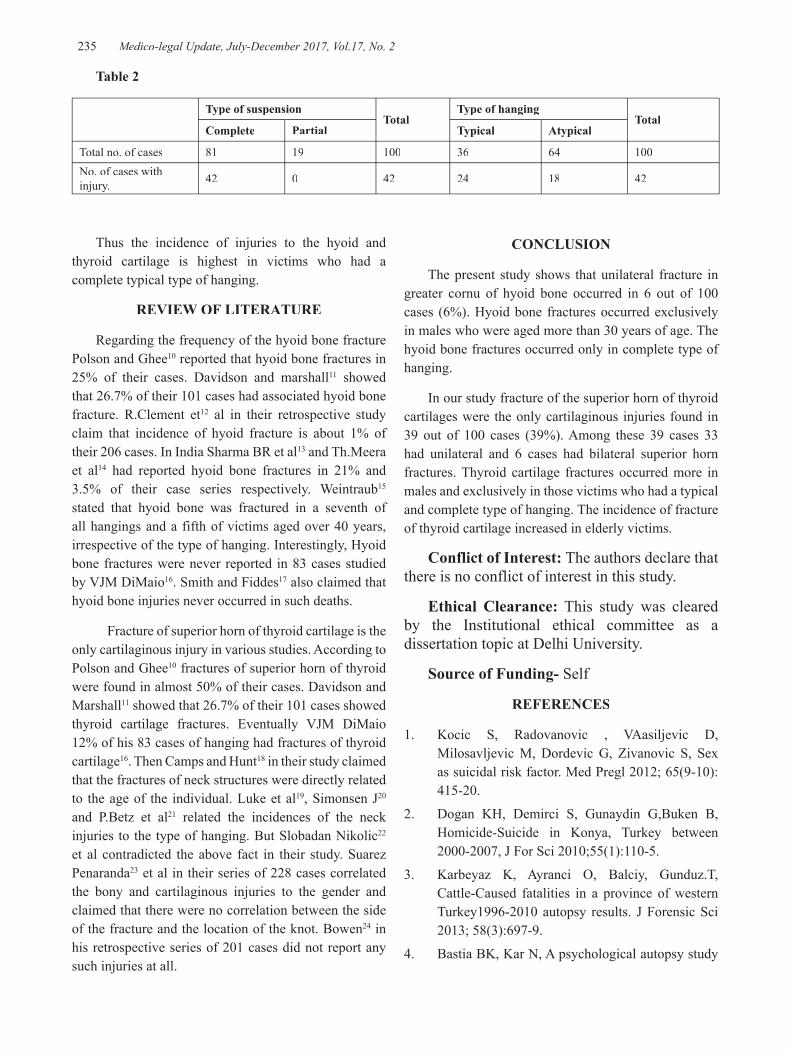
-legal Update, July-December 2017, Vol.17, No. 2 234 235 Medico-legal Update, July-December 2017, Vol.17, No. 2
Table 2
Type of suspensionTotal
Type of hangingTotal
Complete Partial Typical Atypical
Total no. of cases 81 19 100 36 64 100No. of cases with injury. 42 0 42 24 18 42
Thus the incidence of injuries to the hyoid and thyroid cartilage is highest in victims who had a complete typical type of hanging.
REVIEW OF LITERATURE
Regarding the frequency of the hyoid bone fracture Polson and Ghee10 reported that hyoid bone fractures in 25% of their cases. Davidson and marshall11 showed that 26.7% of their 101 cases had associated hyoid bone fracture. R.Clement et12 al in their retrospective study claim that incidence of hyoid fracture is about 1% of their 206 cases. In India Sharma BR et al13 and Th.Meera et al14 had reported hyoid bone fractures in 21% and 3.5% of their case series respectively. Weintraub15 stated that hyoid bone was fractured in a seventh of all hangings and a fifth of victims aged over 40 years, irrespective of the type of hanging. Interestingly, Hyoid bone fractures were never reported in 83 cases studied by VJM DiMaio16. Smith and Fiddes17 also claimed that hyoid bone injuries never occurred in such deaths.
Fracture of superior horn of thyroid cartilage is the only cartilaginous injury in various studies. According to Polson and Ghee10 fractures of superior horn of thyroid were found in almost 50% of their cases. Davidson and Marshall11 showed that 26.7% of their 101 cases showed thyroid cartilage fractures. Eventually VJM DiMaio 12% of his 83 cases of hanging had fractures of thyroid cartilage16. Then Camps and Hunt18 in their study claimed that the fractures of neck structures were directly related to the age of the individual. Luke et al19, Simonsen J20 and P.Betz et al21 related the incidences of the neck injuries to the type of hanging. But Slobadan Nikolic22 et al contradicted the above fact in their study. Suarez Penaranda23 et al in their series of 228 cases correlated the bony and cartilaginous injuries to the gender and claimed that there were no correlation between the side of the fracture and the location of the knot. Bowen24 in his retrospective series of 201 cases did not report any such injuries at all.
CONCLUSION
The present study shows that unilateral fracture in greater cornu of hyoid bone occurred in 6 out of 100 cases (6%). Hyoid bone fractures occurred exclusively in males who were aged more than 30 years of age. The hyoid bone fractures occurred only in complete type of hanging.
In our study fracture of the superior horn of thyroid cartilages were the only cartilaginous injuries found in 39 out of 100 cases (39%). Among these 39 cases 33 had unilateral and 6 cases had bilateral superior horn fractures. Thyroid cartilage fractures occurred more in males and exclusively in those victims who had a typical and complete type of hanging. The incidence of fracture of thyroid cartilage increased in elderly victims.
Conflict of Interest: The authors declare that there is no conflict of interest in this study.
Ethical Clearance: This study was cleared by the Institutional ethical committee as a dissertation topic at Delhi University.
Source of Funding- Self
REFERENCES
1. Kocic S, Radovanovic , VAasiljevic D, Milosavljevic M, Dordevic G, Zivanovic S, Sex as suicidal risk factor. Med Pregl 2012; 65(9-10):415-20.
2. Dogan KH, Demirci S, Gunaydin G,Buken B, Homicide-Suicide in Konya, Turkey between 2000-2007, J For Sci 2010;55(1):110-5.
3. Karbeyaz K, Ayranci O, Balciy, Gunduz.T, Cattle-Caused fatalities in a province of western Turkey1996-2010 autopsy results. J Forensic Sci 2013; 58(3):697-9.
4. Bastia BK, Kar N, A psychological autopsy study
Medico-legal Update, July-December 2017, Vol.17, No. 2 236
of suicidal hanging from Cuttack, India; Focus of stressful life situations. Arch Suicide Res 2009; 13(1):100-4.
5. James R, Silcocks P, Suicidal hanging in Cardiff a 15 year retrospective study. Forensic Sci Int 1992; 56(2):167-75.
6. Cooke CT, Cadden GA, Margolius KA, Death by hanging in Western Australia. Pathology 1995; 27(3):21-9.
7. Dean DE, Kohler IJ, Sterbenz GC, Gillespe PJ, Gonzanga NS, Bauer IJ, et.al. Observed characteristics of suicidal hanging: a 11 year retrospective review. J Forensic Sci 2012; 57(5):1226-30.
8. Uzun I, Buyuk Y, Gurpinar K, Suicidal hanging fatalities in Istanbul retrospective analysis of 761 autopsy cases, J Forensic leg Med 2007 Oct:14(7):406-9.
9. Simonsen J. Patho-anatomic findings in neck structures in asphyxiation due to hanging: a survey of 80 cases. Forensic Sci Int. 1998; 38:83-91.
10. Polson CJ, Gee DJ, Knight B. The essentials of forensic medicine.4th ed. Oxford: Pergamon Press, 1985. P.351-420.
11. Davidson A, Marshall T. Hanging in Northern Ireland- a survey. Med Sci Law 1986; 26(1):23-8.
12. Clement R, Guay JP, Sauvageau A. Fracture of neck structures in suicidal hangings: A retrospective study on contributing variables. Forensic Sci Int. 2011; 207:122-6.
13. Sharma BR, Harish D, Sharma A, Sharma S, Singh H. Injuries to neck structures in deaths due to constriction of neck, with a special reference to hanging. J Forensic Leg Med 2008; 15(5):298-305.
14. Meera Th, Bapin Kumar Singh M. Pattern of neck findings in suicidal hanging – A study in Manipur. JIAFM. 2011; 33(4):352-4.
15. Weintraub CM. Med-leg.J. (Camb.), 21, 209-16.
16. Di Maio VJ, Di Maio D. Forensic Pathology. 2nd
ed. Boca Raton: CRC Press; 2001. p. 245-69.
17. Smith SS, Fiddes FS. Forensic medicine – A Textbook for students and practitioners. 10th ed. J&A Churchill; 1955. p. 252-6.
18. F.E.Camps. Gradwohl’s Legal Medicine.3rd
ed.Bristol: John Wright & Sons ltd.:1976. p. 326-36.
19. Luke JL, Reay DT, Eisele JW,Bonnell HJ. Correlation of circumstances with pathological findings in asphyxia deaths by hanging: A prospective study of 61 cases form Seattle, WA.J Forensic Sci 1985; 30:1140-7.
20. Simonsen J. Patho-anatomic findings in neck structures in asphyxiation due to hanging: a survey of 80 cases. Forensic Sci Int. 1998; 38:83-91.
21. Betz P, Eisenmenger W. Frequency of throat-skeleton fractures in hanging. Am J Forensic Med Pathol 1996; 17:191-3.
22. Nikolic S, Micic J, Atanasijevic T, et al. Analysis of neck injuries in hanging. Am J Forensic Med Pathol. 2003; 24:179-182.
23. Suárez-Peñaranda JM, Alvarez T, Miguéns X, Rodríguez-Calvo MS, de Abajo BL, Cortesão M, Cordeiro C, Vieira DN, Muñoz JI. Characterisation of lesions in hanging deaths. J Forensic Sci. 2008 May; 53(3):720-3.
24. Bowen DAL. Hanging: A review. Forensic Sci Int.1982; 20:247-9.
-legal Update, July-December 2017, Vol.17, No. 2 236
A Review on Violent Behavior among Patients Admitted in Psychiatric Setting
Binil V1, Christopher Sudhakar2, Supriya Hegde3, Ravishankar N4
1Assistant Professor-Senior Scale, Manipal College of Nursing, Manipal, 1Assistant Professor-Senior Scale, Manipal College of Nursing, Manipal, 1 2Deputy Director-Quality & Compliance, 2Deputy Director-Quality & Compliance, 2
Manipal University, 3Professor, Department of Psychiatry, Father Muller Medical College, 4Assistant Professor, Department of Statistics, Manipal University4Assistant Professor, Department of Statistics, Manipal University4
ABSTRACT
Introduction: Prevalence of violent incidents-physical or verbal directed to self or others, is a serious problem in psychiatric setting1. Violent behavior in psychiatric unit affects the safety of patients and any other team member working in that particular setting. There is evidence that majority of the nurses working in psychiatric setting have been attacked by patients at least one time in their career.2
Aim: To estimate the prevalence of aggression and violence among clients admitted in psychiatric units and also to find out the factors associated with aggressive and violent behavior.
Materials & method: A comprehensive search was made on ProQuest, CINAHL, PubMed, Google scholar, and Cochrane databases for published original research articles between 1990 and 2016. We included all types of studies that reported about incidence or prevalence of violent behavior and factors associated with it in psychiatric ward.
Result: Twenty one studies were eligible for meta-analysis and the overall pooled prevalence of violence among patients admitted in psychiatric ward is 16% (0.13. 0.19, CI 95%, i-squared=97.9%, p=0.000). The review also examined the causes accompanying with violent behavior.
Conclusion: Violent behavior in psychiatric setting appear to be extremely variable between different settings. Assessing prevalence of violent behavior and to find out factors associated with violent behavior are a vital part of developing aggression management and violence prevention training program to safeguard clients and staffs from violent behavior.
Keywords: violent behavior, patients, psychiatric setting, prevalence
Correspondence author:Mr. Binil VAssistant Professor-Senior Scale, Manipal College of Nursing, Manipal, Udupi, Karnataka-576104 [email protected]
INTRODUCTION
Violent incidents are more common in psychiatric hospitals.4 It has been observed that the estimated rates of violent behavior in psychiatric setting varies with figures between 6% and 45%.5 When we are forcefully restraining a patient, that may aggravates his or her anger and he or she may involve in more violent activities.
Mentally disturbed people will try to hurt themselves, hospital staffs, other clients, family members or any other objects in the ward6. Aggression in the ward will hampers the efficacy of treatment provided for the client and also it is very difficult to prevent or control the violent incidents. When a patient gets hit from another patient, that may leads to annoyance, irritation, tension, distress, shock, insomnia and also it may leads to depression7. Majority of the nursing staffs working in psychiatric wards got hit from their patients at least one time in their career. Physical or verbal violence against nurses may affect their professional and personal life and also there is more chance to shift or leave their job.8

Medico-legal Update, July-December 2017, Vol.17, No. 2 238 239 -legal Update, July-December 2017, Vol.17, No. 2
There are many patient related factors that are considerably connected with inpatient violence. 8Most common factors include male gender, young age, admission against patients’ will, previous history of aggression, self-destructive behaviors in the past, and also the history of drug addiction. Environmental issues such as overcrowding on wards also lead to violence. Violent behaviors have been found to increase during meal times or when patients mix freely. Incidence of violence was more on wards where staff members were uncertain about their roles, or in a setting where there are more number of temporary staffs. A higher staff-patient ratio also found to increase the violent behavior. Violent behavior also associated with lack of experience among nurses, more number of new employees and lack of staff training in aggression management and violence prevention.9
1.1 Need for the Review:
A better understanding of the prevalence of violent behavior and factors associated with it in psychiatric ward will help to plan aggression management and violence prevention training programme for the health care providers working in psychiatric setting.10The concept of aggression management is not so well implemented in Indian Medical /Psychiatric setting. The aim of this review was to refer the available evidence to understand the prevalence of violent behavior among psychiatric patients and also to identify the associated factors of violent behavior. The review will facilitate to provide safe and secure care in psychiatric and emergency setting for both patients and health care providers.
1.2 Objective:
• To determine the prevalence of violent behavior and factors associated with violent behavior among patients admitted in psychiatric setting.
METHOD
2.1 Design
This review was conducted according to the guidelines described in the Preferred Reporting Items for Systematic Reviews and Meta-analyses (PRISMA). The PRISMA includes a checklist to ensure transparent reporting of systematic reviews and a four-phase diagram.
2.2 Search Strategy
The electronic data bases used for identifying the relevant studies were ProQuest, CINAHL, PubMed, Google scholar, and Cochrane. Enquiries were restricted to articles published between March 1990 and November 2016 and also only the articles which reported data on violent behavior of adult patients those who are admitted in psychiatric ward. Key words used for the search are Population (patients, psychiatric patients, mentally ill, mentally ill patients) Outcomes (aggression, violence, violent behavior, aggressive behavior, assault), Centre (hospital, hospitalization, acute psychiatric setting, psychiatric rehabilitative setting, psychiatric ward, psychiatric unit). In order to improve the quality of the study, we have included peer reviewed articles from different data bases.
2.3 Inclusion and exclusion criteria
Studies were included in this review if they fulfilled the following criteria-Psychiatric patients who are adults, and those who had committed minimum one act of violent behavior. And also reviews done about inpatients only were considered. In the present review violent behavior means any injury to a client or any effort from a client to hurt another person physically or verbally, including other patients, family members or any of the hospital staff. The study excluded from the review were published articles in other foreign languages and methodology other than quantitative approaches.
2.4 Selection of study and extraction of data
A total of 612 articles were identified through data base search and from that 587 articles were selected after removing the duplicate publications. From that 557 articles excluded after screening the title and 30 full text articles assessed for eligibility. From that 09 articles were removed with reason and finally 21 articles were included for meta-analysis.
A form was prepared for data extraction based on the review objective. Two authors individually extracted data from each included study and any dissimilarities were resolved following a discussion.
2.5 Data Synthesis:
Data was assessed for quality of assessment and outcomes
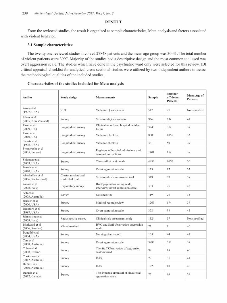
-legal Update, July-December 2017, Vol.17, No. 2 238 239 Medico-legal Update, July-December 2017, Vol.17, No. 2
RESULT
From the reviewed studies, the result is organized as sample characteristics, Meta-analysis and factors associated with violent behavior.
3.1 Sample characteristics:
The twenty one reviewed studies involved 27848 patients and the mean age group was 30-41. The total number of violent patients were 3997. Majority of the studies had a descriptive design and the most common tool used was overt aggression scale. The studies which have done in the psychiatric ward only were selected for this review. JBI critical appraisal checklist for analytical cross sectional studies were utilized by two independent authors to assess the methodological qualities of the included studies.
Characteristics of the studies included for Meta-analysis
Author Study design Measurements Sample Number of Violent Patients
Mean Age of Patients
Asnis et al(1997, USA) RCT Violence Questionnaire 517 21 Not specified
Silver et al(2005, New Zealand) Survey Structured Questionnaire 936 234 41
Fazel et al(2009, UK) Longitudinal survey Clinical record and hospital incident
forms 3743 314 39
Fazel et al (2010, UK) Longitudinal survey Violence checklist 8003 1056 33
Swartz et al(1998, USA) Longitudinal survey Violence checklist 331 59 39
Steanwachs et al(2005, France) Longitudinal survey Registers of hospital admissions and
criminal convictions 1401 154 38
Shipman et al(2003, USA) Survey The conflict tactic scale 4480 1070 30
Bartels et al(2010, USA) Survey Overt aggression scale 133 17 32
Aberhalden et al(2006, Switzerland)
Cluster randomized controlled trial Structured risk assessment tool 519 37 38
Amore et al(2008, Italy) Exploratory survey Brief psychiatric rating scale,
interview, Overt aggression scale 303 75 42
Ash et al(2003, Australia) survey Not specified 119 26 35
Barlow et al(2000, USA) Survey Medical record review 1269 174 37
Beauford et al(1997, USA) Survey Overt aggression scale 328 38 42
Biancosino et al (2009, Italy) Retrospective survey Clinical risk assessment scale 1324 37 Not specified
Bjorkdahl et al(2006, Sweden) Mixed method BVC and Staff observation aggression
scale 73 11 40
Boggild et al(2004, USA) Survey Nursing chart record 105 44 41
Carr et al(2008, Australia) Survey Overt aggression scale 3887 551 37
Cohen et al(2008, Ireland Survey The Staff Observation of aggression
scale-revised 99 18 40
Cookson et al(2012, Australia) Survey OAS 79 35 41
Daffern et al(2010, Australia) Survey OAS 122 10 40
Dumais et al(2012, Canada) Survey The dynamic appraisal of situational
aggression scale 77 16 36

Medico-legal Update, July-December 2017, Vol.17, No. 2 240 241 -legal Update, July-December 2017, Vol.17, No. 2
3.2 Meta-analysis of the Reviewed studies:
A meta-analysis was done on 21 studies and the total number of patients from these studies were 27848. Figure 2 summarizes the forest plot showing the prevalence of violent behavior. A total of 612 studies were identified in the literature search. However, only twenty one studies were eligible for meta-analysis. Meta-analysis showed that the overall pooled prevalence of violence among patients admitted in psychiatric ward is 16% (13 %-19%, CI 95%, i-squared=97.9%, p=0.000).
Forest Plot Showing prevalence of violent behavior
3.3 Factors Associated with Violent Behavior:
A separate review was conducted to find out the factors associated with violent behavior and the below table summarizes that. There is variation among the factors according to the settings.
Author Country Year of Publication Factors Associated with Violence
Raja et al Italy 2005Young age, being single, no children, higher doses of psychoactive drugs, frequent use of neuroleptics, mania, personality disorder, substance and alcohol related disorder
Ridenour et al USA 2015 Personality disorders, middle age group
Lozzino et al Australia 2015 Involuntary admissions, male gender, patients with substance use disorders and schizophrenia
Iversen et al Norway 2016 Number of times hospitalized, no formal education, summer season
Bowers et al UK 2009 High patient turn over, alcohol use by patients, ward doors being locked, higher staffing numbers
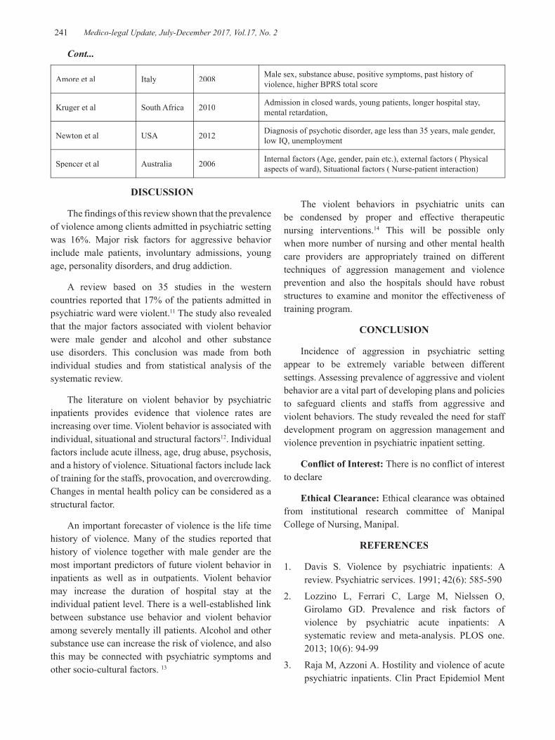
-legal Update, July-December 2017, Vol.17, No. 2 240 241 Medico-legal Update, July-December 2017, Vol.17, No. 2
Amore et al Italy 2008 Male sex, substance abuse, positive symptoms, past history of violence, higher BPRS total score
Kruger et al South Africa 2010 Admission in closed wards, young patients, longer hospital stay, mental retardation,
Newton et al USA 2012 Diagnosis of psychotic disorder, age less than 35 years, male gender, low IQ, unemployment
Spencer et al Australia 2006 Internal factors (Age, gender, pain etc.), external factors ( Physical aspects of ward), Situational factors ( Nurse-patient interaction)
DISCUSSION
The findings of this review shown that the prevalence of violence among clients admitted in psychiatric setting was 16%. Major risk factors for aggressive behavior include male patients, involuntary admissions, young age, personality disorders, and drug addiction.
A review based on 35 studies in the western countries reported that 17% of the patients admitted in psychiatric ward were violent.11 The study also revealed that the major factors associated with violent behavior were male gender and alcohol and other substance use disorders. This conclusion was made from both individual studies and from statistical analysis of the systematic review.
The literature on violent behavior by psychiatric inpatients provides evidence that violence rates are increasing over time. Violent behavior is associated with individual, situational and structural factors12. Individual factors include acute illness, age, drug abuse, psychosis, and a history of violence. Situational factors include lack of training for the staffs, provocation, and overcrowding. Changes in mental health policy can be considered as a structural factor.
An important forecaster of violence is the life time history of violence. Many of the studies reported that history of violence together with male gender are the most important predictors of future violent behavior in inpatients as well as in outpatients. Violent behavior may increase the duration of hospital stay at the individual patient level. There is a well-established link between substance use behavior and violent behavior among severely mentally ill patients. Alcohol and other substance use can increase the risk of violence, and also this may be connected with psychiatric symptoms and other socio-cultural factors. 13
The violent behaviors in psychiatric units can be condensed by proper and effective therapeutic nursing interventions.14 This will be possible only when more number of nursing and other mental health care providers are appropriately trained on different techniques of aggression management and violence prevention and also the hospitals should have robust structures to examine and monitor the effectiveness of training program.
CONCLUSION
Incidence of aggression in psychiatric setting appear to be extremely variable between different settings. Assessing prevalence of aggressive and violent behavior are a vital part of developing plans and policies to safeguard clients and staffs from aggressive and violent behaviors. The study revealed the need for staff development program on aggression management and violence prevention in psychiatric inpatient setting.
Conflict of Interest: There is no conflict of interest to declare
Ethical Clearance: Ethical clearance was obtained from institutional research committee of Manipal College of Nursing, Manipal.
REFERENCES
1. Davis S. Violence by psychiatric inpatients: A review. Psychiatric services. 1991; 42(6): 585-590
2. Lozzino L, Ferrari C, Large M, Nielssen O, Girolamo GD. Prevalence and risk factors of violence by psychiatric acute inpatients: A systematic review and meta-analysis. PLOS one. 2013; 10(6): 94-99
3. Raja M, Azzoni A. Hostility and violence of acute psychiatric inpatients. Clin Pract Epidemiol Ment
Cont...
Medico-legal Update, July-December 2017, Vol.17, No. 2 242
Health. 2005; 1:11
4. Stevenson KN, Jack SM, Mara LO, Legris J. Registered nurses’ experiences of patient violence on acute care psychiatric inpatient units. BMC Nursing. 2015; 14:35
5. Lucioni KA, Pueyo AA, Clotet EP, Sones JG. Predicting violence in psychiatric inpatients: a prospective study with the HCR- 20 violence risk assessment scheme. The journal of forensic psychiatry & psychology. 2011; 22(2). 203-222
6. Stuart H. Violence and mental illness: an overview. World Psychiatry. 2003; 2(2): 121-124
7. Anderson A, West SG. Violence against mental health professionals: When the treater becomes the victim. Innov Clin Neurosci. 2011; 8(3): 34-39
8. Bowers L, Nijmanm H, Allan T, Simpson A, Warren J. Prevention and Management of Aggression Training and Violent Incidents on U.K. Acute Psychiatric Wards. Psychiatric Services. 2006; 57(7): 1022-1026
9. Rueve ME, Welton RS. Violence and Mental Illness. Psychiatry (Edgmont).2008; 5(5):34-48
10. Walsh E, Moran P, Scott C, Mckenzie K, Burns T. Prevalence of violent victimisation in severe mental illness. The British Journal of Psychiatry. 2003; 183(3), 233-238
11. Oram S, Trevillion K, Feder G, Howard L. Prevalence of experiences of domestic violence among psychiatric patients. The British Journal of Psychiatry. 2013; 202(2), 94-99
12. Owen C, Tarantello C, Jones M, Tennant C. Violence and aggression in psychiatric units. Psychiatric services. 1998; 1452-1457
13. Wolf A, Whiting D, fazel S. Violence prevention in psychiatry: an umbrella review of interventions in general and forensic psychiatry. Journal of forensic psychiatry and psychology.2016; 10: 1-15
14. Dack C, Ross J, Papadopoulos C, Stewart D, Bowers L. A review and meta-analysis of the patient factors associated with psychiatric in-patient aggression. Acta Psychiatrica Scandinevica. 2013; 127(4): 255-268
15. Davison SE.The management of violence in general psychiatry. Advances in psychiatric treatment. 2005; 11: 362-370
16. Selenius H, Strand S. Experiences of self-injury and aggression among women admitted to forensic psychiatric care. Nordic journal of psychiatry. 2016; 10: 1-8
17. Bernstein KS, Saladino JP. Clinical assessment and management of psychiatric patient’s violent and aggressive behaviors in general hospital. Medsurg Nurs. 2007; 16(5): 301-309
18. Lantta T, Anttila M, Kontio R, Adams CE, Valimaki M. Violent events, ward climate and ideas for violence prevention among nurses in psychiatric wards: a focus group study. International journal of mental health systems. 2016; 10: 27
19. Nawal R, Magda F, Wafaa EL, Haydy H, Khaled EM. Correlation between aggressive behavior and sociodemographic characteristics in patients with schizophrenia at a psychiatry outpatient clinic. Midddle East Current Psychiatry. 2016; 1: 30-35
20. Rocca P, Villari V, Bogetto F. Managing the aggressive and violent patient in the psychiatry. Prog Neuropsychopharmacol Biol Psychiatry. 2006; 30(4): 586-598
21. Renwick L, Stewart D, Richardson M, Lavelle M, James K, Hardy C, Bowers L. Aggression on inpatient units: Clinical characteristics and consequences. International journal of mental health nursing. 2016; 25(4): 308-318
22. Cowman S, Bjohrkdal A, Clarke E, Gethin G, Maguire J. A descriptive survey study of violence management and priorities among psychiatric staff in mental health services, across seventeen european countries. BMC Health Services research. 2016; 17:59
23. Spencer S, Stone T, McMilan M. Violence and aggression in mental health inpatient units: An evaluation of aggression minimisation programs. The University of Newcastle. 2010; 3(1): 42-48
24. Knezevic V, Mitrovic D, Drezqic VS, Knezevic J, Ivezic A, Siladji MD, Golubovic B. Prevalence and correlates of aggression and hostility in hospitalized schizophrenic patients. Journal of interpersonal violence. 2016; 151-163
Effectiveness of Indian Classical Music on DevelopmentalResponses of Preterm Infants - A Randomized
Controlled Trial Protocol
Sonia R B D'Souza1, Leslie E.S Lewis2, Vijay Kumar3, Ramesh Bhat Y4,Jayashree Purkayastha5, Asha Kamath 6
1Associate Professor, Dept. of Obstetrics and Gynaecological Nursing, Manipal College of Nursing, Manipal,University, Manipal, 2Professor and Incharge, NICU, Dept of Pediatrics, 3Professor and HoD, Dept of Pediatric
Surgery, 4Professor, NICU, Dept of Pediatrics, Kasturba Hospital, Manipal, 5Professor, NICU, Dept of Pediatrics,Kasturba Hospital, Manipal, 6Associate Professor – Statistics, Dept. of Community Medicine, Kasturba Medical,
College, Manipal University Manipal
ABSTRACT
Background: The highly sophisticated Neonatal Intensive Care Unit (NICU) exposes preterm infantsto constant unexpected stimuli especially noise, impacting their growth and development. Modifyingthe environment of the NICU by provision of patterned stimuli like music is essential. There is apaucity of data from large and well-conducted trials on Indian Classical music, which limit theevidence for use of music as therapy for preterm infants.
Methodology: A randomized controlled trial with repeated measures design will be conducted. Asample size of 132 preterm infants who are admitted to the NICU, will be assessed for eligibility to beincluded in the study. The parent(s) of the preterm infants will be approached for informed proxyconsent. A study assistant will manage the allocation of participants and the provision of theintervention independently. The investigators who perform the outcome assessment will be blindedto the allocation of participants to the intervention and control arm of the study.
Outcome measures will be assessed at baseline (day 3 of life). Following the administration of theintervention in the experimental group, the outcome measures will be assessed after completion ofone week of intervention (day 10 of life) and after completion of two weeks of intervention (day 17 oflife).
Keywords: Preterm infants, Indian Classical music, weight gain, gain in head circumference (HC)
Corresponding author:Vijay KumarProfessor and H.O.D.Dept of Pediatric Surgery, Kasturba Hospital, Manipal,Manipal University, Manipal, Udupidistrict-576104,Karnataka, IndiaPhone Number: 0820-2922776Email address: [email protected]
INTRODUCTION
As per the global action report on preterm birth,
the two continents i.e., Africa and South Asia
contribute to the largest number of preterm births that
exceed 60%. India, a country in South Asia, features
among the ten countries listed with the highest
numbers showing an increasing trend of 10-15%
preterm births, estimated to be around 100,000 to
250,000 preterm births.[1] Preterm births are rapidly
rising in India, adding to the disease burden. The
liabilities brought forth by preterm births are manifold.
Prematurity not only contributes to about 35% of
mortality amongst neonates but also is a major
contributor to morbidities in later life. [2] Preterm birth
compels infants born before term, to spend prolonged
periods in the Neonatal Intensive Care Unit (NICU).
Preterm infants thus lose opportunity to continue their
normal growth and development which otherwise
would have occurred, had they continued in the
protective intra-uterine environment of their mothers.
Appropriate stimulation to preterm infants
admitted to the NICU is necessary for their growth
and development. [3] It is necessary to modify the
environment, so that the energy spent by the preterm

244 Medico-legal Update, July-December 2017, Vol.17, No. 2
infants could be utilized by them primarily for growthand development. [4] Developmental care recognizesthe vulnerabilities of preterm infants and is focusedto minimize the potential short and long-termcomplications. [5]
Music is consideredas a patterned stimulus unlikenoise that exists in the environment.[6]Research relatedto music for preterms is required to demonstrate thepromising benefits of music and for framing guidelinesfor its clinical use.[7] Well-designed high quality trialsare required to investigate effects of music on thedevelopmental outcomes in preterminfants.[8]ACochrane systematic review has alsoacknowledged the possibility of including music asan alternative therapeutic modality.[9]
It is supporting to know that over the past decades,studies have determined the effects of Western classicalmusic, mothers’ singing lullabies, and the like. Anupdated meta-analysisalso propagates the effects ofmusic in the form of Western classical music, lullabiesfor preterm infants in the positive direction. [10]
However, no published studies have yet focused onthe therapeutic effects of Indian Classical music ondevelopmental outcomes of infants born prematurely.It is believed that Indian classical music is not only amusical tradition but also an integration of ancienthealing practice. [11] ‘Ragas’ (melodic entities)pertaining to Indian classical music were not merelyentertainment commodities but had healingproperties. [12]
Use of Indian classical music as a therapy is still anemerging field. There is a paucity of data from largeand well-conducted trials on Indian Classical music,which limit the evidence for use of music as therapy.Conclusions that will emerge from this study areexpected to contribute to the evidence base of use ofIndian Classical music for preterm infants.
OBJECTIVES OF THE STUDY
The main objectives are to
• evaluate the effectiveness of Indian classical musicon developmental responses of preterm infantsmeasured in terms of
• weight gain
• gain in head circumference (HC)
HYPOTHESES
There will be a significant difference over the period(baseline to post-tests) and across the groups(intervention and control) with regard to
• weight gain
• gain in HC
BRIEF METHODOLOGY
Background information of the study area:
The present study will be conducted in the NICUof Kasturba Hospital, a tertiary referral hospital,Manipal, Karnataka State, South India. The NICUcaters to an average of 940-1045 new admissions peryear. Out of these, around 250 to 300 are preterminfants.
The rationale for selecting the tertiary level NICUof Kasturba Hospital, Manipal was:
1. NNPD report,[13]from 18 centers of India for aperiod of two years, points out that the incidenceof preterm birth is 14.5 % among 151,436intramural births and 31.5% among 11,026extramural births. The setting of the present studyfeatured as one of the 18 centres of the network.
2. The tertiary level NICU receives services from awell-established department of Speech, Hearing,and Audiology. The Sound Pressure Levels [LAF
(SPL)] of the music sessions will be checked by anaudiologist, using a Sound Level Meter (SLM) -Hand held Analyzer Type 2250, Brüel and Kjær,Denmark, calibrated using Calibrator (Type 4231-Class1), according to the guidelines suggested byAmerican Academy of Pediatrics (AAP).[14, 15]
3. The LAF (SPL) delivered at the preterm infants’ ears,will be checked using the SLM, connected to the‘Artificial Ear’ (Type 4152, Brüel and Kjær,Denmark). The ‘Artificial Ear’ approximates theresponse of the human ear on an A weighted scale.
Study design: Randomized Controlled Trial
A randomized trial with repeated measuresdesignwill be conducted.
The sample size for the intervention and the controlgroup is calculated by considering a power of 80%
243-247.pmd 8/22/2017, 4:54 PM244

Medico-legal Update, July-December 2017, Vol.17, No. 2 245
{Z 1-β = 0.845} and Q=0.05 {Z 1- α/2 = 1.96}, i.e., alphaerror of 5% two-tailed, with an effect size {δ =0.83}[16]
and the standard deviation for weight gain {S=1.7}[17]assuming no variance between groups. Consideringthis, the expected sample size is 132. However, theactual interim analysis may be takeninto account fora different sample size, depending on the recruitmentprogress and funding.
Randomization and allocation
Once the preterm infants are admitted to the NICU,they will be assessed for eligibility to be included inthe study. Once the preterm infants are recruited tothe study, the parent(s) of the preterm infants will beapproached for informed proxy consent andparticipant information sheet will be provided. Astudy assistant, not involved in the outcomeassessment will allocate the preterm infant(s) to eitherthe experimental or control arm after the opening asealed opaque envelope. The study assistant willmanage the allocation and the provision of theintervention independently. The investigators whoperform the outcome assessment will be blinded tothe allocation of participants.
Inclusion criteria
– preterm infants admitted to tertiary level NICU
– Gestational age of 28-30 weeks of postmenstrualage (PMA)
– Appropriate for gestational age (AGA)
– have passed the hearing screening test to confirmhearing ability by Oto-Acoustic Emission (OAE)test
– Parental consent
Exclusion criteria
– Preterm infants who are large for gestationalage(LGA) or small for gestational age (SGA)
– weighing >1500 g at birth
– death in the first 48 hours of life
– having major congenital malformations
– having major surgical complications
– preterm infants who have congenital sepsis(confirmed by blood culture on day 1)
– preterm infants exposed to chorioamnionitis(confirmed in the antenatal record)
Intervention
Instrumental Indian classical music of Hindustanigenre will be selected for the study. The sound pressurelevels (SPL) on a A weighted scale [LAF (SPL)] of themusic sessions will be checked by an audiologist, usinga Sound Level Meter (SLM) - Hand held Analyzer Type2250, Brüel and Kjær, Denmark, calibrated usingCalibrator (Type 4231-Class1), according to theguidelines.[14, 15]
The recorded instrumental Indian classical musicwill be provided to the preterm infants in theexperimental group through headphones. The LAF
(SPL) delivered by the headphones at the preterminfants’ ears, will be checked using the SLM, connectedto the ‘Artificial Ear’ (Type 4152, Brüel and Kjær,Denmark) which in turn will be connected to theamplifier and music player.
Follow –up
Outcome measures will be assessed at baseline (day3 of life). Following the administration of theintervention in the experimental group, the outcomemeasures will be assessed after completion of one weekof intervention (day 10 of life) and after completion oftwo weeks of intervention (day 17 of life).
Outcome measures
• weight
• HC
Data collection instruments
For measuring weight of preterm infants:
The weight of the preterm infants will be checkedusing electronic digital baby weighing scales - EssaeBaby scale DS-252 manufactured by Essae -TeraokaLimited (an Indo Japanese joint venture),4th Block,Koramangala, Bangalore. The Essae Baby scale DS-252is duly certified and stamped by Weight and MeasuresDepartment and conforms to the statutory regulatoryrequirements as per the provisions of ‘The Standardsof Weights and Measures Act’, 1976 [18]and ‘TheStandards of Weights and Measures (Approval ofModels) Rules’, 1987. [19]The weight will be checked asper the standard weight measuring protocol of thetertiary level NICU that is as per the guidelines [20].
243-247.pmd 8/22/2017, 4:54 PM245
246 Medico-legal Update, July-December 2017, Vol.17, No. 2
Calibration of the Electronic digital weighing scale- Essae Baby scale DS-252
The electronic digital weighing scales will becalibrated for accuracy by using dry weights as wellas the tare function. A calibrated electronic weighingscale is said to be precise if the recorded values areclose to that of the true value and accurate if, on theaverage, it measures the true size. The Essae Baby scaleDS-252 has an accuracy of +5 grams.
For measuring HC of preterm infants
Flexible, non-stretchable, narrow plastic inch tapeswill be used to measure the HC/ occipito-frontalcircumference (OFC) of the preterm infants in thestudy.
Calibration and reliability assessment of the inchtapes
The inch tapes will be calibrated and certified by aphysicist for accuracy in the Physics laboratory,Physics Department, Manipal Institute of Technology(MIT), Manipal University, Manipal. A physicist willbe measured the accuracy of the inch tapes.
To ensure the reliability of the inch tapes, inter-raterreliability will be measured using Intra ClassCorrelation (ICC). Interrater reliability is the bestmeasure for interval data, interpreted as the proportionof the total variance in the ratings. For measurementof the HC, a standard protocol as per theguidelines[20]will be followed.
For checking the inter-rater reliability, eachobserver will independently measure and record acomplete set of HC measurements and their readingswill be compared. If the difference between any twomeasurements exceed the maximum allowabledifference, i.e., 5 mm [20] then both observers will retakemeasurements. To estimate the short-term test-retestreliability, the observers will repeat the measurementson two consecutive days.
Ethical considerations
The study protocol is approved by the Institutionalreview committee (IRC), Manipal College of Nursing,Manipal University, Manipal. Ethical committeeclearance for the study is obtained from theInstitutional Ethics Committee of Manipal University,Manipal (UEC/14/2011). The trial will be registeredin the Clinical Trials Registry of India(www.ctri.nic.in).Informed written proxy consent will
be obtained from the parent(s) of the preterm infants.
Validation of the protocol and the music intervention
The protocol was submitted for review to a panelof multidisciplinary experts i.e., from the field ofPediatrics/Neonatology, Clinical Psychology andPerinatal/Neonatal Nursing.
Hindustani Calssical music soloists, whospecialized in Indian classical music, were consultedfor selecting and validating the music sessions usedin the study.
Importance of the proposed study
Research
The proposed study may provide evidence for useof Indian classical music for preterm infants. Literatureon music studies limit implications for practice ofmusic as routine care in the NICU. The data obtainedmay help in planning further studies to address theshortcomings pertaining to use of Indian Classicalmusic for preterm infants.
Education
The findings of the study may equip the health careprofessionals working in the NICU to have an evidencebase to utilize the provision of recorded IndianClassical music.
Quality and effectiveness
Indian Classical music is non-invasive and costeffective modality of DSC that focuses on modulatingthe environment (NICU) of the preterm infants.
Policymaking
The data obtained from the study will providesufficient evidence and support for hospitals acrossIndia with NICUs to employ auditory stimulation likerecorded Indian Classical music for the benefit of thepreterm infants.
CONCLUSION
This randomized controlled trial aims to establishthe effectiveness of Indian classical music ondevelopmental responses of preterm infants. Theresults of the trial will provide evidence on theeffectiveness of Indian Classical music. Furthermore,findings gained may help to further specify musictherapy treatment guidelines for this population.
243-247.pmd 8/22/2017, 4:54 PM246

Medico-legal Update, July-December 2017, Vol.17, No. 2 247
Results of the study will be published after thecompletion of the study.
Source of Funding: Indian Council of MedicalResearch (ICMR), India
Conflict of Interest: Nil
REFERENCES
1. Editors Howson CP, Kinney MV, Lawn JE. Marchof Dimes, PMNCH, Save the Children, WHO.Born Too Soon: The Global Action Report onPreterm Birth. Geneva: World HealthOrganization; 2012.
2. Liu L, Johnson HL, Cousens S, Perin J, Scott S,Lawn JE, et al. Global, regional, and nationalcauses of child mortality: an updated systematicanalysis for 2010 with time trends since 2000. TheLancet; 2012; 379(9832):2151-2161.
3. Wood NS, Marlow N, Costeloe K, Gibson AT,Wilkinson AR. Neurologic and developmentaldisability after extremely preterm birth. N Engl JMed. 2000;343(6):378-384.
4. Bremmer P, Byers JF, Kiehl E. Noise and thePremature Infant: Physiological Effects andPractice Implications. J ObstetGynecol NeonatalNurs. 2003;32(4):447-454.
5. Coughlin M, Gibbins S, Hoath S. Core measuresfor developmentally supportive care in neonatalintensive care units: theory, precedence andpractice. . Journal of Advanced Nursing. 2009;65(10):2239-2248. doi:10.1111/j.1365-2648.2009.05052.x.
6. Burns K, Cunnigham N, White-Traut R, SilvestriJ, Nelson MN. Infant stimulation: Modificationof an intervention based on physiologic andbehavioral cues. J ObstetGynecol Neonatal Nurs.1994; 23,581-589.
7. Neal DO, Lindeke LL. Music as a NursingIntervention for Preterm Infants in the NICU.Neonatal Netw. 2008; 27(5):319-327.
8. Haslbeck FB. Music therapy for prematureinfants and their parents: an integrative review.Nordic J Music Ther. 2012; 21(3):203-226.
9. Hartling L, Shaik MS, Tjosvold L, Leicht R, LiangY, Kumar M. Music for medical indications in theneonatal period: A systematic review ofrandomized controlled trials. Arch Dis ChildFetal Neonatal Ed. 2009; 94:F349–F354.
10. Standley J. Music Therapy Research in the NICU:An Updated Meta-Analysis. Neonatal Netw.2012;31(5):311-316.
11. Sundar S. Traditional healing systems andmodern music therapy in India. Music Therapytoday [Internet]. 2007; 8 (3). Available from: http://musictherapyworld.net
12. Sairam TV. Melody and rhythm -‘Indianness’ inIndian music and music therapy. Music Therapytoday [Internet]. 2006; 7(4):876-891. Availablefrom: http://musictherapyworld.net
13. National Neonatal and Perinatal Database(NNPD) Nodal Centre at Department ofPediatrics. NNPD report (2002-2003). New Delhi,India: WHO Collaborating Centre New-bornTraining & Research, All India Institute ofMedical Sciences; 2005.
14. American Academy of Pediatrics- Committee onEnvironmental Hazards. Noise Pollution:Neonatal Aspects. Pediatr.1974; 54(4):476-479.
15. American Academy of Pediatrics- Committee onEnvironmental Health. Noise: A Hazard for theFetus and Newborn. Pediatr.1997; 100(4):724-727.
16. Standley JM. A Meta-analysis of the Efficacy ofMusic Therapy for Premature Infants. JPediatrNurs. 2002; 17(2):107-113.
17. Cevasco AM. The Effects of Mothers’ Singing onFull-term and Preterm Infants and MaternalEmotional Responses. J Music Ther. 2008;45(3):273-306.
18. Government of India. The Standards of weightsand measures Act, 1976 enacted by Parliamentin the Twenty Seventh Year of the Republic ofIndia. [pamphlet].
19. Ministry of Food and Civil Supplies, Governmentof India. The Standards of Weights and Measures(Approval of Models) Rules, 1987 withAmendments in 2000 [pamphlet].India.
20. Villar J, Giuliani F, Bhutta Z, Bertino E, OhumaEO, Ismail LC, Barros FC, Altman DG, VictoraC, Noble JA, Gravett MG, Purwar M, Pang R,Lamber A, Papgeorghiou AT, Ochieng R, JafferYA, Kennedy SH. Postnatal growth standards forpreterm infants: the Preterm Postnatal Follow-up Study of the INTERGROWTH-21st Project.Lancet Glob Health 2015; 3: e681–91
243-247.pmd 8/22/2017, 4:54 PM247

249 -legal Update, July-December 2017, Vol.17, No. 2
Effectiveness of Early Initiation of Breastfeeding on Sleeping Pattern of Newborns – An Evaluative Study
Geena Louis D’Souza1, Pratibha Kamath2, Sonia R B D’Souza3
1Lecturer, Dept. of Obstetrics and Gynaecological Nursing, New City College of Nursing, Udupi, 2Assistant 2Assistant 2
Professor, Dept. of Obstetrics and Gynaecological Nursing, Manipal College of Nursing, Manipal University, Manipal, 3Associate Professor, Dept. of Obstetrics and Gynaecological Nursing, Manipal College of Nursing, 3Associate Professor, Dept. of Obstetrics and Gynaecological Nursing, Manipal College of Nursing, 3
Manipal University, Manipal
ABSTRACT
Objective: The aim of this study was to evaluate the effect of nurse-led early initiation of breastfeeding on sleeping pattern of newborns.
Materials and Method: An evaluative quasi-experimental study was conducted after ethical approval. The setting of the study was the Labor Theatre (LT) of a tertiary referral hospital of Udupi district in south India. A total of 24 mother-newborn dyads were recruited to the experimental group and 22 mother-newborn dyads were recruited to the control group.
Results: Majority of the mothers in the experimental group 22 (91.7%) and control group 20 (90.9%) were in the age group of 20-30 years. Majority of the newborns in the experimental group 14 (58.3%) were males; whereas majority of the newborns in the control group 12 (54.4%) were females. The independent sample ‘t’ test computed to find the effectiveness of nurse-led early initiation of breastfeeding showed a statistically significant difference between the experimental and the control group (t = 4.038; p=0.001).
Conclusions: This study provides evidence for a link between early initiation of breastfeeding and improved sleeping pattern of newborns. The time to initiate breastfeeding is very crucial for continuation of exclusive breastfeeding. The efforts done to establish early initiation of breastfeeding as best practice would help mothers to breastfeed successfully and for much longer.
Keywords: Early initiation, breastfeeding, sleeping pattern, mother-newborn dyads.
Corresponding author:Pratibha KamathAssistant Professor, Manipal College of Nursing, Manipal University, Manipal, Ph.: 0820-2922973, Email [email protected]
INTRODUCTION
It is a well-known fact that breastfeeding is essential for both maternal well-being and most importantly neonatal health. The United Nations Fund for Children recommends breastfeeding to be initiated early i.e., within the first hour of birth.(1) It is also estimated that if breastfeeding is initiated within the first hour of life, nearly one million newborn infants could be saved each year. If the above statement refers to the global scenario,
in developing countries early initiation of breastfeeding could save as many as 1.45 million lives each year. (2)
Breastfeeding initiated in the immediate postpartum period also predicts long-term breastfeeding.(3) Suckling by the newborn baby not only gives a sense of fulfillment but also strength, composure and emotional gratification to the mother who has endured stress and pain during labor/childbirth.
Initiating breast-feeding immediately within the first hour of birth is recommended due to the various advantages that breastfeeding offers to the mothers as well as the innumerable benefits breastfeeding provides to the newborns. Most important of the benefit is that - breast milk is the optimal and best food that infants can have for the first six months of their lives. Apart from this life-sustaining benefit, breastfeeding helps in preventing

249 Medico-legal Update, July-December 2017, Vol.17, No. 2
illness, promoting growth as well as development in newborns. Newborn sleep is also important for promoting growth and development in newborns. Newborns follow a rather restless sleep state termed the ‘active sleep’(4) and this sleep state accounts to about 75 % of the sleep time in a course of 24 hours in newborns.(5) Newborns who are fed well, settle well and sleep undisturbed. It is well documented in literature that breast milk contains Tryptophan, an amino acid that manufactures Melatonin. If infants consume Tryptophan, which is present in the breast milk then they fall asleep faster.(6) A study done to demonstrate the potential role of Melatonin in breast milk also found that mothers who had breast-fed their infants reported that their infants slept for 9.2 hours during the night as compared to formula-fed infants who slept only for 8.5 hours during the night. The study revealed that breastfeeding increased the duration of nocturnal sleep. (7)
Even though breastfeeding is considered to be a natural phenomenon, successful initiation of breastfeeding is still a complex task for the mother-newborn dyad. Breastfeeding thus is an acquired skill that develops well if initiated early. It is also important that proper support by health care professionals is also needed to ensure that this skill is learned and developed. In addition, it is essential that first suckling or initiation of breastfeeding begins before the occurrence of the period of relative inactivity that takes place in the newborns within 2-3 hours after birth. In this period, the newborns become less interested in external stimuli and are difficult to be aroused.(8,9) Therefore, the optimum time to begin human lactation is the first 2 h of a newborn’s life. Therefore, it is very important that the health care professionals, especially the nurses take this very seriously and help the mother-newborn dyads in learning this mutually benefiting skill. This study was attempted to evaluate the effect of nurse-led early initiation of breastfeeding on sleeping pattern of newborns.
MATERIALS AND METHOD
An evaluative quasi-experimental study was conducted after ethical approval of the Institutional Ethical Committee (IEC) of Kasturba Hospital, Manipal (Reg NO. ECR/146/Inst/KA/2013-IEC/2014). The setting of the study was the Labor Theatre (LT) of a tertiary referral hospital of Udupi district in South India. Participant Information Sheet (PIS) that contained
information regarding the study was given to all primi and multi mothers when they were admitted to the Labour Theatre (LT). Following this, informed written consent was obtained. The mother-newborn dyads were chosen to be included in the study if the following inclusion criteria were fulfilled. A nurse-led early initiation of breastfeeding was planned to be provided for the mother-newborn dyads in the experimental group.
Inclusion criteria
Primi and multi mothers who:
– delivered spontaneously at term
– underwent either normal or instrumental assisted delivery
Exclusion criteria
Primi and multi mothers who:
– were posted for Caesarean section
– had complications like prolonged labour
– had postpartum complications like postpartum hemorrhage
Newborns who:
- had birth asphyxia
- needed resuscitation at birth
- had Apgar scores below 5
Data were collected using the baseline proforma and an observation checklist on newborn sleeping pattern. The tools were validated for content, pretested and assessed for reliability. A total of 24 mother-newborn dyads were recruited to the experimental group and 22 mother-newborn dyads were recruited to the control group. The mother-newborn dyads in the control group received routine care. Observation for sleeping pattern was done for a period of three days for mother-newborn dyads in both groups. The observation was done on the mother-newborn dyads soon after breastfeeding. The newborns were observed for relaxed muscle tone, irritability, excessive crying, sleep duration and type of sleep (active or deep). The number of observations for a period of 24 hours in three days were cumulatively added for obtaining the total score.
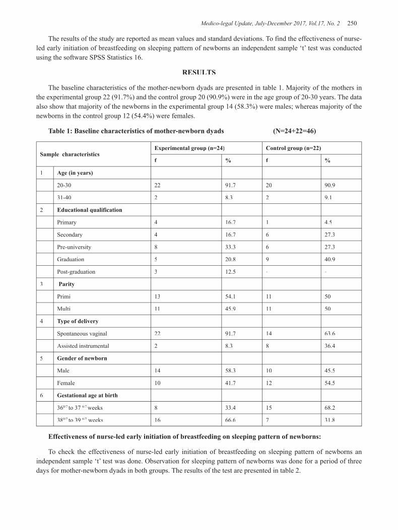
Medico-legal Update, July-December 2017, Vol.17, No. 2 250 251 -legal Update, July-December 2017, Vol.17, No. 2
The results of the study are reported as mean values and standard deviations. To find the effectiveness of nurse-led early initiation of breastfeeding on sleeping pattern of newborns an independent sample ‘t’ test was conducted using the software SPSS Statistics 16.
RESULTS
The baseline characteristics of the mother-newborn dyads are presented in table 1. Majority of the mothers in the experimental group 22 (91.7%) and the control group 20 (90.9%) were in the age group of 20-30 years. The data also show that majority of the newborns in the experimental group 14 (58.3%) were males; whereas majority of the newborns in the control group 12 (54.4%) were females.
Table 1: Baseline characteristics of mother-newborn dyads (N=24+22=46)
Sample characteristicsExperimental group (n=24) Control group (n=22)
f % f %
1 Age (in years)
20-30 22 91.7 20 90.9
31-40 2 8.3 2 9.1
2 Educational qualification
Primary 4 16.7 1 4.5
Secondary 4 16.7 6 27.3
Pre-university 8 33.3 6 27.3
Graduation 5 20.8 9 40.9
Post-graduation 3 12.5 - -
3 Parity
Primi 13 54.1 11 50
Multi 11 45.9 11 50
4 Type of delivery
Spontaneous vaginal 22 91.7 14 63.6
Assisted instrumental 2 8.3 8 36.4
5 Gender of newborn
Male 14 58.3 10 45.5
Female 10 41.7 12 54.5
6 Gestational age at birth
360/7 to 37 6/7 weeks 8 33.4 15 68.2
380/7 to 39 6/7 weeks 16 66.6 7 31.8
Effectiveness of nurse-led early initiation of breastfeeding on sleeping pattern of newborns:
To check the effectiveness of nurse-led early initiation of breastfeeding on sleeping pattern of newborns an independent sample ‘t’ test was done. Observation for sleeping pattern of newborns was done for a period of three days for mother-newborn dyads in both groups. The results of the test are presented in table 2.

-legal Update, July-December 2017, Vol.17, No. 2 250 251 Medico-legal Update, July-December 2017, Vol.17, No. 2
Table 2: Effectiveness of early initiation of breastfeeding on sleeping pattern of newborns between experimental group and control group.
The independent sample ‘t’ test computed to find the effectiveness of nurse-led early initiation of breastfeeding shows a statistically significant difference between the experimental and the control group (t = 4.038; p=0.001). This implies that nurse-led early initiation of breastfeeding is effective in improving the sleeping pattern of newborns.
DISCUSSION
The present study showed that nurse-led early initiation of breastfeeding was effective in improving the sleeping pattern of newborns. These findings are in contrast to the common perception that formula milk promotes infant sleep and encourages the infant to settle
(10) and this is because of the potentially increased time the infant takes to digest the formula feed. (11) However, the results of the present study are in line with a study done to demonstrate the potential role of breast milk Melatonin, which found that breast-fed infants slept for a longer duration (9.2 hours) compared to formula-fed infants who slept only for 8.5 hours during the study.(7)
Another longitudinal study was done to evaluate the relationship between breastfeeding and sleep. The authors of the study report that breast-fed infants had more disrupted sleep at baseline including increased night waking and sleep fragmentation, which refutes the findings of the present study. (12)
There are limitations to the study. Firstly, the sample technique employed was purposive sampling and study adopted a quasi-experimental design. Self-construction of the observation tool on sleeping pattern of newborns may also contribute to bias. Nevertheless, nurse-led early initiation of breastfeeding is cost-effective across all
health care settings and the findings of the study support the recommendations that the health care professionals should be proactive in making early initiation of breastfeeding a best practice.
CONCLUSION
This study provides evidence for a link between early initiation of breastfeeding and improved sleeping pattern of newborns. The time to initiate breastfeeding is very crucial for continuation of exclusive breastfeeding. Health care professionals should support the mother-newborn dyads for this lifetime beneficial effect. The efforts done to establish early initiation of breastfeeding as best practice would help mothers to breastfeed successfully and for much longer.
Source of Funding: Self
Conflict of Interest: Nil
REFERENCES
1. UNICEF. Tracking progress on child and maternal malnutrition: a survival and development priority.2009 ed.
2. Lauer JA, Betran AP, Barros AJ, de Onis M. Deaths and years of life lost due to suboptimal breast-feeding among children in the developing world: a global ecological risk assessment. Health Nutr. Public Health Nutr.2006;9:673-685
3. Holmberg KS, Peterson UM, Oscarsson MG. A two-decade perspective on mothers’ experiences and feelings related to breastfeeding initiation in Sweden. Sex Reprod Health 5: 125–130. doi: 10.1016/j.srhc.2014.04.001 PMID: 25200973
Medico-legal Update, July-December 2017, Vol.17, No. 2 252
4. Anders T F. Night - walking infants during the first year of life. Pediatr.1979; 63:860-864.
5. Poblano A, Haro R, Aeteaga C (2007). Neurophysiological measurement of continuity in the sleep of fetuses during the last week of pregnancy and in newborns during the last week of pregnancy and in newborns. Int J Bio Sci. 2007; 4(1): 23-8.
6. Steinberg LA, Nancy C, O’Connell, Hatch TF, Picciano MF, Birch LL. Tryptophan Intake Influences infants’ sleep latency. J. Nutrit. 1992; Sep 122.9
7. Enger AC, Hardash A, Pillar G. Breastfeeding may improve nocturnal sleep and reduce infantile coli: potential role of breast milk melatonin. Eur J Pediatr. 2012; 171:729–732. doi: 10.1007/s00431-011-1659-3.
8. Zaichkin J, Fraser D. The healthy newborn. In: Evans RJ, Evans MK, Brown YMR, Orshan SA
(Eds.). Canadian Maternity, Newborn, & Women’s Health Nursing. (1 ed.). Philadelphia, PA: Lippincott Williams & Wilkins. 2010; 773-851.
9. Askin D. Newborn adaptation to extrauterine life. In: Simpson KR, Creehan PA (Eds). AWHONN’s Perinatal Nursing (3rd ed.). Philadelphia, PA: Lippincott Williams & Wilkins. 2008; 773-851.
10. Sachs M, Dykes F, Carter B. Feeding by numbers: an ethnographic study of how breastfeeding women understand their babies’ weight charts. Int Breastfeed J. 2006; 1: 29
11. Dewey KG, Heinig MJ, Nommsen LA, Lonnerdal B. Adequacy of energy intake among breast-fed infants in the DARLING study: Relationships to growth velocity, morbidity, and activity levels. J Pediatr. 1991; 119:538-547.
12. Mindell JA, Du Mond C, Jason B. Long-Term Relationship between Breastfeeding and Sleep. Child Health Care. 2012; 41(3).

-legal Update, July-December 2017, Vol.17, No. 2 252
Institute of Medico-Legal Publications501, Manisha Building, 75-76, Nehru Place, New Delhi-110019,
Mob: 09971888542, Fax no.: +91 11 3044 6500
E-mail: [email protected],
Website: www.medicolegalupdate.org
Send all payment to:Send all payment to:Institute of Medico-Legal Publications
501, Manisha Building, 75-76, Nehru Place, New Delhi-110019, Mob: 09971888542, Fax no.: +91 11 3044 6500
E-mail: [email protected], Website: www.medicolegalupdate.org

Related Documents