Mitosis – Cellular Division
Jan 02, 2016
Mitosis – Cellular Division. 30 hours later. 20 hours later. Development of the morula. Blastocyst. 2 types of cells: -cells to become placenta -inner cell mass (stem cells) Stem cells are pluripotent -have ability to read all DNA in their nuclei. - PowerPoint PPT Presentation
Welcome message from author
This document is posted to help you gain knowledge. Please leave a comment to let me know what you think about it! Share it to your friends and learn new things together.
Transcript


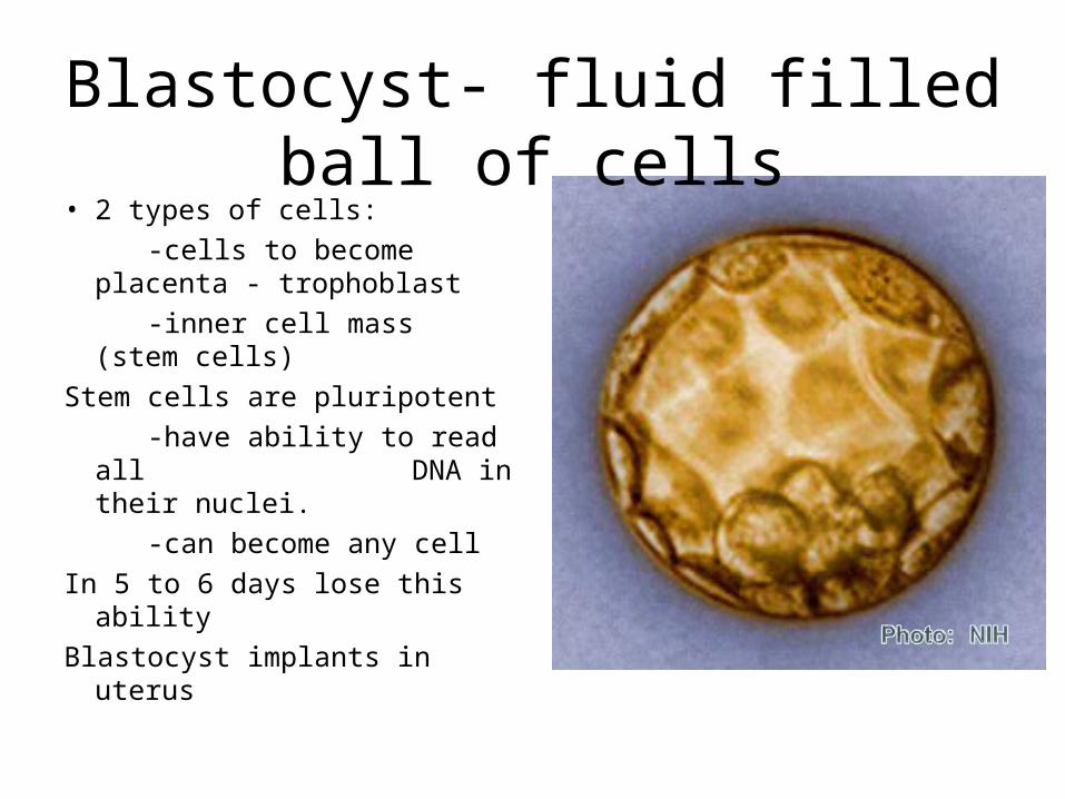
Blastocyst- fluid filled ball of cells
• 2 types of cells:
-cells to become placenta - trophoblast
-inner cell mass (stem cells)
Stem cells are pluripotent
-have ability to read all DNA in their nuclei.
-can become any cell
In 5 to 6 days lose this ability
Blastocyst implants in uterus

Gastrula

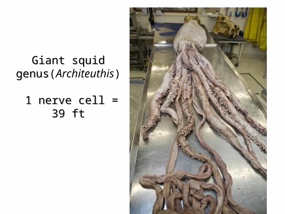
Giant squidGiant squidgenusgenus(Architeuthis)
1 nerve cell = 39 ft1 nerve cell = 39 ft


Fig. 13-2b
(b) Redwoods

Fig. 12-2a
100 µm
(a) Reproduction

Fig. 12-2b
200 µm
(b) Growth and development
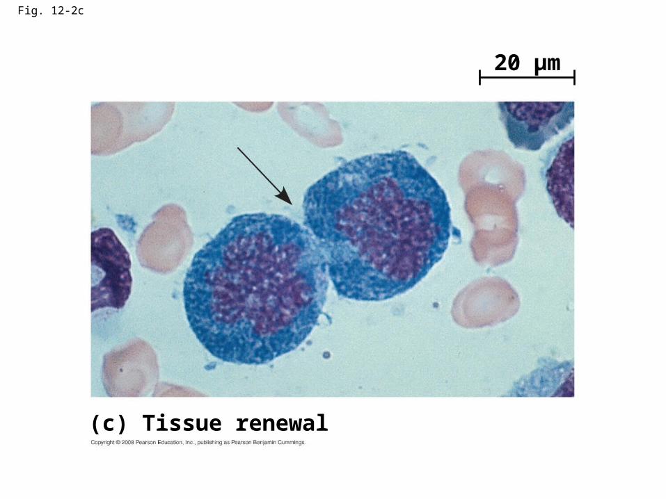
Fig. 12-2c
20 µm
(c) Tissue renewal

Fig. 13-2a
(a) Hydra
0.5 mm
Bud
Parent


Fig. 12-14
SG1
M checkpoint
G2M
Controlsystem
G1 checkpoint
G2 checkpoint

Fig. 12-15
G1
G0
G1 checkpoint
(a) Cell receives a go-ahead signal
G1
(b) Cell does not receive a go-ahead signal

DNA – lots of it in a small space
chromatin
chromosome

Fig. 12-UN3

Fig. 12-40.5 µm Chromosomes
Chromosomeduplication(including DNAsynthesis)
Chromo-some arm
Centromere
Sisterchromatids
DNA molecules
Separation ofsister chromatids
Centromere
Sister chromatids

Fig. 12-UN1
Telophase andCytokinesis
Anaphase
Metaphase
Prometaphase
Prophase
MITOTIC (M) PHASE
Cytokinesis
Mitosis
SG1
G2

Fig. 12-20
Tumor
A tumor growsfrom a singlecancer cell.
Glandulartissue
Lymphvessel
Bloodvessel
Metastatictumor
Cancercell
Cancer cellsinvade neigh-boring tissue.
Cancer cells spreadto other parts ofthe body.
Cancer cells maysurvive andestablish a newtumor in anotherpart of the body.
1 2 3 4

Fig. 12-UN4

Fig. 12-UN6

Fig. 12-6b
PrometaphaseProphaseG2 of Interphase
Nonkinetochoremicrotubules
Fragmentsof nuclearenvelope
Aster CentromereEarly mitoticspindle
Chromatin(duplicated)
Centrosomes(with centriolepairs)
Nucleolus Nuclearenvelope
Plasmamembrane
Chromosome, consistingof two sister chromatids
Kinetochore Kinetochoremicrotubule
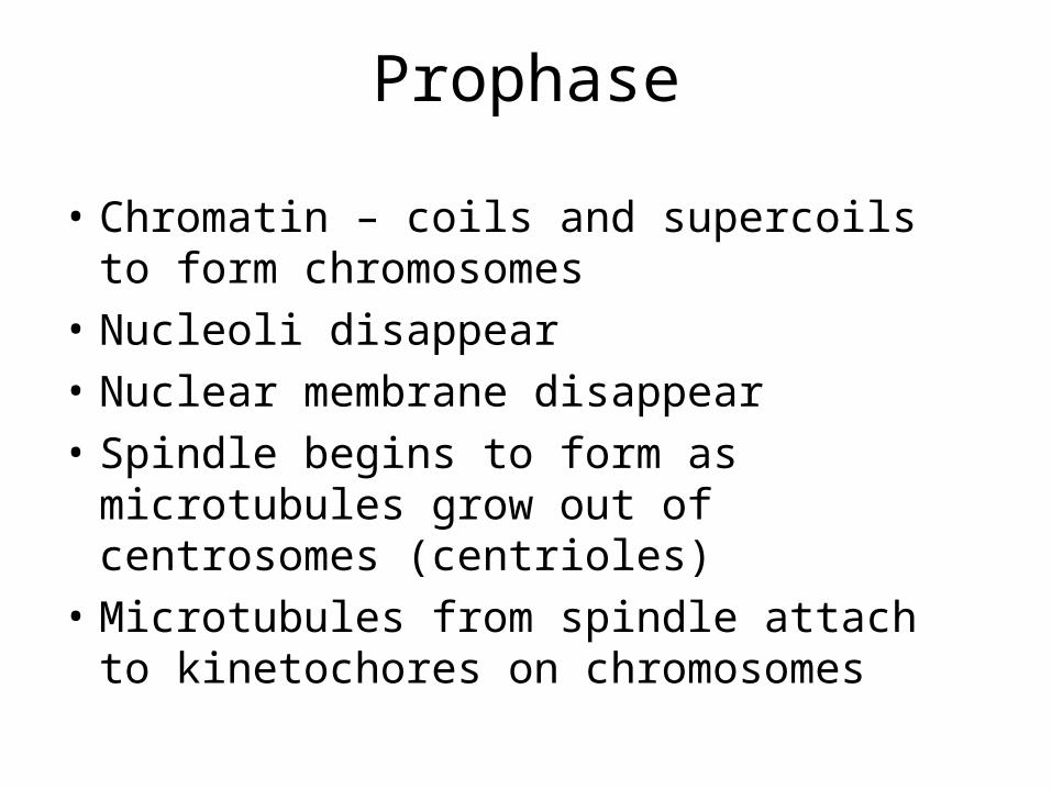
Prophase
• Chromatin – coils and supercoils to form chromosomes
• Nucleoli disappear
• Nuclear membrane disappear
• Spindle begins to form as microtubules grow out of centrosomes (centrioles)
• Microtubules from spindle attach to kinetochores on chromosomes

Fig. 12-3
20 µm
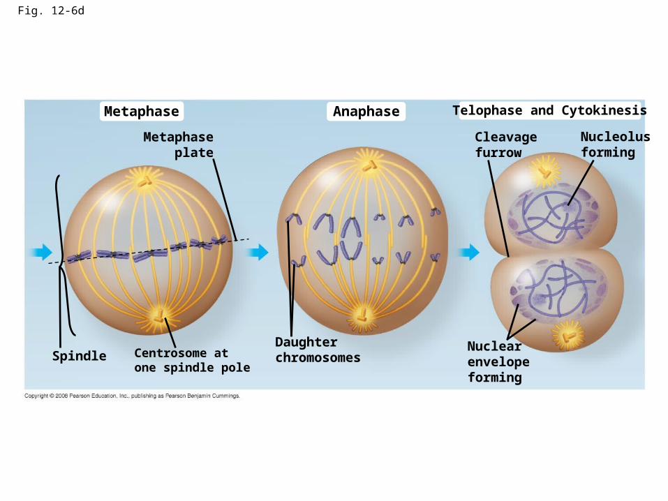
Fig. 12-6d
Metaphase Anaphase Telophase and Cytokinesis
Cleavagefurrow
Nucleolusforming
Metaphaseplate
Centrosome atone spindle pole
SpindleDaughterchromosomes
Nuclearenvelopeforming

Metaphase
• Spindle fully formed
• Poles at opposite end of cell
• Chromosomes now on metaphase plate (middle of cell)

Fig. 12-7
Microtubules Chromosomes
Sisterchromatids
Aster
Metaphaseplate
Centrosome
Kineto-chores
Kinetochoremicrotubules
Overlappingnonkinetochoremicrotubules
Centrosome 1 µm
0.5 µm

Anaphase
• 2 centromeres of each chromosome come apart separating sister chromosomes
• Free chromatid now called a chromosome
• Spindle fibers attached to chromosomes kinetochores contract while those not attached lengthen.
• Poles move further apart and cell elongates

Fig. 12-10d
Anaphase4

Telophase
• Cell continues to elongate
• Nuclear membrane reforms
• Nucleolus forms
• Mitotic spindle disappears
• Cytokinesis occurs
• 2 genetically identical daughter cells form

Cytokinesis
• Division of the cytoplasm
• Occurs along with telophase
• Animal cells form a cleavage furrow which pinches the cell into 2
• Plant cells form a cell plate

Fig. 12-10e
Telophase5
Cell plate10 µm

Cleavage furrow
Fig. 12-9a
100 µm
Daughter cells
(a) Cleavage of an animal cell (SEM)
Contractile ring ofmicrofilaments

Fig. 12-9b
Daughter cells
(b) Cell plate formation in a plant cell (TEM)
Vesiclesformingcell plate
Wall ofparent cell
New cell wallCell plate
1 µm

Fig. 12-9
Cleavage furrow100 µm
Contractile ring ofmicrofilaments
Daughter cells
(a) Cleavage of an animal cell (SEM) (b) Cell plate formation in a plant cell (TEM)
Vesiclesformingcell plate
Wall ofparent cell
Cell plate
Daughter cells
New cell wall
1 µm

Fig. 12-UN2

Fig. 12-UN5
Mitosis video

Fig. 12-16
Pro
tein
kin
as
e a
cti
vit
y (
– )
% o
f d
ivid
ing
ce
lls (
– )
Time (min)300200 400100
0
1
2
3
4
5 30
500
0
20
10
RESULTS

Fig. 12-17
M G1S G2
M G1S G2
M G1
MPF activity
Cyclinconcentration
Time(a) Fluctuation of MPF activity and cyclin concentration during the cell cycle
Degradedcyclin
Cdk
G 1S
G 2
M
CdkG2
checkpointCyclin isdegraded
CyclinMPF
(b) Molecular mechanisms that help regulate the cell cycle
Cy
clin
ac
cu
mu
latio
n

Fig. 12-17a
Time(a) Fluctuation of MPF activity and cyclin concentration during the cell cycle
Cyclinconcentration
MPF activity
M M MSSG1 G1 G1G2 G2
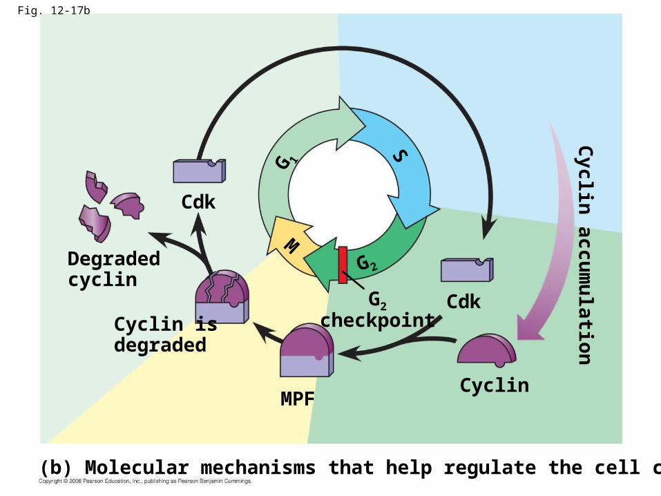
Fig. 12-17b
Cyclin isdegraded
Cdk
MPF
Cdk
MS
G 1G2
checkpoint
Degradedcyclin
Cyclin
(b) Molecular mechanisms that help regulate the cell cycle
G2
Cyclin
accum
ulatio
n

Fig. 13-3APPLICATION
TECHNIQUE
Pair of homologousreplicated chromosomes
5 µm
Centromere
Sisterchromatids
Metaphasechromosome
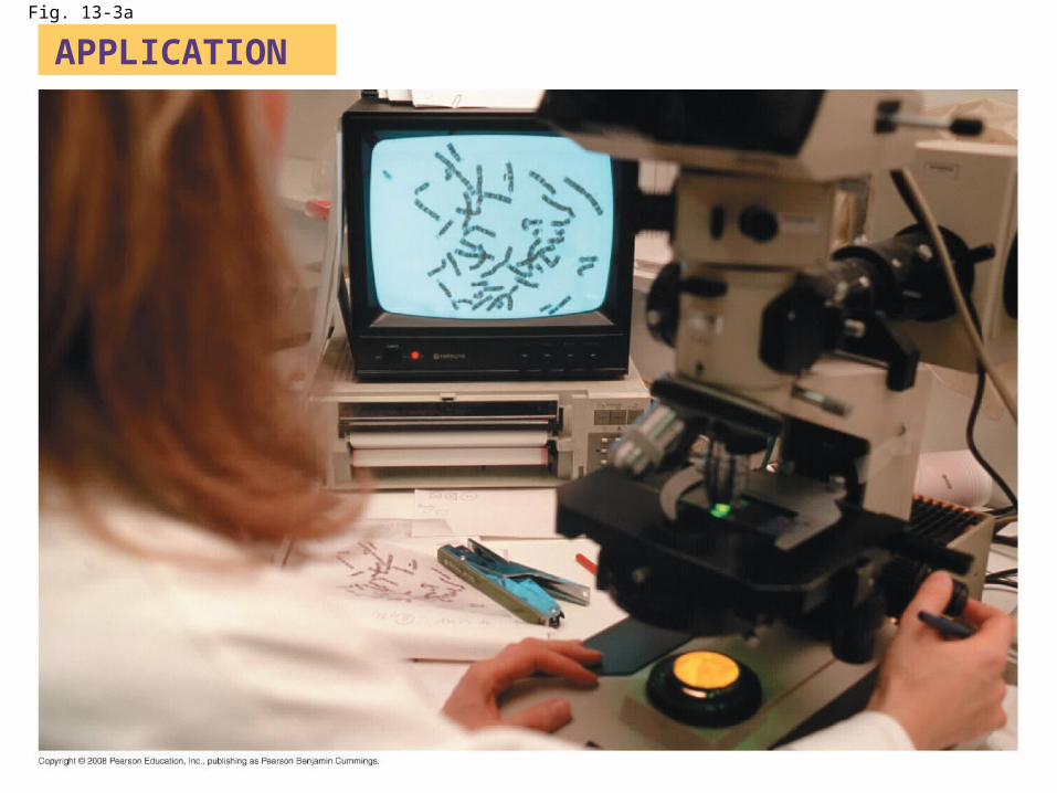
Fig. 13-3a
APPLICATION

Fig. 13-3b
TECHNIQUE
Pair of homologousreplicated chromosomes
Centromere
Sisterchromatids
Metaphasechromosome
5 µm

Fig. 13-4
Key
Maternal set ofchromosomes (n = 3)
Paternal set ofchromosomes (n = 3)
2n = 6
Centromere
Two sister chromatidsof one replicatedchromosome
Two nonsisterchromatids ina homologous pair
Pair of homologouschromosomes(one from each set)


Mitosis – Word Bank
Asters Centrioles Chromatids
Chromosome Cytoplasm Nucleus
Nucleolus Nuclear membrane
Spindle Fibers
Kinetochore Cleavage Furrow

Fig. 12-10a
Nucleus
Prophase1
Nucleolus
Chromatincondensing

Fig. 12-10b
Prometaphase2
Chromosomes

Fig. 12-10c
Metaphase3

Fig. 12-10d
Anaphase4

Fig. 12-10e
Telophase5
Cell plate10 µm
Related Documents