International Journal of Molecular Sciences Review Mitochondrial Modulations, Autophagy Pathways Shifts in Viral Infections: Consequences of COVID-19 Shailendra Pratap Singh 1, * ,† , Salomon Amar 1,2, * ,† , Pinky Gehlot 3 , Sanjib K. Patra 4 , Navjot Kanwar 5 and Abhinav Kanwal 6 Citation: Singh, S.P.; Amar, S.; Gehlot, P.; Patra, S.K.; Kanwar, N.; Kanwal, A. Mitochondrial Modulations, Autophagy Pathways Shifts in Viral Infections: Consequences of COVID-19. Int. J. Mol. Sci. 2021, 22, 8180. https:// doi.org/10.3390/ijms22158180 Academic Editors: Jacek Z Kubiak and Malgorzata Kloc Received: 9 July 2021 Accepted: 24 July 2021 Published: 30 July 2021 Publisher’s Note: MDPI stays neutral with regard to jurisdictional claims in published maps and institutional affil- iations. Copyright: © 2021 by the authors. Licensee MDPI, Basel, Switzerland. This article is an open access article distributed under the terms and conditions of the Creative Commons Attribution (CC BY) license (https:// creativecommons.org/licenses/by/ 4.0/). 1 Department of Pharmacology, New York Medical College, Valhalla, NY 10595, USA 2 Department of Microbiology and Immunology, New York Medical College, Valhalla, NY 10595, USA 3 Department of Pharmacy, School of Chemical Sciences and Pharmacy, Central University of Rajasthan, Bandarsindri, Kishangarh, Ajmer 305817, Rajasthan, India; [email protected] 4 Department of Yoga, Central University of Rajasthan, Bandarsindri, Kishangarh, Ajmer 305817, Rajasthan, India; [email protected] 5 Departmen of Pharmaceutical Sciences and Technology, Maharaja Ranjit Singh Punjab Technical University, Bathinda 151001, Punjab, India; [email protected] 6 Department of Pharmacology, All India Institute of Medical Sciences, Bathinda 151001, Punjab, India; [email protected] * Correspondence: [email protected] (S.P.S.); [email protected] (S.A.) † Authors equally contributed. Abstract: Mitochondria are vital intracellular organelles that play an important role in regulating various intracellular events such as metabolism, bioenergetics, cell death (apoptosis), and innate immune signaling. Mitochondrial fission, fusion, and membrane potential play a central role in maintaining mitochondrial dynamics and the overall shape of mitochondria. Viruses change the dynamics of the mitochondria by altering the mitochondrial processes/functions, such as autophagy, mitophagy, and enzymes involved in metabolism. In addition, viruses decrease the supply of energy to the mitochondria in the form of ATP, causing viruses to create cellular stress by generating ROS in mitochondria to instigate viral proliferation, a process which causes both intra- and extra- mitochondrial damage. SARS-COV2 propagates through altering or changing various pathways, such as autophagy, UPR stress, MPTP and NLRP3 inflammasome. Thus, these pathways act as potential targets for viruses to facilitate their proliferation. Autophagy plays an essential role in SARS-COV2-mediated COVID-19 and modulates autophagy by using various drugs that act on potential targets of the virus to inhibit and treat viral infection. Modulated autophagy inhibits coronavirus replication; thus, it becomes a promising target for anti-coronaviral therapy. This review gives immense knowledge about the infections, mitochondrial modulations, and therapeutic targets of viruses. Keywords: mitochondria; SARS-COV2; potential targets; autophagy; COVID-19; viral infections 1. Introduction Mitochondria are membrane-bound cell organelles which produce energy in the form of adenosine triphosphate (ATP). Mitochondria regulate various intracellular functions like metabolism, bioenergetics, cell death, innate immune signaling, and cellular homeosta- sis [1]. Mitochondrial dynamics and mitochondria, selective autophagy, or mitophagy, work to maintain mitochondrial quality control [2]. By altering mitochondrial dynamics, viruses influence innate immune signaling [which is mediated through the mitochondrial antiviral signaling (MAVS) protein], as well as favoring their propagation by taking advantage of mitochondrial metabolite. Int. J. Mol. Sci. 2021, 22, 8180. https://doi.org/10.3390/ijms22158180 https://www.mdpi.com/journal/ijms

Welcome message from author
This document is posted to help you gain knowledge. Please leave a comment to let me know what you think about it! Share it to your friends and learn new things together.
Transcript
International Journal of
Molecular Sciences
Review
Mitochondrial Modulations, Autophagy Pathways Shifts inViral Infections: Consequences of COVID-19
Shailendra Pratap Singh 1,*,† , Salomon Amar 1,2,*,†, Pinky Gehlot 3, Sanjib K. Patra 4, Navjot Kanwar 5
and Abhinav Kanwal 6
�����������������
Citation: Singh, S.P.; Amar, S.;
Gehlot, P.; Patra, S.K.; Kanwar, N.;
Kanwal, A. Mitochondrial
Modulations, Autophagy Pathways
Shifts in Viral Infections:
Consequences of COVID-19. Int. J.
Mol. Sci. 2021, 22, 8180. https://
doi.org/10.3390/ijms22158180
Academic Editors: Jacek Z Kubiak
and Malgorzata Kloc
Received: 9 July 2021
Accepted: 24 July 2021
Published: 30 July 2021
Publisher’s Note: MDPI stays neutral
with regard to jurisdictional claims in
published maps and institutional affil-
iations.
Copyright: © 2021 by the authors.
Licensee MDPI, Basel, Switzerland.
This article is an open access article
distributed under the terms and
conditions of the Creative Commons
Attribution (CC BY) license (https://
creativecommons.org/licenses/by/
4.0/).
1 Department of Pharmacology, New York Medical College, Valhalla, NY 10595, USA2 Department of Microbiology and Immunology, New York Medical College, Valhalla, NY 10595, USA3 Department of Pharmacy, School of Chemical Sciences and Pharmacy, Central University of Rajasthan,
Bandarsindri, Kishangarh, Ajmer 305817, Rajasthan, India; [email protected] Department of Yoga, Central University of Rajasthan, Bandarsindri, Kishangarh, Ajmer 305817, Rajasthan,
India; [email protected] Departmen of Pharmaceutical Sciences and Technology, Maharaja Ranjit Singh Punjab Technical University,
Bathinda 151001, Punjab, India; [email protected] Department of Pharmacology, All India Institute of Medical Sciences, Bathinda 151001, Punjab, India;
[email protected]* Correspondence: [email protected] (S.P.S.); [email protected] (S.A.)† Authors equally contributed.
Abstract: Mitochondria are vital intracellular organelles that play an important role in regulatingvarious intracellular events such as metabolism, bioenergetics, cell death (apoptosis), and innateimmune signaling. Mitochondrial fission, fusion, and membrane potential play a central role inmaintaining mitochondrial dynamics and the overall shape of mitochondria. Viruses change thedynamics of the mitochondria by altering the mitochondrial processes/functions, such as autophagy,mitophagy, and enzymes involved in metabolism. In addition, viruses decrease the supply ofenergy to the mitochondria in the form of ATP, causing viruses to create cellular stress by generatingROS in mitochondria to instigate viral proliferation, a process which causes both intra- and extra-mitochondrial damage. SARS-COV2 propagates through altering or changing various pathways,such as autophagy, UPR stress, MPTP and NLRP3 inflammasome. Thus, these pathways act aspotential targets for viruses to facilitate their proliferation. Autophagy plays an essential role inSARS-COV2-mediated COVID-19 and modulates autophagy by using various drugs that act onpotential targets of the virus to inhibit and treat viral infection. Modulated autophagy inhibitscoronavirus replication; thus, it becomes a promising target for anti-coronaviral therapy. This reviewgives immense knowledge about the infections, mitochondrial modulations, and therapeutic targetsof viruses.
Keywords: mitochondria; SARS-COV2; potential targets; autophagy; COVID-19; viral infections
1. Introduction
Mitochondria are membrane-bound cell organelles which produce energy in the formof adenosine triphosphate (ATP). Mitochondria regulate various intracellular functionslike metabolism, bioenergetics, cell death, innate immune signaling, and cellular homeosta-sis [1].
Mitochondrial dynamics and mitochondria, selective autophagy, or mitophagy, workto maintain mitochondrial quality control [2]. By altering mitochondrial dynamics, virusesinfluence innate immune signaling [which is mediated through the mitochondrial antiviralsignaling (MAVS) protein], as well as favoring their propagation by taking advantage ofmitochondrial metabolite.
Int. J. Mol. Sci. 2021, 22, 8180. https://doi.org/10.3390/ijms22158180 https://www.mdpi.com/journal/ijms
Int. J. Mol. Sci. 2021, 22, 8180 2 of 24
1.1. Mitochondrial Dynamics
The mitochondrial dynamics network involves two cycles, mitochondrial fissionand Mitochondrial Fusion, to help maintain the functional capacity of mitochondria bydistribution of mitochondrial contents, energy conductance, and responsiveness to cellularcues. Thus, mitochondrial dynamics govern their communication and interaction withother cellular organelles.
1.1.1. Mitochondrial Fission
Mitochondrial fission is required to create new mitochondria, segregate damagedparts of the mitochondria from the dynamic mitochondrial network and remove damagedmitochondria via the mitochondria-selective autophagy process. Dynamin-1 like protein(Drp1) recruitment into mitochondria and its activity is regulated by different processessuch as phosphorylation, nitrosylation, and summoylation to initiate mitochondrial fission.In mammals at least three proteins are required for mitochondrial fission: dynamin-relatedprotein 1 (Drp1), Fis1 mitochondrial fission 1 protein [Fis1), and mitochondrial fission factor(MFF) [3,4]. Drp1 contains three domains: the dynamic like central domain, C-terminalGTPase effector domain, and N-terminal GTPase domain. Full GTPase efficiency andmitochondrial fission requires intermolecular interaction between the GTPase domain andGTPase effector domain [5]. By network lengthening, MFF releases the Drp1 foci from themitochondrial outer membrane, whereas, with the help of mitochondrial fission and thephysical interaction between MFF and Drp1, MFF overexpression stimulates mitochondrialfission [6].
1.1.2. Mitochondrial Fusion
Mitochondrial Fusion mechanisms involve various steps such as outer mitochondrialmembrane (OMM) fusion, and inner mitochondrial membrane (IMM) fusion throughintegral membrane GTPase proteins such as Mitofusin 1 and 2 (Mfn1 and Mfn2), andoptic atrophy 1 (OPA1), respectively. The proteins Mfn1 and Mfn2 are located on theopposite fusion membranes and anchored into the outer membrane with the N-terminalGTPase domain and a predicted coiled coil protruding into cytosol to form homo or hetro-oligomeric complex in trans. The OPA1 protein is located on adjacent fusion membraneand is involved in inner-mitochondrial membrane fusion as well as mitochondria cristaeremodeling, apoptosis, and bioenergetics. The OPA1 protein works with Mfn1 to promotemitochondrial fusion. Mitochondrial fusion isolates dysfunctional and damaged mitochon-dria from the functional network via the joining of healthy discreate mitochondria with thefunctional network.
1.1.3. Role of Mitochondrial Dynamics in Antiviral Signaling
By balancing between two opposite processes, mitochondrial fission and fusion,mammalian cells maintain the overall shapes of their mitochondria. The Fis1 proteinhas a TM domain with the help of the C-terminal of mitochondria anchored into themitochondrial outer membrane [3]. Drp1 does not prevent localized mitochondria via theknockdown of Fis1 with RNA interference [7]. By network lengthening, MFF release theDrp1 foci from the mitochondrial outer membrane, whereas, with the help of mitochondrialfission and the physical interaction between the mitochondrial fission factor (MFF) andDrp1, MFF overexpression stimulates mitochondrial fission [6].
The proteins Mfn1, Mfn2, and OPA are involved in mitochondrial dynamics main-tenance, [8,9]. For mitochondrial fusion, OPA1 needs Mfn1 [10] and forms an oligomerthat regulates mitochondrial cristae morphology and therefore completely unharnessesthe cytochrome C oxidase throughout the process of cell death [9,11,12]. The process ofRLR communication reserves the interaction between Mfn2 and MAVS in high-molecularmass complexes [13]. Once a virus infects the mitochondria, Mfn2 murine embryonic cells(MEFs) improve MAVS communication, whereas overexpression of Mfn2 blocks NF-kB andthe IRF-3 activation downstream of RIG-I, MDA-5, and MAVS [14]. After the manipulation
Int. J. Mol. Sci. 2021, 22, 8180 3 of 24
of its expression level, Mfn1 produces different phenotypes, which indicate that Mfn2has a unique role in regulating MAVS signaling, which is independent of its function inmitochondrial fusion.
Efficient RLR signaling requires the interaction of MAVS with Mfn1, whereas MFF1 orDRP1, as an inhibitor of fusion, decreases virus-induced NF-kB and IRF-3 activation [15].After the depletion of Drp1 and Fis1 in the cells, there is an increase in RLR signaling,and the elongation of the mitochondrial network promotes mitochondrial–endoplasmicreticulum interaction during the viral infection, enhancing the association of MAVS with asting to augment RLR signaling [15].
MAM is a major site of MAVS signaling which links the endoplasmic reticulum tothe mitochondria [16], where Mfn2 may inhibit MAVS [17]. After the activation of RLR,both IRF-3 and IKB- α phosphorylate, which degrades the main 75K Da isoform of MAVSresulting in the release of Mfn1 to promote mitochondrial fusion/elongation [15]. Thus,MAVS acts as a regulator of the Mfn1 function. After mitofusion-deficient MFFs [both Mfn1and Mfn2 proteins), heterogenous mitochondrial membrane potential (MMP) occurs, whichreduces MAVS signaling, resulting in a decrease in RLR-dependent antiviral responses.
When cells treated with a chemical uncoupling compound, mitochondrial membranepotential decreases RLR signaling to NF-KB and IRF-3 as well as lowering the productionof type 1 IFN [18]. Upon viral infection, decreased MMP might quickly thwart the MAVScomplex’s structural rearrangement [18]. Inhibition of ATP synthesis does not inhibitMAVS-mediated signaling, excluding the hypothesis that MAVS, localized at the mitochon-drial surface, is not attributed to an energetic requirement to transduce the signal. Mfn1and Mfn2 have opposite roles in innate viral immunity, whereas they play a similar role inmitochondrial fusion [19].
In the absence of infection, the innate immune system is physiologically activatedand produces inflammation, also known as chronic low-grade inflammation [20], whichincludes genetic susceptibility, cellular senescence, impaired autophagy, dysfunctionalmitochondria, changes in microbiota composition, and oxidative stress [21–23].
In healthy mitochondria, about 90% of energy demand is provided by mitochondriathrough ATP generation [24]. The imbalance between ATP supply and demand causesmitochondrial dysfunction.
Mitochondrial fission includes Drp1 and Fis1, whereas mitochondrial fusion includesMfn1, Mfn2, and OPA1. The deletion of the Drp1 gene causes mitochondrial enlargement,the increased opening of the mitochondrial permeability transition pore (MPTP), apop-tosis, and lethal dilated cardiomyopathy (DCM) [25] by inhibiting mitochondrial fission,whereas deletion of Mfn1 and Mfn2 disrupts mitochondrial structure and respiratory chainfunction [26]. An imbalance between mitochondrial fusion and fission compromises mito-chondrial integrity during aging [27–29]. Mitochondrial from aged C. elegans is indicatedby a significantly enlarged and swollen ultrastructure, which is accompanied by decreasingO2 consumption, increasing carbonylated proteins and decreasing mitochondrial SODactivity [30].
Drp1 knockdown triggers NLRP-3 inflammasome assembly and activates caspase1 &IL-β [31].
The depolarization of membrane and mitochondrial damage is caused by PINK1(PTEN-induced kinase 1), which accumulates on the outer membrane of the mitochondria,mediates phosphorylation, and activates parkin (E3 ubiquitin ligase) for the ubiquitinationof the mitochondrial protein of Mfn-2 [32], resulting in damaged mitochondria interactingwith an LC3-positive phagosome for degradation in the lysosome. Thus, if this process isimpaired, it leads to mitochondrial dysfunction and cell death [33,34]. The deficiency ofparkin increases MMP loss, ROS production, and mtDNA release, which triggers NLRP-3and elevates the activation of IL-1B and caspase, contributing to age-related patholo-gies [35,36]. Upregulation of parkin expression and enhanced mitophagy inhibits NLRP-3inflammasome assembly and activates downstream signaling molecules that promote cellsurvival [37].
Int. J. Mol. Sci. 2021, 22, 8180 4 of 24
1.2. Autophagy
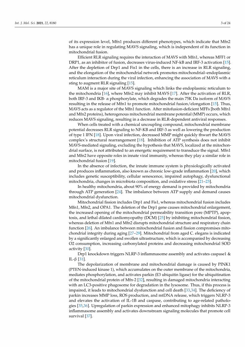
Autophagy is a self-destructive process which involves the removal of dysfunctionaland unnecessary cellular components through various steps such as Omegasomes andthe initiation of isolation membrane; elongation of isolation membrane and formation ofautophagosomes; and autophagosome-lysosome fusion and degradation (Figure 1).
Int. J. Mol. Sci. 2021, 22, x FOR PEER REVIEW 4 of 26
NLRP-3 inflammasome assembly and activates downstream signaling molecules that pro-mote cell survival [37].
1.2. Autophagy Autophagy is a self-destructive process which involves the removal of dysfunctional
and unnecessary cellular components through various steps such as Omegasomes and the initiation of isolation membrane; elongation of isolation membrane and formation of au-tophagosomes; and autophagosome-lysosome fusion and degradation (Figure 1).
Figure 1. —Steps of Autophagy: Autophagy (macro autophagy) means “self” (“auto”) and “eat-ing” (“phagy”). Autophagy removes dysfunctional and unnecessary cellular components through various steps such as Omegasomes and the initiation of isolation membrane; the elongation of the isolation membrane and formation of autophagosomes; and autophagosome-lysosome fusion and degradation.
1.2.1. Omegasomes and the Initiation of Isolation Membrane: Through the mTOR-signaling pathway, the ULK1-Atg13-FIP200-Atg101 kinase com-
plex activates autophagic signaling and forms omegasomes. Under the starvation condi-tion, double FYVE domain-containing protien1(DFCP1) is localized to PI [3]P on an ome-gasome whereas, under nutrient-rich conditions, DFCP1 is localized to ER and Golgi. The formation of DFCP1-positive omegasomes is regulated by the Atg14-VPs34-beclin1 PI3-
Figure 1. —Steps of Autophagy: Autophagy (macro autophagy) means “self” (“auto”) and “eat-ing” (“phagy”). Autophagy removes dysfunctional and unnecessary cellular components throughvarious steps such as Omegasomes and the initiation of isolation membrane; the elongation of theisolation membrane and formation of autophagosomes; and autophagosome-lysosome fusion anddegradation.
1.2.1. Omegasomes and the Initiation of Isolation Membrane:
Through the mTOR-signaling pathway, the ULK1-Atg13-FIP200-Atg101 kinase com-plex activates autophagic signaling and forms omegasomes. Under the starvation condition,double FYVE domain-containing protien1(DFCP1) is localized to PI [3] P on an omegasomewhereas, under nutrient-rich conditions, DFCP1 is localized to ER and Golgi. The forma-tion of DFCP1-positive omegasomes is regulated by the Atg14-VPs34-beclin1 PI3-kinasecomplex. Inside the ring of the omegasome, an isolation membrane is formed, and theAtg12-Atg5/Atg16 complex is localized to this isolation membrane [38–40]. By decreasing
Int. J. Mol. Sci. 2021, 22, 8180 5 of 24
the level of PI [3] P [41,42], 2 PI [3] P, phosphates such as jumpy/MT/MR14 and MTMR3negatively regulate the formation of omegasomes and the isolation membrane.
1.2.2. Elongation of the Isolation Membrane and the Formation of Autophagosomes
The isolation membrane engulfs cytoplasmic components and elongates. At the laterstage of the elongation of the isolation membrane, LC3-11 is localized to both sides of theisolation membrane. It closes the membrane to form an autophagosome resulting in theAtg12-Atg5/Atg16 complex, dissociated from the autophagosome [40]. Moreover, LC3-II,Rab32, and Rab33 also involve the elongation of the isolation membrane [43,44].
1.2.3. Autophagosome-Lysosome Fusion and Degradation
The outer membrane of the autophagosome fuses with the lysosome to form autolyso-some, which requires Rab7 [45,46]. This fusion is positively regulated by the UVRAG-VPS34-Beclin1 PI3-Kinase complex while negatively regulated by the Rubicone-UVRAG-VPS34-Beclin1 PI3-Kinase complex [47–51]. Lysosome hydrolase in autolysosome involvescathepsin, and lipases degrade the intra-autophagosomal content, while cathepsin alonedegrades LC3-II on the intra-autophagosomal surface [52,53].
Coronaviruses such as SARS-Co, SARS-CoV-2, MERS-CoV, and MHV, etc., induce aswell as inhibit autophagy. Modulated autophagy inhibits coronavirus replication; thus, itbecomes a promising target for anticoronaviral treatments (Table 1).
Table 1. Effect of the autophagy inducer and inhibitor on the replication of coronavirus in cell cultures.
Viruses Species Drugs Mechanism of Action at Various Step of Autophagy
Autophagy Inducers Autophagy Inhibitor
SARS-CoV-2 [54] Ivermectin Ivertine inhibited SARS-CoV-2 by inhibiting the AKTphosphorylation [55].
SARS-CoV-2 [56] Nitazoxanide/Alinia These drugs also inhibit SARS-CoV-2 by the blocking oflate- stage lysosome acidification [57].
[56,58] ChloroquineChloroquine increses the PH of lysosome and prevents
the formation of autolysosome [59], thus it inhibitsSARS-CoV-2 virus.
SARS-CoV [60] ValinomycinValinomycin stimulates mitophagy by loss of MMP,
Ref. [61] causing it to inhibit the replication ofSARS-CoV virus.
Aescim Aescim inhibits SARS-CoV virus by activating thesignaling pathway of ROS-MAPK/p38 [62].
[63,64] Chloroquine Chloroquine acts similary to SARS-CoV-2 and inhibitsthe SARS virus [59].
MERS-CoV [65] VenetoclaxVenetoclax inhibits MERS-CoV by releasing the BECN1
from BCL2L1 or by interacting withBCL2L1/Bcl-XL [66].
Everolimus/Afinitor These drugs inhibit MTOR causing it to inhibit theMERS-CoV Virus [67].
[68] Rapamycin/Sirolumus These drugs act in the same way as Everolimus andAfinitor and inhibit virus repliucatin [69].
wortmannin Wortmannin inhibits MERS-CoV by inhibiting thePtdIns3K & PI3KS [70].
UO126 It inhibits the MAPK/ERK pathway and thereforeinhibits the MERS-CoV virus [71].
MHV [72] Rapamycin/Sirolumus It also acts in a similar way to the MERS-CoV virus [69].
[73] 3-MA It acts by inhibiting the class III PtdIns3K and thereforeinhibits the MHV virus [74].
Autophagy inducers antagonize coronavirus replication, whereas autophagy in-hibitors disorganize the Golgi and prevent amohisome/autophagosome-lysosome fusionresulting in blocking the vesicle trafficking system [59,75,76].
Int. J. Mol. Sci. 2021, 22, 8180 6 of 24
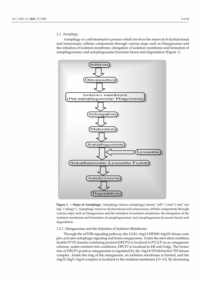
In the mammalian cell, autophagy is categorized into three types: macro-autophagy,micro-autophagy, and chaperone-mediated autophagy (Figure 2), on the basis that thesetypes differ in their routes to the lysosome [77]. Both macro-autophagy and micro-autophagy are non-selective degradations of protein, lipid, and organelles, whereaschaperone-mediated autophagy is a selective protein degradation. Chaperone-mediatedautophagy has a specific-signal sequence called KFERQ, which depends on the molecularchaperone (Heat schock coganate 70) Hsc70.
Int. J. Mol. Sci. 2021, 22, x FOR PEER REVIEW 6 of 26
Autophagy inducers antagonize coronavirus replication, whereas autophagy inhibi-tors disorganize the Golgi and prevent amohisome/autophagosome-lysosome fusion re-sulting in blocking the vesicle trafficking system [59,75,76].
In the mammalian cell, autophagy is categorized into three types: macro-autophagy, micro-autophagy, and chaperone-mediated autophagy (Figure 2), on the basis that these types differ in their routes to the lysosome [77]. Both macro-autophagy and micro-autoph-agy are non-selective degradations of protein, lipid, and organelles, whereas chaperone-mediated autophagy is a selective protein degradation. Chaperone-mediated autophagy has a specific-signal sequence called KFERQ, which depends on the molecular chaperone (Heat schock coganate 70) Hsc70.
Figure 2. —Types of Autophagy: Autophagy categorized into three different categories based on differences in their routes to the lysosome.
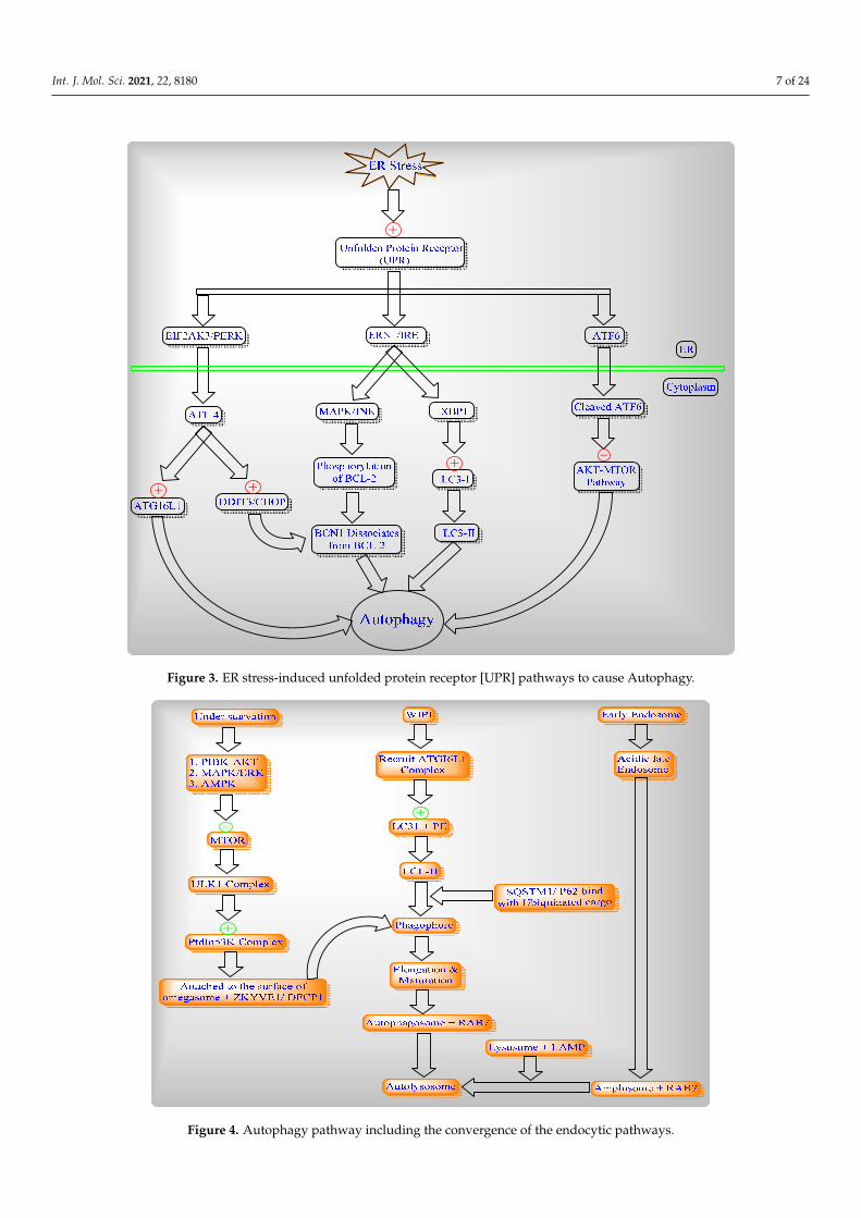
Autophagy induced by three different levels of ER stress and the unfolded protein receptor (UPR) which branches to EIF2AK3/PERK, ERN/IRE1 and/or the ATF6 signaling pathway. (Figure 3).
Figure 2. —Types of Autophagy: Autophagy categorized into three different categories based ondifferences in their routes to the lysosome.
Autophagy induced by three different levels of ER stress and the unfolded proteinreceptor (UPR) which branches to EIF2AK3/PERK, ERN/IRE1 and/or the ATF6 signalingpathway (Figure 3).
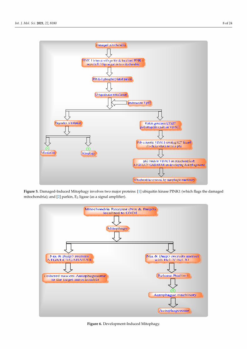
Autophagy conserved the eukaryotic process of cytoplasmic degradation which wasactivated under different conditions of starvation and endoplasmic reticulum [ER] stress tomaintain cellular homeostasis as well as to achieve the complete autophagic flux (Figure 4).
1.3. Mitophagy
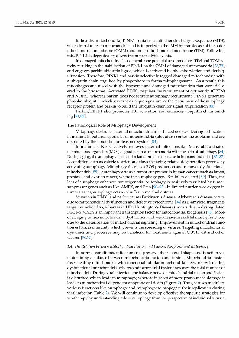
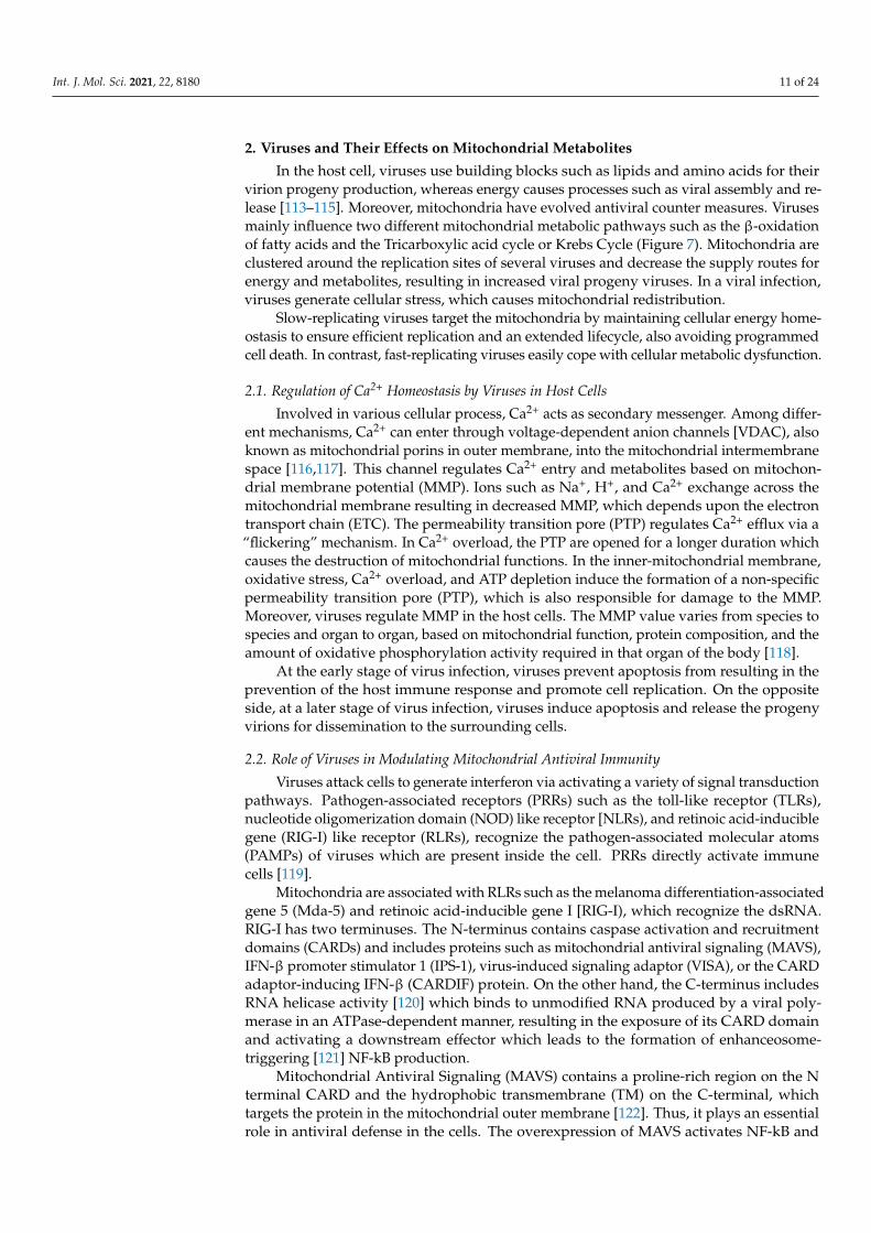
Mitophagy is selective autophagy process in mitochondria which includes the rapidremoval of dysfunctional or damaged mitochondria through two different pathways suchas damage-induced mitophagy (Figure 5) and development-induced mitophagy (Figure 6).
Int. J. Mol. Sci. 2021, 22, 8180 7 of 24Int. J. Mol. Sci. 2021, 22, x FOR PEER REVIEW 7 of 26
Figure 3. ER stress-induced unfolded protein receptor [UPR] pathways to cause Autophagy.
Autophagy conserved the eukaryotic process of cytoplasmic degradation which was activated under different conditions of starvation and endoplasmic reticulum [ER] stress to maintain cellular homeostasis as well as to achieve the complete autophagic flux (Fig-ure 4).
Figure 3. ER stress-induced unfolded protein receptor [UPR] pathways to cause Autophagy.
Int. J. Mol. Sci. 2021, 22, x FOR PEER REVIEW 8 of 26
Figure 4. Autophagy pathway including the convergence of the endocytic pathways.
1.3. Mitophagy Mitophagy is selective autophagy process in mitochondria which includes the rapid
removal of dysfunctional or damaged mitochondria through two different pathways such as damage-induced mitophagy (Figure 5) and development-induced mitophagy (Figure 6).
Figure 4. Autophagy pathway including the convergence of the endocytic pathways.
Int. J. Mol. Sci. 2021, 22, 8180 8 of 24Int. J. Mol. Sci. 2021, 22, x FOR PEER REVIEW 9 of 26
Figure 5. Damaged-Induced Mitophagy involves two major proteins: [1] ubiquitin kinase PINK1 (which flags the dam-aged mitochondria); and [2] parkin, E3 ligase (as a signal amplifier).
Figure 5. Damaged-Induced Mitophagy involves two major proteins: [1] ubiquitin kinase PINK1 (which flags the damagedmitochondria); and [2] parkin, E3 ligase (as a signal amplifier).
Int. J. Mol. Sci. 2021, 22, x FOR PEER REVIEW 10 of 26
Figure 6. Development-Induced Mitophagy.
In healthy mitochondria, PINK1 contains a mitochondrial target sequence (MTS), which translocates to mitochondria and is imported to the IMM by translocase of the outer mitochondrial membrane (OMM) and inner mitochondrial membrane (TIM). Following this, PINK1 is degraded by downstream proteolytic events.
In damaged mitochondria, loose-membrane potential accommodates TIM and TOM activity resulting in the stabilization of PINK1 on the OMM of damaged mitochondria [78,79], and engages parkin ubiquitin ligase, which is activated by phosphorylation and deubiquitination. Therefore, PINK1 and parkin selectively tagged damaged mitochondria with a ubiquitin chain engulfed by phagophore to forma mitophagosome. As a result, this mitophagosome fused with the lysosome and damaged mitochondria that were delivered to the lysosome. Activated PINK1 requires the recruitment of optineurin (OPTN) and NDP52, whereas parkin does not require autophagy recruitment. PINK1 generates phos-pho-ubiquitin, which serves as a unique signature for the recruitment of the mitophagy receptor protein and parkin to build the ubiquitin chain for signal amplification [80].
Parkin/PINK1 also promotes TB1 activation and enhances ubiquitin chain building [81,82].
The Pathological Role of Mitophagy Development Mitophagy destructs paternal mitochondria in fertilized oocytes. During fertilization
in mammals, paternal sperm-born mitochondria (ubiquitin+) enter the ooplasm and are degraded by the ubiquitin–proteasome system [83].
In mammals, Nix selectively removes paternal mitochondria. Many ubiquitinated membranous organelles (MOs) degrad paternal mitochondria with the help of autophagy [84]. During aging, the autophagy gene and related proteins decrease in humans and mice [85–87]. A condition such as caloric restriction delays the aging-related degeneration pro-cess by activating autophagy. Mitophagy decreases ROS production and removes dys-functional mitochondria [88]. Autophagy acts as a tumor suppressor in human cancers
Figure 6. Development-Induced Mitophagy.
Int. J. Mol. Sci. 2021, 22, 8180 9 of 24
In healthy mitochondria, PINK1 contains a mitochondrial target sequence (MTS),which translocates to mitochondria and is imported to the IMM by translocase of the outermitochondrial membrane (OMM) and inner mitochondrial membrane (TIM). Followingthis, PINK1 is degraded by downstream proteolytic events.
In damaged mitochondria, loose-membrane potential accommodates TIM and TOM ac-tivity resulting in the stabilization of PINK1 on the OMM of damaged mitochondria [78,79],and engages parkin ubiquitin ligase, which is activated by phosphorylation and deubiq-uitination. Therefore, PINK1 and parkin selectively tagged damaged mitochondria witha ubiquitin chain engulfed by phagophore to forma mitophagosome. As a result, thismitophagosome fused with the lysosome and damaged mitochondria that were deliv-ered to the lysosome. Activated PINK1 requires the recruitment of optineurin (OPTN)and NDP52, whereas parkin does not require autophagy recruitment. PINK1 generatesphospho-ubiquitin, which serves as a unique signature for the recruitment of the mitophagyreceptor protein and parkin to build the ubiquitin chain for signal amplification [80].
Parkin/PINK1 also promotes TB1 activation and enhances ubiquitin chain build-ing [81,82].
The Pathological Role of Mitophagy Development
Mitophagy destructs paternal mitochondria in fertilized oocytes. During fertilizationin mammals, paternal sperm-born mitochondria (ubiquitin+) enter the ooplasm and aredegraded by the ubiquitin–proteasome system [83].
In mammals, Nix selectively removes paternal mitochondria. Many ubiquitinatedmembranous organelles (MOs) degrad paternal mitochondria with the help of autophagy [84].During aging, the autophagy gene and related proteins decrease in humans and mice [85–87].A condition such as caloric restriction delays the aging-related degeneration process byactivating autophagy. Mitophagy decreases ROS production and removes dysfunctionalmitochondria [88]. Autophagy acts as a tumor suppressor in human cancers such as breast,prostate, and ovarian cancer, where the autophagy gene Beclin1 is deleted [89]. Thus, theloss of autophagy enhances tumorigenesis. Autophagy is positively regulated by tumor-suppressor genes such as Lkt, AMPK, and Pten [90–93]. In limited nutrients or oxygen intumor tissues, autophagy acts as a buffer to metabolic stress.
Mutation in PINK1 and parkin causes Parkinson’s disease. Alzheimer’s disease occursdue to mitochondrial dysfunction and defective cytochrome [94] as β-amyloid fragmentstarget mitochondria, whereas in HD (Huntington’s Disease) occurs due to dysregulatedPGC1-α, which is an important transcription factor for mitochondrial biogenesis [95]. More-over, aging causes mitochondrial dysfunction and weaknesses in skeletal muscle functionsdue to the deterioration of mitochondrial signaling. Improvement in mitochondrial func-tion enhances immunity which prevents the spreading of viruses. Targeting mitochondrialdynamics and processes may be beneficial for treatments against COVID-19 and otherviruses [96,97].
1.4. The Relation between Mitochondrial Fission and Fusion, Apoptosis and Mitophagy
In normal conditions, mitochondrial preserve their overall shape and function viamaintaining a balance between mitochondrial fusion and fission. Mitochondrial fusionfuses healthy mitochondria with functional tubular mitochondrial network by isolatingdysfunctional mitochondria, whereas mitochondrial fission increases the total number ofmitochondria. During viral infection, the balance between mitochondrial fusion and fissionis disturbed which leads to mitophagy, whereas in cases of more pronounced damage itleads to mitochondrial-dependent apoptotic cell death (Figure 7). Thus, viruses modulatevarious functions like autophagy and mitophagy to propagate their replication duringviral infection (Table 2). We will continue to develop effective therapeutic strategies forvirotherapy by understanding role of autophagy from the perspective of individual viruses.

Int. J. Mol. Sci. 2021, 22, 8180 10 of 24
Int. J. Mol. Sci. 2021, 22, x FOR PEER REVIEW 11 of 26
such as breast, prostate, and ovarian cancer, where the autophagy gene Beclin1 is deleted [89]. Thus, the loss of autophagy enhances tumorigenesis. Autophagy is positively regu-lated by tumor-suppressor genes such as Lkt, AMPK, and Pten [90–93]. In limited nutri-ents or oxygen in tumor tissues, autophagy acts as a buffer to metabolic stress.
Mutation in PINK1 and parkin causes Parkinson’s disease. Alzheimer’s disease oc-curs due to mitochondrial dysfunction and defective cytochrome [94] as β-amyloid frag-ments target mitochondria, whereas in HD (Huntington’s Disease) occurs due to dysreg-ulated PGC1-α, which is an important transcription factor for mitochondrial biogenesis [95]. Moreover, aging causes mitochondrial dysfunction and weaknesses in skeletal mus-cle functions due to the deterioration of mitochondrial signaling. Improvement in mito-chondrial function enhances immunity which prevents the spreading of viruses. Target-ing mitochondrial dynamics and processes may be beneficial for treatments against COVID-19 and other viruses [96,97].
1.4. The Relation Between Mitochondrial Fission and Fusion, Apoptosis and Mitophagy In normal conditions, mitochondrial preserve their overall shape and function via
maintaining a balance between mitochondrial fusion and fission. Mitochondrial fusion fuses healthy mitochondria with functional tubular mitochondrial network by isolating dysfunctional mitochondria, whereas mitochondrial fission increases the total number of mitochondria. During viral infection, the balance between mitochondrial fusion and fis-sion is disturbed which leads to mitophagy, whereas in cases of more pronounced damage it leads to mitochondrial-dependent apoptotic cell death (Figure 7). Thus, viruses modu-late various functions like autophagy and mitophagy to propagate their replication during viral infection (Table 2). We will continue to develop effective therapeutic strategies for virotherapy by understanding role of autophagy from the perspective of individual vi-ruses.
Figure 7. Relationships between mitochondrial fission and fusion, apoptosis and mitophagy. Figure 7. Relationships between mitochondrial fission and fusion, apoptosis and mitophagy.
Table 2. Viruses and their effects on mitochondrial dynamics.
S.No. Author & Year Virus Work & Object
1 Horner and Gale,2013 [98]
Hepatitis C virus(HCV) HCV cleaves the MAVS protein and suppresses the host’s antiviral response.
2 Datan et al., 2016 [99],Liang et al., 2016 [100]
Dengue and Zikavirus
With the help of autophagy, the Dengue and Zika viruses improve theirreplication and the induction of autophagy by pharmacological agents (e.g.,
rapamycin) increasing viral dissemination.
3 Joubert et al., 2012 [101] Chikungunya virus Autophagy limits virus-induced cell death and in vivo mortality inChikungunya virus.
4Datan et al., 2016 [99]; Leeet al., 2008 [102]; McLean
et al., 2011 [103]Dengue virus Autophagy inhibits apoptosis to enhance virus replication in the Dengue virus.
5 Zhu et al., 2016 [104]Transmissible
gastroenteritis virus(TGEV)
TGEV-induced complete mitophagy by stimulating DJ1-1 protein deglycasewhich increases cell survival and infection by eliminating virus-induced ROS.
6 Meng et al., 2014 [105] Newcastle diseasevirus (NDV)
Delayed administration of 3 methyl adenine (3-MA) induced more efficientoncolysis in NSCLCs.
7 Barbier et al., 2017 [106] Dengue virus In the Dengue virus, mitochondrial fission is blocked because the Dengue virus’NS4B or NS3 protein promotes mitochondrial fusion by downregulating Drp1.
8 Yu et al., 2015 [107] Dengue virus In the case of the Dengue virus, mitochondrial fusion is suppressed by NS2B3protease which cleaves MFNs.
9 Zamarin et al., 2005 [108] Influenza A virus PB1-F2 have an essential role in the pathogenicity of the viral infection ofinfluenza virus A, via modulation of the host’s mitochondrial dynamics.
10 Kim et al., 2013b [109] Hepatitis C virus(HCV)
HCV stimulates the expression of parkin, PINK1 and induced mitophagy byimpairing oxidative phosphorylation. The resulting HCV infection affects
mitochondrial dynamics.
11 Gou et al., 2017 [110] Classical swine fevervirus (CSFV)
CSFV expresses MFN2 and stimulates parkin and PINK1 expression, resultingin enhanced mitochondrial fission and mitophagy.
12 Ding et al., 2017 [111]Human
parainfluenza virustype 3 (HPIV3)
In HPIV3 infection, a viral protein regulates mitophagy independently ofparkin/PINK1.
13 Xia et al., 2014b [112] Measles virusDuring the measles viral infection, virus-induced antiviral immune response is
enhanced by the knockdown of autophagy-related genes (eg, ATG7, BECN1,SQSTM1, and RAB7).
Int. J. Mol. Sci. 2021, 22, 8180 11 of 24
2. Viruses and Their Effects on Mitochondrial Metabolites
In the host cell, viruses use building blocks such as lipids and amino acids for theirvirion progeny production, whereas energy causes processes such as viral assembly and re-lease [113–115]. Moreover, mitochondria have evolved antiviral counter measures. Virusesmainly influence two different mitochondrial metabolic pathways such as the β-oxidationof fatty acids and the Tricarboxylic acid cycle or Krebs Cycle (Figure 7). Mitochondria areclustered around the replication sites of several viruses and decrease the supply routes forenergy and metabolites, resulting in increased viral progeny viruses. In a viral infection,viruses generate cellular stress, which causes mitochondrial redistribution.
Slow-replicating viruses target the mitochondria by maintaining cellular energy home-ostasis to ensure efficient replication and an extended lifecycle, also avoiding programmedcell death. In contrast, fast-replicating viruses easily cope with cellular metabolic dysfunction.
2.1. Regulation of Ca2+ Homeostasis by Viruses in Host Cells
Involved in various cellular process, Ca2+ acts as secondary messenger. Among differ-ent mechanisms, Ca2+ can enter through voltage-dependent anion channels [VDAC), alsoknown as mitochondrial porins in outer membrane, into the mitochondrial intermembranespace [116,117]. This channel regulates Ca2+ entry and metabolites based on mitochon-drial membrane potential (MMP). Ions such as Na+, H+, and Ca2+ exchange across themitochondrial membrane resulting in decreased MMP, which depends upon the electrontransport chain (ETC). The permeability transition pore (PTP) regulates Ca2+ efflux via a“flickering” mechanism. In Ca2+ overload, the PTP are opened for a longer duration whichcauses the destruction of mitochondrial functions. In the inner-mitochondrial membrane,oxidative stress, Ca2+ overload, and ATP depletion induce the formation of a non-specificpermeability transition pore (PTP), which is also responsible for damage to the MMP.Moreover, viruses regulate MMP in the host cells. The MMP value varies from species tospecies and organ to organ, based on mitochondrial function, protein composition, and theamount of oxidative phosphorylation activity required in that organ of the body [118].
At the early stage of virus infection, viruses prevent apoptosis from resulting in theprevention of the host immune response and promote cell replication. On the oppositeside, at a later stage of virus infection, viruses induce apoptosis and release the progenyvirions for dissemination to the surrounding cells.
2.2. Role of Viruses in Modulating Mitochondrial Antiviral Immunity
Viruses attack cells to generate interferon via activating a variety of signal transductionpathways. Pathogen-associated receptors (PRRs) such as the toll-like receptor (TLRs),nucleotide oligomerization domain (NOD) like receptor [NLRs), and retinoic acid-induciblegene (RIG-I) like receptor (RLRs), recognize the pathogen-associated molecular atoms(PAMPs) of viruses which are present inside the cell. PRRs directly activate immunecells [119].
Mitochondria are associated with RLRs such as the melanoma differentiation-associatedgene 5 (Mda-5) and retinoic acid-inducible gene I [RIG-I), which recognize the dsRNA.RIG-I has two terminuses. The N-terminus contains caspase activation and recruitmentdomains (CARDs) and includes proteins such as mitochondrial antiviral signaling (MAVS),IFN-β promoter stimulator 1 (IPS-1), virus-induced signaling adaptor (VISA), or the CARDadaptor-inducing IFN-β (CARDIF) protein. On the other hand, the C-terminus includesRNA helicase activity [120] which binds to unmodified RNA produced by a viral poly-merase in an ATPase-dependent manner, resulting in the exposure of its CARD domainand activating a downstream effector which leads to the formation of enhanceosome-triggering [121] NF-kB production.
Mitochondrial Antiviral Signaling (MAVS) contains a proline-rich region on the Nterminal CARD and the hydrophobic transmembrane (TM) on the C-terminal, whichtargets the protein in the mitochondrial outer membrane [122]. Thus, it plays an essentialrole in antiviral defense in the cells. The overexpression of MAVS activates NF-kB and
Int. J. Mol. Sci. 2021, 22, 8180 12 of 24
IRF-3, which produce type 1 interferon responses. By interacting MAVS with VDAC [123]preventing apoptosis and the opening of MPTP, the virus cleaves the MAVS from themitochondrial outer membrane and reduces interferon response [124,125].
For example:
1. HCV cleaves MAVS in amino acids (508) and paralyzes the host defense against HCV;2. The flaviviridae GB virus B, NH3/4A protein cleaves MAVS and prevents any inter-
feron product [126]. MAVS is associated with RLRs which produce type 1 interferon[IFNs] and pro-inflammatory cytokines [127] that act against the pathogen interferonregulatory factor (IRF) and produce type 1 IFN in the cytoplasm [128,129]. Peroxi-somal MAVS are involved in the induction of IFN-stimulated genes like encodingviperin [130].
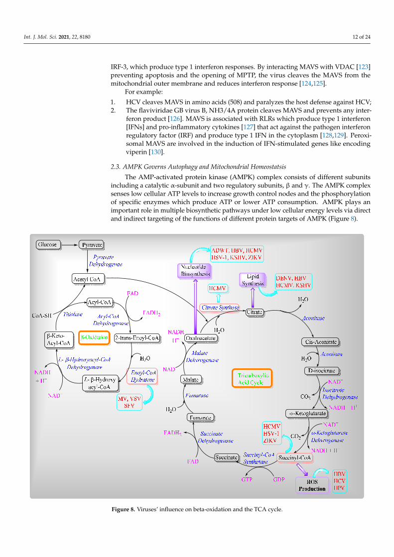
2.3. AMPK Governs Autophagy and Mitochondrial Homeostatsis
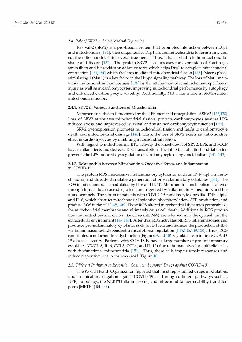
The AMP-activated protein kinase (AMPK) complex consists of different subunitsincluding a catalytic α-subunit and two regulatory subunits, β and γ. The AMPK complexsenses low cellular ATP levels to increase growth control nodes and the phosphorylationof specific enzymes which produce ATP or lower ATP consumption. AMPK plays animportant role in multiple biosynthetic pathways under low cellular energy levels via directand indirect targeting of the functions of different protein targets of AMPK (Figure 8).
Int. J. Mol. Sci. 2021, 22, x FOR PEER REVIEW 14 of 26
2.3. AMPK Governs Autophagy and Mitochondrial Homeostatsis The AMP-activated protein kinase (AMPK) complex consists of different subunits
including a catalytic α-subunit and two regulatory subunits, β and γ. The AMPK complex senses low cellular ATP levels to increase growth control nodes and the phosphorylation of specific enzymes which produce ATP or lower ATP consumption. AMPK plays an im-portant role in multiple biosynthetic pathways under low cellular energy levels via direct and indirect targeting of the functions of different protein targets of AMPK (Figure 8).
Figure 8. Viruses’ influence on beta-oxidation and the TCA cycle.
2.4. Role of SRV2 in Mitochondrial Dynamics Ras val-2 (SRV2) is a pro-fission protein that promotes interaction between Drp1 and
mitochondria [131], then oligomerizes Drp1 around mitochondria to form a ring and cut the mitochondria into several fragments. Thus, it has a vital role in mitochondrial shape and fission [132]. The protein SRV2 also increases the expression of F-actin (as stress fiber) and it provides an adhesive force which helps Drp1 to complete mitochondrial contraction [133,134] which facilates mediated mitochondrial fission [135]. Macro phase stimulating 1 (Mst 1) is a key factor in the Hippo signaling pathway. The loss of Mst 1 maintained mi-tochondrial homeostasis [136] by the attenuation of renal ischemia-reperfusion injury as
Figure 8. Viruses’ influence on beta-oxidation and the TCA cycle.
Int. J. Mol. Sci. 2021, 22, 8180 13 of 24
2.4. Role of SRV2 in Mitochondrial Dynamics
Ras val-2 (SRV2) is a pro-fission protein that promotes interaction between Drp1and mitochondria [131], then oligomerizes Drp1 around mitochondria to form a ring andcut the mitochondria into several fragments. Thus, it has a vital role in mitochondrialshape and fission [132]. The protein SRV2 also increases the expression of F-actin (asstress fiber) and it provides an adhesive force which helps Drp1 to complete mitochondrialcontraction [133,134] which facilates mediated mitochondrial fission [135]. Macro phasestimulating 1 (Mst 1) is a key factor in the Hippo signaling pathway. The loss of Mst 1 main-tained mitochondrial homeostasis [136] by the attenuation of renal ischemia-reperfusioninjury as well as in cardiomyocytes, improving mitochondrial performance by autophagyand enhanced cardiomyocyte viability. Additionally, Mst 1 has a role in SRV2-relatedmitochondrial fission.
2.4.1. SRV2 in Various Functions of Mitochondria
Mitochondrial fission is promoted by the LPS-mediated upregulation of SRV2 [137,138].Loss of SRV2 attenuates mitochondrial fission, protects cardiomyocytes against LPS-induced stress, and improves cell survival and sustained cardiomyocyte function [139].
SRV2 overexpression promotes mitochondrial fission and leads to cardiomyocytedeath and mitochondrial damage [140]. Thus, the loss of SRV2 exerts an antioxidativeeffect in cardiomyocytes by inhibiting mitochondrial fission.
With regard to mitochondrial ETC activity, the knockdown of SRV2, LPS, and FCCPhave similar effects and decrease ETC transcription. The inhibition of mitochondrial fissionprevents the LPS-induced dysregulation of cardiomyocyte energy metabolism [141–143].
2.4.2. Relationship between Mitochondria, Oxidative Stress, and Inflammationin COVID-19
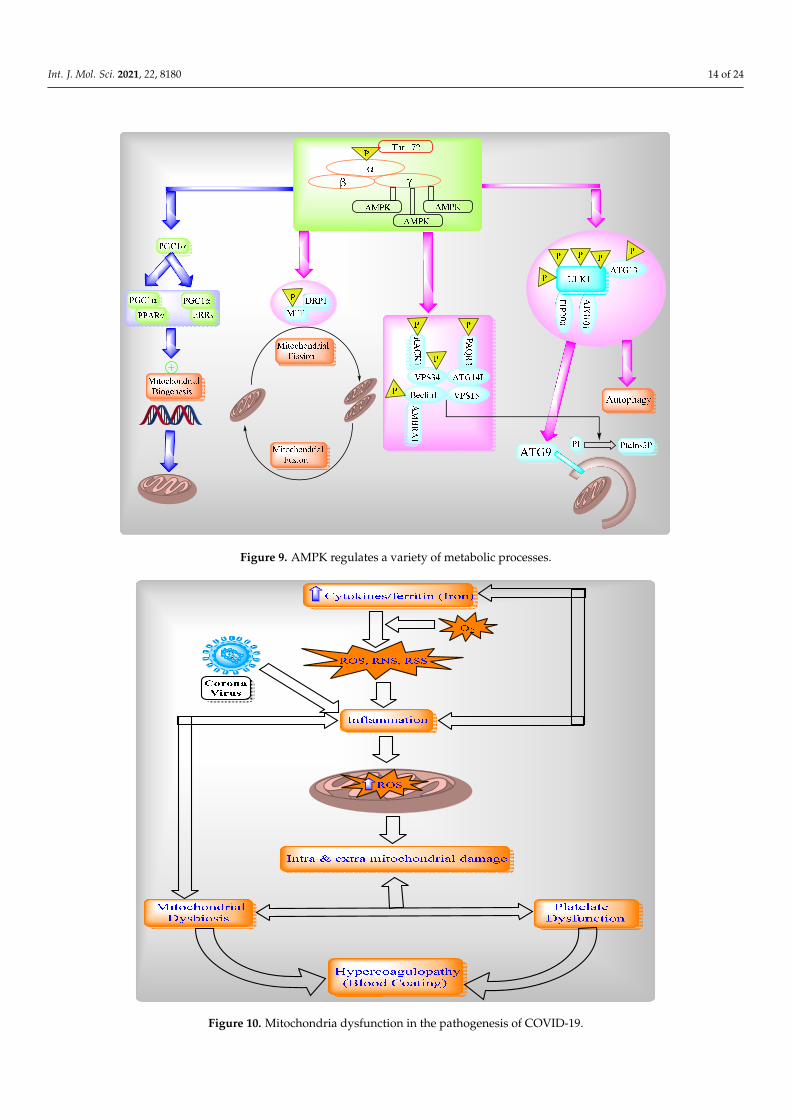
The protein ROS increases via inflammatory cytokines, such as TNF-alpha in mito-chondria, and directly stimulates a generation of pro-inflammatory cytokines [144]. TheROS in mitochondria is modulated by IL-6 and IL-10. Mitochondrial metabolism is alteredthrough intracellular cascades, which are triggered by inflammatory mediators and im-mune sentinels. The serum of patients with COVID-19 contains cytokines like TNF- alphaand IL-6, which obstruct mitochondrial oxidative phosphorylation, ATP production, andproduce ROS in the cell [145,146]. These ROS-altered mitochondrial dynamics permeabilizethe mitochondrial membrane and ultimately cause cell death. Additionally, ROS produc-tion and mitochondrial content (such as mtDNA) are released into the cytosol and theextracellular environment [147,148]. After this, ROS activates NLRP3 inflammasomes andproduces pro-inflammatory cytokines such as IL-1beta and induces the production of IL-6via inflammasome-independent transcriptional regulation [145,146,149,150]. Thus, ROScontributes to mitochondrial dysfunction (Figures 9 and 10). Cytokines can indicate COVID-19 disease severity. Patients with COVID-19 have a large number of pro-inflammatorycytokines (CXCL-8, IL-6, CCL3, CCL4, and IL-12) due to human alveolar epithelial cellswith dysfunctional mitochondria [151]. Thus, these cells impair repair responses andreduce responsiveness to corticosteroid (Figure 10).
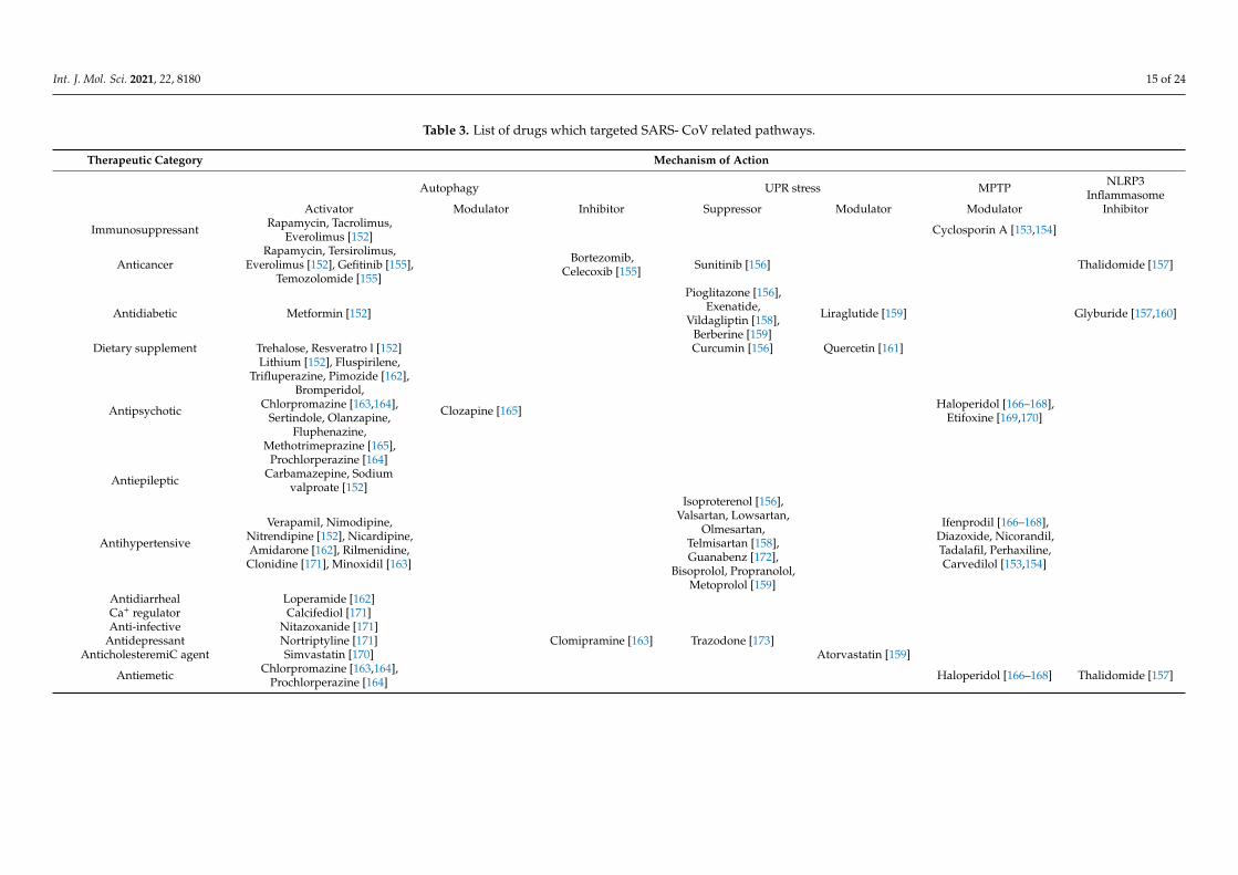
2.5. Different Pathways to Reposition Common Approved Drugs against COVID-19
The World Health Organization reported that most repositioned drugs modulators,under clinical investigation against COVID-19, act through different pathways such asUPR, autophagy, the NLRP3 inflammasome, and mitochondrial permeability transitionpores [MPTP] (Table 3).
Int. J. Mol. Sci. 2021, 22, 8180 14 of 24Int. J. Mol. Sci. 2021, 22, x FOR PEER REVIEW 16 of 26
Figure 9. AMPK regulates a variety of metabolic processes.
Figure 10. Mitochondria dysfunction in the pathogenesis of COVID-19.
Figure 9. AMPK regulates a variety of metabolic processes.
Int. J. Mol. Sci. 2021, 22, x FOR PEER REVIEW 16 of 26
Figure 9. AMPK regulates a variety of metabolic processes.
Figure 10. Mitochondria dysfunction in the pathogenesis of COVID-19. Figure 10. Mitochondria dysfunction in the pathogenesis of COVID-19.
Int. J. Mol. Sci. 2021, 22, 8180 15 of 24
Table 3. List of drugs which targeted SARS- CoV related pathways.
Therapeutic Category Mechanism of Action
Autophagy UPR stress MPTP NLRP3Inflammasome
Activator Modulator Inhibitor Suppressor Modulator Modulator Inhibitor
Immunosuppressant Rapamycin, Tacrolimus,Everolimus [152] Cyclosporin A [153,154]
AnticancerRapamycin, Tersirolimus,
Everolimus [152], Gefitinib [155],Temozolomide [155]
Bortezomib,Celecoxib [155] Sunitinib [156] Thalidomide [157]
Antidiabetic Metformin [152]
Pioglitazone [156],Exenatide,
Vildagliptin [158],Berberine [159]
Liraglutide [159] Glyburide [157,160]
Dietary supplement Trehalose, Resveratro l [152] Curcumin [156] Quercetin [161]
Antipsychotic
Lithium [152], Fluspirilene,Trifluperazine, Pimozide [162],
Bromperidol,Chlorpromazine [163,164],
Sertindole, Olanzapine,Fluphenazine,
Methotrimeprazine [165],Prochlorperazine [164]
Clozapine [165] Haloperidol [166–168],Etifoxine [169,170]
Antiepileptic Carbamazepine, Sodiumvalproate [152]
Antihypertensive
Verapamil, Nimodipine,Nitrendipine [152], Nicardipine,Amidarone [162], Rilmenidine,
Clonidine [171], Minoxidil [163]
Isoproterenol [156],Valsartan, Lowsartan,
Olmesartan,Telmisartan [158],Guanabenz [172],
Bisoprolol, Propranolol,Metoprolol [159]
Ifenprodil [166–168],Diazoxide, Nicorandil,Tadalafil, Perhaxiline,Carvedilol [153,154]
Antidiarrheal Loperamide [162]Ca+ regulator Calcifediol [171]Anti-infective Nitazoxanide [171]
Antidepressant Nortriptyline [171] Clomipramine [163] Trazodone [173]AnticholesteremiC agent Simvastatin [170] Atorvastatin [159]
Antiemetic Chlorpromazine [163,164],Prochlorperazine [164] Haloperidol [166–168] Thalidomide [157]
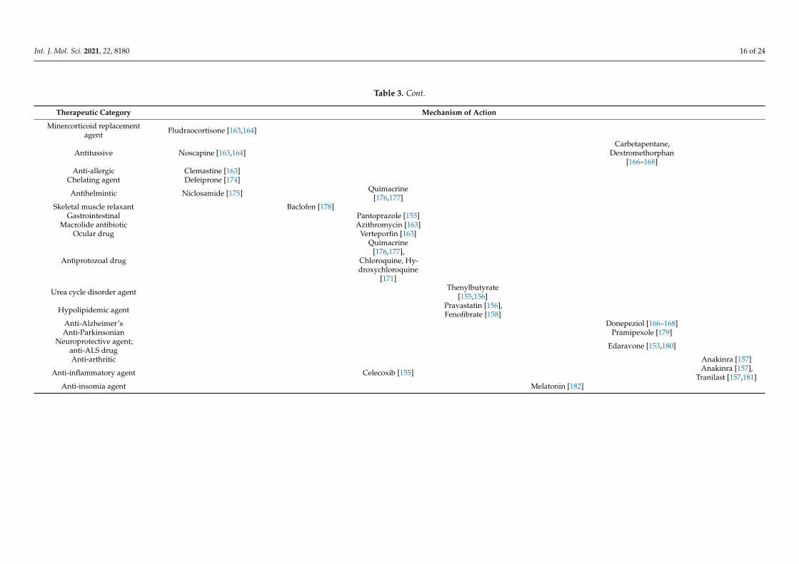
Int. J. Mol. Sci. 2021, 22, 8180 16 of 24
Table 3. Cont.
Therapeutic Category Mechanism of Action
Minercorticoid replacementagent Fludraocortisone [163,164]
Antitussive Noscapine [163,164]Carbetapentane,
Dextromethorphan[166–168]
Anti-allergic Clemastine [163]Chelating agent Defeiprone [174]
Antihelmintic Niclosamide [175] Quimacrine[176,177]
Skeletal muscle relaxant Baclofen [178]Gastrointestinal Pantoprazole [155]
Macrolide antibiotic Azithromycin [163]Ocular drug Verteporfin [163]
Antiprotozoal drug
Quimacrine[176,177],
Chloroquine, Hy-droxychloroquine
[171]
Urea cycle disorder agent Thenylbutyrate[155,156]
Hypolipidemic agent Pravastatin [156],Fenofibrate [158]
Anti-Alzheimer’s Donepeziol [166–168]Anti-Parkinsonian Pramipexole [179]
Neuroprotective agent;anti-ALS drug Edaravone [153,180]
Anti-arthritic Anakinra [157]
Anti-inflammatory agent Celecoxib [155] Anakinra [157],Tranilast [157,181]
Anti-insomia agent Melatonin [182]
Int. J. Mol. Sci. 2021, 22, 8180 17 of 24
3. Expert Opinion
Mitochondria are membrane-bound cell organelles which produce energy in the formof adenosine triphosphate (ATP) as well as regulating various intracellular functions likemetabolism, bioenergetics, cell death, innate immune signaling, and cellular homeostasis.Mitochondria are self-governed by mitochondrial dynamics and mitochondria-selectiveautophagy or mitophagy. During infection, viruses altered mitochondrial dynamics inorder to modulate mitochondria-mediated antiviral immune responses via the alteration ofmitochondrial events such as autophagy, mitophagy, and cellular metabolism to facilitatetheir proliferation.
The pro-fission protein of SRV2 activates mitochondrial fission via the loss of MMP, theROS-overloading suppression antioxidant system, the depletion of cellular ATP, the releaseof the apoptotic factor, the activation of the caspase family, and NLRP3 inflammasomes.The protein SRV2 also promotes mitochondria-associated cardiomyocyte apoptosis to causecardiomyocyte death and mitochondrial damage. The World Health Organization reportedthat most repositioned drugs modulators, under clinical investigation against COVID-19,act through different pathways such as UPR, autophagy, the NLRP3 inflammasome, andmitochondrial permeability transition pores (MPTP) to inhibit SARS-COV2 propagation.Analysis of the functional significance of mitochondrial dynamics and viral pathogenesiswill open up new possibilities for the therapeutic design of approaches to combat viralinfections and associated diseases.
Author Contributions: Conceptualization, S.P.S., methodology, P.G., N.K., S.K.P.; software, P.G.,N.K., S.K.P., A.K.; validation, S.P.S., S.A. and A.K.; formal analysis, S.P.S.; investigation, A.K.;resources, S.P.S.; data curation, S.P.S.; writing—original draft preparation, S.P.S., P.G.; writing—review and editing, S.P.S., S.A., P.G.; visualization, A.K.; supervision, S.P.S.; project administration,S.P.S., S.A., P.G.; funding acquisition, S.A. All authors have read and agreed to the published versionof the manuscript.
Funding: SA is supported by grant support from NHLBI (RO1HL076801) and NIDCR (RO1DE014079).
Acknowledgments: We thank all the authors for carefully reading the manuscript and providingtheir valuable inputs.
Conflicts of Interest: The authors declare no conflict of interest.
References1. Bratic, I.; Trifunovic, A. Mitochondrial energy metabolism and ageing. Biochim. Biophys. Acta (BBA) Bioenerg. 2010, 1797, 961–967.
[CrossRef] [PubMed]2. Westermann, B. Mitochondrial fusion and fission in cell life and death. Nat. Rev. Mol. Cell Biol. 2010, 11, 872–884. [CrossRef]3. James, D.I.; Parone, P.A.; Mattenberger, Y.; Martinou, J.-C. hFis1, a Novel Component of the Mammalian Mitochondrial Fission
Machinery. J. Biol. Chem. 2003, 278, 36373–36379. [CrossRef]4. Smirnova, E.; Shurland, D.-L.; Ryazantsev, S.N.; Van Der Bliek, A.M. A Human Dynamin-related Protein Controls the Distribution
of Mitochondria. J. Cell Biol. 1998, 143, 351–358. [CrossRef] [PubMed]5. Zhu, P.-P.; Patterson, A.; Stadler, J.; Seeburg, D.P.; Sheng, M.; Blackstone, C. Intra- and Intermolecular Domain Interactions of
the C-terminal GTPase Effector Domain of the Multimeric Dynamin-like GTPase Drp1. J. Biol. Chem. 2004, 279, 35967–35974.[CrossRef]
6. Otera, H.; Wang, C.; Cleland, M.M.; Setoguchi, K.; Yokota, S.; Youle, R.J.; Mihara, K. Mff is an essential factor for mitochondrialrecruitment of Drp1 during mitochondrial fission in mammalian cells. J. Cell Biol. 2010, 191, 1141–1158. [CrossRef]
7. Lee, Y.-J.; Jeong, S.-Y.; Karbowski, M.; Smith, C.L.; Youle, R.J. Roles of the Mammalian Mitochondrial Fission and Fusion MediatorsFis1, Drp1, and Opa1 in Apoptosis. Mol. Biol. Cell 2004, 15, 5001–5011. [CrossRef] [PubMed]
8. Chen, H.; Detmer, S.A.; Ewald, A.J.; Griffin, E.E.; Fraser, S.E.; Chan, D.C. Mitofusins Mfn1 and Mfn2 coordinately regulatemitochondrial fusion and are essential for embryonic development. J. Cell Biol. 2003, 160, 189–200. [CrossRef] [PubMed]
9. Olichon, A.; Baricault, L.; Gas, N.; Guillou, E.; Valette, A.; Belenguer, P.; Lenaers, G. Loss of OPA1 Perturbates the MitochondrialInner Membrane Structure and Integrity, Leading to Cytochrome c Release and Apoptosis. J. Biol. Chem. 2003, 278, 7743–7746.[CrossRef]
10. Cipolat, S.; De Brito, O.M.; Zilio, B.D.; Scorrano, L. OPA1 requires mitofusin 1 to promote mitochondrial fusion. Proc. Natl. Acad.Sci. USA 2004, 101, 15927–15932. [CrossRef]

Int. J. Mol. Sci. 2021, 22, 8180 18 of 24
11. Arnoult, D.; Grodet, A.; Lee, Y.-J.; Estaquier, J.; Blackstone, C. Release of OPA1 during Apoptosis Participates in the Rapidand Complete Release of Cytochrome c and Subsequent Mitochondrial Fragmentation. J. Biol. Chem. 2005, 280, 35742–35750.[CrossRef]
12. Frezza, C.; Cipolat, S.; De Brito, O.M.; Micaroni, M.; Beznoussenko, G.V.; Rudka, T.; Bartoli, D.; Polishuck, R.S.; Danial, N.N.;De Strooper, B.; et al. OPA1 Controls Apoptotic Cristae Remodeling Independently from Mitochondrial Fusion. Cell 2006, 126,177–189. [CrossRef]
13. Yasukawa, K.; Oshiumi, H.; Takeda, M.; Ishihara, N.; Yanagi, Y.; Seya, T.; Kawabata, S.-I.; Koshiba, T. Mitofusin 2 InhibitsMitochondrial Antiviral Signaling. Sci. Signal. 2009, 2, ra47. [CrossRef]
14. Biacchesi, S.; LeBerre, M.; Lamoureux, A.; Louise, Y.; Lauret, E.; Boudinot, P.; Brémont, M. Mitochondrial Antiviral SignalingProtein Plays a Major Role in Induction of the Fish Innate Immune Response against RNA and DNA Viruses. J. Virol. 2009, 83,7815–7827. [CrossRef] [PubMed]
15. Castanier, C.; Garcin, D.; Vazquez, A.; Arnoult, D. Mitochondrial dynamics regulate the RIG-I-like receptor antiviral pathway.EMBO Rep. 2010, 11, 133–138. [CrossRef] [PubMed]
16. Horner, S.M.; Liu, H.M.; Park, H.S.; Briley, J.; Gale, J.M.J. Mitochondrial-associated endoplasmic reticulum membranes (MAM)form innate immune synapses and are targeted by hepatitis C virus. Proc. Natl. Acad. Sci. USA 2011, 108, 14590–14595. [CrossRef][PubMed]
17. de Brito, O.M.; Scorrano, L. Mitofusin 2 tethers endoplasmic reticulum to mitochondria. Nature 2008, 456, 605–610. [CrossRef][PubMed]
18. Koshiba, T.; Yasukawa, K.; Yanagi, Y.; Kawabata, S.-I. Mitochondrial Membrane Potential Is Required for MAVS-MediatedAntiviral Signaling. Sci. Signal. 2011, 4, ra7. [CrossRef]
19. Chan, D.C. Mitochondria: Dynamic Organelles in Disease, Aging, and Development. Cell 2006, 125, 1241–1252. [CrossRef][PubMed]
20. Franceschi, C.; Garagnani, P.; Parini, P.; Giuliani, C.; Santoro, A. Inflammaging: A new immune–metabolic viewpoint forage-related diseases. Nat. Rev. Endocrinol. 2018, 14, 576–590. [CrossRef] [PubMed]
21. Bellumkonda, L.; Tyrrell, D.; Hummel, S.L.; Goldstein, D. Pathophysiology of heart failure and frailty: A common inflammatoryorigin? Aging Cell 2017, 16, 444–450. [CrossRef]
22. Cannatà, A.; Marcon, G.; Cimmino, G.; Camparini, L.; Ciucci, G.; Sinagra, G.; Loffredo, F.S. Role of circulating factors in cardiacaging. J. Thorac. Dis. 2017, 9, S17–S29. [CrossRef]
23. Franceschi, C.; Campisi, J. Chronic Inflammation (Inflammaging) and Its Potential Contribution to Age-Associated Diseases. J.Gerontol. Ser. A Biol. Sci. Med. Sci. 2014, 69 (Suppl. S1), S4–S9. [CrossRef] [PubMed]
24. Marín-García, J.; Akhmedov, A.T. Mitochondrial dynamics and cell death in heart failure. Hear. Fail. Rev. 2016, 21, 123–136.[CrossRef]
25. Song, M.; Mihara, K.; Chen, Y.; Scorrano, L.; Dorn, G.W. Mitochondrial Fission and Fusion Factors Reciprocally OrchestrateMitophagic Culling in Mouse Hearts and Cultured Fibroblasts. Cell Metab. 2015, 21, 273–286. [CrossRef] [PubMed]
26. Chen, Y.; Liu, Y.; Dorn, G.W. Mitochondrial Fusion is Essential for Organelle Function and Cardiac Homeostasis. Circ. Res. 2011,109, 1327–1331. [CrossRef] [PubMed]
27. Miyamoto, S. Autophagy and cardiac aging. Cell Death Differ. 2019, 26, 653–664. [CrossRef] [PubMed]28. Seo, A.Y.; Joseph, A.-M.; Dutta, D.; Hwang, J.C.Y.; Aris, J.P.; Leeuwenburgh, C. New insights into the role of mitochondria in
aging: Mitochondrial dynamics and more. J. Cell Sci. 2010, 123, 2533–2542. [CrossRef]29. Wu, N.N.; Zhang, Y.; Ren, J. Mitophagy, Mitochondrial Dynamics, and Homeostasis in Cardiovascular Aging. Oxidative Med. Cell.
Longev. 2019, 2019, 1–15. [CrossRef]30. Yasuda, K.; Ishii, T.; Suda, H.; Akatsuka, A.; Hartman, P.S.; Goto, S.; Miyazawa, M.; Ishii, N. Age-related changes of mitochondrial
structure and function in Caenorhabditis elegans. Mech. Ageing Dev. 2006, 127, 763–770. [CrossRef]31. Park, S.; Won, J.-H.; Hwang, I.; Hong, S.; Lee, H.K.; Yu, J.-W. Defective mitochondrial fission augments NLRP3 inflammasome
activation. Sci. Rep. 2015, 5, 15489. [CrossRef]32. Thai, P.N.; Seidlmayer, L.K.; Miller, C.; Ferrero, M.; Ii, G.W.D.; Schaefer, S.; Bers, D.M.; Dedkova, E.N. Mitochondrial Quality
Control in Aging and Heart Failure: Influence of Ketone Bodies and Mitofusin-Stabilizing Peptides. Front. Physiol. 2019, 10, 382.[CrossRef]
33. Shires, S.E.; Gustafsson, Å.B. Mitophagy and heart failure. J. Mol. Med. Berl. 2015, 93, 253–262. [CrossRef]34. Morciano, G.; Patergnani, S.; Bonora, M.; Pedriali, G.; Tarocco, A.; Bouhamida, E.; Marchi, S.; Ancora, G.; Anania, G.; Wieckowski,
M.R.; et al. Mitophagy in Cardiovascular Diseases. J. Clin. Med. 2020, 9, 892. [CrossRef] [PubMed]35. Kang, Y.; Zhang, H.; Zhao, Y.; Wang, Y.; Wang, W.; He, Y.; Zhang, W.; Zhang, W.; Zhu, X.; Zhou, Y.; et al. Telomere Dysfunction
Disturbs Macrophage Mitochondrial Metabolism and the NLRP3 Inflammasome through the PGC-1α/TNFAIP3 Axis. Cell Rep.2018, 22, 3493–3506. [CrossRef] [PubMed]
36. Van Beek, A.A.; Van den Bossche, J.; Mastroberardino, P.G.; de Winther, M.P.J.; Leenen, P.J.M. Metabolic Alterations in AgingMacrophages: Ingredients for Inflammaging? Trends Immunol. 2019, 40, 113–127. [CrossRef] [PubMed]
37. He, Q.; Li, Z.; Meng, C.; Wu, J.; Zhao, Y. Parkin-Dependent Mitophagy is Required for the Inhibition of ATF4 on NLRP3Inflammasome Activation in Cerebral Ischemia-Reperfusion Injury in Rats. Cells 2019, 8, 897. [CrossRef]
Int. J. Mol. Sci. 2021, 22, 8180 19 of 24
38. Kuma, A.; Mizushima, N.; Ishihara, N.; Ohsumi, Y. Formation of the approximately 350-kDa Apg12-Apg5.Apg16 multimericcomplex, mediated by Apg16 oligomerization, is essential for autophagy in yeast. J. Biol. Chem. 2002, 277, 18619–18625. [CrossRef]
39. Mizushima, N.; Kuma, A.; Kobayashi, Y.; Yamamoto, A.; Matsubae, M.; Takao, T.; Natsume, T.; Ohsumi, Y.; Yoshimori, T. MouseApg16L, a novel WD-repeat protein, targets to the autophagic isolation membrane with the Apg12-Apg5 conjugate. J. Cell Sci.2003, 116, 1679–1688. [CrossRef] [PubMed]
40. Mizushima, N.; Yamamoto, A.; Hatano, M.; Kobayashi, Y.; Kabeya, Y.; Suzuki, K.; Tokuhisa, T.; Ohsumi, Y.; Yoshimori, T.Dissection of Autophagosome Formation Using Apg5-Deficient Mouse Embryonic Stem Cells. J. Cell Biol. 2001, 152, 657–668.[CrossRef]
41. Taguchi-Atarashi, N.; Hamasaki, M.; Matsunaga, K.; Omori, H.; Ktistakis, N.T.; Yoshimori, T.; Noda, T. Modulation of LocalPtdIns3P Levels by the PI Phosphatase MTMR3 Regulates Constitutive Autophagy. Traffic 2010, 11, 468–478. [CrossRef]
42. Vergne, I.; Roberts, E.; Elmaoued, R.A.; Tosch, V.; Delgado, M.A.; Proikas-Cezanne, T.; Deretic, V. Control of autophagy initiationby phosphoinositide 3-phosphatase Jumpy. EMBO J. 2009, 28, 2244–2258. [CrossRef]
43. Hirota, Y.; Tanaka, Y. A small GTPase, human Rab32, is required for the formation of autophagic vacuoles under basal conditions.Cell. Mol. Life Sci. 2009, 66, 2913–2932. [CrossRef]
44. Itoh, T.; Fujita, N.; Kanno, E.; Yamamoto, A.; Yoshimori, T.; Fukuda, M. Golgi-resident Small GTPase Rab33B Interacts withAtg16L and Modulates Autophagosome Formation. Mol. Biol. Cell 2008, 19, 2916–2925. [CrossRef]
45. Gutierrez, M.G.; Munafó, D.B.; Berón, W.; Colombo, M.I. Rab7 is required for the normal progression of the autophagic pathwayin mammalian cells. J. Cell Sci. 2004, 117, 2687–2697. [CrossRef] [PubMed]
46. Jäger, S.; Bucci, C.; Tanida, I.; Ueno, T.; Kominami, E.; Saftig, P.; Eskelinen, E.-L. Role for Rab7 in maturation of late autophagicvacuoles. J. Cell Sci. 2004, 117, 4837–4848. [CrossRef] [PubMed]
47. Itakura, E.; Kishi, C.; Inoue, K.; Mizushima, N. Beclin 1 Forms Two Distinct Phosphatidylinositol 3-Kinase Complexes withMammalian Atg14 and UVRAG. Mol. Biol. Cell 2008, 19, 5360–5372. [CrossRef] [PubMed]
48. Liang, C.; Feng, P.; Ku, B.; Dotan, I.; Canaani, D.; Oh, B.-H.; Jung, J.U. Autophagic and tumour suppressor activity of a novelBeclin1-binding protein UVRAG. Nat. Cell Biol. 2006, 8, 688–698. [CrossRef]
49. Matsunaga, K.; Saitoh, T.; Tabata, K.; Omori, H.; Satoh, T.; Kurotori, N.; Maejima, I.; Shirahama-Noda, K.; Ichimura, T.; Isobe, T.;et al. Two Beclin 1-binding proteins, Atg14L and Rubicon, reciprocally regulate autophagy at different stages. Nat. Cell Biol. 2009,11, 385–396. [CrossRef] [PubMed]
50. Takahashi, Y.; Coppola, D.; Matsushita, N.; Cualing, H.D.; Sun, M.; Sato, Y.; Liang, C.; Jung, J.U.; Cheng, J.Q.; Mul, J.J.; et al. Bif-1interacts with Beclin 1 through UVRAG and regulates autophagy and tumorigenesis. Nat. Cell Biol. 2007, 9, 1142–1151. [CrossRef]
51. Zhong, Y.; Wang, Q.; Li, X.; Yan, Y.; Backer, J.M.; Chait, B.T.; Heintz, N.; Yue, Z. Distinct regulation of autophagic activity byAtg14L and Rubicon associated with Beclin 1–phosphatidylinositol-3-kinase complex. Nat. Cell Biol. 2009, 11, 468–476. [CrossRef][PubMed]
52. Tanida, I.; Ueno, T.; Kominami, E. LC3 and Autophagy. In Methods in Molecular Biology; Springer Science and Business MediaLLC: Tokyo, Japan, 2008; Volume 445, pp. 77–88.
53. Tanida, I.; Wakabayashi, M.; Kanematsu, T.; Minematsu-Ikeguchi, N.; Sou, Y.-S.; Hirata, M.; Ueno, T.; Kominami, E. LysosomalTurnover of GABARAP-Phospholipid Conjugate is Activated During Differentiation of C2C12 Cells to Myotubes withoutInactivation of the mTor Kinase-Signaling Pathway. Autophagy 2006, 2, 264–271. [CrossRef]
54. Caly, L.; Druce, J.D.; Catton, M.G.; Jans, D.A.; Wagstaff, K.M. The FDA-approved drug ivermectin inhibits the replication ofSARS-CoV-2 in vitro. Antivir. Res. 2020, 178, 104787. [CrossRef] [PubMed]
55. Dou, Q.; Chen, H.-N.; Wang, K.; Yuan, K.; Lei, Y.; Li, K.; Lan, J.; Chen, Y.; Huang, Z.; Xie, N.; et al. Ivermectin Induces CytostaticAutophagy by Blocking the PAK1/Akt Axis in Breast Cancer. Cancer Res. 2016, 76, 4457–4469. [CrossRef]
56. Wang, M.; Cao, R.; Zhang, L.; Yang, X.; Liu, J.; Xu, M.; Shi, Z.; Hu, Z.; Zhong, W.; Xiao, G. Remdesivir and chloroquine effectivelyinhibit the recently emerged novel coronavirus (2019-nCoV) in vitro. Cell Res. 2020, 30, 269–271. [CrossRef]
57. Wang, X.; Shen, C.; Liu, Z.; Peng, F.; Chen, X.; Yang, G.; Zhang, D.; Yin, Z.; Ma, J.; Zheng, Z.; et al. Nitazoxanide, an antiprotozoaldrug, inhibits late-stage autophagy and promotes ING1-induced cell cycle arrest in glioblastoma. Cell Death Dis. 2018, 9, 1–15.[CrossRef] [PubMed]
58. Yao, X.; Ye, F.; Zhang, M.; Cui, C.; Huang, B.; Niu, P.; Liu, X.; Zhao, L.; Dong, E.; Song, C.; et al. In Vitro Antiviral Activityand Projection of Optimized Dosing Design of Hydroxychloroquine for the Treatment of Severe Acute Respiratory SyndromeCoronavirus 2 (SARS-CoV-2). Clin. Infect. Dis. 2020, 71, 732–739. [CrossRef]
59. Mauthe, M.; Orhon, I.; Rocchi, C.; Zhou, X.; Luhr, M.; Hijlkema, K.-J.; Coppes, R.P.; Engedal, N.; Mari, M.; Reggiori, F. Chloroquineinhibits autophagic flux by decreasing autophagosome-lysosome fusion. Autophagy 2018, 14, 1435–1455. [CrossRef]
60. Wu, C.-Y.; Jan, J.-T.; Ma, S.-H.; Kuo, C.-J.; Juan, H.-F.; Cheng, Y.-S.E.; Hsu, H.-H.; Huang, H.-C.; Wu, D.; Brik, A.; et al. Smallmolecules targeting severe acute respiratory syndrome human coronavirus. Proc. Natl. Acad. Sci. USA 2004, 101, 10012–10017.[CrossRef] [PubMed]
61. Klein, B.P.; Wörndl, K.; Lütz-Meindl, U.; Kerschbaum, H.H. Perturbation of intracellular K+ homeostasis with valinomycinpromotes cell death by mitochondrial swelling and autophagic processes. Apoptosis 2011, 16, 1101–1117. [CrossRef] [PubMed]
62. Zhu, J.; Yu, W.; Liu, B.; Wang, Y.; Shao, J.; Wang, J.; Xia, K.; Liang, C.; Fang, W.; Zhou, C.; et al. Escin induces caspase-dependentapoptosis and autophagy through the ROS/p38 MAPK signalling pathway in human osteosarcoma cells in vitro and in vivo.Cell Death Dis. 2017, 8, e3113. [CrossRef]
Int. J. Mol. Sci. 2021, 22, 8180 20 of 24
63. Keyaerts, E.; Vijgen, L.; Maes, P.; Neyts, J.; Van Ranst, M. In vitro inhibition of severe acute respiratory syndrome coronavirus bychloroquine. Biochem. Biophys. Res. Commun. 2004, 323, 264–268. [CrossRef]
64. Vincent, M.J.; Bergeron, E.; Benjannet, S.; Erickson, B.R.; Rollin, P.E.; Ksiazek, T.G.; Seidah, N.G.; Nichol, S.T. Chloroquine is apotent inhibitor of SARS coronavirus infection and spread. Virol. J. 2005, 2, 69. [CrossRef] [PubMed]
65. Gassen, N.C.; Niemeyer, D.; Muth, D.; Corman, V.M.; Martinelli, S.; Gassen, A.; Hafner, K.; Papies, J.; Mösbauer, K.; Zellner, A.;et al. SKP2 attenuates autophagy through Beclin1-ubiquitination and its inhibition reduces MERS-Coronavirus infection. Nat.Commun. 2019, 10, 5770. [CrossRef] [PubMed]
66. Malik, S.A.; Orhon, I.; Morselli, E.; Criollo, A.; Shen, S.; Mariño, G.; Ben Younès, A.; Bénit, P.; Rustin, P.; Maiuri, M.C.; et al. BH3mimetics activate multiple pro-autophagic pathways. Oncogene 2011, 30, 3918–3929. [CrossRef] [PubMed]
67. Martinet, W.; Verheye, S.; De Meyer, G. Everolimus-Induced mTOR Inhibition Selectively Depletes Macrophages in AtheroscleroticPlaques by Autophagy. Autophagy 2007, 3, 241–244. [CrossRef]
68. Kindrachuk, J.; Ork, B.; Hart, B.; Mazur, S.; Holbrook, M.R.; Frieman, M.B.; Traynor, D.; Johnson, R.F.; Dyall, J.; Kuhn, J.H.;et al. Antiviral Potential of ERK/MAPK and PI3K/AKT/mTOR Signaling Modulation for Middle East Respiratory SyndromeCoronavirus Infection as Identified by Temporal Kinome Analysis. Antimicrob. Agents Chemother. 2015, 59, 1088–1099. [CrossRef]
69. Noda, T.; Ohsumi, Y. Tor, a phosphatidylinositol kinase homologue, controls autophagy in yeast. J. Biol. Chem. 1998, 273,3963–3966. [CrossRef]
70. Blommaart, E.F.C.; Krause, U.; Schellens, J.P.M.; Vreeling-Sindelarova, H.; Meijer, A.J. The Phosphatidylinositol 3-Kinase InhibitorsWortmannin and LY294002 Inhibit Autophagy in Isolated Rat Hepatocytes. J. Biol. Inorg. Chem. 1997, 243, 240–246. [CrossRef]
71. Zhu, J.-H.; Horbinski, C.; Guo, F.; Watkins, S.; Uchiyama, Y.; Chu, C. Regulation of Autophagy by Extracellular Signal-RegulatedProtein Kinases During 1-Methyl-4-Phenylpyridinium-Induced Cell Death. Am. J. Pathol. 2007, 170, 75–86. [CrossRef]
72. Reggiori, F.; Monastyrska, I.; Verheije, M.H.; Calì, T.; Ulasli, M.; Bianchi, S.; Bernasconi, R.; de Haan, C.A.; Molinari, M.Coronaviruses Hijack the LC3-I-Positive EDEMosomes, ER-Derived Vesicles Exporting Short-Lived ERAD Regulators, forReplication. Cell Host Microbe 2010, 7, 500–508. [CrossRef] [PubMed]
73. Prentice, E.; Jerome, W.G.; Yoshimori, T.; Mizushima, N.; Denison, M.R. Coronavirus Replication Complex Formation UtilizesComponents of Cellular Autophagy. J. Biol. Chem. 2004, 279, 10136–10141. [CrossRef] [PubMed]
74. Seglen, P.O.; Gordon, P.B. 3-Methyladenine: Specific inhibitor of autophagic/lysosomal protein degradation in isolated rathepatocytes. Proc. Natl. Acad. Sci. USA 1982, 79, 1889–1892. [CrossRef]
75. Klionsky, D.J.; Elazar, Z.; Seglen, P.O.; Rubinsztein, D.C. Does bafilomycin A1block the fusion of autophagosomes with lysosomes?Autophagy 2008, 4, 849–850. [CrossRef]
76. Itakura, E.; Kishi-Itakura, C.; Mizushima, N. The Hairpin-type Tail-Anchored SNARE Syntaxin 17 Targets to Autophagosomesfor Fusion with Endosomes/Lysosomes. Cell 2012, 151, 1256–1269. [CrossRef]
77. Cuervo, A.M.; Dice, J.F. A Receptor for the Selective Uptake and Degradation of Proteins by Lysosomes. Sciences 1996, 273,501–503. [CrossRef]
78. Meissner, C.; Lorenz, H.; Weihofen, A.; Selkoe, D.J.; Lemberg, M.K. The mitochondrial intramembrane protease PARL cleaveshuman Pink1 to regulate Pink1 trafficking. J. Neurochem. 2011, 117, 856–867. [CrossRef] [PubMed]
79. Narendra, D.P.; Jin, S.M.; Tanaka, A.; Suen, D.-F.; Gautier, C.A.; Shen, J.; Cookson, M.R.; Youle, R.J. PINK1 is Selectively Stabilizedon Impaired Mitochondria to Activate Parkin. PLoS Biol. 2010, 8, e1000298. [CrossRef]
80. Lazarou, M.; Sliter, D.A.; Kane, L.A.; Sarraf, S.A.; Wang, C.; Burman, J.L.; Sideris, D.P.; Fogel, A.I.; Youle, R.J. The ubiquitin kinasePINK1 recruits autophagy receptors to induce mitophagy. Nature 2015, 524, 309–314. [CrossRef]
81. Heo, J.M.; Ordureau, A.; Paulo, J.A.; Rinehart, J.; Harper, J.W. The PINK1-PARKIN Mitochondrial Ubiquitylation Pathway Drivesa Program of OPTN/NDP52 Recruitment and TBK1 Activation to Promote Mitophagy. Mol. Cell. 2015, 60, 7–20. [CrossRef]
82. Matsumoto, G.; Shimogori, T.; Hattori, N.; Nukina, N. TBK1 controls autophagosomal engulfment of polyubiquitinatedmitochondria through p62/SQSTM1 phosphorylation. Hum. Mol. Genet. 2015, 24, 4429–4442. [CrossRef]
83. Sutovsky, P.; Van Leyen, K.; McCauley, T.; Day, B.N.; Sutovsky, M. Degradation of paternal mitochondria after fertilization:Implications for heteroplasmy, assisted reproductive technologies and mtDNA inheritance. Reprod. Biomed. Online 2004, 8, 24–33.[CrossRef]
84. Rawi, S.; Louvet-Vallée, S.; Djeddi, A.; Sachse, M.; Culetto, E.; Hajjar, C.; Boyd, L.; Legouis, R.; Galy, V. Postfertilization Autophagyof Sperm Organelles Prevents Paternal Mitochondrial DNA Transmission. Science 2011, 334, 1144–1147. [CrossRef] [PubMed]
85. Zhang, C.; Cuervo, A.M. Restoration of chaperone-mediated autophagy in aging liver improves cellular maintenance and hepaticfunction. Nat. Med. 2008, 14, 959–965. [CrossRef] [PubMed]
86. Lipinski, M.M.; Zheng, B.; Lu, T.; Yan, Z.; Py, B.F.; Ng, A.; Li, C.; Yankner, B.A.; Scherzer, C.R.; Yuan, J. Genome-wide analysisreveals mechanisms modulating autophagy in normal brain aging and in Alzheimer’s disease. Proc. Natl. Acad. Sci. USA 2010,107, 14164–14169. [CrossRef]
87. Wang, J.; Ahn, I.; Fischer, T.D.; Byeon, J.; Dunn, W.A.; Behrns, K.E.; Leeuwenburgh, C.; Kim, J. Autophagy Suppresses Age-Dependent Ischemia and Reperfusion Injury in Livers of Mice. Gastroenterology 2011, 141, 2188–2199.e6. [CrossRef]
88. Lemasters, J.J. Selective Mitochondrial Autophagy, or Mitophagy, as a Targeted Defense Against Oxidative Stress, MitochondrialDysfunction, and Aging. Rejuvenation Res. 2005, 8, 3–5. [CrossRef] [PubMed]
89. Aita, V.M.; Liang, X.H.; Murty, V.V.; Pincus, D.L.; Yu, W.; Cayanis, E.; Kalachikov, S.; Gilliam, T.C.; Levine, B. Cloning and genomicorganization of beclin 1, a candidate tumor suppressor gene on chromosome 17q21. Genomics 1999, 59, 59–65. [CrossRef]
Int. J. Mol. Sci. 2021, 22, 8180 21 of 24
90. Cully, M.; You, H.; Levine, A.J.; Mak, T.W. Beyond PTEN mutations: The PI3K pathway as an integrator of multiple inputs duringtumorigenesis. Nat. Rev. Cancer 2006, 6, 184–192. [CrossRef]
91. Liang, J.; Shao, S.H.; Xu, Z.-X.; Hennessy, B.; Ding, Z.; Larrea, M.; Kondo, S.; Dumont, D.J.; Gutterman, J.U.; Walker, C.L.; et al.The energy sensing LKB1–AMPK pathway regulates p27kip1 phosphorylation mediating the decision to enter autophagy orapoptosis. Nat. Cell Biol. 2007, 9, 218–224. [CrossRef]
92. Degtyarev, M.; De Mazière, A.; Orr, C.; Lin, J.; Lee, B.B.; Tien, J.Y.; Prior, W.W.; Dijk, S.v.; Wu, H.; Gray, D.C.; et al. Akt inhibitionpromotes autophagy and sensitizes PTEN-null tumors to lysosomotropic agents. J. Cell Biol. 2008, 183, 101–116. [CrossRef][PubMed]
93. Hezel, A.F.; Bardeesy, N. LKB1; linking cell structure and tumor suppression. Oncogene 2008, 27, 6908–6919. [CrossRef] [PubMed]94. Maurer, I.; Zierz, S.; Möller, H. A selective defect of cytochrome c oxidase is present in brain of Alzheimer disease patients.
Neurobiol. Aging 2000, 21, 455–462. [CrossRef]95. Cui, L.; Jeong, H.; Borovecki, F.; Parkhurst, C.N.; Tanese, N.; Krainc, D. Transcriptional Repression of PGC-1α by Mutant
Huntingtin Leads to Mitochondrial Dysfunction and Neurodegeneration. Cell 2006, 127, 59–69. [CrossRef] [PubMed]96. Casuso, R.A.; Huertas, J.R. The emerging role of skeletal muscle mitochondrial dynamics in exercise and ageing. Ageing Res. Rev.
2020, 58, 101025. [CrossRef] [PubMed]97. Casuso, R.A.; Huertas, J.R. Mitochondrial Functionality in Inflammatory Pathology-Modulatory Role of Physical Activity. Life
2021, 11, 61. [CrossRef]98. Horner, S.M.; Gale, M.J. Regulation of hepatic innate immunity by hepatitis C virus. Nat. Med. 2013, 19, 879–888. [CrossRef]99. Datan, E.; Roy, S.G.; Germain, G.; Zali, N.; McLean, J.E.; Golshan, G.; Harbajan, S.; Lockshin, R.A.; Zakeri, Z. Dengue-induced
autophagy, virus replication and protection from cell death require ER stress (PERK) pathway activation. Cell Death Dis. 2016, 7,e2127. [CrossRef]
100. Liang, Q.; Luo, Z.; Zeng, J.; Chen, W.; Foo, S.-S.; Lee, S.-A.; Ge, J.; Wang, S.; Glodman, S.A.; Zlokovic, B.V.; et al. Zika Virus NS4Aand NS4B Proteins Deregulate Akt-mTOR Signaling in Human Fetal Neural Stem Cells to Inhibit Neurogenesis and InduceAutophagy. Cell Stem Cell 2016, 19, 663–671. [CrossRef] [PubMed]
101. Joubert, P.-E.; Werneke, S.W.; De La Calle, C.; Guivel-Benhassine, F.; Giodini, A.; Peduto, L.; Levine, B.; Schwartz, O.; Lenschow,D.J.; Albert, M.L. Chikungunya virus–induced autophagy delays caspase-dependent cell death. J. Exp. Med. 2012, 209, 1029–1047.[CrossRef] [PubMed]
102. Lee, Y.-R.; Lei, H.-Y.; Liu, M.-T.; Wang, J.-R.; Chen, S.-H.; Jiang-Shieh, Y.-F.; Lin, Y.-S.; Yeh, T.-M.; Liu, C.-C.; Liu, H.-S. Autophagicmachinery activated by dengue virus enhances virus replication. Virology 2008, 374, 240–248. [CrossRef]
103. McLean, J.E.; Wudzinska, A.; Datan, E.; Quaglino, D.; Zakeri, Z. Flavivirus NS4A-induced Autophagy Protects Cells againstDeath and Enhances Virus Replication. J. Biol. Chem. 2011, 286, 22147–22159. [CrossRef]
104. Zhu, L.; Mou, C.; Yang, X.; Lin, J.; Yang, Q. Mitophagy in TGEV infection counteracts oxidative stress and apoptosis. Oncotarget2016, 7, 27122–27141. [CrossRef] [PubMed]
105. Meng, G.; Xia, M.; Wang, D.; Chen, A.; Wang, Y.; Wang, H.; Yu, D.; Wei, J. Mitophagy promotes replication of oncolytic Newcastledisease virus by blocking intrinsic apoptosis in lung cancer cells. Oncotarget 2014, 5, 6365–6374. [CrossRef] [PubMed]
106. Barbier, V.; Lang, D.; Valois, S.; Rothman, A.; Medin, C.L. Dengue virus induces mitochondrial elongation through impairment ofDrp1-triggered mitochondrial fission. Virology 2017, 500, 149–160. [CrossRef] [PubMed]
107. Yu, C.-Y.; Liang, J.-J.; Li, J.-K.; Lee, Y.-L.; Chang, B.-L.; Su, C.-I.; Huang, W.-J.; Lai, M.M.C.; Lin, Y.-L. Dengue Virus ImpairsMitochondrial Fusion by Cleaving Mitofusins. PLoS Pathog. 2015, 11, e1005350. [CrossRef]
108. Zamarin, D.; Garcia-Sastre, A.; Xiao, X.; Wang, R.; Palese, P. Influenza Virus PB1-F2 Protein Induces Cell Death throughMitochondrial ANT3 and VDAC1. PLoS Pathog. 2005, 1, e4. [CrossRef]
109. Kim, S.-J.; Syed, G.; Siddiqui, A. Hepatitis C Virus Induces the Mitochondrial Translocation of Parkin and Subsequent Mitophagy.PLoS Pathog. 2013, 9, e1003285. [CrossRef] [PubMed]
110. Gou, H.; Zhao, M.; Xu, H.; Yuan, J.; He, W.; Zhu, M.; Ding, H.; Yi, L.; Chen, J. CSFV induced mitochondrial fission and mitophagyto inhibit apoptosis. Oncotarget 2017, 8, 39382–39400. [CrossRef] [PubMed]
111. Ding, B.; Zhang, L.; Li, Z.; Zhong, Y.; Tang, Q.; Qin, Y.; Chen, M. The Matrix Protein of Human Parainfluenza Virus Type 3 InducesMitophagy that Suppresses Interferon Responses. Cell Host Microbe 2017, 21, 538–547.e4. [CrossRef]
112. Xia, M.; Meng, G.; Jiang, A.; Chen, A.; Dahlhaus, M.; Gonzalez, P.; Beltinger, C.; Wei, J. Mitophagy switches cell death fromapoptosis to necrosis in NSCLC cells treated with oncolytic measles virus. Oncotarget 2014, 5, 3907–3918. [CrossRef]
113. Dasgupta, A.; Wilson, D.W. ATP depletion blocks herpes simplex virus DNA packaging and capsid maturation. J. Virol. 1999, 73,2006–2015. [CrossRef]
114. Hui, E.K.-W.; Nayak, D.P. Role of ATP in Influenza Virus Budding. Virology 2001, 290, 329–341. [CrossRef]115. Tritel, M.; Resh, M.D. The Late Stage of Human Immunodeficiency Virus Type 1 Assembly Is an Energy-Dependent Process. J.
Virol. 2001, 75, 5473–5481. [CrossRef]116. Green, D.R.; Reed, J.C. Mitochondria and Apoptosis. Science 1998, 281, 1309. [CrossRef]117. Liu, Y.; Gao, L.; Xue, Q.; Li, Z.; Wang, L.; Chen, R.; Liu, M.; Wen, Y.; Guan, M.; Li, Y.; et al. Voltage-dependent anion channel
involved in the mitochondrial calcium cycle of cell lines carrying the mitochondrial DNA A4263G mutation. Biochem. Biophys.Res. Commun. 2011, 404, 364–369. [CrossRef] [PubMed]

Int. J. Mol. Sci. 2021, 22, 8180 22 of 24
118. Hüttemann, M.; Lee, I.; Pecinova, A.; Pecina, P.; Przyklenk, K.; Doan, J.W. Regulation of oxidative phosphorylation, themitochondrial membrane potential, and their role in human disease. J. Bioenerg. Biomembr. 2008, 40, 445–456. [CrossRef][PubMed]
119. Gradzka, I. Mechanisms and regulation of the programmed cell death. Postepy Biochem. 2006, 52, 157–165.120. Yoneyama, M.; Kikuchi, M.; Natsukawa, T.; Shinobu, N.; Imaizumi, T.; Miyagishi, M.; Taira, K.; Akira, S.; Fujita, T. The RNA
helicase RIG-I has an essential function in double-stranded RNA-induced innate antiviral responses. Nat. Immunol. 2004, 5,730–737. [CrossRef] [PubMed]
121. Maniatis, T.; Falvo, J.V.; Kim, T.H.; Kim, T.K.; Lin, C.H.; Parekh, B.S.; Wathelet, M.G. Structure and function of the interferon-betaenhanceosome. Cold Spring Harb. Symp. Quant. Biol. 1998, 63, 609–620. [CrossRef]
122. Seth, R.B.; Sun, L.; Ea, C.K.; Chen, Z.J. Identification and characterization of MAVS, a mitochondrial antiviral signaling proteinthat activates NF-kappaB and IRF 3. Cell 2005, 122, 669–682. [CrossRef] [PubMed]
123. Meylan, E.; Curran, J.; Hofmann, K.; Moradpour, D.; Binder, M.; Bartenschlager, R.; Tschopp, J. Cardif is an adaptor protein in theRIG-I antiviral pathway and is targeted by hepatitis C virus. Nat. Cell Biol. 2005, 437, 1167–1172. [CrossRef] [PubMed]
124. Retracted article: MAVS protects cells from apoptosis by negatively regulating VDAC1. Mol. Cell Biochem. 2013, 375, 219.125. Li, X.D.; Sun, L.; Seth, R.B.; Pineda, G.; Chen, Z.J. Hepatitis C virus protease NS3/4A cleaves mitochondrial antiviral signaling
protein off the mitochondria to evade innate immunity. Proc. Natl. Acad. Sci. USA 2005, 102, 17717–17722. [CrossRef]126. Chen, Z.; Benureau, Y.; Rijnbrand, R.; Yi, J.; Wang, T.; Warter, L.; Lanford, R.E.; Weinman, S.A.; Lemon, S.M.; Martin, A.; et al.
GB virus B disrupts RIG-I signaling by NS3/4A-mediated cleavage of the adaptor protein MAVS. J. Virol. 2007, 81, 964–976.[CrossRef]
127. Yamada, S.; Shimojima, M.; Narita, R.; Tsukamoto, Y.; Kato, H.; Saijo, M.; Fujita, T. RIG-I-Like Receptor and Toll-Like Receptor Sig-naling Pathways Cause Aberrant Production of Inflammatory Cytokines/Chemokines in a Severe Fever with ThrombocytopeniaSyndrome Virus Infection Mouse Model. J. Virol. 2018, 92, e02246-17. [CrossRef]
128. Hou, F.; Sun, L.; Zheng, H.; Skaug, B.; Jiang, Q.X.; Chen, Z.J. MAVS forms functional prion-like aggregates to activate andpropagate antiviral innate immune response. Cell 2011, 146, 448–461. [CrossRef]
129. Xu, H.; He, X.; Zheng, H.; Huang, L.J.; Hou, F.; Yu, Z.; Michael, J.D.L.C.; Brian, B.; Zhang, X.; Chen, Z.J.; et al. Structural basis forthe prion-like MAVS filaments in antiviral innate immunity. eLife 2014, 3, e01489. [CrossRef] [PubMed]
130. Dixit, E.; Boulant, S.; Zhang, Y.; Lee, A.S.; Odendall, C.; Shum, B.; Hacohen, N.; Chen, Z.; Whelan, S.; Fransen, M.; et al.Peroxisomes Are Signaling Platforms for Antiviral Innate Immunity. Cell 2010, 141, 668–681. [CrossRef]
131. Ibáñez Cabellos, J.S.; Pérez-Machado, G.; Seco-Cervera, M.; Berenguer-Pascual, E.; García-Giménez, J.; Pallardó, F. Acutetelomerase components depletion triggers oxidative stress as an early event previous to telomeric shortening. Redox Biol. 2017, 14,398–408. [CrossRef]
132. Cao, J.; Wang, X.; Dai, T.; Wu, Y.; Zhang, M.; Cao, R.; Zhang, R.; Wang, G.; Jiang, R.; Zhou, B.P.; et al. Twist promotes tumormetastasis in basal-like breast cancer by transcriptionally upregulating ROR1. Theranostics 2018, 8, 2739–2751. [CrossRef]
133. Galluzzi, L.; Vitale, I.; Aaronson, S.A.; Abrams, J.M.; Adam, D.; Agostinis, P.; Alnemri, E.S.; Altucci, L.; Amelio, I.; Andrews, D.W.;et al. Molecular mechanisms of cell death: Recommendations of the Nomenclature Committee on Cell Death 2018. Cell DeathDiffer. 2018, 25, 486–541. [CrossRef] [PubMed]
134. Liu, L.; Jin, X.; Hu, C.-F.; Zhang, Y.-P.; Zhou, Z.; Li, R.; Shen, C.-X. Amphiregulin enhances cardiac fibrosis and aggravates cardiacdysfunction in mice with experimental myocardial infarction partly through activating EGFR-dependent pathway. Basic Res.Cardiol. 2018, 113, 12. [CrossRef] [PubMed]
135. Itteboina, R.; Ballu, S.; Sivan, S.K.; Manga, V. Molecular docking, 3D-QSAR, molecular dynamics, synthesis and anticancer activityof tyrosine kinase 2 (TYK 2) inhibitors. J. Recept. Signal Transduct. 2018, 38, 462–474. [CrossRef] [PubMed]
136. Aanhane, E.; Schulkens, I.A.; Heusschen, R.; Castricum, K.; Leffler, H.; Griffioen, A.W.; Thijssen, V.L. Different angioregulatoryactivity of monovalent galectin-9 isoforms. Angiogenesis 2018, 21, 545–555. [CrossRef] [PubMed]
137. Audia, J.P.; Yang, X.-M.; Crockett, E.S.; Housley, N.; Haq, E.U.; O’Donnell, K.; Cohen, M.V.; Downey, J.M.; Alvarez, D.F. Caspase-1inhibition by VX-765 administered at reperfusion in P2Y12 receptor antagonist-treated rats provides long-term reduction inmyocardial infarct size and preservation of ventricular function. Basic Res. Cardiol. 2018, 113, 32. [CrossRef] [PubMed]
138. Erland, L.A.E.; Yasunaga, A.; Li, I.T.S.; Murch, S.J.; Saxena, P.K. Direct visualization of location and uptake of applied melatoninand serotonin in living tissues and their redistribution in plants in response to thermal stress. J. Pineal Res. 2018, 66, e12527.[CrossRef]
139. Ko, A.; Han, S.Y.; Choi, C.H.; Cho, H.; Lee, M.-S.; Kim, S.-Y.; Song, J.S.; Hong, K.-M.; Lee, H.-W.; Hewitt, S.M.; et al. Oncogene-induced senescence mediated by c-Myc requires USP10 dependent deubiquitination and stabilization of p14ARF. Cell Death Differ.2018, 25, 1050–1062. [CrossRef] [PubMed]
140. Jain, R.; Mintern, J.D.; Tan, I.; Dewson, G.; Strasser, A.; Gray, D.H.D. How do thymic epithelial cells die? Cell Death Differ. 2018, 25,1002–1004. [CrossRef]
141. Dickinson, J.D.; Sweeter, J.M.; Warren, K.J.; Ahmad, I.; De Deken, X.; Zimmerman, M.C.; Brody, S.L. Autophagy regulates DUOX1localization and superoxide production in airway epithelial cells during chronic IL-13 stimulation. Redox Biol. 2018, 14, 272–284.[CrossRef] [PubMed]
Int. J. Mol. Sci. 2021, 22, 8180 23 of 24
142. Köhler, D.; Bibli, S.-I.; Klammer, L.P.; Roth, J.M.; Lehmann, R.; Fleming, I.; Granja, T.F.; Straub, A.; Benz, P.M.; Rosenberger, P.Phosphorylation of vasodilator-stimulated phosphoprotein contributes to myocardial ischemic preconditioning. Basic Res. Cardiol.2018, 113, 11. [CrossRef]
143. Gandarillas, A.; Molinuevo, R.; Sanz-Gómez, N. Mammalian endoreplication emerges to reveal a potential developmental timer.Cell Death Differ. 2018, 25, 471–476. [CrossRef] [PubMed]
144. Li, X.; Fang, P.; Mai, J.; Choi, E.T.; Wang, H.; Yang, X.-F. Targeting mitochondrial reactive oxygen species as novel therapy forinflammatory diseases and cancers. J. Hematol. Oncol. 2013, 6, 19. [CrossRef]
145. Jo, E.-K.; Kim, J.K.; Shin, D.-M.; Sasakawa, C. Molecular mechanisms regulating NLRP3 inflammasome activation. Cell. Mol.Immunol. 2016, 13, 148–159. [CrossRef] [PubMed]
146. Naik, E.; Dixit, V.M. Mitochondrial reactive oxygen species drive proinflammatory cytokine production. J. Exp. Med. 2011, 208,417–420. [CrossRef] [PubMed]
147. Twig, G.; Shirihai, O.S. The Interplay Between Mitochondrial Dynamics and Mitophagy. Antioxid. Redox Signal. 2011, 14,1939–1951. [CrossRef]
148. Mittal, M.; Siddiqui, M.R.; Tran, K.; Reddy, S.P.; Malik, A.B. Reactive Oxygen Species in Inflammation and Tissue Injury. Antioxid.Redox Signal. 2014, 20, 1126–1167. [CrossRef]
149. West, A.P.; Khoury-Hanold, W.; Staron, M.; Tal, M.; Pineda, C.M.; Lang, S.M.; Bestwick, M.; Duguay, B.A.; Raimundo, N.;MacDuff, D.A.; et al. Mitochondrial DNA stress primes the antiviral innate immune response. Nat. Cell Biol. 2015, 520, 553–557.[CrossRef]
150. Nakahira, K.; Haspel, J.A.; Rathinam, V.A.; Lee, S.-J.; Dolinay, T.; Lam, H.C.; Englert, J.A.; Rabinovitch, M.; Cernadas, M.; Kim,H.P.; et al. Autophagy proteins regulate innate immune responses by inhibiting the release of mitochondrial DNA mediated bythe NALP3 inflammasome. Nat. Immunol. 2011, 12, 222–230. [CrossRef]
151. Zhou, F.; Yu, T.; Du, R.; Fan, G.; Liu, Y.; Liu, Z.; Xiang, J.; Wang, Y.; Song, B.; Gu, X.; et al. Clinical course and risk factorsfor mortality of adult inpatients with COVID-19 in Wuhan, China: A retrospective cohort study. Lancet 2020, 395, 1054–1062.[CrossRef]
152. Thellung, S.; Corsaro, A.; Nizzari, M.; Barbieri, F.; Florio, T. Autophagy Activator Drugs: A New Opportunity in Neuroprotectionfrom Misfolded Protein Toxicity. Int. J. Mol. Sci. 2019, 20, 901. [CrossRef] [PubMed]
153. Szewczyk, A.; Wojtczak, L. Mitochondria as a pharmacological target. Pharmacol. Rev. 2002, 54, 101–127. [CrossRef]154. Javadov, S.; Karmazyn, M. Mitochondrial Permeability Transition Pore Opening as an Endpoint to Initiate Cell Death and as a
Putative Target for Cardioprotection. Cell. Physiol. Biochem. 2006, 20, 1–22. [CrossRef] [PubMed]155. Kondapuram, S.K.; Sarvagalla, S.; Coumar, M.S. Targeting autophagy with small molecules for cancer therapy. J. Cancer Metastasis
Treat. 2019, 2019. [CrossRef]156. Minamino, T.; Komuro, I.; Kitakaze, M. Endoplasmic Reticulum Stress As a Therapeutic Target in Cardiovascular Disease. Circ.
Res. 2010, 107, 1071–1082. [CrossRef]157. Xu, S.; Li, X.; Liu, Y.; Xia, Y.; Chang, R.; Zhang, C. Inflammasome inhibitors: Promising therapeutic approaches against cancer. J.
Hematol. Oncol. 2019, 12, 1–13. [CrossRef] [PubMed]158. Jung, T.W.; Choi, K.M. Pharmacological Modulators of Endoplasmic Reticulum Stress in Metabolic Diseases. Int. J. Mol. Sci. 2016,
17, 192. [CrossRef] [PubMed]159. Amen, O.M.; Sarker, S.D.; Ghildyal, R.; Arya, A. Endoplasmic Reticulum Stress Activates Unfolded Protein Response Signaling
and Mediates Inflammation, Obesity, and Cardiac Dysfunction: Therapeutic and Molecular Approach. Front. Pharmacol. 2019,10, 977. [CrossRef]
160. Zahid, A.; Li, B.; Kombe, A.J.K.; Jin, T.; Tao, J. Pharmacological Inhibitors of the NLRP3 Inflammasome. Front. Immunol. 2019,10, 2538. [CrossRef]
161. Rivas, A.; Vidal, R.L.; Hetz, C. Targeting the unfolded protein response for disease intervention. Expert Opin. Ther. Targets 2015,19, 1–16. [CrossRef]
162. Zhang, L.; Yu, J.; Pan, H.; Hu, P.; Hao, Y.; Cai, W.; Zhu, H.; Yu, A.D.; Xie, X.; Ma, D.; et al. Small molecule regulators of autophagyidentified by an image-based high-throughput screen. Proc. Natl. Acad. Sci. USA 2007, 104, 19023–19028. [CrossRef]
163. Vakifahmetoglu-Norberg, H.; Xia, H.-G.; Yuan, J. Pharmacologic agents targeting autophagy. J. Clin. Investig. 2015, 125, 5–13.[CrossRef]
164. Shaw, S.Y.; Tran, K.; Castoreno, A.B.; Peloquin, J.M.; Lassen, K.G.; Khor, B.; Aldrich, L.; Tan, P.H.; Graham, D.B.; Kuballa, P.; et al.Selective Modulation of Autophagy, Innate Immunity, and Adaptive Immunity by Small Molecules. ACS Chem. Biol. 2013, 8,2724–2733. [CrossRef] [PubMed]
165. Vucicevic, L.; Misirkic-Marjanovic, M.; Harhaji-Trajkovic, L.; Maric, N.; Trajkovic, V. Mechanisms and therapeutic significance ofautophagy mdulation by antipsychotic drugs. Cell Stress 2018, 2, 282–291. [CrossRef]
166. Christ, M.G.; Clement, A.M.; Behl, C. The Sigma-1 Receptor at the Crossroad of Proteostasis, Neurodegeneration, and Autophagy.Trends Neurosci. 2020, 43, 79–81. [CrossRef] [PubMed]
167. Hayashi, T.; Tsai, S.-Y.; Mori, T.; Fujimoto, M.; Su, T.-P. Targeting ligand-operated chaperone sigma-1 receptors in the treatment ofneuropsychiatric disorders. Expert Opin. Ther. Targets 2011, 15, 557–577. [CrossRef]
168. Albayrak, Y.; Hashimoto, K. Sigma-1 Receptor Agonists and Their Clinical Implications in Neuropsychiatric Disorders. Adv. Exp.Med. Biol. 2017, 964, 153–161. [CrossRef] [PubMed]
Int. J. Mol. Sci. 2021, 22, 8180 24 of 24
169. Liere, P.; Pianos, A.; Oudinet, J.-P.; Schumacher, M.; Akwa, Y. Differential effects of the 18-kDa translocator protein (TSPO) ligandetifoxine on steroidogenesis in rat brain, plasma and steroidogenic glands: Pharmacodynamic studies. Psychoneuroendocrinology2017, 83, 122–134. [CrossRef]
170. Costa, B.; Cavallini, C.; Da Pozzo, E.; Taliani, S.; Da Settimo, F.; Martini, C. The Anxiolytic Etifoxine Binds to TSPO Ro5-4864Binding Site with Long Residence Time Showing a High Neurosteroidogenic Activity. ACS Chem. Neurosci. 2017, 8, 1448–1454.[CrossRef] [PubMed]
171. Panda, P.K.; Fahrner, A.; Vats, S.; Seranova, E.; Sharma, V.; Chipara, M.; Desai, P.; Torresi, J.; Rosenstock, T.; Kumar, D.; et al.Chemical Screening Approaches Enabling Drug Discovery of Autophagy Modulators for Biomedical Applications in HumanDiseases. Front. Cell Dev. Biol. 2019, 7, 38. [CrossRef] [PubMed]
172. Tsaytler, P.; Harding, H.; Ron, D.; Bertolotti, A. Selective Inhibition of a Regulatory Subunit of Protein Phosphatase 1 RestoresProteostasis. Science 2011, 332, 91–94. [CrossRef]
173. Pérez-Arancibia, R.; Rivas, A.; Hetz, C.; Pérez, R. (off)Targeting UPR signaling: The race toward intervening ER proteostasis.Expert Opin. Ther. Targets 2017, 22, 97–100. [CrossRef] [PubMed]
174. Allen, G.F.G.; Toth, R.; James, J.; Ganley, I. Loss of iron triggers PINK1/Parkin-independent mitophagy. EMBO Rep. 2013, 14,1127–1135. [CrossRef] [PubMed]
175. Renna, M.; Jimenez-Sanchez, M.; Sarkar, S.; Rubinsztein, D.C. Chemical Inducers of Autophagy That Enhance the Clearance ofMutant Proteins in Neurodegenerative Diseases. J. Biol. Chem. 2010, 285, 11061–11067. [CrossRef]
176. Goodall, M.L.; Wang, T.; Martin, K.R.; Kortus, M.G.; Kauffman, A.L.; Trent, J.M.; Gately, S.; MacKeigan, J.P. Development ofpotent autophagy inhibitors that sensitize oncogenic BRAF V600E mutant melanoma tumor cells to vemurafenib. Autophagy 2014,10, 1120–1136. [CrossRef] [PubMed]
177. Khurana, A.; Roy, D.; Kalogera, E.; Mondal, S.; Wen, X.; He, X.; Dowdy, S.; Shridhar, V. Quinacrine promotes autophagic cell deathand chemosensitivity in ovarian cancer and attenuates tumor growth. Oncotarget 2015, 6, 36354–36369. [CrossRef] [PubMed]
178. Liu, L.; Li, C.-J.; Lu, Y.; Zong, X.-G.; Luo, C.; Sun, J.; Guo, L.-J. Baclofen mediates neuroprotection on hippocampal CA1 pyramidalcells through the regulation of autophagy under chronic cerebral hypoperfusion. Sci. Rep. 2015, 5, srep14474. [CrossRef]
179. Ferrari-Toninelli, G.; Maccarinelli, G.; Uberti, D.L.; Buerger, E.; Memo, M. Mitochondria-targeted antioxidant effects of S(-) andR(+) pramipexole. BMC Pharmacol. 2010, 10, 2. [CrossRef]
180. Takayasu, Y.; Nakaki, J.; Kawasaki, T.; Koda, K.; Ago, Y.; Baba, A.; Matsuda, T. Edaravone, a Radical Scavenger, InhibitsMitochondrial Permeability Transition Pore in Rat Brain. J. Pharmacol. Sci. 2007, 103, 434–437. [CrossRef]
181. Huang, Y.; Jiang, H.; Chen, Y.; Wang, X.; Yang, Y.; Tao, J.; Deng, X.; Liang, G.; Zhang, H.; Jiang, W.; et al. Tranilast directly targetsNLRP 3 to treat inflammasome-driven diseases. EMBO Mol. Med. 2018, 10, e8689. [CrossRef]
182. Almanza, A.; Carlesso, A.; Chintha, C.; Creedican, S.; Doultsinos, D.; Leuzzi, B.; Luís, A.; McCarthy, N.; Montibeller, L.; More,S.; et al. Endoplasmic reticulum stress signaling—from basic mechanisms to clinical applications. FEBS J. 2019, 286, 241–278.[CrossRef] [PubMed]
Related Documents