Review Mitochondrial ion channels as therapeutic targets Pablo M. Peixoto, Shin-Young Ryu, Kathleen W. Kinnally * New York University College of Dentistry, New York, NY 10010, USA article info Article history: Received 16 December 2009 Revised 12 February 2010 Accepted 16 February 2010 Available online 20 February 2010 Edited by Adam Szewczyk Keywords: Mitochondrial ion channels Patch clamp Mitochondrial disease Therapy abstract The study of mitochondrial ion channels changed our perception of these double-wrapped organ- elles from being just the power house of a cell to the guardian of a cell’s fate. Mitochondria commu- nicate with the cell through these special channels. Most of the time, the message is encoded by ion flow across the mitochondrial outer and inner membranes. Potassium, sodium, calcium, protons, nucleotides, and proteins traverse the mitochondrial membranes in an exquisitely regulated man- ner to control a myriad of processes, from respiration and mitochondrial morphology to cell prolif- eration and cell death. This review is an update on both well established and putative mitochondrial channels regarding their composition, function, regulation, and therapeutic potential. Ó 2010 Federation of European Biochemical Societies. Published by Elsevier B.V. All rights reserved. 1. Introduction Ion channels are central to communication within the body; they are responsible for your thought processes and keep your heart beating. Ion channels are the target of most pharmaceutical products. Hence, it is not surprising that mitochondria rely heavily on a battery of ion channels to interface the communication be- tween the cytosol and the site of energy production, which also holds the key to life and death decisions. Here, we will describe the multiple channels of the outer and inner membranes of mito- chondria that orchestrate a myriad of cellular processes from ATP, steroid, and heme synthesis to protein import and apoptosis. As we are electrophysiologists, we will use the single channel behavior of these channels as our central theme. During the 70s and 80s the notion that mitochondria might have channels was typically dismissed because the consensus was that mitochondria could not possibly maintain the membrane potential and high resistance essential to oxidative phosphoryla- tion if they had channels; opening channels would cause uncou- pling. Historically, VDAC (voltage dependent anion-selective channel), or mitochondrial porin, was the first channel identified in mitochondria. Isolated from paramecium mitochondria, VDAC’s activity was characterized in planar bilayers in a report in 1976 by Schein et al. [1]. Hence, the molecular identity and function of VDAC has been known for many years. The first characterization of the permeability transition pore or PTP quickly followed that of VDAC in 1979, in a series of articles by Hunter and Haworth [2]; these were not electrophysiological, but rather based on swell- ing assays using a photometric approach. At that time, few appre- ciated the impact of this channel, which we now realize is central to ischemia-reperfusion injury and necrosis. Our group and that of Zoratti independently characterized PTP single channel behavior 10 years later by patch clamping mitoplasts, which are mitochon- dria stripped to reveal the inner membrane [3,4]. Intriguingly, 30 years after the initial reports, the molecular identity and func- tion of PTP remain a matter of speculation and open argument. Nevertheless, the persistent application of patch clamping tech- niques to mitoplasts led to the discovery of many other intriguing channels. Sorgato first reported the existence of a novel anion- selective channel, mCS (mitochondrial Centum-pico-Siemen chan- nel) in mitoplasts; the molecular identity and function of this channel remains elusive [5]. Not much later, our lab found the TIM23 channel, which was the first electrophysiological demon- stration of the link between protein import and water-filled chan- nels in mitochondria [6]. This was quickly followed by a report from Juin et al. on the channel activity of TOM, the protein import complex in the outer membrane [7] (Fig. 1). And, there are numer- ous other channels like AAA, ACA, TIM22, and various K + channels, which were discovered by applying these techniques to native membranes. Here, we will provide you with an update on where 0014-5793/$36.00 Ó 2010 Federation of European Biochemical Societies. Published by Elsevier B.V. All rights reserved. doi:10.1016/j.febslet.2010.02.046 Abbreviations: AAA, alkaline-induced anion-selective activity; ACA, alkaline- induced cation-selective activity; ANT, adenosine nucleotide translocator; CLIC, chloride intracellular channel; IMAC, inner membrane anion channel; MAC, mitochondrial apoptosis-induced channel; mCS, mitochondrial Centum-pico-Sie- men channel; MCU, mitochondrial calcium uniporter; PTP, permeability transition pore; TIM, translocase of the inner membrane; TOM, translocase of the outer membrane; VDAC, voltage dependent anion-selective channel * Corresponding author. Address: 345 East 24th Street, New York, NY 10010, USA. Fax: +1 212 9953250. E-mail address: [email protected] (K.W. Kinnally). FEBS Letters 584 (2010) 2142–2152 journal homepage: www.FEBSLetters.org

Welcome message from author
This document is posted to help you gain knowledge. Please leave a comment to let me know what you think about it! Share it to your friends and learn new things together.
Transcript
FEBS Letters 584 (2010) 2142–2152
journal homepage: www.FEBSLetters .org
Review
Mitochondrial ion channels as therapeutic targets
Pablo M. Peixoto, Shin-Young Ryu, Kathleen W. Kinnally *
New York University College of Dentistry, New York, NY 10010, USA
a r t i c l e i n f o a b s t r a c t
Article history:Received 16 December 2009Revised 12 February 2010Accepted 16 February 2010Available online 20 February 2010
Edited by Adam Szewczyk
Keywords:Mitochondrial ion channelsPatch clampMitochondrial diseaseTherapy
0014-5793/$36.00 � 2010 Federation of European Biodoi:10.1016/j.febslet.2010.02.046
Abbreviations: AAA, alkaline-induced anion-selecinduced cation-selective activity; ANT, adenosine nchloride intracellular channel; IMAC, inner membmitochondrial apoptosis-induced channel; mCS, mitmen channel; MCU, mitochondrial calcium uniporterpore; TIM, translocase of the inner membrane; TOmembrane; VDAC, voltage dependent anion-selective
* Corresponding author. Address: 345 East 24th StreFax: +1 212 9953250.
E-mail address: [email protected] (K.W. Kinnally).
The study of mitochondrial ion channels changed our perception of these double-wrapped organ-elles from being just the power house of a cell to the guardian of a cell’s fate. Mitochondria commu-nicate with the cell through these special channels. Most of the time, the message is encoded by ionflow across the mitochondrial outer and inner membranes. Potassium, sodium, calcium, protons,nucleotides, and proteins traverse the mitochondrial membranes in an exquisitely regulated man-ner to control a myriad of processes, from respiration and mitochondrial morphology to cell prolif-eration and cell death. This review is an update on both well established and putative mitochondrialchannels regarding their composition, function, regulation, and therapeutic potential.� 2010 Federation of European Biochemical Societies. Published by Elsevier B.V. All rights reserved.
1. Introduction channel), or mitochondrial porin, was the first channel identified
Ion channels are central to communication within the body;they are responsible for your thought processes and keep yourheart beating. Ion channels are the target of most pharmaceuticalproducts. Hence, it is not surprising that mitochondria rely heavilyon a battery of ion channels to interface the communication be-tween the cytosol and the site of energy production, which alsoholds the key to life and death decisions. Here, we will describethe multiple channels of the outer and inner membranes of mito-chondria that orchestrate a myriad of cellular processes fromATP, steroid, and heme synthesis to protein import and apoptosis.As we are electrophysiologists, we will use the single channelbehavior of these channels as our central theme.
During the 70s and 80s the notion that mitochondria mighthave channels was typically dismissed because the consensuswas that mitochondria could not possibly maintain the membranepotential and high resistance essential to oxidative phosphoryla-tion if they had channels; opening channels would cause uncou-pling. Historically, VDAC (voltage dependent anion-selective
chemical Societies. Published by E
tive activity; ACA, alkaline-ucleotide translocator; CLIC,rane anion channel; MAC,
ochondrial Centum-pico-Sie-; PTP, permeability transitionM, translocase of the outerchannelet, New York, NY 10010, USA.
in mitochondria. Isolated from paramecium mitochondria, VDAC’sactivity was characterized in planar bilayers in a report in 1976 bySchein et al. [1]. Hence, the molecular identity and function ofVDAC has been known for many years. The first characterizationof the permeability transition pore or PTP quickly followed thatof VDAC in 1979, in a series of articles by Hunter and Haworth[2]; these were not electrophysiological, but rather based on swell-ing assays using a photometric approach. At that time, few appre-ciated the impact of this channel, which we now realize is centralto ischemia-reperfusion injury and necrosis. Our group and that ofZoratti independently characterized PTP single channel behavior10 years later by patch clamping mitoplasts, which are mitochon-dria stripped to reveal the inner membrane [3,4]. Intriguingly,30 years after the initial reports, the molecular identity and func-tion of PTP remain a matter of speculation and open argument.Nevertheless, the persistent application of patch clamping tech-niques to mitoplasts led to the discovery of many other intriguingchannels. Sorgato first reported the existence of a novel anion-selective channel, mCS (mitochondrial Centum-pico-Siemen chan-nel) in mitoplasts; the molecular identity and function of thischannel remains elusive [5]. Not much later, our lab found theTIM23 channel, which was the first electrophysiological demon-stration of the link between protein import and water-filled chan-nels in mitochondria [6]. This was quickly followed by a reportfrom Juin et al. on the channel activity of TOM, the protein importcomplex in the outer membrane [7] (Fig. 1). And, there are numer-ous other channels like AAA, ACA, TIM22, and various K+ channels,which were discovered by applying these techniques to nativemembranes. Here, we will provide you with an update on where
lsevier B.V. All rights reserved.
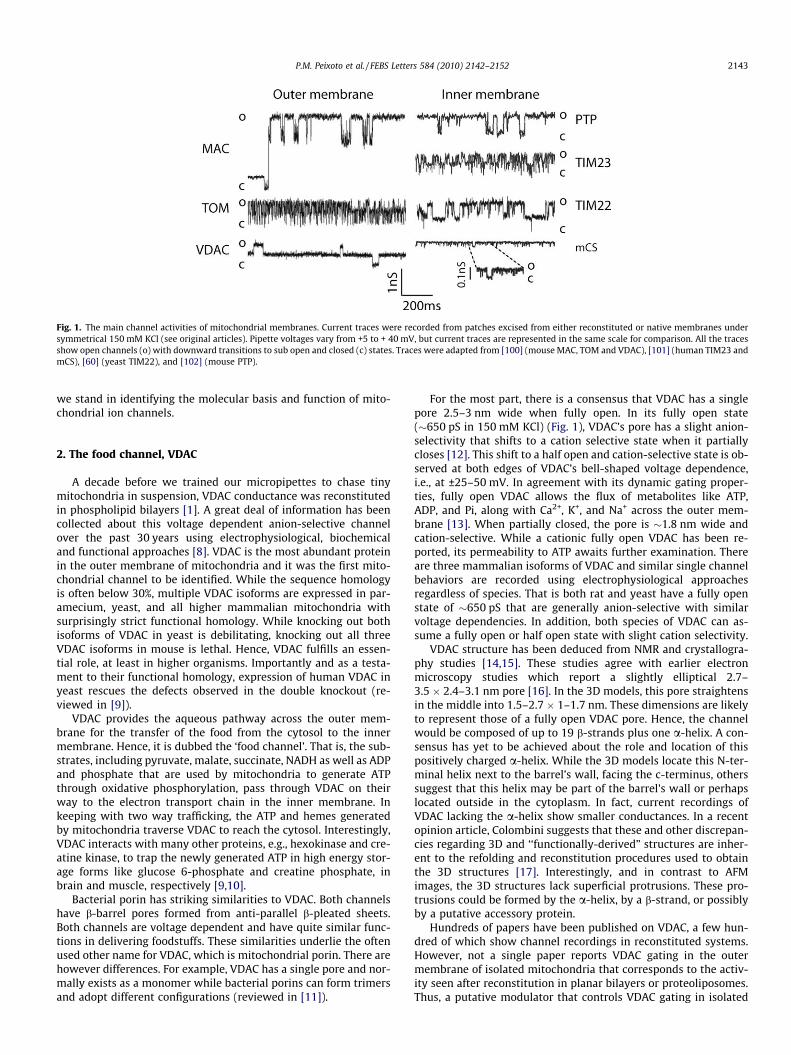
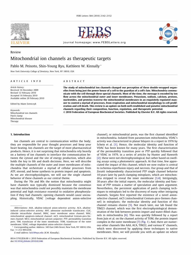
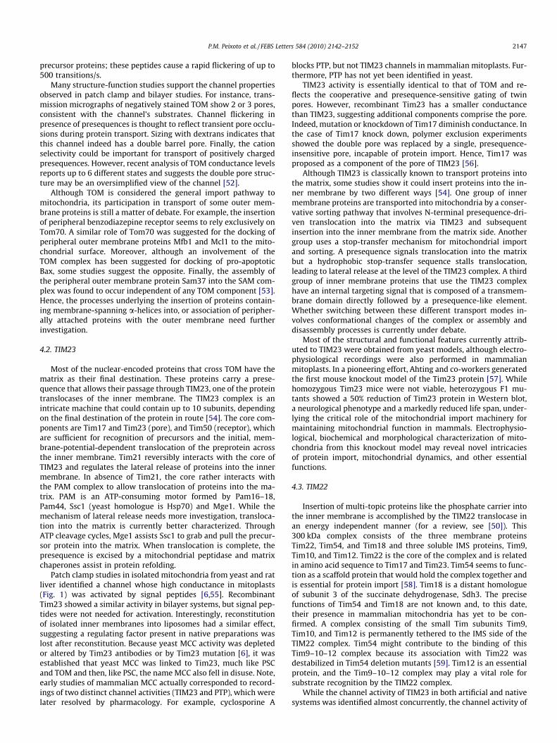
Fig. 1. The main channel activities of mitochondrial membranes. Current traces were recorded from patches excised from either reconstituted or native membranes undersymmetrical 150 mM KCl (see original articles). Pipette voltages vary from +5 to + 40 mV, but current traces are represented in the same scale for comparison. All the tracesshow open channels (o) with downward transitions to sub open and closed (c) states. Traces were adapted from [100] (mouse MAC, TOM and VDAC), [101] (human TIM23 andmCS), [60] (yeast TIM22), and [102] (mouse PTP).
P.M. Peixoto et al. / FEBS Letters 584 (2010) 2142–2152 2143
we stand in identifying the molecular basis and function of mito-chondrial ion channels.
2. The food channel, VDAC
A decade before we trained our micropipettes to chase tinymitochondria in suspension, VDAC conductance was reconstitutedin phospholipid bilayers [1]. A great deal of information has beencollected about this voltage dependent anion-selective channelover the past 30 years using electrophysiological, biochemicaland functional approaches [8]. VDAC is the most abundant proteinin the outer membrane of mitochondria and it was the first mito-chondrial channel to be identified. While the sequence homologyis often below 30%, multiple VDAC isoforms are expressed in par-amecium, yeast, and all higher mammalian mitochondria withsurprisingly strict functional homology. While knocking out bothisoforms of VDAC in yeast is debilitating, knocking out all threeVDAC isoforms in mouse is lethal. Hence, VDAC fulfills an essen-tial role, at least in higher organisms. Importantly and as a testa-ment to their functional homology, expression of human VDAC inyeast rescues the defects observed in the double knockout (re-viewed in [9]).
VDAC provides the aqueous pathway across the outer mem-brane for the transfer of the food from the cytosol to the innermembrane. Hence, it is dubbed the ‘food channel’. That is, the sub-strates, including pyruvate, malate, succinate, NADH as well as ADPand phosphate that are used by mitochondria to generate ATPthrough oxidative phosphorylation, pass through VDAC on theirway to the electron transport chain in the inner membrane. Inkeeping with two way trafficking, the ATP and hemes generatedby mitochondria traverse VDAC to reach the cytosol. Interestingly,VDAC interacts with many other proteins, e.g., hexokinase and cre-atine kinase, to trap the newly generated ATP in high energy stor-age forms like glucose 6-phosphate and creatine phosphate, inbrain and muscle, respectively [9,10].
Bacterial porin has striking similarities to VDAC. Both channelshave b-barrel pores formed from anti-parallel b-pleated sheets.Both channels are voltage dependent and have quite similar func-tions in delivering foodstuffs. These similarities underlie the oftenused other name for VDAC, which is mitochondrial porin. There arehowever differences. For example, VDAC has a single pore and nor-mally exists as a monomer while bacterial porins can form trimersand adopt different configurations (reviewed in [11]).
For the most part, there is a consensus that VDAC has a singlepore 2.5–3 nm wide when fully open. In its fully open state(�650 pS in 150 mM KCl) (Fig. 1), VDAC’s pore has a slight anion-selectivity that shifts to a cation selective state when it partiallycloses [12]. This shift to a half open and cation-selective state is ob-served at both edges of VDAC’s bell-shaped voltage dependence,i.e., at ±25–50 mV. In agreement with its dynamic gating proper-ties, fully open VDAC allows the flux of metabolites like ATP,ADP, and Pi, along with Ca2+, K+, and Na+ across the outer mem-brane [13]. When partially closed, the pore is �1.8 nm wide andcation-selective. While a cationic fully open VDAC has been re-ported, its permeability to ATP awaits further examination. Thereare three mammalian isoforms of VDAC and similar single channelbehaviors are recorded using electrophysiological approachesregardless of species. That is both rat and yeast have a fully openstate of �650 pS that are generally anion-selective with similarvoltage dependencies. In addition, both species of VDAC can as-sume a fully open or half open state with slight cation selectivity.
VDAC structure has been deduced from NMR and crystallogra-phy studies [14,15]. These studies agree with earlier electronmicroscopy studies which report a slightly elliptical 2.7–3.5 � 2.4–3.1 nm pore [16]. In the 3D models, this pore straightensin the middle into 1.5–2.7 � 1–1.7 nm. These dimensions are likelyto represent those of a fully open VDAC pore. Hence, the channelwould be composed of up to 19 b-strands plus one a-helix. A con-sensus has yet to be achieved about the role and location of thispositively charged a-helix. While the 3D models locate this N-ter-minal helix next to the barrel’s wall, facing the c-terminus, otherssuggest that this helix may be part of the barrel’s wall or perhapslocated outside in the cytoplasm. In fact, current recordings ofVDAC lacking the a-helix show smaller conductances. In a recentopinion article, Colombini suggests that these and other discrepan-cies regarding 3D and ‘‘functionally-derived” structures are inher-ent to the refolding and reconstitution procedures used to obtainthe 3D structures [17]. Interestingly, and in contrast to AFMimages, the 3D structures lack superficial protrusions. These pro-trusions could be formed by the a-helix, by a b-strand, or possiblyby a putative accessory protein.
Hundreds of papers have been published on VDAC, a few hun-dred of which show channel recordings in reconstituted systems.However, not a single paper reports VDAC gating in the outermembrane of isolated mitochondria that corresponds to the activ-ity seen after reconstitution in planar bilayers or proteoliposomes.Thus, a putative modulator that controls VDAC gating in isolated

2144 P.M. Peixoto et al. / FEBS Letters 584 (2010) 2142–2152
mitochondria could possibly be lost in reconstituted systems. Inagreement, several cytosolic factors decreased the open probabilityof VDAC reconstituted in planar bilayers. For example, NADH andMg2+-NAD(P)H decreases ADP flux through VDAC. Moreover, anintermembrane space (IMS) protein and tBid (a cleaved form ofBid) has been reported as a putative endogenous inhibitor of VDAC.Finally, VDAC can be phosphorylated and has binding sites forhexokinase I and hexokinase II. Paradoxically, while hexokinase Iseems to close the channel, hexokinase II seems to keep it open.The list of putative VDAC modulators is still growing – Romanand co-workers surveyed 55 novel interactions [18]. While it isreasonable that the most abundant protein in the mitochondrialouter membrane might be the main pathway of communicationwith the cytosol, putative modulators are based on biochemicaland electrophysiological data and also on functional studies.
Determinations of VDAC conductance and modulation ofmetabolite flux have just begun in intact cells. In an elegant varia-tion of the patch clamp techniques, Jonas and co-workers showedthat a large outer membrane channel with voltage dependencetypical of VDAC can be activated in intact neurons by a train of syn-aptic action potentials or by a cleaved form of Bcl-xL. These chan-nels are inhibited by NADH or hypoxia [19]. Other studies includean ethanol induced shift of cytosolic NADH/NAD+ redox potentialto a reduced state which may inhibit ADP flux across the outermembrane and a report that the conductance of VDAC is Ca2+
dependent at physiological cytosolic Ca2+ concentrations [20]. Fi-nally, Ca2+ pretreatment of permeabilized cells enhanced subse-quent ATP uptake, implying a VDAC-mediated regulation ofmetabolism. All these studies show that the best studied mito-chondrial channel VDAC has many faces that are just now begin-ning to be revealed.
3. Apoptosis channels
While generating the energy currency of the cell, ATP, the manyfunctions that mitochondria perform are just now being discov-ered. Mitochondria play a central role in apoptosis. These organ-elles compartmentalize a myriad of death-evoking signalingproteins, like cytochrome c and AIF, which upon their release tothe cytosol wreak havoc and eventually induce cell death throughapoptosome formation and caspase activation. There are at leasttwo ways to break down the permeability barrier of the outermembrane (Fig. 2). Activation of the inner membrane channelPTP causes matrix swelling and ruptures the outer membrane. Thisevent results in a spilling of IMS contents into the cytosol. Alterna-tively, de novo formation of a gigantic channel called MAC (mito-chondrial apoptosis-induced channel) permits cytochrome c andother pro-apoptotic factors to freely diffuse from the IMS to thecytosol. Here, we discuss the channels MAC and PTP, and thepossibility these channels act synergistically in the suicide of thecell.
3.1. MAC
During apoptosis, pro-apoptotic proteins assemble into a largechannel, MAC, which makes the mitochondrial outer membranepermeable to proteins normally constrained within the IMS. Oncein the cytoplasm, these proteins trigger a chain of events that leadto the destruction of the cell (Fig. 2). MAC formation and mitochon-dria outer membrane permeabilization (MOMP) have been consis-tently reported in a variety of cell lines during at least two differentapoptotic insults, IL-3 deprivation [21] and kinase inhibition [22].After induction of apoptosis, a time dependent increase in mem-brane permeability resulted from formation of MACs with conduc-tances of 3–5 nS (in 150 mM KCl) (Fig. 1). Regardless of its
heterogeneous peak conductance, >1.5 nS MAC is voltage indepen-dent and slightly cation-selective, consistent with its proposedfunction as a cytochrome c release channel (cytochrome c is cat-ionic). MAC with a conductance of 4 nS is permeable to up to17 kDa dextran. This polymer exclusion method indicates thatMAC with conductances between 1.5 and 5 nS have pore sizes of2.9–7.6 nm, which should be large enough to allow the passageof 3 nm cytochrome c. Cytochrome c in fact changes MAC conduc-tance in an interesting way. A reversible and dose dependent in-crease in noise and decrease in conductance (4–50%) is observed,suggesting cytochrome c is partitioning into the pore. This effectwas named type 1 and resembles that seen with other channels,e.g., ATP on VDAC [13,23], DNA on a-hemolysin [24], maltodextrinson maltoporins [25], and ampicilin on OmpF [26]. The Type 2 ef-fects of cytochrome c correspond to a more robust reduction inconductance (50–90%) that is dose dependent, irreversible, andvoltage independent. This response corresponds either to a desta-bilization or a plugging of the open state of MAC. Possibly, bindingsites for cytochrome c may exist in the structure of 2–4 nS MAC,which could block the passageway and may be important in syn-chronization of apoptotic events.
Unlike the mitochondrial protein import complexes (see be-low), MAC allows release of soluble proteins from the IMS intothe cytosol. And, although MAC pore is at least twice as big as thosefrom the protein import complexes, its function is in contrast tothat of the import complexes, as this channel allows for the exportof folded proteins from mitochondria. Consistent with its function,MAC conductance decreases in presence of cytochrome c [27], asdiscussed above. Induction of MAC formation by several means,such as staurosporine treatment, IL-3 withdrawal, exogenousexpression of Bax and tBid lead to release of cytochrome c in differ-ent cell lines. For example, staurosporine treatment induced in-creased expression and mitochondrial relocation of greenfluorescent Bax. When mitochondria containing green fluorescentBax where isolated and patch clamped, they showed an increasein conductance of �6 nS, consistent with the presence of MAC.Immunocytochemistry analysis showed cytochrome c leaked tothe cytoplasm under these conditions [22]. Such release can in turnbe prevented by knocking out Bax and Bak, overexpressing Bcl-2 orBcl-xL, or using MAC inhibitors [28]. The result of knocking downMAC’s main components or its pharmacological inhibition is pre-vention of cytochrome c release and protection against apoptosis.
Whether MAC or its components participate in other cellularfunctions is of increasing interest. Some pieces of evidence existof a possible interaction of Bcl-2 family proteins, e.g., Bax andBak with the mitochondrial morphology machinery (for a review,see [29]). Recent studies implicate Bax and Bak with maintenanceof the mitochondrial network in the absence of apoptosis, sincedouble knockout cells had smaller mitochondria than control cells.While there are alternative interpretations, DDP/Timm8a, an IMSprotein that is released presumably through MAC and at the sametime as cytochrome c, can activate the fission protein Drp-1 andpromote mitochondrial fragmentation [30]. These findings supportthe hypothesis that fission of mitochondria occurs after MAC for-mation. Yet another role has been recently proposed for MAC ingeneration of a so called bystander effect, based on evidence thata death signal generated downstream of MAC and cytochrome c re-lease seems to propagate through gap junctions to kill adjacentcells [31].
The molecular identity of MAC, or at least most of it, has beendetermined using functional knockouts, immunoprecipitations,and pharmacological inhibitors. VDAC1 and VDAC3 have been ex-cluded as MAC components, since MAC formation, conductanceand function were unaffected by either knockouts [32]. Neverthe-less, two pro-apoptotic Bcl-2 members, Bax and Bak have beenestablished as functionally and structurally redundant core com-

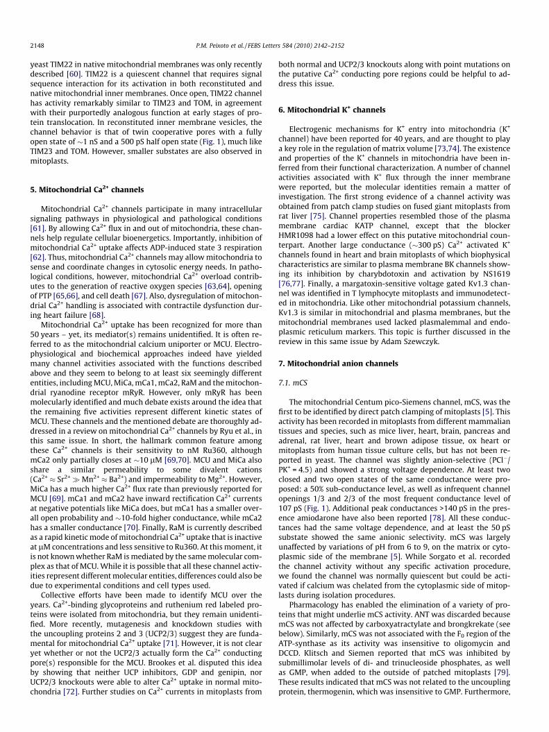
Fig. 2. Mitochondrial ion channels in apoptosis. Left, Apoptotic stimuli induce relocation of Bax from the cytosol into the mitochondrial outer membrane (MOM). Bax, Bak andpossibly other unidentified protein(s) oligomerize and form MAC. An increase in ROS production, possibly due to Ca2+ entry through VDAC, MCU and mRyR, detachescytochrome c from cardiolipin in the inner membrane (MIM). Cytochrome c spills into the cytoplasm and binds Apaf-1 to form apoptosomes and amplify the apoptoticcascade. Right, Necrotic stimuli lead to exacerbated Ca2+ uptake and ROS generation by mitochondria. High levels of Ca2+ and ROS induce a cyclophilin D (Cyp D)-sensitiveopening of PTP that leads to swelling of the matrix and release of Ca2+. Swelling disrupts the outer membrane while released Ca2+ activates proteases, phosphatases andnucleases that lead to necrotic degradation.
P.M. Peixoto et al. / FEBS Letters 584 (2010) 2142–2152 2145
ponents of MAC. That is, either Bax or Bak knockouts are equallycapable of attaining MAC conductance and releasing cytochromec upon apoptotic stimulation. Hence, one can envision that MACexists in three populations: two of them formed by Bax or Bakhomo-oligomers and one by Bax–Bak hetero-oligomers. However,Mikhailov et al. showed that, although Bax and Bak in the oligo-meric state are able to co-immunoprecipitate, they fail to crosslink[33]. Hence, although Bax and Bak are unquestionable components,their structural relationship has yet to be clarified, as well as thepossible existence of additional components, for which an exten-sive proteomic approach might be necessary.
Much of MAC structure has been deduced from functional, bio-chemical, genetic and patch clamp studies. The pore of functionalMAC is thought to be minimally a hexamer of Bax and/or Bakassuming each monomer contributes two transmembrane helices.In fact, a-helices 5 and 6 of Bax are amphipatic, which makes themgood candidates for pore components [34]. Moreover, Bax lackinghelices 5 and 6 do not release cytochrome c [35,36]. Given thestructural similarities between Bax and Bak, the same helices couldunderlie pore formation in MAC by either protein. Single channelanalysis of tBid-induced MAC formation in freshly isolated mito-chondria suggested that MAC formation can be explained bysequential incorporation of additional Bax or Bak monomers toan existing pore [32]. However, other studies with outer mem-brane preparations suggest a lipidic component to the pore [37].
3.2. PTP
The mitochondrial permeability transition pore (PTP) was orig-inally described in swelling experiments on isolated mitochondria30 years ago [2]. Opening of the PTP leads to a transition in perme-ability of the inner membrane from extremely low to freely perme-able to solutes up to 1.5 kDa. These changes were typicallymonitored by light scattering at wavelengths known to reflect
mitochondrial volume. The PTP can be reversibly closed by re-moval of Ca2+ or by the addition of cyclosporine A, magnesium orADP. A myriad of effectors including Ca2+ plus Pi and reactive oxy-gen species have been reported [38]. This channel has been impli-cated in the apoptotic cascade as a means of releasing cytochromec from mitochondria. However, the role of PTP in early stages ofapoptosis is unclear, since its opening suspends oxidative phos-phorylation and leads to depletion of ATP reserves required forapoptosome formation. PTP opening is frequently observed down-stream of MAC formation and outer membrane permeabilization,but it is not clear if PTP has a role in induction of apoptosis. Somemodels, however, advocate for a PTP dependent apoptosis prior toMAC formation [39]. Persistent PTP opening results in loss of mito-chondrial membrane potential and massive swelling of mitochon-dria likely involved in necrotic cell death (Fig. 2).
The connection between the PTP and channel activities re-corded directly from the inner membrane by patch clamping mam-malian mitoplasts was made over a few years mostly by Zoratti’sgroup [40]. In earlier studies, the PTP activity was referred to asMCC (mitochondrial multiple conductance channel) or MMC(mitochondrial megachannel). Although much smaller than MACand Bax channels, PTP has a high conductance of �1.2 nS with mul-tiple conductance levels (Fig. 1). Transitions between sublevels aretypically 0.3–1 nS. Unlike MAC, this slightly cation-selective chan-nel is voltage dependent, closing with matrix positive potentials.Interestingly, while overexpression of Bcl-2 had no detectable ef-fect on the channel activity, this protein suppressed calcium-acti-vation of PTP in both swelling and electrophysiologicalexperiments [41,42]. Recently, Zoratti’s group described a newchannel activity in mitoplasts that resembles plasmalemmalmaxi-anion channels, but differs pharmacologically and was ob-served more frequently at high Ca2+ concentrations, known to in-duce PTP [43]. The channel conductance, gating and selectivityrather seems closer to VDAC than PTP, although its occurrence
2146 P.M. Peixoto et al. / FEBS Letters 584 (2010) 2142–2152
was unaffected in VDAC-1�/�/VDAC-3�/� double knockouts. Be-cause the detection of this channel was inversely proportional tothat of PTP in five different cell lines, the authors coined thisnew activity HP (Half PTP). Interestingly, HP was not affected bycyclosporine A.
The molecular identity of PTP remains unresolved despiteextensive efforts. ANT and VDAC were thought to be pore compo-nents of PTP based on the inhibitory effects of ANT inhibitors, andon co-immunoprecipitation experiments of VDAC with ANT. Thesecandidates were later discarded because both ANT and VDACknockouts expressed PTP activity [44,45]. Cyclophilin D knockouts,however, modified the permeability transition and reduced injuryafter reperfusion. Knockout experiments demonstrated that cyclo-philin D represents the target for PTP inhibition by cyclosporin A,and that it modulates the sensitivity of the PTP to Ca2+ [46]. Re-cently, the mitochondrial phosphate carrier was proposed as anew candidate based on its co-immunoprecipitation with cyclo-philin D [47]. This interaction was prevented by cyclosporin A, aclassical PTP inhibitor, and was promoted by oxidative stress, aclassical PTP inducer. However, cyclophilin D has many partnersin mitochondria. Furthermore, an essential role for the phosphatecarrier in PTP formation awaits a demonstration that knockdownsfail to undergo a permeability transition. While the phosphate car-rier has reported channel activity [48], assignment as the PTPwould require that the channel activity be sensitive to cyclophilinD and cyclosporine A. Finally, as a non-protein candidate, poly-phosphate has also proposed as the molecular basis of the PTP [49].
Studies propose alternative structural candidates in the innermembrane, but what about the outer membrane counterpart(s)?Most of the studies that advocated for or against VDAC participa-tion on PTP used matrix swelling assays. This argument may beover-interpreted because PTP is probably not the sole mediatorof matrix swelling. In fact, PTP inhibitors reduce, but do not com-pletely abolish swelling. But the question remains. What, if one ex-ists, is the outer membrane counterpart of PTP? If PTP really spansboth the inner and outer membranes, its conductance must bedetectable in the outer membrane of intact mitochondria. Virtuallyall current traces attributed to PTP were recorded from mitoplasts,or reconstituted systems, both devoid of the outer membrane. Theonly large conductances reported to date in isolated mitochondria
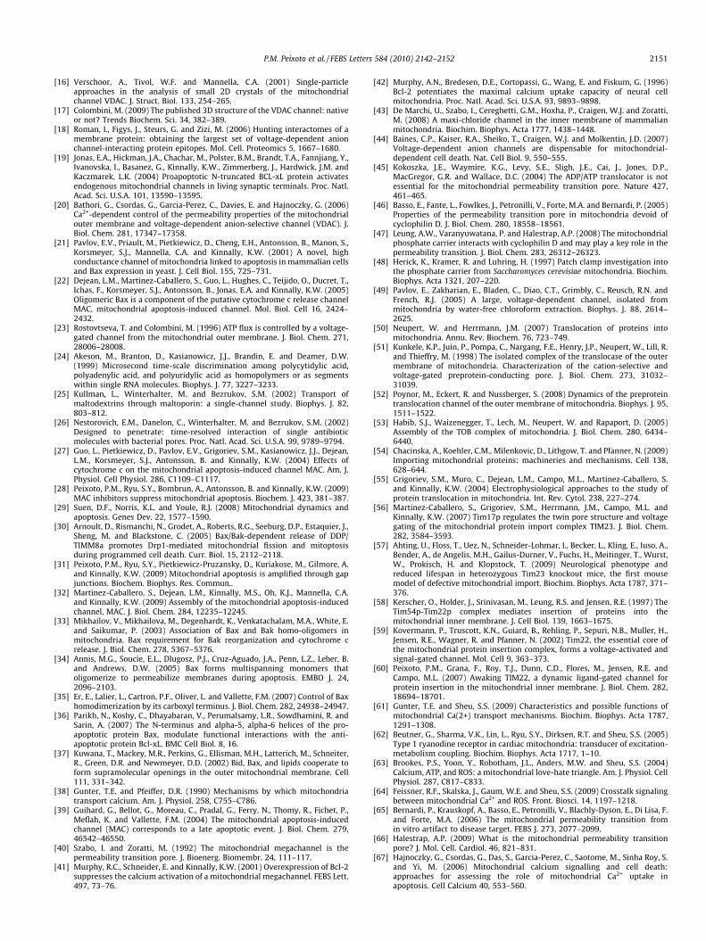
Fig. 3. Mitochondrial ion channels in protein import. The three major protein importweights of subunits and arrows indicate sorting pathways of mitochondrial proteins acomponents are reviewed in [54]. The three complexes rely on ion channels for protein imTOM. In the inner membrane (MIM), Tim23 and Tim17 form the channel of TIM23, whi
are attributed to MAC and depleted in Bax/Bak double knockouts[22,32]. In summary, VDAC function (or dysfunction) can affectROS production and onset of PTP, but knockout studies argueagainst a physical interaction and no other outer membrane pro-tein has been associated with the permeability transition of the in-ner membrane. In addition, the outer membrane is thought to notrepresent an osmotic barrier, thus swelling is probably reliantexclusively on yet unidentified inner membrane components.
4. Protein import channels
4.1. TOM
Only 13 of the �1500 proteins found in human mitochondriaare encoded in its genome. That means mitochondria import al-most all of their proteins. Regardless of their final destination, im-ported proteins must pass through the gatekeeper, TOM, located inthe outer membrane (Fig. 3). In general, precursor proteins carry asignal sequence in their N-terminus signal that targets them tomitochondria. This signal can be a cysteine motif, a presequenceor an internal targeting element [50]. All these targeting sequencesare recognized by the receptor proteins Tom70 and Tom20, whichthen deliver the precursor protein to Tom22. Finally, the precursoris handed off to the translocation pore of the TOM complex. Tom40forms the protein-conducting pore, while the small accessory pro-teins Tom5, Tom6 and Tom7 regulate stability of the complex [50].From this point, the general import pathway diverges into severalmachines that finish the sorting and send the precursor proteins totheir final destination. Two of these machines, TIM23 and TIM22,have inner membrane channels that will be discussed later.
The channel activity of TOM was first discovered in native outermembrane preparations as a peptide sensitive channel, namedthen PSC [7]. Later studies showed recombinant Tom40 was capa-ble of forming a channel with similar characteristics in bilayer sys-tems. Finally, comparative studies confirmed those two channelactivities reflected the same entity, and the name PSC fell into dis-use [51]. TOM activity has a high conductance of�1 nS in yeast and�750 pS in mammals (in 150 mM KCl) (Fig. 1). A half open state isfrequently observed, especially at higher potentials. TOM activity issensitive to peptides whose sequences mimic that used to target
complexes TOM, TIM23, and TIM22 are represented. Numbers indicate molecularccording to their targeting sequence. The detailed pathway, additional routes andport into mitochondria. In the outer membrane (MOM), Tom40 forms the channel ofle Tim22 forms the channel of the TIM22 complex.
P.M. Peixoto et al. / FEBS Letters 584 (2010) 2142–2152 2147
precursor proteins; these peptides cause a rapid flickering of up to500 transitions/s.
Many structure-function studies support the channel propertiesobserved in patch clamp and bilayer studies. For instance, trans-mission micrographs of negatively stained TOM show 2 or 3 pores,consistent with the channel’s substrates. Channel flickering inpresence of presequences is thought to reflect transient pore occlu-sions during protein transport. Sizing with dextrans indicates thatthis channel indeed has a double barrel pore. Finally, the cationselectivity could be important for transport of positively chargedpresequences. However, recent analysis of TOM conductance levelsreports up to 6 different states and suggests the double pore struc-ture may be an oversimplified view of the channel [52].
Although TOM is considered the general import pathway tomitochondria, its participation in transport of some outer mem-brane proteins is still a matter of debate. For example, the insertionof peripheral benzodiazepine receptor seems to rely exclusively onTom70. A similar role of Tom70 was suggested for the docking ofperipheral outer membrane proteins Mfb1 and Mcl1 to the mito-chondrial surface. Moreover, although an involvement of theTOM complex has been suggested for docking of pro-apoptoticBax, some studies suggest the opposite. Finally, the assembly ofthe peripheral outer membrane protein Sam37 into the SAM com-plex was found to occur independent of any TOM component [53].Hence, the processes underlying the insertion of proteins contain-ing membrane-spanning a-helices into, or association of peripher-ally attached proteins with the outer membrane need furtherinvestigation.
4.2. TIM23
Most of the nuclear-encoded proteins that cross TOM have thematrix as their final destination. These proteins carry a prese-quence that allows their passage through TIM23, one of the proteintranslocases of the inner membrane. The TIM23 complex is anintricate machine that could contain up to 10 subunits, dependingon the final destination of the protein in route [54]. The core com-ponents are Tim17 and Tim23 (pore), and Tim50 (receptor), whichare sufficient for recognition of precursors and the initial, mem-brane-potential-dependent translocation of the preprotein acrossthe inner membrane. Tim21 reversibly interacts with the core ofTIM23 and regulates the lateral release of proteins into the innermembrane. In absence of Tim21, the core rather interacts withthe PAM complex to allow translocation of proteins into the ma-trix. PAM is an ATP-consuming motor formed by Pam16–18,Pam44, Ssc1 (yeast homologue is Hsp70) and Mge1. While themechanism of lateral release needs more investigation, transloca-tion into the matrix is currently better characterized. ThroughATP cleavage cycles, Mge1 assists Ssc1 to grab and pull the precur-sor protein into the matrix. When translocation is complete, thepresequence is excised by a mitochondrial peptidase and matrixchaperones assist in protein refolding.
Patch clamp studies in isolated mitochondria from yeast and ratliver identified a channel whose high conductance in mitoplasts(Fig. 1) was activated by signal peptides [6,55]. RecombinantTim23 showed a similar activity in bilayer systems, but signal pep-tides were not needed for activation. Interestingly, reconstitutionof isolated inner membranes into liposomes had a similar effect,suggesting a regulating factor present in native preparations waslost after reconstitution. Because yeast MCC activity was depletedor altered by Tim23 antibodies or by Tim23 mutation [6], it wasestablished that yeast MCC was linked to Tim23, much like PSCand TOM and then, like PSC, the name MCC also fell in disuse. Note,early studies of mammalian MCC actually corresponded to record-ings of two distinct channel activities (TIM23 and PTP), which werelater resolved by pharmacology. For example, cyclosporine A
blocks PTP, but not TIM23 channels in mammalian mitoplasts. Fur-thermore, PTP has not yet been identified in yeast.
TIM23 activity is essentially identical to that of TOM and re-flects the cooperative and presequence-sensitive gating of twinpores. However, recombinant Tim23 has a smaller conductancethan TIM23, suggesting additional components comprise the pore.Indeed, mutation or knockdown of Tim17 diminish conductance. Inthe case of Tim17 knock down, polymer exclusion experimentsshowed the double pore was replaced by a single, presequence-insensitive pore, incapable of protein import. Hence, Tim17 wasproposed as a component of the pore of TIM23 [56].
Although TIM23 is classically known to transport proteins intothe matrix, some studies show it could insert proteins into the in-ner membrane by two different ways [54]. One group of innermembrane proteins are transported into mitochondria by a conser-vative sorting pathway that involves N-terminal presequence-dri-ven translocation into the matrix via TIM23 and subsequentinsertion into the inner membrane from the matrix side. Anothergroup uses a stop-transfer mechanism for mitochondrial importand sorting. A presequence signals translocation into the matrixbut a hydrophobic stop-transfer sequence stalls translocation,leading to lateral release at the level of the TIM23 complex. A thirdgroup of inner membrane proteins that use the TIM23 complexhave an internal targeting signal that is composed of a transmem-brane domain directly followed by a presequence-like element.Whether switching between these different transport modes in-volves conformational changes of the complex or assembly anddisassembly processes is currently under debate.
Most of the structural and functional features currently attrib-uted to TIM23 were obtained from yeast models, although electro-physiological recordings were also performed in mammalianmitoplasts. In a pioneering effort, Ahting and co-workers generatedthe first mouse knockout model of the Tim23 protein [57]. Whilehomozygous Tim23 mice were not viable, heterozygous F1 mu-tants showed a 50% reduction of Tim23 protein in Western blot,a neurological phenotype and a markedly reduced life span, under-lying the critical role of the mitochondrial import machinery formaintaining mitochondrial function in mammals. Electrophysio-logical, biochemical and morphological characterization of mito-chondria from this knockout model may reveal novel intricaciesof protein import, mitochondrial dynamics, and other essentialfunctions.
4.3. TIM22
Insertion of multi-topic proteins like the phosphate carrier intothe inner membrane is accomplished by the TIM22 translocase inan energy independent manner (for a review, see [50]). This300 kDa complex consists of the three membrane proteinsTim22, Tim54, and Tim18 and three soluble IMS proteins, Tim9,Tim10, and Tim12. Tim22 is the core of the complex and is relatedin amino acid sequence to Tim17 and Tim23. Tim54 seems to func-tion as a scaffold protein that would hold the complex together andis essential for protein import [58]. Tim18 is a distant homologueof subunit 3 of the succinate dehydrogenase, Sdh3. The precisefunctions of Tim54 and Tim18 are not known and, to this date,their presence in mammalian mitochondria has yet to be con-firmed. A complex consisting of the small Tim subunits Tim9,Tim10, and Tim12 is permanently tethered to the IMS side of theTIM22 complex. Tim54 might contribute to the binding of thisTim9–10–12 complex because its association with Tim22 wasdestabilized in Tim54 deletion mutants [59]. Tim12 is an essentialprotein, and the Tim9–10–12 complex may play a vital role forsubstrate recognition by the TIM22 complex.
While the channel activity of TIM23 in both artificial and nativesystems was identified almost concurrently, the channel activity of
2148 P.M. Peixoto et al. / FEBS Letters 584 (2010) 2142–2152
yeast TIM22 in native mitochondrial membranes was only recentlydescribed [60]. TIM22 is a quiescent channel that requires signalsequence interaction for its activation in both reconstituted andnative mitochondrial inner membranes. Once open, TIM22 channelhas activity remarkably similar to TIM23 and TOM, in agreementwith their purportedly analogous function at early stages of pro-tein translocation. In reconstituted inner membrane vesicles, thechannel behavior is that of twin cooperative pores with a fullyopen state of �1 nS and a 500 pS half open state (Fig. 1), much likeTIM23 and TOM. However, smaller substates are also observed inmitoplasts.
5. Mitochondrial Ca2+ channels
Mitochondrial Ca2+ channels participate in many intracellularsignaling pathways in physiological and pathological conditions[61]. By allowing Ca2+ flux in and out of mitochondria, these chan-nels help regulate cellular bioenergetics. Importantly, inhibition ofmitochondrial Ca2+ uptake affects ADP-induced state 3 respiration[62]. Thus, mitochondrial Ca2+ channels may allow mitochondria tosense and coordinate changes in cytosolic energy needs. In patho-logical conditions, however, mitochondrial Ca2+ overload contrib-utes to the generation of reactive oxygen species [63,64], openingof PTP [65,66], and cell death [67]. Also, dysregulation of mitochon-drial Ca2+ handling is associated with contractile dysfunction dur-ing heart failure [68].
Mitochondrial Ca2+ uptake has been recognized for more than50 years – yet, its mediator(s) remains unidentified. It is often re-ferred to as the mitochondrial calcium uniporter or MCU. Electro-physiological and biochemical approaches indeed have yieldedmany channel activities associated with the functions describedabove and they seem to belong to at least six seemingly differententities, including MCU, MiCa, mCa1, mCa2, RaM and the mitochon-drial ryanodine receptor mRyR. However, only mRyR has beenmolecularly identified and much debate exists around the idea thatthe remaining five activities represent different kinetic states ofMCU. These channels and the mentioned debate are thoroughly ad-dressed in a review on mitochondrial Ca2+ channels by Ryu et al., inthis same issue. In short, the hallmark common feature amongthese Ca2+ channels is their sensitivity to nM Ru360, althoughmCa2 only partially closes at �10 lM [69,70]. MCU and MiCa alsoshare a similar permeability to some divalent cations(Ca2+ � Sr2+�Mn2+ � Ba2+) and impermeability to Mg2+. However,MiCa has a much higher Ca2+ flux rate than previously reported forMCU [69]. mCa1 and mCa2 have inward rectification Ca2+ currentsat negative potentials like MiCa does, but mCa1 has a smaller over-all open probability and �10-fold higher conductance, while mCa2has a smaller conductance [70]. Finally, RaM is currently describedas a rapid kinetic mode of mitochondrial Ca2+ uptake that is inactiveat lM concentrations and less sensitive to Ru360. At this moment, itis not known whether RaM is mediated by the same molecular com-plex as that of MCU. While it is possible that all these channel activ-ities represent different molecular entities, differences could also bedue to experimental conditions and cell types used.
Collective efforts have been made to identify MCU over theyears. Ca2+-binding glycoproteins and ruthenium red labeled pro-teins were isolated from mitochondria, but they remain unidenti-fied. More recently, mutagenesis and knockdown studies withthe uncoupling proteins 2 and 3 (UCP2/3) suggest they are funda-mental for mitochondrial Ca2+ uptake [71]. However, it is not clearyet whether or not the UCP2/3 actually form the Ca2+ conductingpore(s) responsible for the MCU. Brookes et al. disputed this ideaby showing that neither UCP inhibitors, GDP and genipin, norUCP2/3 knockouts were able to alter Ca2+ uptake in normal mito-chondria [72]. Further studies on Ca2+ currents in mitoplasts from
both normal and UCP2/3 knockouts along with point mutations onthe putative Ca2+ conducting pore regions could be helpful to ad-dress this issue.
6. Mitochondrial K+ channels
Electrogenic mechanisms for K+ entry into mitochondria (K+
channel) have been reported for 40 years, and are thought to playa key role in the regulation of matrix volume [73,74]. The existenceand properties of the K+ channels in mitochondria have been in-ferred from their functional characterization. A number of channelactivities associated with K+ flux through the inner membranewere reported, but the molecular identities remain a matter ofinvestigation. The first strong evidence of a channel activity wasobtained from patch clamp studies on fused giant mitoplasts fromrat liver [75]. Channel properties resembled those of the plasmamembrane cardiac KATP channel, except that the blockerHMR1098 had a lower effect on this putative mitochondrial coun-terpart. Another large conductance (�300 pS) Ca2+ activated K+
channels found in heart and brain mitoplasts of which biophysicalcharacteristics are similar to plasma membrane BK channels show-ing its inhibition by charybdotoxin and activation by NS1619[76,77]. Finally, a margatoxin-sensitive voltage gated Kv1.3 chan-nel was identified in T lymphocyte mitoplasts and immunodetect-ed in mitochondria. Like other mitochondrial potassium channels,Kv1.3 is similar in mitochondrial and plasma membranes, but themitochondrial membranes used lacked plasmalemmal and endo-plasmic reticulum markers. This topic is further discussed in thereview in this same issue by Adam Szewczyk.
7. Mitochondrial anion channels
7.1. mCS
The mitochondrial Centum pico-Siemens channel, mCS, was thefirst to be identified by direct patch clamping of mitoplasts [5]. Thisactivity has been recorded in mitoplasts from different mammaliantissues and species, such as mice liver, heart, brain, pancreas andadrenal, rat liver, heart and brown adipose tissue, ox heart ormitoplasts from human tissue culture cells, but has not been re-ported in yeast. The channel was slightly anion-selective (PCl�/PK+ = 4.5) and showed a strong voltage dependence. At least twoclosed and two open states of the same conductance were pro-posed: a 50% sub-conductance level, as well as infrequent channelopenings 1/3 and 2/3 of the most frequent conductance level of107 pS (Fig. 1). Additional peak conductances >140 pS in the pres-ence amiodarone have also been reported [78]. All these conduc-tances had the same voltage dependence, and at least the 50 pSsubstate showed the same anionic selectivity. mCS was largelyunaffected by variations of pH from 6 to 9, on the matrix or cyto-plasmic side of the membrane [5]. While Sorgato et al. recordedthe channel activity without any specific activation procedure,we found the channel was normally quiescent but could be acti-vated if calcium was chelated from the cytoplasmic side of mitop-lasts during isolation procedures.
Pharmacology has enabled the elimination of a variety of pro-teins that might underlie mCS activity. ANT was discarded becausemCS was not affected by carboxyatractylate and brongkrekate (seebelow). Similarly, mCS was not associated with the F0 region of theATP-synthase as its activity was insensitive to oligomycin andDCCD. Klitsch and Siemen reported that mCS was inhibited bysubmillimolar levels of di- and trinucleoside phosphates, as wellas GMP, when added to the outside of patched mitoplasts [79].These results indicated that mCS was not related to the uncouplingprotein, thermogenin, which was insensitive to GMP. Furthermore,

P.M. Peixoto et al. / FEBS Letters 584 (2010) 2142–2152 2149
Inoue et al. [75] reported that mCS was not affected when millimo-lar Mg2+ or ATP and micromolar ADP was applied on the matrixside of excised patches. Finally, the mitochondrial inner membraneanion channel (IMAC) is a channel inferred from light scattering(matrix swelling) studies, which had broad anion-selectivity andconducted mono-, di-, and trivalent anions [80]. This channel is be-lieved to be an important component of the volume homeostaticmechanism of mitochondria, and was maintained closed or inac-tive by matrix Mg2+ and H+. The lack of effect of Mg2+ [81], DIDS(4,40-diisothiocyanostilbene-2,20-disulfonic acid), quinine or DCCD[82], all of them well known blockers of IMAC, indicated this puta-tive channel is not related to mCS.
7.2. AAA and ACA
Two other pH-sensitive channel activities were reported in livermitoplasts. Both of them displayed greater open probability uponalkalinization of the matrix side of the membrane, and were acti-vated by depletion of Mg2+ [78]. They were referred as ACA (alka-line-induced cation-selective Activity, 15 pS) and AAA (alkaline-induced anion-selective activity, 45 pS), respectively. AAA was ini-tially thought to correspond to IMAC because of their similarinduction by alkaline pH or Mg2+ depletion. However, IMAC is�100-fold more selective than AAA for Cl� over glucouronate.The conductance and voltage dependence of ACA were similar tothose of the ATP-sensitive K+ channel (see above). However ACAwas insensitive to 4-aminopyridine and glibenclamide plus ATP.The selectivity and inhibition by Mg2+ suggested that ACA channelactivity may correspond to one of the cation uniporters implicatedin volume homeostasis, whose existence was inferred from sus-pension studies [83,84].
7.3. CLICs
Chloride intracellular channels (CLICs) represent a new class ofintracellular anion channels that have been identified by theirhomology to the p64 protein. A mitochondrial homolog, mtCLIC,has been identified from differential display analysis of differenti-ating mouse keratinocytes from p53+/+ and p53�/� mice. MtCLICcolocalized with cytochrome oxidase in keratinocyte mitochondriabut also was detected in the cytoplasmic compartment. This p53-regulated putative channel has been associated with apoptosisand may exist as either a soluble or a transmembrane form thatmay translocate to the nucleus in response to cell stress [85].
8. Putative mitochondrial channels
Aside from mitochondrial channel activities with unresolvedidentities, some studies have rather proposed putative channelactivities with known identities. Osmotic experiments in absenceand presence of Hg2+ suggested aquaporin 8 forms water channelsin mitochondria that might regulate mitochondrial shape [86]. Inthis case, a channel activity has yet to be demonstrated. In othercases, purified recombinant proteins like Bcl-2, Bcl-xL andOmp85, a close relative of Sam50 in mammals, were capable offorming a channel after reconstitution in lipid bilayers [87–89].However, it is not clear if these channel activities occur in nativesystems. Finally, protein free mitochondrial membrane extractsmanifested a channel activity in lipid bilayers that is attributedto polyphosphate channels [49].
9. Mitochondrial channels as therapeutic targets
Mitochondrial dysfunction is at the epicenter of many devastat-ing diseases that increasingly affect the human population. In the
United States alone, 1000–4000 children per year are born with atype of mitochondrial disease. Also, many diseases of aging arenow being attributed at least to a great degree to mitochondrialdysfunction. These include but are not limited to: Type 2 diabetes,Parkinson’s disease, atherosclerotic heart disease, stroke, Alzhei-mer’s disease, and cancer. Thus, mitochondria have emerged astherapeutic targets and so have the mitochondrial ion channels,since they are direct transducers of mitochondrial function, or dys-function, to the rest of the cell. This section summarizes recentstudies that raise interest to some mitochondrial ion channels aspotential therapeutic targets (Table 1).
Based on the fact that VDACs are expressed more highly in can-cer than normal cells, some studies propose VDAC-dependent cyto-toxic agents can have cancer-selectivity. For example,furanonaphthoquinones (FNQs) induce caspase-dependent apop-tosis via the production of NADH-dependent reactive oxygen spe-cies (ROS) by VDAC1. The ROS production and the anti-canceractivity of FNQs were increased by VDAC1 overexpression [90]. An-other drug, erastin, induces RAS-RAF-MEK-dependent non-apopto-tic cell death via VDAC2 [91]. VDAC is also a pharmacological targetfor G3139, an 18-mer phosphorothioate antisense oligonucleotidethat targets the initiation codon region of the Bcl-2 mRNA anddown-regulates the expression of Bcl-2. G3139 interacts withand closes VDAC, inducing caspase-dependent apoptosis [92]. Fi-nally, the plant hormone methyl jasmonate has been shown tohave selective anti-cancer activity in preclinical studies and themechanism seems to be through disruption of the interaction be-tween human hexoquinase and VDAC, causing the inhibition ofglycolysis and the induction of MOMP [93].
Because MAC formation and cytochrome c release are the lastcheck points typically prior the activation of the caspase cascade,these events are considered the point of no return in mitochondrialapoptosis. For that reason, MAC is a potential target for novel ther-apies, as the use of agonists or antagonists of this channel could in-duce or prevent cell death, respectively. For example, agonists ofMAC could restore cytochrome c release and cell death in lympho-mas. Some BH3 mimetic compounds like ABT-737 and its orally ac-tive analog, ABT-263, are currently in clinical trials and couldpotentially induce MAC in B-cell malignancies and myelomas[94]. Alternatively, antagonists of MAC potentially could protecttransplanted neuronal precursor cells from apoptosis, as well asprevent HIV-1 induced lymphocyte depletion, severe congenitalneutropenia, and other pathologies associated with Bax-inducedcytochrome c release.
Cyclosporin A, a classical PTP inhibitor has been orally adminis-tered in a clinical study to treat Ullrich congenital muscular dystro-phy and Bethlem myopathy patients. The mechanism of action ofthis drug may be due to its binding to cyclophilin D and inhibitionof peptidyl–prolyl cis–trans isomerase activity. After a month oftreatment, muscle biopsies showed decreased apoptosis markersand improved mitochondrial function [95]. In another diseasemodel, Alzheimer, an accumulation of b-amyloid in mitochondriaand its direct interaction with cyclophilin D promotes calcium-in-duced PTP opening, suggesting this channel could also be a thera-peutic target in this neurodegenerative disease [96].
Although disease models are available for defects in the proteinimport machineries, we found no studies on therapeutic com-pounds targeted to these machineries. Two human diseases havebeen described that are caused by defects of the mitochondrialprotein import machinery: the Mohr-Tranebjaerg syndrome andthe syndrome of dilated cardiomyopathy with ataxia (DCMA).DCMA is a type of 3-methylglutaconic aciduria, which is clinicallycharacterized by severe, early onset dilated cardiomyopathy,growth retardation, ataxia and optic atrophy. It is caused by asplice mutation in the DNAJC19 gene, which putatively corre-sponds to the human homolog of yeast Tim14. The Mohr-Tranebj-

Table 1Mitochondrial ion channels as therapeutic targets.
Location Type Modulators Potential role(s) Therapeutic potential
Outer membrane VDAC Bax, Bak, Bcl-xL, Bcl-2, Tom20, Ca2+,pH, DV, NADH, DIDS
Metabolic transport,apoptosis
FNQ, erastin, G3139, and methyl jasmonate induceapoptosis in cancer cells through VDAC [68–71]
MAC Bax, Bak, tBid, Bcl-2, Dibucaine, TFP,propranolol, Bci1, Bci2, iMACs
MOMP apoptosis iMACs, Bci1, Bci2 and BH3 mimetics modulateapoptosis through MAC [22,72,81,82]
Inner membrane TIM23 Presequences, Ca2+, pH, DV, CsA Protein import Mohr-Tranebjaerg and DCMA syndromes [83–86]PTP CsA, NIM811, bongkrekic acid, Ca2+,
pH, DV, Thiols, Bax, cyclophilin DNecrosis, apoptosis CsA on Ullrich and Bethlem syndromes, Alzheimer,
cardioprotection [74,87]MCU Divalent cations, nucleotides, RuRed,
Ru360, ryanodineCa2+ uptake, apoptosis Inhibition facilitates post reperfusion recovery [76]
MitoKATP ATP, GTP, palmitoyl CoA, Mg2+, Ca2+ Volume regulationprotection against ischemicinjury
Opening with BMS-180448 promotes cardioprotection[75]
UCP Fatty acids Thermogenesis Overexpression protects against oxidative stress [77]
See text for details and abbreviations.
2150 P.M. Peixoto et al. / FEBS Letters 584 (2010) 2142–2152
aerg syndrome (MTS, also called deafness dystonia syndrome, DDS)shows X-linked recessive inheritance and is characterized by pro-gressive sensorineural hearing loss, dystonia, mental deficiency,and visual disability. It is caused by a mutation of the deafness/dys-tonia protein1 gene (DDP1), also called translocase of the innermembrane 8a (TIM8A).
A prototype mitoKATP channel opener, BMS-180448 [97], hascardioprotective effects and is currently in clinical trials. However,it also opens plasmalemmal KATP channels. The role of mitochon-drial potassium channels in cardioprotection is a matter of debatebecause of the lack of molecular identity and the ambiguous orunspecific effects of chemical agents thought to induce or preventischemia-reperfusion injuries.
Mitochondrial calcium influx is an attractive target for thetreatment of reperfusion injury, based on in vitro studies withruthenium red. However, this compound is unsuitable for thera-peutic use and further studies are needed to develop novel com-pounds. One study proposed the use of dinuclear Cobaltcomplexes 10 years ago, but there have been no further develop-ments [98].
Finally, mitochondrial dysfunction is a prominent feature ofexcitotoxic insult in neurons. In vitro studies have demonstratedthese events are dependent on mitochondrial Ca2+ cycling and thata reduction in membrane potential is sufficient to reduce excito-toxic cell death. This concept has gained additional support fromexperiments demonstrating that the overexpression of endoge-nous mitochondrial uncoupling proteins (UCP), which decreasethe mitochondrial membrane potential, decreases cell death fol-lowing oxidative stress. Thus, upregulation of UCP activity can re-duce excitotoxic-mediated ROS production and cell death whereasa reduction in UCP levels increases susceptibility to neuronal in-jury. These findings raise the possibility that mitochondrial uncou-pling could be a potential novel treatment for acute CNS injuries[99].
10. Future perspectives
Mitochondria express a myriad of channel activities and thefunction of many of these channels is not well understood. Oneof the most compelling problems in this field is lack of informationregarding the molecular basis for many of these channels. Thatidentification will bring new insights into the roles channels fulfillin normal and pathological conditions. Mitochondria presenttechnical challenges for monitoring channel activities in nativemembranes which we have overcome. However, monitoring mito-chondrial channels inside cells, e.g., using the approach of the Jonaslab [19], or even in vivo presents additional technical challengesespecially when one considers that the outer membrane shields
the inner membrane from our electrodes. Nevertheless, mitochon-drial channels are emerging as promising therapeutic targets formany diseases like cancer, aging, neurodegenerative diseases,stroke and infarct.
Acknowledgements
This work was supported by the National Institutes of Health[Grant GM57249] to K.W.K. many important papers were not citedhere, because of space constrains. They can be found in the excel-lent reviews we referred to.
References
[1] Schein, S.J., Colombini, M. and Finkelstein, A. (1976) Reconstitution in planarlipid bilayers of a voltage-dependent anion-selective channel obtained fromparamecium mitochondria. J. Membr. Biol. 30, 99–120.
[2] Hunter, D.R. and Haworth, R.A. (1979) The Ca2+-induced membrane transitionin mitochondria. I. The protective mechanisms. Arch. Biochem. Biophys. 195,453–459.
[3] Kinnally, K.W., Campo, M.L. and Tedeschi, H. (1989) Mitochondrial channelactivity studied by patch-clamping mitoplasts. J. Bioenerg. Biomembr. 21,497–506.
[4] Szabo, I. and Zoratti, M. (1991) The giant channel of the inner mitochondrialmembrane is inhibited by cyclosporin A. J. Biol. Chem. 266, 3376–3379.
[5] Sorgato, M.C., Keller, B.U. and Stuhmer, W. (1987) Patch-clamping of theinner mitochondrial membrane reveals a voltage-dependent ion channel.Nature 330, 498–500.
[6] Lohret, T.A., Jensen, R.E. and Kinnally, K.W. (1997) Tim23, a protein importcomponent of the mitochondrial inner membrane, is required for normalactivity of the multiple conductance channel, MCC. J. Cell Biol. 137, 377–386.
[7] Juin, P., Thieffry, M., Henry, J.P. and Vallette, F.M. (1997) Relationshipbetween the peptide-sensitive channel and the mitochondrial outermembrane protein translocation machinery. J. Biol. Chem. 272, 6044–6050.
[8] Blachly-Dyson, E., Peng, S., Colombini, M. and Forte, M. (1990) Selectivitychanges in site-directed mutants of the VDAC ion channel: structuralimplications. Science 247, 1233–1236.
[9] Blachly-Dyson, E. and Forte, M. (2001) VDAC channels. IUBMB Life 52, 113–118.
[10] Mannella, C.A. and Kinnally, K.W. (2008) Reflections on VDAC as a voltage-gated channel and a mitochondrial regulator. J. Bioenerg. Biomembr. 40, 149–155.
[11] Duy, D., Soll, J. and Philippar, K. (2007) Solute channels of the outermembrane: from bacteria to chloroplasts. Biol. Chem. 388, 879–889.
[12] Pavlov, E., Grigoriev, S.M., Dejean, L.M., Zweihorn, C.L., Mannella, C.A. andKinnally, K.W. (2005) The mitochondrial channel VDAC has a cation-selectiveopen state. Biochim. Biophys. Acta 1710, 96–102.
[13] Rostovtseva, T. and Colombini, M. (1997) VDAC channels mediate and gatethe flow of ATP: implications for the regulation of mitochondrial function.Biophys. J. 72, 1954–1962.
[14] Hiller, S., Garces, R.G., Malia, T.J., Orekhov, V.Y., Colombini, M. and Wagner, G.(2008) Solution structure of the integral human membrane protein VDAC-1in detergent micelles. Science 321, 1206–1210.
[15] Ujwal, R., Cascio, D., Colletier, J.P., Faham, S., Zhang, J., Toro, L., Ping, P. andAbramson, J. (2008) The crystal structure of mouse VDAC1 at 2.3 A resolutionreveals mechanistic insights into metabolite gating. Proc. Natl. Acad. Sci.U.S.A. 105, 17742–17747.
P.M. Peixoto et al. / FEBS Letters 584 (2010) 2142–2152 2151
[16] Verschoor, A., Tivol, W.F. and Mannella, C.A. (2001) Single-particleapproaches in the analysis of small 2D crystals of the mitochondrialchannel VDAC. J. Struct. Biol. 133, 254–265.
[17] Colombini, M. (2009) The published 3D structure of the VDAC channel: nativeor not? Trends Biochem. Sci. 34, 382–389.
[18] Roman, I., Figys, J., Steurs, G. and Zizi, M. (2006) Hunting interactomes of amembrane protein: obtaining the largest set of voltage-dependent anionchannel-interacting protein epitopes. Mol. Cell. Proteomics 5, 1667–1680.
[19] Jonas, E.A., Hickman, J.A., Chachar, M., Polster, B.M., Brandt, T.A., Fannjiang, Y.,Ivanovska, I., Basanez, G., Kinnally, K.W., Zimmerberg, J., Hardwick, J.M. andKaczmarek, L.K. (2004) Proapoptotic N-truncated BCL-xL protein activatesendogenous mitochondrial channels in living synaptic terminals. Proc. Natl.Acad. Sci. U.S.A. 101, 13590–13595.
[20] Bathori, G., Csordas, G., Garcia-Perez, C., Davies, E. and Hajnoczky, G. (2006)Ca2+-dependent control of the permeability properties of the mitochondrialouter membrane and voltage-dependent anion-selective channel (VDAC). J.Biol. Chem. 281, 17347–17358.
[21] Pavlov, E.V., Priault, M., Pietkiewicz, D., Cheng, E.H., Antonsson, B., Manon, S.,Korsmeyer, S.J., Mannella, C.A. and Kinnally, K.W. (2001) A novel, highconductance channel of mitochondria linked to apoptosis in mammalian cellsand Bax expression in yeast. J. Cell Biol. 155, 725–731.
[22] Dejean, L.M., Martinez-Caballero, S., Guo, L., Hughes, C., Teijido, O., Ducret, T.,Ichas, F., Korsmeyer, S.J., Antonsson, B., Jonas, E.A. and Kinnally, K.W. (2005)Oligomeric Bax is a component of the putative cytochrome c release channelMAC, mitochondrial apoptosis-induced channel. Mol. Biol. Cell 16, 2424–2432.
[23] Rostovtseva, T. and Colombini, M. (1996) ATP flux is controlled by a voltage-gated channel from the mitochondrial outer membrane. J. Biol. Chem. 271,28006–28008.
[24] Akeson, M., Branton, D., Kasianowicz, J.J., Brandin, E. and Deamer, D.W.(1999) Microsecond time-scale discrimination among polycytidylic acid,polyadenylic acid, and polyuridylic acid as homopolymers or as segmentswithin single RNA molecules. Biophys. J. 77, 3227–3233.
[25] Kullman, L., Winterhalter, M. and Bezrukov, S.M. (2002) Transport ofmaltodextrins through maltoporin: a single-channel study. Biophys. J. 82,803–812.
[26] Nestorovich, E.M., Danelon, C., Winterhalter, M. and Bezrukov, S.M. (2002)Designed to penetrate: time-resolved interaction of single antibioticmolecules with bacterial pores. Proc. Natl. Acad. Sci. U.S.A. 99, 9789–9794.
[27] Guo, L., Pietkiewicz, D., Pavlov, E.V., Grigoriev, S.M., Kasianowicz, J.J., Dejean,L.M., Korsmeyer, S.J., Antonsson, B. and Kinnally, K.W. (2004) Effects ofcytochrome c on the mitochondrial apoptosis-induced channel MAC. Am. J.Physiol. Cell Physiol. 286, C1109–C1117.
[28] Peixoto, P.M., Ryu, S.Y., Bombrun, A., Antonsson, B. and Kinnally, K.W. (2009)MAC inhibitors suppress mitochondrial apoptosis. Biochem. J. 423, 381–387.
[29] Suen, D.F., Norris, K.L. and Youle, R.J. (2008) Mitochondrial dynamics andapoptosis. Genes Dev. 22, 1577–1590.
[30] Arnoult, D., Rismanchi, N., Grodet, A., Roberts, R.G., Seeburg, D.P., Estaquier, J.,Sheng, M. and Blackstone, C. (2005) Bax/Bak-dependent release of DDP/TIMM8a promotes Drp1-mediated mitochondrial fission and mitoptosisduring programmed cell death. Curr. Biol. 15, 2112–2118.
[31] Peixoto, P.M., Ryu, S.Y., Pietkiewicz-Pruzansky, D., Kuriakose, M., Gilmore, A.and Kinnally, K.W. (2009) Mitochondrial apoptosis is amplified through gapjunctions. Biochem. Biophys. Res. Commun..
[32] Martinez-Caballero, S., Dejean, L.M., Kinnally, M.S., Oh, K.J., Mannella, C.A.and Kinnally, K.W. (2009) Assembly of the mitochondrial apoptosis-inducedchannel, MAC. J. Biol. Chem. 284, 12235–12245.
[33] Mikhailov, V., Mikhailova, M., Degenhardt, K., Venkatachalam, M.A., White, E.and Saikumar, P. (2003) Association of Bax and Bak homo-oligomers inmitochondria. Bax requirement for Bak reorganization and cytochrome crelease. J. Biol. Chem. 278, 5367–5376.
[34] Annis, M.G., Soucie, E.L., Dlugosz, P.J., Cruz-Aguado, J.A., Penn, L.Z., Leber, B.and Andrews, D.W. (2005) Bax forms multispanning monomers thatoligomerize to permeabilize membranes during apoptosis. EMBO J. 24,2096–2103.
[35] Er, E., Lalier, L., Cartron, P.F., Oliver, L. and Vallette, F.M. (2007) Control of Baxhomodimerization by its carboxyl terminus. J. Biol. Chem. 282, 24938–24947.
[36] Parikh, N., Koshy, C., Dhayabaran, V., Perumalsamy, L.R., Sowdhamini, R. andSarin, A. (2007) The N-terminus and alpha-5, alpha-6 helices of the pro-apoptotic protein Bax, modulate functional interactions with the anti-apoptotic protein Bcl-xL. BMC Cell Biol. 8, 16.
[37] Kuwana, T., Mackey, M.R., Perkins, G., Ellisman, M.H., Latterich, M., Schneiter,R., Green, D.R. and Newmeyer, D.D. (2002) Bid, Bax, and lipids cooperate toform supramolecular openings in the outer mitochondrial membrane. Cell111, 331–342.
[38] Gunter, T.E. and Pfeiffer, D.R. (1990) Mechanisms by which mitochondriatransport calcium. Am. J. Physiol. 258, C755–C786.
[39] Guihard, G., Bellot, G., Moreau, C., Pradal, G., Ferry, N., Thomy, R., Fichet, P.,Meflah, K. and Vallette, F.M. (2004) The mitochondrial apoptosis-inducedchannel (MAC) corresponds to a late apoptotic event. J. Biol. Chem. 279,46542–46550.
[40] Szabo, I. and Zoratti, M. (1992) The mitochondrial megachannel is thepermeability transition pore. J. Bioenerg. Biomembr. 24, 111–117.
[41] Murphy, R.C., Schneider, E. and Kinnally, K.W. (2001) Overexpression of Bcl-2suppresses the calcium activation of a mitochondrial megachannel. FEBS Lett.497, 73–76.
[42] Murphy, A.N., Bredesen, D.E., Cortopassi, G., Wang, E. and Fiskum, G. (1996)Bcl-2 potentiates the maximal calcium uptake capacity of neural cellmitochondria. Proc. Natl. Acad. Sci. U.S.A. 93, 9893–9898.
[43] De Marchi, U., Szabo, I., Cereghetti, G.M., Hoxha, P., Craigen, W.J. and Zoratti,M. (2008) A maxi-chloride channel in the inner membrane of mammalianmitochondria. Biochim. Biophys. Acta 1777, 1438–1448.
[44] Baines, C.P., Kaiser, R.A., Sheiko, T., Craigen, W.J. and Molkentin, J.D. (2007)Voltage-dependent anion channels are dispensable for mitochondrial-dependent cell death. Nat. Cell Biol. 9, 550–555.
[45] Kokoszka, J.E., Waymire, K.G., Levy, S.E., Sligh, J.E., Cai, J., Jones, D.P.,MacGregor, G.R. and Wallace, D.C. (2004) The ADP/ATP translocator is notessential for the mitochondrial permeability transition pore. Nature 427,461–465.
[46] Basso, E., Fante, L., Fowlkes, J., Petronilli, V., Forte, M.A. and Bernardi, P. (2005)Properties of the permeability transition pore in mitochondria devoid ofcyclophilin D. J. Biol. Chem. 280, 18558–18561.
[47] Leung, A.W., Varanyuwatana, P. and Halestrap, A.P. (2008) The mitochondrialphosphate carrier interacts with cyclophilin D and may play a key role in thepermeability transition. J. Biol. Chem. 283, 26312–26323.
[48] Herick, K., Kramer, R. and Luhring, H. (1997) Patch clamp investigation intothe phosphate carrier from Saccharomyces cerevisiae mitochondria. Biochim.Biophys. Acta 1321, 207–220.
[49] Pavlov, E., Zakharian, E., Bladen, C., Diao, C.T., Grimbly, C., Reusch, R.N. andFrench, R.J. (2005) A large, voltage-dependent channel, isolated frommitochondria by water-free chloroform extraction. Biophys. J. 88, 2614–2625.
[50] Neupert, W. and Herrmann, J.M. (2007) Translocation of proteins intomitochondria. Annu. Rev. Biochem. 76, 723–749.
[51] Kunkele, K.P., Juin, P., Pompa, C., Nargang, F.E., Henry, J.P., Neupert, W., Lill, R.and Thieffry, M. (1998) The isolated complex of the translocase of the outermembrane of mitochondria. Characterization of the cation-selective andvoltage-gated preprotein-conducting pore. J. Biol. Chem. 273, 31032–31039.
[52] Poynor, M., Eckert, R. and Nussberger, S. (2008) Dynamics of the preproteintranslocation channel of the outer membrane of mitochondria. Biophys. J. 95,1511–1522.
[53] Habib, S.J., Waizenegger, T., Lech, M., Neupert, W. and Rapaport, D. (2005)Assembly of the TOB complex of mitochondria. J. Biol. Chem. 280, 6434–6440.
[54] Chacinska, A., Koehler, C.M., Milenkovic, D., Lithgow, T. and Pfanner, N. (2009)Importing mitochondrial proteins: machineries and mechanisms. Cell 138,628–644.
[55] Grigoriev, S.M., Muro, C., Dejean, L.M., Campo, M.L., Martinez-Caballero, S.and Kinnally, K.W. (2004) Electrophysiological approaches to the study ofprotein translocation in mitochondria. Int. Rev. Cytol. 238, 227–274.
[56] Martinez-Caballero, S., Grigoriev, S.M., Herrmann, J.M., Campo, M.L. andKinnally, K.W. (2007) Tim17p regulates the twin pore structure and voltagegating of the mitochondrial protein import complex TIM23. J. Biol. Chem.282, 3584–3593.
[57] Ahting, U., Floss, T., Uez, N., Schneider-Lohmar, I., Becker, L., Kling, E., Iuso, A.,Bender, A., de Angelis, M.H., Gailus-Durner, V., Fuchs, H., Meitinger, T., Wurst,W., Prokisch, H. and Klopstock, T. (2009) Neurological phenotype andreduced lifespan in heterozygous Tim23 knockout mice, the first mousemodel of defective mitochondrial import. Biochim. Biophys. Acta 1787, 371–376.
[58] Kerscher, O., Holder, J., Srinivasan, M., Leung, R.S. and Jensen, R.E. (1997) TheTim54p-Tim22p complex mediates insertion of proteins into themitochondrial inner membrane. J. Cell Biol. 139, 1663–1675.
[59] Kovermann, P., Truscott, K.N., Guiard, B., Rehling, P., Sepuri, N.B., Muller, H.,Jensen, R.E., Wagner, R. and Pfanner, N. (2002) Tim22, the essential core ofthe mitochondrial protein insertion complex, forms a voltage-activated andsignal-gated channel. Mol. Cell 9, 363–373.
[60] Peixoto, P.M., Grana, F., Roy, T.J., Dunn, C.D., Flores, M., Jensen, R.E. andCampo, M.L. (2007) Awaking TIM22, a dynamic ligand-gated channel forprotein insertion in the mitochondrial inner membrane. J. Biol. Chem. 282,18694–18701.
[61] Gunter, T.E. and Sheu, S.S. (2009) Characteristics and possible functions ofmitochondrial Ca(2+) transport mechanisms. Biochim. Biophys. Acta 1787,1291–1308.
[62] Beutner, G., Sharma, V.K., Lin, L., Ryu, S.Y., Dirksen, R.T. and Sheu, S.S. (2005)Type 1 ryanodine receptor in cardiac mitochondria: transducer of excitation-metabolism coupling. Biochim. Biophys. Acta 1717, 1–10.
[63] Brookes, P.S., Yoon, Y., Robotham, J.L., Anders, M.W. and Sheu, S.S. (2004)Calcium, ATP, and ROS: a mitochondrial love-hate triangle. Am. J. Physiol. CellPhysiol. 287, C817–C833.
[64] Feissner, R.F., Skalska, J., Gaum, W.E. and Sheu, S.S. (2009) Crosstalk signalingbetween mitochondrial Ca2+ and ROS. Front. Biosci. 14, 1197–1218.
[65] Bernardi, P., Krauskopf, A., Basso, E., Petronilli, V., Blachly-Dyson, E., Di Lisa, F.and Forte, M.A. (2006) The mitochondrial permeability transition fromin vitro artifact to disease target. FEBS J. 273, 2077–2099.
[66] Halestrap, A.P. (2009) What is the mitochondrial permeability transitionpore? J. Mol. Cell. Cardiol. 46, 821–831.
[67] Hajnoczky, G., Csordas, G., Das, S., Garcia-Perez, C., Saotome, M., Sinha Roy, S.and Yi, M. (2006) Mitochondrial calcium signalling and cell death:approaches for assessing the role of mitochondrial Ca2+ uptake inapoptosis. Cell Calcium 40, 553–560.

2152 P.M. Peixoto et al. / FEBS Letters 584 (2010) 2142–2152
[68] Maack, C. and O’Rourke, B. (2007) Excitation–contraction coupling andmitochondrial energetics. Basic Res. Cardiol. 102, 369–392.
[69] Kirichok, Y., Krapivinsky, G. and Clapham, D.E. (2004) The mitochondrialcalcium uniporter is a highly selective ion channel. Nature 427, 360–364.
[70] Michels, G., Khan, I.F., Endres-Becker, J., Rottlaender, D., Herzig, S.,Ruhparwar, A., Wahlers, T. and Hoppe, U.C. (2009) Regulation of the humancardiac mitochondrial Ca2+ uptake by 2 different voltage-gated Ca2+ channels.Circulation 119, 2435–2443.
[71] Graier, W.F., Trenker, M. and Malli, R. (2008) Mitochondrial Ca2+, the secretbehind the function of uncoupling proteins 2 and 3? Cell Calcium 44, 36–50.
[72] Brookes, P.S., Parker, N., Buckingham, J.A., Vidal-Puig, A., Halestrap, A.P.,Gunter, T.E., Nicholls, D.G., Bernardi, P., Lemasters, J.J. and Brand, M.D. (2008)UCPs – unlikely calcium porters. Nat. Cell Biol. 10, 1235–1237. author reply1237–1240.
[73] Bernardi, P. (1999) Mitochondrial transport of cations: channels, exchangers,and permeability transition. Physiol. Rev. 79, 1127–1155.
[74] Szewczyk, A., Jarmuszkiewicz, W. and Kunz, W.S. (2009) Mitochondrialpotassium channels. IUBMB Life 61, 134–143.
[75] Inoue, I., Nagase, H., Kishi, K. and Higuti, T. (1991) ATP-sensitive K+ channelin the mitochondrial inner membrane. Nature 352, 244–247.
[76] Siemen, D., Loupatatzis, C., Borecky, J., Gulbins, E. and Lang, F. (1999) Ca2+-activated K channel of the BK-type in the inner mitochondrial membrane of ahuman glioma cell line. Biochem. Biophys. Res. Commun. 257, 549–554.
[77] Xu, W., Liu, Y., Wang, S., McDonald, T., Van Eyk, J.E., Sidor, A. and O’Rourke, B.(2002) Cytoprotective role of Ca2+- activated K+ channels in the cardiac innermitochondrial membrane. Science 298, 1029–1033.
[78] Antonenko, Y.N., Kinnally, K.W., Perini, S. and Tedeschi, H. (1991) Selectiveeffect of inhibitors on inner mitochondrial membrane channels. FEBS Lett.285, 89–93.
[79] Klitsch, T. and Siemen, D. (1991) Inner mitochondrial membrane anionchannel is present in brown adipocytes but is not identical with theuncoupling protein. J. Membr. Biol. 122, 69–75.
[80] Beavis, A.D. (1992) Properties of the inner membrane anion channel in intactmitochondria. J. Bioenerg. Biomembr. 24, 77–90.
[81] Szabo, I., Bernardi, P. and Zoratti, M. (1992) Modulation of the mitochondrialmegachannel by divalent cations and protons. J. Biol. Chem. 267, 2940–2946.
[82] Sorgato, M.C., Moran, O., De Pinto, V., Keller, B.U. and Stuehmer, W. (1989)Further investigation on the high-conductance ion channel of the innermembrane of mitochondria. J. Bioenerg. Biomembr. 21, 485–496.
[83] Jung, D.W. and Brierley, G.P. (1999) Matrix free Mg(2+) and the regulation ofmitochondrial volume. Am. J. Physiol. 277, C1194–C1201.
[84] Kim, J.S., He, L. and Lemasters, J.J. (2003) Mitochondrial permeabilitytransition: a common pathway to necrosis and apoptosis. Biochem.Biophys. Res. Commun. 304, 463–470.
[85] Suh, K.S., Mutoh, M., Nagashima, K., Fernandez-Salas, E., Edwards, L.E., Hayes,D.D., Crutchley, J.M., Marin, K.G., Dumont, R.A., Levy, J.M., Cheng, C., Garfield,S. and Yuspa, S.H. (2004) The organellular chloride channel protein CLIC4/mtCLIC translocates to the nucleus in response to cellular stress andaccelerates apoptosis. J. Biol. Chem. 279, 4632–4641.
[86] Calamita, G., Ferri, D., Gena, P., Liquori, G.E., Cavalier, A., Thomas, D. andSvelto, M. (2005) The inner mitochondrial membrane has aquaporin-8 waterchannels and is highly permeable to water. J. Biol. Chem. 280, 17149–17153.
[87] Schendel, S.L., Xie, Z., Montal, M.O., Matsuyama, S., Montal, M. and Reed, J.C.(1997) Channel formation by antiapoptotic protein Bcl-2. Proc. Natl. Acad.Sci. U.S.A. 94, 5113–5118.
[88] Minn, A.J., Velez, P., Schendel, S.L., Liang, H., Muchmore, S.W., Fesik, S.W., Fill,M. and Thompson, C.B. (1997) Bcl-x(L) forms an ion channel in synthetic lipidmembranes. Nature 385, 353–357.
[89] Stegmeier, J.F. and Andersen, C. (2006) Characterization of pores formed byYaeT (Omp85) from Escherichia coli. J. Biochem. 140, 275–283.
[90] Simamura, E., Shimada, H., Hatta, T. and Hirai, K. (2008) Mitochondrialvoltage-dependent anion channels (VDACs) as novel pharmacological targetsfor anti-cancer agents. J. Bioenerg. Biomembr. 40, 213–217.
[91] Yagoda, N., von Rechenberg, M., Zaganjor, E., Bauer, A.J., Yang, W.S., Fridman,D.J., Wolpaw, A.J., Smukste, I., Peltier, J.M., Boniface, J.J., Smith, R., Lessnick,S.L., Sahasrabudhe, S. and Stockwell, B.R. (2007) RAS-RAF-MEK-dependentoxidative cell death involving voltage-dependent anion channels. Nature447, 864–868.
[92] Lai, J.C., Tan, W., Benimetskaya, L., Miller, P., Colombini, M. and Stein, C.A.(2006) A pharmacologic target of G3139 in melanoma cells may be themitochondrial VDAC. Proc. Natl. Acad. Sci. U.S.A. 103, 7494–7499.
[93] Goldin, N., Arzoine, L., Heyfets, A., Israelson, A., Zaslavsky, Z., Bravman, T.,Bronner, V., Notcovich, A., Shoshan-Barmatz, V. and Flescher, E. (2008)Methyl jasmonate binds to and detaches mitochondria-bound hexokinase.Oncogene 27, 4636–4643.
[94] Vogler, M., Butterworth, M., Majid, A., Walewska, R.J., Sun, X.M., Dyer, M.J.and Cohen, G.M. (2009) Concurrent up-regulation of BCL-XL and BCL2A1induces approximately 1000-fold resistance to ABT-737 in chroniclymphocytic leukemia. Blood 113, 4403–4413.
[95] Merlini, L., Angelin, A., Tiepolo, T., Braghetta, P., Sabatelli, P., Zamparelli, A.,Ferlini, A., Maraldi, N.M., Bonaldo, P. and Bernardi, P. (2008) Cyclosporin Acorrects mitochondrial dysfunction and muscle apoptosis in patients withcollagen VI myopathies. Proc. Natl. Acad. Sci. U.S.A. 105, 5225–5229.
[96] Du, H., Guo, L., Fang, F., Chen, D., Sosunov, A.A., McKhann, G.M., Yan, Y., Wang,C., Zhang, H., Molkentin, J.D., Gunn-Moore, F.J., Vonsattel, J.P., Arancio, O.,Chen, J.X. and Yan, S.D. (2008) Cyclophilin D deficiency attenuatesmitochondrial and neuronal perturbation and ameliorates learning andmemory in Alzheimer’s disease. Nat. Med. 14, 1097–1105.
[97] Lee, J.H., Jung, I.S., Lee, S.H., Yang, M.K., Hwang, J.H., Lee, H.D., Cho, Y.S., Song,M.J., Yi, K.Y., Yoo, S.E., Kwon, S.H., Kim, B., Lee, C.S. and Shin, H.S. (2007)Cardioprotective effects of BMS-180448, a prototype mitoK(ATP) channelopener, and the role of salvage kinases, in the rat model of global ischemiaand reperfusion heart injury. Arch. Pharm. Res. 30, 634–640.
[98] Unitt, J.F., Boden, K.L., Wallace, A.V., Ingall, A.H., Coombs, M.E. and Ince, F.(1999) Novel cobalt complex inhibitors of mitochondrial calcium uptake.Bioorg. Med. Chem. 7, 1891–1896.
[99] Sullivan, P.G., Springer, J.E., Hall, E.D. and Scheff, S.W. (2004) Mitochondrialuncoupling as a therapeutic target following neuronal injury. J. Bioenerg.Biomembr. 36, 353–356.
[100] Dejean, L.M., Martinez-Caballero, S. and Kinnally, K.W. (2006) Is MAC theknife that cuts cytochrome c from mitochondria during apoptosis? Cell DeathDiffer. 13, 1387–1395.
[101] Murphy, R.C., Diwan, J.J., King, M. and Kinnally, K.W. (1998) Two highconductance channels of the mitochondrial inner membrane areindependent of the human mitochondrial genome. FEBS Lett. 425, 259–262.
[102] Kinnally, K.W. and Antonsson, B. (2007) A tale of two mitochondrialchannels, MAC and PTP, in apoptosis. Apoptosis 12, 857–868.
Related Documents