Hindawi Publishing Corporation International Journal of Cell Biology Volume 2013, Article ID 243876, 14 pages http://dx.doi.org/10.1155/2013/243876 Research Article Mitochondrial Complex I Inhibitors and Forced Oxidative Phosphorylation Synergize in Inducing Cancer Cell Death Roberta Palorini, 1,2 Tiziana Simonetto, 1 Claudia Cirulli, 1,2 and Ferdinando Chiaradonna 1,2 1 Department of Biotechnology and Biosciences, University of Milano-Bicocca, Piazza della Scienza 2, 20126 Milan, Italy 2 SysBio Centre of Systems Biology, Piazza della Scienza 2, 20126 Milan, Italy Correspondence should be addressed to Ferdinando Chiaradonna; [email protected] Received 2 December 2012; Revised 22 February 2013; Accepted 28 February 2013 Academic Editor: Claudia Cerella Copyright © 2013 Roberta Palorini et al. is is an open access article distributed under the Creative Commons Attribution License, which permits unrestricted use, distribution, and reproduction in any medium, provided the original work is properly cited. Cancer cells generally rely mostly on glycolysis rather than oxidative phosphorylation (OXPHOS) for ATP production. In fact, they are particularly sensitive to glycolysis inhibition and glucose depletion. On the other hand mitochondrial dysfunctions, involved in the onset of the Warburg effect, are sometimes also associated with the resistance to apoptosis that characterizes cancer cells. erefore, combined treatments targeting both glycolysis and mitochondria function, exploiting peculiar tumor features, might be lethal for cancer cells. In this study, we show that glucose deprivation and mitochondrial Complex I inhibitors synergize in inducing cancer cell death. In particular, our results reveal that low doses of Complex I inhibitors, ineffective on immortalized cells and in high glucose growth, become specifically cytotoxic on cancer cells deprived of glucose. Importantly, the cytotoxic effect of the inhibitors on cancer cells is strongly enhanced by forskolin, a PKA pathway activator, that we have previously shown to stimulate OXPHOS. Taken together, we demonstrate that induction in cancer cells of a switch from a glycolytic to a more respirative metabolism, obtained by glucose depletion or mitochondrial activity stimulation, strongly increases their sensitivity to low doses of mitochondrial Complex I inhibitors. Our findings might be a valuable approach to eradicate cancer cells. 1. Introduction As indicated by Otto Warburg many years ago and now accepted as a hallmark of cellular transformation, cancer cells entirely reprogram their metabolism to sustain hyperprolifer- ation and growth also in particular environmental conditions [1]. In particular, differently from normal cells, cancer cells rely mostly on glycolysis rather than oxidative phosphoryla- tion (OXPHOS) for ATP production [2, 3]. Tumor environ- ment, oncogenes, and tumor suppressor mutations have an important role in this energetic shiſt to aerobic glycolysis [4, 5]. Another important feature of metabolic reprogramming of transformed cells is their reduced or strongly impaired mitochondrial function [3, 6]. Despite that, mitochondria cover an important role also in cancer cells, that is, through the maintenance of mitochondrial potential and oxidative equilibrium, necessary for cell viability and apoptosis control, and for the different anabolic processes that use precursors produced in this organelle such as lipid, amino acids, and nucleotides synthesis. us, different therapeutic approaches have been addressed to cancer cell mitochondria. ere is a series of compounds targeting mitochondria, named mito- cans, that are being tested as anticancer drugs. ey usu- ally lead to cancer cell death by inducing mitochondria destabilization with a consequent increase of reactive oxigen species (ROS) and activation of apoptotic signals [7, 8]. Different classes of mitocans exist and can be classified into eight groups, more specifically hexokinase inhibitors, Bcl-2 homology-3 (BH3) mimetics, thiol redox inhibitors, drugs targeting the voltage-dependent anionic channel (VDAC) or the adenine nucleotide translocator (ANT), agents interfering with the electron transport chain (ETC), lipophilic cations targeting the inner membrane, agents interfering with the mitochondrial DNA, and drugs acting on not well-defined sites [8]. Among the compounds acting on the ETC, vitamin E analogues that in particular target Complex II have been

Welcome message from author
This document is posted to help you gain knowledge. Please leave a comment to let me know what you think about it! Share it to your friends and learn new things together.
Transcript

Hindawi Publishing CorporationInternational Journal of Cell BiologyVolume 2013 Article ID 243876 14 pageshttpdxdoiorg1011552013243876
Research ArticleMitochondrial Complex I Inhibitors and Forced OxidativePhosphorylation Synergize in Inducing Cancer Cell Death
Roberta Palorini12 Tiziana Simonetto1 Claudia Cirulli12 and Ferdinando Chiaradonna12
1 Department of Biotechnology and Biosciences University of Milano-Bicocca Piazza della Scienza 2 20126 Milan Italy2 SysBio Centre of Systems Biology Piazza della Scienza 2 20126 Milan Italy
Correspondence should be addressed to Ferdinando Chiaradonna ferdinandochiaradonnaunimibit
Received 2 December 2012 Revised 22 February 2013 Accepted 28 February 2013
Academic Editor Claudia Cerella
Copyright copy 2013 Roberta Palorini et al This is an open access article distributed under the Creative Commons AttributionLicense which permits unrestricted use distribution and reproduction in any medium provided the original work is properlycited
Cancer cells generally rely mostly on glycolysis rather than oxidative phosphorylation (OXPHOS) for ATP production In fact theyare particularly sensitive to glycolysis inhibition and glucose depletion On the other hand mitochondrial dysfunctions involvedin the onset of the Warburg effect are sometimes also associated with the resistance to apoptosis that characterizes cancer cellsTherefore combined treatments targeting both glycolysis and mitochondria function exploiting peculiar tumor features mightbe lethal for cancer cells In this study we show that glucose deprivation and mitochondrial Complex I inhibitors synergize ininducing cancer cell death In particular our results reveal that low doses of Complex I inhibitors ineffective on immortalizedcells and in high glucose growth become specifically cytotoxic on cancer cells deprived of glucose Importantly the cytotoxiceffect of the inhibitors on cancer cells is strongly enhanced by forskolin a PKA pathway activator that we have previously shown tostimulate OXPHOS Taken together we demonstrate that induction in cancer cells of a switch from a glycolytic to amore respirativemetabolism obtained by glucose depletion or mitochondrial activity stimulation strongly increases their sensitivity to low dosesof mitochondrial Complex I inhibitors Our findings might be a valuable approach to eradicate cancer cells
1 Introduction
As indicated by Otto Warburg many years ago and nowaccepted as a hallmark of cellular transformation cancer cellsentirely reprogram theirmetabolism to sustain hyperprolifer-ation and growth also in particular environmental conditions[1] In particular differently from normal cells cancer cellsrely mostly on glycolysis rather than oxidative phosphoryla-tion (OXPHOS) for ATP production [2 3] Tumor environ-ment oncogenes and tumor suppressor mutations have animportant role in this energetic shift to aerobic glycolysis [45] Another important feature of metabolic reprogrammingof transformed cells is their reduced or strongly impairedmitochondrial function [3 6] Despite that mitochondriacover an important role also in cancer cells that is throughthe maintenance of mitochondrial potential and oxidativeequilibrium necessary for cell viability and apoptosis controland for the different anabolic processes that use precursors
produced in this organelle such as lipid amino acids andnucleotides synthesis Thus different therapeutic approacheshave been addressed to cancer cell mitochondria There is aseries of compounds targeting mitochondria named mito-cans that are being tested as anticancer drugs They usu-ally lead to cancer cell death by inducing mitochondriadestabilization with a consequent increase of reactive oxigenspecies (ROS) and activation of apoptotic signals [7 8]Different classes of mitocans exist and can be classified intoeight groups more specifically hexokinase inhibitors Bcl-2homology-3 (BH3) mimetics thiol redox inhibitors drugstargeting the voltage-dependent anionic channel (VDAC) orthe adenine nucleotide translocator (ANT) agents interferingwith the electron transport chain (ETC) lipophilic cationstargeting the inner membrane agents interfering with themitochondrial DNA and drugs acting on not well-definedsites [8] Among the compounds acting on the ETC vitaminE analogues that in particular target Complex II have been
2 International Journal of Cell Biology
tested as anticancer agents [9] Complex I inhibitors haveshown anticancer properties as well for example the aceto-genins such as rollinistatin and bullatacin and also rotenoneitself which exhibits antitumor activity in animal models[10]
On the other hand cancer cells for their peculiar meta-bolism are particularly sensitive to treatments inhibiting gly-colysis and to glucose deprivation [11 12] since in bothcircumstances they lose hyperproliferative ability and ulti-mately die [12ndash15] Therefore combined treatment targetingboth glycolysis and mitochondria exploiting peculiar tumorfeatures may be lethal for cancer cells In this regard it hasbeen shown that cancer cells like osteosarcoma cells treatedwith ETC inhibitors are induced to switch over to gly-colysis becoming hypersensitive to the glycolytic inhibitors[16] Equally it has been shown that inhibition of glucosemetabolism for example by using 2-deoxyglucose (2-DG)can make tumor cells more dependent on OXPHOS andtherefore more sensitive to treatment with ETC inhibitors[17] However glycolytic inhibitors like 2-DG could bepotentially toxic for tissues like the brain retinae and testisthat use glucose as the main energy source In addition theyare also not very potent and must be used at high concentra-tions [11]
In a previous study it has been shown that treatmentof cancer cells with dichloroacetate (DCA) a TCA cycleinducer is able to redirect their metabolism from glycolysisto oxidative phosphorylation and hence to lead them towardsapoptosis [18]Therefore it has been supposed that inductionof a reversion of the Warburg effect coupled to a treatmentable to interfere withmitochondrial activity could specificallykill cancer cells Recently we have shown that exogenous acti-vation of PKA pathway can improve several mitochondrialparameters leading to a Warburg effect reversion in K-rascancer cells where the Protein Kinase A (PKA) pathway isgenerally deregulated [19] In fact cancer cells treated withforskolin (FSK) an activator of adenylate cyclase [20] showan increase of Complex I activity an increase of mitochon-drial ATP production a decrease of ROS generation and anincrease of mitochondria interconnections that may lead tosurvival under glucose depletion [15]
Since nutrient deprivation widely exists in solid tumorsbecause of the poor blood supply [21 22] we decided tostudy the effects on cancer cells of glucose depletion mim-icking physiological tumor condition instead of glycolysisinhibitors combined with treatments with OXPHOS Com-plex I inhibitors As results we demonstrate that in low glu-cose availability different cancer cell lines in a way dependenton their glycolytic metabolism become sensitive to shorttreatment with low doses of Complex I inhibitors as com-pared to optimal glucose condition In fact we observe anincreased cell death Interestingly in such a glucose-depletedcondition we also find evidence that stimulation of mito-chondrial activity by FSK can further sensitize cancer cells toComplex I inhibitors by enhancing cancer cell death Alto-gether our findings indicate that stimulation of respiratorychain activity in low glucose availability makes glycolyticcancer cells more sensitive to OXPHOS inhibitors
2 Material and Methods
21 Cell Cultures Breast cancer cells MDA-MB-231 mousefibroblasts NIH3T3 Normal and Transformed pancreaticcancer cellsMIAPaCa-2 and lung cancer cells A549 [15] wereroutinely cultured in Dulbeccorsquos modified Eaglersquos medium(DMEM) containing 4mM L-glutamine 100UmL peni-cillin and 100mgmL streptomycin (completemedium) sup-plemented with 5ndash10 fetal bovine serum (human cells) or10 newborn calf serum (mouse cells) For the experimentscells were plated in complete growth medium After 16 hourscells were washed twice with phosphate buffer saline (PBS)and incubated in growth medium (time 0) without glucoseand sodiumpyruvate supplementedwith 25 or 1mMglucoseTreatments and analyses were performed at 48 hours (MDA-MB-231 and A549) or 72 hours (NIH3T3 and MIA PaCa-2)after time 0 All reagents for media were purchased from LifeTechnologies (Carlsbad CA USA)
22 Treatments Rotenone oligomycin and FSK were pur-chased from Sigma-Aldrich Inc (St Louis MO USA) Cap-saicin and piericidin A were purchased from Vinci-Biochem(Florence Italy)
23 Viability Assays Cell viable count was performed bystaining cells with Trypan Blue 04 (Life Technologies)
Propidium iodide (PI)Annexin V-FITC staining wasperformed using Apoptosis Assay Kit from Immunologi-cal Sciences (Rome Italy) and analyzed by FACScan flowcytometer (Becton-Dickinson Franklin LakesNJUSA)withCellQuest software (Becton-Dickinson) Flow cytometricdata were then carried out using the freely availableWinMDIsoftware
For the evaluation of PI incorporation 5 times 105 cells wereharvested and stained with 5 120583gmL PI and 5120583gmL Hoechst(Sigma-Aldrich Inc) in PBS for 15min at rt After stainingcells were mounted on a microscope slide with 50 glyceroland analyzed under a Nikon ECLIPSE 90i fluorescencemicroscope (Nikon Tokyo Japan) equipped with a bw CCDcamera (Hamamatsu-CoolSNAP Hamamatsu CorporationHamamatsu City Japan) The images were acquired usingthe imaging software Metamorph 7 and then visualized andprocessed in Image J (freely available)
24 Clonogenic Assay For each sample 3times103 cells were plat-ed in 100mm dish After ge12 days colonies were fixed withPBS-formaldehyde 5 stained with crystal violet 1 andthen counted
25 Intracellular ATP andMitochondrial Potential Quantifica-tion Intracellular ATP levels were measured using CellTiterGlo luciferin-luciferase assay (Promega Madison WI USA)as described in [23]
Mitochondrial potential was analyzed by staining cellswith 20 nM JC-1 (551015840661015840-tetrachloro-111015840331015840-tetraethyl-benzimidazolylcarbocyanine iodide Life Technologies) for10 minutes After staining flow cytometric analysis was per-formed acquiring FL1 (JC-1 monomers low potential) andFL2 (JC-1 aggregates high potential) signals For each sample
International Journal of Cell Biology 3
the ratio FL2FL1 was calculated and used to comparedifferent samples
26 D-Glucose Measurement D-Glucose levels in culturemedium were determined using a spectrophotometric assaykit (R-Biopharm Darmstadt Germany) as specified by man-ufacturerrsquos datasheet
27Western Blot Analysis For the analysis of cleavedCaspase3 and Actin B expression cells were harvested and lysed inLaemli buffer (50mM Tris-HCl pH68 glycerol 6 SDS 2120573-mecaptoethanol 5 bromophenol blue 005) Sampleswere then resolved by sodiumdodecyl sulfate polyacrylamidegel electrophoresis and transferred to nitrocellulose mem-brane which was incubated overnight with antibodies forcleaved Caspase 3 (Cell Signaling Technology Inc DanversMA USA 1 1000) and Actin B (Abcam Cambridge UK1 1000)
28 Oxygen Consumption Rate (OCR)Measurement Oxygenconsumption was determined using Seahorse XF24 extra-cellular Flux analyzer (Seahorse Bioscience North BillericaMA USA) Cells were seeded in the 24-well XF24 cell cul-ture plate in the culture medium containing 25 or 1mMglucose as described above Where indicated cells werealso treated with FSK Culture media were exchanged forbase media (unbuffered DMEM supplemented with 10mMsodium pyruvate and 20mM glucose for cells grown in highglucose or only 10mM sodium pyruvate for cells grown inlow glucose) 1 hour before the assay and for the durationof the experiment Selective inhibitors were injected duringthemeasurements to achieve final concentrations of rotenone3 nM and piericidin A 5 nM The baseline OCR was definedas the average of the values measured from time points 1 to 5(0ndash45min) during the experiments Due to some variationsin the absolute magnitude of OCRmeasurements in differentexperiments the relative OCR levels were used to compareand summarize independent biological replicates After theanalysis the cells were fixed stained with Crystal Violet anddosed at spectrophotometer after colorant solubilization withacetic acid 10 all OCR values obtained by the instrumentwere normalized on cell density
3 Results
31 Complex I Inhibition by Rotenone Influences Cancer CellSurvival Depending on Initial Glucose Availability MDA-MB-231 human breast cancer cells like several other cancercells use mainly glycolysis instead of mitochondrial respira-tion to generate ATP and other anabolic substrates necessaryfor their proliferation and survival [24] In fact in low glucoseavailability these cells show a reduced proliferation and anincrease of cell death because of their inability to maximizethe use of OXPHOS especially for energetic use [15] In thisscenario we tested the ability of rotenone an inhibitor ofOXPHOS to increase their sensitivity to glucose depletionRotenone is a natural compound that has been used tointerfere with mitochondrial respiration in particular withComplex I activity and hence to reduce intracellular ATP
levels especially in OXPHOS-dependent cell lines [25 26] Inorder to evaluate their ability to proliferate and survive underOXPHOS inhibition we treated proliferating MDA-MB-231cells grown for 48 hours in low glucose (1mMglucose 4mMglutamine) or high glucose (25mM glucose 4mM gluta-mine) with rotenoneThe treatmentwas executed at 48 hoursof culture because despite a comparable proliferation ratein the two different glucose concentrations (Figure 1(a)) inlow glucose condition external medium analysis indicatedthat this carbon source was almost completely depletedat this time point (Figure 1(b)) In addition measurementof the basal cellular oxygen consumption rate (OCR) bySeahorse XF analyzer indicated a 40 increase of cellularrespiration rate in cells grown in low glucose (Figure 1(c))suggesting that MDA-MB-213 cells in absence of glucose asmain substrate for glycolysis partially shifted from glycolysisto mitochondrial respiration Short treatment with a lowconcentration of rotenone (3 nM for 4 hours) known to beineffective on normal cell mitochondria activity [27ndash29] inglucose-depleted condition induced a reduction of cell via-bility as confirmed by morphological analysis (Figure 1(d)circle and floating cells) and by Trypan Blue viable cellcount (Figure 1(e)) In fact after treatment Trypan Blue-positive cells increased from 102 to 223This effect on cellsurvival was also supported by clonogenic assays (Figures 1(f)and 1(g)) which showed that treated cells replated in highglucose condition (25mM) formed less colonies (about 50of reduction) as compared to untreated control Such an assayshows that the short treatment is enough to reduce cancer cellability to form a large colony and proliferate suggesting thatrotenone inhibiting the alternative mitochondrial energeticroute of these cancer cells upon glucose deprivation heavilyaffects their viability Rotenone outcome on mitochondrialactivity was evaluated by determination of OCR mainly dueto mitochondrial respiration mitochondrial potential andintracellular ATP levels In particular 3 nM rotenone wasinjected by the instrument into the cells and its effect analyzedbetween 30 minutes and 1 hour after the injection As shownin Figure 1(h) OCR was reduced to sim30 of the baselinerates indicating that 3 nM rotenone is able to decreasemitochondrial respiration Also mitochondrial potential wasreduced by rotenone (Figure 1(i)) confirming the direct effectof the treatment on mitochondrial function In the sameexperimental setting rotenone induced also a sim25 decreaseof the intracellular ATP levels (Figure 1(j))
Importantly rotenone treatment in nonlimiting glucosecondition had no effect on cell survival as confirmed byTrypan Blue viable cell count (Figure 1(k)) and clonogenicassay (Figure 1(l)) Moreover rotenone had no effect onintracellular ATP levels (Figure 1(m)) suggesting that in highglucose availability ATP is generated essentially by glycolysis
32 Under Glucose Depletion Rotenone and FSK Cause En-hanced Mouse K-Ras-Transformed Cell Death as Compared toImmortalized Counterpart It has been proposed that War-burg effect reversion gained by mitochondrial reactivationmay be a promisingmethod for promoting naturally encodedprogrammed cell death and hence kill cancer cells [18] Giventhat we sought to investigate whether the combination of
4 International Journal of Cell Biology
Time (h) Time (h)
0 24 480
300
100
200
25 mM glucose1 mM glucose
(a)
0
02
01
Glu
cose
amou
nt (m
gm
L)
(b)
1250
05
15
25
Glucose (mM)
1
2
lowastlowast
(c)
Untreated
+ rotenone(d)
Tota
l cel
ls (
)
Rotenone
0
80
120
minus +
40
Dead cellsAlive cells
lowastlowast
(e)
+ rotenoneUntreated
(f)
Rotenoneminus +
Col
onie
s num
ber
(rel
ativ
e to
untre
ated
)
0
05
15
1
lowast
(g)
OCR
val
ue
(rel
ativ
e to
cont
rol)
RotenoneVehicle0
05
1
15
Pre injectionPost injection
99 plusmn 133
(h)
(k) (l) (m)
+minusRotenone
0
04
08
12 lowast
(i)
minus
+
0
04
12
08
Rotenone
ATP
valu
e(r
elat
ive t
o un
treat
ed)
lowastlowastlowast
(j)
1 mM glucose1 mM glucose 1 mM glucose 1 mM glucose
1 mM glucose 1 mM glucose 1 mM glucose
Tota
l cel
ls (
)
0
80
120
40
Dead cellsAlive cells
+minusRotenone
25 mM glucose
0
05
1
15
Col
onie
s num
ber
(rel
ativ
e to
untre
ated
)
+minusRotenone
25 mM glucose
ATP
valu
e(r
elat
ive t
o un
treat
ed)
0
04
12
08
+
+
minusRotenone
25 mM glucose
Cel
l num
ber (times103)
0 24 48
OCR
val
ue (r
elat
ive t
o 25
mM
)minus668 plusmn 212
lowastlowast
ΔΨ
(FL2
FL1
)
Figure 1 MDA-MB-231 cells are sensitive to rotenone in condition of glucose deprivation (a) Proliferation curves for MDA-MB-231 cellscultured at 25 and 1mM glucose were obtained counting cells at indicated time points (b) Glucose amount in medium of cells cultured in1mM glucose was measured using enzymatic kit at indicated time points (c) Basal OCR ofMDA-MB-231 cells grown in 25 and 1mM glucosewas determined by Seahorse XF24 analyzer data represent the average plusmn sem of three independent experiments (total number of samplesge 10) lowastlowast119875 lt 001 (Studentrsquos 119905-test) (d)ndash(j) MDA-MB-231 cells cultured for 48 hours in 1mM glucose were treated for 4 hours with 3 nMrotenone After treatment optical microscopy images (d) and viable cell count performed by using Trypan Blue Staining (e) were obtained foruntreated (minus) and treated (+) cells After treatment 3times103 cells were also plated in normal growth medium for clonogenic assay and after ge12days colonies were stained (f) and counted (g) OCR ofMDA-MB-231 cells cultured in low glucose was determined by Seahorse XF24 analyzer30 minutes after injection of vehicle or 3 nM rotenone the percentage of OCR variation after injection is reported (h) In untreated (minus) andtreated (+) cells also mitochondrial potential (ΔΨ) indicated as ratio of mean fluorescence FL2 on mean fluorescence FL1 (see Section 2) (i)and intracellular ATP levels (j) were measured (k)ndash(m) Cell count (k) clonogenic assay (l) and intracellular ATP measurement (m) wereperformed also in cells grown for 48 hours in 25mM glucose and treated with rotenone as above All data represent the average of at leastthree independent experiments (plusmnsd) lowast119875 lt 005 lowastlowast119875 lt 001 lowastlowastlowast119875 lt 0001 (Studentrsquos 119905-test)
International Journal of Cell Biology 5
FSK able to restore mitochondrial activity and rotenonecould synergistically enhance the killing of cancer cells inglucose depletion First we performed such an analysis onNIH3T3 mouse fibroblasts (immortalized cells Normal) anOXPHOS-dependent cell line and NIH3T3 mouse fibrob-lasts expressing an oncogenic K-RAS gene (Transformed)[15 24]
The latter cellular model of transformation is suitablesince it presents a transcriptional profile and different meta-bolic features such as theWarburg effect comparable to sev-eral human cancer cells harboring an oncogenic K-RAS genelike for instance MDA-MB-231 cells [23 24 30 31]
The cells grown for 72 hours in both initial glucose con-centrations were incubated with rotenone and FSK aloneor in combination As shown in Figures 2(a) and 2(b)in nonlimiting glucose condition rotenone had no effecton proliferation of both cell lines confirming that such alow rotenone concentration does not inhibit mitochondrialrespiration of Normal cells (Figure 2(a)) and does not inducecell death in mouse Transformed cells (Figure 2(b)) as pre-viously observed in MDA-MB-231 cells On the contrarycells grown in low glucose for 72 hours the time point atwhich both cell lines have completely consumed the glucosein the culture medium [15] showed a different response tothe treatments with rotenone andor FSK (refer to Figure 2(c)for treatments schedule) Normal cells were found to beinsensitive to rotenone either alone or in combination withFSK (Figure 2(d)) In contrast Transformed cells showed 17of cell death in basal condition 22 upon FSK treatment29 upon rotenone and 42 when the two compoundswere used in combination (Figure 2(e)) These data indicatethat Normal mouse cells relying especially on mitochondrialrespiration are less responsive to low doses of rotenone aswell as to the combined treatment with FSK On the contraryTransformed mouse cells forced to use mitochondrial respi-ration by glucose deprivation or FSK treatment becomemoresensitive to the Complex I inhibitor
33 Under Glucose Depletion the Combined Treatment withRotenone and FSK Leads to an Increase of MDA-MB-231 Cell Death as Compared to Rotenone Alone Since aprevious study has indicated that MDA-MB-231 cells areresponsive to FSK treatment as well as mouse K-ras-transformed fibroblasts [15] we treated MDA-MB-231 can-cer cells grown in low glucose with rotenone and FSKalone or in combination (schedule is shown in Figure 2(c))In order to evaluate cell death upon single or combinedtreatments the cells were analyzed through Trypan Blueviable count (Figure 3(a)) or PIAnnexin V staining fol-lowed by FACS analysis (Figure 3(b) and SupplementaryFigure 1 in the Supplementary Material available online athttpdxdoiorg1011552013243876) As shown in Figures3(a) and 3(b) both analyses indicated an increased celldeath in the samples subjected to the combined treatmentas compared to either untreated or rotenone-alone-treatedsamples In particular Trypan Blue Staining indicated anincrease of positive cells from 22 for rotenone alone to336 in presence of FSK Similar values were observed byPIAnnexin V staining (18 rotenone versus 30 rotenone +
FSK) Importantly the combined treatment further reducedcancer cell ability to form colonies as compared to rotenonealone (Figure 3(c)) The increase of cell death was associatedwith a reduction of about 50 of intracellular ATP levels(Figure 3(d)) and of around 20 of mitochondrial potential(Figure 3(e)) as compared to untreated samples To confirma role of FSK in inducing a positive effect on mitochon-drial activity next we measured basal OCR as previouslydescribed in untreated or 2-to-4-hour FSK-treated cells Theinterval of treatment was chosen since in our assays thecells were treated with FSK (pretreatment plus combinationwith OXPHOS inhibitors) for a maximum time of 5 hoursAs shown in Figure 3(f) FSK-treated samples showed anincrease of around 30 of OCR as compared to untreatedsamples suggesting that the formers aremore respirative thanthe latter ones Moreover we did not observe differences inOCR values obtained in 2 and 4 hours-treated samples Alto-gether these findings indicate that upon glucose depletionthe stimulation of respiratory chain activity makes cells moresensitive to OXPHOS inhibitors
34 Piericidin A and Capsaicin Inhibitors of MitochondrialComplex I Show the Same Effects of Rotenone and Synergizewith FSK in Inducing MDA-MB-231 Cell Death To furtherconfirm the role of mitochondrial inhibition in the cell deathmechanism upon glucose depletion and more specificallythe role of Complex I we used two other inhibitors of thiscomplex namely piericidinA and capsaicin [32] Importantlypiericidin A differently from rotenone that at higher con-centration may affect cell cycle [33 34] does not interferewith the cell cycle execution As shown in Figures 4(a)and 4(b) upon 2 hours of treatment both inhibitors aspreviously observed with rotenone did not induce cell deathwhen added to MDA-MB-231 grown in high glucose On thecontrary their addition to glucose-depleted cells led to anincrease of MDA-MB-231 cell death that was much strongerin the samples treated with piericidin A (52) (Figure 4(c))than with capsaicin (28) (Figure 4(d)) Notably combinedtreatment with FSK further increased the percentage of celldeath that reached a value of 80 in the sample piericidin A+ FSK (Figure 4(c)) and 39 in the sample capsaicin + FSK(Figure 4(d))
Since cell viability was greatly affected by piericidin A weevaluated also the potential of this molecule in combinationwith FSK in clonogenic assays (Figure 4(e)) Obtained dataindicated a significant reduction of colonies number aftertreatment with piericidin A reduction that was furtherincreased upon combination with FSK as compared to un-treated and FSK-treated sample (Figure 4(e)) Notably OCRmeasurement indicated that piericidin Awas able to decreasemitochondrial respiration (Figure 4(f)) as well as previouslyobservedwith rotenone (Figure 1(h)) confirming its effect oncellular mitochondrial respiration
35 FSK Enhances Complex I Inhibitors Effect on Cancer CellViability by Increasing Necrotic Cell Death Glucose depriva-tion has been shown to kill cells either by necrosis or throughthe mitochondrial pathway of apoptosis [35] Similarly pro-longed treatment with mitochondrial OXPHOS inhibitors
6 International Journal of Cell Biology
Tota
l cel
ls (
)
Rotenone
0
80
120
40
minus +
Normal
Dead cellsAlive cells
25 mM glucose
(a)To
tal c
ells
()
minus +Rotenone
0
80
120
40
Transformed
Dead cellsAlive cells
25 mM glucose
(b)
RotenoneFSK
Analysis
0 1 5Time (hrs)
(c)
Tota
l cel
ls (
)
minus minus +minus + minus
++
RotenoneFSK
0
80
120
40
Normal
Dead cellsAlive cells
1 mM glucose
(d)
Tota
l cel
ls (
)
minus minus +minus + minus
++
RotenoneFSK
Transformed
0
80
120
40
Dead cellsAlive cells
1 mM glucose
lowastlowastlowastlowast
(e)
Figure 2 Low doses of rotenone do not affect Normal cells survival as compared to Transformed cells Viable cell count using Trypan Bluewas performed after treatment with rotenone at 72 hours of culture in different growth conditions (a)-(b) NIH3T3 (a) Normal and (b)Transformed cells were cultured in 25mM glucose and counted after 4-hour treatment with 3 nM rotenone (c)ndash(e) Cells were cultured in1mM glucose and treated with 3 nM rotenone 10120583M FSK or both molecules For the combined treatment cells were pretreated for 1 hourwith FSK and then rotenone was also added for 4 hours as represented in (c) After treatment (d) Normal and (e) Transformed cells werecounted All data represent the average of at least three independent experiments (plusmnsd) lowastlowast119875 lt 001 (Studentrsquos 119905-test)
International Journal of Cell Biology 7
Tota
l cel
ls (
)
Rotenone0
80
120
40
minus minus +minus + minus
++FSK
lowastlowastlowastlowast
Dead cellsAlive cells
(a)
Rotenone minus minus +minus + minus
++FSK
Perc
enta
ge o
f dea
th
50
40
0
20
30
10
lowast
lowast
(Ann
V+
and
PI+
cells
)(b)
Col
onie
s num
ber
(rel
ativ
e to
untre
ated
)
05
15
1
0Rotenone minus minus +
minus + minus++FSK
lowastlowast
lowast
(c)
ATP
valu
e(r
elat
ive t
o un
treat
ed)
12
08
0
04
Rotenone minus minus +minus + minus
++FSK
lowastlowastlowast
lowastlowastlowast
(d)
0
04
12
08
Rotenone minus minus +minus + minus
++FSK
lowast
lowastΔΨ
(FL2
FL1
)
(e)
+ FSKUntreated0
05
1
2
15
OCR
val
ue (r
elat
ive t
o un
treat
ed) lowast
(f)
Figure 3 FSK treatment enhances the viability loss induced by rotenone alone in MDA-MB-231 cells MDA-MB-231 cells were cultured in1mM glucose and treated with 3 nM rotenone 10120583MFSK or both molecules at 48 hours of culture Cells were pretreated for 1 hour with FSKand then rotenone was also added for 4 hours as shown in Figure 2(c) After treatment different parameters were investigated in untreated(minus) and treated (+) cells Viable cell count was performed using Trypan Blue (a) Percentage of cell death was evaluated after staining withPI and Annexin V-FITC (AnnV) Cells positive for one or both molecules were considered dead cells (b) After treatment 3 times 103 cells wereplated in normal growth medium for clonogenic assay and after ge12 days colonies were stained and counted as reported in the histogram (c)(d) Intracellular ATP levels and (e) mitochondrial potential were also measured All data represent the average of at least three independentexperiments (plusmnsd) lowast119875 lt 005 lowastlowast119875 lt 001 lowastlowastlowast119875 lt 0001 (Studentrsquos 119905-test) (f) Basal OCR of MDA-MB-231 cells grown 1mM glucose andtreated or not with FSK for 2ndash4 hours was determined by Seahorse XF24 analyzer Analysis was performed in three independent experiments(total number of samples ge 15) and values are indicated as the average plusmn sem lowast119875 lt 005 (Studentrsquos 119905-test)
also lead to necrotic cell death [36] In order to assess whethersingle or combined treatments could induce necrosis or apop-tosis we performed experiments of PI incorporation followedby microscopy analysis and of Western blot As shown inFigures 5(a) and 5(b) Complex I mitochondrial inhibitors(rotenone and piericidin A) caused an increase of PI incorpo-rating cells as compared to untreated or FSK-treated samplesAs expected from previous results FSK treatment furtherenhanced the percentage of PI incorporating cells supportingthe notion that cells were experiencing a necrotic cell deathprocess (Figures 5(a) and 5(b)) Such a cell death mech-anism was confirmed by morphology analysis indicatingcell detachment (data not shown) and plasma membranedamage without nuclear condensation (Figure 5(a) Hoechststaining) In addition this cell death process was not asso-ciated with activation of Caspase 3 (Figures 5(c) and 5(d))and poly(ADP-ribose) polymerase (PARP) (data not shown)both considered apoptotic markers as it was seen withthapsigargin treatment for 6 hours (Figures 5(c) and 5(d))
36 Mitochondrial Complex I Inhibitor Piericidin A Synergizewith FSK in Inducing Death of MIA PaCa-2 and A549Cancer Cells To examine whether other cancer cell linesshowed similar sensitivity to Complex I inhibitors alone andcombined with FSK we examined the effect of the treatmentson two different human cancer cell lines with a differentdependence on glucose availability Previous data in facthave shown thatMIAPaCa-2 (pancreatic cancer cell line) andA549 (non-small-cell lung cancer cell line) are both sensitiveto glucose deprivation in particularMIAPaCa-2 undergoingcell death and in different extend are both protected byFSK treatment because of its ability to restore Complex Iactivity [15] Moreover both cell lines express an oncogenicK-ras that has been shown to promote cell metabolic rewiringand in particular glycolysis [24 37] As shown in Figures6(a) and 6(b) Piericidin A treatment used at concentrationof 10 nM for 8 hours did not affect survival of both celllines grown in 25mM glucose as measured by Trypan Bluevital staining On the contrary as previously observed for
8 International Journal of Cell Biology
Piericidin A
0
80
120
40
minus +
Tota
l cel
ls (
)
Piericidin A25 mM glucose
Dead cellsAlive cells
(a)
Capsaicin
0
80
120
40
minus +
Tota
l cel
ls (
)
Capsaicin25 mM glucose
Dead cellsAlive cells
(b)
minus minus +minus + minus
++
Piericidin AFSK
Tota
l cel
ls (
)
0
80
120
40
Piericidin A
Dead cellsAlive cells
1 mM glucose
lowastlowastlowastlowast
(c)
minus minus +minus + minus
++
CapsaicinFSK
Tota
l cel
ls (
)
Capsaicin
0
80
120
40
lowast
Dead cellsAlive cells
1 mM glucose
lowastlowast
(d)
minus minus +minus + minus
++
Piericidin AFSK
Col
onie
s num
ber
(rel
ativ
e to
untre
ated
)
05
15
1
0
lowastlowast
lowastlowast
(e)
Piericidin AVehicle0
05
1
15
PreinjectionPostinjection
OCR
val
ue (r
elat
ive t
o co
ntro
l) 99 plusmn 133 minus618 plusmn 215lowastlowast
(f)
Figure 4 The mitochondrial Complex I inhibitors piericidin A and capsaicin induce cell death in MDA-MB-231 cells as observed withrotenone Viable cell count using Trypan Blue was performed after treatment with 5 nM piericidin A or 100 120583M capsaicin at 48 hours ofculture in different growth conditions (a)-(b) MDA-MB-231 cells were cultured in 25mM glucose and counted after 2-hour treatment withpiericidin A (a) or capsaicin (b) (c)-(d) MDA-MB-231 cells were cultured in 1mM glucose and treated with (c) piericidin A or (d) capsaicin10120583M FSK or FSK together with Complex I inhibitors In the last case cells were pretreated with FSK for 1 hour and then piericidin A orcapsaicin were added for 2 hours (see Figure 2(c) as example) After treatment cell count was performed (e) After treatment with piericidinA 3 times 103 cells were plated in normal growth medium for clonogenic assay and after ge12 days colonies were stained and counted (f) OCR ofMDA-MB-231 cells cultured in low glucose was determined by Seahorse XF24 analyzer 30minutes after injection of vehicle or 5 nMpiericidinA the percentage of OCR variation after injection is reported All data represent the average of at least three independent experiments (plusmnsd)lowast
119875 lt 005 lowastlowast119875 lt 001 (Studentrsquos 119905-test)
International Journal of Cell Biology 9
PI Merge
Untreated
FSK
Rotenone
Rotenone + FSK
Piericidin A
Piericidin A + FSK
Hoechst
(a)
PI+
cells
()
minus minus +minus minus minus
+minus
RotenonePiericidin A
100
80
0
40
minus + minus +FSK
minus+
minus+
minus +
60
20
lowastlowast lowastlowast
lowastlowastlowast
(b)
Cleaved Caspase 3
Actin B
minus minus + +Rotenoneminus + minus +FSK CTRL+
(c)
Cleaved Caspase 3
Actin B
minus minus + +minus + minus + CTRL+
Piericidin AFSK
(d)
Figure 5 The combined treatment with Complex I inhibitors and FSK induces necrosis of MDA-MB-231 cells Analysis of cell death wasperformed for MDA-MB-231 cells grown in low glucose and treated with 3 nM rotenone and 5 nM piericidin A alone or in combinationwith FSK (a)-(b) Cells were stained with Hoechst and PI (a) As indication of necrosis cells that incorporated PI were counted and shown aspercentage of the total cells (stained with Hoechst) considering at least 100 cells per sample (b) Data represent the average of at least threeindependent experiments (plusmnsd) lowast119875 lt 005 lowastlowast119875 lt 001 (Studentrsquos 119905-test) (c)-(d) As indication of apoptosis the expression of the cleavedCaspase 3 was analyzed in cells treated with (c) rotenone and (d) piericidin A As apoptotic control MDA-MB-231 cells were treated withthapsigargin (CTRL+) As loading control the expression of Actin B was evaluated Data are representative of three independent experiments
10 International Journal of Cell Biology
Tota
l cel
ls (
)
Piericidin A
0
80
120
40
minus +
MIA PaCa-2
Dead cellsAlive cells
25 mM glucose
(a)
Tota
l cel
ls (
)
Piericidin Aminus +
0
80
120
40
A549
Dead cellsAlive cells
25 mM glucose
(b)
minus minus +minus + minus
++
Piericidin AFSK
Tota
l cel
ls (
)
0
80
120
40
MIA PaCa-2
Dead cellsAlive cells
lowastlowast lowast
1 mM glucose
(c)
minus minus +minus + minus
++
Piericidin AFSK
Tota
l cel
ls (
)
A549
0
80
120
40
Dead cellsAlive cells
lowast lowast
1 mM glucose
(d)
Figure 6 Piericidin A and low glucose synergize in inducing death also of pancreatic and lung cancer cells Viable cell count using TrypanBlue was performed after treatment with 10 nM piericidin A in different growth conditions (a)-(b) MIA PaCa-2 (pancreatic A) and A549(lung B) cancer cells were cultured in 25mM glucose and counted after 8-hour treatment (c)-(d) MIA PaCa-2 (c) and A549 (d) cells werecultured in 1mM glucose and treated with piericidin A 10120583M FSK or FSK plus piericidin A For the cotreatment experiments cells werepretreated with FSK for 1 hour and then piericidin A was added for 8 hours (see Figure 2(c) as example) After treatment cell count wasperformed All data represent the average of at least three independent experiments (plusmnsd) lowast119875 lt 005 lowastlowast119875 lt 001 (Studentrsquos 119905-test)
MDA-MB-231 cells its addition in glucose depletion led inboth cell lines to an increase of cell death that was strongerin more glycolytic cells MIA PaCa-2 (379) (Figure 6(c))than in A549 (239) (Figure 6(d)) Notably combinedtreatment with FSK further increased the percentage ofcell death reaching a value of 526 in MIA PaCa-2(Figure 6(c)) and 334 in A549 (Figure 4(d)) Interestinglyin MIA PaCa-2 cells an increase of cell death was alreadyobserved in cells grown in low glucose as compared tohigh glucose (226 versus 108) confirming their majordependence on glucose availability as compared to A549cells
37 Oligomycin-Dependent Cell Death of MDA-MB-231 CellsIncreases in Glucose Deprivation To further confirm themajor sensitivity of cancer cells to inhibitors ofmitochondrialfunction in a condition of low glucose we decided to usealso the mitochondrial ATP synthase inhibitor oligomycinMDA-MB-231 cells are considered a cell line with a highglycolytic rate and a low level of respiration [15 24] For thisreason we supposed that under treatment with oligomycinthese cells could undergo a limited effect in high glucose anda significant effect in low glucose since the latter conditionis characterized by acute stimulation of respiration MDA-MB-231 cells cultured in 25mM glucose were treated for 1
International Journal of Cell Biology 11
Tota
l cel
ls (
)
Oligomycin
0
80
120
minus +
40
25 mM glucose
Dead cellsAlive cells(a)
Oligomycin
0
04
08
minus +
12
ATP
valu
e(r
elat
ive t
o un
treat
ed)
25 mM glucose
(b)
0
04
12
08
minus +Oligomycin
25 mM glucose
ΔΨ
(FL2
FL1
)
lowast
(c)
Oligomycinminus +
Col
onie
s num
ber
(rel
ativ
e to
untre
ated
)
0
05
15
1
25 mM glucose
lowastlowast
(d)
Tota
l cel
ls (
)
Oligomycin
0
80
120
minus +
40
1 mM glucoselowastlowast
Dead cellsAlive cells
(e)
Oligomycin
0
04
08
minus +
12
ATP
valu
e(r
elat
ive t
o un
treat
ed)
1 mM glucoselowastlowastlowast
(f)
ΔΨ (F
L2F
L1)
0
04
12
08
minus +Oligomycin
1 mM glucoselowast
(g)
Oligomycinminus +
Col
onie
s num
ber
(rel
ativ
e to
untre
ated
)
0
05
15
1
1 mM glucoselowastlowast
(h)
Figure 7 Oligomycin treatment induces a decrease of MDA-MB-231 cell viability especially in low glucose growth MDA-MB-231 cells werecultured in (a)ndash(d) 25mM and (e)ndash(h) 1mM glucose and treated for 1 hour with 5120583M oligomycin at 48 hours of culture After treatmentdifferent parameters were investigated in untreated (minus) and treated (+) cells viable cell count by (a) (e) Trypan Blue Staining (b) (f)intracellular ATP levels and (c) (g) mitochondrial potential After treatment 3 times 103 cells were also plated in normal growth medium forclonogenic assay and after ge12 days colonies were stained and counted (d) (h) All data represent the average of at least three independentexperiments (plusmnsd) lowast119875 lt 005 lowastlowast119875 lt 001 lowastlowastlowast119875 lt 0001 (Studentrsquos 119905-test)
hour with 5 120583M oligomycin and then analyzed As shown inFigures 7(a) and 7(b) both cell viability and total intracellularATP were not changed by the treatment whilst a slightdecrease ofmitochondrial potential and a consistent decreaseof colony formation ability were observed (Figures 7(c) and7(d)) On the other hand MDA-MB-231 cells grown inlow glucose and treated with oligomycin showed a strongincrease of cell death as indicated by Trypan Blue viablecount (Figure 7(e)) Moreover a complete depletion of intra-cellular ATP (Figure 7(f)) associated with a partial reductionof mitochondrial potential (Figure 7(g)) was observed Allthese parameters were accompanied by a further decrease inthe ability to form colonies as compared to cells grown in highglucose (Figure 7(h)) Taken together these data indicate thatin glucose shortage glycolytic cancer cells show a stronger andforced dependence on mitochondrial respiration that makethem very sensitive to inhibitors of mitochondria function
4 Discussion
Recently different therapeutic approaches based on targetingtumor mitochondria have been proposed [8] In fact sincethis organelle is central both as producer of cellular ATPand as central regulator of apoptosis it may be considered agood therapeutic hit [38] The primary metabolic function ofmitochondria is OXPHOS an energy-generating process thatcouples the oxidation of respiratory substrates with ATP pro-duction Besides ATP synthesis mitochondria are involvedin several other key metabolic processes such as oxidativedecarboxylation of pyruvate tricarboxylic acid cycle andfatty acids oxidation In addition mitochondria take part inintracellular homeostasis of calcium and phosphate as wellas in the balance of NAD+NADH Besides their centralrole in metabolic activity more recently mitochondria havebeen shown to have a central role also in the cascade of
12 International Journal of Cell Biology
events that leads to programmed cell death In fact mito-chondria represent a central checkpoint of this process byintegrating various signals coming from endogenous factors(ions metabolites second messengers) from endogenoussignaling proteins (kinases and phosphatases) and fromexogenous factors (nutrients oxygen) Therefore the stronginterconnection between metabolism and apoptosis and thecentral role of mitochondria in both processes have led toan explosion of interest in connecting such pathways to thepathophysiology of cancer
In this report we have decided to utilize the main meta-bolic alterations of cancer cells namely hyperglycolytic phe-notype (Warburg effect) and mitochondria dysfunction astargets for combined treatments aimed to specifically killcancer cells In fact as shown by a number of groups cancercell proliferation and tumor aggressiveness correlate withan enhanced glycolysis and a low mitochondrial respiratorychain activity [3 31] and positive or negative modulationof OXPHOS activity depending on the metabolic state ofcancer cells appears to reduce tumor growth [39 40] Hereinby using a glycolytic human breast cancer cell line namelyMDA-MB-231 grown in limiting glucose availability andsome natural inhibitors of mitochondria activity we showthat Complex I inhibition associated with an acute stimula-tion of respiration due to glucose depletion induces specifi-cally necrotic cancer cell death (Figures 1 3 and 5) Notablythis finding has been observed by using three differentComplex I inhibitors [32] that strongly support our results(Figures 4 and 5) Moreover we show that this cell deathprocess induced by the treatment with Complex I inhibitorsis activated also in other three cancer cell lines mouse K-ras transformed fibroblasts human MIA PaCa-2 pancreaticcancer cells and human A549 lung adenocarcinoma cellsImportantly such cell death mechanism is strongly depen-dent on glycolytic rate of the cancer cells In fact MIA PaCa-2 cells known to have a higher glycolysis as compared toA549 appear to be more sensitive to the treatment withinhibitors (Figure 6) Our results are interesting also becauseour and previously published data indicate that at theused concentrations rotenone (3 nM) capsaicin (100120583M)and piericidin A (5 nM) have no effect on immortalizedfibroblasts (Figure 2) on normal pancreatic cells [41] andon dopamine neurons [28] respectively This reduced orabsent effect on normal cells is an important characteristic forexploiting these compounds for cancer therapy Regardlessof the mechanism of action of the three molecules we showthat the sensitivity to them increases upon glucose depletionthat reflects the dependency of the cells on glycolysis Wesuppose that less glycolytic cells such as immortalizedfibroblasts or normal cells will be also less sensitive to thesetreatments Sincewe observed a necrotic cell death (Figure 5)we attribute the synergistic effects more to ATP depletionthan a rapid decrease ofmitochondrial potential Howeverwecannot exclude an increase of ROS levels as shown by otherauthors as a consequence of Complex I inhibition [17 25] Infact it is possible that stimulation of mitochondrial activityupon glucose depletion in the deranged respiratory system ofmalignant cells results in an increased production of oxidantswhich may overwhelm cellular antioxidant protections and
lead to cell death Further experiments exploring this pointwill be addressed in the future
Fromour studies in particular from results obtainedwithFSK an important point emerges stimulation of mitochon-drial activity and restoration of an ATP generating mech-anism more similar to nonmalignant cells might be anefficient tool in anticancer strategy In particular shiftingcellular metabolism towards mitochondrial ATP productionmight overcome the positive effects on glycolytic pathwayof oncogenes like K-ras Akt and HIF-1 [42] The idea isnot completely new since other authors have addressed thispoint by redirection of pyruvate towards oxidation in themitochondria In fact inhibition of pyruvate dehydrogenasekinase (PDK) by DCA or lactate dehydrogenase (LDH) byRNAi has been shown to shift metabolism from glycolysis toglucose oxidation and to strongly reduce cancer cell viabilityand tumor growth [18 43] Our results with FSK suggesta similar mechanism in which reactivation of the mito-chondrial function associated with glucose depletion andmitochondrial Complex I inhibition strongly affects cancercell survivalTherefore strategies involving the manipulationof both glycolytic and mitochondrial pathways might beuseful to eradicate cancer cells Other information in such adirection was derived also by experiments with oligomycinan inhibitor of mitochondrial ATP synthase We show thatit is able to enhance cancer cell death in low glucose ascompared to high glucose (Figure 7) In fact we observedthat in normal glucose conditions it does not induce a strongreduction of cell viability although it is able to reduce thecapability to form colonies We can suppose that such a long-term effect is due to the inhibition of the reverse action ofATPase [44] that in fact is reflected in the mitochondrialpotential decrease not accompanied with loss of ATP Onthe other hand the stronger effect of oligomycin on MDA-MB-231 cells in glucose deprivation is experimental evidenceof their forced dependence on OXPHOS in such conditionexploitable by mitochondrial targeting therapies Moreoverfrom another point of view these data could suggest thatinhibition of residual mitochondrial activity of cancer cellswill further upregulate glycolysis and hence lead to theirdeath by glucose depletionThis finding has been observed inlung carcinoma in which oligomycin treatment upregulatesglycolysis increasing their dependence on this metabolicpathway [45]
Taken together our results provide a rationale for the useof mitochondrial inhibitors in cancer cells exploiting cancercell fragility versus glucose depletion In addition they pointto an energetic switch from glycolysis to OXPHOS as animportant therapeutic approach since normal cells appear tobe resistant to such combined treatments
Conflict of Interests
The authors declare no conflict of interests
Acknowledgments
This work was supported by Grants to F Chiaradonna fromthe Italian Government (FAR) ItalianMinistry of Education
International Journal of Cell Biology 13
University and Research MIUR (PRIN 2008) and ProjectSysBioNet Italian Roadmap Research Infrastructures 2011and grant to C Cirulli from MIUR (Progetto FIRB futuro inricerca 2008) R Palorini has been supported by fellowshipsby MIUR (Fondo Giovani 2008) and SysBioNet The authorswish to thank Neil Campbell for English editing F Chiar-adonna would like to thank his mother for her help supportand encouragement received throughout her life
References
[1] D Hanahan and R AWeinberg ldquoHallmarks of cancer the nextgenerationrdquo Cell vol 144 no 5 pp 646ndash674 2011
[2] R J DeBerardinis J J LumGHatzivassiliou andC BThomp-son ldquoThebiology of cancermetabolic reprogramming fuels cellgrowth and proliferationrdquo Cell Metabolism vol 7 no 1 pp 11ndash20 2008
[3] F Chiaradonna R M Moresco C Airoldi D Gaglio RPalorini F Nicotra et al ldquoFrom cancer metabolism to new bio-markers and drug targetsrdquo Biotechnology Advances vol 30 no1 pp 30ndash51 2012
[4] R G Jones and C B Thompson ldquoTumor suppressors and cellmetabolism a recipe for cancer growthrdquo Genes and Develop-ment vol 23 no 5 pp 537ndash548 2009
[5] P P Hsu and D M Sabatini ldquoCancer cell metabolism warburgand beyondrdquo Cell vol 134 no 5 pp 703ndash707 2008
[6] J Lu L K Sharma and Y Bai ldquoImplications of mitochondrialDNA mutations and mitochondrial dysfunction in tumorigen-esisrdquo Cell Research vol 19 no 7 pp 802ndash815 2009
[7] S J Ralph P Low L Dong A Lawen and J Neuzil ldquoMitocansmitochondrial targeted anti-cancer drugs as improved therapiesand related patent documentsrdquo Recent Patents on Anti-CancerDrug Discovery vol 1 no 3 pp 327ndash346 2006
[8] L Biasutto L F Dong M Zoratti and J Neuzil ldquoMitochondri-ally targeted anti-cancer agentsrdquo Mitochondrion vol 10 no 6pp 670ndash681 2010
[9] J Neuzil J C Dyason R Freeman et al ldquoMitocans as anti-cancer agents targetingmitochondria lessons from studies withvitamin e analogues inhibitors of complex IIrdquo Journal of Bio-energetics and Biomembranes vol 39 no 1 pp 65ndash72 2007
[10] D PathaniaMMillard andN Neamati ldquoOpportunities in dis-covery and delivery of anticancer drugs targeting mitochondriaand cancer cell metabolismrdquo Advanced Drug Delivery Reviewsvol 61 no 14 pp 1250ndash1275 2009
[11] H Pelicano D S Martin R H Xu and P Huang ldquoGlycolysisinhibition for anticancer treatmentrdquo Oncogene vol 25 no 34pp 4633ndash4646 2006
[12] N El Mjiyad A Caro-Maldonado S Ramırez-Peinado and CMoz-Pinedo ldquoSugar-free approaches to cancer cell killingrdquoOncogene vol 30 no 3 pp 253ndash264 2011
[13] N A Graham M Tahmasian B Kohli E Komisopoulou MZhu I Vivanco et al ldquoGlucose deprivation activates ametabolicand signaling amplification loop leading to cell deathrdquoMolecu-lar Systems Biology vol 8 article 589 2012
[14] A L Simons D M Mattson K Dornfeld and D R SpitzldquoGlucose deprivation-induced metabolic oxidative stress andcancer therapyrdquo Journal of Cancer Research and Therapeuticsvol 5 supplement 1 pp S2ndashS6 2009
[15] R Palorini D De Rasmo M Gaviraghi L Sala Danna ASignorile C Cirulli et al ldquoOncogenic K-ras expression is
associated with derangement of the cAMPPKA pathway andforskolin-reversible alterations of mitochondrial dynamics andrespirationrdquo Oncogene vol 32 no 3 pp 352ndash362 2013
[16] H Liu Y P Hu N Savaraj W Priebe and T J LampidisldquoHypersensitization of tumor cells to glycolytic inhibitorsrdquoBiochemistry vol 40 no 18 pp 5542ndash5547 2001
[17] M A Fath A R Diers N Aykin-Burns A L Simons LHua and D R Spitz ldquoMitochondrial electron transport chainblockers enhance 2-deoxy-D-glucose induced oxidative stressand cell killing in human colon carcinoma cellsrdquoCancer BiologyandTherapy vol 8 no 13 pp 1228ndash1236 2009
[18] S Bonnet S L Archer J Allalunis-Turner et al ldquoA mito-chondria-K+ channel axis is suppressed in cancer and its nor-malization promotes apoptosis and inhibits cancer growthrdquoCancer Cell vol 11 no 1 pp 37ndash51 2007
[19] F Chiaradonna C Balestrieri D Gaglio and M Vanoni ldquoRASand PKA pathways in cancer new insight from transcriptionalanalysisrdquo Frontiers in Bioscience vol 13 pp 5257ndash5278 2008
[20] K B Seamon W Padgett and J W Daly ldquoForskolin uniquediterpene activator of adenylate cyclase in membranes and inintact cellsrdquo Proceedings of the National Academy of Sciences ofthe United States of America vol 78 no 6 pp 3363ndash3367 1981
[21] Y N Seon A A Amoscato and Y J Lee ldquoLow glucose-en-hanced TRAIL cytotoxicity is mediated through the ceramide-Akt-FLIP pathwayrdquo Oncogene vol 21 no 3 pp 337ndash346 2002
[22] J Yun C Rago I Cheong et al ldquoGlucose deprivation con-tributes to the development of KRAS pathway mutations intumor cellsrdquo Science vol 325 no 5947 pp 1555ndash1559 2009
[23] F Chiaradonna E Sacco R Manzoni M Giorgio M Vanoniand L Alberghina ldquoRas-dependent carbon metabolism andtransformation in mouse fibroblastsrdquo Oncogene vol 25 no 39pp 5391ndash5404 2006
[24] D Gaglio C M Metallo P A Gameiro K Hiller L S Dannaand C Balestrieri ldquoOncogenic K-Ras decouples glucose andglutaminemetabolism to support cancer cell growthrdquoMolecularSystems Biology vol 7 article 523 2011
[25] Y T Deng H C Huang and J K Lin ldquoRotenone inducesapoptosis in MCF-7 human breast cancer cell-mediated ROSthrough JNK and p38 signalingrdquoMolecular Carcinogenesis vol49 no 2 pp 141ndash151 2010
[26] G Bernard N Bellance D James et al ldquoMitochondrial bioen-ergetics and structural network organizationrdquo Journal of CellScience vol 120 no 5 pp 838ndash848 2007
[27] R Betarbet T B Sherer G MacKenzie M Garcia-Osuna AV Panov and J T Greenamyre ldquoChronic systemic pesticideexposure reproduces features of Parkinsonrsquos diseaserdquo NatureNeuroscience vol 3 no 12 pp 1301ndash1306 2000
[28] W S Choi R D Palmiter and Z Xia ldquoLoss of mitochondrialcomplex I activity potentiates dopamine neuron death inducedby microtubule dysfunction in a Parkinsonrsquos disease modelrdquoJournal of Cell Biology vol 192 no 5 pp 873ndash882 2011
[29] N Li K Ragheb G Lawler et al ldquoMitochondrial complexI inhibitor rotenone induces apoptosis through enhancingmitochondrial reactive oxygen species productionrdquo Journal ofBiological Chemistry vol 278 no 10 pp 8516ndash8525 2003
[30] C Balestrieri M Vanoni S Hautaniemi L Alberghina andF Chiaradonna ldquoIntegrative transcriptional analysis betweenhuman andmouse cancer cells provides a common set of trans-formation associated genesrdquo Biotechnology Advances vol 30no 1 pp 16ndash29 2011
14 International Journal of Cell Biology
[31] F Chiaradonna D Gaglio M Vanoni and L AlberghinaldquoExpression of transforming K-Ras oncogene affects mitochon-drial function and morphology in mouse fibroblastsrdquo Biochim-ica et Biophysica Acta vol 1757 no 9-10 pp 1338ndash1356 2006
[32] J G Okun P Lummen and U Brandt ldquoThree classes ofinhibitors share a common binding domain in mitochondrialcomplex I (NADHUbiquinone oxidoreductase)rdquo Journal ofBiological Chemistry vol 274 no 5 pp 2625ndash2630 1999
[33] J S Armstrong B Hornung P Lecane D P Jones and S JKnox ldquoRotenone-induced G2M cell cycle arrest and apoptosisin a human B lymphoma cell line PWrdquo Biochemical and Bio-physical Research Communications vol 289 no 5 pp 973ndash9782001
[34] P Srivastava and D Panda ldquoRotenone inhibits mammaliancell proliferation by inhibiting microtubule assembly throughtubulin bindingrdquo FEBS Journal vol 274 no 18 pp 4788ndash48012007
[35] A L Edinger and C BThompson ldquoDeath by design apoptosisnecrosis and autophagyrdquoCurrentOpinion inCell Biology vol 16no 6 pp 663ndash669 2004
[36] P Zaccagnino A Corcelli M Baronio andM Lorusso ldquoAnan-damide inhibits oxidative phosphorylation in isolated livermitochondriardquo FEBS Letters vol 585 no 2 pp 429ndash434 2011
[37] L Alberghina D Gaglio C Gelfi R M Moresco G Mauri PBertolazzi et al ldquoCancer cell growth and survival as a system-level property sustained by enhanced glycolysis and mitochon-drial metabolic remodelingrdquo Frontiers in Physiology vol 3article 362 2012
[38] E Alirol and J C Martinou ldquoMitochondria and cancer is therea morphological connectionrdquo Oncogene vol 25 no 34 pp4706ndash4716 2006
[39] T J Schulz RThierbach A Voigt et al ldquoInduction of oxidativemetabolism by mitochondrial frataxin inhibits cancer growthottoWarburg revisitedrdquo Journal of Biological Chemistry vol 281no 2 pp 977ndash981 2006
[40] E Hervouet J Demont P Pecina et al ldquoA new role for thevon Hippel-Lindau tumor suppressor protein stimulation ofmitochondrial oxidative phosphorylation complex biogenesisrdquoCarcinogenesis vol 26 no 3 pp 531ndash539 2005
[41] K C Pramanik S R Boreddy and S K Srivastava ldquoRole ofmitochondrial electron transport chain complexes in capsaicinmediated oxidative stress leading to apoptosis in pancreaticcancer cellsrdquo PLoS ONE vol 6 no 5 Article ID e20151 2011
[42] A J Levine and A M Puzio-Kuter ldquoThe control of the meta-bolic switch in cancers by oncogenes and tumor suppressorgenesrdquo Science vol 330 no 6009 pp 1340ndash1344 2010
[43] A Le C R Cooper A M Gouw R Dinavahi A Maitra LM Deck et al ldquoInhibition of lactate dehydrogenase A inducesoxidative stress and inhibits tumor progressionrdquo Proceedings ofthe National Academy of Sciences of the United States of Americavol 107 no 5 pp 2037ndash2042 2010
[44] M Campanella N Parker C H Tan A M Hall and M RDuchen ldquoIF1 setting the pace of the F1Fo-ATP synthaserdquoTrendsin Biochemical Sciences vol 34 no 7 pp 343ndash350 2009
[45] MWu A Neilson A L Swift et al ldquoMultiparameter metabolicanalysis reveals a close link between attenuated mitochondrialbioenergetic function and enhanced glycolysis dependency inhuman tumor cellsrdquo American Journal of Physiology vol 292no 1 pp C125ndashC136 2007
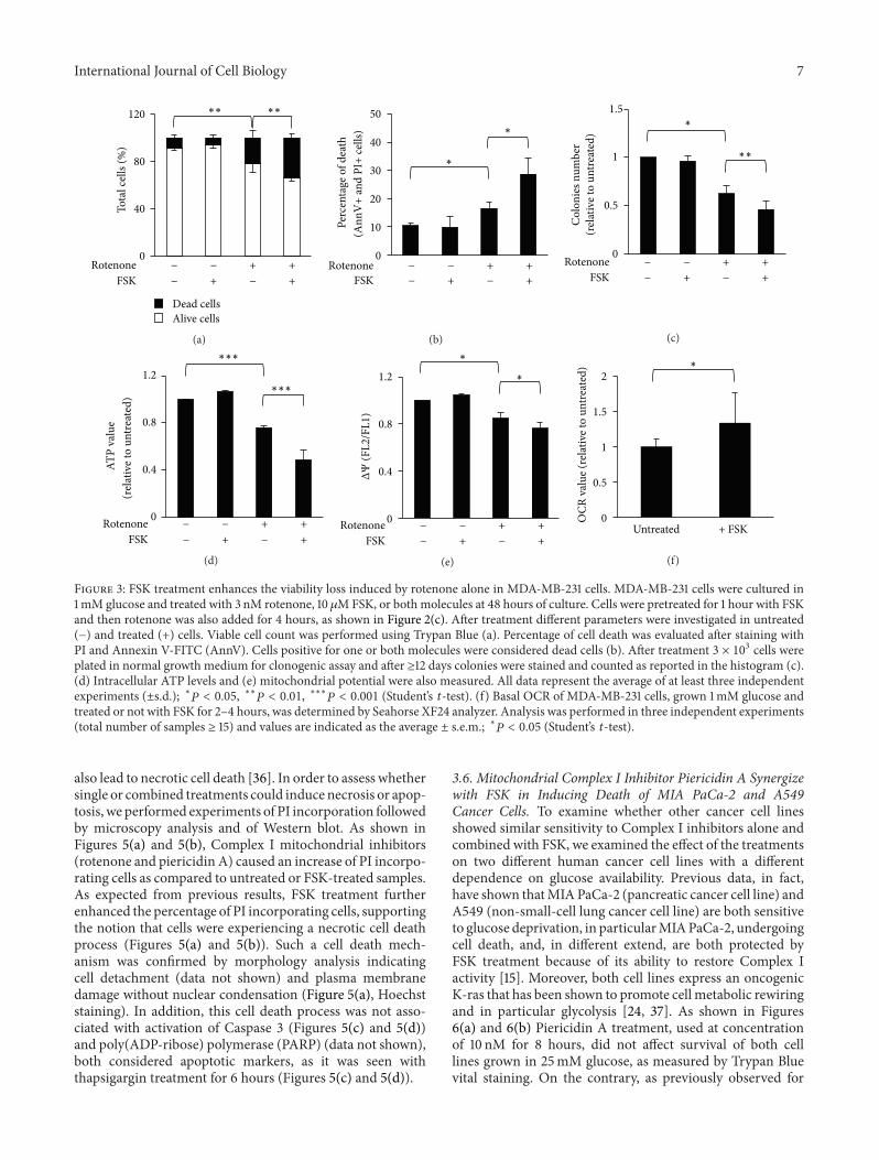
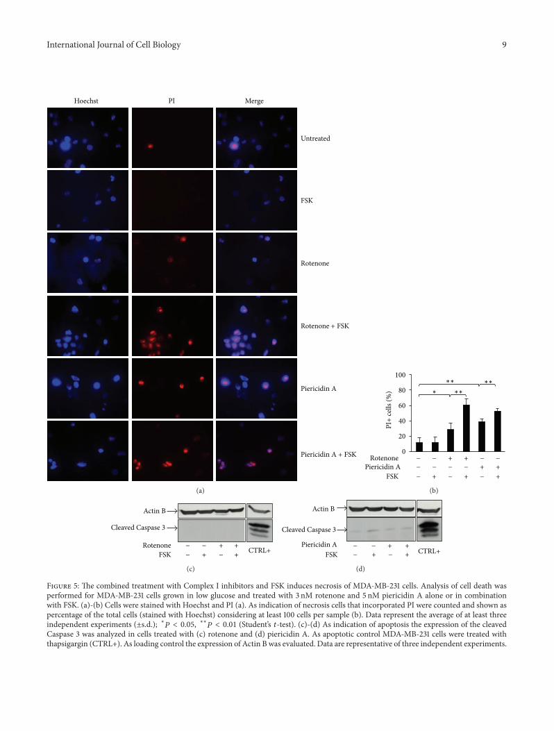
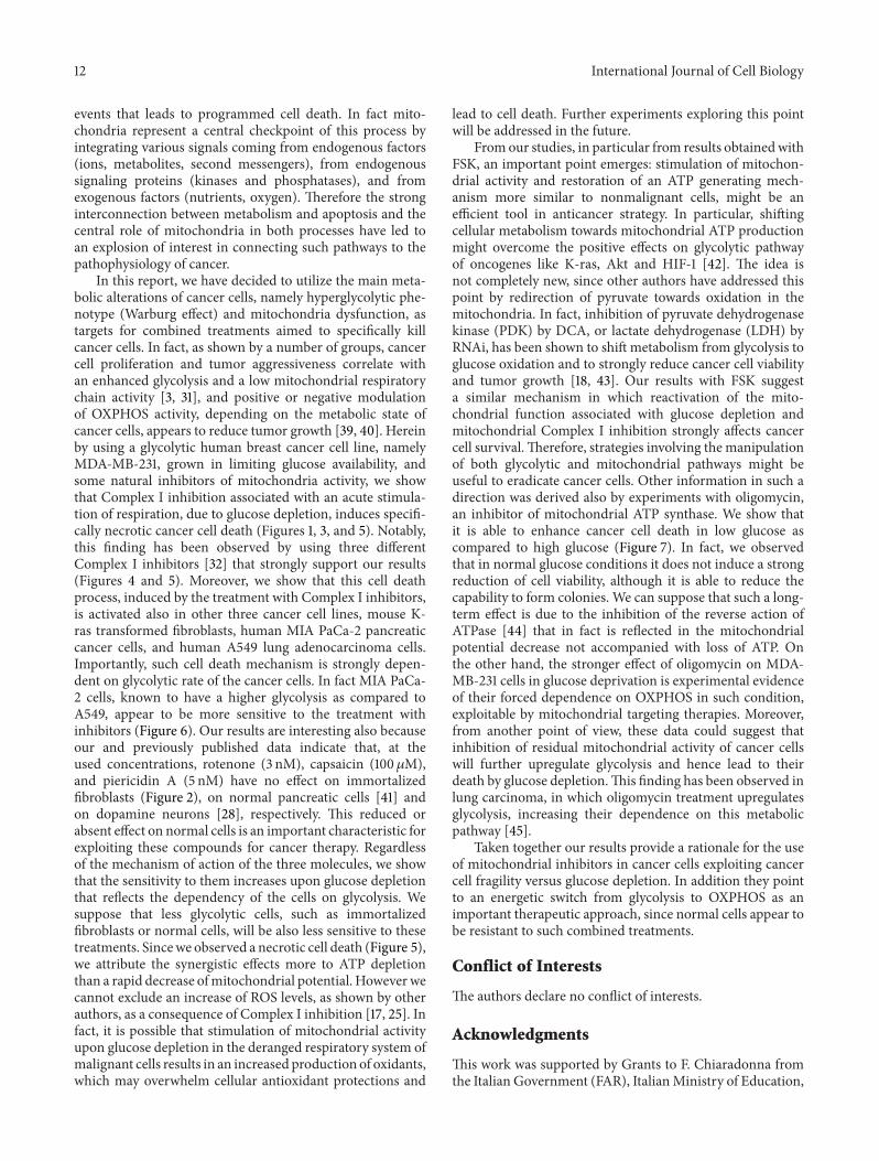
Related Documents