Hindawi Publishing Corporation PPAR Research Volume 2008, Article ID 256251, 10 pages doi:10.1155/2008/256251 Review Article Mitochondria, PPARs, and Cancer: Is Receptor-Independent Action of PPAR Agonists a Key? Roberto Scatena, Patrizia Bottoni, and Bruno Giardina Department of Laboratory Medicine, Catholic University, Largo A. Gemelli 8, 00168 Rome, Italy Correspondence should be addressed to Roberto Scatena, [email protected] Received 26 March 2008; Revised 27 May 2008; Accepted 18 June 2008 Recommended by Dipak Panigrahy Before the discovery of peroxisome proliferator activated receptors (PPARs), it was well known that certain drugs considered as classical PPAR-alpha agonists induced hepatocarcinoma or peroxisome proliferation in rodents. These drugs were derivatives of fibric acid, and they included clofibrate, bezafibrate, and fenofibrate. However, such toxicity has never been observed in human patients treated with these hypolipidemic drugs. Thiazolidinediones are a new class of PPAR activators showing greater specificity for the γ isoform of PPARs. These drugs are used as insulin sensitizers in the treatment of type II diabetes. In addition, they have been shown to induce cell differentiation or apoptosis in various experimental models of cancer. PPAR-α ligands have also been shown to induce cancer cell differentiation and, paradoxically, PPAR-γ drug activators have been reported to act as carcinogens. The confusing picture that emerges from these data is further complicated by the series of intriguing side effects observed following administration of pharmacological PPAR ligands (rhabdomyolysis, liver and heart toxicity, anemia, leucopenia). These side effects cannot be easily explained by simple interactions between the drug and nuclear receptors. Rather, these side effects seem to indicate that the ligands have biological activity independent of the nuclear receptors. Considering the emerging role of mitochondria in cancer and the potential metabolic connections between this organelle and PPAR physiology, characterization of the reciprocal influences is fundamental not only for a better understanding of cancer biology, but also for more defined pharmacotoxicological profiles of drugs that modulate PPARs. Copyright © 2008 Roberto Scatena et al. This is an open access article distributed under the Creative Commons Attribution License, which permits unrestricted use, distribution, and reproduction in any medium, provided the original work is properly cited. 1. INTRODUCTION Since the discovery of the first PPAR by Issemann and Green in 1990 [1], the role of this fascinating class of nuclear recep- tors in normal physiology and pathophysiology has become progressively more important. The potential biological activ- ities attributed to PPARs have been expanding ever since they were identified as potential mediators of the hypolipidemic effect of fibrates in humans and as participants in peroxisome proliferation and hepatocarcinogenicity in rodents. So far, PPARs have been implicated in diverse processes including lipid and carbohydrate metabolism, energy expenditure, immune and inflammatory processes, vascular homeostasis, tissue remodeling, and cell differentiation and proliferation in normal and neoplastic tissues [2–8]. It is evident, but too often ignored, that important interrelationships must exist among PPARs, mitochondria, and cancer. Regardless of the precise nature of these intercon- nections, PPARs undoubtedly have a significant energetic, plastic, and signaling role in the pathophysiology of cancer cells purely by virtue of their central role in lipid metabolism. This role necessarily involves interaction with mitochondria. Mitochondria are not only the main site of lipid oxidative metabolism, but they are also the cellular powerhouses that coordinate cellular metabolism and serve as the origin of important anabolic fluxes and signal transduction pathways [9, 10]. Thus, the role of mitochondria in cancer is under a critical re-evaluation, particularly in light of the so-called Warburg effect: most cancer cells exhibit increased aerobic glycolysis, and use this metabolic pathway for generation of ATP as a main source of their energy supply. Too often this does not justify the complex metabolic alterations present in different types of neoplasia [11–13]. Similarly, the interrelationships between PPARs and cancer are not entirely clear. Some studies show that PPARs

Welcome message from author
This document is posted to help you gain knowledge. Please leave a comment to let me know what you think about it! Share it to your friends and learn new things together.
Transcript
Hindawi Publishing CorporationPPAR ResearchVolume 2008, Article ID 256251, 10 pagesdoi:10.1155/2008/256251
Review ArticleMitochondria, PPARs, and Cancer: Is Receptor-IndependentAction of PPAR Agonists a Key?
Roberto Scatena, Patrizia Bottoni, and Bruno Giardina
Department of Laboratory Medicine, Catholic University, Largo A. Gemelli 8, 00168 Rome, Italy
Correspondence should be addressed to Roberto Scatena, [email protected]
Received 26 March 2008; Revised 27 May 2008; Accepted 18 June 2008
Recommended by Dipak Panigrahy
Before the discovery of peroxisome proliferator activated receptors (PPARs), it was well known that certain drugs considered asclassical PPAR-alpha agonists induced hepatocarcinoma or peroxisome proliferation in rodents. These drugs were derivatives offibric acid, and they included clofibrate, bezafibrate, and fenofibrate. However, such toxicity has never been observed in humanpatients treated with these hypolipidemic drugs. Thiazolidinediones are a new class of PPAR activators showing greater specificityfor the γ isoform of PPARs. These drugs are used as insulin sensitizers in the treatment of type II diabetes. In addition, they havebeen shown to induce cell differentiation or apoptosis in various experimental models of cancer. PPAR-α ligands have also beenshown to induce cancer cell differentiation and, paradoxically, PPAR-γ drug activators have been reported to act as carcinogens.The confusing picture that emerges from these data is further complicated by the series of intriguing side effects observed followingadministration of pharmacological PPAR ligands (rhabdomyolysis, liver and heart toxicity, anemia, leucopenia). These side effectscannot be easily explained by simple interactions between the drug and nuclear receptors. Rather, these side effects seem to indicatethat the ligands have biological activity independent of the nuclear receptors. Considering the emerging role of mitochondria incancer and the potential metabolic connections between this organelle and PPAR physiology, characterization of the reciprocalinfluences is fundamental not only for a better understanding of cancer biology, but also for more defined pharmacotoxicologicalprofiles of drugs that modulate PPARs.
Copyright © 2008 Roberto Scatena et al. This is an open access article distributed under the Creative Commons AttributionLicense, which permits unrestricted use, distribution, and reproduction in any medium, provided the original work is properlycited.
1. INTRODUCTION
Since the discovery of the first PPAR by Issemann and Greenin 1990 [1], the role of this fascinating class of nuclear recep-tors in normal physiology and pathophysiology has becomeprogressively more important. The potential biological activ-ities attributed to PPARs have been expanding ever since theywere identified as potential mediators of the hypolipidemiceffect of fibrates in humans and as participants in peroxisomeproliferation and hepatocarcinogenicity in rodents. So far,PPARs have been implicated in diverse processes includinglipid and carbohydrate metabolism, energy expenditure,immune and inflammatory processes, vascular homeostasis,tissue remodeling, and cell differentiation and proliferationin normal and neoplastic tissues [2–8].
It is evident, but too often ignored, that importantinterrelationships must exist among PPARs, mitochondria,and cancer. Regardless of the precise nature of these intercon-
nections, PPARs undoubtedly have a significant energetic,plastic, and signaling role in the pathophysiology of cancercells purely by virtue of their central role in lipid metabolism.This role necessarily involves interaction with mitochondria.Mitochondria are not only the main site of lipid oxidativemetabolism, but they are also the cellular powerhouses thatcoordinate cellular metabolism and serve as the origin ofimportant anabolic fluxes and signal transduction pathways[9, 10].
Thus, the role of mitochondria in cancer is under acritical re-evaluation, particularly in light of the so-calledWarburg effect: most cancer cells exhibit increased aerobicglycolysis, and use this metabolic pathway for generation ofATP as a main source of their energy supply. Too often thisdoes not justify the complex metabolic alterations present indifferent types of neoplasia [11–13].
Similarly, the interrelationships between PPARs andcancer are not entirely clear. Some studies show that PPARs
2 PPAR Research
have antineoplastic and/or cancer differentiating activities,while others show that they have important carcinogenicproperties [6, 14–20].
These reports highlight the multifaceted role of PPARsin neoplastic cells. Hence, the roles of PPAR in normalphysiology and pathophysiology should be clarified, sincethis may benefit our understanding of how cancer occurs andhow it can be treated.
Discussion of the interrelationships among PPARs, mito-chondria, and cancer should first involve careful evaluationof some misleading factors that have contributed to confu-sion about PPAR biology.
2. PPARs IN PATHOPHYSIOLOGY ANDMISLEADING FACTORS
2.1. Synthetic ligands
Initially, the physiological ligands of PPARs were unknownand PPARs were classified as “orphan receptors.” Theirfunction was studied using synthetic ligands of PPAR-α, thefirst PPAR discovered. These synthetic ligands were a het-erogeneous class of molecules ranging from trichloroaceticacid to plasticizers such as di-2-ethylhexyl phthalate (DEHP)and mono-2-ethylhexyl phthalate (MEHP) [1]. The struc-tural heterogeneity of the ligands seems to reflect theconformation of the PPAR ligand binding domain (LBD),which forms a large, Y-shaped hydrophobic pocket withrelatively low ligand specificity [2, 7]. For PPAR-α, theligands used most often in experiments were fibrates suchas clofibric acid, bezafibrate, and gemfibrozil. Studies onfibrate binding to PPAR-α showed that these drugs causeda hypotriglyceridemic effect by inducing transcription ofseveral genes related to oxidative metabolism of lipids,which occurs primarily in the mitochondria [2–4]. However,some discrepancies soon emerged. In fact, previous PPARresearch failed to provide thorough explanations of thedrugs’ other important biological activities, such as per-oxisome proliferation and hepatocarcinogenicity in rodentsor, most importantly, their side effects. These side effectsinclude angina crisis; elevation of serum aminotransferase,which indicates liver damage; increases in serum creatinephosphokinase concentrations, which can initiate myositis,myopathy and, in rare cases, rhabdomyolysis; increases inserum creatinine concentration; and acute renal failure inrare cases [21].
Some of these biological activities had already ledinvestigators to propose that these ligands had effects inde-pendent of their binding to PPAR. If true, neglecting these“extrareceptor” functions may interfere with our under-standing of PPAR pathophysiology. For example, studies inthe 1980s established that fibrates (clofibric acid, bezafibrate,and gemfibrozil) can pass freely through red blood cellmembranes and bind to human hemoglobin at the levelof the hydrophobic pocket of the alpha chain interfaces.This binding lowers the oxygen affinity of hemoproteinmore strongly than does the natural allosteric effector 2,3-biphosphoglycerate [22], which could, for example, leadto an angina crisis (by perturbation of microcirculation inischemic areas caused by abrupt changes in the level of
oxygen released from the blood) at the usual therapeu-tic plasmatic drug concentrations [23]. Around the sametime, other researchers showed that the agents acting asperoxisome proliferators hampered mitochondrial respira-tion, with potentially significant clinical implications [24–26]. Indeed, treatment with fibrates was found to lead tosome histological and biochemical features characteristic ofhepatic, muscular, and renal toxicities. This result led to thehypothesis that disruption of the mitochondrial electron res-piratory chain in conjunction with other genetic or acquiredpredisposing factors may contribute to these toxic effectsindependently of PPAR activation. Moreover, molecularanalysis of the interactions between human hemoglobin andfibrates indicated that particular physicochemical aspects ofthe drug molecules, specifically their carboxylic group andtheir significant hydrophobicity, might be responsible fortheir biological activity. Interestingly, these physicochemicalcharacteristics of fibrates fit well with the milieu of mito-chondria in general, namely the difference in pH betweenthe more alkaline matrix and the more acidic intermembranespace, as well as with structural features of complex I (NADHcytochrome c reductase) in the mitochondrial electronrespiratory chain, which is a large, hydrophobic proteincomponent in the mitochondria [10, 27].
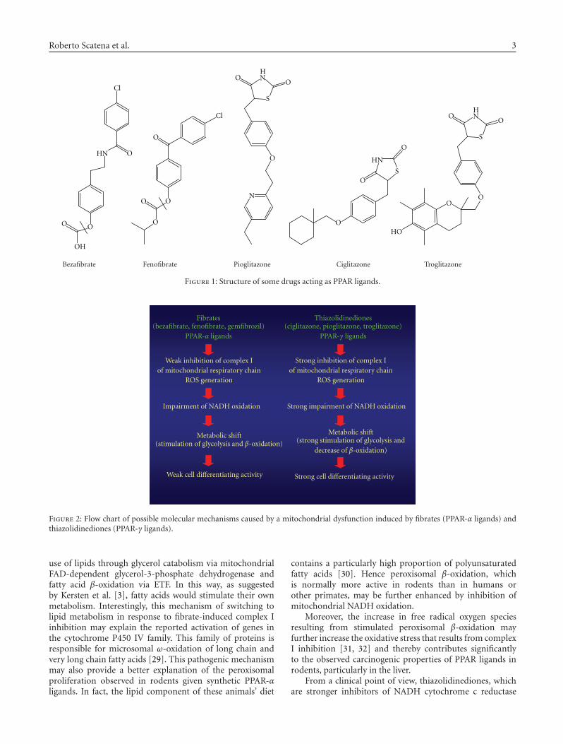
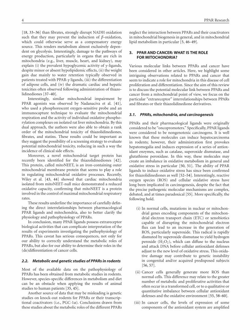
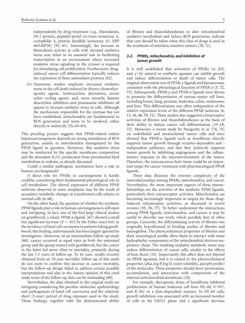
All of these considerations led to studies that examinedhow mitochondria were affected by fibrate administration,and whether these effects might have clinical implications[18, 28]. The results clearly showed that fibrates coulddisrupt the mitochondrial electron respiratory chain at thelevel of NADH cytochrome c reductase [18]. This effect waseven more pronounced for ciglitazone, which was one of thefirst thiazolidinediones to be synthesized. Thiazolidinedionesare a class of molecules that are chemically related to fibrates(Figure 1). Drug-induced mitochondrial dysfunction causesa series of compensatory metabolic mechanisms, which, inaddition to PPAR agonist activity, may be partially respon-sible for some of the pharmacological and toxicologicalproperties of this class of molecules. In fact, the resultingshut-down of mitochondrial NADH oxidation drives cellsto change their oxidative metabolism in a way that isstrictly correlated to the degree of complex I inhibition.Specifically, upon treatment with fibrates, which are lesspotent inhibitors of complex I, cells tend to use thosecomponents of the electron respiratory chain that remainefficient (e.g., complex II). This leads the cell to use FADH2
oxidation to obtain energy. In other words, compensatorymechanisms come into play, which are probably sustainedby glycerol catabolism viamitochondrial FAD-dependentglycerol-3-phosphate dehydrogenase or by fatty acid β-oxidation viaelectron-transferring flavoprotein (ETF). Thesechanges have a significant hypotriglyceridemic effect and aslight hypoglycemic effect. In contrast, using more potentcomplex I inhibitors (i.e., thiazodilinediones) greatly reducesNADH dehydrogenase activity, thus reducing the use of β-oxidation and increasing reliance on glycolysis, resulting ina stronger hypoglycemic effect and a much weaker or nullhypotriglyceridemic effect (Figure 2) [18].
Based on these findings, PPAR-α activation may be due,at least in part, to a shift in the metabolic state: preferential
Roberto Scatena et al. 3
O
OO
OH
HN
Cl
S
NO
O
OO
H
HO
S
N
O
N
OO
H
O
OO
O
Cl
HN
S
O
O
O
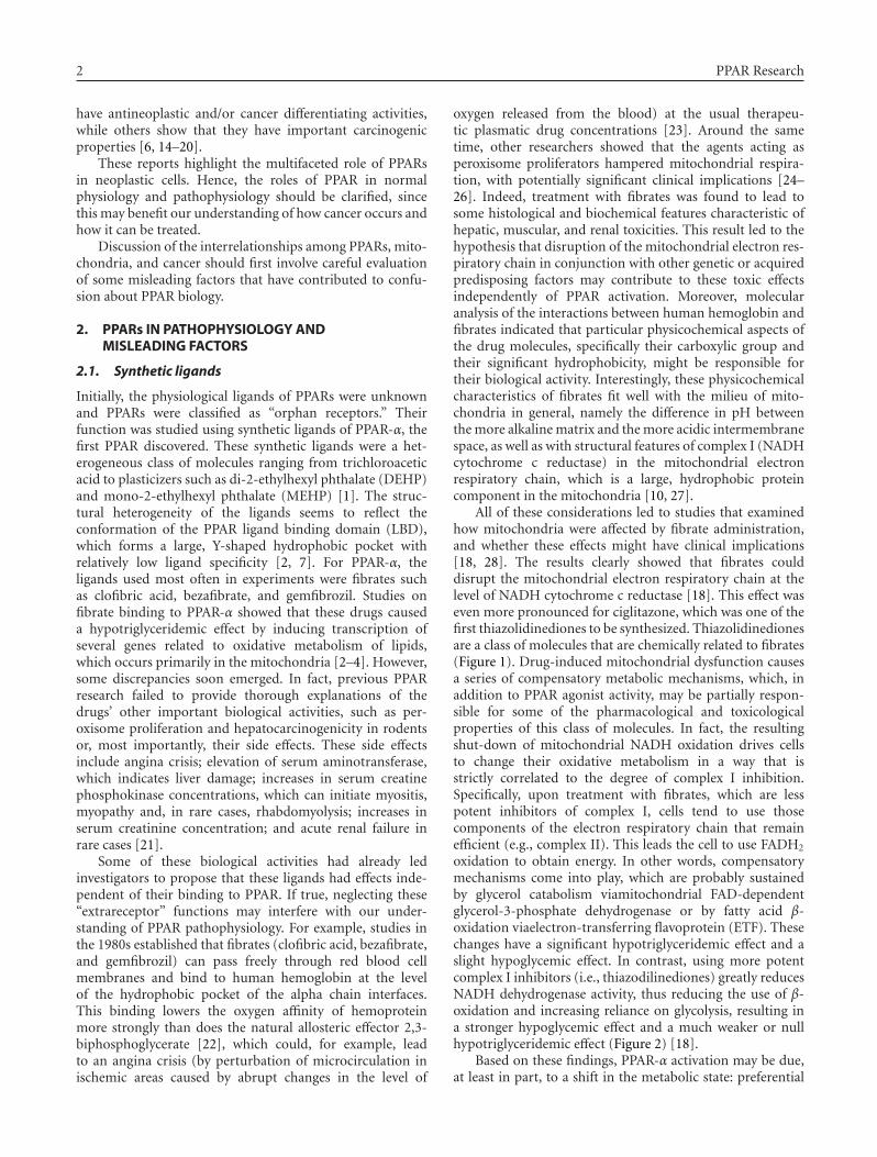
Bezafibrate Fenofibrate Pioglitazone Ciglitazone Troglitazone
Figure 1: Structure of some drugs acting as PPAR ligands.
Fibrates(bezafibrate, fenofibrate, gemfibrozil)
PPAR-α ligands
Weak inhibition of complex Iof mitochondrial respiratory chain
ROS generation
Impairment of NADH oxidation
Metabolic shift(stimulation of glycolysis and β-oxidation)
Weak cell differentiating activity
Thiazolidinediones(ciglitazone, pioglitazone, troglitazone)
PPAR-γ ligands
Strong inhibition of complex Iof mitochondrial respiratory chain
ROS generation
Strong impairment of NADH oxidation
Metabolic shift(strong stimulation of glycolysis and
decrease of β-oxidation)
Strong cell differentiating activity
Figure 2: Flow chart of possible molecular mechanisms caused by a mitochondrial dysfunction induced by fibrates (PPAR-α ligands) andthiazolidinediones (PPAR-γ ligands).
use of lipids through glycerol catabolism via mitochondrialFAD-dependent glycerol-3-phosphate dehydrogenase andfatty acid β-oxidation via ETF. In this way, as suggestedby Kersten et al. [3], fatty acids would stimulate their ownmetabolism. Interestingly, this mechanism of switching tolipid metabolism in response to fibrate-induced complex Iinhibition may explain the reported activation of genes inthe cytochrome P450 IV family. This family of proteins isresponsible for microsomal ω-oxidation of long chain andvery long chain fatty acids [29]. This pathogenic mechanismmay also provide a better explanation of the peroxisomalproliferation observed in rodents given synthetic PPAR-αligands. In fact, the lipid component of these animals’ diet
contains a particularly high proportion of polyunsaturatedfatty acids [30]. Hence peroxisomal β-oxidation, whichis normally more active in rodents than in humans orother primates, may be further enhanced by inhibition ofmitochondrial NADH oxidation.
Moreover, the increase in free radical oxygen speciesresulting from stimulated peroxisomal β-oxidation mayfurther increase the oxidative stress that results from complexI inhibition [31, 32] and thereby contributes significantlyto the observed carcinogenic properties of PPAR ligands inrodents, particularly in the liver.
From a clinical point of view, thiazolidinediones, whichare stronger inhibitors of NADH cytochrome c reductase
4 PPAR Research
[18, 33–36] than fibrates, strongly disrupt NADH oxidationsuch that they may prevent the induction of β-oxidation,which could otherwise serve as a compensatory energysource. This renders metabolism almost exclusively depen-dent on glycolysis. Interestingly, damage to the pathways ofenergy production, particularly in organs that are rich inmitochondria (e.g., liver, muscle, heart, and kidney), mayexplain (i) the prevalent hypoglycemic activity of γ-ligands,despite minor or absent hypolipidemic effects, (ii) the weightgain due mainly to water retention typically observed inpatients treated with PPAR-γ ligands, (iii) the differentiationof adipose cells, and (iv) the dramatic cardiac and hepatictoxicities often observed following administration of thiazo-lidinediones [37–40].
Interestingly, similar mitochondrial impairment byPPAR agonists was observed by Nadanaciva et al. [41],who used a phosphorescent oxygen-sensitive probe and animmunocapture technique to evaluate the mitochondrialrespiration and the activity of individual oxidative phospho-rylation complexes on isolated rat liver mitochondria. By thisdual approach, the authors were also able to obtain a rankorder of the mitochondrial toxicity of thiazolidinediones,fibrates, and statins. These results could be important asthey suggest the possibility of a screening strategy to evaluatepotential mitochondrial toxicity, reducing in such a way theincidence of clinical side effects.
Moreover, a novel mitochondrial target protein hasrecently been identified for the thiazolidinediones [42].This protein, called mitoNEET, is an iron-containing outermitochondrial membrane protein that seems to play a rolein regulating mitochondrial oxidative processes. Recently,Wiley et al. [43, 44] showed that cardiac mitochondriaisolated from mitoNEET-null mice demonstrated a reducedoxidative capacity, confirming that mitoNEET is a proteininvolved in the control of maximal mitochondrial respiratoryrates.
These results underline the importance of carefully defin-ing the direct interrelationships between pharmacologicalPPAR ligands and mitochondria, also to better clarify thephysiology and pathophysiology of PPARs.
In conclusion, many PPAR ligands possess extrareceptorbiological activities that can complicate interpretation of theresults of experiments investigating the pathophysiology ofPPARs. This caveat has serious consequences, not only forour ability to correctly understand the metabolic roles ofPPARs, but also for our ability to determine their roles in the(de)differentiation of cancer cells.
2.2. Metabolic and genetic studies of PPARs in rodents
Most of the available data on the pathophysiology ofPPARs has been obtained from metabolic studies in rodents.However, species-specific differences in metabolism and dietcan be an obstacle when applying the results of animalstudies to human patients [35, 45].
Another source of data that may be misleading is geneticstudies on knock-out rodents for PPARs or their transcrip-tional coactivators (i.e., PGC-1α). Conclusions drawn fromthese studies about the metabolic roles of the different PPARs
neglect the interaction between PPARs and their coactivatorsin mitochondrial biogenesis in general, and in mitochondriallipid metabolism in particular [5, 46–49].
3. PPAR AND CANCER: WHAT IS THE ROLEFOR MITOCHONDRIA?
Various molecular links between PPARs and cancer havebeen considered in other articles. Here, we highlight someintriguing observations related to PPARs and cancer thatseem to indicate a role for mitochondria in this disease of cellproliferation and differentiation. Since the aim of this reviewis to discuss the potential molecular link between PPARs andcancer from a mitochondrial point of view, we focus on theparticular “extrareceptor” interrelationships between PPARsand fibrates or their thiazolidinedione derivatives.
3.1. PPARs, mitochondria, and carcinogenesis
PPARs and their pharmacological ligands were originallyconsidered to be “oncopromoters.” Specifically, PPAR ligandswere considered to be nongenotoxic carcinogens. It is wellknown that these molecules can induce hepatocarcinomain rodents; however, their administration first provokeshepatomegalia and induces expression of a series of antiox-idant enzymes, such as catalase, superoxide dismutase, andglutathione peroxidase. In this way, these molecules maycreate an imbalance in oxidative metabolism in general andoxidative stress in particular [50, 51]. The ability of PPARligands to induce oxidative stress has since been confirmedfor thiazolidinediones as well [52–54]. Interestingly, reactiveoxygen species (ROS) and cellular oxidative stress havelong been implicated in carcinogenesis, despite the fact thatthe precise pathogenic molecular mechanisms are complex,debated, and at times paradoxical [55]. More specifically, thefollowing hold.
(i) In normal cells, mutations in nuclear or mitochon-drial genes encoding components of the mitochon-drial electron transport chain (ETC) or xenobioticscapable of disrupting the mitochondrial electronflux can lead to an increase in the generation ofROS, particularly superoxide. This radical is rapidlydismuted by superoxide dismutase to yield hydrogenperoxide (H2O2), which can diffuse to the nucleusand attack DNA before cellular antioxidant defensesadjust to the new level of oxidative stress. This oxida-tive damage may contribute to genetic instabilityin congenital and/or acquired predisposed subjects[56, 57].
(ii) Cancer cells generally generate more ROS thannormal cells. This difference may relate to the greaternumber of metabolic and proliferative activities thatoften occur in a transformed cell, or to a qualitative orquantitative imbalance between cellular antioxidantdefenses and the oxidative environment [55, 58–60].
(iii) In cancer cells, the levels of expression of somecomponents of the antioxidant system are amplified
Roberto Scatena et al. 5
independently by drug treatment (e.g., thioredoxin,DJ-1 protein, peptidyl-prolyl cis-trans isomerase A,cyclophilin A, protein disulfide isomerase A3, ERP60/GRP58) [55, 61]. Interestingly, the increase inthioredoxin activity in cells with elevated oxidativestress may relate to its essential role in facilitatingtranscription in an environment where increasedoxidative stress signaling in the cytosol is requiredfor stimulating cell proliferation. Furthermore, drug-induced cancer cell differentiation typically reducesthe expression of these antioxidant proteins [62].
(iv) Numerous studies implicate increased oxidativestress in the cell death induced by diverse chemother-apeutic agents. Anthracycline derivatives, newerredox cycling agents, and, more recently, histonedeacetylase inhibitors and proteasome inhibitors allappear to increase oxidative stress in cells. Althoughthe mechanism responsible for the increase has notbeen established, mitochondria are fundamental inROS generation and seem to be involved, eitherdirectly or indirectly [55, 63–65].
This puzzling picture suggests that PPAR-related rodenthepatocarcinogenesis depends on strong stimulation of ROSgeneration, mainly in mitochondria dysregulated by thePPAR ligand in question. Moreover, this oxidative stressmay be reinforced by the specific membrane compositionand the abundant H2O2 production from peroxisomal lipidmetabolism in rodents, as already discussed.
Could a similar pathogenic mechanism have a role inhuman carcinogenesis?
A direct role for PPARs in carcinogenesis is hardlycredible, considering their fundamental physiological role incell metabolism. The altered expression of different PPARisoforms observed in some neoplasias may be the result ofsecondary metabolic changes in transformed cells relative tonormal cells [6, 66].
On the other hand, the question of whether the syntheticPPAR ligands play a role in human carcinogenesis is still openand intriguing. In fact, one of the first large clinical studieson gemfibrozil, a classic PPAR-α ligand, [67] showed a smallbut significant increase (P = .032 by the Fisher exact test) inthe incidence of basal cell carcinoma in patients taking gemfi-brozil; this finding, unfortunately has been largely ignored byinvestigators. Moreover, in an intermediate follow-up study[68], cancer occurred at equal rates in both the untreatedgroup and the group treated with gemfibrozil, but the cancerin the latter led more often to mortality, primarily duringthe last 1.5 years of follow-up. To be sure, results recentlyobtained from an 18-year mortality follow-up of this studydo not seem to confirm this increase in cancer mortality,but the follow-up design failed to address certain possibleinterpretations and also in the Autors opinion of this citedstudy some of the follow-up data can be misleading [69].
Nevertheless, the data obtained in the original study areintriguing considering the peculiar molecular epidemiologyand pathogenesis of basal cell carcinoma and the relativelyshort (5-year) period of drug exposure used in the study.These findings, together with the demonstrated ability
of fibrates and thiazolidinediones to alter mitochondrialoxidative metabolism and induce ROS generation, indicatethat care should be taken when this class of drugs is used inthe treatment of nutrition-sensitive tumors [70, 71].
3.2. PPARs, mitochondria, and inhibition oftumor growth
It is well established that activation of PPARs (α, β/δ,and γ) by natural or synthetic agonists can inhibit growthand induce differentiation or death of tumor cells. Theoriginal observation was of PPAR-γ ligands and liposarcoma,consistent with the physiological function of PPAR-γ [3, 72,73]. Subsequently, PPAR-γ and PPAR-α ligands were shownto promote the differentiation of various tumor cell lines,including breast, lung, prostate, leukemia, colon, melanoma,and liver. This differentiation was often independent of therelative expression levels of the different PPAR isoforms [6,15, 16, 66, 70, 72]. These studies also suggested extrareceptoractivities of fibrates and thiazolidinediones as the basis oftheir ability to induce cancer cell differentiation [15–18,72]. Moreover, a recent study by Panigrahy et al. [74, 75]on endothelial and mesenchymal tumor cells and miceshowed that PPAR-α ligands such as fenofibrate directlysuppress tumor growth through receptor-dependent and -independent pathways, and that they indirectly suppresstumor growth by inhibiting angiogenesis and the inflam-matory response in the microenvironment of the tumor.Therefore, the noncancerous host tissue could be an impor-tant target for cancer treatment with pharmacological PPARligands.
These data illustrate the extreme complexity of theinterrelationships among PPARs, mitochondria, and cancer.Nevertheless, the most important aspects of these interre-lationships are the activities of the synthetic PPAR ligands,particularly their extrareceptor activities. Mitochondria arebecoming increasingly important as targets for these drug-induced extrareceptor activities, as discussed in recentreviews [35, 76, 77]. To better understand the interactionsamong PPAR ligands, mitochondria, and cancer, it may beuseful to describe our work, which parallels that of othergroups. Curiously, the differentiating activity of fibrates wasoriginally hypothesized in binding studies of fibrates andhemoglobin. The physicochemical properties of fibrates andtheir toxicological profile allow them to interact with somehydrophobic components of the mitochondrial electron res-piratory chain. The resulting oxidative metabolic stress mayinduce differentiation of cancer cells, similar to the effectsof heat shock [15]. Importantly, this effect does not dependon PPAR agonism, but it is related to the physicochemicalproperties (pKa, log P, log D, water solubility, and pH profile)of the molecules. These properties should favor permeation,accumulation, and interaction with components of theinternal mitochondrial membrane [10].
For example, therapeutic doses of bezafibrate inhibitedproliferation of human leukemia cell lines HL-60, U-937,and K-562 in a dose-dependent manner. In HL-60 cells,growth inhibition was associated with an increased numberof cells in the G0/G1 phase and a significant decrease
6 PPAR Research
in the number of cells in the G2/M phase. Analysis ofcell differentiation markers (CD) showed a dose-dependentincrease in expression of CD11b and CD14 in HL-60 cellsand of CD14 in U-937 cells. Functional assays confirmedthat the phenotypes of these cells were more mature.Both HL-60 and U-937 cells showed a dose-dependentrestoration of the respiratory burst stimulated by PMAand zymosan. K-562 erythroleukemia cells showed a dose-dependent increase in hemoglobin synthesis. Similar cellulardifferentiation was observed following treatment with twoother fibrate derivatives, clofibric acid and gemfibrozil.Interestingly, fibrate-induced differentiation was partiallyinhibited by antioxidants including acetylcysteine (NAC),and electron microscopy revealed that fibrate-treated cellshad mitochondrial damage [15]. Functional evaluation ofthis drug-induced mitochondriopathy showed that fibratesand ciglitazone specifically inhibited NADH cytochrome creductase activity in a dose-dependent manner in HL-60,TE-671 human rabdomyosarcoma, and Hep-G2 human hep-atocarcinoma cell lines, whereas the activity of other mito-chondrial respiratory chain enzymes remained unchanged[18, 33, 36]. The impairment of NADH oxidation induced acellular metabolic shift towards anaerobic glycolysis and/orβ-oxidation, as shown by the dose-dependent increases ofcertain metabolites (lactate, alanine, glycolytic, and nongly-colytic derived acetate) [18].
A fundamental observation from this research was thecorrelation of mitochondrial dysfunction, metabolic shift,and differentiation activity in tumor cells treated withincreasing concentrations of PPAR ligands. Furthermore,quantitative comparison on a molar ratio basis between thesePPAR ligands (bezafibrate, clofibric acid, gemfibrozil, andciglitazone) for inhibition of NADH cytochrome c reductaseactivity, metabolic adaptations, differentiation potency, andantiproliferative index confirmed a strict correlation betweenthese parameters [18]. These results suggested that inhibitionof mitochondrial NADH dehydrogenase could contribute toboth the pharmacological and toxicological profiles of fibratederivatives (strong hypolipidemic/weak hypoglycemic effect,liver and muscle toxicity) and thiazolidinediones (hypo-glycemic/insulin sensitizer effect, liver and heart toxicity)[17, 18, 25, 26, 33–40].
In terms of mitochondrial oncology, these data sug-gest a possible molecular mechanism for the peroxisomeproliferator activity and carcinogenicity of fibrates typicallyobserved in rodents. These data also indicate a strict correla-tion among fibrate- and thiazolidinedione-induced cellularrespiration dysfunction, stimulation of glycolysis, and cancercell differentiation that strongly implicates mitochondria andoxidative metabolism in the pathophysiology of cancer.
Importantly, these results confirm and extend the resultsof other studies focusing on nongenomic activities of fibratesand thiazolidinediones [16, 17, 78–81]. Furthermore, theseobservations explain some contradictory data related to therole of PPARs in cancer cell differentiation [3, 6, 72, 77].Above all, the intriguing data concerning the inductionof differentiation associated with a shift towards aerobicglycolysis (a paradoxical Warburg effect) confirms the needto reconsider cancer cell metabolism in general and the
Warburg effect in particular [11, 13, 82]. To that end, ourunderstanding of the role of PPARs in cancer should assumea new level of complexity that takes into account theirfundamental functions in lipid metabolism, in inflammationand, directly or indirectly, in angiogenesis [74, 75].
The molecular link among the synthetic PPAR ligands,mitochondria, and cancer indicates the need for a carefulevaluation of some aspects of cancer cell pathophysiology,such as the following.
(i) The possible existence of a transduction pathwaymaster signal as the basis of the complex cellulardifferentiation program related to PPAR. ROS, nitricoxide (NO), and reactive nitric oxide species (RNS)should form an important branch of this program. Inaddition, there should be a role for the NADH/NAD+ratio.
(ii) The role of some oncogenes/oncosuppressors incancer pathogenesis, given that mitochondrial respi-ratory chain dysfunction can induce a more differen-tiated phenotype in tumor cells and thereby influencetheir activity [15–18].
(iii) The significance of the modulation of the expressionof proteins with oncogenic and antioxidant functions(stathmin 1, DJ-1 protein, peroxiredoxin 2, nucle-oside diphosphate kinase A, etc.) in PPAR-relatedcancer cell differentiation. This is an important topicgiven the potential pathophysiological role of PPARin cancer [62, 83].
At last, an understanding of the molecular mechanismsinvolved in the interrelationships between mitochondrialrespiration and PPAR-related cancer regression may haveimportant clinical implications for cancer diagnosis, prog-nosis, and therapy.
4. CONCLUSION
Many molecular mechanisms have been proposed to explainhow PPARs, directly and/or indirectly, may induce cancercell cycle arrest and induction or cancer cell differentiationor dedifferentiation. In spite of this, the molecular interrela-tionships between the mechanisms of functional modulationof PPAR and these important cellular phenotypic changesare still debated. It is clear that the various molecularmodifications observed in different studies (decrease incyclin D1, inhibition of IkB, induction of TSC22, NF-kB,GADD153, PTEN, etc.) may depend on the particular celland cell functional status and that a potential master signalshould be investigated.
Here, we have briefly described the molecular linkbetween PPARs and cancer from a mitochondrial point ofview. In our opinion, the most important factor linkingcancer to PPARs is represented by their “synthetic ligands,”which are characterized by other important and debatedextrareceptor activities. Specifically, these agents can induceoxidative stress, which has an ambiguous role in cancer,leading it to act as a double-edged sword.
Roberto Scatena et al. 7
In this sense, mitochondria plays a critical role as oneof the most important organelles for generating reactivespecies. The metabolic stress and energetic failure that resultfrom fibrate- and thiazolidinedione-induced mitochondrialimpairment may also play an important part in cancerregression, especially in cells that require active and completeanabolic pathways to sustain cancer growth, an aspect thathas not always been completely or correctly evaluated.
Considering their physicochemical properties discussedabove, it is worthy of note that tumor regression induced byPPAR ligands may be a useful approach for the treatmentof neoplasias of the central nervous system, which areclassically difficult to treat with conventional chemotherapy.Specifically, interesting results have already been obtained interms of decrease of cell proliferation, apoptosis induction,and expression of markers typical of a more differentiatedphenotype in glioblastoma and astrocytoma cell lines [84–90], in primary cultures of human glioblastoma cells derivedfrom surgical specimens [91], and above all, in patients withhigh-grade gliomas (glioblastoma or anaplastic glioma) [92].
Moreover, given that pharmacological modulation ofPPAR in cancer cells typically arrests the cell cycle inthe G0/G1 phase, combination therapy with a PPAR ago-nist and an antimitotic antitumor agent deserves carefulconsideration.
Furthermore, it may be useful to distinguish between realdifferentiating agents, which show low cytotoxicity indicesrelative to their differentiation activity (thiazolidinediones,fibrates, retinoids) and spurious differentiating agents, whichshow low differentiating activity and high cytotoxicityindices (old and new HDAC inhibitors) [93, 94].
A mitochondrial approach to analysis of the molecularlink between PPARs and cancer certainly adds new levelsof complexity to the already complicated picture. However,an optimal definition of all molecular mechanisms relatingPPARs, mitochondria, and cancer may be fundamentalto our understanding of the real therapeutic index ofpharmacological modulation of these nuclear receptors. Thisis important not only in cancer, but also in the other diseasesin which PPARs play a significant role, including atheroscle-rosis, hyperlipoproteinemias, metabolic syndrome, diabetesmellitus, and obesity. Moreover, a complete understanding ofthe pharmacotoxicological profile of these agents may reducethe incidence of dangerous side effects that have alreadydramatically afflicted patients treated with PPAR ligands[37–40].
REFERENCES
[1] I. Issemann and S. Green, “Activation of a member ofthe steroid hormone receptor superfamily by peroxisomeproliferators,” Nature, vol. 347, no. 6294, pp. 645–650, 1990.
[2] B. Desvergne and W. Wahli, “Peroxisome proliferators-activated receptors: nuclear control of metabolism,”Endocrinology Review, vol. 20, no. 5, pp. 649–688, 1999.
[3] S. Kersten, B. Desvergne, and W. Wahli, “Roles of PPARS inhealth and disease,” Nature, vol. 405, no. 6785, pp. 421–424,2000.
[4] C.-H. Lee, P. Olson, and R. M. Evans, “Minireview: lipidmetabolism, metabolic diseases, and peroxisome proliferator-
activated receptors,” Endocrinology, vol. 144, no. 6, pp. 2201–2207, 2003.
[5] R. M. Evans, G. D. Barish, and Y.-X. Wang, “PPARs and thecomplex journey to obesity,” Nature Medicine, vol. 10, no. 4,pp. 355–361, 2004.
[6] Z. Nahle, “PPAR trilogy from metabolism to cancer,” CurrentOpinion in Clinical Nutrition and Metabolic Care, vol. 7, no. 4,pp. 397–402, 2004.
[7] L. Michalik, J. Auwerx, J. P. Berger, et al., “International unionof pharmacology. LXI. Peroxisome proliferator-activatedreceptors,” Pharmacological Reviews, vol. 58, no. 4, pp. 726–741, 2006.
[8] L. Michalik and W. Wahli, “Involvement of PPAR nuclearreceptors in tissue injury and wound repair,” Journal of ClinicalInvestigation, vol. 116, no. 3, pp. 598–606, 2006.
[9] V. Darley-Usmar, “The powerhouse takes control of the cell;the role of mitochondria in signal transduction,” Free RadicalBiology & Medicine, vol. 37, no. 6, pp. 753–754, 2004.
[10] R. Scatena, P. Bottoni, G. Botta, G. E. Martorana, and B.Giardina, “The role of mitochondria in pharmacotoxicology:a reevaluation of an old, newly emerging topic,” AmericanJournal of Physiology, vol. 293, no. 1, pp. C12–C21, 2007.
[11] K. Garber, “Energy deregulation: licensing tumors to grow,”Science, vol. 312, no. 5777, pp. 1158–1159, 2006.
[12] R. Moreno-Sanchez, S. Rodrıguez-Enrıquez, A. Marın-Hernandez, and E. Saavedra, “Energy metabolism in tumorcells,” The FEBS Journal, vol. 274, no. 6, pp. 1393–1418, 2007.
[13] J.-W. Kim and C. V. Dang, “Cancer’s molecular sweet toothand the Warburg effect,” Cancer Research, vol. 66, no. 18, pp.8927–8930, 2006.
[14] E. I. Chen, J. Hewel, J. S. Krueger, et al., “Adaptation ofenergy metabolism in breast cancer brain metastases,” CancerResearch, vol. 67, no. 4, pp. 1472–1486, 2007.
[15] R. Scatena, G. Nocca, P. De Sole, et al., “Bezafibrate asdifferentiating factor of human myeloid leukemia cells,” CellDeath and Differentiation, vol. 6, no. 8, pp. 781–787, 1999.
[16] S. S. Palakurthi, H. Aktas, L. M. Grubissich, R. M. Mortensen,and J. A. Halperin, “Anticancer effects of thiazolidinedionesare independent of peroxisome proliferator-activated receptorγ and mediated by inhibition of translation initiation,” CancerResearch, vol. 61, no. 16, pp. 6213–6218, 2001.
[17] S. J. Baek, L. C. Wilson, L. C. Hsi, and T. E. Eling,“Troglitazone, a peroxisome proliferator-activated receptor γ(PPARγ) ligand, selectively induces the early growth response-1 gene independently of PPARγ: a novel mechanism for itsanti-tumorigenic activity,” Journal of Biological Chemistry, vol.278, no. 8, pp. 5845–5853, 2003.
[18] R. Scatena, P. Bottoni, F. Vincenzoni, et al., “Bezafibrateinduces a mitochondrial derangement in human cell lines: aPPAR-independent mechanism for a peroxisome proliferator,”Chemical Research in Toxicology, vol. 16, no. 11, pp. 1440–1447, 2003.
[19] F. S. Harman, C. J. Nicol, H. E. Marin, J. M. Ward, F. J.Gonzalez, and J. M. Peters, “Peroxisome proliferator-activatedreceptor-δ attenuates colon carcinogenesis,” Nature Medicine,vol. 10, no. 5, pp. 481–483, 2004.
[20] R. A. Gupta, D. Wang, S. Katkuri, H. Wang, S. K. Dey, and R.N. DuBois, “Activation of nuclear hormone receptor perox-isome proliferator-activated receptor-δ accelerates intestinaladenoma growth,” Nature Medicine, vol. 10, no. 3, pp. 245–247, 2004.
[21] S. C. Sweetman, Ed., Martindale: The Complete Drug Reference,Pharmaceutical Press, London, UK, 35th edition, 2005.
8 PPAR Research
[22] M. F. Perutz and C. Poyart, “Bezafibrate lowers oxygen affinityof haemoglobin,” The Lancet, vol. 2, no. 8355, pp. 881–882,1983.
[23] R. Scatena, G. Nocca, I. Messana, et al., “Effects of gemfibrozilon the oxygen transport properties of erythrocytes,” BritishJournal of Clinical Pharmacology, vol. 39, no. 1, pp. 25–30,1995.
[24] D. S. Chance and M. K. McIntosh, “Hypolipidemic agentsalter hepatic mitochondrial respiration in vitro,” ComparativeBiochemistry and Physiology Part C, vol. 111, no. 2, pp. 317–323, 1995.
[25] J. Youssef and M. Badr, “Extraperoxisomal targets of peroxi-some proliferators: mitochondrial, microsomal, and cytosoliceffects. Implications for health and disease,” Critical Reviews inToxicology, vol. 28, no. 1, pp. 1–33, 1998.
[26] S. Zhou and K. B. Wallace, “The effect of peroxisomeproliferators on mitochondrial bioenergetics,” ToxicologicalSciences, vol. 48, no. 1, pp. 82–89, 1999.
[27] A. Szewczyk and L. Wojtczak, “Mitochondria as a pharma-cological target,” Pharmacological Reviews, vol. 54, no. 1, pp.101–127, 2002.
[28] R. Scatena, G. Nocca, P. De Sole, R. Fresu, C. Zuppi, andB. Giardina, “The priming effect of gemfibrozil on reactiveoxygen metabolism of phagocytic leucocytes. An intriguingside effect,” Clinica Chimica Acta, vol. 266, no. 2, pp. 173–183,1997.
[29] J. K. Reddy and T. Hashimoto, “Peroxisomal β-oxidationand peroxisome proliferator-activated receptor α: an adaptivemetabolic system,” Annual Review of Nutrition, vol. 21, pp.193–230, 2001.
[30] A. L. Lehninger, D. L. Nelson, and M. M. Cox, Principles ofBiochemistry, Worth, New York, NY, USA, 2000.
[31] S. Raha, A. T. Myint, L. Johnstone, and B. H. Robinson,“Control of oxygen free radical formation from mitochondrialcomplex I: roles for protein kinase A and pyruvate dehydroge-nase kinase,” Free Radical Biology & Medicine, vol. 32, no. 5,pp. 421–430, 2002.
[32] V. Adam-Vizi and C. Chinopoulos, “Bioenergetics and theformation of mitochondrial reactive oxygen species,” Trendsin Pharmacological Sciences, vol. 27, no. 12, pp. 639–645, 2006.
[33] R. Scatena, P. Bottoni, G. E. Martorana, et al., “Mitochondrialrespiratory chain dysfunction, a non-receptor-mediated effectof synthetic PPAR-ligands: biochemical and pharmacologicalimplications,” Biochemical and Biophysical Research Commu-nications, vol. 319, no. 3, pp. 967–973, 2004.
[34] B. Brunmair, K. Staniek, F. Gras, et al., “Thiazolidinediones,like metformin, inhibit respiratory complex I: a commonmechanism contributing to their antidiabetic actions?” Dia-betes, vol. 53, no. 4, pp. 1052–1059, 2004.
[35] R. Scatena, G. E. Martorana, P. Bottoni, and B. Giardina,“Mitochondrial dysfunction by synthetic ligands of peroxi-some proliferator activated receptors (PPARs),” IUBMB Life,vol. 56, no. 8, pp. 477–482, 2004.
[36] R. Scatena, P. Bottoni, G. E. Martorana, et al., “Mitochondria,ciglitazone and liver: a neglected interaction in biochemicalpharmacology,” European Journal of Pharmacology, vol. 567,no. 1-2, pp. 50–58, 2007.
[37] A. Y. Y. Cheng and I. G. Fantus, “Thiazolidinedione-inducedcongestive heart failure,” Annals of Pharmacotherapy, vol. 38,no. 5, pp. 817–820, 2004.
[38] M. Chojkier, “Troglitazone and liver injury: in search ofanswers,” Hepatology, vol. 41, no. 2, pp. 237–346, 2005.
[39] S. E. Nissen and K. Wolski, “Effect of rosiglitazone on therisk of myocardial infarction and death from cardiovascular
causes,” The New England Journal of Medicine, vol. 356, no. 24,pp. 2457–2471, 2007.
[40] A. M. Lincoff, K. Wolski, S. J. Nicholls, and S. E. Nissen,“Pioglitazone and risk of cardiovascular events in patientswith type 2 diabetes mellitus: a meta-analysis of randomizedtrials,” Journal of the American Medical Association, vol. 298,no. 10, pp. 1180–1188, 2007.
[41] S. Nadanaciva, J. A. Dykens, A. Bernal, R. A. Capaldi, andY. Will, “Mitochondrial impairment by PPAR agonists andstatins identified via immunocaptured OXPHOS complexactivities and respiration,” Toxicology and Applied Pharmacol-ogy, vol. 223, no. 3, pp. 277–287, 2007.
[42] J. R. Colca, W. G. McDonald, D. J. Waldon, et al., “Iden-tification of a novel mitochondrial protein (“mitoNEET”)cross-linked specifically by a thiazolidinedione photoprobe,”American Journal of Physiology, vol. 286, no. 2, pp. E252–E260,2004.
[43] S. E. Wiley, A. N. Murphy, S. A. Ross, P. van der Geer,and J. E. Dixon, “MitoNEET is an iron-containing outermitochondrial membrane protein that regulates oxidativecapacity,” Proceedings of the National Academy of Sciences of theUnited States of America, vol. 104, no. 13, pp. 5318–5323, 2007.
[44] M. L. Paddock, S. E. Wiley, H. L. Axelrod, et al., “MitoNEETis a uniquely folded 2Fe-2S outer mitochondrial membraneprotein stabilized by pioglitazone,” Proceedings of the NationalAcademy of Sciences of the United States of America, vol. 104,no. 36, pp. 14342–14347, 2007.
[45] B. Halliwell and J. M. C. Cutteridge, Free Radical in Biology andMedicine, Oxford University Press, New York, NY, USA, 1986.
[46] Z. Wu, P. Puigserver, U. Andersson, et al., “Mechanismscontrolling mitochondrial biogenesis and respiration throughthe thermogenic coactivator PGC-1,” Cell, vol. 98, no. 1, pp.115–124, 1999.
[47] A. L. Hevener, W. He, Y. Barak, et al., “Muscle-specific Ppargdeletion causes insulin resistance,” Nature Medicine, vol. 9, no.12, pp. 1491–1497, 2003.
[48] W. He, Y. Barak, A. Hevener, et al., “Adipose-specific peroxi-some proliferator-activated receptor γ knockout causes insulinresistance in fat and liver but not in muscle,” Proceedings of theNational Academy of Sciences of the United States of America,vol. 100, no. 26, pp. 15712–15717, 2003.
[49] T. C. Leone, J. J. Lehman, B. N. Finck, et al., “PGC-1α defi-ciency causes multi-system energy metabolic derangements:muscle dysfunction, abnormal weight control and hepaticsteatosis,” PLoS Biology, vol. 3, no. 4, p. e101, 2005.
[50] Y. Cai, E. L. Appelkvist, and J. W. DePierre, “Hepaticoxidative stress and related defenses during treatment of micewith acetylsalicylic acid and other peroxisome proliferators,”Journal of Biochemical Toxicology, vol. 10, no. 2, pp. 87–94,1995.
[51] M. L. O’Brien, T. P. Twaroski, M. L. Cunningham, H. P.Glauert, and B. T. Spear, “Effects of peroxisome proliferatorson antioxidant enzymes and antioxidant vitamins in rats andhamsters,” Toxicological Sciences, vol. 60, no. 2, pp. 271–278,2001.
[52] K. Asayama, T. Nakane, K. Dobashi, et al., “Effect of obesityand troglitazone on expression of two glutathione peroxidases:cellular and extracellular types in serum, kidney and adiposetissue,” Free Radical Research, vol. 34, no. 4, pp. 337–347, 2001.
[53] P. K. Narayanan, T. Hart, F. Elcock, et al., “Troglitazone-induced intracellular oxidative stress in rat hepatoma cells: aflow cytometric assessment,” Cytometry, Part A, vol. 52, no. 1,pp. 28–35, 2003.
Roberto Scatena et al. 9
[54] A. Gumieniczek, “Effect of the new thiazolidinedione-pioglitazone on the development of oxidative stress in liverand kidney of diabetic rabbits,” Life Sciences, vol. 74, no. 5, pp.553–562, 2003.
[55] P. T. Schumacker, “Reactive oxygen species in cancer cells: liveby the sword, die by the sword,” Cancer Cell, vol. 10, no. 3, pp.175–176, 2006.
[56] Y. Chen, E. McMillan-Ward, J. Kong, S. J. Israels, and S. B.Gibson, “Mitochondrial electron-transport-chain inhibitorsof complexes I and II induce autophagic cell death mediatedby reactive oxygen species,” Journal of Cell Science, vol. 120,no. 23, pp. 4155–4166, 2007.
[57] H. P. Indo, M. Davidson, H.-C. Yen, et al., “Evidence ofROS generation by mitochondria in cells with impairedelectron transport chain and mitochondrial DNA damage,”Mitochondrion, vol. 7, no. 1-2, pp. 106–118, 2007.
[58] G. J. Kim, G. M. Fiskum, and W. F. Morgan, “A role formitochondrial dysfunction in perpetuating radiation-inducedgenomic instability,” Cancer Research, vol. 66, no. 21, pp.10377–10383, 2006.
[59] B. Halliwell, “Oxidative stress and cancer: have we movedforward?” Biochemical Journal, vol. 401, no. 1, pp. 1–11, 2007.
[60] K. Ishikawa, K. Takenaga, M. Akimoto, et al., “ROS-generatingmitochondrial DNA mutations can regulate tumor cell metas-tasis,” Science, vol. 320, no. 5876, pp. 661–664, 2008.
[61] G. D. Lu, H.-M. Shen, C. N. Ong, and M. C. M. Chung,“Anticancer effects of aloe-emodin on HepG2 cells: cellularand proteomic studies,” Proteomics-Clinical Applications, vol.1, no. 4, pp. 410–419, 2007.
[62] R. Scatena, P. Bottoni, and B. Giardina, “Modulation of cancercell line differentiation: a neglected proteomic analysis withpotential implications in pathophysiology, diagnosis, progno-sis, and therapy of cancer,” Proteomics-Clinical Applications,vol. 2, no. 2, pp. 229–237, 2008.
[63] T. Ozben, “Oxidative stress and apoptosis: impact on cancertherapy,” Journal of Pharmaceutical Sciences, vol. 96, no. 9, pp.2181–2196, 2007.
[64] Y. Chen, P. Jungsuwadee, M. Vore, D. A. Butterfield, and D. K.St. Clair, “Collateral damage in cancer chemotherapy: oxida-tive stress in nontargeted tissues,” Molecular Interventions, vol.7, no. 3, pp. 147–156, 2007.
[65] R. H. Engel and A. M. Evens, “Oxidative stress and apoptosis:a new treatment paradigm in cancer,” Frontiers in Bioscience,vol. 11, no. 1, pp. 300–312, 2006.
[66] P. Sertznig, M. Seifert, W. Tilgen, and J. Reichrath,“Present concepts and future outlook: function of peroxisomeproliferator-activated receptors (PPARs) for pathogenesis,progression, and therapy of cancer,” Journal of CellularPhysiology, vol. 212, no. 1, pp. 1–12, 2007.
[67] M. H. Frick, O. Elo, K. Haapa, et al., “Helsinki Heart Study:primary-prevention trial with gemfibrozil in middle-agedmen with dyslipidemia. Safety of treatment, changes in riskfactors, and incidence of coronary heart disease,” The NewEngland Journal of Medicine, vol. 317, no. 20, pp. 1237–1245,1987.
[68] J. K. Huttunen, O. P. Heinonen, V. Manninen, et al., “TheHelsinki Heart Study: an 8.5-year safety and mortality follow-up,” Journal of Internal Medicine, vol. 235, no. 1, pp. 31–39,1994.
[69] L. Tenkanen, M. Manttari, P. T. Kovanen, H. Virkkunen, andV. Manninen, “Gemfibrozil in the treatment of dyslipidemia:an 18-year mortality follow-up of the Helsinki Heart Study,”Archives of Internal Medicine, vol. 166, no. 7, pp. 743–748,2006.
[70] G. J. Murphy and J. C. Holder, “PPAR-γ agonists: therapeuticrole in diabetes, inflammation and cancer,” Trends in Pharma-cological Sciences, vol. 21, no. 12, pp. 469–474, 2000.
[71] L. Kopelovich, J. R. Fay, R. I. Glazer, and J. A. Crowell, “Perox-isome proliferator-activated receptor modulators as potentialchemopreventive agents,” Molecular Cancer Therapeutics, vol.1, no. 5, pp. 357–363, 2002.
[72] L. Michalik, B. Desvergne, and W. Wahli, “Peroxisome-proliferator-activated receptors and cancers: complex stories,”Nature Reviews Cancer, vol. 4, no. 1, pp. 61–70, 2004.
[73] G. D. Demetri, C. D. M. Fletcher, E. Mueller, et al., “Inductionof solid tumor differentiation by the peroxisome proliferator-activated receptor-γ ligand troglitazone in patients withliposarcoma,” Proceedings of the National Academy of Sciencesof the United States of America, vol. 96, no. 7, pp. 3951–3956,1999.
[74] D. Panigrahy, A. Kaipainen, S. Huang, et al., “PPARα agonistfenofibrate suppresses tumor growth through direct andindirect angiogenesis inhibition,” Proceedings of the NationalAcademy of Sciences of the United States of America, vol. 105,no. 3, pp. 985–990, 2008.
[75] A. Kaipainen, M. W. Kieran, S. Huang, et al., “PPARαdeficiency in inflammatory cells suppresses tumor growth,”PLoS ONE, vol. 2, no. 2, p. e260, 2007.
[76] D. L. Feinstein, A. Spagnolo, et al., “Receptor-independentactions of PPAR thiazolidinedione agonists: is mitochondrialfunction the key?” Biochemical Pharmacology, vol. 70, no. 2,pp. 177–188, 2005.
[77] J.-R. Weng, C.-Y. Chen, J. J. Pinzone, M. D. Ringel, and C.-S. Chen, “Beyond peroxisome proliferator-activated receptorγ signaling: the multi-facets of the antitumor effect ofthiazolidinediones,” Endocrine-Related Cancer, vol. 13, no. 2,pp. 401–413, 2006.
[78] P. Hau, L. Kunz-Schughart, U. Bogdahn, et al., “Low-dosechemotherapy in combination with COX-2 inhibitors andPPAR-gamma agonists in recurrent high-grade gliomas—aphase II study,” Oncology, vol. 73, no. 1-2, pp. 21–25, 2008.
[79] J. M. Seargent, E. A. Yates, and J. H. Gill, “GW9662, a potentantagonist of PPARγ, inhibits growth of breast tumour cellsand promotes the anticancer effects of the PPARγ agonistrosiglitazone, independently of PPARγ activation,” BritishJournal of Pharmacology, vol. 143, no. 8, pp. 933–937, 2004.
[80] A. Galli, E. Ceni, D. W. Crabb, et al., “Antidiabetic thiazo-lidinediones inhibit invasiveness of pancreatic cancer cells viaPPARγ independent mechanisms,” Gut, vol. 53, no. 11, pp.1688–1697, 2004.
[81] D. M. Ray, F. Akbiyik, and R. P. Phipps, “The peroxisomeproliferator-activated receptor γ (PPARγ) ligands 15-deoxy-Δ12,14-prostaglandin J2 and ciglitazone induce human Blymphocyte and B cell lymphoma apoptosis by PPARγ-independent mechanisms,” The Journal of Immunology, vol.177, no. 8, pp. 5068–5076, 2006.
[82] V. R. Fantin, J. St-Pierre, and P. Leder, “Attenuation of LDH-Aexpression uncovers a link between glycolysis, mitochondrialphysiology, and tumor maintenance,” Cancer Cell, vol. 9, no.6, pp. 425–434, 2006.
[83] P. Bottoni, B. Giardina, G. E. Martorana, et al., “A two-dimensional electrophoresis preliminary approach to humanhepatocarcinoma differentiation induced by PPAR-agonists,”Journal of Cellular and Molecular Medicine, vol. 9, no. 2, pp.462–467, 2005.
[84] A. M. Magro, A. D. Magro, C. Cunningham, and M. R.Miller, “Down-regulation of vinculin upon MK886-induced
10 PPAR Research
apoptosis in LN18 glioblastoma cells,” Neoplasma, vol. 54, no.6, pp. 517–526, 2007.
[85] A. Spagnolo, E. N. Grant, R. Glick, T. Lichtor, and D.L. Feinstein, “Differential effects of PPARγ agonists on themetabolic properties of gliomas and astrocytes,” NeuroscienceLetters, vol. 417, no. 1, pp. 72–77, 2007.
[86] K. Schultze, B. Bock, A. Eckert, et al., “Troglitazone sensitizestumor cells to TRAIL-induced apoptosis via down-regulationof FLIP and Survivin,” Apoptosis, vol. 11, no. 9, pp. 1503–1512,2006.
[87] N. Strakova, J. Ehrmann, J. Bartos, J. Malikova, J. Dolezel, andZ. Kolar, “Peroxisome proliferator-activated receptors (PPAR)agonists affect cell viability, apoptosis and expression of cellcycle related proteins in cell lines of glial brain tumors,”Neoplasma, vol. 52, no. 2, pp. 126–136, 2005.
[88] D.-C. Liu, C.-B. Zang, H.-Y. Liu, K. Possinger, S.-G. Fan, andE. Elstner, “A novel PPAR alpha/gamma dual agonist inhibitscell growth and induces apoptosis in human glioblastomaT98G cells,” Acta Pharmacologica Sinica, vol. 25, no. 10, pp.1312–1319, 2004.
[89] R. Morosetti, T. Servidei, M. Mirabella, et al., “The PPARγligands PGJ2 and rosiglitazone show a differential ability toinhibit proliferation and to induce apoptosis and differenti-ation of human glioblastoma cell lines,” International Journalof Oncology, vol. 25, no. 2, pp. 493–502, 2004.
[90] N. Strakova, J. Ehrmann, P. Dzubak, J. Bouchal, and Z. Kolar,“The synthetic ligand of peroxisome proliferator-activatedreceptor-γ ciglitazone affects human glioblastoma cell lines,”Journal of Pharmacology and Experimental Therapeutics, vol.309, no. 3, pp. 1239–1247, 2004.
[91] E. Benedetti, R. Galzio, B. Cinque, et al., “Biomolecularcharacterization of human glioblastoma cells in primarycultures: differentiating and antiangiogenic effects of naturaland synthetic PPARγ agonists,” Journal of Cellular Physiology.In press.
[92] C.-J. Yao, G.-M. Lai, C.-F. Chan, A.-L. Cheng, Y.-Y. Yang,and S.-E. Chuang, “Dramatic synergistic anticancer effectof clinically achievable doses of lovastatin and troglitazone,”International Journal of Cancer, vol. 118, no. 3, pp. 773–779,2006.
[93] R. Ragno, A. Mai, S. Massa, et al., “3-(4-Aroyl-1-methyl-1H-pyrrol-2-yl)-N-hydroxy-2-propenamides as a new class ofsynthetic histone deacetylase inhibitors. 3. Discovery of novellead compounds through structure-based drug design anddocking studies,” Journal of Medicinal Chemistry, vol. 47, no.6, pp. 1351–1359, 2004.
[94] A. Mai, S. Massa, S. Valente, et al., “Aroyl-pyrrolyl hydrox-yamides: influence of pyrrole C4-phenylacetyl substitution onhistone deacetylase inhibition,” ChemMedChem, vol. 1, no. 2,pp. 225–237, 2006.
Related Documents