ORIGINAL ARTICLE Mirror-Induced Visual Illusion of Hand Movements: A Functional Magnetic Resonance Imaging Study Koen Matthys, MD, Marion Smits, MD, PhD, Jos N. Van der Geest, PhD, Aad Van der Lugt, MD, PhD, Ruth Seurinck, MD, PhD, Henk J. Stam, MD, PhD, Ruud W. Selles, PhD ABSTRACT. Matthys K, Smits M, Van der Geest JN, Van der Lugt A, Seurinck R, Stam HJ, Selles RW. Mirror-induced visual illusion of hand movements: a functional magnetic res- onance imaging study. Arch Phys Med Rehabil 2009;90: 675-81. Objective: To identify neural networks associated with the use of a mirror to superimpose movement of 1 hand on top of a nonmoving contralateral hand (often referred to as mirror therapy or mirror-induced visual illusion). Design: A functional magnetic resonance imaging (fMRI) study of mirror-induced visual illusion of hand movements using a blocked design in a 1.5T magnetic resonance imaging scanner. Neural activation was compared in a no-mirror exper- iment and a mirror experiment. Both experiments consisted of blocks of finger tapping of the right hand versus rest. In the mirror experiment, movement of the left hand was simulated by mirror reflection of right hand movement. Setting: University medical center. Participants: Eighteen healthy subjects. Interventions: Not applicable. Main Outcome Measures: Differences in fMRI activation between the 2 experiments. Results: In the mirror experiment, we found supplementary activation compared with the no-mirror experiment in 2 visual areas: the right superior temporal gyrus (STG) and the right superior occipital gyrus. Conclusions: In this study, we found 2 areas uniquely associated with the mirror-induced visual illusion of hand movements: the right STG and the right superior occipital gyrus. The STG is a higher-order visual region involved in the analysis of biological stimuli and is activated by observation of biological motion. The right superior occipital gyrus is located in the secondary visual cortex within the dorsal visual stream. In the literature, the STG has been linked with the mirror neuron system. However, we did not find activation within the frontoparietal mirror neuron system to support further a link with the mirror neuron system. Future studies are needed to explore the mechanism of mirror induced visual illusions in patient populations in more detail. Key Words: Brain mapping; Motor activity; Neurology; Neurosciences; Rehabilitation; Visual perception. © 2009 by the American Congress of Rehabilitation Medicine T HE VIRTUAL REALITY BOX for the treatment of phan- tom limb pain was first introduced by Ramachandran and Rogers-Ramachandran. 1 When a mirror is placed in a sagittal plane between the intact arm and the phantom limb, the mirror reflection of the intact arm is superimposed on the phantom limb, creating the illusion the amputated extremity is still present. This mirror reflection caused the sensation in patients with phantom limb pain that they could move and relax the often cramped phantom limb and experienced pain relief. 2 Since then, the successful use of mirror reflections has been reported in patients with other pain syndromes, such as com- plex regional pain syndrome 3-6 and phantom sensations in brachial plexus avulsion, 7 as well as for upper extremity reha- bilitation after stroke 8-11 or peripheral nerve injury. 12 The idea of using mirror reflection of the uninjured hand superimposed on the injured hand was later referred to as mirror therapy, 4 mirror visual feedback, and mirror-induced visual illusions. 13 In this article, we consistently use the terms mirror therapy and mirror-induced visual illusions. At present, little is known about the influence of the mirror reflections that are used during mirror therapy on brain activa- tion. A study using transcranial magnetic stimulation during hand movements showed a significantly increased excitability of the M1 contralateral to the nonmoving hand behind the mirror compared with a control condition in healthy subjects. 13 In 3 patients with brachial plexus avulsion, Giraux and Sirigu 7 used a virtual reality system—very similar to the concept of mirror therapy— displaying prerecorded movements of a hand to create the illusion of normative hand movement. After an 8-week training program, an increased activation in M1 corre- sponding with the affected limb was found using fMRI. Insight in brain activation during mirror-induced visual illu- sion of hand movements may provide better understanding of the working mechanism of mirror therapy. Several underlying From the Departments of Rehabilitation Medicine (Matthys, Stam, Selles), Radi- ology (Van der Lugt, Smits), and Neuroscience (Van der Geest), Erasmus Medical Center Rotterdam, The Netherlands; the Department of Physical Medicine and Re- habilitation (Matthys) and Laboratory for Neuropsychology (Seurinck), University Hospital Ghent, Belgium. No commercial party having a direct financial interest in the results of the research supporting this article has or will confer a benefit on the authors or on any organi- zation with which the authors are associated. Reprint requests to Ruud W. Selles, PhD, Department of Rehabilitation, Erasmus MC, Room H016, ’s Gravendijkwal 230, PO Box 2040, 3000 CA Rotterdam, The Netherlands, e-mail: [email protected]. 0003-9993/09/9004-00501$36.00/0 doi:10.1016/j.apmr.2008.09.571 List of Abbreviations EPI echo-planar imaging fMRI functional magnetic resonance imaging M1 primary motor cortex MNS mirror neuron system MRI magnetic resonance imaging PMC premotor cortex PPC posterior parietal cortex SMA supplementary motor area SPM statistical parametric mapping STG superior temporal gyrus STS superior temporal sulcus TMS transcranial magnetic stimulation V2 secondary visual cortex 675 Arch Phys Med Rehabil Vol 90, April 2009

Welcome message from author
This document is posted to help you gain knowledge. Please leave a comment to let me know what you think about it! Share it to your friends and learn new things together.
Transcript
O
MAKR
dvo6
uat
susibmb
b
aas
amgabiInfwep
N
oChH
sz
MN
675
RIGINAL ARTICLE
irror-Induced Visual Illusion of Hand Movements:Functional Magnetic Resonance Imaging Study
oen Matthys, MD, Marion Smits, MD, PhD, Jos N. Van der Geest, PhD, Aad Van der Lugt, MD, PhD,
uth Seurinck, MD, PhD, Henk J. Stam, MD, PhD, Ruud W. Selles, PhDM
TRprlpwoSrpbboomIm
rthomIumt8s
st
ABSTRACT. Matthys K, Smits M, Van der Geest JN, Vaner Lugt A, Seurinck R, Stam HJ, Selles RW. Mirror-inducedisual illusion of hand movements: a functional magnetic res-nance imaging study. Arch Phys Med Rehabil 2009;90:75-81.
Objective: To identify neural networks associated with these of a mirror to superimpose movement of 1 hand on top ofnonmoving contralateral hand (often referred to as mirror
herapy or mirror-induced visual illusion).Design: A functional magnetic resonance imaging (fMRI)
tudy of mirror-induced visual illusion of hand movementssing a blocked design in a 1.5T magnetic resonance imagingcanner. Neural activation was compared in a no-mirror exper-ment and a mirror experiment. Both experiments consisted oflocks of finger tapping of the right hand versus rest. In theirror experiment, movement of the left hand was simulated
y mirror reflection of right hand movement.Setting: University medical center.Participants: Eighteen healthy subjects.Interventions: Not applicable.Main Outcome Measures: Differences in fMRI activation
etween the 2 experiments.Results: In the mirror experiment, we found supplementary
ctivation compared with the no-mirror experiment in 2 visualreas: the right superior temporal gyrus (STG) and the rightuperior occipital gyrus.
Conclusions: In this study, we found 2 areas uniquelyssociated with the mirror-induced visual illusion of handovements: the right STG and the right superior occipital
yrus. The STG is a higher-order visual region involved in thenalysis of biological stimuli and is activated by observation ofiological motion. The right superior occipital gyrus is locatedn the secondary visual cortex within the dorsal visual stream.n the literature, the STG has been linked with the mirroreuron system. However, we did not find activation within therontoparietal mirror neuron system to support further a linkith the mirror neuron system. Future studies are needed to
xplore the mechanism of mirror induced visual illusions inatient populations in more detail.Key Words: Brain mapping; Motor activity; Neurology;
eurosciences; Rehabilitation; Visual perception.
From the Departments of Rehabilitation Medicine (Matthys, Stam, Selles), Radi-logy (Van der Lugt, Smits), and Neuroscience (Van der Geest), Erasmus Medicalenter Rotterdam, The Netherlands; the Department of Physical Medicine and Re-abilitation (Matthys) and Laboratory for Neuropsychology (Seurinck), Universityospital Ghent, Belgium.No commercial party having a direct financial interest in the results of the research
upporting this article has or will confer a benefit on the authors or on any organi-ation with which the authors are associated.
Reprint requests to Ruud W. Selles, PhD, Department of Rehabilitation, ErasmusC, Room H016, ’s Gravendijkwal 230, PO Box 2040, 3000 CA Rotterdam, Theetherlands, e-mail: [email protected].
0003-9993/09/9004-00501$36.00/0doi:10.1016/j.apmr.2008.09.571
© 2009 by the American Congress of Rehabilitationedicine
HE VIRTUAL REALITY BOX for the treatment of phan-tom limb pain was first introduced by Ramachandran and
ogers-Ramachandran.1 When a mirror is placed in a sagittallane between the intact arm and the phantom limb, the mirroreflection of the intact arm is superimposed on the phantomimb, creating the illusion the amputated extremity is stillresent. This mirror reflection caused the sensation in patientsith phantom limb pain that they could move and relax theften cramped phantom limb and experienced pain relief.2
ince then, the successful use of mirror reflections has beeneported in patients with other pain syndromes, such as com-lex regional pain syndrome3-6 and phantom sensations inrachial plexus avulsion,7 as well as for upper extremity reha-ilitation after stroke8-11 or peripheral nerve injury.12 The ideaf using mirror reflection of the uninjured hand superimposedn the injured hand was later referred to as mirror therapy,4
irror visual feedback, and mirror-induced visual illusions.13
n this article, we consistently use the terms mirror therapy andirror-induced visual illusions.At present, little is known about the influence of the mirror
eflections that are used during mirror therapy on brain activa-ion. A study using transcranial magnetic stimulation duringand movements showed a significantly increased excitabilityf the M1 contralateral to the nonmoving hand behind theirror compared with a control condition in healthy subjects.13
n 3 patients with brachial plexus avulsion, Giraux and Sirigu7
sed a virtual reality system—very similar to the concept ofirror therapy—displaying prerecorded movements of a hand
o create the illusion of normative hand movement. After an-week training program, an increased activation in M1 corre-ponding with the affected limb was found using fMRI.
Insight in brain activation during mirror-induced visual illu-ion of hand movements may provide better understanding ofhe working mechanism of mirror therapy. Several underlying
List of Abbreviations
EPI echo-planar imagingfMRI functional magnetic resonance imagingM1 primary motor cortexMNS mirror neuron systemMRI magnetic resonance imagingPMC premotor cortexPPC posterior parietal cortexSMA supplementary motor areaSPM statistical parametric mappingSTG superior temporal gyrusSTS superior temporal sulcusTMS transcranial magnetic stimulation
V2 secondary visual cortexArch Phys Med Rehabil Vol 90, April 2009

mriciOwmioibhtmmvtbstEcMifit
emma
iuvi
S
astaiwi
E
mE
676 MIRROR THERAPY: A FUNCTIONAL MAGNETIC RESONANCE IMAGING STUDY, Matthys
A
echanisms for mirror therapy have been proposed. For motorehabilitation, it has been hypothesized that the alternativenput obtained from the mirror reflection might facilitate re-ruitment of the PMC to assist recovery after stroke through anntimate connection between visual input and premotor areas.8
thers describe mirror therapy as a form of motor imagery inhich the mirror creates visual feedback of successful perfor-ance of the imagined action with the impaired limb.10 Motor
magery itself, the mental performance of a movement withoutvert execution of this movement, has proven to be beneficialn the rehabilitation of hemiparesis,14,15 and the visual feed-ack of the imagined movement using a mirror reflection ofand movement may further facilitate this. Finally, some au-hors suggested that the MNS may be the underlying neuralechanism of mirror therapy.11,12 The MNS is a frontoparietalotor network of mirror neurons. Mirror neurons are bimodal
isuomotor neurons discharging both when performing a par-icular action and when observing a similar action performedy another person. The MNS is proven to be activated duringeveral action representations—for example, action observa-ion, mental preparation of movement, and motor execution.16
lectrophysiologic research on action observation showed aorticospinal facilitation of the M1 based on frontoparietalNS activation. It has been shown that this facilitation of M1
s effector-specific, lateralized, and significantly greater in arst-person perspective compared with a third-person perspec-
ive.17-19 Therefore, it could be hypothesized that increased M1
rch Phys Med Rehabil Vol 90, April 2009
xcitability during mirror-induced visual illusions is caused byirror neuron activation because the mirror reflection of theoving hand may provide the ideal image presentation for
ction observation.To evaluate brain activation during mirror-induced visual
llusion of hand movements as used during mirror therapy, wesed fMRI to identify the neural networks associated with theisual perception of a moving hand in healthy subjects super-mposed on the nonmoving hand.
METHODS
ubjectsTen male and 8 healthy female volunteers with an average
ge of 28.5 years (range, 22–48y) were recruited from staff andtudents of the Erasmus Medical Center and were included inhe study. All subjects were right-handed, had good visualcuity, and had no known neurologic history. Subjects were notnformed about the purpose of the experiment. The proceduresere approved by the institutional review board, and written
nformed consent was obtained from all subjects.
xperimental ProcedureIn this study, subjects participated in 2 experiments, a no-irror experiment and a mirror experiment (figs 1A and B).ach experiment (no-mirror and mirror) was performed twice
Fig 1. Illustration of the 2measurement conditions: theno-mirror experiment (left)and the mirror experiment(right). (A) In the fMRI scan-ner, subjects were able tolook toward the outside of thescanner in the direction oftheir feet to see both hands byusing a little mirror that wasattached to the top of thehead coil and was present inall experiments. In the mirrorcondition, a mirror was placedbetween both hands in such away that the finger tapping ofthe right was hand was pro-jected on the left nonmovinghand. (B) Schematic represen-tation of the 2 measurementconditions. FT indicates whichhand is performing the fingertapping, and the arrow is usedto indicate where subjectswere asked to look. In the mir-ror experiment, a mirror waspositioned in such a way thatthe reflection of the movingright hand was projected on
the position of the nonmovingleft hand.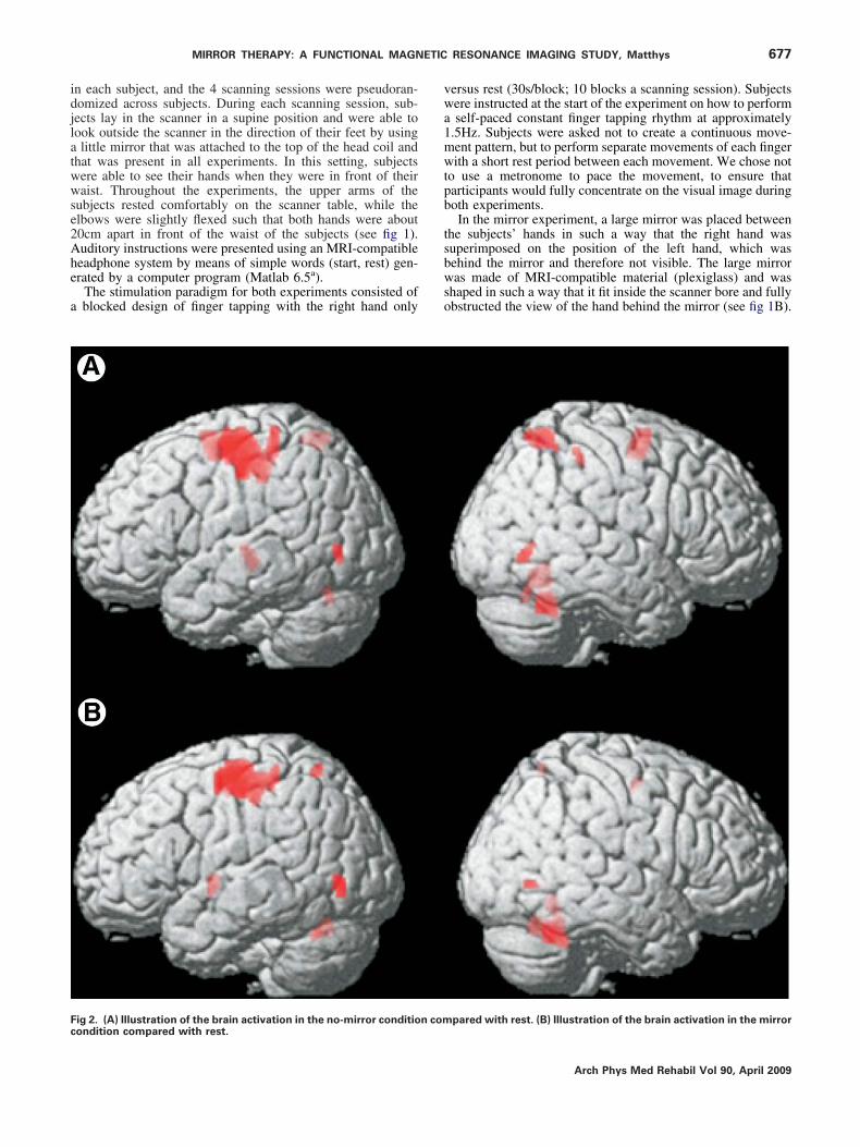
idjlatwwse2Ahe
a
vwa1mwtpb
tsbwso
Fc
677MIRROR THERAPY: A FUNCTIONAL MAGNETIC RESONANCE IMAGING STUDY, Matthys
n each subject, and the 4 scanning sessions were pseudoran-omized across subjects. During each scanning session, sub-ects lay in the scanner in a supine position and were able toook outside the scanner in the direction of their feet by usinglittle mirror that was attached to the top of the head coil and
hat was present in all experiments. In this setting, subjectsere able to see their hands when they were in front of theiraist. Throughout the experiments, the upper arms of the
ubjects rested comfortably on the scanner table, while thelbows were slightly flexed such that both hands were about0cm apart in front of the waist of the subjects (see fig 1).uditory instructions were presented using an MRI-compatibleeadphone system by means of simple words (start, rest) gen-rated by a computer program (Matlab 6.5a).
The stimulation paradigm for both experiments consisted ofblocked design of finger tapping with the right hand only
ig 2. (A) Illustration of the brain activation in the no-mirror condition comondition compared with rest.
ersus rest (30s/block; 10 blocks a scanning session). Subjectsere instructed at the start of the experiment on how to performself-paced constant finger tapping rhythm at approximately
.5Hz. Subjects were asked not to create a continuous move-ent pattern, but to perform separate movements of each fingerith a short rest period between each movement. We chose not
o use a metronome to pace the movement, to ensure thatarticipants would fully concentrate on the visual image duringoth experiments.In the mirror experiment, a large mirror was placed between
he subjects’ hands in such a way that the right hand wasuperimposed on the position of the left hand, which wasehind the mirror and therefore not visible. The large mirroras made of MRI-compatible material (plexiglass) and was
haped in such a way that it fit inside the scanner bore and fullybstructed the view of the hand behind the mirror (see fig 1B).
pared with rest. (B) Illustration of the brain activation in the mirror
Arch Phys Med Rehabil Vol 90, April 2009

ImorsM
2ttemd
fibede
D
stwqeS3gsbe2ct
sa
D
i
aatcattTpGtt
lbrmmadwct
metet
NA
678 MIRROR THERAPY: A FUNCTIONAL MAGNETIC RESONANCE IMAGING STUDY, Matthys
A
n this way, we aimed at creating the visual illusion of aoving instead of a nonmoving left hand. While the presence
r strength of the illusion could not be objectified, subjectseported that the illusion of seeing the left hand moving wasimilar to their experience during mirror exercises outside theRI scanner.Throughout both experiments, the subjects could always seehands. In the no-mirror experiment, subjects were instructed
o focus visually on the right hand both during the fingerapping and during the rest condition (see fig 1A). In the mirrorxperiment, subjects were instructed to focus visually on theirror reflection of the right hand (ie, the illusory left hand)
uring both the finger tapping and the rest condition.To evaluate a potential confounding effect of the number of
nger taps performed in either of the 2 experiments, the num-er of finger taps for each experiment was counted during thexperiments by an observer in a subsample of 9 subjects. Theifference in the average number of finger taps between the 2xperiments was compared using a paired t test.
ata AcquisitionFor each subject, the images were acquired on a 1.5T MRI
cannerb using a dedicated 8-channel receiver head coil. Forhe anatomical image, a high-resolution 3-dimensional T1-eighted fast spoiled gradient-echo inversion recovery se-uence covering the whole brain was acquired (repetition time/cho time/inversion time 9.9/2.0/400ms; Array Spatialensitivity Encoding Technique factor 2; acquisition matrix20�224; field of view, 24cm; slice thickness, 1.6mm; noap). For the functional images, a single shot gradient-echo EPIequence in transverse orientation was used that is sensitive tolood oxygenation level–dependent contrast (repetition time/cho time 3000/40ms; acquisition matrix 96�96; field of view,6cm; slice thickness, 5mm; gap, 1mm). The imaging volumeovered the entire brain including the cerebellum. Acquisitionime was 5:15 minutes a scanning session, which included 15
Table 1: Cortical Activation Patterns Associated With Finger TappFoci of Significant Activation an
No-Mirror
Cluster Size Maximum z Score
Left premotor cortex, leftprimary motor cortex
787
6.24Left somatosensory cortex 5.48Left somatosensory cortex, left
primary motor cortex 5.47Left premotor cortexLeft supplementary motor area
2005.92
Right supplementary motor area 5.56Right cerebellum VI 171 5.53Right cerebellum IV-VCerebellar vermis 4,5 113 6.31Cerebellar vermis 6 5.22Right superior parietal lobule 117 5.61Left thalamus 41 5.44Right middle temporal gyrus 40 5.22Left middle occipital gyrus 26 5.14Left superior parietal lobuleRight somatosensory cortex 16 5.02
OTE: Family-wise error corrected 0.05.bbreviation: MNI, Montreal Neurological Institute index.
rch Phys Med Rehabil Vol 90, April 2009
econds of dummy scans that were discarded from furthernalysis.
ata AnalysisThe imaging data were analyzed using SPM software 2c
mplemented in Matlab 6.5.a
On a single-subject level, all functional images were re-ligned to the first volume of the functional imaging series, anddditional correction for motion artifacts was performed usinghe unwarp toolbox of SPM2.c All functional images were thenoregistered with the subjects’ anatomical (T1-weighted) im-ges. Subsequently, the resulting images were normalized tohe standard space defined by the Montreal Neurological Insti-ute template; the anatomical images were normalized to the1-weighted template, the functional images to the EPI tem-late. The normalized data were spatially smoothed with aaussian filter (kernel with full width half maximum of 8mm)
o compensate for intersubject gyral variability and to ensurehe validity of the inferences.20,21
Statistical parametric maps were calculated using the generalinear model by modeling the active and the rest condition as aox car function convolved with a standard hemodynamicesponse function.22 Realignment parameters were imple-ented into the design matrix as regressors of no interest. Theodel was estimated with removal of global effects, and withhigh-pass filter with a cut-off of 128s. For each experiment,
ata from the 2 scanning sessions were pooled, and a t-contrastas calculated for the active (finger tapping) minus the rest
ondition. This resulted in 2 t-contrast maps a subject: [fingerapping � rest]Mirror and [finger tapping � rest]No-Mirror.
The individual statistical maps of the mirror and the no-irror experiments were then used for a second level random-
ffects group analysis. For each of the experiments, a 1-sampletest across all 18 subjects was performed to assess group
ffects for each of the experiments separately. The significancehreshold was set at P�.05 (family-wise error corrected for
ompared With Rest in the No-Mirror and the Mirror Experiment:ir MNI Stereotaxic Coordinates
Mirror
oordinates
Cluster Size Maximum z Score
Coordinates
y z x y z
�18 60
656
6.2 �38 �18 60�36 64
�26 48 5.71 �38 �24 525.88 �30 �14 56
�4 560 64
�54 32
318
5.9 24 �52 �325.11 8 �52 �20
�58 �10 5.63 4 �56 �10�66 �24 49 5.48 6 �66 �24�58 60�22 2�68 �2 18 5.1 48 �68 0�72 0 77 5.46 �50 �74 �2
17 5.4 �22 �60 62�38 54
ing Cd The
C
x
�38�34
�36
22
24
44
20�14
46�46
38
mvialtvr(wlau
fnedt
CE
fToWwfi
tcglmlanrts
Da
iTome
nhat
4Fts
679MIRROR THERAPY: A FUNCTIONAL MAGNETIC RESONANCE IMAGING STUDY, Matthys
ultiple comparisons) and at a minimum cluster size of 15oxels. Differences between the 2 experiments, presumablynduced by the mirror reflection of the moving hand, weressessed using a paired t test, and a t-contrast map was calcu-ated for the mirror minus the no-mirror experiment ([fingerapping � rest]Mirror � [finger tapping � rest]No-Mirror) andice versa ([finger tapping � rest]Mirror � [finger tapping �est]No-Mirror). A more liberal threshold at voxel level was usedP�.0001; not corrected for multiple comparisons) althoughith a threshold corrected for multiple comparisons at a cluster
evel (P�.05). Minimum cluster size was set at 15 voxels. Fornatomic labeling of the observed activations in SPM2, wesed the Anatomy toolbox.23
RESULTSNo difference in the average number of finger taps was
ound between the 2 experiments (39.9 finger taps/block for theo-mirror experiment vs 40.2 finger taps/block for the mirrorxperiment; P�.54). Visual inspection indicated that subjectsid not move the hand behind the mirror in the mirror condi-ions.
ortical Activation for the No-Mirror and Mirrorxperiments SeparatelyGroup analysis t-contrast maps of finger tapping versus rest
or each of the experiments are presented in figures 2A and B.he corresponding Montreal Neurological Institute index co-rdinates, z scores, and cluster size are summarized in table 1.e found similar activation patterns for both experiments,hich were in accordance with the expected activation for anger tapping task.In both experiments, activation was seen in the left precen-
ral and postcentral gyrus (primary motor and somatosensoryortex, respectively), the left precentral gyrus/superior frontalyrus (premotor cortex), the right middle temporal gyrus, theeft middle occipital gyrus, and the cerebellum (right VI, ver-is 4/5/6). Activation was also seen in the superior parietal
obule: in the right hemisphere during the no-mirror experimentnd in the left hemisphere during the mirror experiment. In theo-mirror experiment, additional activation was seen in theight postcentral gyrus (primary somatosensory cortex), al-hough to a lesser extent than on the left side, the medialuperior frontal gyrus bilaterally (SMA), and the left thalamus.
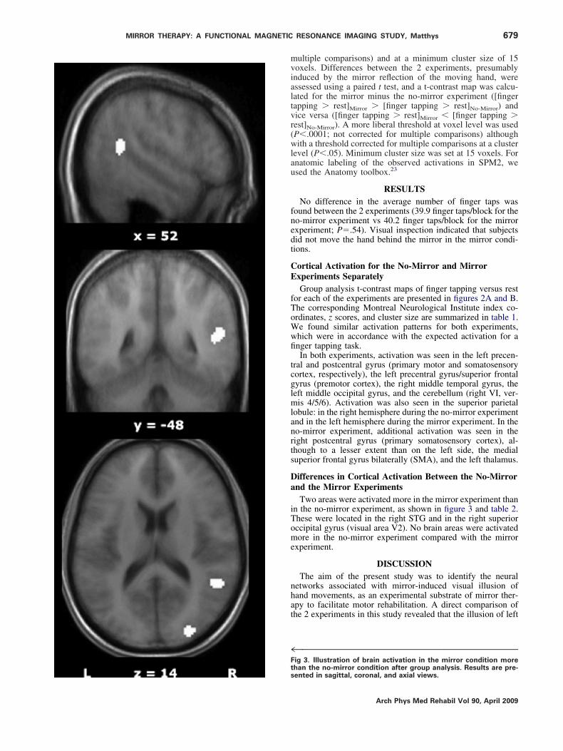
ifferences in Cortical Activation Between the No-Mirrornd the Mirror ExperimentsTwo areas were activated more in the mirror experiment than
n the no-mirror experiment, as shown in figure 3 and table 2.hese were located in the right STG and in the right superiorccipital gyrus (visual area V2). No brain areas were activatedore in the no-mirror experiment compared with the mirror
xperiment.
DISCUSSIONThe aim of the present study was to identify the neural
etworks associated with mirror-induced visual illusion ofand movements, as an experimental substrate of mirror ther-py to facilitate motor rehabilitation. A direct comparison ofhe 2 experiments in this study revealed that the illusion of left
™™™™™™™™™™™™™™™™™™™™™™™™™™™™™™™™™™™ig 3. Illustration of brain activation in the mirror condition more
han the no-mirror condition after group analysis. Results are pre-ented in sagittal, coronal, and axial views.Arch Phys Med Rehabil Vol 90, April 2009

hmt
absaIwptvcdpstmgrm
S
ltfidrsosilcrttbucsttemmwhe
eb
garanMwhtwscsnfiae
mbmtmpdteMcttitmansaaBstaptfr
NoA
680 MIRROR THERAPY: A FUNCTIONAL MAGNETIC RESONANCE IMAGING STUDY, Matthys
A
anded finger tapping (while in fact the left hand was notoving) induced activation in 2 visual areas: the right STG and
he right superior occipital gyrus.The STG is a higher-order visual region involved in the
nalysis of biological stimuli and is activated by observation ofiological motion.24 The coordinates of STG activation in ourtudy are very similar to the coordinates of the STS reported instudy on imitation of hand movements. Based on his results,
acoboni et al25 suggested a model for imitation with feedfor-ard and feedback mechanisms between STS and the fronto-arietal MNS. The right superior occipital gyrus is located inhe V2 and lies within the dorsal visual stream. The dorsalisual stream is connected with the PPC, a large associativeortical region, where afferents from different sensory mo-alities are integrated to provide the basis for perceptualrocesses.26 The PPC is considered a part of the motorystem and may be crucial for visuomotor transformations—hat is, an automatic conversion of visual information intootor commands.27 Based on this, the superior occipital
yrus activation found in this study as a result of the mirroreflections may indicate that mirror-induced visual illusionsay influence the PPC.
tudy LimitationsThe present study has some potential limitations we would
ike to address. One limitation may be that, although we haveried to reproduce the visual illusion that is successfully usedor mirror therapy in a number of clinical studies, it wasmpossible to quantify the strength of the illusion induceduring the fMRI experiment. However, when asked, subjectseported that the illusion during the fMRI measurements wasimilar to the mirror exercises outside the MRI scanner. Sec-nd, we simulated a setting in an MRI scanner with healthyubjects as an experimental substrate of mirror therapy, whichs normally used in a different environment in a patient popu-ation (such as patients with stroke, phantom limb pain, oromplex regional pain syndrome). In these patients, the mirroreflection creates the illusion of normative movement of a handhat is absent or that is not able to move normally. Our results,herefore, need to be evaluated further in patients to understandetter the underlying mechanism of mirror therapy. Third, wesed a statistical threshold which, albeit stringent, was notorrected for multiple comparisons because of the limitedtatistical power of our study. Finally, it should be mentionedhat as a consequence of the difference in the gaze direction,here is also a difference in the amount of visual input in bothxperiments. In the no-mirror experiment, subjects observed 1oving hand, while in the mirror experiment they observed 2oving hands. In addition, in the mirror experiment, subjectsere asked to focus on the mirror reflection of the right movingand superimposed on the left hand, while in the no-mirrorxperiment, subjects observed the right hand.
To our knowledge, our fMRI study is the first to evaluate theffect of mirror-induced visual illusions of hand movements on
Table 2: Group Result of the Mirr
Cluster MNI X MNI Y MNI Z
Mirror � No-Mirror 1 52 �48 142 26 �88 12
OTE: At a threshold of P�.0001 (uncorrected) at voxel level and P�.f 15 voxels.bbreviation: MNI, Montreal Neurological Institute index.
rain activation patterns. Two recent TMS studies have sug-
rch Phys Med Rehabil Vol 90, April 2009
ested that mirror reflections increase the corticospinal excit-bility of M1 corresponding with the hand behind the mir-or.13,28 In the present study, we did not find an increasedctivation of M1 in the right hemisphere. However, it should beoted that both TMS studies reported a significantly increased1 excitability only when the mirror condition was comparedith a control condition in which the subjects moved the rightand but did not directly observe this hand movement. Whenhe mirror condition was compared with a control condition inhich the subjects directly observed the moving right hand, no
ignificant differences were found. The latter situation is moreomparable to the control condition in our study in whichubjects observed hand movements of the right hand during theo-mirror experiment. The apparent contradiction between ourndings and those previously reported with regards to M1ctivation, therefore, is most likely a result of differences inxperimental setup.
CONCLUSIONSIn literature, several hypotheses on underlying workingechanisms for mirror therapy in motor rehabilitation have
een proposed. While Altschuler et al8 suggested that theirror reflections may help to recruit the premotor cortex
hrough the intimate connection between visual input and pre-otor areas, in our study we did not find activation in the
remotor areas that are uniquely associated with mirror-in-uced visual illusions. Other authors suggested that mirrorherapy could be a specific form of visual-guided motor imag-ry.10 Areas found to be activated during motor imagery are1, PMC, SMA, anterior cingulate cortex, parietal lobule, and
erebellum.29 In this study, we did not find activation located inhese areas, suggesting that mirror therapy may not be similaro motor imagery. However, it should be noted that we did notnstruct the subjects to perform imagery of the hand behindhe mirror, but rather to focus on the visual illusion of aoving hand superimposed on a nonmoving hand. It has
lso been suggested that there is involvement of the mirroreuron system in mirror therapy.11,12 The mirror neuronystem is located in the Broca area, the ventral premotorrea, and the posterior parietal lobe with a visual extensionrea in the STS. Given the lack of activation within theroca or premotor area, nor within the parietal lobe in this
tudy, interpretation of the STG activation located withinhe region of STS is difficult, and is not sufficient to proven involvement of the mirror neuron system. But it doesrovide a suggestion of a link between mirror therapy andhe mirror neuron system. However, our study may directuture studies, especially in patient groups, to indicate theelevance of the mirror neuron system for mirror therapy.
References1. Ramachandran VS, Rogers-Ramachandran D. Synaesthesia in
phantom limbs induced with mirrors. Proc R Soc Lond B Biol Sci
Greater Than No-Mirror Contrast
z Score Cluster Size Region
4.53 100 Right STG4.43 71 Right superior occipital gyrus (V2)
mily-wise error corrected) at cluster level and minimum cluster size
or is
05 (fa
1996;263:377-86.

1
1
1
1
1
1
1
1
1
1
2
2
2
2
2
2
2
2
2
2
ab
c
681MIRROR THERAPY: A FUNCTIONAL MAGNETIC RESONANCE IMAGING STUDY, Matthys
2. Ramachandran VS, Hirstein W. The perception of phantom limbs.The D. O. Hebb lecture. Brain 1998;121:1603-30.
3. McCabe CS, Haigh RC, Ring EF, Halligan PW, Wall PD, BlakeDR. A controlled pilot study of the utility of mirror visual feed-back in the treatment of complex regional pain syndrome (type 1).Rheumatology (Oxford) 2003;42:97-101.
4. Moseley GL. Graded motor imagery is effective for long-standingcomplex regional pain syndrome: a randomised controlled trial.Pain 2004;108:192-8.
5. Karmarkar A, Lieberman I. Mirror box therapy for complexregional pain syndrome. Anaesthesia 2006;61:412-3.
6. Vladimir Tichelaar YI, Geertzen JH, Keizer D, Paul van WilgenC. Mirror box therapy added to cognitive behavioural therapy inthree chronic complex regional pain syndrome type I patients: apilot study. Int J Rehabil Res 2007;30:181-8.
7. Giraux P, Sirigu A. Illusory movements of the paralyzed limb restoremotor cortex activity. Neuroimage 2003;20(suppl 1):S107-11.
8. Altschuler EL, Wisdom SB, Stone L, et al. Rehabilitation ofhemiparesis after stroke with a mirror. Lancet 1999;353:2035-6.
9. Sathian K, Greenspan AI, Wolf SL. Doing it with mirrors: a casestudy of a novel approach to neurorehabilitation. NeurorehabilNeural Repair 2000;14:73-6.
0. Stevens JA, Stoykov ME. Using motor imagery in the rehabilita-tion of hemiparesis. Arch Phys Med Rehabil 2003;84:1090-2.
1. Sutbeyaz S, Yavuzer G, Sezer N, Koseoglu BF. Mirror therapyenhances lower-extremity motor recovery and motor functioningafter stroke: a randomized controlled trial. Arch Phys Med Reha-bil 2007;88:555-9.
2. Rosen B, Lundborg G. Training with a mirror in rehabilitation ofthe hand. Scand J Plast Reconstr Surg Hand Surg 2005;39:104-8.
3. Garry MI, Loftus A, Summers JJ. Mirror, mirror on the wall:viewing a mirror reflection of unilateral hand movements facili-tates ipsilateral M1 excitability. Exp Brain Res 2005;163:118-22.
4. Sharma N, Pomeroy VM, Baron JC. Motor imagery: a backdoor tothe motor system after stroke? Stroke 2006;37:1941-52.
5. Page SJ, Levine P, Leonard A. Mental practice in chronic stroke:results of a randomized, placebo-controlled trial. Stroke 2007;38:1293-7.
6. Rizzolatti G, Craighero L. The mirror-neuron system. Annu RevNeurosci 2004;27:169-92.
7. Fadiga L, Craighero L, Olivier E. Human motor cortex excitabilityduring the perception of others’ action. Curr Opin Neurobiol 2005;
15:213-8.8. Aziz-Zadeh L, Maeda F, Zaidel E, Mazziotta J, Iacoboni M.Lateralization in motor facilitation during action observation: aTMS study. Exp Brain Res 2002;144:127-31.
9. Maeda F, Kleiner-Fisman G, Pascual-Leone A. Motor facilitationwhile observing hand actions: specificity of the effect and role ofobserver’s orientation. J Neurophysiol 2002;87:1329-35.
0. Mikl M, Marecek R, Hlustik P, et al. Effects of spatial smoothingon fMRI group inferences. Magn Reson Imaging 2008;26:490-503.
1. Poline JB, Worsley KJ, Holmes AP, Frackowiak RS, Friston KJ.Estimating smoothness in statistical parametric maps: variabilityof p values. J Comput Assist Tomogr 1995;19:788-96.
2. Friston KJ, Holmes AP, Price CJ, Buchel C, Worsley KJ. Multi-subject fMRI studies and conjunction analyses. Neuroimage 1999;10:385-96.
3. Eickhoff SB, Stephan KE, Mohlberg H, et al. A new SPM toolboxfor combining probabilistic cytoarchitectonic maps and functionalimaging data. Neuroimage 2005;25:1325-35.
4. Allison T, Puce A, McCarthy G. Social perception from visualcues: role of the STS region. Trends Cogn Sci 2000;4:267-78.
5. Iacoboni M, Koski LM, Brass M, et al. Reafferent copies ofimitated actions in the right superior temporal cortex. Proc NatlAcad Sci U S A 2001;98:13995-9.
6. Goodale MA, Milner AD. Separate visual pathways for perceptionand action. Trends Neurosci 1992;15:20-5.
7. Fogassi L, Luppino G. Motor functions of the parietal lobe. CurrOpin Neurobiol 2005;15:626-31.
8. Funase K, Tabira T, Higashi T, Liang N, Kasai T. Increasedcorticospinal excitability during direct observation of self-movement and indirect observation with a mirror box. NeurosciLett 2007;419:108-12.
9. Grezes J, Decety J. Functional anatomy of execution, mentalsimulation, observation, and verb generation of actions: a meta-analysis. Hum Brain Mapp 2001;12:1-19.
Suppliers. The Mathworks, 3 Apple Hill Dr, Natick, MA 01760-2098.. Signa CV/I; GE Healthcare Bio-Sciences Corp, 800 Centennial
Ave, PO Box 1327, Piscataway, NJ 08855-1237.. Wellcome Department of Imaging Neuroscience, Institute of Neu-
rology, 12 Queen Sq, London, WCIN 3BG, England. Available at:
http://www.fil.ion.ucl.ac.uk/spm.Arch Phys Med Rehabil Vol 90, April 2009
Related Documents