miR390, Arabidopsis TAS3 tasiRNAs, and Their AUXIN RESPONSE FACTOR Targets Define an Autoregulatory Network Quantitatively Regulating Lateral Root Growth W Elena Marin, a,1 Virginie Jouannet, b,1,2 Aure ´ lie Herz, a Annemarie S. Lokerse, c Dolf Weijers, c Herve Vaucheret, d Laurent Nussaume, a Martin D. Crespi, b,3 and Alexis Maizel b,2,3,4 a Laboratoire de Biologie du De ´ veloppement des Plantes, Commissariat a ` l’Energie Atomique Cadarache, Centre National de la Recherche Scientifique, Universite ´ Aix Marseille, 13108 St. Paul-lez-Durance, France b Institut des Sciences du Ve ´ ge ´ tal, Centre National de la Recherche Scientifique, 91198 Gif-sur-Yvette Cedex, France c Laboratory of Biochemistry, Wageningen University, 6703 HA Wageningen, The Netherlands d Laboratoire de Biologie Cellulaire, Institut Jean-Pierre Bourgin, Institut National de la Recherche Agronomique, 78026 Versailles Cedex, France Plants adapt to different environmental conditions by constantly forming new organs in response to morphogenetic signals. Lateral roots branch from the main root in response to local auxin maxima. How a local auxin maximum translates into a robust pattern of gene activation ensuring the proper growth of the newly formed lateral root is largely unknown. Here, we demonstrate that miR390, TAS3-derived trans-acting short-interfering RNAs (tasiRNAs), and AUXIN RESPONSE FACTORS (ARFs) form an auxin-responsive regulatory network controlling lateral root growth. Spatial expression analysis using reporter gene fusions, tasi/miRNA sensors, and mutant analysis showed that miR390 is specifically expressed at the sites of lateral root initiation where it triggers the biogenesis of tasiRNAs. These tasiRNAs inhibit ARF2, ARF3, and ARF4, thus releasing repression of lateral root growth. In addition, ARF2, ARF3, and ARF4 affect auxin-induced miR390 accumulation. Positive and negative feedback regulation of miR390 by ARF2, ARF3, and ARF4 thus ensures the proper definition of the miR390 expression pattern. This regulatory network maintains ARF expression in a concentration range optimal for specifying the timing of lateral root growth, a function similar to its activity during leaf development. These results also show how small regulatory RNAs integrate with auxin signaling to quantitatively regulate organ growth during development. INTRODUCTION The initiation of lateral roots plays a crucial role in plant devel- opment, since it determines the architecture of the root system and, thus, stability as well as nutrient and water uptake potential for the entire organism. Lateral root development is a typical example of a canalized developmental process (i.e., buffered against perturbation; Siegal and Bergman 2002), yet roots strongly adapt to the local environment to maximize acquisition of water and nutrients from the soil. In recent years, it has become clear that lateral roots initiate from a small number of pericycle cells (initiation) that differentiate into a primordia and grow out of the primary root (emergence) (Hardtke, 2006; De Smet et al., 2006; Parizot et al., 2008; Petricka and Benfey, 2008). Auxin is a morphogenetic trigger for lateral root formation (Benkova ´ et al., 2009), and its local maximum acts as an instructive signal for initiation of these organs (Dubrovsky et al., 2008). Many of auxin’s actions are mediated by transcription factors of the auxin response factor (ARF) family, and several ARFs play critical roles in lateral root development (Okushima et al., 2005b; Wilmoth et al., 2005). Small RNAs, such as microRNAs (miRNAs) and trans-acting short-interfering RNAs (tasiRNAs), control many aspects of de- velopment in eukaryotes. As negative regulators of gene ex- pression, they can act as developmental switches to shut down gene expression programs. Alternatively, small RNAs can fine- tune gene expression to quantitatively adapt developmental processes to endogenous or environmental fluctuations and therefore act as canalization factors (Li et al., 2009). Several reports have implicated miRNAs in the modulation of auxin action during lateral root development supporting this model (Guo et al., 2005; Mallory et al., 2005; Gifford et al., 2008; Yoon et al., 2010). tasiRNAs belong to a plant-specific class of endogenous small RNAs, whose biogenesis requires an initial miRNA-mediated cleavage of their precursors. The cleavage product is then converted to double-stranded RNA through RNA-DEPENDENT RNA POLYMERASE6 (RDR6) activity and sequential DICER- LIKE4 (DCL4)-mediated cleavage events (Peragine et al., 2004; Vazquez et al., 2004; Allen et al., 2005; Gasciolli et al., 2005; Xie 1 These authors contributed equally to this work. 2 Current address: Department of Stem Cell Biology, University of Heidelberg, INF230, 69120 Heidelberg, Germany. 3 These authors contributed equally to this work. 4 Address correspondence to [email protected]. The author responsible for distribution of materials integral to the findings presented in this article in accordance with the policy described in the Instructions for Authors (www.plantcell.org) is: Alexis Maizel ([email protected]). W Online version contains Web-only data. www.plantcell.org/cgi/doi/10.1105/tpc.109.072553 The Plant Cell, Vol. 22: 1104–1117, April 2010, www.plantcell.org ã 2010 American Society of Plant Biologists

Welcome message from author
This document is posted to help you gain knowledge. Please leave a comment to let me know what you think about it! Share it to your friends and learn new things together.
Transcript
miR390, Arabidopsis TAS3 tasiRNAs, and Their AUXINRESPONSE FACTOR Targets Define an AutoregulatoryNetwork Quantitatively Regulating Lateral Root Growth W
Elena Marin,a,1 Virginie Jouannet,b,1,2 Aurelie Herz,a Annemarie S. Lokerse,c Dolf Weijers,c Herve Vaucheret,d
Laurent Nussaume,a Martin D. Crespi,b,3 and Alexis Maizelb,2,3,4
a Laboratoire de Biologie du Developpement des Plantes, Commissariat a l’Energie Atomique Cadarache, Centre National de la
Recherche Scientifique, Universite Aix Marseille, 13108 St. Paul-lez-Durance, Franceb Institut des Sciences du Vegetal, Centre National de la Recherche Scientifique, 91198 Gif-sur-Yvette Cedex, Francec Laboratory of Biochemistry, Wageningen University, 6703 HA Wageningen, The Netherlandsd Laboratoire de Biologie Cellulaire, Institut Jean-Pierre Bourgin, Institut National de la Recherche Agronomique, 78026
Versailles Cedex, France
Plants adapt to different environmental conditions by constantly forming new organs in response to morphogenetic signals.
Lateral roots branch from the main root in response to local auxin maxima. How a local auxin maximum translates into a
robust pattern of gene activation ensuring the proper growth of the newly formed lateral root is largely unknown. Here, we
demonstrate that miR390, TAS3-derived trans-acting short-interfering RNAs (tasiRNAs), and AUXIN RESPONSE FACTORS
(ARFs) form an auxin-responsive regulatory network controlling lateral root growth. Spatial expression analysis using
reporter gene fusions, tasi/miRNA sensors, and mutant analysis showed that miR390 is specifically expressed at the sites of
lateral root initiation where it triggers the biogenesis of tasiRNAs. These tasiRNAs inhibit ARF2, ARF3, and ARF4, thus
releasing repression of lateral root growth. In addition, ARF2, ARF3, and ARF4 affect auxin-induced miR390 accumulation.
Positive and negative feedback regulation of miR390 by ARF2, ARF3, and ARF4 thus ensures the proper definition of the
miR390 expression pattern. This regulatory network maintains ARF expression in a concentration range optimal for
specifying the timing of lateral root growth, a function similar to its activity during leaf development. These results also show
how small regulatory RNAs integrate with auxin signaling to quantitatively regulate organ growth during development.
INTRODUCTION
The initiation of lateral roots plays a crucial role in plant devel-
opment, since it determines the architecture of the root system
and, thus, stability as well as nutrient and water uptake potential
for the entire organism. Lateral root development is a typical
example of a canalized developmental process (i.e., buffered
against perturbation; Siegal and Bergman 2002), yet roots
strongly adapt to the local environment to maximize acquisition
of water and nutrients from the soil. In recent years, it has
become clear that lateral roots initiate from a small number of
pericycle cells (initiation) that differentiate into a primordia and
grow out of the primary root (emergence) (Hardtke, 2006; De
Smet et al., 2006; Parizot et al., 2008; Petricka andBenfey, 2008).
Auxin is a morphogenetic trigger for lateral root formation
(Benkova et al., 2009), and its local maximum acts as an
instructive signal for initiation of these organs (Dubrovsky et al.,
2008). Many of auxin’s actions are mediated by transcription
factors of the auxin response factor (ARF) family, and several
ARFs play critical roles in lateral root development (Okushima
et al., 2005b; Wilmoth et al., 2005).
Small RNAs, such as microRNAs (miRNAs) and trans-acting
short-interfering RNAs (tasiRNAs), control many aspects of de-
velopment in eukaryotes. As negative regulators of gene ex-
pression, they can act as developmental switches to shut down
gene expression programs. Alternatively, small RNAs can fine-
tune gene expression to quantitatively adapt developmental
processes to endogenous or environmental fluctuations and
therefore act as canalization factors (Li et al., 2009). Several
reports have implicated miRNAs in the modulation of auxin
action during lateral root development supporting this model
(Guo et al., 2005; Mallory et al., 2005; Gifford et al., 2008; Yoon
et al., 2010).
tasiRNAs belong to a plant-specific class of endogenous small
RNAs, whose biogenesis requires an initial miRNA-mediated
cleavage of their precursors. The cleavage product is then
converted to double-stranded RNA through RNA-DEPENDENT
RNA POLYMERASE6 (RDR6) activity and sequential DICER-
LIKE4 (DCL4)-mediated cleavage events (Peragine et al., 2004;
Vazquez et al., 2004; Allen et al., 2005; Gasciolli et al., 2005; Xie
1 These authors contributed equally to this work.2 Current address: Department of Stem Cell Biology, University ofHeidelberg, INF230, 69120 Heidelberg, Germany.3 These authors contributed equally to this work.4 Address correspondence to [email protected] author responsible for distribution of materials integral to thefindings presented in this article in accordance with the policy describedin the Instructions for Authors (www.plantcell.org) is: Alexis Maizel([email protected]).WOnline version contains Web-only data.www.plantcell.org/cgi/doi/10.1105/tpc.109.072553
The Plant Cell, Vol. 22: 1104–1117, April 2010, www.plantcell.org ã 2010 American Society of Plant Biologists
et al., 2005; Yoshikawa et al., 2005; Adenot et al., 2006). Of the
four tasiRNAs precursors identified (TAS1-4) in Arabidopsis
thaliana, cleavage of TAS3 is unique since it requires the specific
action of themiR390/ARGONAUTE7 (AGO7) complex for tasiRNA
production (Montgomery et al., 2008).miR390 andTAS3 tasiRNAs
define a pathway that regulates leaf patterning and develop-
mental timing by repressing the ARF family members ARF2,
ARF3, and ARF4 (Figure 1A) (Adenot et al., 2006; Fahlgren et al.,
2006; Garcia et al., 2006; Hunter et al., 2006). We and others have
previously reported thatTAS3,AGO7, andmiR390are expressed in
root tissues (Hirsch et al., 2006;Montgomery et al., 2008); however,
the function of this pathway in root development is unclear.
Here, we show that mutations affecting the abundance of
TAS3-derived tasiRNAs lead to quantitative changes in the rate
of lateral root growth. miR390 is induced during lateral root
initiation and triggers the local production of tasiRNAs. In the
lateral root primordium, the tasiARFs reduce the activity of
ARF2, ARF3, and ARF4, thereby promoting lateral root growth.
In addition, ARF2, ARF3, and ARF4 are required for proper
miR390 expression through different feedback mechanisms.
Thus, auxin, miR390, TAS3, and their ARFs targets define a
regulatory network quantitatively controlling lateral root growth.
This complex network acts to fine-tune local auxin responses
and thus provides robustness and flexibility to lateral root growth.
RESULTS
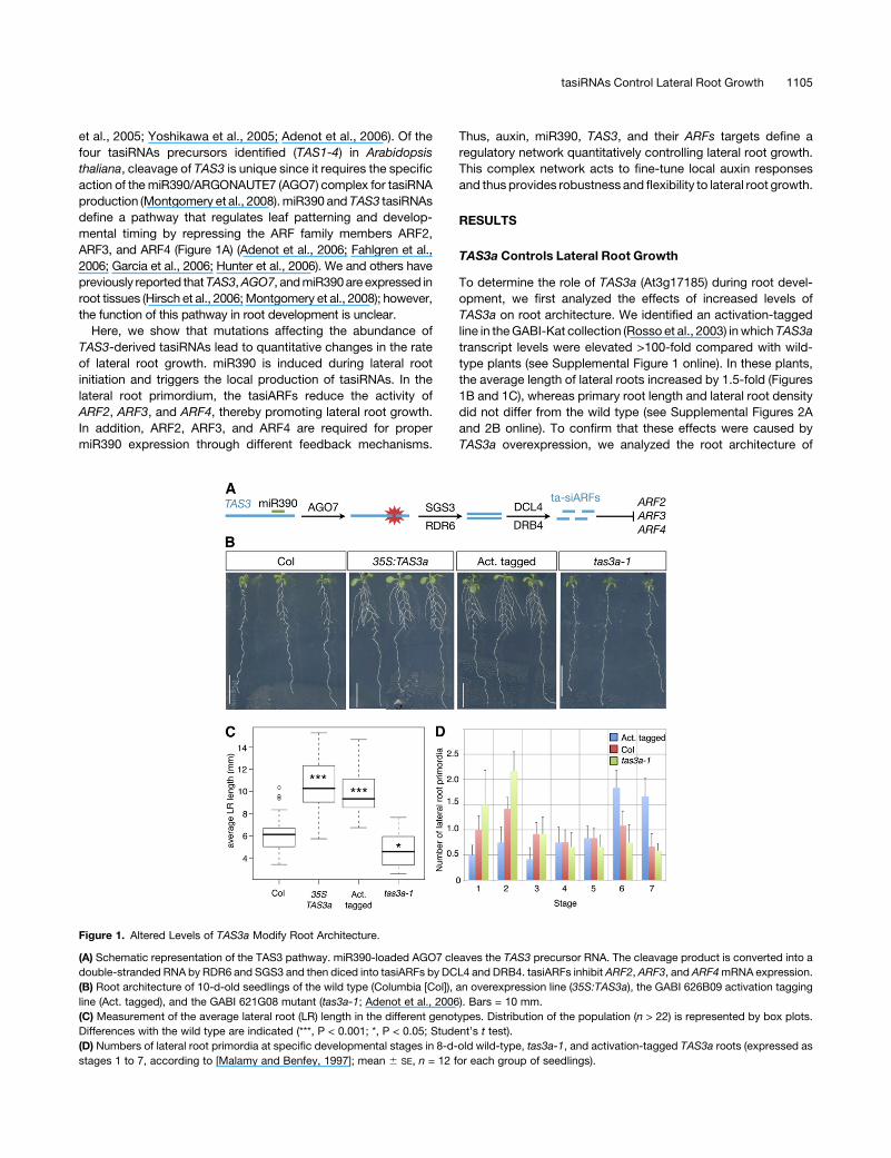
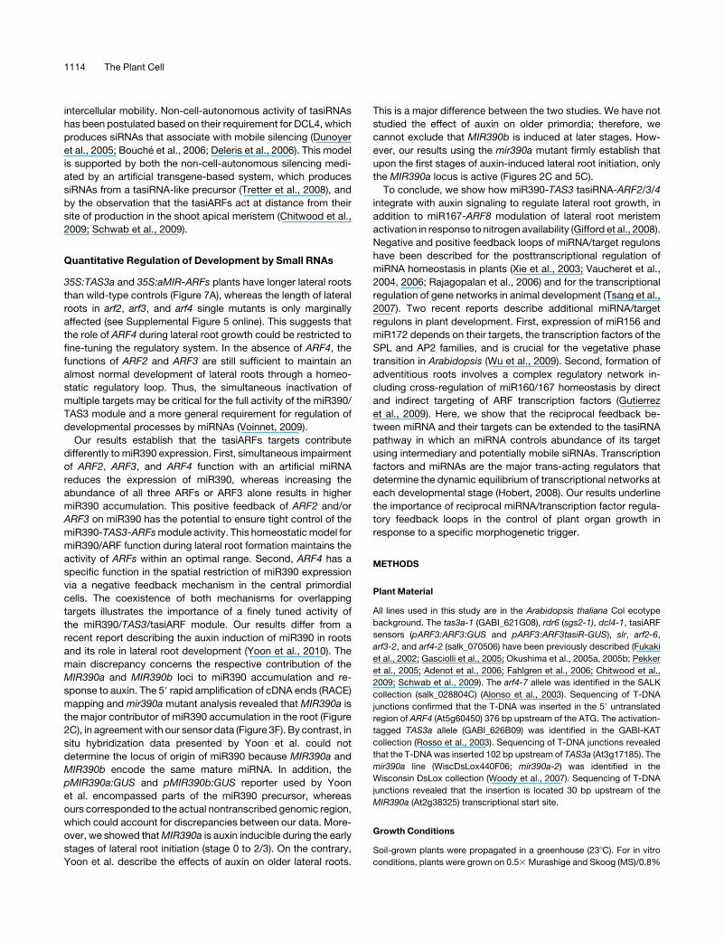
TAS3a Controls Lateral Root Growth
To determine the role of TAS3a (At3g17185) during root devel-
opment, we first analyzed the effects of increased levels of
TAS3a on root architecture. We identified an activation-tagged
line in theGABI-Kat collection (Rosso et al., 2003) inwhich TAS3a
transcript levels were elevated >100-fold compared with wild-
type plants (see Supplemental Figure 1 online). In these plants,
the average length of lateral roots increased by 1.5-fold (Figures
1B and 1C), whereas primary root length and lateral root density
did not differ from the wild type (see Supplemental Figures 2A
and 2B online). To confirm that these effects were caused by
TAS3a overexpression, we analyzed the root architecture of
Figure 1. Altered Levels of TAS3a Modify Root Architecture.
(A) Schematic representation of the TAS3 pathway. miR390-loaded AGO7 cleaves the TAS3 precursor RNA. The cleavage product is converted into a
double-stranded RNA by RDR6 and SGS3 and then diced into tasiARFs by DCL4 and DRB4. tasiARFs inhibit ARF2, ARF3, and ARF4mRNA expression.
(B) Root architecture of 10-d-old seedlings of the wild type (Columbia [Col]), an overexpression line (35S:TAS3a), the GABI 626B09 activation tagging
line (Act. tagged), and the GABI 621G08 mutant (tas3a-1; Adenot et al., 2006). Bars = 10 mm.
(C) Measurement of the average lateral root (LR) length in the different genotypes. Distribution of the population (n > 22) is represented by box plots.
Differences with the wild type are indicated (***, P < 0.001; *, P < 0.05; Student’s t test).
(D) Numbers of lateral root primordia at specific developmental stages in 8-d-old wild-type, tas3a-1, and activation-tagged TAS3a roots (expressed as
stages 1 to 7, according to [Malamy and Benfey, 1997]; mean 6 SE, n = 12 for each group of seedlings).
tasiRNAs Control Lateral Root Growth 1105
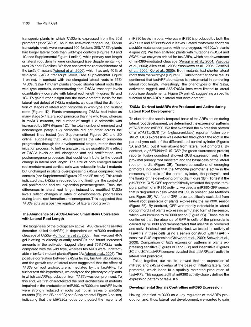
transgenic plants in which TAS3a is expressed from the 35S
promoter (35S:TAS3a). As in the activation-tagged line, TAS3a
transcripts levels were increased 100-fold and 35S:TAS3a plants
had longer lateral roots than wild-type controls (Figures 1B and
1C; see Supplemental Figure 1 online), while primary root length
or lateral root density were unchanged (see Supplemental Fig-
ures 2A and 2B online). We then analyzed the root architecture of
the tas3a-1 mutant (Adenot et al., 2006), which has only 40% of
wild-type TAS3a transcript levels (see Supplemental Figure
1 online). In contrast with the elongated lateral roots in 35S:
TAS3a, tas3a-1 mutant plants showed shorter lateral roots than
wild-type controls, demonstrating that TAS3a transcript levels
quantitatively correlate with lateral root length (Figures 1B and
1C). To gain further insight into the developmental basis for the
lateral root defect of TAS3a mutants, we quantified the distribu-
tion of stages of lateral root primordia in wild-type and mutant
roots (Figure 1D). Plants overexpressing TAS3a had twice as
many stage 5-7 lateral root primordia than the wild type, whereas
in tas3a-1 mutants, the number of stage 1-2 primordia was
increased by 50% (Figure 1D). The total number of emerged and
nonemerged (stage 1-7) primordia did not differ across the
different lines tested (see Supplemental Figures 2C and 2D
online), suggesting that TAS3a regulates the rate of primordia
progression through the developmental stages, rather than the
initiation process. To further analyze this, we quantified the effect
of TAS3a levels on cell elongation and cell proliferation, two
postemergence processes that could contribute to the overall
change in lateral root length. The size of both emerged lateral
rootmeristems and cortical cells was reduced in tas3a-1mutants
but unchanged in plants overexpressing TAS3a compared with
controls (see Supplemental Figures 2E and 2F online). This result
indicated that TAS3a is required but not limiting in the control of
cell proliferation and cell expansion postemergence. Thus, the
differences in lateral root length induced by modified TAS3a
levels reflect changes in rates of developmental progression
during lateral root formation and emergence. This suggested that
TAS3a acts as a positive regulator of lateral root growth.
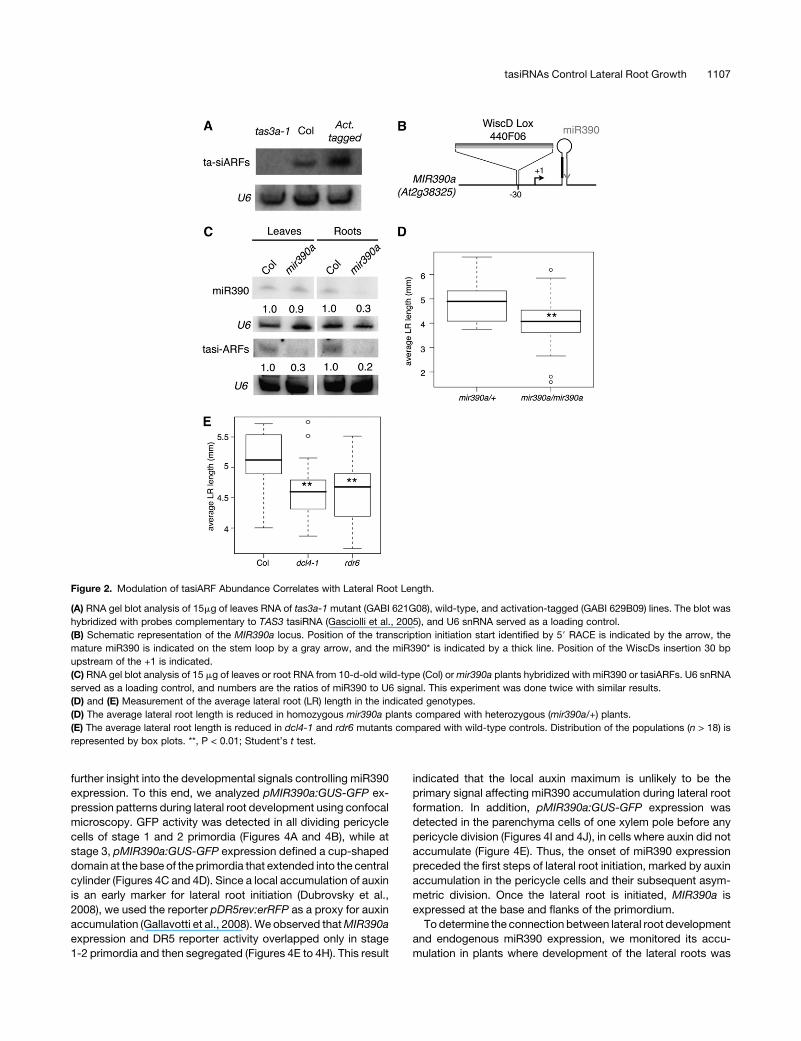
The Abundance of TAS3a-Derived Small RNAs Correlates
with Lateral Root Length
The biogenesis of the biologically active TAS3-derived tasiRNAs
(hereafter called tasiARFs) is dependent on miR390-mediated
cleavageofTAS3a (Montgomery et al., 2008). Thus,weusedRNA
gel blotting to directly quantify tasiARFs and found increased
amounts in the activation-tagged allele and 35S:TAS3a roots
compared with the wild type, whereas tasiARFs were undetect-
able in tas3a-1mutant plants (Figure 2A; Adenot et al., 2006). The
positive correlation between TAS3a levels, tasiARF abundance,
and the growth rate of lateral roots suggested that the effect of
TAS3a on root architecture is mediated by the tasiARFs. To
further test this hypothesis, we analyzed the phenotype of plants
in which tasiARFs production from TAS3awas compromised. To
this end, we first characterized the root architecture of mutants
impaired in the production of miR390. miR390 and tasiARF levels
were strongly reduced in roots but not in leaves of mir390a
mutants (Figures 2B and 2C; see Supplemental Figure 3 online),
indicating that the MIR390a locus contributed the majority of
miR390 levels in roots, whereas miR390 is produced by both the
MIR390a andMIR390b loci in leaves. Lateral rootswere shorter in
mir390amutants compared with heterozygous mir390a/+ plants
(Figure 2D). We then analyzed plants with mutations in DCL4 and
RDR6, two enzymes critical for tasiARFs, which act downstream
of miR390-mediated cleavage (Peragine et al., 2004; Vazquez
et al., 2004; Allen et al., 2005; Yoshikawa et al., 2005; Gasciolli
et al., 2005; Xie et al., 2005). Both mutants had shorter lateral
roots than the wild type (Figure 2E). Taken together, these results
confirmed that tasiARF abundance is instrumental in controlling
lateral root length. Interestingly, the phenotypes of the tas3a,
activation-tagged, and 35S:TAS3a lines were limited to lateral
roots (see Supplemental Figure 2A online), suggesting a specific
function of tasiARFs in lateral root development.
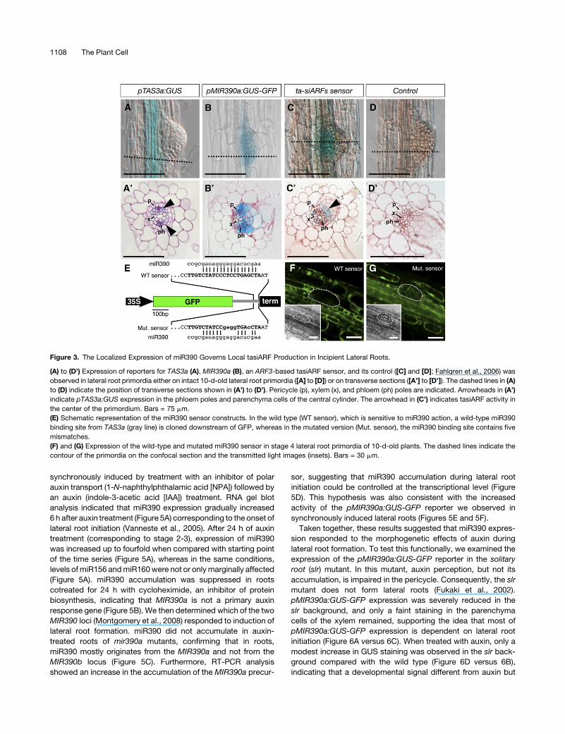
TAS3a-Derived tasiARFs Are Produced and Active during
Lateral Root Development
To elucidate the spatio-temporal basis of tasiARFs action during
lateral root development, we determined the expression patterns
of TAS3a and miR390. We first examined the expression pattern
of a pTAS3a:GUS (for b-glucuronidase) reporter fusion con-
struct. GUS expression was detected throughout the root in the
parenchyma cells of the differentiated central cylinder (Figures
3A and 3A’), but it was absent from lateral root primordia. By
contrast, a pMIR390a:GUS-GFP (for green fluorescent protein)
reporter fusion construct showed GUS expression only in the
proximal primary root meristem and the basal cells of the lateral
root primordia (Figure 3B). Transverse sections of emerging
primordia indicated that the MIR390a promoter is active in the
mesenchymal cells of the central cylinder, the pericycle, and
the flanks of the developing primordia (Figure 3B’). To test if the
pMIR390a:GUS-GFP reporter faithfully reflected the spatio-tem-
poral pattern of miR390 activity, we used a miR390-GFP sensor
that is degraded in cells where miR390 is present (see Methods
and Figure 3E). We found GFP to be specifically excluded from
lateral root primordia of plants expressing the miR390 sensor
(Figure 3F). By contrast, GFP was readily detectable in lateral
root primordia of plants expressing amutated form of the sensor,
which was immune to miR390 action (Figure 3G). These results
confirmed that the absence of GFP in cells of the primordia is
caused by miR390 and demonstrated that miR390 is produced
and active in lateral root primordia. Next, we tested the activity of
tasiARFs in these cells using a sensor construct with tasiARF-
sensitive GUS expression (Chitwood et al., 2009; Schwab et al.,
2009). Comparison of GUS expression patterns in plants ex-
pressing sensitive (Figures 3D and 3D’) and insensitive (Figures
3C and 3C’) tasiARF sensors revealed that tasiARFs are active in
lateral root primordia.
Taken together, our results showed that the expression of
miR390 and TAS3a overlap at the base of initiating lateral root
primordia, which leads to a spatially restricted production of
tasiARFs. This suggested thatmiR390 activity closely defines the
expression pattern of tasiARFs.
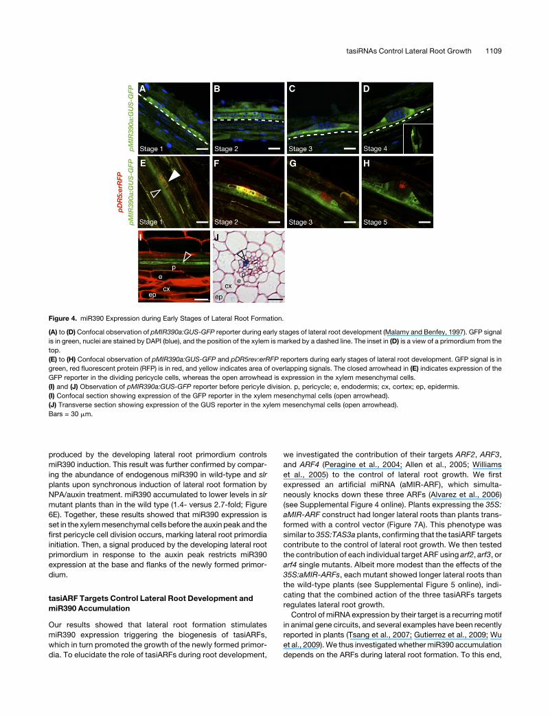
Developmental Signals Controlling miR390 Expression
Having identified miR390 as a key regulator of tasiARFs pro-
duction and, thus, lateral root development, we wanted to gain
1106 The Plant Cell
further insight into the developmental signals controlling miR390
expression. To this end, we analyzed pMIR390a:GUS-GFP ex-
pression patterns during lateral root development using confocal
microscopy. GFP activity was detected in all dividing pericycle
cells of stage 1 and 2 primordia (Figures 4A and 4B), while at
stage 3, pMIR390a:GUS-GFP expression defined a cup-shaped
domain at the base of the primordia that extended into the central
cylinder (Figures 4C and 4D). Since a local accumulation of auxin
is an early marker for lateral root initiation (Dubrovsky et al.,
2008), we used the reporter pDR5rev:erRFP as a proxy for auxin
accumulation (Gallavotti et al., 2008).We observed thatMIR390a
expression and DR5 reporter activity overlapped only in stage
1-2 primordia and then segregated (Figures 4E to 4H). This result
indicated that the local auxin maximum is unlikely to be the
primary signal affecting miR390 accumulation during lateral root
formation. In addition, pMIR390a:GUS-GFP expression was
detected in the parenchyma cells of one xylem pole before any
pericycle division (Figures 4I and 4J), in cells where auxin did not
accumulate (Figure 4E). Thus, the onset of miR390 expression
preceded the first steps of lateral root initiation, marked by auxin
accumulation in the pericycle cells and their subsequent asym-
metric division. Once the lateral root is initiated, MIR390a is
expressed at the base and flanks of the primordium.
Todetermine the connection between lateral root development
and endogenous miR390 expression, we monitored its accu-
mulation in plants where development of the lateral roots was
Figure 2. Modulation of tasiARF Abundance Correlates with Lateral Root Length.
(A) RNA gel blot analysis of 15mg of leaves RNA of tas3a-1 mutant (GABI 621G08), wild-type, and activation-tagged (GABI 629B09) lines. The blot was
hybridized with probes complementary to TAS3 tasiRNA (Gasciolli et al., 2005), and U6 snRNA served as a loading control.
(B) Schematic representation of the MIR390a locus. Position of the transcription initiation start identified by 59 RACE is indicated by the arrow, the
mature miR390 is indicated on the stem loop by a gray arrow, and the miR390* is indicated by a thick line. Position of the WiscDs insertion 30 bp
upstream of the +1 is indicated.
(C) RNA gel blot analysis of 15 mg of leaves or root RNA from 10-d-old wild-type (Col) ormir390a plants hybridized with miR390 or tasiARFs. U6 snRNA
served as a loading control, and numbers are the ratios of miR390 to U6 signal. This experiment was done twice with similar results.
(D) and (E) Measurement of the average lateral root (LR) length in the indicated genotypes.
(D) The average lateral root length is reduced in homozygous mir390a plants compared with heterozygous (mir390a/+) plants.
(E) The average lateral root length is reduced in dcl4-1 and rdr6 mutants compared with wild-type controls. Distribution of the populations (n > 18) is
represented by box plots. **, P < 0.01; Student’s t test.
tasiRNAs Control Lateral Root Growth 1107
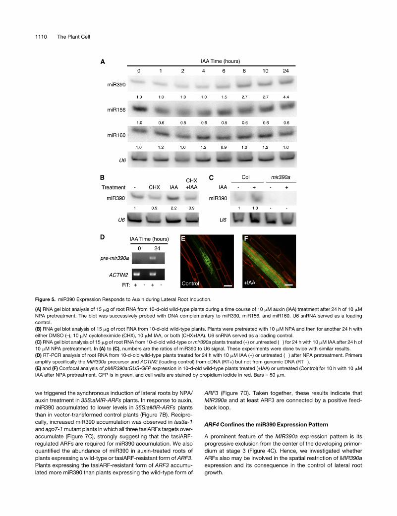
synchronously induced by treatment with an inhibitor of polar
auxin transport (1-N-naphthylphthalamic acid [NPA]) followed by
an auxin (indole-3-acetic acid [IAA]) treatment. RNA gel blot
analysis indicated that miR390 expression gradually increased
6 h after auxin treatment (Figure 5A) corresponding to the onset of
lateral root initiation (Vanneste et al., 2005). After 24 h of auxin
treatment (corresponding to stage 2-3), expression of miR390
was increased up to fourfold when compared with starting point
of the time series (Figure 5A), whereas in the same conditions,
levels of miR156 andmiR160were not or onlymarginally affected
(Figure 5A). miR390 accumulation was suppressed in roots
cotreated for 24 h with cycloheximide, an inhibitor of protein
biosynthesis, indicating that MIR390a is not a primary auxin
response gene (Figure 5B). We then determined which of the two
MIR390 loci (Montgomery et al., 2008) responded to induction of
lateral root formation. miR390 did not accumulate in auxin-
treated roots of mir390a mutants, confirming that in roots,
miR390 mostly originates from the MIR390a and not from the
MIR390b locus (Figure 5C). Furthermore, RT-PCR analysis
showed an increase in the accumulation of theMIR390a precur-
sor, suggesting that miR390 accumulation during lateral root
initiation could be controlled at the transcriptional level (Figure
5D). This hypothesis was also consistent with the increased
activity of the pMIR390a:GUS-GFP reporter we observed in
synchronously induced lateral roots (Figures 5E and 5F).
Taken together, these results suggested that miR390 expres-
sion responded to the morphogenetic effects of auxin during
lateral root formation. To test this functionally, we examined the
expression of the pMIR390a:GUS-GFP reporter in the solitary
root (slr) mutant. In this mutant, auxin perception, but not its
accumulation, is impaired in the pericycle. Consequently, the slr
mutant does not form lateral roots (Fukaki et al., 2002).
pMIR390a:GUS-GFP expression was severely reduced in the
slr background, and only a faint staining in the parenchyma
cells of the xylem remained, supporting the idea that most of
pMIR390a:GUS-GFP expression is dependent on lateral root
initiation (Figure 6A versus 6C). When treated with auxin, only a
modest increase in GUS staining was observed in the slr back-
ground compared with the wild type (Figure 6D versus 6B),
indicating that a developmental signal different from auxin but
Figure 3. The Localized Expression of miR390 Governs Local tasiARF Production in Incipient Lateral Roots.
(A) to (D’) Expression of reporters for TAS3a (A), MIR390a (B), an ARF3-based tasiARF sensor, and its control ([C] and [D]; Fahlgren et al., 2006) was
observed in lateral root primordia either on intact 10-d-old lateral root primordia ([A] to [D]) or on transverse sections ([A’] to [D’]). The dashed lines in (A)
to (D) indicate the position of transverse sections shown in (A’) to (D’). Pericycle (p), xylem (x), and phloem (ph) poles are indicated. Arrowheads in (A’)
indicate pTAS3a:GUS expression in the phloem poles and parenchyma cells of the central cylinder. The arrowhead in (C’) indicates tasiARF activity in
the center of the primordium. Bars = 75 mm.
(E) Schematic representation of the miR390 sensor constructs. In the wild type (WT sensor), which is sensitive to miR390 action, a wild-type miR390
binding site from TAS3a (gray line) is cloned downstream of GFP, whereas in the mutated version (Mut. sensor), the miR390 binding site contains five
mismatches.
(F) and (G) Expression of the wild-type and mutated miR390 sensor in stage 4 lateral root primordia of 10-d-old plants. The dashed lines indicate the
contour of the primordia on the confocal section and the transmitted light images (insets). Bars = 30 mm.
1108 The Plant Cell
produced by the developing lateral root primordium controls
miR390 induction. This result was further confirmed by compar-
ing the abundance of endogenous miR390 in wild-type and slr
plants upon synchronous induction of lateral root formation by
NPA/auxin treatment. miR390 accumulated to lower levels in slr
mutant plants than in the wild type (1.4- versus 2.7-fold; Figure
6E). Together, these results showed that miR390 expression is
set in the xylemmesenchymal cells before the auxin peak and the
first pericycle cell division occurs, marking lateral root primordia
initiation. Then, a signal produced by the developing lateral root
primordium in response to the auxin peak restricts miR390
expression at the base and flanks of the newly formed primor-
dium.
tasiARF Targets Control Lateral Root Development and
miR390 Accumulation
Our results showed that lateral root formation stimulates
miR390 expression triggering the biogenesis of tasiARFs,
which in turn promoted the growth of the newly formed primor-
dia. To elucidate the role of tasiARFs during root development,
we investigated the contribution of their targets ARF2, ARF3,
and ARF4 (Peragine et al., 2004; Allen et al., 2005; Williams
et al., 2005) to the control of lateral root growth. We first
expressed an artificial miRNA (aMIR-ARF), which simulta-
neously knocks down these three ARFs (Alvarez et al., 2006)
(see Supplemental Figure 4 online). Plants expressing the 35S:
aMIR-ARF construct had longer lateral roots than plants trans-
formed with a control vector (Figure 7A). This phenotype was
similar to 35S:TAS3a plants, confirming that the tasiARF targets
contribute to the control of lateral root growth. We then tested
the contribution of each individual target ARF using arf2, arf3, or
arf4 single mutants. Albeit more modest than the effects of the
35S:aMIR-ARFs, each mutant showed longer lateral roots than
the wild-type plants (see Supplemental Figure 5 online), indi-
cating that the combined action of the three tasiARFs targets
regulates lateral root growth.
Control of miRNA expression by their target is a recurringmotif
in animal gene circuits, and several examples have been recently
reported in plants (Tsang et al., 2007; Gutierrez et al., 2009; Wu
et al., 2009).We thus investigated whethermiR390 accumulation
depends on the ARFs during lateral root formation. To this end,
Figure 4. miR390 Expression during Early Stages of Lateral Root Formation.
(A) to (D) Confocal observation of pMIR390a:GUS-GFP reporter during early stages of lateral root development (Malamy and Benfey, 1997). GFP signal
is in green, nuclei are stained by DAPI (blue), and the position of the xylem is marked by a dashed line. The inset in (D) is a view of a primordium from the
top.
(E) to (H) Confocal observation of pMIR390a:GUS-GFP and pDR5rev:erRFP reporters during early stages of lateral root development. GFP signal is in
green, red fluorescent protein (RFP) is in red, and yellow indicates area of overlapping signals. The closed arrowhead in (E) indicates expression of the
GFP reporter in the dividing pericycle cells, whereas the open arrowhead is expression in the xylem mesenchymal cells.
(I) and (J) Observation of pMIR390a:GUS-GFP reporter before pericyle division. p, pericycle; e, endodermis; cx, cortex; ep, epidermis.
(I) Confocal section showing expression of the GFP reporter in the xylem mesenchymal cells (open arrowhead).
(J) Transverse section showing expression of the GUS reporter in the xylem mesenchymal cells (open arrowhead).
Bars = 30 mm.
tasiRNAs Control Lateral Root Growth 1109
we triggered the synchronous induction of lateral roots by NPA/
auxin treatment in 35S:aMIR-ARFs plants. In response to auxin,
miR390 accumulated to lower levels in 35S:aMIR-ARFs plants
than in vector-transformed control plants (Figure 7B). Recipro-
cally, increased miR390 accumulation was observed in tas3a-1
and ago7-1mutant plants in which all three tasiARFs targets over-
accumulate (Figure 7C), strongly suggesting that the tasiARF-
regulated ARFs are required for miR390 accumulation. We also
quantified the abundance of miR390 in auxin-treated roots of
plants expressing a wild-type or tasiARF-resistant form of ARF3.
Plants expressing the tasiARF-resistant form of ARF3 accumu-
lated more miR390 than plants expressing the wild-type form of
ARF3 (Figure 7D). Taken together, these results indicate that
MIR390a and at least ARF3 are connected by a positive feed-
back loop.
ARF4 Confines the miR390 Expression Pattern
A prominent feature of the MIR390a expression pattern is its
progressive exclusion from the center of the developing primor-
dium at stage 3 (Figure 4C). Hence, we investigated whether
ARFs also may be involved in the spatial restriction of MIR390a
expression and its consequence in the control of lateral root
growth.
Figure 5. miR390 Expression Responds to Auxin during Lateral Root Induction.
(A) RNA gel blot analysis of 15 mg of root RNA from 10-d-old wild-type plants during a time course of 10 mM auxin (IAA) treatment after 24 h of 10 mM
NPA pretreatment. The blot was successively probed with DNA complementary to miR390, miR156, and miR160. U6 snRNA served as a loading
control.
(B) RNA gel blot analysis of 15 mg of root RNA from 10-d-old wild-type plants. Plants were pretreated with 10 mM NPA and then for another 24 h with
either DMSO (–), 10 mM cycloheximide (CHX), 10 mM IAA, or both (CHX+IAA). U6 snRNA served as a loading control.
(C) RNA gel blot analysis of 15 mg of root RNA from 10-d-old wild-type ormir390a plants treated (+) or untreated (�) for 24 h with 10 mM IAA after 24 h of
10 mM NPA pretreatment. In (A) to (C), numbers are the ratios of miR390 to U6 signal. These experiments were done twice with similar results.
(D) RT-PCR analysis of root RNA from 10-d-old wild-type plants treated for 24 h with 10 mM IAA (+) or untreated (�) after NPA pretreatment. Primers
amplify specifically the MIR390a precursor and ACTIN2 (loading control) from cDNA (RT+) but not from genomic DNA (RT�).
(E) and (F) Confocal analysis of pMIR390a:GUS-GFP expression in 10-d-old wild-type plants treated (+IAA) or untreated (Control) for 10 h with 10 mM
IAA after NPA pretreatment. GFP is in green, and cell walls are stained by propidium iodide in red. Bars = 50 mm.
1110 The Plant Cell
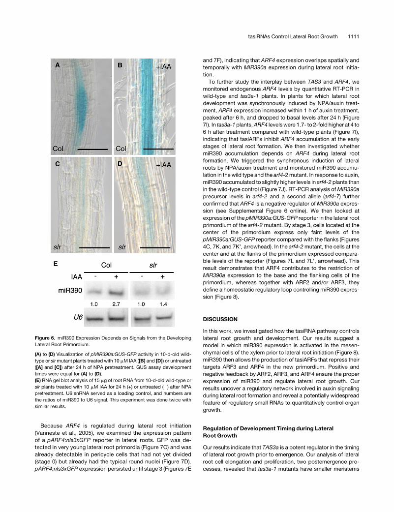
Because ARF4 is regulated during lateral root initiation
(Vanneste et al., 2005), we examined the expression pattern
of a pARF4:nls3xGFP reporter in lateral roots. GFP was de-
tected in very young lateral root primordia (Figure 7C) and was
already detectable in pericycle cells that had not yet divided
(stage 0) but already had the typical round nuclei (Figure 7D).
pARF4:nls3xGFP expression persisted until stage 3 (Figures 7E
and 7F), indicating that ARF4 expression overlaps spatially and
temporally with MIR390a expression during lateral root initia-
tion.
To further study the interplay between TAS3 and ARF4, we
monitored endogenous ARF4 levels by quantitative RT-PCR in
wild-type and tas3a-1 plants. In plants for which lateral root
development was synchronously induced by NPA/auxin treat-
ment, ARF4 expression increased within 1 h of auxin treatment,
peaked after 6 h, and dropped to basal levels after 24 h (Figure
7I). In tas3a-1plants,ARF4 levelswere 1.7- to 2-fold higher at 4 to
6 h after treatment compared with wild-type plants (Figure 7I),
indicating that tasiARFs inhibit ARF4 accumulation at the early
stages of lateral root formation. We then investigated whether
miR390 accumulation depends on ARF4 during lateral root
formation. We triggered the synchronous induction of lateral
roots by NPA/auxin treatment and monitored miR390 accumu-
lation in thewild type and the arf4-2mutant. In response to auxin,
miR390 accumulated to slightly higher levels in arf4-2 plants than
in the wild-type control (Figure 7J). RT-PCR analysis ofMIR390a
precursor levels in arf4-2 and a second allele (arf4-7) further
confirmed that ARF4 is a negative regulator ofMIR390a expres-
sion (see Supplemental Figure 6 online). We then looked at
expression of the pMIR390a:GUS-GFP reporter in the lateral root
primordium of the arf4-2mutant. By stage 3, cells located at the
center of the primordium express only faint levels of the
pMIR390a:GUS-GFP reporter compared with the flanks (Figures
4C, 7K, and 7K’, arrowhead). In the arf4-2mutant, the cells at the
center and at the flanks of the primordium expressed compara-
ble levels of the reporter (Figures 7L and 7L’, arrowhead). This
result demonstrates that ARF4 contributes to the restriction of
MIR390a expression to the base and the flanking cells of the
primordium, whereas together with ARF2 and/or ARF3, they
define a homeostatic regulatory loop controlling miR390 expres-
sion (Figure 8).
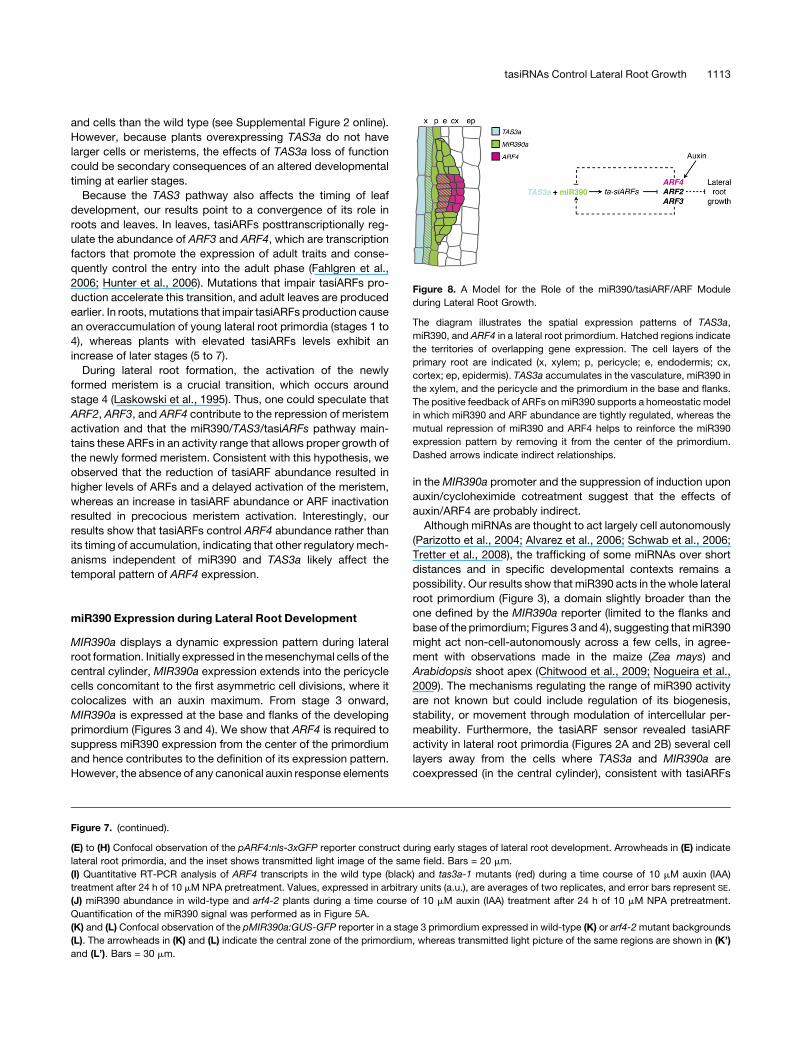
DISCUSSION
In this work, we investigated how the tasiRNA pathway controls
lateral root growth and development. Our results suggest a
model in which miR390 expression is activated in the mesen-
chymal cells of the xylem prior to lateral root initiation (Figure 8).
miR390 then allows the production of tasiARFs that repress their
targets ARF3 and ARF4 in the new primordium. Positive and
negative feedback by ARF2, ARF3, and ARF4 ensure the proper
expression of miR390 and regulate lateral root growth. Our
results uncover a regulatory network involved in auxin signaling
during lateral root formation and reveal a potentially widespread
feature of regulatory small RNAs to quantitatively control organ
growth.
Regulation of Development Timing during Lateral
Root Growth
Our results indicate that TAS3a is a potent regulator in the timing
of lateral root growth prior to emergence. Our analysis of lateral
root cell elongation and proliferation, two postemergence pro-
cesses, revealed that tas3a-1 mutants have smaller meristems
Figure 6. miR390 Expression Depends on Signals from the Developing
Lateral Root Primordium.
(A) to (D) Visualization of pMIR390a:GUS-GFP activity in 10-d-old wild-
type or slrmutant plants treated with 10 mM IAA ([B] and [D]) or untreated
([A] and [C]) after 24 h of NPA pretreatment. GUS assay development
times were equal for (A) to (D).
(E) RNA gel blot analysis of 15 mg of root RNA from 10-d-old wild-type or
slr plants treated with 10 mM IAA for 24 h (+) or untreated (�) after NPA
pretreatment. U6 snRNA served as a loading control, and numbers are
the ratios of miR390 to U6 signal. This experiment was done twice with
similar results.
tasiRNAs Control Lateral Root Growth 1111
Figure 7. ARF2/ARF3/ARF4 Control Lateral Root Growth, and ARF4 Is Required for miR390 Expression.
(A) Measurement of average lateral root length in 10-d-old seedlings expressing either the 35S:aMIR-ARFs (aMIR-ARFs) or the empty vector (Vector).
Distribution of the population (n > 9) is represented by box plots. aMIR-ARFs plants have longer lateral roots than the vector controls (*, P < 0.05;
Student’s t test).
(B) RNA gel blot analysis of 15 mg of root RNA from 10-d-old plants expressing either 35S:aMIR-ARFs or the empty vector. The plants were treated (+)
with 10 mM IAA or untreated (�) after 24 h of 10mM NPA pretreatment.
(C) RNA gel blot analysis of 15 mg of roots RNA from plants of the indicated genotype.
(D)RNA gel blot analysis of 15 mg of root RNAs from 10-d-old plants expressing either ARF3:ARF3:GUS or its tasiARF-resistant version. The plants were
treated as in (B). In (B) to (D), U6 snRNA served as a loading control, and numbers are the ratios of probe to U6 signal. These experiments were done
twice with similar results.
1112 The Plant Cell
and cells than the wild type (see Supplemental Figure 2 online).
However, because plants overexpressing TAS3a do not have
larger cells or meristems, the effects of TAS3a loss of function
could be secondary consequences of an altered developmental
timing at earlier stages.
Because the TAS3 pathway also affects the timing of leaf
development, our results point to a convergence of its role in
roots and leaves. In leaves, tasiARFs posttranscriptionally reg-
ulate the abundance of ARF3 and ARF4, which are transcription
factors that promote the expression of adult traits and conse-
quently control the entry into the adult phase (Fahlgren et al.,
2006; Hunter et al., 2006). Mutations that impair tasiARFs pro-
duction accelerate this transition, and adult leaves are produced
earlier. In roots,mutations that impair tasiARFs production cause
an overaccumulation of young lateral root primordia (stages 1 to
4), whereas plants with elevated tasiARFs levels exhibit an
increase of later stages (5 to 7).
During lateral root formation, the activation of the newly
formed meristem is a crucial transition, which occurs around
stage 4 (Laskowski et al., 1995). Thus, one could speculate that
ARF2, ARF3, and ARF4 contribute to the repression of meristem
activation and that the miR390/TAS3/tasiARFs pathway main-
tains these ARFs in an activity range that allows proper growth of
the newly formed meristem. Consistent with this hypothesis, we
observed that the reduction of tasiARF abundance resulted in
higher levels of ARFs and a delayed activation of the meristem,
whereas an increase in tasiARF abundance or ARF inactivation
resulted in precocious meristem activation. Interestingly, our
results show that tasiARFs control ARF4 abundance rather than
its timing of accumulation, indicating that other regulatory mech-
anisms independent of miR390 and TAS3a likely affect the
temporal pattern of ARF4 expression.
miR390 Expression during Lateral Root Development
MIR390a displays a dynamic expression pattern during lateral
root formation. Initially expressed in themesenchymal cells of the
central cylinder, MIR390a expression extends into the pericycle
cells concomitant to the first asymmetric cell divisions, where it
colocalizes with an auxin maximum. From stage 3 onward,
MIR390a is expressed at the base and flanks of the developing
primordium (Figures 3 and 4). We show that ARF4 is required to
suppress miR390 expression from the center of the primordium
and hence contributes to the definition of its expression pattern.
However, the absence of any canonical auxin response elements
in theMIR390a promoter and the suppression of induction upon
auxin/cycloheximide cotreatment suggest that the effects of
auxin/ARF4 are probably indirect.
Although miRNAs are thought to act largely cell autonomously
(Parizotto et al., 2004; Alvarez et al., 2006; Schwab et al., 2006;
Tretter et al., 2008), the trafficking of some miRNAs over short
distances and in specific developmental contexts remains a
possibility. Our results show that miR390 acts in the whole lateral
root primordium (Figure 3), a domain slightly broader than the
one defined by the MIR390a reporter (limited to the flanks and
base of the primordium; Figures 3 and 4), suggesting thatmiR390
might act non-cell-autonomously across a few cells, in agree-
ment with observations made in the maize (Zea mays) and
Arabidopsis shoot apex (Chitwood et al., 2009; Nogueira et al.,
2009). The mechanisms regulating the range of miR390 activity
are not known but could include regulation of its biogenesis,
stability, or movement through modulation of intercellular per-
meability. Furthermore, the tasiARF sensor revealed tasiARF
activity in lateral root primordia (Figures 2A and 2B) several cell
layers away from the cells where TAS3a and MIR390a are
coexpressed (in the central cylinder), consistent with tasiARFs
Figure 7. (continued).
(E) to (H) Confocal observation of the pARF4:nls-3xGFP reporter construct during early stages of lateral root development. Arrowheads in (E) indicate
lateral root primordia, and the inset shows transmitted light image of the same field. Bars = 20 mm.
(I) Quantitative RT-PCR analysis of ARF4 transcripts in the wild type (black) and tas3a-1 mutants (red) during a time course of 10 mM auxin (IAA)
treatment after 24 h of 10 mM NPA pretreatment. Values, expressed in arbitrary units (a.u.), are averages of two replicates, and error bars represent SE.
(J) miR390 abundance in wild-type and arf4-2 plants during a time course of 10 mM auxin (IAA) treatment after 24 h of 10 mM NPA pretreatment.
Quantification of the miR390 signal was performed as in Figure 5A.
(K) and (L) Confocal observation of the pMIR390a:GUS-GFP reporter in a stage 3 primordium expressed in wild-type (K) or arf4-2mutant backgrounds
(L). The arrowheads in (K) and (L) indicate the central zone of the primordium, whereas transmitted light picture of the same regions are shown in (K’)
and (L’). Bars = 30 mm.
Figure 8. A Model for the Role of the miR390/tasiARF/ARF Module
during Lateral Root Growth.
The diagram illustrates the spatial expression patterns of TAS3a,
miR390, and ARF4 in a lateral root primordium. Hatched regions indicate
the territories of overlapping gene expression. The cell layers of the
primary root are indicated (x, xylem; p, pericycle; e, endodermis; cx,
cortex; ep, epidermis). TAS3a accumulates in the vasculature, miR390 in
the xylem, and the pericycle and the primordium in the base and flanks.
The positive feedback of ARFs onmiR390 supports a homeostatic model
in which miR390 and ARF abundance are tightly regulated, whereas the
mutual repression of miR390 and ARF4 helps to reinforce the miR390
expression pattern by removing it from the center of the primordium.
Dashed arrows indicate indirect relationships.
tasiRNAs Control Lateral Root Growth 1113
intercellular mobility. Non-cell-autonomous activity of tasiRNAs
has been postulated based on their requirement for DCL4, which
produces siRNAs that associate with mobile silencing (Dunoyer
et al., 2005; Bouche et al., 2006; Deleris et al., 2006). This model
is supported by both the non-cell-autonomous silencing medi-
ated by an artificial transgene-based system, which produces
siRNAs from a tasiRNA-like precursor (Tretter et al., 2008), and
by the observation that the tasiARFs act at distance from their
site of production in the shoot apical meristem (Chitwood et al.,
2009; Schwab et al., 2009).
Quantitative Regulation of Development by Small RNAs
35S:TAS3a and 35S:aMIR-ARFs plants have longer lateral roots
than wild-type controls (Figure 7A), whereas the length of lateral
roots in arf2, arf3, and arf4 single mutants is only marginally
affected (see Supplemental Figure 5 online). This suggests that
the role of ARF4 during lateral root growth could be restricted to
fine-tuning the regulatory system. In the absence of ARF4, the
functions of ARF2 and ARF3 are still sufficient to maintain an
almost normal development of lateral roots through a homeo-
static regulatory loop. Thus, the simultaneous inactivation of
multiple targets may be critical for the full activity of the miR390/
TAS3 module and a more general requirement for regulation of
developmental processes by miRNAs (Voinnet, 2009).
Our results establish that the tasiARFs targets contribute
differently to miR390 expression. First, simultaneous impairment
of ARF2, ARF3, and ARF4 function with an artificial miRNA
reduces the expression of miR390, whereas increasing the
abundance of all three ARFs or ARF3 alone results in higher
miR390 accumulation. This positive feedback of ARF2 and/or
ARF3 on miR390 has the potential to ensure tight control of the
miR390-TAS3-ARFsmodule activity. This homeostaticmodel for
miR390/ARF function during lateral root formation maintains the
activity of ARFs within an optimal range. Second, ARF4 has a
specific function in the spatial restriction of miR390 expression
via a negative feedback mechanism in the central primordial
cells. The coexistence of both mechanisms for overlapping
targets illustrates the importance of a finely tuned activity of
the miR390/TAS3/tasiARF module. Our results differ from a
recent report describing the auxin induction of miR390 in roots
and its role in lateral root development (Yoon et al., 2010). The
main discrepancy concerns the respective contribution of the
MIR390a and MIR390b loci to miR390 accumulation and re-
sponse to auxin. The 59 rapid amplification of cDNA ends (RACE)
mapping and mir390a mutant analysis revealed that MIR390a is
the major contributor of miR390 accumulation in the root (Figure
2C), in agreementwith our sensor data (Figure 3F). By contrast, in
situ hybridization data presented by Yoon et al. could not
determine the locus of origin of miR390 because MIR390a and
MIR390b encode the same mature miRNA. In addition, the
pMIR390a:GUS and pMIR390b:GUS reporter used by Yoon
et al. encompassed parts of the miR390 precursor, whereas
ours corresponded to the actual nontranscribed genomic region,
which could account for discrepancies between our data. More-
over, we showed thatMIR390a is auxin inducible during the early
stages of lateral root initiation (stage 0 to 2/3). On the contrary,
Yoon et al. describe the effects of auxin on older lateral roots.
This is a major difference between the two studies. We have not
studied the effect of auxin on older primordia; therefore, we
cannot exclude that MIR390b is induced at later stages. How-
ever, our results using the mir390a mutant firmly establish that
upon the first stages of auxin-induced lateral root initiation, only
the MIR390a locus is active (Figures 2C and 5C).
To conclude, we show how miR390-TAS3 tasiRNA-ARF2/3/4
integrate with auxin signaling to regulate lateral root growth, in
addition to miR167-ARF8 modulation of lateral root meristem
activation in response to nitrogen availability (Gifford et al., 2008).
Negative and positive feedback loops of miRNA/target regulons
have been described for the posttranscriptional regulation of
miRNA homeostasis in plants (Xie et al., 2003; Vaucheret et al.,
2004, 2006; Rajagopalan et al., 2006) and for the transcriptional
regulation of gene networks in animal development (Tsang et al.,
2007). Two recent reports describe additional miRNA/target
regulons in plant development. First, expression of miR156 and
miR172 depends on their targets, the transcription factors of the
SPL and AP2 families, and is crucial for the vegetative phase
transition in Arabidopsis (Wu et al., 2009). Second, formation of
adventitious roots involves a complex regulatory network in-
cluding cross-regulation of miR160/167 homeostasis by direct
and indirect targeting of ARF transcription factors (Gutierrez
et al., 2009). Here, we show that the reciprocal feedback be-
tween miRNA and their targets can be extended to the tasiRNA
pathway in which an miRNA controls abundance of its target
using intermediary and potentially mobile siRNAs. Transcription
factors and miRNAs are the major trans-acting regulators that
determine the dynamic equilibrium of transcriptional networks at
each developmental stage (Hobert, 2008). Our results underline
the importance of reciprocal miRNA/transcription factor regula-
tory feedback loops in the control of plant organ growth in
response to a specific morphogenetic trigger.
METHODS
Plant Material
All lines used in this study are in the Arabidopsis thaliana Col ecotype
background. The tas3a-1 (GABI_621G08), rdr6 (sgs2-1), dcl4-1, tasiARF
sensors (pARF3:ARF3:GUS and pARF3:ARF3tasiR-GUS), slr, arf2-6,
arf3-2, and arf4-2 (salk_070506) have been previously described (Fukaki
et al., 2002; Gasciolli et al., 2005; Okushima et al., 2005a, 2005b; Pekker
et al., 2005; Adenot et al., 2006; Fahlgren et al., 2006; Chitwood et al.,
2009; Schwab et al., 2009). The arf4-7 allele was identified in the SALK
collection (salk_028804C) (Alonso et al., 2003). Sequencing of T-DNA
junctions confirmed that the T-DNA was inserted in the 59 untranslated
region of ARF4 (At5g60450) 376 bp upstream of the ATG. The activation-
tagged TAS3a allele (GABI_626B09) was identified in the GABI-KAT
collection (Rosso et al., 2003). Sequencing of T-DNA junctions revealed
that the T-DNA was inserted 102 bp upstream of TAS3a (At3g17185). The
mir390a line (WiscDsLox440F06; mir390a-2) was identified in the
Wisconsin DsLox collection (Woody et al., 2007). Sequencing of T-DNA
junctions revealed that the insertion is located 30 bp upstream of the
MIR390a (At2g38325) transcriptional start site.
Growth Conditions
Soil-grown plants were propagated in a greenhouse (238C). For in vitro
conditions, plants were grown on 0.53Murashige and Skoog (MS)/0.8%
1114 The Plant Cell
agar (MS agar) plates in controlled-environment chambers under the
following conditions: 150mmol photon·m22·s21 luminance, 16 h light, and
238C temperature. For synchronous induction of lateral root develop-
ment, plants were germinated on nylon sheets (SEFAR NITEX 03-100/44)
on 0.53 MS agar for 8 d, transferred to 0.53 MS agar + 10mM NPA for
24 h, and shifted to 0.53 MS agar + 10 mM IAA for the indicated time.
Phenotypic Analysis
For quantification of root morphology, plates were scanned after 10 d of
growth and examined under a binocular microscope to determine the
number of emerged lateral root primordia. Measurements of primary root
length and lateral root length were made on the scanned picture using
Image-J (http://rsb.info.nih.gov/ij/). Measurements of cell and meristem
size were performed as described (Cazale et al., 2009). We used R (www.
r-project.org) for statistical analysis and graphing of the data.
Construction of Reporter and Sensor Transgenes
For the 35S:TAS3a construct, TAS3a (At3g17185) was amplified (primers
N-0081/82) from genomic DNA to generate a Gateway (Invitrogen)
entry clone in pDONR221, which was then recombined in a home-
made Gateway-compatible version of pCHF3 (Jarvis et al., 1998). For
pTAS3a:GUS reporters, we amplified with primers N-0087/88 550bp
of regulatory sequence able to rescue the phenotype of tas3a-1 mu-
tants (Adenot et al., 2006) to generate an entry clone that was then
recombined in the pMDC163 vector (Curtis and Grossniklaus, 2003).
pMIR390a:GUS-GFP and pMIR390b:GUS-GFP reporters were built by
amplifying 2.6 and 0.5Kbp, respectively, of genomicDNAupstreamof the
transcription initiation start of At2g38325 (MIR390a) and At5g58465
(MIR390b) with primers N-0154/155 and N-0156/157, generating entry
clones (pENTR-D; Invitrogen), which were then recombined in pKGWFS7
(Karimi et al., 2007). The tasiARFs sensors were described by Fahlgren
et al. (2006). For the miR390 sensor constructs, a 200-bp fragment of
TAS3a containing either the wild-type 39 miR390 binding site (wild-type
sensor) or a mutated 39 site that impairs proper miR390 recognition
(mutated sensor) was amplified by PCR (using primers N-2016/2017 and
2018) and placed downstream of GFP under the control of the 35S
promoter using gateway-basedcloning. Themutated versionwasobtained
using a 39 primer that introduces five point mutations between positions
1 and 11 of the miR390 binding site. For the DR5rev:erRFP reporter,
DR5rev:erRFP was amplified by PCR (primers N-2166/2167) and cloned
to generate an entry clone, then recombined in pHGWL7 (Karimi et al.,
2007). The 35S:aMIR-ARF construct was described previously (Alvarez
et al., 2006). For the pARF4:nls-3xGFP constructs, 2.0 kb upstream of the
ARF4 start codon was amplified and conventionally cloned into a
pGREENII-based vector containing the nuclear localized 3xGFP se-
quence and a NOS transcriptional terminator (primers ARF4PFWD and
ARF4PREV). This construct was introduced into Agrobacterium tumefa-
ciens strain GV3101 harboring pGREENII helper plasmid pSOUP. Se-
quences of all primers used can be found in Supplemental Table 1 online,
and all constructs were checked by sequencing. Vectors besides pARF4:
nls-3xGFP were introduced in Agrobacterium (ASE strain), and all con-
structs were transformed into plants by floral dipping (Weigel and
Glazebrook, 2002).
GUS and Confocal Analysis
GUS activity was assayed at 378C for 6 to 18 h using 2 mM ferri/
ferrocyanide as described (Weigel and Glazebrook, 2002). Transverse
sections were obtained after GUS staining using the protocol described
by De Smet et al. (2004), mounted in Eukitt (EMS), and photographed on a
DMI-6000microscope (LeicaMicrosystems). For confocal imaging, roots
were mounted in 5% glycerol and directly imaged on a TCS-SP2 upright
microscope (Leica Microsystems) with 488-nm/543-nm excitation, 488/
543 beamsplitter filter, and 5156 15 nm (green channel) and 6106 25 nm
(red channel) detection windows. Transmitted light was also collected. All
images were acquired with similar gain adjustments. Counterstaining of
cell walls was achieved by 5 min of incubation in 100 mg·mL21 propidium
iodide. For 4’,6-diamidino-2-phenylindole (DAPI) staining of the nuclei,
plants were fixed for 45 min in 4% paraformaldehyde in MTSB (Muller
et al., 1998), washed in 2mMglycine, andmounted in Vectashield (Vector
Laboratories) containing 1.5 mg/mL DAPI. Plants were imaged for GFP
signal as described above and finally imaged for DAPI with a 364-nm UV
laser (no beamsplitter filter set, detection window of 415 to 550 nm).
RNA Extraction, RNA Blot Assays, Quantitative RT-PCR, and
RACE Analysis
Total RNA was extracted as described (Mallory et al., 2001). For RNA gel
blot analysis, 15mg of RNAwere separated by denaturing (7MUrea) 15%
polyacrylamide gel electrophoresis, blotted to a nylon membrane
(Hybond NX; GE Healthcare), and cross-linked as described (Pall et al.,
2007). miRNA probes were prepared by end labeling antisense oligonu-
cleotides with 32P using T4 polynucleotide kinase (Fermentas). RNA gel
blots were hybridized (Mallory et al., 2001) with themiRNA probe together
with U6 probe, stripped, and reprobed successively with different
miRNAs. Nonsaturated signals were quantified on a Molecular Dynamics
Storm 840.
For quantitative RT-PCR analysis, 4 mg of total RNA was treated with
RNase-free DNase (Fermentas) and reverse transcribed (Superscript II;
Invitrogen). cDNA was diluted four times and used for amplification. A
parallel reaction without reverse transcriptase was systematically per-
formed and used as a control for DNA contamination. Quantitative PCR
was performed in capillaries on a Roche LightCycler thermocycler using
the manufacturer’s instructions. Two reference genes (AT1G13320 and
AT4G26410; empirically identified for their stable expression across a
wide range of conditions [Czechowski et al., 2005]) were used to nor-
malize our signal. Efficiency of each primer pair was determined before-
hand. For nonquantitative PCR, Taq polymerase (Fermentas) was used;
amplification was stopped after 25 cycles and analyzed on agarose gels.
All primers used are described in Supplemental Table 1 online.
The 59 RACE was performed using a FirstChoice RLM-RACE Kit
(Ambion) following the manufacturer’s instructions. Twelve cloned RACE
fragments were sequenced to map theMIR390a andMIR390b transcrip-
tion start. All primers used are described in Supplemental Table 1 online.
Accession Numbers
Sequence data from this article can be found in the Arabidopsis Genome
Initiative or GenBank/EMBL databases under the following acces-
sion numbers: TAS3a (AT3G17185), MIR390a (AT2G38325), MIR390b
(AT5G48465), ARF2 (AT5G62000), ARF3 (AT2G33860), ARF4
(AT5G60450), ACTIN2 (AT3G18780), and quantitative PCR references
(AT1G13320 and AT4G26410; Czechowski et al., 2005).
Supplemental Data
The following materials are available in the online version of this article.
Supplemental Figure 1. Level of Expression of TAS3a (At3g17185) in
Loss- and Gain-of-Function Alleles.
Supplemental Figure 2. Phenotypic Characterization of the Root
System of Plants Deregulated for the TAS3 tasiRNA Pathway.
Supplemental Figure 3. MIR390b Expression Pattern.
Supplemental Figure 4. ARF Expression in aMIR-ARFs and Wild-
Type Plants.
tasiRNAs Control Lateral Root Growth 1115
Supplemental Figure 5. Phenotypic Characterization of ARF Mutant
Root Phenotypes.
Supplemental Figure 6. ARF4 and MIR390a Expression in arf4
Knockdown Mutants.
Supplemental Table 1. Primers Used in This Study.
ACKNOWLEDGMENTS
We thank B. Ben Amor for initial experiments, T. Beeckman for the slr
mutant, J. Carrington for the tasiARF sensors, and A. Gallavotti for the
DR5rev:erRFP construct. We thank A. Leibfried, J. Lohmann, and G.
Cristofari for their critical reading of the manuscript. This work was
supported by an ANR-GENOPLANT grant (RIBOROOT-ANR06 GPLA
011) and has benefited from the facilities of the Imagif Cell Biology Unit
of the Gif campus (www.imagif.cnrs.fr), which is supported by the
Conseil General de l’Essonne.
Received November 3, 2009; revised March 16, 2010; accepted March
22, 2010; published April 2, 2010.
REFERENCES
Adenot, X., Elmayan, T., Lauressergues, D., Boutet, S., Bouche, N.,
Gasciolli, V., and Vaucheret, H. (2006). DRB4-dependent TAS3
trans-acting siRNAs control leaf morphology through AGO7. Curr.
Biol. 16: 927–932.
Allen, E., Xie, Z., Gustafson, A.M., and Carrington, J.C. (2005).
MicroRNA-directed phasing during trans-acting siRNA biogenesis in
plants. Cell 121: 207–221.
Alonso, J.M., et al. (2003). Genome-wide insertional mutagenesis of
Arabidopsis thaliana. Science 301: 653–657.
Alvarez, J.P., Pekker, I., Goldshmidt, A., Blum, E., Amsellem, Z., and
Eshed, Y. (2006). Endogenous and synthetic microRNAs stimulate
simultaneous, efficient, and localized regulation of multiple targets in
diverse species. Plant Cell 18: 1134–1151.
Benkova, E., Ivanchenko, M.G., Friml, J., Shishkova, S., and
Dubrovsky, J.G. (2009). A morphogenetic trigger: Is there an emerg-
ing concept in plant developmental biology? Trends Plant Sci. 14:
189–193.
Bouche, N., Lauressergues, D., Gasciolli, V., and Vaucheret, H.
(2006). An antagonistic function for Arabidopsis DCL2 in development
and a new function for DCL4 in generating viral siRNAs. EMBO J. 25:
3347–3356.
Cazale, A.C., Clement, M., Chiarenza, S., Roncato, M.A., Pochon, N.,
Creff, A., Marin, E., Leonhardt, N., and Noel, L.D. (2009). Altered
expression of cytosolic/nuclear HSC70–1 molecular chaperone af-
fects development and abiotic stress tolerance in Arabidopsis thali-
ana. J. Exp. Bot. 60: 2653–2664.
Chitwood, D.H., Nogueira, F.T., Howell, M.D., Montgomery, T.A.,
Carrington, J.C., and Timmermans, M.C. (2009). Pattern formation
via small RNA mobility. Genes Dev. 23: 549–554.
Curtis, M.D., and Grossniklaus, U. (2003). A gateway cloning vector
set for high-throughput functional analysis of genes in planta. Plant
Physiol. 133: 462–469.
Czechowski, T., Stitt, M., Altmann, T., Udvardi, M.K., and Scheible,
W.R. (2005). Genome-wide identification and testing of superior
reference genes for transcript normalization in Arabidopsis. Plant
Physiol. 139: 5–17.
Deleris, A., Gallego-Bartolome, J., Bao, J., Kasschau, K.D.,
Carrington, J.C., and Voinnet, O. (2006). Hierarchical action and
inhibition of plant Dicer-like proteins in antiviral defense. Science 313:
68–71.
De Smet, I., Chaerle, P., Vanneste, S., De Rycke, R., Inze, D., and
Beeckman, T. (2004). An easy and versatile embedding method for
transverse sections. J. Microsc. 213: 76–80.
De Smet, I., Vanneste, S., Inze, D., and Beeckman, T. (2006). Lateral root
initiation or the birth of a new meristem. Plant Mol. Biol. 60: 871–887.
Dubrovsky, J.G., Sauer, M., Napsucialy-Mendivil, S., Ivanchenko,
M.G., Friml, J., Shishkova, S., Celenza, J., and Benkova, E.
(2008). Auxin acts as a local morphogenetic trigger to specify lateral
root founder cells. Proc. Natl. Acad. Sci. USA 105: 8790–8794.
Dunoyer, P., Himber, C., and Voinnet, O. (2005). DICER-LIKE 4 is
required for RNA interference and produces the 21-nucleotide small
interfering RNA component of the plant cell-to-cell silencing signal.
Nat. Genet. 37: 1356–1360.
Fahlgren, N., Montgomery, T.A., Howell, M.D., Allen, E., Dvorak,
S.K., Alexander, A.L., and Carrington, J.C. (2006). Regulation of
AUXIN RESPONSE FACTOR3 by TAS3 ta-siRNA affects develop-
mental timing and patterning in Arabidopsis. Curr. Biol. 16: 939–944.
Fukaki, H., Tameda, S., Masuda, H., and Tasaka, M. (2002). Lateral
root formation is blocked by a gain-of-function mutation in the
SOLITARY-ROOT/IAA14 gene of Arabidopsis. Plant J. 29: 153–168.
Gallavotti, A., Yang, Y., Schmidt, R.J., and Jackson, D. (2008). The
relationship between auxin transport and maize branching. Plant
Physiol. 147: 1913–1923.
Garcia, D., Collier, S.A., Byrne, M.E., and Martienssen, R.A. (2006).
Specification of leaf polarity in Arabidopsis via the trans-acting siRNA
pathway. Curr. Biol. 16: 933–938.
Gasciolli, V., Mallory, A.C., Bartel, D.P., and Vaucheret, H. (2005). Partially
redundant functions of Arabidopsis DICER-like enzymes and a role for
DCL4 in producing trans-acting siRNAs. Curr. Biol. 15: 1494–1500.
Gifford, M.L., Dean, A., Gutierrez, R.A., Coruzzi, G.M., and
Birnbaum, K.D. (2008). Cell-specific nitrogen responses mediate
developmental plasticity. Proc. Natl. Acad. Sci. USA 105: 803–808.
Guo, H.S., Xie, Q., Fei, J.F., and Chua, N.H. (2005). MicroRNA directs
mRNA cleavage of the transcription factor NAC1 to downregulate
auxin signals for arabidopsis lateral root development. Plant Cell 17:
1376–1386.
Gutierrez, L., Bussell, J.D., Pacurar, D.I., Schwambach, J., Pacurar,
M., and Bellini, C. (2009). Phenotypic plasticity of adventitious
rooting in Arabidopsis is controlled by complex regulation of AUXIN
RESPONSE FACTOR transcripts and microRNA abundance. Plant
Cell 21: 3119–3132.
Hardtke, C.S. (2006). Root development–branching into novel spheres.
Curr. Opin. Plant Biol. 9: 66–71.
Hirsch, J., Lefort, V., Vankersschaver, M., Boualem, A., Lucas, A.,
Thermes, C., d’Aubenton-Carafa, Y., and Crespi, M. (2006). Charac-
terization of 43 non-protein-coding mRNA genes in Arabidopsis, includ-
ing the MIR162a-derived transcripts. Plant Physiol. 140: 1192–1204.
Hobert, O. (2008). Gene regulation by transcription factors and micro-
RNAs. Science 319: 1785–1786.
Hunter, C., Willmann, M.R., Wu, G., Yoshikawa, M., de la Luz
Gutierrez-Nava, M., and Poethig, S.R. (2006). Trans-acting siRNA-
mediated repression of ETTIN and ARF4 regulates heteroblasty in
Arabidopsis. Development 133: 2973–2981.
Jarvis, P., Chen, L.J., Li, H., Peto, C.A., Fankhauser, C., and Chory,
J. (1998). An Arabidopsis mutant defective in the plastid general
protein import apparatus. Science 282: 100–103.
Karimi, M., Bleys, A., Vanderhaeghen, R., and Hilson, P. (2007).
Building blocks for plant gene assembly. Plant Physiol. 145: 1183–1191.
Laskowski, M.J., Williams, M.E., Nusbaum, H.C., and Sussex, I.M.
(1995). Formation of lateral root meristems is a two-stage process.
Development 121: 3303–3310.
1116 The Plant Cell
Li, X., Cassidy, J.J., Reinke, C.A., Fischboeck, S., and Carthew, R.W.
(2009). A microRNA imparts robustness against environmental fluc-
tuation during development. Cell 137: 273–282.
Malamy, J.E., and Benfey, P.N. (1997). Organization and cell differen-
tiation in lateral roots of Arabidopsis thaliana. Development 124:
33–44.
Mallory, A.C., Bartel, D.P., and Bartel, B. (2005). MicroRNA-directed
regulation of Arabidopsis AUXIN RESPONSE FACTOR17 is essential
for proper development and modulates expression of early auxin
response genes. Plant Cell 17: 1360–1375.
Mallory, A.C., Ely, L., Smith, T.H., Marathe, R., Anandalakshmi, R.,
Fagard, M., Vaucheret, H., Pruss, G., Bowman, L., and Vance, V.B.
(2001). HC-Pro suppression of transgene silencing eliminates the
small RNAs but not transgene methylation or the mobile signal. Plant
Cell 13: 571–583.
Montgomery, T.A., Howell, M.D., Cuperus, J.T., Li, D., Hansen, J.E.,
Alexander, A.L., Chapman, E.J., Fahlgren, N., Allen, E., and
Carrington, J.C. (2008). Specificity of ARGONAUTE7-miR390 inter-
action and dual functionality in TAS3 trans-acting siRNA formation.
Cell 133: 128–141.
Muller, A., Guan, C., Galweiler, L., Tanzler, P., Huijser, P., Marchant,
A., Parry, G., Bennett, M., Wisman, E., and Palme, K. (1998). AtPIN2
defines a locus of Arabidopsis for root gravitropism control. EMBO J.
17: 6903–6911.
Nogueira, F.T., Chitwood, D.H., Madi, S., Ohtsu, K., Schnable, P.S.,
Scanlon, M.J., and Timmermans, M.C. (2009). Regulation of small
RNA accumulation in the maize shoot apex. PLoS Genet. 5:
e1000320.
Okushima, Y., Mitina, I., Quach, H.L., and Theologis, A. (2005a).
AUXIN RESPONSE FACTOR 2 (ARF2): A pleiotropic developmental
regulator. Plant J. 43: 29–46.
Okushima, Y., et al. (2005b). Functional genomic analysis of the AUXIN
RESPONSE FACTOR gene family members in Arabidopsis thaliana:
Unique and overlapping functions of ARF7 and ARF19. Plant Cell 17:
444–463.
Pall, G.S., Codony-Servat, C., Byrne, J., Ritchie, L., and Hamilton, A.
(2007). Carbodiimide-mediated cross-linking of RNA to nylon mem-
branes improves the detection of siRNA, miRNA and piRNA by
northern blot. Nucleic Acids Res. 35: e60.
Parizot, B., et al. (2008). Diarch symmetry of the vascular bundle in
Arabidopsis root encompasses the pericycle and is reflected in distich
lateral root initiation. Plant Physiol. 146: 140–148.
Parizotto, E.A., Dunoyer, P., Rahm, N., Himber, C., and Voinnet, O.
(2004). In vivo investigation of the transcription, processing, endonu-
cleolytic activity, and functional relevance of the spatial distribution of
a plant miRNA. Genes Dev. 18: 2237–2242.
Pekker, I., Alvarez, J.P., and Eshed, Y. (2005). Auxin response factors
mediate Arabidopsis organ asymmetry via modulation of KANADI
activity. Plant Cell 17: 2899–2910.
Peragine, A., Yoshikawa, M., Wu, G., Albrecht, H.L., and Poethig,
R.S. (2004). SGS3 and SGS2/SDE1/RDR6 are required for juvenile
development and the production of trans-acting siRNAs in Arabidop-
sis. Genes Dev. 18: 2368–2379.
Petricka, J.J., and Benfey, P.N. (2008). Root layers: Complex
regulation of developmental patterning. Curr. Opin. Genet. Dev. 18:
354–361.
Rajagopalan, R., Vaucheret, H., Trejo, J., and Bartel, D.P. (2006). A
diverse and evolutionarily fluid set of microRNAs in Arabidopsis
thaliana. Genes Dev. 20: 3407–3425.
Rosso, M.G., Li, Y., Strizhov, N., Reiss, B., Dekker, K., and
Weisshaar, B. (2003). An Arabidopsis thaliana T-DNA mutagenized
population (GABI-Kat) for flanking sequence tag-based reverse
genetics. Plant Mol. Biol. 53: 247–259.
Schwab, R., Maizel, A., Ruiz-Ferrer, V., Garcia, D., Bayer, M., Crespi,
M., Voinnet, O., and Martienssen, R.A. (2009). Endogenous TasiRNAs
mediate non-cell autonomous effects on gene regulation in Arabidopsis
thaliana. PLoS One 4: e5980.
Schwab, R., Ossowski, S., Riester, M., Warthmann, N., and Weigel,
D. (2006). Highly specific gene silencing by artificial microRNAs in
Arabidopsis. Plant Cell 18: 1121–1133.
Siegal, M.L., and Bergman, A. (2002). Waddington’s canalization
revisited: developmental stability and evolution. Proc. Natl. Acad.
Sci. USA 99: 10528–10532.
Tretter, E.M., Alvarez, J.P., Eshed, Y., and Bowman, J.L. (2008).
Activity range of Arabidopsis small RNAs derived from different
biogenesis pathways. Plant Physiol. 147: 58–62.
Tsang, J., Zhu, J., and van Oudenaarden, A. (2007). MicroRNA-
mediated feedback and feedforward loops are recurrent network
motifs in mammals. Mol. Cell 26: 753–767.
Vanneste, S., et al. (2005). Cell cycle progression in the pericycle is not
sufficient for SOLITARY ROOT/IAA14-mediated lateral root initiation in
Arabidopsis thaliana. Plant Cell 17: 3035–3050.
Vaucheret, H., Mallory, A.C., and Bartel, D.P. (2006). AGO1 homeo-
stasis entails coexpression of MIR168 and AGO1 and preferential
stabilization of miR168 by AGO1. Mol. Cell 22: 129–136.
Vaucheret, H., Vazquez, F., Crete, P., and Bartel, D.P. (2004). The
action of ARGONAUTE1 in the miRNA pathway and its regulation by
the miRNA pathway are crucial for plant development. Genes Dev. 18:
1187–1197.
Vazquez, F., Vaucheret, H., Rajagopalan, R., Lepers, C., Gasciolli,
V., Mallory, A.C., Hilbert, J.L., Bartel, D.P., and Crete, P. (2004).
Endogenous trans-acting siRNAs regulate the accumulation of Arabi-
dopsis mRNAs. Mol. Cell 16: 69–79.
Voinnet, O. (2009). Origin, biogenesis, and activity of plant microRNAs.
Cell 136: 669–687.
Weigel, D., and Glazebrook, J. 2002. Arabidopsis: A Laboratory Manual.
(Cold Spring Harbor, NY: Cold Spring Harbor Laboratory Press).
Williams, L., Carles, C.C., Osmont, K.S., and Fletcher, J.C. (2005). A
database analysis method identifies an endogenous trans-acting
short-interfering RNA that targets the Arabidopsis ARF2, ARF3, and
ARF4 genes. Proc. Natl. Acad. Sci. USA 102: 9703–9708.
Wilmoth, J.C., Wang, S., Tiwari, S.B., Joshi, A.D., Hagen, G.,
Guilfoyle, T.J., Alonso, J.M., Ecker, J.R., and Reed, J.W. (2005).
NPH4/ARF7 and ARF19 promote leaf expansion and auxin-induced
lateral root formation. Plant J. 43: 118–130.
Woody, S.T., Austin-Phillips, S., Amasino, R.M., and Krysan, P.J.
(2007). The WiscDsLox T-DNA collection: An Arabidopsis community
resource generated by using an improved high-throughput T-DNA
sequencing pipeline. J. Plant Res. 120: 157–165.
Wu, G., Park, M.Y., Conway, S.R., Wang, J.W., Weigel, D., and
Poethig, R.S. (2009). The sequential action of miR156 and miR172
regulates developmental timing in Arabidopsis. Cell 138: 750–759.
Xie, Z., Allen, E., Wilken, A., and Carrington, J.C. (2005). DICER-LIKE
4 functions in trans-acting small interfering RNA biogenesis and
vegetative phase change in Arabidopsis thaliana. Proc. Natl. Acad.
Sci. USA 102: 12984–12989.
Xie, Z., Kasschau, K.D., and Carrington, J.C. (2003). Negative feed-
back regulation of Dicer-Like1 in Arabidopsis by microRNA-guided
mRNA degradation. Curr. Biol. 13: 784–789.
Yoon, E.K., Yang, J.H., Lim, J., Kim, S.H., Kim, S.K., and Lee, W.S.
(2010). Auxin regulation of the microRNA390-dependent transacting
small interfering RNA pathway in Arabidopsis lateral root develop-
ment. Nucleic Acids Res. 38: 1382–1391.
Yoshikawa, M., Peragine, A., Park, M.Y., and Poethig, R.S. (2005). A
pathway for the biogenesis of trans-acting siRNAs in Arabidopsis.
Genes Dev. 19: 2164–2175.
tasiRNAs Control Lateral Root Growth 1117
Related Documents
![The MicroRNA390/TAS3 Pathway Mediates Symbiotic · The MicroRNA390/TAS3 Pathway Mediates Symbiotic Nodulation and Lateral Root Growth1[OPEN] ... aInstituto de Biotecnología y Biología](https://static.cupdf.com/doc/110x72/5ba480a709d3f2af168da32f/the-microrna390tas3-pathway-mediates-the-microrna390tas3-pathway-mediates.jpg)