461 www.eymj.org INTRODUCTION Sepsis, a lethal systemic inflammatory disease, is caused by infectious factors, such as viruses, bacteria, or fungi, and con- tributes to multiple organ dysfunction syndromes, shock, and even death. 1,2 Acute kidney injury (AKI), also known as acute renal failure, is a serious complication of sepsis. Increasing studies have reported that acute functional injury, along with the inflammatory response, may occur in the kidney during sepsis. 3 Unfortunately, the physiologic or pathologic mecha- nisms of sepsis-induced AKI remain elusive. In addition, the impairment of renal function may contribute to increased fa- tality rate in septic patients. 4 e fatality rate of septic patients will be significantly reduced if the intervention treatment is performed in the AKI stage. 5 Therefore, studies to further ex- plore the mechanisms of pathophysiological alterations at the early stage of kidney injury in septic patients, as well as to seek an effective therapeutic strategy for sepsis-induced AKI, are urgently needed. Long non-coding RNAs (lncRNAs), which are transcripts with over 200 nucleotides in length and without an open reading frame, are unable to code proteins. 6,7 Although lncRNAs lack MIR210HG Aggravates Sepsis-Induced Inflammatory Response of Proximal Tubular Epithelial Cell via the NF-κB Signaling Pathway Shuai Deng, Bin Gu, Zheng Yu, Zhen Shen, and Houwei Ren Department of Emergency, Jiangsu Taizhou People’s Hospital, Taizhou, China. Purpose: Acute kidney injury (AKI) is a serious complication of sepsis and is characterized by inflammatory response. MicroR- NA-210 host gene (MIR210HG) is upregulated in human proximal tubular epithelial cells under treatment of inflammatory cyto- kines. is study aimed to explore the role of MIR210HG in sepsis-induced AKI. Materials and Methods: Cell viability was detected by a cell counting kit 8 assay. e levels of proinflammatory cytokines were detected by enzyme-linked immunosorbent assay kits. e protein levels of p65, IκBα, and p-IκBα were examined by western blot analysis. e nuclear translocation of nuclear factor kappa B (NF-κB) was detected by immunofluorescence assay. e histological changes of kidneys were analyzed by hematoxylin and eosin staining assay. Results: Lipopolysaccharide (LPS) treatment significantly inhibited cell viability and increased productions of proinflammatory cytokines in proximal tubular epithelial cells (HKC-8). Additionally, MIR210HG levels in HKC-8 cells were increased by LPS treat- ment. MIR210HG silencing inhibited the LPS-induced cell inflammatory response. MIR210HG activated the NF-κB signaling pathway by promoting the phosphorylation of IκBα and nuclear translocation of p65. Rescue assays revealed that the MIR210HG- induced increase of cytokines levels and decline of cell viability were rescued by QNZ treatment. Knockdown of MIR210HG de- creased blood urea nitrogen, serum creatinine, and proinflammatory cytokine levels in AKI rats. Moreover, the knockdown of MIR210HG protected against AKI-induced histological changes of kidneys in rats. Conclusion: MIR210HG promotes sepsis-induced inflammatory response of HKC-8 cells by activating the NF-κB signaling path- way. is novel discovery may be helpful for the improvement of sepsis-induced AKI. Key Words: MIR210HG, sepsis, AKI, NF-κB signaling Original Article pISSN: 0513-5796 · eISSN: 1976-2437 Received: September 23, 2020 Revised: January 18, 2021 Accepted: February 23, 2021 Corresponding author: Shuai Deng, MM, Department of Emergency, Jiangsu Taizhou People’s Hospital, No. 366 Taihu Road, Gaoxin District, Taizhou 225300, China. Tel: 86-0523-86606306, Fax: 86-0523-86606306, E-mail: [email protected] •The authors have no potential conflicts of interest to disclose. © Copyright: Yonsei University College of Medicine 2021 This is an Open Access article distributed under the terms of the Creative Com- mons Attribution Non-Commercial License (https://creativecommons.org/licenses/ by-nc/4.0) which permits unrestricted non-commercial use, distribution, and repro- duction in any medium, provided the original work is properly cited. Yonsei Med J 2021 May;62(5):461-469 https://doi.org/10.3349/ymj.2021.62.5.461

Welcome message from author
This document is posted to help you gain knowledge. Please leave a comment to let me know what you think about it! Share it to your friends and learn new things together.
Transcript
461www.eymj.org
INTRODUCTION
Sepsis, a lethal systemic inflammatory disease, is caused by infectious factors, such as viruses, bacteria, or fungi, and con-tributes to multiple organ dysfunction syndromes, shock, and even death.1,2 Acute kidney injury (AKI), also known as acute
renal failure, is a serious complication of sepsis. Increasing studies have reported that acute functional injury, along with the inflammatory response, may occur in the kidney during sepsis.3 Unfortunately, the physiologic or pathologic mecha-nisms of sepsis-induced AKI remain elusive. In addition, the impairment of renal function may contribute to increased fa-tality rate in septic patients.4 The fatality rate of septic patients will be significantly reduced if the intervention treatment is performed in the AKI stage.5 Therefore, studies to further ex-plore the mechanisms of pathophysiological alterations at the early stage of kidney injury in septic patients, as well as to seek an effective therapeutic strategy for sepsis-induced AKI, are urgently needed.
Long non-coding RNAs (lncRNAs), which are transcripts with over 200 nucleotides in length and without an open reading frame, are unable to code proteins.6,7 Although lncRNAs lack
MIR210HG Aggravates Sepsis-Induced Inflammatory Response of Proximal Tubular Epithelial Cell via the NF-κB Signaling Pathway
Shuai Deng, Bin Gu, Zheng Yu, Zhen Shen, and Houwei RenDepartment of Emergency, Jiangsu Taizhou People’s Hospital, Taizhou, China.
Purpose: Acute kidney injury (AKI) is a serious complication of sepsis and is characterized by inflammatory response. MicroR-NA-210 host gene (MIR210HG) is upregulated in human proximal tubular epithelial cells under treatment of inflammatory cyto-kines. This study aimed to explore the role of MIR210HG in sepsis-induced AKI.Materials and Methods: Cell viability was detected by a cell counting kit 8 assay. The levels of proinflammatory cytokines were detected by enzyme-linked immunosorbent assay kits. The protein levels of p65, IκBα, and p-IκBα were examined by western blot analysis. The nuclear translocation of nuclear factor kappa B (NF-κB) was detected by immunofluorescence assay. The histological changes of kidneys were analyzed by hematoxylin and eosin staining assay.Results: Lipopolysaccharide (LPS) treatment significantly inhibited cell viability and increased productions of proinflammatory cytokines in proximal tubular epithelial cells (HKC-8). Additionally, MIR210HG levels in HKC-8 cells were increased by LPS treat-ment. MIR210HG silencing inhibited the LPS-induced cell inflammatory response. MIR210HG activated the NF-κB signaling pathway by promoting the phosphorylation of IκBα and nuclear translocation of p65. Rescue assays revealed that the MIR210HG-induced increase of cytokines levels and decline of cell viability were rescued by QNZ treatment. Knockdown of MIR210HG de-creased blood urea nitrogen, serum creatinine, and proinflammatory cytokine levels in AKI rats. Moreover, the knockdown of MIR210HG protected against AKI-induced histological changes of kidneys in rats.Conclusion: MIR210HG promotes sepsis-induced inflammatory response of HKC-8 cells by activating the NF-κB signaling path-way. This novel discovery may be helpful for the improvement of sepsis-induced AKI.
Key Words: MIR210HG, sepsis, AKI, NF-κB signaling
Original Article
pISSN: 0513-5796 · eISSN: 1976-2437
Received: September 23, 2020 Revised: January 18, 2021Accepted: February 23, 2021Corresponding author: Shuai Deng, MM, Department of Emergency, Jiangsu Taizhou People’s Hospital, No. 366 Taihu Road, Gaoxin District, Taizhou 225300, China.Tel: 86-0523-86606306, Fax: 86-0523-86606306, E-mail: [email protected]
•The authors have no potential conflicts of interest to disclose.
© Copyright: Yonsei University College of Medicine 2021This is an Open Access article distributed under the terms of the Creative Com-mons Attribution Non-Commercial License (https://creativecommons.org/licenses/by-nc/4.0) which permits unrestricted non-commercial use, distribution, and repro-duction in any medium, provided the original work is properly cited.
Yonsei Med J 2021 May;62(5):461-469https://doi.org/10.3349/ymj.2021.62.5.461
462
Role of MIR210HG in Sepsis Induced AKI
https://doi.org/10.3349/ymj.2021.62.5.461
protein-encoding ability, they can modulate both posttran-scriptional mRNA expression and chromatin levels via the ge-netic regulatory networks.7 Recently, many studies have re-vealed that lncRNAs are dysregulated in AKI.8-10 Additionally, these dysregulated lncRNAs exert effects on various biological processes, including cell viability, cell apoptosis, and inflam-matory response. For instance, the upregulated lncRNA nu-clear paraspeckle assembly transcript 1 predicts unfavorable prognosis of septic patients.11 In addition, the lncRNA metas-tasis-associated lung adenocarcinoma transcript 1 promotes sepsis-induced cardiac dysfunction and inflammatory re-sponse by regulation of miR-125b.12 Furthermore, homeobox A cluster antisense RNA 2 represses sepsis-induced AKI by bind-ing with miR-106b-5p.13 MicroRNA-210 host gene (MIR210HG) consists of 1965 nucleotides, and is transcribed by the gene ENSG00000247095.2 located on human chromosome 11p15.5.14 Most of the studies on MIR210HG have focused on cancers, and revealed the oncogenic role of MIR210HG in lung cancer, he-patocellular carcinoma, osteosarcoma, and glioma.14-17 More-over, MIR210HG is upregulated by 5.69-fold changes in HKC-8 cells in hypoxic conditions at 0.1% O2, and is upregulated by 2.35-fold changes in HKC-8 cells by the treatment of proinflam-matory cytokines consisting of 50 ng/mL of interleukin (IL)-6, 50 ng/mL of tumor necrosis factor (TNF)-α, and 20 ng/mL of interferon-γ.18 We hypothesized that MIR210HG plays a role in the inflammatory response of HKC-8 cells.
Nuclear factor kappa B (NF-κB), a ubiquitous DNA-binding transcription factor, is a key member of the NF-κB signaling pathway, and is widely reported to be implicated with inflam-matory response in sepsis-induced AKI.19,20 Downregulation of tissue inhibitor of metalloproteinase 2 improves cell injury and inflammatory response by the NF-κB pathway in sepsis-induced AKI.21 In addition, lidanpaidu prescription amelio-rates lipopolysaccharide-induced AKI by suppressing the NF-κB signaling pathway.22 However, the relationship between MIR210HG and the NF-κB signaling pathway remains unclear in AKI.
In the current study, we used lipopolysaccharide (LPS) to induce inflammatory response of immortalized human proxi-mal tubular epithelial cells (HKC-8). Then, we evaluated the expression status and biological effects of MIR210HG in LPS-treated HKC-8 cells. Considering that the NF-κB signaling path-way is crucial for inflammatory response in AKI, we then inves-tigated the relationship between MIR210HG and the NF-κB signaling pathway. Lastly, we performed cecal ligation and perforation on rats to imitate sepsis-induced AKI in vivo, and evaluated the role of MIR210HG in AKI rats.
MATERIALS AND METHODS
Cell linesHKC-8 cells, obtained from the Chinese Academy of Science
Cell Bank (Shanghai, China), were cultured in Dulbecco’s modi-fied Eagle medium (DMEM; Gibco, Grand Island, NY, USA)-nutrient mixture F-12 with 5% fetal bovine serum (FBS; GE Life Sciences, Beijing, China), 1% Primocin (Gibco), and 0.1% insulin-transferrin-selenium (Gibco). The HKC-8 cells were mycoplasma-negative, and were incubated at 37°C with 5% CO2. HKC-8 cells were treated with 100 ng/mL of LPS for 24 h to in-duce inflammatory response. In the control group (Con), HKC-8 cells were treated with the same dose of cell culture medium.
Cell transfectionThe short hairpin RNA targeting MIR210HG (sh-RNA#1/2) was used to knock down MIR210HG. The sh-NC served as the negative control. The full length of MIR210HG was subcloned into pcDNA3.1 vector to overexpress MIR210HG. Empty pcD-NA3.1 vector served as a negative control for vector overex-pressing MIR210HG. The vectors were synthesized by Ge-nePharma (Shanghai, China) and transfected into HKC-8 cells by Lipofectamine 2000 (Invitrogen, Carlsbad, CA, USA) ac-cording to the manufacturer’s instructions.
Cell viability measurement The cell viability of HKC-8 cells was detected by a cell count-ing assay kit-8 (CCK-8; Beyotime, Shanghai, China) under the manufacturer’s instructions. HKC-8 cells were seeded onto 96-well plates (5×10³ cells per well). Forty-eight hours after transfection, the CCK-8 solution (10 μL) was added. After-wards, HKC-8 cells were incubated in a humidified incubator with 5% CO2 at 37°C for 2 h. Finally, the absorbance was de-tected by a microplate reader at a wavelength of 450 nm.
Reverse transcription quantitative polymerase chain reaction (RT-qPCR)The extraction of total RNA from HKC-8 cells or renal tissues of rats were performed by TRIzol Reagent (Invitrogen). The RevertAid First Strand cDNA Synthesis Kit (K1622, Thermo Fisher Scientific, Waltham, MA, USA) was applied for reverse transcription of MIR210HG. The qPCR was performed using the SYBR-Green PCR Master Mix kit (Applied Biosystems, Waltham, MA, USA). The relative expression level of MIR210HG was calculated using the 2-ΔΔCt method. Glyceraldehyde-3-phos-phate dehydrogenase (GAPDH) served as a control gene. The primer sequences are listed as follows: MIR210HG, forward, 5'-GCT TGG TAG AGT GTC ACG CC-3' and reverse, 5'-CAT CTG ACC GAG CCA GTT TG-3'; GAPDH, forward, 5'-TAT GAT GAT ATC AAG AGG GTA GT-3' and reverse, 5'-TGT ATC CAA ACT CAT TGT CAT AC-3'.
Preparation of nuclear extractsThe HKC-8 cell suspensions were prepared by trypsin-ethyl-ene diamine tetra acetic acid treatment. The proteins were ex-tracted from the cytoplasm and nucleus of HKC-8 cells using NE-PERTM Nuclear and Cytoplasmic Extraction Reagents
463
Shuai Deng, et al.
https://doi.org/10.3349/ymj.2021.62.5.461
(Thermo Fisher Scientific) according to the manufacturer’s instructions.
Western blotWestern blot analysis was used to measure the protein levels. In brief, protein samples were separated by 12% sodium dodecyl sulfate-polyacrylamide gel electrophoresis gels and then trans-ferred onto polyvinylidene fluoride membranes. After being coated with 5% non-fat milk, the membranes were incubated with primary antibodies against p65 (ab16502, Abcam, Cam-bridge, UK), β-tubulin (ab204947, Abcam), Lamin B (ab194109, Abcam), IκBα (ab32518, Abcam), p-IκBα (sc-52943, Sigma-Al-drich, Saint Louis, MO, USA), and GAPDH (ab9484, Abcam) at a dilution of 1:1000 overnight at 4°C. Next, the membranes were exposed to horseradish peroxidase-conjugated goat anti-rab-bit (1:5000, ab205718, Abcam) secondary antibodies, and the signals were detected with an enhanced chemiluminescence detection system. The protein levels were quantified by the Im-ageJ software.
Immunofluorescent stainingHKC-8 cells treated with LPS were placed on a cover slide in 12-well plates. Then, HKC-8 cells were washed three times with phosphate buffer saline (PBS; Invitrogen), followed by fixation for 10 minutes with 4% paraformaldehyde at room temperature, and washed again. Afterwards, HKC-8 cells were blocked with 1% bovine serum albumin and incubated with primary antibody anti-NF-kB (ab16502, Abcam). Subsequent-ly, the secondary antibody fluorescein isothiocyanate-conju-gated immunoglobulin G (IgG) was added. Finally, the slides were rinsed, mounted, and viewed on a confocal microscope (FV1000, Olympus, Japan) at 488 nm.
Enzyme-linked immunosorbent assayThe levels of TNF-α, IL-6, and IL-1β in cell supernatants or kidneys of rats in each group were measured with ELISA kits according to the manufacturer’s instructions.
Animal model establishmentA total of 40 Sprague Dawley rats (male, 200–250 g, 8–10 weeks) were purchased from Vital River company (Beijing, China), and were randomly divided into four groups (n=10 in each group): sham group, sham+sh-RNA#1 group, AKI group, and AKI+sh-RNA#1 group. To induce AKI, we anesthetized the rats by in-traperitoneal injection of 2% sodium pentobarbital (80 mg/kg). Subsequently, the rats were placed on the laboratory bench, and their abdomens were shaved and disinfected with 70% isopropanol. Then, an incision (1.5 cm) was made in the mid-line of rat abdomen to fully expose the cecum, and the cecum (1 cm away from the cecum tail) was ligated with a sterile sew-ing silk (no. 4). Afterwards, the blind end was perforated by a 20-gauge needle. After restoring the cecum to the abdominal cavity, the abdominal incision was sutured. Finally, the rats
were moved to a thermostatic blanket for rewarming. The sh-am-operated rats underwent similar operation without cecal ligation and perforation. Three days later, all rats were killed, and the kidneys and blood of rats were collected. All experi-mental procedures were approved by the National Institutes of Health Guidelines for the Care and Use of Laboratory Animals (IRB No. 2018-077). The ethics committee of Jiangsu Taizhou People’s Hospital approved the current study.
Adeno-associated virus injectionAdeno-associated virus (AAV) (serotype 9, Vigene Bioscienc-es, Shanghai, China) was packaged, rinsed, and titrated for in-jection. AAV9 (1×10¹² copies) containing the shRNA#1 target-ing MIR210HG was injected into rats through the tail vein 2 weeks before model establishment.
Renal function measurementThe blood urea nitrogen (BUN) and serum creatinine (SCr) lev-els in the serum of rats were detected by a Hitachi 7060 auto-matic biochemistry analyzer (Hitachi, Japan) and a creatinine assay kit (BioAssay Systems, Hayward, CA, USA) according to the manufacturer’s instructions.
Hematoxylin & eosin staining assayH&E staining assay was conducted for histopathological anal-ysis of the rats’ kidneys, as described previously.10 First, sections (4 μm) of rat renal tissues were immobilized in 10% buffer for-malin, dehydrated in ethanol, clarified in xylene, and embed-ded in paraffin resin. Then, hematoxylin and eosin were sepa-rately utilized to immerse the sections for 10 min and 1 min. Afterwards, a microscope was used to visualize the morpho-logical changes.
Statistical analysisStatistical analysis was conducted with SPSS software. Data are presented as the mean±standard deviation. The unpaired Student’s t-tests or one-way analysis of variance were used to compare the differences between the groups. Pearson correla-tion analysis was used to analyze the correlation of expressions between MIR210HG and inflammatory cytokines. p<0.05 was considered statistically significant.
RESULTS
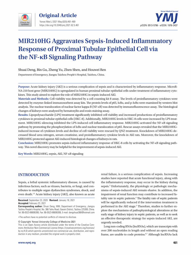
MIR210HG level is increased by LPS treatment in renal epithelial cellsLPS treatment significantly inhibited cell viability (Fig. 1A). Moreover, the levels of inflammatory cytokines (IL-1β, IL-6, and TNF-α) in HKC-8 cells were significantly increased by LPS treatment (Fig. 1B). Next, we evaluated the expression status of MIR210HG in HKC-8 cells. The result of RT-qPCR demon-strated that MIR210HG level was increased by LPS treatment
464
Role of MIR210HG in Sepsis Induced AKI
https://doi.org/10.3349/ymj.2021.62.5.461
in HKC-8 cells (Fig. 1C).
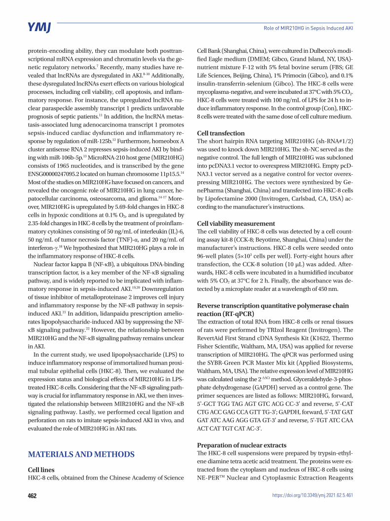
MIR210HG downregulation alleviates inflammatory response and cell injuryWe knocked down MIR210HG levels by transfecting shRNA#1/2 targeting MIR210HG into HKC-8 cells. As shown in Fig. 2A, MIR210HG levels were reduced by transfection of shRNA#1/2. In addition, CCK-8 assay showed that MIR210HG silencing significantly improved cell viability of HKC-8 cells (Fig. 2B). Results of ELISA revealed that MIR210HG silencing signifi-cantly decreased the levels of IL-1β, IL-6, and TNF-α in LPS-treated HKC-8 cells (Fig. 2C).
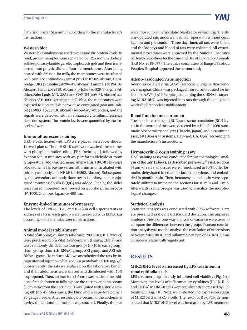
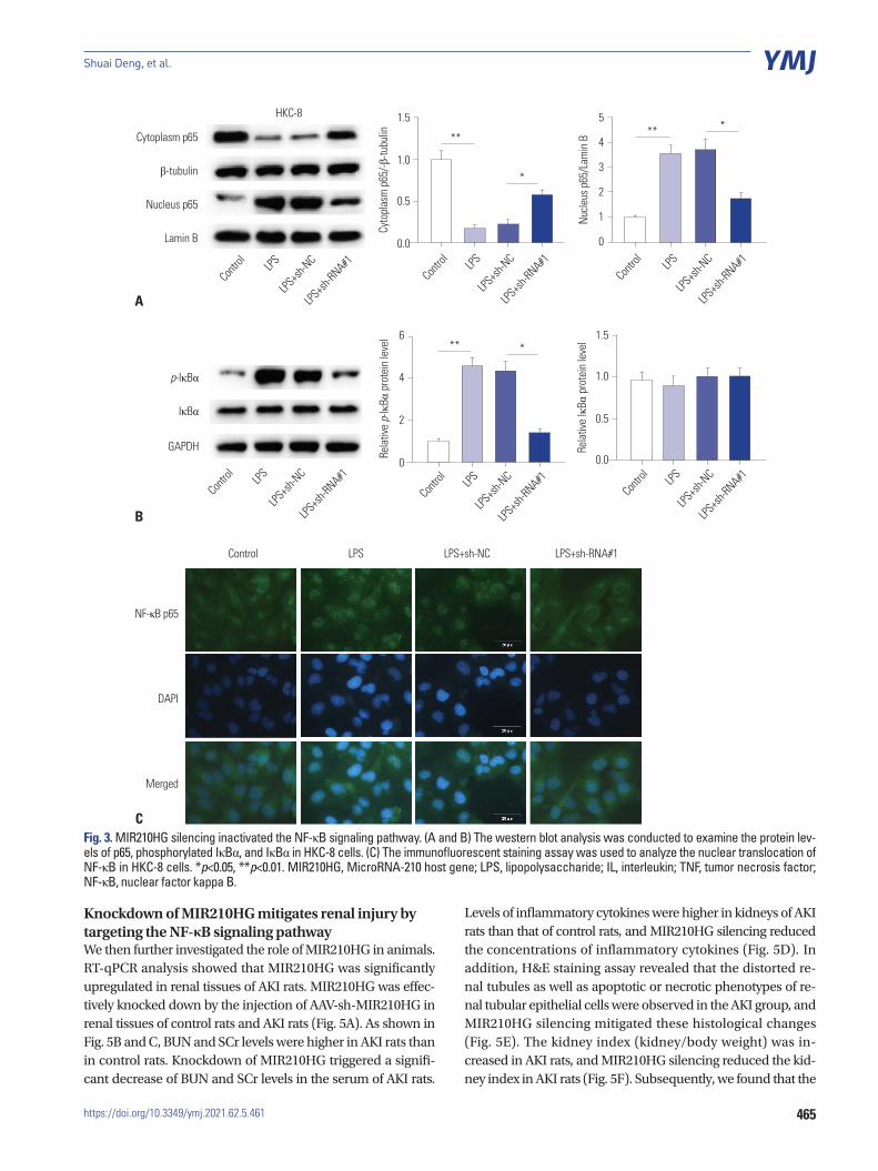
MIR210HG silencing inactivates the NF-κB signalingThe NF-κB signaling has been widely reported to regulate sep-sis-induced inflammatory response.23,24 We hypothesized that MIR210HG may modulate inflammatory response by target-ing the NF-κB signaling pathway. To validate our hypothesis, western blot analysis was conducted. According to Fig. 3A, the increase of nucleus p65 expression and the decline of cytoplasm p65 expression induced by LPS were restored by MIR210HG si-lencing. Moreover, the increase in p-IκBα protein levels result-
ing from LPS treatment was rescued by MIR210HG silencing (Fig. 3B). In addition, results of immunofluorescence staining assay demonstrated that the promotive effect of LPS treatment on NF-κB nuclear translocation was rescued by MIR210HG si-lencing (Fig. 3C).
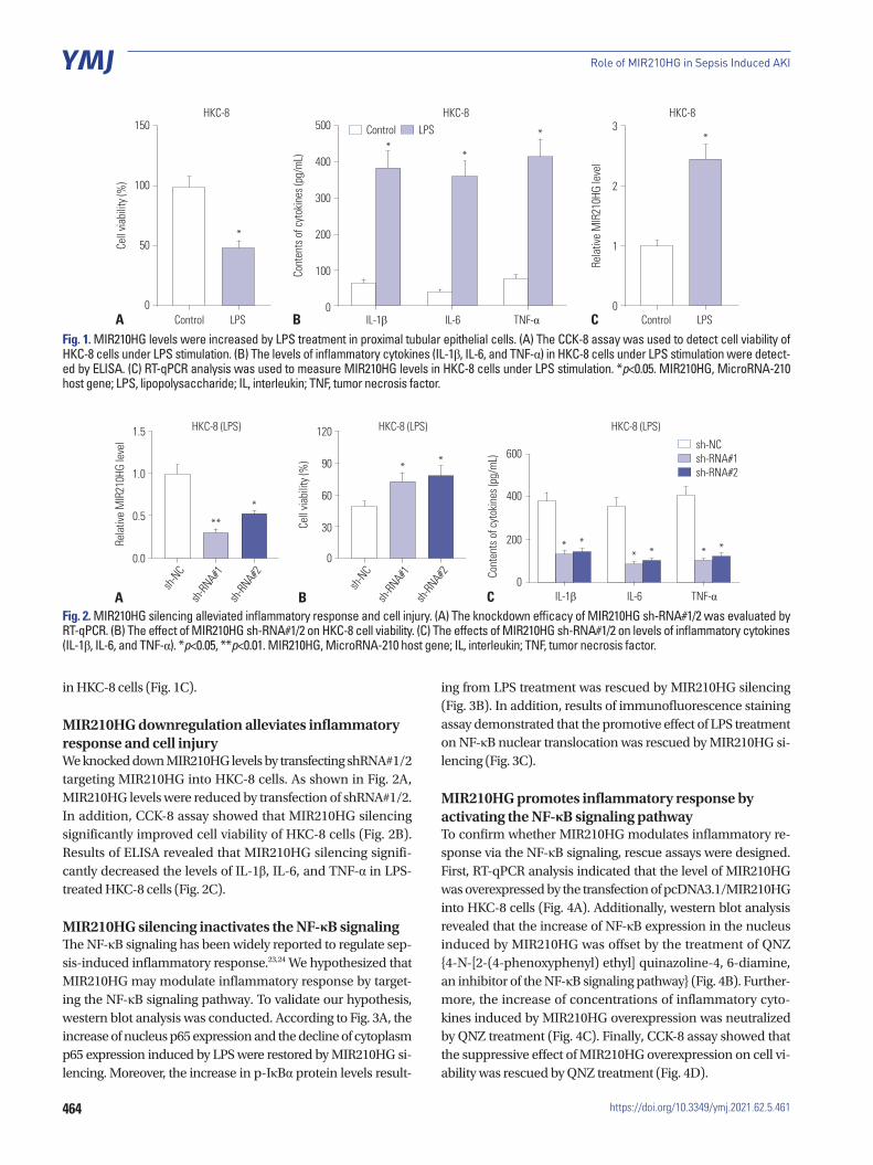
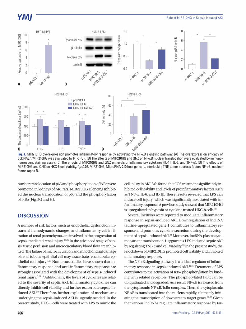
MIR210HG promotes inflammatory response by activating the NF-κB signaling pathwayTo confirm whether MIR210HG modulates inflammatory re-sponse via the NF-κB signaling, rescue assays were designed. First, RT-qPCR analysis indicated that the level of MIR210HG was overexpressed by the transfection of pcDNA3.1/MIR210HG into HKC-8 cells (Fig. 4A). Additionally, western blot analysis revealed that the increase of NF-κB expression in the nucleus induced by MIR210HG was offset by the treatment of QNZ {4-N-[2-(4-phenoxyphenyl) ethyl] quinazoline-4, 6-diamine, an inhibitor of the NF-κB signaling pathway} (Fig. 4B). Further-more, the increase of concentrations of inflammatory cyto-kines induced by MIR210HG overexpression was neutralized by QNZ treatment (Fig. 4C). Finally, CCK-8 assay showed that the suppressive effect of MIR210HG overexpression on cell vi-ability was rescued by QNZ treatment (Fig. 4D).
150
100
50
0
500
400
300
200
100
0
3
2
1
0Control LPS
HKC-8 HKC-8 HKC-8
IL-1β
Control LPS
IL-6 TNF-α
Cell
viabi
lity (
%)
Cont
ents
of c
ytok
ines
(pg/
mL)
Rela
tive
MIR
210H
G le
vel
A B C
*
**
**
Control LPS
1.5
1.0
0.5
0.0
120
90
60
30
0
sh-N
C
sh-N
C
sh-RN
A#1
sh-RN
A#1
sh-RN
A#2
sh-RN
A#2
Rela
tive
MIR
210H
G le
vel
Cell
viabi
lity (
%)
A B
*
*
**
*
HKC-8 (LPS) HKC-8 (LPS) HKC-8 (LPS)
600
400
200
0IL-1β
sh-NC sh-RNA#1 sh-RNA#2
IL-6 TNF-α
Cont
ents
of c
ytok
ines
(pg/
mL)
C
* ** * * *
Fig. 1. MIR210HG levels were increased by LPS treatment in proximal tubular epithelial cells. (A) The CCK-8 assay was used to detect cell viability of HKC-8 cells under LPS stimulation. (B) The levels of inflammatory cytokines (IL-1β, IL-6, and TNF-α) in HKC-8 cells under LPS stimulation were detect-ed by ELISA. (C) RT-qPCR analysis was used to measure MIR210HG levels in HKC-8 cells under LPS stimulation. *p<0.05. MIR210HG, MicroRNA-210 host gene; LPS, lipopolysaccharide; IL, interleukin; TNF, tumor necrosis factor.
Fig. 2. MIR210HG silencing alleviated inflammatory response and cell injury. (A) The knockdown efficacy of MIR210HG sh-RNA#1/2 was evaluated by RT-qPCR. (B) The effect of MIR210HG sh-RNA#1/2 on HKC-8 cell viability. (C) The effects of MIR210HG sh-RNA#1/2 on levels of inflammatory cytokines (IL-1β, IL-6, and TNF-α). *p<0.05, **p<0.01. MIR210HG, MicroRNA-210 host gene; IL, interleukin; TNF, tumor necrosis factor.
465
Shuai Deng, et al.
https://doi.org/10.3349/ymj.2021.62.5.461
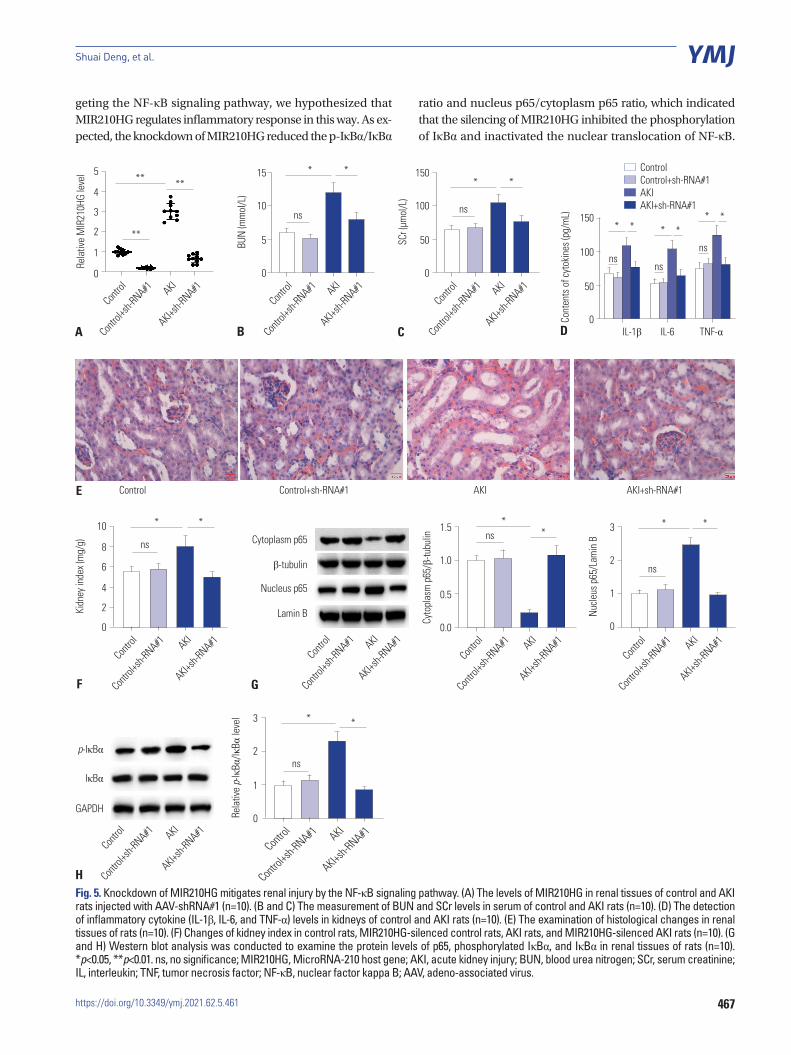
Knockdown of MIR210HG mitigates renal injury by targeting the NF-κB signaling pathwayWe then further investigated the role of MIR210HG in animals. RT-qPCR analysis showed that MIR210HG was significantly upregulated in renal tissues of AKI rats. MIR210HG was effec-tively knocked down by the injection of AAV-sh-MIR210HG in renal tissues of control rats and AKI rats (Fig. 5A). As shown in Fig. 5B and C, BUN and SCr levels were higher in AKI rats than in control rats. Knockdown of MIR210HG triggered a signifi-cant decrease of BUN and SCr levels in the serum of AKI rats.
Levels of inflammatory cytokines were higher in kidneys of AKI rats than that of control rats, and MIR210HG silencing reduced the concentrations of inflammatory cytokines (Fig. 5D). In addition, H&E staining assay revealed that the distorted re-nal tubules as well as apoptotic or necrotic phenotypes of re-nal tubular epithelial cells were observed in the AKI group, and MIR210HG silencing mitigated these histological changes (Fig. 5E). The kidney index (kidney/body weight) was in-creased in AKI rats, and MIR210HG silencing reduced the kid-ney index in AKI rats (Fig. 5F). Subsequently, we found that the
A
Cytoplasm p65
β-tubulin
Nucleus p65
Lamin B
HKC-8
Contro
l
LPSLPS
+sh-N
CLPS
+sh-RNA#1
1.5
1.0
0.5
0.0
Cyto
plas
m p
65/-β
-tubu
lin
Contro
l
LPSLPS
+sh-N
CLPS
+sh-RNA#1
*
**
B
p-IκBα
IκBα
GAPDH
Contro
l
LPSLPS
+sh-N
CLPS
+sh-RNA#1
1.5
1.0
0.5
0.0
Rela
tive
IκBα
pro
tein
leve
l
Contro
l
LPSLPS
+sh-N
CLPS
+sh-RNA#1
6
4
2
0
Rela
tive p-
IκBα
pro
tein
leve
l
Contro
l
LPSLPS
+sh-N
CLPS
+sh-RNA#1
***
5
4
3
2
1
0
Nucle
us p
65/L
amin
B
Contro
l
LPSLPS
+sh-N
CLPS
+sh-RNA#1
***
NF-κB p65
DAPI
Merged
Control LPS LPS+sh-NC LPS+sh-RNA#1
CFig. 3. MIR210HG silencing inactivated the NF-κB signaling pathway. (A and B) The western blot analysis was conducted to examine the protein lev-els of p65, phosphorylated IκBα, and IκBα in HKC-8 cells. (C) The immunofluorescent staining assay was used to analyze the nuclear translocation of NF-κB in HKC-8 cells. *p<0.05, **p<0.01. MIR210HG, MicroRNA-210 host gene; LPS, lipopolysaccharide; IL, interleukin; TNF, tumor necrosis factor; NF-κB, nuclear factor kappa B.
466
Role of MIR210HG in Sepsis Induced AKI
https://doi.org/10.3349/ymj.2021.62.5.461
nuclear translocation of p65 and phosphorylation of IκBα were promoted in kidneys of AKI rats. MIR210HG silencing inhibit-ed the nuclear translocation of p65 and the phosphorylation of IκBα (Fig. 5G and H).
DISCUSSION
A number of risk factors, such as endothelial dysfunction, in-trarenal hemodynamic changes, and inflammatory cell infil-tration of renal parenchyma, are involved in the progression of sepsis-mediated renal injury.25,26 In the advanced stage of sep-sis, tissue perfusion and microcirculatory blood flow are inhib-ited. The failure of microcirculation and mitochondrial damage of renal tubular epithelial cell may exacerbate renal tubular ep-ithelial cell injury.3,27 Numerous studies have shown that in-flammatory response and anti-inflammatory response are strongly associated with the development of sepsis-induced renal injury.3,28,29 Additionally, the levels of cytokines are relat-ed to the severity of septic AKI. Inflammatory cytokines can directly inhibit cell viability and further exacerbate sepsis-in-duced AKI.30 Therefore, further exploration of mechanisms underlying the sepsis-induced AKI is urgently needed. In the present study, HKC-8 cells were treated with LPS to mimic the
cell injury in AKI. We found that LPS treatment significantly in-hibited cell viability and levels of proinflammatory factors such as TNF-α, IL-6, and IL-1β. These results revealed that LPS can induce cell injury, which was significantly associated with in-flammatory response. A previous study showed that MIR210HG is upregulated in hypoxia or cytokine treated HKC-8 cells.18
Several lncRNAs were reported to modulate inflammatory response in sepsis-induced AKI. Downregulation of lncRNA taurine-upregulated gene 1 contributes to inflammatory re-sponse and promotes cytokine secretion during the develop-ment of sepsis-induced AKI.24 Moreover, lncRNA plasmacyto-ma variant translocation 1 aggravates LPS-induced septic AKI by regulating TNF-α and cell viability.23 In the present study, the knockdown of MIR210HG promoted cell viability and inhibited inflammatory response.
The NF-κB signaling pathway is a critical regulator of inflam-matory response in sepsis-induced AKI.22,31 Treatment of LPS contributes to the activation of IκBα phosphorylation by bind-ing with related receptors. The phosphorylated IκBα can be ubiquitinated and degraded. As a result, NF-κB is released from the cytoplasmic NF-κB/IκBα complex. Then, the cytoplasmic NF-κB is translocated into the nucleus rapidly, ultimately initi-ating the transcription of downstream target genes.32,33 Given that various lncRNAs regulate inflammatory response by tar-
Fig. 4. MIR210HG overexpression promotes inflammatory response by activating the NF-κB signaling pathway. (A) The overexpression efficacy of pcDNA3.1/MIR210HG was evaluated by RT-qPCR. (B) The effects of MIR210HG and QNZ on NF-κB nuclear translocation were evaluated by immuno-fluorescent staining assay. (C) The effects of MIR210HG and QNZ on levels of inflammatory cytokines (IL-1β, IL-6, and TNF-α). (D) The effects of MIR210HG and QNZ on HKC-8 cell viability. *p<0.05. MIR210HG, MicroRNA-210 host gene; IL, interleukin; TNF, tumor necrosis factor; NF-κB, nuclear factor kappa B.
10
8
6
4
2
0
1.5
1.0
0.5
0.0
8
6
4
2
0
pcDNA3.1 pcD
NA3.1
pcDNA3.1
pcDNA3.1
MIR210H
GMIR21
0HG
MIR210H
G
MIR210H
G
MIR210H
G+QNZ
MIR210H
G+QNZ
MIR210H
G+QNZRe
lativ
e ex
pres
sion
of M
IR21
0HG
Cyto
plas
m p
65/β
-tubu
lin
Nucle
us p
65/L
amin
B
A B
HKC-8 (LPS) HKC-8 (LPS)
Cytoplasm p65
β-tubulin
Nucleus p65
Lamin B
* *
*
*
pcDNA3.1
MIR210H
G
MIR210H
G+QNZ
80
60
40
20
0
Cell
viabi
lity (
%)
**
C D
800
600
400
200
0The
cont
ents
of c
ytok
ines
(pg/
mL)
IL-1β IL-6 TNF-α
* * * * * *
pcDNA3.1 MIR210HG MIR210HG+QNZ
HKC-8 (LPS) HKC-8 (LPS)
*
467
Shuai Deng, et al.
https://doi.org/10.3349/ymj.2021.62.5.461
geting the NF-κB signaling pathway, we hypothesized that MIR210HG regulates inflammatory response in this way. As ex-pected, the knockdown of MIR210HG reduced the p-IκBα/IκBα
ratio and nucleus p65/cytoplasm p65 ratio, which indicated that the silencing of MIR210HG inhibited the phosphorylation of IκBα and inactivated the nuclear translocation of NF-κB.
5
4
3
2
1
0
15
10
5
0
150
100
50
0
ns ns
nsns
nsRela
tive
MIR
210H
G le
vel
BUN
(mm
ol/L
)
SCr (
µmol
/L)
A
E
B C
***
**
***
**
Contro
l
Contro
l
Contro
l
Contro
l+sh-R
NA#1
Contro
l+sh-R
NA#1
Contro
l+sh-R
NA#1AKI
AKI
AKI
AKI+sh-R
NA#1
AKI+sh-R
NA#1
AKI+sh-R
NA#11.5
1.0
0.5
0.0
3
2
1
0
10
8
6
4
2
0
ns
ns
ns
Cyto
plas
m p
65/β
-tubu
lin
Nucle
us p
65/L
amin
B
Kidn
ey in
dex (
mg/
g)
GF
* ***
**
Contro
l
Contro
l
Contro
l
Contro
l
Contro
l+sh-R
NA#1
Contro
l+sh-R
NA#1
Contro
l+sh-R
NA#1
Contro
l+sh-R
NA#1 AKI
AKI
AKI
AKI
AKI+sh-R
NA#1
AKI+sh-R
NA#1
AKI+sh-R
NA#1
AKI+sh-R
NA#1
Cytoplasm p65
β-tubulin
Nucleus p65
Lamin B
Control Control+sh-RNA#1 AKI AKI+sh-RNA#1
3
2
1
0
ns
Rela
tive p-
IκBα
/IκBα
leve
l * *
Control
Control
+sh-RNA#1 AKI
AKI+sh-R
NA#1
Contro
l
Contro
l+sh-R
NA#1 AKIAKI+s
h-RNA#1
p-IκBα
IκBα
GAPDH
H
D
150
100
50
0Cont
ents
of c
ytok
ines
(pg/
mL)
IL-1β IL-6 TNF-α
* **
* **
Control Control+sh-RNA#1 AKI AKI+sh-RNA#1
Fig. 5. Knockdown of MIR210HG mitigates renal injury by the NF-κB signaling pathway. (A) The levels of MIR210HG in renal tissues of control and AKI rats injected with AAV-shRNA#1 (n=10). (B and C) The measurement of BUN and SCr levels in serum of control and AKI rats (n=10). (D) The detection of inflammatory cytokine (IL-1β, IL-6, and TNF-α) levels in kidneys of control and AKI rats (n=10). (E) The examination of histological changes in renal tissues of rats (n=10). (F) Changes of kidney index in control rats, MIR210HG-silenced control rats, AKI rats, and MIR210HG-silenced AKI rats (n=10). (G and H) Western blot analysis was conducted to examine the protein levels of p65, phosphorylated IκBα, and IκBα in renal tissues of rats (n=10). *p<0.05, **p<0.01. ns, no significance; MIR210HG, MicroRNA-210 host gene; AKI, acute kidney injury; BUN, blood urea nitrogen; SCr, serum creatinine; IL, interleukin; TNF, tumor necrosis factor; NF-κB, nuclear factor kappa B; AAV, adeno-associated virus.
468
Role of MIR210HG in Sepsis Induced AKI
https://doi.org/10.3349/ymj.2021.62.5.461
In contrast, MIR210HG overexpression activated the nuclear translocation of NF-κB. However, this result was rescued by the treatment of QNZ. The rescue assays suggested that the effects of MIR210HG on cell viability and inflammatory response were counteracted by QNZ treatment, which further confirmed that MIR210HG regulated sepsis-induced AKI by the NF-κB signal-ing pathway. Lastly, we established an AKI rat model by CLP operation. MIR210HG was upregulated in the renal tissues of CLP rats. Importantly, MIR210HG promoted the levels of pro-inflammatory cytokines in vivo.
MIR210HG can upregulate the expression of STAT3 and downregulate the expression of miR-874.34 STAT3 was widely reported to induce kidney injury.35-37 MiR-874 was reported to alleviate renal injury38 and reduce high glucose induced in-flammatory response of human renal tubular epithelial cells.39
In conclusion, for the first time, this study demonstrated that MIR210HG promotes LPS-induced inflammatory response of proximal tubular epithelial cells by targeting the NF-κB signal-ing pathway. This novel discovery may be helpful for the im-provement of sepsis-induced AKI. However, our study had some limitations. First, the regulatory mechanisms of MIR210HG on the NF-κB signaling were not investigated. Second, the effects of MIR210HG on other cells, such as HK-2 and endothelial cells, remain to be explored.
AUTHOR CONTRIBUTIONS
Conceptualization: Shuai Deng. Data curation: all authors. Formal analysis: Shuai Deng. Investigation: Shuai Deng, Bin Gu, and Zheng Yu. Methodology: Shuai Deng and Bin Gu. Project administration: Shuai Deng. Resources: Shuai Deng and Zheng Yu. Software: Shuai Deng and Zhen Shen. Supervision: Shuai Deng. Validation: Shuai Deng. Visualization: Shuai Deng. Writing—original draft: all authors. Writing—review & editing: all authors. Approval of final manuscript: all authors.
ORCID iDs
Shuai Deng https://orcid.org/0000-0002-6045-2872Bin Gu https://orcid.org/0000-0003-3180-4258Zheng Yu https://orcid.org/0000-0002-9609-3760Zhen Shen https://orcid.org/0000-0003-1228-5632Houwei Ren https://orcid.org/0000-0002-4853-3629
REFERENCES
1. Poston JT, Koyner JL. Sepsis associated acute kidney injury. BMJ 2019;364:k4891.
2. Romagnoli S, Ricci Z, Ronco C. CRRT for sepsis-induced acute kidney injury. Curr Opin Crit Care 2018;24:483-92.
3. Zarbock A, Gomez H, Kellum JA. Sepsis-induced acute kidney in-jury revisited: pathophysiology, prevention and future therapies. Curr Opin Crit Care 2014;20:588-95.
4. Umbro I, Gentile G, Tinti F, Muiesan P, Mitterhofer AP. Recent ad-vances in pathophysiology and biomarkers of sepsis-induced acute kidney injury. J Infect 2016;72:131-42.
5. Wang P, Huang J, Li Y, Chang R, Wu H, Lin J, et al. Exogenous car-bon monoxide decreases sepsis-induced acute kidney injury and inhibits NLRP3 inflammasome activation in rats. Int J Mol Sci 2015; 16:20595-608.
6. Jarroux J, Morillon A, Pinskaya M. History, discovery, and classifi-cation of lncRNAs. Adv Exp Med Biol 2017;1008:1-46.
7. St Laurent G, Wahlestedt C, Kapranov P. The landscape of long noncoding RNA classification. Trends Genet 2015;31:239-51.
8. Brandenburger T, Salgado Somoza A, Devaux Y, Lorenzen JM. Noncoding RNAs in acute kidney injury. Kidney Int 2018;94:870-81.
9. Lorenzen JM, Thum T. Long noncoding RNAs in kidney and car-diovascular diseases. Nat Rev Nephrol 2016;12:360-73.
10. Wang J, Song J, Li Y, Shao J, Xie Z, Sun K. Down-regulation of Ln-cRNA CRNDE aggravates kidney injury via increasing MiR-181a-5p in sepsis. Int Immunopharmacol 2020;79:105933.
11. Chen Y, Qiu J, Chen B, Lin Y, Chen Y, Xie G, et al. Long non-coding RNA NEAT1 plays an important role in sepsis-induced acute kid-ney injury by targeting miR-204 and modulating the NF-κB path-way. Int Immunopharmacol 2018;59:252-60.
12. Chen H, Wang X, Yan X, Cheng X, He X, Zheng W. LncRNA MALAT1 regulates sepsis-induced cardiac inflammation and dys-function via interaction with miR-125b and p38 MAPK/NFκB. Int Immunopharmacol 2018;55:69-76.
13. Wu H, Wang J, Ma Z. Long noncoding RNA HOXA-AS2 mediates microRNA-106b-5p to repress sepsis-engendered acute kidney injury. J Biochem Mol Toxicol 2020;34:e22453.
14. Wang Y, Li W, Chen X, Li Y, Wen P, Xu F. MIR210HG predicts poor prognosis and functions as an oncogenic lncRNA in hepatocellu-lar carcinoma. Biomed Pharmacother 2019;111:1297-301.
15. Kang X, Kong F, Huang K, Li L, Li Z, Wang X, et al. LncRNA MIR210HG promotes proliferation and invasion of non-small cell lung cancer by upregulating methylation of CACNA2D2 promoter via binding to DNMT1. Onco Targets Ther 2019;12:3779-90.
16. Rajan KS, Ramasamy S, George-William JN, Rajendhran J. Emerg-ing cardiac non-coding landscape: the importance of meta-anal-ysis. Biochimie 2017;133:87-94.
17. Min W, Dai D, Wang J, Zhang D, Zhang Y, Han G, et al. Long non-coding RNA miR210HG as a potential biomarker for the diagnosis of glioma. PLoS One 2016;11:e0160451.
18. Lin J, Zhang X, Xue C, Zhang H, Shashaty MG, Gosai SJ, et al. The long noncoding RNA landscape in hypoxic and inflammatory re-nal epithelial injury. Am J Physiol Renal Physiol 2015;309:F901-13.
19. Shi M, Zeng X, Guo F, Huang R, Feng Y, Ma L, et al. Anti-inflam-matory pyranochalcone derivative attenuates LPS-induced acute kidney injury via inhibiting TLR4/NF-κB pathway. Molecules 2017; 22:1683.
20. Zhang Z, Zhao H, Ge D, Wang S, Qi B. β-casomorphin-7 amelio-rates sepsis-induced acute kidney injury by targeting NF-κB path-way. Med Sci Monit 2019;25:121-7.
21. Li YM, Zhang J, Su LJ, Kellum JA, Peng ZY. Downregulation of TIMP2 attenuates sepsis-induced AKI through the NF-κB path-way. Biochim Biophys Acta Mol Basis Dis 2019;1865:558-69.
22. Zhang F, Lu S, Jin S, Chen K, Li J, Huang B, et al. Lidanpaidu pre-scription alleviates lipopolysaccharide-induced acute kidney in-jury by suppressing the NF-κB signaling pathway. Biomed Phar-macother 2018;99:245-52.
23. Huang W, Lan X, Li X, Wang D, Sun Y, Wang Q, et al. Long non-coding RNA PVT1 promote LPS-induced septic acute kidney in-jury by regulating TNFα and JNK/NF-κB pathways in HK-2 cells. Int Immunopharmacol 2017;47:134-40.
24. Zhang LH, Wang Z, Li LH, Liu YK, Jin LF, Qi XW, et al. Vestigial like family member 3 is a novel prognostic biomarker for gastric can-
469
Shuai Deng, et al.
https://doi.org/10.3349/ymj.2021.62.5.461
cer. World J Clin Cases 2019;7:1954-63.25. Gómez H, Kellum JA. Sepsis-induced acute kidney injury. Curr
Opin Crit Care 2016;22:546-53.26. Tang Y, Wang C, Wang Y, Zhang J, Wang F, Li L, et al. Isoliquiriti-
genin attenuates LPS-induced AKI by suppression of inflamma-tion involving NF-κB pathway. Am J Transl Res 2018;10:4141-51.
27. Sureshbabu A, Patino E, Ma KC, Laursen K, Finkelsztein EJ, Ak-churin O, et al. RIPK3 promotes sepsis-induced acute kidney in-jury via mitochondrial dysfunction. JCI Insight 2018;3:e98411.
28. Mårtensson J, Bellomo R. Sepsis-induced acute kidney injury. Crit Care Clin 2015;31:649-60.
29. Wang K, Xie S, Xiao K, Yan P, He W, Xie L. Biomarkers of sepsis-induced acute kidney injury. Biomed Res Int 2018;2018:6937947.
30. Ferrè S, Deng Y, Huen SC, Lu CY, Scherer PE, Igarashi P, et al. Re-nal tubular cell spliced X-box binding protein 1 (Xbp1s) has a unique role in sepsis-induced acute kidney injury and inflamma-tion. Kidney Int 2019;96:1359-73.
31. Zhou Y, Xu W, Zhu H. CXCL8(3-72) K11R/G31P protects against sepsis-induced acute kidney injury via NF-κB and JAK2/STAT3 pathway. Biol Res 2019;52:29.
32. Lu SJ, Xu JH, He ZF, Wu P, Ning C, Li HY. Innate immune molecule surfactant protein D attenuates sepsis-induced acute kidney in-jury through modulating apoptosis and NFκB-mediated inflam-
mation. Int Wound J 2020;17:100-6.33. Barnes PJ. Nuclear factor-κB. Int J Biochem Cell Biol 1997;29:867-
70.34. Bu L, Zhang L, Tian M, Zheng Z, Tang H, Yang Q. LncRNA
MIR210HG facilitates non-small cell lung cancer progression through directly regulation of miR-874/STAT3 axis. Dose Response 2020;18:1559325820918052.
35. Zheng S, Liu J, Zhao Z, Song R. Role of STAT3/mTOR pathway in chronic kidney injury. Am J Transl Res 2020;12:3302-10.
36. Zhang L, Lu P, Guo X, Liu T, Luo X, Zhu YT. Inhibition of JAK2/STAT3 signaling pathway protects mice from the DDP-induced acute kidney injury in lung cancer. Inflamm Res 2019;68:751-60.
37. Sun S, Wang J, Wang J, Wang F, Yao S, Xia H. Maresin 1 mitigates sepsis-associated acute kidney injury in mice via inhibition of the NF-κB/STAT3/MAPK pathways. Front Pharmacol 2019;10:1323.
38. Yao T, Zha D, Gao P, Shui H, Wu X. MiR-874 alleviates renal injury and inflammatory response in diabetic nephropathy through tar-geting toll-like receptor-4. J Cell Physiol 2018;234:871-9.
39. Lin Y, Zhong L, Li H, Xu Y, Li X, Zheng D. Psoralen alleviates high glucose-induced HK-2 cell injury by inhibition of Smad 2 signal-ing via upregulation of microRNA 874. BMC Pharmacol Toxicol 2020;21:52.
Related Documents